


Bahasa
Halaman
Hukum

Long-Residency Hydration, Cation Binding, and Dynamics of LoopE/Helix IV rRNA-L25 Protein Complex
Kamila Reblova,* Nad’a Spackova,y Jaroslav Koca,* Neocles B. Leontis,z and Jirı Sponery§
*National Centre for Biomolecular Research, Faculty of Sciences, Masaryk University, Kotlarska 2, 61137 Brno, Czech Republic;yInstitute of Biophysics, Academy of Sciences of the Czech Republic, Kralovopolska 135, 61265 Brno, Czech Republic; zChemistryDepartment and Center for Biomolecular Sciences, Bowling Green State University, Bowling Green, Ohio 43403; and §Instituteof Organic Chemistry and Biochemistry, Academy of Sciences of the Czech Republic, Flemingovo n. 2, 16610 Prague, Czech Republic
ABSTRACT Molecular dynamics simulations of RNA-protein complex between Escherichia coli loop E/helix IV (LE/HeIV)rRNA and L25 protein reveal a qualitative agreement between the experimental and simulated structures. The major groove ofLE is a prominent rRNA cation-binding site. Divalent cations rigidify the LE major groove geometry whereas in the absence ofdivalent cations LE extensively interacts with monovalent cations via inner-shell binding. The HeIV region shows bistability of itsmajor groove explaining the observed differences between x-ray and NMR structures. In agreement with the experiments, thesimulations suggest that helix-a1 of L25 is the least stable part of the protein. Inclusion of Mg21 cations into the simulationscauses perturbation of basepairing at the LE/HeIV junction, which does not, however, affect the protein binding. The rRNA-protein complex is mediated by a number of highly specific hydration sites with long-residing water molecules and two of themare bound throughout the entire 24-ns simulation. Long-residing water molecules are seen also outside the RNA-protein contactareas with water-binding times substantially enhanced compared to simulations of free RNA. Long-residency hydration sitesthus represent important elements of the three-dimensional structure of rRNA.
INTRODUCTION
The recently determined atomic-resolution structures of
ribosomal subunits confirm that the ribosomal RNA (rRNA)
molecules (5S, 16S, and 23S) comprise short regions formed
by Watson-Crick basepairs and so-called RNA motifs, i.e.,
specific non-Watson-Crick basepaired regions (Ban et al.,
2000; Wimberly et al., 2000). Recurrent, modular RNA
motifs represent key structural elements in the ribosome
(Leontis and Westhof, 1998b, 2003; Moore, 1999). Studies
of RNA motifs and their molecular interactions in the
ribosome are thus important to understand fundamental
aspects of ribosomal structure and function. Available crystal
structures of ribosomal subunits (Ban et al., 2000; Harms
et al., 2001; Wimberly et al., 2000; Yusupov et al., 2001) and
smaller RNA-protein complexes (Agalarov et al., 2000; Lu
and Steitz, 2000; Nikulin et al., 2003; Perederina et al., 2002;
Wimberly et al., 1999) reveal a wide repertoire of different
types of molecular interactions.
Modern computational methods represent an important
tool that can complement experiments and provide additional
information about structure, dynamics, and molecular
recognition of nucleic acids and proteins (Auffinger and
Westhof, 1998, 1999; Beaurain and Laguerre, 2003; Correll
et al., 1997; Hermann et al., 1998; Cheatham and Young,
2000; Norberg and Nilsson, 2002; Orozco et al., 2003;
Reblova et al., 2003a,b; Sarzynska et al., 2000; Schneider
et al., 2001; Zacharias, 2000). In the previous study (Reblova
et al., 2003b) we have investigated the 5S rRNA loop E (LE)
secondary motif of Escherichia coli and spinach chloroplast
utilizing molecular dynamics (MD) simulations with explicit
inclusion of solvent and counterions. The bacterial loop E
forms a unique duplex architecture due to seven consecutive
non-Watson-Crick basepairs and is involved in both RNA-
RNA and RNA-protein interactions. The simulations indicate
that the LE could serve as a rigid docking segment recognized
by other ribosomal elements. LE has a unique capability to
extensively bind monovalent and divalent cations in the deep
major groove (Auffinger et al., 2003, 2004a; Lu and Steitz,
2000). We employed RNA motif analysis and simulations to
predict that the bacterial and spinach chloroplast LE regions,
despite pronounced sequence variability, adopt nearly
isosteric geometries (Leontis and Westhof, 1998a), which is
also confirmed by independent NMR study (Vallurupalli and
Moore, 2003). The simulations suggest that the unique LE
architecture is complemented by several highly specific
hydration sites with long-residency water molecules that are
not seen in regular RNA duplexes (Reblova et al., 2003b).
Ribosomal RNA-protein complexes are essential in many
biological processes and are intensely studied by experimen-
tal and computational methods. The complex of loop E and
the ribosomal L25 protein is one of the best-characterized
RNA-protein complexes (Lu and Steitz, 2000; Stoldt et al.,
1999). The biological role of L25 is yet to be established. The
L25 protein does not appear to be conserved in all bacterial
ribosomes and has no counterpart in archaea or eukarya
(Nevskaya et al., 2000). On the other hand its extensive
interactions with 5S rRNA LE region and with the adjacent
helix IV (HeIV) of 5S rRNA are quite specific indicating that
it has some biological roles. Furthermore, such specificSubmitted June 4, 2004, and accepted for publication August 26, 2004.
Address reprint requests to Jirı Sponer, E-mail: [email protected].
muni.cz.
� 2004 by the Biophysical Society
0006-3495/04/11/3397/16 $2.00 doi: 10.1529/biophysj.104.047126
Biophysical Journal Volume 87 November 2004 3397–3412 3397

molecular interactions represent a challenge for structural
studies. Although there is no x-ray structure available for the
large ribosomal subunit of E. coli, our preliminary docking
using x-ray structure of the 50S subunit of Haloarculamarismortui (not shown) in fact suggests that the second
A-stack region of bacterial loop E could interact with the
A-site finger in 23S rRNA whereas the first A-stack interacts
with L25 and is outside the ribosome. Then L25 could, for
example, play a role in supporting the E. coli loop E (keeping
it well organized) so that it can interact with the A-site finger
and couple motions of the L9 domain with the A-site finger.
Both L9 domain and A-site finger make bridges to the 30S
subunit and move during translocation. It is notable that 5S
rRNA has been implicated in translocation.
The non-Watson-Crick LE basepairs provide a highly
sequence-specific hydrogen-bonding surface in the minor
groove recognized by the L25 protein. Furthermore, the non-
Watson-Crick basepairs confer considerable plasticity to the
RNA helix, which becomes more accessible to the protein.
The structure of the bacterial complex of loop E/helix IV and
ribosomal L25 protein (LE/HeIV-L25) has been determined
by NMR spectroscopy and x-ray crystallography (Lu and
Steitz, 2000; Stoldt et al., 1999). Free LE/HeIV rRNA and
free L25 protein have also been studied (Correll et al., 1997;
Stoldt et al., 1998). LE and HeIV establish distinct
interactions with L25, as evidenced by both x-ray and
NMR complexes (Figs. 1–3). The bacterial LE motif
contains two A104-A73 and A99-A78 cross-strand adenine
stacks (Fig. 1 a) that significantly distort the sugar-phosphatebackbone and narrow the LE major groove. The adjacent
HeIV contains two wobble basepairs forming G96-G81
cross-strand guanine stack (Fig. 1 a). The major (deep)
groove width of HeIV is enlarged relatively to Watson-Crick
A-RNA duplex due to A99-A78 and G96-G81 cross-strand
purine stacks.
The x-ray structures of the LE/HeIV-L25 complex
(Nucleic Acid Database (NDB) code, PR0018) and of the
free LE/HeIV fragment (NDB code, URL069) reveal several
magnesium ions bound via inner-shell and outer-shell
binding (Correll et al., 1997; Lu and Steitz, 2000). As
discussed recently, magnesium cations can be occasionally
misassigned in experimental structures and some of them
may correspond to anion-binding positions, water molecules,
or two closely spaced cations representing a single alternat-
ing binding site (Auffinger et al., 2004b). Qualitative
geometrical criteria to identify such suspicious binding sites
were suggested (Auffinger et al., 2004b). Based on these
criteria the x-ray structure of the complex does not show any
Mg21 ions that could correspond to anions but there are two
ions that likely represent a single species. The x-ray structure
of the free RNA reveals one suspicious Mg21 ion (binding
site at G100) in the minor groove of LE (see below).
The L25 protein is composed of a seven-stranded closed
b-barrel (strands b1–b7) and three a-helices (a1–a3) (Figs.1 b and 2) (Lu and Steitz, 2000; Stoldt et al., 1999). The
strands b2, b3, b6, b7, and the helix-a1 interact with the LE/HeIV fragment (Fig. 2) via two distinct contact areas. While
the antiparallel b-ribbons (b2, b3, b6, b7) interact with the
minor groove of LE (LE contact area), the N-terminal tip of
the helix-a1 interacts with the widened major groove of
HeIV (Lu and Steitz, 2000; Stoldt et al., 1999) (HeIV contact
area). There are multiple direct H-bonds between amino
acids and bases in the two contact areas. The x-ray structures
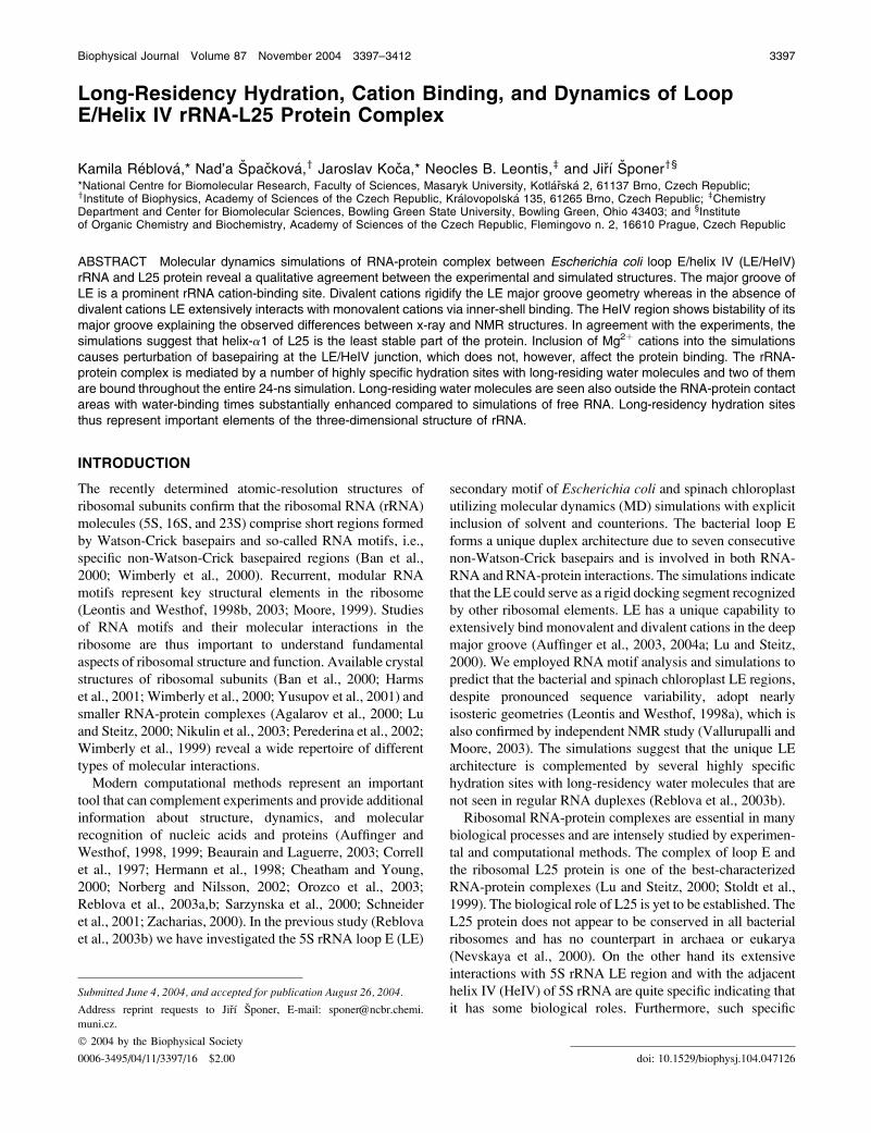
reveal that the LE/HeIV geometry is largely unaffected upon
binding of L25, with the most significant change being
a marked narrowing of the major groove of HeIV by ;5 A
(Fig. 3, a and b) (Correll et al., 1997; Lu and Steitz, 2000).
Bound LE/HeIV fragment thus shows a geometry that we
call ‘‘closed geometry’’ throughout this study whereas the
uncomplexed LE/HeIV fragment shows ‘‘open geometry’’
of the major groove of HeIV (Correll et al., 1997; Lu and
Steitz, 2000). Helix-a1 (residues 14–22) of isolated L25 is
unstructured in solution (Stoldt et al., 1998). The experi-
mental data suggest that the recognition of L25 protein by
LE/HeIV rRNA is mediated by two preformed recognition
elements, i.e., the b-sheet surface of L25 and the widened
FIGURE 1 (a) Sequence of the studied LE/HeIV RNA duplex. The HeIV
part is in shaded field; two A-stacks and one G-stack are in black boxes.
Symbols identify non-Watson-Crick basepairs (Leontis et al., 2002). (b)
Schematic arrangement of secondary structures of the ribosomal L25
protein; the numbers correspond to the individual protein residues.
FIGURE 2 Stereo view of the x-ray LE/HeIV-L25 complex (NDB code,
PR0018); secondary elements of L25 protein are marked.
3398 Reblova et al.
Biophysical Journal 87(5) 3397–3412
minor groove of LE (Stoldt et al., 1998, 1999). In the course
of intermolecular recognition, the helix-a1 is structured and
turns toward the more flexible major groove of HeIV, where
it inserts the side chains of its N-terminal tip and anchors the
structure of the complex (Fig. 2) (Stoldt et al., 1998).
Comparison of NMR and x-ray LE/HeIV-L25 complexes
reveals several congruencies and discrepancies. Differences
in experimental conditions used in the respective x-ray and
NMR studies are listed in the Supplementary Material. The
NMR structure shows several direct intermolecular H-bonds
(Asp90-G75, His88-G75, Arg9-G76) also observed in the
x-ray structure (Lu and Steitz, 2000) but there are other
potential contacts that could not be determined with certainty
(Stoldt et al., 1999). Furthermore, the x-ray structure
describes direct H-bonds between Lys14 and U80, between
Lys14 and G79, and between Gln78 and G76. The geometry
of LE predicted by NMR appears to be identical with the
x-ray structure but there is an apparent difference in NMR
and x-ray geometries of HeIV (Fig. 3, a and c). The major
groove of HeIV is widened in the NMR complex by;5–7 A
in comparison with the x-ray structure and actually
resembles the open geometry of the free x-ray LE/HeIV
fragment (Fig. 3).
We employed molecular dynamics (MD) to study the LE/
HeIV-L25 complex. Our results provide details of the
molecular interactions that explain some of the structural
differences between the NMR and x-ray structures and give
an additional detailed insight into the molecular interactions.
Specifically, the simulations suggest the crucial role of
tightly bound water molecules in ribosomal assemblies. The
residency times of water molecules in selected hydration
sites in the presently investigated complex are longer than in
any MD study reported so far for nucleic acids, being up to
two orders of magnitude longer than in common DNA and
RNA hydration sites.
MATERIALS AND METHODS
Starting structures
The E. coli ribosomal complex LE/HeIV-L25 was taken from x-ray data
(NDB code, PR0018; 1.8-A resolution) (Lu and Steitz, 2000). The packing
interactions of the molecule in the asymmetric unit between RNA and
adjacent proteins involve the top and bottom parts of the RNA, however, no
protein-protein contacts were detected. The structure contains 94 residues of
the L25 protein, 36 residues of the LE/HeIV fragment, and five Mg21 ions.
Four terminal unpaired bases at the ends were omitted. We carried out two
simulations of the LE/HeIV-L25 complex, ‘‘COM1’’ (24 ns) with inclusion
of five Mg21 ions and ‘‘COM2’’ (11 ns) in the absence of Mg21 ions (Table
1). Furthermore, we performed simulations of the individual components of
the complex starting again from the same crystal structure. The LE/HeIV
fragment was simulated with five Mg21 ions (simulation ‘‘RNA1’’; 14.5 ns)
and the L25 protein was simulated for 10 ns (simulation ‘‘PROT1’’) (Table
1). A further simulation of the L25 protein ‘‘PROT2’’ (19 ns) was run at
elevated temperature (400 K) to enhance the sampling. Moreover, the
uncomplexed LE/HeIV fragment was also taken from the x-ray data (NDB
code, URL069; 3.0-A resolution) and was simulated for 18 ns (simulation
FIGURE 3 Experimental structures of the LE/HeIV-
L25 complex and the free LE/HeIV fragment. (a)
X-rayLE/HeIV-L25complexwithnarrowmajorgroove
of HeIV (closed geometry) (NDB code, PR0018). (b)
X-ray structure of free LE/HeIV fragment with wider
major groove of HeIV (open geometry) (NDB code,
URL069). (c) NMR LE/HeIV-L25 complex with wider
major groove of HeIV (open geometry) (Protein Data
Bank (PDB) code, 1D6K). HeIV is inside the black
box.
TABLE 1 Summary of the simulations carried out in this article
Starting structure
(NDB code)
Simulated
system
Name of the
simulation
Length of the
simulation (ns)
Presence
of ions
PR0018 LE/HeIV-L25 complex COM1 24 5 Mg21, 15 Na1
PR0018 LE/HeIV-L25 complex COM2 11 25 Na1
PR0018 L25 protein PROT1 10 5 Cl�
PR0018 L25 protein PROT2* 19 5 Cl�
PR0018 LE/HeIV RNA RNA1 14.5 5 Mg21, 20 Na1
URL069 LE/HeIV RNA RNA2 18 5 Mg21, 20 Na1
*Simulation at 400 K.
MD Simulations of LE rRNA-L25 Complex 3399
Biophysical Journal 87(5) 3397–3412
‘‘RNA2’’) with five Mg21 ions adopting a similar arrangement as in the LE/
HeIV-L25 complex (Correll et al., 1997). The sequence of the uncomplexed
LE/HeIV fragment used in the simulation was identical to the LE/HeIV
fragment in the complex (Fig. 1 a).The simulations were carried out using the AMBER-6.0 program
(Pearlman et al., 1995) with the Cornell et al. force field (Cornell et al.,
1995). The RNAmolecules were neutralized by sodium counterions initially
placed by the Xleap module of AMBER-6.0 at the most negative positions
close to the RNA whereas Mg21 ions were placed based on the x-ray
structures. In case of the low-resolution x-ray structure of the free LE/HeIV
fragment, the x-ray Mg21 cation distribution was somewhat modified, to
make it similar to the distribution seen in the complex. The x-ray structure of
the free LE/HeIV fragment contains eight Mg21 ions, six of them bound in
the area of LE/HeIV. Five Mg21 ions bound in the major groove of LE and
HeIV were rearranged to mimic the Mg21-binding positions in the LE/
HeIV-L25 x-ray structure. Nevertheless, as seen in Table 2, we could not
achieve identical initial ion placement due to difference in starting solute
geometries. As demonstrated below, it has some impact on the simulated
structures. The sixth Mg21 ion that is bound in the minor groove of LE at
G100 was omitted. This cation can actually be mislabeled and may even
correspond to an anion-binding position, as discussed in the literature
(Auffinger et al., 2004b). The remaining two divalent cations not interacting
with the major groove were also omitted.
Cl� ions were used in simulations PROT1 and PROT2 for neutralization
of the protein and their initial positions were suggested by Xleap. The
following parameters were used: Na1 radius 1.868 A and well depth
0.00277 kcal/mol, Mg21 radius 0.7926 A and well depth 0.8947 kcal/mol,
and Cl� radius 1.948 A and well depth 0.265 kcal/mol (Ross and Hardin,
1994). The limitations imposed by the simple force-field description of ions
were briefly discussed in our preceding study (Reblova et al., 2003b). The
polar hydrogens of the protein were added and protonation states of all
histidines in the LE/HeIV-L25 complex were set to allow formation of
proper H-bonds. Thus all three histidines were (d protonated (Amber code
HID)). Crystal water molecules were used in the simulation start and a cubic
box of the TIP3P water molecules was added around the RNA to a depth of
12 A on each side of the solute. The Sander module of AMBER-6.0 was
used for all minimizations and molecular dynamics simulations. All residues
were restrained by force constants, which were gradually reduced from 500
to 0 kcal/mol in the course of 5000 steps while the rest of the system was
allowed to relax. The systems were then heated from 50 K up to 300 K in
100 ps. The production runs were carried out at 300 K with constant-
pressure boundary conditions and the particle-mesh Ewald (PME) method
using Berendsen temperature coupling algorithm (with a time constant of 0.2
ps) (York et al., 1993). One simulation (see above) was carried out at
elevated temperature (400 K) to enhance sampling. For this simulation, the
system was gradually heated from 300 K to 400 K during the first 100 ps
using NPT conditions (constant pressure ensemble). The production run was
then continued at 400 K using NVT (constant volume ensemble). The NVT
simulations imply high pressure that may artificially stabilize the structure
(Zhou et al., 2001). The outcome of the elevated temperature simulations is
also limited by the fact that at elevated temperature the system is not
represented by a Boltzmann distribution equivalent to prolongation of a room
temperature simulation. Nonetheless, elevated temperature simulations often
provide insights into labile parts of the simulated structures. The center of
mass velocity was periodically removed during the production dynamics
(Harvey et al., 1998). Trajectories were analyzed using the Carnal module of
AMBER and structures were visualized using VMD (molecular visualiza-
tion program, http://www.ks.uiuc.edu/research/vmd/) (Humphrey et al.,
1996). Hydration and distribution of ions were calculated with the Ptraj
module of the AMBER-6.0 and visualized using UCSF MidasPlus
(University of California, San Francisco, CA) (Ferrin et al., 1988). Sys-
tematic monitoring of the solute-to-water distances was carried out using the
Carnal module of AMBER. All direct solute-solvent contacts were detected
during thewhole simulation and then analyzed in detail. To obtain some crude
estimate of the energetics, energy analysis was carried out using the Anal
module of AMBER to evaluate energetic contributions between individual
residues of the protein and the LE/HeIV fragment. Electrostatic (Eel) and van
der Waals (Evdw) nonbonding interactions between individual residues were
extracted from the equation describing the molecular mechanics energy:
EMM ¼ Ebond 1Eangle 1Etors 1Evdw 1Eel:
The Evdw interactions were described by Lennard-Jones potential
whereas the Eel interactions were described by Coulomb term. Evdw and
Eel interactions were computed between RNA and protein residues forming
intermolecular H-bonds and the values of energetic contributions were
averaged along the trajectory. The charges of the residues were not modified
after the dissection and the energy calculations were carried out assuming
a dielectric constant of 1. Thus, the interaction energies provide only a very
crude insight in the stability because solvent screening is not included. The
DO_DSSP module of the GROMACS-2.0 program was used for analysis of
the secondary structure elements of the protein (van der Spoel et al., 1999).
RESULTS
Molecular dynamics of the LE/HeIV-L25 complex
We have carried out 24-ns simulation of the LE/HeIV-L25
complex in the presence of Mg21 (simulation COM1). The
root-mean-square deviation (RMSD) value with respect to
the x-ray structure was 2.3 6 0.5 A (see Fig. S1 in Sup-
plementary Material). This is a very low RMSD value, con-
sidering the size of the simulated system, indicating that the
system is very close to the starting x-ray geometry and rather
rigid. The RMSD between the averaged MD structure in the
time period of 19–24 ns and the x-ray structure is;1.7 A. The
Supplementary Material section presents selected PDB files
illustrating this as well as the subsequent simulations. The
complete trajectories may be obtained from the authors upon
request.
LE and HeIV reveal distinct major groove dynamics
The geometry of LE did not show any significant changes in
the course of the simulation. Minor fluctuations of the major
groove width were observed in the range of 1–2 A relative to
the starting x-ray structure (see Table S1 in Supplementary
TABLE 2 Comparison of Mg21 positions in the course of
RNA2 and COM1 simulations (see the text)
Mg21 ion Simulation RNA2 Simulation COM1
A G105(O2P) Inner-shell
binding
G106(O6,O2P) and G105(O2P)
Outer-shell binding
B A101(O2P) Inner-shell
binding
G98(O2P), C97(O2P) and
Gln12(O) Outer-shell binding
C U74(O2P) and A99(O2P)
Inner-shell binding
G100(O2P) Inner-shell binding
D G75(O1P) Inner-shell
binding
U74(O2P), G75(O2P) and
A99(O2P) Outer-shell binding
E C93(O2P) Inner-shell
binding
U95(O4) and G96(O6) Inner-shell
binding
All Mg21 ions were stable in the course of the simulations except Mg21 ion
B in COM1 simulation. Mg21 ion B was involved in outer-shell binding to
G98(O2P), C97(O2P), and Gln12(O) after 20 ns of the simulation COM1.
3400 Reblova et al.
Biophysical Journal 87(5) 3397–3412
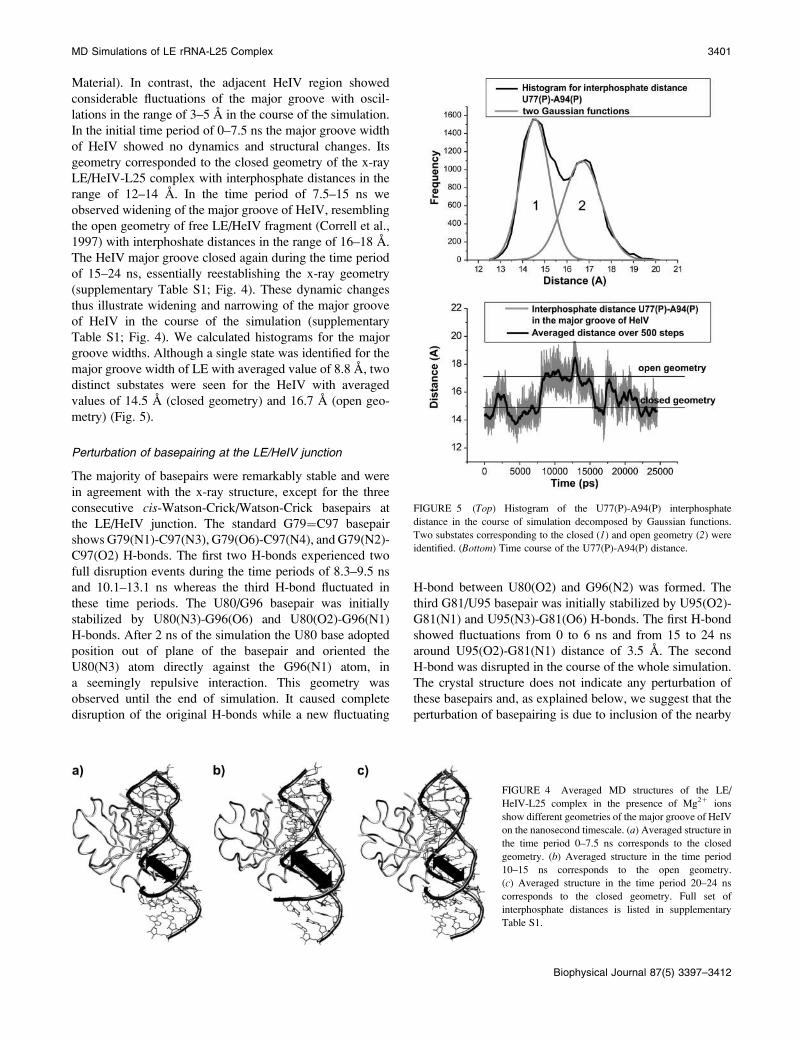
Material). In contrast, the adjacent HeIV region showed
considerable fluctuations of the major groove with oscil-
lations in the range of 3–5 A in the course of the simulation.
In the initial time period of 0–7.5 ns the major groove width
of HeIV showed no dynamics and structural changes. Its
geometry corresponded to the closed geometry of the x-ray
LE/HeIV-L25 complex with interphosphate distances in the
range of 12–14 A. In the time period of 7.5–15 ns we
observed widening of the major groove of HeIV, resembling
the open geometry of free LE/HeIV fragment (Correll et al.,
1997) with interphoshate distances in the range of 16–18 A.
The HeIV major groove closed again during the time period
of 15–24 ns, essentially reestablishing the x-ray geometry
(supplementary Table S1; Fig. 4). These dynamic changes
thus illustrate widening and narrowing of the major groove
of HeIV in the course of the simulation (supplementary
Table S1; Fig. 4). We calculated histograms for the major
groove widths. Although a single state was identified for the
major groove width of LE with averaged value of 8.8 A, two
distinct substates were seen for the HeIV with averaged
values of 14.5 A (closed geometry) and 16.7 A (open geo-
metry) (Fig. 5).
Perturbation of basepairing at the LE/HeIV junction
The majority of basepairs were remarkably stable and were
in agreement with the x-ray structure, except for the three
consecutive cis-Watson-Crick/Watson-Crick basepairs at
the LE/HeIV junction. The standard G79¼C97 basepair
shows G79(N1)-C97(N3), G79(O6)-C97(N4), and G79(N2)-
C97(O2) H-bonds. The first two H-bonds experienced two
full disruption events during the time periods of 8.3–9.5 ns
and 10.1–13.1 ns whereas the third H-bond fluctuated in
these time periods. The U80/G96 basepair was initially
stabilized by U80(N3)-G96(O6) and U80(O2)-G96(N1)
H-bonds. After 2 ns of the simulation the U80 base adopted
position out of plane of the basepair and oriented the
U80(N3) atom directly against the G96(N1) atom, in
a seemingly repulsive interaction. This geometry was
observed until the end of simulation. It caused complete
disruption of the original H-bonds while a new fluctuating
H-bond between U80(O2) and G96(N2) was formed. The
third G81/U95 basepair was initially stabilized by U95(O2)-
G81(N1) and U95(N3)-G81(O6) H-bonds. The first H-bond
showed fluctuations from 0 to 6 ns and from 15 to 24 ns
around U95(O2)-G81(N1) distance of 3.5 A. The second
H-bond was disrupted in the course of the whole simulation.
The crystal structure does not indicate any perturbation of
these basepairs and, as explained below, we suggest that the
perturbation of basepairing is due to inclusion of the nearby
FIGURE 4 Averaged MD structures of the LE/
HeIV-L25 complex in the presence of Mg21 ions
show different geometries of the major groove of HeIV
on the nanosecond timescale. (a) Averaged structure in
the time period 0–7.5 ns corresponds to the closed
geometry. (b) Averaged structure in the time period
10–15 ns corresponds to the open geometry.
(c) Averaged structure in the time period 20–24 ns
corresponds to the closed geometry. Full set of
interphosphate distances is listed in supplementary
Table S1.
FIGURE 5 (Top) Histogram of the U77(P)-A94(P) interphosphate
distance in the course of simulation decomposed by Gaussian functions.
Two substates corresponding to the closed (1) and open geometry (2) wereidentified. (Bottom) Time course of the U77(P)-A94(P) distance.
MD Simulations of LE rRNA-L25 Complex 3401
Biophysical Journal 87(5) 3397–3412
Mg21 cation. Because the perturbation occurs outside the
protein-binding area the protein binding seems to be unaf-
fected.
L25 protein in LE/HeIV-L25 complex is rigid
The protein structure was not significantly modified in the
course of the simulation compared with the x-ray starting
geometry. The instantaneous RMSD value with respect to
the x-ray structure was 2.36 0.3 A. The RMSD value of the
protein backbone (calculated only for Ca atoms) with respect
to the x-ray structure was 1.6 6 0.3 A. Secondary structures
were monitored in the course of simulation. Modest
structural change (turning) was observed for the helix-a2(see Figs. S2 and S3 in Supplementary Material). This
change was identified after 0.5 ns of the simulation and
persisted until the end of the simulation (24 ns). The helix-a2shows no contact with the LE/HeIV fragment and its modest
structural change did not affect the stability of the LE/HeIV-
L25 complex. Other secondary structure elements revealed
no structural changes.
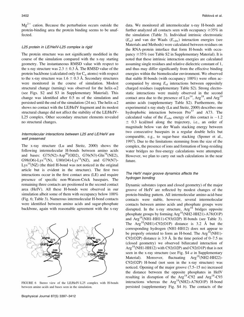
Intermolecular interactions between L25 and LE/HeIV arewell preserved
The x-ray structure (Lu and Steitz, 2000) shows the
following intermolecular H-bonds between amino acids
and bases: G75(N2)-Asp90(OD2), G76(N3)-Gln78(NE2),
G98(O6)-Lys14(N), U80(O4)-Lys14(NZ), and G79(N7)-
Lys14(NZ) (the third H-bond was not noticed in the original
article but is evident in the structure). The first two
interactions occur in the first contact area (LE) and require
presence of specific non-Watson-Crick basepairs. The
remaining three contacts are positioned in the second contact
area (HeIV). All these H-bonds were observed in our
simulation albeit some of them with occupancy below 100%
(Fig. 6; Table 3). Numerous intermolecular H-bond contacts
were identified between amino acids and sugar-phosphate
backbone, again with reasonable agreement with the x-ray
data. We monitored all intermolecular x-ray H-bonds and
further analyzed all contacts seen with occupancy $35% in
the simulation (Table 3). Individual intrinsic electrostatic
(Eel) and van der Waals (Evdw) interaction energies (see
Materials and Methods) were calculated between residues on
the RNA-protein interface that form H-bonds with occu-
pancy$35% (see Table S2 in Supplementary Material). It is
noted that these intrinsic interaction energies are calculated
assuming single residues and relative dielectric constant of 1,
and thus may differ significantly from the effective binding
energies within the biomolecular environment. We observed
that stable H-bonds (with occupancy 100%) were often ac-
companied by strong Eel interactions between oppositely
charged residues (supplementary Table S2). Strong electro-
static interactions were mainly observed in the second
contact area due to the presence of Lys14, Arg18, and Arg19
amino acids (supplementary Table S2). Furthermore, the
experimental x-ray study (Lu and Steitz, 2000) describes one
hydrophobic interaction between Pro37 and A73. The
calculated value of the Evdw energy of this contact is �1.2
6 0.3 kcal/mol along the trajectory, i.e., an order of
magnitude below van der Waals stacking energy between
two consecutive basepairs in a regular double helix but
comparable, e.g., to sugar-base stacking (Sponer et al.,
1997). Due to the limitations stemming from the size of the
complex, the presence of ions and formation of long-residing
water bridges no free-energy calculations were attempted.
However, we plan to carry out such calculations in the near
future.
The HeIV major groove dynamics affects thehydrogen bonding
Dynamic substates (open and closed geometry) of the major
groove of HeIV are reflected by modest changes of the
protein-binding pattern. All intermolecular amino-acid-base
contacts were stable, however, several intermolecular
contacts between amino acids and phosphate groups were
disrupted. In the x-ray structure, Arg18 bridges opposite
phosphate groups by forming Arg18(NH2-HH21)-A78(O1P)
and Arg18(NH1-HH11)-C93(O2P) H-bonds (see Table 3).
The Arg18(NH1)-C92(O1P) distance is 3.2 A but the
corresponding hydrogen (NH1-HH12) does not appear to
be properly oriented to form an H-bond. The Arg18(NH1)-
C92(O2P) distance is 3.9 A. In the time period of 0–7.5 ns
(closed geometry) we observed bifurcated interaction of
Arg18(NH1-HH12) with C92(O2P) and C92(O1P) that is not
seen in the x-ray structure (see Fig. S4 a in Supplementary
Material). Moreover, fluctuating Arg18(NH2-HH22)-
C92(O2P) H-bond (not seen in the x-ray structure) was
noticed. Opening of the major groove (7.5–15 ns) increased
the distance between the opposite phosphates in HeIV
resulting in disruption of the Arg18-C92 and Arg18-C93
interactions whereas the Arg18(NH2)-A78(O1P) H-bond
persisted (supplementary Fig. S4 b). The contacts of theFIGURE 6 Stereo view of the LE/HeIV-L25 complex with H-bonds
between amino acids and bases seen in the simulation.
3402 Reblova et al.
Biophysical Journal 87(5) 3397–3412
Arg18 with residues C92 and C93 were again restored after
18 ns (closed geometry).
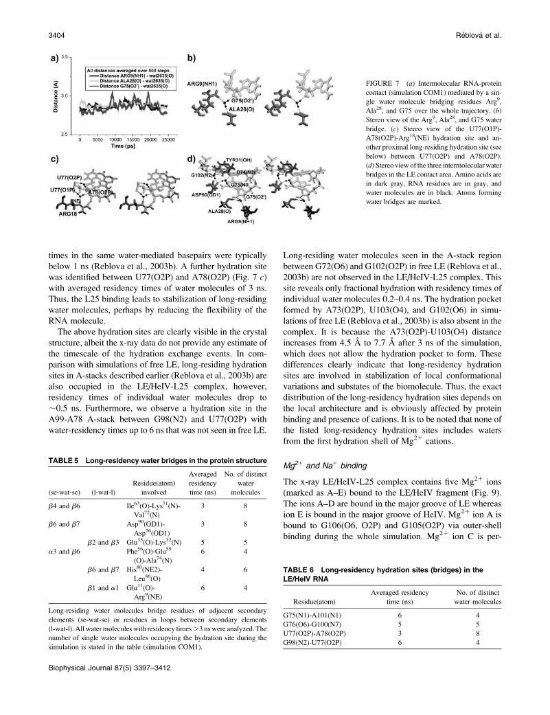
The complex is stabilized by long-residency water bridges
We have detected a number of water-mediated contacts
between the L25 protein and the RNA, most of them
involving long-residency water molecules. We explicitly
listed all long-residency hydration sites with residency times
of individual water molecules longer than 1 ns and with
100% occupancy of hydration sites (Table 4). In both contact
areas long-residing water molecules were identified in
cavities formed between surfaces of the protein and the
RNA. The averaged residency times of these water
molecules ranged from 2 to 8 ns (Table 4) whereas at two
hydration sites a single water molecule was bound with no
exchange event in the course of the entire 24-ns simulation
(Fig. 7). All water bridges listed in Table 4 are clearly visible
in the x-ray structure except for the G79(O2P)-Lys14(NZ)
water bridge. For a comparison, common hydration sites in
nucleic acids have residency times on a scale of 0.05–0.5 ns
(Nagan et al., 1999) whereas in our preceding studies of
unbound LE motif (Reblova et al., 2003b) and (beet western
yellows virus frame-shifting pseudoknot (Csaszar et al.,
2001) we reported several highly structured hydration sites
with residency times up to 5 ns. This study thus reveals
water-residency times longer than any preceding MD
analysis of nucleic acids.
Hydration of L25 and LE/HeIV
There are additional major hydration sites outside the RNA-
protein binding area. Table 5 summarizes L25 hydration sites
with residency times of individual water molecules at least 3
ns. Long-residing water molecules stabilize adjacent sec-
ondary elements of the protein or loops between secondary
elements (see Fig. S5 in Supplementary Material). For the
LE/HeIV fragment we listed hydration sites with residency
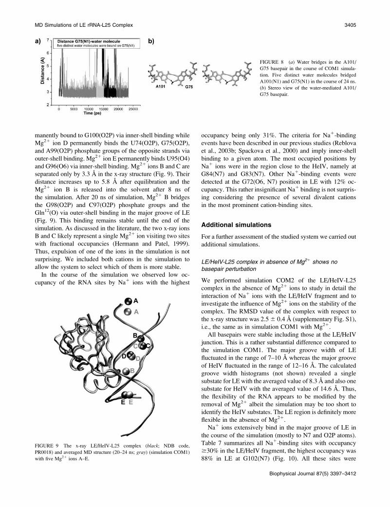
times .1 ns (Table 6). We observed two water-mediated
basepairs, namely G75/A101 and G76/G100, in agreement
with x-ray data (Correll et al., 1997). Both sites were 100%
occupied with averaged water residency times 6 and 5 ns,
respectively. In the G75/A101 basepair one long-residing
water molecule was bound for 11 ns (Fig. 8) whereas in the
G76/G100 basepair the longest water-binding event lasted 8
ns. This is a considerable prolongation of water binding
compared to simulations of free LE where water residency
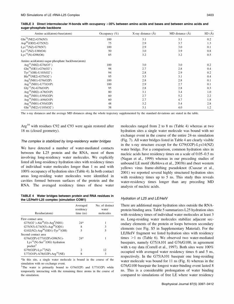
TABLE 3 Direct intermolecular H-bonds with occupancy $35% between amino acids and bases and between amino acids and
sugar-phosphate backbone
Amino acid(atom)-base(atom) Occupancy (%) X-ray distance (A) MD distance (A) SD (A)
Gln78(NE2)-G76(N3) 100 3.1 3.1 0.2
Asp90(OD2)-G75(N2) 75 2.9 3.7 1.3
Lys14(NZ)-G79(N7) 100 2.9 3.0 0.1
Lys14(NZ)-U80(O4) 50 3.0 3.9 0.8
Lys14(N)-G98(O6) 85 3.2 3.2 0.5
Amino acid(atom)-sugar-phosphate backbone(atom)
Arg21(NH2)-G76(O3#) 100 3.0 3.0 0.2
Gln78(OE1)-G76(O2#) 98 2.7 2.8 0.4
Tyr31(OH)-U103(O2#) 94 2.8 2.9 0.2
His88(NE2)-G75(O2#) 95 3.5 3.1 0.4
Arg9(NH1)-G76(O2P) 100 2.8 2.8 0.1
Arg21(NH1)-U77(O1P) 100 2.9 2.7 0.1
Gly13(N)-G76(O1P) 95 2.8 2.8 0.3
Arg18(NH2)-A78(O1P) 80 3.1 3.4 1.0
Arg19(NH1)-U95(O2P) 60 2.7 5.2 2.2
Arg19(NH1)-A94(O1P) 60 2.7 4.7 1.8
Arg18(NH1)-C93(O2P) 48 3.2 5.4 2.8
Gln75(NE2)-U103(O2#) 38 3.1 4.0 1.2
The x-ray distances and the average MD distances along the whole trajectory supplemented by the standard deviations are stated in the table.
TABLE 4 Water bridges between protein and RNA residues in
the LE/HeIV-L25 complex (simulation COM1)
Residue(atom)
Averaged
residency
time (ns)
No. of distinct
water
molecules
First contact area
G75(O2#)-Ala28(O)-Arg9(NH1) 24* 1
G75(N3)-U74(N3)-Asp90(OD1) 8 3
G102(N2)-Asp90(OD1)-Tyr31(OH) 5 5
Second contact area
G76(O2P)-U77(O2P)-G98(N1)-
Lys14(N)-Ser17(OG) hydration
pockety
24* 1
G79(O2P)-Lys14(NZ) 2 12
U77(O1P)-A78(O2P)-Arg18(NE) 8 3
*In this site, a single water molecule is bound in the course of the
simulation with no exchange event.yThe water is primarily bound to G76(O2P) and U77(O2P) while
temporarily interacting with the remaining three atoms in the course of
the simulation.
MD Simulations of LE rRNA-L25 Complex 3403
Biophysical Journal 87(5) 3397–3412
times in the same water-mediated basepairs were typically
below 1 ns (Reblova et al., 2003b). A further hydration site
was identified between U77(O2P) and A78(O2P) (Fig. 7 c)with averaged residency times of water molecules of 3 ns.
Thus, the L25 binding leads to stabilization of long-residing
water molecules, perhaps by reducing the flexibility of the
RNA molecule.
The above hydration sites are clearly visible in the crystal
structure, albeit the x-ray data do not provide any estimate of
the timescale of the hydration exchange events. In com-
parison with simulations of free LE, long-residing hydration
sites in A-stacks described earlier (Reblova et al., 2003b) are
also occupied in the LE/HeIV-L25 complex, however,
residency times of individual water molecules drop to
;0.5 ns. Furthermore, we observe a hydration site in the
A99-A78 A-stack between G98(N2) and U77(O2P) with
water-residency times up to 6 ns that was not seen in free LE.
Long-residing water molecules seen in the A-stack region
between G72(O6) and G102(O2P) in free LE (Reblova et al.,
2003b) are not observed in the LE/HeIV-L25 complex. This
site reveals only fractional hydration with residency times of
individual water molecules 0.2–0.4 ns. The hydration pocket
formed by A73(O2P), U103(O4), and G102(O6) in simu-
lations of free LE (Reblova et al., 2003b) is also absent in the
complex. It is because the A73(O2P)-U103(O4) distance
increases from 4.5 A to 7.7 A after 3 ns of the simulation,
which does not allow the hydration pocket to form. These
differences clearly indicate that long-residency hydration
sites are involved in stabilization of local conformational
variations and substates of the biomolecule. Thus, the exact
distribution of the long-residency hydration sites depends on
the local architecture and is obviously affected by protein
binding and presence of cations. It is to be noted that none of
the listed long-residency hydration sites includes waters
from the first hydration shell of Mg21 cations.
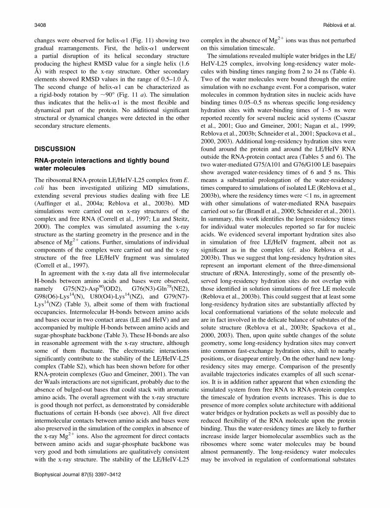
Mg21 and Na1 binding
The x-ray LE/HeIV-L25 complex contains five Mg21 ions
(marked as A–E) bound to the LE/HeIV fragment (Fig. 9).
The ions A–D are bound in the major groove of LE whereas
ion E is bound in the major groove of HeIV. Mg21 ion A is
bound to G106(O6, O2P) and G105(O2P) via outer-shell
binding during the whole simulation. Mg21 ion C is per-
FIGURE 7 (a) Intermolecular RNA-protein
contact (simulation COM1) mediated by a sin-
gle water molecule bridging residues Arg9,
Ala28, and G75 over the whole trajectory. (b)
Stereo view of the Arg9, Ala28, and G75 water
bridge. (c) Stereo view of the U77(O1P)-
A78(O2P)-Arg18(NE) hydration site and an-
other proximal long-residing hydration site (see
below) between U77(O2P) and A78(O2P).
(d) Stereo view of the three intermolecularwater
bridges in the LE contact area. Amino acids are
in dark gray, RNA residues are in gray, and
water molecules are in black. Atoms forming
water bridges are marked.
TABLE 5 Long-residency water bridges in the protein structure
(se-wat-se) (l-wat-l)
Residue(atom)
involved
Averaged
residency
time (ns)
No. of distinct
water
molecules
b4 and b6 Ile63(O)-Lys71(N)-
Val72(N)
3 8
b6 and b7 Asp90(OD1)-
Asp76(OD1)
3 8
b2 and b3 Glu35(O)-Lys32(N) 5 5
a3 and b6 Phe56(O)-Glu59
(O)-Ala74(N)
6 4
b6 and b7 His80(NE2)-
Leu86(O)
4 6
b1 and a1 Glu11(O)-
Arg9(NE)
6 4
Long-residing water molecules bridge residues of adjacent secondary
elements (se-wat-se) or residues in loops between secondary elements
(l-wat-l). All water molecules with residency times.3 ns were analyzed. The
number of single water molecules occupying the hydration site during the
simulation is stated in the table (simulation COM1).
TABLE 6 Long-residency hydration sites (bridges) in the
LE/HeIV RNA
Residue(atom)
Averaged residency
time (ns)
No. of distinct
water molecules
G75(N1)-A101(N1) 6 4
G76(O6)-G100(N7) 5 5
U77(O2P)-A78(O2P) 3 8
G98(N2)-U77(O2P) 6 4
3404 Reblova et al.
Biophysical Journal 87(5) 3397–3412
manently bound to G100(O2P) via inner-shell binding while
Mg21 ion D permanently binds the U74(O2P), G75(O2P),
and A99(O2P) phosphate groups of the opposite strands via
outer-shell binding. Mg21 ion E permanently binds U95(O4)
and G96(O6) via inner-shell binding. Mg21 ions B and C are
separated only by 3.3 A in the x-ray structure (Fig. 9). Their
distance increases up to 5.8 A after equilibration and the
Mg21 ion B is released into the solvent after 8 ns of
the simulation. After 20 ns of simulation, Mg21 B bridges
the G98(O2P) and C97(O2P) phosphate groups and the
Gln12(O) via outer-shell binding in the major groove of LE
(Fig. 9). This binding remains stable until the end of the
simulation. As discussed in the literature, the two x-ray ions
B and C likely represent a single Mg21 ion visiting two sites
with fractional occupancies (Hermann and Patel, 1999).
Thus, expulsion of one of the ions in the simulation is not
surprising. We included both cations in the simulation to
allow the system to select which of them is more stable.
In the course of the simulation we observed low oc-
cupancy of the RNA sites by Na1 ions with the highest
occupancy being only 31%. The criteria for Na1-binding
events have been described in our previous studies (Reblova
et al., 2003b; Spackova et al., 2000) and imply inner-shell
binding to a given atom. The most occupied positions by
Na1 ions were in the region close to the HeIV, namely at
G84(N7) and G83(N7). Other Na1-binding events were
detected at the G72(O6, N7) position in LE with 12% oc-
cupancy. This rather insignificant Na1 binding is not surpris-
ing considering the presence of several divalent cations
in the most prominent cation-binding sites.
Additional simulations
For a further assessment of the studied system we carried out
additional simulations.
LE/HeIV-L25 complex in absence of Mg21 shows nobasepair perturbation
We performed simulation COM2 of the LE/HeIV-L25
complex in the absence of Mg21 ions to study in detail the
interaction of Na1 ions with the LE/HeIV fragment and to
investigate the influence of Mg21 ions on the stability of the
complex. The RMSD value of the complex with respect to
the x-ray structure was 2.56 0.4 A (supplementary Fig. S1),
i.e., the same as in simulation COM1 with Mg21.
All basepairs were stable including those at the LE/HeIV
junction. This is a rather substantial difference compared to
the simulation COM1. The major groove width of LE
fluctuated in the range of 7–10 A whereas the major groove
of HeIV fluctuated in the range of 12–16 A. The calculated
groove width histograms (not shown) revealed a single
substate for LE with the averaged value of 8.3 A and also one
substate for HeIV with the averaged value of 14.6 A. Thus,
the flexibility of the RNA appears to be modified by the
removal of Mg21 albeit the simulation may be too short to
identify the HeIV substates. The LE region is definitely more
flexible in the absence of Mg21.
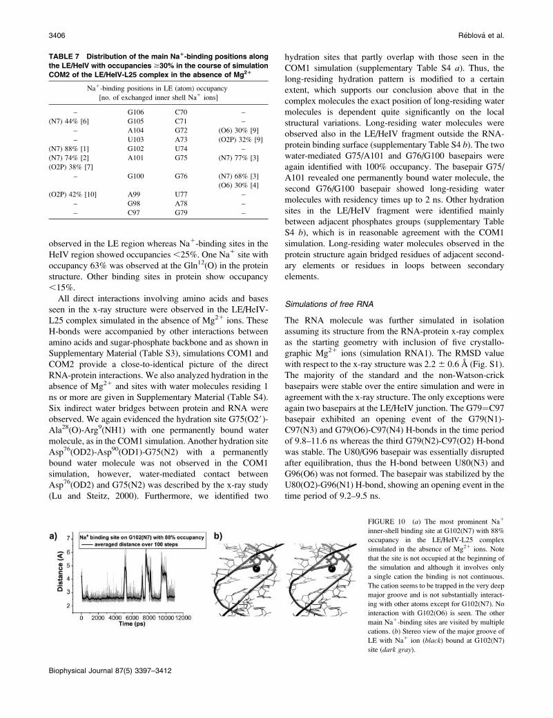
Na1 ions extensively bind in the major groove of LE in
the course of the simulation (mostly to N7 and O2P atoms).
Table 7 summarizes all Na1-binding sites with occupancy
$30% in the LE/HeIV fragment, the highest occupancy was
88% in LE at G102(N7) (Fig. 10). All these sites were
FIGURE 8 (a) Water bridges in the A101/
G75 basepair in the course of COM1 simula-
tion. Five distinct water molecules bridged
A101(N1) and G75(N1) in the course of 24 ns.
(b) Stereo view of the water-mediated A101/
G75 basepair.
FIGURE 9 The x-ray LE/HeIV-L25 complex (black; NDB code,
PR0018) and averaged MD structure (20–24 ns; gray) (simulation COM1)
with five Mg21 ions A–E.
MD Simulations of LE rRNA-L25 Complex 3405
Biophysical Journal 87(5) 3397–3412
observed in the LE region whereas Na1-binding sites in the
HeIV region showed occupancies,25%. One Na1 site with
occupancy 63% was observed at the Gln12(O) in the protein
structure. Other binding sites in protein show occupancy
,15%.
All direct interactions involving amino acids and bases
seen in the x-ray structure were observed in the LE/HeIV-
L25 complex simulated in the absence of Mg21 ions. These
H-bonds were accompanied by other interactions between
amino acids and sugar-phosphate backbone and as shown in
Supplementary Material (Table S3), simulations COM1 and
COM2 provide a close-to-identical picture of the direct
RNA-protein interactions. We also analyzed hydration in the
absence of Mg21 and sites with water molecules residing 1
ns or more are given in Supplementary Material (Table S4).
Six indirect water bridges between protein and RNA were
observed. We again evidenced the hydration site G75(O2#)-Ala28(O)-Arg9(NH1) with one permanently bound water
molecule, as in the COM1 simulation. Another hydration site
Asp76(OD2)-Asp90(OD1)-G75(N2) with a permanently
bound water molecule was not observed in the COM1
simulation, however, water-mediated contact between
Asp76(OD2) and G75(N2) was described by the x-ray study
(Lu and Steitz, 2000). Furthermore, we identified two
hydration sites that partly overlap with those seen in the
COM1 simulation (supplementary Table S4 a). Thus, thelong-residing hydration pattern is modified to a certain
extent, which supports our conclusion above that in the
complex molecules the exact position of long-residing water
molecules is dependent quite significantly on the local
structural variations. Long-residing water molecules were
observed also in the LE/HeIV fragment outside the RNA-
protein binding surface (supplementary Table S4 b). The twowater-mediated G75/A101 and G76/G100 basepairs were
again identified with 100% occupancy. The basepair G75/
A101 revealed one permanently bound water molecule, the
second G76/G100 basepair showed long-residing water
molecules with residency times up to 2 ns. Other hydration
sites in the LE/HeIV fragment were identified mainly
between adjacent phosphates groups (supplementary Table
S4 b), which is in reasonable agreement with the COM1
simulation. Long-residing water molecules observed in the
protein structure again bridged residues of adjacent second-
ary elements or residues in loops between secondary
elements.
Simulations of free RNA
The RNA molecule was further simulated in isolation
assuming its structure from the RNA-protein x-ray complex
as the starting geometry with inclusion of five crystallo-
graphic Mg21 ions (simulation RNA1). The RMSD value
with respect to the x-ray structure was 2.26 0.6 A (Fig. S1).
The majority of the standard and the non-Watson-crick
basepairs were stable over the entire simulation and were in
agreement with the x-ray structure. The only exceptions were
again two basepairs at the LE/HeIV junction. The G79¼C97
basepair exhibited an opening event of the G79(N1)-
C97(N3) and G79(O6)-C97(N4) H-bonds in the time period
of 9.8–11.6 ns whereas the third G79(N2)-C97(O2) H-bond
was stable. The U80/G96 basepair was essentially disrupted
after equilibration, thus the H-bond between U80(N3) and
G96(O6) was not formed. The basepair was stabilized by the
U80(O2)-G96(N1) H-bond, showing an opening event in the
time period of 9.2–9.5 ns.
TABLE 7 Distribution of the main Na1-binding positions along
the LE/HeIV with occupancies $30% in the course of simulation
COM2 of the LE/HeIV-L25 complex in the absence of Mg21
Na1-binding positions in LE (atom) occupancy
[no. of exchanged inner shell Na1 ions]
– G106 C70 –
(N7) 44% [6] G105 C71 –
– A104 G72 (O6) 30% [9]
– U103 A73 (O2P) 32% [9]
(N7) 88% [1] G102 U74 –
(N7) 74% [2] A101 G75 (N7) 77% [3]
(O2P) 38% [7]
– G100 G76 (N7) 68% [3]
(O6) 30% [4]
(O2P) 42% [10] A99 U77 –
– G98 A78 –
– C97 G79 –
FIGURE 10 (a) The most prominent Na1
inner-shell binding site at G102(N7) with 88%
occupancy in the LE/HeIV-L25 complex
simulated in the absence of Mg21 ions. Note
that the site is not occupied at the beginning of
the simulation and although it involves only
a single cation the binding is not continuous.
The cation seems to be trapped in the very deep
major groove and is not substantially interact-
ing with other atoms except for G102(N7). No
interaction with G102(O6) is seen. The other
main Na1-binding sites are visited by multiple
cations. (b) Stereo view of the major groove of
LE with Na1 ion (black) bound at G102(N7)
site (dark gray).
3406 Reblova et al.
Biophysical Journal 87(5) 3397–3412
The geometry of the LE was in agreement with the x-ray
structure of the complex. The LE major groove width
fluctuated in the range of ;6–8 A whereas the HeIV region
showed considerable dynamics of the major groove with
fluctuations in the range of 10–16 A. This resembles the
dynamics seen in the LE/HeIV-L25 complex during the
simulation, however, no distinct substates of the major
groove of HeIV were observed. Histograms for the major
groove widths (data not shown) identified one substate for
LE with average value 7.0 A and one substate for HeIV with
average value 14.6 A. The simulated Mg21-binding sites
were in agreement with x-ray positions except for the Mg21
ion B. The x-ray distance between Mg21 ions B and C is
only 3.3 A (see above) (Fig. 9). We observed redistribution
of Mg21 ions B and C, as in the COM1 simulation. After
equilibration the distance increased to 5.7 A. Mg21 ion C
remained bound to G100(O2P) via inner-shell binding while
Mg21 ion B was attached to G100(O1P), also via inner-shell
binding. This binding persisted until the end of the
simulation.
We observed water molecules in water-mediated G76/
G100 basepair with residency times of 2–3 ns. The second
water-mediated G75/A101 basepair shows residency times
of individual water molecules in the range of 0.5–0.8 ns.
Another long-residing water molecule binds simultaneously
G102(O6) and a water molecule from the first water shell of
Mg21 ion D that interacts with U75(O2P) via inner-shell
binding. The water molecule thus bridges G102(O6) and the
hydrated Mg21 ion D for 12 ns and then is replaced by
another water molecule. Another hydration site was ob-
served between U103(O4) and G102(O6) with one long-
residing water molecule bound for 8 ns of the simulation;
residency times of other water molecules in this site ranged
from 0.8 to 2 ns.
The x-ray structure of the free LE/HeIV fragment (Correll
et al., 1997) with a wide (open) HeIV major groove was used
as the starting structure in another simulation RNA2 (see
Materials and Methods) (Fig. 3 b). Five Mg21 ions used in
this simulation are in a similar but not identical arrangement
compared with the LE/HeIV-L25 complex (see Materials
and Methods).
The RMSD value with respect to the starting x-ray
structure was 2.2 6 0.5 A (supplementary Fig. S1). All
basepairs were entirely stable except for the A73/U103
basepair from the first A-stack of LE. This basepair exhibited
one opening event in the time period of 2.3–3.2 ns. Both
A73(N6)-U103(O2) and A73(N7)-U103(N3) H-bonds were
temporarily disrupted, however, the basepair formed again. It
is noted that a similar fluctuation in the A77/U99 basepair in
the second A-stack was previously reported (Reblova et al.,
2003b) and this indicates that the basepairs involved in the
A-stack arrangements may be somewhat labile.
We observed minor fluctuations of the major groove width
of LE (scale of fluctuations;1–2 A). The Mg21 ions bridge
opposite phosphates across the major groove of LE,
stabilizing its width. The major groove of HeIV showed
substantial fluctuations in the range of 11–19 A. Histograms
identified one substate for LE with the averaged width value
of 6.5 A whereas two substates were found for HeIV with the
averaged values of 12.7 A (closed geometry) and 17.0 A
(open geometry), similar to the COM1 simulation. Mg21
cations did not change binding positions after equilibration
and in the course of 18 ns of the simulation (Table 2). Mg21
C linked the opposite phosphate groups U74(O2P) and
A99(O2P) via inner-shell binding, which considerably
narrowed the major groove of LE. The simulation revealed
two hydration sites with long-residing water molecules with
the range of binding times of 1.5–2 ns. Further, we identified
two water-mediated basepairs G76/G100 and G75/A101.
The first one showed water molecules with residency times
up to 5 ns whereas the second one was characterized by
hydration events in the range of 0.5–0.8 ns, in agreement
with our previous study of smaller free LE (Reblova et al.,
2003b).
Simulations of free protein reveal reduced stability of helix-a1
We carried out simulation PROT1 of the L25 protein (starting
from the crystal structure of the complex) in the absence of
the LE/HeIV fragment. During 10 ns of simulation the
structure of the protein was stable. The RMSD value with
respect to the x-ray structure was 2.3 6 0.4 A (Fig. S1). The
RMSD values of the individual secondary elements with
respect to the x-ray structure were in the range of 0.3–0.5 A
and no structural or dynamic changes were observed. In
contrast, the elevated temperature (400 K) simulation
(PROT2) resulted in RMSD value with respect to the x-ray
structure of 5.6 6 1.3 A (Fig. S1). The most significant
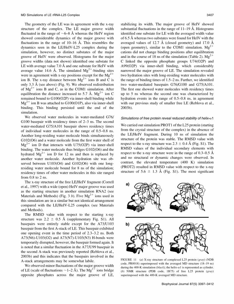
FIGURE 11 (a) X-ray structure of complexed L25 protein (gray) (NDB
code, PR0018) superimposed with the averaged MD structure (18–19 ns)
during the 400-K simulation (black); the helix-a1 is represented as cylinder.(b) NMR structure (PDB code, 1B75) of free L25 protein (gray)
superimposed with the 400-K averaged MD structure.
MD Simulations of LE rRNA-L25 Complex 3407
Biophysical Journal 87(5) 3397–3412
changes were observed for helix-a1 (Fig. 11) showing two
gradual rearrangements. First, the helix-a1 underwent
a partial disruption of its helical secondary structure
producing the highest RMSD value for a single helix (1.6
A) with respect to the x-ray structure. Other secondary
elements showed RMSD values in the range of 0.5–1.0 A.
The second change of helix-a1 can be characterized as
a rigid-body rotation by ;90� (Fig. 11 a). The simulation
thus indicates that the helix-a1 is the most flexible and
dynamical part of the protein. No additional significant
structural or dynamical changes were detected in the other
secondary structure elements.
DISCUSSION
RNA-protein interactions and tightly boundwater molecules
The ribosomal RNA-protein LE/HeIV-L25 complex from E.coli has been investigated utilizing MD simulations,
extending several previous studies dealing with free LE
(Auffinger et al., 2004a; Reblova et al., 2003b). MD
simulations were carried out on x-ray structures of the
complex and free RNA (Correll et al., 1997; Lu and Steitz,
2000). The complex was simulated assuming the x-ray
structure as the starting geometry in the presence and in the
absence of Mg21 cations. Further, simulations of individual
components of the complex were carried out and the x-ray
structure of the free LE/HeIV fragment was simulated
(Correll et al., 1997).
In agreement with the x-ray data all five intermolecular
H-bonds between amino acids and bases were observed,
namely G75(N2)-Asp90(OD2), G76(N3)-Gln78(NE2),
G98(O6)-Lys14(N), U80(O4)-Lys14(NZ), and G79(N7)-
Lys14(NZ) (Table 3), albeit some of them with fractional
occupancies. Intermolecular H-bonds between amino acids
and bases occur in two contact areas (LE and HeIV) and are
accompanied by multiple H-bonds between amino acids and
sugar-phosphate backbone (Table 3). These H-bonds are also
in reasonable agreement with the x-ray structure, although
some of them fluctuate. The electrostatic interactions
significantly contribute to the stability of the LE/HeIV-L25
complex (Table S2), which has been shown before for other
RNA-protein complexes (Guo and Gmeiner, 2001). The van
der Waals interactions are not significant, probably due to the
absence of bulged-out bases that could stack with aromatic
amino acids. The overall agreement with the x-ray structure
is good though not perfect, as demonstrated by considerable
fluctuations of certain H-bonds (see above). All five direct
intermolecular contacts between amino acids and bases were
also preserved in the simulation of the complex in absence of
the x-ray Mg21 ions. Also the agreement for direct contacts
between amino acids and sugar-phosphate backbone was
very good and both simulations are qualitatively consistent
with the x-ray structure. The stability of the LE/HeIV-L25
complex in the absence of Mg21 ions was thus not perturbed
on this simulation timescale.
The simulations revealed multiple water bridges in the LE/
HeIV-L25 complex, involving long-residency water mole-
cules with binding times ranging from 2 to 24 ns (Table 4).
Two of the water molecules were bound through the entire
simulation with no exchange event. For a comparison, water
molecules in common hydration sites in nucleic acids have
binding times 0.05–0.5 ns whereas specific long-residency
hydration sites with water-binding times of 1–5 ns were
reported recently for several nucleic acid systems (Csaszar
et al., 2001; Guo and Gmeiner, 2001; Nagan et al., 1999;
Reblova et al., 2003b; Schneider et al., 2001; Spackova et al.,
2000, 2003). Additional long-residency hydration sites were
found around the protein and around the LE/HeIV RNA
outside the RNA-protein contact area (Tables 5 and 6). The
two water-mediated G75/A101 and G76/G100 LE basepairs
show averaged water-residency times of 6 and 5 ns. This
means a substantial prolongation of the water-residency
times compared to simulations of isolated LE (Reblova et al.,
2003b), where the residency times were,1 ns, in agreement
with other simulations of water-mediated RNA basepairs
carried out so far (Brandl et al., 2000; Schneider et al., 2001).
In summary, this work identifies the longest residency times
for individual water molecules reported so far for nucleic
acids. We evidenced several important hydration sites also
in simulation of free LE/HeIV fragment, albeit not as
significant as in the complex (cf. also Reblova et al.,
2003b). Thus we suggest that long-residency hydration sites
represent an important element of the three-dimensional
structure of rRNA. Interestingly, some of the presently ob-
served long-residency hydration sites do not overlap with
those identified in solution simulations of free LE molecule
(Reblova et al., 2003b). This could suggest that at least some
long-residency hydration sites are substantially affected by
local conformational variations of the solute molecule and
are in fact involved in the delicate balance of substates of the
solute structure (Reblova et al., 2003b; Spackova et al.,
2000, 2003). Then, upon quite subtle changes of the solute
geometry, some long-residency hydration sites may convert
into common fast-exchange hydration sites, shift to nearby
positions, or disappear entirely. On the other hand new long-
residency sites may emerge. Comparison of the presently
available trajectories indicates examples of all such scenar-
ios. It is in addition rather apparent that when extending the
simulated system from free RNA to RNA-protein complex
the timescale of hydration events increases. This is due to
presence of more complex solute architecture with additional
water bridges or hydration pockets as well as possibly due to
reduced flexibility of the RNA molecule upon the protein
binding. Thus the water-residency times are likely to further
increase inside larger biomolecular assemblies such as the
ribosomes where some water molecules may be bound
almost permanently. The long-residency water molecules
may be involved in regulation of conformational substates
3408 Reblova et al.
Biophysical Journal 87(5) 3397–3412
and dynamical motions. Nevertheless, caution is still needed
regarding the above assessment of the long-residency
hydration events. First, in most cases our analysis is based
on a single trajectory on each system and on a comparison of
several related trajectories. Thus quantitative reproducibility
of long-residency hydration states could not be achieved and
the discussion is based rather on observing a few ‘‘snap-
shots’’ of the long-residency hydration architecture. Second,
formation of many long-residency sites (typically those
involving the formation of ‘‘cavities’’) may be associated
with restricted diffusion and thus accompanied with an
entropic cost. Therefore, it is presently not possible to
quantify the actual effect of long-residency hydration on the
stability of the complex.
Cation binding in the LE/HeIV-L25 complex
The x-ray structure of the LE/HeIV-L25 complex contains
five Mg21 ions bound via inner- and outer-shell binding in
the RNA deep major groove. Mg21 ions did not change
binding positions in the course of the simulation and were in
agreement with the x-ray positions, except for Mg21 ion B.
In the x-ray structure Mg21 cations B and C are separated by
only 3.3 A (Fig. 9). It is well established that the B- and
C-binding positions likely represent a single cation-binding
site with fractional occupancy (Hermann and Patel, 1999). In
the course of simulation, Mg21 C stayed in place whereas
Mg21 ion B was expelled from its original position and then
occupied a new position in the major groove. As discussed
by other groups, many x-ray positions of Mg21 cations do
not reflect the biologically important cations and some
unrelated electron densities (anions, water) can even be
misinterpreted as cations (Auffinger et al., 2004b; Ennifar
et al., 2003). Similar redistribution of the Mg21 ions in the
major groove of LE has been also observed in our previous
study (Reblova et al., 2003b). On the other hand, it is fair to
note that modeling of divalent cations suffers from major
limitations imposed by the pair-additive force field, making
the description of the interactions of divalent cations the least
accurate part of the simulations. In reality, there are huge
polarization and charge-transfer effects from the cation to all
its first-shell ligands, further propagating well beyond the first
ligand shell (Gresh et al., 2003; Rulisek and Sponer, 2003).
For example, properties of the first-shell water molecules are
substantially different from bulk water molecules. Further-
more, the simulations are orders of magnitude shorter
compared with a timescale that would be appropriate for
a representative sampling of motions of divalent cations.
Thus, the results of this simulation and related such studies
should be considered primarily as crude estimates of the effect
of Mg21 ions bound in the suggested x-ray positions. It is not
possible to exactly localize the preferred Mg21 positions and
binding patterns via contemporary MD simulations and even
their interactions in known x-ray sites may be biased by the
force-field limitations. The force-field and sampling limita-
tions actually justify the common practice in the majority of
simulations of nucleic acids where the counterion atmosphere
is simply described by a minimal neutralizing set of mono-
valent cations. (Inclusion of monovalent anions such as Cl�
into the simulation is more risky as the force field for anions
has not been widely tested and the pair-additive force fields
are inherently deficient in describing the anions due to the
diffuse nature of their electron distributions (Tobias et al.,
2001)). Fortunately, because the simulations are too short to
lead to any substantial RNA unfolding due to the absence of
the Mg21 species, lack of divalent cations in simulations has
a much smaller effect than it has on experiments.
The description of the monovalent cation interactions with
nucleic acids is considerably better although not perfect. We
carried out the simulations in the presence of Na1 cations
rather than in the presence of the physiologically more
relevant K1 ones because there is currently considerably
more experience with the behavior of sodium simulations in
the MD literature. Anyway, description of both cations is
affected by the same force-field approximations. In the
course of simulation with Mg21 no strong Na1-binding
events were observed. However, the simulated LE/HeIV-
L25 complex in the absence of Mg21 reveals extensive
binding of Na1 ions mostly with multiple exchange events of
the cations (Table 7). The most occupied sites were found in
the major grove of LE with maximal occupancy of one site
reaching as much as 88% (Fig. 10). The most occupied
atoms were N7 and O2P. The area of HeIV shows low Na1
binding with the maximal occupancy in an individual site of
25%. Thus the simulation confirms that the major groove of
LE provides some of the most prominent RNA cation-
binding sites studied to date (Auffinger et al., 2004a; Correll
et al., 1997; Reblova et al., 2003b).
Basepairing
The simulation of the LE/HeIV-L25 complex in the presence
of x-ray Mg21 ions reveals that the standard and non-
Watson-Crick basepairs are stable and in agreement with the
x-ray structure, except for the three consecutive basepairs
(Watson-Crick G79¼C97, wobble U80/G96, and wobble
G81/U95) forming the LE/HeIV junction. The first basepair
shows two opening events, the second basepair is essentially
disrupted, and the third basepair shows fluctuation of one
H-bond. Interestingly, all basepairs are entirely stable when
theLE/HeIV-L25complexissimulatedintheabsenceofMg21
ions whereas Mg21 simulation of the free LE/HeIV fragment
(in the absence of the L25 protein) reveals again instability of
the G79¼C97 and U80/G96 basepairs. In contrast, all
basepairs are stable in another Mg21 simulation of free LE/
HeIV with somewhat different Mg21 distribution (see
Materials and Methods). When considering all data we
suggest that the destabilization of the LE/HeIV junction
basepairs might be related to the presence of the Mg21 ion E
bound to both wobble basepairs, specifically to positions
MD Simulations of LE rRNA-L25 Complex 3409
Biophysical Journal 87(5) 3397–3412
U95(O4) and G96(O6). It is not possible to decide whether
this reflects some error in the initial (or experimental) cation
placement, force-field limitation, or insufficient sampling
(see Discussion above). It also cannot be ruled out that
actually the junction between the LE and HeIV regions may
be the most labile part of the RNA molecule. For example,
the instability of the basepairs at the LE/HeIV junction could
also be partly related to the cross-strand G81/G96 stack.
Stacking interactions can lead to basepair strain that can
result in modest instability of the basepairing or opening
events. Actually, occasional temporary opening events of
A/U basepairs were noticed also in the LEA-stacks; see above
and Reblova et al. (2003b). Nevertheless, the perturbation of
the LE/HeIV junction most likely stems from the limitations
(artifacts) related to inclusion of divalent cations into
simulations. Because the instability at the LE/HeIV junction
did not have any significant effect on the protein binding it
was not necessary to repeat the simulations.
The protein dynamics
L25 protein in the LE/HeIV-L25 complex underwent no
significant structural and dynamic changes in the course of
simulation. Modest rotation of the helix-a2 was noticed
compared with the starting geometry (supplementary Figs.
S2 and S3). This change did not influence the stability of the
LE/HeIV-L25 complex. No additional structural changes
were detected. Simulations of isolated L25 protein (starting
from its geometry in the complex) at 300 K and 400 K reveal
the following picture. There were no structural changes
during the 300-K simulation. At elevated temperature (400
K), substantial structural changes were observed in the helix-
a1. The helix-a1 rotated by;90� and its secondary structurewas partially disrupted (Fig. 11). Interestingly, this observa-
tion is in a full agreement with the experimental studies
(Stoldt et al., 1998, 1999) showing that formation of the
RNA-protein complex induces structuring of the helix-a1region that turns toward the major groove of HeIV. Our
simulation of the L25 protein at elevated temperature could,
at least qualitatively, capture a reversal of this process. After
19 ns of simulation at elevated temperature, the protein
structure resembles the solution NMR structure of free L25
protein (Fig. 11 b) (Stoldt et al., 1998).
Dynamics of the LE/HeIV explains the differencesbetween experimental structures
Different nanosecond dynamic behavior has been observed
for the LE and HeIV motifs. The LE region is very rigid
whereas the HeIV segment shows considerable dynamics.
Two substates (open and closed geometry) of the major
groove of HeIV have been observed for both complexed and
free LE/HeIV fragments in the simulations (Figs. 4 and 5).
Both substates were also observed in the experimental
structures (Correll et al., 1997; Lu and Steitz, 2000; Stoldt
et al., 1999). The ‘‘open geometry’’ is seen in the x-ray
structure of free LE/HeIV RNA duplex (Correll et al., 1997)
and also in the NMR LE/HeIV-L25 complex (Stoldt et al.,
1999) (Fig. 3, b and c) whereas the closed geometry is seen
in the x-ray LE/HeIV-L25 complex (Lu and Steitz, 2000)
(Fig. 3 a).Clear substates of the major groove of HeIV were not
identified in the LE/HeIV-L25 complex in the absence of
Mg21 ions; nevertheless, major grooves of both LE and
HeIV considerably oscillated. The major groove of LE
showed fluctuations in the range of 7–10 A, probably due to
the absence of the Mg21 ions in the major groove (Reblova
et al., 2003b). It has no effect on the RNA-protein binding.
The major groove of HeIV showed breathing in the width
range of 12–16 A. In summary, both complexed and free LE/
HeIV rRNA reveal considerable dynamics in the area of
HeIV on the nanosecond timescale. Notably, the NMR and
x-ray structures of the LE/HeIV-L25 complex show differ-
ences primarily in the HeIV area (Fig. 3). We suggest that the
differences can be explained by our MD simulations that
identify the HeIV as a flexible RNA fragment with variable
width of the major groove, likely sensitive to the molecular
interactions and environmental effects.
CONCLUSIONS
We have carried out a series of MD simulations to study the
complex between E. coli loop E/helix IV 5S rRNA fragment
and ribosomal protein L25. Although the exact biological
role of this complex is yet to be determined (L25 could, for
example, play a role in supporting the E. coli loop E so it can
interact with the A-site finger) it is well characterized by
atomic resolution experimental techniques and reveals very
interesting molecular interactions (Lu and Steitz, 2000;
Stoldt et al., 1999). There is a very good overall agreement
between the experimental and simulated structures, which is
quite promising considering further studies of RNA-protein
interactions. Nevertheless, some minor differences between
computed and experimental structures were observed (see
above) and these might be attributed to force-field limitations
and possibly data and refinement errors in the experimental
structures. They involve temporary oscillations (fractional
occupancy) of some of the direct RNA-protein H-bonds and
perturbations of basepairs at the LE/HeIV junction in some
simulations. This basepair instability has no effect on the
L25 binding. The simulations strongly suggest that this RNA
structural perturbation is caused by a divalent Mg21 cation
present in the area, highlighting the difficulties that are
inherent to simulations with divalent cations. Their inclusion
represents a controversial part of the contemporary simula-
tion techniques, as neither the force field nor the sampling is
satisfactory for realistic modeling of Mg21.
The simulations confirm that the deep major groove of LE
is a prominent rRNA cation-binding site. In the absence of
divalent cations, this part of RNA extensively binds
3410 Reblova et al.
Biophysical Journal 87(5) 3397–3412
monovalent cations via inner-shell binding. The divalent
cations rigidify the LE major groove geometry. The HeIV
region shows clear bistability of its major groove width that
is sensitive to the presence of cations and molecular inter-
actions. TwoHeIV substates, closed and open,were identified
by the simulations. This nicely explains the observed dif-
ference between various HeIV experimental structures (Lu
and Steitz, 2000; Stoldt et al., 1999). In agreement with the ex-
perimental data (Stoldt et al., 1998) the simulations suggest
that helix-a1 of L25 is the least stable part of the protein
that actually is structured upon the formation of the complex
with rRNA.
The simulations reveal that the rRNA-protein interaction
is mediated by a number of highly specific hydration sites
with long-residing water molecules, two of them bound
during the 24-ns simulation with no exchange event. Such
water-binding events are approximately two orders of
magnitude longer compared to water binding in common
hydration sites around standard DNA and RNA duplexes.
Long-residing water molecules are seen also outside the
RNA-protein contact areas and the water-binding times are
substantially enhanced compared to simulations of free
RNA. Long-residency hydration sites thus represent impor-
tant elements of the three-dimensional structure of rRNA and
we suggest that inside the ribosome some of the water
molecules can be bound almost permanently. MD simu-
lations offer an important tool to study long-residency hy-
dration sites in complex RNA systems because the available
experimental methods only rarely reveal the timescale of the
hydration events (Kochoyan and Leroy, 1995). However,
substantial extension of the simulation timescale will be very
vital to obtain a statistically complete and reproducible
picture of the long-residency hydration events in rRNA.
SUPPLEMENTARY MATERIAL
An online supplement to this article can be found by visiting
BJ Online at http://www.biophysj.org.
This work was supported by grants Ministry of Education, Youth and
Sports, Czech Republic (LN00A016, the Wellcome Trust International
Senior Research Fellowship in Biomedical Science in Central Europe
(GR067507), and the National Institutes of Health (grants 2R15 GM55898
and 3R15 GM55898).
REFERENCES
Agalarov, S. C., G. S. Prasad, P. M. Funke, C. D. Stout, and J. R.Williamson. 2000. Structure of the S15,S18-rRNA complex: assembly ofthe 30S ribosome central domain. Science. 288:107–112.
Auffinger, P., L. Bielecki, and E. Westhof. 2003. The Mg21 binding sites ofthe 5S rRNA loop E motif as investigated by molecular dynamicssimulations. Chem. Biol. 10:551–561.
Auffinger, P., L. Bielecki, and E. Westhof. 2004a. Symetric K1 and Mg21
ion-binding sites in the rRNA loop E inferred from molecular dynamicssimulations. J. Mol. Biol. 335:555–571.
Auffinger, P., L. Bielecki, and E. Westhof. 2004b. Anion binding to nucleicacids. Structure. 12:379–388.
Auffinger, P., and E. Westhof. 1998. Simulations of the moleculardynamics of nucleic acids. Curr. Opin. Struct. Biol. 8:227–236.
Auffinger, P., and E. Westhof. 1999. Singly and bifurcated hydrogen-bonded base-pairs in tRNA anticodon hairpins and ribozymes. J. Mol.Biol. 292:467–483.
Ban, N., P. Nissen, J. Hansen, P. B. Moore, and T. A. Steitz. 2000. Thecomplete atomic structure of the large ribosomal subunit at 2.4A resolution. Science. 289:905–920.
Beaurain, F., and M. Laguerre. 2003. MD studies of the DIS/DIS kissingcomplex solution and x-ray structures. Oligonucleotides. 13:501–514.
Brandl, M., M. Meyer, and J. Suhnel. 2000. Water-mediated base pairs inRNA. A quantum-chemical study. J. Phys. Chem. 104:11177–11187.
Cheatham III, T. E., and M. A. Young. 2000. Molecular dynamicssimulation of nucleic acids: successes, limitations, and promise.Biopolymers. 56:232–256.
Cornell, W. D., P. Cieplak, C. I. Bayly, I. R. Gould, K. M. Merz, D. M.Ferguson, Jr., D. C. Spellmeyer, T. Fox, J. W. Caldwell, and P. A.Kollman. 1995. A 2nd generation force-field for the simulation ofproteins, nucleic-acids, and organic molecules. J. Am. Chem. Soc.117:5179–5197.
Correll, C. C., B. Freeborn, P. B. Moore, and T. A. Steitz. 1997. Metals,motifs, and recognition in the crystal structure of a 5S rRNA domain.Cell. 91:705–712.
Csaszar, K., N. Spackova, R. Stefl, J. Sponer, and N. B. Leontis. 2001.Molecular dynamics of the frame-shifting pseudoknot from beet westernyellows virus: The role of non-Watson-Crick base-pairing, orderedhydration, cation binding and base mutations on stability and unfolding.J. Mol. Biol. 313:1073–1091.
Ennifar, E., P. Walter, and P. Dumas. 2003. A crystallographic study of thebinding of 13 metal ions to two related RNA duplexes. Nucleic AcidsRes. 31:2671–2682.
Ferrin, T. E., C. C. Huang, L. E. Jarvis, and R. Langridge. 1988. TheMIDAS display system. J. Mol. Graph. 6:13–27.
Gresh, N., J. E. Sponer, N. Spackova, J. Leszczynski, and J. Sponer. 2003.Theoretical study of binding of hydrated Zn(II) and Mg(II) cations to5#-guanosine monophosphate. Toward polarizable molecular mechanicsfor DNA and RNA. J. Phys. Chem. B. 107:8669–8681.
Guo, J. X., and W. H. Gmeiner. 2001. Molecular dynamics simulation ofthe human U2B$ protein complex with U2 snRNA hairpin IV in aqueoussolution. Biophys. J. 81:630–642.
Harms, J., F. Schluenzen, R. Zarivach, A. Bashan, S. Gat, I. Agmon,H. Bartels, F. Franceschi, and A. Yonath. 2001. High resolution structureof the large ribosomal subunit from a mesophilic Eubacterium. Cell.107:679–688.
Harvey, S. C., R. K. Z. Tan, and T. E. Cheatham, Iii. 1998. The flying icecube: velocity rescaling in molecular dynamics leads to violation ofenergy equipartition. J. Comput. Chem. 19:726–740.
Hermann, T., P. Auffinger, and E. Westhof. 1998. Molecular dynamicsinvestigations of hammerhead ribozyme RNA. Eur. Biophys. J. 27:153–165.
Hermann, T., and D. J. Patel. 1999. Stitching together RNA tertiaryarchitectures. J. Mol. Biol. 294:829–849.
Humphrey, W., A. Dalke, and K. Schulten. 1996. VMD - Visual moleculardynamics. J. Mol. Graph. 14:33–38.
Kochoyan, M., and J. L. Leroy. 1995. Hydration and solution structure ofnucleic-acids. Curr. Opin. Struct. Biol. 5:329–333.
Leontis, N. B., J. Stombaugh, and E. Westhof. 2002. The non-Watson-Crick base pairs and their associated isostericity matrices. Nucleic AcidsRes. 30:3497–3531.
Leontis, N. B., and E. Westhof. 1998a. The 5S rRNA loop E: chemicalprobing and phylogenetic data versus crystal structure. RNA. 4:1134–1153.
MD Simulations of LE rRNA-L25 Complex 3411
Biophysical Journal 87(5) 3397–3412

Leontis, N. B., and E. Westhof. 1998b. Conserved geometrical base-pairingpatterns in RNA. Q. Rev. Biophys. 31:399–455.
Leontis, N. B., and E. Westhof. 2003. Analysis of RNA motifs. Curr. Opin.Struct. Biol. 13:300–308.
Lu, M., and T. A. Steitz. 2000. Structure of Escherichia coli ribosomalprotein L25 complexed with a 5S rRNA fragment at 1.8-angstromresolution. Proc. Natl. Acad. Sci. USA. 97:2023–2028.
Moore, P. B. 1999. Structural motifs in RNA. Annu. Rev. Biochem. 68:287–300.
Nagan, M. C., S. S. Kerimo, K. Musier-Forsyth, and C. J. Cramer. 1999.Wild-type RNA microhelix(Ala) and 3:70 variants: molecular dynamicsanalysis of local helical structure and tightly bound water. J. Am. Chem.Soc. 121:7310–7317.
Nevskaya, N., S. Tishchenko, R. Fedorov, S. Al-Karadaghi, A. Liljas, A.Kraft, W. Piendl, M. Garber, and S. Nikonov. 2000. Archaeal ribosomalprotein L1: the structure provides new insights into RNA binding of theL1 protein family. Structure. 8:363–371.
Nikulin, A., I. Eliseikina, S. Tishchenko, N. Nevskaya, N. Davydova, O.Platonova, W. Piendl, M. Selmer, A. Liljas, D. Drygin, R. Zimmermann,M. Garber, and S. Nikonov. 2003. Structure of the L1 protuberance in theribosome. Nat. Struct. Biol. 10:104–108.
Norberg, J., and L. Nilsson. 2002. Molecular dynamics applied to nucleicacids. Acc. Chem. Res. 35:465–472.
Orozco, M., A. Perez, A. Noy, and F. J. Luque. 2003. Theoretical methodsfor the simulation of nucleic acids. Chem. Soc. Rev. 32:350–364.
Pearlman, D. A., D. A. Case, J. W. Caldwell, W. S. Ross, T. E. CheathamIII, and S. DeBolt. 1995. AMBER, a package of computer programs forapplying molecular mechanics, normal mode analysis, moleculardynamics and free energy calculations to simulate the structural andenergetic properties of molecule. Comput. Phys. Commun. 91:1–41.
Perederina, A., N. Nevskaya, O. Nikonov, A. Nikulin, P. Dumas, M. Yao,I. Tanaka, M. Garber, G. Gongadze, and S. Nikonov. 2002. Detailedanalysis of RNA-protein interactions within the bacterial ribosomalprotein L5/5S rRNA complex. RNA. 8:1548–1557.
Reblova, K., N. Spackova, J. E. Sponer, J. Koca, and J. Sponer. 2003a.Molecular dynamics simulations of RNA kissing-loop motifs revealstructural dynamics and formation of cation-binding pockets. NucleicAcids Res. 31:6942–6952.
Reblova, K., N. Spackova, R. Stefl, K. Csaszar, J. Koca, N. B. Leontis, andJ. Sponer. 2003b. Non-Watson-Crick basepairing and hydration in RNAmotifs: molecular dynamics of 5S rRNA loop E. Biophys. J. 84:3564–3582.
Ross, W. S., and C. C. Hardin. 1994. Ion-induced stabilization of theG-DNA quadruplex: free-energy perturbation studies. J. Am. Chem. Soc.116:6070–6080.
Rulisek, L., and J. Sponer. 2003. Outer-shell and inner-shell coordination ofphosphate group to hydrated metal ions (Mg21, Cu21, Zn21, Cd21) inthe presence and absence of nucleobase. The role of nonelectrostaticeffects. J. Phys. Chem. B. 107:1913–1923.
Sarzynska, J., T. Kulinski, and L. Nilsson. 2000. Conformational dynamicsof a 5S rRNA hairpin domain containing loop D and a single nucleotidebulge. Biophys. J. 79:1213–1227.
Schneider, C., M. Brandl, and J. Suhnel. 2001. Molecular dynamicssimulation reveals conformational switching of water-mediated uracil-cytosine base-pairs in an RNA duplex. J. Mol. Biol. 305:659–667.
Spackova, N., I. Berger, and J. Sponer. 2000. Nanosecond moleculardynamics of zipper-like DNA duplex structures containing sheared G.Amismatch pairs. J. Am. Chem. Soc. 122:7564–7572.
Spackova, N., T. E. Cheatham III, F. Ryjacek, F. Lankas, L. V. Meervelt, P.Hobza, and J. Sponer. 2003. Molecular dynamics simulations andthermodynamics analysis of DNA-drug complexes. Minor groovebinding between 4#,6-diamidino-2-phenylindole (DAPI) and DNAduplexes in solution. J. Am. Chem. Soc. 125:1759–1769.
Sponer, J., H. A. Gabb, J. Leszczynski, and P. Hobza. 1997. Base-baseand deoxyribose-base stacking interactions in B-DNA and Z-DNA:a quantum-chemical study. Biophys. J. 73:76–87.
Stoldt, M., J. Wohnert, M. Gorlach, and L. R. Brown. 1998. The NMRstructure of Escherichia coli ribosomal protein L25 shows homologyto general stress proteins and glutaminyl-tRNA synthetases. EMBO J.17:6377–6384.
Stoldt, M., J. Wohnert, O. Ohlenschlager, M. Gorlach, and L. R. Brown.1999. The NMR structure of the 5S rRNA E-domain-protein L25complex shows preformed and induced recognition. EMBO J. 18:6508–6521.
Tobias, D. J., P. Jungwirth, and M. Parrinello. 2001. Surface solvation ofhalogen anions in water clusters: an ab initio molecular dynamics studyof the Cl-(H2O)6 complex. J. Chem. Phys. 114:7036–7044.
Vallurupalli, P., and P. B. Moore. 2003. The solution structure of the loopE region of the 5 S rRNA from spinach chloroplasts. J. Mol. Biol. 325:843–856.
van der Spoel, D., A. R. van Buuren, E. Apol, P. J. Maulenhoff, P. D.Tieleman, A. L. T. M. Sijbers, B. Hess, K. A. Feenstra, E. Lindhal, R. V.Drunen, and H. J. C. Berendsen. 1999. Gromacs User Manual Version2.0. Groningen, The Netherlands.
Wimberly, B. T., D. E. Brodersen, W. M. Clemons, R. J. Morgan-Warren,A. P. Carter, C. Vonrhein, T. Hartsch, and V. Ramakrishnan. 2000.Structure of the 30S ribosomal subunit. Nature. 407:327–339.
Wimberly, B. T., R. Guymon, J. P. McCutcheon, S. W. White, and V.Ramakrishnan. 1999. A detailed view of a ribosomal active site: thestructure of the L11-RNA complex. Cell. 97:491–502.
York, D. M., T. A. Darden, and L. G. Pedersen. 1993. The effect of long-range electrostatic interactions in simulations of macromolecular crystal:a comparison of the Ewald and truncated list methods. J. Chem. Phys.99:8345–8348.
Yusupov, M. M., G. Z. Yusupova, A. Baucom, K. Lieberman, T. N.Earnest, J. H. D. Cate, and H. F. Noller. 2001. Crystal structure of theribosome at 5.5 angstrom resolution. Science. 292:883–896.
Zacharias, M. 2000. Simulation of the structure and dynamics of nonhelicalRNA motifs. Curr. Opin. Struct. Biol. 10:311–317.
Zhou, R., B. J. Berne, and R. Germain. 2001. The free energy landscape forbeta hairpin folding in explicit water. Proc. Natl. Acad. Sci. USA.18:14931–14936.
3412 Reblova et al.
Biophysical Journal 87(5) 3397–3412
Top Related
Copyright © 2022 FDOKUMEN

