


Bahasa
Halaman
Hukum

JOURNAL OF VIROLOGY, Apr. 2003, p. 4848–4857 Vol. 77, No. 80022-538X/03/$08.00�0 DOI: 10.1128/JVI.77.8.4848–4857.2003Copyright © 2003, American Society for Microbiology. All Rights Reserved.
Infection of Cattle with a Bovine Herpesvirus 1 Strain ThatContains a Mutation in the Latency-Related Gene Leads
to Increased Apoptosis in Trigeminal Ganglia duringthe Transition from Acute Infection
to LatencyLuciane Lovato, Melissa Inman, Gail Henderson, Alan Doster, and Clinton Jones*Department of Veterinary and Biomedical Sciences, Nebraska Center for Virology, University of Nebraska
at Lincoln, Lincoln, Nebraska 68583-0905
Received 7 November 2002/Accepted 14 January 2003
Bovine herpesvirus 1 (BHV-1) is an important pathogen of cattle and infection is usually initiated via theocular or nasal cavity. After acute infection, the primary site for BHV-1 latency is sensory neurons in thetrigeminal ganglia (TG). Reactivation from latency occurs sporadically, resulting in virus shedding andtransmission to uninfected cattle. The only abundant viral transcript expressed during latency is the latency-related (LR) RNA. An LR mutant was constructed by inserting three stop codons near the beginning of the LRRNA. This mutant grows to wild-type (wt) efficiency in bovine kidney cells and in the nasal cavity of acutelyinfected calves. However, shedding of infectious virus from the eye and TG was dramatically reduced in calvesinfected with the LR mutant. Calves latently infected with the LR mutant do not reactivate after dexametha-sone treatment. In contrast, all calves latently infected with wt BHV-1 or the LR rescued mutant reactivatefrom latency after dexamethasone treatment. In the present study, we compared the frequency of apoptosis incalves infected with the LR mutant to calves infected with wt BHV-1 because LR gene products inhibitapoptosis in transiently transfected cells. A sensitive TUNEL (terminal deoxynucleotidyltransferase-mediateddUTP-biotin nick end labeling) assay and an antibody that detects cleaved caspase-3 were used to identifyapoptotic cells in TG. Both assays demonstrated that calves infected with the LR mutant for 14 days had higherlevels of apoptosis in TG compared to calves infected with wt BHV-1 or to mock-infected calves. Viral geneexpression, except for the LR gene, is extinguished by 14 days after infection, and thus this time frame isoperationally defined as the establishment of latency. Real-time PCR analysis indicated that lower levels ofviral DNA were present in the TG of calves infected with the LR mutant throughout acute infection. Takentogether, these results suggest that the antiapoptotic properties of the LR gene play an important role duringthe establishment of latency.
Bovine herpesvirus 1 (BHV-1) is an important viral patho-gen of cattle that can cause severe respiratory infection, con-junctivitis, abortions, vulvovaginitis, balanopostitis, and sys-temic infection in neonate calves (66). BHV-1 infection is alsoan important component of an upper respiratory tract infec-tion referred to as “shipping fever” or bovine respiratory com-plex (60). BHV-1 is not the sole infectious agent associatedwith shipping fever, but it can initiate the disorder by immu-nosuppressing infected cattle, which leads to secondary bacte-rial infections (5, 15–17). CD8�-T-cell recognition of infectedcells is impaired because the expression of major histocompat-ibility complex class I and antigen presentation are inhibited(21, 24, 41). CD4�-T-cell function is repressed because BHV-1can infect CD4� T cells and induces apoptosis (62). Althoughmodified live vaccines are available, the vaccine strains cancause disease in young calves or abortions in cows, and the
vaccine strains establish latency and reactivate from latency(34).
BHV-1 is a member of the Alphaherpesvirinae subfamily andshares certain biological properties with herpes simplex virustype 1 (HSV-1) and HSV-2 (33). BHV-1, like HSV-1 andHSV-2, establishes lifelong latency in ganglionic neurons of theperipheral nervous system after initial replication in the mu-cosal epithelium. Corticosteroid-induced stress consistentlyleads to reactivation from latency and virus transmission (50,56). Although the primary site of BHV-1 latency is sensoryneurons, there is evidence that long-term persistence and re-activation occurs within germinal centers of pharyngeal tonsil(65).
The latency-related (LR) RNA is the only abundant viraltranscript detected in latently infected neurons (35, 50, 51). Afraction of the LR RNA is polyadenylated and alternativelyspliced in the trigeminal ganglia (TG), suggesting this RNA istranslated into more than one LR protein (9, 25). LR geneproducts promote cell survival after induction of apoptosis intransiently transfected cells (7) and inhibit S phase entry (54),and LR protein is associated with cyclin-dependent kinase2–cyclin complexes (32). We recently constructed an LR mu-tant virus that contains a 25-bp deletion near the beginning of
* Corresponding author. Mailing address: Department of Veteri-nary and Biomedical Sciences, Nebraska Center for Virology, Univer-sity of Nebraska, Lincoln, Fair Street at East Campus Loop, Lincoln,NE 68583-0905. Phone: (402) 472-1890. Fax: (402) 472-9690. E-mail:[email protected].
4848
LR open reading frame 2 (ORF2) and an insertion of threestop codons near the beginning of the LR RNA (27). Calvesinfected with the LR mutant consistently had diminished clin-ical symptoms and ocular shedding of the virus compared tocalves infected with wild-type (wt) or the LR rescued virus.Conversely, the LR mutant had similar growth properties inproductively infected bovine kidney cells (MDBK) and thenasal cavity of calves during acute infection. Calves infectedwith the LR mutant were not able to reactivate from latency(shed infectious virus from ocular or nasal cavities) after dexa-methasone treatment (26). Reduced levels of viral DNA weredetected in TG of calves latently infected with the LR mutant,suggesting that the LR gene played a role in establishingand/or maintaining latency.
HSV-1 establishes latency in ganglionic sensory neurons (33,61) and the latency-associated transcript (LAT) is abundantlytranscribed in latently infected neurons (52, 58). Numerousmutants that do not express detectable levels of LAT havebeen constructed. LAT enhances establishment of latency inmice (53, 59) and rabbits (49) because certain LAT-null mu-tants contain lower levels of viral DNA in TG relative to wtvirus (8, 38). LAT is also important for in vivo reactivation intwo different rabbit eye infection models (reviewed in refer-ences 33 and 61). The McKrae strain of HSV-1 is frequentlyshed in the tears of infected rabbits as a result of spontaneousreactivation, and LAT is necessary for efficient spontaneousreactivation (44–48). LAT interferes with apoptosis in tran-siently transfected cells and in infected mice or rabbits (1, 28,43). The antiapoptotic functions of LAT correlate with itsability to promote spontaneous reactivation (28). The LR geneof BHV-1 restored high levels of spontaneous reactivationfrom latency to a LAT-null mutant (42), suggesting that theability of LAT and the LR gene to inhibit apoptosis is impor-tant for the latency reactivation cycle.
In the present study, we examined the frequency of apopto-sis in TG of calves infected with the LR mutant or wt BHV-1during the course of acute infection and the establishment oflatency. Apoptosis was measured by using the TUNEL (termi-nal deoxynucleotidyltransferase-mediated dUTP-biotin nickend labeling) assay and an antibody that recognizes cleavedcaspase-3. At 14 days after infection, but not 6 days afterinfection, calves infected with the LR mutant contained higherlevels of TUNEL-positive (TUNEL�) cells and cleavedcaspase-3-positive (caspase-3�) neurons in the TG comparedto calves infected with wt BHV-1. Calves infected with wtBHV-1 consistently had higher levels of viral DNA in the TGduring acute infection compared to calves infected with the LRmutant. These results indicate that the antiapoptotic proper-ties of LR gene products inhibit neuronal death and conse-quently promote the establishment of latency.
MATERIALS AND METHODS
Virus and cells. Bovine kidney cells (MDBK, ATCC CCL-22) were plated ata density of 5 � 105 cells per 100-mm2 plastic dish in Earle modified medium.The medium was supplemented with 5% fetal bovine serum, penicillin (10 U/ml),and streptomycin (100 �g/ml).
The Cooper strain of BHV-1 (wt virus) was obtained from the NationalVeterinary Services Laboratory, Animal and Plant Health Inspection Services,Ames, Iowa. The LR mutant virus was developed by replacing wt (Cooper strain)LR gene sequences (25 bp) with an oligonucleotide that contains a unique EcoRIrestriction site and three stop codons. The three stop codons are designed to
prevent protein expression from all three reading frames. A complete descriptionof the LR mutant virus has been published (27). Viral stocks were prepared byinfecting MBDK cells with a plaque-purified virus at a multiplicity of infection of0.001. Virus was titrated on MDBK cells by using 10-fold dilutions and deter-mining the 50% tissue culture infectious dose or PFU.
Animal experiments. BHV-1-free cross-bred calves (�250 kg) were randomlyassigned and housed in isolation rooms to prevent cross-contamination. Calveswere anesthetized with Rompun (ca. 50 mg/50 kg [body weight]; Bayer Corp.,Shawnee Mission, Kans.). Calves were then inoculated with 107 PFU of theindicated virus into each nostril and eye, without scarification, for a total of 4 �107 PFU per animal as described previously (55, 62, 64, 65). Experiments withanimals were performed in accordance with the American Association of Lab-oratory Animal Care guidelines. Calves were housed under strict isolation con-tainment and given antibiotics before and after BHV-1 infection to preventsecondary bacterial infection.
DNA extractions. BHV-1 DNA from productively infected MDBK cells andtotal DNA from the TG were extracted as described previously (26, 27, 34, 55).
Real-time PCR. The BHV-1 gC gene primers used for the present study wereas follows: upstream (�445 bp), 5�-ATGTTAGCGCTCTGGAACC-3�, anddownstream (�567 bp) 5�-CTTTACGGTCGACGACTCC-3�. The dual-labeledfluorogenic probe that is specific for gC was 5�-FAM-ACGGACGTGCGCGAAAAGA-BHQ-3� (�526 bp). The numbers for the primers or probes arerelative to the start site of transcription. The black hole quencher (BHQ) absorbsfluorescence until FAM is cleaved by Taq (36). Bovine growth hormone geneprimers used for the present study were as follows: upstream (847 bp), 5�-GGCGAAGGGAAAATATAGTTGT-3�; downstream (917 bp), 5�-CGTGCTGCTGAGAACGG-3�; and the fluorogenic probe (874 bp), 5�-HEX-AGCTCCGCGCTACGGTACGC-BHQ-3�. FAM fluoresces at 518 nM, whereas HEX fluorescesat 556 nM. Primers and probes were synthesized by Integrated DNA Technol-ogies (Coralview, Iowa). The PCR was performed by using the TaqMan PCR kit(PE Applied Biosystems [catalog no. 4324018]). One �g of total TG DNA wasadded to a PCR mixture containing a 300 nM concentration of each primer, a150 nM concentration of the probe, and 25 �l of 2� TaqMan universal PCRmaster mix for a final volume of 50 �l. The cycle parameters were as follows:activation of the AmpliTaq at 95°C for 10 min, followed by 40 cycles of 15 s at95°C, 20 s at 55, and 1 min at 60°C, carried out with a 7700 Sequence Detector(PE Applied Biosystems). The level of fluorescence was analyzed at each step ofthe cycle.
Determination of viral copy numbers in bovine TG. The number of viral copieswas determined by using a standard curve generated by serially diluting the viralgenome and performing real-time PCR with the gC primers and probes. Startingwith 680 pg of viral DNA that corresponds to a 4.5 � 106 viral copy number (55),the DNA was serially diluted 10-fold. Then, 1 �g of bovine DNA was added toeach well of the diluted viral DNA. By using this method we were able to detect0.45 viral copies in 1 �g of DNA. Sequence detector software (PE AppliedBiosystems) was used to determine the number of viral copy numbers in 1 �g ofbovine TG by the threshold cycle value (CT). A CT value for each sample wascalculated by determining the point at which the fluorescence exceeded a thresh-old limit (10 times the standard deviation of the baseline), and there is a directcorrelation between the CT value and the amount of starting material (23). Aduplicate plate was run with the gH primers and probe to ensure that similaramounts of starting material were used.
Detection of apoptosis in fixed tissue. The TUNEL assay for in situ apoptosiswas used to detect apoptotic cells in tissue sections from TG as describedpreviously (62, 65).
Immunohistochemistry to detect cleaved caspase-3. Tissue sections weredeparaffinized and rehydrated in graded ethanol. For antigen unmasking, sec-tions were immersed in 10 mM sodium citrate buffer (pH 6.0) and heated for 10min in a microwave. The sections were then washed in distilled water andincubated in 1% hydrogen peroxide for 10 min. Incubation with 5% normal goatserum for 1 h at room temperature blocked nonspecific binding of antibody tosections. The antibody directed against cleaved caspase-3 (catalog no. 9661; CellSignaling, Beverly, Mass.) was diluted 1:50, added to the slide, and incubated at4°C overnight. The secondary antibody was applied for 30 min at room temper-ature. The ABC system from Vector Laboratories, Inc. (Burlingame, Calif.), wasused for secondary antibody binding. These steps were done according to themanufacturer’s recommendations. Some sections were counterstained with he-matoxylin-eosin, mounted, and observed under the microscope.
To detect differences between wt and LR mutant virus-induced apoptosis inTG sections, the number of neurons positive for cleaved caspase-3 was counted.Computer images were collected from seven nonoverlapping fields containingpositive neurons on the sections for 6 days after infection and 11 for 14 days afterinfection. The criteria used to choose the fields and number of fields were the
VOL. 77, 2003 INFECTION WITH BHV-1 MUTANT STRAIN 4849
same for all sections, including mock-infected tissue. The number of positiveneurons was counted and compared to the total number of neurons present inthe respective fields.
Statistical analysis. Analysis of variance and the Student t test were performedby using SAS software (SAS Institute, Inc., Cary, N.C.).
RESULTS
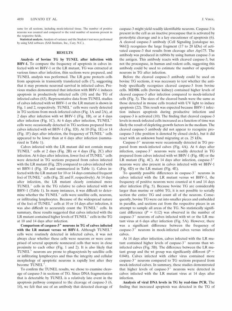
Analysis of bovine TG by TUNEL after infection withBHV-1. To compare the frequency of apoptosis in calves in-fected with wt BHV-1 or the LR mutant, TG was extracted atvarious times after infection, thin sections were prepared, andTUNEL analysis was performed. The LR gene protects cellsfrom apoptosis in transiently transfected cells (7), suggestingthat it may promote neuronal survival in infected calves. Pre-vious studies demonstrated that infection with BHV-1 inducesapoptosis in productively infected cells (10) and the TG ofinfected calves (63). Representative data from TUNEL studiesof calves infected with wt BHV-1 or the LR mutant is shown inFig. 1 and 2, respectively. TUNEL� cells were rarely detectedin TG sections from mock-infected calves (Fig. 1A and 2A), at2 days after infection with wt BHV-1 (Fig. 1B), or at 4 daysafter infection (Fig. 1C). At 6 days after infection, TUNEL�
cells were occasionally detected in TG sections prepared fromcalves infected with wt BHV-1 (Fig. 1D). At 10 (Fig. 1E) or 14(Fig. 1F) days after infection, the frequency of TUNEL� cellsappeared to be lower than at 6 days after infection (summa-rized in Table 1).
Calves infected with the LR mutant did not contain manyTUNEL� cells at 2 days (Fig. 2B) or 4 days (Fig. 2C) afterinfection. At 6 days after infection, fewer foci of TUNEL� cellswere detected in TG sections prepared from calves infectedwith the LR mutant (Fig. 2D) compared to calves infected withwt BHV-1 (Fig. 1D and summarized in Table 1). Calves in-fected with the LR mutant for 10 or 14 days contained frequentfoci of TUNEL� cells (Fig. 2E and F, respectively). At 14 daysafter infection, the LR mutant clearly contained moreTUNEL� cells in the TG relative to calves infected with wtBHV-1 (Table 1). In many instances, it was difficult to deter-mine whether the TUNEL� cells were satellite cells, neurons,or infiltrating lymphocytes. Because of the widespread natureof the foci of TUNEL� cells at 10 or 14 days after infection, itwas also difficult to accurately count the TUNEL� cells. Insummary, these results suggested that calves infected with theLR mutant contained higher levels of TUNEL� cells in the TGat 10 and 14 days after infection.
Comparison of caspase-3� neurons in TG of calves infectedwith the LR mutant versus wt BHV-1. Although TUNEL�
cells were routinely detected in infected calves, it was notalways clear whether these cells were neurons or were com-prised of several apoptotic nonneural cells that were in closeproximity to each other (Fig. 1 and 2). It is also likely thatTUNEL� neurons are prone to phagocytosis by satellite cellsor infiltrating lymphocytes and thus the integrity and cellularmorphology of apoptotic neurons is rapidly lost after theybecome TUNEL�.
To confirm the TUNEL results, we chose to examine cleav-age of caspase-3 in sections of TG. Since DNA fragmentationthat is detectable by TUNEL is a relatively late event in theapoptosis pathway compared to the cleavage of caspase-3 (6,14), we felt that use of an antibody that detected cleavage of
caspase-3 might yield readily identifiable neurons. Caspase-3 ispresent in the cell as an inactive procaspase that is activated byproteolytic cleavage and is a key executioner of apoptosis (6).A cleaved caspase-3 antibody (Cell Signaling [catalogue no.9661]) recognizes the large fragment (17 to 20 kDa) of acti-vated caspase-3 that results from cleavage after Asp175. Theantibody was produced in rabbits by using human caspase-3 asthe antigen. This antibody reacts with cleaved caspase-3, butnot the procaspase, in human and rodent cells, suggesting thisantibody could be used to estimate the number of apoptoticneurons in TG after infection.
Before the cleaved caspase-3 antibody could be used onbovine TG sections, it was necessary to test whether the anti-body specifically recognizes cleaved caspase-3 from bovinecells. MDBK cells (bovine kidney) contained higher levels ofcleaved caspase-3 after infection compared to mock-infectedcells (Fig. 3). The sizes of the cleaved bands were the same asthose detected in mouse cells treated with UV light to induceapoptosis (22). This result was expected because BHV-1 infec-tion induces apoptosis during productive infection andcaspase-3 is activated (10). The finding that cleaved caspase-3levels in mock-infected cells increased as a function of time waslikely the result of depleting growth factors in the medium. Thecleaved caspase-3 antibody did not appear to recognize pro-caspase-3 (the position is denoted by closed circle), but it didreact with an unknown band migrating at ca. 25 kDa.
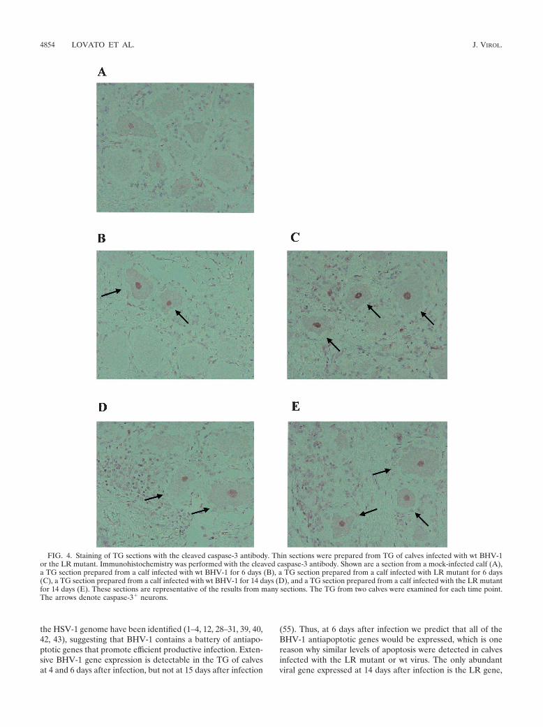
Caspase-3� neurons were occasionally detected in TG pre-pared from mock-infected calves (Fig. 4A). At 6 days afterinfection, caspase-3� neurons were readily detected in TGprepared from calves infected with wt BHV-1 (Fig. 4B) or theLR mutant (Fig. 4C). At 14 days after infection, caspase-3�
neurons were also present in calves infected with wt BHV-1(Fig. 4D) or the LR mutant (Fig. 4E).
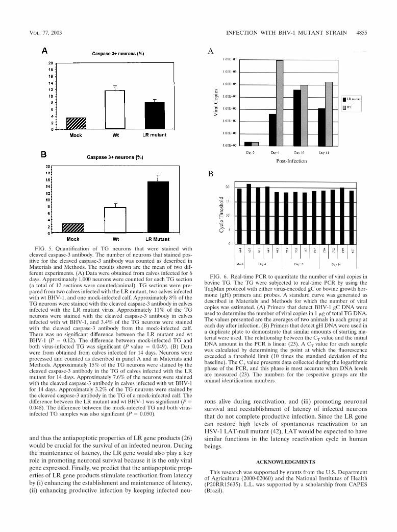
To quantify possible differences in caspase-3� neurons incalves infected with the LR mutant versus wt BHV-1, thefrequency of positive neurons were counted at 6 and 14 daysafter infection (Fig. 5). Because bovine TG are considerablylarger than murine or rabbit TG, it is not possible to seriallysection the entire TG and count all of the neurons. Conse-quently, bovine TG were cut into smaller pieces and embeddedin paraffin, and sections cut from the respective pieces in anattempt to sample all areas of the TG. No statistically signifi-cant difference (P � 0.12) was observed in the number ofcaspase-3� neurons of calves infected with wt or the LR mu-tant virus at 6 days after infection (Fig. 5A). However, therewas a significant difference between the frequency ofcaspase-3� neurons in mock-infected calves versus infectedcalves.
At 14 days after infection, calves infected with the LR mu-tant contained higher levels of caspase-3� neurons than wt-infected calves (Fig. 5B). The difference between the LR mu-tant group and the wt group was significantly different (P �0.048). Calves infected with either virus contained morecaspase-3� neurons compared to TG sections prepared frommock-infected calves. In summary, these studies demonstratedthat higher levels of caspase-3� neurons were detected incalves infected with the LR mutant virus at 14 days afterinfection.
Analysis of viral DNA levels in TG by real-time PCR. Thefinding that increased apoptosis was detected in the TG of
4850 LOVATO ET AL. J. VIROL.
calves infected with the LR mutant for 14 days suggested thatthe LR mutant did not establish latency efficiently. A previousstudy demonstrated that calves infected with the LR mutantcontained 10 to 100 times less infectious virus in the TG duringacute infection by using a semiquantitative assay and that lessviral DNA was present in the TG of latently infected calves(26). However, the present study did not compare DNA levelsin the TG of acutely infected calves. At 4 days after infection,
calves infected with the LR mutant contained more than 400times less viral DNA in TG compared to calves infected withwt BHV-1, but by 10 days after infection there was �10-foldfewer copies of the viral genome (Fig. 6). At 14 days afterinfection, the LR mutant contained ca. 100 times less viralgenome equivalents in TG compared to TG of calves infectedwith wt BHV-1.
These studies suggest that there is an initial delay in the
FIG. 1. TUNEL analysis of calves infected with wt BHV-1. Thin sections were prepared from the TG of calves infected with wt BHV-1. TUNELassays were performed as described in Materials and Methods. Shown are a section from a mock-infected calf (A) and sections obtained 2 days(B), 4 days (C), 6 days (D), 10 days (E), and 14 days (F) after infection. These sections are representative of the results from many sections. TheTG from two calves were examined for each time point. The arrows denote foci of TUNEL� cells.
VOL. 77, 2003 INFECTION WITH BHV-1 MUTANT STRAIN 4851

accumulation of viral genomes in the TG of calves infectedwith the LR mutant. Although there appeared to be an in-crease in the numbers of viral genomes at 10 days after infec-tion in calves infected with the LR mutant, fewer viral genomeswere present at 14 days after infection (Fig. 6) and duringlatency (26). When the same experiment was performed withprimers directed against the bovine growth hormone gene, itwas evident that similar levels of DNA were included in the
assays (Fig. 6B). In summary, we demonstrated here that re-duced levels of viral DNA were present in the TG of calves thatwere acutely infected with the LR mutant.
DISCUSSION
We compared here the apoptotic frequencies in the TG ofcalves infected with the LR mutant versus the apoptotic fre-
FIG. 2. TUNEL analysis of calves infected with the LR mutant. Thin sections were prepared from the TG of calves infected with the LRmutant. TUNEL assays were performed as described in Materials and Methods. Shown are a section from a mock-infected calf (A) and sectionsobtained 2 days (B), 4 days (C), 6 days (D), 10 days (E), and 14 days (F) after infection. These sections are representative of the results from manysections. The TG from two calves were examined for each time point. The arrows denote foci of TUNEL� cells.
4852 LOVATO ET AL. J. VIROL.

quencies in the TG of calves infected with wt BHV-1. Twoindependent techniques, TUNEL assay and the use of an an-tibody that specifically recognizes cleaved caspase-3, demon-strated that calves infected with the LR mutant had higherlevels of apoptosis in calves infected with wt BHV-1. Consid-ering that reduced levels of viral DNA were detected in TG ofcalves latently infected with the LR mutant (26) and duringacute infection (Fig. 6), it was somewhat surprising to seeincreased levels of apoptosis at 14 days after infection. If oneconsiders the level of apoptosis per unit of viral DNA in theTG, calves infected with the LR mutant had much higherfrequencies of apoptosis at 6 and 14 days after infection rela-tive to calves infected with wt BHV-1. Consequently, calvesinfected with the LR mutant demonstrated a reduced estab-lishment of latency, which, in part, would explain why the LR
mutant does not reactivate from latency after dexamethasonetreatment (26).
These studies indicated that it was easier to morphologicallyidentify cleaved caspase-3� neurons than TUNEL� neurons.Since caspase-3 cleavage is an earlier event in the apoptoticcascade relative to the extensive DNA cleavage that is requiredfor a TUNEL� cell, this result should be expected. Manyneurons that were stained with the cleaved caspase-3 antibodyhad a strong nuclear signal. Prior to apoptosis, caspase-3 islocalized in the cytoplasm and mitochondrial fraction in Jurkatcells (human T cells) (67). After induction of apoptosis,caspase-3 can be readily detected in the nucleus. Whencaspase-3 is expressed as a green fluorescent protein fusionprotein, strong nuclear localization is observed in HeLa cells,but weak nuclear localization is observed in 293 (human epi-thelial cell-like cells) or Jurkat cells (57), indicating that thelocalization of caspase-3 can be cell type dependent. Prevent-ing nuclear localization of activated caspases correlates withinhibiting apoptosis (11), indicating that detection of cleavedcaspase-3 in the nucleus has biological relevance.
The LR mutant contains three stop codons near the begin-ning of the LR RNA that are designed to prevent proteinexpression from all three reading frames, and the LR mutantalso lacks 25 bp from wt sequence to prevent reversion to wt(27). This mutation would likely interfere with expression ofLR ORF2 because the first stop codon is eight bases down-stream of the first in-frame ATG of LR ORF2. A peptideantibody that is directed against amino acid sequences withinLR ORF2 recognizes a protein of ca. 40 kDa in cells trans-fected with a wt LR gene construct (7, 25, 32), but not whencells are transfected with a plasmid containing the mutationused to make the LR mutant virus (7). Because the LR RNAis alternatively spliced (9), there is a possibility that more thanone protein is expressed. Although we believe that the expres-sion of a protein encoded by the LR gene regulates the latencyreactivation cycle, it is possible that differences in LR RNAexpression in the LR mutant is responsible for the attenuatedphenotype. Regardless of whether an LR protein or changes inLR RNA mediate the altered phenotype of the LR mutant, itis clear that wt expression of LR gene products is crucial forthe latency reactivation cycle in cattle.
Plasmids expressing LR gene products (7) and LAT (1, 22,28, 43) can inhibit apoptosis in transiently transfected cells.Two independent studies have also concluded that rabbits (43)or mice (1) infected with a LAT mutant have more apoptoticneurons during acute infection than do animals infected withthe respective LAT-positive HSV-1 strains. The present studyprovides evidence that the antiapoptotic properties of the LRgene play a crucial role in the latency reactivation cycle ofBHV-1. Taken together, these three studies strongly suggestthat the HSV-1 gene encoding LAT has a similar role in thelatency reactivation cycle in humans. Since the LR gene isrequired for dexamethasone-induced reactivation from latencyin cattle (26) and promotes virus shedding in the ocular cavityof infected calves (27), it is also tempting to speculate that theimportance of LAT has been underestimated with small ani-mal models.
HSV-1 (2, 3, 12, 13, 37) and BHV-1 (10, 18–20) can induceor inhibit apoptosis in a cell type-dependent manner afterinfection of cultured cells. Several antiapoptotic genes within
FIG. 3. Western blot analysis of cleaved caspase-3 after productiveinfection. Bovine kidney cells (MDBK) were infected with wt BHV-1(4 PFU/cell). At the designated times (hours postinfection), cell lysatewas prepared from infected or mock-infected cells. Western blot anal-ysis with 50 �g of protein was performed with the antibody that rec-ognizes cleaved caspase-3. To further ensure that similar levels of totalprotein were added to each lane, a Western blot was performed withan antibody that recognizes �-actin (data not shown). The arrowsdenote the two major cleaved products of caspase-3. The closed circledenotes the position of uncleaved caspase-3.
TABLE 1. Summary of TUNEL results
Day(dpi)a
TUNEL resultb
Mock LR mutant 1 LR mutant 2 wt 1 wt 2
2 4 6 �� ��
10 ���� ���� ���� ����14 ����� ��� �� ��
a TG sections from two mock-infected calves (Mock), two calves infected withthe LR mutant (LR mutant 1 and LR mutant 2), or two calves infected with wtBHV-1 (wt 1 and wt 2) were tested for each time point. dpi, days postinfection.
b –, Sections lacking TUNEL� cells. The relative numbers of TUNEL� focipresent in each section were scored as follows: ��, 5–8 foci of TUNEL� cells;���, 9–12 foci of TUNEL� cells; ����, 13–16 foci of TUNEL� cells;�����, 16 foci of TUNEL� cells.
VOL. 77, 2003 INFECTION WITH BHV-1 MUTANT STRAIN 4853
the HSV-1 genome have been identified (1–4, 12, 28–31, 39, 40,42, 43), suggesting that BHV-1 contains a battery of antiapo-ptotic genes that promote efficient productive infection. Exten-sive BHV-1 gene expression is detectable in the TG of calvesat 4 and 6 days after infection, but not at 15 days after infection
(55). Thus, at 6 days after infection we predict that all of theBHV-1 antiapoptotic genes would be expressed, which is onereason why similar levels of apoptosis were detected in calvesinfected with the LR mutant or wt virus. The only abundantviral gene expressed at 14 days after infection is the LR gene,
FIG. 4. Staining of TG sections with the cleaved caspase-3 antibody. Thin sections were prepared from TG of calves infected with wt BHV-1or the LR mutant. Immunohistochemistry was performed with the cleaved caspase-3 antibody. Shown are a section from a mock-infected calf (A),a TG section prepared from a calf infected with wt BHV-1 for 6 days (B), a TG section prepared from a calf infected with LR mutant for 6 days(C), a TG section prepared from a calf infected with wt BHV-1 for 14 days (D), and a TG section prepared from a calf infected with the LR mutantfor 14 days (E). These sections are representative of the results from many sections. The TG from two calves were examined for each time point.The arrows denote caspase-3� neurons.
4854 LOVATO ET AL. J. VIROL.
and thus the antiapoptotic properties of LR gene products (26)would be crucial for the survival of an infected neuron. Duringthe maintenance of latency, the LR gene would also play a keyrole in promoting neuronal survival because it is the only viralgene expressed. Finally, we predict that the antiapoptotic prop-erties of LR gene products stimulate reactivation from latencyby (i) enhancing the establishment and maintenance of latency,(ii) enhancing productive infection by keeping infected neu-
rons alive during reactivation, and (iii) promoting neuronalsurvival and reestablishment of latency of infected neuronsthat do not complete productive infection. Since the LR genecan restore high levels of spontaneous reactivation to anHSV-1 LAT-null mutant (42), LAT would be expected to havesimilar functions in the latency reactivation cycle in humanbeings.
ACKNOWLEDGMENTS
This research was supported by grants from the U.S. Departmentof Agriculture (2000-02060) and the National Institutes of Health(P20RR15635). L.L. was supported by a scholarship from CAPES(Brazil).
FIG. 5. Quantification of TG neurons that were stained withcleaved caspase-3 antibody. The number of neurons that stained pos-itive for the cleaved caspase-3 antibody was counted as described inMaterials and Methods. The results shown are the mean of two dif-ferent experiments. (A) Data were obtained from calves infected for 6days. Approximately 1,000 neurons were counted for each TG section(a total of 12 sections were counted/animal). TG sections were pre-pared from two calves infected with the LR mutant, two calves infectedwith wt BHV-1, and one mock-infected calf. Approximately 8% of theTG neurons were stained with the cleaved caspase-3 antibody in calvesinfected with the LR mutant virus. Approximately 11% of the TGneurons were stained with the cleaved caspase-3 antibody in calvesinfected with wt BHV-1, and 3.4% of the TG neurons were stainedwith the cleaved caspase-3 antibody from the mock-infected calf.There was no significant difference between the LR mutant and wtBHV-1 (P � 0.12). The difference between mock-infected TG andboth virus-infected TG was significant (P value � 0.049). (B) Datawere from obtained from calves infected for 14 days. Neurons wereprocessed and counted as described in panel A and in Materials andMethods. Approximately 15% of the TG neurons were stained by thecleaved caspase-3 antibody in the TG of calves infected with the LRmutant for 14 days. Approximately 7.6% of the neurons were stainedwith the cleaved caspase-3 antibody in calves infected with wt BHV-1for 14 days. Approximately 3.2% of the TG neurons were stained bythe cleaved caspase-3 antibody in the TG of a mock-infected calf. Thedifference between the LR mutant and wt BHV-1 was significant (P �0.048). The difference between the mock-infected TG and both virus-infected TG samples was also significant (P � 0.050).
FIG. 6. Real-time PCR to quantitate the number of viral copies inbovine TG. The TG were subjected to real-time PCR by using theTaqMan protocol with either virus-encoded gC or bovine growth hor-mone (gH) primers and probes. A standard curve was generated asdescribed in Materials and Methods for which the number of viralcopies was estimated. (A) Primers that detect BHV-1 gC DNA wereused to determine the number of viral copies in 1 �g of total TG DNA.The values presented are the averages of two animals in each group ateach day after infection. (B) Primers that detect gH DNA were used ina duplicate plate to demonstrate that similar amounts of starting ma-terial were used. The relationship between the CT value and the initialDNA amount in the PCR is linear (23). A CT value for each samplewas calculated by determining the point at which the fluorescenceexceeded a threshold limit (10 times the standard deviation of thebaseline). The CT value presents data collected during the logarithmicphase of the PCR, and this phase is most accurate when DNA levelsare measured (23). The numbers for the respective groups are theanimal identification numbers.
VOL. 77, 2003 INFECTION WITH BHV-1 MUTANT STRAIN 4855
We thank B. Clowser for assistance with cattle experiments.
REFERENCES
1. Ahmed, M., M. Lock, C. G. Miller, and N. W. Fraser. 2002. Regions of theherpes simplex virus type 1 latency-associated transcript that protect cellsfrom apoptosis in vitro and protect neuronal cells in vivo. J. Virol. 76:717–729.
2. Asano, S., T. Honda, F. Goshima, D. Watanabe, Y. Miyake, Y. Sugiura, andY. Nishiyama. 1999. US3 protein kinase of herpes simplex virus type 2 playsa role in protecting corneal epithelial cells from apoptosis in infected mice.J. Gen. Virol. 80(Pt. 1):51–56.
3. Aubert, M., and J. A. Blaho. 1999. The herpes simplex virus type 1 regulatoryprotein ICP27 is required for the prevention of apoptosis in infected humancells. J. Virol. 73:2803–2813.
4. Blaho, J. A., and M. Aubert. 2001. Modulation of apoptosis during herpessimplex virus infection in human cells. Microbes Infect. 3:1–8.
5. Carter, J. J., A. D. Weinberg, A. Pollard, R. Reeves, J. A. Magnuson, andN. S. Magnuson. 1989. Inhibition of T-lymphocyte mitogenic responses andeffects on cell functions by bovine herpesvirus 1. J. Virol. 63:1525–1530.
6. Chang, H. Y., and X. Yang. 2000. Proteases for cell suicide: functions andregulation of caspases. Microbiol. Mol. Biol. Rev. 64:821–846.
7. Ciacci-Zanella, J., M. Stone, G. Henderson, and C. Jones. 1999. The latency-related gene of bovine herpesvirus 1 inhibits programmed cell death. J. Virol.73:9734–9740.
8. Devi-Rao, G. B., D. C. Bloom, J. G. Stevens, and E. K. Wagner. 1994. Herpessimplex virus type 1 DNA replication and gene expression during explant-induced reactivation of latently infected murine sensory ganglia. J. Virol.68:1271–1282.
9. Devireddy, L. R., and C. Jones. 1998. Alternative splicing of the latency-related transcript of bovine herpesvirus 1 yields RNAs containing uniqueopen reading frames. J. Virol. 72:7294–7301.
10. Devireddy, L. R., and C. J. Jones. 1999. Activation of caspases and p53 bybovine herpesvirus 1 infection results in programmed cell death and efficientvirus release. J. Virol. 73:3778–3788.
11. Fankhauser, C., R. M. Friedlander, and V. Gagliardini. 2000. Prevention ofnuclear localization of activated caspases correlates with inhibition of apo-ptosis. Apoptosis 5:117–132.
12. Galvan, V., R. Brandimarti, and B. Roizman. 1999. Herpes simplex virus 1blocks caspase-3-independent and caspase-dependent pathways to celldeath. J. Virol. 73:3219–3226.
13. Galvan, V., and B. Roizman. 1998. Herpes simplex virus 1 induces and blocksapoptosis at multiple steps during infection and protects cells from exoge-nous inducers in a cell-type-dependent manner. Proc. Natl. Acad. Sci. USA95:3931–3936.
14. Granville, D. J., C. M. Carthy, H. Jiang, G. C. Shore, B. M. McManus, andD. W. Hunt. 1998. Rapid cytochrome c release, activation of caspases 3, 6, 7,and 8 followed by Bap31 cleavage in HeLa cells treated with photodynamictherapy. FEBS Lett. 437:5–10.
15. Griebel, P. J., H. B. Ohmann, M. J. Lawman, and L. A. Babiuk. 1990. Theinteraction between bovine herpesvirus type 1 and activated bovine T lym-phocytes. J. Gen. Virol. 71(Pt. 2):369–377.
16. Griebel, P. J., L. Qualtiere, W. C. Davis, A. Gee, H. Bielefeldt Ohmann, M. J.Lawman, and L. A. Babiuk. 1987. T lymphocyte population dynamics andfunction following a primary bovine herpesvirus type-1 infection. Viral Im-munol. 1:287–304.
17. Griebel, P. J., L. Qualtiere, W. C. Davis, M. J. Lawman, and L. A. Babiuk.1987. Bovine peripheral blood leukocyte subpopulation dynamics following aprimary bovine herpesvirus-1 infection. Viral Immunol. 1:267–286.
18. Hanon, E., S. Hoornaert, F. Dequiedt, A. Vanderplasschen, J. Lyaku, L.Willems, and P. P. Pastoret. 1997. Bovine herpesvirus 1-induced apoptosisoccurs at the G0/G1 phase of the cell cycle. Virology 232:351–358.
19. Hanon, E., G. Meyer, A. Vanderplasschen, C. Dessy-Doize, E. Thiry, andP. P. Pastoret. 1998. Attachment but not penetration of bovine herpesvirus1 is necessary to induce apoptosis in target cells. J. Virol. 72:7638–7641.
20. Hanon, E., A. Vanderplasschen, S. Lyaku, G. Keil, M. Denis, and P. P.Pastoret. 1996. Inactivated bovine herpesvirus 1 induces apoptotic cell deathof mitogen-stimulated bovine peripheral blood mononuclear cells. J. Virol.70:4116–4120.
21. Hariharan, M. J., C. Nataraj, and S. Srikumaran. 1993. Downregulation ofmurine MHC class I expression by bovine herpesvirus 1. Viral Immunol.6:273–284.
22. Henderson, G., W. Peng, L. Jin, G.-C. Perng, A. B. Nesburn, S. L. Wechsler,and C. Jones. 2002. Suppression of caspase 8- and caspase 9-induced apo-ptosis by the herpes simplex virus (HSV-1) encoded latency-associated tran-script (LAT). J. Neurovirol., 8(Suppl. 2):103–111.
23. Higuchi, R., C. Fockler, G. Dollinger, and R. Watson. 1993. Kinetic PCRanalysis: real-time monitoring of DNA amplification reactions. Bio/Technol-ogy 11:1026–1030.
24. Hinkley, S., A. B. Hill, and S. Srikumaran. 1998. Bovine herpesvirus-1infection affects the peptide transport activity in bovine cells. Virus Res.53:91–96.
25. Hossain, A., L. M. Schang, and C. Jones. 1995. Identification of gene prod-
ucts encoded by the latency-related gene of bovine herpesvirus 1. J. Virol.69:5345–5352.
26. Inman, M., L. Lovato, A. Doster, and C. Jones. 2002. A mutation in thelatency related gene of bovine herpesvirus 1 interferes with the latency-reactivation cycle of latency in calves. J. Virol. 76:6771–6779.
27. Inman, M., L. Lovato, A. Doster, and C. Jones. 2001. A mutation in thelatency-related gene of bovine herpesvirus 1 leads to impaired ocular shed-ding in acutely infected calves. J. Virol. 75:8507–8515.
28. Inman, M., G. C. Perng, G. Henderson, H. Ghiasi, A. B. Nesburn, S. L.Wechsler, and C. Jones. 2001. Region of herpes simplex virus type 1 latency-associated transcript sufficient for wild-type spontaneous reactivation pro-motes cell survival in tissue culture. J. Virol. 75:3636–3646.
29. Jerome, K. R., Z. Chen, R. Lang, M. R. Torres, J. Hofmeister, S. Smith, R.Fox, C. J. Froelich, and L. Corey. 2001. HSV and glycoprotein J inhibitcaspase activation and apoptosis induced by granzyme B or Fas. J. Immunol.167:3928–3935.
30. Jerome, K. R., R. Fox, Z. Chen, A. E. Sears, H. Lee, and L. Corey. 1999.Herpes simplex virus inhibits apoptosis through the action of two genes, Us5and Us3. J. Virol. 73:8950–8957.
31. Jerome, K. R., J. F. Tait, D. M. Koelle, and L. Corey. 1998. Herpes simplexvirus type 1 renders infected cells resistant to cytotoxic T-lymphocyte-in-duced apoptosis. J. Virol. 72:436–441.
32. Jiang, Y., A. Hossain, M. T. Winkler, T. Holt, A. Doster, and C. Jones. 1998.A protein encoded by the latency-related gene of bovine herpesvirus 1 isexpressed in trigeminal ganglionic neurons of latently infected cattle andinteracts with cyclin-dependent kinase 2 during productive infection. J. Virol.72:8133–8142.
33. Jones, C. 1998. Alphaherpesvirus latency: its role in disease and survival ofthe virus in nature. Adv. Virus Res. 51:81–133.
34. Jones, C., T. J. Newby, T. Holt, A. Doster, M. Stone, J. Ciacci-Zanella, C. J.Webster, and M. W. Jackwood. 2000. Analysis of latency in cattle afterinoculation with a temperature sensitive mutant of bovine herpesvirus 1(RLB106). Vaccine 18:3185–3195.
35. Kutish, G., T. Mainprize, and D. Rock. 1990. Characterization of the latency-related transcriptionally active region of the bovine herpesvirus 1 genome.J. Virol. 64:5730–5737.
36. Lee, L. G., C. R. Connell, and W. Bloch. 1993. Allelic discrimination bynick-translation PCR with fluorogenic probes. Nucleic Acids Res. 21:3761–3766.
37. Leopardi, R., and B. Roizman. 1996. The herpes simplex virus major regu-latory protein ICP4 blocks apoptosis induced by the virus or by hyperther-mia. Proc. Natl. Acad. Sci. USA 93:9583–9587.
38. Maggioncalda, J., A. Mehta, Y. H. Su, N. W. Fraser, and T. M. Block. 1996.Correlation between herpes simplex virus type 1 rate of reactivation fromlatent infection and the number of infected neurons in trigeminal ganglia.Virology 225:72–81.
39. Munger, J., A. V. Chee, and B. Roizman. 2001. The U(S)3 protein kinaseblocks apoptosis induced by the d120 mutant of herpes simplex virus 1 at apremitochondrial stage. J. Virol. 75:5491–5497.
40. Munger, J., and B. Roizman. 2001. The US3 protein kinase of herpes simplexvirus 1 mediates the posttranslational modification of BAD and preventsBAD-induced programmed cell death in the absence of other viral proteins.Proc. Natl. Acad. Sci. USA 98:10410–10415.
41. Nataraj, C., S. Eidmann, M. J. Hariharan, J. H. Sur, G. A. Perry, and S.Srikumaran. 1997. Bovine herpesvirus 1 downregulates the expression ofbovine MHC class I molecules. Viral Immunol. 10:21–34.
42. Perng, G.-C., B. Maguen, L. Jin, K. R. Mott, N. Osorio, S. M. Slanina, A.Yukht, H. Ghiasi, A. B. Nesburn, M. Inman, G. Henderson, C. Jones, S. L.Wechsler. 2002. A gene capable of blocking apoptosis can substitute for theherpes simplex virus type 1 latency-associated transcript gene and restorewild-type reactivation levels. J. Virol. 76:1224–1235.
43. Perng, G.-C., C. Jones, J. Ciacci-Zanella, M. Stone, G. Henderson, A. Yukht,S. M. Slanina, F. M. Hoffman, H. Ghiasi, A. B. Nesburn, S. Wechsler. 2000.Virus-induced neuronal apoptosis blocked by the herpes simplex virus la-tency-associated transcript (LAT). Science 287:1500–1503.
44. Perng, G. C., K. Chokephaibulkit, R. L. Thompson, N. M. Sawtell, S. M.Slanina, H. Ghiasi, A. B. Nesburn, and S. L. Wechsler. 1996. The region ofthe herpes simplex virus type 1 LAT gene that is colinear with the ICP34.5gene is not involved in spontaneous reactivation. J. Virol. 70:282–291.
45. Perng, G. C., E. C. Dunkel, P. A. Geary, S. M. Slanina, H. Ghiasi, R. Kaiwar,A. B. Nesburn, and S. L. Wechsler. 1994. The latency-associated transcriptgene of herpes simplex virus type 1 (HSV-1) is required for efficient in vivospontaneous reactivation of HSV-1 from latency. J. Virol. 68:8045–8055.
46. Perng, G. C., H. Ghiasi, S. M. Slanina, A. B. Nesburn, and S. L. Wechsler.1996. The spontaneous reactivation function of the herpes simplex virus type1 LAT gene resides completely within the first 1.5 kilobases of the 8.3-kilobase primary transcript. J. Virol. 70:976–984.
47. Perng, G. C., S. M. Slanina, H. Ghiasi, A. B. Nesburn, and S. L. Wechsler.1996. A 371-nucleotide region between the herpes simplex virus type 1(HSV-1) LAT promoter and the 2-kilobase LAT is not essential for efficientspontaneous reactivation of latent HSV-1. J. Virol. 70:2014–2018.
48. Perng, G. C., S. M. Slanina, A. Yukht, B. S. Drolet, W. Keleher, Jr., H.
4856 LOVATO ET AL. J. VIROL.
Ghiasi, A. B. Nesburn, and S. L. Wechsler. 1999. A herpes simplex virus type1 latency-associated transcript mutant with increased virulence and reducedspontaneous reactivation. J. Virol. 73:920–929.
49. Perng, G. C., S. M. Slanina, A. Yukht, H. Ghiasi, A. B. Nesburn, and S. L.Wechsler. 2000. The latency-associated transcript gene enhances establish-ment of herpes simplex virus type 1 latency in rabbits. J. Virol. 74:1885–1891.
50. Rock, D., J. Lokensgard, T. Lewis, and G. Kutish. 1992. Characterization ofdexamethasone-induced reactivation of latent bovine herpesvirus 1. J. Virol.66:2484–2490.
51. Rock, D. L., S. L. Beam, and J. E. Mayfield. 1987. Mapping bovine herpes-virus type 1 latency-related RNA in trigeminal ganglia of latently infectedrabbits. J. Virol. 61:3827–3831.
52. Rock, D. L., A. B. Nesburn, H. Ghiasi, J. Ong, T. L. Lewis, J. R. Lokensgard,and S. L. Wechsler. 1987. Detection of latency-related viral RNAs in tri-geminal ganglia of rabbits latently infected with herpes simplex virus type 1.J. Virol. 61:3820–3826.
53. Sawtell, N. M., and R. L. Thompson. 1992. Herpes simplex virus type 1latency-associated transcription unit promotes anatomical site-dependentestablishment and reactivation from latency. J. Virol. 66:2157–2169.
54. Schang, L. M., A. Hossain, and C. Jones. 1996. The latency-related gene ofbovine herpesvirus 1 encodes a product which inhibits cell cycle progression.J. Virol. 70:3807–3814.
55. Schang, L. M., and C. Jones. 1997. Analysis of bovine herpesvirus 1 tran-scripts during a primary infection of trigeminal ganglia of cattle. J. Virol.71:6786–6795.
56. Sheffy, B. E., and D. H. Davies. 1972. Reactivation of a bovine herpesvirusafter corticosteroid treatment. Proc. Soc. Exp. Biol. Med. 140:974–976.
57. Shikama, Y., M. U, T. Miyashita, and M. Yamada. 2001. Comprehensivestudies on subcellular localizations and cell death-inducing activities of eightGFP-tagged apoptosis-related caspases. Exp. Cell Res. 264:315–325.
58. Stevens, J. G., E. K. Wagner, G. B. Devi-Rao, M. L. Cook, and L. T. Feldman.1987. RNA complementary to a herpesvirus alpha gene mRNA is prominentin latently infected neurons. Science 235:1056–1059.
59. Thompson, R. L., and N. M. Sawtell. 1997. The herpes simplex virus type 1latency-associated transcript gene regulates the establishment of latency.J. Virol. 71:5432–5440.
60. Tikoo, S. K., M. Campos, and L. A. Babiuk. 1995. Bovine herpesvirus 1(BHV-1): biology, pathogenesis, and control. Adv. Virus Res. 45:191–223.
61. Wagner, E. K., and D. C. Bloom. 1997. Experimental investigation of herpessimplex virus latency. Clin. Microbiol. Rev. 10:419–443.
62. Winkler, M. T., A. Doster, and C. Jones. 1999. Bovine herpesvirus 1 caninfect CD4� T lymphocytes and induce programmed cell death during acuteinfection of cattle. J. Virol. 73:8657–8668.
63. Winkler, M. T., A. Doster, J. H. Sur, and C. Jones. 2002. Analysis of bovinetrigeminal ganglia following infection with bovine herpesvirus 1. Vet. Micro-biol. 86:139–155.
64. Winkler, M. T., L. S. Schang, A. Doster, T. Holt, and C. Jones. 2000. Analysisof cyclins in trigeminal ganglia of calves infected with bovine herpesvirus-1.J. Gen. Virol. 81(Pt. 12):2993–2998.
65. Winkler, M. T. C., A. Doster, and C. Jones. Bovine herpesvirus-1 can infectCD4� T lymphocytes and induce programmed cell death during acute in-fection of cattle. 2000. Persistence and reactivation of bovine herpesvirus 1 inthe tonsil of latently infected calves. J. Virol. 74:5337–5346.
66. Wyler, R., M. Engels, and M. Schwyzer. 1989. Infectious bovine rhinotra-cheitis/vulvovaginitis (BHV-1), p. 1–72. In G. Witman (ed.), Herpesvirusdiseases of cattle, horses, and pigs. Kluwer Academic Publishers, Boston,Mass.
67. Zhivotovsky, B., A. Samali, A. Gahm, and S. Orrenius. 1999. Caspases: theirintracellular localization and translocation during apoptosis. Cell Death Dif-fer. 6:644–651.
VOL. 77, 2003 INFECTION WITH BHV-1 MUTANT STRAIN 4857
Top Related
Copyright © 2022 FDOKUMEN

