


Bahasa
Halaman
Hukum

Original
Pap
er
Tsakalof, Bairachtari, Aslani, Chryssoulakis, Kolisis 167
J. Sep. Sci. 2004, 27, 167–173 www.jss-journal.de i 2004WILEY-VCH Verlag GmbH&Co. KGaA,Weinheim
Andreas K. Tsakalof1
Kyriaki A. Bairachtari1, 2
Ioanna S. Aslani2
Ioannis D. Chryssoulakis1, 2
Fragiskos N. Kolisis2
1“Ormylia” Art Diagnosis Center,Sacred Convent of Annunciation,63071 Ormylia-Chalkidiki,Greece
2National Technical University ofAthens, Department of ChemicalEngineering, Iroon Polytechniou9, 15780 Zografou Campus,Athens, Greece
Impact of biological factors on bindingmediaidentification in art objects: Identification of animalglue in the presence ofAspergillus niger
The materials and especially organic materials used for creation of art objects can beutilized by various microorganisms for their growth and facilitate the microbial coloni-zation of the object. An understanding of the chemical alterations in artefacts causedby the presence of microorganisms can be crucial for correct identification of thematerials initially used for the artefact creation – nowadays an important step inrestoration and/or art-historical investigation of the art object. The present articledescribes a model experiment in which we investigated the possible chemical altera-tions in animal glue films used as substrate for growth of the fungus Aspergillus niger.The sterilized animal glue solution was poured into Petri dishes, inoculated withAspergillus niger, and subsequently incubated at 158C for 0, 7, 9, 14, and 28 days.After interruption of incubation, the content of the Petri dish was analyzed for aminoacid composition by the GC-MS based method. It was found that the growth of Asper-gillus niger on animal glue films did not cause significant changes in the amino acidcomposition of the film and had no impact on animal glue identification.
KeyWords:Artefacts; Biodegradation; Bindingmedia identification;
Received: June 3, 2002; revised: September 25, 2003; accepted: October 30, 2003
DOI 10.1002/jssc.200301627
1 Introduction
The deterioration of art objects due to the action of envi-ronmental physicochemical factors (light, temperature,moisture, atmospheric pollutants) has been extensivelystudied [1–10] with the main emphasis being placed onchemical alterations of the substrate. The importance ofbiological factors in art object deterioration has also beenrecognized [11–18]. However, in this case attention hasbeen paid mainly to the characterization of the microbialflora present on mural or easel paintings and most of therelevant publications are essentially catalogues of micro-organisms isolated from painted surfaces. The factorsfacilitating or inhibiting the flora development have beenalso investigated [19]. In contrast, the chemical nature ofalterations induced by the biological factor has been stud-ied substantially less. This is especially true for alterationsin organic constituents of the artefacts such as proteins,carbohydrates, oils, and waxes. Such changes can resultprimarily from the microorganisms’ presence on an artobject when these species themselves inherently containproteins, carbohydrates, and lipids. The second reason isthe metabolic activity of the colonising microorganisms(fungi, bacteria, or algae) which can utilize the organicconstituents of the art object for their nutrition, metabolize
them, and ultimately synthesize and excrete new com-pounds. If the former case, microorganism deposition onan art object, cause mainly aesthetic alterations, themetabolic activity can be the reason of structural damageto the object, e.g. disintegration and detachment of paint-ing layer.
It is also obvious that a knowledge of the chemical altera-tions in an artefact caused by the presence of microorgan-isms can be crucial for correct identification of the materi-als used for artefact creation. Information about materialsused is important for proper object restoration and art-his-torical investigation, e.g. through revealing the paintingtechnique.
The problem can be also formulated in a more specificmanner which is more relevant to the scientific diagnosis:does the colonization of art object by microorganismsinterfere with identification of organic material used for theobject creation and if it does then how?
The present article describes amodel experiment in whichwe investigated the possible chemical alterations in ani-mal glue films used as substrate for the growth of the fun-gus Aspergillus niger. Animal glue is one of the main pro-teinaceous binders or vehicles that hold together pigmentparticles in paint. It has been extensively used in paintinggrounds [19, 20]. Other commonly used proteinaceousbinders are milk casein, whole egg, or egg constituents –egg white or egg yolk [19, 20]. The amino acid content is
Correspondence: A.K. Tsakalof, University of Thessaly, Depart-ment of Medicine, 22 Papakiriazi Str., 41222 Larissa, Greece.Phone: +30 2410 565 242. Fax: +30 2410 565 054.E-mail: [email protected].
168 Tsakalof, Bairachtari, Aslani, Chryssoulakis, Kolisis
characteristic for each binder and used for binder identifi-cation [21–24].
Aspergillus nigerwas selected for the study because fungiare an important group of microorganisms present on sur-faces exposed to the atmosphere and this species is oneof the most ubiquitous contaminants [12, 15]. It is alsoimportant that the growth of fungal cells usually precedesand favours the growth of bacterial species [12–14].
The described experiment is the first in a series plannedfor the detailed research into chemical alterations in artobjects induced by their colonization bymicroorganisms.
2 Experimental
2.1 Chemicals and reagents
Analytical grade hydrochloric acid (37%) and ammonia(25%) solutions were supplied by Merck, Germany. Hexa-decane and norleucine (both Merck) were used as internalstandards. Amino acid standard solution (2.5 lmol/mL of17 amino acid in 0.1 M HCl), hydroxyproline, isooctane(2,2,4 trimethylpentane, anhydrous, purity 99.8%), andderivatization agent N-tert-butyldimethylsilyl-N-methyltri-fluoroacetamide (MTBSTFA, derivatization grade) wereall supplied by Sigma-Aldrich Chemie, Germany. N,N-Dimethylformamide was purchased fromCarlo Erba, Italy.
Type I reagent grade water with resistivity up to18.3 MX cm and organic content a 5 ppb was producedby passing de-ionized water through a Barnstead EASY-pure RF water purification system (Fisher Scientific, UK)and was used for solutions preparations.
Aspergillus niger wild type strain was isolated at theLaboratory of Biotechnology, School of Chemical Engi-neering, National Technical University of Athens.
Egg (EY), casein (CAS), and animal glue (GL) used asreference materials as well as reference painted sampleswere provided by the Icon Painting workshop of theSacred Convent of the Annunciation. The referencepainted samples were prepared by use of lead white,minium, or cinnabar as a pigment and egg yolk as a binderand indicated respectively in Table 2 as Pb EY, MIN EY,and CIN EY.
2.2 Sample preparation – Aspergillus nigergrowth on animal glue films
The 12% w/v animal glue aqueous solution was preparedand sterilized at 1208C for 20 min in an autoclave. Subse-quently 10 mL aliquots of the sterilized solution werepored into Petri dishes and infected with 100 lL of Asper-gillus niger solution. Inoculated samples were incubatedat 158C for 0, 7, 9, 14, 28 days in duplicate. Sample freez-ing at –758C interrupted the fungal growth.
The extent of fungal colonization of animal glue substratewas estimated as total fungus weight versus days of incu-bation: the content of the Petri dishes was redissolved inwater, the fungi were filtered off and dried to the constantweight.
2.3 Apparatus and conditions
Acid hydrolysis of reference samples (egg, casein, animalglue) and infected animal glue samples was conductedwith the help of a QLAB6000Microwave Digestion Systemprovided with Very High Pressure (VHP625psi) closedvessels (Questrone Technologies Corp., Canada). Thesamples were hydrolyzed by microwave assisted vapourphase acid hydrolysis in a nitrogen atmosphere. Micro-wave oven program: temperature control heating to1608C, 10 min at 1608C and 250 W following 30 min at1608C and 500 W.
A Polaris gas chromatograph-ion trap mass spectrometer(GC-MS) provided with an AS2000 autosampler (Thermo-Finnigan, San Jose, USA) was used for amino acid quanti-fication in reference and infected samples. The gas chro-matograph was equipped with a split/splitless injector andan ATTM-5MS 30 m60.25 mm column with 0.25 lm filmthickness of 5%phenyl-95%methylpolysiloxane stationaryphase (Alltech Associates, USA). The oven temperatureprogram started from 1008C isothermal for 2 min, thenheated up to 2808C at 8 K/min and kept isothermal for10 min. The heating zones were kept at the following tem-peratures: injector 2808C, transfer line 2908C, ion source2508C.
2.4 Analytical procedure
The analytical procedure used originated from previouspublications [23, 25] and was modified for available instru-mentation and optimized. The analysis scheme comprisesthe main steps of 1) sample hydrolysis, 2) amino acid deri-vatization, 3) amino acid quantification by GC-MS, and4) acquired data (sample amino acid content) treatmentby Principle Component Analysis for binding media identi-fication.
For hydrolysis samples were placed in glass test tubesand wetted with 6N HCl. The test tubes were placed in theVHP vessel containing 10 mL of 6N HCl on the bottom.The vessel was evacuated, refilled with nitrogen, sealed,and placed in the microwave for hydrolysis in accordancewith the previously described program (Section 2.3).
The hydrolysates were diluted by water. To the aliquot ofhydrolysate solution 4 lL of norleucine (internal standard)302 lg/mL water solution was added and subsequentlythe hydrolysate was evaporated to dryness by vacuumapplication and heating at 608C.
The dried residue was redissolved in 10 lL of DMF andderivatized by 40 lL of MTBSTFA at 608C for 30 min.
J. Sep. Sci. 2004, 27, 167–173 www.jss-journal.de i 2004WILEY-VCH Verlag GmbH&Co. KGaA,Weinheim
Impact of biological factors on binding media 169
After cooling, 250 lL of hexadecane solution (20 ng/lL inisooctane) was added. Hexadecane was used for moni-toring of the sample injection efficiency.
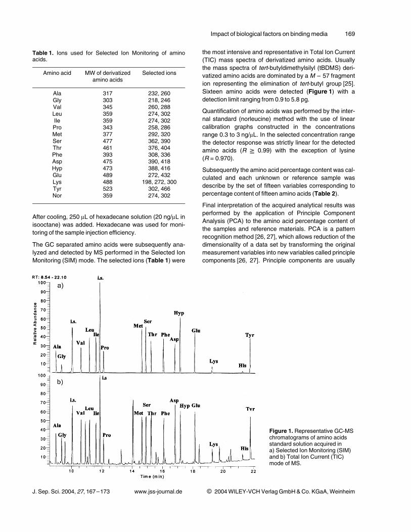
The GC separated amino acids were subsequently ana-lyzed and detected by MS performed in the Selected IonMonitoring (SIM) mode. The selected ions (Table 1) were
the most intensive and representative in Total Ion Current(TIC) mass spectra of derivatized amino acids. Usuallythe mass spectra of tert-butyldimethylsilyl (tBDMS) deri-vatized amino acids are dominated by aM – 57 fragmention representing the elimination of tert-butyl group [25].Sixteen amino acids were detected (Figure 1) with adetection limit ranging from 0.9 to 5.8 pg.
Quantification of amino acids was performed by the inter-nal standard (norleucine) method with the use of linearcalibration graphs constructed in the concentrationsrange 0.3 to 3 ng/lL. In the selected concentration rangethe detector response was strictly linear for the detectedamino acids (R G 0.99) with the exception of lysine(R = 0.970).
Subsequently the amino acid percentage content was cal-culated and each unknown or reference sample wasdescribe by the set of fifteen variables corresponding topercentage content of fifteen amino acids (Table 2).
Final interpretation of the acquired analytical results wasperformed by the application of Principle ComponentAnalysis (PCA) to the amino acid percentage content ofthe samples and reference materials. PCA is a patternrecognition method [26, 27], which allows reduction of thedimensionality of a data set by transforming the originalmeasurement variables into new variables called principlecomponents [26, 27]. Principle components are usually
J. Sep. Sci. 2004, 27, 167–173 www.jss-journal.de i 2004WILEY-VCH Verlag GmbH&Co. KGaA,Weinheim
Table 1. Ions used for Selected Ion Monitoring of aminoacids.
Amino acid MW of derivatizedamino acids
Selected ions
Ala 317 232, 260Gly 303 218, 246Val 345 260, 288Leu 359 274, 302Ile 359 274, 302Pro 343 258, 286Met 377 292, 320Ser 477 362, 390Thr 461 376, 404Phe 393 308, 336Asp 475 390, 418Hyp 473 388, 416Glu 489 272, 432Lys 488 198, 272, 300Tyr 523 302, 466Nor 359 274, 302
Figure 1. Representative GC-MSchromatograms of amino acidsstandard solution acquired ina) Selected Ion Monitoring (SIM)and b) Total Ion Current (TIC)mode of MS.
170 Tsakalof, Bairachtari, Aslani, Chryssoulakis, Kolisis
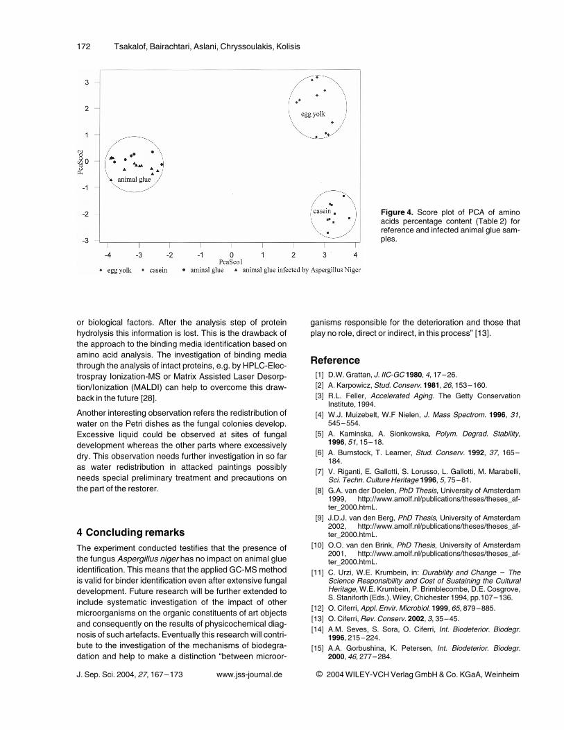
arranged in the order of decreasing information content orvariance. In our case, we use two first principal compo-nents to map the objects into two-dimensional space,where the clusters of proteinaceous binders can bevisually observed (Figure 4).
3 Results and discussionApplication of PCA analysis to the determined amino acidcontent of reference samples (Table 2) allows three clus-ters to be clearly distinguished corresponding to the mainproteinaceous binding media – casein, animal glue, andegg yolk (Figure 4). The reference samples were repeat-
edly analyzed by the method described on different daysover a period of several months of the experiment imple-mentation. The robustness of the analytical method canbe concluded from the stability of the binders’ clusters.Consequently the method was applied to the analysis offungus-infected animal glue samples.
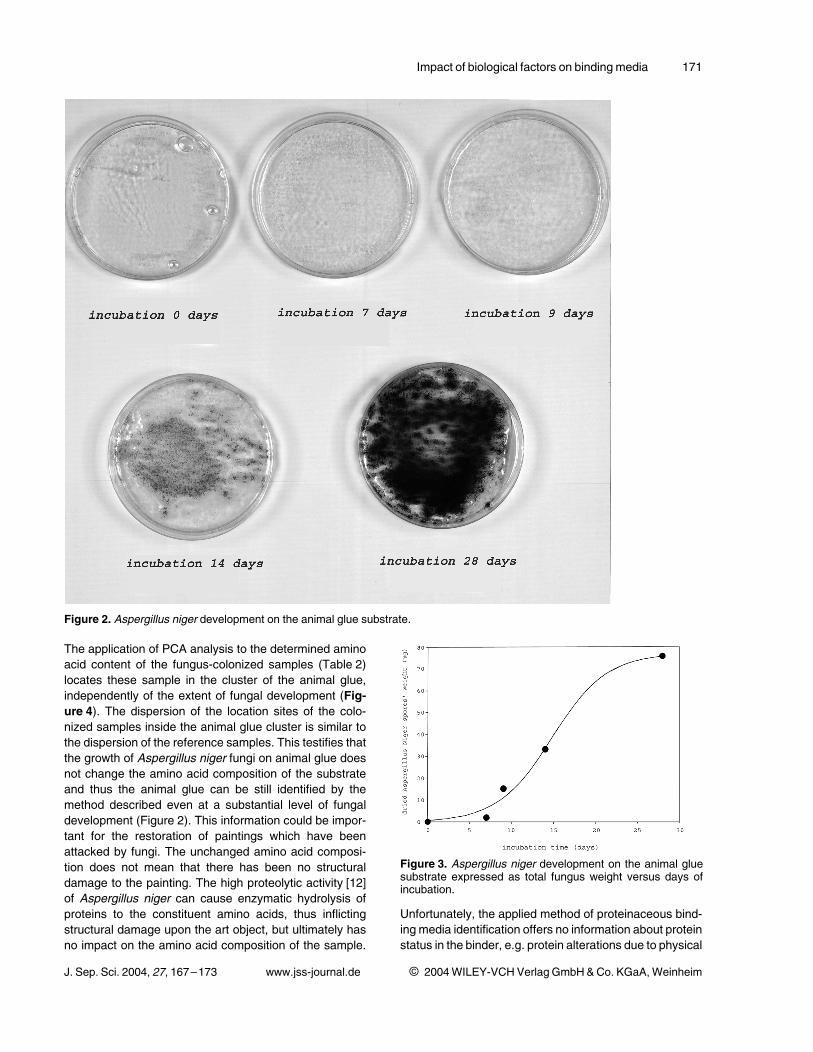
The extend of fungal colonization of animal glue substrate(Figure 2) can be expressed as total fungus weight versusdays of incubation and approximated by sigmoidal logisticfunction (Figure 3). It demonstrates that after a certaindelay (about 7 days) of fungal development a period ofaccelerated growth follows. Some deceleration in growthcan be noted after about 20 days.
J. Sep. Sci. 2004, 27, 167–173 www.jss-journal.de i 2004WILEY-VCH Verlag GmbH&Co. KGaA,Weinheim
Table 2. Relative amino acid percentage content of reference samples and infected animal glue samples at different days offungal developmenta), b).
Samples Ala Gly Val Leu Ile Met Ser Thr Pro Phe Asp Glu Lys Hyp Tyr
Pb EY 1 8.9 9.3 8.2 8.0 2.3 0.0 9.2 3.7 4.6 3.0 10.3 12.9 15.5 0.0 4.2Pb EY 2 10.4 9.5 5.9 8.9 2.1 0.0 9.7 4.2 5.1 4.5 11.0 13.8 9.8 0.0 4.9Pb EY 3 6.7 7.6 7.8 7.0 1.7 0.0 10.9 5.0 4.2 3.4 12.8 18.1 9.5 0.0 5.2Pb EY 4 8.3 7.1 4.9 7.0 2.2 0.0 11.3 4.5 3.8 4.1 13.8 16.7 11.0 0.0 5.2MIN EY 1 7.9 13.3 5.6 5.8 3.9 0.0 16.2 5.9 3.1 3.3 8.0 10.4 11.2 0.0 5.3CIN EY 1 8.1 12.2 7.6 5.9 2.8 0.0 13.7 6.4 2.3 3.3 8.6 16.0 9.9 0.0 3.1EY 1 8.9 5.2 4.5 11.0 3.0 2.0 10.0 4.5 5.0 4.3 13.0 19.5 5.3 0.0 3.8EY 2 8.1 4.5 3.5 9.2 2.4 2.1 9.3 3.8 3.9 3.4 11.1 14.1 20.7 0.0 3.8EY 3 7.0 5.2 3.8 9.1 2.8 2.4 10.5 3.3 4.6 4.3 13.3 15.2 13.8 0.0 4.8EY 4 7.6 4.8 2.9 8.9 2.6 2.5 11.1 4.1 4.4 4.1 12.5 14.4 15.0 0.0 5.2CAS 1 5.0 4.4 4.0 6.2 3.0 5.9 5.3 3.2 9.3 5.7 6.7 15.1 19.2 0.0 7.1CAS 2 4.2 3.1 4.0 8.8 3.3 4.2 6.3 3.4 13.6 5.8 8.0 15.6 12.9 0.0 6.7CAS 3 4.2 2.4 4.1 8.4 2.9 2.6 7.7 4.3 10.5 5.9 8.0 19.8 11.0 0.0 8.1CAS 4 3.6 2.8 3.4 7.0 2.6 3.9 5.1 4.3 9.4 4.9 6.1 16.6 23.1 0.0 7.2CAS 5 4.3 3.1 3.8 8.3 2.7 3.1 3.8 4.2 11.3 5.3 9.0 21.8 12.1 0.0 7.2CAS 6 4.5 4.3 3.9 8.6 2.9 3.0 5.6 3.8 9.9 5.0 7.8 18.8 14.5 0.0 7.5CAS 7 4.9 4.2 3.8 8.5 2.8 3.1 5.5 3.7 9.7 5.0 8.2 19.2 13.8 0.0 7.6CAS 8 5.4 3.5 4.4 8.2 3.8 4.2 4.7 3.9 7.4 6.1 6.4 17.6 16.1 0.0 8.1CAS 9 6.2 4.9 4.7 8.5 3.5 4.0 6.2 3.7 10.6 6.9 4.8 17.6 12.2 0.0 6.2GL 1 9.5 23.0 1.2 3.0 0.7 0.4 5.3 2.6 11.1 2.0 5.6 9.9 7.0 18.2 0.5GL 2 10.1 22.7 1.1 3.1 0.8 0.5 5.3 2.7 10.5 2.0 5.6 10.2 7.2 17.7 0.5GL 3 10.1 30.3 1.2 3.2 0.7 0.2 4.3 1.8 12.3 1.7 5.8 11.7 6.0 10.4 0.2GL 4 12.0 18.7 1.2 6.3 0.9 1.7 4.9 2.5 10.2 2.5 5.6 10.7 9.7 12.5 0.5GL 5 11.8 28.1 1.6 3.3 0.7 0.5 4.1 1.5 14.0 2.0 5.8 11.7 2.7 12.1 0.2GL 6 11.1 21.1 1.3 2.9 0.8 0.4 5.2 2.7 9.3 1.9 5.6 10.1 15.2 11.9 0.4INF 0-1 12.7 29.2 1.0 2.9 0.6 2.2 3.4 1.2 13.4 1.2 4.8 12.9 4.1 10.1 0.2INF 0–1 10.6 23.0 2.8 3.4 1.6 0.6 3.3 1.8 13.7 1.8 6.3 12.1 6.0 12.9 0.2INF 7–1 9.9 28.2 2.5 3.2 1.5 0.5 3.3 1.6 12.8 1.9 5.9 10.9 6.6 11.0 0.2INF 7–2 11.6 20.6 3.0 3.8 1.7 0.6 3.8 1.8 15.4 1.9 6.0 12.7 3.8 13.1 0.2INF 9–1 10.9 28.1 2.8 3.4 1.6 0.6 3.2 1.5 13.0 2.0 5.9 11.0 5.1 10.6 0.2INF 9–2 9.4 20.1 3.0 3.7 1.6 0.6 3.8 1.8 13.8 1.9 6.1 12.8 9.2 12.1 0.2INF 9–3 12.3 26.7 1.0 2.9 0.7 0.3 4.5 1.5 13.4 1.5 6.0 12.6 3.4 12.9 0.2INF 14–1 11.3 25.7 1.0 3.0 0.5 0.3 4.7 1.4 13.5 1.4 6.6 12.7 3.5 14.3 0.2INF 14–2 9.9 21.6 2.9 3.6 1.8 0.6 4.0 2.0 14.9 2.0 6.3 13.2 5.0 12.1 0.2INF 28–1 10.2 26.0 2.5 3.0 1.5 0.6 3.5 1.6 15.8 1.8 6.0 10.2 4.3 12.9 0.2INF 28–2 11.1 26.6 1.0 2.7 0.5 0.2 4.6 1.4 13.0 1.3 6.0 11.5 5.7 14.1 0.2INF 28–3 8.6 21.0 2.7 3.3 1.5 0.6 3.9 2.0 15.4 1.9 6.4 12.6 7.6 12.3 0.2
a) The abbreviations for reference sample are explained in Section 2.1.b) INF 7–1: animal glue infected samples with the indication of the day of incubation and number of the sample.
Impact of biological factors on binding media 171
The application of PCA analysis to the determined aminoacid content of the fungus-colonized samples (Table 2)locates these sample in the cluster of the animal glue,independently of the extent of fungal development (Fig-ure 4). The dispersion of the location sites of the colo-nized samples inside the animal glue cluster is similar tothe dispersion of the reference samples. This testifies thatthe growth of Aspergillus niger fungi on animal glue doesnot change the amino acid composition of the substrateand thus the animal glue can be still identified by themethod described even at a substantial level of fungaldevelopment (Figure 2). This information could be impor-tant for the restoration of paintings which have beenattacked by fungi. The unchanged amino acid composi-tion does not mean that there has been no structuraldamage to the painting. The high proteolytic activity [12]of Aspergillus niger can cause enzymatic hydrolysis ofproteins to the constituent amino acids, thus inflictingstructural damage upon the art object, but ultimately hasno impact on the amino acid composition of the sample.
Unfortunately, the applied method of proteinaceous bind-ing media identification offers no information about proteinstatus in the binder, e.g. protein alterations due to physical
J. Sep. Sci. 2004, 27, 167–173 www.jss-journal.de i 2004WILEY-VCH Verlag GmbH&Co. KGaA,Weinheim
Figure 2. Aspergillus niger development on the animal glue substrate.
Figure 3. Aspergillus niger development on the animal gluesubstrate expressed as total fungus weight versus days ofincubation.
172 Tsakalof, Bairachtari, Aslani, Chryssoulakis, Kolisis
or biological factors. After the analysis step of proteinhydrolysis this information is lost. This is the drawback ofthe approach to the binding media identification based onamino acid analysis. The investigation of binding mediathrough the analysis of intact proteins, e.g. by HPLC-Elec-trospray Ionization-MS or Matrix Assisted Laser Desorp-tion/Ionization (MALDI) can help to overcome this draw-back in the future [28].
Another interesting observation refers the redistribution ofwater on the Petri dishes as the fungal colonies develop.Excessive liquid could be observed at sites of fungaldevelopment whereas the other parts where excessivelydry. This observation needs further investigation in so faras water redistribution in attacked paintings possiblyneeds special preliminary treatment and precautions onthe part of the restorer.
4 Concluding remarksThe experiment conducted testifies that the presence ofthe fungus Aspergillus niger has no impact on animal glueidentification. This means that the applied GC-MSmethodis valid for binder identification even after extensive fungaldevelopment. Future research will be further extended toinclude systematic investigation of the impact of othermicroorganisms on the organic constituents of art objectsand consequently on the results of physicochemical diag-nosis of such artefacts. Eventually this research will contri-bute to the investigation of the mechanisms of biodegra-dation and help to make a distinction “between microor-
ganisms responsible for the deterioration and those thatplay no role, direct or indirect, in this process” [13].
Reference[1] D.W. Grattan, J. IIC-GC 1980, 4, 17–26.[2] A. Karpowicz,Stud. Conserv. 1981, 26, 153–160.[3] R.L. Feller, Accelerated Aging. The Getty Conservation
Institute, 1994.
[4] W.J. Muizebelt, W.F Nielen, J. Mass Spectrom. 1996, 31,545–554.
[5] A. Kaminska, A. Sionkowska, Polym. Degrad. Stability,1996, 51, 15–18.
[6] A. Burnstock, T. Learner, Stud. Conserv. 1992, 37, 165–184.
[7] V. Riganti, E. Gallotti, S. Lorusso, L. Gallotti, M. Marabelli,Sci. Techn. Culture Heritage 1996, 5, 75–81.
[8] G.A. van der Doelen, PhD Thesis, University of Amsterdam1999, http://www.amolf.nl/publications/theses/theses_af-ter_2000.htmL.
[9] J.D.J. van den Berg, PhD Thesis, University of Amsterdam2002, http://www.amolf.nl/publications/theses/theses_af-ter_2000.htmL.
[10] O.O. van den Brink, PhD Thesis, University of Amsterdam2001, http://www.amolf.nl/publications/theses/theses_af-ter_2000.htmL.
[11] C. Urzi, W.E. Krumbein, in: Durability and Change – TheScience Responsibility and Cost of Sustaining the CulturalHeritage, W.E. Krumbein, P. Brimblecombe, D.E. Cosgrove,S. Staniforth (Eds.). Wiley, Chichester 1994, pp.107–136.
[12] O. Ciferri, Appl. Envir. Microbiol. 1999, 65, 879–885.[13] O. Ciferri,Rev. Conserv. 2002, 3, 35–45.[14] A.M. Seves, S. Sora, O. Ciferri, Int. Biodeterior. Biodegr.
1996, 215–224.[15] A.A. Gorbushina, K. Petersen, Int. Biodeterior. Biodegr.
2000, 46, 277–284.
J. Sep. Sci. 2004, 27, 167–173 www.jss-journal.de i 2004WILEY-VCH Verlag GmbH&Co. KGaA,Weinheim
Figure 4. Score plot of PCA of aminoacids percentage content (Table 2) forreference and infected animal glue sam-ples.
Impact of biological factors on binding media 173
[16] Th. Dornieden, A.A. Gorbushina,W.E. Krumbein, Int. Biode-terior. Biodegr. 2000, 46, 261–270.
[17] J.P. Petushkova, N.N. Lyalikova, Stud. Conserv. 1986, 31,65–69.
[18] M.E. Florian,Stud. Conserv. 1996, 41, 65–75.
[19] R.J. Gettens, G.L. Stout, Painting Materials, A Short Ency-clopaedia. Dover Publications, New York 1966.
[20] M. Doerner, The Materials of the Artist and Their Use inPainting. Harcourt Brace, Orlando 1984.
[21] J.S. Mills, R. White, The Organic Chemistry of MuseumsObjects, Second Ed. Butterworth Heinemann, London 2000.
[22] S.M. Halpine, Conservation Research. National Gallery ofArt, Washington D.C. 1995, pp. 28–69.
[23] M.P. Colombini, F. Modugno, A. Giacomelli, S. Francesconi,J. Chromatogr. A 1999, 846, 113–124.
[24] A. Casoli, P.C. Musini, G. Palla, J. Chromatogr. A 1996, 731,237–246.
[25] T.P. Mawhinney, R.S.R. Robintt, A. Atalay, M.A. Madson, J.Chromatogr. 1986, 358, 231–242.
[26] L. Kryger, Talanta 1981, 28, 871–887.
[27] I.T. Jolliffe, Principal Component Analysis. Springer Verlag,2nd edition, 2002.
[28] S.J. Gaskel, J. Mass Spectrom. 1997, 32, 677–688.
J. Sep. Sci. 2004, 27, 167–173 www.jss-journal.de i 2004WILEY-VCH Verlag GmbH&Co. KGaA,Weinheim
Top Related
Copyright © 2022 FDOKUMEN

