


Bahasa
Halaman
Hukum

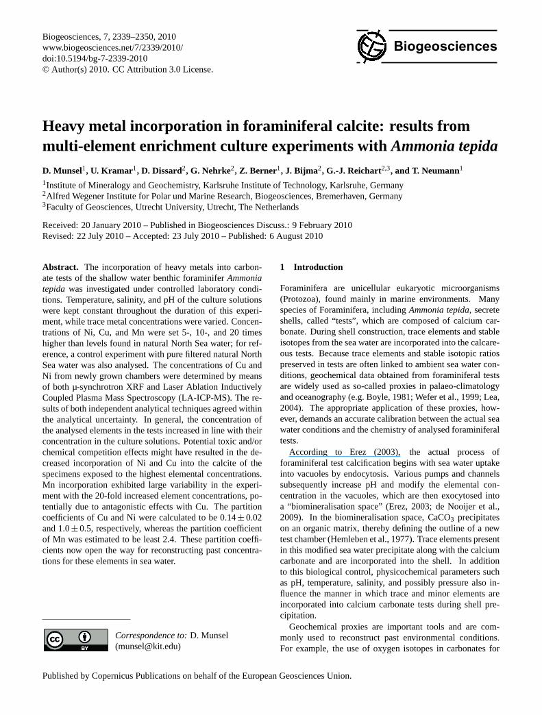
Biogeosciences, 7, 2339–2350, 2010www.biogeosciences.net/7/2339/2010/doi:10.5194/bg-7-2339-2010© Author(s) 2010. CC Attribution 3.0 License.
Biogeosciences
Heavy metal incorporation in foraminiferal calcite: results frommulti-element enrichment culture experiments withAmmonia tepida
D. Munsel1, U. Kramar 1, D. Dissard2, G. Nehrke2, Z. Berner1, J. Bijma2, G.-J. Reichart2,3, and T. Neumann1
1Institute of Mineralogy and Geochemistry, Karlsruhe Institute of Technology, Karlsruhe, Germany2Alfred Wegener Institute for Polar und Marine Research, Biogeosciences, Bremerhaven, Germany3Faculty of Geosciences, Utrecht University, Utrecht, The Netherlands
Received: 20 January 2010 – Published in Biogeosciences Discuss.: 9 February 2010Revised: 22 July 2010 – Accepted: 23 July 2010 – Published: 6 August 2010
Abstract. The incorporation of heavy metals into carbon-ate tests of the shallow water benthic foraminiferAmmoniatepida was investigated under controlled laboratory condi-tions. Temperature, salinity, and pH of the culture solutionswere kept constant throughout the duration of this experi-ment, while trace metal concentrations were varied. Concen-trations of Ni, Cu, and Mn were set 5-, 10-, and 20 timeshigher than levels found in natural North Sea water; for ref-erence, a control experiment with pure filtered natural NorthSea water was also analysed. The concentrations of Cu andNi from newly grown chambers were determined by meansof both µ-synchrotron XRF and Laser Ablation InductivelyCoupled Plasma Mass Spectroscopy (LA-ICP-MS). The re-sults of both independent analytical techniques agreed withinthe analytical uncertainty. In general, the concentration ofthe analysed elements in the tests increased in line with theirconcentration in the culture solutions. Potential toxic and/orchemical competition effects might have resulted in the de-creased incorporation of Ni and Cu into the calcite of thespecimens exposed to the highest elemental concentrations.Mn incorporation exhibited large variability in the experi-ment with the 20-fold increased element concentrations, po-tentially due to antagonistic effects with Cu. The partitioncoefficients of Cu and Ni were calculated to be 0.14± 0.02and 1.0± 0.5, respectively, whereas the partition coefficientof Mn was estimated to be least 2.4. These partition coeffi-cients now open the way for reconstructing past concentra-tions for these elements in sea water.
Correspondence to:D. Munsel([email protected])
1 Introduction
Foraminifera are unicellular eukaryotic microorganisms(Protozoa), found mainly in marine environments. Manyspecies of Foraminifera, includingAmmonia tepida, secreteshells, called “tests”, which are composed of calcium car-bonate. During shell construction, trace elements and stableisotopes from the sea water are incorporated into the calcare-ous tests. Because trace elements and stable isotopic ratiospreserved in tests are often linked to ambient sea water con-ditions, geochemical data obtained from foraminiferal testsare widely used as so-called proxies in palaeo-climatologyand oceanography (e.g. Boyle, 1981; Wefer et al., 1999; Lea,2004). The appropriate application of these proxies, how-ever, demands an accurate calibration between the actual seawater conditions and the chemistry of analysed foraminiferaltests.
According to Erez (2003), the actual process offoraminiferal test calcification begins with sea water uptakeinto vacuoles by endocytosis. Various pumps and channelssubsequently increase pH and modify the elemental con-centration in the vacuoles, which are then exocytosed intoa “biomineralisation space” (Erez, 2003; de Nooijer et al.,2009). In the biomineralisation space, CaCO3 precipitateson an organic matrix, thereby defining the outline of a newtest chamber (Hemleben et al., 1977). Trace elements presentin this modified sea water precipitate along with the calciumcarbonate and are incorporated into the shell. In additionto this biological control, physicochemical parameters suchas pH, temperature, salinity, and possibly pressure also in-fluence the manner in which trace and minor elements areincorporated into calcium carbonate tests during shell pre-cipitation.
Geochemical proxies are important tools and are com-monly used to reconstruct past environmental conditions.For example, the use of oxygen isotopes in carbonates for
Published by Copernicus Publications on behalf of the European Geosciences Union.
2340 D. Munsel et al.: Heavy metal incorporation in foraminiferal calcite:Ammonia tepida
palaeothermometry, as suggested by Urey (1947), is still oneof the most widely used proxies (e.g. Bemis et al., 1998;Lea, 2004). The proxy toolbox has increased enormouslyever since. More recently, proxies based on Mg/Ca ratios(e.g. Nurnberg et al., 1996; Lea et al., 1999) and Ca isotoperatios (e.g. Nagler et al., 2000) preserved in foraminiferal testcarbonate have also been used to reconstruct sea water tem-perature. Other geochemical proxies useful in environmentalreconstructions include B isotopes, which are used to deter-mine pH changes in the ocean water (Sanyal et al., 2001; Yuet al., 2007), and Cd, which is a proxy for nutrient concen-trations (Boyle, 1988; Rickaby and Elderfield, 1999; Martinand Lea, 1998).
Most calibrations of foraminiferal proxies are based oncorrelating average ambient conditions to the analyses ofForaminifera isolated from recent (i.e. core-top) sedimentsamples. Not only does this approach rely on the assump-tion that conditions have remained stable for a considerablelength of time, but in order to eliminate secondary contam-inations and overgrowths, complex cleaning protocols areusually implemented (e.g. Boyle and Keigwin, 1985/1986;Lea and Boyle, 1991; Martin and Lea, 2002). Moreover,under natural conditions environmental parameters often co-vary and thus are difficult to disentangle. To circumvent suchcomplications, the use of controlled laboratory cultures wasinitially pioneered by Christoph Hemleben from Germanyand Alan Be from the USA. The first experiments focussingon quantifying foraminiferal elemental uptake of Li, Sr, Mg,and Na were carried out by Delaney et al. (1985). Subsequentstudies examined the uptake of Cd (Boyle, 1988; Mashiottaet al., 1997; Marechal-Abram et al., 2004), Ba (Lea andSpero, 1992, 1994), U (Russel et al., 1994), and V (Hast-ings et al., 1996). One of the most recent studies publishedaddressed Cu uptake (de Nooijer et al., 2007).
Biological productivity in the ocean is strongly impactedby trace metal distribution, which generally exhibits anutrient-type depth profile within the water column. In par-ticular, low concentrations of some first row transition met-als are known to be essential for productivity. However, thedistribution of trace metals in the ocean is affected by somegeogenic processes (e.g. hydrothermal activity) and anthro-pogenic forces (e.g. pollution), as described below. Locally,geogenic processes such as hydrothermal activity can dra-matically enrich the trace element composition of sea wa-ter for up to several kilometres from the active hydrothermalvent (German and von Damm, 2004). Although Foraminiferado not reside within hydrothermal vents, the overall increaseof trace metal concentrations in the surrounding sea waterdue to hydrothermal activity will affect Foraminifera, partic-ularly those living near the vent (e.g. Panieri, 2006). Thesehydrothermal effects are recorded in foraminiferal tests. Forexample, a field site which is known to be influenced byhydrothermal activity (DSDP Site 216, Ninety-east Ridge)yields specimens of benthic Foraminifera that are enrichedin “hydrothermal” trace elements such as Cu, Co, and Pb
(Meudt, unpublished data). Changes in redox conditions(e.g. Morford and Emerson, 1999) or anthropogenic pollu-tion sources may also influence trace and minor element dis-tribution in sea water and will be recorded by Foraminifera.Despite this, most pollution studies focus exclusively onbulk sediment analyses and/or foraminiferal distribution pat-terns (e.g. Alve, 1991, 1995; Vilela et al., 2004; Buzas-Stephens and Buzas, 2005; Carnahan et al., 2008), partlydue to the fact that accurate distribution coefficients havenot yet been determined for linking sea water chemistry withforaminiferal carbonates for many of the elements of inter-est. As polluted sediments are usually suboxic or anoxic dueto enrichment in organic matter with high levels of sulphatereduction, heavy metals are often accumulated and bound tohighly insoluble minerals such as sulphides. Hence, thesemetals are not bio-available and as a result, true pollution lev-els remain unknown. Heavy metals can only be remobilisedin the sediment and diffused into the water with increasingoxygen levels. Analyses of foraminiferal calcite and parti-tion coefficients of minor and trace metals could potentiallyreveal true trace metal availability. Thus foraminiferal calcitehas the potential to become an excellent tool for monitoringmarine pollution. However, except for Cu (de Nooijer et al.,2007), there are no experimental data available for first rowtransition metals such as Mn and Ni. To shed additional lighton this subject, we conducted a multi-element experimentunder controlled laboratory conditions for oxidising condi-tions using the shallow-water, benthic foraminiferAmmoniatepida.
2 Methods
2.1 Sampling and culturing procedures
Ammonia tepida(a shallow-water benthic foraminifer) isknown to have a broad tolerance range for temperature andsalinity, to withstand strong seasonal regimes, and to sur-vive severe environmental conditions. Furthermore,Ammo-nia tepidaare also easy to grow in the laboratory, where theycan form new chambers and even reproduce (e.g. Bradshaw,1957; Alve and Murray, 1999; Stouff et al., 1999; Pascualet al., 2002; le Cadre and Debenay, 2006; de Nooijer et al.,2007). Ammonia tepidawas collected by sampling the up-per 0.5 cm of sediment at low-tide in an intertidal mudflat ofthe Wadden Sea, part of the North Sea. The locality is sit-uated near the Lower Saxony town of Dorum Neufeld, Ger-many (withinNationalpark Niedersachsisches Wattenmeer).In the laboratory, the sediment was sieved over a 125 µmmesh sieve and was washed with filtered (0.2 µm) sea wa-ter. Foraminifera were hand picked under a binocular mi-croscope using a very fine brush and screened under a ZeissAxiovert 200 M inverted microscope for pseudopodial acti-vity – which expresses vitality. Four hundred live individualswere transferred into and divided evenly among four sepa-rate sediment-free aquaria, each containing 1.25 L of culture
Biogeosciences, 7, 2339–2350, 2010 www.biogeosciences.net/7/2339/2010/
D. Munsel et al.: Heavy metal incorporation in foraminiferal calcite:Ammonia tepida 2341
Fig. 1
Fig. 1
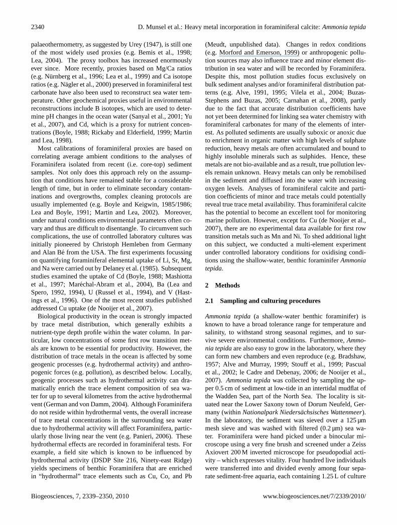
Fig. 1. Ammonia tepidaunder the light microscope (top),Ammoniatepidaunder UV light showing newly grown chambers marked withcalcein (bottom).
solution. Before use, the aquaria were thoroughly cleanedwith 10 vol.-% HNO3 and rinsed with reverse osmosis water(ROW, conductivity<0.067 µS cm−1).
A stock solution with defined concentrations of Mn,Co, Ni, and Cu was prepared using ICP-standard solutions(1000 µg/mL in 5 vol.-% HNO3 each) (Specpure, Alfa Ae-sar, Germany) and ROW water. Concentrations used in theexperiments are listed in Table 1. The pH of the stock so-lution was raised to 8.0± 0.1 with 1 M NaOH (p.a.) inorder to match the pH of the natural sea water. Then,the stock solutions were added to the sea water (filteredwith a 0.2 µm membrane filter) until trace metal concen-trations within the culture solutions reached approximately5-, 10-, and 20 times that of the natural North Sea wa-ter. In addition, a reference batch without any additionaltrace metals was prepared. Finally, the culture solutionswere filtered again using a 0.2 µm membrane filter to avoidintroducing possible inorganic precipitates in the aquaria.To mark newly grown chambers (Fig. 1), the fluorescent
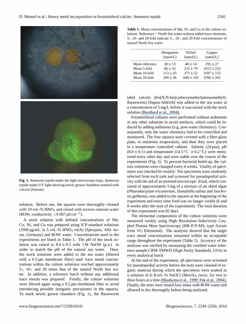
Table 1. Mean concentrations of Mn, Ni, and Cu in the culture so-lutions. Reference = North Sea water without added trace elements,5-, 10- and 20-fold indicate 5-, 10-, and 20-fold concentrations ofnatural North Sea water.
Manganese Nickel Copper[nmol/L] [nmol/L] [nmol/L]
Mean reference 30± 13 48± 14 195± 27Mean 5-fold 86± 33 231± 70 1012± 233Mean 10-fold 113± 45 371± 52 1697± 155Mean 20-fold 209± 56 649± 105 3290± 263
label calcein (bis[N,N-bis(carboxymethyl)aminomethyl]-fluorescein) (Sigma-Aldrich) was added to the sea water ata concentration of 5 mg/L before it was mixed with the stocksolution (Bernhard et al., 2004).
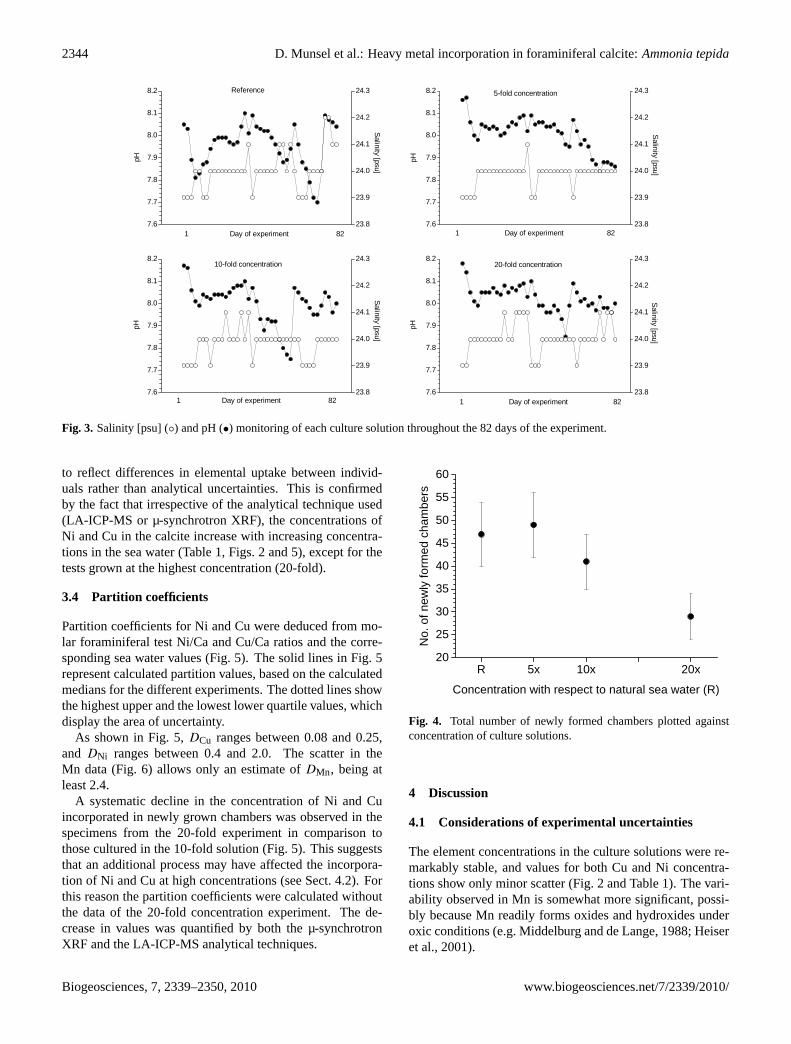
Foraminiferal cultures were performed without sedimentsor any other substrate to avoid artefacts, which could be in-duced by adding sediments (e.g. pore water chemistry). Con-sequently, only the water chemistry had to be controlled andmonitored. The four aquaria were covered with a fibre-glassplate, to minimise evaporation, and then they were placedin a temperature controlled cabinet. Salinity (24 psu), pH(8.0± 0.1) and temperature (14.5◦C ± 0.2◦C) were moni-tored every other day and were stable over the course of theexperiments (Fig. 3). To prevent bacterial build-up, the cul-ture solutions were changed every 4 weeks. Vitality of speci-mens was checked bi-weekly: Ten specimens were randomlyselected from each tank and screened for pseudopodial acti-vity with the aid of an inverted microscope. Food, which con-sisted of approximately 5 mg of a mixture of air dried algae(Phaeodactylum triconortum, Dunaliella salinaandIsochri-sis galbana), was added to the aquaria at the beginning of theexperiment and every time food was no longer visible (6 and8 weeks after the start of the experiment). The total durationof this experiment was 82 days.
The elemental composition of the culture solutions weremeasured weekly using High Resolution Inductively Cou-pled Plasma Mass Spectroscopy (HR-ICP-MS, type Axiomfrom VG Elemental). The analyses showed that the targettrace metal concentrations remained within an acceptablerange throughout the experiment (Table 1). Accuracy of theanalyses was verified by measuring the certified water refer-ence sample CRM-TMWD (High Purity Standards, USA) inevery analytical batch.
At the end of the experiment, all specimens were screenedfor pseudopodial activity before the tests were cleaned of or-ganic material during which the specimens were soaked ina solution of 6–8 vol.-% NaOCl (Merck), twice, for two tothree hours at a time (Mashiotta et al., 1999; Pak et al., 2004).Finally, the tests were rinsed four times with ROW water andallowed to dry thoroughly before being analysed.
www.biogeosciences.net/7/2339/2010/ Biogeosciences, 7, 2339–2350, 2010
2342 D. Munsel et al.: Heavy metal incorporation in foraminiferal calcite:Ammonia tepida
2.2 Analytics
2.2.1 µ-synchrotron XRF
Using a liquid glue (Tesa, Beiersdorfer, Germany), two rows(each consisting of five individuals) of tests were affixedwith their spiral side onto a 3 µm mylar film that was at-tached to plastic slide mounts. The glue as well as the my-lar film were tested for their trace element constituents be-fore use. The abundances of all elements of interest wereclearly below the detection limits for µ-synchrotron X-RayFluorescence Spectroscopy (XRF) determinations. Newlygrown chambers as well as some old chambers were anal-ysed by µ-synchrotron XRF at the FLUO-beamline of ANKAsynchrotron facility (Karlsruhe) and at HASYLAB beam-line L (DESY, Hamburg) where confocal µ-XRF measure-ments were carried out (for details about the confocal mea-surements, see Kramar et al., 2010). Using an excitation en-ergy of 12.5 keV, and focussing refractive lenses to a pointsize of 2×5µm, the chambers were analysed by line scans,averaging five points per line in newly grown chambers andthree points per line in old chambers. In unfiltered samplespectra, the Ni-K lines were strongly overlain by the intenseCa sum-peak due to the calcium carbonate matrix of the in-vestigated foraminiferal tests. To reduce this interference,a 20 µm secondary beam aluminium filter was mounted infront of the fluorescence detector. Due to low trace elementconcentrations in the sub µg/g and lower µg/g range, longmeasuring times (500 s) were necessary at each point. Traceelement contents in the tests were quantified by fundamentalparameters using PyMCA 4.2.2 (Sole et al., 2007). The fun-damental parameter calculations were recalibrated for traceelement calibration purposes by using a pressed calcite pellet(containing 756 µg/g Cr and 727 µg/g As) and the MPI-DINGreference glass StHS6/80-G standard (Max-Planck-Institutfur Chemie, Germany) (Jochum et al., 2000) as primary stan-dards. Geometry effects caused by the uneven shape of thetests were corrected by calculating the absorption of the in-coming beam from the count rate of the primary monitor(in front of the sample) and the secondary monitor (behindthe sample). Because the calcium concentration (40 wt%) isconstant in calcite, the average path length of the outgoingbeam could be calculated using Ca as internal standard (Kra-mar et al., 2010). Replicate measurements of the same spotslay within 5% relatively, while errors resulting from the pathlength calculations through the chamber walls are estimatedto 10–20% relatively. These effects are discussed in detail inKramar et al. (2010).
2.2.2 LA-ICP-MS
The non-destructive nature of µ-synchrotron XRF allowedLaser Ablation Inductively Coupled Plasma Mass Spec-troscopy (LA-ICP-MS) measurements of the same chambers.For these measurements, specimens were fixed on a double-sided adhesive tape and mounted on plastic stubs.
The analyses were conducted at Utrecht University (De-partment of Earth Sciences) by means of an excimer laser(Lambda Physik, Germany) equipped with GeoLas 200Q op-tics at a wavelength of 193 nm. The ablation was conductedin a helium atmosphere. Pulse rate was adjusted to six pulsesper second with an energy density of 1 J/cm2 at the surface ofthe sample. The laser beam was set to a diameter of 80 µm.The ablated material was analysed with a quadrupole ICP-MS (Micromass Platform). A collision and reaction cell wasused to minimise spectral interferences on the minor isotopesof Ca (Mason and Kraan, 2002). Calcium was used as an in-ternal standard, via the44Ca isotope, with simultaneous mon-itoring of 42Ca and43Ca. For calibration of trace elementcontents, the NIST SRM 610 glass standard (National Insti-tute of Science and Technology, USA) (Pearce et al., 1997)and an in-house matrix-matched calcite standard (GJR) wereapplied. Concentrations of Cu and Ni were calculated usingthe 63Cu and60Ni isotopes, to correct for their natural iso-topic abundances. Mn was quantified using its only naturalisotope,55Mn. Internal relative precision of Mn, Ni, and Cuwas better than 4.5%, 4.7%, and 4.8%, respectively.
2.3 Calculation of partition coefficients
Partition coefficients were calculated using the resulting traceelement (TE) and calcium concentrations according to thefollowing expression:
DTE = (TE/Ca)calcite/(TE/Ca)sea water.
DTE were calculated using the medians of each set of con-centration measurements. Because the analytical data did notshow a Gaussian distribution, the medians are a more reliableand robust parameter (e.g. Hoaglin et al., 1983; Zhou, 1987)than the means. Subsequently, the mean of the medians ofthe reference, 5-, and 10-fold concentrations was calculatedas the distribution coefficient. The error envelope was cal-culated using the standard error of the mean (e.g. Howarth,1983). The results of the 20-fold concentration solution ex-periment were not included in the calculations. For moredetails see Sect. 3.4 and the Discussion.
3 Results
3.1 Culture media
Continuous monitoring of the culture solution revealed thatthe trace element concentrations corresponded well to thetargeted concentrations. The elements of the stock solutiontherefore did not precipitate or absorb when mixed with theculture solution, nor were they consumed to an appreciabledegree over the course of the experiment (Fig. 2 and Table 1).
Biogeosciences, 7, 2339–2350, 2010 www.biogeosciences.net/7/2339/2010/
D. Munsel et al.: Heavy metal incorporation in foraminiferal calcite:Ammonia tepida 2343
Table 2. Chamber addition of the cultured foraminifera.
Aquarium No. of Living No. of chambers formed Individuals No. ofindividuals individuals that offsprings
(start of (end of formedexperiment) experiment) 1 2 3 4 5 chambers
[%] [%]
Reference 100 98 10 14 12 9 2 47± 7 05-fold NSW 100∗ 97 22 19 8 0 0 53± 7 010-fold NSW 100 94 22 16 0 0 0 41± 6 120-fold NSW 100 93 20 9 0 0 0 29± 5 0
∗ Seven individuals were lost during water changes; NSW = North Sea water.
R 5x 10x 20x0
306090
120150180210240270300
(= 10.92) (= 54.60) (= 109.20) (= 218.40)
Mn
Mea
n [n
mol
/L]
Concentration with respect to natural sea water (R) [nmol/L]
R 5x 10x 20x0
100
200
300
400
500
600
700
800
900
(= 35.44) (= 177.19) (= 354.38) (= 708.76)
Ni
Mea
n [n
mol
/L]
Concentration with respect to natural sea water (R) [nmol/L]
R 5x 10x 20x0
500
1000
1500
2000
2500
3000
3500
4000
(= 210.56)(= 1052.78)(= 2105.56) (= 4211.12)
Cu
Mea
n [n
mol
/L]
Concentration with respect to natural sea water (R) [nmol/L]
Fig. 2
R 5x 10x 20x0
306090
120150180210240270300
(= 10.92) (= 54.60) (= 109.20) (= 218.40)
Mn
Mea
n [n
mol
/L]
Concentration with respect to natural sea water (R) [nmol/L]
R 5x 10x 20x0
100
200
300
400
500
600
700
800
900
(= 35.44) (= 177.19) (= 354.38) (= 708.76)
Ni
Mea
n [n
mol
/L]
Concentration with respect to natural sea water (R) [nmol/L]
R 5x 10x 20x0
500
1000
1500
2000
2500
3000
3500
4000
(= 210.56)(= 1052.78)(= 2105.56) (= 4211.12)
Cu
Mea
n [n
mol
/L]
Concentration with respect to natural sea water (R) [nmol/L]
Fig. 2
Fig. 2. Variation in the concentration [nmol/L] of Mn (top), Ni (lower left) and Cu (lower right) of each culture solution during the experiment;R = reference (natural sea water). Values in parentheses are theoretical values calculated on basis of pure sea water measurements performedbefore the experiment.
3.2 Newly grown foraminiferal calcite
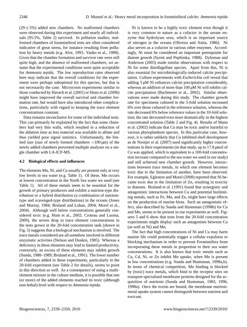
Nearly all specimens, 95.5% on average, survived the cul-ture period of 82 days – exact values for each experimentare listed in Table 2. In one of the aquaria (10-fold con-centration), reproduction occurred, but only one juvenileforaminifer was recovered after termination of the experi-ments. Seven individuals of the 5-fold concentration experi-ment were lost during water changes. New chambers formedin all experiments. For the reference experiment, 47% of thespecimens formed at least one new chamber. In the 5-, 10-and 20-fold concentration experiments, 53%, 41%, and 29%of the individuals formed at least one new chamber, respec-tively (Table 2 and Fig. 4).
3.3 Comparison of the applied analytical techniques
The precision of measurements for Co analyses was low asthe measured concentrations were too close to or under thedetection limit for both analytical methods. For the other ele-ments (especially Cu and Ni) the LA-ICP-MS measurementsdisplay a similar range to that measured by µ-synchrotronXRF.
Although the analyses show a wide range of variability intrace metal uptake among individual specimens, clear trendsare observed for Cu and Ni. Similar variability betweenspecimens grown under identical conditions was observedfor both Mg and Sr (e.g. Reichart et al., 2003; Duenas-Bohorquez et al., 2009; Dissard et al., 2010a,b) and seems
www.biogeosciences.net/7/2339/2010/ Biogeosciences, 7, 2339–2350, 2010
2344 D. Munsel et al.: Heavy metal incorporation in foraminiferal calcite:Ammonia tepida
7.6
7.7
7.8
7.9
8.0
8.1
8.2
23.8
23.9
24.0
24.1
24.2
24.3
7.6
7.7
7.8
7.9
8.0
8.1
8.2
23.8
23.9
24.0
24.1
24.2
24.3
7.6
7.7
7.8
7.9
8.0
8.1
8.2
23.8
23.9
24.0
24.1
24.2
24.3
7.6
7.7
7.8
7.9
8.0
8.1
8.2
23.8
23.9
24.0
24.1
24.2
24.3
pH
1 Day of experiment 82
5-fold concentration
Salinity [psu]pH
1 Day of experiment 82
10-fold concentration
Salinity [psu]
1 Day of experiment 82
pH
20-fold concentration
Salinity [psu]
Reference
pH
1 Day of experiment 82
Salinity [psu]
Fig. 3
7.6
7.7
7.8
7.9
8.0
8.1
8.2
23.8
23.9
24.0
24.1
24.2
24.3
7.6
7.7
7.8
7.9
8.0
8.1
8.2
23.8
23.9
24.0
24.1
24.2
24.3
7.6
7.7
7.8
7.9
8.0
8.1
8.2
23.8
23.9
24.0
24.1
24.2
24.3
7.6
7.7
7.8
7.9
8.0
8.1
8.2
23.8
23.9
24.0
24.1
24.2
24.3
pH
1 Day of experiment 82
5-fold concentration
Salinity [psu]pH
1 Day of experiment 82
10-fold concentration
Salinity [psu]
1 Day of experiment 82pH
20-fold concentration
Salinity [psu]
Reference
pH
1 Day of experiment 82
Salinity [psu]
Fig. 3
Fig. 3. Salinity [psu] (◦) and pH (•) monitoring of each culture solution throughout the 82 days of the experiment.
to reflect differences in elemental uptake between individ-uals rather than analytical uncertainties. This is confirmedby the fact that irrespective of the analytical technique used(LA-ICP-MS or µ-synchrotron XRF), the concentrations ofNi and Cu in the calcite increase with increasing concentra-tions in the sea water (Table 1, Figs. 2 and 5), except for thetests grown at the highest concentration (20-fold).
3.4 Partition coefficients
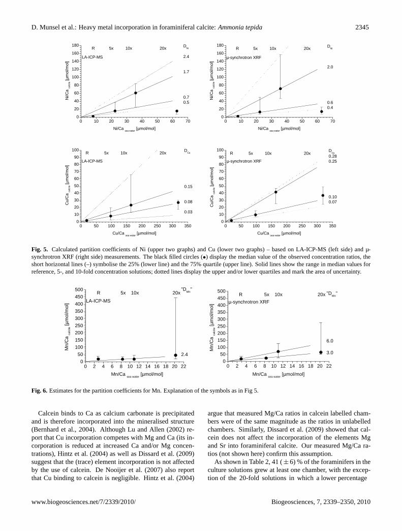
Partition coefficients for Ni and Cu were deduced from mo-lar foraminiferal test Ni/Ca and Cu/Ca ratios and the corre-sponding sea water values (Fig. 5). The solid lines in Fig. 5represent calculated partition values, based on the calculatedmedians for the different experiments. The dotted lines showthe highest upper and the lowest lower quartile values, whichdisplay the area of uncertainty.
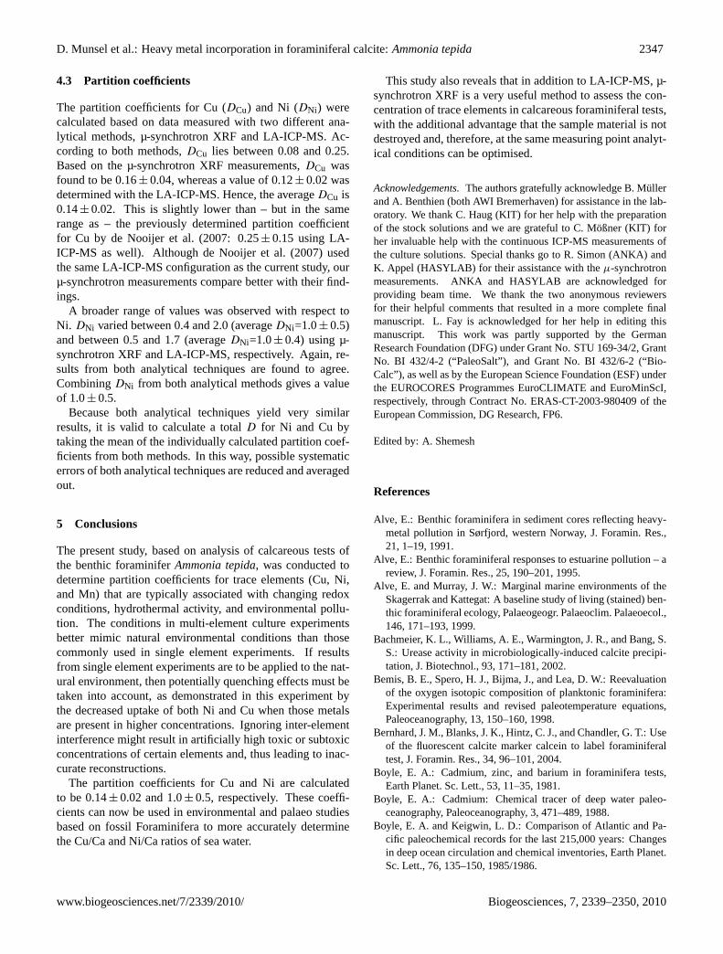
As shown in Fig. 5,DCu ranges between 0.08 and 0.25,and DNi ranges between 0.4 and 2.0. The scatter in theMn data (Fig. 6) allows only an estimate ofDMn, being atleast 2.4.
A systematic decline in the concentration of Ni and Cuincorporated in newly grown chambers was observed in thespecimens from the 20-fold experiment in comparison tothose cultured in the 10-fold solution (Fig. 5). This suggeststhat an additional process may have affected the incorpora-tion of Ni and Cu at high concentrations (see Sect. 4.2). Forthis reason the partition coefficients were calculated withoutthe data of the 20-fold concentration experiment. The de-crease in values was quantified by both the µ-synchrotronXRF and the LA-ICP-MS analytical techniques.
R 5x 10x 20x20
25
30
35
40
45
50
55
60
No.
of n
ewly
form
ed c
ham
bers
Concentration with respect to natural sea water (R)
Fig. 4
Fig. 4. Total number of newly formed chambers plotted againstconcentration of culture solutions.
4 Discussion
4.1 Considerations of experimental uncertainties
The element concentrations in the culture solutions were re-markably stable, and values for both Cu and Ni concentra-tions show only minor scatter (Fig. 2 and Table 1). The vari-ability observed in Mn is somewhat more significant, possi-bly because Mn readily forms oxides and hydroxides underoxic conditions (e.g. Middelburg and de Lange, 1988; Heiseret al., 2001).
Biogeosciences, 7, 2339–2350, 2010 www.biogeosciences.net/7/2339/2010/
D. Munsel et al.: Heavy metal incorporation in foraminiferal calcite:Ammonia tepida 2345
0 10 20 30 40 50 60 700
20
40
60
80
100
120
140
160
180
0 50 100 150 200 250 300 3500
10
20
30
40
50
60
70
80
90
100
0 50 100 150 200 250 300 3500
10
20
30
40
50
60
70
80
90
100
0 10 20 30 40 50 60 700
20
40
60
80
100
120
140
160
180
LA-ICP-MS
DNi
2.4
1.7
0.70.5
R 5x 10x 20x
Ni/C
a ca
lcite [µ
mol
/mol
]
Ni/Ca sea water
[µmol/mol]
µ-synchrotron XRF
DCu
0.280.25
0.100.07
R 5x 10x 20x
Cu/
Ca
calc
ite [µ
mol
/mol
]
Cu/Ca sea water [µmol/mol]
DCu
0.15
0.08
0.03
R 5x 10x 20x
LA-ICP-MS
Cu/
Ca
calc
ite [µ
mol
/mol
]
Cu/Ca sea water [µmol/mol]
µ-synchrotron XRF
DNi
2.0
0.60.4
R 5x 10x 20x
Ni/C
a ca
lcite [µ
mol
/mol
]
Ni/Ca sea water
[µmol/mol]
Fig. 5
Fig. 5. Calculated partition coefficients of Ni (upper two graphs) and Cu (lower two graphs) – based on LA-ICP-MS (left side) and µ-synchrotron XRF (right side) measurements. The black filled circles (•) display the median value of the observed concentration ratios, theshort horizontal lines (–) symbolise the 25% (lower line) and the 75% quartile (upper line). Solid lines show the range in median values forreference, 5-, and 10-fold concentration solutions; dotted lines display the upper and/or lower quartiles and mark the area of uncertainty.
0 2 4 6 8 10 12 14 16 18 20 220
50100150200250300350400450500
R 5x 10x 20x"DMn"
2.4
LA-ICP-MS
Mn/
Ca
calc
ite [µ
mol
/mol
]
Mn/Ca sea water [µmol/mol]
0 2 4 6 8 10 12 14 16 18 20 220
50100150200250300350400450500 "DMn"
6.0
3.0
R 5x 10x 20xµ-synchrotron XRF
Mn/
Ca
calc
ite [µ
mol
/mol
]
Mn/Ca sea water [µmol/mol]
Fig. 6
Fig. 6. Estimates for the partition coefficients for Mn. Explanation of the symbols as in Fig 5.
Calcein binds to Ca as calcium carbonate is precipitatedand is therefore incorporated into the mineralised structure(Bernhard et al., 2004). Although Lu and Allen (2002) re-port that Cu incorporation competes with Mg and Ca (its in-corporation is reduced at increased Ca and/or Mg concen-trations), Hintz et al. (2004) as well as Dissard et al. (2009)suggest that the (trace) element incorporation is not affectedby the use of calcein. De Nooijer et al. (2007) also reportthat Cu binding to calcein is negligible. Hintz et al. (2004)
argue that measured Mg/Ca ratios in calcein labelled cham-bers were of the same magnitude as the ratios in unlabelledchambers. Similarly, Dissard et al. (2009) showed that cal-cein does not affect the incorporation of the elements Mgand Sr into foraminiferal calcite. Our measured Mg/Ca ra-tios (not shown here) confirm this assumption.
As shown in Table 2, 41 (± 6) % of the foraminifers in theculture solutions grew at least one chamber, with the excep-tion of the 20-fold solutions in which a lower percentage
www.biogeosciences.net/7/2339/2010/ Biogeosciences, 7, 2339–2350, 2010
2346 D. Munsel et al.: Heavy metal incorporation in foraminiferal calcite:Ammonia tepida
(29± 5%) added new chambers. No malformed chamberswere observed during this experiment and nearly all individ-uals (95.5%, Table 2) survived. In pollution studies, mal-formed chambers of foraminiferal tests are often consideredindicative of great stress, for instance resulting from pollu-tion by heavy metals (e.g. Alve, 1991; Yanko et al., 1998).Given that the chamber formation and survivor rate were stillquite high, and the absence of malformed chambers, we as-sume that the experimental conditions were quite acceptablefor Ammonia tepida. The low reproduction rates observedhere may indicate that the overall conditions for the exper-iment were perhaps suboptimal for this species, but that isnot necessarily the case. Microcosm experiments similar tothose conducted by Havach et al. (2001) or Hintz et al. (2006)might have improved the overall survival and chamber for-mation rate, but would have also introduced other complica-tions, particularly with regard to keeping the trace elementconcentrations constant.
Data remains inconclusive for some of the individual tests.This can primarily be explained by the fact that some cham-bers had very thin walls, which resulted in a reduction ofthe ablation time as less material was available to ablate andthus yielded poor signal statistics. Unfortunately, the lim-ited size (size of newly formed chambers<100 µm) of thenewly added chambers prevented multiple analyses on a sin-gle chamber with LA-ICP-MS.
4.2 Biological effects and influences
The elements Mn, Ni, and Cu usually are present only at verylow levels in sea water (e.g. Table 1). Of these, Mn occursat lowest concentration in the North Sea water we used (seeTable 1). All of these metals seem to be essential for thegrowth of primary producers and exhibit a nutrient-type dis-tribution or a hybrid distribution (a combination of nutrient-type and scavenged-type distributions) in the oceans (Jonesand Murray, 1984; Bruland and Lohan, 2004; Morel et al.,2004). Although well below concentrations generally con-sidered toxic (e.g. Hunt et al., 2002; Croteau and Luoma,2009), the severe drop in trace element concentrations inthe tests grown in the 20-fold concentration tank (shown inFig. 5) suggests that a biological mechanism is involved. Thetrace metals considered are all somehow involved in differentenzymatic activities (Nelson and Donkin, 1985). Whereas adeficiency in these elements may lead to limited productivity,conversely, an excess of these elements may inhibit growth(Sunda, 1988–1989; Bruland et al., 1991). The lower numberof chambers added in these experiments, particularly in the20-fold experiment (see Table 2 for details), seems to pointin this direction as well. As a consequence of using a multi-element mixture in the culture medium, it is possible that one(or more) of the added elements reached its toxic (althoughnon-lethal) level with respect toAmmonia tepida.
Ni is known to be a highly toxic element even though itis very common in nature as a cofactor in the urease en-zyme that hydrolyses urea, which is an important sourceof nitrogen in the oceans (Oliveira and Anita, 1986). Nialso serves as a cofactor in various other enzymes. Accord-ingly, Ni must be considered an important prerequisite fordiatom growth (Syrett and Peplinska, 1988). Dyhrman andAnderson (2003) made similar observations with respect toNi for some dinoflagellate species. Apart from this, Ni isalso essential for microbiologically-induced calcite precipi-tation. Culture experiments withEscherichia colireveal thatadding 5 µM Ni enhances calcite precipitation considerably,whereas an addition of more than 100 µM Ni will inhibit cal-cite precipitation (Bachmeier et al., 2002). Similar obser-vations were made during this study. The chamber growthrate for specimens cultured in the 5-fold solution increased6% over those cultured in the reference solution, whereas therate decreased 6% below reference values in the 10-fold solu-tion; the rate decreased even more dramatically in the highestconcentrated solution (Table 2 and Fig. 4). Results of Mannet al. (2002) indicate that Cu may be toxic and/or harmful tovarious phytoplankton species. In this particular case, how-ever, it is rather unlikely that Cu inhibited shell developmentas de Nooijer et al. (2007) used significantly higher concen-trations in their experiments (in that study, up to 17.8 µmol ofCu was applied, which is equivalent to a 100-fold concentra-tion increase compared to the sea water we used in our study)and still achieved new chamber growth. However, interac-tions between trace metals, in which one element becomestoxic due to the limitation of another, have been observed.For example, Egleston and Morel (2008) reported that Ni be-came toxic due to the limitation of Zn, resulting in toxicityto diatoms. Bruland et al. (1991) found that synergistic andantagonistic interactions between Cu and potential biolimit-ing metals, such as Fe, Mn, and Zn, might have large effectson the production of marine biota. Such an antagonistic ef-fect, also described by Sunda and Huntsman (1998b) for Cuand Mn, seems to be present in our experiments as well. Fig-ures 5 and 6 show that tests from the 20-fold concentrationexperiments might display such an antagonism between Cu(as well as Ni) and Mn.
The fact that high concentrations of Ni and Cu may harmmarine life could potentially trigger a cellular expulsion orblocking mechanism in order to prevent Foraminifera fromincorporating these metals in proportion to their sea waterconcentrations. It is also known that toxic metals such asCu, Cd, Ni, or Zn inhibit Mn uptake, when Mn is presentin low concentrations (e.g. Sunda and Huntsman, 1998a,b).In terms of chemical competition, Mn binding is blockedby (toxic) trace metals, which bind to the receptor sites ontransport-specialised membrane proteins designed for the ac-quisition of nutrients (Sunda and Huntsman, 1983, 1996,1998a). Once the toxins are bound, the membrane nutrient-metal uptake system cannot distinguish between nutrient andtoxicant.
Biogeosciences, 7, 2339–2350, 2010 www.biogeosciences.net/7/2339/2010/
D. Munsel et al.: Heavy metal incorporation in foraminiferal calcite:Ammonia tepida 2347
4.3 Partition coefficients
The partition coefficients for Cu (DCu) and Ni (DNi) werecalculated based on data measured with two different ana-lytical methods, µ-synchrotron XRF and LA-ICP-MS. Ac-cording to both methods,DCu lies between 0.08 and 0.25.Based on the µ-synchrotron XRF measurements,DCu wasfound to be 0.16± 0.04, whereas a value of 0.12± 0.02 wasdetermined with the LA-ICP-MS. Hence, the averageDCu is0.14± 0.02. This is slightly lower than – but in the samerange as – the previously determined partition coefficientfor Cu by de Nooijer et al. (2007: 0.25± 0.15 using LA-ICP-MS as well). Although de Nooijer et al. (2007) usedthe same LA-ICP-MS configuration as the current study, ourµ-synchrotron measurements compare better with their find-ings.
A broader range of values was observed with respect toNi. DNi varied between 0.4 and 2.0 (averageDNi=1.0± 0.5)and between 0.5 and 1.7 (averageDNi=1.0± 0.4) using µ-synchrotron XRF and LA-ICP-MS, respectively. Again, re-sults from both analytical techniques are found to agree.CombiningDNi from both analytical methods gives a valueof 1.0± 0.5.
Because both analytical techniques yield very similarresults, it is valid to calculate a totalD for Ni and Cu bytaking the mean of the individually calculated partition coef-ficients from both methods. In this way, possible systematicerrors of both analytical techniques are reduced and averagedout.
5 Conclusions
The present study, based on analysis of calcareous tests ofthe benthic foraminiferAmmonia tepida, was conducted todetermine partition coefficients for trace elements (Cu, Ni,and Mn) that are typically associated with changing redoxconditions, hydrothermal activity, and environmental pollu-tion. The conditions in multi-element culture experimentsbetter mimic natural environmental conditions than thosecommonly used in single element experiments. If resultsfrom single element experiments are to be applied to the nat-ural environment, then potentially quenching effects must betaken into account, as demonstrated in this experiment bythe decreased uptake of both Ni and Cu when those metalsare present in higher concentrations. Ignoring inter-elementinterference might result in artificially high toxic or subtoxicconcentrations of certain elements and, thus leading to inac-curate reconstructions.
The partition coefficients for Cu and Ni are calculatedto be 0.14± 0.02 and 1.0± 0.5, respectively. These coeffi-cients can now be used in environmental and palaeo studiesbased on fossil Foraminifera to more accurately determinethe Cu/Ca and Ni/Ca ratios of sea water.
This study also reveals that in addition to LA-ICP-MS, µ-synchrotron XRF is a very useful method to assess the con-centration of trace elements in calcareous foraminiferal tests,with the additional advantage that the sample material is notdestroyed and, therefore, at the same measuring point analyt-ical conditions can be optimised.
Acknowledgements.The authors gratefully acknowledge B. Mullerand A. Benthien (both AWI Bremerhaven) for assistance in the lab-oratory. We thank C. Haug (KIT) for her help with the preparationof the stock solutions and we are grateful to C. Moßner (KIT) forher invaluable help with the continuous ICP-MS measurements ofthe culture solutions. Special thanks go to R. Simon (ANKA) andK. Appel (HASYLAB) for their assistance with theµ-synchrotronmeasurements. ANKA and HASYLAB are acknowledged forproviding beam time. We thank the two anonymous reviewersfor their helpful comments that resulted in a more complete finalmanuscript. L. Fay is acknowledged for her help in editing thismanuscript. This work was partly supported by the GermanResearch Foundation (DFG) under Grant No. STU 169-34/2, GrantNo. BI 432/4-2 (“PaleoSalt”), and Grant No. BI 432/6-2 (“Bio-Calc”), as well as by the European Science Foundation (ESF) underthe EUROCORES Programmes EuroCLIMATE and EuroMinScI,respectively, through Contract No. ERAS-CT-2003-980409 of theEuropean Commission, DG Research, FP6.
Edited by: A. Shemesh
References
Alve, E.: Benthic foraminifera in sediment cores reflecting heavy-metal pollution in Sørfjord, western Norway, J. Foramin. Res.,21, 1–19, 1991.
Alve, E.: Benthic foraminiferal responses to estuarine pollution – areview, J. Foramin. Res., 25, 190–201, 1995.
Alve, E. and Murray, J. W.: Marginal marine environments of theSkagerrak and Kattegat: A baseline study of living (stained) ben-thic foraminiferal ecology, Palaeogeogr. Palaeoclim. Palaeoecol.,146, 171–193, 1999.
Bachmeier, K. L., Williams, A. E., Warmington, J. R., and Bang, S.S.: Urease activity in microbiologically-induced calcite precipi-tation, J. Biotechnol., 93, 171–181, 2002.
Bemis, B. E., Spero, H. J., Bijma, J., and Lea, D. W.: Reevaluationof the oxygen isotopic composition of planktonic foraminifera:Experimental results and revised paleotemperature equations,Paleoceanography, 13, 150–160, 1998.
Bernhard, J. M., Blanks, J. K., Hintz, C. J., and Chandler, G. T.: Useof the fluorescent calcite marker calcein to label foraminiferaltest, J. Foramin. Res., 34, 96–101, 2004.
Boyle, E. A.: Cadmium, zinc, and barium in foraminifera tests,Earth Planet. Sc. Lett., 53, 11–35, 1981.
Boyle, E. A.: Cadmium: Chemical tracer of deep water paleo-ceanography, Paleoceanography, 3, 471–489, 1988.
Boyle, E. A. and Keigwin, L. D.: Comparison of Atlantic and Pa-cific paleochemical records for the last 215,000 years: Changesin deep ocean circulation and chemical inventories, Earth Planet.Sc. Lett., 76, 135–150, 1985/1986.
www.biogeosciences.net/7/2339/2010/ Biogeosciences, 7, 2339–2350, 2010
2348 D. Munsel et al.: Heavy metal incorporation in foraminiferal calcite:Ammonia tepida
Bradshaw, J. S.: Laboratory studies of the growth rate of theforaminifera “Streblus beccarii(Linne) var.tepida(Cushman)”,J. Paleontol., 31, 1138–1147, 1957.
Bruland, K. W. and Lohan, M. C.: Controls of trace metals in sea-water, in: The Oceans and Marine Geochemistry, Treatise ongeochemistry volume 6, edited by: Elderfield, H., Holland, H.D., and Turekian, K. K., Elsevier, Amsterdam, Heidelberg, 23–47, 2004.
Bruland, K. W., Donat, J. R., and Hutchins, D. A.: Interactive in-fluences of bioactive trace metals on biological production inoceanic waters, Limnol. Oceanogr., 36, 1555–1577, 1991.
Buzas-Stephens, P. and Buzas, M. A.: Population dynamics anddissolution of foraminifera in Nueces Bay, Texas, J. Foramin.Res., 35, 248–258, 2005.
Carnahan, E. A., Hoare, A. M., Hallock, P., Lidz, B. H., and Reich,C. D.: Distribution of heavy metals and foraminiferal assem-blages in sediments of Biscayne Bay, Florida, USA, J. CoastalRes., 24, 159–169, 2008.
Croteau, M.-N. and Luoma, S. N.: Predicting dietborne metal toxi-city from metal influxes, Environ. Sci. Technol., 43, 4915–4921,2009.
Delaney, M. L., Be, A. W. H., and Boyle, E.: Li, Sr, Mg, and Nain foraminiferal calcite shells from laboratory culture, sedimenttraps, and sediment cores, Geochim. Cosmochim. Ac., 49, 1327–1341, 1985.
de Nooijer, L. J., Langer, G., Nehrke, G., and Bijma, J.: Phys-iological controls on seawater uptake and calcification in thebenthic foraminiferAmmonia tepida, Biogeosciences, 6, 2669–2675, doi:10.5194/bg-6-2669-2009, 2009.
de Nooijer, L. J., Reichart, G. J., Duenas-Bohorquez, A., Wolthers,M., Ernst, S. R., Mason, P. R. D., and van der Zwaan, G. J.: Cop-per incorporation in foraminiferal calcite: results from cultur-ing experiments, Biogeosciences, 4, 493–504, doi:10.5194/bg-4-493-2007, 2007.
Dissard, D., Nehrke, G., Reichart, G.-J., Nouet, J., and Bijma, J.:Effect of the fluorescent indicator calcein on Mg and Sr incorpo-ration into foraminiferal calcite, Geochem. Geophy. Geosy., 10,2009GC002417, doi:10.1029/2009GC002417, 2009.
Dissard, D., Nehrke, G., Reichart, G. J., and Bijma, J.: Impactof seawaterpCO2 on calcification and Mg/Ca and Sr/Ca ra-tios in benthic foraminifera calcite: results from culturing ex-periments with Ammonia tepida, Biogeosciences, 7, 81–93,doi:10.5194/bg-7-81-2010, 2010a.
Dissard, D., Nehrke, G., Reichart, G.-J., and Bijma, J.: The im-pact of salinity on the Mg/Ca and Sr/Ca ratio in the benthicforaminiferaAmmonia tepida: Results from culture experiments,Geochim. Cosmochim. Ac., 74, 928–940, 2010b.
Duenas-Bohorquez, A., da Rocha, R. E., Kuroyanagi, A., Bijma, J.,and Reichart, G.-J.: Effect of salinity and seawater calcite sat-uration state on Mg and Sr incorporation in cultured planktonicforaminifera, Mar. Micropaleontol., 73, 178–189, 2009.
Dyhrman, S. T. and Anderson, D. M.: Urease activity in culturesand field populations of the toxic dinoflagellateAlexandrium,Limnol. Oceanogr., 48, 647–655, 2003.
Egleston, E. S. and Morel, F. M. M.: Nickel limitation and zinctoxicity in a urea-grown diatom, Limnol. Oceanogr., 53, 2462–2471, 2008.
Erez, J.: The source of ions for biomineralization in foraminiferaand their implications for paleoceanographic proxies, in:Biomineralization, edited by: Dove, P. M., de Yoreo, J. J.,and Weiner, S., Reviews in Mineralogy and Geochemistry, vol-ume 54, Mineralogical Society of America, Washington, DC,115–149, 2003.
German, C. R. and von Damm, K. L.: Hydrothermal processes,in: The Oceans and Marine Geochemistry, Treatise on geochem-istry volume 6, edited by: Elderfield, H., Holland, H. D., andTurekian, K. K., Elsevier, Amsterdam, Heidelberg, 181–222,2004.
Hastings, D. W., Emerson, S. R., Erez, J., and Nelson, B. K.: Vana-dium in foraminiferal calcite: Evaluation of a method to deter-mine paleo-seawater vanadium concentrations, Geochim. Cos-mochim. Ac., 60, 3701–3715, 1996.
Havach, S. M., Chandler, G. T., Wilson-Finelli, A., and Shaw, T.J.: Experimental determination of trace element partition coef-ficients in cultured benthic foraminifera, Geochim. Cosmochim.Ac., 65, 1277–1283, 2001.
Heiser, U., Neumann, T., Scholten, J., and Stuben, D.: Recycling ofmanganese from anoxic sediments in stagnant basins by seawaterinflow: A study of surface sediments from the Gotland Basin,Baltic Sea, Mar. Geol., 177, 151–166, 2001.
Hemleben, C., Be, A. W. H., Anderson, R., and Tuntivate, S.: Testmorphology, organic layers and chamber formation of the plank-tonic foraminifer Globorotalia menardii (d’Orbigny), J. Foramin.Res., 7, 1–25, 1977.
Hintz, C. J., Shaw, T. J., Chandler, G. T., Bernhard, J. M., Mc-Corkle, D. C., and Blanks, J. K.: Trace/minor element:calciumratios in cultured benthic foraminifera, Part I: Inter-speciesand inter-individual variability, Geochim. Cosmochim. Ac., 70,1952–1963, 2006.
Hintz, C. J., Chandler, G. T., Bernhard, J. M., McCorkle, D. C.,Havach, S. M., Blanks, J. K., and Shaw, T. J.: A physico-chemically constrained seawater culturing system for productionof benthic foraminifera, Limnol. Oceanogr.-Meth., 2, 160–170,2004.
Hoaglin, D. C., Mosteller, F., and Tuckey, P. A.: UnderstandingRobust and Exploratory Data Analysis, John Wiley & Sons, NewYork, 1–447, 1983.
Howarth, R. J.: Statistics and data analysis in geochemical prospect-ing, in: Handbook of Geochemical exploration, edited by: Gov-ett, G. J. S., Volume 2, Elsevier, Amsterdam, 1–437, 1983.
Hunt, J. W., Anderson, B. S., Phillips, B. M., Tjeerdema, R. S.,Puckett, H. M., Stephenson, M., Tucker, D. W., and Watson, D.:Acute and chronic toxicity of nickel to marine organisms: Impli-cations for water quality criteria, Environ. Toxicol. Chem., 21,2423–2430, 2002.
Jochum, K. P., Dingwell, D. B., Rocholl, A., Stoll, B., Hofmann, A.W., Becker, S., Besmehn, A., Bessette, D., Dietze, H.-J., Dulski,P., Erzinger, J., Hellebrand, E., Hoppe, P., Horn, I., Janssens, K.,Jenner, G. A., Klein, M., McDonough, W. F., Maetz, M., Mezger,K., Munker, C., Nikogosian, I. K., Pickhardt, C., Raczek, I.,Rhede, D., Seufert, H. M., Simakin, S. G., Sobolev, A. V., Spet-tel, B., Straub, S., Vincze, L., Wallianos, A., Weckwerth, G.,Weyer, S., Wolf, D., and Zimmer, M.: The preparation and pre-liminary characterisation of eight geological MPI-DING refer-ence glasses for in-site microanalysis, Geostandard Newslett.,24, 87–133, 2000.
Biogeosciences, 7, 2339–2350, 2010 www.biogeosciences.net/7/2339/2010/
D. Munsel et al.: Heavy metal incorporation in foraminiferal calcite:Ammonia tepida 2349
Jones, C. J. and Murray, J. W.: Nickel, cadmium, and copper in thenortheast Pacific off the coast of Washington, Limnol. Oceanogr.,29, 711–720, 1984.
Kramar, U., Munsel, D., Berner, Z., Bijma, J., and Nehrke, G.: De-termination of trace element incorporation into tests of in vitrogrown foraminifera by micro-SYXRF – a basis for the devel-opment of paleoproxies, in: X-Ray Optics and Microanalysis,edited by: Denecke, M. A. and Walker, C. T., American Instituteof Physics Conference Proceedings 1221, ICXOM20, New York,154–159, 2010.
le Cadre, V. and Debenay, J.-P.: Morphological and cytologicalresponses ofAmmonia(foraminifera) to copper contamination:Implication for the use of foraminifera as bioindicators of pollu-tion, Environ. Pollut., 143, 304–317, 2006.
Lea, D. W.: Elemental and isotopic proxies of past ocean temper-atures, in: The Oceans and Marine Geochemistry, Treatise ongeochemistry volume 6, edited by: Elderfield, H., Holland, H.D., and Turekian, K. K., Elsevier, Amsterdam, Heidelberg, 365–390, 2004.
Lea, D. W. and Boyle, E. A.: Ba in planktonic foraminifera,Geochim. Cosmochim. Ac., 55, 3321–3331, 1991.
Lea, D. W. and Spero, H. J.: Experimental determination of bariumuptake in shells of planktonic foraminiferaOrbulina universaat22◦C, Geochim. Cosmochim. Ac., 56, 2673–2680, 1992.
Lea, D. W. and Spero, H. J.: Assessing the reliability of pale-ochemical tracers: Barium uptake in the shells of planktonicforaminifera, Paleoceanography, 9, 445–452, 1994.
Lea, D. W., Mashiotta, T. A., and Spero, H. J.: Controls on magne-sium and strontium uptake in planktonic foraminifera determinedby live culturing, Geochim. Cosmochim. Ac., 63, 2369–2379,1999.
Lu, Y. F. and Allen, H. E.: Characterization of copper complexa-tion with natural dissolved organic matter (DOM) – link to acidicmoieties of DOM and competition by Ca and Mg, Water Res., 36,5083–5101, 2002.
Mann, E. L., Ahlgren, N., Moffett, J. W., and Chisholm, S. W.:Copper toxicity and cyanobacteria ecology in the Sargasso Sea,Limnol. Oceanogr., 47, 976–988, 2002.
Marechal-Abram, N., Debenay, J.-P., Kitazato, H., and Wada, H.:Cadmium partition coefficients of cultured benthic foraminiferaAmmonia beccarii, Geochem. J., 38, 271–283, 2004.
Martin, P. A. and Lea, D. W.: Comparison of water mass changesin the deep tropical Atlantic derived from Cd/Ca and carbon iso-tope records: Implications for changing Ba composition of deepAtlantic water masses, Paleoceanography, 13, 572–585, 1998.
Martin, P. A. and Lea, D. W.: A simple evaluation of cleaning pro-cedures on fossil benthic foraminiferal Mg/Ca, Geochem. Geo-phys. Geosy. 3, 2001GC000280, doi:10.1029/2001GC000280,2002.
Mashiotta, T. A., Lea, D. W., and Spero, H. J.: Experimental deter-mination of cadmium uptake in shells of planktonic foraminiferaOrbulina universaandGlobigerina bulloides: Implications forsurface water paleoreconstructions, Geochim. Cosmochim. Ac.,61, 4053–4065, 1997.
Mashiotta, T. A., Lea, D. W., and Spero, H. J.: Glacial-interglacialchanges in Subantarctic sea surface temperature andδ18O-waterusing foraminiferal Mg, Earth Planet. Sc. Lett., 170, 417–432,1999.
Mason, P. R. D. and Kraan, W. J.: Attenuation of spectral inter-
ferences during laser ablation inductively coupled plasma massspectroscopy (LA-ICP-MS) using an rf only collision and reac-tion cell, J. Anal. Atom. Spectrom., 17, 858–867, 2002.
Middelburg, J. J. and de Lange, G. J.: Particulate manganese andiron framboids in Kau Bay, Halmahera (eastern Indonesia), Mar.Chem., 23, 353–364, 1988.
Morel, F. M. M., Milligan, A. J., and Saito, M. A.: Marine bioinor-ganic chemistry: The role of trace metals in the oceanic cyclesof major nutrients, in: The Oceans and Marine Geochemistry,Treatise on geochemistry volume 6, edited by: Elderfield, H.,Holland, H. D., and Turekian, K. K., Elsevier, Amsterdam, Hei-delberg, 113–143, 2004.
Morford, J. L. and Emerson, S.: The geochemistry of redox sensi-tive trace metals in sediments, Geochim. Cosmochim. Ac., 63,1735–1750, 1999.
Nagler, T. F., Eisenhauer, A., Muller, A., Hemleben, C., andKramers, J.: Theδ44Ca-temperature calibration on fossil andculturedGlobigerinoides sacculifer: New tool for reconstructionof past sea surface temperatures, Geochem. Geophy. Geosy., 1,2000GC000091, doi:10.1029/2000GC000091, 2000.
Nelson, A. and Donkin, P.: Processes of bioaccumulation: The im-portance of chemical speciation, Mar. Pollut. Bull., 16, 164–169,1985.
Nurnberg, D., Bijma, J., and Hemleben, C.: Assessing the reliabilityof magnesium in foraminiferal calcite as a proxy for water masstemperatures, Geochim. Cosmochim. Ac., 60, 803–814, 1996.
Oliveira, L. and Anita, N. J.: Some observations on the urea-degrading enzyme of the diatomCyclotella crypticaand the roleof nickel in its production, J. Plankton Res., 8, 235–242, 1986.
Pak, D. K., Lea, D. W., and Kennett, J.P.: Seasonal and interannualvariation in Santa Barbara Basin water temperatures observed insediment trap foraminiferal Mg/Ca, Geochem. Geophy. Geosy.,5, 2004GC000760, doi: 10.1029/2004GC000760, 2004.
Panieri, G.: The effect of shallow marine hydrothermal vent acti-vity on benthic foraminifera (Aeolian Arc, Tyrrhenian Sea), J.Foramin. Res., 36, 3–14, 2006.
Pascual, A., Rodriguez-Lazaro, J., Weber, O., and Jouanneau, J. M.:Late Holocene pollution in the Gernika estuary (southern Bay ofBiscay) evidenced by study of foraminifera and ostracoda, Hy-drobiologia, 475, 477–491, 2002.
Pearce, N. J. G., Perkins, W. T., Westgate, J. A., Gorton, M. P.,Jackson, S. E., Neal, C. R., and Chernery, S. P.: A compi-lation of new and published major and trace element data forNIST SRM 610 and NIST SRM 612 glass reference materials,Geostandard Newslett., 21, 115–144, 1997.
Reichart, G.-J., Jorissen, F., Anschutz, P., and Mason, P. R. D.: Sin-gle foraminiferal test chemistry records in the marine environ-ment, Geology, 31, 355–358, 2003.
Rickaby, R. E. M. and Elderfield, H.: Planktonic foraminiferalCd/Ca: Paleonutrients or paleotemperature?, Paleoceanography,14, 293–323, 1999.
Russell, A. D., Emerson, S., Nelson, B. K., Erez, J., and Lea, D.W.: Uranium in foraminiferal calcite as a recorder of seawateruranium concentrations, Geochim. Cosmochim. Ac., 58, 671–681, 1994.
Sanyal, A., Bijma, J., Spero, H., and Lea, D. W.: Empirical rela-tionship between pH and the boron isotope isotopic compositionof Globigerinoides sacculifer: Implications for the boron isotopepleo-pH proxy, Paleoceanography, 16, 515–519, 2001.
www.biogeosciences.net/7/2339/2010/ Biogeosciences, 7, 2339–2350, 2010
2350 D. Munsel et al.: Heavy metal incorporation in foraminiferal calcite:Ammonia tepida
Sole, V. A., Papillon, E., Cotte, M., Walter, P., and Susini, J.: Amultiplatform code for the analysis of energy-dispersive X-rayfluorescence spectra, Spectrochim. Acta B, 62, 63–68, 2007.
Stouff, V., Lesourd, M., and Debenay, J.-P.: Laboratory observa-tions on asexual reproduction (schizogony) and ontogeny ofAm-monia tepidawith comments on the life cycle, J. Foramin. Res.,29, 75–84, 1999.
Sunda, W. G.: Trace metal interactions with marine phytoplankton,Biol. Oceanogr., 6, 411–442, 1988–1989.
Sunda, W. G. and Huntsman, S. A.: Effect of competitive interac-tions between manganese and copper on cellular manganese andgrowth in estuarine and oceanic species of the diatomThalas-siosira, Limnol. Oceanogr., 28, 924–934, 1983.
Sunda, W. G. and Huntsman, S. A.: Antagonisms between cadmiumand zinc toxicity and manganese limitation in a coastal diatom,Limnol. Oceanogr., 41, 373–387, 1996.
Sunda, W. G. and Huntsman, S. A.: Processes regulating cellularmetal accumulation and physiological effects: Phytoplankton asmodel systems, Sci. Total Environ., 219, 165–181, 1998a.
Sunda, W. G. and Huntsman, S. A.: Interactions among Cu2+,Zn2+, an Mn2+ in contolling cellular Mn, Zn, and growth rate inthe coastal algaChlamydomonas, Limnol. Oceanogr., 43, 1055–1064, 1998b.
Syrett, P. J. and Peplinska, A. M.: The effect of nickel and nitrogendeprivation on the metabolism of urea by the diatomPhaedacty-lum tricornutum, Eur. J. Phycol., 23, 387–390, 1988.
Urey, H. C.: The thermodynamic properties of isotopic substances,J. Chem. Soc., 562–581, doi:10.1039/JR9470000562, 1947.
Vilela, C. G., Batista, D. S., Bapista-Neto, J. A., Crapez, M., andMcAllister, J. J.: Benthic foraminifera distribution in high pol-luted sediments from Niteroi Harbor (Guanabara Bay), Rio deJaneiro, Brazil, An. Acad. Bras. Cienc., 76, 161–171, 2004.
Wefer, G., Berger, W. H., Bijma, J., and Fischer, G.: Clues to oceanhistory: A brief overview of proxies, in: Use of proxies in pa-leoceanography, edited by: Fischer, G. and Wefer, G., Springer,Berlin, Heidelberg, New York, Barcelona, Hong Kong, London,Milan, Paris, Singapore, Tokyo, 1–68, 1999.
Yanko, V., Ahmad, M., and Kaminski, M.: Morphological defor-mities of benthic foraminiferal tests in response to pollution byheavy metals: Implications for pollution monitoring, J. Foramin.Res., 28, 177–200, 1998.
Yu, J., Elderfield, H., and Honisch, B.: B/Ca in planktonicforaminifera as a proxy for surface seawater pH, Paleoceanog-raphy, 22, PA2202, doi: 10.1029/2006PA00134, 2007.
Zhou, D.: Robust statistics and geochemical data analysis, Math.Geol., 19, 207–218, 1987.
Biogeosciences, 7, 2339–2350, 2010 www.biogeosciences.net/7/2339/2010/
Top Related
Copyright © 2022 FDOKUMEN

