


Bahasa
Halaman
Hukum


This article appeared in a journal published by Elsevier. The attachedcopy is furnished to the author for internal non-commercial researchand education use, including for instruction at the authors institution
and sharing with colleagues.
Other uses, including reproduction and distribution, or selling orlicensing copies, or posting to personal, institutional or third party
websites are prohibited.
In most cases authors are permitted to post their version of thearticle (e.g. in Word or Tex form) to their personal website orinstitutional repository. Authors requiring further information
regarding Elsevier’s archiving and manuscript policies areencouraged to visit:
http://www.elsevier.com/authorsrights

Author's personal copy
TECHNOLOGIES
DRUG DISCOVERY
TODAY
Evaluation of symptomatic drug effectsin Alzheimer’s disease: strategies forprediction of efficacy in humansJ. Deguil1, L. Ravasi1, A. Auffret2,a, C. Babiloni3,4, D. Bartres Faz5, V. Bragulat6,
C. Casse-Perrot2, V. Colavito7, M.T. Herrero Ezquerro8, Y. Lamberty9,
L. Lanteaume2, D. Pemberton10, F. Pifferi11, J.C. Richardson6, E. Schenker12,
O. Blin13, E. tarragon8, R. Bordet1,*1University Lille Nord de France, Department of Pharmacology, Lille, France2CIC-UPCET, CHU La Timone, APHM, UMR CNRS INT 6193, Univ Aix-Marseille, Marseille, France3University of Foggia, Department of Clinical and Experimental Medicine, Foggia, Italy4I.R.C.C.S.S. Raffaele Pisana, Rome, Italy5Dept de Psiquiatria i Psicobiologia Clinica, Facultat de Medicina, Universitat de Barcelona & Institut d’Investigacions Biomediques August Pi Sunyer (IDIBAPS)
Barcelona, Catalonia, Spain6GlaxoSmithKline, R&D U.K. China Group, Gunnels Wood Road, Stevenage SG1 2NY, UK7Department of Neurological Sciences (DSNNMM), University of Verona, Italy8Clinical & Experimental Neuroscience (NiCE) and Centro de Investigacion Biomedica en Red sobre Enfermedades Neurodegenerativas (CIBERNED),
School of Medicine, University of Murcia, Campus de Espinardo, Murcia 30100, Spain9UCB Pharma s.a., Neuroscience Therapeutic Area, Braine l’Alleud, Belgium10Department of Neuroscience, Janssen Research and Development, Division of Janssen Pharmaceutica NV, Beerse, Belgium11UMR 7179 Centre National de la Recherche Scientifique, Museum National d’Histoire Naturelle, Brunoy, France12Institut de Recherche Servier, Paris, France13Discovery Medicine, R&D China GlaxoSmithKline
In chronic diseases such as Alzheimer’s disease (AD),
the arsenal of biomarkers available to determine the
effectiveness of symptomatic treatment is very limited.
Interpretation of the results provided in literature is
cumbersome and it becomes difficult to predict their
standardization to a larger patient population. Indeed,
cognitive assessment alone does not appear to have
sufficient predictive value of drug efficacy in early
clinical development of AD treatment. In recent years,
research has contributed to the emergence of new
tools to assess brain activity relying on innovative
technologies of imaging and electrophysiology. How-
ever, the relevance of the use of these newer markers
in treatment response assessment is waiting for valida-
tion. This review shows how the early clinical assess-
ment of symptomatic drugs could benefit from the
inclusion of suitable pharmacodynamic markers. This
review also emphasizes the importance of re-evaluat-
ing a step-by-step strategy in drug development.
Section Editor:Oscar Della Pasqua – Leiden/Amsterdam Center for DrugResearch, Leiden, The Netherlands
Drug Discovery Today: Technologies Vol. 10, No. 3 2013
Editors-in-Chief
Kelvin Lam – Simplex Pharma Advisors, Inc., Arlington, MA, USA
Henk Timmerman – Vrije Universiteit, The Netherlands
Animal Pharmacology
*Corresponding author.: R. Bordet ([email protected])a Current address: Brain and Spine Institute, ICM, Hopital de la Salpetriere, Paris,
France.
1740-6749/$ � 2013 Elsevier Ltd. All rights reserved. http://dx.doi.org/10.1016/j.ddtec.2013.03.003 e329
Author's personal copy
Introduction
Process optimization in drug discovery is a laborious chal-
lenge that depends primarily on the acceptance that a para-
digm shift is needed. Despite scientific progress in the
development program of drugs for AD, some persisting meth-
odological issues highlight the necessity to develop innova-
tive strategies that assess the therapeutic potential of a drug-
candidate before initiating phase II/III studies. A key problem
in Alzheimer’s and other neurodegenerative diseases is that
cognitive tests (Alzheimer’s Disease Assessment Scale-Cogni-
tive subscale (ADASCog) and Mini Mental State Examination
(MMSE) scores) currently used to assess the clinical benefit of
symptomatic drugs might suffer from subjectivity and little
sensitivity to subtle changes with extended evaluation time
(over six months). Furthermore, there is no equivalent task
for animals in particular because a verbal component is
predominantly used in these tests. This fact emphasizes the
need to develop new markers sensitive to pharmacological
intervention of utility both in patients and in healthy volun-
teers (HVT) to establish the pharmacologically active range
(for efficacy) before testing on larger groups of patients.
Validation of these predictive markers could reduce delays
and decrease the sample size required to demonstrate benefit
from new therapeutic agents.
In this context, adding physiological and functional ima-
ging markers to the current battery of neurocognitive and
neurophysiological measures in the drug discovery process
would seem pertinent. Indeed, the assessment of cognitive
function based on task categories may be associated with
brain activity which can be more directly linked to biomar-
kers (electrophysiology and functional imaging) currently
applied to the clinical diagnosis of AD.
Nevertheless, to reliably determine the predictive value of
these new tools, an extensive study should be undertaken in
both AD and HVT populations to assess their sensitivity to
current symptomatic drugs, specific to the cholinergic path-
way or not. The complementary information provided by
the intrinsic specificities of these techniques, that is high
spatial resolution for functional imaging versus high tem-
poral resolution for EEG, suggests the need for combined
biomarkers rather than a single one. In addition, these non-
invasive markers reflect basic mechanisms of brain func-
tioning rather than species-dependent cognitive tasks. This
promotes their use across species and may contribute to
enhance the predictive value of pre-clinical studies and
facilitate the translation of research evidence from animal
to human. It is worth noting that great strides are being
made to implement these techniques in the preclinical field
to expand their use as modern translational tool in drug
discovery. These new tools may compensate for the lack
of translatability of the neuropsychological assessment
applied to patients.
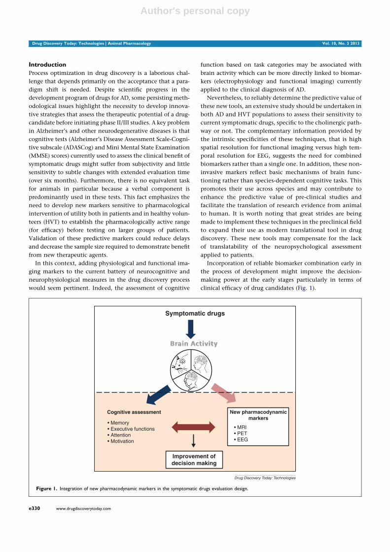
Incorporation of reliable biomarker combination early in
the process of development might improve the decision-
making power at the early stages particularly in terms of
clinical efficacy of drug candidates (Fig. 1).
Drug Discovery Today: Technologies | Animal Pharmacology Vol. 10, No. 3 2013
Symptomatic drugs
Cognitive assessment
• Memory• Executive functions• Attention• Motivation
New pharmacodynamicmarkers
• MRI• PET• EEG
Improvement ofdecision making
Drug Discovery Today: Technologies
Figure 1. Integration of new pharmacodynamic markers in the symptomatic drugs evaluation design.
e330 www.drugdiscoverytoday.com

Author's personal copy
It is worth keeping in mind that these biomarkers may not
be sensitive enough to detect changes in HVT, so their
sensitivity in response to a challenge test may be useful. This,
however, leads to a further barrier regarding the emergence of
novel AD drugs. In fact, as of today, the most commonly used
pharmacological challenge is with scopolamine, a non-selec-
tive muscarinic receptor antagonist. Novel cognitive enhan-
cing therapies have been assessed with scopolamine both pre-
clinically and clinically with varying degrees of success. The
weakness of this ‘pharmacological’ model might be related to:
(a) its targeted action on a single neurotransmitter system
whereas AD is a multifocal disease in which cognitive impair-
ment results from alteration of multiple systems, compromis-
ing its use to test drugs that are not exclusively targeting the
cholinergic system; (b) the potential pharmacological inter-
action with therapeutic drugs could compromise the inter-
pretation of the data.
To provide a ‘non pharmacological’ alternative to the
commonly used scopolamine model, we suggest investigat-
ing three potential models with different approaches: sleep
deprivation; hypoxia; repetitive Transcranial Magnetic Sti-
mulation (rTMS) to induce transient cognitive impairment in
HVTs and rodents. The basic requirement of these challenge
models is to (i) be able to induce cognitive impairment
and modify electrophysiological and imaging parameters
and (ii) be sensitive to current therapy for AD with different
mechanisms of action to provide a basis for new potential
drugs assessment (Fig. 2).
In this review, we seek to identify the most accurate
markers to assess drug effect (of commercially available AD
symptomatic drugs) among neuropsycological, neurophysio-
logical and imaging tools (PET/MRI), based on literature data.
Our investigation includes clinical and pre-clinical popula-
tions: (i) healthy versus AD subjects in humans and rodents
and (ii) translational physiological challenge models in
humans and rodents.
Evaluation of symptomatic drug effect on biomarker
battery
Currently, the field of AD drug discovery research is lacking
in pharmacodynamic biomarkers capable of (i) rapidly
detecting central activity (cognitive changes) in response
to treatment, in healthy groups of animals and humans
and (ii) predicting early therapeutic efficacy in AD clinical
and pre-clinical populations.
The following paragraph summarizes the current situation.
Insights on biomarkers in the clinical field
ADAS-Cog is the most used cognitive scale to evaluate dis-
turbances of memory, language, praxis, attention and other
cognitive abilities often referred to as the core symptoms
of AD. Several systematic reviews reported significant
Vol. 10, No. 3 2013 Drug Discovery Today: Technologies | Animal Pharmacology
Symptomaticdrugs
Clinical and preclinicalassessment
• Cognition• MRI• PET• EEG
Challenge models
• Scopolamine• Sleep deprivation• rTMS• Hypoxia
Drug Discovery Today: Technologies
Figure 2. Use of a biomarker battery at early stage of clinical development to assess effect of symptomatic drugs alone or in combination with a challenge
test.
www.drugdiscoverytoday.com e331

Author's personal copy
improvement in ADAS-Cog score in mild cognitive impair-
ment (MCI) or AD subjects treated with donepezil, reflecting
beneficial effects on cognitive status. In HVT, donepezil
induces a slight improvement in the retention of training
on complex aviation tasks [1], verbal memory for semanti-
cally processed words [2] and might improve long term visual
memory [3]. Nevertheless, some studies reported transient
negative effects on episodic memory [4,5] and no improve-
ment in the Cambridge Neuropsychological Test Automated
Battery (CANTAB), a computer-based cognitive assessment
system consisting of a battery of neuropsychological tests. In
a recent study conducted in healthy older individuals, done-
pezil has been shown to impair cognitive function associated
with alteration in additional neural markers including EEG
and fMRI markers [6]. Moreover anticholinesterase treatment
also improved attention in MCI and AD patients [7–13].
Evaluation of the effect of symptomatic drugs through the
measurement of electroencephalogram variables considered
as indices of cognitive processing has been extensively stu-
died in AD patients. Data available in the literature on resting
state EEG indicated that long- and short-term treatment with
donepezil reduced significantly the deterioration of EEG
spectral activity and correlated with cognitive improvement
rate on the ADAS-cog. This improvement is mainly charac-
terized by a reduction of the slow-wave activity (theta and
delta power) in frontal and temporo-parietal areas [14–17]. In
a small study, Sneddon and his colleagues demonstrated the
ability of a qEEG variance combined with a delayed recogni-
tion memory task to measure accurately treatment effects on
patients with AD [18]. In healthy elderly subjects, a single
dose of memantine has been shown to compensate diurnal
vigilance fluctuation measured by EEG recording [19].
Another EEG component, the P300 is of special interest as
it is related to brain functions such as cognition and atten-
tion, which are severely impaired in patients with dementia
[20]. In AD patients performing auditory and visual oddball
tasks, significant changes expressed by a reduction of the
P300 latency were observed already during the first month
of donepezil administration [21,22] and were significantly
correlated with various neuropsychological score changes
[22–24].
With regards to the MR technique, various studies have
undertaken anatomical analysis of hippocampal volume.
They indicated a decrease in hippocampal volume loss/in
the rate of hippocampal atrophy following treatment with
donepezil or memantine that closely relates to neuropatho-
logic and clinical data [25–27]. Functional MRI is uniquely
suited for evaluation of cognitive-enhancing agents. In 2
cognitive paradigms of visual memory, donepezil has been
reported to produce activation in the ventrolateral prefrontal
cortex (PFC) and in the fusiform gyrus in patients with MCI or
AD [28,29]. Another study demonstrated that donepezil
reversed the deficit of activation in fronto parietal region
during a working memory task in MCI patients [30]. Finally, a
reduced activation in the PFC and anterior cingulate cortex
was observed after memantine administration during an
auditory attention control task [31]. Using [18F]fluorodeox-
yglucose (FDG-PET) imaging, a study under resting condi-
tions demonstrated that treatment with donepezil in AD
patients may slow the decline in functional brain activity
in the right parietal lobe, left temporal lobe and right and left
frontal lobes [32]. In addition, Teipel et al. showed that the
metabolic changes induced by donepezil, during a passive
audio-visual stimulation, were limited to the right hippocam-
pus and the left PFC and independent of the effects on
cognitive performance [33]. Finally, in patients treated with
memantine for 52 weeks, glucose metabolism in all brain
areas was preserved [27].
These insights support the potential suitability of electro-
physiological and imaging measurements to monitor the
effects of symptomatic AD drugs in a context of disturbed
(in AD patients) or non-disturbed (in HVT) subjects.
Insights on biomarkers in the pre-clinical field
To bridge the gap between pre-clinical and clinical research
studies, great progress has been made in the development of a
wide range of pathological models of AD and improvement of
functional analysis techniques very similar to those per-
formed in the clinical setting (micro PET, wireless microchip
technologies for EEG).
The disease models attempt to reproduce most of the
neuropathology including neurophysiological and beha-
vioral hallmarks of AD pathology [34,35]. Since the promi-
nent feature of AD is the loss of neurons and synapses in the
hippocampal formation and related areas, neurobehavioral
phenotyping of mouse models of AD have focused on the
evaluation of hippocampal-related cognitive areas including
spatial-, contextual-, working-and recognition memory.
These cognitive functions have been extensively explored
through a classical battery of experimental tasks that con-
tributed to the characterization of cognitive profile associated
with each transgenic line [35]. Despite their limitations, the
comprehensive study of these transgenic mouse models pro-
vided a good opportunity to evaluate their translational and
predictive value of drug efficacy. The next section sum-
marizes the data on the sensitivity of cognitive tests and of
new technological biomarkers that aim to measure the
response to AD drugs in genetically modified and wild type
mice.
In a substantial work, Van Dam and colleagues attempted
to validate the APP23 model [36] by studying the reversibility
of spatial learning and memory impairment in response to
therapeutically relevant doses of three acetylcholinesterase
(AChE) inhibitors and memantine. They reported that
the lowest dose of Donepezil, rivastigmine and to a lesser
extent galantamine, elicited beneficial effects on learning and
Drug Discovery Today: Technologies | Animal Pharmacology Vol. 10, No. 3 2013
e332 www.drugdiscoverytoday.com

Author's personal copy
memory performance measured in the Morris Water Maze
(MWM) task [37]. As for memantine, modest effects have
been noted with only partial improvement of spatial learning
abilities [37]. However, in a subsequent report, the authors
demonstrated that longer administration of memantine (8
weeks) improved the effect [38]. In the Tg2576 mouse model
of [39] AD, Dong and colleagues also demonstrated beneficial
effects of donepezil on spatial learning/memory with regards
to improvement of acquisition in the water T-maze and in
contextual memory in a conditioned fear test [40]. Further-
more, administration of memantine through drinking water
for 3 weeks to APP/PS1 transgenic mice [41] improved acqui-
sition but no retention abilities in the water maze task [42].
Romberg and colleagues demonstrated that Donepezil res-
cues the attention deficit by improving response accuracy on
the 5-CSRTT adversely affected in the 3xTgAD mice model
[43,44].
With respect to investigations of electrophysiological and
imaging markers in experimental AD models, there is limited
data. An interesting paper reviewed the findings obtained to
date from studies using PET imaging and EEG to monitor
pathological changes in rodent AD models. They compared
FDG-PET phenotypes in 3 cohorts of transgenic strains and
indicated that metabolic activity was reliably compromised
in PSAPP and PLB1triple transgenic mice [45,46] but not in
APP/PS1 mice [41] which carry a different mutation in the PS1
transgene [47]. In addition, with FDG-based autoradiogra-
phy, metabolic changes have also been reported in the 3xTg
mouse [48]. However the AD-relevant brain region specificity
is discussed. Furthermore, a comprehensive analysis of sleep
patterns, vigilance staging and qEEG conducted in APP/PS1-
overexpressing mice highlighted a decreased in low and an
increased in high frequency spectral EEG power [49]. Simi-
larly, AD-like shifts in spectral power recorded in the parietal
cortex area, have been reported in the triple transgenic mice
(PLB1 triple), in agreement with FDG hypometabolism maps
[50]. Moreover the authors suggested that these changes
might precede the cognitive decline onset [44]. Although
the method to evoke a P300 in rodent is now well established,
data are missing in the field of transgenic animals of AD.
Thus, many of these technological tools are so recent that
their sensitivity to therapeutic intervention has not yet been
fully investigated. Only Scholtzova and colleagues followed
up the therapeutic response to memantine in APP/PS1 trans-
genic mice regarding amyloid deposition by micromagnetoc
MRI. Despite modest changes between control and treated
groups this tool was able to detect reduction in amyloid
burden, which correlates with cognitive improvement [51].
Fortunately, the good correlation between the preclinical
preliminary results on transgenic strains and human out-
comes encourages the analysis of the modulation of these
markers in a therapeutic context. Nevertheless, it is interest-
ing to underline that no change in acquisition or retention
performance was observed in wild type (WT) mice among
treated groups [37], contrasting with some results obtained in
HVT. However, Spowart-Manning and co-workers reported
that the T-maze continuous alternation task assessing the
spatial exploratory performances developed by Gerlai [52,53]
was sensitive enough to respond to donepezil administration
by improving the spontaneous alternation rate in control
mice [54]. Overall, studies on healthy animals cannot be
discussed since the differences of performance between
strains are too heterogeneous and the majority of tasks are
sensitive to genetic differences. The sensitivity of a particular
experiment depends at least partially, upon the value of the
control group.
This literature overview highlights the heterogeneity of the
cognitive assessment performed in the preclinical studies
with various different stimuli and some methodological issue
(confounding factors). These data cannot yet conclude which
biomarker array is most pertinent.
Evaluation of the effect of a cognitive challenge on the
biomarker battery - combined effect with
symptomatic drugs
Due to the inherent difficulty of detecting significant
improvements in cognitive performance in normal healthy
subjects, pre-clinical and clinical scientists have developed a
number of experimental paradigms to artificially induce
cognitive impairments akin to those observed in AD. The
identification and validation of HVT challenge models sui-
table for use in early clinical development might support
‘hint of efficacy’ studies that can be back translated to pre-
clinical studies. The complexity of AD prevents recapitulating
both the pathophysiology and symptoms in one animal
model. Currently, each animal model only partially reflects
the underlying pathology, for example, the amyloid aggre-
gates. Conversely, pharmacodynamic models are more
straightforward than disease models and should exhibit
one or more transient cognitive impairments (also typical
of AD) that are sensitive to the pharmacological active range
of the compound in question. Validation of such models
will rely on (a) the characterization of the cognitive and
neurofunctional deficits induced, (b) their sensitivity to cur-
rent symptomatic treatment and (c) their bidirectional trans-
latability. The nature of these challenge models is very
important. We propose to study three distinct non-pharma-
cological models that develop a transient cognitive impair-
ment in HVT or rodents following sleep deprivation, hypoxia
and rTMS challenges. Thus, in the next section, we will review
studies that have examined the effects of provocation chal-
lenges and how these dynamically alter cognitive, electro-
physiological and neuroimaging markers and how these
markers respond to symptomatic treatment. To highlight
the translational properties of each challenge model,
clinical and preclinical findings will be discussed in parallel.
Vol. 10, No. 3 2013 Drug Discovery Today: Technologies | Animal Pharmacology
www.drugdiscoverytoday.com e333

Author's personal copy
Beyond identifying the most appropriate model, the second
objective, in this context of challenge models, is to select the
most relevant biomarkers for the assessment of treatment
efficacy.
Sleep deprivation challenge
Sleep deprivation (SD) has been largely investigated in clin-
ical trials due to its association with deficits in the memory
process. Many behavioral adverse effects of SD including the
degradation of a wide range of cognitive functions have been
fully described (for review [55]). On the basis of studies that
have focussed on total sleep deprivation (TSD) protocols,
there is extensive literature reporting the deterioration of
executive functioning including decision making, flexible
thoughts, semantic processing and inhibition as well as work-
ing memory [56–59]. Episodic memory was also impaired in
verbal and visual domains and simple and complex attention
processes have been shown to be disrupted [60–62]. Interest-
ingly, the duration of TSD is not correlated with the magni-
tude of cognitive impairments in that under prolonged TSD
performance remains stable [63–65]. In addition, cognitive
deficits are inversely correlated with cerebral activity as mea-
sured by functional imaging investigations. In a PET study,
Thomas and colleagues noted a decreased glucose metabo-
lism in fronto-parietal and thalamic areas [66]. Other authors
demonstrated a hypometabolism in the frontal regions that
correlated with disturbances in working memory perfor-
mances [67–69]. Furthermore, metabolic decreases in poster-
ior parietal cortices have been associated with deficits of
verbal working memory [70]. More recently, the modulation
of blood-oxygen-level-dependent (BOLD) fMRI signal inten-
sity has been assessed in sleep-deprived subjects performing a
divided attention task and the vulnerability of pre-frontal
cortex to sleep deprivation previously observed has been
confirmed [71,72]. However some inconsistent findings
may be noticed between clinical trials. These differences
may be partly explained by the inter-individual heterogene-
ity in the subjects’ sensitivity to SD. Studies of SD on EEG
power spectra have revealed an increase in low-frequency
EEG power (delta and theta frequencies) after TSD [73–75].
Similarly, significant reduction of alpha power in rapid eye
movement (REM) sleep has been detected after total and
partial SD [73,76]. Topographical analysis combining EEG
to PET has been suggested to access the spatio-temporal
changes of EEG signal in pharmacological studies [75]. Simi-
larly, there is evidence of a negative impact of sleep depriva-
tion on the auditory event related potential (ERP). SD has
been shown to induce a delay of P300 latency and a reduction
of its amplitude [77–79]. Other ERP components seem sensi-
tive to SD to a lesser extent [77,79].
On the basis of this core feature of sleep deprivation
some authors have investigated whether such read-outs are
reversed in response to treatment with symptomatic drugs. In
two consecutive studies, Chuah and colleagues have explored
the effects of donepezil on cognitive and neuroimaging
deficits that developed in a cohort of total sleep-deprived
subjects. The first study conducted on 28 HVT showed that
donepezil administered for 17 days enhanced both visual
short-term memory and visual attention in sleep-deprived
HVT. This behavioral benefit, observed in individuals vulner-
able to the effects of sleep deprivation, primarily correlates
with an increase in neural activation in the parieto-occipital
regions that mediate attention and visual sensory processing
[80]. The second study performed on the same subjects
showed that donepezil could improve performance in an
episodic memory task. They also demonstrated a significant
correlation between left inferior prefrontal activation at
encoding, and performances in recognition memory that
may relate specifically to episodic memory [81]. This relation-
ship between fMRI pattern and behavioral drug effects
emphasize the value of combining these two measures for
future drug candidate assessment. Furthermore, in the con-
text of sleep deprivation-induced episodic memory deficit,
donepezil enhanced activation of cerebral regions involved
in attention and memory encoding processing during a
semantic judgement task [81]. A recent work failed to find
any donepezil-induced improvement of memory or non-
memory cognitive tasks impaired by sleep deprivation [82].
Many analogies have been proven in pre-clinical studies
challenging TSD (REM sleep deprivation has not been taken
into account). In terms of cognition, long-term and short-
term SD has been shown to alter hippocampal-dependent
spatial learning and memory in the MWM task [83,84].
Repetition of brief epochs of SD over a long period of time
[85] or protocols of sleep fragmentation [86–88] lead to
similar effects. Nevertheless, in light of these studies, the
consolidation process of spatial information seems to be most
vulnerable to sleep deprivation. Furthermore, in an alterna-
tion paradigm measuring working memory ability, where SD
was given after training, disturbed the alternation rate
[89,90]. The fear conditioning paradigm has also been used
to assess declarative-like memory in sleep-deprived animals.
Findings revealed that SD negatively affects the acquisition
and consolidation phase of contextual fear conditioning
related to the hippocampus [91–94] whereas performance
of cued fear conditioning related to the amygdala is spared
[91]. Using the Novel Object Recognition (NOR) task, con-
sidered to measure episodic memory as that in human [95],
researchers indicated that SD administered after the acquisi-
tion phase severely impairs object recognition during the
retention test [96,97]. Finally, attention processes have also
been explored in the SD context in rodents. SD was shown to
impair animals’ speed and accuracy in the five choice reaction
time task however no deficit in the executive control of the
task was observed [98]. These findings are in agreement with
another study using a sleep fragmentation protocol [99].
Drug Discovery Today: Technologies | Animal Pharmacology Vol. 10, No. 3 2013
e334 www.drugdiscoverytoday.com

Author's personal copy
In terms of electrophysiology, studies in rats and mice have
shown an overall increase in slow wave activity after SD [100–
102]. ERP parameters have not been explored in the SD
preclinical challenge. Effect of SD on modulation of neural
activity measured by functional imaging in rodents has yet to
be reported. Similarly, the reversal effect of symptomatic
drugs on modification of markers induced by sleep depriva-
tion has not been probed.
rTMS challenge
Transcranial magnetic stimulation is a painless method based
on the induction of a small electrical current in the brain by a
magnetic field applied to a small area of the skull. This
technique has been described as a non-invasive transient
way to interfere with cognitive functions by the stimulation
of specific areas of interest [103,104]. Despite the incontest-
able interest of this method as a ‘virtual lesion technique’ to
study cognitive functions, its application remains limited due
to its limited depth of penetration [105]. At present, deep
areas of the brain located many centimeters below the scalp
such as hippocampus, amygdala or mammillary bodies can-
not be directly reached. However, combination of TMS with
fMRI seems to demonstrate that the activity of these areas can
be actually modulated by TMS, presumably due to transy-
naptic effects. Thus, research is mainly focused on stimula-
tion of frontoparietal networks involved in working memory
as well as the encoding and retrieval of novel items. In
particular several studies targeted the role of the PFC in
episodic memory. These works, using different experimental
tasks requiring wording, demonstrated that recall perfor-
mances were impaired following rTMS over the left dorso-
lateral PFC during the encoding process [106–111]. A similar
effect was observed, for some of these works, following rTMS
over the right dorsolateral PFC during retrieval suggesting
that there is a left encoding, right recognition PFC asymetry
for memory processes [108–111]. These findings were in
agreement with a recent study demonstrating that paired-
pulse TMS applied over the left or right dorsolateral PFC
interfered with episodic encoding and retrieval, irrespective
of the type of material used (verbal versus non-verbal) [112].
In addition, TMS over the left antero-ventral inferior PFC
region has been shown to disrupt episodic encoding of non-
verbal material [113]. Regarding working memory, many
investigations highlighted the negative impact of rTMS in
various brain areas on this cognitive function (for review
[114]). Mottaghy and colleagues reported disruption in work-
ing memory for faces (face recognition delayed response task)
when rTMS was applied to ventral PFC and dorsolateral PFC
while spatial working memory was affected following rTMS
over the dorsolateral PFC and dorsomedial PFC [115]. Impair-
ment of visual working memory after left dorsolateral PFC
stimulation has also been described [116–118]. Moreover
Wagner and colleagues provided evidence for delayed rTMS
effects on divided attention task performances since visual
reaction time is slowed over 30 min after rTMS session at the
dorsolateral PFC [119]. Other studies report that applying
rTMS to the superior parietal lobule produces a disruption of
cognitive behavior [120–122]. Kessel and colleagues also
indicated that the reaction times measured in a spatial work-
ing memory task were slower during right-parietal rTMS than
during left-parietal rTMS [123]. However discrepant findings
were observed for the effect of rTMS on attentional processes
[124]. Finally, cerebellar TMS is able to influence memory
abilities including working memory [125,126]. Multimodal
approaches have also been conducted by combining TMS
with neuroimaging or electrophysiological methods to assess
spatial distribution of TMS-evoked activity and brain con-
nectivity [127]. These investigations suggested an activation
of brain structures distant from the TMS-stimulated area
[128–135]. Other studies assessed the brain–behavior rela-
tionship, that is the modulation of brain function during a
cognitive task, by combination of TMS with PET during
cognitive performances. Mottaghy and co-workers empha-
sized the concomitant alteration of working memory task
performance with a change in the activation pattern as
revealed by PET, in the condition with rTMS of the left or
right dorsolateral PFC [136]. The same team demonstrated a
positive correlation between the regional cerebral blood flow
changes in the superior frontal gyrus functionally connected
to the stimulation site and performances in the working
memory task [137] reviewed in [114].
The emergence of a new method to avoid the artefact
problem caused by the magnetic pulse proposed by Thut
and colleagues have provided a means for studying the
impact of TMS on the functional electrophysiological signals
associated with (cognitive) task performance, such as ERPs
[138]. An equivalent procedure has also been developed in
animals [139]. Effect of rTMS on auditory oddball task has
been investigated after stimulation over dorsolateral PFC.
Using an auditory oddball task, the authors demonstrated
a widespread significantly reduced alphe desynchronisation
post-TMS while the P300 component (amplitudes or laten-
cies) was not affected [140]. In contrast, stimulation of the
supramarginal gyrus by TMS applied at 200ms after the odd-
ball sounds presentation delayed the peak response of P300
[141]. Moreover, EEG power spectrum has also been exam-
ined immediately following rTMS. Studies reported a tem-
poral increase in frequency and amplitude of EEG within the
first 2 min after high frequency rTMS over left frontal cortex
[142]. Schutter and colleagues also indicated that medial
cerebellar rTMS affected the high frequency band (gamma
band) of EEG spectrum [143].
The effect of acetylcholinesterase inhibitors or meman-
tine on neurophysiological parameters, fMRI or cerebral
glucose metabolism has not been yet studied in the context
of rTMS.
Vol. 10, No. 3 2013 Drug Discovery Today: Technologies | Animal Pharmacology
www.drugdiscoverytoday.com e335

Author's personal copy
Compared to the large number of studies in humans, there
a few studies describing the effects of rTMS on memory and
learning in animal models have been little studied. This could
be related to the difficulty of precisely stimulating a specific
brain area with this technique in animals. Moreover, many
stimulation parameters such as frequency, intensity, dura-
tion and number of pulse can modulate the behavioral
response. An interesting study has investigated the impact
of the modulation of stimulation frequency (chronic versus
acute) on memory performance in rats [144]. The findings
indicated that chronic low frequency stimulation could
impair the retrieval of both short- and long-term spatial
reference memory sparing the acquisition process; while after
acute rTMS only the long-term reference memory was
impaired. In another study Ahmed and Wieraszko high-
lighted the importance of the magnetic field properties
applied. They demonstrated a temporary deterioration of
performances in the NOR test performed immediately after
stimulation at lower frequencies (1 and 8 Hz) while perfor-
mance was improved after stimulation at higher frequency
(15 Hz). However, when the test is performed 1 h or 3 days
after high frequency rTMS, performances in NOR test are also
impaired [145]. Other researchers concluded that 50 brief
pulse transcranial magnetic stimulations may cause a disrup-
tion of retrograde memory for conditioned taste aversions
[146]. Regarding cortical electrical activity, rats subjected to
low frequency rTMS display a reduction in amplitude of the
power spectra in the high frequency bands (beta and gamma)
[147] consistent with clinical findings [142,143].
Hypoxia challenge
The interest in hypoxia as a potential inducer of cognitive
disorder results from studies conducted in the context of
aviation and mountain ascent. Nevertheless the role of
hypoxia on cognitive domains remains relatively unex-
plored. Overall, studies in the literature indicated that cog-
nitive impairment caused by hypoxia is modulated by the
severity of hypoxic level and the population considered
(experienced versus non experienced participants). To accu-
rately compare clinical and preclinical data, we focused our
interest on behavioral and neurophysiological effects of
hypoxia on non-experienced subjects (naıve from hypoxic
environment). Cognitive functions including executive func-
tioning, working memory and attention have been reported
to be affected by hypoxia. Unfortunately, results seem some-
what contradictory due to a lack of methodological consis-
tency among studies. Nevertheless a core of findings seems to
demonstrate alteration of performance in serial recognition
of words, the binary choice task and auditory and visual
reaction time [148]. Noble and co-workers also reported an
overall slowing on task measuring executive functioning
[149] that was not confirmed by others [148,150]. Studies
have provided evidence of an impairment in executive
processes indicated by a decline in the Grammatical Logical
Reasoning task [151,152] associated to an inability to learn a
novel task [152]. Working memory is also affected following
short-term exposure to hypoxia shown by alteration of per-
formances in a go/no-go discrimination task [153]. These
disturbances were not observed in the rapid visual categorical
task [154]. In addition, many studies showed disruption of
some aspect of attentional processes through various tests
[56,155–159]. Episodic memory has not been studied.
Although neuropsychological results are highly controver-
sial, more consistent results come from electrophysiological
investigations. Most studies have shown EEG changes char-
acterized by increases in delta and theta power and a decrease
in alpha activity under hypoxia [158,160–162]. Moreover an
identical trend, with regards to ERP P300 assessment, emerges
from the literature. Indeed, long- and short-term exposures to
hypoxia have induced an increase of P300 latency with
minimal effects on P300 amplitude [163–165]. Other para-
meters such as N100, P200 and N200 seem less sensitive to
hypoxia [164].
Using neuroimaging techniques various clinical studies
have investigated the cerebrovascular response to hypoxia
analyzing regional cerebral blood flow distribution [166–
168]. Data demonstrated differential physiological sensitivity
of various brain regions. Functional neuroimaging data
demonstrated specific sensitivity of frontal lobes [169] sup-
ported by the metabolic reduction observed in this region
[170]. Thus, hypoxia seems to affect the cerebral network
involved in the attention process, which is consistent with
neuropsychological deterioration. As for the challenge of
rTMS, the effect of symptomatic treatment has not been
tested under hypoxia challenge.
Studies of hypoxia in rodents have experienced identical
discrepancies regarding cognitive deficits since intensity,
frequency and duration of hypoxia exposure modulate the
magnitude of cognitive alteration in rodents [171,172]. Find-
ings demonstrated detrimental effects of hypoxia on both
acquisition and consolidation processes in different para-
digms of cognitive tests. Chronic exposure to hypoxia can
alter working and reference spatial memory assessed in MWM
task [173–180]. These spatial memory dysfunctions have also
been noted after acute exposure of hypoxia beyond a thresh-
old level [172,181]. Using place-discrimination task in a radial
arm maze, Titus confirmed these spatial impairments that
occur following several days of hypoxia [182]. In addition,
Zhang reported dysfunction in acquisition and retention
processes in a shuttle box paradigm which models non-
declarative memory [183]. In the passive avoidance task,
Udayabanu and co-workers revealed that hypoxia could only
disrupt retrograde memory suggesting an effect of hypoxia on
consolidation [184]. Finally brief exposure to hypoxia led to
milder and more transient cognitive alteration in spatial
working memory assessed in the Y maze task and in the
Drug Discovery Today: Technologies | Animal Pharmacology Vol. 10, No. 3 2013
e336 www.drugdiscoverytoday.com

Author's personal copy
NOR task [185–187]. In addition acute exposure interfered
with consolidation of memory in the step-through avoidance
task [188]. The sensitivity of other markers to hypoxia has not
been studied in rodents. The effect of symptomatic drugs on
cognitive impairment induced by hypoxia has been explored.
For example, Physostigmine and Galantamine has been
tested to improve spatial learning and working memory
induced by chronic hypoxia [180]. In addition, administra-
tion of physostigmine prior to hypoxic exposure was shown
to preserve recognition memory sensitive to hypoxia [185].
Strengths and weaknesses of current strategies and identification of
translational requirements
Currently, the majority of symptomatic treatments fail due to
a lack of efficacy in patients detected during phase II/III
clinical studies. This suggests that current preclinical and
clinical methods to test drug efficacy are not effective. To
improve the current process of drug development, we pro-
posed to introduce new pharmacodynamic biomarkers at
each stage of preclinical and clinical development, in addi-
tion to current clinical and neuropsychological assessments.
These markers are based on neurophysiological and neuroi-
maging measurements already commonly used in the clinical
field (for diagnosis and better understanding of the patho-
physiology). They are a direct reflection of cerebral activity
patterns and may overcome the frequenly criticized neurop-
sychological evaluation, which lacks objectivity. Moreover,
unlike cognitive enhancement that must be evaluated over a
period of six months, drug effects on physiological biomar-
kers might be detectable over a shorter time. Our detailed
review of the literature indicates that these alternative bio-
markers display a good sensitivity to symptomatic treatment
in AD or MCI patients since in most cases improvement in the
pharmacodynamic markers correlates with cognitive gain. In
HVT, studies are scarce and do not allow an assessment of the
contribution of these biomarkers as predictive tools. How-
ever, further studies to assess if their use is pertinent may
clarify this issue.
From a preclinical point of view, the development of these
biomarkers reflecting brain activity in a physiological resting
state might reduce the gap between preclinical and clinical
phases. Indeed, some evidence suggests that the limited
translatability of animal cognitive tests might explain dis-
appointing results in the development of successful treat-
ments for AD. The use of EEG, PET and MRI in the preclinical
assessment protocol of new pharmacological compounds
should facilitate the extrapolation of preclinical outcomes
to the clinical field. Results obtained from studies on geneti-
cally engineered animal models indicate a great sensitivity of
these markers to AD pathogenesis supporting their use as
translational tools. However their sensitivity to clinically
approved symptomatic treatments has not yet been
explored. Nevertheless, we cannot overlook the importance
of cognitive endpoints in the predictive validity of preclinical
models. As evidenced by the review, cognitive tests used
today are disparate and poorly comparable to those per-
formed in humans. These limitations are reinforced by the
fact that the neuropsychological battery used as a reference
standard to assess clinical drug efficacy, the ADAS-Cog, is
almost impossible to model in rodents as this specific scale
mainly explores unavailable functions in rodents, that is
verbal memory, language and praxis. Over the past decade,
a new technological instrument specifically designed to facil-
itate cross-species has emerged. The touchscreen technology
relies on a set of straightforward cognitive tasks procedurally
similar to clinical neuropsychological tests belonging to the
CANTAB battery (computer-based cognitive test procedures
becoming more prevalent in the clinical cognitive testing
domain). They explore through computer-automated cogni-
tive tests a range of cognitive domains (memory, attention,
executive functions) thought to be affected in AD. In addi-
tion, this standardized cognitive exploration tool uses the
same types of visual stimuli used in the CANTAB tasks. The
predictive validity remains to be demonstrated by extensive
studies. Validation of translational cognitive tasks may
potentially expand the role of EEG and PET in the preclinical
models whereby these markers can be measured under spe-
cific tasks [189,190]. MRI performed in rodents under
anesthesia, cannot benefit from such studies.
In light of these preliminary results and observations, it
appears that these alternative markers – combined with
appropriate cognitive assessment- would contribute to
improve the predictive and translational value of drug devel-
opment stages preceding Phase II/III studies. However, the
drug discovery process may still suffer from a lack of predict-
ability in early stage experimental medicine studies due to the
difficulties in identifying benefits of cognition enhancer
compounds in HVT without cognitive impairment. It might
be easier to reverse existing deficits than improve normal
functioning. In this view, our strategy to improve predictive
capacity is to include, in preclinical and clinical (phase 1)
trials translational pharmacodynamic models that exhibit
cognitive impairment reminiscent of that observed in
patients with AD. The choice of non-pharmacological pro-
vocation challenge – sleep deprivation, rTMS and hypoxia –
with a broader action in the CNS might provide an alternative
model to the scopolamine one, limited by its pharmacologi-
cal nature.
Overall, this review confirms the feasibility of inducing a
transient cognitive disorder in human and rodent models.
The cognitive deficits relate to multiple functions such as
executive functions, working memory and attention. Inves-
tigations on neurophysiological and imaging biomarkers are
less extensive but seem to show modulations of these para-
meters in response to the challenges. However, methodolo-
gical issues and lack of harmonization of clinical assessment
Vol. 10, No. 3 2013 Drug Discovery Today: Technologies | Animal Pharmacology
www.drugdiscoverytoday.com e337

Author's personal copy
tools make it difficult to compare these challenges. It would
be interesting to evaluate the intrinsic limits of each model by
performing parallel studies with standardized assessment
protocols based on a biomarker battery including cognitive,
neurophysiological and imaging endpoints. The reversibility
of changes in response to symptomatic drug exposure will
define their predictability and consequently their own ability
to support ‘hint of efficacy studies’ in early clinical stages as
well as in preclinical studies.
The identification of the most sensitive combination of
pharmacodynamic markers (as clinical efficacy indices)
and pharmacodynamic models (for early prediction of effi-
cacy) will increase the effectiveness of the drug discovery
process in AD through a multidimensional matrix approach
(Fig. 3).
Conclusion
A fairly abundant literature is now available on: (i) the effect
of AD symptomatic drugs on different biomarkers assessing
brain activity; (ii) the effect of challenge tests on cognitive
function and these similar biomarkers of brain activity. In
contrast, the data on the combination of symptomatic drug
and challenge test remain poor and with controversial results.
We feel that this combination coupled with biomarker assess-
ment could be a new and stringent paradigm to test new
symptomatic drugs in the context of AD, in particular at the
early stage of disease (Fig. 3). A systematic evaluation and
validation of such an approach remains necessary and it is
one of the goals of the IMI Pharmacog consortium, which
aims to further explore the preclinical ans clinical studies in
the context of gold standard symptomatic treatments for AD.
Conflict of interest
The authors have no conflict of interest to declare.
Acknowledgements
This research was developed under the ethical requirements
complied with the European Community Council Directive
(2010/63/UE). The research leading to these results was con-
ducted as part of the PharmaCog consortium funded by the
European Community’s Seventh Framework Programme for
the Innovative Medicine Initiative under Grant Agreement
no. 115009. For further information please refer to http://
www.pharmacog.org/
References1 Yesavage, J.A. et al. (2002) Donepezil and flight simulator performance:
effects on retention of complex skills. Neurology 59, 123–125
2 FitzGerald, D.B. et al. (2008) Effects of donepezil on verbal memory
after semantic processing in healthy older adults. Cogn. Behav. Neurol. 21,
57–64
3 Gron, G. et al. (2005) Cholinergic enhancement of episodic memory in
healthy young adults. Psychopharmacology (Berl.) 182, 170–179
4 Beglinger, L.J. et al. (2004) Neuropsychological test performance in
healthy volunteers before and after donepezil administration. J.
Psychopharmacol. 18, 102–108
5 Beglinger, L.J. et al. (2005) Neuropsychological test performance in
healthy elderly volunteers before and after donepezil administration: a
randomized, controlled study. J. Clin. Psychopharmacol. 25, 159–165
6 Balsters, J.H. et al. (2011) Donepezil impairs memory in healthy older
subjects: behavioural, EEG and simultaneous EEG/fMRI biomarkers. PLoS
ONE 6, e24126
7 Bentley, P. et al. (2008) Cholinesterase inhibition modulates visual and
attentional brain responses in Alzheimer’s disease and health. Brain 131
(Pt 2), 409–424
8 Blin, J. et al. (1998) Physostigmine results in an increased decrement in
brain glucose consumption in Alzheimer’s disease. Psychopharmacology
(Berl.) 136, 256–263
Drug Discovery Today: Technologies | Animal Pharmacology Vol. 10, No. 3 2013
Cognition
EEG
MRI
PET
WT
mic
e
Anim
al c
halle
nge
mod
el
AD m
odel
AD p
atie
nts
HVT
Hum
an c
halle
nge
mod
elPOSITIVE EFFECT
NEGATIVE EFFECT Phase I/IIGO / NO GO
Phase III
Drug Discovery Today: Technologies
Figure 3. Simulation of an AD drug development using a matrix battery.
e338 www.drugdiscoverytoday.com

Author's personal copy
9 Foldi, N.S. et al. (2005) Detecting effects of donepezil on visual selective
attention using signal detection parameters in Alzheimer’s disease. Int. J.
Geriatr. Psychiatry 20, 485–488
10 Lawrence, A.D. and Sahakian, B.J. (1995) Alzheimer disease, attention,
and the cholinergic system. Alzheimer Dis. Assoc. Disord. 9 (Suppl 2),
43–49
11 Rockwood, K. (2004) Size of the treatment effect on cognition of
cholinesterase inhibition in Alzheimer’s disease. J. Neurol. Neurosurg.
Psychiatry 75, 677–685
12 Sahakian, B.J. and Coull, J.T. (1993) Tetrahydroaminoacridine (THA) in
Alzheimer’s disease: an assessment of attentional and mnemonic
function using CANTAB. Acta Neurol. Scand. Suppl. 149, 29–35
13 Sahakian, B.J. et al. (1993) Further analysis of the cognitive effects of
tetrahydroaminoacridine (THA) in Alzheimer’s disease: assessment of
attentional and mnemonic function using CANTAB. Psychopharmacology
(Berl.) 110, 395–401
14 Balkan, S. et al. (2003) Effect of donepezil on EEG spectral analysis in
Alzheimer’s disease. Acta Neurol. Belg. 103, 164–169
15 Brassen, S. and Adler, G. (2003) Short-term effects of acetylcholinesterase
inhibitor treatment on EEG and memory performance in Alzheimer
patients: an open, controlled trial. Pharmacopsychiatry 36, 304–308
16 Reeves, R.R. et al. (2002) The effects of donepezil on quantitative EEG in
patients with Alzheimer’s disease. Clin. Electroencephalogr. 33, 93–96
17 Rodriguez, G. et al. (2002) Quantitative EEG changes in Alzheimer
patients during long-term donepezil therapy. Neuropsychobiology 46, 49–
56
18 Sneddon, R. et al. (2006) QEEG monitoring of Alzheimer’s disease
treatment: a preliminary report of three case studies. Clin. EEG Neurosci.
37, 54–59
19 Schulz, H. et al. (1996) The use of diurnal vigilance changes in the EEG to
verify vigilance-enhancing effects of memantine in a clinical
pharmacological study. Neuropsychobiology 33, 32–40
20 Juckel, G. et al. (2008) Diagnostic usefulness of cognitive auditory event-
related p300 subcomponents in patients with Alzheimers disease? J. Clin.
Neurophysiol. 25, 147–152
21 Reeves, R.R. et al. (1999) The effects of donepezil on the P300 auditory
and visual cognitive evoked potentials of patients with Alzheimer’s
disease. Am. J. Geriatr. Psychiatry 7, 349–352
22 Thomas, A. et al. (2001) Donepezil, rivastigmine, and vitamin E in
Alzheimer disease: a combined P300 event-related potentials/
neuropsychologic evaluation over 6 months. Clin. Neuropharmacol. 24,
31–42
23 Katada, E. et al. (2003) Long-term effects of donepezil on P300 auditory
event-related potentials in patients with Alzheimer’s disease. J. Geriatr.
Psychiatry Neurol. 16, 39–43
24 Werber, E.A. et al. (2003) The clinical use of P300 event related potentials
for the evaluation of cholinesterase inhibitors treatment in demented
patients. J. Neural Transm. 110, 659–669
25 Hashimoto, M. et al. (2005) Does donepezil treatment slow the
progression of hippocampal atrophy in patients with Alzheimer’s
disease? Am. J. Psychiatry 162, 676–682
26 Krishnan, K.R. et al. (2003) Randomized, placebo-controlled trial of the
effects of donepezil on neuronal markers and hippocampal volumes in
Alzheimer’s disease. Am. J. Psychiatry 160, 2003–2011
27 Schmidt, R. et al. (2008) Longitudinal multimodal imaging in mild to
moderate Alzheimer disease: a pilot study with memantine. J. Neurol.
Neurosurg. Psychiatry 79, 1312–1317
28 Kircher, T.T. et al. (2005) Cortical activation during cholinesterase-
inhibitor treatment in Alzheimer disease: preliminary findings from a
pharmaco-fMRI study. Am. J. Geriatr. Psychiatry 13, 1006–1013
29 Petrella, J.R. et al. (2009) Effects of donepezil on cortical activation in
mild cognitive impairment: a pilot double-blind placebo-controlled trial
using functional MR imaging. Am. J. Neuroradiol. 30, 411–416
30 Saykin, A.J. et al. (2004) Cholinergic enhancement of frontal lobe activity
in mild cognitive impairment. Brain 127 (Pt 7), 1574–1583
31 van Wageningen, H. et al. (2009) Evidence for glutamatergic
neurotransmission in cognitive control in an auditory attention task.
Neurosci. Lett. 454, 171–175
32 Tune, L. et al. (2003) Donepezil HCl (E2020) maintains functional
brain activity in patients with Alzheimer disease: results of a 24-week,
double-blind, placebo-controlled study. Am. J. Geriatr. Psychiatry 11,
169–177
33 Teipel, S.J. et al. (2006) Effects of donepezil on cortical metabolic response
to activation during (18)FDG-PET in Alzheimer’s disease: a double-blind
cross-over trial. Psychopharmacology (Berl.) 187, 86–94
34 Higgins, G.A. and Jacobsen, H. (2003) Transgenic mouse models of
Alzheimer’s disease: phenotype and application. Behav. Pharmacol. 14,
419–438
35 Bryan, K.J. et al. (2009) Transgenic Mouse Models of Alzheimer’s Disease:
Behavioral Testing and Considerations.
36 Sturchler-Pierrat, C. et al. (1997) Two amyloid precursor protein
transgenic mouse models with Alzheimer disease-like pathology. Proc
Natl Acad Sci U S A 94, 13287–13292
37 Van Dam, D. et al. (2005) Symptomatic effect of donepezil, rivastigmine,
galantamine and memantine on cognitive deficits in the APP23 model.
Psychopharmacology (Berl.) 180, 177–190
38 Van Dam, D. and De Deyn, P.P. (2006) Cognitive evaluation of disease-
modifying efficacy of galantamine and memantine in the APP23 model.
Eur. Neuropsychopharmacol. 16, 59–69
39 Hsiao, K. et al. (1996) Correlative memory deficits. Abeta elevation, and
amyloid plaques in transgenic mice. Science 274, 99–102
40 Dong, H. et al. (2005) Acetylcholinesterase inhibitors ameliorate
behavioral deficits in the Tg2576 mouse model of Alzheimer’s disease.
Psychopharmacology (Berl.) 181, 145–152
41 Borchelt, D.R. et al. (1997) Accelerated amyloid deposition in the brains
of transgenic mice coexpressing mutant presenilin 1 and amyloid
precursor proteins. Neuron 19, 939–945
42 Minkeviciene, R. et al. (2004) Memantine improves spatial learning in a
transgenic mouse model of Alzheimer’s disease. J. Pharmacol. Exp. Ther.
311, 677–682
43 Oddo, S. et al. (2003) Triple-transgenic model of Alzheimer’s disease with
plaques and tangles: intracellular Abeta and synaptic dysfunction. Neuron
39, 409–421
44 Romberg, C. et al. (2011) Impaired attention in the 3xTgAD mouse model
of Alzheimer’s disease: rescue by donepezil (Aricept). J. Neurosci. 31,
3500–3507
45 Holcomb, L. et al. (1998) Accelerated Alzheimer-type phenotype in
transgenic mice carrying both mutant amyloid precursor protein and
presenilin 1 transgenes. Nat Med 4, 97–100
46 Platt, B. et al. (2011) Abnormal cognition, sleep, EEG and brain
metabolism in a novel knock-in Alzheimer mouse, PLB1. PLoS One 6,
e27068
47 Platt, B. et al. (2011) FDG-PET imaging, EEG and sleep phenotypes as
translational biomarkers for research in Alzheimer’s disease. Biochem. Soc.
Trans. 39, 874–880
48 Nicholson, R.M. et al. (2010) Regional cerebral glucose uptake in the
3xTG model of Alzheimer’s disease highlights common regional
vulnerability across AD mouse models. Brain Res. 1347, 179–185
49 Jyoti, A. et al. (2010) EEG, activity, and sleep architecture in a transgenic
AbetaPPswe/PSEN1A246E Alzheimer’s disease mouse. J. Alzheimers Dis.
22, 873–887
50 Platt, B. et al. (2011) Abnormal cognition, sleep, EEG and brain
metabolism in a novel knock-in Alzheimer mouse, PLB1. PLoS ONE 6,
e27068
51 Scholtzova, H. et al. (2008) Memantine leads to behavioral improvement
and amyloid reduction in Alzheimer’s-disease-model transgenic mice
shown as by micromagnetic resonance imaging. J. Neurosci. Res. 86, 2784–
2791
52 Gerlai, R. (1998) A new continuous alternation task in T-maze detects
hippocampal dysfunction in mice. A strain comparison and lesion study.
Behav. Brain Res. 95, 91–101
53 Gerlai, R. et al. (1994) T-maze spontaneous alternation rate is decreased in
S100 beta transgenic mice. Behav. Neurosci. 108, 100–106
54 Spowart-Manning, L. and van der Staay, F.J. (2004) The T-maze
continuous alternation task for assessing the effects of putative cognition
enhancers in the mouse. Behav. Brain Res. 151, 37–46
Vol. 10, No. 3 2013 Drug Discovery Today: Technologies | Animal Pharmacology
www.drugdiscoverytoday.com e339
Author's personal copy
55 McCoy, J.G. and Strecker, R.E. (2011) The cognitive cost of sleep lost.
Neurobiol. Learn. Mem. 96, 564–582
56 Blogg, S.L. and Gennser, M. (2006) Cerebral blood flow velocity and
psychomotor performance during acute hypoxia. Aviat. Space Environ.
Med. 77, 107–113
57 Chee, M.W. and Choo, W.C. (2004) Functional imaging of working
memory after 24 hr of total sleep deprivation. J. Neurosci. 24, 4560–4567
58 Gottselig, J.M. et al. (2006) Random number generation during sleep
deprivation: effects of caffeine on response maintenance and stereotypy.
J. Sleep Res. 15, 31–40
59 Killgore, W.D. et al. (2009) Executive functions and the ability to sustain
vigilance during sleep loss. Aviat. Space Environ. Med. 80, 81–87
60 Chee, M.W. et al. (2010) Sleep deprivation and its effects on object-
selective attention. Neuroimage 49, 1903–1910
61 Tucker, M.A. and Fishbein, W. (2009) The impact of sleep duration and
subject intelligence on declarative and motor memory performance: how
much is enough? J. Sleep Res. 18, 304–312
62 Yoo, S.S. et al. (2007) A deficit in the ability to form new human memories
without sleep. Nat. Neurosci. 10, 385–392
63 Chee, M.W. et al. (2006) Functional imaging of working memory
following normal sleep and after 24 and 35 h of sleep deprivation:
correlations of fronto-parietal activation with performance. Neuroimage
31, 419–428
64 Sagaspe, P. et al. (2006) Effects of sleep deprivation on Color-Word,
Emotional, and Specific Stroop interference and on self-reported anxiety.
Brain Cogn. 60, 76–87
65 Van Dongen, H.P. et al. (2003) The cumulative cost of additional
wakefulness: dose–response effects on neurobehavioral functions and
sleep physiology from chronic sleep restriction and total sleep
deprivation. Sleep 26, 117–126
66 Thomas, M. et al. (2000) Neural basis of alertness and cognitive
performance impairments during sleepiness. I. Effects of 24 h of sleep
deprivation on waking human regional brain activity. J. Sleep Res. 9, 335–
352
67 Chee, M.W. and Chuah, Y.M. (2007) Functional neuroimaging and
behavioral correlates of capacity decline in visual short-term memory
after sleep deprivation. Proc. Natl. Acad. Sci. U. S. A. 104, 9487–9492
68 Choo, W.C. et al. (2005) Dissociation of cortical regions modulated by
both working memory load and sleep deprivation and by sleep
deprivation alone. Neuroimage 25, 579–587
69 Mu, Q. et al. (2005) Decreased brain activation during a working memory
task at rested baseline is associated with vulnerability to sleep
deprivation. Sleep 28, 433–446
70 Mu, Q. et al. (2005) Decreased cortical response to verbal working
memory following sleep deprivation. Sleep 28, 55–67
71 Drummond, S.P. et al. (2001) Increased cerebral response during a divided
attention task following sleep deprivation. J. Sleep Res. 10, 85–92
72 Jackson, M.L. et al. (2011) The effect of sleep deprivation on BOLD
activity elicited by a divided attention task. Brain Imaging Behav. 5, 97–
108
73 Borbely, A.A. et al. (1981) Sleep deprivation: effect on sleep stages and
EEG power density in man. Electroencephalogr. Clin. Neurophysiol. 51, 483–
495
74 Dijk, D.J. et al. (1993) Dynamics of electroencephalographic sleep
spindles and slow wave activity in men: effect of sleep deprivation. Brain
Res. 626, 190–199
75 Landolt, H.P. et al. (2000) Zolpidem and sleep deprivation: different effect
on EEG power spectra. J. Sleep Res. 9, 175–183
76 Brunner, D.P. et al. (1993) Repeated partial sleep deprivation
progressively changes in EEG during sleep and wakefulness. Sleep 16, 100–
113
77 Lee, H.J. et al. (2004) Auditory event-related potentials and psychological
changes during sleep deprivation. Neuropsychobiology 50, 1–5
78 Morris, A.M. et al. (1992) The P300 event-related potential. The effects of
sleep deprivation. J. Occup. Med. 34, 1143–1152
79 Ray, K. et al. (2012) Modafinil improves event related potentials P300 and
contingent negative variation after 24 h sleep deprivation. Life Sci. 91,
94–99
80 Chuah, L.Y. and Chee, M.W. (2008) Cholinergic augmentation
modulates visual task performance in sleep-deprived young adults.
J. Neurosci. 28, 11369–11377
81 Chuah, L.Y. et al. (2009) Donepezil improves episodic memory in young
individuals vulnerable to the effects of sleep deprivation. Sleep 32,
999–1010
82 Dodds, C.M. et al. (2011) Effects of donepezil on cognitive performance
after sleep deprivation. Hum. Psychopharmacol. 26, 578–587
83 Chang, H.M. et al. (2009) Melatonin preserves longevity protein (sirtuin
1) expression in the hippocampus of total sleep-deprived rats. J. Pineal
Res. 47, 211–220
84 Guan, Z. et al. (2004) Sleep deprivation impairs spatial memory and
decreases extracellular signal-regulated kinase phosphorylation in the
hippocampus. Brain Res. 1018, 38–47
85 Xu, Z.Q. et al. (2010) The mechanism and characterization of learning
and memory impairment in sleep-deprived mice. Cell Biochem. Biophys.
58, 137–140
86 Hairston, I.S. et al. (2005) Sleep restriction suppresses neurogenesis
induced by hippocampus-dependent learning. J. Neurophysiol. 94,
4224–4233
87 Tartar, J.L. et al. (2006) Hippocampal synaptic plasticity and spatial
learning are impaired in a rat model of sleep fragmentation. Eur. J.
Neurosci. 23, 2739–2748
88 Ward, C.P. et al. (2009) Experimental sleep fragmentation impairs spatial
reference but not working memory in Fischer/Brown Norway rats. J. Sleep
Res. 18, 238–244
89 Hagewoud, R. et al. (2010) Sleep deprivation impairs spatial working
memory and reduces hippocampal AMPA receptor phosphorylation. J.
Sleep Res. 19, 280–288
90 Pierard, C. et al. (2007) Modafinil restores memory performance and
neural activity impaired by sleep deprivation in mice. Pharmacol.
Biochem. Behav. 88, 55–63
91 Anagnostaras, S.G. et al. (2001) Hippocampus and contextual fear
conditioning: recent controversies and advances. Hippocampus 11,
8–17
92 Graves, L.A. et al. (2003) Sleep deprivation selectively impairs
memory consolidation for contextual fear conditioning. Learn. Mem. 10,
168–176
93 Hagewoud, R. et al. (2010) Sleep deprivation impairs contextual fear
conditioning and attenuates subsequent behavioural, endocrine and
neuronal responses. J. Sleep Res. 20, 259–266
94 Ruskin, D.N. and Lahoste, G.J. (2008) Aspects of learned fear related to
the hippocampus are sleep-dependent. Behav. Brain Res. 191, 67–71
95 Dere, E. et al. (2004) Higher order memories for objects encountered in
different spatio-temporal contexts in mice: evidence for episodic
memory. Rev. Neurosci. 15, 231–240
96 Halassa, M.M. et al. (2009) Astrocytic modulation of sleep homeostasis
and cognitive consequences of sleep loss. Neuron 61, 213–219
97 Palchykova, S. et al. (2009) Sleep deprivation in the dark period does not
impair memory in OF1 mice. Chronobiol. Int. 26, 682–696
98 Cordova, C.A. et al. (2006) Sleep deprivation in rats produces attentional
impairments on a 5-choice serial reaction time task. Sleep 29, 69–76
99 McCoy, J.G. et al. (2007) Experimental sleep fragmentation impairs
attentional set-shifting in rats. Sleep 30, 52–60
100 Friedman, L. et al. (1979) Effects of sleep deprivation on sleepiness, sleep
intensity, and subsequent sleep in the rat. Sleep 1, 369–391
101 Huber, R. et al. (2000) Topography of EEG dynamics after sleep
deprivation in mice. J. Neurophysiol. 84, 1888–1893
102 Tobler, I. and Borbely, A.A. (1990) The effect of 3-h and 6-h sleep
deprivation on sleep and EEG spectra of the rat. Behav. Brain Res. 36,
73–78
103 Guse, B. et al. (2010) Cognitive effects of high-frequency repetitive
transcranial magnetic stimulation: a systematic review. J. Neural Transm.
117, 105–122
104 Sparing, R. and Mottaghy, F.M. (2008) Noninvasive brain stimulation
with transcranial magnetic or direct current stimulation (TMS/tDCS) –
from insights into human memory to therapy of its dysfunction. Methods
44, 329–337
Drug Discovery Today: Technologies | Animal Pharmacology Vol. 10, No. 3 2013
e340 www.drugdiscoverytoday.com

Author's personal copy
105 Rudiak, D. and Marg, E. (1994) Finding the depth of magnetic brain
stimulation: a re-evaluation. Electroencephalogr. Clin. Neurophysiol. 93,
358–371
106 Grafman, J. et al. (1994) Induction of a recall deficit by rapid-rate
transcranial magnetic stimulation. Neuroreport 5, 1157–1160
107 Rami, L. et al. (2003) Effects of repetitive transcranial magnetic
stimulation on memory subtypes: a controlled study. Neuropsychologia
41, 1877–1883
108 Rossi, S. et al. (2001) Prefrontal cortex in long-term memory: an
‘interference’ approach using magnetic stimulation. Nat. Neurosci. 4,
948–952
109 Rossi, S. et al. (2006) rTMS for PTSD: induced merciful oblivion or
elimination of abnormal hypermnesia? Behav. Neurol. 17, 195–199
110 Rossi, S. et al. (2004) Age-related functional changes of prefrontal cortex
in long-term memory: a repetitive transcranial magnetic stimulation
study. J. Neurosci. 24, 7939–7944
111 Sandrini, M. et al. (2003) The role of prefrontal cortex in verbal episodic
memory: rTMS evidence. J. Cogn. Neurosci. 15, 855–861
112 Gagnon, G. et al. (2010) Paired-pulse transcranial magnetic stimulation
over the dorsolateral prefrontal cortex interferes with episodic encoding
and retrieval for both verbal and non-verbal materials. Brain Res. 1344,
148–158
113 Floel, A. et al. (2004) Prefrontal cortex asymmetry for memory encoding
of words and abstract shapes. Cereb. Cortex 14, 404–409
114 Mottaghy, F.M. (2006) Interfering with working memory in humans.
Neuroscience 139, 85–90
115 Mottaghy, F.M. et al. (2002) Segregation of areas related to visual working
memory in the prefrontal cortex revealed by rTMS. Cereb. Cortex 12, 369–
375
116 Oliveri, M. et al. (2001) Parieto-frontal interactions in visual-object and
visual-spatial working memory: evidence from transcranial magnetic
stimulation. Cereb. Cortex 11, 606–618
117 Sligte, I.G. et al. (2011) Magnetic stimulation of the dorsolateral
prefrontal cortex dissociates fragile visual short-term memory from
visual working memory. Neuropsychologia 49, 1578–1588
118 Turatto, M. et al. (2004) The role of the right dorsolateral prefrontal
cortex in visual change awareness. Neuroreport 15, 2549–2552
119 Wagner, M. et al. (2006) Repetitive transcranial magnetic stimulation of
the dorsolateral prefrontal cortex affects divided attention immediately
after cessation of stimulation. J. Psychiatr. Res. 40, 315–321
120 Feredoes, E.A. and Sachdev, P.S. (2006) Differential effects of transcranial
magnetic stimulation of left and right posterior parietal cortex on mental
rotation tasks. Cortex 42, 750–754
121 Postle, B.R. et al. (2006) Repetitive transcranial magnetic stimulation
dissociates working memory manipulation from retention functions in
the prefrontal, but not posterior parietal, cortex. J. Cogn. Neurosci. 18,
1712–1722
122 Terao, Y. and Ugawa, Y. (2006) Studying higher cerebral functions by
transcranial magnetic stimulation. Suppl. Clin. Neurophysiol. 59, 9–17
123 Kessels, R.P. et al. (2000) Spatial working memory performance after
high-frequency repetitive transcranial magnetic stimulation of the left
and right posterior parietal cortex in humans. Neurosci. Lett. 287, 68–70
124 Szczepanski, S.M. and Kastner, S. (2009) Transcranial magnetic
stimulation studies of visuospatial attentional control. F1000 Biol. Rep. 1,
81
125 Desmond, J.E. et al. (2005) Cerebellar transcranial magnetic stimulation
impairs verbal working memory. Ann. Neurol. 58, 553–560
126 Minks, E. et al. (2010) Transcranial magnetic stimulation of the
cerebellum. Biomed. Pap. Med. Fac. Univ. Palacky Olomouc Czech Repub.
154, 133–139
127 Ilmoniemi, R.J. et al. (1999) Transcranial magnetic stimulation – a new
tool for functional imaging of the brain. Crit. Rev. Biomed. Eng. 27, 241–
284
128 Bohning, D.E. et al. (2000) BOLD-f MRI response to single-pulse
transcranial magnetic stimulation (TMS). J. Magn. Reson. Imaging 11,
569–574
129 Fox, P. et al. (1997) Imaging human intra-cerebral connectivity by PET
during TMS. Neuroreport 8, 2787–2791
130 Ilmoniemi, R.J. et al. (1997) Neuronal responses to magnetic stimulation
reveal cortical reactivity and connectivity. Neuroreport 8, 3537–3540
131 Komssi, S. et al. (2002) Ipsi- and contralateral EEG reactions to
transcranial magnetic stimulation. Clin. Neurophysiol. 113, 175–184
132 Paus, T. et al. (2001) Cortico-cortical connectivity of the human mid-
dorsolateral frontal cortex and its modulation by repetitive transcranial
magnetic stimulation. Eur. J. Neurosci. 14, 1405–1411
133 Paus, T. et al. (1997) Transcranial magnetic stimulation during positron
emission tomography: a new method for studying connectivity of the
human cerebral cortex. J. Neurosci. 17, 3178–3184
134 Siebner, H.R. et al. (2000) Lasting cortical activation after repetitive
TMS of the motor cortex: a glucose metabolic study. Neurology 54,
956–963
135 Strafella, A.P. and Paus, T. (2001) Cerebral blood-flow changes induced
by paired-pulse transcranial magnetic stimulation of the primary motor
cortex. J. Neurophysiol. 85, 2624–2629
136 Mottaghy, F.M. et al. (2000) Modulation of the neuronal circuitry
subserving working memory in healthy human subjects by repetitive
transcranial magnetic stimulation. Neurosci. Lett. 280, 167–170
137 Mottaghy, F.M. et al. (2003) Modulation of a brain-behavior relationship
in verbal working memory by rTMS. Brain Res. Cogn. Brain Res. 15,
241–249
138 Thut, G. et al. (2005) A new device and protocol for combining TMS and
online recordings of EEG and evoked potentials. J. Neurosci. Methods 141,
207–217
139 Ives, J.R. et al. (2006) Electroencephalographic recording during
transcranial magnetic stimulation in humans and animals. Clin.
Neurophysiol. 117, 1870–1875
140 Cooper, N.R. et al. (2008) Effects of rTMS on an auditory oddball task: a
pilot study of cortical plasticity and the EEG. Clin. EEG Neurosci. 39, 139–
143
141 Iwahashi, M. et al. (2009) Effect of transcranial magnetic stimulation on
P300 of event-related potential. Conf. Proc. IEEE Eng. Med. Biol. Soc. 2009,
1359–1362
142 Okamura, H. et al. (2001) EEG modification induced by repetitive
transcranial magnetic stimulation. J. Clin. Neurophysiol. 18, 318–325
143 Schutter, D.J. et al. (2003) High frequency repetitive transcranial
magnetic over the medial cerebellum induces a shift in the prefrontal
electroencephalography gamma spectrum: a pilot study in humans.
Neurosci. Lett. 336, 73–76
144 Li, W. et al. (2007) Effect of chronic and acute low-frequency repetitive
transcranial magnetic stimulation on spatial memory in rats. Brain Res.
Bull. 71, 493–500
145 Ahmed, Z. and Wieraszko, A. (2006) Modulation of learning and
hippocampal, neuronal plasticity by repetitive transcranial magnetic
stimulation (rTMS). Bioelectromagnetics 27, 288–294
146 Kling, J.W. et al. (1990) Memory for conditioned taste aversions is
diminished by transcranial magnetic stimulation. Physiol. Behav. 48,
713–717
147 Li, L. et al. (2007) The influence of low-frequency rTMS on EEG of rats.
Neurosci. Lett. 412, 143–147
148 van der Post, J. et al. (2002) Evaluation of tests of central nervous system
performance after hypoxemia for a model for cognitive impairment. J.
Psychopharmacol. 16, 337–343
149 Noble, J. et al. (1993) Cognitive function during moderate hypoxaemia.
Anaesth. Intensive Care 21, 180–184
150 Berry, D.T. et al. (1989) Isocapnic hypoxemia and neuropsychological
functioning. J. Clin. Exp. Neuropsychol. 11, 241–251
151 Green, R.G. and Morgan, D.R. (1985) The effects of mild hypoxia on a
logical reasoning task. Aviat. Space Environ. Med. 56, 1004–1008
152 Paul, M.A. and Fraser, W.D. (1994) Performance during mild acute
hypoxia. Aviat. Space Environ. Med. 65 (Pt 1), 891–899
153 Kida, M. and Imai, A. (1993) Cognitive performance and event-related
brain potentials under simulated high altitudes. J. Appl. Physiol. 74,
1735–1741
154 Tsarouchas, N. et al. (2008) Effects of moderate hypobaric hypoxia on
evoked categorical visuocognitive responses. Clin. Neurophysiol. 119,
1475–1485
Vol. 10, No. 3 2013 Drug Discovery Today: Technologies | Animal Pharmacology
www.drugdiscoverytoday.com e341
Author's personal copy
155 Elmenhorst, D. et al. (2009) Performance impairment during four days
partial sleep deprivation compared with the acute effects of alcohol and
hypoxia. Sleep Med. 10, 189–197
156 Fowler, B. et al. (1987) The effects of hypoxia on reaction time and
movement time components of a perceptual-motor task. Ergonomics 30,
1475–1485
157 Fowler, B. et al. (1982) The effects of hypoxia on serial response time.
Ergonomics 25, 189–201
158 Saletu, B. et al. (1996) Effects of the novel neuroprotective agent, riluzole,
on human brain function and behavior: II. Double-blind, placebo-
controlled EEG mapping and psychometric studies under hypoxia.
Methods Find. Exp. Clin. Pharmacol. 18, 67–81
159 Van Dorp, E. et al. (2007) Inspired carbon dioxide during hypoxia: effects
on task performance and cerebral oxygen saturation. Aviat. Space Environ.
Med. 78, 666–672
160 Kraaier, V. et al. (1988) Quantitative EEG changes due to hypobaric
hypoxia in normal subjects. Electroencephalogr. Clin. Neurophysiol. 69,
303–312
161 Saletu, B. et al. (1990) Brain protection of nicergoline against hypoxia:
EEG brain mapping and psychometry. J. Neural Transm. Park. Dis.
Dement. Sect. 2, 305–325
162 Van der Worp, H.B. et al. (1991) Quantitative EEG during progressive
hypocarbia and hypoxia. Hyperventilation-induced EEG changes
reconsidered. Electroencephalogr. Clin. Neurophysiol. 79, 335–341
163 Fowler, B. and Prlic, H. (1995) A comparison of visual and auditory
reaction time and P300 latency thresholds to acute hypoxia. Aviat. Space
Environ. Med. 66, 645–650
164 Thakur, L. et al. (2011) Event related potential (ERP) P300 after 6 months
residence at 4115 meter. Indian J. Med. Res. 134, 113–117
165 Wesensten, N.J. et al. (1993) Effects of simulated high altitude exposure
on long-latency event-related brain potentials and performance. Aviat.
Space Environ. Med. 64, 30–36
166 Binks, A.P. et al. (2008) Gray matter blood flow change is unevenly
distributed during moderate isocapnic hypoxia in humans. J. Appl.
Physiol. 104, 212–217
167 Buck, A. et al. (1998) Changes of cerebral blood flow during short-term
exposure to normobaric hypoxia. J. Cereb. Blood Flow Metab. 18, 906–910
168 Pagani, M. et al. (2000) Effects of acute hypobaric hypoxia on regional
cerebral blood flow distribution: a single photon emission computed
tomography study in humans. Acta Physiol. Scand. 168, 377–383
169 Pagani, M. et al. (2011) Impact of acute hypobaric hypoxia on blood flow
distribution in brain. Acta Physiol. (Oxf.) 202, 203–209
170 Hochachka, P.W. et al. (1991) Metabolic and work efficiencies during
exercise in Andean natives. J. Appl. Physiol. 70, 1720–1730
171 Shukitt-Hale, B. et al. (1996) Morphological alterations in the
hippocampus following hypobaric hypoxia. Hum. Exp. Toxicol. 15,
312–319
172 Shukitt-Hale, B. et al. (1994) Hypobaric hypoxia impairs spatial memory
in an elevation-dependent fashion. Behav. Neural Biol. 62, 244–252
173 Barhwal, K. et al. (2009) Acetyl-l-carnitine (ALCAR) prevents hypobaric
hypoxia-induced spatial memory impairment through extracellular
related kinase-mediated nuclear factor erythroid 2-related factor 2
phosphorylation. Neuroscience 161, 501–514
174 Barhwal, K. et al. (2007) Acetyl-L-carnitine ameliorates hypobaric
hypoxic impairment and spatial memory deficits in rats. Eur. J.
Pharmacol. 570, 97–107
175 Hota, S.K. et al. (2009) Bacopa monniera leaf extract ameliorates
hypobaric hypoxia induced spatial memory impairment. Neurobiol. Dis.
34, 23–39
176 Hota, S.K. et al. (2008) Ceftriaxone rescues hippocampal neurons from
excitotoxicity and enhances memory retrieval in chronic hypobaric
hypoxia. Neurobiol. Learn. Mem. 89, 522–532
177 Hota, S.K. et al. (2007) Differential temporal response of hippocampus,
cortex and cerebellum to hypobaric hypoxia: a biochemical approach.
Neurochem. Int. 51, 384–390
178 Jayalakshmi, K. et al. (2007) N-acetyl cysteine supplementation prevents
impairment of spatial working memory functions in rats following
exposure to hypobaric hypoxia. Physiol. Behav. 92, 643–650
179 Maiti, P. et al. (2008) Hypobaric hypoxia induces dendritic plasticity in
cortical and hippocampal pyramidal neurons in rat brain. Behav. Brain
Res. 189, 233–243
180 Muthuraju, S. et al. (2009) Acetylcholinesterase inhibitors enhance
cognitive functions in rats following hypobaric hypoxia. Behav. Brain Res.
203, 1–14
181 Shukitt-Hale, B. et al. (1996) Tyrosine administration prevents hypoxia-
induced decrements in learning and memory. Physiol. Behav. 59, 867–871
182 Titus, A.D. et al. (2007) Hypobaric hypoxia-induced dendritic atrophy of
hippocampal neurons is associated with cognitive impairment in adult
rats. Neuroscience 145, 265–278
183 Zhang, J.X. et al. (2006) Intermittent hypoxia impairs performance of
adult mice in the two-way shuttle box but not in the Morris water maze. J.
Neurosci. Res. 84, 228–235
184 Udayabanu, M. et al. (2008) Nitric oxide associated with iNOS expression
inhibits acetylcholinesterase activity and induces memory impairment
during acute hypobaric hypoxia. Brain Res. 1230, 138–149
185 Bekker, A. et al. (2007) Physostigmine reverses cognitive dysfunction
caused by moderate hypoxia in adult mice. Anesth. Analg. 105,
739–743
186 Bekker, A. et al. (2006) Isoflurane preserves spatial working memory in
adult mice after moderate hypoxia. Anesth. Analg. 102, 1134–1138
187 Haile, M. et al. (2009) Nimodipine prevents transient cognitive
dysfunction after moderate hypoxia in adult mice. J. Neurosurg.
Anesthesiol. 21, 140–144
188 Viu, E. et al. (2000) Glycine(B) receptor antagonists and partial agonists
prevent memory deficits in inhibitory avoidance learning. Neurobiol.
Learn. Mem. 74, 146–160
189 Platt, B. et al. (2010) Epidural EEG recordings using microchips in
behavioural context. Proceedings of Measuring Behavior
190 Ravasi, L. et al. (2011) Use of [18F]fluorodeoxyglucose and the ATLAS
small animal PET scanner to examine cerebral functional activation
by whisker stimulation in unanesthetized rats. Nucl. Med. Commun. 32,
336–342
Drug Discovery Today: Technologies | Animal Pharmacology Vol. 10, No. 3 2013
e342 www.drugdiscoverytoday.com
Top Related
Copyright © 2022 FDOKUMEN

