


Bahasa
Halaman
Hukum

Int J Environ Res Public Health 2014 11 7524-7536 doi103390ijerph110707524
International Journal of
Environmental Research and
Public Health ISSN 1660-4601
wwwmdpicomjournalijerph
Article
Enhancement of Arsenic Trioxide-Mediated Changes in Human
Induced Pluripotent Stem Cells (IPS)
Barbara Graham 1dagger
Jacqueline Stevens 2 Phatia Wells
1 Jennifer Sims
1 Christian Rogers
4
Sophia S Leggett 3 Stephen Ekunwe
4 and
Kenneth Ndebele
1dagger
1 Laboratory of Cancer Biology and Target Validation Department of Biology Jackson State
University Jackson MS 39217 USA E-Mails pjwells27yahoocom (PW)
jen_nsimsyahoocom (JS) 2
RCMI Molecular Core Lab Department of Biology Jackson State University Jackson MS 39217
USA E-Mail Jacquelinestevensjsumsedu 3
Department of Behavioral and Environmental Health Jackson State University Jackson
MS 39217 USA E-Mail Sophiasleggettjsumsedu 4
Department of Biology Jackson State University Jackson MS 39217 USA
E-Mails Christiansrogersjsumsedu (CR) Stephenekunwejsumsedu (SE)
dagger These authors contributed equally to this work
Authors to whom correspondence should be addressed E-Mails Barbaraegrahamjsumsedu (BG)
Kennethnndebelejsumsedu (KN) Tel +1-601-979-1624 Fax +1-601-979-5853
Received 31 January 2014 in revised form 4 May 2014 Accepted 7 May 2014
Published 22 July 2014
Abstract Induced pluripotent stem cells (IPS) are an artificially derived type of
pluripotent stem cell showing many of the same characteristics as natural pluripotent stem
cells IPS are a hopeful therapeutic model however there is a critical need to determine
their response to environmental toxins Effects of arsenic on cells have been studied
extensively however its effect on IPS is yet to be elucidated Arsenic trioxide (ATO) has
been shown to inhibit cell proliferation induce apoptosis and genotoxicity in many cells
Based on ATOs action in other cells we hypothesize that it will induce alterations in
morphology inhibit cell viability and induce a genotoxic effect on IPS Cells were treated
for 24 hours with ATO (0ndash9 microgmL) Cell morphology viability and DNA damage were
documented Results indicated sufficient changes in morphology of cell colonies mainly in
cell ability to maintain grouping and ability to remain adherent Cell viability decreased in
OPEN ACCESS
Int J Environ Res Public Health 2014 11 7525
a dose dependent manner There were significant increases in tail length and moment as
well as destruction of intact DNA as concentration increased Exposure to ATO resulted in
a reproducible dose dependent sequence of events marked by changes in morphology
decrease of cell viability and induction of genotoxicity in IPS
Keywords arsenic induced pluripotent stem cells genotoxicity
1 Introduction
Arsenic is a naturally occurring element widely distributed throughout the environment found in
rocks soil water air plants and animals [12] The inorganic form of arsenic is highly toxic
(combined with oxygen iron chlorine and sulfur) while the organic form is not thought to be linked
to cancer [3] Based on epidemiological evidence arsenic has been listed as a human carcinogen [4ndash6]
Humans are exposed to inorganic compounds via inhalation ingestion of food contaminated drinking
water (the major exposure route) [7] and eye or dermal contact [8] Chronic exposure to arsenic has
been associated with different types of cancer and provoking formation of various solid tumorsndash lung
skin liver bladder [9ndash14] renal [15] and prostate [16] cancersmdashas well as other malignancies
including hypertension type 2 diabetes [17] and blackfoot disease [18] Arsenic compounds alone or
in combination with other agents have also been used as a therapeutic agent [19] for human disease
(acute promyelocytic leukemia) as a first-line therapy resulting in high rates of complete and molecular
remission [2021] as well as agricultural applications (insecticides and fertilizers) [2223] and
poultry [2425] and swine [26] feed In mammalian cells various studies have found that arsenic has a
cytotoxic [27ndash29] andor genotoxic potential [30ndash36] A major mechanism by which arsenic exhibits
its effects on target cells is through generation of reactive oxygen species (ROS) [37] loss of
mitochondrial membrane potential and release of cytochrome c resulting in programmed cell death
(apoptosis) [38minus41] Studies show that there is a clear induction of genotoxic effects and a decrease in
the proliferation index that reflects its toxic potential [4243] Elucidation of the precise molecular
mechanisms of arsenicrsquos mode of operation is critical to our understanding of how it wields its toxicity
in different cells
The great concern to the well-being and health security of humans has made arsenic a target for
extensive study Because of arsenics potential use as a therapeutic agent it was important to determine
its effect on a relatively new line of cells known as human induced pluripotent stem cells (IPS) IPS is
not to be confused with embryonic stem cells (ES) ES cells and IPS cells are similar in their functions
however they harbor subtle differences such as distinct origins and modes of derivation [44] IPS cells
have the key features of ES cells in that they have the ability to propagate in culture indefinitely and
the capacity to generate cells from all three embryonic germ layers [45minus47]
Both play a major role in research individually as well as complementary The discovery and
isolation of stem cells brought with it the potential to understand early human development tissue
formation and differentiation through in vitro As IPS become more prominent in research it is
Int J Environ Res Public Health 2014 11 7526
expected that discoveries made using these cells will enhance future drug development or other
therapeutic interventions [48]
The potential of IPS cells include drug discovery transformation by providing toxic compound
identification target validation and tool discovery [49minus53] ATO has been shown to inhibit cell
proliferation induce apoptosis and genotoxicity in many cells The aim of this study is to determine
the role ATO has on cell morphology growth and DNA changes on IPS cells
2 Experimental Section
21 Chemicals Reagents and Supplies
The following reagents and supplies were used Matrigeltrade (354230 BD Biosciencesreg
San Jose
CA USA) mTeSRtrade1 Medium (05850 Stem Cell Technologies Vancouver BC Canada) DMEM-
F-12 Medium (11330-057 Invitrogen city Grand Island NY USA) Dispase (17105-041
Invitrogen) Comet assay kit (Trevigen Inc Gaithersburg MD USA) Arsenic trioxide (ATO
1000 ppm SA449-100) 6-well plates (140675 Nunc) and sterile glass serological pipettes (13-678-
27E Fisher) were purchased from Thermo Fisher Scientific (Suwanee GA USA) All other chemicals
(analytical reagent grade) were purchased from commercial sources
22 Cell Line
IPS cells (Foreskin)1-MCB-01 were purchased from University of Wisconsin (Madison WI
USA) Laboratory of Dr James Thomson through the supporting organization of WiCell Research
Institute (Madison WI USA)
23 Cell Culture and Exposure
Protocol for plating cells were followed based on WiCell Feeder Independent Pluripotent Stem Cell
Protocols (SOP Number SOP-SH-002) Briefly matrigel plates were prepared at least 2 hours before
cell culturing (05 mg6 well plate) and matrigel removed immediately before adding cell suspension
Cells were suspended in 3 ml mTeSRtrade1 Medium To the 6-well plates 15 mL mTeSRtrade1 medium
and 05 mL of the cell suspension was added drop-wise into each well Plates were placed gently into
37 degC in 5 CO2 humidified incubators Media was changed daily Cells were passaged using 1 mL of
room temperature filtered sterile dispase solution (dispase 2 mgmL DMEM-F12 Medium) Plate(s)
was incubated for 3 minutes and viewed under microscope to determine if cells were partially
detached from plate Cells were gently washed two times with 1 mL of DMEMF-12 followed by cell
scaping using a 5 mL glass pipette containing 1 mL of medium Contents were pooled into a sterile
conical tube and gently pipetted to dislodge any colonies Cells were resuspended to make a total of
20 mL of medium and cells in each of the new wells (05 mL of cell suspension + 15 mL of
mTeSRTM1 medium) Cells were allowed to grow until signs of differentiation were noticed (day 4)
Cell colonies were counted in each well of the 6 plates to ensure consistency in plating Each of the six
plates represented a concentration of arsenic A stock solution of ATO (100 microg mL) was prepared and
Int J Environ Res Public Health 2014 11 7527
diluted to appropriate concentrations in cell culture medium (mTeSRtrade1) The plates were treated with
ATO at concentrations of 0 1 3 5 7 and 9 microgmL for 24 hours
24 Cell Morphology
After the 24 hour incubation period with arsenic morphology of IPS was observed using an
Olympus Inverted Phase Contrast Microscope with Camera (C-Squared magnification 200times) and
photographs of each well was taken
25 Cell ViabilityCytotoxicity
After exposure to arsenic cell colonies were counted in each well and compared with the control
using the phase-contrast microscope Untreated sets (0) were used as the controls To demonstrate the
growth inhibition induced in induced pluripotent stem cells by arsenic after 24 hours viable cell
numbers were also counted using trypan blue staining
26 Determination of DNA Damage (Genotoxicity)
Comet Assay was used to evaluate genotoxicity by quantifying and analyzing DNA damage in
individual cells The assay was performed according to the instructions of the manufacturer (Trevigen
Inc Gaithersburg MD USA)) with slight modifications Cells were incubated for 24 hours in 5
CO2 at 37 C in the presence of ATO After incubation cells were detached using dispase centrifuged
washed three times with cold PBS and viability evaluated using the trypan blue exclusion assay The
pellet was re-suspended (1 times 105cellsmL) in PBS (Ca
2+ and Mg
2+ free) The cells were combined with
molten LMAgarose (37 degC) at a ratio of 110 (vv) and 75 μL was immediately pipetted onto
CometSlideTM
The slides were placed flat in a refrigerator at 4 degC for 10ndash20 min and then immersed
in prechilled lysis solution at 4 degC for 45 minutes Excess buffer was drained from slides and they were
immersed in alkaline Unwinding Solution for 60 minutes in the dark at room temperature Slides were
placed in electrophoresis tank and covered with 950 mL prechilled alkaline electrophoresis solution
with power supply set at 21 V for 30 minutes Excess electrophoresis solution was drained slides were
immersed twice in dH2O for 5 minutes each then in 70 ethanol for 5 minutes Samples were then
allowed to dry overnight at room temperature stained with SYBR Green and allowed to set for
24 h For examining stained comet slides 150 comets were scored per concentration and 75 comets
were randomly selected and viewed using the Olympus Epifluorescence Microscope and analyzed by
the LAI Automated Comet Assay Analysis System (Loates Associates Inc Westminister MD USA)
The parameters (tail length and olive tail moment) were selected for DNA damage quantification in the
IPS as determined by the software
27 Statistical Analysis
A minimum of three independent experiments were carried out in duplicate for each experiment
Data was expressed as the mean (plusmnstandard deviation) Studentrsquos paired t-test was used to analyze the
difference between the control and ATO-treated cells All p-values lt005 were considered to be
significant QI Macros software was used
Int J Environ Res Public Health 2014 11 7528
3 Results
31 Effect of Arsenic Trioxide on Morphological Changes
Comparison of the morphology of controluntreated and ATO-treated IPS cells was observed using
an Olympus Inverted Phase Contrast Microscope with Camera (C-Squared mag 200times) at
concentrations of 0 1 3 5 7 and 9 microgmL (Figure 1AminusF) After 24 hour ATO exposure changes in
the morphology were visible in cells with 3 μgmL and above (Figure 1CminusF) Cells began to detach
from the surface of the plate and lose their round shape adopting a more spherical one Cells were no
longer in a uniform state being held together in colonies As concentration of arsenic increased (5 7
and 9 microgmL) the cell colonies began to disaggregate into single cells (Figure 1DminusF)
Figure 1 Morphological changes in IPS colonies after 24 hour ATO exposure Control (A)
and ATO-treated (B) 10 (C) 30 (D) 50 (E) 70 and (F) 90 μgmL IPS were observed
using an Olympus Inverted Phase Contrast Microscope with Camera (C-Squared mag
200times) A and B maintained their original shape and continued to grow in a uniform manner
while at (C) 3 microgmL cells begin to lose adherence to plate and become disengaged At the
higher concentrations 5minus9 microgmL (DminusF) cells lost all ability to remain attached and lost
the round shape associated with IPS
32 Effect of Arsenic Trioxide on Cell Viability
We also quantified the extent of cell viability of human IPS in the presence of ATO using Trypan
Blue Exclusion Assay Light microscopy was used to distinguish viable from non-viable cells The
results demonstrated a concentration-dependent cytotoxicity after exposure to arsenic (Figure 2) The
results observed after 24 h of exposure to ATO (0 1 3 5 7 and 9 microgmL) were 100 78 620
45 37 and 12 respectively showing a LD50 value of ~45 μgmL
Int J Environ Res Public Health 2014 11 7529
Figure 2 Cell viability of ATO-treated IPS after 24 hour exposure IPS were treated for
treated for 24 hours with ATO (0 1 3 5 7 and 9 microgmL) Results showed a dose
dependent decrease in cell viability with the a LD50 value being ~45 μgmL Each value
represents the mean plusmn standard error of three experiments performed in triplicate Studentrsquos
paired t-test was used to analyze the difference between the control and ATO-treated cells
All p-values lt005 were considered to be significant denoted by
33 Arsenic Trioxide Promotes DNA Damage
To examine whether ATO induces DNA damage we performed a comet assay which can be used
to detect single cell DNA damage by the cellular elution patterns in agarose gels The comet assay
showed that ATO altered the elution profiles by promoting the accumulation of DNA damage
Comparison of DNA damage for controls (0) and ATO treated cells was measured as percent tail DNA
and olive tail moment The microphotographs (Figure 3AminusE) represent changes due to damaged DNA
at 0 1 3 5 and 7 μgmL respectively after 24 h ATO exposure The cells exposed to increasing
concentrations of ATO showed more DNA damage in cells than the control cells At the highest
concentration (9 μgmL) cells were too disintegrated for analysis therefore are not shown (Figure 3)
In the Comet assay the tail length measure the distance of DNA migration from the body of the
nuclear core after 24 h of exposure to different concentrations of ATO Results showed an increase in
the extent (tail length) of DNA damage as concentrations increased (Figure 4A) Olive tail moment
shows the smallest detectable size of migrating DNA (reflected in the comet tail length) and the
number of relaxedbroken pieces (represented by the intensity of DNA in the tail) was recorded
Results were consistent with tail length percent (Figure 4B)
Figure 3 Representative microphotographs of Comets indicating changes in DNA content
based on increasing ATO concentration after 24 h exposure (A) 0 (B) 1 (C) 3 (D) 5 and
(E) (7 μgmL ATO) represent damaged DNA results showed increased damage as
indicative of the increase in tail length
Int J Environ Res Public Health 2014 11 7530
Figure 4 Tail Length and Olive tail moment after 24 hour of ATO exposure to IPS cells
(A) The tail length increased as dose increased showing an increase in DNA damage
(B) Olive tail moment which shows the smallest detectable size of migrating DNA
(reflected in tail length) these results were consistent with tail length and the comet
representative All p-values lt005 were considered to be significant as denoted by times
4 Discussion
Epidemiologic studies have demonstrated that a close association exists between the elevated levels
of arsenic and the incidence of certain cancers Arsenic has been categorized as a human carcinogen
based on its cytotoxic effects There are several reports on the effect of arsenic on certain cell lines but
its action on the newly derived induced pluripotent stem cells has yet to be discovered In this study
our team reports on the effect that ATO has on morphology proliferationsurvival and genotoxicity of
human induced pluripotent stem cells This study is very important to the scientific world because
scientist can focus on the potential use of IPS as a tool for drug development transplantation medicine
and other therapeutic modeling [48]
Arsenic has been shown to induce changes in cell morphology [54minus56] therefore we first
examined morphological changes that ATO causes on IPS As studies have shown in other cell lines
changes in cell colony integrity and adherence occurred when IPS was treated with ATO Cell
shrinkage occurred in all treated cells but was most prominent at higher concentrations Cells were no
longer able to maintain adherence as levels increased above 3 microgmL Once the cells lost adherence
ability survival decreased Our results as does existing literature demonstrated that ATO induce
morphological changes including inability to maintain a round shape and cells begin to scatter having no
definite shape this in turn affected cells ability to maintain its consistent shape and adherence pattern
The growth and survival of many different cells have been markedly inhibited by arsenic at low
levels of treatment [2735404357minus60] In this study cell proliferation and survival were assessed
using the trypan blue exclusion assay Results showed that arsenic inhibited cell viability in a dose
dependent manner when exposed to human induced pluripotent stem cells which is consistent with
previous studies suggesting that the arsenic decreases cell viability in a dose dependent manner
A B
Int J Environ Res Public Health 2014 11 7531
Arsenic trioxidersquos effect on genotoxicity has been analyzed extensively in a wide range of in vitro
studies [29353643] Overall studies have shown that it is likely that arsenic directly andor indirectly
induces genotoxic effects including an increase in micronucleus frequency and a decrease in the
proliferation index that reflects its toxic potential In use of the Comet assay studies have shown that
ATO treatment increased accumulation of DNA damage [343561] In the present study various
concentrations of ATO caused an increase in tail length and olive tail moment which was indicative of
DNA damage after 24 hours of treatment when compared with controls
Arsenic is a chemotherapy agent its mode of action is not completely understood but it has been
shown to block the growth of some cancer cells The dosage use in humans is based on body weight
and range from 006 to 02 mgkgday [62] Because this is the first published study using IPS and
ATO we used concentrations of ATO that have been shown to cause an effect in vitro Further studies
will be done to look at different time periods and dosage
5 Conclusions
In conclusion ATO cause significant changes in induced pluripotent stem cells Of interest for
future investigations will be the long-term effect of arsenic at very low levels on this particular cell line
as well as alterations in gene expression Our results demonstrate ATO enhanced changes in
morphology by decreasing adherence and cells lost cell membrane integrity allowing for non-
conformity cell viability decreased as concentration increased and an induction of DNA strand breaks
in human induced pluripotent stem cells Exposure to arsenic may change the way cells communicate
with each other as well as alter their functionality Although much additional research is needed this
is a major step toward identifying some of the changes afforded human induced pluripotent stem cells
by arsenic As more information is reported on arsenic it is important to broaden the knowledge of its
human health effects both short term and long term
Acknowledgments
This research was supported by the National Center for Research resources Grant
No 5G12RR01345915 and National Institute on Minority Health and Health Disparities Grant No
(8G12MD007581-15) from NIH Support was also provided by grant No 1008708 Transforming the
Climate and Advancing STEM Women at JSU an HBCU in the South (JSUAdvance) We would like
to thank Candace King for her assistance in the lab
Author Contributions
Barbara Graham principally conceived the idea for the study and was responsible for the design of
the study wrote the initial draft of the manuscript and wrote all successive drafts of the manuscript
Kenneth Ndebele was instrumental in designing the study the interpretation of findings and editing the
manuscript Jacqueline Stevens and Sophia Leggette provided critical review and manuscript editing
Stephen Ekunwe contributed to the methodological design of the study and data analysis Phatia Wells
was responsible for setting up experiments along with Jennifer Sims and Christian Rogers completed
Int J Environ Res Public Health 2014 11 7532
the experiments and retrieved data All authors participated in some form in the concept
experimentation writing andor editing of this manuscript
Conflicts of Interest
The authors report no conflicts of interest in this work
References
1 WHO Arsenic Available online wwwwhointentitymediacentrefactsheetsfs372en-35k
(accessed on 13 June 2013)
2 Asiedu-Steiner M Anderson KA Vuvor F Asiedu K Exposure to arsenic in drinking
watermdashPublic health debates and concerns Res J Environ Earth Sci 2009 2 1minus5
3 ACS Arsenic Available online httpwwwcancerorgcancercancercausesothercarcinogens
intheworkplacearsenic (accessed on 13 June 2013)
4 IARC Monographs on the Evaluation of the Carcinogenic Risk to Humans Arsenic and Arsenic
Compounds (Group I) Available online httpmonographsiarcfrENGMonographsPDFs
(accessed on 1 May 2013)
5 ATSDR Toxicological Profile for Arsenic (Update) US Public Health Service US Department
of Health and Human Services Atlanta GA USA Available onlinehttpwwwatsdrcdcgov
toxprofilestp2pdf (accessed on 2 April 2013)
6 Basu A Mahata J Gupta S Giri AK Genetic toxicology of a paradoxical human
carcinogen arsenic A review Mutat Res 2001 488 171minus194
7 Mandal KB Suzuki TK Arsenic round the world Talanta 2002 58 201minus235
8 US Department of Health and Human Services Center for Disease Control Occupational Safety
and Health Guideline for Inorganic Arsenic and Its Compounds (as As) Potential Human
Carcinogen Available online wwwcdcgovnioshdocs81-123pdfs0038pdf (accessed on 20
June 2013)
9 Smith AH Smith MMH Arsenic drinking water regulations in developing countries with
extensive exposure Toxicology 2004 198 39minus44
10 NRC Arsenic in Drinking Water Available online wwwnapedubooks0309063337htmllrm
(accessed on 20 May 2013)
11 NRC Arsenic in Drinking Water (2001 Update) Available online wwwnapedubooks
0309063337html (accessed on 20 May 2013)
12 Smith AH Hopenhayn-Rich C Bates MN Goeden HM Hertz-Picciotto I
Duggan HM Wood R Kosnett MJ Smith MT Cancer risks from arsenic in drinking water
Environ Health Perspect 1992 97 259minus267
13 Abernathy CO Liu YP Longfellow D Aposhian HV Beck B Fowler B
Goyer R Menzer R Rossman T Thompson C Waalkes M Arsenic Health effects
mechanisms of actions and research issues Environ Health Perspect 1999 107 593ndash597
14 IARC Arsenic and Arsenic Compounds IARC Monogr Eval Carcinog Hum 100C41ndash93
Available online httpmonographsiarcfrENGMonographsvol100Cmono100C-6pdf
(accessed on 5 June 2013)
Int J Environ Res Public Health 2014 11 7533
15 Kurttio P Pukkala E Kahelin H Auvinen A Pekkanen J Arsenic concentrations in well
water and risk of bladder and kidney cancer in Finland Environ Health Perspect 1999 107
705ndash710
16 Treas J Tyagi T Singh KP Chronic exposure to arsenic estrogen and their combination
causes increased growth and transformation in human prostate epithelial cells potentially by
hypermethylation-mediated silencing of MLH1 Prostate 2013 73 1660minus1672
17 Li X Li B Xi S Zheng Q Lv X Sun G Prolonged environmental exposure of arsenic
trioxide through drinking water on the risk of hypertension and type 2 diabetes Environ Sci
Pollut Res Int 2013 20 8151minus8161
18 Tseng CH Blackfoot disease and arsenic A never-ending story J Environ Sci Health C
Environ Carcinog Ecotoxicol Rev 2005 23 55minus74
19 Douer D Tallman MS Arsenic trioxide New clinical experience with an old medication in
hematologic malignancies J Clin Oncol 2005 23 2396ndash2410
20 Lily L Mohassel L Arsenic trioxide as first-line treatment for acute promyelocytic leukemia
Am J Health Syst Pharm 2009 66 1913minus1918
21 Zhang TD Chen GQ Chen SJ Chen Z Arsenic trioxide a therapeutic agent for APL
Oncogene 2001 20 7146minus7153
22 Antman K Introduction The history of arsenic trioxide in cancer therapy Oncologist 2001 6
1minus2
23 Abernathy OC Thomas JD Calderon LR Health effects and risk assessment of arsenic
J Nutr 2003 133 (Suppl 1) S156minusS158
24 Nachman KE Baron PA Raber G Francesconi KA Navas-Acien A Love DC
Roxarsone inorganic arsenic and other arsenic species in chicken A US-based market basket
sample Environ Health Perspect 2013 Available online wwwsciencedailycomreleases2013
05130513095030htmlrm (accessed on 29 July 2013)
25 Silbergeld EK Nachman K The environmental and public health risks associated with
arsenical use in animal feeds Ann NY Acad Sci 2008 1140 346ndash357
26 Makris KC Quazi S Punamiya P Sarkar D Datta R Fate of arsenic in swine waste from
concentrated animal feeding operations J Environ Qual 2008 37 1626minus1633
27 Lu M Levin J Sulpice E Sequeira-Le Grand A Alemany M Caen JP Han ZC Effect
of arsenic trioxide on viability proliferation and apoptosis in human megakaryocytic leukemia
cell lines Exp Hematol 1999 27 845minus852
28 Chow SK Chan JY Fung KP Suppression of cell proliferation and regulation of estrogen
receptor alpha signaling pathway by arsenic trioxide on human breast cancer MCF-7 cells
J Endocrinol 2004 182 325minus337
29 Kitchin KT Recent advances in arsenic carcinogenesis Modes of action animal model systems
and methylated arsenic metabolites Toxicol Appl Pharmacol 2001 172 249minus261
30 Guillamet E Creus A Ponti J Sabbioni E Fortaner S Marcos R In vitro DNA damage by
arsenic compounds in a human lymphoblastoid cell line (TK6) assessed by the alkaline Comet
assay Mutagenesis 2004 19 129minus135
31 Ratnaike NR Acute and chronic arsenic toxicity Postgrad Med J 2003 79 391minus396
Int J Environ Res Public Health 2014 11 7534
32 Lui S Davidson M Tang X Walker W Athar M Ivanov V Hei T Mitochondial
damage mediates genotoxicity of arsenic in mammalian cells Cancer Res 2005 65 3236minus3242
33 Alarifi S Ali D Alkahtani S Siddiqui MA Ali BA Arsenic trioxide-mediated oxidative
stress and genotoxicity in human hepatocellular carcinoma cells Onco Targets Ther 2013 6
75minus84
34 Yedjou CG Tchouwou PB In-vitro cytotoxic and genotoxic effects of arsenic trioxide on
human leukemia (HL-60) cells using the MTT and alkaline single cell electrophoresis (comet)
assays Mol Cell Biochem 2007 301 123ndash130
35 Graham-Evans B Cohly HHP Yu H Tchounwou PB Arsenic trioxide-induced genotoxic
and cytotoxic effects in human keratinocytes melanocytes and dendritic cells Int J Environ
Res Public Health 2004 1 83ndash89
36 Hughes MF Arsenic toxicity and potential mechanisms of action Toxicol Lett 2002 133 1ndash16
37 Udensi UK Graham-Evans BE Rogers CS Isokpehi RD Cytotoxicity patterns of arsenic
trioxide exposure on HaCaT keratinocytes Clin Cosmet Investig Dermatol 2011 4 183ndash190
38 Wang ZG Rivi R Delva L Koumlnig A Scheinberg DA Gambacorti-Passerini C
Gabrilove JL Warrell RP Pandolfi PP Arsenic trioxide and melarsoprol induce
programmed cell death in myeloid leukemia cell lines and function in a PML and PML-RARalpha
independent manner Blood 1998 92 1497ndash1504
39 Jing Y Dai J Chalmers-Redman RM Tatton WG Waxman S Arsenic trioxide selectively
induces acute promyelocytic leukemia cell apoptosis via a hydrogen peroxide-dependent pathway
Blood 1999 94 2102ndash2111
40 Park WH Seol JG Kim ES Hyun JM Jung CW Lee CC Kim BK Lee YY
Arsenic trioxide-mediated growth inhibition in MCCAR myeloma cells via cell cycle arrest in
association with induction of cyclin-dependent kinase inhibitor p21 and apoptosis Cancer Res
2000 60 3065ndash3071
41 Mahieux R Pise-Masison C Gessain A Brady JN Olivier R Perret E Misteli T Nicot
C Arsenic trioxide induces apoptosis in human T-cell leukemia virus type 1- and type
2-infected cells by a caspase-3-dependent mechanism involving Bcl-2 cleavage Blood 2001 98
3762ndash3769
42 Colognato R Coppedegrave F Ponti J Sabbioni E Migliore L Genotoxicity induced by arsenic
compounds in peripheral human lymphocytes analysed by cytokinesis-block micronucleus assay
Mutagenesis 2007 22 255minus261
43 Dopp E Hartmann LM Florea AM von Recklinghausen U Pieper R Shokouhi B
Rettenmeier AW Hirner AV Obe G Uptake of inorganic and organic derivatives of arsenic
trioxide associated with induced cytotoxic and genotoxic effects in Chinese hamster ovary (CHO)
cells Toxicol Appl Pharmacol 2004 201156minus165
44 Okita K Ichisaka T Yamanaka S Generation of germline-competent induced pluripotent stem
cells Nature 2007 448 313minus317
45 What are Induced Pluripotent Stem Cells In Stem Cell Information National Institutes of Health
US Department of Health and Human Services Bethesda MD USA Available online
stemcellsnihgov (accessed on 23 July 2013)
Int J Environ Res Public Health 2014 11 7535
46 Yee J Turning somatic cells into pluripotent stem cells Nature Educ 2010 3 25
47 Yu1 J Vodyanik AM Smuga-Otto K Antosiewicz-Bourget J Franel LJ Tian S Nie J
Jonsdottir AG Ruotti V Stewart R Slukvin II Thomson AJ Induced pluripotent stem
cell lines derived from human somatic cells Science 2007 318 1917minus1920
48 The Promise of Induced Pluripotent Stem Cells (IPS) In Stem Cell Information National
Institutes of Health US Department of Health and Human Services Bethesda MD USA
Available online stemcellsnihgov (accessed on 23 July 2013)
49 Zhou H Ding S Evolution of induced pluripotent stem cell technology Curr Opin Hematol
2010 17 276minus280
50 Egashira T Yuasa S Fukuda K Novel insights into disease modeling using induced
pluripotent stem cells Biol Pharm Bull 2013 36 182minus188
51 Sommer CA Mostoslavsky G The evolving field of induced pluripotency Recent progress and
future challenges J Cell Physiol 2013 228 267minus275
52 Inoue H Yamanaka S The use of induced pluripotent stem cells in drug development Clin
Pharmacol Ther 2011 89 655minus661
53 Kiskinis EM Eggan K Progress toward the clinical application of patient-specific pluripotent
stem cells J Clin Invest 2010 120 51minus59
54 Shen ZY Shen J Li QS Chen CY Chen JY Yi Z Morphological and functional
changes of mitochondria in apoptotic esophageal carcinoma cells induced by arsenic trioxide
World J Gastroenterol 2002 8 31minus35
55 Shen Z Shen J Chen M Li Q Hong C Morphological changes of mitochondria in
apoptosis of esophageal carcinoma cells induced by As2O3 Zhonghua Bing Li Xue Za Zhi 2000
29 200minus203
56 Subbarayan PR Lee K Ardalan B Arsenic trioxide suppresses thymidylate synthase in
5-FU-resistant colorectal cancer cell line HT29 In Vitro re-sensitizing cells to 5-FU Anticancer
Res 2010 30 1157minus1162
57 Yeh JY Cheng LC Liang YC Ou BR Modulation of the arsenic effects on cytotoxicity
viability and cell cycle in porcine endothelial cells by selenium Endothelium 2003 10 127minus139
58 Yedjou CG Tchounwou PB Oxidative stress in human leukemia (HL-60) human liver
carcinoma (HepG2) and human Jurkat-T cells exposed to arsenic trioxide Metal Ions Biol Med
2006 9 293minus297
59 Walker AM Stevens JJ Ndebele K Tchounwou PB Arsenic trioxide modulates DNA
synthesis and apoptosis in lung carcinoma cells Int J Environ Res Public Health 2010 7
1996minus2007
60 Shen ZY Shen J Cai WJ Hong C Zheng MH The alteration of mitochondria is an early
event of arsenic trioxide induced apoptosis in esophageal carcinoma cells Int J Mol Med 2000
5 155minus158
61 Nakamura S Nagano S Nagao H Ishidou Y Yokouchi M Abematsu M Yamamoto T
Komiya S Setoguchi T Arsenic trioxide prevents osteosarcoma growth by inhibition of GLI
transcription via DNA damage accumulation PLoS One 2013 8 doi101371
journalpone0069466
Int J Environ Res Public Health 2014 11 7536
62 Soignet SL Maslak P Wang ZG Jhanwar S Calleja E Dardashti LJ Corso D
DeBlasio A Gabrilove J Scheinberg DA Pandolfi PP Warrell RP Jr Complete
remission after treatment of acute promyelocytic leukemia with arsenic trioxide N Engl J Med
1998 339 1341minus1348
copy 2014 by the authors licensee MDPI Basel Switzerland This article is an open access article
distributed under the terms and conditions of the Creative Commons Attribution license
(httpcreativecommonsorglicensesby30)
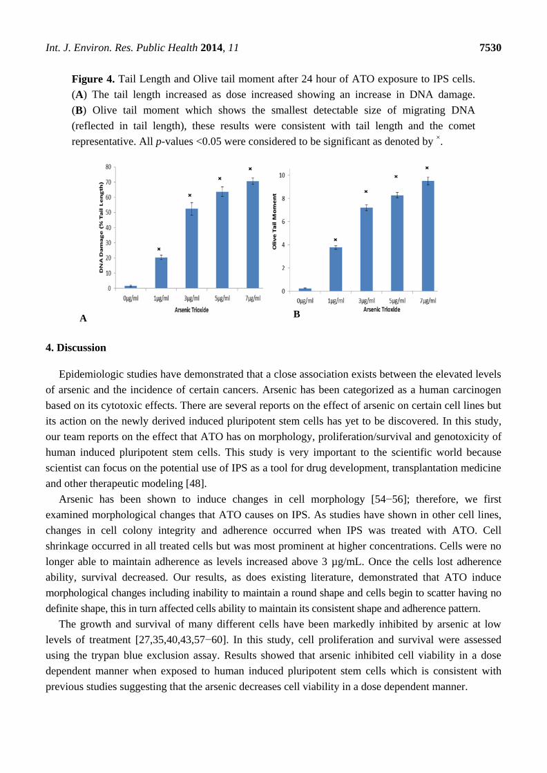
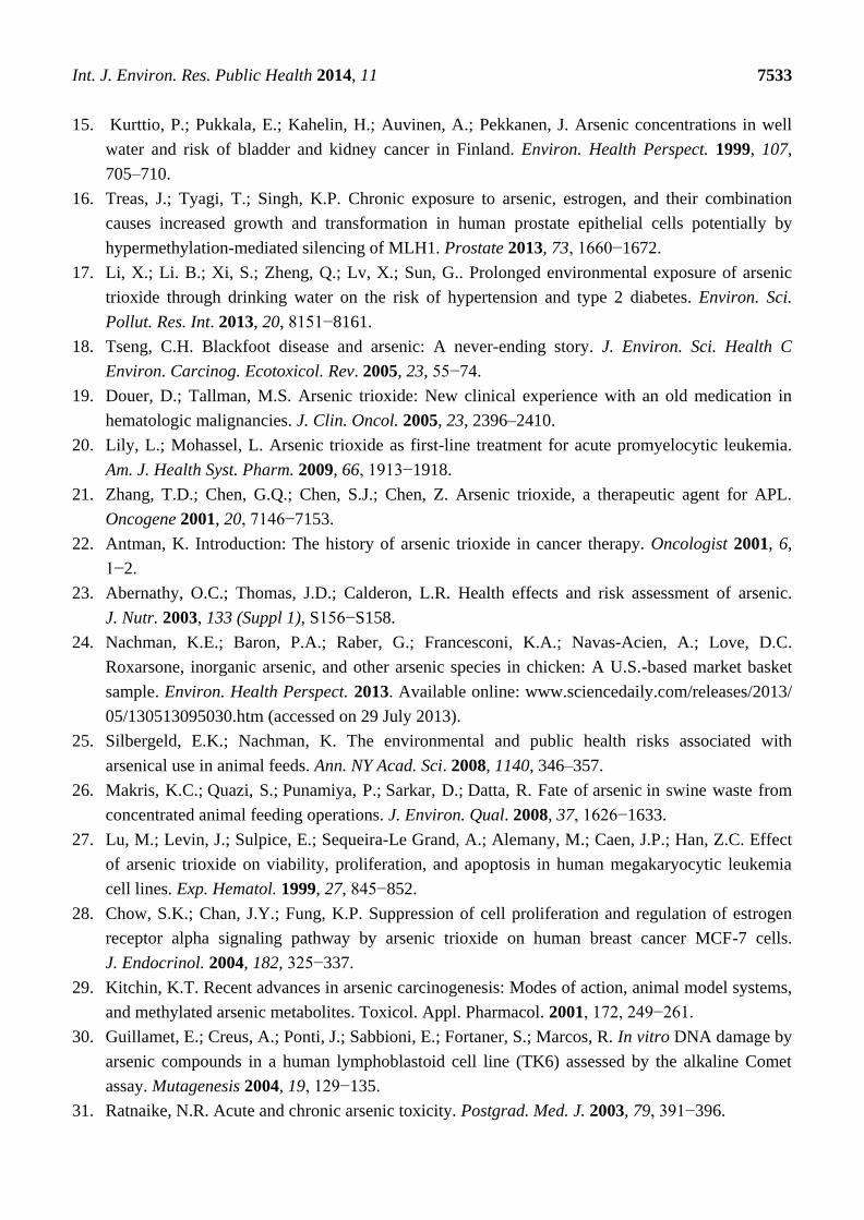
Top Related
Copyright © 2022 FDOKUMEN

