


Bahasa
Halaman
Hukum

10.1128/JVI.78.20.11219-11232.2004.
2004, 78(20):11219. DOI:J. Virol. Deborah H. SpectorTamrakar, Charles L. Clark, Rachel A. Schwartz and Veronica Sanchez, Anita K. McElroy, Judy Yen, Sama Later Times for Virus ProductionUL37 Immediate-Early Transcripts and atHuman Cytomegalovirus UL122-123 and Processing and Accumulation of theRequired at Early Times for Accurate Cyclin-Dependent Kinase Activity Is
http://jvi.asm.org/content/78/20/11219Updated information and services can be found at:
These include:
REFERENCEShttp://jvi.asm.org/content/78/20/11219#ref-list-1at:
This article cites 74 articles, 51 of which can be accessed free
CONTENT ALERTS more»articles cite this article),
Receive: RSS Feeds, eTOCs, free email alerts (when new
http://journals.asm.org/site/misc/reprints.xhtmlInformation about commercial reprint orders: http://journals.asm.org/site/subscriptions/To subscribe to to another ASM Journal go to:
on August 7, 2014 by guest
http://jvi.asm.org/
Dow
nloaded from
on August 7, 2014 by guest
http://jvi.asm.org/
Dow
nloaded from

JOURNAL OF VIROLOGY, Oct. 2004, p. 11219–11232 Vol. 78, No. 200022-538X/04/$08.00�0 DOI: 10.1128/JVI.78.20.11219–11232.2004Copyright © 2004, American Society for Microbiology. All Rights Reserved.
Cyclin-Dependent Kinase Activity Is Required at Early Times forAccurate Processing and Accumulation of the Human CytomegalovirusUL122-123 and UL37 Immediate-Early Transcripts and at Later Times
for Virus ProductionVeronica Sanchez, Anita K. McElroy, Judy Yen, Sama Tamrakar, Charles L. Clark,
Rachel A. Schwartz,† and Deborah H. Spector*Molecular Biology Section and Center for Molecular Genetics, University of California, San Diego, La Jolla, California
Received 18 February 2004/Accepted 3 June 2004
Human cytomegalovirus (HCMV) infection leads to dysregulation of multiple cell cycle-regulatory proteins.In this study, we examined the effects of inhibition of cyclin-dependent kinase (cdk) activity on viral replication.With the drug Roscovitine, a specific inhibitor of cyclin-dependent kinases 1, 2, 5, 7, and 9, we have shown thatduring the first 6 h of infection, cyclin-dependent kinase-dependent events occurred that included the regulatedprocessing and accumulation of the immediate-early (IE) UL122-123 transcripts and UL36-37 transcripts.Altered processing of UL122-123 led to a loss of IE1-72 and an increase in IE2-86. The ratio of spliced tounspliced UL37 transcripts also changed. These effects did not require de novo protein synthesis or degrada-tion of proteins by the proteasome. Addition of Roscovitine at the beginning of the infection was also associatedwith inhibition of expression of selected viral early gene products, viral DNA replication, and late viral geneexpression. When Roscovitine was added after the first 6 h of infection, the effects on IE gene expression wereno longer observed and viral replication proceeded through the late phase, but viral titers were reduced. Thereduction in viral titer was observed even when Roscovitine was first added at 48 h postinfection, indicatingthat cyclin-dependent kinase activity is required at both IE and late times. Flavopiridol, another specificinhibitor of cyclin-dependent kinases, had similar effects on IE and early gene expression. These resultsunderscore the importance of accurate RNA processing and reiterate the significant role of cell cycle-regulatoryfactors in HCMV infection.
Human cytomegalovirus (HCMV), a member of the familyHerpesviridae, is the major viral cause of birth defects and isassociated with significant morbidity in immunocompromisedindividuals (52). Its genome is 230 kbp and has the capacity toencode approximately 150 open reading frames (19, 44).HCMV cell entry, gene expression, replication, and virion mat-uration are multilayered processes that require many viral aswell as cellular factors during a productive infection. There arethree major phases of viral gene expression. Viral immediate-early (IE) genes are the first to be expressed in an infected cell,and their transcription requires no de novo protein synthesis.A major site of IE transcription includes two genetic units, IE1and IE2 (for review, see references 27 and 44) (Fig. 1B). Thepredominant IE RNA (IE1) consists of four exons; a singleopen reading frame (UL123) initiates in exon 2 and specifies a72-kDa nuclear protein designated IE1-72. The major IE2gene product, IE2-86 (open reading frame UL122), is an 86-kDa protein that is encoded by an alternatively spliced RNAthat contains the first three exons of IE1 and a different ter-minal exon.
IE1-72 and IE2-86 are essential transactivators of both earlyand late viral gene expression. Included in the early class are
genes that are important for viral DNA replication. Someexamples are the UL112-113 nuclear phosphoproteins, the vi-ral polymerase (UL54), a DNA polymerase processivity factor(UL44), and the single-stranded-DNA-binding protein (UL57)(2, 3, 29, 35, 37). Late gene expression occurs after the initia-tion of viral DNA synthesis at approximately 24 h postinfection(p.i.), and these late genes encode primarily the structuralcomponents of the virion. Tegument proteins such as pp28(UL99) and pp65 (UL83) as well as components of the capsidare products of the late genes (for review, see reference 44).
A second locus of IE transcription is UL36-38. This regionincludes three promoters and gives rise to at least five tran-scripts (Fig. 1C). One of the IE promoters directs the synthesisof several spliced 3.2- to 3.4-kb RNAs (UL37 and UL37M) thatare present in small amounts only at IE times. It also is thepromoter for an abundant 1.7-kb unspliced RNA that encodesthe UL37 exon 1 (UL37X1) gene product, which is presentthroughout the infection. The cleavage-polyadenylation sitefor the UL37X1 RNA is located 8 nucleotides upstream of thesplice acceptor site for exon 2 of the UL37 and UL37M RNAs.The second IE promoter is responsible for the synthesis of a1.65-kb spliced RNA (UL36) that increases in abundance atearly times and is 3� coterminal with the UL37 spliced RNAs(36, 66, 67). In addition, another promoter within UL37X1directs the synthesis of an abundant early transcript of 1.35 kb(UL38) that is 3� coterminal with the UL37X1 RNA. The startsite of the UL38 transcript is 70 nucleotides downstream of thesplice donor site for the UL37 and UL37M RNAs (66, 67).
* Corresponding author. Mailing address: Molecular Biology Sec-tion, Mail Code 0366, University of California, San Diego, 9500 Gil-man Dr., La Jolla, CA 92093-0366. Phone: (858) 534-9737. Fax: (858)534-6083. E-mail: [email protected].
† Present address: The Salk Institute for Biological Studies, La Jolla,CA 92037.
11219
on August 7, 2014 by guest
http://jvi.asm.org/
Dow
nloaded from
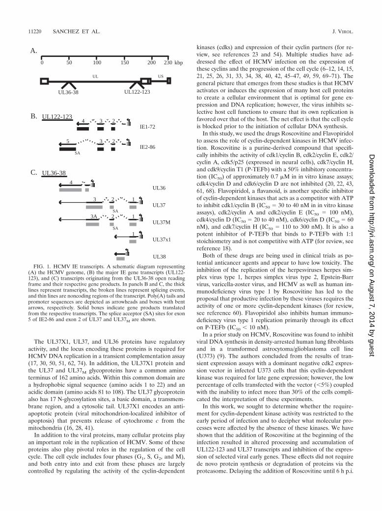
The UL37X1, UL37, and UL36 proteins have regulatoryactivity, and the locus encoding these proteins is required forHCMV DNA replication in a transient complementation assay(17, 30, 50, 51, 62, 74). In addition, the UL37X1 protein andthe UL37 and UL37M glycoproteins have a common aminoterminus of 162 amino acids. Within this common domain area hydrophobic signal sequence (amino acids 1 to 22) and anacidic domain (amino acids 81 to 108). The UL37 glycoproteinalso has 17 N-glycosylation sites, a basic domain, a transmem-brane region, and a cytosolic tail. UL37X1 encodes an anti-apoptotic protein (viral mitochondrion-localized inhibitor ofapoptosis) that prevents release of cytochrome c from themitochondria (16, 28, 41).
In addition to the viral proteins, many cellular proteins playan important role in the replication of HCMV. Some of theseproteins also play pivotal roles in the regulation of the cellcycle. The cell cycle includes four phases (G1, S, G2, and M),and both entry into and exit from these phases are largelycontrolled by regulating the activity of the cyclin-dependent
kinases (cdks) and expression of their cyclin partners (for re-view, see references 23 and 54). Multiple studies have ad-dressed the effect of HCMV infection on the expression ofthese cyclins and the progression of the cell cycle (6–12, 14, 15,21, 25, 26, 31, 33, 34, 38, 40, 42, 45–47, 49, 59, 69–71). Thegeneral picture that emerges from these studies is that HCMVactivates or induces the expression of many host cell proteinsto create a cellular environment that is optimal for gene ex-pression and DNA replication; however, the virus inhibits se-lective host cell functions to ensure that its own replication isfavored over that of the host. The net effect is that the cell cycleis blocked prior to the initiation of cellular DNA synthesis.
In this study, we used the drugs Roscovitine and Flavopiridolto assess the role of cyclin-dependent kinases in HCMV infec-tion. Roscovitine is a purine-derived compound that specifi-cally inhibits the activity of cdk1/cyclin B, cdk2/cyclin E, cdk2/cyclin A, cdk5/p25 (expressed in neural cells), cdk7/cyclin H,and cdk9/cyclin T1 (P-TEFb) with a 50% inhibitory concentra-tion (IC50) of approximately 0.7 �M in in vitro kinase assays;cdk4/cyclin D and cdk6/cyclin D are not inhibited (20, 22, 43,61, 68). Flavopiridol, a flavanoid, is another specific inhibitorof cyclin-dependent kinases that acts as a competitor with ATPto inhibit cdk1/cyclin B (IC50 � 30 to 40 nM in in vitro kinaseassays), cdk2/cyclin A and cdk2/cyclin E (IC50 � 100 nM),cdk4/cyclin D (IC50 � 20 to 40 nM), cdk6/cyclin D (IC50 � 60nM), and cdk7/cyclin H (IC50 � 110 to 300 nM). It is also apotent inhibitor of P-TEFb that binds to P-TEFb with 1:1stoichiometry and is not competitive with ATP (for review, seereference 18).
Both of these drugs are being used in clinical trials as po-tential anticancer agents and appear to have low toxicity. Theinhibition of the replication of the herpesviruses herpes sim-plex virus type 1, herpes simplex virus type 2, Epstein-Barrvirus, varicella-zoster virus, and HCMV as well as human im-munodeficiency virus type 1 by Roscovitine has led to theproposal that productive infection by these viruses requires theactivity of one or more cyclin-dependent kinases (for review,see reference 60). Flavopiridol also inhibits human immuno-deficiency virus type 1 replication primarily through its effecton P-TEFb (IC50 � 10 nM).
In a prior study on HCMV, Roscovitine was found to inhibitviral DNA synthesis in density-arrested human lung fibroblastsand in a transformed astrocytoma/glioblastoma cell line(U373) (9). The authors concluded from the results of tran-sient expression assays with a dominant negative cdk2 expres-sion vector in infected U373 cells that this cyclin-dependentkinase was required for late gene expression; however, the lowpercentage of cells transfected with the vector (�5%) coupledwith the inability to infect more than 30% of the cells compli-cated the interpretation of these experiments.
In this work, we sought to determine whether the require-ment for cyclin-dependent kinase activity was restricted to theearly period of infection and to decipher what molecular pro-cesses were affected by the absence of these kinases. We haveshown that the addition of Roscovitine at the beginning of theinfection resulted in altered processing and accumulation ofUL122-123 and UL37 transcripts and inhibition of the expres-sion of selected viral early genes. These effects did not requirede novo protein synthesis or degradation of proteins via theproteasome. Delaying the addition of Roscovitine until 6 h p.i.
B. UL122-123
IE1-72234
IE2-86235
C. UL36-38
4UL36
UL3723 1
UL37x11
UL37M23A 1
UL38
0 50 100 150 200 230
A.
kbp
SA
SA
SA
UL
UL122-123UL36-38
US
FIG. 1. HCMV IE transcripts. A schematic diagram representing(A) the HCMV genome, (B) the major IE gene transcripts (UL122-123), and (C) transcripts originating from the UL36-38 open readingframe and their respective gene products. In panels B and C, the thicklines represent transcripts, the broken lines represent splicing events,and thin lines are noncoding regions of the transcript. Poly(A) tails andpromoter sequences are depicted as arrowheads and boxes with bentarrows, respectively. Solid boxes indicate gene products translatedfrom the respective transcripts. The splice acceptor (SA) sites for exon5 of IE2-86 and exon 2 of UL37 and UL37M are shown.
11220 SANCHEZ ET AL. J. VIROL.
on August 7, 2014 by guest
http://jvi.asm.org/
Dow
nloaded from
abolished these effects and viral replication proceeded throughlate gene expression, but viral titers were still significantlyreduced. We also demonstrated that inhibiting cyclin-depen-dent kinase activity with the drug Flavopiridol had similareffects on IE and early gene expression.
MATERIALS AND METHODS
Cell culture and virus. Human foreskin fibroblasts (HFF) were obtained fromthe University of California, San Diego, Medical Center and cultured in Earle’sminimal essential medium (Invitrogen) supplemented with 10% heat-inactivatedfetal bovine serum (Invitrogen), 50 �g of gentamicin sulfate (Invitrogen) per ml,1.5 �g of amphotericin B (Invitrogen) per ml, 2 mM L-glutamine (Invitrogen),100 U of penicillin (Invitrogen) per ml, and 100 �g of streptomycin (Invitrogen)per ml. Cells were kept in incubators maintained at 37°C and 7% CO2. TheTowne strain of HCMV was obtained from the American Type Culture Collec-tion (VR 977) and propagated as previously described (65).
Cell synchronization and infections. Cells were synchronized in the G0 phaseby allowing them to grow to confluence as previously described (59). Three daysafter confluence, the cells were trypsinized, replated at a lower density to allowprogression into the cell cycle, and infected at a multiplicity of infection (MOI)of 3 to 5 with HCMV Towne or mock infected with tissue culture supernatants.At designated times p.i., Roscovitine (Calbiochem) or Flavopiridol (gift from J.Brady, National Institutes of Health) was added to the medium. The Roscovitinestock solution was 10 mM in dimethyl sulfoxide, and the Flavopiridol stocksolution was 0.1 mM in dimethyl sulfoxide. Control samples were treated withappropriate volumes of dimethyl sulfoxide. At various times p.i., cells werewashed with phosphate-buffered saline, scraped or trypsinized, and processed asdescribed.
Cytotoxicity assays. The toxic effects of Roscovitine on HFF cells were eval-uated with the LIVE/DEAD viability/cytotoxicity assay kit (Molecular Probes)according to the manufacturer’s protocol. This assay uses two-color fluorescenceto simultaneously determine the presence of live and dead cells in a culture. Inlive cells, the nonfluorescent cell-permeable calcein acetoxymethyl (AM) dye isconverted by intracellular esterase activity to calcein, which fluoresces green.Ethidium homodimer-1 (EthD-1) enters cells with damaged membranes andundergoes a 40-fold enhancement of red fluorescence upon binding to nucleicacids. Cells were plated on coverslips at a density of 50% confluency in theabsence or presence of 5, 15, 25, or 50 �M Roscovitine. In cultures treated withRoscovitine for more than 24 h, fresh medium with the appropriate concentra-tion of the drug was added every 24 h. Coverslips were washed in Dulbecco’sphosphate-buffered saline prior to incubation with reagents for the viabilityassay. As a positive control for dead cells, cells were treated with ice-coldmethanol for 10 min and washed with Dulbecco’s phosphate-buffered saline.Coverslips were incubated in 150 �l of combined assay reagents (0.5 �M EthD-1and 0.25 �M calcein AM) for 30 min at room temperature. To ensure minimaldamage to the cells, coverslips were mounted on 10 �l of Dulbecco’s phosphate-buffered saline on a microscope slide and sealed to prevent evaporation. Greenand red fluorescent cells were counted, and the percentage of live and dead cellswas determined for mock- and virus-infected cells treated with increasing con-centrations of Roscovitine at 24, 48, 72, 96, 120, and 144 h p.i.
Determining the effects of Roscovitine on virus titer. G0-synchronized cellswere released from confluence and infected with the Towne strain of HCMV atan MOI of 3 or 5. At the designated times p.i., the indicated concentration ofRoscovitine or dimethyl sulfoxide was added to the medium. The medium waschanged every 24 h, and fresh drug or control dimethyl sulfoxide was added.Viral supernatants were collected at days 4, 5, and 6, and the titers were deter-mined by plaque assay (65).
Cycloheximide and actinomycin D experiments. Cells were synchronized inG0, trypsinized, and reseeded at a lower density. After allowing the cells torecover for 1 h, the cells were treated for an hour with either 100 �g of cyclo-heximide (stock solution, 10 mg/ml in distilled H2O) per ml in medium ormedium alone prior to infection. Cycloheximide was kept on the treated cellsuntil 6 h p.i. All cells were infected with the Towne virus at an MOI of 3, and 15�M Roscovitine was added at the time of infection to designated cell cultures. At6 h p.i., all cells were washed twice with phosphate-buffered saline and freshmedium was added. Roscovitine and actinomycin D (stock solution, 10 mg/ml inmethanol) were added to the designated cell cultures to final concentrations of15 �M and 20 �g/ml, respectively, and the cells were harvested at 18 h p.i.
Experiments with the proteasome inhibitor MG132. G0-synchronized cellswere released from confluence and infected with Towne at an MOI of 3 at thetime of replating; 15 �M Roscovitine and 2.5 �M MG132 (stock solution, 10 mM
in dimethyl sulfoxide) were added to designated plates at the time of infection.At 8 h p.i., all cells were washed twice with phosphate-buffered saline, and freshmedium with the appropriate drugs was added back to the designated cellcultures. Cells were harvested at various times p.i.
Slot blot analysis. DNA was isolated from infected cells at 24, 48, and 72 h p.i.with the Blood and Cell Culture minikit (Qiagen). DNA samples (5, 25, and 125ng of each sample) were spotted onto 45-�m Nytran (Osmonics) with a slot blotapparatus (Schleicher & Schuell) according to the manufacturer’s instructions.The DNA was then cross-linked to the blot with a Stratalinker (Stratagene) asrecommended by the manufacturer. The blot was probed with a radiolabeledApaLI-NcoI fragment of the EcoRI B fragment of HCMV strain AD169 (63) perstandard protocols and exposed to autoradiograph film. Band intensity wasquantified by measuring the integrated pixel densities as determined with NIHImage and Photoshop 7.0 software. Differences were determined for sampleswithin the linear range.
Western blot analysis. Cells were lysed in Laemmli reducing sample buffer(2% sodium dodecyl sulfate, 10% glycerol, 100 mM dithiothreitol, 60 mM Tris,pH 6.8, 2 �g each of aprotinin and leupeptin per ml, 1 mM phenylmethylsulfonylfluoride, 50 mM NaF, 0.5 mM Na3VO4, 4 mM EDTA, 10 mM Na4P2O7, 1 mMbenzamidine, and 1 mM NaS2O5). The lysates were then sonicated, boiled for 5min, and centrifuged for 1 min at 16,000 � g. Samples were run on 10%polyacrylamide gels. Proteins were transferred to Immobilon P (Millipore) orProtran (Schleicher & Schuell), and the blots were stained with amido black toensure that each lane had an equivalent amount of protein. Lysates were alsoassayed by Western blot for actin. Western blot analysis was performed with theappropriate mouse or rabbit antibody followed by the appropriate horseradishperoxidase-linked secondary antibody (Calbiochem). Proteins were visualizedwith the West Femto or West Pico (Pierce) detection method (per the manu-facturer’s instructions). Band intensity was quantified by measuring integratedpixel densities as determined with NIH Image and Photoshop 7.0 software.
Antisera. The antibodies used in Western analysis were anti-IE1 and IE2(1203; Goodwin Institute), anti-UL112-113 (73), anti-UL44 (1202; GoodwinInstitute), anti-UL57 (1209, Goodwin Institute), anti-pp28 (UL99), and anti-major capsid protein (UL86) (gifts from William Britt, University of Alabama),and anti-pp65 (UL83) (1205S; Goodwin Institute).
Northern blot analysis. Total RNA was isolated at 24 h p.i. with the RNAque-ous midikit (Ambion). RNA samples (10 �g of each sample) were separated byelectrophoresis on a 1% agarose–formaldehyde gel and transferred to 0.45-�mNytran (Osmonics) as previously described (4). Following cross-linking in aStratalinker (Stratagene), the blot was probed for the UL44 viral transcriptsaccording to standard protocols with the 183-bp EcoRI fragment of the UL44gene (a gift from Greg Pari, University of Nevada) that was radiolabeled with 32Pby random priming. The blot was then exposed to autoradiograph film. Bandintensity was quantified by measuring integrated pixel densities as determinedwith NIH Image and Photoshop 7.0 software.
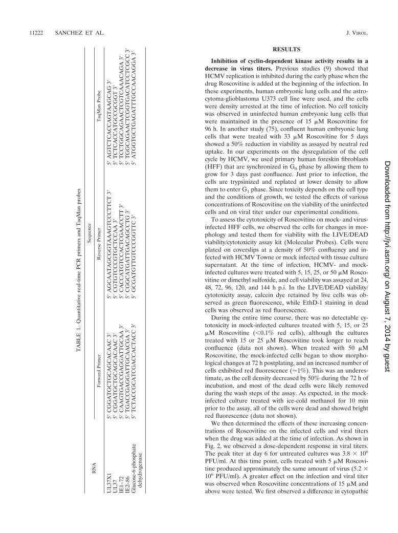
Quantitative real-time PCR. Cells (107 per sample) were infected with theTowne virus at an MOI of 5, and 15 �M Roscovitine or dimethyl sulfoxide alonewas added to the medium at designated times p.i. The cells were harvested atvarious times p.i., and total RNA was isolated from the harvested cells with aNucleoSpin RNA II purification kit (BD Biosciences Clontech, Palo Alto, Calif.).The concentration of each sample was determined by UV spectrophotometry.Quantitative real-time reverse transcription-PCR was performed in an AppliedBiosystems ABI Prism 7700 sequence detection system or in an Applied Biosys-tems ABI Prism 7000 sequence detection system with the TaqMan One-Stepreverse transcription-PCR master mix reagents kit (Applied Biosystems) andoligonucleotide primers and TaqMan dual-labeled (5� fluorescein (FAM) 3�
Black hole quencher) probes (Integrated DNA Technologies, Coralville, Iowa)(see Table 1 for sequences).
Primers and probes for RNAs expressing UL37, IE1-72, IE2-86, and glucose-6-phosphate dehydrogenase have been previously described (64, 72) and spannedthe splice junction when applicable. The primers and probes were added toreagents from the TaqMan One-Step reverse transcription-PCR master mixreagents kit (Applied Biosystems) and then mixed with 50 ng of each total RNAsample. The RNA isolated at 8 h p.i. from the untreated infected cells was usedto generate a standard curve for each gene examined. The standard curve wasthen used to calculate the relative amount of specific RNA present in a sample,from which the induction of transcription of the gene was calculated by compar-ison to the value obtained for the specific RNA from untreated infected cells thatwere harvested at 8 h p.i. As an additional control for the amount of RNA ineach reaction, samples were analyzed with primers and a TaqMan probe specificto the cellular housekeeping gene glucose-6-phosphate dehydrogenase.
VOL. 78, 2004 HCMV REQUIRES CDK ACTIVITY 11221
on August 7, 2014 by guest
http://jvi.asm.org/
Dow
nloaded from
RESULTS
Inhibition of cyclin-dependent kinase activity results in adecrease in virus titers. Previous studies (9) showed thatHCMV replication is inhibited during the early phase when thedrug Roscovitine is added at the beginning of the infection. Inthese experiments, human embryonic lung cells and the astro-cytoma-glioblastoma U373 cell line were used, and the cellswere density arrested at the time of infection. No cell toxicitywas observed in uninfected human embryonic lung cells thatwere maintained in the presence of 15 �M Roscovitine for96 h. In another study (75), confluent human embryonic lungcells that were treated with 33 �M Roscovitine for 5 daysshowed a 50% reduction in viability as assayed by neutral reduptake. In our experiments on the dysregulation of the cellcycle by HCMV, we used primary human foreskin fibroblasts(HFF) that are synchronized in G0 phase by allowing them togrow for 3 days past confluence. Just prior to infection, thecells are trypsinized and replated at lower density to allowthem to enter G1 phase. Since toxicity depends on the cell typeand the conditions of growth, we tested the effects of variousconcentrations of Roscovitine on the viability of the uninfectedcells and on viral titer under our experimental conditions.
To assess the cytotoxicity of Roscovitine on mock- and virus-infected HFF cells, we observed the cells for changes in mor-phology and tested them for viability with the LIVE/DEADviability/cytotoxicity assay kit (Molecular Probes). Cells wereplated on coverslips at a density of 50% confluency and in-fected with HCMV Towne or mock infected with tissue culturesupernatant. At the time of infection, HCMV- and mock-infected cultures were treated with 5, 15, 25, or 50 �M Rosco-vitine or dimethyl sulfoxide, and cell viability was assayed at 24,48, 72, 96, 120, and 144 h p.i. In the LIVE/DEAD viability/cytotoxicity assay, calcein dye retained by live cells was ob-served as green fluorescence, while EthD-1 staining in deadcells was observed as red fluorescence.
During the entire time course, there was no detectable cy-totoxicity in mock-infected cultures treated with 5, 15, or 25�M Roscovitine (�0.1% red cells), although the culturestreated with 15 or 25 �M Roscovitine took longer to reachconfluence (data not shown). When treated with 50 �MRoscovitine, the mock-infected cells began to show morpho-logical changes at 72 h postplating, and an increased number ofcells exhibited red fluorescence (�1%). This was an underes-timate, as the cell density decreased by 50% during the 72 h ofincubation, and most of the dead cells were likely removedduring the wash steps of the assay. As expected, in the mock-infected culture treated with ice-cold methanol for 10 minprior to the assay, all of the cells were dead and showed brightred fluorescence (data not shown).
We then determined the effects of these increasing concen-trations of Roscovitine on the infected cells and viral titerswhen the drug was added at the time of infection. As shown inFig. 2, we observed a dose-dependent response in viral titers.The peak titer at day 6 for untreated cultures was 3.8 � 106
PFU/ml. At this time point, cells treated with 5 �M Roscovi-tine produced approximately the same amount of virus (5.2 �106 PFU/ml). A greater effect on the infection and viral titerwas observed when Roscovitine concentrations of 15 �M andabove were tested. We first observed a difference in cytopathic
TA
BL
E1.
Qua
ntita
tive
real
-tim
ePC
Rpr
imer
san
dT
aqM
anpr
obes
RN
ASe
quen
ce
For
war
dPr
imer
Rev
erse
Prim
erT
aqM
anPr
obe
UL
37X
15�
CG
GA
TG
CT
GC
AG
CA
CA
AC
3�5�
AG
CA
AT
AG
CG
GT
AA
AG
TC
CC
TT
CT
3�5�
AG
TC
TC
AC
CA
GT
AA
GC
AG
3�U
L37
5�C
GG
AT
GC
TG
CA
GC
AC
AA
C3�
5�C
GT
GT
CC
CG
TG
CT
CC
AA
3�5�
TC
TC
AC
CA
TG
CC
GC
GG
T3�
IE1–
725�
CA
AG
TG
AC
CG
AG
GA
TT
GC
AA
3�5�
CA
CC
AT
GT
CC
AC
TC
GA
AC
CT
T3�
5�T
CC
TG
GC
AG
AA
CT
CG
TC
AA
AC
AG
A3�
IE2–
865�
TG
AC
CG
AG
GA
TT
GC
AA
CG
A3�
5�C
GG
CA
TG
AT
TG
AC
AG
CC
TG
3�5�
TG
GC
AG
AA
CT
CG
GT
GA
CA
TC
CT
CG
CC
3�G
luco
se-6
-pho
spha
tede
hydr
ogen
ase
5�T
CT
AC
CG
CA
TC
GA
CC
AC
TA
CC
3�5�
GC
GA
TG
TT
GT
CC
CG
GT
TC
3�5�
AT
GG
TG
CT
GA
GA
TT
TG
CC
AA
CA
GG
A3�
11222 SANCHEZ ET AL. J. VIROL.
on August 7, 2014 by guest
http://jvi.asm.org/
Dow
nloaded from
effect at 24 h p.i. in cells treated with these higher concentra-tions of the drug. At this time, both untreated cells and thosetreated with 5 �M Roscovitine were completely rounded, aneffect of early gene expression, while the cells treated with 15,25, and 50 �M Roscovitine showed progressively less cyto-pathic effect.
Starting at approximately 72 h p.i., the infected cells treatedwith 25 and 50 �M Roscovitine began to die, and this processcontinued through the rest of the time course. This cytotoxiceffect on infected cells was not observed at Roscovitine con-centrations of 5 and 15 �M; however, at a concentration of 15�M Roscovitine, infected cells remained rounded and did notflatten as normally happens late in infection (data not shown).Cultures treated with 15 �M Roscovitine contained approxi-mately the same number of cells as the dimethyl sulfoxide-treated control cultures at day 6, and there was an approxi-mately 75-fold reduction in virus titer (5.1 � 104 PFU/ml)compared to the untreated culture. Only a very low level ofvirus was detected in the cultures treated with 25 and 50 �MRoscovitine (140 and 20 PFU/ml, respectively).
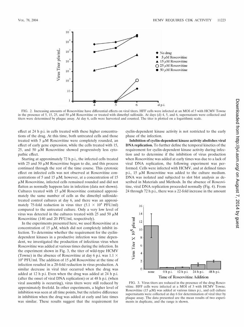
In the experiments presented here, we used Roscovitine at aconcentration of 15 �M, which did not completely inhibit in-fection. To determine whether the requirement for the cyclin-dependent kinases in a productive infection was time depen-dent, we investigated the production of infectious virus whenRoscovitine was added at various times during the infection. Inthe experiment shown in Fig. 3, the titer of wild-type HCMV(Towne) in the absence of Roscovitine at day 6 p.i. was 1.1 �106 PFU/ml. The addition of 15 �M Roscovitine at the time ofinfection resulted in a 20-fold reduction in virus production. Asimilar decrease in viral titer occurred when the drug wasadded at 12 h p.i. Even when the drug was added at 24 h p.i.(after the onset of viral DNA replication) or at 48 h p.i. (whenviral assembly is occurring), virus titers were still reduced byapproximately fivefold. In other experiments, a higher level ofinhibition was seen at all time points, but the relative differencein inhibition when the drug was added at early and late timeswas similar. These results suggest that the requirement for
cyclin-dependent kinase activity is not restricted to the earlyphase of the infection.
Inhibition of cyclin-dependent kinase activity abolishes viralDNA replication. To further define the temporal kinetics of therequirement for cyclin-dependent kinase activity during infec-tion and to determine if the inhibition of virus productionwhen Roscovitine was added at early times was due to a lack ofviral DNA replication, the following experiment was per-formed. Cells were infected with HCMV, and at defined timesp.i., 15 �M Roscovitine was added to the culture medium.DNA was isolated and subjected to slot blot analysis as de-scribed in Materials and Methods. In the absence of Roscovi-tine, viral DNA replication proceeded normally (Fig. 4). From24 through 72 h p.i., there was a 22-fold increase in the amount
FIG. 2. Increasing amounts of Roscovitine have differential effects on viral titers. HFF cells were infected at an MOI of 5 with HCMV Townein the presence of 5, 15, 25, and 50 �M Roscovitine or treated with dimethyl sulfoxide. At days (d) 4, 5, and 6, supernatants were collected andtiters were determined by plaque assay. At day 6, cells were harvested and counted. The titer is plotted on a logarithmic scale.
FIG. 3. Virus titers are reduced in the presence of the drug Rosco-vitine. HFF cells were infected at a MOI of 3 with HCMV Towne.Roscovitine (15 �M) was added at various times p.i., and cell culturesupernatants were collected at day 6 for determination of viral titer byplaque assay. The data presented are the mean results of two experi-ments in duplicate, and the range is shown.
VOL. 78, 2004 HCMV REQUIRES CDK ACTIVITY 11223
on August 7, 2014 by guest
http://jvi.asm.org/
Dow
nloaded from
of viral DNA present in the untreated cells. When 15 �MRoscovitine was added at the time of infection, however, therewas no increase in the level of viral DNA during the same 48-hinterval. Surprisingly, this defect was not observed when thedrug was added at 6 h p.i., which marks the transition from IEto early gene expression. Although there was a lag in viralDNA replication, at 72 h p.i. the level of viral DNA was onlythreefold lower than that in the untreated cells at the sametime point. These results suggested that the viral DNA poly-merase was not directly inhibited by the drug and that viralDNA replication requires cyclin-dependent kinase-dependentevents that occur at very early times in the infection, prior tothe initiation of DNA synthesis.
Steady-state levels of several viral proteins are altered wheninfected cells are treated with Roscovitine. The life cycle ofHCMV is temporally regulated, and each step in the life cyclerequires the products of the previous step before it can pro-ceed. For example, early gene expression cannot occur withoutIE gene expression, and viral DNA replication cannot occurwithout viral early gene expression. The effects of Roscovitineon viral DNA replication may be direct or may occur throughan indirect mechanism, such as alteration of viral IE or early
gene expression. To investigate these possibilities, we exam-ined the steady-state levels of a number of viral gene productsin the presence of Roscovitine.
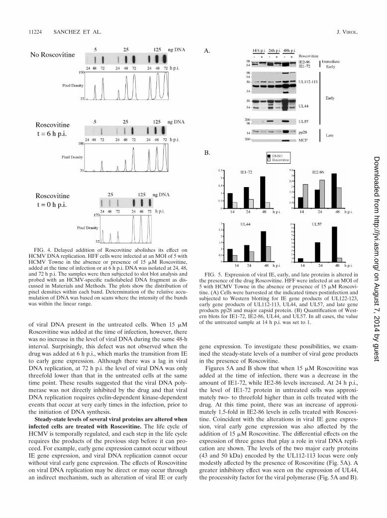
Figures 5A and B show that when 15 �M Roscovitine wasadded at the time of infection, there was a decrease in theamount of IE1-72, while IE2-86 levels increased. At 24 h p.i.,the level of IE1-72 protein in untreated cells was approxi-mately two- to threefold higher than in cells treated with thedrug. At this time point, there was an increase of approxi-mately 1.5-fold in IE2-86 levels in cells treated with Roscovi-tine. Coincident with the alterations in viral IE gene expres-sion, viral early gene expression was also affected by theaddition of 15 �M Roscovitine. The differential effects on theexpression of three genes that play a role in viral DNA repli-cation are shown. The levels of the two major early proteins(43 and 50 kDa) encoded by the UL112-113 locus were onlymodestly affected by the presence of Roscovitine (Fig. 5A). Agreater inhibitory effect was seen on the expression of UL44,the processivity factor for the viral polymerase (Fig. 5A and B).
FIG. 4. Delayed addition of Roscovitine abolishes its effect onHCMV DNA replication. HFF cells were infected at an MOI of 5 withHCMV Towne in the absence or presence of 15 �M Roscovitine,added at the time of infection or at 6 h p.i. DNA was isolated at 24, 48,and 72 h p.i. The samples were then subjected to slot blot analysis andprobed with an HCMV-specific radiolabeled DNA fragment as dis-cussed in Materials and Methods. The plots show the distribution ofpixel densities within each band. Determination of the relative accu-mulation of DNA was based on scans where the intensity of the bandswas within the linear range.
FIG. 5. Expression of viral IE, early, and late proteins is altered inthe presence of the drug Roscovitine. HFF were infected at an MOI of5 with HCMV Towne in the absence or presence of 15 �M Roscovi-tine. (A) Cells were harvested at the indicated times postinfection andsubjected to Western blotting for IE gene products of UL122-123,early gene products of UL112-113, UL44, and UL57, and late geneproducts pp28 and major capsid protein. (B) Quantification of West-ern blots for IE1-72, IE2-86, UL44, and UL57. In all cases, the valueof the untreated sample at 14 h p.i. was set to 1.
11224 SANCHEZ ET AL. J. VIROL.
on August 7, 2014 by guest
http://jvi.asm.org/
Dow
nloaded from
At 24 and 48 h p.i., the levels of UL44 were at least twofoldhigher in lysates from untreated samples than from Roscovi-tine-treated cells.
The most striking effect of Roscovitine, however, was thealmost complete absence of UL57, the major single-stranded-DNA-binding protein (Fig. 5A and B). Since viral early geneproducts are required for viral DNA replication and viral DNAreplication is required for viral late gene expression, it was notsurprising that the drug also inhibited the synthesis of lateproteins. Representative examples of the effect of Roscovitineon the viral tegument protein pp28 (UL99) and on the majorcapsid protein (UL86) are shown in Figs. 5A. Figure 5A alsoshows the inhibitory effect on expression of the late proteins(84 and 34 kDa) encoded by the UL112-113 gene.
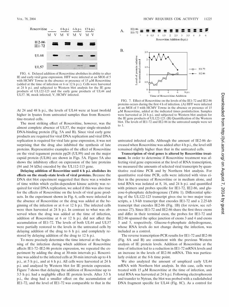
Delaying addition of Roscovitine until 6 h p.i. abolishes itseffects on the steady-state levels of viral proteins. Because theDNA slot blot experiment suggested that there was a windowof time within which cyclin-dependent kinase activity was re-quired for viral DNA replication, we asked if this was also truefor the effects of Roscovitine on the levels of viral gene prod-ucts. In the experiment shown in Fig. 6, cells were infected inthe absence of Roscovitine or the drug was added at the be-ginning of the infection or at 6 or 12 h p.i. The infected cellswere then harvested at 24 h p.i. In contrast to what was ob-served when the drug was added at the time of infection,addition of Roscovitine at 6 or 12 h p.i. did not affect theaccumulation of IE1-72. The levels of both UL44 and UL57were partially restored to the levels in the untreated cells bydelaying addition of the drug to 6 h p.i. and completely re-stored by delaying addition of the drug to 12 h p.i.
To more precisely determine the time interval at the begin-ning of the infection during which addition of Roscovitineaffects IE1-72–IE2-86 protein expression, we repeated the ex-periment by adding the drug at 30-min intervals p.i. Roscovi-tine was added to the infected cells at 30-min intervals up to 4 hp.i., at 5 h p.i., and at 6 h p.i. All cells were harvested at 24 hp.i. and analyzed by Western blot for IE protein expression.Figure 7 shows that delaying the addition of Roscovitine up to3.5 h p.i. had a negligible effect IE protein levels. After 3.5 hp.i., the drug had a smaller effect on the accumulation ofIE1-72, and the level of IE1-72 was comparable to that in the
untreated infected cells. Although the amount of IE2-86 de-creased when Roscovitine was added after 4 h p.i., the level stillremained slightly higher than that in the untreated cells.
Transcription of viral genes is altered by Roscovitine treat-ment. In order to determine if Roscovitine treatment was af-fecting viral gene expression at the level of RNA transcription,we measured the amounts of selected viral transcripts by quan-titative real-time PCR and by Northern blot analysis. Forquantitative real-time PCR, cells were infected with virus ei-ther in the presence of Roscovitine or in medium alone, andtotal RNA was isolated at 8, 16, and 24 h p.i. and quantifiedwith primers and probes specific for IE1-72, IE2-86, and glu-cose-6-phosphate dehydrogenase (Table 1). Differential splic-ing of the UL122-123 transcript gives rise to two major tran-scripts, a 1.9-kb transcript that encodes IE1-72 and a 2.25-kbtranscript that encodes IE2-86 (Fig. 1B) (for review, see ref-erence 27). Since IE1-72 and IE2-86 share the first three exonsand differ in their terminal exon, the probes for IE1-72 andIE2-86 spanned the splice junction of exons 3 and 4 and exons3 and 5, respectively. Glucose-6-phosphate dehydrogenase,whose RNA levels do not change during the infection, wasincluded as a control.
The reverse transcription-PCR results for IE1-72 and IE2-86(Fig. 8A and B) are consistent with the previous Westernanalysis of IE protein levels. Addition of Roscovitine at thetime of infection led to a reduction in IE1-72 mRNA levels andan increase in the levels of IE2-86 mRNA. This was particu-larly evident at the 8-h time point.
We also analyzed the amount of unspliced early UL44mRNA with Northern blot analysis. In this case, cells weretreated with 15 �M Roscovitine at the time of infection, andtotal RNA was harvested at 24 h p.i. Following electrophoresisand transfer to Nytran, the blot was probed with a radiolabeledDNA fragment specific for UL44 (Fig. 8C). As a control for
FIG. 6. Delayed addition of Roscovitine abolishes its ability to alterIE and early viral gene expression. HFF were infected at an MOI of 5with HCMV Towne in the absence or presence of 15 �M Roscovitine(added at the time of infection or 6 or 12 h p.i.). Cells were harvestedat 24 h p.i. and subjected to Western blot analysis for the IE geneproducts of UL122-123 and the early gene products of UL44 andUL57. M, mock infected; V, HCMV infected.
FIG. 7. Effect of Roscovitine on the levels of the IE1-72 and IE2-86proteins occurs during the first 4 h of infection. (A) HFF were infectedat an MOI of 5 with HCMV Towne in the absence or presence of 15�M Roscovitine, added at the indicated times postinfection. Sampleswere harvested at 24 h p.i. and subjected to Western blot analysis forthe IE gene products of UL122-123. (B) Quantification of the Westernblot. The levels of IE1-72 and IE2-86 in the untreated sample were setto 1.
VOL. 78, 2004 HCMV REQUIRES CDK ACTIVITY 11225
on August 7, 2014 by guest
http://jvi.asm.org/
Dow
nloaded from
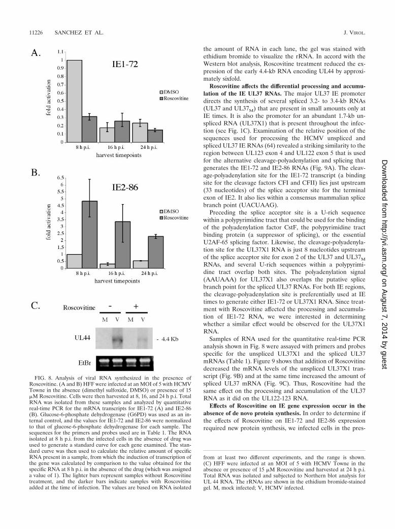
the amount of RNA in each lane, the gel was stained withethidium bromide to visualize the rRNA. In accord with theWestern blot analysis, Roscovitine treatment reduced the ex-pression of the early 4.4-kb RNA encoding UL44 by approxi-mately sixfold.
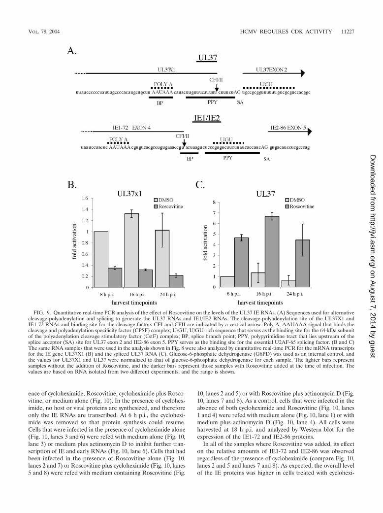
Roscovitine affects the differential processing and accumu-lation of the IE UL37 RNAs. The major UL37 IE promoterdirects the synthesis of several spliced 3.2- to 3.4-kb RNAs(UL37 and UL37M) that are present in small amounts only atIE times. It is also the promoter for an abundant 1.7-kb un-spliced RNA (UL37X1) that is present throughout the infec-tion (see Fig. 1C). Examination of the relative position of thesequences used for processing the HCMV unspliced andspliced UL37 IE RNAs (64) revealed a striking similarity to theregion between UL123 exon 4 and UL122 exon 5 that is usedfor the alternative cleavage-polyadenylation and splicing thatgenerates the IE1-72 and IE2-86 RNAs (Fig. 9A). The cleav-age-polyadenylation site for the IE1-72 transcript (a bindingsite for the cleavage factors CFI and CFII) lies just upstream(33 nucleotides) of the splice acceptor site for the terminalexon of IE2. It also lies within a consensus mammalian splicebranch point (UACUAAG).
Preceding the splice acceptor site is a U-rich sequencewithin a polypyrimidine tract that could be used for the bindingof the polyadenylation factor CstF, the polypyrimidine tractbinding protein (a suppressor of splicing), or the essentialU2AF-65 splicing factor. Likewise, the cleavage-polyadenyla-tion site for the UL37X1 RNA is just 8 nucleotides upstreamof the splice acceptor site for exon 2 of the UL37 and UL37M
RNAs, and several U-rich sequences within a polypyrimi-dine tract overlap both sites. The polyadenylation signal(AAUAAA) for UL37X1 also overlaps the putative splicebranch point for the spliced UL37 RNAs. For both IE regions,the cleavage-polyadenylation site is preferentially used at IEtimes to generate either IE1-72 or UL37X1 RNA. Since treat-ment with Roscovitine affected the processing and accumula-tion of IE1-72 RNA, we were interested in determiningwhether a similar effect would be observed for the UL37X1RNA.
Samples of RNA used for the quantitative real-time PCRanalysis shown in Fig. 8 were assayed with primers and probesspecific for the unspliced UL37X1 and the spliced UL37mRNAs (Table 1). Figure 9 shows that addition of Roscovitinedecreased the mRNA levels of the unspliced UL37X1 tran-script (Fig. 9B) and at the same time increased the amount ofspliced UL37 mRNA (Fig. 9C). Thus, Roscovitine had thesame effect on the processing and accumulation of the UL37RNA as it did on the UL122-123 RNA.
Effects of Roscovitine on IE gene expression occur in theabsence of de novo protein synthesis. In order to determine ifthe effects of Roscovitine on IE1-72 and IE2-86 expressionrequired new protein synthesis, we infected cells in the pres-
FIG. 8. Analysis of viral RNA synthesized in the presence ofRoscovitine. (A and B) HFF were infected at an MOI of 5 with HCMVTowne in the absence (dimethyl sulfoxide, DMSO) or presence of 15�M Roscovitine. Cells were then harvested at 8, 16, and 24 h p.i. TotalRNA was isolated from these samples and analyzed by quantitativereal-time PCR for the mRNA transcripts for IE1-72 (A) and IE2-86(B). Glucose-6-phosphate dehydrogenase (G6PD) was used as an in-ternal control, and the values for IE1-72 and IE2-86 were normalizedto that of glucose-6-phosphate dehydrogenase for each sample. Thesequences for the primers and probes used are in Table 1. The RNAisolated at 8 h p.i. from the infected cells in the absence of drug wasused to generate a standard curve for each gene examined. The stan-dard curve was then used to calculate the relative amount of specificRNA present in a sample, from which the induction of transcription ofthe gene was calculated by comparison to the value obtained for thespecific RNA at 8 h p.i. in the absence of the drug (which was assigneda value of 1). The lighter bars represent samples without Roscovitinetreatment, and the darker bars indicate samples with Roscovitineadded at the time of infection. The values are based on RNA isolated
from at least two different experiments, and the range is shown.(C) HFF were infected at an MOI of 5 with HCMV Towne in theabsence or presence of 15 �M Roscovitine and harvested at 24 h p.i.Total RNA was isolated and subjected to Northern blot analysis forUL 44 RNA. The rRNAs are shown in the ethidium bromide-stainedgel. M, mock infected; V, HCMV infected.
11226 SANCHEZ ET AL. J. VIROL.
on August 7, 2014 by guest
http://jvi.asm.org/
Dow
nloaded from
ence of cycloheximide, Roscovitine, cycloheximide plus Rosco-vitine, or medium alone (Fig. 10). In the presence of cyclohex-imide, no host or viral proteins are synthesized, and thereforeonly the IE RNAs are transcribed. At 6 h p.i., the cyclohexi-mide was removed so that protein synthesis could resume.Cells that were infected in the presence of cycloheximide alone(Fig. 10, lanes 3 and 6) were refed with medium alone (Fig. 10,lane 3) or medium plus actinomycin D to inhibit further tran-scription of IE and early RNAs (Fig. 10, lane 6). Cells that hadbeen infected in the presence of Roscovitine alone (Fig. 10,lanes 2 and 7) or Roscovitine plus cycloheximide (Fig. 10, lanes5 and 8) were refed with medium containing Roscovitine (Fig.
10, lanes 2 and 5) or with Roscovitine plus actinomycin D (Fig.10, lanes 7 and 8). As a control, cells that were infected in theabsence of both cycloheximide and Roscovitine (Fig. 10, lanes1 and 4) were refed with medium alone (Fig. 10, lane 1) or withmedium plus actinomycin D (Fig. 10, lane 4). All cells wereharvested at 18 h p.i. and analyzed by Western blot for theexpression of the IE1-72 and IE2-86 proteins.
In all of the samples where Roscovitine was added, its effecton the relative amounts of IE1-72 and IE2-86 was observedregardless of the presence of cycloheximide (compare Fig. 10,lanes 2 and 5 and lanes 7 and 8). As expected, the overall levelof the IE proteins was higher in cells treated with cyclohexi-
FIG. 9. Quantitative real-time PCR analysis of the effect of Roscovitine on the levels of the UL37 IE RNAs. (A) Sequences used for alternativecleavage-polyadenylation and splicing to generate the UL37 RNAs and IE1/IE2 RNAs. The cleavage-polyadenylation site of the UL37X1 andIE1-72 RNAs and binding site for the cleavage factors CFI and CFII are indicated by a vertical arrow. Poly A, AAUAAA signal that binds thecleavage and polyadenylation specificity factor (CPSF) complex; U/GU, U/GU-rich sequence that serves as the binding site for the 64-kDa subunitof the polyadenylation cleavage stimulatory factor (CstF) complex; BP, splice branch point; PPY, polypyrimidine tract that lies upstream of thesplice acceptor (SA) site for UL37 exon 2 and IE2-86 exon 5. PPY serves as the binding site for the essential U2AF-65 splicing factor. (B and C)The same RNA samples that were used in the analysis shown in Fig. 8 were also analyzed by quantitative real-time PCR for the mRNA transcriptsfor the IE gene UL37X1 (B) and the spliced UL37 RNA (C). Glucose-6-phosphate dehydrogenase (G6PD) was used as an internal control, andthe values for UL37X1 and UL37 were normalized to that of glucose-6-phosphate dehydrogenase for each sample. The lighter bars representsamples without the addition of Roscovitine, and the darker bars represent those samples with Roscovitine added at the time of infection. Thevalues are based on RNA isolated from two different experiments, and the range is shown.
VOL. 78, 2004 HCMV REQUIRES CDK ACTIVITY 11227
on August 7, 2014 by guest
http://jvi.asm.org/
Dow
nloaded from
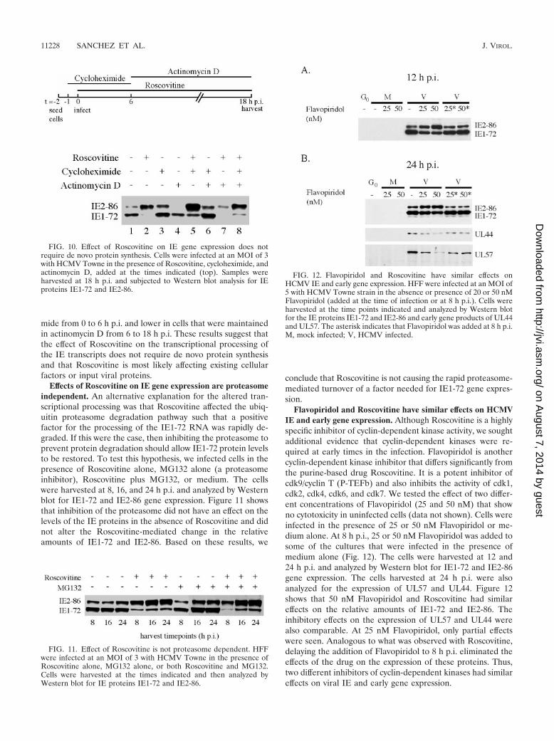
mide from 0 to 6 h p.i. and lower in cells that were maintainedin actinomycin D from 6 to 18 h p.i. These results suggest thatthe effect of Roscovitine on the transcriptional processing ofthe IE transcripts does not require de novo protein synthesisand that Roscovitine is most likely affecting existing cellularfactors or input viral proteins.
Effects of Roscovitine on IE gene expression are proteasomeindependent. An alternative explanation for the altered tran-scriptional processing was that Roscovitine affected the ubiq-uitin proteasome degradation pathway such that a positivefactor for the processing of the IE1-72 RNA was rapidly de-graded. If this were the case, then inhibiting the proteasome toprevent protein degradation should allow IE1-72 protein levelsto be restored. To test this hypothesis, we infected cells in thepresence of Roscovitine alone, MG132 alone (a proteasomeinhibitor), Roscovitine plus MG132, or medium. The cellswere harvested at 8, 16, and 24 h p.i. and analyzed by Westernblot for IE1-72 and IE2-86 gene expression. Figure 11 showsthat inhibition of the proteasome did not have an effect on thelevels of the IE proteins in the absence of Roscovitine and didnot alter the Roscovitine-mediated change in the relativeamounts of IE1-72 and IE2-86. Based on these results, we
conclude that Roscovitine is not causing the rapid proteasome-mediated turnover of a factor needed for IE1-72 gene expres-sion.
Flavopiridol and Roscovitine have similar effects on HCMVIE and early gene expression. Although Roscovitine is a highlyspecific inhibitor of cyclin-dependent kinase activity, we soughtadditional evidence that cyclin-dependent kinases were re-quired at early times in the infection. Flavopiridol is anothercyclin-dependent kinase inhibitor that differs significantly fromthe purine-based drug Roscovitine. It is a potent inhibitor ofcdk9/cyclin T (P-TEFb) and also inhibits the activity of cdk1,cdk2, cdk4, cdk6, and cdk7. We tested the effect of two differ-ent concentrations of Flavopiridol (25 and 50 nM) that showno cytotoxicity in uninfected cells (data not shown). Cells wereinfected in the presence of 25 or 50 nM Flavopiridol or me-dium alone. At 8 h p.i., 25 or 50 nM Flavopiridol was added tosome of the cultures that were infected in the presence ofmedium alone (Fig. 12). The cells were harvested at 12 and24 h p.i. and analyzed by Western blot for IE1-72 and IE2-86gene expression. The cells harvested at 24 h p.i. were alsoanalyzed for the expression of UL57 and UL44. Figure 12shows that 50 nM Flavopiridol and Roscovitine had similareffects on the relative amounts of IE1-72 and IE2-86. Theinhibitory effects on the expression of UL57 and UL44 werealso comparable. At 25 nM Flavopiridol, only partial effectswere seen. Analogous to what was observed with Roscovitine,delaying the addition of Flavopiridol to 8 h p.i. eliminated theeffects of the drug on the expression of these proteins. Thus,two different inhibitors of cyclin-dependent kinases had similareffects on viral IE and early gene expression.
FIG. 10. Effect of Roscovitine on IE gene expression does notrequire de novo protein synthesis. Cells were infected at an MOI of 3with HCMV Towne in the presence of Roscovitine, cycloheximide, andactinomycin D, added at the times indicated (top). Samples wereharvested at 18 h p.i. and subjected to Western blot analysis for IEproteins IE1-72 and IE2-86.
FIG. 11. Effect of Roscovitine is not proteasome dependent. HFFwere infected at an MOI of 3 with HCMV Towne in the presence ofRoscovitine alone, MG132 alone, or both Roscovitine and MG132.Cells were harvested at the times indicated and then analyzed byWestern blot for IE proteins IE1-72 and IE2-86.
FIG. 12. Flavopiridol and Roscovitine have similar effects onHCMV IE and early gene expression. HFF were infected at an MOI of5 with HCMV Towne strain in the absence or presence of 20 or 50 nMFlavopiridol (added at the time of infection or at 8 h p.i.). Cells wereharvested at the time points indicated and analyzed by Western blotfor the IE proteins IE1-72 and IE2-86 and early gene products of UL44and UL57. The asterisk indicates that Flavopiridol was added at 8 h p.i.M, mock infected; V, HCMV infected.
11228 SANCHEZ ET AL. J. VIROL.
on August 7, 2014 by guest
http://jvi.asm.org/
Dow
nloaded from
DISCUSSION
The drug Roscovitine is a purine analogue that inhibits cy-clin-dependent kinase activity by binding to the active site ofthe enzyme. It is an effective and specific inhibitor of cyclin-dependent kinases 1, 2, 5, 7, and 9 at very low concentrations(20, 22, 43, 61, 68). In our experiments, uninfected cells treatedwith up to 25 �M Roscovitine for 6 days showed no cytotox-icity. For this study, data are presented from experiments inwhich the cells were treated with 15 �M Roscovitine. Previ-ously, Evers et al. (24) reported that in an HCMV plaquereduction assay, the inhibition of plaque formation in humanforeskin fibroblasts showed a steep dose-response curve toRoscovitine. In their experiments, they saw no reduction in thenumber of plaques at concentrations of up to 20 �M andcomplete inhibition at a concentration of 50 �M or higher.This likely explains the variability that we observed at a con-centration of 15 �M (20- to 100-fold reduction in viral titer), aswe also noted that when the concentration was increased from15 to 25 �M, there was a large decrease in viral titer.
In accord with those of others (9, 24), our experimentsshowed that addition of Roscovitine to the medium at thebeginning of the infection significantly reduced HCMV titers.Viral DNA replication was also inhibited; however, both ofthese phenomena could have been due to the lack of viral geneexpression. Given that HCMV infection proceeds in a tempo-ral fashion, where one event is required before the next canoccur, the loss of viral gene expression would lead to the lossof replication and in turn the loss of virus production.
When Roscovitine was added at the beginning of the infec-tion, the amount of the IE1-72 protein decreased, while thelevels of IE2-86 increased. Because IE1-72 is a known trans-activator of early gene expression, it was not surprising that thelevels of some early gene products (UL44 and UL57) were alsoreduced. Along similar lines, because UL44 and UL57 arerequired for HCMV replication (51), it followed that viralDNA replication would also be affected. With immunofluores-cence analysis, we noted that replication centers formed butnever grew and coalesced in the presence of Roscovitine (datanot shown). Morphologically, the replication centers in Rosco-vitine-treated cells at late times of infection were similar inappearance to the replication centers that are seen very earlyin untreated cells.
Importantly, it is unlikely that the effects of Roscovitine aredue entirely to decreased levels of IE1-72, as it has been shownthat an IE1-72 deletion mutant can replicate efficiently at ahigh MOI. Since all of the experiments reported in this paperwere conducted at a high MOI, it is almost certain that otherevents are occurring early during the infection that are cyclin-dependent kinase dependent. Most of these events likely occurprior to 6 h p.i., because a 6-h delay in the addition of Rosco-vitine to the infected cells allowed viral gene expression toprogress normally to the late phase, although viral titers werestill affected. These results suggest that although cyclin-depen-dent kinase activity is not directly required for viral DNAreplication, it is required at later times, possibly for virionassembly.
The early effect on IE1-72 and IE2-86 gene expression ap-pears to be at the level of processing of the RNA. During thefirst 24 h of the normal infection, IE1-72 RNA accumulates to
a higher level than IE2-86 RNA. As time progresses, moreIE2-86 transcripts accumulate and there is a slight increase inIE1-72 transcripts. However, when Roscovitine was presentduring the first 24 h of the infection, the ratio of IE1-72 RNAto IE2-86 RNA was reversed; there was a decrease in IE1-72RNA and an increase in the level of the IE2-86 transcript. Themost likely explanation for these data is that the differentialsplicing of the UL122-123 transcript was altered by Roscovi-tine treatment, but we cannot exclude the possibility that therewas a change in the relative stability of the RNAs. Treatmentwith the drug appeared to affect the differential splicing andpolyadenylation of the IE UL37 RNAs in a similar way.
We hypothesize that for both the UL122-123 and UL37transcripts, Roscovitine and Flavopiridol treatment leads tosuppression of the first cleavage-polyadenylation site and en-hanced utilization of the adjacent downstream 3� splice accep-tor site. Relevant to this are the recent studies of Su et al. (64).Although their experiments used mutant target minigenes andtransient transfection assays in uninfected cells, the resultsindicated that the cis polyadenylation and 3� splice acceptorsites for the UL37 RNAs caused steric hindrance on the pre-cursor mRNA for the alternative processes of polyadenylationand splicing, with polyadenylation of UL37X1 being dominant.In the case of the UL122-123 transcripts, the polyadenylationsite generating the IE1-72 RNA is also juxtaposed to the 3�splice acceptor site that is used to produce the IE2-86 RNA. Inthis case, the U-rich polypyrimidine sequence upstream of the3� splice acceptor could provide a binding site for the cleavage-polyadenylation complex CstF, the splicing suppressor poly-pyrimidine tract binding protein, or the essential U2AF-65splicing factor (Fig. 9).
Likewise, the sequence where the cleavage occurs at the 3�end of the IE1-72 RNA could be used for the binding of thecleavage factors CFI/II. However, this sequence also lies withinthe sequence UACUAAG, which matches the mammaliansplicing branch point consensus sequence (YNYURAY) ex-cept for the last position, which can be changed without asignificant decrease in activity (53), and thus it could serve asthe site for the binding of SF1 and U2 splicing factors. Su et al.additionally reported that during HCMV infection, there was atransient increase in CstF64, the splicing suppressor polypyri-midine tract binding protein, and the hypophosphorylatedform of the splicing factor SF2 at 4 h p.i. We also examined theexpression of these factors but did not detect these changesduring the first 8 h p.i. even when the infected cells wereanalyzed every hour (data not shown). Their expression wasalso not affected by treatment of the cells with Roscovitine.The reason for the discrepancy is not clear but may be due tothe use of different strains of HCMV (AD169 in the studies ofSu et al. versus Towne in our studies) or the state of thefibroblasts (unsynchronized versus synchronized).
There is a large body of evidence showing that the processesof mRNA capping, splicing, and cleavage-polyadenylation oc-cur cotranscriptionally and are highly interdependent. Giventhe proximity of the polyadenylation site and the splice accep-tor site, it seems likely that there is competition for sites on theRNA that may be required for both processes. Since numerousfactors are involved in both splicing and cleavage-polyadeny-lation, a change in the abundance, activity, or localization ofany of the factors could alter the balance. The phosphorylation
VOL. 78, 2004 HCMV REQUIRES CDK ACTIVITY 11229
on August 7, 2014 by guest
http://jvi.asm.org/
Dow
nloaded from

state of many of the factors involved in both processes deter-mines their activity, and therefore the cyclin-dependent ki-nases may play a direct or indirect role in their phosphoryla-tion. Because multiple negative and positive splicing factorsare differentially phosphorylated, it is difficult to predictwhether inhibition of the cyclin-dependent kinases results insuppression of the cleavage-polyadenylation of IE1-72 andUL37X1 by the loss of activity of a positive factor or gain ofactivity of a negative factor. It remains to be seen which cyclin-dependent kinase might be the important one for the differ-ential processing of the UL122-123 and UL37 transcripts andwhich specific proteins are important for this level of regula-tion of viral gene expression. Based on the results of the ex-periments with cycloheximide and MG132, it appears that theeffects of Roscovitine on the IE RNAs do not require de novoprotein synthesis or proteasome-mediated degradation of pro-teins.
It is also possible that the effects of Roscovitine and Fla-vopiridol are related to the phosphorylation of the C-terminaldomain of the large subunit of RNA polymerase II. There isincreasing evidence that the C-terminal domain, which in hu-man cells consists of 52 repeats of the consensus heptapeptidesequence Tyr-Ser-Pro-Thr-Ser-Pro-Ser, plays a central regula-tory role in all steps of transcription by serving as the bindingdomain and transporter of factors involved in RNA initiation,elongation, 5� capping, splicing, and cleavage-polyadenylation(for review, see references 5, 48, and 57). The C-terminaldomain is differentially phosphorylated primarily at the serine2 and serine 5 positions, and the level of phosphorylation variesconsiderably during the transcription cycle (for review, seereference 39 and 55).
Briefly, hypophosphorylated-RNA polymerase II is re-cruited to the initiation complex. The C-terminal domain isthen phosphorylated, with the phosphorylation of the serinesin position 5 by cdk7/cyclin H (which is a component of thebasal transcription factor complex TFIIH) mostly occurringprior to the phosphorylation of the serines in position 2 bycdk9/cyclin T (P-TEFb). The commitment of the RNA poly-merase II complex to the elongation step is associated with thisphosphorylation, particularly with the modification at position2 by P-TEFb. Multiple proteins involved in 5� capping, elon-gation, and processing of the RNA are recruited to the C-terminal domain, and their pattern of binding appears to beinfluenced by the differential phosphorylation of serine 2 andserine 5 (and possibly by ubiquitylation, glycosylation, andphosphorylation of other residues) within the 52 repeats. Therules governing this association in uninfected cells have yet tobe elucidated, and it is likely that viral infections will introducenew layers to the complexity.
In this regard, several studies have already shown that therecruitment of P-TEFb to RNA polymerase II by human im-munodeficiency virus type 1 Tat and the resulting phosphory-lation of the C-terminal domain are necessary for the activa-tion of human immunodeficiency virus long terminal repeattranscription (for review, see reference 56). Human immuno-deficiency virus type 1 transcription is much more sensitivethan cellular transcription to low concentrations of Flavopiri-dol; the IC50 for human immunodeficiency virus transcriptionis less than 10 nM, while significant inhibition of cellular tran-scription is only seen at concentrations above 100 nM (13). In
herpes simplex virus type 1-infected cells, a new intermediateform of phosphorylated RNA polymerase II has been de-scribed (32, 58). Although preliminary, we also have evidencethat there is a change in the phosphorylation of the C-terminaldomain during HCMV infection, but it appears to differ fromthat reported for herpes simplex virus type 1.
The question of why a delay in the addition of Roscovitineand Flavopiridol until 6 to 8 h p.i. abolished the effects of thedrug on IE and early RNAs is an interesting one. It is possiblethat by this time, transcription of the HCMV genome is local-ized to a nuclear domain (possibly next to a residual ND10domain) that is inaccessible to the drug. Alternatively, once theRNA polymerase II is committed to the transcription of theviral genes, it might be able to engage in multiple rounds oftranscription without having to completely disengage from theDNA and repeat all of the initiation steps. Relevant to this arerecent reports showing that there are many protein-proteininteractions between the factors involved in initiation at thepromoter and termination of transcription, suggesting thatthese two processes are interconnected (for review, see refer-ence 48).
These are only a few of the possible ways that the cyclin-dependent kinases might affect infection. Unfortunately, theuse of inhibitors such as Roscovitine and Flavopiridol, whichinhibit more than one cyclin-dependent kinase, makes it diffi-cult to distinguish between these possibilities. There are likelymany more functions for cyclin-dependent kinase activity thantranscription during the course of an HCMV infection, andthis is evidenced by the fact that addition of Roscovitine atlater times still resulted in a decrease in viral titers. Anotherpotential role for cyclin-dependent kinase activity during anHCMV infection is the phosphorylation of viral proteins. In arelated virus, herpes simplex virus type 1, it was demonstratedthat UL42 (the herpes simplex virus equivalent of UL44)bound to and was phosphorylated by cdk1 (1). The effects thatthis may have on viral replication and the potential down-stream targets of this kinase complex are unknown; however,the implications of these data as they pertain to HCMV infec-tion are promising. Experiments with dominant negative cy-clin-dependent kinases and the technology of small interferingRNAs coupled with analysis of the splicing machinery and ofviral targets of cyclin-dependent kinase activity are in progressand aim to elucidate the role of the individual cyclin-depen-dent kinases during infection.
ACKNOWLEDGMENTS
We thank William Britt for providing antibodies for pp28 and themajor capsid protein, Greg Pari for the gift of the 183-bp EcoRIfragment of the UL44 gene, J. Brady for the gift of Flavopiridol, andRandall Johnson for help with the real-time reverse transcription-PCRassays. We are grateful to Elizabeth White for helpful suggestions andcomments on the manuscript.
This work was supported by NIH grants CA73490 and CA34729 toD.H.S. A.K.M. was supported by NIH training grant CA09345. J.Y.Y.was supported by NIH training grant GM07240.
REFERENCES
1. Advani, S. J., R. R. Weichselbaum, and B. Roizman. 2001. cdc2 cyclin-dependent kinase binds and phosphorylates herpes simplex virus 1 UL42DNA synthesis processivity factor. J. Virol. 75:10326–10333.
2. Anders, D. G., and W. Gibson. 1988. Location, transcript analysis, and partialnucleotide sequence of the cytomegalovirus gene encoding an early DNA-binding protein with similarities to ICP8 of herpes simplex virus type 1.J. Virol. 62:1364–1372.
11230 SANCHEZ ET AL. J. VIROL.
on August 7, 2014 by guest
http://jvi.asm.org/
Dow
nloaded from
3. Anders, D. G., A. Irmiere, and W. Gibson. 1986. Identification and charac-terization of a major early cytomegalovirus DNA-binding protein. J. Virol.58:253–262.
4. Ausubel, F. M., R. Brent, R. E. Kingston, D. D. Moore, J. G. Seidman, J. A.Smith, and K. Struhl (ed.). 1989. Current protocols in molecular biology.Greene Publishing Associates Wiley-Interscience, New York, N.Y.
5. Bentley, D. 2002. The mRNA assembly line:transcription and processingmachines in the same factory. Curr. Opin. Cell Biol. 14:336–342.
6. Biswas, N., V. Sanchez, and D. H. Spector. 2003. Human cytomegalovirusinfection leads to accumulation of geminin and inhibition of the licensing ofcellular DNA replication. J. Virol. 77:2369–2376.
7. Bonin, L. R., and J. K. McDougall. 1997. Human cytomegalovirus IE286-kilodalton protein binds p53 but does not abrogate G1 checkpoint func-tion. J. Virol. 71:5831–5870.
8. Bresnahan, W. A., T. Albrecht, and E. A. Thompson. 1998. The cyclin Epromoter is activated by human cytomegalovirus 86-kDa immediate earlyprotein. J. Biol. Chem. 273:22075–22082.
9. Bresnahan, W. A., I. Boldogh, P. Chi, E. A. Thompson, and T. Albrecht.1997. Inhibition of cellular CDK2 activity blocks human cytomegalovirusreplication. Virology 231:239–247.
10. Bresnahan, W. A., I. Boldogh, E. A. Thompson, and T. Albrecht. 1996.Human cytomegalovirus inhibits cellular DNA synthesis and arrests produc-tively infected cells in late G1. Virology 224:156–160.
11. Bresnahan, W. A., E. A. Thompson, and T. Albrecht. 1997. Human cytomeg-alovirus infection results in altered Cdk2 subcellular localization. J. Gen.Virol. 78:1993–1997.
12. Castillo, J. P., A. Yurochko, and T. F. Kowalik. 2000. Role of human cyto-megalovirus immediate-early proteins in cell growth control. J. Virol. 74:8028–8037.
13. Chao, S.-H., and D. H. Price. 2001. Flavopiridol inactivates P-TEFb andblocks most RNA polymerase II transcription in vivo. J. Biol. Chem. 276:31793–31799.
14. Chen, Z., E. Knutson, A. Kurosky, and T. Albrecht. 2001. Degradation ofp21cip1 in cells productively infected with human cytomegalovirus. J. Virol.75:3613–3625.
15. Choi, K. S., S.-J. Kim, and S. Kim. 1995. The retinoblastoma gene productnegatively regulates transcriptional activation mediated by the human cyto-megalovirus IE2 protein. Virology 208:450–456.
16. Colberg-Poley, A. M., M. B. Patel, D. P. Erezo, and J. E. Slater. 2000. Humancytomegalovirus UL37 immediate-early regulatory proteins traffic throughthe secretory apparatus and to mitochondria. J. Gen. Virol. 81:1779–1789.
17. Colberg-Poley, A. M., L. D. Santomenna, P. P. Harlow, P. A. Benfield, andD. J. Tenney. 1992. Human cytomegalovirus US3 and UL36-38 immediate-early proteins regulate gene expression. J. Virol. 66:95–105.
18. Dai, Y., and S. Grant. 2003. Cyclin-dependent kinase inhibitors. Curr. Opin.Pharmacol. 3:362–370.
19. Davison, A. J., A. Dolan, P. Akter, C. Addison, D. J. Dargan, D. J. Alcendor,D. J. McGeoch, and G. S. Hayward. 2003. The human cytomegalovirusgenome revisited: comparison with the chimpanzee cytomegalovirus ge-nome. J. Gen. Virol. 84:17–28.
20. De Azevedo, W. F., S. Leclerc, L. Meijer, L. Havlicek, M. Strnad, and S. H.Kim. 1997. Inhibition of cyclin-dependent kinases by purine analogues: crys-tal structure of human cdk2 complexed with roscovitine. Eur. J. Biochem.243:518–526.
21. Dittmer, D., and E. S. Mocarski. 1997. Human cytomegalovirus infectioninhibits G1/S transition. J. Virol. 71:1629–1634.
22. Edamatsu, H., C. L. Gau, T. Nemoto, L. Guo, and F. Tamanoi. 2000. Cdkinhibitors, roscovitine and olomoucine, synergize with farnesyltransferaseinhibitor (FTI) to induce efficient apoptosis of human cancer cell lines.Oncogene 19:3059–3068.
23. Ekholm, S. V., and S. I. Reed. 2000. Regulation of G(1) cyclin-dependentkinases in the mammalian cell cycle. Curr. Opin. Cell Biol. 12:676–684.
24. Evers, D. L., J. M. Breitenbrach, K. Z. Borysko, L. B. Townsend, and J. C.Drach. 2002. Inhibition of cyclin-dependent kinase 1 by purines and pyr-rolo[2,3-d]pyrimidines does not correlate with antiviral activity. Antimicrob.Agents Chemother. 46:2470–2476.
25. Fortunato, E. A., V. Sanchez, J. Y. Yen, and D. H. Spector. 2002. Infection ofcells with human cytomegalovirus during S phase results in a blockade toimmediate-early gene expression that can be overcome by inhibition of theproteasome. J. Virol. 76:5369–5379.
26. Fortunato, E. A., and D. H. Spector. 1998. p53 and RPA are sequestered inviral replication centers in the nuclei of cells infected with human cytomeg-alovirus. J. Virol. 72:2033–2039.
27. Fortunato, E. A., and D. H. Spector. 1999. Regulation of human cytomega-lovirus gene expression. Adv. Virus Res. 54:61–128.
28. Goldmacher, V. S., L. M. Bartle, A. Skaletskaya, C. A. Dionne, N. L. Ked-ersha, C. A. Vater, J. W. Han, R. J. Lutz, S. Watanabe, E. D. C. McFarland,E. D. Kieff, E. S. Mocarski, and T. Chittenden. 1999. A cytomegalovirus-encoded mitochondria-localized inhibitor of apoptosis structurally unrelatedto Bcl-2. Proc. Natl. Acad. Sci. USA 96:12536–12541.
29. Heilbronn, R., G. Jahn, A. Burkle, U.-K. Fresse, B. Fleckenstein, and H.ZurHausen. 1987. Genomic localization, sequence analysis, and transcrip-
tion of the putative human cytomegalovirus DNA polymerase gene. J. Virol.61:119–124.
30. Iskenderian, A. C., L. Huang, A. Reilly, R. M. Stenberg, and D. G. Anders.1996. Four of eleven loci required for transient complementation of humancytomegalovirus DNA replication cooperate to activate expression of repli-cation genes. J. Virol. 70:383–392.
31. Jault, F. M., J.-M. Jault, F. Ruchti, E. A. Fortunato, C. Clark, J. Corbeil,D. D. Richman, and D. H. Spector. 1995. Cytomegalovirus infection induceshigh levels of cyclins, phosphorylated RB, and p53, leading to cell cyclearrest. J. Virol. 69:6697–6704.
32. Jenkins, H. L., and C. A. Spencer. 2001. RNA polymerase II holoenzymemodifications accompany transcription reprogramming in herpes simplexvirus type 1-infected cells. J. Virol. 75:9872–9884.
33. Kalejta, R. F., J. T. Bechtel, and T. Shenk. 2003. Human cytomegaloviruspp71 stimulates cell cycle progression by inducing the proteasome-depen-dent degradation of the retinoblastoma family of tumor suppressors. Mol.Cell. Biol. 23:1885–1895.
34. Kalejta, R. F., and T. Shenk. 2003. The human cytomegalovirus UL82 geneproduct (pp71) accelerate progression through the G1 phase of the cell cycle.J. Virol. 77:3451–3459.
35. Kemble, G. W., A. L. McCormick, L. Pereira, and E. S. Mocarski. 1987. Acytomegalovirus protein with properties of herpes simplex virus ICP8; partialpurification of the polypeptide and map position of the gene. J. Virol.61:3143–3151.
36. Kouzarides, T., A. T. Bankier, A. C. Satchwell, E. Preddy, and B. G. Barrell.1988. An immediate early gene of human cytomegalovirus encodes a poten-tial membrane glycoprotein. Virology 165:151–164.
37. Kouzarides, T., A. T. Bankier, A. C. Satchwell, K. Weston, P. Tomlinson, andB. G. Barrell. 1987. Sequence and transcription analysis of the human cyto-megalovirus DNA polymerase gene. J. Virol. 61:125–133.
38. Lu, M., and T. Shenk. 1996. Human cytomegalovirus infection inhibits cellcycle progression at multiple points, including the transition from G1 to S.J. Virol. 70:8850–8857.
39. Majello, B., and G. Napolitano. 2001. Control of RNA polymerase II activityby dedicated CTD kinases and phosphatases. Front. Biosci. 6:D1358–D1368.
40. Margolis, M. J., S. Panjovic, E. L. Wong, M. Wade, R. Jupp, J. A. Nelson,and J. C. Azizkhan. 1995. Interaction of the 72-kilodalton human cytomeg-alovirus IE1 gene product with E2F1 coincides with E2F-dependent activa-tion of dihydrofolate reductase transcription. J. Virol. 69:7759–7767.
41. McCormick, A. L., V. L. Smith, D. Chow, and E. S. Mocarski. 2003. Disrup-tion of mitochondrial networks by the human cytomegalovirus UL37 geneproduct viral mitochondrion-localized inhibitor of apoptosis. J. Virol. 77:631–641.
42. McElroy, A. K., R. S. Dwarakanath, and D. H. Spector. 2000. Dysregulationof cyclin E gene expression in human cytomegalovirus-infected cells requiresviral early gene expression and is associated with changes in the Rb-relatedprotein p130. J. Virol. 74:4192–4206.
43. Meijer, L., A. Borgne, O. Mulner, J. P. J. Chong, J. J. Blow, N. Inagaki, M.Inagaki, J. G. Delcros, and J. P. Molinoux. 1997. Biochemical and cellulareffects of roscovitine, a potent and selective inhibitor of the cyclin-dependentkinases cdc2, cdk2 and cdk5. Eur. J. Biochem. 243:527–536.
44. Mocarski, E. S., and C. T. Courcelle. 2001. Cytomegaloviruses and theirreplication, p. 2629–2673. In D. M. Knipe and P. M. Howley (ed.), Fieldsvirology, 4th ed., vol. 2. Lippincott Williams & Wilkins, Philadelphia, Pa.
45. Muganda, P., O. Mendoza, J. Hernandez, and Q. Qian. 1994. Human cyto-megalovirus elevates levels of the cellular protein p53 in infected fibroblasts.J. Virol. 68:8028–8034.
46. Murphy, E. A., D. N. Streblow, J. A. Nelson, and M. F. Stinski. 2000. Thehuman cytomegalovirus IE86 protein can block cell cycle progression afterinducing transition into the S phase of the cell cycle. J. Virol. 74:7108–7118.
47. Noris, E., C. Zannetti, A. Demurtas, J. Sinclair, M. DeAndrea, M. Gariglio,and S. Landolfo. 2002. Cell cycle arrest by human cytomegalovirus 86-kilodalton IE2 protein resembles premature senescence. J. Virol. 76:12135–12148.
48. Orphanides, G., and D. Reinberg. 2002. A unified theory of gene expression.Cell 108:439–451.
49. Pajovic, S., E. L. Wong, A. R. Black, and J. C. Azizkhan. 1997. Identificationof a viral kinase that phosphorylates specify E2Fs and pocket proteins. Mol.Cell. Biol. 17:6459–6464.
50. Pari, G. S., and D. G. Anders. 1993. Eleven loci encoding trans-acting factorsare required for transient complementation of human cytomegalovirusoriLyt-dependent DNA replication. J. Virol. 67:6979–6988.
51. Pari, G. S., M. A. Kacica, and D. G. Anders. 1993. Open reading framesUL44, IRS1/TRS1, and UL36-38 are required for transient complementa-tion of human cytomegalovirus oriLyt-dependent DNA synthesis. J. Virol.67:2575–2582.
52. Pass, R. F. 2001. Cytomegalovirus, p. 2675–2705. In D. M. Knipe and P. M.Howley (ed.), Fields virology, 4th ed., vol. 2. Lippincott Williams & Wilkins,Philadelphia, Pa.
53. Peled-Zehavi, H., J. A. Berglund, M. Rosbash, and A. D. Frankel. 2001.Recognition of RNA branch point sequences by the KH domain of splicing
VOL. 78, 2004 HCMV REQUIRES CDK ACTIVITY 11231
on August 7, 2014 by guest
http://jvi.asm.org/
Dow
nloaded from
factor 1 (mammalian branch point binding protein) in a splicing factorcomplex. Mol. Cell. Biol. 21:5232–5241.
54. Pines, J. 1993. Cyclins and cyclin-dependent kinases: take your partners.Trends Biochem. Sci. 18:195–197.
55. Prelich, G. 2002. RNA polymerase II carboxy-terminal domain kinases:emerging clues to their function. Eukaryot. Cell 1:153–162.
56. Price, D. H. 2000. P-TEFb, a cyclin-dependent kinase controlling elongationby RNA polymerase II. Mol. Cell. Biol. 20:2629–2634.
57. Proudfoot, N. J., A. Furger, and M. J. Dye. 2002. Integrating mRNA pro-cessing with transcription. Cell 108:501–512.
58. Rice, S. A., M. C. Long, V. Lam, and C. A. Spencer. 1994. RNA polymeraseII is aberrantly phosphorylated and localized to viral replication compart-ments following Herpes Simplex virus infection. J. Virol. 68:988–1001.
59. Salvant, B. S., E. A. Fortunato, and D. H. Spector. 1998. Cell cycle dysregu-lation by human cytomegalovirus: Influence of the cell cycle phase at thetime of infection and effects on cyclin transcription. J. Virol. 72:3729–3741.
60. Schang, L. M. 2004. Effects of pharmacological cyclin-dependent kinaseinhibitors on viral transcription and replication. Biochim. Biophys. Acta1697:197–209.
61. Schang, L. M., A. Bantly, M. Knockaert, F. Shaheen, L. Meijer, M. H.Malim, N. S. Gray, and P. A. Schaffer. 2002. Pharmacological cyclin-depen-dent kinase inhibitors inhibit replication of wild-type and drug-resistantstrains of herpes simplex virus and human immunodeficiency virus type 1 bytargeting cellular, not viral, proteins. J. Virol. 76:7874–7882.
62. Smith, J. A., and G. S. Pari. 1995. Expression of human cytomegalovirusUL36 and UL37 genes is required for viral DNA replication. J. Virol.69:1925–1931.
63. Spector, D. H., L. Hock, and J. C. Tamashiro. 1982. Cleavage maps forhuman cytomegalovirus DNA strain AD169 for restriction endonucleasesEcoRI, BglII, and HindIII. J. Virol. 42:558–582.
64. Su, Y., R. Adair, C. D. Davis, N. L. DiFronzo, and A. M. Colberg-Poley. 2003.Convergence of RNA cis elements and cellular polyadenylation factors in theregulation of human cytomegalovirus UL37 exon 1 unspliced RNA produc-tion. J. Virol. 77:12729–12741.
65. Tamashiro, J. C., L. J. Hock, and D. H. Spector. 1982. Construction of acloned library of the EcoRI fragments from the human cytomegalovirusgenome (strain AD169). J. Virol. 42:547–557.
66. Tenney, D. J., and A. M. Colberg-Poley. 1991. Expression of the humancytomegalovirus UL36-38 immediate early region during permissive infec-tion. Virology 182:199–210.
67. Tenney, D. J., and A. M. Colberg-Poley. 1991. Human cytomegalovirusUL36-38 and US3 immediate-early genes: temporally regulated expressionof nuclear, cytoplasmic, and polysome-associated transcripts during infec-tion. J. Virol. 65:6724–6734.
68. Wang, D., C. de la Fuente, L. Deng, L. Wang, I. Zilberman, C. Eadie, M.Healey, D. Stein, T. Denny, L. E. Harrison, L. Meijer, and F. Kashanchi.2001. Inhibition of human immunodeficiency virus type 1 transcription bychemical cyclin-dependent kinase inhibitors. J. Virol. 75:7266–7279.
69. Weibusch, L., J. Asmar, R. Uecker, and C. Hagemeier. 2003. Human cyto-megalovirus immediate-early over protein 2 (IE2)-mediated activation ofcyclin E is cell-cycle-independent and forces S-phase entry in IE2-arrestedcells. J. Gen. Virol. 84:51–60.
70. Weibusch, L., and C. Hagemeier. 1999. Human cytomegalovirus 86-kilodal-ton IE2 protein blocks cell cycle progression in G1. J. Virol. 73:9274–9283.
71. Weibusch, L., and C. Hagemeier. 2001. The human cytomegalovirus imme-diate early 2 protein dissociates cellular DNA synthesis from cyclin depen-dent kinase activation. EMBO J. 20:1086–1098.
72. White, E. A., C. L. Clark, V. Sanchez, and D. H. Spector. 2004. Small internaldeletions in the human cytomegalovirus IE2 gene result in nonviable recom-binant viruses with differential defects in viral gene expression. J. Virol.78:1817–1830.
73. Wright, D. A., S. I. Staprans, and D. H. Spector. 1988. Four phosphoproteinswith common amino termini are encoded by human cytomegalovirus AD169.J. Virol. 62:331–340.
74. Zhang, H., H. O. al-Barazi, and A. M. Colberg-Poley. 1996. The acidicdomain of the human cytomegalovirus UL37 immediate early glycoprotein isdispensable for its transactivating activity and localization but is not for itssynergism. Virology 223:292–302.
75. Zimmermann, A., H. Wilts, M. Lenhardt, M. Hahn, and T. Mertens. 2000.Indolocarbazoles exhibit strong antiviral activity against human cytomegalo-virus and are potent inhibitors of the pUL97 protein kinase. Antivir. Res.48:49–60.
11232 SANCHEZ ET AL. J. VIROL.
on August 7, 2014 by guest
http://jvi.asm.org/
Dow
nloaded from
Top Related
Copyright © 2022 FDOKUMEN

