


Bahasa
Halaman
Hukum

Breast Cancer Research and Treatment 66: 225–237, 2001.© 2001 Kluwer Academic Publishers. Printed in the Netherlands.
Report
Breast cancer survival and in vitro tumor response in the extreme drugresistance assay
Rita S. Mehta1,4, Richard Bornstein2, Ing-Ru Yu1, Ricardo J. Parker1, Christine E. McLaren3,4,Khanh P. Nguyen4, Kuo-Tung Li4, and John P. Fruehauf1,4
1Oncotech, Inc., CA; 2Center for Breast Health, OH; 3Epidemiology Division, Department of Medicine, Universityof California, Irvine; 4Chao Family Comprehensive Cancer Research Center, University of California, Irvine,Orange, CA, USA
Key words: breast cancer, chemotherapy, drug resistance, in vitro assay, survival
Summary
Purpose. To determine whether in vitro extreme drug resistance (EDR) assay results for patients with breastcarcinoma were associated with clinical outcome after chemotherapy.
Patients and methods. EDR assays were performed on tumor tissue obtained from 103 newly diagnosed breastcancer cases. EDR scores of 2 for low, 1 for intermediate, or 0 for extreme drug resistance were determined foreach agent tested. In vitro EDR scores for 4-hydroxycyclophosphamide (4HC) and doxorubicin were summed forpatients treated with AC, or for 4HC and 5-FU for patients treated with CMF. Treatment selection was blinded toassay results.
Results. Median time to progression was significantly shorter for patients with extreme or intermediate in vitroresistance (n= 55, 48 months), compared to patients with low in vitro resistance, (n= 41, 100 months, p = 0.022).Patients demonstrating extreme to intermediate drug resistance also showed poorer survival than the low resistancegroup (49.5 months vs. not reached, median follow-up 48 months, p = 0.011). Summed EDR scores, stage,and number of lymph nodes were significantly associated with survival in univariate and multivariate analysis.Compared to EDR scores of 4, summed EDR scores of 0–1 and summed EDR scores of 2–3 were associated witha relative risk of death of 3.09 (95%, CI 1.05–9.06, Cox proportional hazards model, p = 0.040) and 2.35 (95%,CI 1.07–5.15, Cox proportional hazards model, p = 0.033), respectively.
Conclusion. Extreme drug resistance testing identified patients with individual patterns of drug resistanceprior to therapy. In this cohort of breast cancer patients treated with chemotherapy, summed EDR scores weresignificantly associated with time to tumor progression and overall survival. EDR results may offer a method foroptimizing treatment selection.
Abbreviations: EDR: extreme drug resistance; IDR: intermediate drug resistance; LDR: low drug resistance; CTX:cyclophosphamide; 4HC: 4-hydroxycyclophosphamide; 5-FU: 5-fluorouracil; MTX: methotrexate; AC: doxorubi-cin and cyclophosphamide; CMF: cyclophosphamide, methotrexate and fluorouracil; CI: confidence interval; ER:estrogen receptor
Introduction
Multiagent chemotherapy is an important compon-ent of treatment for invasive breast cancers > 1 cm insize. Combination chemotherapy exploits the Goldie–
Coldman hypothesis by targeting the heterogeneousmalignant clones within each patient [1, 2]. Thisstrategy led to the development and clinical valida-tion of various standard combination chemotherapyregimens comprised of non-cross-resistant agents. Re-
226 RS Mehta et al.
cent clinical trials have demonstrated that treatmentwith combination chemotherapy can significantly pro-long the lives of breast cancer patients. The degreeof improved clinical outcomes, however, is mod-est and comparable among the various standard re-gimens [3, 4]. In a recently reported Inter Grouptrial for node negative patients, the marginally super-ior disease free and overall survival with the CAFregimen, compared to the CMF regimen, was bal-anced by modestly increased toxicity [5]. For nodepositive patients, FAC/CAF, AC followed by paclit-axel, doxorubicin followed by CMF, or CMF aloneare all considered to be appropriate options [4, 6–8]. The majority of randomized clinical trials havefailed to demonstrate a benefit from high dose chemo-therapy with stem cell rescue for high risk patients[9–15].
The inability to demonstrate a clear superiority ofone regimen over another, or the superiority of highdose combination regimens over standard dose chemo-therapy, suggests that a plateau in benefit may havebeen reached using the current non-targeted, empiricalapproach to treatment selection. The empirical use ofone of the standard regimens for a specific patient doesnot routinely take into account that patient’s unique tu-mor biology. On the other hand, treatment targeted tothe patient’s tumor characteristics holds promise. Theinitial proof of principle that targeted therapy couldbe a useful strategy stemmed from observations thattamoxifen treatment could significantly improve sur-vival in patients with estrogen receptor (ER) positivetumors [16]. In addition, the benefit of targeted treat-ment has been demonstrated in two retrospective trialswhere dose escalated doxorubicin-based chemother-apy was found to be superior in lymph node-positivepatients whose tumors overexpressed Her-2/neu [17,18]. More importantly, Herceptin therapy targeted toHer-2/neu overexpressing breast cancer patients hasyielded improved survival in a prospective randomizedtrial when given in combination with cisplatin or pacl-itaxel chemotherapy [19]. These observations supportthe notion that tailoring treatment to each patient’stumor characteristics can be advantageous. Clinicaltrials with cross over designs in breast cancer havevalidated the concept of individual patterns of drug-specific resistance, with some patients failing singleagent paclitaxel, yet subsequently responding to non-cross resistant doxorubicin on cross over, or vice versa[20, 21]. Additionally, docetaxel has demonstratedsignificant responses in doxorubicin resistant patients,while capecitabine has shown modest responses in
doxorubicin and taxane resistant breast cancer patients[22–24].
These observations suggest that the ability toidentify individual patterns of resistance prior to ini-tiating chemotherapy might have a substantial clinicalimpact. Tailoring treatment regimens by eliminatingagents found to be inactive in vitro prior to therapyadministration would potentially avoid the toxicity,lost time, and costs associated with ineffective treat-ment [25–27]. This is especially important in an erathat has witnessed the introduction of a variety ofnew, non-cross-resistant classes of agents (taxanes[28, 29], gemcitabine [30], and vinorelbine). Treat-ment with paclitaxel, vinorelbine, or docetaxel has ledto similar durations of quality- adjusted progression-free survival in anthracycline-resistant breast cancer[31]. With the advent of more agents to choose from,and third-generation in vitro drug response assays, itmay be possible to tailor combination therapy to theunique drug response characteristics of each patient’stumor [32, 33]. For this potential to be realized, in vitroassays must be evaluated for their ability to predictresponse and survival.
Although the predictive accuracy of the in vitro ex-treme drug resistance (EDR) assay to identify drugsunlikely to demonstrate clinical response with greaterthan 99% accuracy has been established in a doubleblind retrospective study [33], there is paucity of dataon the relationship between EDR assay results andsurvival. The present study was, therefore, conductedin breast cancer patients to examine the relationshipbetween in vitro EDR assay results, and progression-free and overall survival. EDR assay results wereobtained for a serial cohort of 103 cases prior to firstline chemotherapy. The treating physician was blindedto EDR results, and lab personnel were blinded toclinical characteristics. We report here the relationshipbetween EDR assay results and patient outcomes aftertreatment with agents tested in the assay.
Patients and methods
Patient selection
Between October 1990 and March 1996, tissuesamples from 187 serial patients with newly diagnosedinvasive breast cancer for which tumor tissue could beobtained were sent from a single NSABP institution toOncotech, Inc. (Irvine, CA) for in vitro drug resistancetesting. We performed a retrospective double-blindedstudy on a serial group of patients who had specimens
In vitro drug response and breast cancer survival 227
submitted for EDR assays. Our primary objective wasto determine if in vitro drug response was associ-ated with clinical outcomes. Treating physicians wereblinded to EDR results, as were the personnel whocollected clinical outcome data. EDR assays wereunsuccessful in 17 cases (9%). Thus, EDR resultswere available for 170 patients (91%). Eleven patientswere lost to follow-up. Of the remaining 159 cases,103 cases were treated with primary chemotherapy,of which 96 cases had a complete EDR profile of 4-hydroxycyclophosphamide (4HC) and doxorubicin forpatients treated with AC, or for 4HC and 5-fluorouracil(5-FU) for patients treated with CMF.
Primary treatment consisted of mastectomy withaxillary lymph node dissection, or lumpectomy withaxillary node dissection and local radiation. A small(0.2–1 g) section of representative malignant tissuefrom the primary tumor or involved lymph node wasplaced in transport media and sent overnight to On-cotech for analysis in the in vitro EDR assay. Chemo-therapy was started 14 to 21 days after definitivesurgery. The following two regimens were employed:
1. CMF – cyclophosphamide 100 mg/m2 days 1–14, methotrexate 40 mg/m2 and 5-fluorouracil600 mg/m2, day 1 and day 8, every 28 days for6 cycles.
2. AC – cyclophosphamide 600 mg/m2 and doxoru-bicin 60 mg/m2, day 1 every 21 days for 4 cycles.
White blood cell and platelet counts were monitored,with dosages adjusted prior to each cycle if needed.EDR assays were performed prior to the initiationof chemotherapy. Patients were subsequently treatedwith classical CMF or AC regimens independent ofthe assay results. Less than one third of the patientsreceived hormone therapy, and these cases were bal-anced between the two in vitro drug response groupsthat were compared.
Lab protocol
Once received at Oncotech, tumor tissue sampleswere accessioned and assigned a tracking number. Tu-mor specimens were mechanically disaggregated intosuspensions of small tumor clumps. Stained tissuesections and cytospin preparations of tumor suspen-sions were reviewed by a pathologist to confirm thediagnosis and the presence of adequate numbers ofmalignant cells. Tissue culture was performed as pre-viously described [32, 33]. Cell viability was determ-ined by trypan blue exclusion. Approximately 30,000
viable malignant cells per well were suspended in softagarose and growth media in a 24-well plates andexposed to the following chemotherapeutic agents:doxorubicin, 5-FU, or 4-hydroxycyclophosphamide(4HC), the active metabolite of cyclophosphamide.Melphalan was employed as a cyclophosphamide sur-rogate for the first 10 cases because the 4-hydroperoxyformulation of cyclophosphamide was not availablefor in vitro use when this study was initiated. When4HC became available, a series of cases were run tocompare melphalan with 4HC on the same specimens.Using spearman’s correlation coefficient, a highly sig-nificant association was found between in vitro per-cent cell inhibition (PCI) produced by 4HC versusmelphalan (R = 0.43, p < 0.0001) (n= 468). Basedon this association, we included the in vitro responsedata for the 10 cases tested against melphalan as acyclophosphamide surrogate.
Drugs were added to wells containing the malig-nant cells at doses that approximated their in vivopeak plasma concentrations [32, 33]. Treated cell sus-pensions were incubated for 72 h with drug and thenpulsed with 5 µCi 3H-thymidine. After an additional48-h incubation period, agarose-cell suspensions wereliquefied at 96◦C, well contents were harvested ontoglass fiber filters, and cells were lysed with deionizedwater. The incorporated radioactivity in the filter-trapped macromolecular DNA was measured by liquidscintillation as counts per minute (CPM). Positivecontrol (supralethal cisplatin-exposed) and negativecontrol (media-exposed) cultures were performed witheach assay. Results were reported as percent cell in-hibition (PCI) compared with media-exposed controlcultures after subtraction of positive control CPM. Theperformance characteristics, including the populationmedian PCI and standard deviation (SD), were de-termined for 4HC (n= 372), 5-FU (n> 6,000) anddoxorubicin (n> 3,000) on independent cases evalu-ated using the same methods. Individual patient PCIvalues were compared to the median and SD estab-lished for each agent to determine their EDR score.
EDR scores
For each patient’s tumor specimen, in vitro responsesto individual drugs were scored as ‘0’ for extremedrug resistance (EDR) when the PCI was ≥ 1SD belowthe median, ‘1’ for intermediate drug resistance (IDR)when the PCI was between the median and 1SD be-low the median, or ‘2’ for low drug resistance (LDR)when the PCI was above the median. EDR scores
228 RS Mehta et al.
for 4HC and doxorubicin were summed for patientstreated with AC. 4HC and 5-FU scores were selectedfor summation for patients treated with CMF. Me-thotrexate was not tested in the EDR assay becausethe addition of thymidine for labeling rescues DNAsynthesis, thereby yielding an inaccurate measure ofproliferation inhibition. Further, methotrexate has onlylimited activity by itself, but it significantly potentiates5-FU toxicity when both compounds are administered.Summed EDR Scores ranged from 0 to 4. For ex-ample, a patient treated with AC and having a tumorwith extreme resistance to 4HC and low resistance todoxorubicin was assigned an EDR Score of 0+2 = 2.
Statistical analysis
We examined the relationship between the frequenciesof tumors with low resistance scores (4), or extremeto intermediate resistance scores (0–3), and patientclinical characteristics using contingency table ana-lysis with the chi-squared test or Fisher’s exact test.Prognostic indicators considered as categorical vari-ables included age, stage, number of involved lymph-nodes, tumor size, and ER receptor status. Similarly,we examined the association between the dichotom-ized EDR scores and treatment modalities, includingmastectomy versus lumpectomy/radiation, hormonaltreatment and chemotherapeutic regimen. Time to tu-mor progression and overall survival were calculatedfrom the date of diagnosis to date of progression ordeath, respectively. For overall survival, death fromany cause was considered as an event. Data from pa-tients without events were censored at the date of lastfollow up. Progression free and overall survival curveswere constructed using the Kaplan–Meier method, anddifferences between groups were assessed by the log-rank test. Univariate analysis included age, stage,degree of lymph node involvement, ER status, surgicaltreatment with or without radiation, chemotherapy re-gimens, hormonal treatment status, and single agentEDR scores, and composite EDR scores. We used theCox Proportional Hazards Model to evaluate the influ-ence of prognostic factors on survival in univariate andmultivariate models. To assess the relative influence ofprognostic factors on progression-free and overall sur-vival, we included the variables that were predictive ofsurvival in univariate analyses (EDR scores, stage andlymph nodes) in multivariate models. We also usedthe log-rank test for trend to assess the associationof progression-free survival, or overall survival, withEDR scores trichotomized as 0–1, 2–3, and 4. All stat-
istical tests were two-tailed and a significance level of0.05 was used. All clinical outcomes and laboratorydata were independently reviewed by the Biostatisticssection at UC Irvine.
Results
Patient characteristics
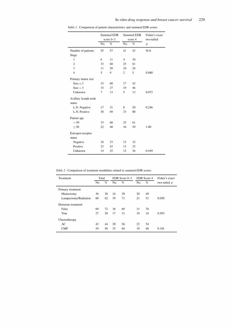
At a median duration of follow-up of 48.17 months,there were 59 surviving patients out of 96 patientswho had successful EDR assay results for at least twoof the chemotherapy agents they received. Details ofpatient characteristics for the cohort of 96 patientstreated with chemotherapy, and for whom a completeEDR profile was available are detailed in Table 1.Patients were treated with classical CMF or AC re-gimens. Some patients received additional hormonetherapy. Treatment profiles are shown in Table 2.
In vitro drug resistance assay
Single agent EDR assay results for the 96 patientstreated with chemotherapy that had summed EDRscores are shown in Table 3. The incidence of ex-treme drug resistance to the treatment drugs varied:13% for 4HC (or the index alkylator, Melphalan), 5%for doxorubicin, and 19% for 5-FU. The incidence ofintermediate resistance ranged from 25% to 32%, andthe incidence of low drug resistance ranged from 49%to 62%, respectively, for the individual drugs. TheEDR scores for 4HC and doxorubicin were summedfor patients treated with AC (n= 42), and EDR scoresfor 4HC and 5-FU were summed for patients treatedwith CMF (n= 54). Table 4 shows the EDR assayresults and the corresponding summed EDR score cat-egories for the study set. Fifty-five tumors (57%) ex-hibited extreme or intermediate drug resistance, withsummed EDR scores for these cases ranging from 0 to1 and 2 to 3. Of these tumors, only two demonstratedEDR to both drugs (summed EDR score of 0), andonly eight cases showed EDR to one drug in conjunc-tion with IDR to the other (summed EDR score of 1).Forty-one (42%) patients were treated with a two-drugcombination to which their tumors showed low in vitroresistance (summed EDR score of 4). Patients withsummed EDR scores of 0 to 1 and 2 to 3 were groupedinto the extreme and intermediate resistance categor-ies, respectively, while patients with summed EDRscores of 4 were placed into the low drug resistancecategory.
In vitro drug response and breast cancer survival 229
Table 1. Comparison of patient characteristics and summed EDR scores
Summed EDR Summed EDR Fisher’s exact
score 0–3 score 4 two-tailed
No. % No. % p
Number of patients 55 57 41 43 N/A
Stage
1 6 11 4 10
2 33 60 25 61
3 11 20 10 24
4 5 9 2 5 0.880
Primary tumor size
Size ≤ 3 33 60 17 42
Size > 3 15 27 19 46
Unknown 7 13 5 12 0.072
Axillary lymph node
status
L.N. Negative 17 31 8 20 0.246
L.N. Positive 38 69 33 80
Patient age
< 50 33 60 25 61
≥ 50 22 40 16 39 1.00
Estrogen receptor
status
Negative 18 33 13 32
Positive 23 42 13 32
Unknown 14 25 15 36 0.449
Table 2. Comparison of treatment modalities related to summed EDR scores
Treatment Total EDR Score 0–3 EDR Score 4 Fisher’s exact
No. % No. % No. % two-tailed p
Primary treatment
Mastectomy 36 38 16 39 20 49
Lumpectomy/Radiation 60 62 39 71 21 51 0.058
Hormone treatment
False 69 72 38 69 31 76
True 27 28 17 31 10 24 0.503
Chemotherapy
AC 42 44 20 36 22 54
CMF 54 56 35 64 19 46 0.101
230 RS Mehta et al.
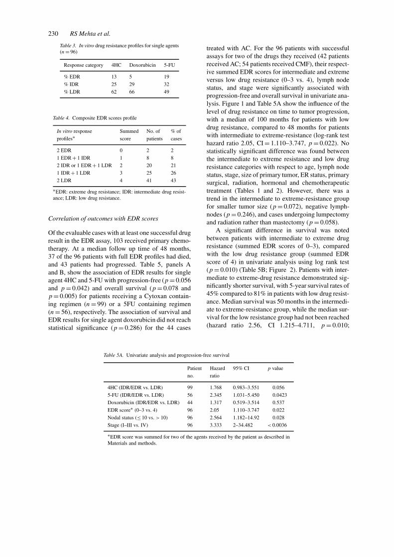
Table 3. In vitro drug resistance profiles for single agents(n= 96)
Response category 4HC Doxorubicin 5-FU
% EDR 13 5 19
% IDR 25 29 32
% LDR 62 66 49
Table 4. Composite EDR scores profile
In vitro response Summed No. of % of
profiles∗ score patients cases
2 EDR 0 2 2
1 EDR + 1 IDR 1 8 8
2 IDR or 1 EDR + 1 LDR 2 20 21
1 IDR + 1 LDR 3 25 26
2 LDR 4 41 43
∗EDR: extreme drug resistance; IDR: intermediate drug resist-ance; LDR: low drug resistance.
Correlation of outcomes with EDR scores
Of the evaluable cases with at least one successful drugresult in the EDR assay, 103 received primary chemo-therapy. At a median follow up time of 48 months,37 of the 96 patients with full EDR profiles had died,and 43 patients had progressed. Table 5, panels Aand B, show the association of EDR results for singleagent 4HC and 5-FU with progression-free (p = 0.056and p = 0.042) and overall survival (p = 0.078 andp = 0.005) for patients receiving a Cytoxan contain-ing regimen (n= 99) or a 5FU containing regimen(n= 56), respectively. The association of survival andEDR results for single agent doxorubicin did not reachstatistical significance (p = 0.286) for the 44 cases
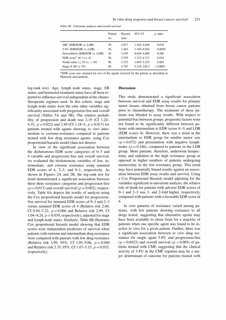
Table 5A. Univariate analysis and progression-free survival
Patient Hazard 95% CI p value
no. ratio
4HC (IDR/EDR vs. LDR) 99 1.768 0.983–3.551 0.056
5-FU (IDR/EDR vs. LDR) 56 2.345 1.031–5.450 0.0423
Doxorubicin (IDR/EDR vs. LDR) 44 1.317 0.519–3.514 0.537
EDR score∗ (0–3 vs. 4) 96 2.05 1.110–3.747 0.022
Nodal status (≤ 10 vs. > 10) 96 2.564 1.182–14.92 0.028
Stage (I–III vs. IV) 96 3.333 2–34.482 < 0.0036
∗EDR score was summed for two of the agents received by the patient as described inMaterials and methods.
treated with AC. For the 96 patients with successfulassays for two of the drugs they received (42 patientsreceived AC; 54 patients received CMF), their respect-ive summed EDR scores for intermediate and extremeversus low drug resistance (0–3 vs. 4), lymph nodestatus, and stage were significantly associated withprogression-free and overall survival in univariate ana-lysis. Figure 1 and Table 5A show the influence of thelevel of drug resistance on time to tumor progression,with a median of 100 months for patients with lowdrug resistance, compared to 48 months for patientswith intermediate to extreme-resistance (log-rank testhazard ratio 2.05, CI = 1.110–3.747, p = 0.022). Nostatistically significant difference was found betweenthe intermediate to extreme resistance and low drugresistance categories with respect to age, lymph nodestatus, stage, size of primary tumor, ER status, primarysurgical, radiation, hormonal and chemotherapeutictreatment (Tables 1 and 2). However, there was atrend in the intermediate to extreme-resistance groupfor smaller tumor size (p = 0.072), negative lymph-nodes (p = 0.246), and cases undergoing lumpectomyand radiation rather than mastectomy (p = 0.058).
A significant difference in survival was notedbetween patients with intermediate to extreme drugresistance (summed EDR scores of 0–3), comparedwith the low drug resistance group (summed EDRscore of 4) in univariate analysis using log rank test(p = 0.010) (Table 5B; Figure 2). Patients with inter-mediate to extreme-drug resistance demonstrated sig-nificantly shorter survival, with 5-year survival rates of45% compared to 81% in patients with low drug resist-ance. Median survival was 50 months in the intermedi-ate to extreme-resistance group, while the median sur-vival for the low resistance group had not been reached(hazard ratio 2.56, CI 1.215–4.711, p = 0.010;
In vitro drug response and breast cancer survival 231
Table 5B. Univariate analysis and overall survival
Patient Hazard 95% CI p value
no. ratio
4HC (IDR/EDR vs. LDR) 99 2.077 1.102–4.649 0.078
5-FU (IDR/EDR vs. LDR) 56 3.463 1.449–8.034 0.0050
Doxorubicin (IDR/EDR vs. LDR) 44 1.639 0.629–4.809 0.286
EDR score∗ (0–3 vs. 4) 96 2.559 1.215–4.711 0.010
Nodal status (≤ 10 vs. > 10) 96 3.333 1.695–3.333 0.004
Stage (I–III vs. IV) 96 4.783 5.218–148.3 < 0.0001
∗EDR score was summed for two of the agents received by the patient as described inMaterials and methods.
log-rank test). Age, lymph node status, stage, ERstatus, and hormonal treatment status have all been re-ported to influence survival independent of the chemo-therapeutic regimen used. In this cohort, stage andlymph node status were the only other variables sig-nificantly associated with progression-free and overallsurvival (Tables 5A and 5B). The relative probab-ility of progression and death was 2.19 (CI 1.24–4.35, p = 0.022) and 2.49 (CI 1.18–5, p = 0.017) forpatients treated with agents showing in vitro inter-mediate to extreme-resistance compared to patientstreated with low drug resistance agents, using Coxproportional hazards model (data not shown).
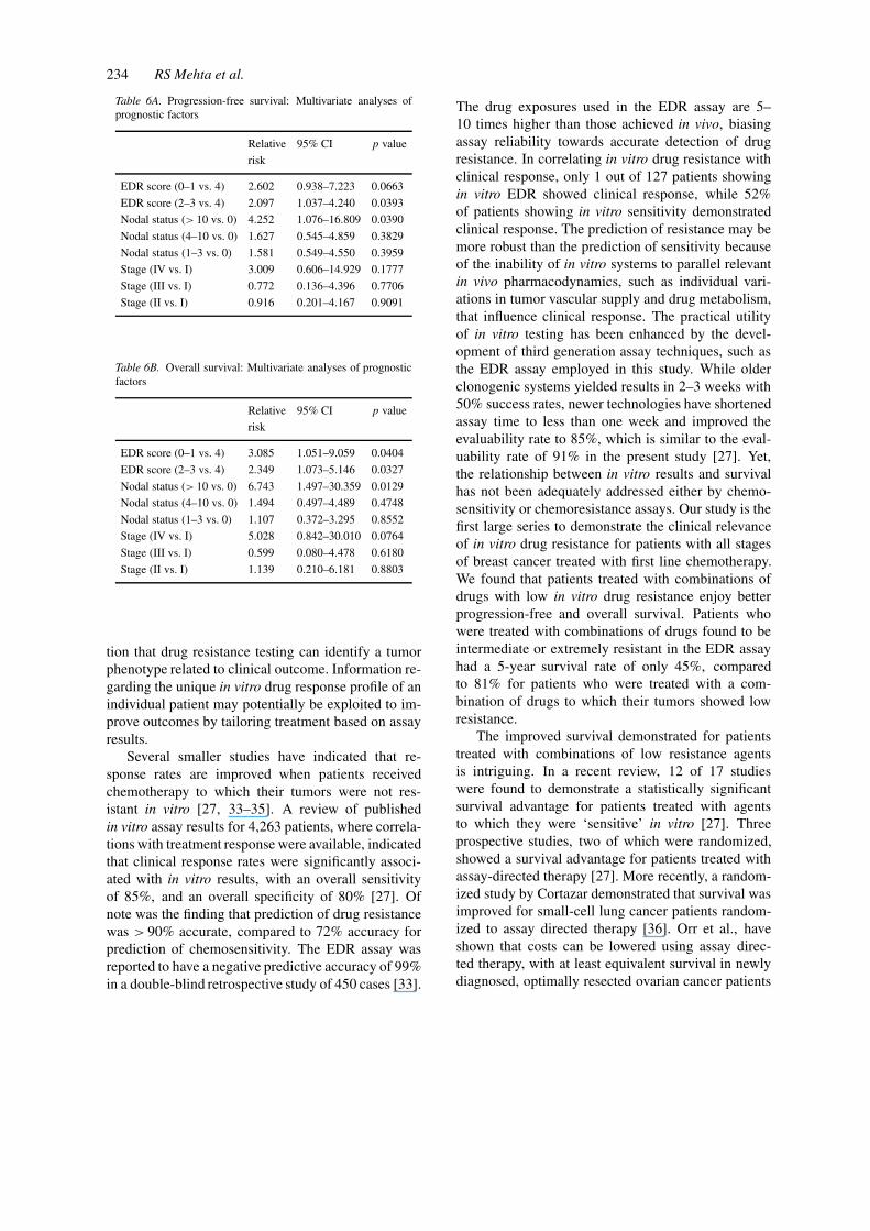
In view of the significant association betweenthe dichotomous EDR score categories of 0–3 and4 variable and progression free and overall survival,we evaluated the trichotomous variables of low, in-termediate, and extreme resistance using summedEDR scores of 4, 2–3, and 0–1, respectively. Asshown in Figures 2A and 2B, the log-rank test fortrend demonstrated a significant association betweenthese three resistance categories and progression-free(p = 0.013) and overall survival (p = 0.002), respect-ively. Table 6A depicts the results of analysis usingthe Cox proportional hazards model for progression-free survival for summed EDR scores of 0–1 and 2–3versus summed EDR scores of 4 (Relative risk 2.60,CI 0.94–7.22, p = 0.066 and Relative risk 2.09, CI1.04–4.24, p = 0.039, respectively), adjusted for stageand lymph node status. Similarly, Table 6B illustratesCox proportional hazards model showing that EDRscores were independent predictors of survival whenpatients with extreme and intermediate drug resistancewere compared with patients with low drug resistance(Relative risk 3.09, 95%, CI 1.05–9.06, p = 0.040and Relative risk 2.35, 95%, CI 1.07–5.15, p = 0.033,respectively).
Discussion
This study demonstrated a significant associationbetween survival and EDR assay results for primarytumor tissues obtained from breast cancer patientsprior to chemotherapy. The treatment of these pa-tients was blinded to assay results. With respect topotential bias between groups, prognostic factors werenot found to be significantly different between pa-tients with intermediate to EDR (score 0–3) and LDR(EDR scores 4). However, there was a trend in theintermediate to EDR group for smaller tumor size(p = 0.072) and presentation with negative lymph-nodes (p = 0.246), compared to patients in the LDRgroup. More patients, therefore, underwent lumpec-tomy and radiation in the high resistance group asopposed to higher numbers of patients undergoingmastectomy in the low resistance group. This trendmay have potentially biased results against an associ-ation between EDR assay results and survival. Usinga Cox Proportional Hazards model adjusting for thevariables significant in univariate analysis, the relativerisk of death for patients with adverse EDR scores of0–1 and 2–3 was 3- and 2-fold higher, respectively,compared with patients with a favorable EDR score of4.
In vitro patterns of resistance varied among pa-tients, with few patients showing resistance to alldrugs tested, suggesting that alternative agents mayhave been available to chose from for a majority ofpatients when one specific agent was found to be in-active in vitro for a given patient. Further, there wasa significant association between in vitro drug res-istance for single agent 5-FU and progression-free(p < 0.0423) and overall survival (p < 0.005) of pa-tients treated with CMF, suggesting that the clinicalactivity of 5-FU in the CMF regimen may be a ma-jor determinant of outcome for patients treated with
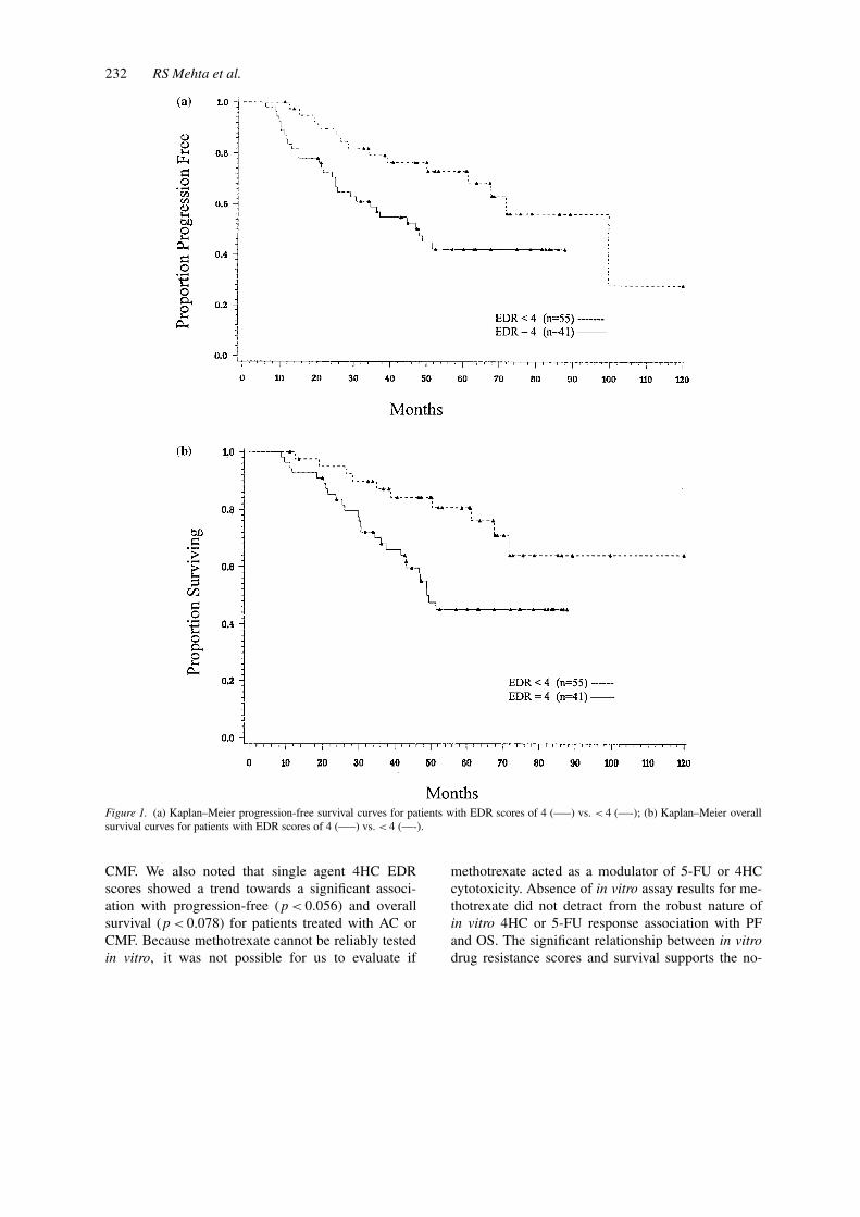
232 RS Mehta et al.
Figure 1. (a) Kaplan–Meier progression-free survival curves for patients with EDR scores of 4 (—–) vs. < 4 (—-); (b) Kaplan–Meier overallsurvival curves for patients with EDR scores of 4 (—–) vs. < 4 (—-).
CMF. We also noted that single agent 4HC EDRscores showed a trend towards a significant associ-ation with progression-free (p < 0.056) and overallsurvival (p < 0.078) for patients treated with AC orCMF. Because methotrexate cannot be reliably testedin vitro, it was not possible for us to evaluate if
methotrexate acted as a modulator of 5-FU or 4HCcytotoxicity. Absence of in vitro assay results for me-thotrexate did not detract from the robust nature ofin vitro 4HC or 5-FU response association with PFand OS. The significant relationship between in vitrodrug resistance scores and survival supports the no-
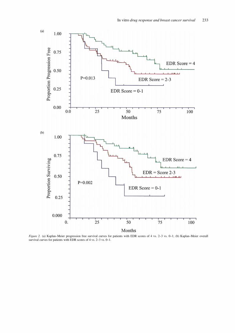
In vitro drug response and breast cancer survival 233
Figure 2. (a) Kaplan–Meier progression free survival curves for patients with EDR scores of 4 vs. 2–3 vs. 0–1; (b) Kaplan–Meier overallsurvival curves for patients with EDR scores of 4 vs. 2–3 vs. 0–1.
234 RS Mehta et al.
Table 6A. Progression-free survival: Multivariate analyses ofprognostic factors
Relative 95% CI p value
risk
EDR score (0–1 vs. 4) 2.602 0.938–7.223 0.0663
EDR score (2–3 vs. 4) 2.097 1.037–4.240 0.0393
Nodal status (> 10 vs. 0) 4.252 1.076–16.809 0.0390
Nodal status (4–10 vs. 0) 1.627 0.545–4.859 0.3829
Nodal status (1–3 vs. 0) 1.581 0.549–4.550 0.3959
Stage (IV vs. I) 3.009 0.606–14.929 0.1777
Stage (III vs. I) 0.772 0.136–4.396 0.7706
Stage (II vs. I) 0.916 0.201–4.167 0.9091
Table 6B. Overall survival: Multivariate analyses of prognosticfactors
Relative 95% CI p value
risk
EDR score (0–1 vs. 4) 3.085 1.051–9.059 0.0404
EDR score (2–3 vs. 4) 2.349 1.073–5.146 0.0327
Nodal status (> 10 vs. 0) 6.743 1.497–30.359 0.0129
Nodal status (4–10 vs. 0) 1.494 0.497–4.489 0.4748
Nodal status (1–3 vs. 0) 1.107 0.372–3.295 0.8552
Stage (IV vs. I) 5.028 0.842–30.010 0.0764
Stage (III vs. I) 0.599 0.080–4.478 0.6180
Stage (II vs. I) 1.139 0.210–6.181 0.8803
tion that drug resistance testing can identify a tumorphenotype related to clinical outcome. Information re-garding the unique in vitro drug response profile of anindividual patient may potentially be exploited to im-prove outcomes by tailoring treatment based on assayresults.
Several smaller studies have indicated that re-sponse rates are improved when patients receivedchemotherapy to which their tumors were not res-istant in vitro [27, 33–35]. A review of publishedin vitro assay results for 4,263 patients, where correla-tions with treatment response were available, indicatedthat clinical response rates were significantly associ-ated with in vitro results, with an overall sensitivityof 85%, and an overall specificity of 80% [27]. Ofnote was the finding that prediction of drug resistancewas > 90% accurate, compared to 72% accuracy forprediction of chemosensitivity. The EDR assay wasreported to have a negative predictive accuracy of 99%in a double-blind retrospective study of 450 cases [33].
The drug exposures used in the EDR assay are 5–10 times higher than those achieved in vivo, biasingassay reliability towards accurate detection of drugresistance. In correlating in vitro drug resistance withclinical response, only 1 out of 127 patients showingin vitro EDR showed clinical response, while 52%of patients showing in vitro sensitivity demonstratedclinical response. The prediction of resistance may bemore robust than the prediction of sensitivity becauseof the inability of in vitro systems to parallel relevantin vivo pharmacodynamics, such as individual vari-ations in tumor vascular supply and drug metabolism,that influence clinical response. The practical utilityof in vitro testing has been enhanced by the devel-opment of third generation assay techniques, such asthe EDR assay employed in this study. While olderclonogenic systems yielded results in 2–3 weeks with50% success rates, newer technologies have shortenedassay time to less than one week and improved theevaluability rate to 85%, which is similar to the eval-uability rate of 91% in the present study [27]. Yet,the relationship between in vitro results and survivalhas not been adequately addressed either by chemo-sensitivity or chemoresistance assays. Our study is thefirst large series to demonstrate the clinical relevanceof in vitro drug resistance for patients with all stagesof breast cancer treated with first line chemotherapy.We found that patients treated with combinations ofdrugs with low in vitro drug resistance enjoy betterprogression-free and overall survival. Patients whowere treated with combinations of drugs found to beintermediate or extremely resistant in the EDR assayhad a 5-year survival rate of only 45%, comparedto 81% for patients who were treated with a com-bination of drugs to which their tumors showed lowresistance.
The improved survival demonstrated for patientstreated with combinations of low resistance agentsis intriguing. In a recent review, 12 of 17 studieswere found to demonstrate a statistically significantsurvival advantage for patients treated with agentsto which they were ‘sensitive’ in vitro [27]. Threeprospective studies, two of which were randomized,showed a survival advantage for patients treated withassay-directed therapy [27]. More recently, a random-ized study by Cortazar demonstrated that survival wasimproved for small-cell lung cancer patients random-ized to assay directed therapy [36]. Orr et al., haveshown that costs can be lowered using assay direc-ted therapy, with at least equivalent survival in newlydiagnosed, optimally resected ovarian cancer patients
In vitro drug response and breast cancer survival 235
[25]. Gambino et al., demonstrated high response rateswith assay directed therapy in patients with chemo-therapy refractory gynecological malignancies [37].Similarly, a prospective trial by Kurbacher et al.,demonstrated a high response rate and promising sur-vival outcomes in recurrent ovarian cancer treated withtherapy tailored according to their in vitro assay res-ults [38]. One shortcoming of in vitro testing is thenecessity of obtaining adequate amounts of malig-nant tissue for cell culture, which limited the EDRtesting to those with tumors ≥ 0.5 cm in size. How-ever, patients with tumors < 1 cm in size, which areincreasingly being detected mammographically, arenot routinely considered to be candidates for chemo-therapy. A second shortcoming of this study was theabsence of HER2 profiling, which may affect the clin-ical outcome in patients treated with AC. BecauseHER2 was not routinely tested at the time we beganthis study, these data are not available.
Our study was performed in newly diagnosedbreast cancer patients. Our finding of a significant as-sociation between survival and in vitro drug resistanceto the agents used clinically suggests that completing4–6 cycles of ineffective adjuvant chemotherapy mayadversely delay selection of effective chemotherapy,potentially induce or select for additional chemores-istance, and may also decrease the patient’s capacityto undergo further therapy. EDR assay results iden-tified patients with inferior survival after treatmentwith agents their tumor was resistant to in vitro. Theclinical utility of this finding may stem from the elim-ination of such agents from treatment planning. Inaddition, such testing may identify alternative formsof treatment with a greater probability of success.This is of major importance when considering the re-cent introduction of several new non-cross resistantdrugs into clinical practice. Our study was performedwhen fewer chemotherapeutic choices were availablefor first line treatment of breast cancer. The utilityof in vitro testing is increased with the addition ofthese newer agents. While we await the results of fu-ture randomized trials of assay directed treatment, itis logical to conclude that the risk/benefit ratio favorsthe utility of EDR assay results when considering theelimination of ineffective treatment options for breastcancer.
Acknowledgements
This study was supported, in part, by a grant fromPharmacia & Upjohn.
The following authors have a financial interest inOncotech, the Principle Sponsor of this study: R.Mehta, I.Y. Yu, R. Parker, and J.P. Fruehauf.
References
1. Goldie JH, Coldman AJ: A mathematic model for relating thedrug sensitivity of tumors to the spontaneous mutation rate.Cancer Treat Rep 63: 1727–1733, 1979
2. Goldie JH, Coldman AJ, Gadauskas GA: Rationale for the useof alternating non-cross-resistant chemotherapy. Cancer TreatRep 66: 439–449, 1982
3. Fisher F, Brown AM, Dimitrov NV, Poisson R, RedmondC, Margolese RG, Bowman D, Wolmark N, Wickerham DL,Kardinal CG: Two months of doxorubicin-cyclophosphamidewith and without interval reinduction therapy compared to6 months of cyclophosphamide, methotrexate, and fluor-ouracil in node-positive breast cancer patients with tamoxifen-nonresponsive tumors. Results from the National SurgicalAdjuvant Breast and Bowel Project B-15. J Clin Oncol 8:1483–1496, 1990
4. Early Breast Cancer Trialists’ Collaborative Group: Poly-chemotherapy for early breast cancer: an overview of therandomized trials. Lancet 352: 930–942, 1998
5. Hutchins L, Green S, Ravdin P, Lew D, Martino S, Abeloff M,Lyss A, Henderson C, Allred C, Dakhil S, Pierce I, GoodwinW, Caton J, Rivkin S, Chapman R, Osborne K: CMF versusCAF with and without tamoxifen in high-risk node-negativebreast cancer and a natural history follow-up study in low-risknode-negative patients: first results of Intergroup Trial INY0102. Proc Am Soc Clin Oncol 17: 1a, 1998 (abstr 2)
6. Henderson IC, Berry D, Demetri G, Cirrincione C, GoldsteinL, Martino S, Ingle JN, Cooper MR, Canellos G, Borden E,Fleming G, Holland JF, Graziano S, Carpenter J, Muss H,Norton L: Improved disease-free (DFS) and overall survival(OS) from the addition of sequential paclitaxel (T) but notfrom dose escalation of doxorubicin (A) dose level in theadjuvant chemotherapy of patients (pts) with node-positiveprimary breast cancer (BC) (abstract). Proc Am Soc ClinOncol 17: 101a, 1998 (abstr 390)
7. Carlson RW, Goldstein LJ, Gradishar WJ, Lichter AS, Mc-Cormick B, Moe RE, Theriault RL: NCCN breast cancer prac-tice guidelines. The national comprehensive cancer network.Oncology 10(11 Suppl): 47–75, 1996
8. Anderson BO, Bensinger W, Cox CE, Davidson NE, EdgeSB, Farrar WB, Goldstein LJ, Gradishar WJ, Lichter AS, Mc-Cormick B, Nabell LM, Reed EC, Silver SM, Smith ML,Somlo G, Theriault R, Ward JH, Winer EP, Wolff A: Update:NCCN Practice Guidelines for Breast Cancer. Oncology 14(11Suppl): 33–49, 2000
9. Hortabagyi GN, Buzdar AU, Champlin R, Gajewski J, HolmesFA, Booser D, Valero V, Theriault RL: Lack of efficacy ofadjuvant high dose (HD) tandem combination chemotherapy(CT) for high risk primary breast cancer – a randomized trial(abstract). Proc Am Soc Clin Oncol 17: 123a, 1998 (abstr 471)
10. Rodenhuis S, Richel DJ, van der Wall E, Schornagel JH, BaarsJW, Koning CC, Peterse JL, Borger JH, Nooijen WJ, Bakx R,Dalesio O, Rutgers E: Randomized trial of high-dose chemo-therapy and hematopoietic progenitor-cell support in operablebreast cancer with extensive axillary lymph-node involvement.Lancet 352: 515–521, 1998
236 RS Mehta et al.
11. Peters W, Rosner G, Vredenburgh J, Shpall E, Crump M,Richardson P, Marks L, Cirrincione C, Wood W, HendersonI, Hurd D, Norton L: A prospective, randomized comparisonof two doses combination alkylating agents (AA) as consolid-ation after CAF in high-risk primary breast cancer involvingten or more axillary lymph nodes (LN): Preliminary results ofCALGB 9082/SWOG 9114/NCIC MA-13. Proc Am Soc ClinOncol 18: 1a, 1999 (abstr 2)
12. Bezwoda WR: Randomized, controlled trial of high dosechemotherapy (HD-CNVp) vs. standard dose (CAF) chemo-therapy for high risk, surgically treated, primary breast cancer.Proc Am Soc Clin Oncol 18: 2a, 1999 (abstr 4)
13. The Scandinavian Breast Study Group 9401: Results froma randomized adjuvant breast cancer study with high dosechemotherapy with CTCb supported by autologous bone mar-row stem cells versus dose escalated and tailored FEC therapy.Proc Am Soc Clin Oncol 18: 2a, 1999 (abstr 3)
14. Lotz J-P, Curé H, Janvier M, Morvan F, Asselain B, GuillemotM, Laadem A, Maraninchi D, Gisselbrecht C, Roché H: High-Dose Chemotherapy (HD-CT) with hematopoietic stem cellstransplantation (HSCT) for metastatic breast cancer: Resultsof the French Protocol Pegase 04. Proc Am Soc Clin Oncol18: 43a, 1999 (abstr 161)
15. Stadtmauer EA, O’Neill A, Goldstein PG, Crilley P, Man-gan KF, Ingle JN, Lazarus HM, Erban J, Sickles C, GlickJH: Phase III Randomized trial of high-dose chemotherapy(HDC) and stem cell support (SCT) shows no difference inoverall survival or severe toxicity compared to maintenancechemotherapy with cyclophosphomide, methotrexate and 5-fluorouracil (CMF) for women with metastatic breast cancerwho are responding to conventional induction chemotherapy:The Philadelphia Intergroup Study (PBT-01), ECOG, U. ofPennsylvania. Proc Am Soc Clin Oncol 18: 1a, 1999 (abstr1)
16. Early Breast Cancer Trialists’ Collaborative Group: Tamox-ifen for early breast cancer: an overview of the randomizedtrials. Lancet 351(9114): 1451–1467, 1998
17. Thor AD, Berry DA, Budman DR, Muss HB, Kute T, Hender-son IC, Barcos M, Cirrincione C, Edgerton S, Allred C, NortonL, Liu ET: erbB-2, p53, and efficacy of adjuvant therapyin lymph node-positive breast cancer. J Natl Cancer Inst 90:1346–1360, 1998
18. Paik S, Bryant J, Park C, Fisher B, Tan-Chiu E, Hyams D,Fisher ER, Lippman ME, Wickerham DL, Wolmark N: erbB-2 and response to doxorubicin patients with axillary lymphnode-positive, hormone receptor-negative breast cancer. J NatlCancer Inst 90: 1361–1370, 1998
19. Norton L, Slamon D, Leyland-Jones B, Wolter J, Fleming T,Eirmann W, Baselga J, Mendelsohn J, Bajamonde A, AshM, Shak S: Overall survival (OS) advantage to simultan-eous chemotherapy (CRx) plus the Humanized Anti-HER2Monoclonal Antibody Herceptin (H) in HER2-Overexpressing(HER2 +) metastatic breast cancer (MBC). Proc Am Soc ClinOncol 18: 127a,1999 (abstr 483)
20. Sledge GW Jr, Neuberg D, Ingle J, Martino S, Wood W:Phase III trial of doxorubicin vs. paclitaxel vs. doxoru-bicin + paclitaxel as first-line therapy for metastatic breastcancer. Proc Am Soc Clin Oncol 16: 1a, 1997 (abstr 2)
21. Gammuci T, Piccart M, Brüning P, Klijn J, Biganzoli L, Hous-ton S, Coleman R, Van Vreckem A, Cleall S, Curran D, AwadaA, Paridaens R: Single agent taxol (T) versus doxorubicin(D) as first-line chemotherapy (CT) in advanced breast cancer(ABC): result of an EORTC randomized study with crossover.Proc Am Soc Clin Oncol 17: 11a, 1998 (abstr 428)
22. Ravdin P, Burris HA, Cook G, Eisenberg P, Kane M,Bierman WA, Mortimer J, Genevois E, Bellet RE: PhaseII trial of docetaxel in advanced anthracycline-resistant oranthracenedione-resistant breast cancer. J Clin Oncol 13:2879–2885, 1995
23. Valero V, Holmes FA, Walters RS, Theriault RL, EsparzaL, Fraschini G, Fonseca GA, Bellet RE, Buzdar AU, Horto-bagyi GN: Phase II trial of docetaxel: A new, highly effect-ive antineoplastic agent in the management of patients withanthracycline-resistant metastatic breast cancer. J Clin Oncol13: 2886–2894, 1995
24. Blum JL, Jones SE, Buzdar AU, LoRusso PM, Kuter I, VogelC, Osterwalder B, Burger HU, Brown CS, Griffin T: Multi-center phase II study of capecitabine in paclitaxel-refractorymetastatic breast cancer. J Clin Oncol 17: 485–493, 1999
25. Orr JW, Orr P, Kern D: Cost-effective treatment of womenwith advanced breast cancer by cytoreductive surgery andchemotherapy directed by an in vitro assay for drug resistance.The Cancer J 5: 174–178, 1999
26. DeVita VT Jr: In: DeVita, Hellman, Rosenberg (eds), Cancer:Principles and Practice of Oncology. 5th edn, Lippincott-Raven Publishers, Philadelphia, 1997, 345 p
27. Fruehauf JP, Bosanquet AG: In vitro determination of drugresponse: A discussion of clinical applications, In: DeVita VTJr, Hellman S, Rosenberg SA (eds), Principles and Practice ofOncology Updates, 4th edn, JB Lippincott, Philadelphia, PA,1993, pp 1–16
28. Seidman AD, Reichman BS, Crown JP, Yao TJ, Currie V,Hakes TB, Hudis CA, Gilewski TA, Baselga J, Forsythe P,et al.: Paclitaxel as second and subsequent therapy for meta-static breast cancer: activity independent of prior anthracyclineresponse. J Clin Oncol 13: 1152–1159, 1995
29. Seidman AD, Hudis CA, Norton L: Memorial Sloan Ketter-ing Cancer Center experience with paclitaxel in the treatmentof breast cancer: from advanced disease to adjuvant therapy.Semin Oncol; 22(4 Suppl 8): 3–8, 1995
30. Mavroudis D, Malamos N, Alexopoulos A, Kourousis C,Agelaki S, Sarra E, Potamianou A, Kosmos C, Rigatos G, Gi-annakakis T, Kalbakis K, Apostolaki F, Vlachonicolis J, Kako-lyris S, Samonis G, Georgoulias V: Salvage chemotherapyin anthracycline-pretreated metastatic breast cancer patientswith docetaxel and gemcitabine. Ann Oncol 10: 211–215,1999
31. Leung PP, Tannock IF, Oza AM, Puodziunas A, DranitsarisG: Cost-Utility analysis of chemotherapy using paclitaxel,docetaxel, or vinorelbine for patients with Anthracycline-resistant breast cancer. J Clin Oncol 17: 3082–3090, 1999
32. Kern DH, Drogemuller CR, Kennedy MC, Hildebrand-ZankiSU, Tanigawa N, Sondak VK: Development of a miniatur-ized, improved nucleic acid-precursor incorporation assay forchemosensitivity testing of human solid tumors. Cancer Res45: 5436–5441, 1985
33. Kern DH, Weisenthal LM: Highly specific prediction of an-tineoplastic drug resistance with an in vitro assay usingsuprapharmacologic drug exposures. J Natl Cancer Inst 82:582–588, 1990
34. Elledge RM, Clark GM, Hon J, Thant M, Belt R, Maguire YP,Brown J, Bartels P, Von Hoff DD: Rapid in vitro assay forpredicting response to fluorouracil in patients with metastaticbreast cancer. J Clin Oncol 17: 419–423, 1995
35. Cortazar P, Johnson BE: Review of the efficacy of indi-vidualized chemotherapy selected by in vitro drug sensitivitytesting for patients with cancer. J Clin Oncol 17: 1125–1631,1999

In vitro drug response and breast cancer survival 237
36. Cortazar P, Gazdar AF, Woods E, Russell E, Steinberg SM,Williams J, Ihde DC, Johnson BE: Survival of patients withlimited-stage small cell lung cancer treated with individualizedchemotherapy selected by in vitro drug sensitivity testing. ClinCancer Res 3: 741–747, 1997
37. Gambino A, Favalli G, Salinaro, Pecorelli S: The use of Ex-treme Drug Resistance Assay in Gynecologic malignanciesafter failure of conventional chemotherapy. IGCS Oncol 17:F139a, 1999 (abstr)
38. Kurbacher CM, Bruckner HW, Cree IA, Bruckner HW,Brenne U, Kurbacher JA, Muller K, Ackermann T, Gilster TJ,
Wilhelm LM, Engel H, Mallmann PK, Andreotti PE: Aprospective clinical trial on individualized chemotherapyfor recurrent ovarian cancer selected by the Use of anex vivo ATP luminescence assay to direct chemotherapyfor recurrent ovarion cancer. Anticancer Drugs 9: 51–57,1998
Address for offprints and correspondence: John P. Fruehauf,Oncotech, Inc., 1791 Kaiser Avenue, Irvine, CA 92614; Tel.:949-474-9262; E-mail: [email protected]
Top Related
Copyright © 2022 FDOKUMEN

