


Bahasa
Halaman
Hukum


Brain, Behavior, and Immunity 32 (2013) 112–121
Contents lists available at SciVerse ScienceDirect
Brain, Behavior, and Immunity
journal homepage: www.elsevier .com/locate /ybrbi
Blocking toll-like receptor 2 and 4 signaling during a stressor preventsstress-induced priming of neuroinflammatory responses to a subsequentimmune challenge
Michael D. Weber ⇑, Matthew G. Frank, Julia L. Sobesky, Linda R. Watkins, Steven F. MaierDepartment of Psychology and Neuroscience, Center for Neuroscience, University of Colorado, Boulder, CO, USA
a r t i c l e i n f o
Article history:Received 28 January 2013Received in revised form 26 February 2013Accepted 8 March 2013Available online 15 March 2013
Keywords:StressMicrogliaToll-like receptorCytokineNeuroinflammationPriming
0889-1591/$ - see front matter � 2013 Elsevier Inc. Ahttp://dx.doi.org/10.1016/j.bbi.2013.03.004
⇑ Corresponding author. Address: Department of PCenter for Neuroscience, University of Colorado BouldUSA. Tel.: +1 614 937 2613; fax: +1 303 492 2967.
E-mail address: [email protected] (M.D. We
a b s t r a c t
Acute and chronic stressors sensitize or prime the neuroinflammatory response to a subsequent periph-eral or central immunologic challenge. However, the neuroimmune process(es) by which stressors primeor sensitize subsequent neuroinflammatory responses remains unclear. Prior evidence suggested thattoll-like receptors (TLRs) might be involved in the mediation of primed neuroinflammatory responses,but the role of TLRs during a stressor has never been directly tested. Here, a novel TLR2 and TLR4 antag-onist, OxPAPC, was used to probe the contribution of TLRs in the stress sensitization phenomenon.OxPAPC has not previously been administered to the brain, and so its action in blocking TLR2 andTLR4 action in brain was first verified. Administration of OxPAPC into the CNS prior to stress preventedthe stress-induced potentiation of hippocampal pro-inflammatory response to a subsequent peripheralLPS challenge occurring 24 h later. In addition, in vivo administration of OxPAPC prior to stress preventedthe sensitized pro-inflammatory response from isolated microglia following administration of LPS ex vivo,further implicating microglia as a key neuroimmune substrate that mediates stress-induced sensitizedneuroinflammation.
� 2013 Elsevier Inc. All rights reserved.
1. Introduction
Acute and chronic stress sensitize the neuroinflammatory re-sponse to subsequent peripheral and central inflammatory chal-lenges, creating an exaggerated neuroinflammatory response (dePablos et al., 2006; Espinosa-Oliva et al., 2011; Frank et al., 2002,2007, 2010; Johnson et al., 2003, 2004; Munhoz et al., 2006;Wohleb et al., 2011). For example, exposure to a single session ofintermittent tailshocks (Johnson et al., 2002) or to chronic unpre-dictable stress (Munhoz et al., 2006), potentiates the hippocampaland frontal cortical pro-inflammatory mediators (i.e. interleukin-1b (IL-1b), inducible nitric oxide synthase (iNOS), tumor necrosisfactor-a (TNF-a), and nuclear factor kappa b (NF-jB) activity) in-duced by a subsequent systemic inflammatory challenge occurring24 h after the stressor regimen. These inflammatory mediators inthe brain are produced predominantly by microglia (Gehrmannet al., 1995), and other studies have shown that both acute andchronic stress activate microglia, as assessed by up-regulated ma-jor histocompatibility complex-II (MCHII) (de Pablos et al., 2006;Frank et al., 2007), F4/80 antigen (Nair and Bonneau, 2006; Nair
ll rights reserved.
sychology and Neuroscience,er, Boulder, CO 80309-0345,
ber).
et al., 2007), and microglia proliferation (Nair and Bonneau,2006). Furthermore, microglia isolated from rats that had receiveda single session of tail shock 24 h earlier, exhibited up regulatedMCHII. Interestingly, these microglia from stressed subjects didnot produce increased amounts of pro-inflammatory cytokines(PICs) beyond basal levels. However, if the microglia from stressedrats were stimulated with LPS ex vivo, exaggerated amounts of PICswere detected (Frank et al., 2007). This pattern suggests thatstress ‘primes’ microglia, as defined by Ransohoff and Perry(2009). That is, the microglia shift to a state in which they arenot frankly inflammatory, but produce an exaggerated inflamma-tory response if stimulated. Taken together, these findings suggestthat exposure to a stressor shifts the neuroimmune microenviron-ment towards a pro-inflammatory state, thereby predisposingcertain regions of the CNS to a heightened pro-inflammatory re-sponse if the organism is exposed to a subsequent inflammatorychallenge.
Secretion of glucocorticoids (GCs) from the adrenals (cortisol inhumans and corticosterone (CORT) in rodents) is often taken as ahallmark of the stress response. Since increased levels of GCs arealmost universally considered to be anti-inflammatory (Boumpaset al., 1993), the results described above might appear contradic-tory. However, there is strong evidence demonstrating that GCscan sensitize pro-inflammatory responses, particularly within theCNS (Frank et al., 2010, 2012; Munhoz et al., 2010; Sorrells and
M.D. Weber et al. / Brain, Behavior, and Immunity 32 (2013) 112–121 113
Sapolsky, 2007). Replacing the experience of a stressor with aphysiologically relevant dose of GCs that mimics the elevated lev-els of GCs observed during a stressor, produces both exaggeratedneuroinflammatory (hippocampus) responses to a systemic LPSchallenge 24 h later (Frank et al., 2010) and ‘primed’ microglia thatproduce an exaggerated inflammatory response to LPS ex vivo(Frank et al., 2012). Further, the glucocorticoid receptor (GR) seemsto be critical for GC-induced sensitization. Several studies haveshown that stress-induced microglial activation and potentiationof neuroinflammatory processes is blocked by a GC receptor antag-onist (de Pablos et al., 2006; Espinosa-Oliva et al., 2011; Munhozet al., 2006; Nair and Bonneau, 2006). We have demonstrated thatblocking GR activity during a stressor with RU486 prevents stress-induced sensitization to a subsequent immune challenge in vivo,and the priming of microglia observed ex vivo (Frank et al., 2012).
Although the effects of stress-induced sensitization appear to bemediated, at least in part, by increased GC levels, the mechanism(s)whereby stress and GCs sensitize neuroinflammatory responses islargely unknown. Interestingly, GCs upregulate the expression ofthe pattern recognition receptors (PRR) toll-like receptors TLR2and TLR4. These PRRs are involved in the recognition of both path-ogen associated molecular patterns (PAMPS) and danger associatedmolecular patterns (DAMPS), and initiate signaling cascades thatlead to the synthesis and release of inflammatory mediators (Ka-wai and Akira, 2007; Salminen et al., 2008). In vitro studies havedemonstrated that GCs can up-regulate TLR2 expression in epithe-lial cells via MAPK phosphatase-1 (MKP-1), which in turn inhibitsp38 MAPK activity, a negative regulator for TLR2. This increasedexpression of TLR2 leads to enhanced cytokine expression, includ-ing TNF-a, IL-1b, and IL-8, upon challenge with an inflammatorystimulus (Imasato et al., 2002). Similarly, Rozkova et al. (2006)found increased TLR2 and TLR4 expression on dendritic cells (DC)following GC treatment. In addition, TNF-a and GCs cooperate tostimulate the promoter for TLR2 and potentially TLR4, increasingreceptor expression (Hermoso et al., 2004). Finally, in vivo findingsdemonstrate that TLR2 mRNA is upregulated 24 h after subcutane-ous (SC) injection of GCs (Frank et al., 2010) and TLR4 protein is in-creased following repeated social stress (Wohleb et al., 2011).
These data suggest that elevated levels of GCs, produced by stressexposure, may sensitize the neuroimmune microenvironment byupregulating expression of TLR2 and TLR4 on CNS innate immunecells. The purpose of the present study was to investigate theinvolvement of TLR2 and TLR4 during a stressor and assess whetherthese receptors do mediate the stress-induced sensitized inflamma-tory response. A novel TLR2 and TLR4 antagonist, Oxidized 1-palmi-toyl-2-arachidonyl-sn-glycero-3-phosphorylcholine (OxPAPC), wasused to block TLR2 and TLR4 activity during a stressor. Here we dem-onstrate that administration of OxPAPC into the CNS prior to stressprevents the exaggerated central (hippocampus) inflammatory re-sponse to a subsequent immune challenge. In vivo administrationof central OxPAPC prior to stress also prevented potentiated inflam-matory responses of microglia to LPS ex vivo.
2. Methods
2.1. Animals
Male Sprague–Dawley rats (60–90 day-old; Harlan Sprague–Dawley, Inc., Indianapolis, IN, USA) were pair-housed with foodand water available ad libitum. The colony was maintained at25 �C on a 12-h light/dark cycle (lights on at 07:00 h). All animalswere allowed 1 week of acclimatization to the colony rooms beforeexperimentation. All experimental procedures were conducted inaccordance with the University of Colorado Institutional AnimalCare and Use Committee.
2.2. Reagents
Lipopolysaccharide (LPS; Escherichia coli serotype 0111:B4) is aTLR4 agonist obtained from Sigma (St. Louis, MO). Lipoteichoic acid(LTA; Staphylococcus aureus) is a TLR2 agonist obtained from Inviv-ogen (San Diego, CA). Pam3CSK4 is a TLR1/2 agonist obtained fromInvivogen (San Diego, CA). OxPAPC (Invivogen; San Diego, CA) is anoxidized phospholipid that inhibits TLR2 and TLR4 signaling bycompetitively interfering with extra-cellular accessory proteinssuch as CD14, LPS-binding protein (LBP), and MD2 (Erridge et al.,2008). OxPAPC was suspended in 500 ll chloroform for a lipid con-centration of 1 mg/ml and carefully vortexed. The homogeneoussolution was aliquoted and evaporated under a stream of nitrogengas. On the day of experiment, saline was added to create the de-sired concentration. At higher concentrations, OxPAPC can induceinflammation (Oskolkova et al., 2010). Therefore, an Invivogen rec-ommended concentration of 30 lg/ml was not exceeded.
2.3. Drug administration
LPS was administered i.p. (10 lg/kg) or intra-cisterna magna(ICM) (30 ng suspended in 4 ll sterile saline), depending on exper-imental design. We selected 10 lg/kg i.p. LPS because we have pre-viously shown that this dose results in a sub-thresholdhippocampal pro-inflammatory response (Johnson et al., 2002).30 ng/4 ll was selected for ICM administration because pilot stud-ies found that this dose of LPS produces robust pro-inflammatorygene expression as measured by real time RT-PCR in the hippocam-pus (data not shown).
LTA was administered ICM (40 ng suspended in 4 ll sterile sal-ine). Similarly, this dose was selected because pilot studies indi-cated that this dose of LTA produces robust pro-inflammatorygene expression as measured by real time RT-PCR in the hippocam-pus (data not shown).
OxPAPC was administered ICM (150 ng suspended in 5 ll sterilesaline). In vivo and ex vivo preliminary work demonstrated that thisdose sufficiently inhibited TLR2 and TLR4 activation as measuredby proinflammatory gene expression via real time RT-PCR (datashown below).
2.4. ICM administration
ICM administration was chosen to deliver drugs centrally be-cause it avoids surgery and canulae implantation, and the longlasting neuroinflammation which results (Holguin et al., 2007).Rats were briefly anesthetized (<2 min) with halothane. The dorsalaspect of the skull was shaved and swabbed with 70% ETOH. A 27-gauge needle attached via PE50 tubing to a 25 ll Hamilton syringewas inserted into the cisterna magna. To verify entry into the cis-terna magna, �2 ll of CSF was drawn. In all cases, CSF was clear ofred blood cells indicating entry into the cisterna magna.
2.5. Inescapable tailshock (IS)
Details of the present stressor protocol have been publishedpreviously, and the protocol reliably potentiates pro-inflammatorycytokine responses in the hippocampus after peripheral immunechallenge (Johnson et al., 2002), as well as in isolated hippocampalmicroglia to LPS ex vivo (Frank et al., 2007). Briefly, animals wereplaced in Plexiglas tubes (23.4 cm in length � 7 cm in diameter)and exposed to 100 1.6 mA, 5 s tailshocks with a variable intertrialinterval (ITI) ranging from 30 to 90 s (average ITI = 60 s). All IStreatments occurred between 09:00 and 11:00 h. IS animals werereturned to their home cages immediately after termination ofshock. HCC animals remained undisturbed in their home cages.
114 M.D. Weber et al. / Brain, Behavior, and Immunity 32 (2013) 112–121
2.6. Tissue collection
Animals were given a lethal dose of sodium pentobarbital. Ani-mals were fully anesthetized and transcardially perfused with ice-cold saline (0.9%) for 3 min to remove peripheral immune cellsfrom the CNS vasculature. Brains were rapidly extracted and placedon ice, and hippocampus dissected. For in vivo experiments, hippo-campus and liver were flash frozen in liquid nitrogen and stored at�80 �C. For ex vivo experiments, hippocampal microglia wereimmediately isolated. Analysis was restricted to the hippocampusbecause we have shown that it is sensitize to IS and produces ro-bust IS-induced priming effects in vivo (Johnson et al., 2002) andex vivo (Frank et al., 2007). Hippocampus also yields a sufficientnumber of microglia to conduct ex vivo experiments. Liver wasused as an indicator of peripheral pro-inflammatory responses toinflammatory agents with or without OxPAPC.
2.7. Real time RT-PCR measurement of gene expression
Gene expression was measured using real time RT-PCR. TotalRNA was isolated from whole hippocampus utilizing a standardmethod of phenol:chloroform extraction (Chomczynski and Sacchi,1987). For detailed descriptions of RNA isolation, cDNA synthesis,and PCR amplification protocols refer to prior publication (Franket al., 2006). cDNA sequences were obtained from Genbank atthe National Center for Biotechnology Information (NCBI;www.ncbi.nlm.nih.gov). Primer sequences were designed to ampli-fy several cytokines and inflammatory activation markers. Primersequences were designed using the Qiagen Oligo Analysis & Plot-ting Tool (oligos.qiagen.com/oligos/toolkit.php?) and tested for se-quence specificity using the Basic Local Alignment Search Tool atNCBI (Altschul et al., 1997). Primers were obtained from Invitro-gen. Primer specificity was verified by melt curve analysis. Primersequences are as follows: NFKBIAei, F-CACCAACTACAACGGCCACA,R-GCTCCTGAGCGTTGACATCA, TNFa, F-CAAGGAGGAGAAGTTCCCA,R-TTGGTGGTTTGCTACGACG; IL-1b, F-CCTTGTGCAAGTGTCTGAAG,R-GGGCTTGGAAGCAATCCTTA; IL-6, F-AGAAAAGAGTTGT-GCAATGGCA, R-GGCAAATTTCCTGGTTATATCC; GAPDH F-TCTTCCAGGAGCGAGATCCC, R-TTCAGGTGAGCCCCAGCCTT. PCRamplification of cDNA was performed using the Quantitect SYBRGreen PCR Kit (Qiagen, Valencia, CA). Formation of PCR productwas monitored in real time using the MyiQ Single-Color Real-TimePCR Detection System (BioRad, Hercules, CA). Relative gene expres-sion was determined using the 2�DDCT (Livak and Schmittgen,2001). Mean CT of triplicate measures (C.V. <10%) was computedfor each sample. Sample mean CT of GAPDH (internal control)was subtracted from the sample mean CT of the respective geneof interest (DCT). The sample with the highest absolute DCT wasselected as a calibrator and subtracted from the DCT of each exper-imental sample (DDCT). 2�DDCT yields fold change in gene expres-sion of the gene of interest normalized to the internal controlgene expression and relative to the calibrator sample.
2.8. Experimental designs
2.8.1. Effect of OxPAPC on TLR2 & TLR4 signaling in vitroThis experiment was a preliminary experiment designed to ver-
ify that OxPAPC does function as a TLR2&4 antagonist. We havepreviously described and used (Hutchinson et al., 2010) a humanembryonic kidney-293 (HEK293) cell line stably transfected to ex-press human TLR4 to assess TLR4 activity. This HEK293 cell line ex-presses high levels of TLR4, the required TLR4 co-signalingmolecules (MD-2 and CD14), and an optimized alkaline phospha-tase reporter gene under the control of a promoter inducible byseveral transcription factors such as NF-jB and AP-1 (Invivogen,San Diego, CA, USA; 293-htlr4a-md2cd14). A parallel HEK-TLR2
(Invivogen, San Diego, CA, USA) cell line was also employed hereto examine TLR2 activity. The cells were plated for 48 h in 96 wellplates (Microtest 96 well flat bottom plate, Becton Dickinson,Franklin Lakes, NJ, USA; 5 � 103 cells/well) in normal supplementselection media (DMEM with 10% fetal bovine serum (FBS). After48 h, supernatant was removed and 160 ll of fresh media wasadded. 20 ll of OxPAPC in different concentrations (5, 10, and20 lg) were added to cells stimulated with 20 ll of LPS(10 ng). ATLR4 ligand, or PAM3CSK4 (100 ng), a TLR2 ligand, and incubatedfor 24 h. Supernatants (15 lL) were then collected from each wellfor immediate assay.
TLR2 and TLR4 activity was assessed by measuring the expres-sion of secreted alkaline phosphatase (SEAP) protein. SEAP in thesupernatants was assayed using the Phospha-Light System (AppliedBiosystems, Foster City, California, USA) according to the manufac-turer’s instructions. This is a chemiluminescence assay that incorpo-rates Tropix CSPD chemiluminescent substrate. The 15-lL testsamples were diluted in 45 lL of 1� dilution buffer, transferred to96-well plates (Thermo, Walthma, MA, USA), heated at 65 �C in awater bath (Model 210; Fisher Scientific, Pittsburgh, PA, USA) for30 min, and then cooled on ice to room temperature. Assay buffer(50 lL/well) was added and, 5 min later, reaction buffer (50 lL/well) was added and allowed to incubate for 20 min at room tem-perature. The light output was then measured in a microplate lumi-nometer (#IL213.1191; Dynex Technologies, Chantilly, VA, USA).
2.8.2. Effect of ICM OxPAPC co-administered with ICM LPS or LTA onhippocampal pro-inflammatory cytokine gene expression in vivo
Prior studies of OxPAPC have not administered it centrally. Toverify that OxPAPC inhibits TLR2 and TLR4 activation in the brain,OxPAPC (150 ng/5 ll, ICM) or vehicle was co-administered withthe TLR2 agonist LTA (40 ng/4 ll, ICM), the TLR4 agonist LPS(30 ng/4 ll, ICM) or vehicle, with a 1 ll air bubble separating thetwo reagents. 2 h after injection of either LPS or vehicle, geneexpression of IL-1b and Hippocampus was collected for pro-inflam-matory gene mRNA analysis 2 h after injection. The experimentwas conducted as two separate cohorts.
2.8.3. Effect of central TLR2 and TLR4 antagonism on peripheral LPS-induced pro-inflammatory cytokine gene expression in vivo
Systemically injected LPS does not cross the blood–brain barrier(BBB) (Banks and Robinson, 2010), yet produces robust increases inpro-inflammatory cytokines in the brain and microglia activationmarkers (Frank et al., 2010). The activating signal that induces thisresponse within the brain remains unknown and may not bedependent on brain TLR4 or TLR2 ligation. To test the involvementof brain TLR2 and TLR4 on CNS pro-inflammatory responses to sys-temic LPS, OxPAPC (150 ng/4 ll, ICM) was administered immedi-ately followed by LPS (10 lg/kg, i.p.). Hippocampus was collectedfor inflammatory marker analysis 1, 2, or 4 h after injection. Sincepeak inflammatory gene expression occurred 2 h post treatment,liver was also collected at that time point to measure peripheralpro-inflammatory gene expression. To verify that the effects of Ox-PAPC were mediated within the CNS, OxPAPC (150 ng) and LPS(10 lg/kg) were injected i.p. Hippocampus and liver were collected2 h post injection for pro-inflammatory gene mRNA analysis. Theexperiment was conducted as two separate cohorts.
2.8.4. Effect of central TLR2 and TLR4 antagonism on stress-inducedsensitization of hippocampal pro-inflammatory gene expression toperipheral LPS in vivo
To assess whether TLR2 and TLR4 mediate stress-induced sensi-tized pro-inflammatory cytokine responses, animals were injectedwith OxPAPC (150 ng/4 ll, ICM) or vehicle prior to onset of inescap-able tailshock (IS) or home cage control (HCC). 24 h post-IS, IS andHCC animals were injected with LPS (10 lg/kg, i.p.) or vehicle. Thus,
M.D. Weber et al. / Brain, Behavior, and Immunity 32 (2013) 112–121 115
the design was a 2 � 2 � 2 factorial. Two hours post-LPS or vehicle,hippocampal pro-inflammatory cytokines were measured. 2 h postinjection was chosen because this was the time at which peak pro-inflammatory cytokine expression was detected in experiment2.8.3. The experiment was conducted as three separate cohorts.
2.8.5. Effect of central TLR2 and TLR4 antagonism on stress-inducedsensitization of hippocampal microglia IL-1b gene expression to LPSex vivo
OxPAPC (150 ng/4 ll, ICM) or vehicle injections and the IS pro-tocol were identical to those in experiment 2.8.4. Hippocampalmicroglia from each animal were isolated separately 24 h afterstressor termination or HCC using procedures, previously de-scribed, that result in highly pure microglia Hippocampal microgliafrom each animal were isolated 24 h after stressor terminationusing procedures, previously described, that result in highly puremicroglia (Iba-1+/MHCII+/CD163�/GFAP�) (Frank et al., 2006)with a yield of �40,000–50,000 cells per hippocampus. Microgliawere suspended in DMEM+10% FBS and microglia concentrationfor each animal was estimated to be at a density of 10 � 103cells/100 ll, as determined by trypan blue exclusion. 100 ll wasadded to individual wells of 96-well v-bottom plate. LPS was uti-lized to challenge microglia ex vivo as we have previously deter-mined the optimal in vitro conditions under which LPS stimulatesa microglia pro-inflammatory cytokine response (Frank et al.,2006). Cells were plated with LPS (0.1, 1.0, 10, 100 ng/ml) or mediaalone for 4 h at 37 �C, 5% CO2. The 100 ng/ml LPS group was ex-cluded from analysis due to cells becoming unviable for unknownreasons in this experiment. The plate was centrifuged at 1000g for10 min at 4 �C to pellet cells and cells washed 1� in ice cold PBSand centrifuged at 1000g for 10 min at 4 �C. Cell lysis/homogeniza-tion and cDNA synthesis was performed according to the manufac-turer’s protocol using the SuperScript III CellsDirect cDNASynthesis System (Invitrogen, Carlsbad, CA). The experiment wasconducted as three separate cohorts.
2.9. Statistical analysis
All data are presented as mean + SEM. Statistical analyses con-sisted of ANOVA followed by t tests with a Newman–Keuls
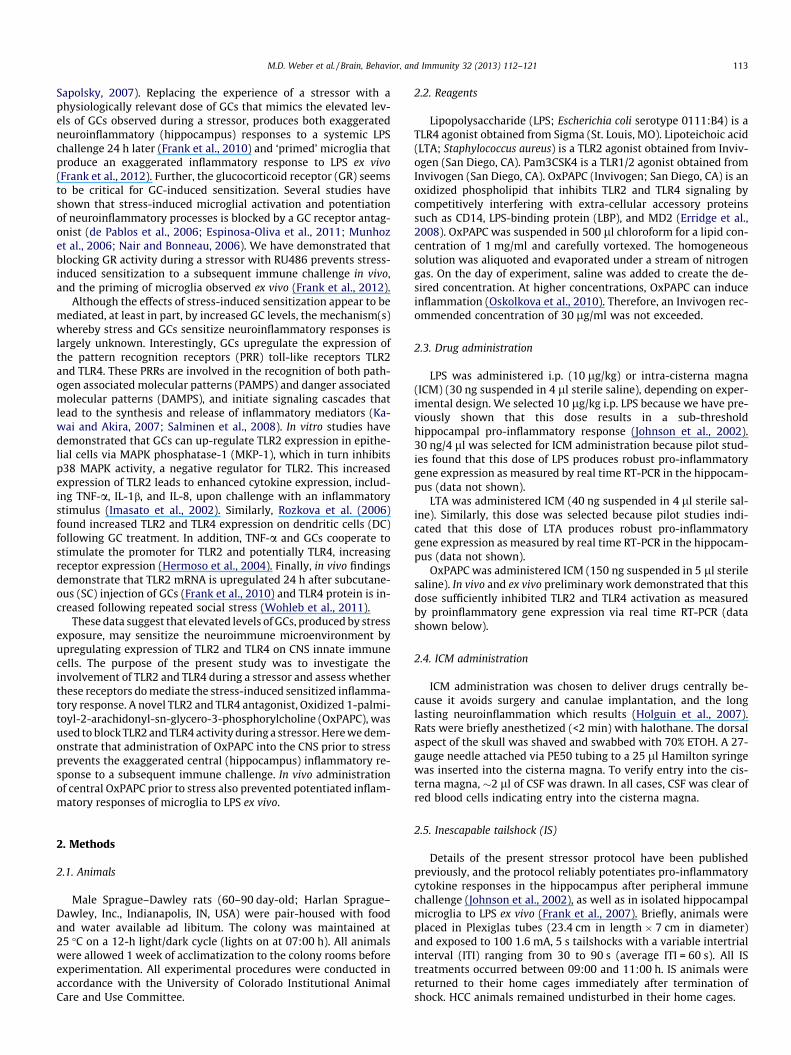
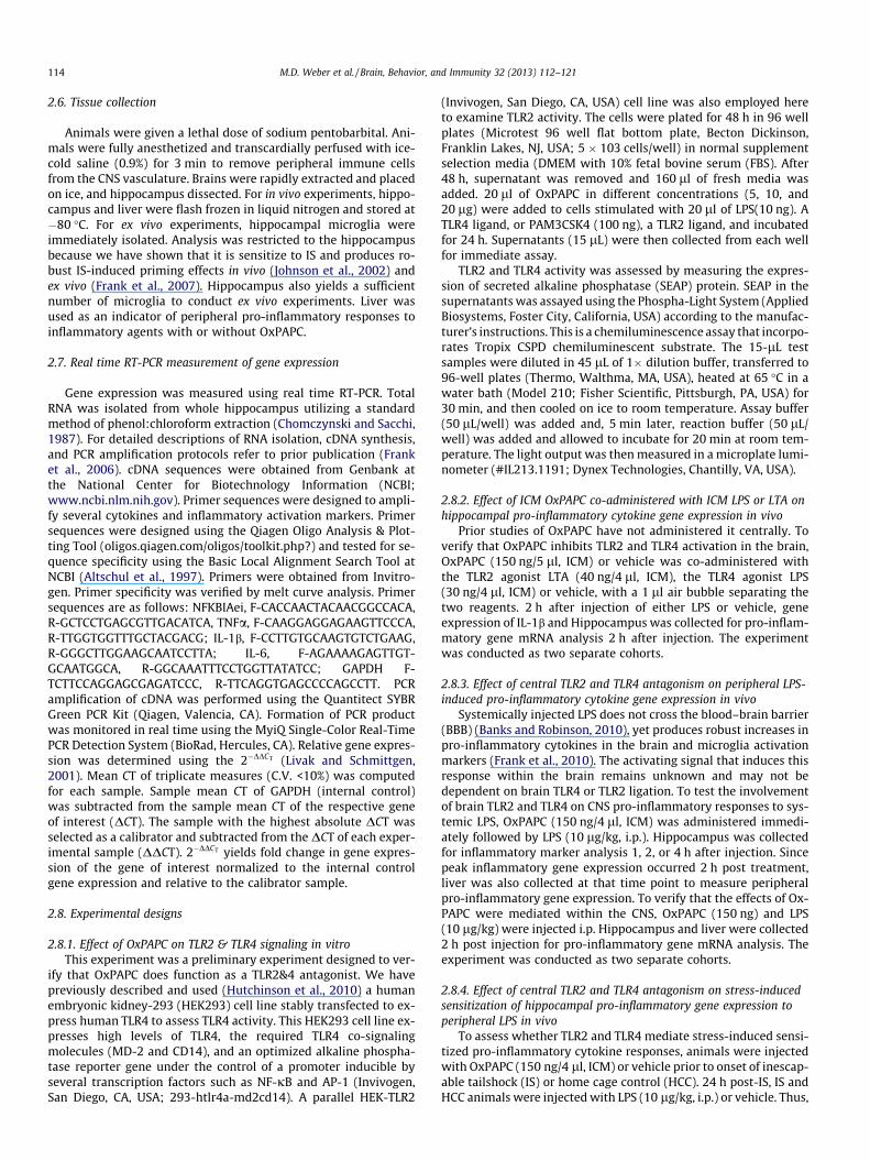
Fig. 1. Effects of OxPAPC (150 ng/5 ll;ICM) on hippocampal pro-inflammatory gene exprTLR4 agonist (LPS; 30 ng/4 ll). Animals received simultaneous ICM administration ofinflammatory gene expression analysis. Means with different letters are significantly di
correction. Threshold for statistical significance was set at a = .05.Outliers that were two standard deviations from the mean were re-moved from analysis. Group numbers are reported in each figure.
3. Results
3.1. Effects of OxPAPC on TLR2 & TLR4 signaling in vitro
To verify that OxPAPC inhibits TLR2 & TLR4 activation, NF-jb-dependent SEAP expression was measured in HEK cells expressingonly TLR2 or expressing only TLR4. The data are shown in Supple-mental Fig. 1. Both Pam3CSK4 and LPS significantly increased SEAPexpression. Even the high dose of OxPAPC on its own did not havean effect on SEAP expression, but all three concentrations of Ox-PAPC significantly blunted Pam3CSK4 or LPS-induced SEAP expres-sion. A one-way ANOVA was conducted for each group. There wasa significant effect in the TLR2 HEK cells (F5,12 = 56.06, P < .0001)and TLR4 HEK cells (F5,12 = 131.2, P < .0001). Post-hoc analysesshowed that OxPAPC significantly reduced expression at concen-trations of 5 (p < .001), 10 (p < .001), and 20 (p < .001) lg/ml inboth cell lines. These results validate the efficacy of OxPAPC to in-hibit TLR2 and TLR4 signaling in vitro.
3.2. Effect of ICM OxPAPC co-administered with ICM LPS or LTA onhippocampal pro-inflammatory cytokine gene expression in vivo
A preliminary study was conducted here to assess the efficacy ofOxPAPC in blocking TLR2 (Fig. 1A) and TLR4 (Fig. 1B) signaling inthe CNS because all previous studies using OxPAPC in vivo werelimited to peripheral effects. Hippocampal IL-1b and ijBa mRNAwere measured to determine whether OxPAPC blocked the pro-inflammatory response to a TLR2 agonist (LTA) or a TLR4 agonist(LPS). IL-1b was measured based on prior evidence indicating brainIL-1b as the key mediator in neuroinflammatory responses to LPS(Laye et al., 2000). ijBa mRNA was measured as an indicator ofNF-jb activation, a key transcription factor involved in initiatingpro-inflammatory cytokine expression (Brown et al., 1993). Thedata are shown in Fig. 1. Clearly, both ICM LPS and LTA producedlarge increases in hippocampal IL-1b and ijBa gene expression.Importantly, OxPAPC had no effects of its own, but almost
ession following ICM administration of a (A) TLR2 agonist (LTA; 40 ng/4 ll) or a (B)OxPAPC and TLR agonist. Hippocampus was collected 2 h post-injection for pro-
fferent (p < .05). Data are presented as mean ± SEM. N = 5–6 animals/group.

116 M.D. Weber et al. / Brain, Behavior, and Immunity 32 (2013) 112–121
completely blocked the effects of LPS and LTA. The interactions be-tween OxPAPC and LTA (IL-1b; F1,20 = 14.56, p < .01 and ijBa;F1,20 = 11.07, p < .01) and OxPAPC and LPS (IL-1b; F1,16 = 4.92,p < .05 and ijBa; F1,17 = 12.63, p < .01) were statistically significant.In animals that did not receive OxPAPC, both LTA and LPS signifi-cantly increased IL-1b and ijBa. Co-administration of OxPAPCblocked LTA and LPS-induced expression of IL-1b to levels similarto veh/veh groups. Co-administration of OxPAPC blocked LTA-in-duced expression of ijBa to levels similar to veh/veh groups. How-ever, Co-administration of OxPAPC only blunted LPS-inducedexpression of ijBa but was still significantly increased comparedto the veh/veh group. Animals that received OxPAPC/veh did notdiffer from veh/veh. These results validated the efficacy of OxPAPCto inhibit TLR2 and TLR4 signaling within the brain.
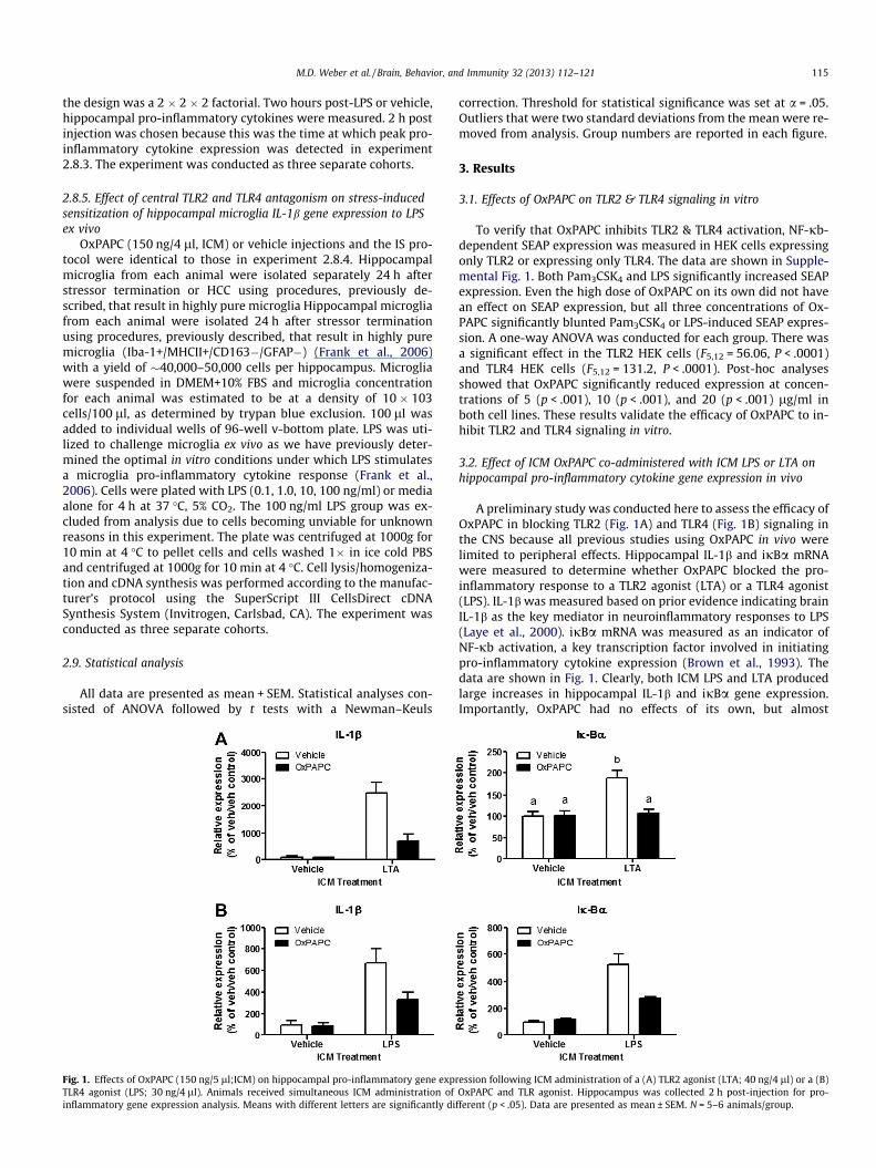
Fig. 2. Effects of OxPAPC (150 ng/5 ll;ICM) on pro-inflammatory gene expression followgene expression were measured 1, 2, and 4 h post treatment. Since the peak inflammatory1b and (D) ijBa gene expression in the liver. Means with different letters (a–c) are signifi
3.3. Effect of central TLR2 and TLR4 antagonism on peripheral LPS-induced cytokine production in vivo
To test whether blocking TLR2 and TLR4 activity in the brainwould reduce the neuroinflammatory response to systemic LPS,OxPAPC was administered ICM prior to peripheral administrationof LPS. Hippocampal IL-1b (Fig. 2A) and ijBa (Fig. 2B) mRNA wereexamined at three time points (1, 2, and 4 h) post-treatment. LiverIL-1b and ijBa was also measured 2 h post treatment as an indica-tor of peripheral inflammatory response (Fig. 2C). Peripheral LPSinduced robust increases in hippocampal IL-1b and ijBa mRNAsthat were evident 1 h after LPS, and were still present 4 h afterLPS. ICM OxPAPC again had no effects on its own, but completelyblocked the inflammatory mRNA increases at the 1 h timepoint
ing systemic LPS (10 lg/kg;ip) administration. Hippocampal (A) IL-1b and (B) ijBaresponse was 2 h post-LPS treatment, this time point was chosen to examine (C) IL-
cantly different (p < .05). Data are presented as mean ± SEM. N = 4–6 animals/group.
M.D. Weber et al. / Brain, Behavior, and Immunity 32 (2013) 112–121 117
after LPS, and reduced the mRNA increases at the later timepoints,suggesting that the impact of the drug was dissipating. Interest-ingly, intra-ICM OxPAPC reduced the liver increases produced bythe peripheral LPS. A 2 � 2 (OxPAPC/veh x LPS/veh) ANOVA wasconducted for each time point.
In the hippocampus, there was a significant main effect of Ox-PAPC and LPS on IL-1b gene expression at 1 h (F1,16 = 8.033,p < .05) and 2 h (F1,17 = 4.991, p < .05) post treatment. Similarly,there was also a main effect on ijBa at 1 h (F1,16 = 23.02,p < .001) and 2 h (F1,19 = 9.513, p < .01) post treatment. At thesetime points LPS administered without OxPAPC significantly in-creased IL-1b and ijBa expression, compared to veh/veh and Ox-PAPC/veh groups. Administration of OxPAPC with LPSsignificantly reduced IL-1b and ijBa mRNAs when compared tothe veh/LPS group. Additionally, IL-1b and ijBa gene expressiondid not differ between the OxPAPC/LPS and the veh/veh group.4 h post treatment, LPS significantly increased IL-1b (F1,12 = 7.759,p < .05) and ijBa (F1,12 = 54.89, p < .001) gene expression, but therewas no interaction between OxPAPC and LPS.
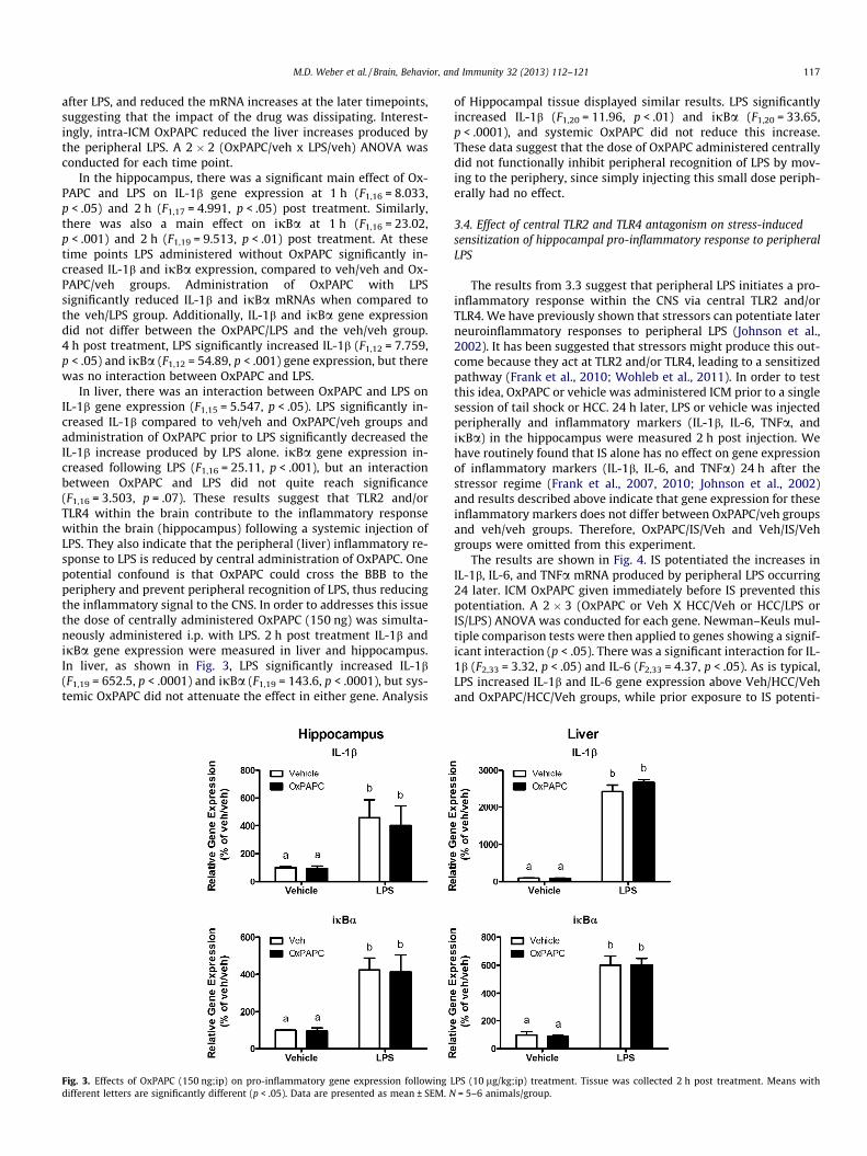
In liver, there was an interaction between OxPAPC and LPS onIL-1b gene expression (F1,15 = 5.547, p < .05). LPS significantly in-creased IL-1b compared to veh/veh and OxPAPC/veh groups andadministration of OxPAPC prior to LPS significantly decreased theIL-1b increase produced by LPS alone. ijBa gene expression in-creased following LPS (F1,16 = 25.11, p < .001), but an interactionbetween OxPAPC and LPS did not quite reach significance(F1,16 = 3.503, p = .07). These results suggest that TLR2 and/orTLR4 within the brain contribute to the inflammatory responsewithin the brain (hippocampus) following a systemic injection ofLPS. They also indicate that the peripheral (liver) inflammatory re-sponse to LPS is reduced by central administration of OxPAPC. Onepotential confound is that OxPAPC could cross the BBB to theperiphery and prevent peripheral recognition of LPS, thus reducingthe inflammatory signal to the CNS. In order to addresses this issuethe dose of centrally administered OxPAPC (150 ng) was simulta-neously administered i.p. with LPS. 2 h post treatment IL-1b andijBa gene expression were measured in liver and hippocampus.In liver, as shown in Fig. 3, LPS significantly increased IL-1b(F1,19 = 652.5, p < .0001) and ijBa (F1,19 = 143.6, p < .0001), but sys-temic OxPAPC did not attenuate the effect in either gene. Analysis
Fig. 3. Effects of OxPAPC (150 ng;ip) on pro-inflammatory gene expression following Ldifferent letters are significantly different (p < .05). Data are presented as mean ± SEM. N
of Hippocampal tissue displayed similar results. LPS significantlyincreased IL-1b (F1,20 = 11.96, p < .01) and ijBa (F1,20 = 33.65,p < .0001), and systemic OxPAPC did not reduce this increase.These data suggest that the dose of OxPAPC administered centrallydid not functionally inhibit peripheral recognition of LPS by mov-ing to the periphery, since simply injecting this small dose periph-erally had no effect.
3.4. Effect of central TLR2 and TLR4 antagonism on stress-inducedsensitization of hippocampal pro-inflammatory response to peripheralLPS
The results from 3.3 suggest that peripheral LPS initiates a pro-inflammatory response within the CNS via central TLR2 and/orTLR4. We have previously shown that stressors can potentiate laterneuroinflammatory responses to peripheral LPS (Johnson et al.,2002). It has been suggested that stressors might produce this out-come because they act at TLR2 and/or TLR4, leading to a sensitizedpathway (Frank et al., 2010; Wohleb et al., 2011). In order to testthis idea, OxPAPC or vehicle was administered ICM prior to a singlesession of tail shock or HCC. 24 h later, LPS or vehicle was injectedperipherally and inflammatory markers (IL-1b, IL-6, TNFa, andijBa) in the hippocampus were measured 2 h post injection. Wehave routinely found that IS alone has no effect on gene expressionof inflammatory markers (IL-1b, IL-6, and TNFa) 24 h after thestressor regime (Frank et al., 2007, 2010; Johnson et al., 2002)and results described above indicate that gene expression for theseinflammatory markers does not differ between OxPAPC/veh groupsand veh/veh groups. Therefore, OxPAPC/IS/Veh and Veh/IS/Vehgroups were omitted from this experiment.
The results are shown in Fig. 4. IS potentiated the increases inIL-1b, IL-6, and TNFa mRNA produced by peripheral LPS occurring24 later. ICM OxPAPC given immediately before IS prevented thispotentiation. A 2 � 3 (OxPAPC or Veh X HCC/Veh or HCC/LPS orIS/LPS) ANOVA was conducted for each gene. Newman–Keuls mul-tiple comparison tests were then applied to genes showing a signif-icant interaction (p < .05). There was a significant interaction for IL-1b (F2,33 = 3.32, p < .05) and IL-6 (F2,33 = 4.37, p < .05). As is typical,LPS increased IL-1b and IL-6 gene expression above Veh/HCC/Vehand OxPAPC/HCC/Veh groups, while prior exposure to IS potenti-
PS (10 lg/kg;ip) treatment. Tissue was collected 2 h post treatment. Means with= 5–6 animals/group.

118 M.D. Weber et al. / Brain, Behavior, and Immunity 32 (2013) 112–121
ated IL-1b and IL-6 following LPS, relative to animals that only re-ceived LPS. Interestingly, pretreatment with OxPAPC prior to ISprevented the exaggerated IL-1b and IL-6 mRNA responses toLPS. Animals that received OxPAPC then IS, and 24 h later receivedLPS, were significantly different from animals that had receivedVeh/IS/LPS, and did not differ from Veh/HCC/LPS or OxPAPC/HCC/LPS groups. Importantly, the OxPAPC/HCC/LPS group did not differfrom the Veh/HCC/LPS group, demonstrating that OxPAPC is notactively inhibiting the inflammatory response within the hippo-campus to systemic LPS 24 h after OxPAPC administration. TNFaexpression displayed a similar pattern to IL-1b and IL-6 expression,although an interaction between OxPAPC treatment and LPS withor without stress did not quite reach significance (F2,32 = 2.93,p = .06). Given that the pattern of expression for TNFa is highly cor-related with that of IL-1b and IL-6, and regulations of these genesare closely interconnected, post hoc tests were conducted on TNFagene expression as well. Similar to IL-1b and IL-6, LPS increasedTNFa expression and exposure to IS potentiated the response to
Fig. 4. Effects of the TLR2 and TLR4 antagonist OxPAPC on stress-induced sensitizedpro-inflammatory response to LPS in hippocampus. Animals were pretreated withOxPAPC (150 ng/5 ll;ICM) or vehicle. Animals were then exposed to inescapabletailshock (IS) or served as home cage controls (HCC). Animals were injected withLPS (10 lg/kg;i.p.) or vehicle 24 house post-IS exposure. Hippocampal IL-1b, IL-6,and TNFa were measured two hours post-LPS or vehicle injection. Means withdifferent letters are significantly different (p < .05). Data are presented as mean ± -SEM. N = 6–8 animals/group.
LPS. Administration of OxPAPC prior to IS prevented the exagger-ated response to LPS, which was similar to that in animals thatdid not experience IS. Lastly, there was no interaction for ijBagene expression (F2,34 = 3.285, p = .25).
3.5. Effect of central TLR2 and TLR4 antagonism on stress-inducedsensitization of hippocampal microglia IL-1b gene expression to LPSex vivo
We have previously demonstrated that microglia are a neuro-immune substrate for stress-induced potentiation of CNS pro-inflammatory immune responses (Frank et al., 2007). In order todetermine whether OxPAPC prevented stress-induced ‘priming’ ofmicroglial cells, OxPAPC was administered prior to stress and hip-pocampal microglia were isolated 24 h post stress. IL-1b geneexpression was measured as an indicator of an inflammatory re-sponse to LPS based on prior reports suggesting IL-1b as the keymediator in the neuroinflammatory response and ‘‘sickness behav-ior’’ following LPS exposure (Laye et al., 2000; Luheshi et al., 1996).As can be seen in Fig. 5, LPS increased IL-1b gene expression in aconcentration dependent manner in all experimental groups. Todetermine whether OxPAPC blunted stress-induced sensitizationof the microglial IL-1b gene response to LPS challenge, area underthe LPS concentration curve (AUC) was computed for each subjectas an indicator of the overall LPS response, and a two-way ANOVAdetermined the interaction between OxPAPC treatment and stress.In HCC animals, IS significantly potentiated the microglial IL-1b re-sponse, which was completely blocked by prior OxPAPC treatment(F1,18 = 5.651, p < .05). Prior treatment with OxPAPC did not affectIL-1b gene response to LPS in HCC animals.
4. Discussion
The data from the present set of experiments implicate TLR2and/or TLR4 as a mediator of stress-induced priming of neuroin-flammatory responses to subsequent inflammatory challenges.Pharmacological (OxPAPC) antagonism of TLR2 and TLR4 duringthe experience of stress prevented a primed hippocampal inflam-matory response (IL-1b, IL-6, and TNFa) to a subsequent peripheralLPS challenge 24 h later. In addition, in vivo administration of Ox-PAPC prior to IS prevented the sensitized response to LPS adminis-tered directly to isolated microglial cells ex vivo, further supportingthe idea that microglia are a neuroimmune substrate for stress-in-duced TLR2 and TLR4 activity. These conclusions are consistentwith previous findings demonstrating that microglia become acti-vated or primed following exposure to stress or increased GCs(Espinosa-Oliva et al., 2011; Frank et al., 2007, 2012; Nair and Bon-neau, 2006; Wohleb et al., 2011).
The oxidized phospholipid (OxPL), OxPAPC, was used to blockTLR2 and TLR4 signaling. In the past, OxPLs were primarily knownas augmenters of inflammatory events. However, a recent litera-ture shows that OxPLs possess a wide array of anti-inflammatoryeffects as well, particularly at lower concentrations (Erridgeet al., 2008; Oskolkova et al., 2010; Starosta et al., 2012; vonSchlieffen et al., 2009). In particular, OxPAPC has been show to in-hibit TLR2 and TLR4 dependent signaling by competing with theextracellular binding proteins CD-14 and MD-2 at a concentrationup to 50 lg/ml, but becomes toxic at higher concentrations (100–300 lg/ml) (Erridge et al., 2008). Further, we have conductedin vitro work indicating that OxPAPC directly blocks TLR2 andTLR4 dependent NF-jb signaling (Supplemental Fig. 1). In vitrostudies have also shown that OxPAPC does not inhibit signaling in-duced by any other TLR agonist, demonstrating specificity to TLR2and TLR4 (Erridge et al., 2008). To date, in vivo characterization ofthis drug has been limited to studies within the periphery and it

Fig. 5. Effects of OxPAPC on stress-induced sensitization of microglial IL-1b response to LPS ex vivo. Animals were pretreated with OxPAPC (150 ng/5 ll;ICM) or vehicle.Animals were then exposed to inescapable tailshock (IS) or served as home cage controls (HCC). Twenty-four hours post-IS exposure, microglia were isolated fromhippocampus and challenged with LPS (0, 0.1, 1, and 10 ng/ml) for 4 h and microglial IL-1b gene expression was measured. LPS increased IL-1b gene expression in aconcentration dependent manner in all experimental groups. To determine whether OxPAPC treatment blunted stress-induced sensitization of the microglial IL-1b responseto LPS challenge, area under the LPS concentration curve (AUC) was computed for each animal and means compared. Means with different letters are significantly different(p < .05). Data are presented as mean ± SEM. N = 5–6 animals/group.
M.D. Weber et al. / Brain, Behavior, and Immunity 32 (2013) 112–121 119
has never been functionally characterized within the CNS. The datafrom the present set of experiments demonstrates that centrallyadministered OxPAPC, at a concentration of 30 lg/ml, blocks theneuroinflammatory response (IL-1b & ijBa) to a centrally adminis-tered TLR2 agonist (LTA) and a TLR4 agonist (LPS). Although it isclear that OxPAPC inhibits TLR2 and TLR4 signaling, it is evidentthat other pathways are involved in the anti-inflammatory effectsof OxPAPC. For example, previous studies have shown that OxPAPCcan initiate adaptive antioxidant defenses in vascular cells, includ-ing activation of the Nrf2 pathway that leads to anti-oxidant re-sponse element (ARE) binding of glutamate-cysteine ligasemodifier and catalytic (GCLM and GCLC, respectively) and hemeoxygenase (HO)-1 (Jyrkkanen et al., 2008). Other transcription fac-tors including activation transcription factor (ATF) 3 are also in-creased by OxPAPC, particularly at high concentrations (>80 lM)(Oskolkova et al., 2008) As mentioned above, OxPAPC does notinterfere with other TLR signaling, demonstrating specificity toTLR2 and TLR4 (Erridge et al., 2008). However, effects at non-TLRlocations cannot be ruled out, and this should be noted.
As previously discussed, exposure to acute stress primes theneuroinflammatory response to peripheral LPS. LPS is recognizedby TLR4, however, systemic LPS does not cross the BBB. Initialinflammatory responses within the brain can derive from cells atthe vascular interface of the BBB and circumventricular organs(Quan et al., 1998; Singh and Jiang, 2004; Vitkovic et al., 2000),which can trigger a series of inflammatory events that result in asustained neuroinflammatory response. The signaling that main-tains inflammation within the brain may not be dependent onTLR4 recognition inside of the parenchyma and is currently notfully understood. In the present study, central administration ofOxPAPC attenuated central (hippocampal) and peripheral (liver)pro-inflammatory gene expression to a simultaneous injection ofsystemic (ip) LPS. To verify that OxPAPC did not diffuse into theperiphery and block initial recognition of LPS, the same dose of Ox-PAPC was administered ip and was not effective in preventing aninflammatory response. This suggests that TLR2 and/or TLR4 lo-cated in the brain is critical for the peripheral-to-central signalingthat occurs following peripheral LPS administration. Of course,these data do not address the question of what the ligand(s) withinthe brain for these receptors might be.
Since the half life of OxPAPC is unknown, one potential con-founding factor in the blockade of stress-induced priming foundhere is that OxPAPC might still have been functional 24 h afteradministration, and so, was merely attenuating the neuroinflam-matory response to the systemic LPS injection, not necessarily pre-venting stress-induced exaggerated neuroinflammatory responses.It should be noted that the neuroinflammatory response (IL-1b, IL-
6, and TNFa) from HCC animals that received OxPAPC and 24 h la-ter were administered LPS did not differ from the HCC animals thatwere given a saline injection and 24 h later administered LPS, sug-gesting that OxPAPC is no longer functional at that time. In addi-tion, an ex vivo approach was taken to examine the ‘state’ ofhippocampal microglia following in vivo treatment with OxPAPCand IS. Hippocampal microglia were isolated 24 h after OxPAPCand IS treatment. LPS was used to stimulate the cells ex vivo toprobe the ‘state’ of the microglia (i.e., are they sensitized to LPS).Prior administration of OxPAPC prevented the sensitized inflam-matory response due to stress, while maintaining the ‘normal’inflammatory response to LPS treatment. Since OxPAPC is no long-er blocking TLR2 and TLR4 signaling 24 h post injection, the onlyperiod of time in which OxPAPC could functionally inhibit TLR2and TLR4 signaling is during, and directly following, tail shock. Thissuggests that sometime between the experience of tail shock andthe LPS challenge, an unidentified ligand binds to, and activates,TLR2 and/or TLR4, which drives the neuroinflammatory microenvi-ronment to a ‘primed’ or ‘sensitized’ state, resulting in exaggeratedinflammatory responses if further stimulated, in this case, withLPS.
The present results may help to understand how stressors sen-sitize inflammatory reactions to a later inflammatory challenge.Although this set of experiments does not identify a potential li-gand(s), it does demonstrate that the TLR2 and or TLR4 receptorare likely involved. Interestingly, a new perspective comes fromfindings that TLRs can be activated by endogenous molecules thatare synthesized and secreted in response to ‘‘danger’’. These mole-cules have been called ‘‘alarmins’’ (Bianchi, 2007; Klune et al.,2008). Alarmins have similar characteristics to PAMPS, such asLPS, meaning that they can activate TLRs and initiate neuroinflam-matory responses (Bianchi, 2007). Of these alarmins, HMGB1 isknown to activate TLR2 and TLR4 and produce the full array ofinflammatory responses, including NF-jb activity and synthesisof inflammatory cytokines (Mazarati et al., 2011; Park et al.,2004; Yang and Tracey, 2009; Yang et al., 2005). Activation ofNF-jb via TLRs induces the formation of a multiprotein signalingcomplex known as the inflammasome (Leemans et al., 2011). Theinflammasome involves members of the nod-like receptor family(NLRs), with NLRP3 being of particular relevance here. Assemblyand activation of the NLRP3 inflammasome is key for cleavingpro-caspase-1 to form the mature and active pro-caspase-1, whichin turn cleaves pro-IL-1b to form mature IL-1b, resulting in extra-cellular release (Martinon et al., 2009). Formation of the NLRP3inflammasome requires a ‘priming’ signal, such as TLR activation,leading to NLRP3 transcription. A secondary signal is required toassemble the inflammasome, leading to IL-1b maturation (Kersse

120 M.D. Weber et al. / Brain, Behavior, and Immunity 32 (2013) 112–121
et al., 2011). One possibility is that stress or stress-induced GCs,initiates the ‘priming signal’ that induces NLRP3 transcriptionthrough activity at TLR2 and/or TLR4 via endogenous ‘alarmins’such as HMGB-1. A subsequent inflammatory challenge, such asLPS, then assembles the inflammasome resulting in an exaggeratedinflammatory response. At this point, this idea is purely specula-tion. However, there is some evidence the GCs might function inthis way. Busillo et al. (2011) found that in vitro, GCs increaseNLRP3 transcription and protein, thereby priming NLRP3 inflam-masome formation to a subsequent stimulus such as LPS or ATP,resulting in a potentiated pro-inflammatory cytokine response.
In sum, the present results suggest that exposure to an acutestressor ‘primes’ the CNS innate immune system via a signal thatactivates TLR2 and/or TLR4. This signal(s) may include endogenousdanger signals that are known to be released in response to a widearray of stimuli including infection or sterile injury. Further inves-tigation is required to identify potential signals and determine thecellular processes that drive stress-induced ‘priming’ of innate im-mune function.
Acknowledgment
This work is supported by NIH grant # R21MH096224.
Appendix A. Supplementary data
Supplementary data associated with this article can be found, inthe online version, at http://dx.doi.org/10.1016/j.bbi.2013.03.004.
References
Altschul, S.F., Madden, T.L., Schaffer, A.A., Zhang, J., Zhang, Z., Miller, W., Lipman, D.J.,1997. Gapped BLAST and PSI-BLAST: a new generation of protein databasesearch programs. Nucleic Acids Res. 25, 3389–3402.
Banks, W.A., Robinson, S.M., 2010. Minimal penetration of lipopolysaccharide acrossthe murine blood–brain barrier. Brain Behav. Immun. 24, 102–109.
Bianchi, M.E., 2007. DAMPs, PAMPs and alarmins: all we need to know aboutdanger. J. Leukoc. Biol. 81, 1–5.
Boumpas, D.T., Chrousos, G.P., Wilder, R.L., Cupps, T.R., Balow, J.E., 1993.Glucocorticoid therapy for immune-mediated diseases: basic and clinicalcorrelates. Ann. Intern. Med. 119, 1198–1208.
Brown, K., Park, S., Kanno, T., Franzoso, G., Siebenlist, U., 1993. Mutual regulation ofthe transcriptional activator NF-kappa B and its inhibitor, I kappa B-alpha. Proc.Natl. Acad. Sci. USA 90, 2532–2536.
Busillo, J.M., Azzam, K.M., Cidlowski, J.A., 2011. Glucocorticoids sensitize the innateimmune system through regulation of the NLRP3 inflammasome. J. Biol. Chem.286, 38703–38713.
Chomczynski, P., Sacchi, N., 1987. Single-step method of RNA isolation by acidguanidinium thiocyanate-phenol-chloroform extraction. Anal. Biochem. 162,156–159.
de Pablos, R.M., Villaran, R.F., Arguelles, S., Herrera, A.J., Venero, J.L., Ayala, A., Cano,J., Machado, A., 2006. Stress increases vulnerability to inflammation in the ratprefrontal cortex. J. Neurosci. 26, 5709–5719.
Erridge, C., Kennedy, S., Spickett, C.M., Webb, D.J., 2008. Oxidized phospholipidinhibition of toll-like receptor (TLR) signaling is restricted to TLR2 and TLR4:roles for CD14, LPS-binding protein, and MD2 as targets for specificity ofinhibition. J. Biol. Chem. 283, 24748–24759.
Espinosa-Oliva, A.M., de Pablos, R.M., Villaran, R.F., Arguelles, S., Venero, J.L.,Machado, A., Cano, J., 2011. Stress is critical for LPS-induced activation ofmicroglia and damage in the rat hippocampus. Neurobiol. Aging 32, 85–102.
Frank, M.G., Wieseler-Frank, J.L., Watkins, L.R., Maier, S.F., 2006. Rapid isolation ofhighly enriched and quiescent microglia from adult rat hippocampus:immunophenotypic and functional characteristics. J. Neurosci. Methods 151,121–130.
Frank, M.G., Baratta, M.V., Sprunger, D.B., Watkins, L.R., Maier, S.F., 2007. Microgliaserve as a neuroimmune substrate for stress-induced potentiation of CNS pro-inflammatory cytokine responses. Brain Behav. Immun. 21, 47–59.
Frank, M.G., Miguel, Z.D., Watkins, L.R., Maier, S.F., 2010. Prior exposure toglucocorticoids sensitizes the neuroinflammatory and peripheralinflammatory responses to E. coli lipopolysaccharide. Brain Behav. Immun. 24,19–30.
Frank, M.G., Thompson, B.M., Watkins, L.R., Maier, S.F., 2012. Glucocorticoidsmediate stress-induced priming of microglial pro-inflammatory responses.Brain Behav. Immun. 26, 337–345.
Gehrmann, J., Matsumoto, Y., Kreutzberg, G.W., 1995. Microglia: intrinsicimmuneffector cell of the brain. Brain Res. Brain Res. Rev. 20, 269–287.
Hermoso, M.A., Matsuguchi, T., Smoak, K., Cidlowski, J.A., 2004. Glucocorticoids andtumor necrosis factor alpha cooperatively regulate toll-like receptor 2 geneexpression. Mol. Cell Biol. 24, 4743–4756.
Holguin, A., Frank, M.G., Biedenkapp, J.C., Nelson, K., Lippert, D., Watkins, L.R., Rudy,J.W., Maier, S.F., 2007. Characterization of the temporo-spatial effects of chronicbilateral intrahippocampal cannulae on interleukin-1beta. J. Neurosci. Methods161, 265–272.
Hutchinson, M.R., Loram, L.C., Zhang, Y., Shridhar, M., Rezvani, N., Berkelhammer, D.,Phipps, S., Foster, P.S., Landgraf, K., Falke, J.J., Rice, K.C., Maier, S.F., Yin, H.,Watkins, L.R., 2010. Evidence that tricyclic small molecules may possess toll-like receptor and myeloid differentiation protein 2 activity. Neuroscience 168,551–563.
Imasato, A., Desbois-Mouthon, C., Han, J., Kai, H., Cato, A.C., Akira, S., Li, J.D., 2002.Inhibition of p38 MAPK by glucocorticoids via induction of MAPK phosphatase-1 enhances nontypeable Haemophilus influenzae-induced expression of toll-like receptor 2. J. Biol. Chem. 277, 47444–47450.
Johnson, J.D., O’Connor, K.A., Deak, T., Stark, M., Watkins, L.R., Maier, S.F., 2002. Priorstressor exposure sensitizes LPS-induced cytokine production. Brain Behav.Immun. 16, 461–476.
Johnson, J.D., O’Connor, K.A., Hansen, M.K., Watkins, L.R., Maier, S.F., 2003. Effects ofprior stress on LPS-induced cytokine and sickness responses. Am. J. Physiol.Regul. Integr. Comp. Physiol. 284, R422–R432.
Johnson, J.D., O’Connor, K.A., Watkins, L.R., Maier, S.F., 2004. The role of IL-1[beta] instress-induced sensitization of proinflammatory cytokine and corticosteroneresponses. Neuroscience 127, 569–577.
Jyrkkanen, H.K., Kansanen, E., Inkala, M., Kivela, A.M., Hurttila, H., Heinonen, S.E.,Goldsteins, G., Jauhiainen, S., Tiainen, S., Makkonen, H., Oskolkova, O.,Afonyushkin, T., Koistinaho, J., Yamamoto, M., Bochkov, V.N., Yla-Herttuala, S.,Levonen, A.L., 2008. Nrf2 regulates antioxidant gene expression evoked byoxidized phospholipids in endothelial cells and murine arteries in vivo. Circ.Res. 103, e1–e9.
Kawai, T., Akira, S., 2007. Signaling to NF-kappaB by toll-like receptors. Trends Mol.Med. 13, 460–469.
Kersse, K., Bertrand, M.J., Lamkanfi, M., Vandenabeele, P., 2011. NOD-like receptorsand the innate immune system: coping with danger, damage and death.Cytokine Growth Factor Rev. 22, 257–276.
Klune, J.R., Dhupar, R., Cardinal, J., Billiar, T.R., Tsung, A., 2008. HMGB1: endogenousdanger signaling. Mol. Med. 14, 476–484.
Laye, S., Gheusi, G., Cremona, S., Combe, C., Kelley, K., Dantzer, R., Parnet, P., 2000.Endogenous brain IL-1 mediates LPS-induced anorexia and hypothalamiccytokine expression. Am. J. Physiol. Regul. Integr. Comp. Physiol. 279, R93–R98.
Leemans, J.C., Cassel, S.L., Sutterwala, F.S., 2011. Sensing damage by the NLRP3inflammasome. Immunol. Rev. 243, 152–162.
Livak, K.J., Schmittgen, T.D., 2001. Analysis of relative gene expression data usingreal-time quantitative PCR and the 2(-Delta Delta C(T)) method. Methods 25,402–408.
Luheshi, G., Miller, A.J., Brouwer, S., Dascombe, M.J., Rothwell, N.J., Hopkins, S.J.,1996. Interleukin-1 receptor antagonist inhibits endotoxin fever and systemicinterleukin-6 induction in the rat. Am. J. Physiol. 270, E91–E95.
Martinon, F., Mayor, A., Tschopp, J., 2009. The inflammasomes: guardians of thebody. Annu. Rev. Immunol. 27, 229–265.
Mazarati, A., Maroso, M., Iori, V., Vezzani, A., Carli, M., 2011. High-mobility groupbox-1 impairs memory in mice through both toll-like receptor 4 and receptorfor advanced glycation end products. Exp. Neurol. 232, 143–148.
Munhoz, C.D., Lepsch, L.B., Kawamoto, E.M., Malta, M.B., Lima lde, S., Avellar, M.C.,Sapolsky, R.M., Scavone, C., 2006. Chronic unpredictable stress exacerbateslipopolysaccharide-induced activation of nuclear factor-kappaB in the frontalcortex and hippocampus via glucocorticoid secretion. J. Neurosci. 26, 3813–3820.
Munhoz, C.D., Sorrells, S.F., Caso, J.R., Scavone, C., Sapolsky, R.M., 2010.Glucocorticoids exacerbate lipopolysaccharide-induced signaling in thefrontal cortex and hippocampus in a dose-dependent manner. J. Neurosci. 30,13690–13698.
Nair, A., Bonneau, R.H., 2006. Stress-induced elevation of glucocorticoids increasesmicroglia proliferation through NMDA receptor activation. J. Neuroimmunol.171, 72–85.
Nair, A., Hunzeker, J., Bonneau, R.H., 2007. Modulation of microglia and CD8+ T cellactivation during the development of stress-induced herpes simplex virus type-1 encephalitis. Brain Behav. Immun. 21, 791–806.
Oskolkova, O.V., Afonyushkin, T., Leitner, A., von Schlieffen, E., Gargalovic, P.S., Lusis,A.J., Binder, B.R., Bochkov, V.N., 2008. ATF4-dependent transcription is a keymechanism in VEGF up-regulation by oxidized phospholipids: critical role ofoxidized sn-2 residues in activation of unfolded protein response. Blood 112,330–339.
Oskolkova, O.V., Afonyushkin, T., Preinerstorfer, B., Bicker, W., von Schlieffen, E.,Hainzl, E., Demyanets, S., Schabbauer, G., Lindner, W., Tselepis, A.D., Wojta, J.,Binder, B.R., Bochkov, V.N., 2010. Oxidized phospholipids are more potentantagonists of lipopolysaccharide than inducers of inflammation. J. Immunol.185, 7706–7712.
Park, J.S., Svetkauskaite, D., He, Q., Kim, J.Y., Strassheim, D., Ishizaka, A., Abraham, E.,2004. Involvement of toll-like receptors 2 and 4 in cellular activation by highmobility group box 1 protein. J. Biol. Chem. 279, 7370–7377.
Quan, N., Whiteside, M., Herkenham, M., 1998. Time course and localizationpatterns of interleukin-1b messenger rna expression in brain and pituitaryafter peripheral administration of lipopolysaccharide. Neuroscience 83, 281–293.

M.D. Weber et al. / Brain, Behavior, and Immunity 32 (2013) 112–121 121
Ransohoff, R.M., Perry, V.H., 2009. Microglial physiology: unique stimuli, specializedresponses. Annu. Rev. Immunol. 27, 119–145.
Rozkova, D., Horvath, R., Bartunkova, J., Spisek, R., 2006. Glucocorticoids severelyimpair differentiation and antigen presenting function of dendritic cells despiteupregulation of toll-like receptors. Clin. Immunol. 120, 260–271.
Salminen, A., Huuskonen, J., Ojala, J., Kauppinen, A., Kaarniranta, K., Suuronen, T.,2008. Activation of innate immunity system during aging: NF-kB signaling isthe molecular culprit of inflamm-aging. Ageing Res. Rev. 7, 83–105.
Singh, A.K., Jiang, Y., 2004. How does peripheral lipopolysaccharide induce geneexpression in the brain of rats? Toxicology 201, 197–207.
Sorrells, S.F., Sapolsky, R.M., 2007. An inflammatory review of glucocorticoid actionsin the CNS. Brain Behav. Immun. 21, 259–272.
Starosta, V., Wu, T., Zimman, A., Pham, D., Tian, X., Oskolkova, O., Bochkov, V.,Berliner, J.A., Birukova, A.A., Birukov, K.G., 2012. Differential regulation ofendothelial cell permeability by high and low doses of oxidized 1-palmitoyl-2-arachidonyl-sn-glycero-3-phosphocholine. Am. J. Respir. Cell Mol. Biol. 46, 331–341.
Vitkovic, L., Konsman, J.P., Bockaert, J., Dantzer, R., Homburger, V., Jacque, C., 2000.Cytokine signals propagate through the brain. Mol. Psychiatry 5, 604–615.
von Schlieffen, E., Oskolkova, O.V., Schabbauer, G., Gruber, F., Bluml, S., Genest, M.,Kadl, A., Marsik, C., Knapp, S., Chow, J., Leitinger, N., Binder, B.R., Bochkov, V.N.,2009. Multi-hit inhibition of circulating and cell-associated components of thetoll-like receptor 4 pathway by oxidized phospholipids. Arterioscler. Thromb.Vasc. Biol. 29, 356–362.
Wohleb, E.S., Hanke, M.L., Corona, A.W., Powell, N.D., Stiner, L.M., Bailey, M.T.,Nelson, R.J., Godbout, J.P., Sheridan, J.F., 2011. Beta-adrenergic receptorantagonism prevents anxiety-like behavior and microglial reactivity inducedby repeated social defeat. J. Neurosci. 31, 6277–6288.
Yang, H., Tracey, K.J., 2009. Targeting HMGB1 in inflammation. Biochim. Biophys.Acta Gene Regul. Mech. 1799, 149–156.
Yang, H., Wang, H., Czura, C.J., Tracey, K.J., 2005. The cytokine activity of HMGB1. J.Leukoc. Biol. 78, 1–8.
Top Related
Copyright © 2022 FDOKUMEN

