


Bahasa
Halaman
Hukum

�������� ����� ��
Aerobic exercise improves hippocampal function and increases BDNF in theserum of young adult males
Eadaoin W. Griffin, Sinead Mulally, Carole Foley, Stuart A. Warming-ton, Shane M. O’Mara, Aine M. Kelly
PII: S0031-9384(11)00308-8DOI: doi: 10.1016/j.physbeh.2011.06.005Reference: PHB 9518
To appear in: Physiology & Behavior
Received date: 29 October 2009Revised date: 1 June 2011Accepted date: 6 June 2011
Please cite this article as: Griffin Eadaoin W., Mulally Sinead, Foley Carole, WarmingtonStuart A., O’Mara Shane M., Kelly Aine M., Aerobic exercise improves hippocampalfunction and increases BDNF in the serum of young adult males, Physiology & Behavior(2011), doi: 10.1016/j.physbeh.2011.06.005
This is a PDF file of an unedited manuscript that has been accepted for publication.As a service to our customers we are providing this early version of the manuscript.The manuscript will undergo copyediting, typesetting, and review of the resulting proofbefore it is published in its final form. Please note that during the production processerrors may be discovered which could affect the content, and all legal disclaimers thatapply to the journal pertain.
ACC
EPTE
D M
ANU
SCR
IPT
ACCEPTED MANUSCRIPT
1
Aerobic exercise improves hippocampal function and increases BDNF in the
serum of young adult males
Éadaoin W. Griffin1,3, Sinéad Mulally2,3, Carole Foley1, Stuart A. Warmington1,4,
Shane M. O’Mara2,3 and Áine M. Kelly1,3*
Department of Physiology, School of Medicine1, School of Psychology2 and Trinity
College Institute of Neuroscience3, University of Dublin, Trinity College, Dublin 2,
Ireland. School of Exercise and Nutrition Sciences, Deakin University, Burwood
3125, Victoria, Australia4.
Running title: Exercise increases learning and BDNF
Number of (i) Pages=31 (ii) Figures=6
Number of words in (i) Manuscript=4794 (ii) Abstract=200; (iii) Introduction=436;
*Corresponding author: E-mail: [email protected]; Phone: 0035318963794; Fax:
00353 16793545
ACC
EPTE
D M
ANU
SCR
IPT
ACCEPTED MANUSCRIPT
2
Abstract
Physical activity has been reported to improve cognitive function in humans and
rodents, possibly via a brain-derived neurotrophic factor (BDNF)-regulated
mechanism. In this study of human subjects, we have assessed the effects of
acute and chronic exercise on performance of a face-name matching task,
which recruits the hippocampus and associated structures of the medial
temporal lobe, and the Stroop word-colour task, which does not, and have
assessed circulating concentrations of BDNF and IGF-1 in parallel. The results
show that a short period of high-intensity cycling results in enhancements in
performance of the face-name matching, but not the Stroop, task. These
changes in cognitive function were paralleled by increased concentration of
BDNF, but not IGF-1, in the serum of exercising subjects. 3 weeks of cycling
training had no effect on cardiovascular fitness, as assessed by VO2 scores,
cognitive function, or serum BDNF concentration. Increases in fitness, cognitive
function and serum BDNF response to acute exercise were observed following
5 weeks of aerobic training. These data indicate that both acute and chronic
exercise improve medial temporal lobe function concomitant with increased
concentrations of BDNF in the serum, suggesting a possible functional role for
this neurotrophic factor in exercise-induced cognitive enhancement in humans.
Keywords: Exercise; learning; BDNF; hippocampus
ACC
EPTE
D M
ANU
SCR
IPT
ACCEPTED MANUSCRIPT
3
Introduction
The benefits that physical activity confers on cardiovascular health are
well known, while recent evidence has also demonstrated the ability of exercise
to promote brain health. The evidence that physically active older people,
particularly those that have been active throughout their lifespan, are at
decreased risk of developing Alzheimer’s disease and other forms of dementia
relative to their sedentary counterparts [1-3] strongly suggests that exercise
may be a powerful protective strategy against age-related neurodegenerative
decline. In addition to its neuroprotective actions, exercise enhances cognitive
function in elderly people and slows the progression of dementia-related
cognitive symptoms [4-6]. Thus, exercise may reduce the risk of developing
dementia or ameliorate cognitive impairment already present in those suffering
from neurodegenerative decline.
Moreover, exercise may also enhance cognitive function in young,
healthy, adults. High impact running has been shown to improve vocabulary
learning [7], while cycling has been shown to improve performance in a map
recognition task [8] and in the Stroop word-colour task [9]. However, Grego et
al. [8] also showed that prolonged exercise leading to fatigue compromises
cognitive function. It has been suggested that intense exercise may facilitate
aspects of cognitive function in a manner dependent on an individual’s
cardiovascular fitness [10]. A recent meta-analysis indicates that cognitive
performance may be enhanced or impaired depending on when, relative to an
acute exercise bout, performance is measured, the type of cognitive task
selected, and the type of exercise performed [11].
ACC
EPTE
D M
ANU
SCR
IPT
ACCEPTED MANUSCRIPT
4
Evidence available from animal studies provides some insight into the
mechanisms by which exercise may enhance cognition. In rodent models,
exercise has consistently been shown to enhance learning and persistently
upregulate expression of brain-derived neurotrophic factor (BDNF) in the
hippocampus [12-16]. The indisputable importance of the hippocampus in
learning and memory and the role of BDNF in mediating hippocampal synaptic
plasticity are well established [17-20]; while additional evidence indicates a role
for insulin-like growth factor (IGF-1) in mediating the cognitive effects of
exercise [21-23]. Interestingly, serum BDNF concentration has repeatedly been
reported to increase following exercise in humans [9, 24-26] (for review see
[27]), while IGF-1 responses to exercise are more variable [28-30].
Here, we have investigated the effect of acute exercise and aerobic
exercise training on cognitive function in young, sedentary men. Given the
evidence from animal models that hippocampal function is particularly
responsive to exercise intervention, we assessed the impact of acute and
aerobic exercise training on performance in a face-name matching task that
recruits the hippocampus, and also on circulating concentrations of BDNF and
IGF-1, in an attempt to investigate the possible causal links between increased
availability of these growth factors and enhancements in cognitive function.
ACC
EPTE
D M
ANU
SCR
IPT
ACCEPTED MANUSCRIPT
5
Materials and Methods
Participants
The experimental protocol was approved by the Ethical Committee for
Research Involving Human Participants, Faculty of Health Sciences, Trinity
College Dublin. Forty-seven healthy male students volunteered to participate
(age, height, weight: 22 ± 2 yrs, 180 ± 7 cm, 82 ± 11 kg respectively, mean ±
SD). All subjects were sedentary (not involved in any regular physical training)
prior to commencement of the study, and each received a routine medical
examination before providing written informed consent in accordance with the
declaration of Helsinki. Exclusion criteria included any contraindications to
intense exercise discovered during the medical examination, intake of
prescription medication, history of neurological problems, pre-existing injuries,
smoking and intake of recreational drugs. All subjects were required to fast for 2
hours and to refrain from consumption of caffeine for 12 hours prior to testing.
Experimental protocol
Participants were allocated to an exercise group (EX) or a sedentary control
group (CON; Fig 1b). During the first testing session all participants performed
a set of cognitive tasks, including the face-name matching task and the Stroop
task. Following this, the EX group completed a graded exercise test, which
served as the acute exercise bout, while CON participants had a 30min rest
(Fig 1a). Blood samples were collected from the EX group throughout the
testing session, in order to assess the effect of acute exercise on serum BDNF
and IGF-1 concentrations. A baseline blood sample was taken at 0min, followed
ACC
EPTE
D M
ANU
SCR
IPT
ACCEPTED MANUSCRIPT
6
by the first set of cognitive tests. Another blood sample was taken at 30min,
prior to the graded exercise test (30min rest period for CON). A third, post-
acute exercise, blood sample was taken at 60min, the second set of cognitive
tests was completed and a final blood sample was drawn at 90min (Figure1a).
Two chronic exercise protocols were used to assess the effects of both a 3-
week and a 5-week aerobic training intervention on cognition and on serum
concentrations of BDNF and IGF-1. These aerobic training programmes were
identical in all aspects except for duration. Following the first testing session the
EX group was split into the subgroups: C-EX3, C-EX5, A-EX3 & A-EX5 (Fig
1a). The chronic-exercise subgroups, C-EX3 and C-EX5, completed 3 and 5
weeks of aerobic training respectively. The acute-exercise subgroups; A-EX3
and A-EX5, remained sedentary for the corresponding 3 and 5-week intervals
and exercised only when performing the graded exercise test. All subgroups
repeated the testing session (session 2, as in Fig 1a) following the appropriate
3 or 5-week interval. The CON group remained sedentary both during the
testing sessions and during the 3-week or 5-week interval between sessions.
Acute exercise
The acute exercise protocol consisted of a graded exercise test (GXT) to
volitional exhaustion, performed on a stationary cycle ergometer (Lode
Excalibur, Groningen, Netherlands), to establish maximal oxygen consumption
rate (VO2 max). The initial workload was set at 75W and increased by 50W
increments every 3min, until 9min, and subsequently by 25W increments each
min until volitional exhaustion was reached. The subject wore a facemask
throughout the test in order to collect expired air, which was analysed for
ACC
EPTE
D M
ANU
SCR
IPT
ACCEPTED MANUSCRIPT
7
volume and gas composition using an online system (Metalyser, Cortex
Biophysik, Leipzig, Germany) and from which VO2 max was determined as a
measure of aerobic fitness. Subjects wore a heart rate monitor (Polar, Oulu,
Finland) throughout the test and were verbally encouraged to continue cycling
until heart rate (beats.min-1) approached the age predicted maximum (220 –
age (yrs)).
Cognitive testing
Subjects were seated before a computer screen and were tested using the
Face-Name matching task, followed immediately by the Stroop Word-Colour
task.
Face-Name matching task
The face-name task was adapted from a paradigm by Zeineh et al. [31] and
included encoding, distractor and recall phases. During the encoding block the
subject was presented with images of ten unfamiliar faces paired with names.
Each was presented in sequence for 3.5 s via a computer screen display. A 40
s distractor task was performed between each encoding and recall phase to
prevent rote rehearsal of the face-name associations. During the distractor task,
a fixation cross was presented on the screen at random intervals (0.5 to 3.5 s),
and was replaced for 300 ms on screen by a black circle; the subject was
required to press a response button on the keypad when presented with a black
circle. The recall block consisted of a randomized presentation of the
previously-viewed faces but without the paired names. Subjects were requested
to recall the correct name and communicate it verbally to the experimenter.
ACC
EPTE
D M
ANU
SCR
IPT
ACCEPTED MANUSCRIPT
8
Subjects viewed the same face-name combinations four times per task,
providing a maximum possible score of 40. With each subject eventually
completing two testing sessions comprising two cognitive trials, four different
series of faces and names were used during the study, with no subject
repeating the cognitive assessment on the same series of faces. Results are
presented as the number of pairs recalled.
Stroop Word-Colour task
The Stroop word-colour task consisted of 2 trials of equal duration with a short
(<1 min) inter-trial rest period. Participants were presented with a series of
colour words (red, yellow, green, blue) on a computer screen. Words were
presented in the same (congruent) or different (incongruent) coloured font.
Words appeared on screen for 1.3 s. Subjects were required to inhibit their
automatic response to read the word stimulus presented, and instead to report
the font colour in which words were presented by pressing colour-coded
buttons on a response pad. The protocol consisted of frequent congruent
stimuli with randomized infrequent incongruent stimuli, in order to maximize
cognitive interference. Results are presented as percentage response
accuracy. Tasks were programmed and run using E-Prime version 1.1 software
(Psychology Software Tools, Pittsburgh Tools, Pittsburgh, USA).
Aerobic Training
The chronic-exercise groups, C-EX3 and C-EX5 were required to attend the lab
for supervised aerobic cycle training three times per week until session 2.
Training was performed on a stationary cycle ergometer for between 30 and 60
min per session. The workload and duration of the exercise were increased
ACC
EPTE
D M
ANU
SCR
IPT
ACCEPTED MANUSCRIPT
9
gradually until subjects could maintain a workload estimated to require 60%
VO2max for 60 min. Heart rate was monitored during each training session as a
second method of ensuring training intensity was near to the expected workload
and as such that the training was sub-maximal and progressive over the entire
training period (3 or 5 weeks).
Blood sampling
During each testing session venous blood samples were obtained at t=0, 30, 60
and 90 min via an indwelling catheter located in a forearm vein. Following each
sample collection the catheter was flushed with sterile saline (NaCl 0.9% (w/v))
to prevent clot formation within the catheter, while the catheter was cleared of
saline prior to each sample collection. Samples were collected into coagulant-
free 6 ml vaccutainer specimen tubes, incubated for 20 min at room
temperature to allow clotting, then centrifuged at 5000 rpm for 20 min. The
resulting supernatant was removed and stored at -80°C for later analysis of the
serum concentration of IGF-1 and BDNF.
Analysis of BDNF & IGF-1
Serum concentrations of BDNF (Emax® Immunoassay system; Promega
Corporation, Madison, WI, USA) and IGF-1 (Human IGF-1 DuoSet ELISA
Development kit; R&D Systems Europe, U.K.) were assessed by ELISA. In the
case of BDNF, 96-well plates were coated with anti-BDNF monoclonal antibody
(1:1000 dilution in carbonate coating buffer; 50 µl) and incubated overnight at
4°C. Plates were washed with Tris-buffered saline-Tween 20 wash buffer (TBS-
T; 150 mM NaCl, 50 mM Tris-HCl, and 0.05% v/v Tween 20, pH 7.4) using an
ACC
EPTE
D M
ANU
SCR
IPT
ACCEPTED MANUSCRIPT
10
automated plate washer (Columbus Plus, Tecan, Austria), and blocked with
block and sample buffer (100 µl) for 1hr at room temperature. Plates were
washed with TBS-T and samples and standards were added (50 µl) and
incubated for 2 hr at room temperature on an automated plate shaker. After a
further five washes with TBS-T, anti-Human BDNF polyclonal antibody was
added (1:500 dilution in 1X block buffer; 50 µl) and incubated for 2 hr at room
temperature. Plates were rinsed five times with TBS-T, anti-IgY HRP conjugate
(1:200 dilution in 1X block buffer; 50 µl) was added and the plates incubated for
1 hr at room temperature on a plate shaker. After a final wash with TBS-T, TMB
One solution (50 µl) was added and plates incubated for 30 min on a plate
shaker. The reaction was stopped with 1N HCL (50 µl) and the absorbance of
samples and standards were read at 450 nm using a plate reader (Sunrise
basic, Tecan, Austria).
For the IGF-1 ELISA, 96-well plates were coated with capture antibody (mouse
anti-human IGF-1, 1:180 dilution in PBS; 80 µl) and incubated overnight at
room temperature. Plates were washed and blocked with block buffer (5%
Tween 20, 5% Sucrose in PBS). Samples and standards were added (50 µl)
and incubated for 2 hr at room temperature. Plates were washed, incubated
with detection antibody (biotinylated goat anti-human IGF-1, 1:180 dilution in
reagent diluent; 80 µl) for 2 hr at room temperature, and reacted with
Streptavidin-HRP (1:200 dilution in reagent diluent; 80 µl) for 20 min. The
reaction was stopped with 1N H2SO4 (50 µl). The absorbance of samples and
standards were read at 450 nm, standard curves were constructed for each
plate and concentrations of BDNF and IGF-1 in the samples were extrapolated
from the curves.
ACC
EPTE
D M
ANU
SCR
IPT
ACCEPTED MANUSCRIPT
11
Statistical Analysis
Statistical analyses were performed using Graphpad Prism 5 for Mac OSX.
Data are expressed as mean ± standard deviation (SD). Group n numbers are
indicated in Figure 1b. Where n numbers differ from Figure 1b, it is due to the
removal of outliers (values greater than two standard deviations outside the
mean) and is clearly indicated in the results. A possible limitation of our study is
that a priori power calculations were not completed to determine optimal
sample size. For analysis of the face-name task and the Stroop task, two-way
repeated measures analysis of variance (ANOVA) were used to assess both
the effect of trial (the repeated measure) and the effect of group. Where a
significant difference occurred, Bonferroni post hoc analyses were performed.
For the serum analysis, no blood samples were taken from the CON group,
hence one-way repeated measures ANOVA with post hoc Newman-Keuls were
used to analyse serum BDNF changes over time for session 1. Two-way
repeated measures ANOVA with post hoc Bonferroni were used to analyse
BDNF concentrations for session 2, to assess both the effect of time (the
repeated measure) and group. A value of p<0.05 was considered to be
significant.
Results
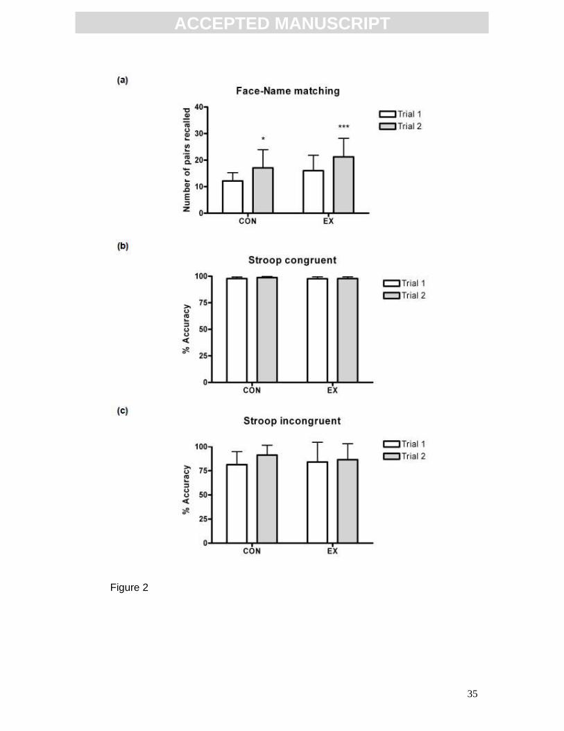
Acute exercise selectively enhanced cognitive function
Acute exercise induced an enhancement in hippocampal-dependent memory,
as assessed by the face-name task (Fig. 2a). There was a significant effect of
ACC
EPTE
D M
ANU
SCR
IPT
ACCEPTED MANUSCRIPT
12
trial (p<0.0001, F(1,41)=19.42; two-way repeated measures ANOVA and post
hoc Bonferroni; CON n=13; EX n=30), indicating that performance in trial 2 was
greater than trial 1. There was also a significant effect of group, (p=0.0219,
F(1,41)=5.681), indicating that exercise altered task performance. Post hoc
analysis revealed that while CON scores increased across trial, (trial 1: 12.15 ±
3.13 pairs recalled, trial 2: 17.08 ± 6.85 pairs recalled, p<0.05), EX improved in
the performance of the task to a greater extent, (trial 1: 16.03 ± 5.77 pairs
recalled, trial 2: 21.20 ± 7.01 pairs recalled, p<0.001). This suggests that,
although familiarization with the task may have resulted in an improved score,
the acute-exercise intervention resulted in an enhancement in the performance
of this task. Results are expressed as mean ± SD of the total number of pairs
recalled across the 4 recall blocks, hence 40 is the maximum possible score.
Acute exercise did not alter performance of the Stroop word-colour task in
either congruent trials (Fig. 2b) or incongruent trials (Fig. 2c). Statistical
analysis was by two-way repeated measures ANOVA (CON n=13, EX n=29).
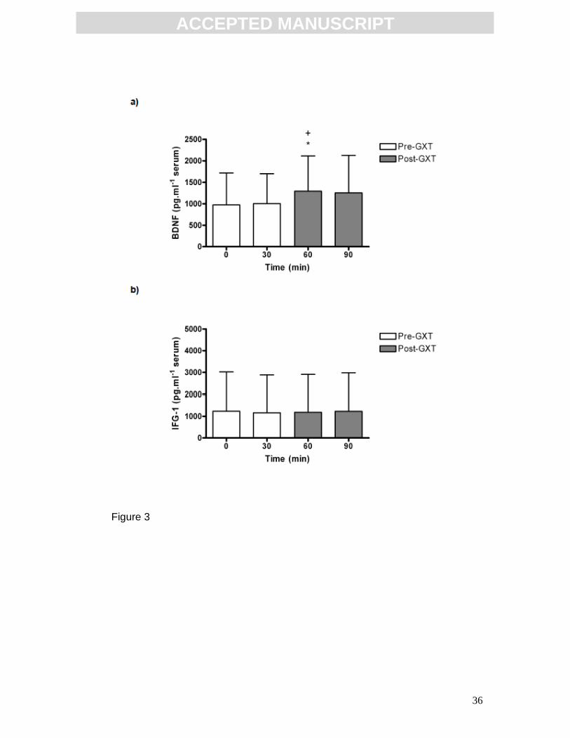
Effect of acute exercise on serum BDNF and IGF-1
Acute exercise in the form of a graded exercise test (GXT) induced an increase
in serum BDNF concentration (Fig. 3a). There was a significant difference
between timepoints (p=0.0126, F(3,93)=3.813; one-way repeated measures
ANOVA and post hoc Newman-Keuls, n=32). Post hoc analysis revealed a
significant increase in serum BDNF concentration immediately post-GXT (60
min: 1294.0 ± 820.5 pg.ml-1), relative to baseline (0 min: 974.7 ± 741.6 pg.ml-1,
p<0.05) and relative to the 30 min timepoint (1004.0 ± 694.1 pg.ml-1, p<0.05).
At 90 min (ie. 30 min post-exercise) serum BDNF was not significantly different
ACC
EPTE
D M
ANU
SCR
IPT
ACCEPTED MANUSCRIPT
13
to baseline, 30 min or to 60 min (90 min: 1254.0 ± 871.4 pg.ml-1). Results are
expressed as mean ± SD. Acute exercise did not alter serum IGF-1
concentration (Fig. 3b) although the degree of inter-individual variation was
large. Statistical analysis was by one-way repeated measures ANOVA and post
hoc Newman-Keuls (0 min: 1087.0 ± 1780.0 pg.ml-1, 30 min: 1020.0 ± 1721.0
pg.ml-1, 60 min: 1041.0 ± 1718 pg.ml-1, 90 min: 1084.0 ± 1744 pg.ml-1; n=28).
Results are expressed as mean ± SD.
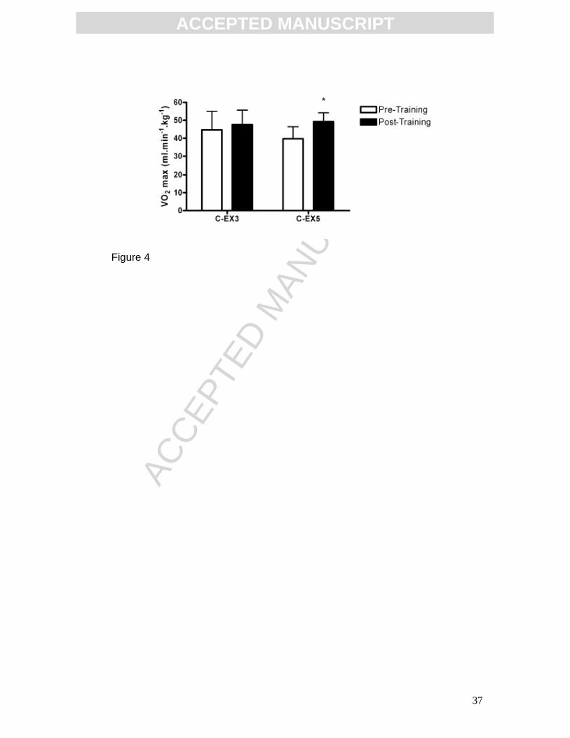
Effect of training on aerobic fitness
Aerobic fitness, as assessed by the VO2 max test, was altered by the aerobic
training paradigms (Fig. 4). There was a significant effect of training (p=0.0061,
F(1,14)=10.42; two-way repeated measures ANOVA and post hoc Bonferroni; C-
EX3 n=9; C-EX5 n=7). Post hoc analysis revealed a significant increase in post-
training VO2 max scores relative to pre-training values in the C-EX5 group (pre-
training: 39.70 ± 6.74 ml.min-1.kg-1, post-training: 49.26 ± 4.97 ml.min-1.kg-1,
p<0.05) indicating that 5 weeks of training enhanced aerobic fitness. 3 weeks of
aerobic training had no effect on VO2 max scores in the C-EX3 group (pre-
training: 44.69 ± 10.36 ml.min-1.kg-1, post-training: 47.65 ± 8.01 ml.min-1.kg-1).
Results are expressed as ml oxygen consumed per min per kg body mass,
mean ± SD.
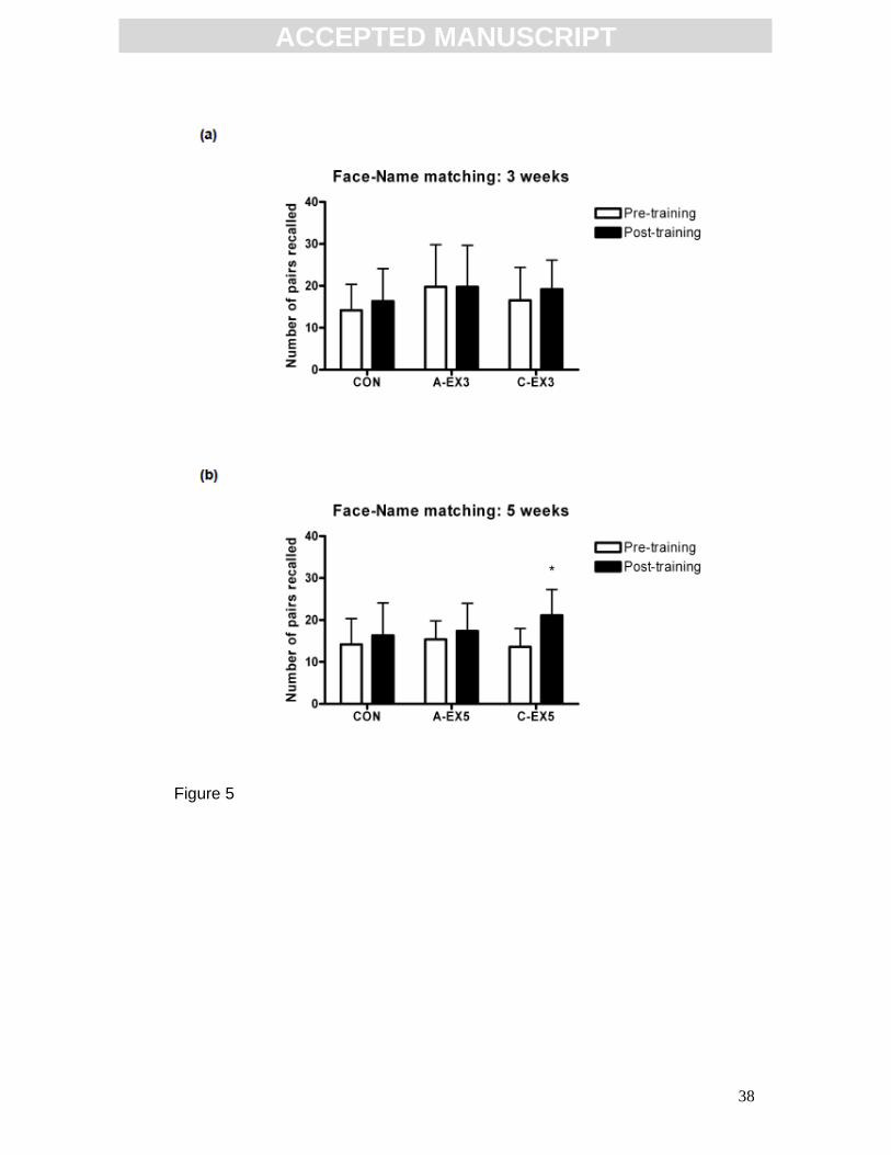
Effect of training on cognitive function
Hippocampal function, as assessed by the face-name task, was not altered by
3 weeks of aerobic training (Fig. 5a). Statistical analysis was by two-way
repeated measures ANOVA (CON n=15, A-EX3 n= 5, C-EX3 n=9). However, 5
ACC
EPTE
D M
ANU
SCR
IPT
ACCEPTED MANUSCRIPT
14
weeks of aerobic training enhanced performance in the face-name task (Fig.
5b). There was a significant effect of training (p=0.0172, F(1,28)=6.414; two-way
repeated measures ANOVA with post hoc Bonferroni; CON n=15, A-EX5 n=8,
C-EX5 n=8) indicating that the number of pairs recalled post-training was
greater that pre-training. Post hoc analysis revealed that there was a significant
increase in face-name performance in the C-EX5 group (pre-training: 13.6 ± 4.4
pairs, post-training: 21.1 ± 6.2 pairs, p<0.05), while the CON and A-EX5 groups
remained unchanged. Results are expressed as mean ± SD.
Effect of training on serum BDNF response to acute exercise
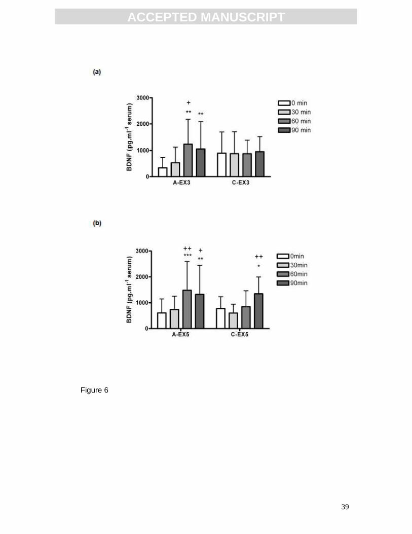
3 weeks of aerobic training had a significant effect on the serum BDNF
response to acute exercise (Fig. 6a). There was a significant effect of time
(p=0.0075, F(3,33)=4.725; two-way repeated measures ANOVA with post hoc
Bonferoni; A-EX3 n=5, C-EX3 n=9) and a significant interaction (p=0.0109,
F(3,33)=4.349). Post hoc analysis revealed an increase in the A-EX3 group
immediately post-exercise (60 min: 1226.0 ± 957.6 pg.ml-1) relative to 0 min
(339.0 ± 389.0 pg.ml-1, p<0.01) and relative to 30min (531.1 ± 585.0 pg.ml-1,
p<0.05). This increase was sustained at 30 min post exercise (90 min: 1059.0 ±
1032.0 pg.ml-1, p<0.01). However, serum BDNF concentration did not change
from baseline in the C-EX3 group (0 min: 898.3 ± 796.3 pg.ml-1, 30 min: 882.4 ±
821.9 pg.ml-1, 60 min: 878.8 ± 500.6 pg.ml-1, 90 min: 955.7 ± 560.7 pg.ml-1)
indicating that 3 weeks of aerobic training altered the profile of the serum BDNF
response to acute exercise.
5 weeks of aerobic training also has a significant effect on the serum BDNF
response to acute exercise (Fig. 6b). There was a significant effect of time
ACC
EPTE
D M
ANU
SCR
IPT
ACCEPTED MANUSCRIPT
15
(p=0.0001, F(3,42)=8.973; two-way repeated measures ANOVA with post hoc
Bonferroni; A-EX5 n=8, C-EX5 group n=8). Post hoc analysis revealed a
significant increase in the A-EX5 group immediately post-exercise (60 min:
1485.0 ± 1110.1 pg.ml-1) relative to 0 min (608.4 ± 538.9 pg.ml-1, p<0.001) and
relative to 30min (741.8 ± 512.3 pg.ml-1, p<0.01). This increase was sustained
at 90 min (1326.4 ± 1121.0 pg.ml-1, p<0.01). There was also a significant
increase in BDNF concentration in the C-EX5 group, but this did not occur until
90 min (1345.0 ± 650.9 pg.ml-1, p<0.05 relative to 0 min: 778.4 ± 458.2 pg.ml-1,
and p<0.01 relative to 30 min: 602.7 ± 340.5 pg.ml-1) indicating that 5 weeks of
chronic exercise has altered the temporal profile of the serum BDNF response
to acute exercise. Results are expressed mean ± SD.
Discussion
Here we present evidence that an acute bout of intense aerobic exercise
(in the form of a graded exercise test) selectively improves performance in a
hippocampal-dependent learning task in parallel with increased BDNF
concentration in the serum. We also demonstrate that 5 weeks, but not 3
weeks, of aerobic training improves performance in hippocampal learning and
alters the serum BDNF response to acute exercise. We have recently shown
that intracerebroventricular injection of exogenous BDNF protein mimics
exercise-induced enhancements in hippocampal-dependent learning in the rat
[16] and although circulating BDNF originates both from central and peripheral
sources [27], evidence suggests that the brain is a major source of circulating
BDNF, both at rest and during exercise, in humans [32]. We propose that
ACC
EPTE
D M
ANU
SCR
IPT
ACCEPTED MANUSCRIPT
16
exercise-induced enhancements in cognitive function in human subjects may
be stimulated via a BDNF-linked mechanism.
Acute exercise
An acute bout of exercise induced an enhancement in cognitive function,
as shown by the improvement in face-name task performance. This is in
agreement with previous studies which suggest that intense acute exercise
enhances learning and memory as assessed by a language-learning model [7],
and the Stroop task [9]. Face recognition has been shown to recruit the right
medial-temporal lobe (MTL), as evidenced by the inability of patients with right
amygdalo-hippocampectomy to recognise previously viewed faces [33]. It has
also been repeatedly demonstrated, using high-resolution functional magnetic
resonance imaging (fMRI) acquisition and analysis methods, that the face-
name association task used in the present study engages the hippocampus [31]
and nearby MTL cortical areas, including the amygdala, parahippocampal
cortex, perirhinal cortex and entorhinal cortex [34]. Hence, in the present study,
the acute exercise-induced cognitive enhancement appears to be selectively
MTL-dependent. To our knowledge, this is the first evidence for an acute
exercise-induced enhancement in function of these MTL structures in humans.
We observed no change in performance of the Stroop word-colour task,
which recruits the anterior cingulate cortex and other frontal regions [35], after
an acute exercise bout. In contrast to the present study, Ferris and colleagues
[9] have reported post-exercise improvements in the performance of the Stroop
word and colour tests. However, given that no control, non-exercising, groups
ACC
EPTE
D M
ANU
SCR
IPT
ACCEPTED MANUSCRIPT
17
were included in the study design, it is possible that the improvements they
observed were a result of a practice effect.
In agreement with the literature, the serum analysis revealed an acute
exercise-induced increase in BDNF concentration in sedentary young men.
According to a recent review, 69% of studies in healthy human subjects
reported a ‘mostly transient’ increase in peripheral BDNF concentration
following acute exercise [27]. In the present study, acute exercise induced an
increase in BDNF that had not quite returned to baseline at 30 min post-
exercise, however given that increases in basal BDNF concentrations were not
found in the chronic analysis, it may be presumed that the increase in serum
BDNF reported here is also transient.
The source of the BDNF increase remains unclear. Evidence indicates
that the brain is a major, but not the sole contributor to circulating BDNF [32]
and platelets also represent a likely source of serum BDNF, as has consistently
been reported [36, 37]. In this context, reports of the ability of BDNF to cross
the blood-brain barrier may be of relevance, with movement of BDNF from brain
to blood said to occur via bulk flow associated with the reabsorption of the
cerebrospinal fluid [38]. It has also been suggested that exercise transiently
increases the permeability of the blood brain barrier as demonstrated by an
increase in the extravasation of Evans blue albumin into the brain following 30
min of forced swim exercise in rats [39] or post-exercise increases in serum
S100β concentration in humans [40].
The acute exercise bout did not alter serum IGF-1 concentrations,
although the degree of inter-individual variation was large. This result is in
broad agreement with the literature. It has been reported that one hour of
ACC
EPTE
D M
ANU
SCR
IPT
ACCEPTED MANUSCRIPT
18
treadmill running had no effect on serum IGF-1 concentrations in the rat,
although increased uptake of serum IGF-1 into the brain was reported [21], and
this has been causally linked with learning enhancements and the induction of
BDNF in the brain post-exercise. Data from human subjects indicates that the
IGF-1 response to exercise is highly variable and may depend on exercise
intensity, duration and modality. While IGF-1 concentration in the serum has
been reported to increase following short bouts of cycling [41, 42] and high-
intensity running [43] other studies have reported no increase in serum IGF-1
following running bouts of varying durations and intensities [44-46].
Chronic exercise
A comparison of VO2 max scores revealed that 5 weeks of aerobic
training increased maximal oxygen consumption rates relative to pre-training
values, indicating a significant increase in cardiovascular fitness, while 3 weeks
of aerobic training did not improve aerobic capacity. As we observed no
changes in performance of the Stroop task following acute exercise, we
assessed the impact of training on the face-name task alone. 5 weeks of
aerobic training enhanced face-name task performance, while 3-weeks of
aerobic training had no effect on MTL-dependent learning and memory. This
suggests a positive correlation between aerobic fitness and this learning task.
Taken together these results indicate that both acute exercise and 5
weeks of aerobic training enhance MTL-dependent learning. We hypothesized
that the mechanism by which physical activity enhances MTL-dependent
cognition may be linked with BDNF. An increase in serum BDNF concentration
with acute exercise has been demonstrated and was associated with a
ACC
EPTE
D M
ANU
SCR
IPT
ACCEPTED MANUSCRIPT
19
concomitant enhancement in the performance of the face-name task.
Furthermore, this serum BDNF response to acute exercise was shown to be
reproducible in session 2 at both the 3 week and 5 week timepoints. We also
monitored IGF-1 following chronic training and saw no change at any timepoint
(data not shown). This is consistent with the lack of effect of acute exercise on
IGF-1 concentration that we have reported herein.
There is strong evidence from studies using humans or animal models
that alterations in BDNF concentration may have functional consequences for
cognition. Missense polymorphisms in BDNF in human subjects are linked with
decreased synaptic abundance in the hippocampus and poor performance in
memory tasks [47]. The binding of BDNF to its TrkB receptor mediates plastic
changes involved in recognition memory in sheep [48], while exercise has
shown to enhance object recognition learning in association with an increased
concentration of BDNF in the dentate gyrus of young rats [15, 16]. Evidence
from the literature suggests that BDNF can facilitate neurotransmitter release
and enhance synaptic transmission [49, 50], leading to the hypothesis that the
acute exercise-induced enhancement in hippocampal function may be
mediated by the actions of BDNF on synaptic transmission.
Neither 3 weeks nor 5 weeks of aerobic training had any effect on basal
BDNF concentration. Furthermore, following 3 weeks of aerobic training, the
ability of acute exercise to increase serum BDNF concentration was abolished.
Similarly, 5 weeks of aerobic training altered the profile of the BDNF response
to acute exercise, in that the BDNF increase was delayed until 30 min post-
exercise. These results indicate that aerobic training is affecting the induction of
increased serum BDNF by acute exercise, altering the temporal profile of the
ACC
EPTE
D M
ANU
SCR
IPT
ACCEPTED MANUSCRIPT
20
acute-exercise effect. It is unclear whether this is a result of an alteration in the
mechanism of BDNF release. It is possible that the effect of chronic exercise on
cognition may be mediated by an alternative mechanism involving BDNF.
BDNF infusion has been shown to induce neurogenesis in rats [51]. It has been
demonstrated that hippocampal neurogenesis also occurs in the dentate gyrus
of adult humans [52] and that aerobic exercise training can increase the size of
the anterior hippocampus in older adults in parallel with increased concentration
of BDNF in the serum [53]. Potential functional consequences of adult
neurogenesis must occur as long-term adaptations, rather than acute benefits,
due to the time required for newly-generated neurons to mature and become
integrated into a network [54]. While this provides an intriguing potential
mechanism underlying the effects of exercise training on cognition,
experimental limitations inherent in assessing the cellular mechanisms
mediating cognition in humans means that investigation of such a hypothesis is
currently unfeasible.
The results presented here provide evidence for a link between acute
exercise and cognitive function. Acute exercise has been shown to increase
serum BDNF and selectively improve MTL-dependent memory. Hence, BDNF
is proposed as a mediator of the cognitive enhancements described, possibly
through its reported role in short-term mechanisms underlying synaptic
plasticity. Furthermore, it has been shown that while the 3-week training
programme was insufficient to improve aerobic fitness or augment memory test
performance, the 5-week chronic exercise programme resulted in enhanced
fitness scores and improvements in MTL-dependent cognition. A role for BDNF

ACC
EPTE
D M
ANU
SCR
IPT
ACCEPTED MANUSCRIPT
21
in these improvements in central nervous system function is tentatively
suggested.
ACC
EPTE
D M
ANU
SCR
IPT
ACCEPTED MANUSCRIPT
22
Figure legends
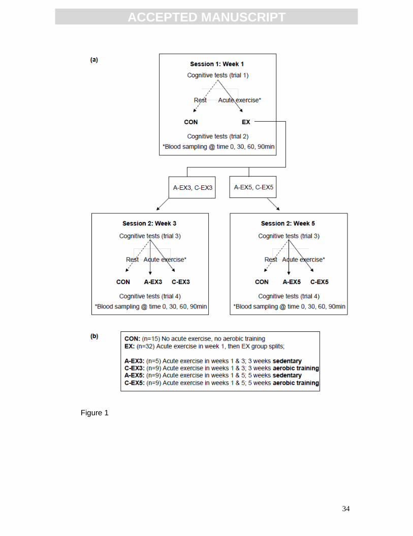
FIG 1: Outline of the experimental protocol. (a) Sessions 1 and 2 assessed
the effects of acute exercise on cognitive function. During session 1, subjects
were divided into CON and EX groups and underwent a cognitive testing trial
(trial 1), following which EX completed an acute exercise bout while CON
rested for a corresponding 30min period. Then all subjects completed a second
cognitive testing trial (trial 2). Blood samples were collected from EX at times 0,
30, 60 and 90 min, with acute exercise occurring between times 30 min and 60
min. An overview of the subject groups is shown in (b). Baseline measures of
cognitive function and serum BDNF concentration were obtained during session
1. The effect of both 3 and 5 weeks of chronic exercise then was assessed by
subdividing the EX group. C-EX3 and C-EX5 completed 3 and 5 weeks of
aerobic training respectively, prior to session 2, while A-EX3 and A-EX5
remained sedentary for the corresponding 3-week or 5-week period prior to
session 2. The CON group completed session 2 after a 3 or 5-week interval and
were sedentary both during the interval and during the testing session.
FIG 2: Effect of an acute exercise bout on cognitive function. (a) Acute
exercise enhanced performance of the face-name task. The total number of
face-name pairs recalled in trial 2 was greater than trial 1, *p<0.05, ***p<0.001
(CON n=13, EX n=30). Acute exercise did not alter performance of the Stroop
word-colour task in either congruent (b) or incongruent (c) trials (CON n=14, EX
n=28). Statistical analysis by two-way repeated measures ANOVA and post hoc
Bonferroni. Data expressed as mean ± SD.
ACC
EPTE
D M
ANU
SCR
IPT
ACCEPTED MANUSCRIPT
23
FIG 3: Effect of acute exercise on serum BDNF and IGF-1 concentrations.
(a) Acute exercise (graded exercise test; GXT) induced an increase in serum
BDNF concentration, *p<0.05 relative to 0 min, +p<0.05 relative to 30min
(n=32). (b) Acute exercise did not alter serum IGF-1 concentrations (n=32).
Statistical analysis by one-way repeated measures ANOVA and post hoc
Newman-Keuls. Data expressed as mean ± SD.
FIG 4: Effect of aerobic training on VO2 max scores. 3 weeks of aerobic
training did not affect fitness, as assessed by VO2 max scores. 5 weeks of
aerobic training significantly increased VO2 max scores, *p<0.05 relative to pre-
training values (C-EX3 n=9, C-EX5 n=7). Statistical analysis by two-way
repeated measures ANOVA and post hoc Bonferroni. Data expressed as mean
± SD.
FIG 5: Effect of aerobic training on performance of the face-name task. (a)
3 weeks of aerobic training did not affect performance of the face-name task
(CON n=15, A-EX3 n=5, C-EX3 n=9). (b) 5 weeks of aerobic training enhanced
performance of the face-name task. There was a significant increase in face-
name performance in the C-EX5 group, *p<0.05 relative to pre-training value
(CON n=15, A-EX5 n=8, C-EX5 n=8). Statistical analysis by two-way repeated
measures ANOVA and post hoc Bonferroni. Data expressed as mean ± SD.
FIG 6: Effect of aerobic training on serum BDNF response to acute
exercise. (a) 3 weeks of aerobic training had a significant effect on the serum
ACC
EPTE
D M
ANU
SCR
IPT
ACCEPTED MANUSCRIPT
24
BDNF response to acute exercise. There was a significant increase in serum
BDNF in the A-EX3 group immediately post acute-exercise (60 min) which was
sustained at 90 min, **p<0.01 relative to 0min, +p<0.05 relative to 30 min.
However, serum BDNF concentration did not change from baseline in the C-
EX3 group (A-EX3 n=5, C-EX3 n=9). (b) 5 weeks of aerobic training had a
significant effect on the serum BDNF response to acute exercise. There was a
significant increase in serum BDNF in the A-EX5 group immediately post acute-
exercise (60 min) which was sustained at 90 min, ***p<0.001 relative to 0 min,
**p<0.01 relative to 0 min, ++p<0.01 relative to 30 min, +p<0.05 relative to 30
min. There was also a significant increase in the C-EX5 group in response to
acute exercise, however this did not occur until 30 min post acute-exercise (90
min), *p<0.05 relative to 0 min, ++p<0.01 relative to 30 min (A-EX5 n=6, C-EX5
n=8). Statistical analysis by two-way repeated measures ANOVA with post hoc
Bonferroni. Data expressed as mean ± SD.
Acknowledgments
Funded by the Irish Research Council for Science, Engineering and Technology
(Embark Initiative) and Faculty of Engineering, Mathematics and Science,
Trinity College Dublin.
ACC
EPTE
D M
ANU
SCR
IPT
ACCEPTED MANUSCRIPT
25
References
[1] Abbott RD, White LR, Ross GW, Masaki KH, Curb JD, Petrovitch H.
Walking and dementia in physically capable elderly men. Jama 2004; 292(12):
1447-53.
[2] Larson EB, Wang L, Bowen JD, McCormick WC, Teri L, Crane P, et al.
Exercise is associated with reduced risk for incident dementia among persons
65 years of age and older. Ann Intern Med 2006; 144(2): 73-81.
[3] Rovio S, Kareholt I, Helkala EL, Viitanen M, Winblad B, Tuomilehto J, et
al. Leisure-time physical activity at midlife and the risk of dementia and
Alzheimer's disease. Lancet Neurol 2005; 4(11): 705-11.
[4] Cassilhas RC, Viana VA, Grassmann V, Santos RT, Santos RF, Tufik S,
et al. The impact of resistance exercise on the cognitive function of the elderly.
Med Sci Sports Exerc 2007; 39(8): 1401-7.
[5] Heyn P, Abreu BC, Ottenbacher KJ. The effects of exercise training on
elderly persons with cognitive impairment and dementia: a meta-analysis. Arch
Phys Med Rehabil 2004; 85(10): 1694-704.
[6] Stevens J, Killeen M. A randomised controlled trial testing the impact of
exercise on cognitive symptoms and disability of residents with dementia.
Contemp Nurse 2006; 21(1): 32-40.
[7] Winter B, Breitenstein C, Mooren FC, Voelker K, Fobker M, Lechtermann
A, et al. High impact running improves learning. Neurobiol Learn Mem 2007;
87(4): 597-609.
ACC
EPTE
D M
ANU
SCR
IPT
ACCEPTED MANUSCRIPT
26
[8] Grego F, Vallier JM, Collardeau M, Rousseu C, Cremieux J, Brisswalter
J. Influence of exercise duration and hydration status on cognitive function
during prolonged cycling exercise. Int J Sports Med 2005; 26(1): 27-33.
[9] Ferris LT, Williams JS, Shen CL. The effect of acute exercise on serum
brain-derived neurotrophic factor levels and cognitive function. Med Sci Sports
Exerc 2007; 39(4): 728-34.
[10] Tomporowski PD. Effects of acute bouts of exercise on cognition. Acta
Psychol (Amst) 2003; 112(3): 297-324.
[11] Lambourne K, Tomporowski P. The effect of exercise-induced arousal
on cognitive task performance: a meta-regression analysis. Brain Res 2010;
1341: 12-24.
[12] Chen HI, Lin LC, Yu L, Liu YF, Kuo YM, Huang AM, et al. Treadmill
exercise enhances passive avoidance learning in rats: the role of down-
regulated serotonin system in the limbic system. Neurobiol Learn Mem 2008;
89(4): 489-96.
[13] Farmer J, Zhao X, van Praag H, Wodtke K, Gage FH, Christie BR.
Effects of voluntary exercise on synaptic plasticity and gene expression in the
dentate gyrus of adult male Sprague-Dawley rats in vivo. Neuroscience 2004;
124(1): 71-9.
[14] Gobbo OL, O'Mara SM. Exercise, but not environmental enrichment,
improves learning after kainic acid-induced hippocampal neurodegeneration in
association with an increase in brain-derived neurotrophic factor. Behav Brain
Res 2005; 159(1): 21-6.
ACC
EPTE
D M
ANU
SCR
IPT
ACCEPTED MANUSCRIPT
27
[15] O'Callaghan RM, Ohle R, Kelly AM. The effects of forced exercise on
hippocampal plasticity in the rat: A comparison of LTP, spatial- and non-spatial
learning. Behav Brain Res 2007; 176(2): 362-6.
[16] Griffin EW, Bechara RG, Birch AM, Kelly AM. Exercise enhances
hippocampal-dependent learning in the rat: evidence for a BDNF-related
mechanism. Hippocampus 2009; 19(10): 973-80.
[17] Gooney M, Shaw K, Kelly A, O'Mara SM, Lynch MA. Long-term
potentiation and spatial learning are associated with increased phosphorylation
of TrkB and extracellular signal-regulated kinase (ERK) in the dentate gyrus:
evidence for a role for brain-derived neurotrophic factor. Behav Neurosci 2002;
116(3): 455-63.
[18] Hennigan A, Callaghan CK, Kealy J, Rouine J, Kelly AM. Deficits in LTP
and recognition memory in the genetically hypertensive rat are associated with
decreased expression of neurotrophic factors and their receptors in the dentate
gyrus. Behav Brain Res 2009; 197(2): 371-7.
[19] Hennigan A, O'Callaghan RM, Kelly AM. Neurotrophins and their
receptors: roles in plasticity, neurodegeneration and neuroprotection. Biochem
Soc Trans 2007; 35(Pt 2): 424-7.
[20] Tyler WJ, Alonso M, Bramham CR, Pozzo-Miller LD. From acquisition to
consolidation: on the role of brain-derived neurotrophic factor signaling in
hippocampal-dependent learning. Learn Mem 2002; 9(5): 224-37.
ACC
EPTE
D M
ANU
SCR
IPT
ACCEPTED MANUSCRIPT
28
[21] Carro E, Nunez A, Busiguina S, Torres-Aleman I. Circulating insulin-like
growth factor I mediates effects of exercise on the brain. J Neurosci 2000;
20(8): 2926-33.
[22] Ding Q, Vaynman S, Akhavan M, Ying Z, Gomez-Pinilla F. Insulin-like
growth factor I interfaces with brain-derived neurotrophic factor-mediated
synaptic plasticity to modulate aspects of exercise-induced cognitive function.
Neuroscience 2006; 140(3): 823-33.
[23] Trejo JL, Llorens-Martin MV, Torres-Aleman I. The effects of exercise on
spatial learning and anxiety-like behavior are mediated by an IGF-I-dependent
mechanism related to hippocampal neurogenesis. Mol Cell Neurosci 2008;
37(2): 402-11.
[24] Gold SM, Schulz KH, Hartmann S, Mladek M, Lang UE, Hellweg R, et al.
Basal serum levels and reactivity of nerve growth factor and brain-derived
neurotrophic factor to standardized acute exercise in multiple sclerosis and
controls. J Neuroimmunol 2003; 138(1-2): 99-105.
[25] Rojas Vega S, Struder HK, Vera Wahrmann B, Schmidt A, Bloch W,
Hollmann W. Acute BDNF and cortisol response to low intensity exercise and
following ramp incremental exercise to exhaustion in humans. Brain Res 2006;
1121(1): 59-65.
[26] Tang SW, Chu E, Hui T, Helmeste D, Law C. Influence of exercise on
serum brain-derived neurotrophic factor concentrations in healthy human
subjects. Neurosci Lett 2008; 431(1): 62-5.
ACC
EPTE
D M
ANU
SCR
IPT
ACCEPTED MANUSCRIPT
29
[27] Knaepen K, Goekint M, Heyman EM, Meeusen R. Neuroplasticity -
exercise-induced response of peripheral brain-derived neurotrophic factor: a
systematic review of experimental studies in human subjects. Sports Med 2010;
40(9): 765-801.
[28] Abellan R, Ventura R, Pichini S, Di Giovannandrea R, Bellver M, Olive R,
et al. Effect of physical fitness and endurance exercise on indirect biomarkers
of recombinant growth hormone misuse: insulin-like growth factor I and
procollagen type III peptide. Int J Sports Med 2006; 27(12): 976-83.
[29] Kraemer WJ, Aguilera BA, Terada M, Newton RU, Lynch JM, Rosendaal
G, et al. Responses of IGF-I to endogenous increases in growth hormone after
heavy-resistance exercise. J Appl Physiol 1995; 79(4): 1310-5.
[30] Nguyen UN, Mougin F, Simon-Rigaud ML, Rouillon JD, Marguet P,
Regnard J. Influence of exercise duration on serum insulin-like growth factor
and its binding proteins in athletes. Eur J Appl Physiol Occup Physiol 1998;
78(6): 533-7.
[31] Zeineh MM, Engel SA, Thompson PM, Bookheimer SY. Dynamics of the
hippocampus during encoding and retrieval of face-name pairs. Science 2003;
299(5606): 577-80.
[32] Rasmussen P, Brassard P, Adser H, Pedersen MV, Leick L, Hart E, et
al. Evidence for a release of brain-derived neurotrophic factor from the brain
during exercise. Exp Physiol 2009; 94(10): 1062-9.
ACC
EPTE
D M
ANU
SCR
IPT
ACCEPTED MANUSCRIPT
30
[33] Crane J, Milner B. Do I know you? Face perception and memory in
patients with selective amygdalo-hippocampectomy. Neuropsychologia 2002;
40(5): 530-8.
[34] Kirwan CB, Stark CE. Medial temporal lobe activation during encoding
and retrieval of novel face-name pairs. Hippocampus 2004; 14(7): 919-30.
[35] Leung HC, Skudlarski P, Gatenby JC, Peterson BS, Gore JC. An event-
related functional MRI study of the stroop color word interference task. Cereb
Cortex 2000; 10(6): 552-60.
[36] Rosenfeld RD, Zeni L, Haniu M, Talvenheimo J, Radka SF, Bennett L, et
al. Purification and identification of brain-derived neurotrophic factor from
human serum. Protein Expr Purif 1995; 6(4): 465-71.
[37] Yamamoto H, Gurney ME. Human platelets contain brain-derived
neurotrophic factor. J Neurosci 1990; 10(11): 3469-78.
[38] Pan W, Banks WA, Fasold MB, Bluth J, Kastin AJ. Transport of brain-
derived neurotrophic factor across the blood-brain barrier. Neuropharmacology
1998; 37(12): 1553-61.
[39] Sharma HS, Cervos-Navarro J, Dey PK. Increased blood-brain barrier
permeability following acute short-term swimming exercise in conscious
normotensive young rats. Neurosci Res 1991; 10(3): 211-21.
[40] Watson P, Black KE, Clark SC, Maughan RJ. Exercise in the heat: effect
of fluid ingestion on blood-brain barrier permeability. Med Sci Sports Exerc
2006; 38(12): 2118-24.
ACC
EPTE
D M
ANU
SCR
IPT
ACCEPTED MANUSCRIPT
31
[41] Cappon J, Brasel JA, Mohan S, Cooper DM. Effect of brief exercise on
circulating insulin-like growth factor I. J Appl Physiol 1994; 76(6): 2490-6.
[42] Schwarz AJ, Brasel JA, Hintz RL, Mohan S, Cooper DM. Acute effect of
brief low- and high-intensity exercise on circulating insulin-like growth factor
(IGF) I, II, and IGF-binding protein-3 and its proteolysis in young healthy men. J
Clin Endocrinol Metab 1996; 81(10): 3492-7.
[43] Kraemer RR, Durand RJ, Acevedo EO, Johnson LG, Kraemer GR,
Hebert EP, et al. Rigorous running increases growth hormone and insulin-like
growth factor-I without altering ghrelin. Exp Biol Med (Maywood) 2004; 229(3):
240-6.
[44] Banfi G, Marinelli M, Roi GS, Colombini A, Pontillo M, Giacometti M, et
al. Growth hormone and insulin-like growth factor I in athletes performing a
marathon at 4000 m of altitude. Growth Regul 1994; 4(2): 82-6.
[45] Jahreis G, Hesse V, Schmidt HE, Scheibe J. Effect of endurance
exercise on somatomedin-C/insulin-like growth factor I concentration in male
and female runners. Exp Clin Endocrinol 1989; 94(1-2): 89-96.
[46] Koistinen H, Koistinen R, Selenius L, Ylikorkala Q, Seppala M. Effect of
marathon run on serum IGF-I and IGF-binding protein 1 and 3 levels. J Appl
Physiol 1996; 80(3): 760-4.
[47] Egan MF, Kojima M, Callicott JH, Goldberg TE, Kolachana BS, Bertolino
A, et al. The BDNF val66met polymorphism affects activity-dependent secretion
of BDNF and human memory and hippocampal function. Cell 2003; 112(2):
257-69.
ACC
EPTE
D M
ANU
SCR
IPT
ACCEPTED MANUSCRIPT
32
[48] Broad KD, Mimmack ML, Keverne EB, Kendrick KM. Increased BDNF
and trk-B mRNA expression in cortical and limbic regions following formation of
a social recognition memory. Eur J Neurosci 2002; 16(11): 2166-74.
[49] Jovanovic JN, Czernik AJ, Fienberg AA, Greengard P, Sihra TS.
Synapsins as mediators of BDNF-enhanced neurotransmitter release. Nat
Neurosci 2000; 3(4): 323-9.
[50] Xu B, Gottschalk W, Chow A, Wilson RI, Schnell E, Zang K, et al. The
role of brain-derived neurotrophic factor receptors in the mature hippocampus:
modulation of long-term potentiation through a presynaptic mechanism
involving TrkB. J Neurosci 2000; 20(18): 6888-97.
[51] Scharfman H, Goodman J, Macleod A, Phani S, Antonelli C, Croll S.
Increased neurogenesis and the ectopic granule cells after intrahippocampal
BDNF infusion in adult rats. Exp Neurol 2005; 192(2): 348-56.
[52] Eriksson PS, Perfilieva E, Bjork-Eriksson T, Alborn AM, Nordborg C,
Peterson DA, et al. Neurogenesis in the adult human hippocampus. Nat Med
1998; 4(11): 1313-7.
[53] Erickson KI, Voss MW, Prakash RS, Basak C, Szabo A, Chaddock L, et
al. Exercise training increases size of hippocampus and improves memory.
Proc Natl Acad Sci U S A 2011; 108(7): 3017-22.
[54] Kempermann G, Wiskott L, Gage FH. Functional significance of adult
neurogenesis. Curr Opin Neurobiol 2004; 14(2): 186-91.
ACC
EPTE
D M
ANU
SCR
IPT
ACCEPTED MANUSCRIPT
34
Figure 1
ACC
EPTE
D M
ANU
SCR
IPT
ACCEPTED MANUSCRIPT
35
Figure 2
ACC
EPTE
D M
ANU
SCR
IPT
ACCEPTED MANUSCRIPT
36
Figure 3
ACC
EPTE
D M
ANU
SCR
IPT
ACCEPTED MANUSCRIPT
37
Figure 4
ACC
EPTE
D M
ANU
SCR
IPT
ACCEPTED MANUSCRIPT
38
Figure 5
ACC
EPTE
D M
ANU
SCR
IPT
ACCEPTED MANUSCRIPT
39
Figure 6
ACC
EPTE
D M
ANU
SCR
IPT
ACCEPTED MANUSCRIPT
40
Highlights
An acute bout of aerobic exercise improves performance of the face-name task (an index of hippocampal function), but not the stroop task, in young male subjects.
This cognitive improvement is associated with increased concentration of BDNF in the serum.
Chronic training, resulting in improved VO2max, improves performance of the face-name task and alters the profile of circulating BDNF.
The data suggest a role for BDNF in exercise-induced cognitive enhancement in humans.
Top Related
Copyright © 2022 FDOKUMEN

