


Bahasa
Halaman
Hukum

ACADEMIC STUDIES
IN HEALTH SCIENCES-2019/2
Editors
Prof. Dr. Belgin SIRIKEN
Asst. Prof. Dr. Ayhan GÜLER
Dr. Taşkın ERKİNÜRESİN
Cetinje 2019
Editors
Prof. Dr. Belgin SIRIKEN
Asst. Prof. Dr. Ayhan GÜLER
Dr. Taşkın ERKİNÜRESİN
First Edition •© September 2019 /Cetinje-Montenegro
ISBN • 978-9940-540-99-9
© copyright
All Rights Reserved
Ivpe
web: www.ivpe.me
Tel. +382 41 234 709
e-mail: [email protected]
Ivpe
Cetinje, Montenegro
I
CONTENTS PREFACE ...................................................................................................................... III
REFEREE BOARD ..................................................................................................... IV
MULTI DISCIPLINARY HEALTH SCIENCE ............................................... 1
THE PROBLEM WITH THE AGE OF TECHNOLOGY: NOMOPHOBIA ..... 2
DENTISTRY STUDIES .................................................................................... 8
THE EFFECT OF PERIODONTAL TREATMENT ON ADVERSE
PREGNANCY OUTCOMES BEFORE, DURING AND AFTER PREGNANCY
........................................................................................................................................... 9
MEDICAL STUDIES....................................................................................... 24
THE KYNURENINE PATHWAY IN ALZHEIMER'S DISEASE: THE
ALTERNATION OF NOGO-A AND KLOTHO ACTIVITIES BY
INFLUENCING N-METHYL-D-ASPARTATE RECEPTOR-NITRIC OXIDE
PATHWAY .................................................................................................................. 25
PRINCIPLES OF PALLIATIVE CARE AND DEMOGRAPHIC ASPECTS OF
CANCER PATIENTS ................................................................................................ 51
OFF-PUMP CORONARY ARTERY BYPASS (OPCAB) SURGERY ............. 59
DEEP VENOUS THROMBOSIS ............................................................................ 71
THE IMPORTANCE OF BIRADS 0 IMAGING LESIONS (BREAST
IMAGING REPORTING AND DATA SYSTEM), IN TERMS OF
MALIGNANCY IN MAMMOGRAPHY ................................................................. 79
SURGICAL APPROACH IN CAROTID ARTERY STENOSIS........................ 85
DIAGNOSIS AND TREATMENT OF CHRONIC VENOUS INSUFFICIENCY
AND VARICOSE VEINS .......................................................................................... 98
UTERINE SPARING SURGERY in POSTPARTUM HEMORRHAGE ..... 109
FOOD ADDICTION/OBESITY AND SPORTS ............................................... 118
WOMAN – SPORTS AND SOCIAL GENDER ................................................ 125
SURGICAL WOUNDS AND WOUND HEALING PROCESS ...................... 132
ANTIBACTERIAL EFFECTS OF HONEY AND HYPERTONIC SALINE 142
METHICILLIN-RESISTANT STAPHYLOCOCCUS AUREUS
COLONIZATION IN NASAL CULTURES OF THE BABIES
HOSPITALIZED AT NEWBORN INTENSIVE CARE UNIT ...................... 150

II
ADENOVIRUSES IN OPHTHALMOLOGY REVIEW ................................... 158
PERIANAL HIDRADENOMA PAPILLIFERUM ............................................ 170
CURRENT TRENDS IN THE TREATMENT MODALITIES OF OPEN
ANGLE GLAUCOMA ............................................................................................. 173
CORONARY ARTERY BYPASS GRAFTING SURGERY AND
COMPLICATIONS .................................................................................................. 192
VETERINARY MEDICINE ......................................................................... 203
HEALTH MONITORING ...................................................................................... 204
ALTERNATIVE APPROACHES TO TREATMENT OF MASTITIS
INSTEAD OF TRADITIONAL METHODS ...................................................... 217

III
PREFACE
The book “Academic Studies in Health Sciences” is serving an
academic forum for both academics and researchers working in such fields.
Health sciences research is an interdisciplinary by nature. So it covers
several fields such as dentistry, veterinary medicine, medical sciences. In
this book, the academics working in different fields share their results with
the scientific community. Thus more researchers will be aware of these
studies and have some new ideas for their future studies. The selected
articles have been reviewed and approved for publication by referees. It is
hoped that the book will be of interest and of value to academics and
researchers.
IV
REFEREE BOARD
Prof. Dr. Belgin SIRIKEN, Ondokuz Mayıs University, Turkey
Prof. Dr. Gönül DÖNMEZ, Ankara University, Turkey
Prof. Dr. Nurcan ÇETİNKAYA, Ondokuz Mayıs University, Turkey
Prof. Dr.Mehmet Rüştü KAHRAMAN, Ankara University, Turkey
Prof. Dr. Robert C. SHNAIDER, New York University, USA
Doç.Dr Gülten ÖKMEN, Muğla Sıtkı Koçman Mayıs University, Turkey
Doç. Dr. Meriç ERASLAN, Akdeniz University, Turkey
Dr.Öğr.Üyesi Ceren BAŞKAN, Amasya University, Turkey
Dr. Mir Hamid Sleaihan, Islamic Azad University, Iran
Dr. Fezail Ismayil BÜYÜKKİŞİ, Azerbaijan Medical University, Azerbaijan
Dr. Rahib ALİYEV, Azerbaijan Medical University, Azerbaijan
Dr. Razbaygul BOLATKIZI, Hovd State University, Mongolia
Dr. Taşkın ERKİNÜRESİN, Health Sciences University Bursa Higher Specialization Training And Research Hospital, Turkey

1
MULTI DISCIPLINARY HEALTH SCIENCE
2
THE PROBLEM WITH THE AGE OF TECHNOLOGY:
NOMOPHOBIA
Canan BİRİMOĞLU OKUYAN& Süreyya NUR
Introduction
Nomophobia (no mobile phone phobia) is defined as the fear of being
away from one’s cell phones or not being able to communicate via mobile
phone (1, 2, 3). It first became prominent in England with a study (4). This
study revealed the symptoms of nomophobia such as using mobile phones
all the time and spending most of the time on the phone, feeling
uncomfortable when s/he is away from mobile phones, checking the phone
for notifications, sleeping with the phone (4, 5). These symptoms indicate
that mobile phone usage causes health problems.
One of the groups that use mobile phones the most is university students
(6). According to the study of Yıldırım in 2014, university students who
are between 18-24 age range use smartphones excessively. Therefore, this
group is considered as a risk factor (2, 5, 7, 8, 9). Although there have been
many studies about smartphone usage, there have not been enough studies
about nomophobia of university students (10). Thus, the study group of the
present study consists of university students.
1.The Concept of Mobile Phone
Mobile phones are devices that can be carried around and provide
communication via radio stations.
Cell phones are the most practical and preferred mobile devices. Cell
phones have been turned into smartphones with the addition of computer
features.
Wireless communication was first developed in 1985 (11). Nowadays,
there are features like receiving and sending e-mails, accessibility to social
media accounts, online shopping, and banking operations (12). These
features have increased the number of addicts, especially in the young
population.
(Dr.Öğr.Üyesi); Hatay Mustafa Kemal University, Faculty of Health Sciences, Hatay,
Turkey. Email: [email protected], (Dr.Öğr.Üyesi); Hatay Mustafa Kemal University, Vocational School of Health
ServicesHatay, Turkey. Email: [email protected]
3
2. Mobile Phone Addiction
With the developments in technology, the functions of mobile phones
increase together with the rate of use (9).
Smartphones bring various problems as they are becoming more
involved in their daily lives. Even though there is no clear definition for it
in the field of psychiatry, mobile phone addiction is a condition develops
based on the frequency of use of smartphones and has been the topic of
research for some time (13). In a study about smartphones, it is stated that
smartphones are the latest evolution of 21st-century technology (14).
In a study conducted in Turkey, it was revealed that the majority of the
people (92%) owned smartphones. Another study in 2015 found that this
rate has increased. Therefore, the increased rate of mobile phone usage
implies the number of time spent on the phone. Another study (79% and
72% respectively) it was determined that individuals check their phones 15
minutes after waking up and 15 minutes before sleep (15). Also, visual
communication in mobile phones popularize these devices and cause them
to have an impact on the daily lives of the individuals (16).
According to the “Digital in 2017 Global Overview” report published
by We Are Social and Hootsuite there are more than 4 billion mobile phone
users worldwide. 2.5 billion of them are active social users. In Turkey, it
was found that the number of active social mobile phone users is 42 million
(17). In terms of the usage rate of mobile phone applications, the most
frequently used applications at the beginning of 2017 are social media
applications (18). The same year, Turkey ranked the second in Twitter
usage, the third in Facebook and the fourth in Youtube usage (19). The
addiction increases since it is easily accessible to social media platforms
that support and meet social needs. Therefore, most of the people
obsessively check their Facebook and Twitter accounts day and night. This
creates pressure for checking social media activities (20). This pressure
encourages people to stay connected with social media. Moreover,
primarily because of economic reasons, company owners use various
techniques to make sharing more and more accessible by developing
notification features of the applications. In this regard, technology
addiction has become less controllable since computer-assisted technology
has become more fragile (20) Furthermore, peoples’ need for staying
connected to social media causes them to control the environmental factors
less creating problems such as violation of privacy and exploitation of
digital labor. Addiction to smartphones cannot be considered independent
of the ability of smartphones to connect to networks anytime and
anywhere. Also, human-computer (human-smartphone) interaction
constitutes an indirect aspect of addiction. The individual connects to a
personalized network via a smartphone while interacting with a
4
technological tool. Two of Sherry Turkle's findings on this interaction are
particularly important (20).
3. Dangers of Mobile Phone Usage
The radiation around us is generally divided into two groups as ionizing
and non-ionizing radiation. Ionizing radiation can be given as examples of
X-rays used for therapeutic and diagnostic purposes, and they have
carcinogenic effects. The radiation generated by EM (electromagnetic)
waves takes place in the non-ionized group, and its effect is still a research
subject (21).
The health effects of electromagnetic fields are divided into thermal and
non-thermal effects. Thermal effects are modeled by the so-called Specific
Absorption Rate (W / kg). Non-thermal effects are biological, genetic,
psychological, and so on. SAR, which is a primary parameter, is not easily
measurable but can be obtained by equivalent models in specialized
laboratories or by computer simulations. Therefore, electric field strength
(E – V / m), a derived parameter, is used. The radio stations are measured
accordingly and checked against specified limit values. According to the
standards prepared by ICNIRP (International Committee on Non-Ionizing
Radiation Protection) for mobile phones, the limit values are determined
as 42V / m at 900 MHz and 59 V / m at 1800 MHz for the places people
have been in the last 24 hours. Like the US and many European countries,
Turkey has adopted these limit values as well. The measurements showed
that the limit values could be reduced up to 4-6 V / m without
compromising communication quality. The latest regulation of the ICTA
(formerly the Telecom Authority, the new Information Technologies
Authority) on the subject (Regulation on the Design, Installation, and
Sharing of Cellular System Antenna Facilities) was published in the
Official Gazette dated 6 December 2016 and numbered 29910 (22)
although the directive states that the limit value is 10 V / m at 900 MHz
and 15 V / m at 1800 MHz, this is not the general limit value, but the
permissible limit value per operator (23).
Turkey met the mobile phone in the mid-1990s, and a few years, the
use of it began to increase rapidly in a few years. In the last 20 years,
radiofrequency waves emitted from mobile phones have been increasing,
and the harmful effects of electromagnetic fields (EMA) on human health
have become the subject of much debate. A study has revealed that these
waves are harmful to human health, and it may have some side effects on
the neuroendocrine system. The period of one second of the EM wave is
one hertz (Hz) (24).
Mann et al. found a temporary increase in cortisol levels in individuals
within the first hour of EMA (25). According to another study on
experimental animals, corticoid levels were higher in experimental animals
5
exposed to 50 Hz magnetic field compared to control animals (26).
Similarly, Arnetz and colleagues found that EM waves emitted from the
computer screen significantly increased ACTH levels in their study on
office workers (27). Cell phones with 900 MHz EMA, which are widely
used, affect the cortex of the adrenal gland, increasing cortisol, a stress
hormone, and also reducing testosterone hormone by affecting testes that
are rapidly affected by external influences. These two hormones are
essential hormones for human health.
Increased EM pollution increases the risk of exposure of living
organisms. This field requires long-term interdisciplinary studies. The
continuation of interdisciplinary studies on this subject has brought a
particular case to the subject. As in any other country in the world, in
Turkey, the mean age of cell phone users is quite low as well. More
recently, the risks of using mobile phones for young people under the age
of sixteen and younger children are being emphasized.
References
1. King, A. L. S., Valença, A. M., Silva, A. C. O., Baczynski, T.,
Carvalho, M. R., & Nardi, A. E, (2013), Nomophobia: Dependency On
Virtual Environments Or Social Phobia? Computers İn Human Behavior,
Vol. 29, Issue 1, pp.140–144.
2. Yildirim, C.,& Correia, A. P, (2015), Exploring The Dimensions
Of Nomophobia: Development And Validation Of A Self-Reported
Questionnaire. Computers İn Human Behavior, Vol. 49, pp.130–137.
3. Scientists Study Nomophobia: Fear Of Being Without A Mobile
Phone, Scientific American, 2015, (Çevrimiçi)
https://www.Scientificamerican.com/Article/Scientistsstudy-
Nomophobia-Mdash-Fear-Of-Being-Without-Amobile-Phone/ 2 Temmuz
2019.
4. Erdem, H.,Türen, U.ve Kalkın, G. ,(2017), “Mobil Telefon
Yoksunluğu Korkusu (Nomofobi) Yayılımı: Türkiye’den Üniversite
Öğrencileri Ve Kamu Çalışanları Örneklemi”, Bilişim Teknolojileri
Dergisi, Vol.10,Issue 1,pp. 1-12.
5. Bragazzi, N. L.,& Del Puente, G, (2014), A Proposal For Including
Nomophobia In The New Dsm-V. Psychology Research And Behavior
Management, Vol.7, pp.155– 160. https://Doi.Org/10.2147/Prbm.S41386;
6. Lee, S. Y, (2014), Examining The Factors That Influence Early
Adopters’ Smartphone Adoption: The Case Of College Students.
Telematics And Informatics, Vol. 31, Issue 2,pp. 308–318.
https://Doi.Org/10.1016/J.Tele.2013.06.001
7. Adnan, M.,& Gezgin, D. M, (2016), A Modern Phobia: Prevalence
Of Nomophobia Among College Students, Ankara University, Journal Of
Faculty Of Educational Sciences, Vol.49, Issue 1, pp.141–158.
6
8. Augner, C.,& Hacker, G. W, (2012), Associations Between
Problematic Mobile Phone Use And Psychological Parameters In Young
Adults. International Journal Of Public Health,Vol. 57, Issue 2, pp. 437–
441.
9. Yildirim, C., Sumuer, E., Adnan, M., & Yildirim, S, (2016), A
Growing Fear: Prevalence Of Nomophobia Among Turkish College
Students, Information Development, Vol.32, Issue 5,pp. 1322–1331.
10. King, A. L. S., Valença, A. M., Silva, A. C. O., Baczynski, T.,
Carvalho, M. R., & Nardi, A. E, (2013), Nomophobia: Dependency On
Virtual Environments Or Social Phobia? Computers İn Human
Behavior,Vol. 29, Issue 1, pp.140–144.
11. Cıvak, A,(2011), Mobil İletişime Geçiş Ve Bunun Kurumsal
Şirketlere Getirdiği Yenilikler Ve Uygulamaları, Yüksek Lisans Tezi,
Haliç Üniversitesi Fen Bilimleri Enstitüsü Yönetim Bilişim Sistemleri
Programı, İstanbul.
12. Karaaslan, İ. A. ve Budak, L,(2012), Üniversite Öğrencilerinin
Cep TelefonuÖzelliklerini Kullanımlarının Ve Gündelik İletişimlerine
Etkisinin Araştırılması, Journal Of Yaşar University, pp.4548-4571.
13. Kuyucu, M,(2017), Gençlerde Akıllı Telefon Kullanımı Ve Akıllı
Telefon Bağımlılığı Sorunsalı: “Akıllı Telefon (Kolik)” Üniversite
Gençliği, Global Media Journal TR Edition.Vol.7, Issue 14.
14. Oulasvirta, A., Rattenbury, T., Ma, L., & Raita, E, (2012), Habits
Make Smartphone Use More Pervasive. Personal And Ubiquitous
Computing, Vol.16, Issue 1, pp.105–114.
15. Deloitte, (2017), Deloitte Global Mobil Kullanıcı Anketi 2017:
Türkiye Yönetici Özeti. Erişim tarihi: 22.06.2019,
https://www2.deloitte.com/tr/tr/pages/about-deloitte/articles/deloitte-
global-mobil-kullaici-arastirmasi-2017.html adresinden erişilmiştir
16. Pavithra, M. B., Madhukumar, S. & Mahadeva Murthy, T. S,
(2015), A Study On Nomophobia Mobile Phone Dependence Among
Students Of A Medical. National Journal Of Community Medicine, Vol.6,
Issue 2, pp.340–344.
17. Global Review, We Are Social, 2017, Erişim tarihi: 23.06.2019,
(Çevrimiçi)https://wearesocial.com/Special-Reports/Digital-İn-2017-
Global-Overviewadresinden erişilmiştir.
18. Number Of Internet Users Worldwide From 2005 to 2017, Statista,
Erişim tarihi: 22.06.2019,https://www.statista.com/Markets/424/
Topic/537/Demographics-Use/adresinden erişilmiştir.
19. “Global Social Media Research Summary”,Smartinsights, 2018,
Erişim tarihi: 25.06.2019, https://www.smartinsights.com/Social-
Mediamarketing/Social-Media-Strategy/New-Global-Socialmedia-
Research/ adresinden erişilmiştir.

7
20. L. Van Dijk A. Hayton D. C. J. Main A. Booth A. King D. C.
Barrett H. J. Buller K. K. Reyher, (2017), Participatory Policy Making By
Dairy Producers To Reduce Anti‐Microbial Use On Farms. Vol. 64, Issue
6, pp.476-484.
21. Ulutin, H.C, Güden, M, Oysul, K, Sürenkök, S, Pak, Y, (2000),
EM Alanların Kanser Oluşumuna Etkileri. Sendrom, Vol. 12, Issue 10,
pp.96-98.
22. Resmî gazete, Hücresel sistem anten tesisleri ile telsiz erişim
şebekelerini paylaşımına ilişkin usul ve esaslar hakkında yönetmelik.
Erişim tarihi: 17.07.2019, http://www.resmigazete.gov.tr/eskiler/2016/
12/20161206-4.htm adresinden erişilmiştir.
23. Sevgi L, (2013), Teknoloji, toplum ve sağlık: Cep Telefonları ve
Elektromanyetik Kirlilik Tartışmaları, pp.279-83.
24. Irmak, Mk, Fadillioglu, E, Guleç, M, Erdogan, H, Yagmurca, M,
Akyol, O, (2002), Effcets Of Electromagnetic Radiation From A Cellular
Telephone On The Oxidant And Antioxidant Levels İn Rabbits. Cell
Biochem Funct. Vol. 20,pp.279-83.
25. Mann, K, Wagner, P, Brunn, G, Hassan, F, Hiemke, C, Röschke,
J, (1998), Effects Of Pulsed High-Frequency Electromagnetic Fields On
The Neuroendocrine System. Neuroendocrinology,Vol. 67:pp.39-44.
26. Udintsev, Na, Moroz, Vv, (1996), Mechanism Of Reaction Of The
Hypophyseo-Adrenal System To The Stress Of Exposure To An
Alternating Magnetic Field. Jprs L.Vol.976;pp.69:93.
27. Arnetz, Bb, Berg, M.,(1996), Melatonin And Adrenocorticotropic
Hormone Levels İn Video Display Unit Workers During Work And
Leisure. J Occup Environ Med. Vol. 38(Ll):pp.l08-l0.

8
DENTISTRY STUDIES
9
THE EFFECT OF PERIODONTAL TREATMENT ON ADVERSE
PREGNANCY OUTCOMES BEFORE, DURING AND AFTER
PREGNANCY
Ebru SAĞLAM*
Introduction
During pregnancy immunological modifications increase mother’s
susceptibility to diseases; such as diabetes mellitus (DM), hypertension
and periodontal disease (PD) (Armitage, 2013). Also periodontal infection
sourced by periodontal disease (PD) may cause adverse pregnancy
outcomes (APO) such as gestational diabetes mellitus (GDM), pregnancy
hypertension (PH), preeclampsia (PE), preterm birth (PTB), low birth
weight (LBW) and fetal loss (Armitage, 2013; Boggess , 2008). It can be
said that there is a bidirectional relationship between PD and pregnancy.
In this review we aimed to evaluate this bidirectional relationship with
current scientific evidence.
Immunological, Microbiological and Physiological Changes in
Pregnancy Related With Periodontal Disease
During pregnancy for surviving of fetus, maternal immune system is
suppressed. In this process stimulated Th2 cells activate cytokines, such as
interleukin-4, interleukin-5, interleukin-10, and suppress cell mediated
immune response. Th1 cells secrete cytokines, such as interleukin-2 (IL-
2), interferon-c (INF-c), tumor necrosis factor-β (TNF-β) and stimulate
cellular immunity. CD25+ CD4+ T-regulatory cells inhibit antigen-
specific immune responses which are considerable for maternal
immunological tolerance in the presence of fetal antigens. Additionally
during pregnancy progresses, amniotic fluid levels of prostaglandin E2 and
inflammatory cytokines, such as tumor necrosis factor -α and interleukin–
1 β (IL-1β) increase constantly, until a critical threshold level and
overcoming of this level can stimulate rupture of the amniotic sac
membranes, cause of uterine contraction, cervical dilation and delivery
(Haram, 2003; Madianos, 2013). This signaling process presents a
triggering mechanism that can be changed by external stimuli including
infection and inflammatory stressors (Armitage, 2013; Boggess , 2008;
Madianos, 2013). Also immunosuppressant condition of pregnants can
cause higher susceptibility, thus infections induced by some viruses,
* (Assist. Prof.); Health Sciences University, Faculty of Dentistry, Istanbul, Turkey. e-
mail: [email protected]
10
chronic autoimmune diseases and other chronic diseases such as PD;
gingival inflammation ( Armitage, 2013; Kaja & Watanabe, 2005) or
periodontitis (Cruz & Martos, 2010) can develop.
Periodontal disease is defined as a chronic destructive inflammatory
condition of periodontal tissues and mostly caused by dental plaque, in
which pathogenic oral microorganisms are present. When the
inflammation is restricted with soft tissue, it is defined as gingivitis. When
gingivitis is not treated, periodontitis may occurs which is related with loss
of bone and connective tissue around teeth (Kinane, 2001). The microflora
which are responsible pathogenesis of PD is heterogeneous and consist of
anaerobic and microaerophilic Gram-negative bacteria in the biofilm. The
most important bacteria in this process are Porphyromonas gingivalis (Pg),
Tannerella forsythia (Tf), Aggregatibacter actinomycetemcomitans (Aa),
Treponema denticola (Td), Fusobacterium nucleatum (Fn) ssp, and
Prevotella intermedia (Pi) (Moura da Silva, 2012). Gram-negative
anaerobic microorganisms and endotoxins cause initation of PD (direct
damage), and the inflammatory response of the host causes periodontal
breakdown (indirect damage) (Offenbacher, 2006). The process of PD
involves direct and indirect tissue damage.
The number of periodontal bacteria can change during pregnancy
process and according to pregnancy phase. For example the number of Pi
increases in the subgingival biofilm of pregnants during the 2nd trimester
and this was firstly reported in 1980 (Kornman & Loeshe, 1980; Machado,
2012). Highest amounts of periodontal pathogens and the major changes
in clinical parameters occur in the 2nd trimester of pregnancy (Kornman
& Loeshe, 1980; Gursoy, 2009; Andraens, 2009). In a cross-sectional study
20 pregnants in 2nd trimester of pregnancy were compared to 20 non-
pregnants and higher amount of Pi was found in pregnants ( Machado,
2012). Emmatty et al.(2013) suggested that definite increase in the
proportions of Pi occurs in the subgingival plaque microflora during
pregnancy. The exaggerated gingival response during pregnancy may be
associated with enhanced prevalence of Pi in the subgingival plaque.
Furthermore, bacterial endotoxins that originate from the periodontal
lesions may stimulate systemic activation of the inflammatory response
and trigger the synthesis of pro-inflammatory cytokines. The major
quantity of pro-inflammatory cytokines are mainly IL–1β, IL–6, TNF–α,
prostaglandin E2 and C-reactive protein (CRP), which may activate the
inflammatory response to generate a low grade chronic systemic effects on
the host (Offenbacher, 2001; Page, 1991). Additionally PD may burden
pregnant patients systematically with oxidative stressors. Increased
oxidative stress is cytotoxic to maternal vascular endothelium injury,
which may be the key feature in the pathogenesis of PE and reverse
obstetric outcomes including miscarriage, stillbirth and PTB
11
(Moutsopoulos & Madianos, 2006; Klebanoff & Searle, 2006; Boggess,
2003).
Beside this, after stimulation of sex hormones a down-regulation of IL-
6 production has been seen in some studies which render the gingiva less
efficient to bacterial challenge (Lapp and Lapp, 2005) while other
researches recommended that the production of IL-6 was importantly
increased by the stimulation of estradiol and progesterone (Yokoyama,
2005; Carillo-de-Albornoz, 2012). TNF-α, another pro-inflammatory
biomarker, is affected by hormonal variations. Estrogen deficiency has
been showed to enhance T-cell production of TNF-α ( Carillo-de-
Albornoz, 2012, Weitzmann & Pacifici, 2006), supporting the notion of
type 1-cytokine down-regulation by sex steroids (Piccinni, 2010; Shiau &
Reynolds, 2010).
Thus, pregnants are more suitable for PDs because the level of sex
hormones change during pregnancy. In addition PD as an inflammatory
disorder can adversely effect systemic condition and pregnancy process of
these patients.
Alteration of Progesterone and Estrogen Levels During Pregnancy
Progesterone and estrogen levels showed a significant increase during
pregnancy process (Mariotti, 1994). From the 2nd trimester to the delivery,
the level of female sex hormone increase, which are produced by placenta.
Hormone reached to the peak level in serum at the end of the 3rd trimester
of pregnancy. By the delivery, the placenta is disengaged, so that a
significant diminish in female sex hormone levels is seen and within 2 to
3 days, the hormone levels return to their non-pregnant concentrations (
Mariotti, 1994).
The high level of these hormones in blood and saliva may cause some
periodontal reaction and increase periodontal disorders. Estrogen
stimulates wound healing and increases level of vascular endothelial
growth factor produced by macrophages, which can be related to increased
gingival inflammation during pregnancy (Kanda & Watanabe, 2005)
Immunosuppressant effect of progesterone in the gingival tissues against
plaque, prevent rapid acute inflammatory response, but increased chronic
tissue reaction which results clinically in an exaggerated appearance of
inflammation (Ojanotko-Harri, 1991).
Also, during pregnancy, estrogen and progesterone levels lead to hyper-
vascularization of the periodontium and changes in collagen formation.
These changes can cause the increased of vascular permeability and
susceptibility to local irritant factors such as bacterial biofilm at the
gingival tissues. Thus, aggravation of inflammatory alterations may
happen. This may resulted occurring of pregnancy gingivitis (PG), gingival
12
hyperplasia, pregnancy tumor and worsening of pre-existing periodontitis
(Offenbacher, 2006; Barak, 2003).
The Effect of Periodontal Diseases on Adverse Pregnancy
Outcomes
Periodontal disease, such as gingivitis and chronic periodontitis are
among the most common microbial and inflammatory diseases affecting
mankind. In addition, PD may effect systemic conditions by focal infection
theory which was suggested by Hunter in 1900s (Hunter, 1900). Due to the
this theory, bacteria and their products from local infections could be
spreaded throughout the body and cause diseases in other organs.
Periodontal inflammation is known to produce increased secretion of
several pro-inflammatory cytokines found in saliva and gingival crevicular
fluid (GCF). Most notably, levels of IL-1β, IL-6, TNF-α, and PGE2 are
increased. These inflammatory mediators are seen in the systemic
circulation and eventually cross the chorioamniotic barrier so that, finally
seen in the amniotic fluid. Thus, pathogens or inflammatory products such
as cytokines can affect embryonic tissue or amniotic fluid through
haematogenous transport (Offenbacher, 1998). So, maternal-fetal unit may
be affected by these processes. Fetal development can be altered and
premature uterine contractions may be occurred (Gibbs, 1992; Brown,
1998; Damare, 1997). Gram-negative periodontal pathogens and their
products such as lipopolysaccharides (LPS) have been shown to enter the
systemic circulation and produce low-grade bacteremia, (Guntheroth,
1984) which may transport to the placenta or uterus and so that birth
outcomes may affected negatively by this process (Han, 2006; Barak,
2007). Offenbacher et al. (1996) emphasized that LPS producted by
periodontal pathogens are released into the bloodstream and may increase
susceptibility to genitourinary infections by decreasing expression of the
endothelial receptor E-selectin by endothelial cells. Because a normal
neutrophil infiltrate is not produced, Gram-negative bacteria may irrupt the
genitourinary tract and cause an infection that affects pregnancy
negatively.
Additionally, Fn is a gram-negative anaerobe bacteria of the oral cavity,
which also was isolated from the amniotic fluid, placenta and
chorioamnionic membranes of women delivering prematurely (Kim &
Talani, 2006). In another study, periodontal pathogens such as Fn, Pi, Tf,
Aa and Td were detected in some oral samples taking from high risk
pregnants and normal pregnants, while Fn alone was defined in chorionic
tissues from high risk pregnants (Tateishi, 2012).
The current evidence of studies suggested that periodontal infections
lead to haematogenous spread of oral bacteria and bacterial products,
which reach the foetal–placental unit. This pathway is related with immune
13
responses in the foetal– placental unit that induce a range of APO, which
are dependent on timing and severity of exposure.
A number of studies have assessed the association between PTB or
LBW and PD, with conflicting results. Numerous reviews have assessed
these associations (Wimmer & Pihlstrom, 2008; Scannapieco, 2003;
Xiong, 2006), some including meta-analysis (Khader & Ta'ani, 2005;
Vergnes & Sixou, 2007), and most have expressed a relationship between
periodontitis and APO, although ‘a high and unexplained degree of
heterogeneity between studies’ was also noted (Chambrone, 2011).
Whereas numerous studies have failed to observe such an association
(Davenport, 2002; Mitchell-Lewis, 2001; Machtei, 1992). On account of
this, these studies could not establish clear conclusions, mainly owing to
methodological differences, which included diverse PD and APO
definitions, New studies are required to determine if maternal PD is an
independent risk factor for PTB and LBW.
Shortly, two non-reciprocally special hypothesis exist to explain the
relationship between PD and APO. The first hypothesis suggests that PD
affect the maternal and fetal immune responses systemically, leading to
premature delivery. The other hypothesis suggests that oral bacterial
translocation into the pregnants uterus may cause localized inflammation
and APO in the presence or absence of clinical periodontitis. The oral-
uterine transmitting is not restricted to the only well-recognized
periodontal bacterial pathogens, but instead may also include the
commensal species. The bidirectional mechanism between PD and
pregnancy were not clear yet, it is considered that PD have some effects
during pregnancy and APO, such as GDM, PE, PTB, LBW, PH.
In conclusion, the number of studies that have proposed to investigate
the relationship between PD and APO can still be considered extremely
low. The present studies in the literature defined data involved distinct
populations, considered heterogeneous exposure to risk factors and lack of
appropriate controls for recognized risk factors (age, ethnicity, socio-
economic status, smoking, multiple gestations, etc.) and statistical
heterogeneity. For this reason, the reported results are contradictory.
Because of these methodologic differences among studies, comparison and
discussion with this study’s results are very difficult. Moreover,
differences in criteria for the diagnosis of APO and the description of PD
may also have caused great effect on the study results. Further new studies
are needed which should be conducted in different population groups,
include large samples, and present different methodologic designs.
14
The Effect of Periodontal Treatment on Adverse Pregnancy
Outcomes Before, During and After Pregnancy
Periodontal treatment consist of the preventive, diagnostic and
therapeutic recommendations for the oral and periodontal health (Bansal,
2013). When the periodontal treatment could not be performed before
pregnancy, to avoid adverse effects of PDs, preventive and therapeutic
recommendations can be suggested to pregnants (Sanz & Kornman, 2013;
Kaur, 2014). To avoid oral diseases and their adverse effects on pregnancy
the best way is the supporting of oral care. Initial periodontal treatment
involves scaling and root planning, and is safe and easily applicable in
pregnancy. The health professionals, dentists and periodontists should
inform the pregnants about that, PDs can be associated with APO and
therefore visiting for oral health check-up during early gestation is
important ( Sanz & Kornman, 2013).
Various strategies about the time and intensity of periodontal treatment
has been suggested in the epidemiological and plausibility studies which
have found an association between PDs and APO ( Sanz & Kornman,
2013; Kaur, 2014; Weidlich & Pihlstrom, 2013; Fiorini, 2013).
Organogenesis occurs within the 1st trimester during this process the fetus
is highly susceptible to environmental hazards and toxic substances. In the
second half of the 3rd trimester, there is a risk of PTB because the uterus
is very sensitive to external stimulus. General obstetric guidelines,
suggested that in the 1st trimester elective procedures should be delayed
due to the possible stress effect on the fetus and preferably made during
the 2nd trimester ( Sanz & Kornman, 2013). For these reasons the first
session of etiologic periodontal treatment, consist of oral hygiene
instructions and scaling and root planning must be performed at the end of
the 1st trimester of pregnancy until 20th to 28th weeks. In addition, during
the dental treatment, prolonged chair time should be avoided and the
patient should be positioned on the dental chair in a left lateral supine
position to avoid the development of supine hypotensive syndrome (
Bansal, 2013).
The other defined factor is intensity of treatment protocol. As the
gingival conditions change throughout pregnancy, generally a single
session during the 2nd trimester may not be enough to avoid gingival
inflammation and PD and provide and maintain periodontal health later in
pregnancy. In the current treatment protocol periodontal therapy has been
suggested as a possible intervention to prevent PTLBW ( Bansal, 2013;
Sanz & Kornman, 2013). But, several well-conducted interventional
studies, over the past decade showed that, in some cases non-surgical
periodontal therapy delivered during the 2nd trimester were insufficient to
improve pregnancy outcomes (Bansal, 2013; Sanz & Kornman, 2013;
Michalowicz, 2006). A RCT investigating the potential relationship
15
between maternal periodontitis and APO, including PTB and LBW
suggested that intrapregnancy non-surgical periodontal treatment,
completed at 20 to 24 weeks, is insufficient in reducing the risk of PTB
and LBW delivery in this population (Pirie, 2013). Conversely, Weidlich
et al. (2013) were assessed the effect of comprehensive non-surgical
periodontal therapy and strict plaque control performed during the 2nd
trimester pregnancy on the reduction of PTB and LBW rates. They
suggested that comprehensive periodontal treatment and strict plaque
control significantly improved periodontal disease and supported
periodontal health; but can not reduce PTB and LBW rates. Kaur et al.
(2014) suggested intensive instructions and non-surgical periodontal
therapy provided over 8 weeks during early pregnancy resulted in
decreased gingival inflammation and a generalized improvement in
periodontal health and APO.
Shah et al. (2013) in their systematic review showed significant
reduction in PTB and LBW rates when periodontitis were treated. A low
cost, low morbidity oral hygiene intervention may be beneficial and cost
effective in overall improvement of maternal oral and systemic health and
to reduce APO in these high-risk populations. Polyzos et al. (2009)
suggested a beneficial effect of periodontal treatment on PT and LBW
rates. A clinical investigation demonstrated a powerful relationship
between periodontal outcomes and full-term birth supporting the notion
that possible favorable effects on APO can be dependent on the
performance of periodontal treatment (Jeffcoat, 2011). Another study
showed that the treatment of periodontitis significantly reduced the
occurrence of PTB from 6.8% to 1.1% (Lopez, 2002).
The most common form of PD in pregnant is pregnancy gingivitis,
however there is limited data about gingivitis as a potential risk factor for
PTB and LBW (Loe & Silness, 1963). There have been very few
investigations to assess the effect of intervention on PTB in pregnants
diagnosed with gingivitis (Lopez, 2005; Armitage, 2008). A study about
Chilean women showed that pregnants with gingivitis who were not treated
were at a higher risk of PT and LBW rates than pregnants who received
periodontal treatment ( Lopez, 2005).
In several RCTs, which conducting in low socioeconomic status
populations or low- and middle-income countries, beneficial outcomes of
periodontal treatment during pregnancy was showed ( Lopez, 2002;
Tarannum & Faizuddin, 2007). Several pilot studies (Mitchell-Lewis,
2001; Jeffcoat, 2003; Offenbacher, 2006) or some studies with relatively
small sample sizes (Sadatmansouri, 2006; Radnai, 2009) reported that
periodontal therapy during pregnancy was decreased rates of PTB and
LBW. However, RCTs conducted in high-income countries failed to find
that periodontal treatment during pregnancy reduced the incidence of APO

16
(PTB and LBW) (Michalowicz & Durand, 2007; Newnham, 2009;
Offenbacher, 2009). Other interventional studies focusing on the effect of
periodontal therapy to reduce the risk of APO have been unable to
consistently demonstrate the decrease in PTB and LBW rates (
Michalowicz & Durand, 2007, Offenbacher, 2009). In their multicenter
clinical study in USA, Michalowicz & Durand (2007) showed that
periodontal therapy had no beneficial effect on the occurrence of PTB or
LBW. Furthermore, although some studies (Jeffcoat, 2011; Jeffcoat, 2003)
have shown the benefit of the treatment of PD during pregnancy on birth
outcomes (Lopez, 2002), others have failed to show such effects (
Michalowicz & Durand; Macones, 2010; Boggess, 2010). Fogacci et al.
(2011) found no beneficial effect of periodontal therapy on the rates of
PTB and LBW. Maybe this can be related remaining periodontal
inflammation after treatment because of unsufficient performance, thus the
lack of effect of periodontal therapy on PT and LBW. Fiorini et al. (2013)
aimed to evaluate the effect of periodontal therapy during pregnancy on
the levels of six cytokines in GCF and serum, which related with PD and
PTB. They suggested that if successful periodontal therapy can be
performed during pregnancy, periodontal inflammation and cytokine
levels in GCF can be reduced, but it did not have a significant impact on
serum biomarkers.
Periodontal diseases, because of its high prevalence, acceptable
association with PTB in many studies and in the meta-analyses, and
plausible biologic pathway despite the negative RCT results, remains an
inviting target for reducing APO (especially PTB and LBW). But, most
suitable time and treatment for periodontal treatment during pregnancy is
unknown, so that it is essential to support oral and periodontal health care
before pregnancy to refrain adverse outcomes. According to a RCT
evaluating pre-pregnancy women, providing periodontal therapy during
pregnancy may be too late to decrease local and systemic inflammatory
responses that lead to APO (Xiong, 2011). Because, once the
inflammatory cascade is activated, interventions targeting inflammatory
pathways and are not effective, so that intra-pregnancy treatment may be
insufficient to prevent APO. Targeting women prior to conception is
appealing because the treatments could be more aggressive and include a
longer post-treatment maintenance phase, in theory enabling any
periodontitis-induced systemic inflammation to more completely resolve.
Pre-pregnancy periodontal therapy can be more intensive than which can
be performed during pregnancy (eg, the use of adjunctive antibiotics) and
can lead to better healing of PD. Preconception periodontal maintenance
would be less intensive and designed to maintain health, throughout the
hormonal and immunomodulatory stresses of pregnancy (Xiong, 2011).
Pre-pregnancy treatments might provide a more definitive conclusion as to
whether PD is a causal risk factor for PTB and other APO. Additionally,

17
periodontal treatment before pregnancy may lead to a reduction in rates of
PTB and infant morbidity and mortality worldwide. Jiang et al. (2013)
studied the efficacy of preconception PD treatment in the prevention of
APO and reported, since periodontal therapy is performed before
pregnancy, it can avoid potential risks on pregnants and fetus. Thus,
pregnancy may not be an appropriate period for periodontal intervention(s)
and there is a lack of knowledge of whether pre-pregnancy periodontal
therapy may reduce the risk of APO, future RCTs are needed to test if pre-
pregnancy periodontal therapy can reduce the incidence of APO.
In conclusion, although reported inconsistent results; a low cost, low
morbidity oral hygiene intervention may be beneficial and cost effective in
overall improvement of maternal oral and systemic health and to reduce
APO in these high-risk populations. Also, if necessary periodontal
treatment can be performed during pregnancy otherwise can be awaited
after delivery. This decision can be determined by the assessment of
physicians. Thus, to protect pregnants and fetus from unwanted conditions,
it can be advised to be under control of gynecologist and periodontologist
during the pregnants.
References
Adriaens, L.M., Alessandri, R., Sporri, S., Lang, N.P. & Persson, G.R.
(2009). Does pregnancy have an impact on the subgingival microbiota?
Journal of Periodontology, 80, 72-81.
Armitage, G.C. (2013). Bi-directional relationship between pregnancy
and periodontal disease. Periodontology 2000, 61, 160-176.
Armitage, G.C. (2008) Effect of periodontal therapy on general health-
-is there a missing component in the design of these clinical trials? Journal
of Clinical Periodontology, 35, 1011-1012.
Bansal, M., Khatri, M., Kumar, A., & Bhatia, G. (2013). Relationship
between maternal periodontal status and and preterm low birth weight.
Reviews in Obstetrics and Gynecology, 6, 135-140.
Barak, S., Oettinger-Barak, O., Machtei, E.E., Sprecher, H. & Ohel G.
(2007). Evidence of periopathogenic microorganisms in placentas of
women with preeclampsia. Journal of Periodontology, 78, 670-676.
Barak, S., Oettinger-Barak, O., Oettinger, M., Machtei, E.E., Peled, M.
& Ohel G. (2003). Common oral manifestations during pregnancy: a
review. Obstetrical & Gynecological Survey, 58, 624-628.
Boggess, K.A., Lieff, S., Murtha, A.P., Moss, K., Beck, J. &
Offenbacher, S. (2003). Maternal periodontal disease is associated with
an increased risk for preeclampsia. Obstetrics & Gynecolocy, 101, 227-
231.
18
Boggess, K.A. (2008). Maternal oral health in pregnancy. Obstetrics
& Gynecology, 111, 976-986.
Boggess, K.A. (2010). Treatment of localized periodontal disease in
pregnancy does not reduce the occurrence of preterm birth: results from
the Periodontal Infections and Prematurity Study (PIPS). American
Journal of Obstetrics & Gynecology, 202, 101-102.
Brown, N.L., Alvi, S.A., Elder, M.G., Bennett, P.R. & Sullivan, M.H.
(1998). A spontaneous induction of fetal membrane prostaglandin
production precedes clinical labour. Journal of Endocrinology, 157, R1-
6.
Carrillo-de-Albornoz, A., Figuero, E., Herrera, D., Cuesta, P. &
Bascones-Martinez, A. (2012). Gingival changes during pregnancy: III.
Impact of clinical, microbiological, immunological and socio-
demographic factors on gingival inflammation. Journal of Clinical
Periodontology, 39, 272-283.
Chambrone, L., Guglielmetti, M.R., Pannuti, C.M. & Chambrone, L.A.
(2011). Evidence grade associating periodontitis to preterm birth and/or
low birth weight: I. A systematic review of prospective cohort studies.
Journal of Clinical Periodontology, 38, 795-808.
Cruz, L.E. & Martos J. (2010). Granuloma gravidarum (pyogenic
granuloma) treated with periodontal plastic surgery. International Journal
of Gynaecology & Obstetrics, 109, 73-74.
Damare, S.M., Wells, S. & Offenbacher, S. (1997). Eicosanoids in
periodontal diseases: potential for systemic involvement. Advances in
Experimental Medicine and Biology, 433, 23-35.
Davenport, E.S., Williams, C.E., Sterne, J.A., Murad, S.,
Sivapathasundram, V. & Curtis, M.A. (2002). Maternal periodontal
disease and preterm low birthweight: case-control study. Journal of Dental
Research, 81, 313-318.
Emmatty, R., Mathew, J.J. & Kuruvilla, J. (2013). Comparative
evaluation of subgingival plaque microflora in pregnant and non-pregnant
women: A clinical and microbiologic study. Journal of Indian Society of
Periodontology, 17, 47-51.
Fiorini, T., Susin, C., da Rocha, J.M. et al. (2013). Effect of nonsurgical
periodontal therapy on serum and gingival crevicular fluid cytokine levels
during pregnancy and postpartum. Journal of Periodontal Research, 48,
126-133.
Fogacci, M.F., Vettore, M.V. & Leao, A.T. (2011). The effect of
periodontal therapy on preterm low birth weight: a meta-analysis.
Obstetrics & Gynecology, 117, 153-165.
19
Gibbs, R.S., Romero, R., Hillier, S.L., Eschenbach, D.A. & Sweet, R.L.
(1992). A review of premature birth and subclinical infection. American
Journal of Obstetrics & Gynecology, 166, 1515-1528.
Guntheroth, W.G. (1984). How important are dental procedures as a
cause of infective endocarditis? American Journal of Cardiology, 54, 797-
801.
Gursoy, M., Haraldsson, G., Hyvonen, M., Sorsa, T., Pajukanta, R. &
Kononen, E. (2009) Does the frequency of Prevotella intermedia increase
during pregnancy? Oral Microbiology and Immunology, 24, 299-303.
Han, Y.W., Ikegami, A., Bissada, N.F., Herbst, M., Redline, R.W. &
Ashmead, G.G. (2006). Transmission of an uncultivated Bergeyella strain
from the oral cavity to amniotic fluid in a case of preterm birth. Journal of
Clinical Microbiology, 44, 1475-1483.
Haram, K., Mortensen, J.H. & Wollen, A.L. (2003). Preterm delivery:
an overview. Acta Obstetricia et Gynecologica Scandinavica, 82, 687-704.
Hunter, W. (1900). Oral Sepsis as a Cause of Disease. British Medical
Journal, 2, 215-216.
Jeffcoat, M., Parry, S., Sammel, M., Clothier, B., Catlin, A. &
Macones, G. (2011). Periodontal infection and preterm birth: successful
periodontal therapy reduces the risk of preterm birth. BJOG: An
International Journal of Obstetrics & Gynaecology, 118, 250-256.
Jeffcoat, M.K., Hauth, J.C., Geurs, N.C., et al. (2003). Periodontal
disease and preterm birth: results of a pilot intervention study. Journal of
Periodontology, 74, 1214-1218.
Jiang, H., Xiong, X., Su, Y., et al. (2013). A randomized controlled trial
of pre-conception treatment for periodontal disease to improve
periodontal status during pregnancy and birth outcomes. BMC Pregnancy
and Childbirth, 13, 228.
Kaaja, R.J. & Greer, I.A. (2005). Manifestations of chronic disease
during pregnancy. JAMA, 294, 2751-2757.
Kanda, N. & Watanabe, S. (2005). Regulatory roles of sex hormones
in cutaneous biology and immunology. Journal of Dermatological Science,
38: 1-7.
Kaur, M., Geisinger, M.L., Geurs, N.C., et al. (2014). Effect of intensive
oral hygiene regimen during pregnancy on periodontal health, cytokine
levels, and pregnancy outcomes: a pilot study. Journal of Periodontology,
85, 1684-1692.
20
Khader, Y.S. & Ta'ani, Q. (2005) Periodontal diseases and the risk of
preterm birth and low birth weight: a meta-analysis. Journal of
Periodontology 76; 161-165.
Kim, J. & Amar, S. (2006). Periodontal disease and systemic
conditions: a bidirectional relationship. Odontology, 94, 10-21.
Kinane, D.F. (2001). Causation and pathogenesis of periodontal
disease. Periodontology 2000, 25, 8-20.
Klebanoff, M, & Searle, K. (2006). The role of inflammation in preterm
birth--focus on periodontitis. BJOG: An International Journal of Obstetrics
& Gynaecology, 113 Suppl 3, 43-45.
Kornman, K.S. & Loesche, W.J. (1980). The subgingival microbial
flora during pregnancy. Journal of Periodontal Research, 15: 111-122.
Lapp, C.A. & Lapp, D.F. (2005) Analysis of interleukin-activated
human gingival fibroblasts: modulation of chemokine responses by female
hormones. Journal of Periodontology, 76, 803-812.
Loe, H. & Silness, J. (1963). Periodontal Disease in Pregnancy. I.
Prevalence and Severity. Acta Odontolologica Scandinavia, 21, 533-551.
Lopez, N.J., Da Silva, I., Ipinza, J. & Gutierrez, J. (2005). Periodontal
therapy reduces the rate of preterm low birth weight in women with
pregnancy-associated gingivitis. Journal of Periodontology, 76, 2144-
2153.
Lopez, N.J., Smith, P.C. & Gutierrez, J. (2002) Periodontal therapy may
reduce the risk of preterm low birth weight in women with periodontal
disease: a randomized controlled trial. Journal of Periodontology, 73, 911-
924.
Machado, F.C., Cesar, D.E., Assis, A.V., Diniz, C.G. & Ribeiro, R.A.
(2012). Detection and enumeration of periodontopathogenic bacteria in
subgingival biofilm of pregnant women. Brazilian Oral Research, 26, 443-
449.
Machtei, E.E., Christersson, L.A., Grossi, S.G., Dunford, R., Zambon,
J.J. & Genco, R.J. (1992). Clinical criteria for the definition of "established
periodontitis". Journal of Periodontology, 63, 206-214.
Macones, G.A., Parry, S. , Nelson, D.B., et al. (2010). Treatment of
localized periodontal disease in pregnancy does not reduce the occurrence
of preterm birth: results from the Periodontal Infections and Prematurity
Study (PIPS). American Journal Obstetrics and Gynecology, 202, 147. e1-
8.
21
Madianos, P.N., Bobetsis, Y.A. & Offenbacher, S. (2013). Adverse
pregnancy outcomes (APOs) and periodontal disease: pathogenic
mechanisms. Journal of Periodontology, 84, 170-180.
Mariotti A. (1994). Sex steroid hormones and cell dynamics in the
periodontium. Critical Reviews in Oral Biology and Medicine, 5, 27-53.
Michalowicz, B.S & Durand, R. (2007). Maternal periodontal disease
and spontaneous preterm birth. Periodontology 2000, 44, 103-112.
Michalowicz, B.S., Hodges, J.S., DiAngelis, A.J., et al. (2006).
Treatment of periodontal disease and the risk of preterm birth. New
England Journal of Medicine, 355, 1885-1894.
Mitchell-Lewis, D., Engebretson, S.P., Chen, J., Lamster, I.B. &
Papapanou, P.N. (2001). Periodontal infections and pre-term birth: early
findings from a cohort of young minority women in New York. European
Journal of Oral Sciences, 109, 34-39.
Moura da Silva, G., Coutinho, S.B., Piscoya, M.D., Ximenes, R.A. &
Jamelli, S.R. (2012). Periodontitis as a risk factor for preeclampsia.
Journal of Periodontology, 83, 1388-1396.
Moutsopoulos, N.M. & Madianos, P.N. (2006). Low-grade
inflammation in chronic infectious diseases: paradigm of periodontal
infections. Annals of the New York Academy of Sciences, 1088, 251-264.
Newnham, J.P., Newnham, I.A., Ball, C.M., et al. (2009). Treatment of
periodontal disease during pregnancy: a randomized controlled trial.
Obstetrics & Gynecology, 114, 1239-1248.
Offenbacher, S., Beck, J.D., Jared, H.L., et al. (2009). Effects of
periodontal therapy on rate of preterm delivery: a randomized controlled
trial. Obstetrics & Gynecology, 114, 551-559.
Offenbacher, S., Boggess, K.A., Murtha, A.P., et al. (2006).
Progressive periodontal disease and risk of very preterm delivery.
Obstetrics & Gynecology, 107, 29-36.
Offenbacher, S., Jared, H.L., O'Reilly, P.G., et al. (1998). Potential
pathogenic mechanisms of periodontitis associated pregnancy
complications. Annals of Periodontology, 3, 233-250.
Offenbacher, S., Katz, V., Fertik, G., et al. (1996). Periodontal infection
as a possible risk factor for preterm low birth weight. Journal of
Periodontology, 67, 1103-1113.
Offenbacher, S., Lieff, S., Boggess, K.A., et al. (2001). Maternal
periodontitis and prematurity. Part I: Obstetric outcome of prematurity
and growth restriction. Annals of Periodontology, 6, 164-174.
22
Offenbacher, S., Lin, D., Strauss R., et al. (2006). Effects of periodontal
therapy during pregnancy on periodontal status, biologic parameters, and
pregnancy outcomes: a pilot study. Journal of Periodontology, 77, 2011-
2024.
Ojanotko-Harri, A.O., Harri, M.P., Hurttia, H.M. & Sewon, L.A.
(1991). Altered tissue metabolism of progesterone in pregnancy gingivitis
and granuloma. Journal of Clinical Periodontology, 18, 262-266.
Page, R.C. (1991). The role of inflammatory mediators in the
pathogenesis of periodontal disease. Journal of Periodontal Research, 26,
230-242.
Piccinni, M.P. (2010). T cell tolerance towards the fetal allograft. J
Reprod Immunol, 85, 71-75.
Pirie, M., Linden, G. & Irwin, C. (2013). Intrapregnancy non-surgical
periodontal treatment and pregnancy outcome: a randomized controlled
trial. Journal of Periodontology; 84, 1391-1400.
Polyzos, N.P., Polyzos, I.P., Mauri, D., et al. (2009). Effect of
periodontal disease treatment during pregnancy on preterm birth
incidence: a metaanalysis of randomized trials. American Journal of
Obstetrics & Gynecology, 200, 225-232.
Radnai, M., Pal, A., Novak, T., Urban, E., Eller, J. & Gorzo. I. (2009).
Benefits of periodontal therapy when preterm birth threatens. Journal of
Dental Research, 88, 280-284.
Sadatmansouri, S., Sedighpoor, N. & Aghaloo, M. (2006). Effects of
periodontal treatment phase I on birth term and birth weight. Journal of
Indian Society of Pedodontics and Preventive Dentistry, 24, 23-26.
Sanz, M. & Kornman, K. (2013). Periodontitis and adverse pregnancy
outcomes: consensus report of the Joint EFP/AAP Workshop on
Periodontitis and Systemic Diseases. Journal of Periodontology, 84, 164-
169.
Scannapieco, F.A., Bush, R.B. & Paju, S. (2003). Periodontal disease
as a risk factor for adverse pregnancy outcomes. A systematic review.
Annals of Periodontology, 8, 70-78.
Shah, M., Muley, A. & Muley, P. (2013). Effect of nonsurgical
periodontal therapy during gestation period on adverse pregnancy
outcome: a systematic review. The Journal of Maternal-Fetal and Neonatal
Medicine, 26, 1691-1695.
Shiau, H.J. & Reynolds, M.A. (2010). Sex differences in destructive
periodontal disease: exploring the biologic basis. Journal of
Periodontology, 81, 1505-1517.
23
Tarannum, F. & Faizuddin, M. (2007) Effect of periodontal therapy on
pregnancy outcome in women affected by periodontitis. Journal of
Periodontology, 78, 2095-2103.
Tateishi, F,. Hasegawa-Nakamura, K., Nakamura, T., et al. (2012)
Detection of Fusobacterium nucleatum in chorionic tissues of high-risk
pregnant women. Journal of Clinical Periodontology, 39, 417-424.
Vergnes, J.N. & Sixou, M. (2007). Preterm low birth weight and
maternal periodontal status: a meta-analysis. American Journal of
Obstetrics & Gynecology, 196, 135 e131-137.
Weidlich, P., Moreira, C.H., Fiorini, T., et al. (2013). Effect of
nonsurgical periodontal therapy and strict plaque control on preterm/low
birth weight: a randomized controlled clinical trial. Clinical Oral
Investigation, 17, 37-44.
Weitzmann, M.N. & Pacifici R. (2006). Estrogen regulation of immune
cell bone interactions. Annals of the New York Academy of Sciences,
1068, 256-274.
Wimmer, G. & Pihlstrom, B.L. (2008). A critical assessment of adverse
pregnancy outcome and periodontal disease. Journal of Clinical
Periodontology, 35, 380-397.
Xiong, X., Buekens, P., Fraser, W.D., Beck, J. & Offenbacher, S.
(2006) Periodontal disease and adverse pregnancy outcomes: a systematic
review. BJOG: An International Journal of Obstetrics & Gynaecology,
113, 135-143.
Xiong, X., Buekens, P., Goldenberg, R.L., Offenbacher, S. & Qian, X.
(2011). Optimal timing of periodontal disease treatment for prevention of
adverse pregnancy outcomes: before or during pregnancy? American
Journal of Obstetrics & Gynecology, 205, 111 e111-116.
Yokoyama, M., Hinode, D., Masuda, K., Yoshioka, M. & Grenier, D.
(2005). Effect of female sex hormones on Campylobacter rectus and human
gingival fibroblasts. Oral Microbiology and Immunology, 20: 239-243.

24
MEDICAL STUDIES
25
THE KYNURENINE PATHWAY IN ALZHEIMER'S DISEASE:
THE ALTERNATION OF NOGO-A AND KLOTHO ACTIVITIES
BY INFLUENCING N-METHYL-D-ASPARTATE RECEPTOR-
NITRIC OXIDE PATHWAY
Çagatay Han TÜRKSEVEN*
Introduction
Alzheimer's Disease (AD) is an irreversible neurological disease. It is
characterized by progressive deterioration in cognitive functioning due to
age, diminished competence in carrying out basic activities of daily life,
and fluctuations in personality traits (Castellani et al., 2010). It is the most
common cause of dementia, accounting for 50-80% of all dementia
(Selekler, 2010). The number of people with AD worldwide is increasing
by 3 seconds. There are about 46.8 million people with AD in 2015, and it
is believed that this figure is more than 50 million by 2017. The data show
that the number of people with AD will double every 20 years. It is
estimated to increase to 75 million in 2030 and 131.5 million in 2050
(Martin et al., 2015). In addition, AD is a progressive brain disease which
is known to be seen in women higher than in men (Breitner et al., 1988;
Jorm et al., 1987; McGonigal et al., 1993; Rocca et al., 1991; Jorm et al.,
1998). Considering that AD is an age-old disease, there are studies that
show that estrogen has neuroprotective effects on the brain, and that
women with relatively high endogenous estrogen levels after menopause
have less risk of AD (Manly et al., 2000). However, age-related
mitochondrial dysfunction is an important evidence for AD and other
neurodegenerative diseases (Lin& Beal, 2006). Loss of cholinergic neuron
in the hippocampus and other brain regions, cerebrovascular inflammation,
and accumulation of amyloid plaques (Aβ) in the cerebral blood vessels
and brain parenchyma occur in AD (Yu&Takahisa, 2017; Elena et al.,
2017). Studies show that mitochondrial dysfunction occurs before
extracellular accumulation of amyloid plaques (Schuh et al., 2014). The
pathophysiology of AD involves complex processes. The histopathology
of AD is composed of an excessive amount of senile plaques and
neurofibrillary tangles, a significant loss of synapses and cholinergic
neurons, and neuronal atrophy in certain brain regions (Askarova et al.,
2011). It is known that patients with AD have a synaptic loss of 45-55%,
* Department of Biophysics, Faculty of Medicine, Mersin University, Mersin TURKEY,

26
especially in the hippocampus (Scheff et al., 2006). The most obvious
neuropathologic change in AD is condensation of senile plaques especially
in the hippocampus and the cortex. The β-amyloid peptide (βAP) is the
major component of senile plaques and has numerous toxic effects on
neurons, astrocytes, glial cells, and brain endothelium (Askarova et al.,
2011). It is formed by proteolytic processing of a larger transmembrane
protein, amyloid precursor protein (APP) (Castellani et al., 2010;
Glenner&Wong, 2012). Aβ40 and Aβ42 peptides, which are formed as a
result of this proteolytic process, has neurotoxic effects. Aβ accumulates
to form plaques in the brain tissues and cerebral vessels of Alzheimer's
patients, thus causing neurovascular dysfunction and chronic
neurodegeneration. Moreover, oxidative stress triggers long-term
deterioration of electrical activity by disrupting apoptosis, calcium balance
and structure of ion channels in neuron membranes, thus leading loss of
neuronal function and eventually neuronal death (Masters et al., 1985;
Yang et al., 2010; Wong et al., 2009). AD, whose formation mechanism
contains one or more of these pathways, is the most common cause of
dementia.
In light of these information, there are three basic hypotheses that occur
in the relevant neuroanatomical areas of the brain in AD
(Parihar&Hemnani, 2004). These are amyloid cascade, cholinergic and
excitotoxicity hypotheses. The amyloid cascade hypothesis occurs as a
result of the intracellular accumulation of neurofibrillary tangles and the
extracellular accumulation of amyloid beta (Aβ) protein. In the cholinergic
hypothesis, the transmission of nerve stimuli deteriorates as a result of a
reduction in the level of acetylcholine due to decreased choline
acetyltransferase (ChAT) activity and increased acetylcholine esterase
(AChE) activity (Parihar&Hemnani, 2004). Dysfunction of the cholinergic
system, including the loss of cholinergic cells in the basal forebrain and
hippocampus, plays a critical role in the pathogenesis of dementia (Guo et
al., 2016; Becker et al., 1988). In people with AD, there is a negative
correlation between cognitive impairment and levels of acetylcholine
(ACh) found in the cerebrospinal fluid (CSF) (Tohgi et al., 1996). In the
exotoxicity hypothesis, neuronal damage occurs after an increase in
intracellular Ca+2 level as a result of increased glutamate levels and NMDA
receptor hyperactivity (Parihar&Hemnani, 2004).
As a result of oxidative stress induced by neuronal damage and chronic
neuroinflammation, antiinflammatory and proinflammatory cytokines
secreted by astrocytes and microglial activates the kynurenine pathway
(Heneka&O’Banion, 2007; Meyer et al., 2011). Kynurenine is an
intermediate metabolite that emerges during the degradation reactions of
L-tryptophan (Tayfun&Şadan, 2005). The plasma kynurenine/tryptophan
(Kyn/Trp) ratio is increased in AD (Gulaj et al., 2010). This altered rate

27
increases the level of indoleamine 2,3-dioxygenase (IDO) in the brain
tissue with AD (Bonda et al., 2010). This increase leads to an increase in
levels of quinolinic acid (QUIN) (an agonist of NMDA receptor) and 3-
hydroxykynurenine (3-HK) (which produces free radicals) by increasing
the activation of kynurenine 3-monooxygenase (KMO). QUIN and 3-HK
have neurotoxic effects (Dzamba et al., 2016). Elevation of the QUIN and
3-HK levels is also associated with elevation of the NO level because a
study found that the administration of norharman (NOR) (an IDO
inhibitor) also reduced the NO level (Chiarugi et al., 2000). This suggests
us that Ca+2-dependent inducible nitric oxide synthase (NOS) is activated
by an increase in the QUIN level, leading to an increase in the production
of NO. Another study also found that the 3-HK and QUIN levels decreased
as a result of the inhibition of KMO activity, which causes the formation
of 3-HK and QUIN that are associated with neurotoxicity in the kynurenine
pathway, with 3,4-dimethoxy-N-[4-(3-nitrophenyl) thiazol-2-
yl]benzenesulfonamide (Ro 61-8048), and also that L-kynurenine (L-
KYN) increased the formation of kynurenic acid (KYNA) (a NMDA
receptor antagonist) via kynurenine aminotransferase (KAT) (Chiarugi et
al., 2001). It has been shown that inhibition of KMO activity with Ro 61-
8048 led to a decrease in glutamatergic/NMDAR activity and reduced
extracellular glutamate levels by shifting the direction of kynurenine
towards the production of KYNA (Moroni et al., 2005). It is therefore
likely that glutamate/NMDA-mediated excitotoxicity in AD is associated
with an increase in intracellular NOS and NO activities (Figure 1).
28
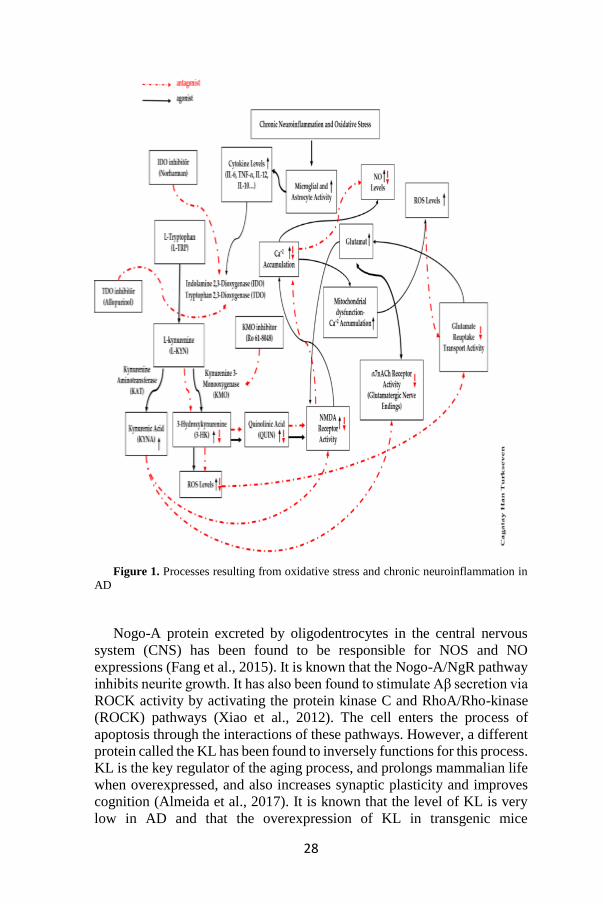
Figure 1. Processes resulting from oxidative stress and chronic neuroinflammation in
AD
Nogo-A protein excreted by oligodentrocytes in the central nervous
system (CNS) has been found to be responsible for NOS and NO
expressions (Fang et al., 2015). It is known that the Nogo-A/NgR pathway
inhibits neurite growth. It has also been found to stimulate Aβ secretion via
ROCK activity by activating the protein kinase C and RhoA/Rho-kinase
(ROCK) pathways (Xiao et al., 2012). The cell enters the process of
apoptosis through the interactions of these pathways. However, a different
protein called the KL has been found to inversely functions for this process.
KL is the key regulator of the aging process, and prolongs mammalian life
when overexpressed, and also increases synaptic plasticity and improves
cognition (Almeida et al., 2017). It is known that the level of KL is very
low in AD and that the overexpression of KL in transgenic mice
29
(EFmKL46 and EFmKL48) results in suppression of senescence
phenotypes and significantly prolongs the life span (Kurosu et al., 2005).
There are also studies that show that the excessive increase of pathological
findings of AD caused deteriorations in peripheral tissues.
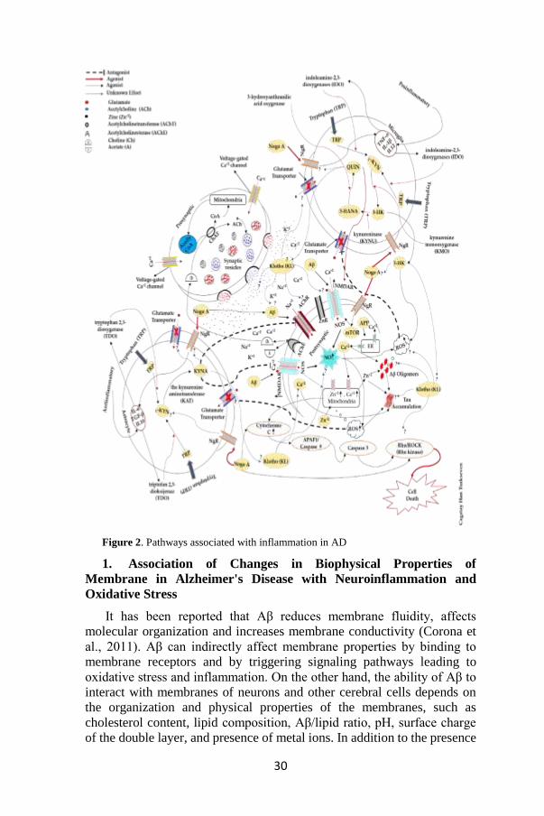
In this section, we will discuss the exact effect and the interactions of
the underlying mechanisms in the light of previous studies, although the
Nogo-A/NgR and RhoA/ROCK pathways and the KL protein play a role
in the treatment and prevention of inflammatory diseases such as AD
(Figure 2). It has been shown that amyloid cascade, cholinergic and
excitotoxicity hypotheses play a main role in the pathological development
of AD. However, recent studies on the associations between structural
variants have pointed that there are many underlying biological pathways
and that cognitive disorders occur when these pathways become impaired.
It sheds light especially on the pathways that develop with oxidative stress
and neuroinflammation. Although there are a large number of biological
pathways leading to AD, one of these pathways, which is relatively rare in
the population, should have a broad influence for useful treatments. It will
be argued whether the kynurenine pathway, which is believed to lead to
the initiation of the pathologic cascade in AD, may have such a wide effect
for treatment. In addition, the results of studies that demonstrated how the
inhibition of KMO or IDO in the kynurenine pathway altered the
inflammatory mechanisms will be discussed. The predictions on how the
relationship between the Nogo-A/NgR and RhoA/ROCK pathways (which
have significant roles in the inhibition of KMO and IDO in the kynurenine
pathway), the NO and NMDA receptors and the KL protein develops will
be revealed. In addition, this section will examine studies that reveal the
possible association between the KL protein (whose effects on aging have
been understood and which is known to be expressed in the brain) and
NMDA and NO. We believe that this interaction may create a new idea for
studies investigating the relationship between NMDA receptors (which are
the most common receptors in brain cells) and different pathways (which
are involved in the development of AD).
30
Figure 2. Pathways associated with inflammation in AD
1. Association of Changes in Biophysical Properties of
Membrane in Alzheimer's Disease with Neuroinflammation and
Oxidative Stress
It has been reported that Aβ reduces membrane fluidity, affects
molecular organization and increases membrane conductivity (Corona et
al., 2011). Aβ can indirectly affect membrane properties by binding to
membrane receptors and by triggering signaling pathways leading to
oxidative stress and inflammation. On the other hand, the ability of Aβ to
interact with membranes of neurons and other cerebral cells depends on
the organization and physical properties of the membranes, such as
cholesterol content, lipid composition, Aβ/lipid ratio, pH, surface charge
of the double layer, and presence of metal ions. In addition to the presence

31
of Aβ oligomers and tau phosphorylation, the concentration imbalances of
particularly Ca+2 and Zn+2 ions in the synaptic gap and neurons participate
in the development and progression of AD (Corona et al., 2011). Ca+2 level
is always lower in the cytosol compared with the extracellular space. This
equilibrium is achieved by the activity of Ca+2-binding proteins and by
transporting Ca+2 into the extracellular space through Ca+2-ATPase pump.
The increase in intracellular Ca+2 occurs by transporting Ca+2 into the
intracellular space through ionotropic glutamate receptors such as voltage-
gated Ca+2 channels, NMDA receptor and α-amino-3-hydroxy-5-methyl-
4-isoxazolepropionic acid (AMPA) receptor or by releasing intracellular
Ca+2 in the cytoplasm. Like Ca+2, Zn+2 is a metal ion that is lower in the
intracellular space. Unlike Ca+2, Zn+2 is stored at very high levels in some
presynaptic vesicles in glutamatergic neurons in the central nervous system
and is released into the synaptic cleft along with neurotransmitters
(Qian&Noebels, 2005). Thus, Zn+2 can accumulate in the synaptic cleft
during intense synaptic activity and enter postsynaptic neurons using some
access pathways such as AMPA receptor and less effectively NMDA
receptor. The concentration gradient between intracellular and
extracellular Ca+2 and Zn+2 levels are necessary for some central nervous
system functions such as neuronal conduction, mitochondrial function, and
various enzymatic activities. Imbalances in these concentration changes
can be a potential trigger in AD-associated neurodegeneration processes,
such as Aβ oligomerization, neurofibrillary tangle formation, and
production of reactive oxygen species (ROS) (Sensi et al., 2003; Mattson,
2010). Therefore, Aβ, tau, oxidative stress, increase in glutamate receptor
activity, and imbalance in the concentrations of Ca+2 and Zn+2 can enhance
synaptic and neuronal loss related to AD by affecting each other. When
Zn+2 ion is present in low concentrations, it inhibits NMDA receptor
activity. Because the binding affinity of Zn+2 to Aβ plaques is increased
after the accumulation of Aβ plaques, its blockage on NMDA receptor is
removed (Corona et al., 2011). However, as Aβ also activates AMPA
receptors that are directly permeable to Ca+2, there is an excessive influx
of Ca+2 into cells through both pathways. This excessive Ca+2 increase in
the cell leads to release of mitochondrial ROS and results in increased
production of NO due to Ca+2-dependent NOS. Then, ROS leads to the
release of Zn+2 ions from the mitochondria, bringing Zn+2 level in the cell
to toxic concentration.
In this situation, it causes more deterioration in mitochondrial function,
leading to further formation of reactive oxygen species and release of pro-
apoptotic factors. The rise in Zn+2 level caused by ROS can also provide
Aβ accumulation. ROS begins to be released from the cell after the
accumulation of Aβ oligomers in the cell, which prevents glutamate
reuptake by blocking glutamate transport in glial cells (Sensi et al., 2003).
Therefore, increased glutamate concentration outside the cell causes
32
excitotoxicity by leading to the hyperactivity of NMDA and AMPA
receptors (Annweiler&Beauchet, 2012). For this reason, NMDA and
AMPA receptors activity may further increase. Both increased Ca+2 level
caused by NMDA receptor activity and oxidative stress have been shown
to lead to tau hyperphosphorylation in patients with AD (Zempei et al.,
2010; Lovell et al., 2004). In addition, the human nicotinic acetylcholine
receptor (nAChR) has two functional models. One of these is the
presynaptic heteroreceptor which mediates the increase of glutamate
release and is found in glutamatergic nerve endings. The other is the
presynaptic autoreceptor that mediates the increase of acetylcholine release
and is found in cholinergic nerve endings. The autoreceptor represents the
α4β2NAChR subtype, whereas the heteroreceptor represents the
α7NAChR subtype. The pharmacology of the human nAChRs
characterized here is quite similar to the pharmacology of the
corresponding receptors in rats (Marchi et al., 2002). Some studies have
shown that (nAChR) agonists protect against neuronal excitotoxicity
caused by glutamate through alpha4 and alpha7 type nicotinic
acetylcholine receptor (Akaike et al., 2010; Shen et al., 2010). However,
the progressive loss of nicotinic receptors, which play a role in memory
and cognitive impairments during AD, has also been reported (Pettenati et
al., 2003). Recent studies have shown that acetylcholine esterase (AChE)
inhibitors suppress synaptic dysfunction, Aβ plaque formation, T cell
activation, CNS inflammation, and other neuronal damage reasons such as
inflammatory reactions caused by cytokines (Kim et al., 2014; León et al.,
2013). Increased production of ROS has been shown in mice injected with
SCO (a muscarinic receptor antagonist) (Tao et al., 2014). Oxidative stress-
induced cell death is an important cause of neurodegeneration in this rat
model of AD. Changes in the expression levels of synaptic proteins,
neurotrophic factors, and antioxidant enzymes may be responsible for
SCO-induced neurotoxicity, and regulation of such factors may be a way
to protect the cells or to return them to their original state (Xiong et al.,
2015). The brain is highly sensitive to oxidative stress due to its high
oxygen consumption and relatively poor antioxidant defense system. In
addition, the brain contains a large number of substances such as iron,
ascorbate, glutamate and unsaturated fatty acidsCwhich produce free
radicals (Walton et al., 2012). Moreover, elevated oxidative stress is
strongly associated with mitochondrial dysfunction (Leuner et al., 2012).
This induces increased processing of APP and leads to an increase in Aβ
production in AD (Anandatheerthavarada et al., 2003). Furthermore,
oxidative stress causes degeneration of cholinergic neurons and
consequently cognitive and memory impairment (Melo et al., 2003).
33
2. Association of Alzheimer's Disease with the Kynurenine
Pathway
Accumulation of Aβ plaques caused by oxidative stress activates
microglias and astrocytes to form proinflammatory cytokines such as
tumor necrosis factor alpha (TNF-alpha) and interleukins (ILs)
(Heneka&O’Banion, 2007). Proinflammatory cytokines caused by Aβ can
cause proapoptotic and synaptotoxic effects which have excessive toxic
effects on neurons (Li et al., 2011). Therefore, AD is a complex
neurodegenerative disease. Studies on only neurons are not enough to
resolve AD. In addition to the degeneration of cholinergic neurons, it is
important to recognize the role of glial and microglia cells in the
pathogenesis of AD. In a study conducted, it has been reported that glial
and microglia cells should be the key elements of AD (Dzamba et al.,
2016). Antiinflammatory and proinflammatory cytokines secreted by
astrocytes and microglial activate the kynurenine pathway (Meyer et al.,
2011). Kynurenine is an intermediate metabolite that emerges during the
degradation reactions of L-tryptophan. The vast majority of dietary
tryptophan is metabolized by the kynurenine pathway, which begins with
the oxidative opening of indole ring and ends with the production of
nicotinamide adenine dinucleotide (NAD+) (a cofactor for various
enzymes). With the breakdown of tryptophan (TRP) in this pathway, it
produces various neuroactive intermediates such as 3-HK (which produces
free radicals), QUIN, and KYNA (an antagonist of NMDA and α7 nicotinic
acetylcholine receptor) (Tayfun&Şadan, 2005). The kynurenine pathway
play a role in various physiological functions such as behavior, sleep,
thermoregulation, and pregnancy. In addition, the kynurenine pathway is
associated with many diseases such as AIDS, dementia complex,
inflammatory brain diseases, Huntington's disease, Down syndrome,
sepsis-associated encephalopathy, toxoplasmosis, malaria, epilepsy and
traumatic brain injury. It is also thought to be related to the pathogenesis
of AD (Tayfun&Şadan, 2005). Although a small amount of L-KYN can be
produced in the brain, the kynurenine pathway in the brain is maintained
by L-KYNs that enter the brain from the circulation, essentially using the
neutral amino acid transporter. L-KYN in the brain is rapidly taken up by
astrocytes and microglial cells. Due to its functional significance, KYNA
is synthesized mostly in these cells because astrocytes do not contain 3-
HK. However, since microglial cells have very low kynurenine
aminotransferase (KAT) activity, these cells preferably form intermediates
of the QUIN branch of the pathway (Tayfun&Şadan, 2005). As the
proinflammatory cytokines secreted by microglial cells (especially TNF-
α, IL-12, IL-1 beta) stimulate the enzyme indolamine 2,3-dioxygenase
(IDO1 and IDO2), they increase the KMO activity via L-KYN and the
levels of QUIN and 3-HK.
34
QUIN and 3-HK have neurotoxic effects (Miech et al., 2002). As QUIN
and 3-HK (a neuroactive metabolite) act as an effective free radical
generator, QUIN also exhibits an agonist of NMDA receptor. Furthermore,
QUIN induces lipid peroxidation and causes the level of extracellular
glutamate to rise, thus resulting in an increase in excitotoxicity (Majláth et
al., 2014). As the anti-inflammatory cytokines (especially IL-6, TGF-β,
IL10) secreted by astrocytes stimulate the enzyme tryptophan 2,3-
dioxygenase (TDO), they increase the KMO activity via L-KYN and the
production of KYNA from L-tryptophan (L-TRP). KYNA is a potent
antagonist of NMDA and α7 nicotinic acetylcholine receptor (α7 nAChR)
(Meyer et al., 2011). KYNA exhibits a competitive antagonist effect by
binding to the glycine-binding regions of NMDA receptors (Kessler et al.,
1989). KYNA has a dual effect on the AMPA receptors depending on the
concentration: It exhibits an agonist effect at low concentrations and an
antagonist effect at high concentrations (Prescott et al., 2006; Rozsa et al.,
2008). Because KYNA inhibits NMDA receptors, it exerts neuroprotective
effect by preventing glutamate excitotoxicity. However, it inhibits
α7NACh receptors, which may contribute to this effect. Because these
receptors are associated with the regulation of presynaptic glutamate
release (Marchi et al., 2002). The L-KYN/TRP ratio was found to higher
in the blood and cerebrospinal fluid (CSF) in patients with AD compared
to healthy subjects. This situation has been reported to be strongly related
to the disruption of the KP (Gulaj et al., 2010). This altered rate has been
associated with an increase in IDO1 levels in the brain tissue (Bonda et al.,
2010) and in 3-HK levels in blood serum (Schwarz et al., 2013) in AD.
IDO and QUIN exhibited a significant immunoreactivity in hippocampal
neurons, astrocytes, and microglia in AD. This activity was observed at
high levels, especially around senile plaques (Guillemin et al., 2005). IDO
and QUIN are also present in the neurofibrillary tangles formed in AD.
QUIN is also present in the intracellular granular deposits of cortical
neurons (Bonda et al., 2010; Guillemin et al., 2005). Aβ peptides induce
IDO1 expression and increase QUIN production in human macrophages
and microglia (Guillemin et al., 2003). Studies in both humans and rats
have shown that the increased Aβ production is associated with deep
induction of KP by inflammatory cytokines which trigger IDO, TDO and
KMO activity (Yamada et al., 2009; Lue et al., 2001; Akimoto et al., 2007).
In addition, treatment of human neurons with QUIN results in the
upregulation of genes associated with tau phosphorylation. This potentially
provides a mechanism in which neurofibrillary tangles are formed in AD
(Rahman et al., 2009). Various studies have shown that 3-HK and QUIN
levels increased in the blood and CSF obtained from patients with AD,
while KYNA levels decreased. The fact that this change has led to the
development of neurotoxic metabolites instead of neuroprotective species
has suggested that it contributes to the pathogenesis of AD
35
(Maddison&Giorgini, 2015). Another study assessed 3-HK, QUIN (which
are neurotoxic metabolites of the kynurenine pathway) and NO levels in
interferon-activated macrophages. It was found that 3-HK, QUIN and NO
accumulations reached the neurotoxic levels in the incubation medium
with the induction of IDO and NOS by interferon-ɣ. The exposure of
macrophages with norharman (an IDO inhibitor) has reduced not only the
kynurenine pathway metabolites but also the NO formation. (Chiarugi et
al., 2000). There are also studies on the inhibition of KMO activity which
is responsible for the formation of 3-HK and QUIN associated with
neurotoxicity in the kynurenine pathway except for IDO inhibitors.
In a study of mouse model of multiple sclerosis, it was reported that
KMO activity was increased excessively and that this increase reached
neurotoxic levels with 3-HK and QUIN. It was shown that the inhibition
of KMO activity in the kynurenine pathway with 3,4-dimethoxy-N-[4-(3-
nitrophenyl) thiazol-2-yl] reduced 3-HK and QUIN levels and also caused
the accumulation of KYNA (Chiarugi et al., 2001). In addition, it was
found that the inhibition of KMO activity with Ro 61-8048 in the rat brain
increased KYNA levels and that the extracellular glutamate concentration
decreased in parallel to this (Moroni et al., 2005). In a study of alcoholic
rat model performed with the idea that high glutamatergic activity and
NMDA receptor activation may lead to drug craving and relapse, it was
reported that the inhibition of KMO activity with Ro 61-8048 caused a
decrease in glutamatergic/NMDA receptor activity through different
mechanisms by shifting the metabolic kynurenine pathway towards the
production of KYNA (Vengeliene et al., 2016). In the light of these
information, it is possible that the increase in intracellular Ca+2 due to
NMDA receptor overactivity depending on the increase in 3-HK and
QUIN levels in patients with AD leads to the release of mitochondrial
reactive oxygen species and that the increase in NO production caused by
Ca+2-induced NOS causes an increase in the formation of neurotoxic
effects and in the accumulation level of Aβ oligomers.
3. Interaction of NMDA receptor with association between Nogo-
A/Nogo receptor and Rho/ROCK pathway in Alzheimer's Disease
Recent studies have shown that the Nogo-A/Nogo receptor (NgR) is
associated with pro-inflammatory and anti-inflammatory cytokines or
cytotoxic factors released by microglias and astrocytes which are
hyperactivated due to inflammation in CNS. Nogo-A has been found to be
responsible for the expression of these inflammatory mediators (iNOS,
cyclooxygenase-2 (COX-2), IL-1, TNF-α, NO) (Fang et al., 2015). It is a
protein that is expressed by oligodendrocytes, an important component of
myelin production in CNS (Ineichen et al., 2017). It is known that the
Nogo-A/NgR pathway contributes to the inhibition of neurite regeneration
by neurons after CNS damage and limits axonal regeneration (Huebner et

36
al., 2009; Schwab&Strittmatter, 2014; Wälchli et al., 2013). Recent
findings have shown that Nogo-A is overexpressed by hippocampal
neurons and is associated with Aβ accumulation in patients with AD.
Nogo-A and NgR also play a role in the pathology of AH (Park et al.,
2006). It has been shown that more than 50% of hippocampal pyramidal
cells in the brain with AD have NgR immunoreactivity and that this may
be associated with fibrillar tangle formation (Zhu et al., 2007). Loss of both
Nogo/NgR in the brain tissue of patients with AD reduces Aβ accumulation
(Park et al., 2006). Nogo-A binds to its functional receptor NgR to inhibit
axonal growth by activating the intracellular signal molecules with the
small GTP-binding protein RhoA and its effector ROCK (Yin et al., 2014; Zagrebelsky&Korte, 2014). One study has revealed that the accumulation
of Aβ oligomers which increases via inflammatory mediators in the brain
tissue with AD is associated with the Nogo-A/NgR signal (Xiao et al.,
2012). It was found that the activation of NgR not only inhibited neurite
growth in cortical neurons but also activated Aβ release by ROCK by
activating the protein kinase C and Rho/Rho-kinase (ROCK) pathways.
These findings have indicated that Nogo-A overexpression and
activation of NgR alter neuronal metabolism by inhibiting neurite growth,
leading to overproduction and/or secretion of Aβ (Xiao et al., 2012). In a
study conducted on Nogo-A/NgR and glutamate receptors NMDA and
AMPA interactions, it was found that the transcription and translation of
Nogo A in hippocampal neurons were regulated under the control of NgR1
stimulation via the Rho-ROCK and MAPK pathways and that the
interaction between neuronal Nogo-A/NgR1 regulated glutamatergic
transmission by altering NMDA and AMPA receptor levels via rapamycin-
sensitive mTOR pathway (Peng et al., 2011). Levels of Ca+2 and Zn+2 ions
in the cell increase due to neuronal cell death and glutamatergic
excitotoxicity in AD. This leads to the release of cytochrome c and
caspase-dependent apoptosis. Released cytochrome c binds to apoptotic
protease activating factor-1 (Apaf-1) and procaspase-9 to form an
apoptosome, which regulates the caspase activation cascade. The
apoptosome complex activates caspase-9, which promotes caspase-3
activation, eventually leading to cell death (Elmore, 2007). Membrane
budding, which is observed with the intrusions and protrusions of the cell
membrane, indicates the beginning of cell death. This membrane bud
formation occurs via some G proteins such as Rho protein. Rho protein is
effective in signaling pathways that control gene expression and actin
cytoskeleton. ROCKs have been discovered as the first effectors of Rho
kinases. ROCK has two isoforms including ROCK1 and ROCK2 (Leung
et al., 1995; Ishizaki et al., 1996; Matsui et al., 1996). ROCK1 is cleaved
by caspase 3 at the cleavage site DETD1113 during apoptosis (Coleman et
al., 2001; Sebbagh et al., 2001). The activation of ROCK1 via caspase 3 is
responsible for membrane bud formation in all apoptotic events. Many
37
studies have shown that this pathway mediates inflammatory events and
tissue/organ damage (Büyükafşar et al., 2004). There are many enzymes
and proteins involved in neuropathology and neuroinflammation of AD,
the most important of which is Rho kinase (ROCK) (Kubo et al., 2008).
Agents that inhibit the Rho/Rho-kinase pathway may be new class
analgesic and anti-inflammatory drugs (Büyükafşar et al., 2006). On the
other hand, the inhibition of Rho-kinase activity may stimulate axonal
regeneration (Borisoff et al., 2003). ROCK inhibitors such as Y-27632 and
fasudil bind to the kinase domain of the enzyme and inhibit both ROCK1
and ROCK2 (Breitenlechner et al., 2003). In a study conducted using
fasudil (a ROCK inhibitor) in rats, fasudil has been found to exert a
protective effect by suppressing the inflammatory response to hippocampal
neurodegeneration caused by AD (Song et al., 2013). A different study
demonstrated that NgR inhibition in murine retinal ganglion cells caused a
decrease in the activity of Rho/ROCK (Zeng et al., 2017). On the other
hand, the inhibition of the Rho-ROCK or MEK-MAPK signaling pathway
in hippocampal neurons caused a similar decrease in neuronal Nogo-A,
mRNA and protein (Peng et al., 2011). All these processes are considered
to be the main pathways leading to loss of synaptic function and apoptosis
of neuronal cells in AD.
4. Association between Klotho protein and Alzheimer's Disease
The Japanese study group found a protein named KL that exert an effect
in opposite direction of the Nogo-A/NgR and Rho/ROCK pathways
(Almeida et al., 2017). They have identified a new gene (KL) that encodes
a membrane protein that suppresses certain physiological properties
associated with shortening of life, infertility, atherosclerosis, skin atrophy,
aging phenotype in mice, emphysema and osteoporosis (Almeida et al.,
2017). Later studies have shown that the KL gene is present on
chromosome 13 in humans and encodes membrane-bound and secreted
proteins. They have observed that the human KL gene expression
decreases in elderly people compared to newborns, and therefore it is
thought that decreased KL expression shortens human life (Almeida et al.,
2017). One study has demonstrated that increased expression of KL protein
in human amyloid precursor protein (hAPP) transgenic mice, which mimic
key aspects of AH, reduces premature mortality and network dysfunction.
In addition, increased expression of KL inhibited the reduction of NMDA
receptor subunits in the hippocampus and enhanced spatial learning and
memory in hAPP mice. Increased expression of KL in hAPP mice
increased the abundance of the GluN2B subunit of NMDA receptor at
postsynaptic densities and the long-term potentiation dependent on NMDA
receptor, which is critical for learning and memory. Thus, increased KL
expression or activitiy improved synaptic and cognitive functions (Dubal
et al., 2015). KL is most expressed in the brain, kidneys and reproductive
38
organs. In the brain, it is expressed by the choroid plexus and neurons,
especially by cerebellar Purkinje fibers in the hippocampus and
hypophysis. It is a protein that plays a role in intracellular calcium changes,
which we think is involved in the metabolic pathway (Nabeshima et al.,
2008). The human KL gene is composed of 5 exons and 4 introns and
ranges over 50 kb on chromosome 13q12 (Kuro-o et al., 1997). In mice
and humans, the KL gene encodes a transcript of 5.2 kb. In the third exon,
there is an alternative splicing donor site that can generate two different
transcripts: one encoding a transmembrane form (full-length transcript,
1014 amino acids), and the other, a secreted form of the protein (truncated
transcript, 550 amino acids). The full-length transcript encodes a single
pass transmembrane protein with a molecular weight of approximately 130
kDa (m-KL). The transcript generated by alternative splicing generates a
secreted form of the protein (s-KL) that is formed solely by the KL1
domain, with an approximate weight of 70 kDa (Shiraki-Iida et al., 1998).
Studies have suggested that the soluble KL protein, which acts as a
circulating hormone, has an effect on ion transport and has anti-aging and
anti-oxidant properties (Dubal et al., 2014). Additionally, it plays a role in
the regulation of calcium metabolism by suppressing 1α-hydroxylase in the
kidneys and in the regulation of parathormone (PTH) synthesis in the
parathyroid gland (Olauson et al., 2013). The secreted KL protein has been
detected in the blood, urea, and cerebrospinal fluid. In a study investigating
whether KL affects physiological brain function and more specifically
whether it plays a role in the prevention or inactivation of cognitive
impairment in human aging, elevated serum level of KL has been shown
to increase cognitive performance (Zeldich et al., 2014).
Cerebrospinal fluid KL levels were found to be lower in patients with
AD compared to healthy subjects (Semba et al., 2014). The overexpression
of KL leads to a suppression of senescence phenotypes and a significant
prolongation of the life span in transgenic mice (EFmKL46 and
EFmKL48) compared to healthy mice (Kurosu et al., 2005). Further studies
have shown that KL is associated with the regulation of calcium and
phosphate homeostasis and the inhibition of intracellular insulin and
insulin-like growth factor-1 activity (Kuro-o, 2010). Elevated lipid
peroxidation and oxidative DNA damage in the hippocampus of KL
mutant mice occurs before the formation of cognitive impairments (Nagai
et al., 2003).
5. Conclusion
AD is a progressive neurological disorder that is associated with aging
and leads to various mental and behavioral disorders, primarily
forgetfulness. Although AD is associated with aging, its pathogenesis
develops as a consequence of abnormal brain processes. Early detection of
the disease is very difficult, and there is not yet a treatment for AD. For
39
this reason, studies targeting diagnosis and therapy are very important for
AD (Castellani et al., 2010). Many studies have focused on basically three
hypotheses that occur in the relevant neuroanatomical areas of the brain in
AD (Parihar&Hemnani, 2004). The amyloid cascade hypothesis occurs as
a result of the intracellular accumulation of neurofibrillary tangles and the
extracellular accumulation of amyloid beta (Aβ) protein. In primary
cultures prepared from cortical neurons of Wistar rats, it was found that Aβ
increased Ca+2 release from the endoplasmic reticulum (ER). It was also
shown that the mitochondria could capture large amounts of Ca+2 because
the endoplasmic reticulum is closely juxtaposed to the mitochondria, and
that this also activated the mitochondrial apoptotic pathway. Thus, one of
the mechanisms of neuronal loss seen in AD could have been understood
(Ferreiro et al., 2008). It is known that disruption of ion balance in neuronal
cells plays a role in the pathogenesis of AD. It was demonstrated using
standard fluorescence imaging techniques in cell cultures that Aβ
modulated Ca+2 channels in the lipid vesicles and altered intracellular Ca+2
concentration in astrocytes and neuron cells (Abramov et al., 2004).
Electrophysiological studies have shown that Aβ enhances long-term
potential in the rat gyrus and thus facilitates synaptic plasticity under in
vitro conditions. It was shown that whereas acute treatment of young rat
(70-120 days) hippocampal slices with low concentration (100-200 nM) of
bath-applied Aβ1-40 did not change basal synaptic transmission, there was
an increase in tetanus-induced LTP (Wu et al., 1995).
Another function of Aβ that plays a role in neurodegeneration is that
when it accumulates in the mitochondrial membrane, it leads to the
formation of oxygen free radicals by causing mitochondrial dysfunction
(Pieczenik&Neustadt, 2007; Mattson, 2004). Free radicals are toxic
because they can interact with most of cellular macromolecules such as
proteins, lipids, and DNA. Excessive amounts of oxygen free radicals in
various pathological conditions or increased free radicals as a result of
insufficient antioxidant system in the structure of the organism lead to cell
damage by affecting various components of the cell (Defeng&Cederbaum,
2003). As a result of oxidative stress induced by neuronal damage and
chronic neuroinflammation, antiinflammatory and proinflammatory
cytokines secreted by astrocytes and microglial activates the kynurenine
pathway (Bonda et al., 2010). A lot of evidence suggests that kynurenine
pathway metabolites play a role in the pathology of many neurocognitive
and neurodegenerative disorders (Maddison&Giorgini, 2015). Levels of
IDO and TDO increase in the brain with AD. This increase increases KMO
and KAT activities. It is known that this increases levels of QUIN and 3-
HK. Increased levels of QUIN and 3-HK exhibit neurotoxic effects
(Moroni et al., 2005). It was shown that increased level of QUIN increased
tau pathology by inducing tau hyperphosphorylation which occurred in
AD, and that this directly caused excitotoxicity (Rahman et al., 2009).
40
Furthermore, a study in AD Tg2576 transgenic mouse model suggests that
a synergistic effect between Aβ and proinflammatory cytokines has an
important role in elevating IDO level (Akimoto et al., 2007). This suggests
us that Ca+2-dependent inducible NOS is activated by an increase in the
QUIN (an NMDA agonist) level, leading to an increase in the production
of NO (Moroni et al., 2005). Nogo-A protein has been found to be
responsible for NOS and NO expressions (Fang et al., 2015). It is known
that the Nogo-A/NgR pathway inhibits neurite growth. It has also been
found to activate Aβ release by ROCK by activating the protein kinase C
and RhoA/Rho-kinase (ROCK) pathways (Xiong et al., 2015). It is known
that the KL protein, which acts inversely to the Nogo-A/NgR and
Rho/ROCK pathways, enhances cognitive performance and prolongs the
life span (Dubal et al., 2015; Zeldich et al., 2014). There are studies
showing that lipid peroxidation and oxidative damage prior to the onset of
cognitive impairment are associated with the KL protein and that the
interaction of the KL protein with NMDA receptor increases plasticity
(Dubal et al., 2015; Ferreiro et al., 2008). Although the Nogo-A/NgR and
RhoA/ROCK pathways and the KL protein play a role in the treatment and
prevention of inflammation-related diseases such as AD, there is a need for
determining the exact effect and underlying mechanism. Recent genetic
and pharmacological approaches suggest that KP may be a valid
therapeutic approach in neurodegenerative disorders and can complete
previous metabolic studies in this direction (Maddison&Giorgini, 2015).
It is clear that increased synthesis of QUIN and 3-HK and reduced
production of KYNA have been demonstrated in the majority of
neurodegenerative diseases linked to KP and that this can be addressed by
reversed interventions (Maddison&Giorgini, 2015). KMO has shown a
great potential as a therapeutic target in the treatment of
neurodegeneration, especially in KP enzymes. Because inhibition of this
enzyme leads to an increase in the production of neuroprotective KYNA
and reduces levels of QUIN and 3-HK. In addition, studies have shown
that IDO or TDO inhibition can also prove to be an effective
neuroprotective strategy (Maddison&Giorgini, 2015). Clinical trials
planned with KMO inhibitors may be an important first step in
understanding the therapeutic value of KP manipulation in the context of
neurodegenerative diseases in general (Maddison&Giorgini, 2015). In
light of these findings, KP can be defined as a new underlying mechanism
of AD neurodegeneration.
41
References
Abramov AY, Canevarib L, Duchen MR. Calcium signals induced by
amyloid β peptide and their consequences in neurons and astrocytes in
culture. Biochimica et Biophysica Acta. 2004;1742(1-3):81-87.
Akaike A, Takada Y, Takatori Y, Kume T, Izumi Y. Mechanisms of
neuroprotective effects of nicotine and acetylcholinesterase inhibitors:
role. of alpha4 and alpha7 receptors in neuroprotection. J. Mol. Neurosci.
2010; 40:211–16.
Akimoto H, Yamada A, Takikawa O. Up-regulation of the brain
indoleamine 2,3-dioxygenase activity in a mouse model of Alzheimer’s
disease by systemic endotoxin challenge. Int Cong Ser 2007; 1304: 357–
61.
Almeida OP, Morar B, Hankey GJ, Yeap BB, Golledge J, Jablensky A,
Flicker L. Longevity Klotho gene polymorphism and the risk of dementia
in older men. Maturitas. 2017; 101:1-5.
Anandatheerthavarada HK, Biswas G, Robin M-A, Avadhani NG.
Mitochondrial targeting and a novel transmembrane arrest of Alzheimer’s
amyloid precursor protein impairs mitochondrial function in neuronal
cells. J Cell Biol. 2003; 161(1):41–54.
Annweiler C, Beauchet O. Possibility of a new anti-alzheimer's disease
pharmaceutical composition combining memantine and vitamin D. Drugs
Aging 2012;29(2):81-91.
Askarova S, Yang X, Lee J. Impacts of Membrane Biophysics in
Alzheimer’s Disease: From Amyloid Precursor Protein Processing to Aβ
Peptide-Induced Membrane Changes. International Journal of Alzheimer’s
Disease 2011;10(4061):1-12.
Becker R, Giacobini E, Elble R, McIlhany M, Sherman K. Potential
pharmacotherapy of Alzheimer’s disease. A comparison of various forms
of physostigmine administration. Acta Neurol. Scand. Suppl. 1988;
116:19–32.
Bonda DJ, Mailankot M, Stone JG, Garrett MR, Staniszewska M,
Castellani RJ, Siedlak SL, Zhu X, Lee HG, Perry G, Nagaraj RH, Smith
MA. Indoleamine 2,3-dioxygenase and 3-hydroxykynurenine
modifications are found in the neuropathology of Alzheimer’s disease,
Redox Rep. 2010;15:161–8.
Borisoff JF, Chan CC, Hiebert GW, Oschipok L, Robertson GS,
Zamboni R, Steeves JD, Tetzlaff W. Suppression of Rho-kinase activity
promotes axonal growth on inhibitory CNS substrates. Mol Cell Neurosci
2003; 22:405-16.
42
Breitenlechner C, Gassel M, Hidaka H, Kinzel V, Huber R, Engh RA,
Bossemeyer D. Protein kinase A in complex with Rho-kinase inhibitors Y-
27632, Fasudil, and H-152P: structural basis of selectivity. Structure
2003;11:1595-607.
Breitner JC, Silverman JM, Mohs RC, Davis KL. Familial aggregation
in Alzheimer's disease: comparison of risk among relatives of early-and
late-onset cases, and among male and female relatives in successive
generations. Neurology. 1988;38(2):207-12.
Buyukafsar K, Arıkan O, Ark M, Seçilmiş A, Ün I, Şingirik E. Rho-
kinase expression and its contribution to the control of perfusion pressure
in the isolated rat mesenteric vascular bed. European journal of
pharmacology 2004;485(1):263-268.
Buyukafsar K, Yalçın İ, Kurt AH, Tiftik RN, Şahan S, Fırat S, Aksu F.
Rho-kinase inhibitor, Y-27632, has an antinociceptive effect in mice.
European journal of pharmacology 2006; 541:49-52.
Castellani RJ, Rolston RK, Smith MA. Alzheimer Disease. Dis Mon.
2010;56(9):484-546.
Chiarugi A, Cozzi A, Ballerini C, Massacesi L, Moroni F. Kynurenine
3-mono-oxygenase activity and neurotoxic kynurenine metabolites
increase in the spinal cord of rats with experimental allergic
encephalomyelitis. Neuroscience. 2001;102(3):687-95.
Chiarugi A, Dello Sbarba P, Paccagnini A, Donnini S, Filippi S, Moroni
F. Combined inhibition of indoleamine 2,3-dioxygenase and nitric oxide
synthase modulates neurotoxin release by interferon-gamma-activated
macrophages. J Leukoc Biol. 2000; 68(2):260-6.
Coleman ML, Sahai EA, Yeo M, Bosch M, Dewar A, Olson MF.
Membrane blebbing during apoptosis results from caspase-mediated
activation of ROCK 1. Nat Cell Biol. 2001; 3:339-45.
Corona C, Pensalfini A, Frazzini V, Sensi SL. New therapeutic targets
in Alzheimer’s disease: brain deregulation of calcium and zinc. Cell Death
and Disease 2011; 57:2041-4889.
Defeng W, Cederbaum AI. Alcohol, oxidative stres and free radical
damage. Alcohol Res Heath 2003;27 (4):277-284.
Dubal DB, Zhu L, Sanchez PE, Worden K, Broestl L, Johnson E, Ho
K, Yu GQ, Kim D, Betourne A, Kuro-O M, Masliah E, Abraham CR,
Mucke L. Life extension factor klotho prevents mortality and enhances
cognition in hAPP transgenic mice, J Neurosci. 2015; 35:2358-71.
Dubal D, Yokoyama JS, Zhu L, Broestl L, Worden K, Wang D, Sturm
VE, Kim D, Klein E, Yu GQ, Ho K, Eilertson KE, Yu L, Kuro-o M, De
43
Jager PL, Coppola G, Small GW, Bennett DA, Kramer JH, Abraham CR,
Miller BL, Mucke, L. Life Extension Factor Klotho Enhances Cognition,
Cell Reports 2014:7:1065–1076.
Dzamba D, Harantova L, Butenko O, Anderova M. Glial cells-the key
elements of Alzheimer´ s disease. Curr Alzheimer Res. 2016; 13(8):894-
911.
Elena Z, Gennj P, Gabriela C. The blood-brain barrier in Alzheimer's
disease. Neurobiology of Disease 2017; 107:41-56.
Elmore, S. Apoptosis: a review of programmed cell death. Toxicol
Pathol. 2007;35(4):495-516.
Fang Y, Yan J, Li C, Zhou X, Yao L, Pang T, Yan M, Zhang L, Mao L,
Liao H. The Nogo/Nogo Receptor (NgR) Signal Is Involved in
Neuroinflammation through the Regulation of Microglial Inflammatory
Activation. J Biol Chem. 2015;290(48):28901-14.
Ferreiro E, Oliveira CR, Pereira CMF. The release of calcium from the
endoplasmic reticulum induced by amyloid-beta and prion peptides
activates the mitochondrial apoptotic pathway. Neurobiology of Disease
2008;30(2008):331–342.
Glenner GG, Wong CW. Alzheimer’s disease: initial report of the
purification and characterization of a novel cerebrovascular amyloid
protein. Biochem Biophys Res Commun. 2012;425(3):534-539.
Guillemin GJ, Brew BJ, Noonan CE, Takikawa O, Cullen KM.
Indoleamine 2,3-dioxygenase and quinolinic acid immunoreactivity in
Alzheimer’s disease hippocampus. Neuropathol Appl Neurobiol. 2005;
31:395–404.
Guillemin GJ, Smythe GA, Veas LA, Takikawa O, Brew BJ. A beta 1–
42 induces production of quinolinic acid by human macrophages and
microglia. Neuroreport. 2003; 14:2311–5.
Gulaj E, Pawlak K, Bien B, Pawlak D. Kynurenine and its metabolites
in Alzheimer’s disease patients. Adv Med Sci. 2010; 55:204–11.
Guo C, Shen J, Meng Z, Yang X, Li F. Neuroprotective effects of
polygalacic acid on scopolamine-induced memory deficits in mice.
Phytomedicine 2016;23(2):149-55.
Heneka MT, O’Banion MK. Inflammatory processes in Alzheimer’s
disease. J. Neuroimmunol. 2007;184: 69–91.
Huebner EA, Strittmatter SM. Axon regeneration in the peripheral and
central nervous systems. Results Probl Cell Differ. 2009; 48:339-351.
44
Ineichen BV, Kapitza S, Bleul C, Good N, Plattner PS, Seyedsadr MS,
Kaiser J, Schneider MP, Zörner B, Martin R, Linnebank M, Schwab ME.
Nogo-A antibodies enhance axonal repair and remyelination in neuro-
inflammatory and demyelinating pathology, Acta Neuropathol. 2017;
134:423-440.
Ishizaki T, Maekawa M, Fujisawa K, Okawa K, Iwamatsu A, Fujita A,
Watanabe N, Saito Y, Kakizuka A, Morii N, Narumiya S. The small
GTPbinding protein Rho binds to and activates a 160 kDa Ser/Thr protein
kinase homologous to myotonic dystrophy kinase. EMBO J. 1996;
15:1885-93.
Jorm AF, Jolley D. The incidence of dementia: a meta-analysis.
Neurology. 1998;51(3):728-33.
Jorm AF, Korten AE, Henderson AS. The prevalence of dementia: a
quantitative integration of the literature. Acta Psychiatr Scand.
1987;76(5):465-79.
Kessler M, Terramani T, Lynch G, Baudry M. A glycine site associated
with N-methyl-D-aspartic acid receptors: characterization and
identification of a new class of antagonists. J Neurochem.
1989;52(4):1319–1328.
Kim MH, Kim S, Yang WM. Mechanisms of action of phytochemicals
from medicinal herbs in the treatment of Alzheimerʼs disease. Planta Med.
2014; 80:1249-1258.
Kubo T, Yamaguchi A, Iwata N, Yamashita T. The therapeutic effects
of Rho-ROCK inhibitors on CNS disorders. Ther. Clin. Risk Manag. 2008;
4:605–15.
Kuro-o M. Klotho. Eur J Physiol. 2010;459:333–343.
Kuro-o M, Matsumura Y, Aizawa H, Kawaguchi H, Suga T, Utsugi T,
Ohyama Y, Kurabayashi M, Kaname, T, Kume E, Iwasaki H, Iida A, .
Shiraki-Iida T, Nishikawa S, Nagai R, Nabeshima YI. Mutation of the
mouse klotho gene leads to a syndrome resembling ageing, Nature 1997;
390:45-51.
Kurosu H, Yamamoto M, Clark JE, Pastor JV, Nandi A, Gurnani P,
McGuinness OP, Chikuda H, Yamaguchi M, Kawaguchi H, Shimomura I,
Takayama Y, Herz J, Kahn CR, Rosenblatt KP, Kuro-o M. Suppression of
aging in mice by the hormone klotho, Science 2005; 308:1829–1833.
León R, Garcia AG, Marco-Contelles J. Recent advances in the
multitarget‐directed ligands approach for the treatment of Alzheimer's
disease. Medicinal research reviews 2013; 33:139-189.
45
Leuner K, Müller WE, Reichert AS. From mitochondrial dysfunction
to amyloid beta formation: novel insights into the pathogenesis
ofAlzheimer’s disease. MolNeurobiol. 2012;46(1):186–193.
Leung T, Manser E, Tan L, Lim L. A novel serine/threonine kinase
binding the Ras-related RhoA GTPase which translocates the kinase to
peripheral membranes. J. Biol Chem. 1995; 270:29051-4.
Li B, Zhong L, Yang X, Andersson T, Huang M, Tang SJ. WNT5A
signaling contributes to Ab-induced neuroinflammation and neurotoxicity.
PLoS ONE 2011;6:e22920.
Lin MT, Beal MF. Mitochondrial dysfunction and oxidative stress in
neurodegenerative diseases. Nature. 2006;443(7113):787–795.
Lovell MA., Xiong S, Xie C, Davies P, Markesbery WR. Induction of
hyperphosphorylated tau in primary rat cortical neuron cultures mediated
by oxidative stress and glycogen synthase kinase-3. J Alzheimers Dis.
2004; 6:659–671.
Lue LF, Rydel R, Brigham EF, Yang LB, Hampel H, Jr Murphy GM,
Brachova L, Yan SD, Walker DG, Shen Y, Rogers J. Inflammatory
repertoire of Alzheimer’s disease and nondemented elderly microglia in
vitro, Glia. 2001; 35:72–9.
Maddison DC, Giorgini F. The kynurenine pathway and
neurodegenerative disease. Semin Cell Dev Biol. 2015; 40:134-41.
Majláth Z, Toldi J, Vécsei L. The potential role of kynurenines in
Alzheimer's disease: pathomechanism and therapeutic possibilities by
influencing the glutamate receptors. J Neural Transm (Vienna).
2014;121(8):881-9.
Manly JJ, Merchant CA, Jacobs DM, Small SA, Bell K, Ferin M,
Mayeux R. Endogenous estrogen levels and Alzheimer's disease among
postmenopausal women. Neurology 2000; 54:833-7.
Marchi M, Risso F, Viola C, Cavazzani P, Raiteri M. Direct evidence
that release-stimulating alpha7* nicotinic cholinergic receptors are
localized on human and rat brain glutamatergic axon terminals. J
Neurochem 2002;80(6):1071–1078.
Martin P, Anders W, Maëlenn G, Gemma-Claire A, Yu-Tzu W,
Matthew P (Eds). World Alzheimer Report 2015 The Global Impact of
Dementia An analysis of prevalence, incidence, cost & trends Alzheimer’s
Disease International. London, 2015; 1-92.
Masters CL, Simms G, Weinman NA, Multhaup G, Mcdonald L. B,
Beyreuther K. Amyloid plaque core protein in Alzheimer disease and
Down syndrome. Proc Natl Acad Sci USA. 1985; 82:4245-4249.
46
Matsui T, Amano M, Yamamoto T, Chihara K, Nakafuku M, Ito M,
Nakano T, Okawa K, Iwamatsu A, Kaibuchi K. Rho-associated kinase, a
novel serine threonine kinase, as a putative target for small GTP binding
protein Rho, EMBO J. 1996; 15:2208-16.
Mattson M.P. ER calcium and Alzheimer’s disease: in a state of flux.
Sci Signal. 2010; 3:10.
Mattson M.P. Pathways towards and away from Alzheimer’s disease.
Nature 2004;430 (7000):631-639.
McGonigal G, Thomas B, McQuade C, Starr JM, MacLennan WJ,
Whalley LJ. Epidemiology of Alzheimer's presenile dementia in Scotland.
BMJ. 1993;306(6879):680-3.
Melo JB, Agostinho P, Oliveira CR. Involvement of oxidative stress in
the enhancement of acetylcholinesterase activity induced by amyloid beta-
peptide. Neurosci Res. 2003;45(1):117–127.
Meyer U, Schwarz MJ, Müller N. Inflammatory processes in
schizophrenia: a promising neuroimmunological target for the treatment of
negative/cognitive symptoms and beyond. Pharmacol Ther. 2011;
132(1):96-110.
Miech RA, Breitner JCS, Zandi PP, Khachaturian AS, Anthony JC,
Mayer L. Incidence of AD may decline in the early 90s for men, later for
women: The Cache County study. Neurology. 2002;58 (2):209-18.
Moroni F, Cozzi A, Carpendo R, Cipriani G, Veneroni O, Izzo E.
Kynurenine 3-mono-oxygenase inhibitors reduce glutamate concentration
in the extracellular spaces of the basal ganglia but not in those of the cortex
or hippocampus. Neuropharmacology. 2005;48(6):788-95.
Nabeshima Y, Imura H. α-Klotho: A Regulator That Integrates Calcium
Homeostasis. Am J Nephrol. 2008; 28:455-464.
Nagai T, Yamada K, Kim HC, Kim YS, Noda Y, Imura A, Nabeshima
Y, Nabeshima T. Cognition impairment in the genetic model of aging
klotho gene mutana mice: a role of oxidative stress. FASEB J 2003; 17:50–
52.
Olauson H, Lindberg K, Amin R, Sato T, Jia T, Goetz R, Mohammadi
M, Andersson G, Lanske B, Larsson TE. Parathyroid-Specific Deletion of
Klotho Unravels a Novel Calcineurin-Dependent FGF23 Signaling
Pathway That Regulates PTH Secretion. PLOS Genetics 2013;9(12):1-10.
Parihar MS, Hemnani T. Alzheimer’s disease pathogenesis and
therapeutic İnterventions. Journal of Clinical Neuroscience
2004;11(5):456–467.
47
Park JH, Gimbel DA, GrandPre T, J. Lee K., Kim JE, Li W, Lee DH,
Strittmatter SM. Alzheimer precursor protein interaction with the Nogo-66
receptor reduces amyloid- beta plaque deposition. J Neurosci. 2006;
26:1386-1395.
Peng X, Kim J, Zhou Z, Fink DJ, Mata M. Neuronal Nogo-A regulates
glutamate receptor subunit expression in hippocampal neurons. J
Neurochem. 2011;119(6):1183-93.
Pettenati C, Annicchiarico R, Caltagirone C. Clinical pharmacology of
anti-Alzheimer drugs. Fundam. Clin. Pharmacol. 2003; 17:659-72.
Pieczenik SR, Neustadt J. Mitochondrial dysfunction and molecular
pathways of disease. Experimental and Molecular Pathology. 2007;83
(1):84-92.
Prescott C, Weeks AM, Staley KJ, Partin KM. Kynurenic acid has a
dual action on AMPA receptor responses. Neurosci Lett. 2006; 402(1–
2):108–112.
Qian J, Noebels JL. Visualization of transmitter release with zinc
fluorescence detection at the Mouse hippocampal mossy fibre synapse. J
Physiol. 2005; 566:747–758.
Rahman A, Ting K, Cullen KM, Braidy N, Brew BJ, Guillemin GJ. The
excitotoxin quinolinic acid induces tau phosphorylation inhuman neurons.
PLoSONE 2009;4:e6344.
Rocca WA, Hofman A, Brayne C, Breteler MM, Clarke M, Copeland
JR, Dartigues JF, Engedal K, Hagnell O, Heeren TJ, Jonker C, Lindesay J,
Lobo A, Mann AH, Mölsä PK, Morgan K, O'Connor DW, Droux AS,
Sulkava R, Kay DWK, Amaducci L. Frequency and distribution of
Alzheimer's disease in Europe: a collaborative study of 1980-1990
prevalence findings. The EURODEM-Prevalence Research Group, Ann
Neurol. 1991;30(3):381-90.
Rozsa E, Robotka H, Vecsei L, Toldi J. The Janus-face kynurenic acid.
J Neural Transm. 2008;115(8):1087–1091.
Scheff SW, Price DA, Schmitt FA, Mufson EJ. Hippocampal synaptic
loss in early Alzheimer's disease and mild cognitive impairment. Neurobiol
Aging 2006;27(10):1372-1384.
Schuh RA, Jackson KC, Schlappal AE, Spangenburg EE, Ward CW,
Park JH, Dugger N, Shi GL, Fishman PS. Mitochondrial oxygen
consumption deficits in skeletal muscle isolated from an Alzheimer’s
disease-relevant murine model. BMC Neuroscience 2014;15(24):1471-
2202.
48
Schwab ME, Strittmatter SM. Nogo limits neural plasticity and
recovery from injury. Curr Opin Neurobiol. 2014; 27:53-60.
Schwarz MJ, Guillemin GJ, Teipel SJ, Buerger K, Hampel H. Increased
3-hydroxykynurenine serum concentrations differentiate Alzheimer’s
disease patients from controls. Eur Arch Psychiatry Clin Neurosci.
2013;263: 345–52.
Sebbagh M, Renvoize C, Hamelin J, Riche N, Bertoglio J, Breard J.
Caspase -3-mediated cleavage of ROCK 1 induces MLC phosphorylation
and apoptotic membrane blebbing. Nat Cell Biol. 2001; 3:346-52.
Selekler K. Alois Alzheimer and Alzheimer's Disease. Turkish Journal
of Geriatrics 2010;13(3):9-14.
Semba RD, Moghekar AR, Hu J, Sun K, Turner R, Ferrucci L, O'Brien
R. Klotho in the cerebrospinal fluid of adults with and without Alzheimer's
disease. Neurosci Lett. 2014; 558:37-40.
Sensi SL, Ton-That D, Sullivan PG, Jonas EA, Gee KR, Kaczmarek
LK. Modulation of mitochondrial function by endogenous Zn2+ pools. Proc
Natl Acad Sci USA 2003; 100:6157–6162.
Shen H, Kihara T, Hongo H, Wu X, Kem WR, Shimohama S, Akaike
A, Niidome T, Sugimoto H. Neuroprotection by donepezil against
glutamate excitotoxicity involves stimulation of alpha7 nicotinic receptors
and internalization of NMDA receptors. Br. J. Pharmacol. 2010; 161:127–
39.
Shiraki-Iida T, Aizawa H, Matsumura Y, Sekine S, Iida A, Anazawa H,
Nagai R, Kuro-o M, Nabeshima Y. Structure of the mouse klotho gene and
its two transcripts encoding membrane and secreted protein. FEBS Lett.
1998; 424:6–10.
Song Y, Chen X, Wang LT, Gao W, Zhu MJ. Rho Kinase Inhibitor
Fasudil Protects against β-Amyloid-Induced Hippocampal
Neurodegeneration in Rats. CNS Neuroscience & Therapeutics. 2013;
19:603–10.
Tao L, Xie J, Wang Y, Wang S, Wu S, Wang Q, Ding H. Protective
effects of aloe-emodin on scopolamine-induced memory impairment in
mice and H2O2-induced cytotoxicity in PC12 cells. Bioorganic &
medicinal chemistry letters 2014; 24:5385-5389.
Tayfun F, Şadan G. Dıseases Related Wıth Impaırments In Kynurenıne
Pathway Metabolısm And Therapeutıc Approaches: Revıew. Turkiye
Klinikleri J Med Sci. 2005;25(1):93-7.
Tohgi H, Abe T, Kimura M, Saheki M, Takahashi S. Cerebrospinal
fluid acetylcholine and choline in vascular dementia of Binswanger and
49
multiple small infarct types as compared with Alzheimer-type dementia.
Journal of neural transmission. 1996; 103:1211-1220.
Vengeliene V, Cannella N, Takahashi T, Spanagel R. Metabolic shift
of the kynurenine pathway impairs alcohol and cocaine seeking and
relapse. Psychopharmacology (Berl). 2016;233(18):3449-59.
Wälchli T, Pernet V, Weinmann O, Shiu JY, Guzik-Kornacka A,
Decrey G, Yüksel D, Schneider H, Vogel J, Ingber DE, Vogel V, Frei K,
Schwab ME. Nogo-A is a negative regulator of CNS angiogenesis. Proc
Natl Acad Sci USA 2013; 110:E1943-E1952.
Walton NM, Shin R, Tajinda K, Heusner CL Kogan JH, Miyake S,
Chen Q, Tamura K, Matsumoto M. Adult neurogenesis transiently
generates oxidative stress. PLoS One 2012;7(4): e35264.
Wong PT, Schauerte JA, Wisser KC, Ding H, Lee EL, Steel DG, Gafni
A. Amyloid-β Membrane Binding and Permeabilization are Distinct
Processes Influenced Separately by Membrane Charge and Fluidity. J Mol
Biol. 2009; 386:81–96.
Wu J, Anwyl R, Rowan MJ. β-amyloid selectively augments NMDA
receptor-mediated synaptic transmission in rat hippocampus. Neuroreport.
1995;6 (17):2409-2413.
Xiao F, Lin LF, Cheng X, Gao Q, Luo HM. Nogo-66 receptor activation
inhibits neurite outgrowth and increases β-amyloid protein secretion of
cortical neurons. Mol Med Rep. 2012;5(3):619-24.
Xiong W, Garfinkel AEM, Li Y, Benowitz LI, Cepko CL. NRF2
promotes neuronal survival in neurodegeneration and acute nerve damage.
The Journal of clinical investigation 2015;125(4):1433-45.
Yamada A, Akimoto H, Kagawa S, Guillemin GJ, Takikawa O.
Proinflammatory cytokine interferon-gamma increases induction of
indoleamine 2,3-dioxygenase in monocytic cells primed with amyloid beta
peptide 1–42: implications for the pathogenesis of Alzheimer’s disease. J
Neurochem 2009;110: 791–800.
Yang X, Askarova S, Lee J. Membrane Biophysics and Mechanics in
Alzheimer's Disease. Mol Neurobiol. 2010; 41:138–148.
Yin HL, Wang YL, Li JF, Han B, Zhang XX, Wang YT, Geng S.
Effects of curcumin on hippocampal expression of NgR and axonal
regeneration in Aβ-induced cognitive disorder rats. Genet Mol Res.
2014;13(1):2039-47.
Yu Y, Takahisa K. Blood-Brain Barrier Dysfunction and the
Pathogenesis of Alzheimer’s Disease. Int J Mol Sci. 2017;18(9):1965.
50
Zagrebelsky M, Korte M. Maintaining stable memory engrams: New
roles for Nogo-A in the CNS. Neuroscience. 2014; 283:17–25.
Zeldich E, Chen CD, Colvin TA, Bove-Fenderson EA, Liang J, Tucker
Zhou TB, Harris DA, Abraham CR. The neuroprotective effect of klotho
is mediated via regulation members of the redox system. J. Biol. Chem.
2014; 289(35):24700-15.
Zempei H, Thies E, Mandelkow E, Mandelkow EM. A beta oligomers
cause localized Ca(2+) elevation, missorting of endogenous Tau in to
dendrites, Tau phosphorylation, and destruction of microtubules and spines
JNeurosci. 2010; 30:11938–11950.
Zeng K, Zhong B, Shen XL, Fang M, Lin BT, Ma DH. RNAi targeting
Nogo Receptor enhanced survival and proliferation of murine retinal
ganglion cells during N-methyl-D-aspartate-induced optic nerve crush.
Oncotarget. 2017;8(39):65009-65021.
Zhu HY, Guo HF, Hou HL, Liu YJ, Sheng SL, Zhou JN. Increased
expression of the Nogo receptor in the hippocampus and its relation to the
neuro pathology in Alzheimer's disease. Hum Pathol. 2007; 38:426-434.

51
PRINCIPLES OF PALLIATIVE CARE AND DEMOGRAPHIC
ASPECTS OF CANCER PATIENTS
Dilek DÜLGER*
Introduction: In this chapter,the principles of palliative care were
examined and the demographic patterns of the patients who were followed
up and treated and were examined in the palliative care unit. The primary
diseases in this patient group, the age and sex distribution of the patients,
the mean length of hospital stay and transferred places to the palliative care
were investigated.
Material and Method: Between June 2015 and March 2019,the
patient records of the palliative care unit were reviewed
retrospectively.Mean and standard deviation in continuous variables and
percentage and frequency values in dichotom variables were used.
According to Kolmogrov Smirnov test, according to the normal
distribution of variables, continuous independent variables;Student t test or
Mann WhitneyU test were used in the independent variables, while
dichotom variables chi-square were used. Results: Of the 62cancer
cases,37(58.7%)were male vs. 26(41.3%) were female.There was a
difference between male and female patients in terms of age[(67.4±10.4
vs72.6±13.2)(p=0.048)] There was no difference between the sources of
arrival of the patients in palliative care according to gender
(p=0.407).When subclassification is made according to the gender of the
male and female; Urgent (4vs2),home(15vs8),hospital (16vs12),intensive
care (2vs4),liver, 9 colon, 3breast, 1 malignant melanoma, 7
gastric,3ovarian, 4 pancreas,1 esophagus, 1cordoma, 2 prostate, 3 urinary
tract,1 rectal and 1 gallbladder cancer.The most common metastatic organ
was liver with 8 cases.In terms of male and female sex for length of
hospitalization, there was no difference[(31.02±63.7)vs(20.6±29.5)
(p=0.536)]. Blood ürea nitrogen(BUN) (6.4 ±18.9) (p = 0.02)] were
significantly different and much higher in males.
Conclusion: In this study, in pallative care unit that among the mortal
patients the male was found the younger than females.BUN values of the
females were more normal than the males. Additionally, we believe that
hepatic metastasis are the more frequency according to the other type of
metastatic cancers.
* (Asst. Prof.) The Karabuk University Faculty of Medicine The Cilinic of Microbiology -
Karabük&Turkey.
52
Principles of Palliative Care and Demographic Aspects of Cancer
Patients;
Introduction :
Palliative Care:
In fact, at first, when we look at the meaning of this word itself, we see
that it means ’Partial’. When used in medical sentences, we see that there
is a partial relief of the discomfort that cannot be completely resolved and
the prevention or minimization of further complications and cater for
patient comfort.
If we look at this term more briefly; It can be describing as, follow-
up that requires special treatment, elimination of the patient symptoms that
give for suffer to the patient and 24-hour patient care and monitoring,
special treatments and specific programs.
At this point, we can say that the principles of chronic patient care
should be applied.
So what are the principles of chronic patient care?
1. For good treatment and follow up; Medical partnership
formation , with patient.
2. Priority of the patient’s concerns forefront.
3. ESTAR (Evaluate, suggest, take, attend, regulate) (In terms of ease
of remembering).
4. The case self-managing serving with positive medical
management .
5. Requirement of the good organization for proactive follow-up.
6. Include“experienced cases,” for education and supporting in your
health institution.
7. There is a requirement to strengthing in the patient for the social axis sources .
8. Be faithful to good health registration system for complet follow
up.(Please don't forget all points are too important in terms of patient
monitoring and treatment)
9. Be a compatible clinical team.
10. Catering of the continuity of care (1) .
Where are the palliative care services is provided?
In the hospital environment
Primary health care / health center
At home
End-care homes
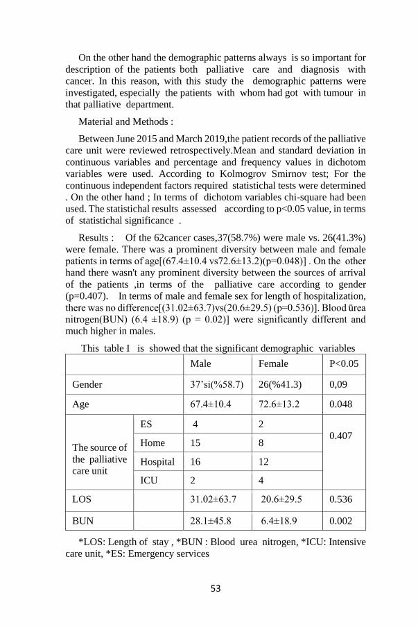
53
On the other hand the demographic patterns always is so important for
description of the patients both palliative care and diagnosis with
cancer. In this reason, with this study the demographic patterns were
investigated, especially the patients with whom had got with tumour in
that palliative department.
Material and Methods :
Between June 2015 and March 2019,the patient records of the palliative
care unit were reviewed retrospectively.Mean and standard deviation in
continuous variables and percentage and frequency values in dichotom
variables were used. According to Kolmogrov Smirnov test; For the
continuous independent factors required statistichal tests were determined
. On the other hand ; In terms of dichotom variables chi-square had been
used. The statistichal results assessed according to p<0.05 value, in terms
of statistichal significance .
Results : Of the 62cancer cases,37(58.7%) were male vs. 26(41.3%)
were female. There was a prominent diversity between male and female
patients in terms of age[(67.4±10.4 vs72.6±13.2)(p=0.048)] . On the other
hand there wasn't any prominent diversity between the sources of arrival
of the patients ,in terms of the palliative care according to gender
(p=0.407). In terms of male and female sex for length of hospitalization,
there was no difference[(31.02±63.7)vs(20.6±29.5) (p=0.536)]. Blood ürea
nitrogen(BUN) (6.4 ±18.9) (p = 0.02)] were significantly different and
much higher in males.
This table I is showed that the significant demographic variables
Male Female P<0.05
Gender 37’si(%58.7) 26(%41.3) 0,09
Age 67.4±10.4 72.6±13.2 0.048
The source of
the palliative
care unit
ES 4 2
0.407 Home 15 8
Hospital 16 12
ICU 2 4
LOS 31.02±63.7 20.6±29.5 0.536
BUN 28.1±45.8 6.4±18.9 0.002
*LOS: Length of stay , *BUN : Blood urea nitrogen, *ICU: Intensive
care unit, *ES: Emergency services

54
For Table 1, we found that, in general, the BUN value was significantly
different between genders and was also lower in women.
As a secondary result, we found that men at a younger age were at risk
for this care department. Although it was not as prominent as the BUN
value, men were found to be younger than women.
In terms of gender as male and female; In our analysis in terms of the
sources that transport patients to this unit; We detected the cases were
Urgent (4vs2),home(15vs8),hospital (16vs12),intensive care (2vs4), in
distribution .
According to our results(Table 1); Among the sources providing
patients to this unit, patients who were transferred from another hospital or
another unit of the hospital were the first.
The second was the patients who were transferred from home. The third
place was the patients coming from the emergency department.
These results show us that for this unit's cases mainly sources from
that the patients in hospital worsening or having difficulty in care.
The variables detailed was given below table;
Table II is show that the cancer distrubition of the palliative care
patients
Lung cancer 12
Colon cancers 9
Brain cancers 7
Gastric cancer 7
Hepatic cancers 5
Pancreatic cancers 4
Breast Cancers 3
Over cancers 3
Thyroid cancers 1
Chordoma 1
Prostatic cancers 2
Urinary system cancers 3
Rectum cancers 1
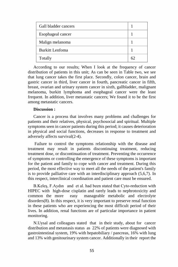
55
Gall bladder cancers 1
Esophageal cancer 1
Malign melanoma 1
Burkitt Lenfoma 1
Totally 62
According to our results; When I look at the frequency of cancer
distribution of patients in this unit; As can be seen in Table two, we see
that lung cancer takes the first place. Secondly, colon cancer, brain and
gastric cancer in third, liver cancer in fourth, pancreatic cancer in fifth,
breast, ovarian and urinary system cancer in sixth, gallbladder, malignant
melanoma, burkitt lymphoma and esophageal cancer were the least
frequent. In addition, liver metastatic cancers; We found it to be the first
among metastatic cancers.
Discussion :
Cancer is a process that involves many problems and challenges for
patients and their relatives, physical, psychosocial and spiritual. Multiple
symptoms seen in cancer patients during this period; it causes deterioration
in physical and social functions, decreases in response to treatment and
adversely affects survival(2-4).
Failure to control the symptoms relationship with the disease and
treatment may result in patients discontinuing treatment, reducing
treatment dose, or discontinuation of treatment. Preventing the occurrence
of symptoms or controlling the emergence of these symptoms is important
for the patient and family to cope with cancer and treatment. During this
period, the most effective way to meet all the needs of the patient's family
is to provide palliative care with an interdisciplinary approach (5,6,7). In
this respect, interclinical coordination and patient care must be ensured.
B.Keleş, F.Aydın and et al. had been stated that Cyto-reduction with
HIPEC with high-dose cisplatin and rarely leads to nephrotoxicity and
common the more easy manageable metabolic and electrolyte
disorders(8). In this respect, it is very important to preserve renal function
in these patients who are experiencing the most difficult period of their
lives. In addition, renal functions are of particular importance in patient
monitoring.
N.Uysal and colleagues stated that in their study, about for cancer
distribution and metastasis status as 22% of patients were diagnosed with
gastrointestinal system, 19% with hepatobiliary / pancreas, 16% with lung
and 13% with genitourinary system cancer. Additionally in their report the

56
51% of patients had more than one year with diagnosed and 87% were
metastasis. On the other hand they stated that found that 9% of the
patients were still treated as radiotherapy or chemotherapy and 36% of
them were discontinued in the last three months.(9)
In our study, gastrointestinal system-related cancers were more
prominent with 23(37.1%) cases.
As in patients receiving palliative therapy and other intensive care
patients, blood urea nitrogen is one of the important factors indicating fluid
balance, and in our study, it was found to be significantly different between
males and females, and blood urea nitrogen was higher in males than the
females [28.1±45.8 vs 6.4±18.9 (p<0.002)].
A.A Kabalak and et al. (10) they stated that if patients are dehydrated,
parenteral fluid administration is not the first choice , in their review(11)
Additionally they had been declared in the oral intake is inadequate and
severe dehydration due to nausea and vomiting in the early period and this
situation increases other symptoms of the patient due to renal failure and
drug toxicity (delirium, somnolence, etc.), short-term parenteral fluid
replacement can be performed. In short term fluid replacement ;
Subcutaneous fluid replacement (hypodermoclysis), which is a less
invasive method, should be the first choice for terminal patients. Fluid
administration should be performed with careful follow-up, as lymphatic
drainage and hypoalbuminemia may cause edema due to tumor invasion.
Dry mouth can be prevented by frequent oral water administration and
mucosal moisturizers(10)(12). As mentioned in the above literature, it is
very important to monitor fluid, that is, strict monitoring of the amount of
fluid the patient receives and the amount of fluid released
On the other side the other important corner stone in our study; In
terms odf the source of the patient coming primary place 28 hospital,
6 ICU , 23 from home and 6 ES was not found any significance in term's
of gender . But furthermore; When we looked in the detailed analysis
showed that statisticahlly significantly differences between the hospital
(Hospital+ICU+ES) and homedepartments is seen first place with 40 vs
23.
The needs of advanced cancer patients to be met in relation to multiple
symptoms and high burden of disease are expected to be high. (13,14)In
this study, stated lead to an increased frequency of unmet needs. In studies,
advanced cancer patients have been reported to have unmet needs and
problems of different sizes, moderate or high levels, 40-89%. 89, daily
living activities requirements 1-52%, economic requirements 13-60%,
psychological requirements 16-41%, psychosocial requirements 7-44%,
communication requirements 34-36%, information requirements 39% and
spiritual requirements 14-33% rate (15). Additionally, Ö.Turan and et al.

57
were stated that in their study on trauma patients in 2019 in critical patients;
They stated that early physiological data related to mortality could be
identified (16). Therefore, a sufficient number and quality of education
personnel are needed to meet all the biopsychosocial needs of this group
of patients.
Although our study did not include trauma patients, in terms of end-
stage cancer patients; We think that the BUN values determined in this
study may be important. On the other hand ; According to our idea too
especially gastrointestinal tumours the more higher risk rates over among
the these humans. At last point we can say that prominently the most
metastasis had included the hepatis structures. Here for hepatic metastasis,
we believe that primary source from breast and colon cancer metastases
are especially prominent.
Conclusion : Blood urea nitrogen levels may be lower in female cancer
patients who come to palliative care than male cancer lines, and most of
the patients who are transferred to palliative care are transferred from a
hospital that is insufficient for this care to a higher level hospital. However,
there was no difference between the genders in terms of the source of
admission. According the our idea; Still end stage cancer disorders are
overwhelmingly the first line for this risk group patients when compared
with the other risk groups (trauma, stroke etc.).
References:
1.WHO/CDS/IMAI/2004.3 , page :5, Rev. 1
2.Esther Kim JE, Dodd MJ, Aouizerat BE, Jahan T, Miaskowski C. A
review of the prevalence and impact of multiple symptoms in oncology
patients. J Pain Symptom Manage 2009;37(4):715-36. CrossRef
3. Walsh D, Rybicki L. Symptom clustering in advanced cancer.
Support Care Cancer 2006;14(8):831-6. CrossRef
4. Bektaş-Aydın H, Akdemir N. Kanserli bireylerde fonksiyonel
durumun değerlendirilmesi. Türkiye Klinikleri J Med Sci 2006;26:488-99.
5.Shoemaker LK, Estfan B, Induru R, Walsh TD. Symptom
management: an important part of cancer care. Cleve Clin J Med
2011;78(1):25-34. CrossRef
6. Gómez-Batiste X, Porta-Sales J, Espinosa-Rojas J, PascualLópez A,
Tuca A, Rodriguez J. Effectiveness of palliative care services in symptom
control of patients with advanced terminal cancer: a spanish, multicenter,
prospective, quasiexperimental, pre-post study. J Pain Symptom Manage
2010;40(5):652-60. CrossRef
7. Strasser F, Sweeney C, Willey J, Benisch-Tolley S, Palmer JL,
Bruera E. Impact of a half-day multidisciplinary symptom control and
palliative care outpatient clinic in a comprehensive cancer center on
58
recommendations, symptom intensity, and patient satisfaction: a
retrospective descriptive study. J Pain Symptom Manage 2004;27(6):481-
91. CrossRef
8. Aslan Bilge. Biomed J Sci & Tech Res. Volume 3- Issue 4: 2018
9.Symptoms seen in inpatient palliative care and impact of palliative
care unit on symptom control. Neşe Uysal, Gülçin Şenel, Şerife Karaca,2
Nihal Kadıoğulları,2 Nesteren Koçak, Gonca Oğuz. AĞRI
2015;27(2):104-110 doi: 10.5505/agri.2015.26214)
10.End of Life Care Organization; Palliative Care, Afife Ayla Kabalak,
Hakkı Öztürk, Hasan Çağıl
Yoğun Bakım Dergisi 2013;11(2):56-70)Yoğun Bakım Dergisi
2013;11(2):56-70)
11.Fainsinger RL, MacEachern T, Miller MJ, Bruera E, Spachynski K,
Kuehn N, et al. The use of hypodermoclysis for rehydration in terminally
ill cancer patients. J Pain Symptom Manage 1994;9:298-302. )Cross ref..
12.Fainsinger RL, MacEachern T, Miller MJ, Bruera E, Spachynski K,
Kuehn N, et al. The use of hypodermoclysis for rehydration in terminally
ill cancer patients. J Pain Symptom Manage 1994;9:298-302. )Cross ref.
13. Osse BH, Vernooij-Dassen MJ, de Vree BP, Schadé E, et al.
Assessment of the need for palliative care as perceived by individual
cancer patients and their families: a review of instruments for improving
patient participation in palliative care. Cancer 2000;88:900-11.
14. Uchida M, Akechi T, Okuyama T, Sagawa R, et al. Patients’
supportive care needs and psychological distress in advanced breast cancer
patients in Japan. Jpn J Clin Oncol 2011;41:530-6. 15. Wen KY, Gustafson
DH. Needs assessment for cancer patients and their families. Health Qual
Life Outcomes 2004;2:11.
16. Ö.Turan, M. Eryılmaz, Ö.Albuz. Ulus Travma Acil Cerrahi Derg,
May 2019, Vol. 25, No. 3
59
OFF-PUMP CORONARY ARTERY BYPASS (OPCAB) SURGERY
Erdem ÇETİN*
1-History:
In recent years, although not a new method, OBCAB surgery has
become popular again. OPCAB was first proposed in 1910 by Alexis
Carrel for the treatment of angina pectoris, and he attempted
experimentally to perform coronary artery bypass surgery on the beating
heart of a dog (1). After many successful unsuccessful studies by
researchers over the years, Vasilii I. Kolesov (1904-1992) initiated modern
OPCAB surgery in the sixties (2).
Subsequently, between 1964 and 1976, Kolesov et al. Performed more
than 130 coronary revascularizations with OPCAB surgery (3). In the 70s
and 80s, OPCAB surgery continued to be performed by some teams due to
their economic advantages (3-5). In this period, OBCAB surgery was
preferred in selected cases with one or two vessel coronary artery disease
on the anterior surface of the heart. Surgeons had to wait until the
development and diffusion of coronary stabilizers until the 90s to ensure
complete revascularization by safely accessing the posterior wall of the
heart. Today, OBCAB surgery has become a part of modern cardiac
surgery with developing technology and increasing experience.
2-Advantages of OPCAB Surgery:
Inflammatory Response and Oxidative Stress:
With the introduction of extra-corporeal circulatory technology in
cardiac surgery in the 50s, discussions of systemic inflammatory reactions
have started. OBCAB surgery has advantages in terms of organ
dysfunction and peri-operative morbidity caused by inflammatory
reaction.
Brasil et al. (1998) conducted the first of these studies. When
prospectively examined 20 patients operated with on-pump and off-pump
techniques, plasma TNF increased in 60% of on-pump patients and did not
increase in any of the off-pump patients. Clinically, less fever,
hypotension, inotropic drug use and postoperative bleeding were found in
* Gülhane Eğitim ve Araştırma Hastanesi
60
off pump patients. Shorter intubation time was detected in off-pump
patients. They concluded that all these data are due to low TNF (6).
Another prospective study by Matata et al. In 20 randomly selected
patients, inflammatory mediators such as complement C3a, interleukin-8
(IL-8), elastase and TNFa were evaluated after off pump and on pump
surgery. Oxidative stress markers such as lipid hydroperoxide and protein
carbonyl were found to increase in the on pump group. They concluded
that off-pump surgery can significantly reduce oxidative stress (7).
Myocardial Protection:
Many authors have reported the potential benefit of off pump surgery
on myocardial protection. In Pfister's study, it was shown that there was a
decrease in the incidence of low cardiac output and decreased intra-aortic
balloon pump use in Off pump patients (8).
In a prospective randomized study of 30 patients, Czerny et al. Found
that postoperative myoglobin, CK-MB and troponin I values of off pump
patients were much lower than those of ten pump patients(9).
Pulmonary Physiology:
Although there are many studies suggesting that extracorporeal
circulation and disruption of lung physiology are related, the number of
studies on off-pump surgery in this area is scarce. In the studies of Cox et
al. And Taggard, no relationship was found between off pump surgery and
impaired lung physiology. In these studies, it was concluded that
pulmonary physiology disorders observed during CABG were not related
to CPB use, but were associated with sternotomy-induced surgical
approach (10-11).
Brain Physiology:
The risk of stroke during coronary artery surgery ranges from 1% to 8%
(12-13), mainly due to plaque mobilization rupture and peri-operative
arrhythmia. Off-pump coronary surgery, despite the use of full arterial
grafts, requires manipulation of the ascending aorta for proximal vein graft
anastomoses if preferred. However, with the widespread use of off pump
surgery, an increase in the studies on this subject has started. As a result of
a randomized study of 281 patients by Van Djick et al., Results were
reported in favor of OPCAB (14).
Renal Physiology:
After the introduction of CPB in cardiac surgery, it was seen as the main
cause of postoperative renal failure. Pulse flow loss, hemodilution,
hypothermia, and inflammatory reaction are all contributing factors to this
major complication. Off pump surgery can also prevent the deterioration
of renal function as it allows avoiding CPB. In a prospective, randomized
61
study of 50 patients by Ascione et al., They concluded that in off-pump
surgery, there is a better conserved glomerular filtration with low N-acetyl
glucosidase excretion as a renal tubular biochemical marker (15). As a
result of a non-randomized prospective study of 55 OPCAB and 635
ONCPB patients by Maribel et al., They concluded that OPCAB grafting
may be a procedure that can be used regardless of the increase in serum
creatinine and clearance (16). OPCAB surgery has recently increased its
popularity in the world of coronary revascularization. New techniques and
strategies are reported and success rates increase as surgeons experience
increases. The cost advantage has become the preferred technique for
patients and surgeons because of the low risk of complications arising from
the inflammatory effects of CPB.
3-Principles of Stabilization and Preservation of Hemodynamics in
OPCAB Surgery:
Stabilization of the myocardium during coronary grafting is essential in
OPCAB surgery. In the early stages, only the anterior coronary arteries
were targeted in OPCAB surgery and simple and primitive stabilization
methods were used. However, this was not sufficient to provide safe access
to the posterior wall of the heart and caused incomplete revascularization
of the circumflex arteries. During the last two decades, various techniques
and stabilization devices have been developed and used for myocardial
stabilization during OPCAB surgery.
Current Techniques:
Myocardial Mobilization and Preservation of Hemodynamics:
Left Anterior Descending (LAD) Artery:
The LAD artery is usually accessed by simple manipulation.
Alternatively, a few sponges to be placed under the heart or pericardial
sutures placed just above the left phrenic nerve may be used to lift the apex
of the heart. These methods have little hemodynamic effect. Manipulation
of the anterior region of the heart with a mechanical stabilizer leads to an
interruption of coronary blood flow during coronary grafting, a 6 to 15%
reduction in cardiac output, and is generally well tolerated (20-21) (Figure
1).
Hemodynamics may deteriorate during diagonal grafting, Nierich et al.
They reported a decrease in left ventricular diastolic volume during
diagonal artery manipulation in OPCAB and that they used trendelenburg
position and used dopamine to cope with this (22). The use of intracoronary
shunt should be used in cases of surgeons with low OPCAB experience
and slow anastomosis rate, as well as in all LAD grafts with moderate
lesions and less developed collateral.
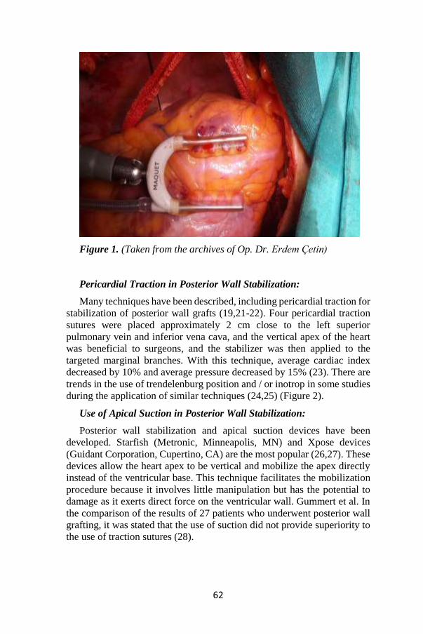
62
Figure 1. (Taken from the archives of Op. Dr. Erdem Çetin)
Pericardial Traction in Posterior Wall Stabilization:
Many techniques have been described, including pericardial traction for
stabilization of posterior wall grafts (19,21-22). Four pericardial traction
sutures were placed approximately 2 cm close to the left superior
pulmonary vein and inferior vena cava, and the vertical apex of the heart
was beneficial to surgeons, and the stabilizer was then applied to the
targeted marginal branches. With this technique, average cardiac index
decreased by 10% and average pressure decreased by 15% (23). There are
trends in the use of trendelenburg position and / or inotrop in some studies
during the application of similar techniques (24,25) (Figure 2).
Use of Apical Suction in Posterior Wall Stabilization:
Posterior wall stabilization and apical suction devices have been
developed. Starfish (Metronic, Minneapolis, MN) and Xpose devices
(Guidant Corporation, Cupertino, CA) are the most popular (26,27). These
devices allow the heart apex to be vertical and mobilize the apex directly
instead of the ventricular base. This technique facilitates the mobilization
procedure because it involves little manipulation but has the potential to
damage as it exerts direct force on the ventricular wall. Gummert et al. In
the comparison of the results of 27 patients who underwent posterior wall
grafting, it was stated that the use of suction did not provide superiority to
the use of traction sutures (28).
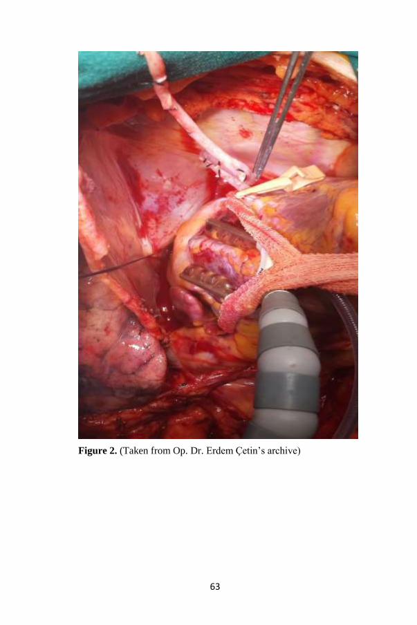
63
Figure 2. (Taken from Op. Dr. Erdem Çetin’s archive)

64
Figure 3. (Taken from the archives of Op. Dr. Erdem Çetin)
Auxiliary Techniques:
Trendelenburg Position:
Many auxiliary techniques have been described for stabilizing the
myocardium and maintaining hemodynamics. As mentioned earlier, the
use of Trendelenburg position is the most popular.
Trendelenburg position during OPCAB surgery contributes to
increased venous hydrostatic pressure. In addition, it is safe to use during
apex verticalization and posterior stabilization of the heart, especially in
circumflex artery, posterior descending and right coronary artery grafts.
65
Slope Right and Left:
In addition to the Trendelenburg position, turn the table to the right
(towards the surgeon)
It is commonly used during posterior grafting. In posterior descending
and right coronary artery grafts, left rotation is applied. Myocardial
stabilization has undergone major technological advances since the early
90s. The development and use of coronary stabilizers, the widespread use
of coronary shunting, the surgeons' experience in OPCAB surgery, and the
development of alternative retraction methods have contributed to the
success rates of off-pump operations.
4-Anesthesia in OPCAB Surgery:
Preoperative Evaluation:
The preoperative evaluation of OPCAB patients does not differ from
patients who will undergo routine surgery.
Premedication:
It aims to alleviate negative effects such as premedication, anxiety and
hypertension. It also provides comfort to the patient during the placement
of intravascular catheters. Narcotics (morphine 0.1-0.15 mg / kg IM) and
sedative (scopolamine 5 μg / kg IM or midazolam 0.05 mg / kg IM) are the
most commonly used drugs.
Prevention of Hypothermia:
Care should be taken to prevent hypothermia, especially if early
extubation is planned. All intravenous fluids are applied warmer, washing
fluids to be used on the heart are heated.
Normothermia is maintained using sub-patient warmers, thus avoiding
the adverse effects of hypothermia, wound healing, clotting and
arrhythmia.
Anesthesia Induction:
The purpose of OPCAB surgical anesthesia is early extubation.
Medications with short or moderate duration of action are selected. Several
drugs can be combined for this purpose (29). Fentanyl 15-50 / g / kg and
sufentanil 1.5-3.0 μg / kg are commonly used before intubation. These
doses are considerably lower than normal use. Remifentanil, a strong and
ultra-short acting narcotic, can be given at an infusion rate of 0.15-0.4 μg /
kg / min. In addition, one of the pentotal 2-3 mg / kg, midazolam 0.1-0.2
mg / kg or propofol 1-2 mg / kg may be used. In addition to the narcotics
described, anesthesia is provided by propofol infusion. Maintenance of
isoflurane or sevoflurane gases is also utilized. Between the induction of
anesthesia and the onset of coronary grafting, 2-5 g of magnesium sulfate
66
is administered. Magnesium has the effect of reducing the incidence of
arrhythmias (29-32) and may also reduce arterial graft spasm (33).
Preservation of Hemodynamics:
Since hemodynamic changes can be sudden and obvious, the surgeon
and anesthesiologist should be very careful and experienced. If
hypotension occurs, fluid loading or phenylephrine (vasopressor) is the
most commonly used method and rarely norepinephrine or epinephrine
may be necessary. Β-agonist drugs should be avoided as they increase
myocardial and systemic oxygen consumption during coronary
revascularization. At the beginning of the surgery, a low dose of
nitroglycerin infusion (0.1-0.3 μg / kg / min) is initiated. Thus,
nitroglycerin can be utilized to increase Sub-endocardial flow and to
increase myocardial perfusion by reducing preload. In addition, the
preventive effect of arterial graft vasospasm is utilized.
Preservation of Hemodynamics by Revascularization Areas:
In OBCAB surgery, revascularization is first initiated from the coronary
artery with the highest stenosis. Unfractionated heparin 100U / kg is
administered before starting anastomoses. In OPCAB cases, the
requirement of heparin is checked by the active coagulation time (ACT)
measured every 30 minutes. ACT Add heparin bolus of 25-50U / kg for
300 to 400 seconds.
The perfusionist should be prepared and wait for the operating room at
all times, with CPB installed. In appropriate patients, minimal doses of
beta-blockers may sometimes be administered (metoprolol 2.5-5 mg IV),
thereby reducing myocardial oxygen consumption and keeping the heart
rate within the range of 50-75 / min. Despite increasing technical advances
in OPCAB surgery, circumflex coronary artery grafting is still the most
challenging part of the operation for both the surgeon and the
anesthesiologist. The anesthesiologist should have a temporary pacemaker
available for hypotension for possible bradycardia which is difficult to
predict. Although fluid loading may help hemodynamics due to possible
hypotension, it is useful to avoid overloading the fluid. Anostomosis can
be performed in all parts of the heart with gentle surgical manipulations
and a good hemodynamic protection strategy. Proximal anastomosis of the
saphenous vein bypass grafts should be gently manipulated once to reduce
the risk of embolism and avoid the risk of dissection of the aorta when
performing the ascending aorta (34). To reduce aortic wall stress during
aortic anostomosis, it is necessary to lower systemic blood pressure to 90
mmHg systolic levels. Protamine sulfate and heparin should be reversed
when surgery is completed without any problems.
67
Indications and Important Strategies for OPCAB Surgery:
Today, OPCAB surgery is routinely performed by experienced teams
in coronary revascularization compared to previous years. But there are, of
course, some situations in which this surgical method is contraindicated.
Definite Contraindication:
* Preoperative hemodynamic instability
* Deep intra myocardial LAD
* Moderate (3+) or severe (4+) mitral insufficiency
Relative Contraindications:
* Pulmonary hypertension
* Diffuse myocardial adhesion during reoperative surgery
* Enlarged ascending aorta
With the increasing experience and scientific studies in OPCAB
surgery, some strategies of this coronary revascularization technique have
been formed.In OBCAP surgery, the most severe stenosis of the corner
artery should be grafted first. Proximal anostomoses should be performed
with single side clamp technique to the ascending aorta. Moderate
hypotension should be observed during proximal anastomosis.
Results from various centers around the world have demonstrated the
effectiveness of OPCAB, even in high-risk patients. According to the 2018
ESC / EACTS myocardial revascularization guidelines, OPCAB surgery is
recommended in high-risk patients (35). Routine use of this technique,
which is recommended in high-risk patients, is undoubtedly used in some
centers as a routine surgical procedure. We believe that the prevalence and
success rates of OPCAB will increase with each passing year as the
technique, which has been the focus of the discussions for years, develops
methods that each surgeon feels safe and comfortable while remaining
within the indications.
References
1. Carrel A. On the experimental surgery of the thoracic aorta and the
heart. Ann Surg 1910; 52:83-95.
2. Olearchyk AS, Kolesov VI. A pioneer of coronary revascularization
by internal mammary-coronary artery grafting. J Thorac Cardiovasc Surg
1988; 96:13-8.
3. Ankeney JL. To use or not to use the pump oxygenator in coronary
artery bypass operations. Ann Thorac Surg 1975; 19:108-9.
68
4. Benetti FJ, Naseli G, Wood M et al. Direct myocardial
revascularization without extracorporeal circulation: Experience in 700
patients. Chest 1991; 100:310-6.
5. Bufalo E, Andrade JCS, Branco JNR et al. Coronary artery bypass
grafting without cardiopulmonary bypass. Ann Thorac Surg 1996; 61:63-
6.
6. Brasil LA, Gomes WJ, Salomao R et al. Inflammatory response after
myocardial revascularization with or without cardiopulmonary bypass.
Ann Thorac Surg 1998; 66:56-9.
7. Matata BM, Sosnowski AW, Galinanes M. Off-pump bypass graft
operation significantly reduces oxidative stress and inflammation. Ann
Thorac Surg 2000; 69:785-91.
8. Pfister AJ, Zaki MS, Garcia JM et al. Coronary bypass without
cardiopulmonary bypass. Ann Thorac Surg 1992; 54:1085-92.
9.Czerny M, Baumer H, Kilo J et al. Inflammatory response and
myocardial injury following coronary artery bypass grafting with or
without cardiopulmonary bypass. Eur J Cardiothorac Surg 2000; 17:737-
42.
10. Cox CM, Ascione R, Cohen AM et al. Effects of cardiopulmonary
bypass on pulmonary gas exchange: A prospective randomized study. Ann
Thorac Surg 2000; 69:140-5.
11. Taggart DP. Respiratory dysfunction after cardiac surgery: Effects
of avoiding cardiopulmonary bypass and the use of bilateral internal
mammary arteries. Eur J Cardiothorac Surg 2000; 18:31-7.
12. Roach GW, Kanchuger M, Mora M et al. Adverse cerebral
outcomes after coronary bypass surgery. N Engl J Med 1996; 335:1857-
63.
13. Mickleborough LL, Walker PM, Tagagi Y et al. Risk factors for
stroke in patients undergoing coronary artery grafting. J Thorac Cardiovasc
Surg 1996; 112:1258-9.
14. Van Djick D, Jansen EW, Hijman R et al. The Octopus Study
Group. Cognitive outcome after off-pump and on-pump coronary artery
bypass graft surgery: A randomized trial. JAMA 2002; 287:1405-12.
15. Ascione R, Lloyd CT, Underwood MJ et al. On pump versus off
pump coronary revascularization: Evaluation of renal function. Ann
Thorac Surg 1999; 68:493-8.
16. Maribel G, Phillips-Bute B, Landolfo KP et al. Off-pump versus on-
pump coronary artery bypass surgery and postoperative renal dysfunction.
Anesth Analg 2000; 91:1080-84.
69
17. Borst C, Jansen EWL, Grundeman PF et al. Regional cardiac wall
immobilization for open chest and closed chest artery bypass grafting in
the beating heart: The “Octopus” method [abstract]. Circulation 1995;
92:I-177.
18. Baumgartner FJ, Gheissari A, Capouya ER et al. Technical aspects
of total revascularization in off-pump coronary bypass via sternotomy
approach. Ann Thorac Surg 1999; 67:1653-8.
19. Cartier R, Brann S, Dagenais F et al. Systematic off-pump coronary
artery revascularization in multivessel disease: Experience of three
hundred cases. J Thorac Cardiovasc Surg 2000; 119:221-9.
20. Do Q-B, Goyer C, Chavanon O et al. Hemodynamic changes during
off-pump CABG surgery. Eur J Cardiothorac Surg 2002; 21:385-90.
21. Nierich AP, Diephuis J, Jansen EWL et al. Heart displacement
during off-pump CABG: How well is it tolerated. Ann Thorac Surg 2000;
70:466-72.
22. Bergsland J, Schimd S, Yanulevich J et al. Coronary artery bypass
grafting (CABG) without cardiopulmonary bypass (CPB): A strategy for
improving results in surgical revascularization. Heart Surgery Forum 1998;
1:107-110.
23. Do QB, Cartier R. Hemodynamic changes during beating-heart
CABG surgery (in French). Ann Chir 1999; 53:706-11.
24. Grundeman PF, Borst C, van Herwaarden JA et al. Hemodynamic
changes during displacement of the beating heart by the Utrecht Octopus
method. Ann Thorac Surg 1997; 63:S88-92.
25. Watters MPR, Ascione R, Ryder IG et al. Haemodynamic changes
during beating heart coronary surgery with the “Bristol Technique”. Eur J
Cardiothorac Surg 2001; 19:33-40.
26. Niinami H, Takeuchi Y, Ichikawa S et al. Multivessel off-pump
coronary aortic bypass grafting with an impaired and severely dilated left
ventricle using the Starfish heart positioner. Kyobu Geka 2002; 55(9):773-
7.
27. Dullum MK, Resano FG. Xpose: A new device that provides
reproducible and easy access for multivessel beating heart bypass grafting.
Heart Surg Forum 2000; 3(2):113-7.
28. Gummert JF, Raumanns J, Bossert T et al. Suction device versus
pericardial retraction sutures. Comparison of hemodynamics using
different exposure systems in beating heart coronary surgery. Heart Surg
Forum 2003; 6(Suppl 1):S32.
70
29. Howie MB, Cheng D, Newman MF et al. A randomized double-
blinded multicenter comparison of remifentanil vs fentanyl when
combined with isoflurane/propofol for early extubation in coronary artery
bypass graft surgery. Anesth Analg 2001; 92:1084-93.
30. Maslow AD, Regan MM, Heindle S et al. Postoperative atrial
tachyarrhythmias in patients undergoing coronary artery bypass surgery
without cardiopulmonary bypass: A role for intraoperative magnesium
supplementation. J Cardiothorac Vasc Anesth 2000; 14:524-30.
31. Speziale G, Ruvolo G, Fattouch K et al. Arrhythmias prophylaxis
after coronary artery bypass grafting: Regiments of magnesium sulfate
administration. J Thorac Cardiovasc Surg 2000; 48:22-6.
32. Bert AA, Reinert SE, Singh AK. A beta-blocker, not magnesium, is
effective prophylaxis for atrial tachyarrhythmias after coronary artery
bypass graft surgery. J Cardiothorac Vasc Anesth 2001; 15:204-9.
33. Teragawa H, Kato M, Yamagata T et al. The preventive effect of
magnesium on coronary spasm in patients with vasospastic angina. Chest
2000; 118:1690-5.
34. Dagenais F, Cartier R. Pulmonary hypertension during beating heart
coronary surgery: Intermittent vena cava snaring. Ann Thorac Surg 1999;
68:1094-5.
35. Neumann F-J, Sousa-Uva M, Ahlsson A et al. 2018 esc/eacts
guidelines on myocardial revascularization. European Heart Journal 2018;
40(2):87-165.
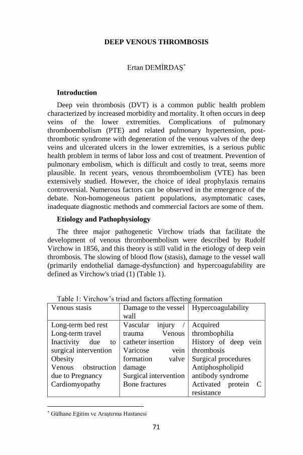
71
DEEP VENOUS THROMBOSIS
Ertan DEMİRDAŞ*
Introduction
Deep vein thrombosis (DVT) is a common public health problem
characterized by increased morbidity and mortality. It often occurs in deep
veins of the lower extremities. Complications of pulmonary
thromboembolism (PTE) and related pulmonary hypertension, post-
thrombotic syndrome with degeneration of the venous valves of the deep
veins and ulcerated ulcers in the lower extremities, is a serious public
health problem in terms of labor loss and cost of treatment. Prevention of
pulmonary embolism, which is difficult and costly to treat, seems more
plausible. In recent years, venous thromboembolism (VTE) has been
extensively studied. However, the choice of ideal prophylaxis remains
controversial. Numerous factors can be observed in the emergence of the
debate. Non-homogeneous patient populations, asymptomatic cases,
inadequate diagnostic methods and commercial factors are some of them.
Etiology and Pathophysiology
The three major pathogenetic Virchow triads that facilitate the
development of venous thromboembolism were described by Rudolf
Virchow in 1856, and this theory is still valid in the etiology of deep vein
thrombosis. The slowing of blood flow (stasis), damage to the vessel wall
(primarily endothelial damage-dysfunction) and hypercoagulability are
defined as Virchow's triad (1) (Table 1).
Table 1: Virchow’s triad and factors affecting formation
Venous stasis Damage to the vessel
wall
Hypercoagulability
Long-term bed rest
Long-term travel
Inactivity due to
surgical intervention
Obesity
Venous obstruction
due to Pregnancy
Cardiomyopathy
Vascular injury /
trauma Venous
catheter insertion
Varicose vein
formation valve
damage
Surgical intervention
Bone fractures
Acquired
thrombophilia
History of deep vein
thrombosis
Surgical procedures
Antiphospholipid
antibody syndrome
Activated protein C
resistance
* Gülhane Eğitim ve Araştırma Hastanesi
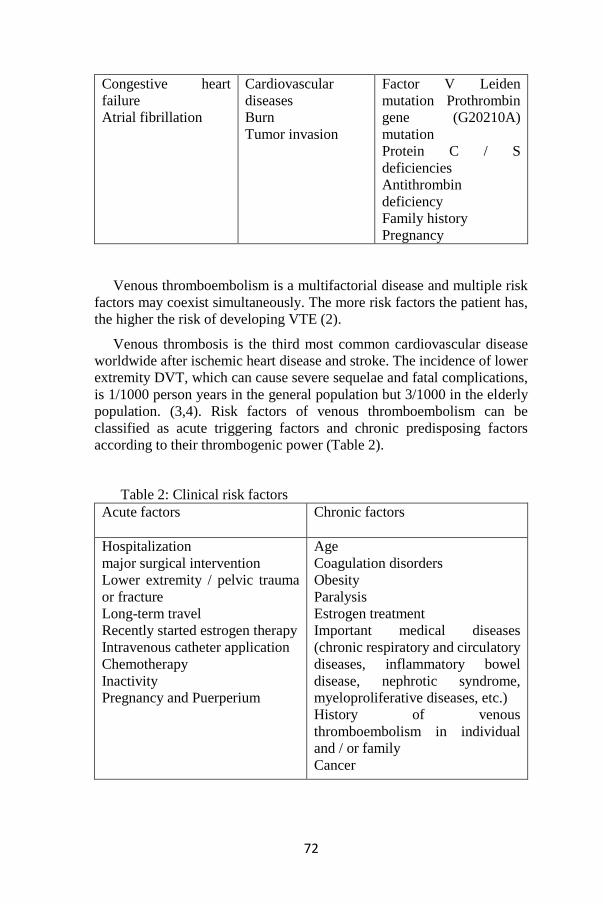
72
Congestive heart
failure
Atrial fibrillation
Cardiovascular
diseases
Burn
Tumor invasion
Factor V Leiden
mutation Prothrombin
gene (G20210A)
mutation
Protein C / S
deficiencies
Antithrombin
deficiency
Family history
Pregnancy
Venous thromboembolism is a multifactorial disease and multiple risk
factors may coexist simultaneously. The more risk factors the patient has,
the higher the risk of developing VTE (2).
Venous thrombosis is the third most common cardiovascular disease
worldwide after ischemic heart disease and stroke. The incidence of lower
extremity DVT, which can cause severe sequelae and fatal complications,
is 1/1000 person years in the general population but 3/1000 in the elderly
population. (3,4). Risk factors of venous thromboembolism can be
classified as acute triggering factors and chronic predisposing factors
according to their thrombogenic power (Table 2).
Table 2: Clinical risk factors
Acute factors
Chronic factors
Hospitalization
major surgical intervention
Lower extremity / pelvic trauma
or fracture
Long-term travel
Recently started estrogen therapy
Intravenous catheter application
Chemotherapy
Inactivity
Pregnancy and Puerperium
Age
Coagulation disorders
Obesity
Paralysis
Estrogen treatment
Important medical diseases
(chronic respiratory and circulatory
diseases, inflammatory bowel
disease, nephrotic syndrome,
myeloproliferative diseases, etc.)
History of venous
thromboembolism in individual
and / or family
Cancer
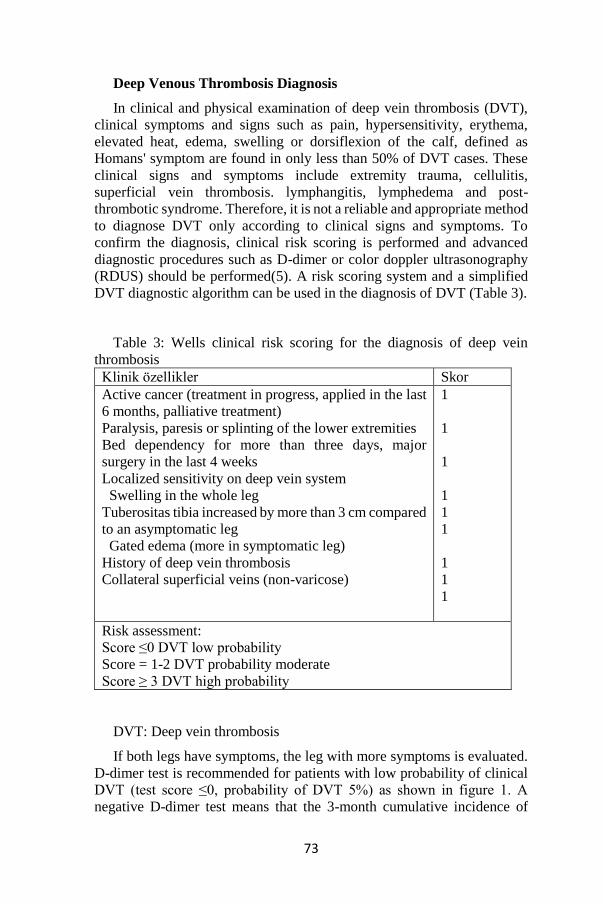
73
Deep Venous Thrombosis Diagnosis
In clinical and physical examination of deep vein thrombosis (DVT),
clinical symptoms and signs such as pain, hypersensitivity, erythema,
elevated heat, edema, swelling or dorsiflexion of the calf, defined as
Homans' symptom are found in only less than 50% of DVT cases. These
clinical signs and symptoms include extremity trauma, cellulitis,
superficial vein thrombosis. lymphangitis, lymphedema and post-
thrombotic syndrome. Therefore, it is not a reliable and appropriate method
to diagnose DVT only according to clinical signs and symptoms. To
confirm the diagnosis, clinical risk scoring is performed and advanced
diagnostic procedures such as D-dimer or color doppler ultrasonography
(RDUS) should be performed(5). A risk scoring system and a simplified
DVT diagnostic algorithm can be used in the diagnosis of DVT (Table 3).
Table 3: Wells clinical risk scoring for the diagnosis of deep vein
thrombosis
Klinik özellikler Skor
Active cancer (treatment in progress, applied in the last
6 months, palliative treatment)
Paralysis, paresis or splinting of the lower extremities
Bed dependency for more than three days, major
surgery in the last 4 weeks
Localized sensitivity on deep vein system
Swelling in the whole leg
Tuberositas tibia increased by more than 3 cm compared
to an asymptomatic leg
Gated edema (more in symptomatic leg)
History of deep vein thrombosis
Collateral superficial veins (non-varicose)
1
1
1
1
1
1
1
1
1
Risk assessment:
Score ≤0 DVT low probability
Score = 1-2 DVT probability moderate
Score ≥ 3 DVT high probability
DVT: Deep vein thrombosis
If both legs have symptoms, the leg with more symptoms is evaluated.
D-dimer test is recommended for patients with low probability of clinical
DVT (test score ≤0, probability of DVT 5%) as shown in figure 1. A
negative D-dimer test means that the 3-month cumulative incidence of
74
VTE is 0.5%. If the D-dimer level is high, ultrasonography should be
performed. A positive ultrasonography confirms the diagnosis of DVT, and
if negative, the diagnosis of DVT is avoided.3 Direct ultrasonography is
recommended for patients with a moderate (test score 1-2, DVT probability
33%) and high (test score ≥3, probability of DVT 85%) of clinical DVT.
The negative results of compression ultrasonography do not exclude the
diagnosis, but the risk of DVT occurring within 3-6 months continues;
3.6% of moderate-risk patients and 31% of high-risk patients may develop
DVT during this period. In this case, D-dimer test is appropriate.
Since D-dimer test is a laboratory test whose sensitivity and specificity
vary according to the method used, this issue should be taken into
consideration when interpreting. High levels are not always specific to
DVT; cancer, infectious diseases, recent surgical intervention, trauma, and
pregnancy. D-dimer has a high negative predictive value, especially in
cases with suspected DVT from outpatients, ie non-triggered DVT. Color
Doppler ultrasonography (RDUSG) is the most appropriate first-line
diagnostic method in the diagnosis of DVT because it is non-invasive, fast
and easily applicable in patients suspected of DVT. RDUSG has 97%
sensitivity and 94% specificity in the diagnosis of DVT (6). However,
detection of iliac vein thrombosis can be difficult especially with RDUSG.
In these cases, computed tomography (CT), magnetic resonance imaging
(MRI) and venography have an important role. The primary diagnostic
criteria for DVT is that the vein lumen is not completely compressed. The
sensitivity and specificity of compression ultrasonography in the diagnosis
of symptomatic proximal DVT is above 95%; In isolated distal DVT,
sensitivity is accepted as 70% and positive predictive value as 80% (7).
Pulmonary Embolism
PE, which can also be described as a complication of DVT, is usually
characterized by sudden onset dyspnea, tachypnea, tachycardia and chest
pain, and these findings are not specific to this disease. So the path to
diagnosis begins with clinical suspicion. In addition to initial symptoms
and signs, the patient should be evaluated for risk factors(8). The lack of
specific clinical findings and the high risk of bleeding from anticoagulant
therapy require confirmation or exclusion of the diagnosis as soon as
possible. For this purpose, the possibility of clinical diagnosis. The first
method to be used for imaging is chest radiography. Chest radiography is
normal in 20–40% of patients with PE without cardiopulmonary disease.
Normal chest X-ray does not exclude the possibility of PE(9).
Ventilation / perfusion scintigraphy is a reliable diagnostic test in
patients with suspected PE. While high probability scintigraphy results are
sufficient to confirm the diagnosis, further investigation is required to
confirm the diagnosis in case of a medium and low probability result. CT
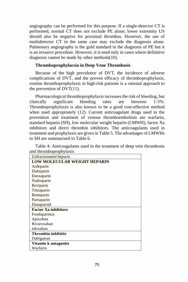
75
angiography can be performed for this purpose. If a single-detector CT is
performed, normal CT does not exclude PE alone; lower extremity US
should also be negative for proximal thrombus. However, the use of
multidetector CT in the same case may exclude the diagnosis alone.
Pulmonary angiography is the gold standard in the diagnosis of PE but it
is an invasive procedure. However, it is used only in cases where definitive
diagnosis cannot be made by other methods(10).
Thromboprophylacsia in Deep Vene Thrombosis
Because of the high prevalence of DVT, the incidence of adverse
complications of DVT, and the proven efficacy of thromboprophylaxis,
routine thromboprophylaxis in high-risk patients is a rational approach to
the prevention of DVT(11).
Pharmacological thromboprophylaxis increases the risk of bleeding, but
clinically significant bleeding rates are between 1-5%.
Thromboprophylaxin is also known to be a good cost-effective method
when used appropriately (12). Current anticoagulant drugs used in the
prevention and treatment of venous thromboembolism are warfarin,
standard heparin (SH), low molecular weight heparin (LMWH), factor Xa
inhibitors and direct thrombin inhibitors. The anticoagulants used in
treatment and prophylaxis are given in Table 5. The advantages of LMWHs
to SH are summarized in Table 6.
Table 4: Anticoagulants used in the treatment of deep vein thrombosis
and thromboprophylaxis Unfractionated heparin
LOW MOLECULAR WEIGHT HEPARIN
Ardeparin
Dalteparin
Enoxaparin
Nadroparin
Reviparin
Tinzaparin
Bemiparin
Parnaparin
Danaparoid
Factor Xa inhibitors
Fondaparinux
Apixoban
Rivaroxaban
edoxaban
Thrombin inhibitör
Dabigatran
Vitamin k antagonist
Warfarin
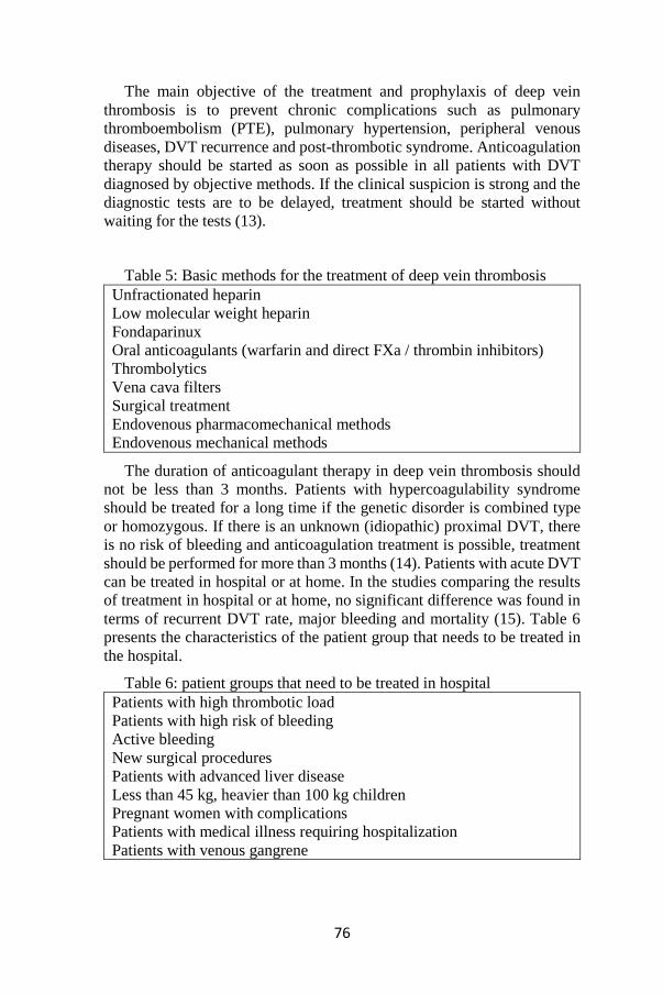
76
The main objective of the treatment and prophylaxis of deep vein
thrombosis is to prevent chronic complications such as pulmonary
thromboembolism (PTE), pulmonary hypertension, peripheral venous
diseases, DVT recurrence and post-thrombotic syndrome. Anticoagulation
therapy should be started as soon as possible in all patients with DVT
diagnosed by objective methods. If the clinical suspicion is strong and the
diagnostic tests are to be delayed, treatment should be started without
waiting for the tests (13).
Table 5: Basic methods for the treatment of deep vein thrombosis
Unfractionated heparin
Low molecular weight heparin
Fondaparinux
Oral anticoagulants (warfarin and direct FXa / thrombin inhibitors)
Thrombolytics
Vena cava filters
Surgical treatment
Endovenous pharmacomechanical methods
Endovenous mechanical methods
The duration of anticoagulant therapy in deep vein thrombosis should
not be less than 3 months. Patients with hypercoagulability syndrome
should be treated for a long time if the genetic disorder is combined type
or homozygous. If there is an unknown (idiopathic) proximal DVT, there
is no risk of bleeding and anticoagulation treatment is possible, treatment
should be performed for more than 3 months (14). Patients with acute DVT
can be treated in hospital or at home. In the studies comparing the results
of treatment in hospital or at home, no significant difference was found in
terms of recurrent DVT rate, major bleeding and mortality (15). Table 6
presents the characteristics of the patient group that needs to be treated in
the hospital.
Table 6: patient groups that need to be treated in hospital
Patients with high thrombotic load
Patients with high risk of bleeding
Active bleeding
New surgical procedures
Patients with advanced liver disease
Less than 45 kg, heavier than 100 kg children
Pregnant women with complications
Patients with medical illness requiring hospitalization
Patients with venous gangrene

77
In the studies conducted to evaluate the economic cost of DVT and its
associated complications, the annual cost in the United States is between
US $ 7.5-39.5 billion in total and the preventable amount of this cost is US
$ 2.5-19.5 billion annually (16). With effective prophylaxis and
appropriate treatment options, the prevention of DVT and its complications
seems to be able to achieve a nearly half reduction in economic cost (17).
References
1.Kyrle PA, Eichinger S. Deep vein thrombosis. Lancet.
2005;365:1163-4)
2.Gerotziafas GT, Samama MM. Prophylaxis of venous
thromboembolism in medical patients. Curr Opin Pulm Med. 2004;10:356-
365
3.Goldhaber SZ. Venous thromboembolism: Epidemiology and
magnitude of the problem. Best Practice & Research Clinical
Haematology. 2012;25:235–42.
4.Spencer FA, Emery C, Lessard D, Anderson F, Emani S, Aragam J,
et al. The Worcester venous thromboembolism study: A population-based
study of the clinical epidemiology of venous thromboembolism. Journal of
General Internal Medicine. 2006;21:722-7.
5.Devecioğlu Ö, Dündar S, Demir M, Karadoğan İ (Eds). Tromboz El
Kitabı. Türk Hematoloji Derneği Yayınları 2004, Ankara.
6.Kearon C, Julian JA, Newman TE, Ginsberg JS. Noninvasive
diagnosis of deep venous thrombosis: Mc Master diagnostic imaging
practice guidelines initiative. Ann Intern Med 1998;128:663–77.
7.Kearon C, Kahn SR, Agnelli G, et al. Antithrombotic Therapy for
Venous Thromboembolic Disease. American College of Chest Physicians
Evidence-Based Clinical Practice Guidelines (8th Edition). Chest
2008;133;454-545
8.Türk Kalp Damar Cerrahisi Derneği. Periferik Arter ve Ven
Hastalıkları Tedavi Kılavuzu - 2008. Öncü Basımevi. 2008, Ankara
9.Mahan CE, Holdsworth MT, Welch SM, Borrego M, Spyropoulos
AC.Deep-vein thrombosis: A United States cost model for a preventable
and costly adverse event. Thromb Haemost. 2011 Sep;106(3):405-15.)
10. Falck-Ytter Y, Francis CW, Johanson NA, Curley C, Dahl OE,
Schulman S, Ortel TL, Pauker SG, Colwell CW Jr. Prevention of VTE in
orthopedic surgery patients: antithrombotic therapy and prevention of
thrombosis, 9th ed: American College of Chest Physicians Evidence-Based
Clinical Practice Guidelines. Chest 2012;141(2 Suppl):e278S–325S.
78
11. Dorr LD, Gendelman V, Maheshwari AV, Boutary M, Wan Z, Long
WT. Multimodal thromboprophylaxis for total hip and knee arthroplasty
based on risk assessment. J Bone Joint Surg Am 2007;89(12):2648–57.
12. Hitos K, Fletcher JP. Venous thromboembolism following primary
total knee arthroplasty. Int Angiol 2006;25(4):343–51.
13. Ogonda L, Hill J, Doran E, Dennison J, Stevenson M, Beverland D.
Aspirin for thromboprophylaxis after primary lower limb arthroplasty:
early thromboembolic events and 90 day mortality in 11,459 patients. Bone
Joint J 2016;98- B(3):341–8.
14. Lee YK, Chung CY, Koo KH, Lee KM, Ji HM, Park MS. Conflict
of interest in the assessment of thromboprophylaxis after total joint
arthroplasty: a systematic review. J Bone Joint Surg Am 2012;94(1):27–
33.
15. Lieberman JR, Heckmann N. Venous thromboembolism
prophylaxis in total hip arthroplasty and total knee arthroplasty patients:
from guidelines to practice. J Am Acad Orthop Surg 2017;25(12):789–98.
16. Cooper JW, Groce J 3rd. DVT/PE prophylaxis in medically ill
patients: a new avenue of clinical management in the long term care
setting. Consult Pharm 2001;16(Suppl D):7–17.
17. Sharrock NE, Go G, Harpel PC, Ranawat CS, Sculco TP, Salvati
EA. The John Charnley Award: Thrombogenesis during total hip
arthroplasty. Clin Orthop Relat Res 1995;(319):16–27.

79
THE IMPORTANCE OF BIRADS 0 IMAGING LESIONS
(BREAST IMAGING REPORTING AND DATA SYSTEM), IN
TERMS OF MALIGNANCY IN MAMMOGRAPHY
Feray AYDIN (MD)* & Özgür ALBUZ (MD)**
Introduction:
Breast cancer (BC), is the most prevalent tumour among female gender
worldwide and is the main reason of cancer death. In 2012, approximately
1.7 million new BC cases were reported, about a quarter of all newly
diagnosed cancer cases occured . The increase in BC incidence remained
at the highest level in more developed regions, but was relatively higher in
less developed countries due to an increase in mortality, lack of early
diagnosis, and inadequate access to treatment facilities (1,2). On the other
hand, there is consensus that mammography(MMG) screening can detect
breast cancer early and reduce mortality(3). However, Liberman et al.
stated that a 7-10% malignancy rate in the 367 high-risk women with
normal mammography, in their study(4). At this reason, we tried to touch
the BIRADS 0 lesions in the BIRADS evaluation system .
As a first step let's take a look at the BIRADS system used with
mammography;
BIRADS 0: Insufficient examination.
Additional examination is required to make a decision (compression,
magnification, mammography, ultrasound(USG), other mammographies
should be examined)
BIRADS I: Negative.
The breasts are symmetrical and additionally there aren’t any mass,
no suspicious calcification.
BIRADS II: Benign findings.
Calcified fibroadenomas
Benign calcifications;
Vascular Calcifications
Structural Calcifications. Surgery or biopsy develops at the site.
* The Department of General Surgery -29 May State Hospital. -Ankara &Turkey ** The Department of General Surgery -Keçiören Training and Research Hospital-
Ankara&Turkey
80
Secretory calcifications (seen in ductal ectasia)
Fat-containing lesions
Fat cysts
Breast lipomas
Fibroadenolipoma, hamartoma
Galactocele
Simple breast cysts
BIRADS III: Probably benign
A short interval check is recommended.
BIRADS IV: Suspected of malignancy. Biopsy recommended
BIRADS V: Strong suspicion of malignancy. Necessary actions
should be taken.
BIRADS VI: Known malignancy(proofed with biopsy)
Breast Imaging-Reporting and Data System (Breast Imaging-Reporting
and Data System). MMG had been developed to standardize breast
ultrasound and breast MR reports, making it easier for non-radiologists to
understand the report (5)updated in American College of Radiology,
2015).
The feature or advantage of BI-RADS classification is that it not only
identifies the lesion but also gives information about what the lesion might
be. The more importantly, too, the lesion should be evaluated by
considering the possibility of being benign - malignant. For example,
normal follow-up, frequent follow-up (3 months-6 months), such as biopsy
is required. BI-RADS classification is mostly made according to
mammography. Sometimes, however, mamaography alone is not
sufficient, old mamaographies are examined, breast ultrasonography and
even breast MRI are performed, after all are evaluated and classified
according to BI-RADS (6)
Material and Methods:
Between June 2016 and April 2019,the patient records of the radiology
BIRADS reporting results were reviewed retrospectively and then among
BIRADS 0 lesions the malignancy cases identified. Mean and standard
deviation in continuous variables and percentage and frequency values in
dichotom variables were used. As a resuult of Kolmogrov Smirnov test,
according to the normal distribution of variables, continuous independent
variables;Student t test or Mann WhitneyU test were used in the
independent variables, while dichotom variables chi-square were used.
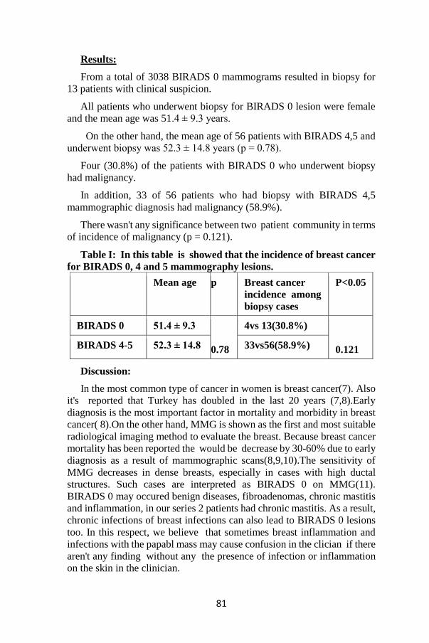
81
Results:
From a total of 3038 BIRADS 0 mammograms resulted in biopsy for
13 patients with clinical suspicion.
All patients who underwent biopsy for BIRADS 0 lesion were female
and the mean age was 51.4 ± 9.3 years.
On the other hand, the mean age of 56 patients with BIRADS 4,5 and
underwent biopsy was 52.3 ± 14.8 years (p = 0.78).
Four (30.8%) of the patients with BIRADS 0 who underwent biopsy
had malignancy.
In addition, 33 of 56 patients who had biopsy with BIRADS 4,5
mammographic diagnosis had malignancy (58.9%).
There wasn't any significance between two patient community in terms
of incidence of malignancy (p = 0.121).
Table I: In this table is showed that the incidence of breast cancer
for BIRADS 0, 4 and 5 mammography lesions.
Mean age p Breast cancer
incidence among
biopsy cases
P<0.05
BIRADS 0 51.4 ± 9.3
0.78
4vs 13(30.8%)
0.121 BIRADS 4-5 52.3 ± 14.8 33vs56(58.9%)
Discussion:
In the most common type of cancer in women is breast cancer(7). Also
it's reported that Turkey has doubled in the last 20 years (7,8).Early
diagnosis is the most important factor in mortality and morbidity in breast
cancer( 8).On the other hand, MMG is shown as the first and most suitable
radiological imaging method to evaluate the breast. Because breast cancer
mortality has been reported the would be decrease by 30-60% due to early
diagnosis as a result of mammographic scans(8,9,10).The sensitivity of
MMG decreases in dense breasts, especially in cases with high ductal
structures. Such cases are interpreted as BIRADS 0 on MMG(11).
BIRADS 0 may occured benign diseases, fibroadenomas, chronic mastitis
and inflammation, in our series 2 patients had chronic mastitis. As a result,
chronic infections of breast infections can also lead to BIRADS 0 lesions
too. In this respect, we believe that sometimes breast inflammation and
infections with the papabl mass may cause confusion in the clician if there
aren't any finding without any the presence of infection or inflammation
on the skin in the clinician.

82
In this reason; Especially in the clinical and radiolical suspicious cases
must be breast biopsy with needle (11). However, proper conditions and
meticulously breast biopsy are very important for good sample removal
and nosocomial infections. D.Dülger and M.Berktaş in their study on the
clinical importance of S.Maltophilia strains in the importance of
nosocomial infections. It is reported that trimethoprim-sulfamethoxazole
should be used as the most effective antibiotic against the agent when it is
necessary to start empirical treatment in infections caused by S. maltophilia
strains. An interesting point is the increase in S. maltophilia septicemia
after the introduction of imipenem, a broad-spectrum antibiotic against
gram-negative bacteria(12,13). In this respect, although the breast masses
enter the general surgery field, the clinician should always be vigilant and
show a multidisciplinary study when necessary. On the other hand, E.Kaya
emphasized that cefozolin reduces surgical site infections in breast surgery
due to breast cancer. As a result, it should be considered that there may be
surgical site infections after the diagnosis of breast cancer starting with
MMG(14).The category BI-RADS 0 indicates that advanced evaluation
should be made by other available methods other than USG, since precise
ultrasonographic evaluation is not possible(15).Additionally,
Dobrosavljević A, etal. had stated that diagnostic values of breast USG in
MMG BI-RADS 0 and clinically vague or suspicious of malignancy breast
lesions six times more common cause for biopsy than BI-RADS 5
category(15) In this study we too detected there aren’t any differences
between that BIRADS 4 - 5 and BIRADS 0 lesions, in the biopsy cases.
In the evaluation of breast cancers, these cases are still overlooked within
the scope of screening programs and we think that the experience of the
clinician is especially prominent. According to our idea this is depend on
attributed to the fact that an experienced clinician does not make decisions
based solely on laboratory and imaging methods and attaches importance
to physical examination and anamnesis.
Conclusion: As a result, the suspicion of malignancy should always be
kept in mind in BIRADS 0 lesions that are biopsied on clinical suspicion.
83
References:
1. International Agency for Research on Cancer, Latest world cancer
statistics global cancer burden rises to 14.1 million new cases in 2012:
marked increase in breast cancers must be addressed, 2013
(http://www.iarc.fr/en/media-centre/pr/2013/pdfs/pr223_E.pdf). ](Cross-
ref)
2. A Cross-sectional Survey on Knowledge regarding Breast Cancer
and Breast Self-examination among Bangladeshi Women. N.STithi, et al.
IOSR Journal of Pharmacy and Biological Sciences (IOSR-JPBS) e-
ISSN:2278-3008, p-ISSN:2319-7676. Volume 13, Issue 1 Ver. I (Jan. –
Feb. 2018), PP 38-44 www.iosrjournals.org DOI
3.Nyström L, Andersson I, Bjurstam N, Frisell J, Nordenskjöld B,
Rutqvist LE. Long-term effects of mammography screening: updated
overview of the Swedish randomised trials. Lancet. 2002; 359(9310):909–
919. [PubMed: 11918907)
4. Liberman L , Morris EA , Benton CL , Abramson AF , Dershaw
DD . Probably benign lesions at breast magnetic resonance imaging:
preliminary experience in highrisk women . Cancer 2003 ; 98 ( 2 ): 377
– 388 .
5. American College of Radiology 1891 Preston White Drive Reston,
VA 20191 .http:///www.acr.org/guidelines 2015 Practice Parameters and
Technical Standards Handbook.
6.Milošević Z. Newspapers mammography in the diagnosis of breast
cancer. In: Nešković-Konstantinović Z, Borojević N, Vučković-Dekić Lj,
editors. Newspapers in the diagnosis and treatment of breast cancer.
Belgrade: Akademija medicinskih nauka Srpskog lekarskog društva,
Institut za onkologiju i radiologiju Srbije; 2008. p. 41−52. (Serbian)
7. The challenge of breast cancer in low- and middle-income
countries—implementing the breast health global initiative guidelines.
Ozmen V, Anderson BO. US Oncology 2008:76-79)
8. Mammografi Görüntülemelerinin Retrospektif Analizi. B. Alan ve
ark. Dicle Tıp Derg / Dicle Med J www.diclemedj.org Cilt / Vol 43, No 2,
339-343.
9. Breast cancer screening: current controversies. Özmen V. J Breast
Health 2011:7:1.
10. Meme mikrokalsifikasyonlarının BIRADS kriterlerine göre
değerlendirilmesi ve yorumcular arası uyum. Gülsün M, Demirkazık FB,
Köksal A, et al. Tanısal Girişimsel Rad 2002:8:358-363.
11.Accuracy of clinical diagnosis, mammography and ultrasonography
in preoperative assessment of breast cancer. Augustina Badu-Peprah and
Yaw Adu-Sarkodie.Ghana Med J. 2018 Sep; 52(3): 133–139.
doi: 10.4314/gmj.v52i3.5

84
12.Stenotrophomonas maltophilia suşlarının klinik önemi
.D.Dülger,M.Berktaş. Van Tıp Dergisi. Yıl: 2007Cilt: 14Sayı: 3ISSN:
1300-2694Sayfa Aralığı: 90 – 95)
13. Antibiotic use and development of resistance in blood culture
isolates: 8 years of experience from a cancer referral center. Krcmery V Jr,
Sykora P, Trupl J, et al. J Chemother 13: 133-142, 2001.
14.Hastane Enefeksiyonlarında Antibiotik Kullanımı, Manisa&2012,
Celal bayar Üniversitesi Hafsa Sultan Hastanesi, Hastane Enfeksiyonları
Kontrol Komitesi. Cerrahi Alan Enfeksiyonları ve Antimikrobiyal
Proflaksi. Chapter:7, Kaynak: sf.81
15. Diagnostic value of breast ultrasound in mammography BI-RADS
0 and clinically indeterminate or suspicious of malignancy breast lesions .
Dobrosavljević A, et al. Vojnosanit Pregl 2016; 73(3): 239–245.
85
SURGICAL APPROACH IN CAROTID ARTERY STENOSIS
Hakan KARTAL*
History
The importance of the carotid artery is known from Hippocrates to the
present day in Ancient Greece. Hippocrates did not define ‘apoplexy
sadece as a term only in the 4th century BC and described the full
development of stroke, transient ischemic attacks of the brain and
hemiplegia on the opposite side in carotid artery disease (1). Rufus of
Ephesus (100 BC) stated that the word meaning of the carotid artery comes
from ‘deep sleep ‘(1). In 1552, Ambroise Pare tied the main carotid artery
in a traumatized patient and mentioned the relationship of this vessel with
deep sleep (2). In 1893, the relationship between extracranial vessel
occlusion and hemiplegia was the subject again. Gowers described this
condition as otid Carotid hemiplegia.. In a textbook published in 1871, the
German clinician Gerhardt emphasized the importance of vascular
auscultation in the definition of carotid stenosis. Gluck reported in 1898
that the blood flow of the brain could be corrected for the first time by an
animal experiment using a vein graft to the main carotid artery (1,4). In
1896, M. Jaboulay performed an end-to-end anastomosis of the carotid
artery. The most valuable information about extracranial occlusive carotid
artery diseases came from Rumsey Hunt in 1914. Hunt reported that
contralateral hemiplegia and ipsilateral amorosis fugax develop in diseases
of the cervical carotid arteries and that examination of pulses of these
vessels is important (1).
Epidemiology
Cerebrovascular disease is the second leading cause of death worldwide
and accounts for approximately 9.5% of all deaths. Approximately 15% of
strokes are fatal, 15% to 20% result in severe disability, and 15% to 20%
of patients who recover have another stroke that results in disability in the
future. Atherosclerotic carotid artery disease is the most commonly treated
vascular disease after coronary artery disease. The incidence of significant
carotid artery disease in patients with coronary bypass is 3-12%, while the
incidence of symptomatic coronary artery disease in patients with carotid
* Gülhane Eğitim ve Araştırma Hastanesi
86
endarterectomy is around 50%. A statistical study of the American Heart
Association (AHA) in the past years is quite striking. On average, the
patient has an American stroke every 24 to 45 seconds. Each year,
approximately 700,000 people have new or recurrent strokes. Of this,
500,000 are new and 200,000 are repetitive stroke. 88% of all strokes are
ischemic, 9% are hemorrhagic and 3% are due to subarachnoid
hemorrhage.
Anatomy
The brain derives from the four main arterial trunks, which originate
from the arcus aorta and consist of two internal carotid arteries and two
vertebral arteries (10). Carotid System Internal carotid artery (ICA): This
artery emerges from the CCA (common carotid artery) with the external
carotid artery in the cervical region and passes through the carotid canal to
the middle cranial fossa. It then passes through the cavernous sinus and
pierces the dura and proceeds to the subarachnoid space. The internal
carotid artery (ICA) is divided into two terminal branches lateral to the
optic chiasm on the surface of the brain: ACA (anterior cerebral artery) and
MCA (medial cerebral artery). The ICA completes this course mainly in
four segments: 1. cervical segment 2. petrosal segment 3. cavernous
segment 4. cerebral (supraclinoid segment)
-Cervical segment: The main carotid artery from the carotid channel
entry, the branch does not branch.
-Petrosal segment: The segment of the temporal bone within the
petrosis segment is divided into two branches: the caroticotympanic artery
(feeding the tympanic cavity) and the pterygoid artery (waters of the
pterygoid channel).
-Cavernous segment: The segment within the cavernous sinus is
divided into three branches: 1. Pituitary artery (nourishes the
neurohypophysis). 2. Anterior meningeal artery (waters the base of the
anterior fossa). 3. Ophthalmic artery (feeding the optic nerve, retina,
frontal and ethmoid sinuses and nasal ridge with anastomosis of branches
of external carotid artery).
Clinical Findings in Carotic Artery Disease
The most important finding in physical examination is the murmur.
However, there is no relationship between murmur and severity and
prevalence of stenosis. At 30-30% stenosis on the carotid, murmur begins
to be heard.

87
In carotid artery disease, symptomatology can be determined by 5 main
findings (16,17). These:
1. Amarosis Fugax (Transient mononuclear vision loss):
Amarosis is a potential symptom of carotid artery disease with
unilateral visual loss occurring in the form of fugax or retinal artery
occlusion (18).
2. Transient Ischemic Attack (TIA):
TIA is a focal brain dysfunction that occurs due to ischemia of one of
the vascular systems and lasts less than 24 hours (19,20).
3. Reversible Ischemic Neurological Deficit (RIND)
4. Ischemic Stroke:
“World Health Organization WHO (WHO) defined the term stroke as
bul clinical manifestations of sudden, focal or global cerebral dysfunction
of vascular origin, which may last 24 hours or more, leading to death”.
Infarction or bleeding due to trauma, infection, tumor or transient attacks
due to cerebral ischemia were excluded from the definition (21).
5. Cerebral Infarction:
80-85% of all strokes are cerebral infarction, 10-15% intracerebral
hematoma and 6-8% subarachnoid hemorrhage. The main feature of stroke
is the sudden onset of neurological findings.
Diagnosis Methods in Carotic Artery Disease:
Dublex Ultrasonography
Duplex ultrasonography is a non-invasive method for real-time two-
dimensional imaging, doppler flow analysis, vascular structures and blood
flow velocity. Arterial diameter and degree of stenosis are not measured
directly with this method. The degree of stenosis is determined by
evaluating the blood flow velocity. Speed charts were used to determine
carotid stenosis by duplex ultrasound. ICA peak systolic velocity, ICA
PSV / CCA PSV values were correlated with angiographic methods in
carotid stenosis grading. When compared with conventional angiography,
sensitivity and specificity were found to be 85% and 90%, respectively, in
the evaluation of 70% carotid artery stenosis (23).
Color Doppler Us (CDUS)
88
Carotid arteries should be evaluated with RDUS in the axial plane in all
areas that can be traced from the most proximal to the distal, as in the gray
scale. First of all, whether the carotid arteries and internal and external
branches are open or not are evaluated. The level of carotid detachment, if
any variation or tortuosity is determined. There are pathologies that can be
diagnosed directly by RDUS. A critical main carotid artery stenosis in the
proximal and associated external-internal carotid artery stealing
phenomenon and main carotid artery dissection can be counted among
these (24).
Spectral Doppler Us
The most important criterion for assessing the severity of carotid artery
stenosis is flow velocity. Spectral flow samples are taken from the distal
part of the plate at the determined level, from the areas where velocity
values are expected to be the highest due to jet flow. Peak systolic velocity
and end-diastolic velocity values are measured from flow samples and are
proportional to the values obtained from the bilateral main carotid artery
(24-22).
Computerized Tomographic Angiography (CTA)
It is a radiological examination obtained by combining thin and
continuous images after intravenous radiopaque injection. Anatomical
imaging of aorta, aortic branches and willis polygon is provided. Multi-
plane reconstruction and analysis allows evaluation of highly tortuous
veins. In contrast to US and MRA, CTA provides direct assessment of
vascular lumen and stenosis. In severe stenosis, the vessel lumen affects
the accuracy of the measurement when the gate approaches the BTA
resolution limits. With the increase in the number of detectors, higher
resolution images of larger areas can be obtained. When compared with
catheter angiography, the sensitivity and specificity of CTA is 100%, the
specificity is 63% and 70% in patients with carotid and vertebral artery
stenosis. (23).
Magnetic Resonance Angiography (MRA)
High resolution non-invasive images of cervical arteries are obtained
with MRA. It provides high quality accurate anatomical imaging of the
aorta, cervical and cerebral arteries without exposure to ionizing radiation.
Compared with conventional angiography, the sensitivity and specificity
were 97-100% and 82-96%, respectively.
Digital Subtraction Angiography (DSA)

89
DSA is the gold standard method for vascular imaging. DSA should be
withdrawn when conflicting results are obtained with less invasive imaging
methods. DSA may be performed in patients with renal insufficiency (to
minimize contrast load), obese, permanent ferromagnetic material, in cases
of suspected duplex ultrasonography, CTA and MRA withdrawal. Since it
is an invasive imaging method, it may have some complications. The most
serious complication is stroke and its incidence is less than 1% when
performed by experienced physicians. With the widespread use of stent
revascularization, the use of DSA as a diagnostic method is increasing
(23,25).
Surgical Treatment Approach
Surgical Treatment Of Asemptomatic Carotic Artery Stenosis
There are two major studies on the application of endarterectomy for
the first time in 1954 in asymptomatic carotid stenosis: Asymptomatic
Carotid Surgery Trial (ACST) (60-99% stenosis) and Asymptomatic
Carotid Atherosclerosis Study (ACAS) (Asymptomatic Carotid
Atherosclerosis Study (ACAS)). It was completed with a total of 4782
patients with asymptomatic carotid stenosis (70-99%) and decreased the
risk of ipsilateral stroke more than 5 years in comparison with medical
treatment.Resar risk reduction with ACAS was found to be 53%. It has
been reported with ACST: 0.5% decrease each year, while the 5-year
stroke risk with ACST was 6.4% and 11.8% with medical treatment alone,
and it was concluded that women benefited less from endarterectomy with
ACAS (26). Studies have shown that CEA is beneficial in asymptomatic
patients (especially men) between 40-75 years of age, carotid stenosis>
60%, life expectancy> 5 years, and operating mortality <3% (27).
Surgical Treatment Of Symptomatic Carotic Artery Stenosis
There are two major studies, NASCET (North American Symptomatic
Carotid Endarterectomy Trial) and ECST (European Carotid Surgery
Trial), which show indications for surgical treatment in patients with
symptomatic carotid artery stenosis. With NASCET, 2226 patients with
symptomatic stenosis were randomized for medical treatment and
endarterectomy. Ipsilateral cerebral attack significantly decreased with
endarterectomy in patients with a stenosis rate greater than 70%; Surgery
has been reported as a more effective preservative in patients with 50-69%
stenosis. It was concluded that there is no benefit in patients with a degree
of stenosis below 50% (28).
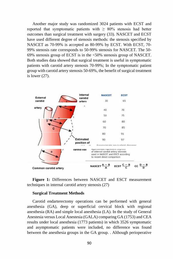
90
Another major study was randomized 3024 patients with ECST and
reported that symptomatic patients with ≥ 80% stenosis had better
outcomes than surgical treatment with surgery (33). NASCET and ECST
have used different degree of stenosis methods: the stenosis specified by
NASCET as 70-99% is accepted as 80-99% by ECST. With ECST, 70-
99% stenosis rate corresponds to 50-99% stenosis for NASCET. The 50-
69% stenosis group of ECST is in the <50% stenosis group of NASCET.
Both studies data showed that surgical treatment is useful in symptomatic
patients with carotid artery stenosis 70-99%; In the symptomatic patient
group with carotid artery stenosis 50-69%, the benefit of surgical treatment
is lower (27).
Figure 1: Differences between NASCET and ESCT measurement
techniques in internal carotid artery stenosis (27)
Surgical Treatment Methods
Carotid endarterectomy operations can be performed with general
anesthesia (GA), deep or superficial cervical block with regional
anesthesia (RA) and simple local anesthesia (LA). In the study of General
Anestesia versus Local Anestesia (GALA) comparing GA (1753) and CEA
results under local anesthesia (1773 patients) in which 3526 symptomatic
and asymptomatic patients were included, no difference was found
between the anesthesia groups in the GA group. . Although perioperative
91
stroke, myocardial infarction and death rates in the first 30 days were
higher in the GA group compared to the local anesthesia group, no
significant difference was found between the two groups (29).
There are 3 options to use shunt in KEA; The lack of routine shunting
has been published in very large series. Another is the routine use of shunt
in all patients. Although those who prefer this method have published
successful results, they have stated the risk of stroke due to technical
problems. The last option is to use shunt in patients with high risk of
ischemic stroke when shunt is not used.
Various methods are used to accurately determine the need for shunt
use in patients under GA; carotid stump pressure measurement (collateral
circulation is sufficient if above 50mmHg), electroencepholography,
transcranial doppler of the middle cerebral artery, cerebral oximeter.
Another method is to evaluate the changes in the patient's neurological
responses under RA and make decisions. Intraoperative stroke may be
ischemic or embolic. Although our aim in using shunt is to prevent
ischemic stroke, there is a risk of embolic stroke (air. Particle) and
dissection during shunt placement (30).
The patient should be in the supine position on the operating table, with
a slight hyperextension of the neck and with the head facing the opposite
side of the lesion. If GA is to be administered, the patient is intubated first.
If LA or RA is to be applied, the Mayo stand is placed to prevent
claustrophobia (30).
Two types of incisions are used. First; mastoid protrusion and
sternoclavicular joint, sternocloidomastoid muscle incision parallel to the
anterior border. The incision extends deep through the platysma muscle
and reaches the fascia extending between the sternocloidomastoid muscle
and the trachea. The second incision is 1-2 cm below the jaw angle and
parallel to the skin fold. The incision is deepened along the platysma to
reach the underlying fascia. Although this type of incision has more
cosmetic results than vertical incision, access to proximal and distal areas
is more difficult in this form. There is no difference between these two
incisions in the rates of stroke, cranial nerve injury, wound complications
or restenosis (31).
Complications after Carotis Endarterectomy
Cardiac Complications: Myocardial infarction is responsible for 25% to
50% of deaths after CEA. Symptomatic coronary artery disease is present

92
in at least 40-50% of patients with CEA as a result of systemic
atherosclerosis (35). But in the last 20 years
11th operative mortality significantly decreased. In a 10-year study on
1566 patients, the rate of postoperative 30-day myocardial infarction was
found to be 1.5% (30).
Cranial Neural Injury: It is the most common neurological complication
after CEA. The incidence of peroperative cranial nerve damage is 4.7%.
The most commonly affected cranial nerve n. And hypoglossal. Although
clinical deficit is transient, it usually returns within weeks or months (30).
Cranial nerve injury is more common in recurrent carotid
endarterectomy. Cranial nerve injury rate was found to be 21% in 89
patients with recurrent carotid endarterectomy, 88% of which were
transient and 22% were permanent damage (35).
Hemodynamic Immunity:
If hypotension develops after CEA, it is usually associated with
bradycardia within the first 2 hours. This is often the result of impaired
baroreceptor function. After endarterectomy of the atheromatous plaque
placed in bifurcation, there is an increase in baroreceptor sensitivity and
hypotension and / or bradycardia develops by decreasing the activity of the
central nervous system. Hypertension after CEA is thought to develop due
to preoperative uncontrolled hypertension, but its mechanism is not fully
understood (30).
Eversion endarterectomy has more sympathetic activation and
postoperative hypertension than the traditional method. Although this
hypertension regresses within the first 24 hours in the first 80% of patients,
eversion endarterectomy can last up to 4 days (36, 37).
Hyperperfusion Syndrome:
Cerebral hyperperfusion syndrome usually occurs days after CEA and
is usually associated with severe hypertension. Severe headache is usually
observed before acute neurological deficit. The most devastating
manifestation of hyperperfusion syndrome that may develop after CEA is
intracerebral hemorrhage. It occurs between 0.4% and 7.7% after CEA
(30). As the cause of hyperperfusion syndrome, increased cerebral blood
flow secondary to impaired intracerebral autoregulation is seen after the
removal of a high degree of carotid stenosis with severe lesion in the
contralateral carotid artery (38).
93
The SVS clinical guideline recommends that systolic blood pressure be
kept below 140 mm Hg diastolic blood pressure below 80 mm Hg after
CEA (39).
Infection:
Wound infection is not common due to the rich blood supply to the
neck. Cellulitis is usually observed, but its incidence is between 0.09% and
0.15%. The possibility of infectious complications increases in
arteriotomies repaired with synthetic patch. However, the true incidence of
patch infection could not be detected and is extremely rare (30).
Bleeding:
Postperative hemorrhage is an uncommon complication of CEA,
although its incidence is between 0.7% and 3.0%. It is more common in
patients receiving clopidogrel. However, we should not stop the use of
clopidogrel in cases such as a history of previous coronary artery stents or
symptomatic carotid disease that should use this agent. When the neck
incision is closed, active red bleeding is observed in the drain and the
development of hematoma compressing the trachea is again indications for
surgery (30).
Repeating Stenosis:
The most important arterial complication after CEA is recurrent carotid
stenosis. It is more common in women, those who continue to smoke, and
in hypercholesterolemic, diabetic and hypertensive individuals (30). In the
EVEREST study, no significant difference was detected between CEA
using eversion and patch in terms of recurrent stenosis at 4-year follow-up
(40). Early stenosis occurs within the first 2 years and develops as a result
of intimal hyperplasia. Stenosis seen after 2 years is called late stenosis and
typically shows atheromatous plaque characteristics (30).
Referaces
1. Robicsek F, Roush TS, Cook JW, Reames MK. From Hippocrates to
Palmaz-Schatz, the history of carotid surgery. Eur J Vasc Endovasc Surg.
2004 Apr;27(4):389-97
2. Pare A. The Works of That Famous Chirurgion Ambroise Parey.
New York: Milford House, 1968
3. Wolpert SM. The circle of Willis.AJNR Am J Neuroradiol. 1997

94
JunJul;18(6):1033-4
4. ROSS RS, McKUSICK VA. Aortic arch syndromes; diminished or
absent pulses in arteries arising from arch of aorta. AMA Arch Intern Med.
1953 Nov;92(5):701-40.
5. DeBakey ME. Successful carotid endarterectomy for cerebrovascular
insufficiency. Nineteen-year follow-up. JAMA. 1975 Sep 8;233(10):1083-
5.
6. EASTCOTT HH, PICKERING GW, ROB CG. Reconstruction of
internal carotid artery in a patient with intermittent attacks of hemiplegia.
Lancet. 1954 13;267(6846):994-6.
7. AL-NAAMAN YD, CARTON CA, COOLEY DA. Surgical
treatment of arteriosclerotic occlusion of common carotid artery. J
Neurosur 1956 Sep;13(5):500-6
8. Moore OS, Karlan M, Sigler L. Factors influencing the safety of
carotid ligation. Am J Surg. 1969 Nov;118(5):666-8.
9. Callow AD. David M. Hume Memorial Lecture. An overview of the
stroke problem in the carotid territory. Am J Surg. 1980 Aug;140(2):181-
91
10. Balkan S.Serebrovasküler Hastalıklar.Güneş Kitabevi Yayınları
200211.Öge AE İstanbul Tıp Fakültesi Temel ve Klinik Bilimler
Kitabı..Nobel Yayınları .2004
12. Ross R. The pathogenesis of atherosclerosis: a perspective for the
1990s. Nature 1993;362: 801-809
13. Libby P. The Pathogenesis of Atherosclerozis. İn: Braunwald E.
Fauci A.S, Kasper D.L, Hauser S.L, Longo D.L, Jameson L. Harrison‟s
Principles of Internal Medicine, 15th Ed. New york: McGraw Hill
2001:1377-86
14. Buğan B. Çelik T, Risk Factors for Coronary Artery Disease. J Clin
Anal Med 2014;5(2):161-5
15. Helderman F, Segers D, Rini de Cromb, Hiercke BP, Poelmanne
RE, Evansd P.C, Kramsc R. Kan akıs geriliminin (shear stress) vasküler
inflamasyon ve plak gelisimi üzerine etkisi Current Opinion in Lipidology
2007;2(4):226-232
16. Goldstein LB, Adams R, Becker K, Furberg CD, Gorelick PB,
95
Hademenos G, Hill M, Howard G, Howard VJ, Jacobs B, Levine SR,
Mosca L, Sacco RL, Sherman DG, Wolf PA, 14
del Zoppo GJ. Primary prevention of ischemic stroke: A statement for
healthcare professionals from the Stroke Council of the American Heart
Association. Stroke. 2001 Jan;32(1):280-99
17. Weisberg LA, Garcia C, Strub R. Evaluation of Common
Neurologic Signs and Symptoms in Essentials of Clinical Neurology.
Mosby Inc Portland 1996 Ch 10.
18. FISHER M. Transient monocular blindness associated with
hemiplegia. AMA Arch Ophthalmol. 1952 Feb; 47(2):167-203
19. Norris JW, Zhu CZ. Silent stroke and carotid stenosis. Stroke. 1992
Apr; 23(4):483-5
20. Humphrey P. Stroke and transient ischaemic attacks. J Neurol
Neurosurg Psychiatry. 1994 May; 57(5):534-43. Review.
21. Kumral K, Kumral E: Santral Sinir Sisteminin Damarsal
Hastalıkları. Ege Üniversitesi Tıp Fakültesi Yayınları No:72, Yücesahil 4-
446.
22. Biler J, Love BB : Vascular Disease of the Nervous System . In;
Bradley WG, Daroff RB, Fenichel GM; Marsden CD (eds). Neurology in
Clinical Practice. 3 th ed. Vol 2. USA: Butterworth-Heinemann, 2000: ch
57, 1125- 1166.
23. Brott et al. Guideline on the Management of Patients With
ExtracranialCarotid and Vertebral Artery Disease journal of the J
American College of Cardiology 2011; 57: 8
24. Seçil M. Temel ultrasonografi ve doppler, Karotis ve Vertebral
Doppler US, Ege kitapevi, İzmir, 2013: 479-498
25. Ricotta JJ, Aburahma A, Ascher E, Eskandari M, Faries P, Lal BK.
Updated Society for Vascular Surgery guidelines for management of
extracranial carotid disease. J Vasc Surg. 2011;54(3):131
26.Benli ÜS: Yaşlılık Karotis Hastalığı ve İnme. Turkiye Klinikleri J
Cardiol-Special Topics 2013;6(1):44-51.
27. Avrupa Kardiyoloji Derneği (ESC) .Periferik damar hastalıkları tanı
tedavi kılavuzu 2011;10-65.
96
28. North American Symptomatic Carotid Endarterectomy Trial
Collaborators. Beneficial effect of carotid endarterectomy in symptomatic
patients with high grade carotid stenosis..N Eng J Med 1991; 325(7):445-
53
29. Rerkasem K,Rothwell PM:Local Versus General Anesthetic for
Carotid Endarterectomy. Stroke. 2009;40:e584-85.
30. Rutherford’s Vascular Surgery.Eight edition.Volume 2.Elsevier
Saunders Philadelphia 2014; 1517-38 15
31. Marcucci G, Antonelli R, Gabrielli R, Accrocca F, Giordano AG,
Siani A: Short longitudinal versus transverse skin incision for carotid
endarterectomy: impact on cranial and cervical nerve injuries and esthetic
outcome. J Cardiovasc Surg (Torino). 201; 52(2):145-52.
32. Al-Rawi PG, Sigaudo-Roussel D, Gaunt ME. Effect of lignocaine
injection in carotid sinus on baroreceptor sensitivity during carotid
endarterectomy. J Vasc Surg. 2004; 39(6):1288-94.
33.Abbott AL: Medical (nonsurgical) intervention alone is now best for
prevention of stroke associated with asymptomatic severe carotid stenosis:
results of a systematic review and analysis. Stroke 2009; 40: e573–83.
34. Biller J, Feinberg WM, Castaldo JE , Biller J,Feinberg
WM,Castaldo JE, Whittemore AD, Harbaugh RE,Dempsey RJ, Caplan
LR,Kresowik TF,Matchar DB,Toole JF,Easton JD,Adams HP,Brass LM,
Hobson II RW, Brott TG,Sternau L.AHA Scientific StatementGuidelines
for Carotid Endarterectomy. Circulation. 1998; 97: 501; 9.
35.Abu Rahma AF, Choueiri MA. Cranial and cervical nerve injuries
after repeat carotid endarterectomy. J Vasc Surg. 2000; 32(4):649-54.
36. Demirel S, Bruijnen H, Attigah N,Hakimi M, MD, Böckler D.The
effect of eversion and conventional-patch technique in carotid surgery on
postoperative hypertension. J Vasc Surg. 2011; 54(1):80-6.
37.Demirel S, Attigah N, Bruijnen H,Macek L,Hakimi M,Able
T,Böckler D.Changes in baroreceptor sensitivity after eversion carotid
endarterectomy. J Vasc Surg. 2012 May; 55(5):1322-8.
38. Abou-Chebl A,Yadav JS,Reginelli JP,Bajzer C,Bhatt D,Krieger
DW. Intracranial hemorrhage and hyperperfusion syndrome following
carotid artery stenting: risk factors, prevention, and treatment. J Am Coll
Cardiol 43:1596-1601, 2004.

97
39.Ricotta JJ, AbuRahma A,Ascher E,Eskandari M, Faries P, Lal KB .
Updated Society for Vascular Surgery guidelines for management of
extracranial carotid disease: executive summary. J Vasc Surg 54:832-6,
2011.
40. Cao P, Giordano G,Rango PD, Zannetti S,Chiesa R,Coppi
G,Palombo D,Peinetti F,Spartera C, Stancanelli V, Vecchiati E : Eversion
versus conventional carotid endarterectomy: late results of a prospective
multicenter randomized trial. J Vasc Surg 31:19- 30, 2000.
98
DIAGNOSIS AND TREATMENT OF CHRONIC VENOUS
INSUFFICIENCY AND VARICOSE VEINS
Hüseyin SİCİM & Hakan KARTAL
Introduction
Chronic venous insufficiency (CVD) and related lower extremity
varices are a common problem all over the world. Varicose veins affect
approximately 32% of women and 40% of men. Varicose veins are seen in
1/3 of men and women between the ages of 18-64 (1). Varicose veins of
the lower extremities are divided into three groups as spider veins, reticular
veins and varicose veins according to their size and distance from the skin.
Varicose veins may be asymptomatic or cause severe symptoms. Standing
for a long time may cause pain, itching, burning, tingling, night cramps,
edema, skin changes and venous ulcers in chronic cases. It interferes with
the daily activities of individuals and may cause loss of work and time (2).
Varicose veins thrombophlebitis and associated pulmonary embolism may
develop (3,4). Even deaths related to spontaneous variceal bleeding have
been reported in the literature (5,6)
Etiology And Pathophysiology
The return of lower extremity venous blood to the heart is a complex
mechanism. There are 3 major components of this mechanism. These;
stability of the lower limb calf muscle pump, non-obstructed venous flow
and venous valves Is the qualification. Valve function has the most
important role in venous system physiology (7,8). Normal covers are
bicuspid, collagen-containing structures that allow only one-way flow.
Normally operating valves are defined as competent and those that allow
retrograde flow are defined as incompetent. Primary valve failure is usually
loss of elasticity. In secondary valve failure, the causative agent is DVT
(9,18).
Venous Anatomy
Superficial venous system
VSM is the longest vein in the body. The median margin of the medial
edge of the foot ridge begins as a continuation of the medialis. It passes
through the anterior of the medial malleolus on the ankle and on the medial
side of the leg. sapheneus. Knee at the level of the joint joint, passing
through the medial condyle and the sinephal compartment on the inner face
99
of the thigh, which is a course between the deep muscular fascia and the
saphenous fascia on the surface, passes through the hiatus saphenus in the
deep fascia and opens into the main femoral vein approximately 3 cm in
the ligamentum inguinal. The saphenofemoral junction (SFB) is where the
VSM enters the main femoral vein of the deep venous system at the groin
level. Duplication can be observed in VSM at a rate of 1-2% (7,19,20).
Deep venous system
Anterior tibial, posterior tibial and peroneal veins provide drainage of
the calf. These veins, which are found as a pair, accompany the arteries of
the same name. Proximally, two posterior tibial veins merge into a short
posterior tibial root and the peroneal pair becomes a short root in the same
way. These two roots m. at the lower edge of the popliteus, popliteal to
form the vein. The paired anterior tibial veins form a short root and join
the popliteal vein. The most important muscular veins draining the calf
muscles are the gastrocnemius and soleal veins. Gastrocnemius veins can
be seen in the medial head of the gastrocnemius muscle and drain into one
of the popliteal vein or posterior tibial vein. Soleal veins are centrally
located and the soleal muscle in the posterior tibia It is located inside.
These veins, which may be larger than one centimeter, drain into the
posterior tibial or peroneal system. The popliteal vein extends
longitudinally along the popliteal fossa at the posterior of the popliteal
artery and is directed medially to enter the adductor canal. The popliteal
vein is duplicated by 5% (22).
Perforating vein
Perforators are bridging channels between the superficial and deep
venous systems. These veins obliquely perforate the deep fascia and play
a key role in equilibrating blood flow during calf muscle contraction
because of valves that prevent reflux from the deep venous system to the
superficial venous system. Perforating veins are numerous and highly
variable in arrangement, connection, and size. There are four clinically
important perforator groups: upper thigh (Hunterian), lower thigh
(Dodd’s), at knee level (Boyd’s), and in the calf region (Cockett’s).
Although perforator valve incompetence is always associated with CVI,
the cause of perforator insufficiency is not known, and the routine
treatment of perforating veins in C2 patients is not supported (16).
Varicose veins of the lower extremities are divided into 3 types as
spider (telangiectatic), reticular and varicose veins.
Spider Veins
They are located intradermally and there is no skin protrusion.
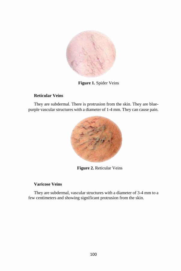
100
Figure 1. Spider Veins
Reticular Veins
They are subdermal. There is protrusion from the skin. They are blue-
purple vascular structures with a diameter of 1-4 mm. They can cause pain.
Figure 2. Reticular Veins
Varicose Veins
They are subdermal, vascular structures with a diameter of 3-4 mm to a
few centimeters and showing significant protrusion from the skin.

101
Figure 3. Varicose Vein
Diagnosis of CVI
Computed tomography and magnetic resonance venography
Venography is useful in the anatomical and hemodynamic evaluation
of the venous system. End Ascending venography için is the gold standard
for the diagnosis of DVT. Descendant venography is used to evaluate
valvular insufficiency (17,18). In ascending venography, superficial veins
are occluded by applying a tourniquet at the ankle level and contrast agent
is injected into the vein of the foot back. In the femoropopliteal region,
computed tomography and magnetic resonance venography provide
information close to US and venography. They are effective in
demonstrating pelvic veins and inferior vena cava.
Venous duplex ultrasonography
When creating an image in the gray scale US, the reflection intensity of
the rotating echo (amplitude of the rotating wave) and the time between
sending and receiving the sound wave reaching the probe are calculated
and the rotating wave is coded in different gray tones. In addition to this
information, the reflected sound wave includes phase, wavelength and
frequency information which is not taken into account when creating
images in the B-mode examination. Using this information, an image is
generated in the DU (19). Blood is an inhomogeneous medium consisting
of a large number of blood elements of various diameters, in which the
erythrocytes are distributed randomly. When the wavelength of the
transmitted ultrasound sound wave is too large than the reflective surface
(such as erythrocytes) The ultrasound waves transmitted show a scattering
called Rayleigh-Tyndall scattering from the surface of erythrocytes within
the vascular structures. The amount of scattering is directly proportional to
the 4th order exponent of sound frequency (19,20). In fixed tissues, the
wavelength (λ) and frequency (f) of the ultrasound sound wave are the
same as the wavelength and frequency of the wave reaching the probe after
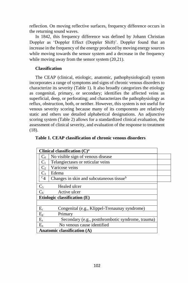
102
reflection. On moving reflective surfaces, frequency difference occurs in
the returning sound waves.
In 1842, this frequency difference was defined by Johann Christian
Doppler as ‘Doppler Effect (Doppler Shift)’. Doppler found that an
increase in the frequency of the energy produced by moving energy sources
while moving towards the sensor system and a decrease in the frequency
while moving away from the sensor system (20,21).
Classification
The CEAP (clinical, etiologic, anatomic, pathophysiological) system
incorporates a range of symptoms and signs of chronic venous disorders to
characterize its severity (Table 1). It also broadly categorizes the etiology
as congenital, primary, or secondary; identifies the affected veins as
superficial, deep, or perforating; and characterizes the pathophysiology as
reflux, obstruction, both, or neither. However, this system is not useful for
venous severity scoring because many of its components are relatively
static and others use detailed alphabetical designations. An adjunctive
scoring system (Table 2) allows for a standardized clinical evaluation, the
assessment of clinical severity, and evaluation of the response to treatment
(18).
Table 1. CEAP classification of chronic venous disorders
Clinical classification (C)a
C0 No visible sign of venous disease
C1 Telangiectases or reticular veins
C2 Varicose veins
C3 Edema C4 Changes in skin and subcutaneous tissueb
C5 Healed ulcer
C6 Active ulcer
Etiologic classification (E)
Ec Congenital (e.g., Klippel-Trenaunay syndrome)
Ep Primary
Es Secondary (e.g., postthrombotic syndrome, trauma)
En No venous cause identified
Anatomic classification (A)
103
CEAP, clinical, etiologic, anatomic, pathophysiological. aThe
descriptor A (asymptomatic) or S (symptomatic) is placed after the C
clinical class. bC4 is subdivided into A and B, with B indicating higher
severity of disease and having a higher risk for ulcer devel-opment.
Table 2. Venous Clinical Severity Score
As Superficial
Ad Deep
Ap Perforator
An No venous location identified
Pathophysiologic classification (P)
Pr Reflux Po Obstruction, thrombosis Pr,o Reflux and obstruction
Pn No venous pathophysiology identified
Attribute Absent
= 0
Mild = 1 Moderate = 2 Severe = 3
Pain None Occasional, not
restricting
Daily,
interfering but
not
Daily, limits
most daily
daily activity preventing
daily activity
activity
Varicose
veins
None Few, isolated
branch varices,
Confined to
calf or thigh
Involves calf
and thigh
or clusters,
includes ankle
flare
Venous
edema
None Limited to foot
and ankle
Extends above
the ankle but
Extends to
knee and
above
below knee
Skin
pigmentatio
n
None
or focal
Limited to
perimalleolar
Diffuse, over
lower third of
Wider
distribution
above
calf lower third of
calf
104
An aggregate score for the limb is calculated by adding the individual
component scores. The range of the total score is 0 to 30. NA, not
applicable.
Treatment of CVI
Conservative Management
The initial management of CVI involves conservative measures to
reduce symptoms and prevent development of secondary complications
and progression of disease. The use of compressive stockings is the
mainstay of conservative management and is further described below. If
conservative measures fail or provide an unsatisfactory response, further
treatment should be considered on the basis of anatomic and
pathophysiologic features (Figure 7). Regarding the management of CVI,
the practitioner should be able to recognize the manifestations of CVI and
use confirmatory testing, such as venous duplex reflux studies and perhaps
APG for advanced and recurrent disease. Specific treatment is based on
severity of disease with CEAP classes C4 to C6 often requiring invasive
treatment. A referral to a vascular specialist should be made for patients
with CEAP classes C4 to C6 (and probably for CEAP class C3 for
extensive edema). These patients with uncorrected advanced CVI are at
risk for ulceration, recurrent ulceration, and nonhealing ulcers with
progressive infection and even veno-lymphedema. A healthy lifestyle
including maintaining an ideal body weight or weight reduction if
Inflammatio
n
None Mild cellulitis,
ulcer margin
limited to
perimalleolar
Diffuse over
lower third of
calf
Wider
distribution
above
lower third of
calf
Induration None Limited to
perimalleolar
Diffuse over
lower third of
calf
Wider
distribution
above calf
Ulcer
number
0 1 2 3
Ulcer
duration
NA < 3 mon > 3 mon but <
1 yr
Not healed > 1
yr
Ulcer size NA Diameter < 2
cm
Diameter 2–6
cm
Diameter > 6
cm
Compressive
therapy
Not
used
Intermittent Most days Full
compliance
105
overweight may improve manifestations of CVI. Obesity is a well-
established risk factor for the development of CVI and its complications.
Weight reduction after bariatric surgery has been shown to improve
manifestations of CVI, including edema and ulcers. It may be extrapolated
that weight reduction by other means might also assist in the treatment of
CVI.
Compressive Leg Garments
Elastic compression stockings play a fundamental role in the treatment
of CVD. Elastic compression stockings are recommended for patients with
low complaints or high risk of surgery. Appropriate elastic compression
regresses the symptoms of patients with varicose veins, increases venous
hemodynamics, reduces edema and provides relief (41,42). Compression
stockings are available in different pressure ranges and are used in the
required pressure ranges depending on the patient's complaints and the
degree of disease. Compression stockings are divided into 5 classes
according to their pressure;
Class A: 10-14 mmHg (very little pressure)
Class I: 15-21 mmHg (light pressure)
Class II: 25-32 mmHg (medium pressure)
Class III: 34-46 mmHg (strong pressure)
Klass IV:> 49 mmHg (very strong pressure)
Pharmacologic Therapy
Venoactive drugs used in the medical treatment of CVI are composed
of several drug groups. They may be of vegetable origin or synthetic.
Venoactive drugs relieve edema and symptoms of CVD by antioxidant
mechanism. While these drugs do not correct the existing disease, they
provide symptomatic improvement. They reduce symptoms such as
edema, pain, fatigue, and muscle cramps.
Surgical therapy
Open surgical therapy of varicose veins with high ligation and stripping
of the GSV combined with the excision of large varicose veins has been
the standard of care for more than a century. This therapy is performed in
the following sequence: incisions are made in the groin and upper calf; the
GSV is ligated (high ligation) below the SFJ, and a wire is inserted into the
GSV and advanced distally; the proximal part of the GSV is secured to the
wire and retrieved (stripping) via the calf incision. Stripping of the GSV
below the knee and stripping of the SSV are not usually performed because
of the high risk of nerve injury(19).
106
Endovenous thermal ablation
Using laser energy for the ablation of veins, EVLA is a minimally
invasive method that is an alternative to surgery in superficial venous
insufficiency and related varices. The widely used EVLA technique has
been reported for the first time in the literature by Navarro and Min (12).
EVLA is based on the principle of the energy generated by a laser generator
to be transmitted to the vein lumen by fiberoptic catheter and the ablation
of the vein with the high temperature obtained from this energy. EVLA
releases thermal energy into the blood and venous wall, causing vascular
endothelial damage and vein collapse. Diode laser devices with 810, 940,
980 nm wavelengths are commonly used in the treatment of EVLA (20).
810, 940 and 980 nm wavelength diode laser devices by targeting
hemoglobin, 1320 nm wavelength laser devices by targeting water,
saphenous reflux with minimal side effect was developed to quickly and
effectively treat (22). Vein contraction is a gradual process that leads to the
resorption of the vein in a few months when endothelial damage, focal
coagulative necrosis and thrombotic occlusion of the vein occur.
Thrombotic occlusion enables the saphenous vein to become fibrotic cord
after endovenous treatment. EVLA was approved by the FDA (food and
drug administration) in 2002 for the treatment of varicose veins (22).
The treatment of CVI will be based on the severity of disease and
guided by anatomic and pathophysiologic considerations. Compressive
garments have been a mainstay in the management of CVI. Traditional
surgical techniques and newer interventional methods are often reserved
for unsatisfactory response to conservative measures, although earlier use
of venous ablation should be considered in symptomatic patients.
References
1. Beebe-Dimmer JL, Pfeifer JR, Engle JS, Schottenfeld D. The
epidemiology of chronic venous insufficiency and varicose veins. Ann
Epidemiol. 2005;15:175–184.
2. Raju S, Neglen P. Clinical practice. Chronic venous insufficiency
and varicose veins. N Engl J Med 2009;360:2319- 2327. 2. Eberhardt RT,
Raffetto JD. Chronic venous insufficiency. Circulation 2014;130:333-346.
3. Baliyan V, Tajmir S, Hedgire SS, Ganguli S, Prabhakar AM.
Lower extremity venous reflux. Cardiovasc Diagn Ther 2016;6:533-543.
4. Santler B, Goerge T. Chronic venous insufficiency: a review of
pathophysiology, diagnosis, and treatment. J Dts
5. Callam MJ. Epidemiology of varicose veins. Br J Surg
1994;81:167-173.

107
6. Evans CJ, Fowkes FG, Ruckley CV, Lee AJ. Prevalence of
varicose veins and chronic venous insufficiency in men and women in the
general population: Edinburgh Vein Study. J Epidemiol Community
Health 1999;53:149-153.
7. Thorisson HM, Poljak JS, Scoutt L. The role of ultrasound in the
diagnosis and treatment of chronic venous insufficiency. Ultrasound
Quarterly. 2007; 23: 137-150.
8. Brasic N, Lopresti D, McSwain H. Endovenous laser ablation and
sclerotherapy for treatment of varicose veins. Semin Cutan Med Surg.
2008; 27:264-275.
9. Scott TE, LaMorte WW, Gorin DR, Menzoian JO. Risk factors for
chronic venous insufficiency: a dual case-control study. J Vasc Surg. 1995;
22:622-628.
10. Meissner MH, Gloviczki P, Bergan J, et al. Primary chronic
venous disorders. J Vasc Surg. 2007; 46:54-67.
11. McLafferty RB, Passman MA, Caprini JA, Rooke TW, Markwell
SA, Lohr JM, Meissner MH, Eklöf BG, Wakefield TW, Dalsing MC.
Increasing awareness about venous disease: the American Venous Forum
expands the National Venous Screening Program. J Vasc Surg.
2008;48:394–399.
12. Raffetto J, Eberhardt RT. Chronic venous disorders: general
considerations. In: Cronenwett JL, Johnston KW, eds. Rutheford’s
Textbook of Vascular Surgery, 7th Edition. Philadelphia, PA: Saunders-
Elsevier; 2010:831–843.
13. Evans CJ, Fowkes FG, Ruckley CV, Lee AJ. Prevalence of
varicose veins and chronic venous insufficiency in men and women in the
general population: Edinburgh Vein Study. J Epidemiol Community
Health. 1999;53:149–153.
14. Ruckley CV, Evans CJ, Allan PL, Lee AJ, Fowkes FG. Chronic
venous insufficiency: clinical and duplex correlations: the Edinburgh Vein
Study of venous disorders in the general population. J Vasc Surg.
2002;36:520–525.
15. Cesarone MR, Belcaro G, Nicolaides AN, Geroulakos G, Griffin
M, Incandela L, De SM, Sabetai M, Geroulakos G, Agus G, Bavera P,
Ippolito E, Leng G, Di RA, Cazaubon M, Vasdekis S, Christopoulos D,
Veller M. ‘Real’ epidemiology of varicose veins and chronic venous
diseases: the San Valentino Vascular Screening Project. Angiology.
2002;53:119–130.
16. Brand FN, Dannenberg AL, Abbott RD, Kannel WB. The
epidemiology of varicose veins: the Framingham Study. Am J Prev Med.
108
1988;4:96–101.
17. Rabe E, Guex JJ, Puskas A, Scuderi A, Fernandez Quesada F; VCP
Coordinators. Epidemiology of chronic venous disorders in geographically
diverse populations: results from the Vein Consult Program. Int Angiol.
2012;31:105–115.
18. Scott TE, LaMorte WW, Gorin DR, Menzoian JO. Risk factors for
chronic venous insufficiency: a dual case-control study. J Vasc Surg.
1995;22:622–628.
19. Verlato F, Zucchetta P, Prandoni P, et al. An unexpectedly high
rate of pulmonary embolism in patients with superficial thrombophlebitis
of the thigh. J Vasc Surg. 1999; 30:1113-1115.
20. Hejna P. A case of fatal spontaneous varicose vein rupture-an
example of incorrect first aid. J Forensic Sci. 2009; 54:1146-1148.
21. Racette S, Sauvageau A. Unusual sudden death: two case reports
of hemorrhage by rupture of varicose vein. Am J Forensic Med Pathol.
2005; 26:294-296.
22. Thorisson HM, Poljak JS, Scoutt L. The role of ultrasound in the
diagnosis and treatment of chronic venous insufficiency. Ultrasound
Quarterly. 2007; 23: 137-150
109
UTERINE SPARING SURGERY in POSTPARTUM
HEMORRHAGE
Kemine UZEL*
Postpartum hemorrhage (PPH) is the leading cause of maternal
mortality. All women who have a pregnancy of more than 20 weeks are at
risk of PPH and its consequences. The World Health Organization
indicates that 60% of maternal deaths in developing countries are
attributable to PPHs, which account for more than 100,000 maternal deaths
per year [1]. A practical bulletin from the American College of
Obstetricians and Gynecologists indicates that 140,000 maternal deaths
occur annually in the world or 1 woman dies every PPH every 4 minutes
[2]. The frequency of PPHs increased from 1.5% in 1999 to 4.1% in 2009,
and the level of atonic PPHs increased from 1% in 1999 to 3.4% in 2009.
The risk of PPH with placental abnormalities is markedly higher [3].
The definition of PPH is somewhat arbitrary and problematic. A PPH
is defined as a blood loss of more than 500 ml after vaginal delivery or
more than 1000 ml after a cesarean section [4,5]. This article is about an
early PPH that appeared in the first 2 hours after delivery.
Etiology:
PPH has many potential causes, but atony of the uterus is the most
common - inability of the uterus to contract after childbirth. PPH, with a
previous pregnancy is a major risk factor, and every effort should be made
to determine its severity and cause. In a recent randomized study in the
United States, birth weight, induction and weight gain, chorioamnionitis,
history of magnesium sulfate and PPH, were positively associated with an
increased risk of PPH. [6]
To better memorize the causes of PPH, it was proposed to use “4 T” as
a mnemonic: tone, tissue, trauma and thrombin [7].
Tone:
Uterine overstretching is a major risk factor for atony. Uterus
overdrainment can be caused by multiple pregnancies, fetal macrosomia,
polyhydramnios, abnormal fetal development (for example, severe
* Sağlık Bilimleri Üniversitesi Derince Eğitim Araştırma Hastanesi.
110
hydrocephalus) or structural uterine abnormality. It may also be the result
of inhibition of contractions with drugs such as anesthetics, nitrates,
nonsteroidal anti-inflammatory drugs, magnesium sulfate, beta
sympathomimetics, and nifedipine [7]. Other causes include: low-
lying placenta, bacterial toxins (eg, chorioamnionitis, endomyometritis,
septic) ), hypoxia due to hypoperfusion or uterus Couvelaire with abruptio
placentae and hypothermia due to massive resuscitation. Recent evidence
suggests that multiparity is not an independent risk factor for PPHs [8].
Tissue:
Contraction of the uterus and its retraction lead to the separation and
excretion of the placenta. Complete separation of the placenta promotes
better retraction and optimal occlusion of blood vessels. Delay of parts of
the placenta in the uterus is often found with an extra lobule. Disturbed
separation of the placenta often occurs during very early preterm labor
(especially during the 24 weeks of gestation), which can also cause
bleeding [9]. Recent studies show that using misoprostol for terminating
pregnancy in the second trimester reduces the frequency of latency in the
placenta parts in the uterus of the placenta compared with methods based
on intrauterine instillation of prostaglandins or hypertonic saline [9].
Anomalies of attachment of the placenta can lead to massive PPH.
Trauma:
Damage to the genital tract can occur spontaneously or during operative
delivery. Caesarean section doubles the mean blood loss [10].
Uterine rupture is most common in patients with a scar in the uterus
after cesarean section. The risk of uterine rupture exists after conservative
myomectomy, metroplasty, uterine horn resection, uterine perforation
during dilatation, curettage, biopsy, hysteroscopy, laparoscopy, or the
introduction of intrauterine contraceptives. Injury can occur during
prolonged labor, especially when clinically narrow pelvis, on the
background of rodoactivation by oxytocin or prostaglandins. Finally, an
attempt to remove the ingrown placenta manually or with the help of a tool
[11] can lead to injury.
Cervical rupture often occur after applying obstetric forceps. Vaginal
ruptures are also most often associated with operative vaginal delivery, but
they can also occur spontaneously.
Thrombin:
In the early postpartum period, violations of the coagulation system and
platelets usually do not lead to excessive blood loss. This reflects the
importance of uterine contraction in preventing PPH [4]. However,
disorders of the hemostatic system are often the cause of bleeding in the
late postpartum period. Thrombocytopenia may be associated with a
111
preexisting disease, such as idiopathic thrombocytopenic purpura. It may
also be secondary to the background of HELLP syndrome (hemolysis,
elevated lymphatic lymphocyte), premature detachment of a normally
located placenta, disseminated within a lymphatic lymphocytosis, and
premature detachment of a normally located placenta, disseminated within
a lymphatic lymphocytosis), premature detachment of the normally located
placenta, disseminated within the oral lymphocytedenia, and orally, there
is a process of lymphocytosis). Functional platelet abnormalities may
rarely occur. Most of them are already existing, although sometimes
previously undiagnosed [12].
Pathophysiology:
During pregnancy, the circulating blood volume increases by about
50% (from 4 liters to 6 liters). In the period of birth uterine blood flow is
500-800 ml / min, which is 10-15% of cardiac output. Most of this flow
passes through the uteroplacental circuit. As these fibers contract after
delivery, myometrial retraction occurs. Retraction is a unique
characteristic of the uterus muscle to maintain its shortened length after
each successive contraction. The blood vessels are clamped and screwed
into this muscle mass, which provides uterine hemostasis. This
arrangement of muscle bundles is known as “live ligatures” or
“physiological stitches” of the uterus [13].
Uterine atony is the complete inability of the uterus to contract. This is
the most important reason for early PPH. and usually occurs immediately
after childbirth, up to 4 hours after birth. Trauma to the genital tract
(rupture of the uterus, cervix, vagina, labia, clitoris) during pregnancy
leads to significantly more bleeding than in the non-pregnant state due to
increased blood supply to these tissues [13]. Tissue injury during vaginal
delivery, as well and operative abdominal delivery can be quite significant
and involve PPH.
Surgical treatment:
Surgical treatments for postpartum hemorrhage include both
hysterectomy and organ-sparing approaches. The purpose of this article is
to systematically evaluate all existing methods. Methods to preserve the
uterus with PPH are listed below: ligation of the uterine and uterine-
ovarian vessels; compression stitches on the uterus; balloon uterus
tamponade; ligation of the ileal artery; selective arterial embolization;
ligation of the uterine and uterine ovarian artery [14,15,20,24,27].
When applying these surgical methods, it is not necessary to follow a
certain order. Depending on the clinical experience and the capabilities of
the operating physician, one of them or a combination of these methods
can be used.
112
1. Uterine artery ligation
Ligation of the uterine artery is a relatively simple procedure and can
be very effective in controlling bleeding from the uterus. These arteries
provide approximately 90% of the blood flow to the uterus.
The first choice is ligation of the uterus vessels in postpartum bleeding
[14]. may be insufficient with uterine atony, but can save time and reduce
blood loss during hysterectomy. Easy access to the uterine arteries and
branches, as well as simplicity, are an advantage of the method. The risk
of injury to large vessels and ureters is minimal [15].
The technique was first described by Waters in 1952 and Oleri in 1966.
In 1972, in addition to ligation of the uterine vessels, the utero-ovarian
vessels and the round ligaments of the uterus were first tied to stop M.C.
Tsirulnikov [16].
Ligation of the uterine artery requires abdominal access, and usually the
Pfannenstiel incision can provide sufficient visibility for the surgeon.
Intersection of the round ligament is not required when ligating the uterine
vessels. Absorbable suture material is used for suturing the vascular
bundles about 2 cm below the incision of the uterus for cesarean section.
If the control of bleeding is insufficient, closer to the corners of the uterus
can be tied uterine-ovarian vessels.
Ligation of the uterine and uterine-ovarian vessels successfully controls
bleeding in approximately 90% of patients [14,17]. Necrosis of the uterus
and placental dysfunction during subsequent pregnancy was not observed
in the studies [17,18]. However, in several cases, dysfunction of the ovaries
and postpartum synechia of the uterus were found. In a series of 265 cases,
95% success was reported using this procedure for uterotonic uterotonic
atrophy after cesarean section [14]. Another series of 103 cases was 100%
successful against the background of the use of the step-by-step approach
[19]. After the initial ligation of the uterine artery, subsequent sutures were
placed 2-3 cm below this level after the bladder was lowered, and finally,
if necessary, were performed by ligating the ovarian vessels. Subsequently,
menstrual dysfunction and fertility decline were not observed.
2.Uterine compression sutures:
Uterine compression sutures are effective methods for atony of the
uterus. Stitches uterus and reduce blood flow. Although the seams by B-
Lynch are the most commonly used method, but there are various methods,
such as the seams by Hayman, Pereira, Cho [20]. Complications such as
uterine necrosis, erosion and pyometra have been reported in patients
treated with compression joints [21]. It has been reported that in some
women, the presence of intrauterine synechia was noted. No adverse
113
effects on fertility were found during long-term observation of women who
were subjected to compression stitches on the uterus [22].
The use and localization of compression joints of the uterus can vary
depending on the experience and decision of the surgeon. Longitudinal
sutures are preferable to comparing transverse sutures. Against the
background of uterine atony, this procedure must be performed very
quickly, otherwise the need for hysterectomy increases [23].
B-Lynch suture: The technique was first described in 1977 by B-Lynch
and his colleagues. In a small number of cases, patients who did not
respond to other types of treatment were quite successful [24]. The
technique is ineffective in bleeding, which is associated with abnormalities
of attachment of the placenta, such as plasenta assreta, although it is quite
effective in atony of the uterus. At the first stage, the needle, which begins
under the hystereomy incision, pierces the lower segment of the uterus and
enters the uterus, then the needle passes through the incision and is
released. Secure the suture and make a ring around the bottom towards the
back of the uterus. Then the needle enters the uterine cavity again through
the back wall and is brought out from the other side to continue the suture
in the opposite direction. At the last stage, the needle enters the uterus
above the incision. Then it comes out through the lower lip of the incision
and the stitches under the incision are tied.
Other methods are less commonly used as modifications to the seams
of B-Lynch. In the technique described by Hayman, vertical sutures are
used without cutting hysteretomas. In the Pereira technique, many
transverse and vertical sutures are connected through a layer of
myometrium, without entering the uterine cavity. In the Cho technique,
multiple square or rectangular sutures are superimposed on the uterus [20].
3. Balloon tamponade of the uterus:
Tamponade balloon can be applied after vaginal delivery and cesarean
section, which acts by increasing the pressure in the uterus. It is used for
bleeding from the lower segment due to placenta previa or placenta accrete.
Use balloons Bakri, Belfort-Dildy, BT-Cath. The balloon is introduced
directly into the uterine cavity through the cervix. Up to 500 ml of fluid is
injected into the balloon. In double-lumen balloons, drained blood
indicates continued bleeding. Complications include uterine rupture and
infection [25].
4. Ligation of the internal iliac artery:
The first cases of ligation of the internal iliac artery were published in
the 1960s. This is one of the most well-known conservative surgical
methods [15]. It acts by reducing blood flow and pressure on the uterus.
During ligation of the distal arteries, the pulse pressure in the uterus is
114
reduced by about 85%. This method is effective not only for uterine
bleeding of the uterus, but also for bleeding due to anomalies of attachment
of the placenta, deep ruptures of the neck and vagina. Ligation of the
internal ileal artery requires retroperitoneal access. According to the
literature, the ligation success rate of the internal iliac artery varies from
42% to 93%. Complications include damage to the internal iliac vein,
damage to and ligation of the ureter, ligation of the external iliac artery,
injury to the peripheral nerves and necrosis of the gluteal muscles. These
complications are associated with insufficient experience of the surgeon
[15.26]. It is preferable to perform a lower median laparotomy. The
retroperitoneal space should be opened to reveal the ileal artery bifurcation.
After detecting the iliac artery about 2 cm below the bifurcation, an
absorbator suture material should be ligated under the artery without any
effort. After ligation, check the pulsation of the external iliac artery and go
to the other side. Unilateral ligation of the internal iliac artery does not
affect PPH control. Therefore, the arteries should be ligated on both sides.
5. Selective arterial embolization:
Angiographic embolization for the treatment of PPH was first described
more than 30 years ago [27]. Since selective uterine artery embolization
showed outcome for PPH in 1979 by Brown et al. [28], it has showed as a
safe, effective and minimally invasive alternative to traditional surgical
treatments such as hypogastric artery ligation or hysterectomy. Later, some
authors have reported the usefulness of this technique as a first-line
treatment for PPH in those patients refractory to conservative treatment
[29].Several series of cases have shown that selective arterial embolization
may be useful in situations where preservation of fertility is desirable,
when surgical options have been exhausted, and also in the presence of
pelvic hematomas[30]. In a retrospective study Park showed that
transcatheter arterial embolization (TAE) is safe and effective at pc. In the
study, this procedure was clinically successful in 47 of 52 patients (90.4%)
with PPH, caused by an increase in the placenta in 23 cases. Gelatin sponge
particles were used in 48 patients either alone or in combination with
permanent embolic materials (for example, microcolys, N-
butylcyanoacrylate). Embolization was performed only with permanent
materials in the remaining four patients. Regular menstruation occurred in
44 patients who were observed (average of 12.6 months), and five patients
subsequently became pregnant [31].
Summary:
Postpartum hemorrhage is the leading preventable cause of maternal
mortality. Determining patients with a high risk of postpartum hemorrhage,
early diagnosis and attempts to prevent bleeding play a key role in
preventing maternal mortality. Although many risk factors for postpartum
115
hemorrhage have been identified, most cases do not have an identifiable
risk factor. Active management of the third stage of labor is recommended
for the prevention of postpartum hemorrhage. Managing postpartum
hemorrhage requires a multidisciplinary approach.
References:
1. WHO. Reducing the Global Burden: Postpartum
Haemorrhage. Making Pregnancy Safer. 2007. [Full Text].
2.American College of Obstetricians and Gynecologists. ACOG
Practice Bulletin: Clinical Management Guidelines for Obstetrician-
Gynecologists Number 76, October 2006: postpartum hemorrhage. Obstet
Gynecol. 2006 Oct. 108(4):1039-47. [Medline].
3. Lutomski J, Byrne B, Devane D, Greene R. Increasing trends in
atonic postpartum haemorrhage in Ireland: an 11-year population-based
cohort study. BJOG. 2012 Feb. 119(3):306-14. [Medline].
4. Baskett TF. Complications of the third stage of labour. Essential
Management of Obstetrical Emergencies. 3rd ed. Bristol, England:
Clinical Press; 1999. 196-201.
5. Sentilhes L, Vayssière C, Deneux-Tharaux C, et al. Postpartum
hemorrhage: guidelines for clinical practice from the French College of
Gynaecologists and Obstetricians (CNGOF): in collaboration with the
French Society of Anesthesiology and Intensive Care (SFAR). Eur J
Obstet Gynecol Reprod Biol. 2016 Mar. 198:12-21. [Medline].
6. Jackson KW Jr, Allbert JR, Schemmer GK, Elliot M, Humphrey A,
Taylor J. A randomized controlled trial comparing oxytocin administration
before and after placental delivery in the prevention of postpartum
hemorrhage. Am J Obstet Gynecol. 2001 Oct. 185(4):873-7. [Medline].
7. Society of Obstetrics and Gynecology of Canada. Postpartum
hemorrhage. ALARM Manual. 15th Ed. 2008.
8.Jaleel R. Khan A. Post-partum haemorrhage--a risk factor analysis.
Mymensingh Medical Journal: MMJ. 19(2):282-9, 2010 Apr. Abstract
9. Rogers MS, Yuen PM, Wong S. Avoiding manual removal of
placenta: evaluation of intra-umbilical injection of uterotonics using the
Pipingas technique for management of adherent placenta. Acta Obstet
Gynecol Scand. 2007. 86(1):48-54. [Medline].
10.Visually estimated and calculated blood loss in vaginal and cesarean
delivery Irene Stafford, MD; Gary A. Dildy; Steven L. Clark; Michael A.
Belfort, http://www.ncbi.nlm.nih.gov/pubmed/18639209
11. Rogers J, Wood J, McCandlish R, Ayers S, Truesdale A, Elbourne
D. Active versus expectant management of third stage of labour: the
116
Hinchingbrooke randomised controlled trial. Lancet. 1998 Mar 7.
351(9104):693-9. [Medline]
12.[Guideline] James AH, Kouides PA, Abdul-Kadir R, et al. Von
Willebrand disease and other bleeding disorders in women: consensus on
diagnosis and management from an international expert panel. Am J Obstet
Gynecol. 2009 Jul. 201(1):12.e1-8. [Medline].
13.Baskett TF. Complications of the third stage of labour. Essential
Management of Obstetrical Emergencies. 3rd ed. Bristol, England:
Clinical Press; 1999. 196-201.
14.O’Leary JA. Uterine artery ligation in the control of postcesarean
hemorrhage.JReprod Med 1995;40:189.
15. Pelvic arterial ligations for severe post-partum hemorrhage.
Indications and techniques. Morel O Malartic C Muhlstein J ,Gayat E,
Judlin P, Soyer P, Barranger E. J Visc Surg. 2011 Apr;148(2):e95-102.
Doi: 10.1016/j.jviscsurg.2011.02.002.Epub 2011 Apr 7.
16. Tsirulnikov MS. Ligation of the uterine vessels during obstetrical
hemorrhages. Immediate and long-term results. J Gynecol Obstet Biol
Reprod 1979;8:751-3.
17. AbdRabbo SA. Stepwise uterine devascularization: a novel
technique for management of uncontrolled postpartum hemorrhage with
preservation of the uterus. Am J Obstet Gynecol 1994; 171:694.
18. Sentilhes L, Trichot C Resch B, et al. Fertility and pregnancy
outcomes following uterine devascularization for severe postpartum
haemorrhage. Hum Reprod 2008;23:1087.
19. AbdRabbo SA. Stepwise uterine devascularization: a novel
technique for management of uncontrolled postpartum hemorrhage with
preservation of the uterus. Am J Obstet Gynecol. 1994 Sep. 171(3):694-
700. [Medline].
20. The B-Lynch technique for postpartum haemorrhage: an option for
every gynaecologist. Holtsema H, Nijland R, Huisman A, Dony J, van den
Berg PP. Eur J Obstet Gynecol Reprod Biol. 2004 Jul 15;115(1):39-42.
21. Gottlieb G, Pandipati S, Davis KM, Gibbs RS. Uterine necrosis: a
complication of uterine compression sutures. Obstet Gynecol
2008;112:429.
22. Gizzo S, Saccardi C, Patrelli TS, et al. Fertility rate and subsequent
pregnancy outcomes after conservative surgical techniques in postpartum
hemorrhage: 15 years of literature. Fertil Steril 2013;99:2097

117
23. Doumouchtsis SK, Nikolopoulos K, Talaulikar V, et al. Menstrual
and fertility outcomes following the surgical management of postpartum
haemorrhage: a systematic review. BJOG 2014; 121:382.
24. Technical description of the B-Lynch brace suture for treatment of
massive postpartum hemorrhage and reviev of published cases. Price N, B-
Lynch C.
25.Dildy GA, Belfort MA, Adair CD, et al. Initial experience with a
dual-balloon catheter for the management of postpartum hemorrhage. Am
J Obstet Gynecol 2014; 210:136.el.
26. Joshi VM, Otiv SR, Majumder R, et al. Internal iliac artery ligation
for attesting postpartum haemorrhage. BJOG 2007;114:356.
27. Vedantham S, Goodwin SC, McLucas B, Mohr G. Uterine artery
embolization: an underused method of controlling pelvic hemorrhage. Am
J Obstet Gynecol. 1997 Apr. 176(4):938-48. [Medline].
28. Brown BJ, Heaston DK, Poulson AM, Gabert HA, Mineau DE,
Miller FJ., Jr Uncontrollable postpartum bleeding: a new approach to
hemostasis through angiographic arterial embolization. Obstet
Gynecol. 1979;54:361–365. [PubMed] [Google Scholar]
29. Gonsalves M, Belli A. The role of interventional radiology in
obstetric hemorrhage. Cardiovasc Intervent Radiol. 2010;33:887–
895. [PubMed] [Google Scholar]
30. Pelage JP, Le Dref O, Mateo J, et al. Life-threatening primary
postpartum hemorrhage: treatment with emergency selective arterial
embolization. Radiology. 1998 Aug. 208(2):359-62. [Medline].
31. Park HS, Shin JH, Yoon HK, et al. Transcatheter Arterial
Embolization for Secondary Postpartum Hemorrhage: Outcome in 52
Patients at a Single Tertiary Referral Center. J Vasc Interv Radiol. 2014
Jun 27. [Medline].
118
FOOD ADDICTION/OBESITY AND SPORTS
Kerim GÜZEL* & Nermin GÜRHAN**
Introduction
In recent years, food addiction-obesity has been an important one
among the world's serious health problems, and it still is. According to the
studies, especially very delicious and high-energy processed foods cause
addiction. According to DSM-5, the criteria of the substance addiction and
food addiction are similar. Popularization of high-calorie foods, alienation
from pot meals, intake of calories more than we burn, doing exercises like
walking, sports, etc. very limitedly or not doing them at all can be
mentioned among the most important reasons of obesity. Likewise, since
the high-calorie foods generally include fat and sugar combination, there
is a relation between compulsive eating disorder and reward dysfunction,
as is the case with substance addiction. While the fat increases body
weight, sugar causes addiction-like behaviors. Obesity is not a simple
overweight problem that can be solved with conventional methods; to the
contrary, it is a clinical and social health problem having serious health
risks, which is thankfully preventable. It is a situation requiring individuals
to change their lifestyles by adding walking and sports to their medical
treatments (1, 2, 3).
Sports
The word sports, coming from “disportare and desport” in Latin, means
“distributing, separating from each other, entertainment, joy”. It is
suggested that it has become “sports” over time through haplology of the
first syllable. Sports is a pattern of behavior carried out in series, for a
certain purpose, individually or as a team, as per certain rules, and can be
observed in a certain place and a certain timeframe (4).
In addition to their positive and desired effects on body health, the
benefits of sports and physical activities with regard to mental health are a
well-known and unignorable fact. The sports and physical activities in the
adolescence period especially do not only have profound effects on the
psychological and physical health at later ages but also constitute a good
agent of socialization. It is a well-known fact that playing sports and
participating in sporting activities increase the individual's interest in their
own body, have positive effects on the psychological and social structure,
* Assst. Prof.MD, Near East Universty Medical School, lefkoşe ** Assoc. Prof.Ph.D ,Gazi University Faculty of Healty Sciences, Ankara
119
and affect the self-respect positively through satisfaction with the body
image, being pleased with oneself, etc. (5, 6)
Being engaged in sports since childhood provides the individual with a
more desired body structure and a healthy life at later ages thanks to
building up of the muscular tissue instead of the adipose tissue.
Obesity
Today, obesity is a serious health problem we encounter in all societies
and all age groups. We can define obesity simply as high fat percentage.
The normal fat percentage is specified as 30% for women and 25% for
men. Numerous studies specify the obesity/overweight, etc. with Body
Mass Index (BMI). It is also a beneficial and useful technique for
classification on the basis of body fat percentage. According to several
sources, the obesity percentage in developed countries is 31% for adults,
15.5% for adolescents, and 15.3% for children. The obesity that is a
problem that has multiple factors such as genetics, psychology, lifestyle,
etc. occupies a considerable place in our daily lives due to health problems
it causes each passing day increasingly. Sports (physical activity) have an
important prophylactic effect in addition to numerous other psychological,
social and physical positive and important effects. Today, lack of physical
activity, malnutrition, and overweight constitute the most common
secondary reason of death. In 2009, Slentz et al. discovered that regular
physical activity/sports burns and reduces the body fat of obese individuals
(7, 8, 9, 10).
Food Addiction
In recent years, food addiction-obesity has been an important one
among the world's serious health problems. According to the studies,
especially very delicious and high-energy processed foods cause addiction.
According to DSM-5, the criteria of the substance addiction and food
addiction are similar.
Similarities have been found between the hyperactivity findings
discovered in the reward-related brain areas, caused by the dopaminergic
signalization changes caused by obesity and binge eating, and the process
observed in the drug addicts. These results have raised the scientific
interest in the subject, and laboratory studies have been conducted on the
matter. In consequence of these studies, evidences have been found such
as clinical similarity between obesity and substance addiction, common
sensitivity between obesity and substance addition, compulsive food
search behavior-deprivation and tolerance in animals subjected to high
sugar/fat diets, similarity between the low levels of the striatal dopamine
receptors and the findings discovered in drug addicts, and changing brain
120
replies to food-related stimuli in obese individuals compared to non-obese
control individuals in functional imaging studies (11, 12, 13).
Sports/Food Addiction/Obesity
Two important reasons of the recent increase in obesity are the shift
from active lifestyle to a more limited and sedentary lifestyle in parallel
with the industrial-technological developments simultaneously with the
shift from traditional pot-meal-based eating habits to fast-food-based
eating habits (14).
It is said and argued that children are always active, but a fall in
children's activity and a rise in time they spend inactively by using tablets,
phones, etc. have been observed in recent years. Therefore, especially the
industrialized and developed countries develop various policies to guide
children to sports. Obesity, which we have said to arise in children who are
future's adults due to inactive and monotonous lifestyle, does not only
affect the children's lifestyles negatively but also paves the way for obesity
in the future's adults. One of the important factors affecting obesity in
childhood in particular is obese parents; and malnutrition and unrestrained
eating habits of the family also constitute an important factor increasing
the risk. The studies conducted show that the ignorance and denial of
treatment of the obese children by healthcare professionals causes the
obesity to increase and become chronic (15, 16, 17, 18).
According to the result of the study conducted by Altunkan in 2013, of
the children over six years of age studying in Karaman, 8.6% were
overweight and 7.9% were obese (17). In a four-year study conducted on
the children living in Isparta downtown by Akçam et al. in 2013, the first
measurements revealed 12.2% overweight and 11.6% obesity while the
same measurements at the end of four years revealed 11% overweight and
12.5% obesity. Likewise, another study conducted by Süzek et al.
discovered the prevalence of obesity among the children of 6 to 15 years
of age in Muğla as 57.6% for girls and 9.1% for boys (18, 19). Another
study conducted in Istanbul has discovered the prevalence of obesity
among the children of 6 to 15 years of age as 8.4% (20). Many such studies
show that obesity has become a serious social problem in our country as is
the case with many other countries. According to the World Health
Organization, there are approximately 250 million obese people across the
world, which is going to rise to 300 million in the year 2025 (21, 22, 23).
The last resort treatment of obesity, which is selected when all other
methods remain incapable, is surgery. In obesity, the surgical approach is
categorized in two groups: bariatrics and reconstructive. The purpose of
bariatric surgery is to reduce absorption of foods in the gastrointestinal
system to reduce the energy intake from foods. To that end, the methods
such as bypass, gastroplasty, gastric balloon, etc. are used. In
121
reconstructive surgery, on the other hand, the purpose is to remove fat
tissues localized in certain parts of the body. However, even in those
treatment methods, if eating and exercise rules are ignored, re-
accumulation of fat and re-emergence of obesity constitute an inescapable
truth (24, 25, 26).
Prevention of obesity or its post-treatment recurrence definitely
requires teamwork and behavioral change. The basic purpose of behavioral
change treatment is to enable individuals to change their eating habits,
activities, and mentalities positively, and discipline themselves. The
treatment plan generally includes these steps: self-observation, stimulant
control, development-reinforcement of alternative behaviors, self-
rewarding, cognitive structuring, and social support. Behavioral strategies
are very important for improving the health and preventing unhealthy
weight gain. This treatment is also very important for preventing or treating
obesity as well as maintaining its absence for it eliminates the food
addiction (24, 25, 26).
Place and Importance of Sports in Prevention of Obesity and Food
Addiction
It is an important issue that must be handled starting from the childhood.
It is a well-known fact that the obesity can be prevented through correct
eating habits and sports/physical activity starting from childhood. Starting
from childhood, treatment of obesity and the diseases it causes is rather
costly. On the other hand, it is an obvious truth that the children fed
healthily since infancy and enabled to do sports and physical activities
starting from very early ages will have a healthy life at both childhood and
later ages, and this in return will provide a great profit in economic terms.
Body fat reduction and weight loss are rather important in prevention of
obesity and minimization of the obese persons' risks. The obesity is treated
through reduction of body fat percentages and the changes created in the
energy metabolism through sports and physical activities. Light and
regular sports not only provide regular weight loss but also prevent regain
of the lost weight. Sports included in the lifestyle starting from childhood
are rather important in prevention of childhood and adult obesity.
The childhood period is the period when sports are most effective.
Considering a child's physical features and psychological composition, the
sports played in the childhood period will assist the development of their
physical capacity, boost their self-confidence, teach them to follow the
rules and respect other people's rights, and protect them against addiction
and obesity by ensuring their self-confidence (27). According to the results
of a study conducted in Germany in 2002 on 1000 high-school students of
14 to 18 years of age, divided into two groups as those who play sports and
those who do not, the students who play sports have more positive self-
122
image, consume less alcohol and drugs, and have lower depression and
anxiety scores compared to the students who do not play sports (28). It is
a well-known fact that depression-related alcohol consumption is also a
reason of weight gain as it includes surplus energy. In addition to its direct
effect indicating itself with positively developing body perception,
developing muscular tissue, and in parallel to this, decreasing adipose
tissue, sports also prevents weight gain and obesity indirectly by reducing
depression scores and alcohol consumption.
References
1. Özkan I., Devrim A., Bilgiç P. (2017). Evaluation of the
Relationship between Food Addiction, Nutritional Status and Depression
in Overweight and Obese Women Bes Diy Derg. 45(3):242-249
2. Gearhardt An, White Ma, Potenza Mn. Binge Eating Disorder And
Food Addiction. Curr Drug Abuse Rev. 2011;4(3):201–207.
3. Meule A, Gearhardt An. Food Addiction in The Light Of Dsm-5.
Nutrients 2014;6:3653–3671.
4. Alpaslan H.A., (2012). Adolescent Mental Health and Sports.
Kocatepe Medical Journal 13: 181-185
5. Doğan O. (2005). Sports Psychology. 2nd Edition. Nobel
Bookstore. Adana
6. Tazegül Ü., (2014). The Investigation of the Effect of Sports on
Personality. 25, pp. 537-544
7. US Dept. of Health and Human Services. (2000) Healthy People
2010: Understanding and Improving Health. 2nd Ed. Washington, DC: US
Government Printing Office.
8. American Academy Pediatrics. (2003) Policy Statement:
Prevention of Pediatric Overweight and Obesity. Pediatrics, 112, pp: 424-
430.
9. Flegal, K.M., Carroll, M.D., Ogden, C.L. (2002) Prevalence and
Trend in Obesity among Adults, 1999-2000. J of The Amer Med Assos,
288, Pp: 1723-1727.
10. Slentz, C.A., Duscha, B.D., Johnson, J.L., Et Al. (2004) Effects Of
The Amount Of Exercise On Body Weight, Body Composition, And
Measures Of Central Obesity: Strrıde—Arandomized Controlled Study.
Arch Intern Med, 164, Pp: 31–39.
11. Ziauddeen H, Farooqi IS, Fletcher PC (2012) Obesity and the
Brain: How convincing is the Addiction Model? Nat. Rev. Neurosci. 13
(4): 279-286.
12. Meule A, Gearhardt An. (2014). Food Addiction In The Light Of
Dsm-5. Nutrients. 6 (9): 3653-3671.
123
13. Meule A. (2015). Focus: Addiction: Back by Popular Demand: A
Narrative Review on the History of Food Addiction Research. Yale J. Biol.
Med. 88 (3): 295.
14. Photiou, A., Anning, J.H., Meszaros, J., Vajda, I., Meszaros, Z.,
Sziva, A., Prokai, A. (2008) Lifestyle, Body Composition, And Physical
Fitness Changes in Hungarian School Boys. Res Q Exerc Sport, 2, Pp:166-
73.
15. Uskun, E., Öztürk, M., Kişioğlu, A .N., Kırbıyık, S ., Demirel, R.
(2005). "Risk Factors Influencing the Development of Obesity in the
Primary Education Students", Medical Journal of Süleyman Demirel
University Faculty of Medicine, N. 12.2, P. 19-25.
16. Gürel, F. Serdar., Inan, Gülten. (2001). Childhood Obesity-
Diagnostic Methods, Prevalence and Etiology. Journal of ADU Medical
Faculty, N. 2.3, P. 39-46.
17. Altunkan, Handan. (2013). "Prevalence of Obesity in Children in
Age Group of 6-19 in Karaman Province", The Journal of Medical
Research, N. 11.1, P. 6-11.
18. Akçam, Mustafa; Boyaci, Aslihan; Pirgon, Özgür, Dündar, Bumin
(2013). "Evaluation of the Change in the Prevalence of Childhood Obesity
in Ten Schools of Isparta City", Turkish Archives of Pediatrics, N. 152.5,
P. 152-155.
19. Süzek, Hüseyin; Ari, Zeki; Uyanik, Bekir Sami (2005). "The
Overweight and Obesity Prevalence in 6-15-Years-Old School Children
Living in Muğla", Turkish Journal of Biochemistry, N. 30.4, P. 290-295.
20. Öztora, S., Hatipoğlu, S., Barutçugil, M.B., et al. (2006).
"Estimating the Prevalence of Obesity and Related Risk Factors for the
Primary School Children" Medical Journal of Bakirkoy, N. 2, P. 11-14.
21. Aslan D., Atilla S. (2002). An Important Health Problem:
Overweight. STED 11.5. Pp. 169-171
22. Parlak, A., Çetinkaya, Ş. (2007). "Factors Influencing the
Development of Obesity in Children", Firat Healthcare Services Journal,
N. 2.5, Pp. 24-35.
23. Uskun E., Öztürk M., et al. (2005). "Risk Factors Influencing the
Development of Obesity in the Primary Education Students". Medical
Journal of Süleyman Demirel University Faculty of Medicine, N. 12.2, P.
19-25
24. Turkey Obesity Prevention and Control Program (2010-2014).
R.O.T Ministry of Health General Directorate of Basic Healthcare
Services. Ankara. 2010
25. Pyle, S. and Poston, C. (2006). Fighting an epidemic: the role of
schools in reducing childhood obesity. Psychology in the Schools, 43 (3):
361-376.

124
26. Akkurt S., (2012). Obesity and Exercise Treatment. Sports
Medicine Journal. 47. pp. 123-130
27. Çelik A., Şahin M. (2013). Sports and Child Development. The
Journal of Academic Social Science Studies. International Journal of
Social Science. 6.1. pp. 467-478
28. Kirkcaldy BD, Shephard R, Siefen G. The relationship between
physical activity and self–image and problem behaviour among
adolescents. Soc Psychiatry Psychiatr Epidemiol 2002; 37: 544–50.
125
WOMAN – SPORTS AND SOCIAL GENDER
Nermin GÜRHAN & Gökcçen ERDOGAN
Introduction
It is useful to know the concept of asymmetry between genders in order
to understand how gender relations are shaped within societies.
Asymmetry between genders means that one gender in a society is in a
worthless/non-preferential position compared to the other gender. In this
social order, the biological differences between men and women and the
idea of male superiority created by these differences are the factors that
enable women to gain experiences and be successful in sports fields, etc.
While the biological differences of women create the idea of the “other”
second class against male superiority, they also reveal the sexuality and
protection of men (1).
Woman
While a matriarchal life was lived in the primitive ages with female
fertility and their active roles in production, later on there has been a
decrease in the status of women due to the effect of private property,
religion, class distinctions, pressures and gender distinctions and it was
switched to a patriarchal society from a matriarchal society.
For women and femininity, women have been defined and continue to
be defined through “home” in line with the culture they have started to live
in today, regardless of the variables such as age-education-class etc. For
this reason, it is almost always encountered as a key area for the
establishment and making sense of women's care. Housings and
housework, which have always occupied and continue to occupy an
important place for ages, are differentiated depending on the class, status,
generation and education level of the women. The in-house space that
constitutes the definition of women is a place where gender inequality is
produced. The family where the women first get in contact and start to
socialize has been an important key point for the identification of women
(2,3).
The results of the study carried out by the SPO (State Planning
Organization) in 1988 with approximately twenty thousand households
126
indicate that, among the most important tasks of women within the family
such as doing housework, raising and upbringing their children, giving
moral support to their spouse and children, taking care of their children and
husband, giving birth to children and contributing to the family budget,
have not changed today. The results of the study conducted by TSI
(Turkish Statistical Institute) in 2006 indicate that while women undertake
more roles in traditional housework such as cooking, ironing and laying
the table, men undertake more roles in the payment of bills, maintenance
and repair. The same study indicates that a large part of men do not have
any expectation from women to work and generate income (2,4).
The study conducted and similar studies indicate that women belittle
themselves as insignificant, worthless and under submission compared to
men as they have perceived and learned the responsibilities in the house as
their natural tasks. They are first dominated by the father and brother and
then the husband/spouse. The woman is either someone's daughter, his
sister or his wife. She is not an individual in her own and such a woman
cannot take decisions about herself and cannot do anything without their
permission. A dependent woman and independent men, who can take their
own decisions, are formed with the influence of games, family, close
relatives etc.
Sports
The sports, which initially emerged as being away from work,
relaxation and leisure time, became professional later on and have been a
profession. Over time, the competition that emerged within the game gave
rise to interest in sports, and the sports became more important not only for
those who do it, but also for those who watch it, and the increasing interest
has created more audience and more political importance and support.
If we define sports according to Kurthan Fisek, sports control the
aggressive impulse of human nature and make it a desired race. It ensures
the adaptation of the individual to the society and his/her socialization. It
provides the mental and physical health of the individual. It is a process
that develops success and requires effort for winning and being elected (5).
Sports, which constitute such an important place for human life and
development, has unfortunately been and established historically almost
always for men. Hence, sports were included especially in military
trainings. Importance has been given to the sports for the formation of
healthy young men before the military service and creation of warriors with
healthy physical strength during military service. Sports, which have an
important place in military training, have gained a great importance later
on in terms of public health, as well (6,7,8).
Nowadays, it is accepted that sports prepare individuals
psychologically for daily life together with cultural activities. The sport,
127
which was a game previously, has become a profession that provides
economic income together with professionalization with the influence of
globalization. Factors such as winning culture, achieving success and
recognition have taken precedence over sports (9,10).
Social Gender
Gender/Social Gender; While gender explains existing genetic,
biological and physiological changes, social gender is a situation that
includes the duties and responsibilities given by the society, how it is
deemed and perceived by the society, and the expectations of the society
(11).
Social gender equality due to gender differences has been discussed for
years and continues to be discussed in the 21st century. Babies who are
born as boys or girls become men or women with their experiences and
living conditions according to the societies in which they live. As can be
understood, while the nature determines the gender, cultures determine the
social gender. Women exposed to social gender discrimination suffer
inequalities legally, in ownership of land and property, and having and
gaining social, economical and political rights.
However, when we look at the women in Turks from a historical
perspective, Turkish women were under equal conditions with men before
Islam. Turkish women were mentioned with respect in VIII. century
Orkhon inscriptions. During this period, Turkish women were riding
horses, girding on swords, shooting arrows, and taking part besides their
men in the battles (12). There was even a cavalry troop constituted of
women during the period of Gokturks. They were primarily responsible for
the training of children, they were participating in social and political
decisions and were involved in all aspects of everyday life. Turkish women
were negatively affected by the adoption of Islam during the Ottoman
period. The social life of women was strictly limited in the Ottoman
Empire, which had a theocratic structure. Women who are voiceless in the
family could not receive equal rights from the inheritance. Divorce was a
unilateral right entitled to men and the testimony of two women was equal
to the testimony of a man in the courts. Changes started with the imperial
edict of Gulhane, high schools started to be opened only in Istanbul in
1913-1914 and the first university where girls could attend was opened in
1914 (13,14).
When we look at the applicable laws in our country, it is seen that there
is an attitude towards equality. According to some sources, although it is
stated that this equality table started and developed with the Convention on
the Elimination of All Forms of Discrimination Against Women
(CEDAW), which was signed in 1985, the “Advisory Board on the Policies
for Women” developed in parallel to this and established in 1987 in the
128
State Planning Organization, and in 1990, “Directorate of Women's Status
and Problems” affiliated to the Prime Ministry, in fact, equality and
appreciating women was started and developed with the declaration of the
Republic in our country before many developed countries in the world
(15,16,17). After founding the young Republic of Turkey, Ataturk attached
great importance to the women's rights. “The reason for the failure of our
social society results from our neglect against our women. Living means
activity. Therefore, if one organ of a social society does not function while
the other does, that society is paralytic” he said and well summarized the
importance he attached to women. With switching to coeducation and
granting to women the right to elect and to be elected before many
developed European countries, the ongoing reforms have also found a
place in sports. Atatürk gave great support and contribution to sports, did
not restrict the sports depending on gender, religion, race, etc., and despite
all the impossibilities, he ensured participation in the Paris Olympics in
1924. In 1926, Turkish women showed themselves on the athletics track
for the first time. Now, we began to regress to a situation, where social
gender prevails, from a system in which social gender was eliminated and
both men and women were equal (13,18). Compared to the rights entitled
to Turkish women and the steps taken in sports field in the 1924s with the
establishment of the Young Republic, women in Europe took their place
again in the sports with the trend initiated by the modern olympics and
great struggles at the end of the 19th century (19).
Sports and Social Gender
We can say that the ideological process that legitimizes women's sports
experience and engagement within the gender order and the biological
difference between men and women adversely reinforce that women are
second-class against men. As sport activities are defined by top
performance, superior physical characteristics, talent, ambition, success,
and records and as a result of sports activities are limited to those other
than these definitions, biological differences were normalized and women
became socially second. It is because of a patriarchal perspective that
sports are integrated with the athletic structure of men and regarded as a
situation specific to men. This patriarchal perspective is closely related to
the participation and experiences of women and men in sports. As a
patriarchal society, family and teachers play an important role in learning
the social gender roles and socialization in our country. While boys are
directed to sports to have an athletic body and reinforce their masculinity,
it is claimed that girls are especially kept away from heavy sports that
require strength for not losing their femininity and protecting their body
and therefore the number of girls engaged in sports is less than boys. The
rate of quitting sports early is higher among Turkish female athletes. The
reasons for this are stated as early marriages, young motherhood, high

129
domestic responsibilities and increase in the pressure of the father or
spouse as a result of the activation of women in sports (20,21).
In a sense, sports have been and continue to be one of the important
tools in which the masculinity is built against femininity in the formation
of the hierarchy of cultural power. Although there are recent studies
indicating an increase in participation of especially women in sports
activities, these sporting activities include mostly the light exercises such
as aerobic and fitness. The sports field has always been identified by
masculine references. As a field that concentrates on physical performance
and power and normalizes the differences in this way, sports have been and
continue to be an ideological mechanism that serves the most for the social
gender ideologies. Some sports are privileged for men; while they are
football, wrestling, weight lifting and boxing for our country, they include
ice hockey, baseball and rugby in addition to these for the EU and USA
(22,23). The evolution has started with tennis, ice skating, etc., among
these sport branches which are considered as specific to men only, and
women have started to be involved also in the elite discrimination category
such as weight lifting and wrestling which are considered more masculine.
In order to be involved in these elite female athletes, they are expected to
perform more masculine behaviors and it is an inevitable fact that they are
excluded because of their socially masculine appearances and behaviors.
Today, some Scandinavian countries have made significant progress
inachieving equality between men and women. For example, the position
of women in Norway is much better than in many other European
countries. They are equal with men in many fields such as participation in
labor force, wage received, and their position in politics. However, because
of the male-dominated structure of the Physical Education and Sports
Committee, the curriculum prepared in schools that train physical
education teachers show male dominance. This shows us that, although it
is stated that it is developing and it is told that women now take part in
sports, women's participation in sports is still not at the desired level and
conditions (24).
Coaches also have an important role in the maintenance of social
gender. In particular, the language they use reinforces social gender and
gives rise to violence. For example, they say to passive or weak players
“do not play like a girl”, “play like a man” (25). Hence, according to the
results of the study conducted by Karahuseyinoglu in 2008, it was
determined that traditional sports branches were mostly based on physical
strength and 13 female athletes participated in traditional sports branches
compared to 787 male athletes due to the lack of facilities and opportunities
provided for female athletes to participate in them. These 13 female
athletes who participated in the sports took part in a javelin sports team
(26). While Turkish women were shooting arrows, wrestling and
130
competing with men, etc. during the pre-Islamic period, today it is seen
that we are regressing seriously.
References
Aktaş G. (2013). Woman Identity in the Context of Feminist Discourses: Being
Woman in a Male-Dominant Society. Faculty of Literature Journal. 30.1. pp. 53-72.
Aktükün İ. (2010). Side Notes to the Political History of Football. Yapı Kredi
Publications. 63.
Atabeyoğlu C. (2000). Turkish Girls in Olympic Games. Ok Publications, Istanbul.
Atalay A. Women and Sports in Turkey during Ottoman Period and the Process of
Nationalization. (2007). Journal of Sports Management and Information Technologies.
2.2. pp. 1306-4171.
Bora A. (2011). Women’s Class: Paid House Work and Building of Women’s
Subjectivity. Istanbul Iletişim Publishing.
Bora A., Üstün İ. (2005). Warm Family Environment: Women and Men in the
Process of Democratization. TESEV Publications.
Doğan A. (2013). An Assessment of the Public Institutions and Services Serving
for Provision of Social Gender Equality in Turkey. Kahramanmaraş Sütçü İmam
University Social Sciences Journal. 10.2. pp.75-95.
Dülgerbaki T. (2005). Leisure Time Activities and Sports in Old Times. Grafik
House Publications. Istanbul.
Ekmekçi D A Y., Ekmekçi R., İrmiş A (2013). Globalization and Sports Industry.
Pamukkale Journal of Sports Sciences. 4.2. pp. 91-117.
Fişek K. (1980). Sports Management in terms of Its Relations with Government
Policy and Social Structure: In the World and in Turkey. A.U. Political Sciences
Publications. 445.
Karahüseyinoğlu F. M. (2008). Determination of the Profile of Traditional Turkish
Public Opinion on Sports. E-Journal of New World Sciences Academy. 3.2. pp.66-74.
Koca C. (2006). Social Gender Relations in Physical Education and Sports Areas.
Hacettepe Journal of Sport Sciences. 17.2. pp. 81-99.
Koca C. (2006). Social Gender Relations in Physical Education and Sports.
Hacettepe Journal of Sports Sciences .17.2. pp. 81-99.
Koca C., Aşçı F.H., Kirazcı S. (2004). Social Gender Role Tendency of
Sportswomen and Sportsmen Involved in Team and Individual Sports and of Those not
Involved in Sports. Gazi Journal of Physical Education and Sport Sciences. 9.2. pp. 3-
10.
Koca C., Bulgu N. (2005). Sports and Social Gender. Society and Science. 103. pp.
163-184.
Memiş A.U., Yıldıran İ. (2011). The Historical Development of Women’s
Involvement in Sports in Western Cultures. Gazi Journal of Physical Education and
Sport Sciences. 16.3. pp. 17-26.
131
Rowe D. (1996). Popular Cultures: Rock Music, Sport and the Politics of Pleasure.
Trans. Küçük M. Ayrıntı Publishing, Istanbul.
Soyer F. (2004). A Study on Institutional Structurings in Physical Education and
Sports Areas and their Places in School Curricula in the Ottoman Empire (1839-1908
Tanzimat Reform Era). Gazi University Faculty of Education Journal. 24.1.
Talimciler A. (2008). Not Football But Business: Industrial Football. Journal of
Communication Theory & Research. 26. pp. 89-114.
Tükenmez M. (2009). Sociology and Sport. Kaynak Publications, Istanbul.
Turkish Statistical Institute (2006). Research on Family Structure.
http://www.tuik.gov.tr/aileyapi/menu.do. May 2019.
Tuzcuoğulları C. et al. (2001). Atatürk and Sport. Takau Printing and Publishing,
Ankara.
Uslu A.İ. (2015). Being Woman in Male-Dominated World and Gender Equality
Policies. Celal Bayar University. 360 Degrees with Youth, National Student Congress:
Public Financial Policies. April 28-29.
Yiğit A. (2005). Physical Education and Sport Policies in the Republican Period.
Social Science. Istanbul Publications. 103.
Yıldırım E.İ., Ergüt Ö., Camkıran C. (2018). The Research on Academicians to
Determine the Awareness of Gender Inequality. Marmara University Journal of
Research on Women and Social Gender. 2. pp.37-46.
Yüksel M. (2014). Gender and Sport. School of History Journal. 7.19. pp. 663-684.
132
SURGICAL WOUNDS AND WOUND HEALING PROCESS
Özgür ALBUZ*
Introduce
It can be described as, a pysical damage source from an agent that
arising from inside or outside the organism.
Wound Types:
1- According to the time of occurrence; Acute or chronic wounds
a)Acute wounds
• Usually secondary to trauma
• The agent is temporary
• Improves sooner than expected
• There are few factors that prevent recovery
• Continuous improvement
b)Chronic wounds :
There is a healing problem in the wound.
• There is a continous negative effect that disrupt wound healing
process.
• If these factors depend on the organism itself; It can be vascular,
neural, cellular.
Clinically; Examples include venous ulcer or arterial ulcers, cancer-
related ulcers, pyoderma gangrenosum due to immunological reasons,
and diabetic foot (F.C.Brunicardi, 2013),(Onat DA.,1989).
Factors that can be considered as the main causes of chronic
wound;
* Physics: Thermal, cold, radiation, trauma
* Bite
* Infection
* Op. Dr. , Keçiören Eğitim ve Araştırma Hastanesi-Genel Cerrahi Kliniği- Ankara.
133
* Arteriosclerosis
* Increased coagulation
* Vascular occlusive diseases
* Pyoderma gangrenosum
* Cancers
* Systemic sclerosis
* Blood diseases
* Medicines (Warfarin, heparin, etc.)
* Gout
* The reasons preventing leukocyte migration
2- According to skin integrity;
a) Open wounds
b) Closed wounds
3- According to the change caused by the skin (According the shape
of wound)
Scraped wound: As a result of the rubbing of the skin on the hard
surface, the loss of a part of the outer layer of the skin, capillary
hemorrhage ,also nerve endings are also damaged because it is very painy.
• Cut wound: Comes with sharp and pointed objects. It can be
superficial and deep. Tissues such as nerves, vessels, muscles can be
damaged deeply.
• Crushed wound: It is caused by debris, weight, stone sticks, fist
shock, traffic accidents. The wound edges are dented. There is less risk of
bleeding. Life can be endangered by internal organ damage.
• Pierced wound: knife, pocket knife, spit, broken glass, bullet
wounds. Depth is high. Puncture of large vessels and organs.
• Segmented wound: It is caused by a death or separation of the skin
or subcutaneous tissues. Tissues are usually separated from anatomical
regions. There is a lot of bleeding.
4- According to the contamination:
*Clean
*Clean – Contaminated
*Contaminated
134
*Dirty(infected)
(Mc Graw Hill Eight edition. 2005), (Bruce E. Jarrell, 1999)
The classification of surgical wounds:
Clean wounds: It includes incisions in the skin that have been cleansed
with disinfectants. It hadn't been entered any gastrointestinal or
genitourinary system. The infection rate is at most 2%.
Clean contaminated wounds: In the incisions with made to the skin
prepared with disinfectant in accordance ; It is done with the conrolled
enterance for minimal contamination to the genitourinary, gastrointestinal
or respiratory systems. The expected infection rate is between 3-5%.
Contaminated wounds: There is a major contamination of the
wound.
All trauma-related wounds are included in this group.The expected
infection rate is between 5-10%.
Infected wounds: There is a built-in infection before the wound
forms and expected infection rate is 50% or higher.
(F.C.Brunicardi, 2013), (Bruce E.Jarrell,1999)
Additionally other describments relationship with wound :
Erosion: Superficial loss in the dermis, There aren't any trace.
Fissure: It is shaped as cracked a vertical wound.
Ulcer: Deeper than erosion, passes into the dermis, becomes chronic,
difficult to treat and it leaves a scar.
(Alarcon LH et al. 2008), (Khir. 2006 Nov-Dec), (J.M Velesko et al. ,
2011)
Wound Healing Phases:
There are four main phases in wound healing;
a) Hemostasis and inflamation phase
b) Migratuar phase
c) Proleferative phase
d) Maturation and remodeling phase
a) Hemostasis and inflamation phase(It covers first 5 days): After
tissue injury, the cell contents and the debris flow into the wound. At this
stage, the inflammation phase lasts until the leukocytes clear out all
inflammatory waste.In clean wounds, this period is approximately 1 week.
135
In the first step, vasoconstriction that causes the damaged area to
appear white view. In the second stage, there is vasodilatation causing
erythema.
As a result of increased capillary permeability; Erythrocytes,
leukocytes and platelets pass to the wound. Chemoattractants in the injured
tissue increase white blood cell migration to the wound.
Depending on cell migration, fluid accumulation results in a
marked wound edema.
(Khir. 2006 Nov-Dec), (Bruce E.Jarrell,1999).
In another way of expression, we can summarize it as ;
Following tissue damage, cytokines are immediately secreted within
seconds.
The tasks of cytokines at this stage; Directing complex events in
hemostasis and healing process.
- When vascular endothelium is damaged, platelets drop contact with
the collagen in the wall to form a temporary clot, and hemostasis (TXA2
activates platelets).
In the same time it is vasoconstructive.
Coagulation factors are activated after the first coagulation.
Afterwards; Fibrin is formed and the loose clot formed by platelet
becomes convert to the more resistant form.
b) Migratuar and proleferative phase phase: ( Covers days 5th
and 14th days):
As inflammation decreases, macrophages replace polymorphic
leukocytes.
Bacteria and debris from the wound are cleaned by macrophages.
Macrophages also secrete growth factors necessary for wound healing.
The dominant cells were macrophages 72 hours after injury.
Fibroblasts move towards the wound at this stage to initiate wound
healing.
Angiogenesis has not started at this stage and the wound tissue is
hypoxic.
Hypoxia and growth factors stimulate angiogenesis and begin to
develop into the wound tissue.
136
Granulation tissue formation begins on 5th day. Good oxygenation and
nutrition are essential for this phase to begin. At this stage, vitamin C is
essential for the conversion of proline to hydroxyproline.
The epithelium on the wound edge develops onto the wound. On the
other hand, this epithelium is thinner than intact skin and does not contain
real skin characteristics.
It is the stage in which the inflammation phase begins to decline and
fibroblasts migrating to the wound begin to synthesize collagen.
This is the stage in which the wound tensile force begins to strengthen.
Myofibroblasts initiate wound contraction at this stage.
In addition, endothelial cells begin to proliferate.
Collagen production takes about 3 weeks at this stage and then
gradually returns to normal.
(F.C.Brunicardi, 2013), (Khir. 2006 Nov-Dec), (Bruce E.Jarrell,1999).
c) Maturation & Remodellling phase: Usually covers period
from day 15th day (This phase continues for years
*Approximately 3 weeks after the injury, mainly wound healing and
scar tissue were formed. The wound continues its shaping for years.
*Collagen which is scattered in different directions in the wound tissue
is rearranged along the stress lines.
*In the remodeling phase, type III collagen eventually convert in to
condense type I collagen.
*Wound tissue reaches 90% strength only on average after 6 weeks or
more with respect to the original tissue strength
(F.C.Brunicardi, 2013), (J.M Velesko et al. , 2011), (Alarcon LH et al.
2008).
Leading wound chemoattractants;
Factors affecting wound healing positively and negatively:
*Positive affecting factors
High dose of vitamin A
Hyperbaric oxygen
Growth factors
Cytokines

137
*Negative affecting factors
Diabetes mellitus
Obesity
Chemotherapy
Radiotherapy
Infections
Hypoxia
Anemia
Steroids
Jaundice
Uremia
Cytokines, molecules of protein structure that enable communication
between cells in the body, are critical in wound healing.
Cytokines involved in wound healing:
- FGF (Fibroblast growth factor)
- IL 1, IL 2, TNF (Tumor necrosis factor) alpha
- EGF (Epidermal growth factor)
- TGF-Beta (Transforming growth factor-Beta)
- IGF (Insulin-like growth factor)
-Platelet derived growth factor (Platelet derived growth factor)
- GMCSF (Granulocyte macrophage colony stimulating factor)
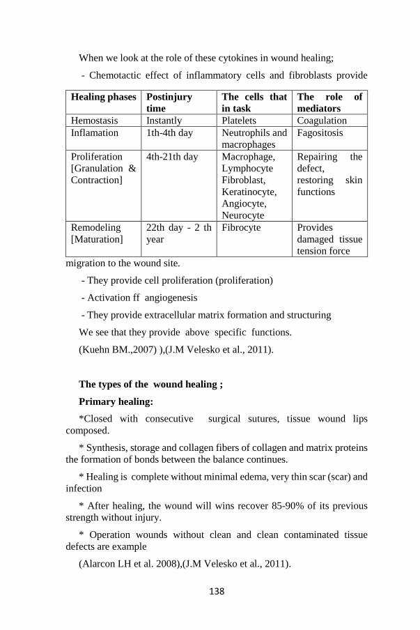
138
When we look at the role of these cytokines in wound healing;
- Chemotactic effect of inflammatory cells and fibroblasts provide
migration to the wound site.
- They provide cell proliferation (proliferation)
- Activation ff angiogenesis
- They provide extracellular matrix formation and structuring
We see that they provide above specific functions.
(Kuehn BM.,2007) ),(J.M Velesko et al., 2011).
The types of the wound healing ;
Primary healing:
*Closed with consecutive surgical sutures, tissue wound lips
composed.
* Synthesis, storage and collagen fibers of collagen and matrix proteins
the formation of bonds between the balance continues.
* Healing is complete without minimal edema, very thin scar (scar) and
infection
* After healing, the wound will wins recover 85-90% of its previous
strength without injury.
* Operation wounds without clean and clean contaminated tissue
defects are example
(Alarcon LH et al. 2008),(J.M Velesko et al., 2011).
Healing phases Postinjury
time
The cells that
in task
The role of
mediators
Hemostasis Instantly Platelets Coagulation
Inflamation 1th-4th day Neutrophils and
macrophages
Fagositosis
Proliferation
[Granulation &
Contraction]
4th-21th day Macrophage,
Lymphocyte
Fibroblast,
Keratinocyte,
Angiocyte,
Neurocyte
Repairing the
defect,
restoring skin
functions
Remodeling
[Maturation]
22th day - 2 th
year
Fibrocyte Provides
damaged tissue
tension force
139
Secondary healing:
There is full-thickness wound and particularly occur in the cases with
tissue defects.
After occurance of the granulation tissue, wound contraction and
epithelization occur.
Sutures must not be put for sew these wounds .
• As like primary healing; Matrix formation, epithelialization and scar
tissue maturation occur.
It is important to maintain the integrity of epithelialization in secondary
healing.
• Epithelialisation is more important in forming the tensile force of the
scar tissue.
• Secondary healing is a slow-growing process and may take 4-8 weeks
for epithelial development. The scar is wider.
• Secondary healing is preferred in cases where primary closure is not
preferred in patients with plonidal sinus and perianal fistulotomy(J.M
Velesko et al., 2011), (Kuehn BM.,2007) (Alarcon LH et al. 2008).
Tertiary healing (Delayed primary wound healing )
In large tissue injuries, due to the foreign body the risk of severe
bacterial wound infection to prevent wound infection with contamination;
The wound is closed after a few days. In this process, the wound is left
open and sterile serum covered with physiological pads.
*The wound is left open for several days to reduce the risk of infection.
*At the end of the primary closure is reached values equal to the tensile
force reached.
*Contaminated and dirty postoperative wounds due to peritonitis or
empyema and such healing in infected traumatic wounds beyond 24 hours
may be preferred (Onat DA., 1989),(J.M Velesko et al., 2011), (Kuehn
BM.,2007).
140
Covering complex physiological and biochemical events and
compatible with each other for effective tissue repair progression is a
dynamic event in which different processes continue sequentially.
• The main cells are macrophages, fibroblast and collagen fibers, in the
wound healing process.
• In conclusion; The most appropriate and least scarring wound healing
model is the primary healing model
(F.C.Brunicardi, 2013), (Kuehn BM.,2007) (J.M Velesko et al., 2011).
Basic principles of wound healing;
*To minimize tissue damage and loss
* Ensuring adequate tissue perfusion and blood oxygenation
* Ensuring good nutrition and moistening of tissue
* Ensuring adequate tissue epithelialization
* The most important result of all reactions during wound healing is to
try to normalize the wound tension force.
(J.M Velesko et al., 2011)
Basic principles for surgical wound incision healings;
There are tension lines in the human body known as Langer’s lines.
When incision is made, the incision should be planned parallel to these
lines.
If the incision is made perpendicular to these lines, the wound will heal
by pulling the lips and a bad form will occur.
In addition, scar formation and wound healing are related to the surgical
technique used during wound closure.
Minimal traumatic approach, debridement of necrotic tissues, absence
of foreign bodies in the wound and tension-free suturing are necessary for
a minimal scar tissue(Sherris DA,1999), (Khir. 2006) (F.C.Brunicardi,
2013).
Prolonged surgery, prolonged exposure to air and trauma of tissues may
adversely affect healing. In addition, factors such as the temperature and
light of the operating room may dry the wound. During surgery, hot serum
soaked compresses will reduce the drying of the wound(J.M Velesko et al.,
2011), (Barbul A., 2005).
In the incision line; Squeezing the suture too far away from the wound
will also result in poor scarring. The reason for this is ischemia in a wider
area (Sherris DA,1999) ,(Peacock EE Jr.,1984).
141
The type of suture material also affects scar formation. Especially silk
is one of the materials with the highest rate of reaction and infection. On
the other hand, suture materials such as polyproprolene, polyamide and
polyglecapron have the lowest reaction and infection rates. The suture
should be removed as soon as possible. Scar formation will be adversely
affected as the time elapses. Face and neck sutures should be taken 1-3
days and other incisions should be taken within 7 days at the latest(Kuehn
BM.,2007) (J.M Velesko et al., 2011), (Barbul A., 2005).
In particular, it is essential to manage this process within the principles
appropriate to the wound healing process after surgery. As a result; The
wound healing process is a vital issue that needs to be managed carefully
in the event of different diseases or surgical interventions and in one step.
In addition, diseases such as direct wound healing and diabetes are need to
a very focused and meticulous process management.
References:
Bruce E.Jarrell,1999- NMS Cerrahi, Çeviri Editörü : Prof.Dr.Öztekin
Oto, sf:17-21-Review of Surgery, Fifth Edition, J.M Velesko et al., 2011,
page :10-21
Barbul A. Wound healing. Schwartz’s Principles of Surgery. Mc Graw
Hill Eight edition. 2005; 223.
Kuehn BM. Chronic wound care guidelines issued. JAMA. 2007 Mar
7;297(9):938-9.
Sherris DA, Kern EB. The Wound. In Basic Surgical Skills, Mayo
Foundation for medical Education and research, Rochester, 1999:8-12
Peacock EE Jr. Wound Repair, 3rd ed. Philadelphia: WB Saunders 1984
Kyiv, Ukraine] Klin Khir .Wounds, Wound Infections, and Early
Postoperative Complications in Abdominal and Vascular Surgery.
Scientific-Practical Conference with International Participation. December
14-15, 2006,. 2006 Nov-Dec;(11-12):4-109.
Onat DA. Yara İyileşmesi. Temel Klinik Bilimler. Saraçoğlu F(ed),
cilt2, Güneş Kitabevi Ltd. Şti., Ankara, 1989:635-636.
Alarcon LH, Fink MP:Mediators of the inflammatory response. In
Townsend CM. Sabiston Text Book of Surgery:2008, (Editiond 18th)
F.Charles Brunicardi&Schwartz’s Principles of Surgery.& Bölüm:9,
Çeviri Editörü: M.Özmen-9. Baskı. Sf:69-82-2013.
142
ANTIBACTERIAL EFFECTS OF HONEY AND HYPERTONIC
SALINE
Sevgi KESİCİ* & Mehmet DEMİRCİ** & Uğur KESİCİ***
Introduction
Honey is a super-saturated sugar solution with 82.4% carbohydrate.
Honey also contains 17.1% water, 0.5% protein, organic acids,
multiminerals, amino acids, vitamins, phenols, and numerous other minor
components. Phenolic compounds in honey contribute to its antioxidant,
antimicrobial, anti-inflammatory, wound healing and cardioprotective
effects (1, 2). The use of honey for medical purposes has been indicated
throughout history since ancient Egyptians (3). Honey is commonly used
for wound dressing and it is the oldest wound dressing material known to
humankind (3-5). The antimicrobial activity of honey was first reported by
Van Ketel in the late 19th century (6). Hypertonic saline solution is also
reported to inhibit bacterial growth with hypertonic environment (7).
Decubitus ulcer (DU) is the localized tissue damage in the skin and
subcutaneous tissues, usually where the bone spurs are, with the effect of
pressure, friction, tearing, and other factors (National Pressure Ulcer
Advisory Panel (NPUAP) ve European Pressure Ulcer Advisory Panel
(EPUAP)) (8). Decubitis ulcers are localized pressure sores on the skin and
in subcutaneous tissues (9). It is reported that an estimation of 1.6 million
patients develop pressure ulcers annually in the Intensive Care Units (10).
Decubitis ulcers lead to increased hospitalization and high treatment costs,
as well as increased workload for health workers (11). A number of
treatment methods, including wound dressings with antimicrobial
properties, are widely used in its treatment (9).
Especially in infected decubitis wounds which are seen frequently
intensive care units, choosing effective and low cost dressing materials,
taking into account the difficulties of wound care and the high cost of
treatment, is very important in terms of increasing treatment success and
reducing treatment costs. For this purpose, in this study, the antibacterial
* (MD)University of Health Sciences, Hamidiye Etfal Training and Research Hospital,
Department of Anesthesiology and Reanimation, Istanbul, Turkey, [email protected] ** (Assi. Prof.)University of Beykent, Faculty of Medicine, Department of Microbiology,
Istanbul, Turkey, [email protected] *** (Assi. Prof.)University of Beykent, Faculty of Medicine, Department of General
Surgery, Istanbul, Turkey, [email protected]
143
activities of honey and HS solutions used in wound treatment were tried to
be revealed comparatively with mupirocin, an antibacterial agent.
The primary aim of this in vitro study is to reveal the most effective
wound care method for wound healing in difficult infected wounds, in
order to contribute to the reduction of treatment costs and hospitalization
time, as well as clinically fast wound healing.
Method
S. aureus ATCC 29213, P. aeruginosa ATCC 27853 and E. coli ATCC
25922 origins were plated on liquid Müller Hinton broth (Oxoid, UK) and
Group C (Control): 1 mL sterile physiological saline solution, Group H
(Honey): 1 mL honey, Group M (Mupirocine): 1 mL mupirocine and
Group HS (Hypertonic saline): 1 mL of 3% isotonic solutions' in-vitro
effect to these bacteria after 24 hours of 37oC incubation was investigated.
0.5 McFarland turbidity standard suspension (with the final measurement
concentration of 106 CFU / mL) were prepared in 2 different tubes for each
origin. While honey and liquid Müller Hinton broth (Oxoid, UK) medium
were mixed to be 10% (volume/volume) for Group H and Group HC; 100
microliters of mupirocin and 3% hypertonic saline were mixed with liquid
Müller Hintron broth (Oxoid, UK) medium to be 10 mg/mL for Group M
and Group HS, respectively. Müller Hinton medium was used for Group
C. By using Epoch spectrophotometer (BioTek Inst. Inc. Vermont, USA)
for OD600 (in 600 nm wavelength) at 0th and 24th hours, the growth in
the wells was checked by comparison with the standard curve.
Statistical analysis: In the study, the data were analyzed with SPSS 23
(IBM) packaged software. Central and prevalence criteria such as number,
percentage, minimum, maximum and median were used to create
descriptive statistics. Conformity of numeric variables to the normal
distribution was visually (histogram) and analytically (Shapiro-Wil) tested,
while Kruskal Wallis test and post hoc Mann Whitney U test were used for
determining the difference between independent variables that don't
conform with the normal distribution. Wilcoxon test was used to determine
the difference between the dependent variables that did not conform with
the normal distribution. A p value that is less than 0.05 was considered
statistically significant in the study.
Results
The quantitative distribution (CFU/mL) of the origins of S. aureus, P.
aeruginosa and E. coli against active substances at 0th and 24th hours was
shown in Table 1.
Table 1. The quantitative distribution (CFU/mL) of the origins of S.
aureus, P. aeruginosa and E. coli against active substances in 0th and
24th.
144
The reproduction inhibitory effect of S. aereus was significantly higher
in Group B, Group M, Group HS than Group C (p<0.01) (Group
H=Group M=Group HS>Group C). The reproduction inhibitory effect
of P. aeruginosa was significantly higher in Group B and Group HS than
Group M and Group C (p<0.01) (Group H= Group HS> Group M=
Group C). The reproduction inhibitory effect of E. coli was significantly
higher in Group HS and Group B than Group C and M (p<0.01) (Group
HS> Group H>Group M= Group C). Table 2 shows the comparison of
bacterial amounts at the beginning and 24th hour according to different
groups of substances applied.
Table 2. Comparison of the amounts of bacteria at the beginning and
24th hour according to the different groups of substances applied
In Group H, the inhibitory effect on S. aureus and P. aeruginosa was
found to be significantly higher than that of E. coli (p<0.01). In Group M,
the inhibitory effect on S. aureus was significantly higher than that of P.
aeruginosa and E. coli (p <0.01). There was no significant difference
between the groups in terms of inhibitory effect in Group HS (p=0.45).
There was no inhibitory effect on bacterial colonies in Group C, and P.
aeruginosa reproduction growth was significantly higher than S. aureus
and E. coli (p=0.02). Table 3 shows the comparison of the amounts of
bacteria at the beginning and 24th hour in all groups.
Table 3. Comparison of the amounts of bacteria at the beginning and
24th hour in all groups
A significant inhibitory effect was detected in Group H and Group HS
against all bacteria (p <0.05). In Group M, a significant inhibitory effect in
S. Aureus, and significant reproduction growth in P. Aeruginosa and E.
coli were detected (p<0.05). In group C, a significant increase of
reproduction was detected in all bacteria (p<0.05).
Discussion
There are clinical trials that have demonstrated the efficacy of honey in
wound healing, and some cases have been reported which show its
accelerating effects on wound healing, even in antibiotic-resistant infected
wounds. Honey also contributes to autolytic debridement and accelerates
wound healing (1, 12, 13). With the application of honey to the wounded
area; therapeutic effects such as rapid healing, cleaning of the wound and
infections, tissue regeneration, minimization of inflammation, and low
tissue adhesion are observed (1, 14). In our study, in accordance with the
results of the studies in the literature, it was determined that honey had a
significant antibacterial effect on S. Aureus, P. aeruginosa and E. coli. In
fact, the antibacterial effect on S. aureus was found to be statistically at the
same level with mupirocin.
145
Recently, honey has been shown to be effective in decubitus ulcers,
cesarean section wounds and chronic venous ulcers in lower extremities
(15). Complex wounds such as decubitus ulcers create an ideal
environment for microbial contamination (16). Wound infections also
negatively affect wound healing (17). This leads to prolonged
hospitalization, high cost of treatment and increased morbidity (18). In a
study on DUs by Khadanga et al. (15) , it was reported that honey showed
an antibacterial activity similar to povidone iodine and also showed a
significant reduction in Visual Pain Scores (VAS). Therefore, it has been
reported that honey can be used safely in DUs because of its effective
antibacterial effect and significant decrease in VAS. Considering the
results of this study, it can be thought that due to the significant decrease
in VAS levels, it may contribute to decrease in the doses of analgesics,
especially in intensive care patients, and this may contribute to decrease in
morbidity. Many studies have shown that honey has antibacterial effect
against a variety of gram-positive and negative bacteria. (15). In our study,
it was determined that honey has a strong antibacterial effect on both gram-
positive and gram-negative bacteria. In the study performed on 20 patients
with spinal cord injury by Biglari et al. (19), it was reported that honey
stopped bacterial growth in Grade 3 and Grade 4 DUs in all patients after
1 week, and complete wound healing was observed without any side
effects in 18 patients after 4 weeks. In our study, a meaningful antibacterial
effect was detected on all bacteria at the 24th hour. In a clinical study
conducted on 50 patients by Molan et al. (20), it was reported that honey
had eradicated infection in postoperative wound infection in the group
treated with honey, and that group had a faster wound healing than the
antiseptic group which was treated with 70% ethanol and povidone iodine.
Considering all these clinical and in-vitro studies, honey can be safely used
for treatment purposes, especially in infected wounds, due to its
antibacterial activity and lack of side effects. The use of topical honey may
be a good alternative in terms of cost due to the development of resistance
due to antibiotic use and the high cost of antibiotics used in treatment. (15).
There are limited studies on the use of HS solution in wound treatment
in the literature. Hypertonic saline solution is also reported to inhibit
bacterial growth with hypertonic environment (7). In the study conducted
by Elkins et al. (21), it was observed that the use of HS in patients with
cystic fibrosis decreased the use of antibiotics in exacerbation, compared
to isotonic saline. This result demonstrates the antibacterial activity of the
HS solution. A comparative study showing the antibacterial efficacy of
honey and HS solution was not found in the English literature. In our study,
it was determined that both HS and honey showed significant antibacterial
effect on S. Aureus, P. Aeruginosa and E. coli. However, it was seen that
HS solution showed significantly stronger antibacterial effect than honey
against E. Coli. This result suggests that HS solution may be preferable to
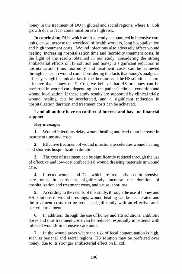
146
honey in the treatment of DU in gluteal and sacral regions, where E. Coli
growth due to fecal contamination is a high risk.
In conclusion; DUs, which are frequently encountered in intensive care
units, cause increase the workload of health workers, long hospitalization
and high treatment costs. Wound infections also adversely affect wound
healing, increasing hospitalization time and morbidity treatment costs. In
the light of the results obtained in our study, considering the strong
antibacterial effects of HS solution and honey; a significant reduction in
hospitalization time, morbidity and treatment costs can be achieved
through its use in wound care. Considering the facts that honey's analgesic
efficacy is high in clinical trials in the literature and the HS solution is more
effective than honey on E. Coli, we believe that HS or honey can be
preferred in wound care depending on the patient's clinical condition and
wound localization. If these study results are supported by clinical trials;
wound healing can be accelerated, and a significant reduction in
hospitalization duration and treatment costs can be achieved.
I and all author have no conflict of interest and have no financial
support
Key messages
1. Wound infections delay wound healing and lead to an increase in
treatment time and costs.
2. Effective treatment of wound infections accelerates wound healing
and shortens hospitalization duration.
3. The cost of treatment can be significantly reduced through the use
of effective and low-cost antibacterial wound dressing materials in wound
care.
4. Infected wounds and DUs, which are frequently seen in intensive
care units in particular, significantly increase the duration of
hospitalization and treatment costs, and cause labor loss.
5. According to the results of this study, through the use of honey and
HS solutions in wound dressings, wound healing can be accelerated and
the treatment costs can be reduced significantly with an effective anti-
bacterial treatment.
6. In addition, through the use of honey and HS solutions, antibiotic
doses and thus treatment costs can be reduced, especially in patients with
infected wounds in intensive care units.
7. In the wound areas where the risk of fecal contamination is high,
such as perianal and sacral regions, HS solution may be preferred over
honey, due to its stronger antibacterial effect on E. coli.
147
References
1. Pasupuleti VR, Sammugam L, Ramesh N, Gan SH. Honey,
Propolis, and Royal Jelly: A Comprehensive Review of Their Biological
Actions and Health Benefits. Oxid Med Cell Longev. 2017;2017:1259510.
2. H.-K. Biesalski, L. O. Dragsted, I. Elmadfa et al.,“Bioactive
compounds: definition and assessment of activity,”Nutrition 2009;.25:
1202–5.
3. Saikaly SK, Khachemoune A. Honey and Wound Healing: An
Update. Am J Clin Dermatol. 2017;18:237-51.
4. Sahih Bukhari. Volume 7; Book 71: medicine. Hadith #588.
5. Carter DA, Blair SE, Cokcetin NN, et al. Therapeutic manuka
honey: no longer so alternative. Front Microbiol. 2016;7:569.
6. Dustmann JH. Antibacterial effect of honey. Apiacta.1979;14:7–
11.
7. Strauss FG, Holmes DL, Nortman DF, Friedman S. Hypertonic
saline compresses: therapy for complicated exit-site infections. Adv Perit
Dial. 1993;9:248-50.
8. European Pressure Ulcer Advisory Panel and National Pressure
Ulcer Advisory Panel. Treatment of pressure ulcers: Quick Reference
Guide [online] 2009. Elektronik adresi: http://www.epuap.org/
guidelines/Final_Quick_Treatment.pdf.
9. Norman G, Dumville JC, Moore ZEH, Tanner J, Christie J, Goto
S. Antibiotics and antiseptics for pressure ulcers. Cochrane Database of
Systematic Reviews 2016, Issue 4. Art. No.: CD011586.
10. Rafiei Z, Mazaheri M, Eghbali-Babadi M, Yazdannik A. The
Effect of Henna (Lawsonia Inermis) on Preventing the Development of
Pressure Ulcer Grade One in Intensive Care Unit Patients. Int J Prev Med.
2019 Feb 15;10:26.
11. Gul S. Prevention of Pressure Ulcers in Patients Undergoing
Surgery. Hacettepe Üniversitesi Hemşirelik Fakültesi Dergisi 2014; 54–
61.
12. H. Vermeulen, D. Ubbink, A. Goossens, R. De Vos, and D.
Legemate,“Systematic review of dressings and topical agents for surgical
wounds healing by secondary intention,”British Journal of Surgery 2005;
92: 665–72.
148
13. M. Subrahmanyam,“A prospective randomised clinical and
histological study of superficial burn wound healing with honey and silver
sulfadiazine,” Burns 1998;24;157–61.
14. P. Molan, “Why honey is effective as a medicine: 2. The scientific
explanation of its effects,” Bee World 2001;82:22–40.
15. Khadanga SDD, Karuna T, Khetri R, et al. Effects of topical honey
dressing in decubitus ulcer. Asian J Med Sci. 2015;6:99–101.,
16. Bowler PG, Duerden BI, Armstrong DG. Wound microbiology
and associated approaches to wound management. Clinical Microbiology
Reviews 2001;14:244–69.
17. Edwards R, Harding KG. Bacteria and wound healing. Curr Opin
Infect Dis. 2004;17:91-6.
18. Lu CW, Lin TY, Shieh JS, Wang MJ, Chiu KM. Antimicrobial
effect of continuous lidocaine infusion in a Staphylococcus aureus-induced
wound infection in a mouse model. Ann Plast Surg. 2014;73:598-601.
19. Biglari B, vd Linden PH, Simon A, et al. Use of Medihoney as a
non-surgical therapy for chronic pressure ulcers in patients with spinal cord
injury. Spinal Cord. 2012;50:165–9.
20. Molan PC and Betts JA. Clinical usage of honey as a wound
dressing: An update. J Wound Care 2004; 13:353-6.
21. Elkins MR, Robinson M, Rose BR, Harbour C, Moriarty CP,
Marks GB et al. A controlled trial of long-term inhaled hypertonic saline
in patients with cystic fibrosis. N Engl J Med. 2006 19;354:229-40.
TABLES
Table 1. The quantitative distribution (CFU/mL) of the origins of S. aureus, P.
aeruginosa and E. coli against active substances in 0th and 24th. Group H
(Honey)
Group M
(Mupirocine)
Group HS
(Hypertonic
saline)
Group C
(Control)
Hour 0 24 0 24 0 24 0 24
S. aureus
ATCC
29213
1090000 307 1140000 1860 1050000 1080 1050000
2130000
P.
aeruginosa
ATCC
27853
1120000 2460 1070000
2190000
1070000 940 1040000
2250000
E. coli
ATCC
25922
1100000
616000
1060000
2150000
1060000 640 1070000
2190000

149
Table 2. Comparison of the amounts of bacteria at the beginning and 24th hour
according to the different groups of substances applied Group H M HS C
Bacteria
difference
(0-24. Hour)
Med
(Min, Max)
Med
(Min, Max)
Med
(Min, Max)
Med
(Min, Max)
p⁕
S. Aureus 1130a
(980,1180)
1121a
(1061,1162)
1039a
(979,1159)
-1060b
(-1160,-
1059)
<0.01
P.Aeruginosa 1117a
(1037, 1227)
-1140b
(-1150, -1020)
1059a
(1039, 1109)
-1220b
(-1240, -
1150)
<0.01
E. Coli 484a
(386, 564)
-1080b
(-1140, -1030)
1059c
(989, 1119)
-1140b
(-1160, -
1050)
<0.01
*Kruskal Wallis Test p value
a, b, c : The difference between the groups shown in different letters is significant
-:Bacterium
increase
Med: Median
Table 3. Comparison of the amounts of bacteria at the beginning and 24th hour in
all groups Bacteria difference (0-24. Hour)
S. Aureus P.Aeruginosa E.Coli
Group Med
(Min, Max)
Med
(Min, Max)
Med
(Min, Max)
p⁕
H 1130a
(980,1180)
1117a
(1037, 1227) 484b
(386, 564) <0.01
M 1121a
(1061, 1162)
-1140b
(-1150, -1020)
-1080b
(-1140, -1030) <0.01
HS 1039
(979, 1159)
1059
(1039, 1109)
1059
(989, 1119)
0.45
C -1060a
(-1160, -1059)
-1220b
(-1240, -1150)
-1140a
(-1160, -1050) 0.2
*Kruskal Wallis Test p value
a, b, c : The difference between the groups shown in different letters is significant
-: Bacterium increase
Med: Median
150
METHICILLIN-RESISTANT STAPHYLOCOCCUS AUREUS
COLONIZATION IN NASAL CULTURES OF THE BABIES
HOSPITALIZED AT NEWBORN INTENSIVE CARE UNIT
Asst. Prof. MD. Sinan TÜFEKÇİ
Introduction
Prevalence of Staphylococcus aureus carrier status is among 30-70% in
humans. This ratio is 0.3-25.9% for nasal MRSA carrier status at NICU.
Likelihood of S.aureus exposure just after delivery is high for the
newborns. S.aureus colonization is most common in umbilical cord, skin,
naso-pharynx and gastro-intestinal system. Nose and umbilical cord are the
most common origin for MRSA.
About 20-30% of the hospitalized patients are colonized with the
microorganism which is common at the hospital within the first 5-10 days.
Staphylococcus aureus is in normal flora in skin, mucosa and many other
body parts in humans. S.aureus which is colonized in nasal mucosa of the
patients and the health staff is one of the important risk factors for
nosocomial staphylococcus infection development. Epidemics originated
from the S.aureus-colonized patients and health staff were reported. The
main source for S.aureus colonization was reported to be maternal
S.aureus carrier status in the newborns in several papers published in
recent years. Methicillin resistance is important for Staphylococcus
colonization. MRSA is resistant to many antibiotics. MRSA leads to
infections which requires long-term hospitalization and antibiotic use and
which has a high mortality rate at intensive care, newborn, hemodialysis
and burn units. MRSA colonization facilitates bacteria spread in hospital
environment and intensive care units. Detection and isolation of MRSA
carriers is one of the infection control measures and this may both reduce
nosocomial infection prevalence and control MRSA epidemics (1,2).
The present study investigated the prevalence of MRSA colonization in
nasal cultures at the first admission of the patients who were hospitalized
at NICU.
Methods
Nasal cultures were obtained from 560 out of 643 patients who were
admitted to intensive care unit from the delivery room of Afyonkarahisar
State Hospital or at another institution between January 1, 2016 and
December 31, 2017. The study design was prospective. Informed consent
151
was obtained from the family and ethics committee approval was obtained
from the local ethics committee prior to the study. Babies who were under
30 weeks of gestation and/or 1000 gr of birth weight, who had multiple
congenital anomalies, open wounds like meningo-myelocele, neural tube
defect, who were not screened for MRSA at the first hospitalization and
whose families did not give consent for participation were excluded from
the study. MRSA isolation from the nasal mucosa without the presence of
infection signs was defined as “colonization” (carrier) and, presence of
MRSA in normally sterile regions and presence of clinical signs was
defined as “infection”.
Nasal cultures were obtained by using a swab at 1-2 mm depth of
nostrils. Smear cultures were cultivated in 5% sheep blood agar and
incubated at 35 °C for 24 hours. The colonies which showed gram positive
coccus morphology were applied catalase and coagulase tests. Methicillin
resistance was determined with disc diffusion method in accordance with
the recommendations of National Committee for Clinical laboratory
Standards (NCCLS) (3). Babies who had nasal MRSA positivity were
taken into an isolated room and applied decolonization protocol with local
mupirocin tid for 5 days. Control nasal cultures were obtained one week
after the treatment protocol.
Maternal age, gestational week, gender, type of delivery, birth weight,
Apgar scores at 1st and 5th minutes, low birth weight (birth weight less than
10% according to gestational week), time of admission, premature rupture
of membranes (longer than 18 hours), blood pressure during pregnancy,
gestational diabetes, time of nasal culture were inquired. Data were
analyzed with SPSS 18.0 statistical package program by using Fisher’s
test, chi-square test and Wilcoxon for calculating median and a p level of
<0.05 was accepted as statistically significant.
Results
Prevalence of MRSA colonization was found as 6.8% (95% CI 2.6-
10.9) in nasal culture of 560 babies hospitalized at NICU. This ratio was
6.1% (95% CI 2.8-10.6) in 2016 and 7.5% (95% CI 2.7-11.7) in 2017 and
it was seen to tend to increase. Of the babies, 288 (51.5%) were boys and
272 (48.5%) were girls. While ratio of MRSA colonization was higher
among boys (55% vs 45%), the difference was not statistically significant.
Number of normal vaginal deliveries was 354 (63%) and number of
Caesarean sections was 206 (37%). Ratio of MRSA colonization was
higher among the babies delivered by normal vaginal delivery compared
to Caesarean sections (8.5% VS S 3.9%). 0f 38 babies colonized by MRSA,
8 (21%) were delivered by Caesarean section and 30 (79%) were delivered
by normal vaginal delivery. Mean gestational week was 36.8 (range 31-42
weeks , median 37.3 weeks), mean birth weight was 3000 g (range 1210-
152
4790 g, median 30160 g) in the group with negative nasal culture for
MRSA. Mean gestational week was 36.9 (range 31-42 weeks , median 37.4
weeks), mean birth weight was 3020 g (range 1170-4870 g, median 30950
g) in the group with positive nasal culture for MRSA.
Ratio of nasal MRSA positivity was higher among the babies who
received care by the mother after delivery or who were monitored for 1-4
hours in delivery room (10.4% vs 5%) compared to the ones who did not
receive care. Ratio of nasal MRSA positivity was 3.8% in the babies who
were hospitalized within the first 24 hours after delivery and 15.2% in the
babies who were hospitalized after the first 24 hours. Ratio of admission
to NICU from another center was 10.5% within the first 24 hours (42/400)
and 25.4% (31/122) after the first 24 hours. Ratio of nasal MRSA
colonization was higher among the babies who were hospitalized after the
first 24 hours.
No significant difference was detected between groups with regard to
gestational week, gender, birth weight, Apgar scores at 1st and 5th min, low
birth weight, premature rupture of membranes, pregnancy-induced
hypertension, gestational diabetes, duration of hospital stay. Impetigo,
boiled skin syndrome or sepsis did not develop in the patients who were
colonized with MRSA. MRSA-related sepsis developed in two babies who
were not detected nasal MRSA colonization and who were referred from
another center. Risk factors were endotracheal intubation and umbilical
venous catheterization in these babies however growing did not occur in
tracheal aspirate and catheter cultures.
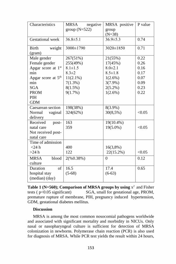
153
Characteristics MRSA negative
group (N=522)
MRSA positive
group
(N=38)
P value
Gestational week 36.8±5.1 36.9±5.3 0.74
Birth weight
(gram)
3000±1790 3020±1850 0.71
Male gender
Female gender
Apgar score at 1st
min
Apgar score at 5th
min
SGA
PROM
PIH
GDM
267(51%)
255(49%)
8.1±1.5
8.3±2
11(2.1%)
7(1.3%)
8(1.5%)
9(1.7%)
21(55%)
17(45%)
8.0±2.1
8.5±1.8
1(2.6%)
3(7.9%)
2(5.2%)
1(2.6%)
0.22
0.26
0.16
0.17
0.07
0.09
0.23
0.22
Caesarean section
Normal vaginal
delivery
198(38%)
324(62%)
8(3.9%)
30(8,5%)
<0.05
Received post-
natal care
Not received post-
natal care
163
359
19(10.4%)
19(5.0%)
<0.05
Time of admission
<24 h
>24 h
400
122
16(3,8%)
22(15.2%)
<0.05
MRSA blood
culture
2(%0.38%) 0 0.12
Duration of
hospital stay
(median) (day)
16.5
(5-68)
17.4
(6-63)
0.65
Table 1 (N=560); Comparison of MRSA groups by using x2 and Fisher
tests ( p<0.05 significant) SGA, small for gestational age, PROM,
premature rupture of membrane, PIH, pregnancy induced hypertension,
GDM, gestational diabetes mellitus.
Discussion
MRSA is among the most common nosocomial pathogens worldwide
and associated with significant mortality and morbidity in NICUs. Only
nasal or nasopharyngeal culture is sufficient for detection of MRSA
colonization in newborns. Polymerase chain reaction (PCR) is also used
for diagnosis of MRSA. While PCR test yields the result within 24 hours,
154
culture results are obtained within the first 48 hours. Sensitivity and
specificity of PCR test is reported to be higher for diagnosis of MRSA (4).
In our study, culture method was used for diagnosis. MRSA colonization
leads to prolonged duration of hospital stay, increased treatment costs,
pneumonia, osteomyelitis, sepsis, endocarditis, muco-cutaneous infections
(5,6,7). The source of MRSA colonization and infection development
thereafter at NICU could not be fully understood. MRSA contamination
occurs through the contaminated clothes and hands of the health staff,
family members of the baby, equipment, breast milk or vertical route (8,9).
The unit’s being overcrowded, insufficient number of staff, antibiotic use,
long-term use of catheters were reported to contribute to MRSA-related
epidemics in NICU (10,11). Control of nasal MRSA colonization is
recommended for reducing MRSA-related infection and sepsis in
newborns.
In our study, prevalence of nasal MRSA colonization is 6.8% among
the babies at NICU. This ratio was found to be 6.1% in cultures obtained
from nasal mucosa, axilla and diapers of the babies in literature (12). Nasal
MRSA colonization was found to be 5.7% among pregnant Chinese
women and nasal MRSA colonization was detected as 4.4% at NICU in
Taiwan (13).Ratio of nasal MRSA colonization varies among countries
and regions during the first month of life. This is suggested to be associated
with hygiene conditions of the mothers, hand hygiene of the staff at NICU
and environmental hygiene. Nasal MRSA colonization is higher in the
patients hospitalized at NICU compared to healthy newborns. In the
present study, nasal MRSA was not investigated in mothers and in the staff
as MRSA epidemic was not seen.
Nasal MRSA colonization is reported 2.9 fold greater in normal vaginal
deliveries compared to Caesarean sections (14). Ratio of nasal MRSA
positivity was 3.9 in Caesarean sections and 8.5% in normal vaginal
deliveries. However papers are available in literature reporting that nasal
MRSA colonization is higher among babies delivered with Caesarean
section and hospitalized at NICU, particularly in preterm and low birth
weight group (12). Vertical transfer of MRSA from the mother to the baby
was reported recently (15,16). Maternal MRSA was associated with
maternal chorio-amnionitis and MRSA sepsis in the newborn (17).
MRSA colonization leads to severe conditions through facilitating
spread of bacteriae in hospital environment. Prevalence of Staphylococcus
infection is four-fold greater than methicillin-sensitive Staphylococcus
aureus (MSSA) infection in presence of MRSA colonization (18).
Systemic or local MRSA infection did not develop in babies who were
detected to have MRSA colonization in our study. Mupirocin, vancomycin
and teicoplanin resistance did not develop in our patients who were
detected to have MRSA growing in nasal smear. Topical drugs (mupirocin,
155
bacitracin) is recommended for treatment of MRSA-colonized individuals
and controlling epidemics (19). In our study, decolonization could be
achieved in all babies one week after administering local mupirocin tid for
five days when the nasal MRSA-colonized babies had been transferred to
isolated rooms.
In Maraqa study, MRSA colonization was detected in 138 out of 2048
NICU patients (6.74%) between 2004 and 2006, and infection was reported
in 30 (21%) of the colonized patients. Duration of hospital stay and
colonization was shown to be significantly associated with infection in
logistic regression analyses (20). In the present study, not encountering
sepsis in nasal MRSA-colonized patients may be explained with
decolonization, strictly following hand hygiene rules in the clinic, isolation
measures and not including high risk babies. Analysis of molecular
characteristics of MRSA isolates was not required in newborns with nasal
MRSA colonization in our study.
Conclusion
In our study, nasal MRSA colonization results are consistent with the
literature and colonization-related sepsis did not develop. Early detection
of colonized patients before infection development may reduce MRSA
spread in babies hospitalized at NICU. Detection of risk factors, isolation
measures, hand disinfection and decolonization with local therapy are the
required surveillance measures in patients with nasal MRSA colonized
patients.
References
1. Wertheim HFI, Meles DC, Vos MC, et al. The role of nasal
carriage in Staphylococcus aureus infect Dis 2005; 5: 751-62
2. Öncül O, Erdemoğlu A, Özsoy MF, Altunay H, Erdem Z,
Çavuşoğlu Ş. Hastane personelinde nazal Staphylococcus aureus
taşıyıcılığı. KLİMİK Derg 2002; 15:74-7.
3. Brandt CM, Rouse MS, Tallan BM, Laue NW, Wilson WR,
Steckelberg JM. Effective treatment or cephalosporin- rifampicin
combinations against cryptic methicillin-resistant ß-lactamase producing
coagulase-negative staphylococcal experimental endocarditis. Antimicrob
Agents Chemother 1995; 39, 1815–19.
4. Francis ST, Rawal S, Roberts H, Riley P, Planche T, Kennea NL
. Detection of meticillin-resistant staphylococcus aureus (MRSA)
colonization in newborn infants using real-time polymerase chain reaction
(PCR). Acta Paediatrica 2010;99, 1691-1694.
5. Andrews WW, Schelonka R, Waites K, Stamm A, Cliver SP,
Moser S. Genital tract methicillin-resistant Staphylococcus aureus: risk of
vertical transmission in pregnant women. Obstet Gynecol 2008;111:113-
8.
156
6. Song X, Perencevich E, Campos J, Short BL, Singh N. Clinical
and economic impact of methicillin-resistant Staphylococcus aureus
colonization or infection on neonates in intensive care units. Infect Control
Hosp Epidemiol 2010;31:177-82.
7. Pinter DM, Mandel J, Hulten KG, Minkoff H, Tosi MF. Maternal-
infant perinatal transmission of methicillin-resistant and methicillin-
sensitive Staphylococcus aureus. Am J Perinatol 2009;26:145-51.
8. McDonald JR, Carriker CM, Pien BC, et al. Methicillin-resistant
Staphylococcus aureus outbreak in an intensive care nursery: potential for
interinstitutional spread. Pediatr Infect Dis J 2007;26:678-83.
9. Gastelum DT, Dassey D, Mascola L, Yasuda LM. Transmission of
community-associated methicillin-resistant Staphylococcus aureus from
breast milk in the neonatal intensive care unit. Pediatr Infect Dis J
2005;24:1122-4.
10. Lessa FC, Edwards JR, Fridkin SK, Tenover FC, Horan TC,
Gorwitz RJ. Trends in incidence of late oncet methicilline-resistant
Staphylococcus aureus infection in neonatal intensive care units: data from
the National Nozocomial infections Surveillance System, 1995-2004.
Pediatr İnfect Dis J 2009;28:577-81.
11. Andersen BM, Lindemann R, Bergh K, Nesheim BI, Syversen G,
Solheim N, et al. Spread of methicilline-resistant Staphylococcus aureus in
a neonatal intensive care unit associated with understaffing, overcrowding
and mixing of patients. J Hosp İnfect 2002;50:18-24.
12. Lazenby GB, Soper DE, Beardsley W, D Salgado DS.
Methicillin-resistant Staphylococcus aureus colonization among women
admitted for preterm delivery. American Journal of Obstetrics &
Gynecology.2012;206:329;1-5.
13. Lin J, Wu C, Yan C, Ou Q, Lin D, Zhou J, et al. A pospective
cohort study of Staphylococcus aureus and methicillin-resistant
Staphylococcua aureus carriage in neonates.The role of maternal carriage
and phenotypic and molecular characteristics. İnfection and Drug
Resistance 2018:11;555-565.
14. Jimenez-Truque N, Tedeschi S, Saye EJ, et al. Realtionship
between maternal and neonatal Staphylococcus aerius colonization.
Pediatrics.2012;129(5).e1252-e1259.
15. Seybold U, Halsova JS, White N, et al:Emergence of and risk
factors for methicilline-resistant Staphylococcus aureus of community
orijin in intensive care nurseries. Pediatrics 122:1039-1046, 2008.
16. Shiojima T, Ohki Y, Nako Y, et al: İmmediate control of a
methicilline-resistant Staphylococcus aureus outbreak in a neonatal
intensive care unit. J Infect Chemother 9:243-247, 2003.
17. Pimentel JD, Meier FA, Samuel LP:Chorioamnionitis and
neonatal sepsis from community-associated MRSA. Emerg Infect Dis
15:2069-2071, 2009.
157
18. Gül M, Çıragil P, Aral M. Kahraman Maraş Sütçü İmam
Üniversitesi Tıp Fakültesi hastane personelinde burun ve el Stafilokokkus
aureus taşıyıcılığı. ANKEM Derg 2004;18:36-9.
19. Doebbelling BN, Reagen DR, Pfaller MA; long tern efficiency of
intranasal mupirocin oilment: a prospective cohort study of
Staphylococcus aureus carriage. Arch intern Med 1994;1504-1505.
20. Maraqa NF, Aigbivbalu L, MD, Masnita-Iusan C, MS, Wludyka
P, Shareef Z, Bailey C, RN, Rathore MH. Prevalence of and risk factors
for methicillin-resistant Staphylococcus aureus colonization and infection
among infants at a level III neonatal intensive care unit. American Journal
of Infection Control 2011;39:35-41.
158
ADENOVIRUSES IN OPHTHALMOLOGY REVIEW
Taha AYYILDIZ*
Introduction
Adenoviruses are icosahedral, enveloped and double-stranded DNA
viruses and cause a number of diseases such as conjunctivitis,
gastroenteritis, hepatitis, myocarditis and pneumonia.1 Adenoviruses are
highly contagious organisms with different antigenic identifiers and over
50 known serotypes. The most common type of adenoviral eye
involvement is epidemic keratoconjunctivitis (EKC) followed by
pharyngoconjunctival fever (PCF). The appearance of all ocular surface
involvement, including conjunctiva and corneal epithelium, is a distinctive
feature of EKC. Pseudomembranous, symblepharon, and multifocal
subepithelial infiltrates, which are seen in severe cases, may result in
decreased visual acuity.2-4 PCF is characterized by fever, pharyngitis, acute
follicular conjunctivitis and preauricular lymphadenopathy. Also isolated
adenoviral conjunctivitis can be seen without corneal or systemic
involvement. Contact with hand eye contact, ocular secretions, aerosols,
ophthalmic caregivers and ophthalmic devices are main routes of
transmission.5 Adenoviral conjunctivitis is a biphasic disease that begins
with an infectious phase followed by an inflammatory phase. Viruses
continue to shed in ocular secretions for up to 7-10 days from the onset of
infection. The patient will continue to be infectious until 2-3 weeks.6
Adenoviruses are the most common cause of acute viral infections of
the conjunctiva, affecting up to 75% of cases.7 According to the data from
the Japan national surveillance center, adenoviral conjunctivitis affects 1
million people every year.8 Precise statistical data, prevalence and
incidence values are not available because of the inability to access medical
care or access to health facilities. According to the data of Germany; while
most affected group is adults by disease, the disease is seen in all age
groups and gender dominance is not observed.9 Although EKC is generally
observed between 20-40 years, PCF tends to be seen mostly in children.10,11
Adenoviruses are divided into 7 different species (A-G) and a number
of serotypes according to their immunohistochemical properties, nucleic
acid similarities, hexose and fiber protein lengths, biological properties and
phylogenetic analyzes.12,13 Adenoviral conjunctivitis may occur
sporadically or epidemically throughout the year. The severity and
prevalence of the disease is related to the serological subtype. Serotype 8,
* Ahi Evran Üniversitesi Tıp Fakültesi.

159
9 and 17 (type D) were associated with EKC; serotype 3, 5, 7 and 11 (types
B and C) PCF; serotype 1-11 (species B-E) is associated with isolated
follicular conjunctivitis without sore throat and auricular
lymphadenopathy.14,15 According to the results of epidemiological studies
in Japan, adenoviral conjunctivitis cases with serotype 3, 4 and 37 were
found to increase with increasing air temperature.16 Germany's data also
support an increase in adenoviral conjunctivitis cases in hot weather.10
Outbreaks of EKC are common in hospitals (especially in ophthalmology
units),17-19 intensive care units,20,21 and care homes;22 PCF outbreaks are
more common in schools, nurseries and summer camps.23
Both the EKC and PCf tend to be more frequent in the environments
where close relationships need to be demonstrated. Direct contact with
ocular secretions is the most common route of transmission. The infection
can also be transmitted through the tonometer used by the eye caregiver,
the cover speculum, and the split lamp. In one study, the positivity rate of
hand sweep of infected patients was 46%.5 Although the risk of infection
with a family-friendly contact infection is 10%, the risk is increased when
the infection is prolonged.24 Nasocomial EKC outbreaks are widespread
especially in ophthalmology units; postponement of ophthalmologic
surgery, early discharge after surgery and closure of the operating room for
a while.8
Epidemic Keratoconjunctivitis (EKC)
At least 19 of the adenovirus serotype that is 60 or more can cause EKC.
Serotypes 8,9 and 17 are the most common causes, and less frequent 2-5,
7, 9, 10, 11, 14, 16, 21 and 29 can lead to the EKC table. The incubation
period varies from 4 to 24 days and the symptoms tend to last for 7 to 21
days. After the symptoms have cleared, the patient continues to be
contagious for 10-14 days. The virus can not be detected by PCR analysis
in ocular secretions before the onset of symptoms.25 Although the first
phase of the EKC is unilateral; has been shown to cause double-sided
disease in up to 70% of cases.11 Red and pink eyes, excessive watering,
foreign body sensation and photophobia are the most common symptoms.
In severe cases, the patient may complain of decreased visual acuity
associated with orbital and periorbital pain.
They are usually have recent eye examinations, affected family
members or professional contacts in their anamnesis. Sometimes there may
be flu-like symptoms, fever, vomiting, diarrhea, myalgia and difficult
breathing before infection. Ocular findings are marked conjunctival
redness, chemosis, tarsal follicular reaction, petechiae or subconjunctival
hemorrhage.
160
Pharyngoconjunctival Fever (PFC)
PFC is most often caused by adenovirus serotype 3 and less frequently
by serotypes 2, 4, 7, and 14. Serotypes 1, 5, 6, 8, 11 and 19 are the cause
of sporadic outbreaks.26,27 It is an acute and highly contagious disease
accompanied by fever, pharyngitis, rhinitis, acute follicular conjunctivitis,
and sensitive preauricular lymphadenopathy. Ocular inflammation is
unilaterally starts and affects other eye within 1-3 days. The most common
ocular findings are swelling and scaling of eyelids, epifora, conjunctival
injection, conjunctival chemosis, follicular and papillary reaction, and
subconductival haemorrhage. Contamination pathway is direct contact
with droplet inhalation and ocular secretions. Although the incubation
period varies between 5-12 days, the symptoms end within 3-5 days,
limiting themselves and often without causing any complications.
Complications
Pseudomembranes containing a fibrin-rich exudate lacking blood
vessels and lymphatics adhering to the upper and lower tarsal conjunctiva
are more common in EKC cases and have been shown to be associated
with adenovirus serotypes 8,19 and 37.28 Unlike the true membranes,
pseudomembranes can be separated without damaging the underlying
epithelium, thus leading to little or no bleeding. Although
pseudomembranes are generally observed in EKC cases, true membranes
can also be observed according to the degree of the disease and the
intensity of inflammation. Removal of true membranes leads to bleeding,
leaving it in situ leads to subepithelial fibrosis and symblepharon
formation.29
Another common complication observed in EKC cases is multifocal
subepithelial infiltrates, which are pathognomonic for adenoviral
infections. It has been shown in up to 50% of patients and has been shown
to occur more frequently with serotype 8.29 These infiltrates are a type of
cellular immunological response to viral antigens stored in the corneal
stroma beneath the bowman's membrane.30 In vivo confocal microscopy
study showing the clinical course of the EKC has shown that dendritic cells
at the 1 st stage of diffuse epithelial keratitis are associated with
subepithelial bowman membrane level accumulation. During the second
week of focal epithelial keratitis, it has been shown that the hyperreflective
basal cell clusters on dendritic cells are surrounded by a complex network
of leukocytes. There is a hyperreflective layer of cells on the subepithelial
infiltrate area in the anterior stroma.31 The reduction in corneal sensitivity
seen in the early phase of the disease probably improves on average of 8.5
days.32
161
Disease Progression and Treatment
Subepithelial infiltrates persist for weeks to years. These can cause
visual impairment if they involve the visual axis. Most of these infiltrates
heal spontaneously without scarring. Although topical corticosteroids
shorten the healing time of infiltrates, there is no effect on long-term
outcomes.33 It has been shown that phototherapeutic keratectomy treatment
combined with low dose mitomycin has a positive effect on photophobia,
visual acuity and contrast sensitivity in the treatment of persistent corneal
opacities developing as a result of EKC.34
Adenoviral conjunctivitis is usually a self-limited disease which results
in complete recovery within about 3 weeks. Conservative approaches such
as artificial tears and cold application provide symptomatic relief without
any side effects. Topical antibiotics are used to prevent or treat bacterial
superinfection.35 The use of topical antihistamines and vasoconstrictors
may also reduce the duration of the disease and complaints related to the
disease, but there is also a risk of local toxicity.36 Topical steroid use is
controversial. Topical steroids are generally preferred in the acute phase of
the disease and the effect is transient. The use of topical steroids in animal
studies has been shown to increase the adenovirus replication rate and
therefore the disease recovery process is prolonged.37-39 Steroid therapy
should be restricted for complicated disease with visible
pseudomembranes or subepithelial infiltrates with decreased visual
acuity.33,37 It has also been shown that topical nonsteroidal
antiinflammatory use does not reduce adenovirus replication in animal
models.40 Because topical nonsteroidal antiinflammatory use has no effect
on subepithelial infiltrates, it may be a safer alternative to steroid use for
symptomatic relief.41
The effects of the virustatic agents trifluridine, vidarabine and
ganciclovir on adenoviruses are limited, and the use of adenoviral
conjunctivitis therapy is still controversial.42-44 Topical ganciclovir
experimentally reduces adenovirus burden42-45 but clinical studies have
shown no effect on adenoviral conjunctivitis.46 Significant antiviral
activity of topical cidofovir against adenoviruses has been demonstrated
in experimental studies and in animal models.47-50 Topical cidofovir
administration reduces the duration of the disease by reducing viral titers
and is also effective in the prophylaxis of adenovirus.51 In a clinical trial
comparing the rate of symptomatic treatment of acute adenoviral
conjunctivitis, combining topical cidofovir alone or with topical
cyclosporine has not been shown to be a superior superiority of the disease
to the disease.52,53 It has been shown in animal models that the
162
antiadenoviral effect of 2'3'-dideoxycytidine, an antiviral agent, is higher
than that of sidofovir.54
Interferons are proteins that are responsible for stopping viral spread in
response to viral infection and released from the cells. In some studies,
topical interferon beta treatment has been shown to be effective in reducing
the duration and complications of the disease.55-57 Interferon gamma
therapy can also be used as a treatment option because of its antiadenoviral
activity.58-60 Therapeutic effect of interferon alpha treatment has not been
demonstrated.61-63 Anti adenoviral activity is shown in animal models of
topical immunoglobulin treatment; there is no human study.64
Povidone-iodine is a broad-spectrum antiseptic and, despite high
activity against free adenoviruses, its activity against intracellular
adenovirus particles in infected cells is poor.65,66 The combination of
topical povidone-iodine and dexamethasone has been shown to reduce
viral secretion and disease duration.67,68 Topical steroids provide
symptomatic relief; topical povidone-iodine also kills viruses that shed in
tears, thereby reducing the risk of spreading the disease. In a recent study,
it has been shown that conjunctival washing with 2.5% povidone-iodine in
infants is effective in the treatment of adenoviral conjunctivitis.69
Unlike acute disease, topical steroids have been shown to reduce
adenovirus-associated subepithelial infiltrates.33,70,71 However, this
condition recurs after the cessation of treatment. This does not only lead to
treatment dependency; at the same time steroids may result in cataracts and
increased intraocular pressure. Cyclosporins used as an alternative to
steroids do not provide symptomatic relief in the acute phase of the disease
and have no activity on the natural course of the disease but are used in the
treatment of subepithelial infiltrates following adenoviral
conjunctivitis.52,53 Topical cyclosporine treatment, varying between 0.05%
and 2%, reduces subepithelial infiltrates and associated foreign body
sensations and visual complaints.72-75 Although topical cyclosporine
treatment increases viral efflux in an animal study and therefore poses a
risk for local epidemics, it is still a relatively safe treatment.76
Phototherapeutic keratectomy therapy combined with low dose mitomycin
C should be reserved for chronic subepithelial infiltrates with severe visual
disturbances and treatment has shown positive effects on photophobia,
visual acuity and contrast sensitivity.77,78
Since adenovirus conjunctivitis and complications are not effective
treatments, strict sterilization of the hands and instruments is essential
controlling the spread of the infection.10,11,80 Because adenoviruses are
resistant to many antiseptics, the type and amount of antiseptic used is
important.81 Human adenoviruses are resistant to dryness and can survive
significant periods of time outside the human body. Plastic and metal
163
surfaces have been shown to produce infectious concentrations even 28
days after settlement.82,83 Simply deleting surfaces with general
disinfectants such as isopropyl alcohol or hydrogen peroxide is ineffective
against adenoviruses.81,84 When adenovirus is suspected, it is
recommended to be taken into an isolated contamination room and all
surfaces and instruments should be disinfected with 70% ethyl alcohol and
1/10 of bleach after examination.81,85 Disinfection should be done starting
from the cleaning of organic matter.81 The use of gloves, single-use
tonometer heads and disposable eye drops can reduce transmission.5,80
Patients should be informed about the course of the disease when they
arrive in the acute phase of the disease and be cautious at the point of call
for control at this time that they are contagious.
References
1. Shenk T.: Adenoviridae: the viruses and their replication. In Fields
B.N., Knipe D.M., and Howley P.M. (eds): Fields Virology, 3rd ed.
Philadelphia, PA: Lippincott-Raven, 1996. pp. 2111-2148
2. Butt AL, Chodosh J. Adenoviral keratoconjunctivitis in a tertiary
eye clinic. Cornea. 2006;25:199-202
3. Chodosh J, Miller D, Stroop WG, et al. Adenovirus epithelial
keratitis. Cornea. 1995;14:167-74
4. Dawson CR, Hanna L, Togni B. Adenovirus type 8 infections in
the united states. 4. Observations on the pathogenesis of lesions in severe
eye disease. Arch Ophtalmol. 1972;87:258-68
5. Azar MJ, Dhaliwal DK, Bower KS, et al. Possible consequences
of shaking hands with your patients with epidemic keratoconjunctivitis.
Am J Ophtalmol. 1996;721:711-2
6. Kaufmann HE. Adenovirus advances: new diagnostic and
therapeutic options. Curr Opin Ophtalmol. 2011;22:290-3
7. Woodland RM, Darougar S, Thaker U, et al. Causes of
conjunctivitis and keratoconjunctivitis in Karachi, Pakistan.Trans R Soc
Trop Med Hyg. 1992;86:317e20
8. Kaneko H, Suzutani T, Aoki K, et al. Epidemiological and
virological features of epidemic keratoconjunctivitis due to new human
adenovirus type 54 in Japan. Br J Ophthalmol.2011;95:32e6
9. Adlhoch C, Schoneberg I, Fell G, et al. Increasing case numbers
of adenovirus conjunctivitis in Germany, 2010. Euro Surveill.
2010;15(45). pii: 19707.
10. Ghebremedhin B. Human adenovirus: viral pathogen with
increasing importance. Eur J Microbiol Immunol (Bp). 2014;4:26e33
11. Gonzalez-Lopez JJ, Morcillo-Laiz R, Munoz-Negrete FJ.
Adenoviral keratoconjunctivitis: an update. Arch Soc Esp Oftalmol.
2013;88:108e15
164
12. Aoki K, Ishiko H, Konno T, et al. Epidemic keratoconjunctivitis
due to the novel hexon-chimericintermediate 22,37/H8 human adenovirus.
J Clin Microbiol. 2008;46:3259e69
13. Swenson PD, Wadell G, Allard A, et al. Adenovirus, in Murray
PR, Baron EJ, Jorgensen JH, et al (eds) Manual of Clinical Microbiology,
Vol. 2. Washington, DC, ASM Press; 8th ed, 2003, pp 1404e17
14. Schrauder A, Altmann D, Laude G, et al. Epidemic conjunctivitis
in Germany, 2004. Euro Surveill. 2006;11:185e7
15. Walls T, Shankar AG, Shingadia D. Adenovirus: an increasingly
important pathogen in paediatric bone marrow transplant patients. Lancet
Infect Dis. 2003;3:79e86
16. Aoki K, Tagawa Y. A twenty-one year surveillance of adenoviral
conjunctivitis in Sapporo, Japan. Int Ophthalmol Clin. 2002;42:49e54
17. Dart JK, El-Amir AN, Maddison T, et al. Identification and control
of nosocomial adenovirus keratoconjunctivitis in an ophthalmic
department. Br J Ophthalmol. 2009;93:18e20
18. Hamada N, Gotoh K, Hara K, et al. Nosocomial outbreak of
epidemic keratoconjunctivitis accompanying environmental
contamination with adenoviruses. J Hosp Infect. 2008;68:262e8
19. Sambursky RP, Fram N, Cohen EJ. The prevalence of adenoviral
conjunctivitis at the Wills Eye Hospital Emergency Room. Optometry.
2007;78:236e9
20. Calkavur S, Olukman O, Ozturk AT, et al. Epidemic adenoviral
keratoconjunctivitis possibly related to ophthalmological procedures in a
neonatal intensive care unit: lessons from an outbreak. Ophthalmic
Epidemiol. 2012;19:371e9
21. Ersoy Y, Otlu B, Turkcuoglu P, et al. Outbreak of adenovirus
serotype 8 conjunctivitis in preterm infants in a neonatal intensive care
unit. J Hosp Infect. 2012;80:144e9
22. Ghanaiem H, Averbuch D, Koplewitz BZ, et al. An outbreak of
adenovirus type 7 in a residential facility for severely disabled children.
Pediatr Infect Dis J. 2011;30:948e52
23. Gonzalez-Lopez JJ, Morcillo-Laiz R, Munoz-Negrete FJ.
Adenoviral keratoconjunctivitis: an update. Arch Soc Esp Oftalmol.
2013;88:108e15
24. Guyer B, O’Day DM, Hierholzer JC, et al. Epidemic
keratoconjunctivitis: a community outbreak of mixed adenovirus type 8
and type 19 infection. J Infect Dis. 1975;132:142e50
25. Kimura R, Migita H, Kadonosono K, et al. Is it possible to detect
the presence of adenovirus in conjunctiva before the onset of
conjunctivitis? Acta Ophthalmol. 2009;87:44e7
26. Ishiko H, Aoki K. Spread of epidemic keratoconjunctivitis due to
a novel serotype of human adenovirus in Japan. J Clin Microbiol.
2009;47:2678e9

165
27. Melendez CP, Florentino MM, Martinez IL, et al. Outbreak of
epidemic keratoconjunctivitis caused by adenovirus in medical residents.
Mol Vis. 2009;15:557e62
28. Tabbara KF, Omar N, Hammouda E, et al. Molecular
epidemiology of adenoviral keratoconjunctivitis in Saudi Arabia. Mol Vis.
2010;16:2132e6
29. Chintakuntlawar AV, Chodosh J. Cellular and tissue architecture
of conjunctival membranes in epidemic keratoconjunctivitis. Ocul
Immunol Inflamm. 2010;18:341e5
30. Jones BR. The clinical features of viral keratitis and a concept of
their pathogenesis. Proc R Soc Med. 1958;51:917e24
31. Dosso AA, Rungger-Brandle E. Clinical course of epidemic
keratoconjunctivitis: evaluation by in vivo confocal microscopy. Cornea.
2008;27:263e8
32. Ozturk HE, Sonmez B, Beden U. Corneal sensitivity may decrease
in adenoviral epidemic keratoconjunctivitis_a confocal microscopic study.
Eye Contact Lens. 2013;39:264e8
33. Laibson PR, Dhiri S, Oconer J, et al. Corneal infiltrates in
epidemic keratoconjunctivitis. Response to double-blind corticosteroid
therapy. Arch Ophthalmol. 1970;84:36e40
34. Yamazaki ES, Ferraz CA, Hazarbassanov RM, et al.
Phototherapeutic keratectomy for the treatment of corneal opacities after
epidemic keratoconjunctivitis. Am J Ophthalmol. 2011;151:35e43.e1
35. Isenberg SJ, Apt L, Valenton M, et al. A controlled trial of
povidone-iodine to treat infectious conjunctivitis in children. Am J
Ophthalmol. 2002;134:681e8
36. Majeed A, Naeem Z, Khan DA, et al. Epidemic adenoviral
conjunctivitis report of an outbreak in a military garrison and
recommendations for its management and prevention. J Pak Med Assoc.
2005;55:273e5
37. Romanowski EG, Roba LA, Wiley L, et al. The effects of
corticosteroids of adenoviral replication. Arch Ophthalmol.
1996;114:581e5
38. Romanowski EG, Yates KA, Gordon YJ. Short-term treatment
with a potent topical corticosteroid of an acute ocular adenoviral infection
in the New Zealand white rabbit. Cornea. 2001;20:657e60
39. Romanowski EG, Yates KA, Gordon YJ. Topical corticosteroids
of limited potency promote adenovirus replication in the Ad5/NZW rabbit
ocular model. Cornea. 2002;21:289e91
40. Romanowski EG, Gordon YJ. Effects of diclofenac or ketorolac
on the inhibitory activity of cidofovir in the Ad5/NZW rabbit model. Invest
Ophthalmol Vis Sci. 2001;42:158e62
166
41. Gordon YJ, Araullo-Cruz T, Romanowski EG. The effects of
topical nonsteroidal anti-inflammatory drugs on adenoviral replication.
Arch Ophthalmol. 1998;116:900e5
42. Trousdale MD, Goldschmidt PL, Nobrega R. Activity of
ganciclovir against human adenovirus type-5 infection in cell culture and
cotton rat eyes. Cornea. 1994;13:435e9
43. Ward JB, Siojo LG, Waller SG. A prospective, masked clinical
trial of trifluridine, dexamethasone, and artificial tears in the treatment of
epidemic keratoconjunctivitis. Cornea. 1993;12:216e21
44. Waring GOE, Laibson PR, Satz JE, Joseph NH. Use of vidarabine
in epidemic keratoconjunctivitis due to adenovirus types 3, 7, 8, and 19.
Am J Ophthalmol. 1976;82:781e5
45. Huang J, Kadonosono K, Uchio E. Antiadenoviral effects of
ganciclovir in types inducing keratoconjunctivitis by quantitative
polymerase chain reaction methods. Clin Ophthalmol. 2014;8:315e20
46. Yabiku ST, Yabiku MM, Bottos KM, et al. Ganciclovir 0.15%
ophthalmic gel in the treatment of adenovirus keratoconjunctivitis. Arq
Bras Oftalmol. 2011;74:417e21
47. de Oliveira CB, Stevenson D, LaBree L, et al. Evaluation of
cidofovir (HPMPC, GS-504) against adenovirus type 5 infection in vitro
and in a New Zealand rabbit ocular model. Antiviral Res. 1996;31:165e72
48. Gordon YJ, Romanowski EG, Araullo-Cruz T. Topical HPMPC
inhibits adenovirus type 5 in the New Zealand rabbit ocular replication
model. Invest Ophthalmol Vis Sci. 1994;35:4135e43
49. Kaneko H, Mori S, Suzuki O, et al. The cotton rat model for
adenovirus ocular infection: antiviral activity of cidofovir. Antiviral Res.
2004;61:63e
50. Romanowski EG, Gordon YJ. Efficacy of topical cidofovir on
multiple adenoviral serotypes in the New Zealand rabbit ocular model.
Invest Ophthalmol Vis Sci. 2000;41:460e3
51. Romanowski EG, Yates KA, Gordon YJ. Antiviral prophylaxis
with twice daily topical cidofovir protects against challenge in the
adenovirus type 5/New Zealand rabbit ocular model. Antiviral Res.
2001;52:275e80
52. Hillenkamp J, Reinhard T, Ross RS, et al. Topical treatment of
acute adenoviral keratoconjunctivitis with 0.2% cidofovir and 1%
cyclosporine: a controlled clinical pilot study. Arch Ophthalmol.
2001;119:1487e91
53. Hillenkamp J, Reinhard T, Ross RS, et al. The effects of cidofovir
1% with and without cyclosporin a 1% as a topical treatment of acute
adenoviral keratoconjunctivitis: a controlled clinical pilot study.
Ophthalmology. 2002;109:845e50
167
54. Romanowski EG, Yates KA, Gordon YJ. The in vitro and in vivo
evaluation of ddC as a topical antiviral for ocular adenovirus infections.
Invest Ophthalmol Vis Sci. 2009;50:5295e9
55. Romano A, Ladizensky E, Guarari-Rotman D, et al. Clinical effect
of human-fibroblast-derived (Beta) interferon in treatment of adeno-virus
epidemic keratoconjunctivitis and its complications. Tex Rep Biol Med.
1981;41:559e65
56. Romano A, Revel M, Guarari-Rotman D, et al. Use of human
fibroblast-derived (beta) interferon in the treatment of epidemic adenovirus
keratoconjunctivitis. J Interferon Res. 1980;1:95e100
57. Watanabe Y, Uchio E, Itoh N, et al. Bacterial Infection in the
Conjunctiva of Patients with Adenoviral Conjunctivitis. Jpn J Ophthalmol.
2001;45:115
58. Mistchenko AS, Diez RA, Falcoff R. Recombinant human
interferon-gamma inhibits adenovirus multiplication without modifying
viral penetration. J Gen Virol. 1987;68:2675e9
59. Mistchenko AS, Diez RA, Falcoff R. Inhibitory effect of
interferon-gamma on adenovirus replication and late transcription.
Biochem Pharmacol. 1989;38: 1971e8
60. Uchio E, Inoue H, Fuchigami A, et al. Anti-adenoviral effect of
interferon-beta and interferon-gamma in serotypes that cause acute
keratoconjunctivitis. Clin Experiment Ophthalmol. 2011;39:358e63
61. Adams CP Jr, Cohen EJ, Albrecht J, et al. Interferon treatment of
adenoviral conjunctivitis. Am J Ophthalmol. 1984;98:429e32
62. Reilly S, Dhillon BJ, Nkanza KM, et al. Adenovirus type 8
keratoconjunctivitisean outbreak and its treatment with topical human
fibroblast interferon. J Hyg (Lond). 1996;96:557e75
63. Sundmacher R, Wigand R, Cantell K. The value of exogenous
interferon in adenovirus keratoconjunctivitis. Preliminary results. Graefes
Arch Clin Exp Ophthalmol. 1982;218:139e40
64. Nwanegbo EC, Romanowski EG, Gordon YJ, et al. Efficacy of
topical immunoglobulins against experimental adenoviral ocular infection.
Invest Ophthalmol Vis Sci. 2007;48:4171e6
65. Monnerat N, Bossart W, Thiel MA. Povidone-iodine for treatment
of adenoviral conjunctivitis: an in vitro study. Klin Monbl Augenheilkd.
2006;223:349e52
66. Kawana R, Kitamura T, Nakagomi O, et al. Inactivation of human
viruses by povidone-iodine in comparison with other antiseptics.
Dermatology. 1997;195(Suppl 2):29e35
67. Clement C, Capriotti JA, Kumar M, et al. Clinical and antiviral
efficacy of an ophthalmic formulation of dexamethasone povidone-iodine
in a rabbit model of adenoviral keratoconjunctivitis. Invest Ophthalmol Vis
Sci. 2011;52:339e44

168
68. Pelletier JS, Stewart K, Trattler W, et al. A combination povidone-
iodine 0.4%/dexamethasone 0.1% ophthalmic suspension in the treatment
of adenoviral conjunctivitis. Adv Ther. 2009;26:776e83
69. Ozen Tunay Z, Ozdemir O, Petricli IS. Povidone iodine in the
treatment of adenoviral conjunctivitis in infants. Cutan Ocul Toxicol.
2015;34:12e5
70. Romanowski EG, Yates KA, Teuchner B, et al. N- chlorotaurine
is an effective antiviral agent against adenovirus in vitro and in the
Ad5/NZW rabbit ocular model. Invest Ophthalmol Vis Sci.
2006;47:2021e6
71. Trauzettel-Klosinski S, Sundmacher R, Wigand R. The effects of
topical steroids in epidemic kerato-conjunctivitis (author’s transl). Klin
Monbl Augenheilkd. 1980;176:899e906
72. Jeng BH, Holsclaw DS. Cyclosporine A 1% eye drops for the
treatment of subepithelial infiltrates after adenoviral keratoconjunctivitis.
Cornea. 2011;30:958e61
73. Levinger E, Slomovic A, Sansanayudh W, et al. Topical treatment
with 1% cyclosporine for subepithelial infiltrates secondary to adenoviral
keratoconjunctivitis. Cornea. 2010;29:638e40
74. Okumus S, Coskun E, Tatar MG, et al. Cyclosporine a 0.05% eye
drops for the treatment of subepithelial infiltrates after epidemic
keratoconjunctivitis. BMC Ophthalmol. 2012;12:42
75. Reinhard T, Sundmacher R. Local cyclosporin A therapy of
nummuli after epidemic keratoconjunctivitisecase report. Klin Monbl
Augenheilkd. 1997;210:165e8
76. Romanowski EG, Pless P, Yates KA, et al. Topical cyclosporine
A inhibits subepithelial immune infiltrates but also promotes viral
shedding in experimental adenovirus models. Cornea. 2005;24:86e91
77. Alevi D, Barsam A, Kruh J, et al. Photorefractive keratectomy
with mitomycin-C for the combined treatment of myopia and subepithelial
infiltrates after epidemic keratoconjunctivitis. J Cataract Refract Surg.
2012;38:1028e33
78. Quentin CD, Tondrow M, Vogel M. Phototherapeutic keratectomy
(PTK) after epidemic keratoconjunctivitis. Ophthalmologe. 1999;96:92e6
79. Gottsch JD. Surveillance and control of epidemic
keratoconjunctivitis. Trans Am Ophthalmol Soc. 1996;94:539e87
80. Meyer-Rusenberg B, Loderstadt U, Richard G, et al. Epidemic
keratoconjunctivitis: the current situation and recommendations for
prevention and treatment. Dtsch Arztebl Int. 2011;108:475e80
81. Rutala WA, Peacock JE, Gergen MF, et al. Efficacy of hospital
germicides against adenovirus 8, a common cause of epidemic
keratoconjunctivitis in health care facilities. Antimicrob Agents
Chemother. 2006;50:1419e24

169
82. Nauheim RC, Romanowski EG, Araullo-Cruz T, et al. Prolonged
recoverability of desiccated adenovirus type 19 from various surfaces.
Ophthalmology. 1990;97: 1450e3
83. Gordon YJ, Gordon RY, Romanowski E, et al. Prolonged recovery
of desiccated adenoviral serotypes 5, 8, and 19 from plastic and metal
surfaces in vitro. Ophthalmology. 1993;100:1835e9
84. Threlkeld AB, Froggatt JW 3rd, Schein OD, et al. Efficacy of a
disinfectant wipe method for the removal of adenovirus 8 from tonometer
tips. Ophthalmology. 1993;100:1841e5
85. O’Brien TP, Jeng BH, McDonald M, et al. Acute conjunctivitis:
truth and misconceptions. Curr Med Res Opin. 2009;25:1953e61
170
PERIANAL HIDRADENOMA PAPILLIFERUM
Uğur KESİCİ*
Introduction
Hidradenoma papilliferum (HP) was first reported by Worth in 1878
(1). HP is a rare papillary apocrine gland tumor often seen in women (2,
3). For some authors, it can be considered an analog of intraductal
papilloma of the breast (4). It is most frequently located in vulvar and
perianal area. Rarely, it may also be located outside of genital area.
Extragenital localization of HP is defined as ectopic HP (2). Ectopic HP is
generally encountered in head and neck areas (4). The ones localized in
genital area are most frequently seen between 30-60 years of age, while
extragenital localization may be encountered in different age groups
(between 8-78 years of age).
Though it is generally asymptomatic, sometimes it may manifest itself
with itching, pain, bleeding and ulceration (2). They usually appear as
slow-growing, solitary, red or skin-colored nodular lesions (4). In this case
report, the discussion topic was a female patient that sought medical advice
due to painful nodular lesion in perianal area.
Results
In this case report, a 60 year-old female patient who was operated due
to a palpable mass in perianal area and reported to be HP as a pathological
result was discussed. The patient complained of having a slow-growing
painful mass around the anus for approximately 9-10 months. No finding
was detected in patient history. Palpable mass was not detected during
rectal examination. A nodular lesion with similar appearance to
thrombosed hemorrhoid was present at perianal area at 9 o'clock lithotomy
position. Excision was planned due to pain complaint of the patient. Gross
total excision together with mass base was applied to the patient under
spinal anaesthesia. Histopathological examination of surgical specimen
was reported as HP.
* (Assi. Prof.) University of Beykent, Faculty of Medicine, Department of General Surgery,
Istanbul, Turkey. [email protected]
171
Discussion
HP which was previously thought to be an apocrine tumour, is now
believed to be a mammary-like gland (MLG) adenoma (5, 6). HP is a
benign tumor often localized in anogenital area, mostly seen in women and
clinically asymptomatic in general (2, 4). But rarely, it may manifest itself
with symptoms such as itching, pain, bleeding and ulceration. While
lesions are typically smaller than 2 cm, in literature there are lesions
reported to be bigger than 4 cm (2). In this case report, the patient had a
painful red-colored nodular lesion sized around 1-1.5 cm located in
perianal area.
In differential diagnosis, perianal abscess, genital warts, external
thrombosed hemorrhoidal disease along with less commonly encountered
perianal localization diseases such as epidermoid carcinoma, lipoma,
neurofibroma, angioma and sebaceous cyst should be considered (2, 7).
Also, very rarely seen but clinically quite similar granular cell tumors with
perianal localization should also be taken into account in differential
diagnosis (8).
Definitive diagnosis is not possible clinically and it may only be
reached by histopathological examination. Generally benign and rarely
malign transformation is reported histopathologically and it may
incorrectly be diagnosed as adenocarcinoma. If there is mitotic figures,
cellular pleomorphism or atypia in histopathological examination,
existence of malign transformation is accepted (2). In literature, while
malign transformation is reported in cases with perianal localization, it is
not reported in cases with ectopic localization. (4). In the study performed
by Scurry et al. malignancy was not reported in 46 patients with vulva
located HP (5). In this case report, histopathological examination results of
the patient did not show any malign transformation.
Gross total resection with negative surgical margin is recommended for
treatment and recurrence is quite rare (2, 9). Patient in this case report had
a total excision with lesion base and recurrence was not detected after 18
months follow-up.
Conclusions
HP is a rarely encountered benign lesion with perianal localization that
shows slow progression and is generally asymptomatic. For this reason, it
is quite difficult to diagnose. But after considering the fact that it can
seldom go through malign transformation and definitive diagnosis can only
be reached after histopathological examination, we are of the opinion that
172
all surgeons should be quite alert regarding surgical excision of slow
progressing, asymptomatic nodular lesions with perianal localization.
References
1. Tiwana KK, Nibhoria S, Kaur H, Bajaj A, Phutela R Benign
Vulvar Adnexal Tumours - A 5 year Study in a Tertiary Care Hospital. J
Family Reprod Health. 2015;9:199-201.
2. Daniel F, Mahmoudi A, de Parades V, Fléjou JF, Atienza P. An
uncommon perianal nodule: hidradenoma papilliferum. Gastroenterol Clin
Biol. 2007;31:166-8.
3. Huddleston MK, Jenkins CP, Nelson EC. Hidradenoma
papilliferum: a case report of an uncommon perianal lesion. Am Surg.
2016;82:43-4.
4. Rosmaninho AD, de Almeida MT, Costa V, Sanches MM, Lopes
C, Selores Gomes Meirinhos MM. Ectopic hidradenoma papilliferum.
Dermatol Res Pract. 2010;2010:709371.
5. Scurry J, van der Putte SC, Pyman J, Chetty N, Szabo R.
Mammary-like gland adenoma of the vulva: review of 46 cases. Pathology.
2009;41:372-8.
6. Kurashige Y, Kishida K, Kurashige K, Minemura T, Nagatani T.
Hidradenoma papilliferum of the vulva in association with an anogenital
mammary-like gland. J Dermatol. 201;41:411-3
7. Kesici U, Sakman G, Mataraci E. Retrorectal/Presacral
Epidermoid Cyst; Report of A Case. Eurasian J Med 2013; 45: 207-10.
8. Kesici U, Mataraci E, Kesici S, Zeren S. Granular cell tumor on
perıanal regıon; report of a case. Acta Med Iran 2013; 51: 509-11.
9. Handa Y, Yamanaka N, Inagaki H, Tomita Y. Large ulcerated
perianal hidradenoma papilliferum in a young female. Dermatol Surg.
2003;29:790-2.
173
CURRENT TRENDS IN THE TREATMENT MODALITIES OF
OPEN ANGLE GLAUCOMA
Yalçın KARAKÜÇÜK*,
Introduction
Glaucoma includes the conditions having the common feature of an
acquired, degenerative optic neuropathy [1,2]. Glaucoma-related optic
neuropathy is characterized by abnormalities of the optic nerve complex
(optic nerve head [ONH], retinal nerve fibre layer [RNFL], and
peripapillary region) in a specific pattern and corresponding damage to the
visual field (VF) [3]. Glaucoma is frequently associated with elevated
intraocular pressure (IOP), but an increase in IOP is not necessary to
diagnose this condition [1,2]. Changes in the optic nerve are permanent,
progressive resulting in reduced quality of life [4]. In this article, we would
focus on the current modalities in the medical and surgical management of
open angle glaucoma.
Epidemiology And Risk Factors:
Globally, It is estimated that 64.3 million people have glaucoma, of
which three fourths are open-angle [5]. Glaucoma (both open-angle and
angle-closure) is the second leading cause of irreversible blindness both
worldwide and in Europe, with approximately 8.4 million people becoming
blind from the disease [5]. Globally, the number of people with glaucoma
is estimated to become 111.8 million in 2040 [5]. The estimated prevalence
of glaucoma is 2.65% in people above 40 years of age [6]. In Europe, the
prevalence of glaucoma varies from 1.54 to 3.89% [7-18], which amounts
from 3.99 millions to 7.11 millions. [7-18]. The risk factors of Primary
open angle glaucoma are increasing age, high refractive error( both myopia
and hypermetropia), thin central corneal thickness, Large optic disc
diameter, elevated IOP, hypertension, cardiovascular disease, thyroid
disorders, low physical activity, positive family history. African people
and Hispanic people have higher prevalence of Open angle glaucoma than
other ethnic groups [1,19,20].
Classification and Subtypes:
Glaucoma is classified based on the underlying anatomy and
pathophysiology, with open-angle and angle closure representing the two
* (MD) Selçuk University, Faculty of Medicine, Department of Ophthalmology, Konya/
Turkey, [email protected]
174
major subtypes [1,2,21]. Idiopathic or primary glaucoma occurs with no
identifiable cause. Glaucoma in which there is an identifiable cause of
increased IOP, resulting in optic nerve damage, is known as secondary
glaucoma. Pseudoexfoliative glaucoma is the most common type of
secondary glaucoma [22]. Normal-tension glaucoma, one of the sub types
of POAG, is characterized by glaucomatous optic neuropathy in patients
with IOP measurements consistently lower than 21mmHg [23-25]. Optic
nerve damage occurs at relatively low IOPs in NTG patients as compared
to patients with POAG. Angle closure can be either acute or chronic.
Mixed-mechanism glaucoma includes characteristics of both open and
closed-angle glaucoma.
Pathogenesis:
Although the pathogenesis of glaucoma is not fully understood, the
level of intraocular pressure is related to retinal ganglion cell death. Intra-
ocular pressure is determined by the balance between secretion of aqueous
humor by the ciliary body and its drainage through 2 independent
pathways—the trabecular meshwork and uveoscleral outflow pathway.
There is increased resistance to aqueous outflow through the trabecular
meshwork in patients with open angle glaucoma. In contrast, the access to
the drainage pathways is obstructed by the iris in patients with angle-
closure glaucoma. The sclera is perforated at the lamina where the optic
nerve fibers (retinal ganglion cell axons) exit the eye, because of the
mechanical stress and strain on the posterior structures of the eye, resulting
in mechanical axonal damage and disruption of axonal transport
[26,27,28]. Disrupted axonal transport occurs early in the pathogenesis of
glaucoma in experimental systems resulting in collections of vesicles and
disorganization of microtubules and neurofilaments in the prelaminar and
postlaminar regions [26]. There may be mitochondrial dysfunction in
retinal ganglion cells and astrocytes [29]. Even in individuals with normal
IOP, glaucomatous optic neuropathy can occur. Impaired microcirculation,
altered immunity, excitotoxicity, and oxidative stress may also cause
glaucoma [30].
Dıagnosis:
Although increased intraocular pressure is a significant risk factor for
the presence of glaucoma, in many studies, it was found intraocular
pressure was lower than 22 mm Hg in one fourth to half of the individuals
with glaucoma [1]. Glaucoma progresses without causing symptoms until
the disease is advanced with substantial amounts of neural damage. When
symptoms do occur, the disease results in vision loss with concomitant
reduction in quality of life and inability to perform daily activities, such as
driving. Early detection and intervention is essential to slow the
progression of the disease. With retinal ganglion cell death and optic nerve
175
fiber loss in glaucoma, characteristic changes in the appearance of the optic
nerve head and retinal nerve fiber layer occur [31]. Diagnosis can be
confirmed by the presence of characteristic visual field defects, but as
many as 30% to 50% of retinal ganglion cells may be lost before defects
are detectable by standard visual field testing [26,31]. However, subjective
identification of optic disc damage from glaucoma can be challenging, with
large disagreement in grading observed even among glaucoma specialists
[32]. Several recently developed laser scanning imaging techniques
provide more objective and quantitative information about the amount of
optic nerve fiber loss [32-35].
Management:
Glaucoma management is aimed at reducing IOP, but the ultimate goal
is to slow or halt the structural and functional progression of disease so that
overall quality of life would be maintained or improved. [36-39] The target
IOP range is a dynamic concept and it should be individualized and
constantly reevaluated, taking into consideration stage of disease, patient
risk factors, life expectancy, and social circumstances.
Medical Management: Unless contraindicated, medical therapy
remains the most common initial treatment modality for lowering IOP and
are delivered topically as eye drops [1, 2].
Prostaglandins; First Choice for Glaucoma Treatment: The
effectiveness and tolerability of PGAs such as latanoprost, bimatoprost,
and travoprost makes them popular first choices for treating glaucoma.
PGAs have better IOP-lowering ability and fewer systemic adverse effects
than 𝛽-blockers [40-42]. They act by increasing uveoscleral and TM
outflow and reduction in IOP starts 2–4 hours after first administration.
Peak therapeutic effect is reached at 8-12 hours. IOP fluctuations are
minimized during a 24-hour period and maximum effect is achieved 3–5
weeks after initiation of therapy. Although prostaglandins have an
excellent systemic safety profile, they are associated with several cosmetic
ocular adverse effects like conjunctival hyperemia, elongation and
darkening of eyelashes, and induced iris darkening. Prostaglandin-
associated periorbitopathy can occur which includes manifestation like
periocular skin pigmentation and fat atrophy can result in a sunken looking
appearance, deepening of the upper eyelid sulcus, upper lid ptosis,
enophthalmos, and loss of the inferior orbital fat pads [43-45].
𝛽-BLOCKERS: Topical 𝛽-blockers, indicated for once or twice daily
use, have a proven efficacy and known contraindications. As 𝛽-blockers
are systemically absorbed, they were historically contraindicated in patient
with cardiac or pulmonary disease [46-48]. Tachyphylaxis can occur
during treatment with 𝛽 -blockers due to rapid increase in the density of 𝛽-
adrenergic receptors on the cell surface. Morning administration of 𝛽-
176
blockers may provide more benefit since circadian variation exists in the
rate of aqueous humour production, with the rate approximately 50% lower
at night compared to daytime [46-48].
𝛼-2 AGONISTS: 𝛼-2 Agonists works by constricting of the afferent
ciliary vasculature, leading to decreased aqueous humour production and
also by increasing uveoscleral outflow resulting in decreased IOP [49]. 𝛼-
2 agonists are relatively safe long-term IOP-reducing agents. Ocular
allergy is the major side effect leading to discontinuation of treatment in
10% to 20% of patients [49,50].
CAIs: The misconceptions about contraindications for CAIs in patients
with proven or putative sulfa drug allergies have been clarified resulting in
wider use of CAIs. [51-53]. With proper monitoring and follow up, patients
with a documented sulfa allergy might still benefit from CAIs.
Fixed Combination Therapy: European Glaucoma Society (EGS) and
COS guidelines recommends least amount of medications to achieve the
desired IOP reductions [1,2]. However, many patients require more than
one agent to reach the desired target IOP [54]. Benefits of fixed-dose
combinations [55,56] are as follows: Washout of the first drug by the
second is avoided, It is convenient to use single medication bottle and to
dispense reduced number eye drops, lower cost, ocular exposure to
preservatives is reduced and hence ocular surface disease is prevented
[57,58].
Certain combination therapies that include 𝛽-blockers and/or 𝛼 agonists
have negative effects on diastolic ocular perfusion pressure (DOPP),
especially in NTG patients and in smaller patients or children [58,59]. Most
fixed combinations include the 𝛽-blocker timolol along with either a PGA,
CAI, or 𝛼 agonist. The most recent fixed-dose combination entry on the
market is brinzolamide/brimonidine. This first non-𝛽-blocker combination
provides an additional, costeffective option that is expected to have a
positive impact on patient adherence. Both brinzolamide and brimonidine
are used as a part of fixed dose combination theraphies, either with timolol
or PGA resulting in relevant IOP reductions [60-63].
Brinzolamide/brimonidine eye drops were statistically significantly
superior to either constituent drug administered alone as monotherapy in
reducing IOP at threemonths in two phase 3 RCTs [64,65].
Generic Fixed Combination Drugs: Although it is generally accepted
that generic drugs are bioequivalent to that of brand-name drugs, a study
of latanoprost showed that the IOP-lowering effect of the brand-name drug
was better compared with the corresponding generic drug in POAG and
ocular hypertension patients [66]. The difference in IOP lowering could be
due to the difference in adjuvants and/or the stability of the active
ingredient once the bottle is opened [66]. Difference in adjuvants results in
177
differences in viscosity, surface tension, and pH, which could affect
efficacy and safety eventhough generic drugs have the same quantitative
compositions in terms of active ingredients [67-72].
The Need for Novel Therapies: No novel class of drugs for the
treatment of glaucoma has been approved since latanoprost in 1996. In
spite of recent advances, currently available medical therapy often fails to
meet the desired outcomes. Hence, effective alternatives are needed that
have a longer duration of action and offer patients simple dosing regimens.
One of the emerging therapies is the 𝜌 kinase inhibitor [73-75]. 𝜌
kinase is a serine/threonine kinase that plays a key role in regulating the
contractile tone of smooth muscle tissues in a calcium-independent
manner, directly targeting aqueous humour trabecular outflow. 𝜌 kinase
inhibitors reduce IOP by enhancing aqueous humour drainage through the
TM and also lower episcleral venous pressure (EVP) [79,80].
Adenosine receptor agonists increase the conventional aqueous
outflow and are under experimental stage for lowering IOP (76-77]. These
agents also cause cell shrinkage and secretion of metalloproteases in
human trabecular meshwork, resulting in remodeling of the extracellular
matrix and reduced outflow resistance [78].
Latrunculin B is another novel agent targeting the trabecular
meshwork. Twice daily latrunculin B (0.005%, 0.01%, 0.02%, or 0.05%
solution) significantly lowered IOP compared with contralateral, placebo-
treated eyes, with few and mild ocular adverse events in patients with
ocular hypertension or early POAG. [79].
Neuroprotective agents are being explored for treating glaucoma [80].
Although significant evidence from preclinical studies has suggested a
potential role of neuroprotectors (such as brimonidine or memantine) in the
prevention of glaucomatous degeneration, clinical research has been
inconclusive on the neuroprotective effects of oral and topical medical
therapy for glaucoma in adults.
Citicolline is a newer drug used in OAG. OAG patients had an
improvement of ganglion cell function and of neural conduction along the
visual pathways after intramuscular or oral treatment with citicolline [81-
83]. Goldberg [83] suggested that citicoline may have a neuroenhancing
effect that could explain the amelioration of the glaucomatous perimetric
condition and the reduction of the progression of visual field defects as
recently reported in glaucomatous eyes (by using 500 mg of citicoline in
oral solution). Topical citicoline inOAG patients may induce an
enhancement in the glaucomatous retinal function (PERG improvement)
with a consequent better neural conduction along the visual pathways
(VEP improvement).
178
Different drug formulations and delivery methods are also being
investigated with a goal to reduce inconvenience associated with topical
drug delivery. Some of them include punctal or tear duct plugs, topical ring
inserts, subconjunctival injections and inserts, and intraocular inserts.
Surgical Management:
Filtering Surgery: Filtering surgery in glaucoma is indicated when
medical treatment and laser therapy are unable to prevent, stop or delay the
progression of the disease. The traditional filtering surgery is a challenge,
especially in advanced glaucoma because it can be accompanied by
complications and failure.
Trabeculectomy is considered the “gold standard” of non-penetrating
surgery in glaucoma; it is the surgical technique from which all the others
are derived, newer procedures, with better efficacy and safety.
Surgical procedures are based on one of the two mechanisms to reduce
IOP:
1. Improving drainage of aqueous humor (AH)
2. Reducing production of AH.
The first technique is the one usually used. There are two types of
procedures: ab externo and ab interno.
1.1 non-penetrating anterior filtering techniques:
1.1.1 deep sclerectomy (DS) (simple or assisted by a CO2 laser)
1.1.2 canaloplasty
1.2 penetrating anterior filtering techniques:
1.2.1 trabectome
1.2.2 Fugo blade
1.2.3 Excimer laser assisted trabeculectomy
1.3 penetrating posterior filtering techniques
1.1 Non-penetrating anterior filtering techniques
The efficacy of antiglaucoma non-penetrating surgery: Success rate
varies between 45% and 69%. The rate of complete success is 34.6% for
deep sclerectomy and 63.4% for deep sclerectomy with collagen implant,
after 48 months [84]. Qualified success can be up to 69-100%.
Indications for non-penetrating anterior filtering techniques:
Primary open angle glaucoma (POAG), pigmentary glaucoma,
pseudoexfoliative glaucoma
179
Contraindications for non-penetrating anterior filtering
techniques: Patients with scaring of the Schlemm’s canal caused by laser,
surgical procedures or corneo-scleral trauma, anomalies of the camerular
angle, primary angle-closure, open-angle glaucoma due to ocular trauma
Complications of non-penetrating anterior filtering techniques:
Hyphema, early or late elevation of IOP, suture detachment, Descemet
detachment, iris prolapse, hypotonia, inflammation, failure of the
procedure
1.1.1 Deep sclerectomy
1.1.1.1 Non-penetrating simple deep sclerectomy: In this technique
the Schlemm’s canal is opened and partial excision of the external and
internal walls and of a part of the trabecular meshwork is made.
The most frequently used technique is to make two scleral flaps, one
superficially situated with the base at the limbus and another, more
profound, as a mark for the opening of Schlemm’s canal. The profound
flap is excised, eliminating the internal and external walls of the
Schlemm’s canal. The drainage of the aqueous humor is observed. All the
anatomical planes are sutured [85,86].
1.1.1.1 CO2 laser assisted deep sclerectomy (CLASS): Here, the
conjunctiva is incised for making a flap with the ab externo base. A
rectangular sclera flap is made (1/3 or 1/2 of the sclera’s thickness), with
the base at the limbus. The CO2 laser is applied, along with the HENE
guidance. The CO2 laser will excise the external wall of the Schlemm’s
canal. An average IOP reduction of 13.1±4.3 mm Hg (45.1%) and 11.5±5.5
mm Hg (39.2%) was seen at 6 months and 12 months in a study conducted
on 15 patients. The complete success rate was 45.5% after 12 months, and
qualified success was 90.9% [87].
Trabeculectomy versus deep sclerectomy: There are numerous
randomized studies that reveal the superiority of non-penetrating surgery
concerning safety with fewer complications (84-88); when it comes to
efficacy, the success rates are higher in trabeculectomy in some studies
(85,88,89) and equal in others(86,90)
1.1.1 Canaloplasty: In this type of non-penetrating surgery, a wire is
introduced in the Schlemm’s canal, guided by an optic fiber micro catheter.
The wire dilates and keeps tension in Schlemm’s canal, reestablishing the
flow of aqueous humor through the physiological paths. A viscoelastic
product is injected for opening the Schlemm’s canal. The tension created
improves the flow through the trabecular meshwork [91].
Advantages: The 360° controlled opening of Schlemm’s canal, the
possibility to combine the procedure with deep sclerectomy for better
lowering of the IOP
180
Disadvantages: Expensive device, unknown long term effect of the
implant in Schlemm’s canal
1.2 Penetrating anterior filtering techniques:
1.2.1 Trabectome: The trabectome is a surgical instrument used for
controlled partial endoelectro ablation of the trabecular mesh, under
gonioscopic guidance. This is an ab interno procedure which dissects the
juxtatrabecular region and create a direct flow of the aqueous humor
through the Schlemm’s canal and the collector channels by eliminating
resistance [92].
Indications: POAG, pigmentary and pseudoexfoliative glaucoma,
POAG with ineffective filtering.
Contraindications: Angle closure, with or without
peripheralsynechiae.
Complications: Hyphema, iridodialysis, Ciclodialysis, the abrupt
elevation of the IOP.
Advantages: Creating a large communication between the anterior
chamber and the collector channels; lack of adjacent tissue damage,
unaffected conjunctival integrity, no filtering bleb.
Disadvantages: The opening of the Schlemm’s canal is not
circumferential, a limited lowering of the IOP caused by the pressure in the
aqueous veins, expensive procedure.
1.2.2 Fugo plasma blade – transciliary filtration: Fugo plasma blade
is the first plasma ablation system that can create precise low-energy
incisions on the ocular surface without damaging the adjacent elements,
with instantaneous hemostasis. The Fugo blade technique can also be ab
interno by creating fenestrations in the internal wall of Schlemm’s canal,
that allow the AH to flow without resistance. The procedure was found to
be safe and effective [93].
Advantages: Posterior drainage, no antimetabolites needed, the price
is not very high, the short duration of the intervention.
Disadvantages: The risk of a hyperfiltering, hypotonia
1.2.3 Excimer laser assisted trabeculectomy: The procedure is ab
interno; it creates small gaps in the trabecular meshwork so that aqueous
humor passes through the Schlemm’s canal without encountering
resistance. An Excimer xenon-chloride laser is used.
Advantages: The ab interno approach keeps the conjunctiva intact,
control of the ablation, without alternative tissue damage, can be combined
easily with phacoemulsification, short duration of the intervention.

181
1.3 Non-penetrating posterior filtering techniques
Indications: Glaucoma unresponsive to medical treatment, POAG with
moderate IOP, early onset POAG, failure of trabeculectomy, pigmentary
glaucoma, pseudoexfoliative glaucoma, Sturge-Weber syndrome, aphakic
glaucoma.
Contraindications: Primary angle closure, secondary glaucoma, acute
angle-closure, uveitic glaucoma, traumatic glaucoma, neovascular
glacoma, iridocorneal endothelial syndrome, active ocular infection, ocular
surgery in the last 12 months, active ocular pathologies.
Complications: Blockage of the aqueous humor flow, possible
peripheral anterior synechiae, hyphema, hypotonie, supracoroidian
hemorrhage, migration of the device, discoloration of the Descemet
membrane.
Advantages: Lowered risk of hypotonia, continuous control of
filtering, reduced inflammation: no iridectomy needed, fixed and
continuous drainage through metallic stent, maintaining the integrity of the
conjunctiva, possibility of combined surgery, possibility of modulating the
IOP by changing parameters of the devices, low risk of creating a filtering
bleb, a permanent communication between the anterior chamber and the
supracoroidian space, excellent biocompatibility, a permanent
communication between the anterior chamber and the subconvunctival
space, no adjacent tissue damage.
Disadvantages: The risk of antimetabolites, the risk of contact with the
iris or cornea, the necessity of multiple stents for obtaining a higher flow,
the risk of device migration, difficult procedure, expensive device, creating
a filtering bleb, the possibility of conjunctival damage, risk of fibrosis.
1.3.1 Ex-Press device: It is an alternative to conventional surgery. Ex-
Press stent has better efficacy than trabeculectomy in reducing IOP [94].
Complication rates were similar for the two types of surgery, except
development of hyphema in few patients in the Ex-PRESS group [95].
1.1.1 IStent (Glaukos Corp): The IStent shunt is the first micro-bypass
ab interno implant for glaucoma treatment. It was designed for
reestablishing the physiological flow of the AH, by creating a by-pass
through the trabecular meshwork of the Schlemm’s canal. The IStent is
positioned ab interno in the inferior nasal quadrant, through a small
incision in the Schlemm’s canal [96].
1.1.1 Gold micro shunt: The Gold micro shunt is a micro gold plaque
of 24 karats that contains 19 canals. It is used for creating a communication
between the anterior chamber of the eye and the supracoroidian region. The
pressure difference between the anterior chamber and the supraciliary
space determines flow of the aqueous humor through the micro-canals. It
182
is the first implant that uses natural pressure gradient for continuous flow
of the AH [97,98].
1.3.4 EyePass: EyePass glaucoma implant is a micro tube shaped as a
“Y”; it by-passes the trabecular meshwork and make a communication
between the Schlemm’s canal and the anterior chamber. The procedure is
ab externo; both arms of the device have to be placed inside the Schlemm’s
canal [99].
1.3.4 CyPass Micro-Stent: CyPass Micro- Stent is a polyamide
cannula, with a 6.25 mm length and a diameter of 300 μm. It is placed in
the supraciliary space so that the aqueous humor will drain through the
gaps of this tube [100]. CyPass Micro-Stent implantation resulted in
minimal complications and reduced IOP 12 months postoperatively in a
study conducted on 142 patients [101].
1.3.6 Hydrus microstent: Hydrus microstent is an “intracanalicular
scaffolding”. The procedure is done ab interno; it is a micro-by-pass that
reestablishes flow through the trabecul-Schlemm’s canal, using a direct
communication between the anterior chamber and the Schlemm’s canal.
The device is made of nitinol (nickel-titanium) and has the size of an
eyelash; it is very elastic and biocompatible. The two most common
complications were transient hyphema and Peripheral Anterior Synechiae
(PAS) formation in 15% and 10% of patients respectively [102].
1.3.7 AqueSys: The AqueSys system is described as the first procedure
with an ab interno subconjunctival approach for lowering the IOP. It
creates a direct link between the anterior chamber and the subconjunctival
space. The implant is flexible, with a gel-like structure; its diameter is that
of a human strand of hair (65μm) [103].
1.4. Conclusion
Glaucoma causes irreversible damage to the optic nerve, thereby
causing damage to the vision and leading to poor quality of life. Early
diagnosis and prompt treatment is essential to halt the progression of the
disease. Many novel medical and surgical modalities of treatment are
available at present. It is the duty of the physician to explain the patient
about various modalities of treatment and select the appropriate treatment
based on the individual characteristics, their needs and preferences.
Regular follow up and adherence to treatment has to be ensured to stop or
slow the disease progression, thereby reducing the overall morbidity
caused by the disease.
References
1. “Canadian Ophthalmological Society evidence-based clinical
practice guidelines for themanagement of glaucoma in the adult eye,”
183
2009, Canadian Journal of Ophthalmology, vol. 44, supplement 1, pp. S7–
S93.
2. European Glaucoma Society, 2015, Terminology and Guidelines
for Glaucoma, 4th edition, http://www.eugs.org/eng/EGS guidelines4. asp.
3. N. Gupta and Y. H. Y¨ucel, 2007, “Glaucoma as a
neurodegenerative disease,” Current Opinion in Ophthalmology, vol. 18,
no. 2, pp. 110–114.
4. K. Singh and D. S. Greenfield, 2011, “PPG: to treat or not to treat,”
Ophthalmology Times,
http://ophthalmologytimes.modernmedicine.com/news/ppg-treat-or-not-
treat?page=full.
5. Y.-C. Tham, X. Li, T. Y.Wong, H. A. Quigley, T. Aung, and C.-
Y. Cheng, 2014, “Global prevalence of glaucoma and projections of
glaucoma burden through 2040: a systematic review and metaanalysis,”
Ophthalmology, vol. 121, no. 11, pp. 2081–2090.
6. Saxena R, Singh D, Vashist P., 2013, Glaucoma: An emerging
peril. Indian J Community Med;38:135-7.
7. R. Susanna Jr., C. G. DeMoraes, G. A. Cioffi, andR. Ritch, 2015,
“Why do people (still) go blind from glaucoma?” Translational Vision
Science and Technology, vol. 4, no. 2, p. 1.
8. M. Hatanaka, M. Babic, and R. Susanna Jr., 2013,
“Reproducibility of the mean, fluctuation, and IOP peak in the diurnal
tension curve,” Journal of Glaucoma, vol. 22, no. 5, pp. 390–392.
9. I. Tajunisah, S. C. Reddy, and J. Fathilah, 2007, “Diurnal variation
of intraocular pressure in suspected glaucoma patients and their outcome,”
Graefe’s Archive for Clinical and Experimental Ophthalmology, vol. 245,
no. 12, pp. 1851–1857.
10. S.N.Kletke, D. K. Varma, A. S. Rai, andI.K.Ahmed, 2014,
Proportion of Undetected Narrow Angles or Angle Closure in Cataract
Surgery Referrals, ASCRS & ASOA, https://ascrs.confex
com/ascrs/14am/webprogram/Paper4839.html.
11. M. Baskaran, J. V. Iyer, A. K. Narayanaswamy et al., 2015,
“Anterior segment imaging predicts incident gonioscopic angle closure,”
Ophthalmology, vol. 122, no. 12, pp. 2380–2384.
12. G. Vizzeri, R. N. Weinreb, J. M. Martinez de la Casa et al., 2009,
“Clinicians agreement in establishing glaucomatous progression using the
Heidelberg retina tomograph,” Ophthalmology, vol. 116, no. 1, pp. 14–24.
13. F. Larocca, A.-H. Dhalla, M. P. Kelly, S. Farsiu, and J. A. Izatt,
2013, “Optimization of confocal scanning laser ophthalmoscope design,”
Journal of Biomedical Optics, vol. 18, no. 7, Article ID 076015.
14. C. Alexandrescu, A. M. Dascalu, A. Panca et al., 2010, “Confocal
scanning laser ophthalmoscopy in glaucoma diagnosis and management,”
Journal of Medicine and Life, vol. 3, no. 3,pp. 229– 234.
184
15. I. I. Bussel, G.Wollstein, and J. S. Schuman, 2014, “OCT for
glaucoma diagnosis, screening and detection of glaucoma progression,”
British Journal ofOphthalmology, vol. 98, supplement 2, pp. ii15– ii19.
16. P. V. Le, O. Tan, V. Chopra et al., 2013, “Regional correlation
among ganglion cell complex, nerve fiber layer, and visual field loss in
glaucoma,” Investigative Ophthalmology and Visual Science, vol. 54, no.
6, pp. 4287–4295.
17. J. M. Wessel, F. K. Horn, R. P. Tornow et al., 2013, “Longitudinal
analysis of progression in glaucoma using spectral-domain optical
coherence tomography,” Investigative Ophthalmology & Visual Science,
vol. 54, no. 5, pp. 3613–3620.
18. F.A.Medeiros, L.M. Zangwill, C. Bowd,K.Mansouri, andR.N.
Weinreb, 2012, “Thestructure and function relationship in glaucoma:
implications for detection of progression and measurement of rates of
change,” InvestigativeOphthalmology andVisual Science, vol. 53, no. 11,
pp. 6939–6946.
19. C. Cook and P. Foster, 2012, “Epidemiology of glaucoma: What’s
new?” Canadian Journal of Ophthalmology, vol. 47, no. 3, pp. 223–226.
20. J. M. Cross, C. A. Girkin, C. Owsley, and G. McGwin Jr., 2008,
“The association between thyroid problems and glaucoma,” British Journal
of Ophthalmology, vol. 92, no. 11, pp. 1503–1505.
21. Harasymowycz P, Birt C, Gooi P, Heckler L, Hutnik C,Jinapriya
D et al., 2016, Medical Management of Glaucoma in the 21st Century from
a Canadian Perspective. Journal of Ophthalmology. Volume 2016, Article
ID 6509809, 22 pages. http://dx.doi.org/10.1155/2016/6509809.
22. U. Schl¨otzer-Schrehardt, F. Pasutto, P. Sommer et al., 2008,
“Genotypecorrelated expression of lysyl oxidase-like 1 in ocular tissues of
patients with pseudoexfoliation syndrome/glaucoma and normal patients,”
The American Journal of Pathology, vol. 173, no. 6, pp. 1724–1735.
23. X.-S.Mi, T.-F. Yuan, andK.-F. So, 2014, “Thecurrent research
status of normal tension glaucoma,” Clinical Interventions in Aging, vol.
9, pp. 1563–1571.
24. S. Mroczkowska, A. Benavente-Perez, A. Negi, V. Sung, S. R.
Patel, and D. Gherghel, 2013, “Primary open-angle glaucoma vs normal-
tension glaucoma: the vascular perspective,” JAMA Ophthalmology, vol.
131, no. 1, pp. 36–43.
25. R. C. Mudumbai, 2013, “Clinical update on normal tension
glaucoma,” Seminars in Ophthalmology, vol. 28, no. 3, pp. 173–179.
26. Quigley HA, Addicks EM, Green WR, Maumenee AE, 1981,
Optic nerve damage in human glaucoma, II: the site of injury and
susceptibility to damage, Arch Ophthalmol, 99(4):635–649.
27. Fechtner RD, Weinreb RN, 1994, Mechanisms of optic nerve
damage in primary open angle glaucoma, Surv Ophthalmol.; 39(1):23–42.
185
28. Burgoyne CF, Downs JC, Bellezza AJ, Suh JK, Hart RT, 2005,
The optic nerve head as a biomechanical structure: a new paradigm for
understanding the role of IOP-related stress and strain in the
pathophysiology of glaucomatous optic nerve head damage, Prog Retin
Eye Res, 24(1):39–73.
29. Ju WK, Kim KY, Lindsey JD, et al., 2008, Intraocular pressure
elevation induces mitochondrial fission and triggers OPA1 release in
glaucomatous optic nerve. Invest Ophthalmol Vis Sci, 49(11):4903–4911.
30. Almasieh M, Wilson AM, Morquette B, Cueva Vargas JL, Di Polo
A, 2012, The molecular basis of retinal ganglion cell death in glaucoma.
Prog Retin Eye Res, 31(2):152–181.
31. Weinreb RN, Khaw PT, 2004, Primary open-angle glaucoma,
Lancet, 363(9422):1711–1720.
32. Harwerth RS, Wheat JL, Fredette MJ, Anderson DR, 2010,
Linking structure and function in glaucoma. Prog Retin Eye Res,
29(4):249–271.
33. Medeiros FA, Alencar LM, Zangwill LM, Bowd C, Sample PA,
Weinreb RN, 2009, Prediction of functional loss in glaucoma from
progressive optic disc damage. Arch Ophthalmol, 127(10):1250–1256.
34. Medeiros FA, Vizzeri G, Zangwill LM, Alencar LM, Sample PA,
Weinreb RN, 2008, Comparison of retinal nerve fiber layer and optic disc
imaging for diagnosing glaucoma in patients suspected of having the
disease. Ophthalmology, 115(8):1340–1346.
35. Medeiros FA, Zangwill LM, Bowd C, Weinreb RN, 2004,
Comparison of the GDx VCC scanning laser polarimeter, HRT II confocal
scanning laser ophthalmoscope, and stratus OCT optical coherence
tomograph for the detection of glaucoma, Arch Ophthalmol, 122(6):827–
837.
36. Chauhan BC, O’Leary N, Almobarak FA, et al., 2013, Enhanced
detection of open-angle glaucoma with an anatomically accurate optical
coherence tomography-derived neuroretinal rim parameter.
Ophthalmology, 120(3):535–543.
37. Medeiros FA, Zangwill LM, Anderson DR, et al., 2012,
Estimating the rate of retinal ganglion cell loss in glaucoma. Am J
Ophthalmol, 154(5):814e1–24e1.
38. Strouthidis NG, Gardiner SK, Sinapis C, Burgoyne CF, Garway-
Heath DF, 2009, The spatial pattern of neuroretinal rim loss in ocular
hypertension. Invest Ophthalmol Vis Sci, 50(8):3737–3742.
39. J. Caprioli, J.M. de Leon, P. Azarbod et al., 2016,
“Trabeculectomy can improve long-term visual function in glaucoma,”
Ophthalmology, vol. 123, no. 1, pp. 117–128.
40. American Academy of Ophthalmology Glaucoma Panel,
“Preferred Practice Pattern_ Guidelines,” Primary Open-Angle Glaucoma,
http://www.aao.org/ppp.
186
41. D. F. Garway-Heath, D. P. Crabb, C. Bunce et al., 2015 ,
“Latanoprost for open-angle glaucoma (UKGTS): a
randomised,multicentre, placebo-controlled trial,” The Lancet, vol. 385,
no. 9975, pp. 1295–1304.
42. O. Eyawo, J. Nachega, P. Lefebvre et al., 2009, “Efficacy and
safety of prostaglandin analogues in patients with predominantly primary
open-angle glaucoma or ocular hypertension: a metaanalysis”, Clinical
Ophthalmology, vol. 3, no. 1, pp. 447–456.
43. L.Quaranta, I. Riva, A. Katsanos, I. Floriani,M. Centofanti, and A.
G. P. Konstas, 2015, “Safety and efficacy of travoprost solution for the
treatment of elevated intraocular pressure”, Clinical Ophthalmology, vol.
9, pp. 633–643.
44. A. Alm, I. Grierson, and M. B. Shields, 2008, “Side effects
associated with prostaglandin analog therapy,” Survey of Ophthalmology,
vol. 53, no. 6, pp. S93–S105.
45. M. Shah, G. Lee, D. R. Lefebvre et al., 2013, “A cross-sectional
survey of the association between bilateral topical prostaglandin analogue
use and ocular adnexal features,” PLoS ONE, vol. 8, no. 5.
46. A. Khouri, P. J. Lama, and R. D. Fechtner, 2008, “Beta blockers,”
in Glaucoma Medical Therapy: Principles and Management, P. A. Netland,
Ed.,pp. 55–78, OxfordUniversityPress, NewYork, NY, USA, 2nd edition.
47. P. J. Lama, 2002, “Systemic adverse effects of beta-adrenergic
blockers: an evidence-based assessment,” American Journal of
Ophthalmology, vol. 134, no. 5, pp. 749–760.
48. S. Salpeter, T. Ormiston, and E. Salpeter, 2005, “Cardioselective
betablockers for chronic obstructive pulmonary disease,” Cochrane
Database of Systematic Reviews, vol. 19, no. 4.
49. S. J. Fudemberg, C. Batiste, and L. J. Katz, 2008, “Efficacy, safety,
and current applications of brimonidine,” Expert Opinion on Drug Safety,
vol. 7, no. 6, pp. 795–799.
50. R. van der Valk, C. A. B. Webers, J. S. A. G. Schouten, M. P.
Zeegers, F. Hendrikse, and M. H. Prins, 2005, “Intraocular
pressurelowering effects of all commonly used glaucoma drugs: a
metaanalysis of randomized clinical trials,” Ophthalmology, vol. 112, no.
7, pp. 1177–1185.
51. B. L. Strom, R. Schinnar, A. J. Apter et al., 2003, “Absence of
crossreactivity between sulfonamide antibiotics and sulfonamide
nonantibiotics,”The New England Journal of Medicine, vol. 349, no. 17,
pp. 1628–1635.
52. K. K. Johnson, D. L. Green, J. P. Rife, and L. Limon, 2005,
“Sulfonamide cross-reactivity: fact or fiction?” Annals of
Pharmacotherapy, vol. 39, no. 2, pp. 290–301.
53. M. A. Kass, D. K. Heuer, E. J. Higginbotham et al., 2002, “The
Ocular Hypertension Treatment Study: a randomized trial determines that
187
topical ocular hypotensive medication delays or prevents the onset of
primary open-angle glaucoma,” Archives of Ophthalmology, vol. 120, no.
6, pp. 701–713.
54. S. S. Chrai, M. C. Makoid, S. P. Eriksen, and J. R. Robinson, 1974,
“Drop size and initial dosing frequency problems of topically applied
ophthalmic drugs,” Journal of Pharmaceutical Sciences, vol. 63, no. 3, pp.
333–338.
55. G. Holl´o, F. Topouzis, and R. D. Fechtner, 2014, “Fixed-
combination intraocular pressure-lowering therapy for glaucoma and
ocular hypertension: advantages in clinical practice,” Expert Opinion on
Pharmacotherapy, vol. 15, no. 12, pp. 1737–1747.
56. M. A. Kass, D. K. Heuer, E. J. Higginbotham et al., 2002, “The
Ocular Hypertension Treatment Study: a randomized trial determines that
topical ocular hypotensive medication delays or prevents the onset of
primary open-angle glaucoma,” Archives of Ophthalmology, vol. 120, no.
6, pp. 701–713.
57. S. S. Chrai, M. C. Makoid, S. P. Eriksen, and J. R. Robinson, 1974,
“Drop size and initial dosing frequency problems of topically applied
ophthalmic drugs,” Journal of Pharmaceutical Sciences, vol. 63, no. 3, pp.
333–338.
58. G. Holl´o, F. Topouzis, and R. D. Fechtner, 2014, “Fixed-
combination intraocular pressure-lowering therapy for glaucoma and
ocular hypertension: advantages in clinical practice,” Expert Opinion on
Pharmacotherapy, vol. 15, no. 12, pp. 1737–1747.
59. N. Jaenen, C. Baudouin, P. Pouliquen, G.Manni, A. Figueiredo,
and T. Zeyen, 2007, “Ocular symptoms and signs with preserved and
preservative-free glaucoma medications,” European Journal of
Ophthalmology, vol. 17, no. 3, pp. 341–349.
60. P. J. Pisella, P. Pouliquen, and C. Baudouin, 2002, “Prevalence of
ocular symptoms and signs with preserved and preservative free glaucoma
medication,” British Journal of Ophthalmology, vol. 86, no. 4, pp. 418–
423.
61. T. Krupin, J. M. Liebmann, D. S. Greenfield, R. Ritch, and S.
Gardiner, 2011, “A randomized trial of brimonidine versus timolol in
preserving visual function: results from the low-pressure glaucoma
treatment study,”American Journal ofOphthalmology, vol. 151, no. 4, pp.
671–681.
62. L. Quaranta, A. Katsanos, I. Floriani, I. Riva, A. Russo, and A. G.
P. Konstas, 2012, “Circadian intraocular pressure and blood pressure
reduction with timolol 0.5% solution and timogel 0.1% in patients with
primary open-angle glaucoma,” Journal of Clinical Pharmacology, vol. 52,
no. 10, pp. 1552–1557.
63. T. E. Bournias and J. Lai, 2009, “Brimonidine tartrate 0.15%,
dorzolamide hydrochloride 2%, and brinzolamide 1% compared as
188
adjunctive therapy to prostaglandin analogs,” Ophthalmology, vol. 116,
no. 9, pp. 1719–1724.
64. D. G. Day and D. A. Hollander, 2008 “Brimonidine purite 0.1%
versus brinzolamide 1% as adjunctive therapy to latanoprost in patients
with glaucoma or ocular hypertension,” Current Medical Research and
Opinion, vol. 24, no. 5, pp. 1435–1442.
65. R. Reis, C. F. Queiroz, L. C. Santos,M. P. Avila, and L.Magacho,
2006, “A randomized, investigator-masked, 4-week study comparing
timolol maleate 0.5%, brinzolamide 1%, and brimonidine tartrate 0.2%as
adjunctive therapies to travoprost 0.004%in adults with primary open-
angle glaucoma or ocular hypertension,” Clinical Therapeutics, vol. 28, no.
4, pp. 552–559.
66. Q. H. Nguyen, M.G.McMenemy, T. Realini, J. T. Whitson, and S.
M. Goode, 2013, “Phase 3 randomized 3-month trial with an ongoing 3-
month safety extension of fixed-combination brinzolamide
1%/brimonidine 0.2%,” Journal of Ocular Pharmacology andTherapeutics,
vol. 29, no. 3, pp. 290–297.
67. G. Katz, H. DuBiner, J. Samples, S. Vold, and K. Sall, 2013,
“Threemonth randomized trial of fixed-combination brinzolamide, 1%,
and brimonidine, 0.2%,” JAMA Ophthalmology, vol. 131, no. 6, pp. 724–
730.
68. A.Narayanaswamy,A.Neog,M. Baskaran et al., 2007,
“Arandomized, crossover, open label pilot study to evaluate the efficacy
and safety of Xalatan_ in comparison with generic Latanoprost (Latoprost)
in subjects with primary open angle glaucoma or ocular hypertension,”
Indian Journal of Ophthalmology, vol. 55, no. 2, pp. 127–131.
69. R. G. Fiscella, B. I. Gaynes, and M. Jensen, 2001, “Equivalence of
generic and brand-name ophthalmic products,” American Journal of
Health-System Pharmacy, vol. 58, no. 7, pp. 616–617.
70. Z. N. Mammo, J. G. Flanagan, D. F. James, and G. E. Trope, ,
2012, “Generic versus brand-nameNorth American topical glaucoma
drops,” Canadian Journal of Ophthalmology, vol. 47, no. 1, pp. 55–61.
71. L.Van Santvliet andA. Ludwig, 2004, “Determinants of eye drop
size,” Survey of Ophthalmology, vol. 49, no. 2, pp. 197–213.
72. T. Loftsson, P. Jansook, and E. Stef´ansson, 2012, “Topical drug
delivery to the eye: dorzolamide,” ActaOphthalmologica, vol. 90, no. 7,
pp. 603–608.
73. H.Tanihara, T. Inoue, T. Yamamoto, Y.Kuwayama, H. Abe, and
M. Araie, 2013, “Phase 2 randomized clinical study of a Rho kinase
inhibitor, k-115, in primary open-angle glaucoma and ocular
hypertension,” American Journal of Ophthalmology, vol. 156, no. 4, pp.
731–e2.
74. H. Tanihara, T. Inoue, T. Yamamoto et al., 2015, “Additive
intraocular pressure-lowering effects of the rho kinase inhibitor ripasudil

189
(K-115) combined with timolol or latanoprost: a report of 2 randomized
clinical trials,” JAMA Ophthalmology, vol. 133, no. 7, pp. 755–761.
75. H. Tanihara, T. Inoue, T. Yamamoto et al., 2016, “One-year
clinical evaluation of 0.4% ripasudil (K-115) in patients with open-angle
glaucoma and ocular hypertension,” Acta Ophthalmologica, vol. 94, no. 1,
pp. e26–e34.
76. Y. Zhong, Z. Yang, W.-C. Huang, and X. Luo, 2013, “Adenosine,
adenosine receptors and glaucoma: an updated overview,” Biochimica et
Biophysica Acta (BBA)—General Subjects, vol. 1830, no. 4, pp. 2882–
2890.
77. R. Agarwal and P. Agarwal, 2014, “Newer targets for modulation
of intraocular pressure: focus on adenosine receptor signaling pathways,”
ExpertOpinion onTherapeutic Targets, vol. 18, no. 5, pp. 527–539.
78. M. O. Karl, K. Peterson-Yantorno, and M. M. Civan, 2007,
“Cellspecific differential modulation of human trabecularmeshwork cells
by selective adenosine receptor agonists,” Experimental Eye Research, vol.
84, no. 1, pp. 126–134.
79. C. A. Rasmussen, P. L. Kaufman, R. Ritch, R. Haque, R. K.
Brazzell, and J. L. Vittitow, 2014, “Latrunculin B reduces intraocular
pressure in human ocular hypertension and primary openangle glaucoma,”
Translational Vision Science&Technology, vol. 3, no. 5, article 1.
80. A. Doozandeh and S. Yazdani, 2016, “Neuroprotection in
glaucoma,” Journal of Ophthalmic and Vision Research, vol. 11, no. 2, pp.
209–220.
81. C. Cook and P. Foster, , 2012, “Epidemiology of glaucoma:
What’s new?” Canadian Journal of Ophthalmology, vol. 47, no. 3, pp. 223–
226.
82. Y. M. Buys, 2011, “The need for ‘made in Canada’ glaucoma
epidemiology data,” Canadian Journal of Ophthalmology, vol. 46, no. 4,
pp. 300–302.
83. J. A. Lusthaus and I. Goldberg, 2016, “Emerging drugs to treat
glaucoma: targeting prostaglandin F and E receptors,” Expert Opinion on
Emerging Drugs, vol. 21, no. 1, pp. 117–128.
84. Shaarawy T, Mermoud A, 2005, Deep sclerectomy in one eye vs
deep sclerectomy with collagen implant in the contralateral eye of the same
patient: long-term follow-up, Eye, 19:298–302.
85. Chiselita D, 2001, Non-penetrating deep sclerectomy versus
trabeculectomy in primary open-angle glaucoma surgery, Eye (Lond),
15(Pt 2):197-201.
86. El Sayyad F, Helal M, El-Kholify H, Khalil M, El-Maghraby A,
2000, Nonpenetrating deep sclerectomy versus trabeculectomy in bilateral
primary open-angle glaucoma, Ophthalmology, 107(9):1671-4.
190
87. Skaat A, Goldenfeld M, Cotlear D, Melamed S., 2014, CO2 laser-
assisted deep sclerectomy in glaucoma patients, J Glaucoma, 23(3):179-
84.
88. Carassa RG, Bettin P, Fiori M, Brancato R., 2003,
Viscocanalostomy versus trabeculectomy in white adults affected by open-
angle glaucoma: a 2-year randomized, controlled trial, Ophthalmology,
110(5):882-7.
89. Yalvac IS, Sahin M, Eksioglu U, Midillioglu IK, Aslan BS,
Duman S., 2004, Primary viscocanalostomy versus trabeculectomy for
primary open-angle glaucoma: three-year prospective randomized clinical
trial, J Cataract Refract Surg, 30(10):2050-7.
90. Karlen ME, Sanchez E, Schnyder CC, Sickenberg M, Mermoud
A, 1999, Deep sclerectomy with collagen implant: medium term results,
Br J Ophthalmol, 83(1):6-11.
91. Grieshaber MC, Pienaar A, Olivier J, Stegmann R, 2010,
Canaloplasty for primary open-angle glaucoma: long-term outcome, Br J
Ophthalmol, 94(11):1478-82.
92. Masaki Tanito, Masato Matsuo, 2019, Ab-interno trabeculotomy-
related glaucoma surgeries, Taiwan J Ophthalmol, 9(2): 67–71.
93. Dow CT, deVenecia G, 2008, Transciliary filtration (Singh
filtration) with the Fugo plasma blade, Ann Ophthalmol (Skokie), 40(1):8-
14.
94. Chan Jessica, Netland Peter A, 2015, EX-PRESS Glaucoma
Filtration Device: efficacy, safety, and predictability, Med Devices
(Auckl), 8: 381–388.
95. Guohai Chen, Wensheng Li, Fangzheng Jiang, Sihong Mao, and
Yuhua Tong, 2014, Ex-PRESS Implantation versus Trabeculectomy in
Open-Angle Glaucoma: A Meta-Analysis of Randomized Controlled
Clinical Trials, PLoS One, 9(1): e86045
96. National Institute for Health and Clinical Excellence, 2011,
Interventional procedure overview of trabecular stent bypass microsurgery
for open angle glaucoma, London: NICE.
97. Engels JF, Dinslage BF, 2006, A novel approach to suprachoroidal
drainage for the surgical treatment of intractable glaucoma, J Glaucoma,
15:200–205.
98. Agnifili L, Ciancaglini L, 2010, In vivo analysis of conjunctiva in
gold micro shunt implantation for glaucoma. Br J Ophthalmol.; 94:1592-
6.
99. Jordan TS, Schild JF, 2008, Combined cataract-glaucoma surgery
using the intracanalicular Eyepass glaucoma implant: first clinical results
of a prospective pilot study, J Cataract Refract Surg, 34:247-52.
100. Garcia-Feifoo J, Rau M, Ahmed I, et al., 2012, Safety and efficacy
of CyPass Micro-Stent as a stand-alone treatment for open-angle

191
glaucoma: worldwide clinical experience, European Society of Cataract
and Refractive Surgeons annual meeting, Milan.
101. Hoeh H, Vold SD, Ahmed IK, Anton A, Rau M, Singh K, Chang
DF, Shingleton BJ, Ianchulev T.,2014, Initial Clinical Experience With the
CyPass Micro-Stent: Safety and Surgical Outcomes of a Novel
Supraciliary Microstent, J Glaucoma.
102. Kevin Kaplowitz, Joel S. Schuman, Nils A. Loewen, 2014,
Techniques and outcomes of minimally-invasive trabecular ablation and
by-pass, Br J Ophthalmol, 98(5): 579–585.
103. Vinod K, Gedde SJ, 2017, Clinical investigation of new glaucoma
procedures, Curr Opin Ophthalmol, 28(2):187-193.

192
CORONARY ARTERY BYPASS GRAFTING SURGERY AND
COMPLICATIONS
Hüseyin SİCİM - Ertan DEMİRDAŞ
Introduction
James Herrick and pathologist Ludvig Hektoen of Rush Medical
College in Chicago in 1912, they published an article entitled Klinik
Clinical Indicators of Sudden Obstruction in Coronary Arteries Amerikan
in the Journal of the American Medical Association. His papers have led to
observations that many researchers, such as Osler, Dock, Hammer and
Fothergill, have been making observations for centuries since William
Heberden fully described angina pectoris and natural development in 1768.
This was the first true and integrative explanation of myocardial ischemia
syndrome and its pathological correlation with coronary occlusion.
The history of CAD treatment methods can be divided into many
branches that may be associated with CABG, from a modern point of view
these interventions can be considered as the best early interventions, such
as the worst false onset or treatment options such as transmyocardial
revascularization and coronary endarterectomy. As an example,
sympathectomy and indirect revascularization methods which can be
considered as a first in terms of palliative but surgical intervention
pericardial and cardiac powdering, pectoral muscle, omentum, jejunum,
skin or spleen myocardial grafting, direct implantation of vessels such as
LIMA in myocardium and collateral mammary arteries, coronary sinus and
bronchial artery in order to change the direction of circulation interferences
involving ligation of structures such as arteries.
Risk factors for perioperative mortality and morbidity after CABG
surgery have been extensively studied and can be divided into three
categories: patient characteristics, provider characteristics, and
postoperative factors. Patient characteristics include older age, female sex,
African American race, greater body surface area, and previous myocardial
infarction (MI) within one week. Additionally, patient comorbidities
associated with increased morbidity and mortality include history of

193
femoral/popliteal disease, chronic obstructive pulmonary disease,
congestive heart failure, calcified ascending aorta, carotid/cerebrovascular
disease, aortoiliac disease hepatic failure, renal failure, previous MI, and
previous open-heart surgery.
Operation Technique And Graft Options Used In Aortocoronary
Bypass Surgery
The age of the patient, the characteristics of the target vessel, the
presence of other graft options, the medical history of the patient and the
preferences of the surgeon may play a role in the selection of the graft to be
used. Other than saphenous vein grafts, other graft options are mentioned
by name only. Internal thoracic artery (uni- or bilateral), radial artery,
gastropepiploic artery, and inferior epigastric arteries are among the options
of arterial grafts. In addition to v.safena magna grafts which are the most
commonly used grafts, v.safena parva and cephalic vein grafts are
theoretically possible. What is clear today is that anastomosis of at least one
pedicular arterial graft to a distal high flow arterial system has a direct effect
on reducing early and late mortality of patients.
Conduits
Multiple conduits may be employed to establish cardiac
revascularization.The LIMA is the vessel of first choice. IMAs usually are
patent for many years postoperatively (10-year patency >90%) because of
the fact that <4% of IMAs develop atherosclerosis, and only 1% have
atherosclerotic stenoses of hemodynamic significance.
Reversed saphenous vein grafts (SVGs) are commonly used in patients
undergoing CABG. Their disadvantage is a declining patency with time:
10% to as many as 25% of them occlude within 1 year of CABG; an
additional 1% to 2% occlude each year during the 1 to 5 years after surgery;
and 4% to 5% occlude each year between 6 and 10 years postoperatively.
Therefore, 10 years after CABG, 50% to 60% of SVGs are patent, only half
of which have no angiographic evidence of atherosclerosis.
Other arterial conduits, such as the radial, gastroepiploic, and inferior
epigastric arteries, have been used in CABG. Radial artery graft patency is
best when used to graft a left-sided coronary artery with high grade stenosis
and worst when utilized on the lower pressure right heart. The
gastroepiploic artery is most often used to bypass the right coronary artery
or its branches, but it is prone to spasm. The 1-, 5-, and 10-year patency
rates of the gastroepiploic artery are reportedly 91%, 80%, and 62%,
respectively.
194
Complications
Deep sternal wound infection
Wound infection after cardiac surgery, especially mediastinitis, is rare
but important because it is life-threatening. The risk of mediastinitis after
cardiac surgery varies between 0.4% and 5% (1). The risk of mortality is
high in patients who develop mediastinitis and range from 20-40%.
Prolongs hospital stay and increases costs. In addition, right ventricular
rupture due to mediastinitis, rupture of saphenous vein graft, aortic fistula
formation complications may develop or even vertebral osteomyelitis may
occur after mediastinitis, retrograde or through tissue penetration with the
common venous drainage system between the vertebra and mediastinum.
The reason for the increased risk of DSWI in obese patients can be
related to the poor perfusion of subcutaneous adipose layers with low levels
of prophylactic antibiotics in this tissue.
Convincing evidence has emerged that the control of blood glucose
levels during surgery and the immediate postoperative period with frequent
monitoring and protocols for continuous intravenous administration insulin
can decrease DSWI rate. A large prospective study of diabetic patients
undergoing cardiac surgery demonstrated that hyperglycemia was an
independent risk factor for death, length of hospital stay, and infection rates,
and found that a continuous insulin infusion reduced these risks.
The patient’s carriage of Staphylococcus aureus on skin and nares has
been identified as an important risk factor for DSWI. The Society of
Thoracic Surgeons practice guidelines upon antimicrobial prophylaxis
recommend routine 5-d mupirocin 2% nasal administration for all patients
undergoing cardiac surgery in the absence of a documented negative testing
for staphylococcal colonization. However, concerns still remain about the
extensive use of mupirocin because of lack of efficacy, risk of widespread
high-level resistance, and costs[13]. Of note, the only prospective,
randomized, and double-blinded trial of mupirocin in cardiac surgery
patients did not show benefit: No patients with poststernotomy
mediastinitis caused by methicillin-resistant Staphylococcus
aureus (MRSA) had identical isolates in preoperative and surgical-site
cultures.
Venous Thromboembolism
Venous thromboembolism (VTE) is the general name of all pathological
thrombosis occurring in venous circulation. Most commonly in deep veins
195
of the lower extremities (deep vein thrombosis, DVT); more rarely in the
upper extremity, pelvis and other veins. The most important life threatening
component of VTE is pulmonary embolism (PE). Venous
thromboembolism is generally seen as DVT per year in the general
population in 1-2 of every 1000 people. It rises up to 1 in 100 years of age.
It is estimated that 2 to 5 out of 100 people will have a VTE at least once in
their lifetime. It is estimated that there are approximately 2 million DVT
cases and 600000 PE cases in the United States of America (USA). In
addition, approximately 200000 people die every year in the US due to PE.
This number; AIDS (acquired immune deficiency syndrome), breast cancer
and traffic accidents. Venous thromboembolism is a multifactorial disease
and the more risk factors can be found together. The more risk factors the
patient has, the higher the risk of developing VTE. Three major
pathogenetic mechanisms that facilitate the development of venous
thromboembolism have been described by Virchow about 150 years ago.
These basic pathogenetic mechanisms are still accepted, and with today's
technology genetic changes (polymorphisms / mutations) have been added
to these mechanisms.
Pulmonary hypertension
Pulmonary hypertension following CABG surgery presents a significant
diagnostic and therapeutic challenge, as it is associated with high morbidity
and mortality secondary to right ventricular failure, arrhythmias,
myocardial ischemia, and intractable hypoxia. While the mechanism for
development of pulmonary hypertension is complex, several factors induce
pulmonary hypertension in the postoperative setting, including left
ventricular dysfunction, underlying pulmonary hypertension, pulmonary
inflammation and ischemia, mitral or aortic patient-prosthesis-mismatch,
pulmonary emboli, and mechanical compression of the pulmonary
vessels[15].
Pericardial effusion and cardiac tamponade
Despite recent improvements in intraoperative management, surgical
technique, and postoperative care, pericardial effusions continue to be a
significant cause of morbidity after CABG surgery. While these pericardial
effusions may delay recovery, they can be life-threatening with tamponade
and hemodynamic compromise. Additionally, there has been an increase in
the incidence of postoperative pericardial effusions, likely due to the
widespread use of chronic anticoagulation and increased complexity of
cardiothoracic operations.

196
Cerebrovascular disease
The presence of neurological abnormalities after CABG is a frightening
complication. Its incidence varies from 0.4% to 80% depending on what is
defined [19-20]. Neurological deficit after CABG is divided into 2 types:
Type 1 deficit is characterized by destructive, focal motor loss, stupor and
coma, while Type 2 deficit is characterized by intellectual function and
memory disorientation. Roach et al. [10] reported a multicenter prospective
study to determine the true incidence of Type 1 (stroke) and Type 2
(encephalopathy) deficits after CABGO. In their study, 2108 patients were
operated in 24 centers and were observed neurologically after CABG. 129
patients (6.1%) had cerebral complications; Type 1 (3.1%) and Type 2
(3.0%) rates were found.
Renal dysfunction
Although cardiopulmonary bypass is an indispensable element in the
vast majority of open heart surgeries, it is also the cause of undesirable
inflammatory responses (7-9). Many factors during the KPB or the material
used (blood non-physiological surfaces) or is material-independent
(surgical trauma, ischemereperfusion injury of organs, changes in body
temperature, release of endotoxins). Complement activation of KPB,
cytokine release, leukocyte activation and expression of adhesion
molecules, free oxygen radicals, arachidonic acid metabolites, platelet
activating factor. It has been shown in previous studies that it causes
complex inflammatory response involving the production of various
substances including nitric oxide, and endothelin (1,2). This inflammatory
cycle may contribute to the development of respiratory insufficiency, renal
dysfunction, bleeding disorders, neurological dysfunction, liver
dysfunction and ultimately multiple organ dysfunction.
When the factors activating the inflammatory system during CPB are
considered, extracorporeal lines constitute only one of these factors;
cardiotomy suction blood, blood-air contact, hypothermia, heparin and
protamine, can be defined as the manipulation of the aorta and reperfusion
injury (10-13).
Based on all this, much effort has been made on measures to reduce the
inflammatory response during CPB. The spectrum of renal damage after
CPB is broad, ranging from subclinical injury to defined renal failure
requiring dialysis. Despite advances in anesthetic techniques and
perioperative management of cardiac surgery patients, acute renal failure
remains a frequent and serious complication of cardiac surgery. According
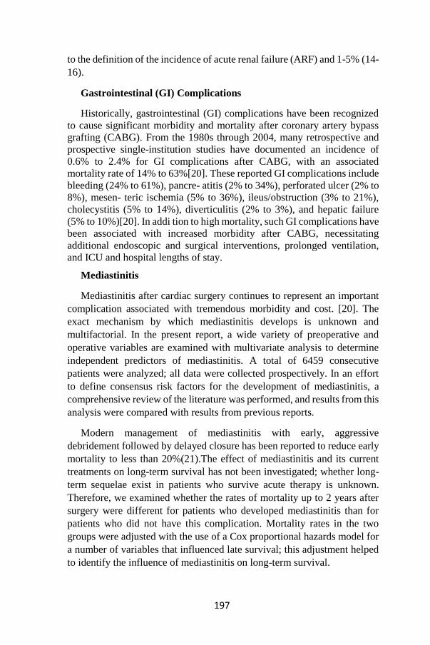
197
to the definition of the incidence of acute renal failure (ARF) and 1-5% (14-
16).
Gastrointestinal (GI) Complications
Historically, gastrointestinal (GI) complications have been recognized
to cause significant morbidity and mortality after coronary artery bypass
grafting (CABG). From the 1980s through 2004, many retrospective and
prospective single-institution studies have documented an incidence of
0.6% to 2.4% for GI complications after CABG, with an associated
mortality rate of 14% to 63%[20]. These reported GI complications include
bleeding (24% to 61%), pancre- atitis (2% to 34%), perforated ulcer (2% to
8%), mesen- teric ischemia (5% to 36%), ileus/obstruction (3% to 21%),
cholecystitis (5% to 14%), diverticulitis (2% to 3%), and hepatic failure
(5% to 10%)[20]. In addi tion to high mortality, such GI complications have
been associated with increased morbidity after CABG, necessitating
additional endoscopic and surgical interventions, prolonged ventilation,
and ICU and hospital lengths of stay.
Mediastinitis
Mediastinitis after cardiac surgery continues to represent an important
complication associated with tremendous morbidity and cost. [20]. The
exact mechanism by which mediastinitis develops is unknown and
multifactorial. In the present report, a wide variety of preoperative and
operative variables are examined with multivariate analysis to determine
independent predictors of mediastinitis. A total of 6459 consecutive
patients were analyzed; all data were collected prospectively. In an effort
to define consensus risk factors for the development of mediastinitis, a
comprehensive review of the literature was performed, and results from this
analysis were compared with results from previous reports.
Modern management of mediastinitis with early, aggressive
debridement followed by delayed closure has been reported to reduce early
mortality to less than 20%(21).The effect of mediastinitis and its current
treatments on long-term survival has not been investigated; whether long-
term sequelae exist in patients who survive acute therapy is unknown.
Therefore, we examined whether the rates of mortality up to 2 years after
surgery were different for patients who developed mediastinitis than for
patients who did not have this complication. Mortality rates in the two
groups were adjusted with the use of a Cox proportional hazards model for
a number of variables that influenced late survival; this adjustment helped
to identify the influence of mediastinitis on long-term survival.
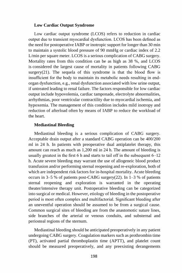
198
Low Cardiac Output Syndrome
Low cardiac output syndrome (LCOS) refers to reduction in cardiac
output due to transient myocardial dysfunction. LCOS has been defined as
the need for postoperative IABP or inotropic support for longer than 30 min
to maintain a systolic blood pressure of 90 mmHg or cardiac index of 2.2
L/min per square meter. LCOS is a serious complication of CABG surgery.
Mortality rates from this condition can be as high as 38 %, and LCOS
is considered the largest cause of mortality in patients following CABG
surgery(21). The sequela of this syndrome is that the blood flow is
insufficient for the body to maintain its metabolic needs resulting in end-
organ dysfunction, e.g., renal dysfunction associated with low urine output,
if untreated leading to renal failure. The factors responsible for low cardiac
output include hypovolemia, cardiac tamponade, electrolyte abnormalities,
arrhythmias, poor ventricular contractility due to myocardial ischemia, and
hypoxemia. The management of this condition includes mild inotropy and
reduction of afterload often by means of IABP to reduce the workload of
the heart.
Mediastinal Bleeding
Mediastinal bleeding is a serious complication of CABG surgery.
Acceptable drain output after a standard CABG operation can be 400/200
ml in 24 h. In patients with preoperative dual antiplatelet therapy, this
amount can reach as much as 1,200 ml in 24 h. The amount of bleeding is
usually greatest in the first 6 h and starts to tail off in the subsequent 6–12
h. Acute severe bleeding may warrant the use of allogeneic blood product
transfusion and/or performing sternal reopening and re-exploration, both of
which are independent risk factors for in-hospital mortality. Acute bleeding
occurs in 3–5 % of patients post-CABG surgery(22). In 1–3 % of patients
sternal reopening and exploration is warranted in the operating
theater/intensive therapy unit. Postoperative bleeding can be categorized
into surgical or medical. However, etiology of bleeding in the postoperative
period is most often complex and multifactorial. Significant bleeding after
an uneventful operation should be assumed to be from a surgical cause.
Common surgical sites of bleeding are from the anastomotic suture lines,
side branches of the arterial or venous conduits, and substernal and
periosteal regions of the sternum.
Mediastinal bleeding should be anticipated preoperatively in any patient
undergoing CABG surgery. Coagulation markers such as prothrombin time
(PT), activated partial thromboplastin time (APTT), and platelet count
should be measured preoperatively, and any preexisting derangements
199
should be corrected. In patients on oral anticoagulation, warfarin should be
withheld 4 days prior to surgery. Antiplatelet therapy with aspirin and/or
clopidogrel should ideally be withheld 5–7 days prior to operation to
minimize the risk of bleeding. Antiplatelet therapy is continued in cases of
urgent CABG surgery in patients with acute coronary syndrome (ACS) up
to the day of surgery; higher than average bleeding risk should be
anticipated in these cases. Alternatively glycoprotein IIb/IIIa inhibitor
agents such as Eptifibatide can be utilized to bridge patients with ACS up
to 2 h prior to CABG surgery to minimize progression of disease prior to
surgery and reduce the risk of bleeding due to their shorter half-life.
Meticulous surgical technique and surgical hemostasis are perhaps the most
important aspect in the prevention of major postoperative hemorrhage.
Rewarming the patient to normothermia prior to termination of
cardiopulmonary bypass would improve coagulation system function and
reduce the risk of postoperative bleeding.
In postoperative patients, in whom bleeding is suspected, any
hemodynamic instability should be addressed immediately. Chest drain
output should be meticulously measured, and drain patency should be
verified at all times. Patients should be warmed if hypothermic to improve
the coagulation system function. Coagulation studies should urgently be
performed, and any derangements should be corrected with the use of
appropriate blood products. Transesophageal/thoracic echocardiography
(TEE/TTE) should be performed to identify any signs of cardiac
tamponade. Sternal reopening should be performed if life-threatening
mediastinal bleeding is suspected.
Arrhythmias
Arrhythmias are common phenomenon after CABG surgery. Atrial
fibrillation (AF) is usually of benign nature and is said to occur in about 25
% of patients postoperatively after CABG. Although the etiology of
postoperative AF is still unclear, it can lead to devastating complications
such as stroke. Ventricular arrhythmias on the other hand occur rarely after
CABG surgery but are likely to signify more sinister causes such as
myocardial injury and should trigger further investigations by means of
bedside TTE, CT angiography scan, and/or coronary angiography.
Conduction abnormalities occur commonly post-CABG surgery[23]. 25
% of patients develop transient conduction abnormalities, which generally
resolve in 24–48 h and are associated with the use of cold cardioplegia
solution. Persistent conduction abnormalities do not influence the long-term
outcome after CABG surgery. The prevalence of more serious conduction
200
abnormalities such as atrioventricular block (AV) (Mobitz type II) and
complete AV block is very low after CABG surgery. The risk of developing
AV block post-CABG surgery increases with the higher number of
coronary vessels bypassed, longer CPB time, and longer cross-clamp times.
Potassium channel blockers (e.g., amiodarone) are used very commonly
for rhythm control in patients with postoperative AF. In refractory AF rate
control can be achieved with digoxin, nondihydro-pyridine calcium channel
blockers (e.g., diltiazem), or beta-blockers (e.g., metoprolol).
Anticoagulation with heparin and warfarin should be considered if
arrhythmias persist for >48 h. Bradyarrhythmias are often encountered after
CABG surgery. Epicardial pacing by means of epicardial pacing wires
should be commenced to maintain hemodynamic stability. In cases where
no epicardial pacing wires have been inserted at the time of surgery,
transcutaneous/transvenous cadiac pacing is an alternative. As per
management of tachyarrhythmias, electrolyte disturbances should also be
addressed in bradyarrhythmias. In patients with unresolved
bradyarrhythmias, insertion of a permanent pacemaker should be
considered.
References
Livesay J. J. (2000). Landmarks In Cardiac Surgery. Texas Heart
Institute Journal, 27(2), 222–223.
M. K. Davies and A. Hollman, “History of cardiac surgery,” Heart, vol.
87, no. 6, p. 509, 2002.
Ho, C. H., Chen, Y. C., Chu, C. C., Wang, J. J., & Liao, K. M. ().
Postoperative Complications After Coronary Artery Bypass Grafting in
Patients With Chronic Obstructive Pulmonary Disease. Medicine, 95(8),
e2926. doi:10.1097/MD.0000000000002926
D. K. C. Cooper, Open Heart: The Radical Surgeons Who
Revolutionized Medicine, Kaplan, New York, NY, USA, 2010.
B.Shrager,“Thevinebergprocedure:theimmediateforerunner of coronary
artery bypass grafting,” Annals of Thoracic Surgery, vol. 57, no. 5, pp.
1354–1364, 1994.
W. P. Longmire, J. Cannon, and A. A. Kattus, “Direct-vision
coronaryendarterectomyforanginapectoris,”TheNewEngland Journal of
Medicine, vol. 259, no. 21, pp. 993–999, 1958.
201
R. H. Goetz, M. Rohman, J. D. Haller, R. Dee, and S. S. Rosnak,
“Internal mammary-coronary artery anastomosis. A nonsuture method
employing tantalum rings,” The Journal of Thoracic and Cardiovascular
Surgery, vol. 41, pp. 378–386, 1961.
W. L. Proudfit, E. K. Shirey, and F. M. Sones Jr., “Selective cine
coronary arteriography. Correlation with clinical findings in 1,000
patients,” Circulation, vol. 33, no. 6, pp. 901–910, 1966.
A. S. Olearchyk, “Vasilii I. Kolesov: a pioneer of coronary revas-
cularization by internal mammary-coronary artery grafting,” Journal of
Thoracic and Cardiovascular Surgery, vol. 96, no. 1, pp. 13–18, 1988.
Higgins TL, Estafanous FG, Loop FD, et al. Stratification of morbidity
and mortality outcome by preoperative risk factors in coronary artery
bypass patients. A clinical severity score. JAMA 1992; 267:2344–2348.
Grover FL, Johnson RR, Marshall G, et al. Factors predictive of
operative mortality among coronary artery bypass subsets. Ann Thorac
Surg 1993; 56:1296–1306.
Durand M, Combes P, Eisele JH, et al. Pulmonary function tests predict
outcome after cardiac surgery. Acta Anaesthesiol Belg 1993; 44:17–23.
Kurki TS, Kataja M. Preoperative prediction of postoperative morbidity
in coronary artery bypass grafting. Ann Thorac Surg 1996; 61:1740–1745.
Samuels LE, Kaufman MS, Morris RJ, et al. Coronary artery bypass
grafting in patients with COPD. Chest 1998; 113:878–882.
Bruce JL, Cathy SR, Brian Spence, et al. Long-term survival of patients
with chronic obstructive pulmonary disease undergoing coronary artery
bypass surgery. Circulation 2006; 114:I-430–I-434.
Louis ES, Marla SK, Rohinton J. Morris, et al. Coronary artery bypass
grafting in patients with COPD. Chest 1998; 113:878–882.
Manganas H, Lacasse Y, Bourgeois S, et al. Postoperative outcome
after coronary artery bypass grafting in chronic obstructive pulmonary
disease. Can Respir J 2007; 14:19–24.
Medalion B, Katz MG, Cohen AJ, et al. Long-term beneficial effect of
coronary artery bypass grafting in patients with
COPD. Chest 2004; 125:56–62.
202
DeRose JJ, Jr, Toumpoulis IK, Balaram SK, et al. Preoperative
prediction of long-term survival after coronary artery bypass grafting in
patients with low left ventricular ejection fraction. J Thorac Cardiovasc
Surg 2005; 129:314–321.
van Domburg RT, Takkenberg JJ, van Herwerden LA, et al. Short-term
and 5-year outcome after primary isolated coronary artery bypass graft
surgery: results of risk stratification in a bilocation center. Eur J
Cardiothorac Surg 2002; 21:733–740.
Jan Jesper A, Laura Sommer H, Vibeke Elisabeth H, et al. 30-day
mortality after coronary artery bypass grafting and valve surgery has
greatly improved over the last decade, but the 1-year mortality remains
constant. Ann Cardiac Anaesth 2015; 18:138–142.
Clive K. Natural history of venous
thromboembolism. Circulation 2003; 107:I-22–I-130.
G. Captur, “Memento for Rene ́ Favaloro,” Texas Heart Institute
Journal, vol. 31, no. 1, pp. 47–60, 2004.
203
VETERINARY MEDICINE
204
HEALTH MONITORING
Buğra GENÇ*
Science, like other activities in human life, is globalizing. With the
globalization of science, the number of research is increasing and the
negative effects of regional and cultural differences on research are
decreasing. In order to make the results obtained in the biomedical
researches more reliable, in vivo methods using live animal models are
increasingly preferred. Among these methods, the creation of laboratory
animal models is one of the most reliable options. In order to reach the
most accurate and realistic result, it is of utmost importance that the most
appropriate model is formed and the continuity of the model characteristics
is achieved.
The health status of the animals to be used in the research has the
potential to directly affect the results of the research (Sellers et al., 2012;
Treuting et al., 2012). Even the possibility of infectious agents present at
the breeding line or at any critical point in the study raises the need for
microbiological quality monitoring of the animals. (Mansfield et al., 2010).
For these reasons, the use of animals with known biological properties in
scientific studies using animal models is important to ensure repeatability
of experimental results. The point of these findings is to harmonize health
monitoring programs (such as design, sampling, monitoring, reporting and
interpretation) that help to meet scientific, legal and animal welfare
requirements and provide information on the microbiological quality
information of animals used in research (Mahler et al., 2014).
Laboratory animals are threatened by a wide variety of microorganisms
that can cause infection. Many of these microorganisms can cause infection
without clinical symptoms. Therefore, it is not enough to try to perform
health monitoring by observing clinical symptoms. Latent form and
invisible symptoms may affect the outcome of the research. Symptomatic
and non-symptomatic infections may lead to complex results in the
research and may increase the number of animals used due to their negative
effects. Presence of tumors and other tissues, cell lines, serum, embryos
and gametes may be the cause of a latent infection. (Nicklas et al., 1993,
Mahabir et al., 2008). It should be kept in mind that infective agents that
may be present in laboratory animals may have zoonotic character. This
not only affects animal health and research quality in a negative way, but
it can also reach dimensions that threaten public health. For these and other
reasons, it is important that all centers and affiliated units working with the
* Assist. Prof. Dr. Ondokuz Mayis University Veterinary Medicine Faculty, Department
of Laboratory Animals. Samsun-Turkey
205
laboratory animal establish a laboratory animal health monitoring program
to cover every critical point and as an integrated part of any quality
assurance system. At this point, the cost of preventive measures and health
monitoring program requirements may seem high, but is very low in
relation to the total cost of the research project and the continuity in the
organization (Nicklas et al., 2002). The fact that the costs of health
surveillance are salvage against a possible loss requires that the cost be
ignored (Newcomer & Fox, 2007).
Health monitoring is a complex issue, and the training and competence
of the person to install this system will directly affect the success of the
system and should be performed by an expert and equipped with full
authority. The existence of a culture of communication among all those
involved in the animal care and use program is inevitable in order to ensure
good health monitoring and accurate interpretation of the results. (Koszdin
& Di Giacomo, 2002).
Unit Concept in Health Monitoring
According to health monitoring programs, animal facilities should be
structured and organized on a microbiological basis (microbiological unit).
A microbiological unit is defined as a stand-alone concept with separate
space and traffic for animals, personnel and supplies. According to this
definition, places where personnel, equipment and animals move freely or
where animals are kept in open cages, a barrier facility, an isolator, micro
isolation cage and IVC cage, can be named as microbiological unit. The
identification of the microbiological units is a critical an done of the most
important steps should be considered carefully in the design of the health
monitoring program as it will affect the sampling program, the quality and
frequency of tests, and the interpretation of the results. For example, since
the risk factors and consequences of microbiological contamination may
vary between experimental units, the design of the health monitoring
program should be based on this diversity. Strict rules to be applied within
the facility are also required for the implementation of health monitoring.
The most important of these rules is to ensure that the access within the
facility is under control and to ensure access to the minimum number of
authorized persons, traffic order at the material and animal exits and the
order so as not to cause controlled and cross-contamination. In addition,
the use of technologically superior material can be an aid to program
effectiveness. The use of IVC (individually ventilated cages), which has
become rapidly widespread recently, provides a great advantage over
conventional open cages in preventing potential contamination among
animals. With the use of these units, transmission of not only infectious
agents but also allergen agents commonly seen in laboratory animals will
be minimized.

206
In general, a health monitoring program should be established taking
into account the microbiological unit, animal species, number and
immunological status, monitoring frequency, sample material diversity to
be collected and frequency of sampling, and specific conditions for that
facility. If there are more than one species in the microbiological unit,
separate surveillance of the animals belonging to these species should be
ensured.
Risky conditions that may lead to the introduction of unwanted factors
in an animal unit are examined in two groups. Including multiple animals
per month, physical proximity of different microbiological units, animal
entry from different colonies, postoperation animal movements, insects
and wild rodent presence, facility serves multi-purpose research, mobility
of staff into the unit very often, common use non-disinfectable material are
considered high risky activations and situations. Inbreeding, occasional
staff mobility into the unit and a limited variety of studies constitute a low
risk group (Mahler et al., 2014).
Contamination
Some microorganisms are specific to one species or a very limited
number of more than one species, while others may be active in a large
number of different species. Some microorganisms are even known as
zoonoses having animal-human transmission characteristics. In addition to
the aforementioned traffic, animal-human interaction, material-borne and
environmental-related contamination, the transmission of these factors is
also possible during the use of biological material applied by the research
protocol. Currently, humanized immunosuppressed animals are used for
studies of human immune system, xenotransplantation and infection
models. Together with the transplants, these animals can also transfer HIV,
the human-acquired AIDS disease virüs (Berges & Rowan, 2011). For this
reason, it is necessary to check the cell lines that are intended to be used in
animals under the health monitoring protocol.
Many microorganisms that do not cause clinical symptoms may cause
diseases in animals whose immune system is suppressed and whose
resistance is reduced due to disease or medication used for research
purposes. As a matter of fact, genetically modified rodents constitute the
group with unexpected cases and findings. In these species, it is possible
to see diseases caused by organisms already known or commensally
present in animals, as well as the emergence of unplanned phenotypes that
may be considered important for the disease. (Treuting et al., 2012;
Franklin, 2006).
Based on current literature knowledge, it is not possible to list all
opportunistic microorganisms separately for all species and strains, but
periodic controls should be performed at the discretion of the facility


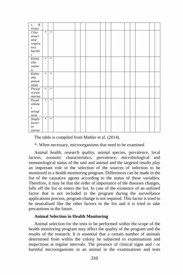
210
s, K virus)
Cilia-
associated
respira
tory bacillu
s
* *
Klebsi
ella oxytoc
a,
* *
Klebsiella
pneum
oniae
* *
Pneumocystis
murina
* *
Pseudomona
s
aeruginosa
* *
Staphy
lococcus
aureus
* *
The table is compiled from Mahler et al. (2014).
*: When necessary, microorganisms that need to be examined
Animal health, research quality, animal species, prevalence, local
factors, zoonotic characteristics, prevalence, microbiological and
immunological status of the unit and animal and the targeted results play
an important role in the selection of the sources of infection to be
monitored in a health monitoring program. Differences can be made in the
list of the causative agents according to the status of these variables.
Therefore, it may be that the order of importance of the diseases changes,
falls off the list or enters the list. In case of the existence of an unlisted
factor that is not included in the program during the surveillance
applications process, program change is not required. This factor is tried to
be neutralized like the other factors in the list and it is tried to take
precautions in the future.
Animal Selection in Health Monitoring
Animal selection for the tests to be performed within the scope of the
health monitoring program may affect the quality of the program and the
results of the research. It is essential that a certain number of animals
determined from within the colony be subjected to examinations and
inspections at regular intervals. The presence of clinical signs and / or
harmful microorganisms in an animal in the examinations and tests
211
performed within the scope of the program necessitates the same treatment
in other animals in the colony. Therefore, the observations and operations
are based on the logic that a sample within the whole cluster represents the
whole cluster. It is assumed that an agent not detected in selected sample
animals is not present in other animals in the colony. In this regard, despite
this factor, no treatment and administration is required. Room, cage,
building structure, size, barrier systems and protection systems for animals
can be used as a facilitator for the identification of microbiological
presence and increase the workload as they constitute more control points.
In case of increased workload, the details of health monitoring practices
(number of animals, etc.) are at the discretion of the staff and may vary.
Number of Animals in Health Monitoring Program
In animal researches, it is very important that the number of animals to
be used is sufficient in terms of ensuring animal welfare as well as
scientific reality. The same importance appears within the health
monitoring program. Colony size, physiological, microbiological, genetic
and individual characteristics of animals, and research details are key
parameters in determining the number of animals to be allocated for
monitoring. In this system, the health status of the colony can be observed
by the selected animals, the importance of the number and quality of the
animals under monitoring is revealed.
It is important for the accuracy and reliability of the reports to be given
by the production centers to make the applications performed using
laboratory animals within a certain standardization. For this reason, the
implementation of the health monitoring protocol prepared by the
Federation of European Laboratory Animal Science Associations
(FELASA) is considered to be the most appropriate of the existing systems.
(Nicklas et al., 2002). On the basis of this protocol, additions which are not
specified in the protocol but may also be made. For this reason, criteria
such as the number of animals to be selected and methods of examination
can be determined in accordance with the quality of the research and not
by systems that contain strict internationally accepted rules. With this
approach, health monitoring program can become healthier.
Number of animals to be selected for health monitoring and formulas
for some values are given by Hansen (2003).
Number of samples for colonies of more than 1000 animals: S
S ≥ log C/log (1-(p*N1 ))
Number of samples for colonies less than 1000 animals: S
S ≥ (1 – C1/D )* (T- ((D-1)/2))
Nosografic sensitivity: N1

212
N1 = Number of infected animals reacting in the study/ Number of
infected animals reacting or not in the study.
Estimated prevalence in colony = p
p = Number of infected animals /Number of total animals
Risk of false negative results in an infected colony: C
C = Number of infected colonies tested with negative results / Total
number of animals in infected colonies tested
D= Number of infected animals T= Total number of animals
Use of Sentinel Animal
In some studies, it is not possible to select animals from trial groups for
health monitoring. In such cases, an animal with the same genetic and
physiological characteristics other than the trial is kept in the trial
environment and health surveillance is carried out on that animal. The
animal used for this purpose is called sentinel animal (Hansen & Jensen,
1995). It is important for the disease course and detection that an animal
designated as a lookout is exactly the same as the animals in the other trial.
The age and genetic characteristics of sentinel animals should be
considered specifically for the infections to be monitored and should not
create an unfavorable situation (Hansen et al., 1994) .
As a general practice in the cages where the animals are located, the
sentinels are located on the bottom shelves, and the contaminated bedding
materials of other animals are added to their bedding materials by mixing.
Dirty bedding materials are effective to accelerate the spread and detection
of a possible infection. There are cases where this method cannot be safe.
This method is not suitable for Sendai virus (Artwohl et al., 1994), ascarids
and other directly diagnosed fecal infections. In addition, it should not be
forgotten that not every infection can be transmitted from the stool and
urine.
Method Selection
Some factors can be easily isolated from animals, while others can be
obtained depending on very specific techniques and conditions. Spirillum
minus, a rodent agent, cannot be produced in vitro, but many endo and
ectoparasites can be easily isolated and observed under a microscope
(Hansen, 2003). In order to obtain accurate results in a short time, first of
all, signs of illness should be sought by observation, if necessary, more
detailed tests should be carried out so that economies can be provided in
time and in financial terms, and animal discomfort can be minimized
within the framework of 3R rules.
213
Cultivation is a method in which microorganisms (bacteria and fungi)
can be grown in artificial environments. Samples from organs for
bacteriological examination can be detected using selective or non-
selective media. More specific methods are needed to obtain the agent in
its purest form. FELASA recommends that some serology tests
(immunofluorescence assay (IFA), enzymelinked immunosorbent assay
(ELISA) and western immunoblotting) be performed for microorganisms
that cannot be cultured easily (Nicklas et al., 2002). Molecular detection
methods can be used for factors that cannot be detected by serological
and/or culture methods. Today, the most modern molecular methods (PCR,
qPCR, in situ hybridization, FISH) can detect DNA, RNA and fractions of
microorganisms.
Reporting
The health monitoring report is not considered a laboratory test result
report. The health monitoring report provides more detailed information
than any laboratory test result report. This report must be prepared by a
qualified and authorized personnel. If an agent is detected in one or more
animals, it must be reported in the health monitoring report. After reporting
an agent in the unit, tests must be repeated at certain time intervals until
the agent is eradicated. (Mahler et al. 2014).
Appendix 3, published by FELASA, provides a uniform health
monitoring report. In this appendix, possible disease factors are given, as
well as examples of data that should be included in the health surveillance
report, such as test name, method, last test date, findings from previous
tests, findings based on inspection, and laboratory where the test was
performed (Nicklas et al., 2002). The results of all tests and observations
should be recorded in a timely and complete manner and should be clearly
shared with all other concerned. In report document, characteristics
describing the animal should be specified and agents must be recorded in
alphabetic order. Testing laboratory, test frequency, results in current test
and last previous tests are other important datas should be given in report
(Table II). In this way, data that may be very important in future studies
will be widely available.
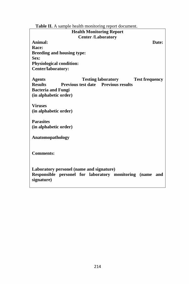
214
Table II. A sample health monitoring report document.
Health Monitoring Report
Center /Laboratory
Animal: Date:
Race:
Breeding and housing type:
Sex:
Physiological condition:
Center/laboratory:
Agents Testing laboratory Test frequency
Results Previous test date Previous results
Bacteria and Fungi
(in alphabetic order)
Viruses
(in alphabetic order)
Parasites
(in alphabetic order)
Anatomopathology
Comments:
Laboratory personel (name and signature)
Responsible personel for laboratory monitoring (name and
signature)
215
References
ARTWOHL, J.E., CERA, L.M., WRİGHT, M.F., MEDİNA, L.V.,
KİM, L.J., (1994).The efficacy of a dirty bedding sentinel system for
detecting Sendai virus infection in mice: a comparison of clinical signs and
seroconversion, Lab. Anim. Sci ., 44, 73.
BERGES, B.K. and ROWAN, M.R. (2011). The utility of the new
generation of humanized mice to study HIV-1 infection: transmission,
prevention, pathogenesis, and treatment. Retrovirology. 8: 65.
Franklin, C.L. (2006). Microbial considerations in genetically
engineered mouse research. ILAR J. 47: 141–155.
HANSEN, A.K., ANDERSEN, H.V., SVENDSEN, O. (1994). Studies
on the diagnosis of Tyzzer’s disease in laboratory rat colonies with
antibodies against Bacillus Piliformis (Clostridium Piliforme), Lab. Anim.
Sci. , 44, 424.
HANSEN, A.K. and JENSEN, H.J.S. (1995). Experience from sentinel
health monitoring in units containing rats and mice in experiments, Scand.
J. Lab. Anim. Sci. , 22, 1.
HANSEN, A.K. (2003). Health Status and Health Monitoring. In
Handbook of Laboratory Animal Science Second Edition Ed. Jann Hau
and Gerald L. Van Hoosier, Jr. Vol I. Essential, Principles, and Practices.
by CRC Press LLC. US.
KOSZDIN, K.L. and Di GIACOMO, R.F. (2002). Outbreak: detection
and investigation. Contemp Top Lab Anim Sci. 41: 18–27.
MAHABIR, E., BAUER, B., SCHMIDT, J. (2008). Rodent and
germplasm trafficking: risks of microbial contamination in a high-tech
biomedical world. ILAR J. 49: 347–355.
MAHLER M., BERARD, M, FEINSTEIN, R., GALLAGHER, A.,
ILLGEN-WILCKE, B., PRITCHETT-CORNING, K. (2014). FELASA
recommendations for the health monitoring of mouse, rat, hamster, guinea
pig and rabbit colonies in breeding and experimental units Raspa
Laboratory Animals. Vol. 48(3) 178–192
MANSFIELD, K.G., RILEY, L.K., KENT, M.L. (2010). Workshop
summary: detection, impact, and control of specific pathogens in animal
resource facilities. ILAR J. 51: 171–179.
NEWCOMER, C.E. and FOX, J.G.(2007) Zoonoses and other human
health hazards. In: Fox JG, Barthold SW, Davisson MT, Newcomer CE,
Quimby FW and Smith AL (eds) The mouse in biomedical research. Vol.
II. Diseases , 2nd ed. San Diego: Academic Press, pp.719–745.
216
NICKLAS, W., KRAFT, V., MEYER, B. (1993). Contamination of
transplantable tumors, cell lines, and monoclonal antibodies with rodent
viruses. Lab Anim Sci. 43: 296–300.
NICKLAS, W., BANEUX, P., BOOT, R., DECELLE, T., DEENY,
A.A.(2002). Fumanelli, M., and Illgen-Wilcke, B., Recommendations
forthehealthmonitoringofrodent and rabbit colonies in breeding and
experimental units, Lab. Anim. , 36, 20.
SELLERS, R.S., CLIFFORD, C.B., TREUTING, P.M., BRAYTON,
C. (2012). Immunological variation between inbred laboratory mouse
strains: points to consider in phenotyping genetically immunomodified
mice. Vet Pathol. 49: 32–43.
TREUTING, P.M., CLIFFORD, C.B., SELLERS, R.S., BRAYTON,
C.F. (2012) Of mice and microflora: considerations for genetically
engineered mice. Vet Pathol. 49: 44–63.
217
ALTERNATIVE APPROACHES TO TREATMENT OF
MASTITIS INSTEAD OF TRADITIONAL METHODS
Hande GÜRLER
Introduction
Mastitis, which decreases in milk quantity and quality, is a major
problem in dairy animals such as cattle, buffalo, ewe and goat. It is also a
health condition that affects human health by causing changes in milk
composition. Bovine mastitis plays a decisive role in the conventional and
organic dairy industry (Kanbur and Gürbulak, 2017; Krömker and
Leimbach, 2017).
Mastitis is a disease that is difficult to control due to the complex
multifactorial etiopathology. The traditional treatment of mastitis is
antibiotics (Grave et al., 1999). However, antibiotic treatment is
moderately effective in an established mammary infection. Because of the
drainage of the drug due to lactation, inflammation of the glandular cells,
immunosuppressive effect, the development of new concomitant infections
often fail the effectiveness of antibiotics (Sandholm et al, 1990).
Conventional treatment of mastitis using antibiotics has led to the
emergence of antimicrobial resistance (AMR) against most of the
commonly used antibacterial agents (Alekish et al., 2018). AMR is a
serious threat to global public health and food security. According to
O’Neil et al. (2014), 700,000 people die each year from infections caused
by multiresistant bacteria.
It also increases animal suffering and production losses in animals. In
addition, the passage of drug residues in humans through milk and milk
products, the development of resistance to these drugs and the high cost
are increasing the interest in alternative treatment interventions (Krömker
and Leimbach, 2017; Reshi et al.,2017).
It is thought that mastitis is a disease that needs to be protected rather
than treated. It is reported that in modern herd health management, the
implementation of preventive mastitis programmes against contagious
pathogens resulted in an increase of clinical mastitis cases caused by
environmental pathogens like Streptococcus uberis, Escherichia coli and
minor pathogens. It should be noted that the effectiveness of the therapy
used is critical for udder health status at the farm level (Werner et al.,
2010).
218
For these reasons, alternative methods for antibiotic therapy such as
phytotheraphy and homeopathic treatment, acupuncture, clay treatment,
oxygen therapy, vaccination, bacteriophage teraphy, nanoparticles
treatment, cytokine application, photodynamic therapy, probiotic and
vitamin adminastration are being studied. In this article, we will give an
overview of alternative methods and give information about the current
situation.
Phytotheraphy and Homeopathic Treatment
Plants or herbal extracts can be administered orally, intramammary or
topically in the form of gel in the treatment of mastitis. There are also
commercial mammary preparations containing herbal ingredients.
Traditional Chinese medicine (TCM) for the treatment of bovine
mastitis is commonly prepared from Taraxacum mongolicum (Pu Gong
Ying), Lonicera japonica Thunb. (Jin Yin Hua), Viola patrinu (Zi Hua Di
Ding) and Folium isatidis (Da Qing Ye), based on oral heat-cleaning,
detoxification, anti-inflammatory and antibacterial effects (Yang et al.,
2019) Many other TCM plants have similar pharmacological factors, such
as Angelica dahurica (Bai Zhi), Coptis chinensis (Huang Lian),
Phellodendron amurense (Huang Bo), Rheum o cinale (Da Huang), and
Scutellaria baicalensis (Huang Qin) (Cai et al., 2004, Muluye et al., 2014)
According to TCM concept, ese herbs, which are “cold (寒, hán) ve and
can clean the internal heat and are generally used as antibiotic and
antipyretic agents, are considered as anti-inflammatory and antimicrobial
agents and have been reported to be effective in the treatment of
inflammatory diseases and microbial infections (Yang et al, 2019).
Kher et al., (2019) reported that they could be used as an alternative
treatment to antibiotics in bovine subclinical mastitis as a result of their
study with Terminalia chebula. Terminalia chebula (T. chebula,
Combretaceae) is one of the most versatile medicinal plants with great
biological activities. T. chebula is called “Wonder Medicine Ay in
Ayurveda and King Medicine King Tib in Tibet and is always at the top of
the list in Ayurveda Materia Medica because of its extraordinary healing
power (21). This is one of the main components of the well-known
Ayurvedic medicine known as “Triphala Powder.The fruits, roots and bark
of this plant are used in the preparation of herbal medicines. It has been
reported to be used to treat a variety of conditions such as fever, cough,
diarrhea, gastroenteritis, skin diseases, candidiasis, urinary tract infection
and wound infections.
Medicinal plants such as Calendula officinalis, Ocimum sanctum, Aloe
vera, Houttuynia cordata, Persicaria senegalense, Panax ginseng,
Minthostachys verticillata and many others have been reported to reduce
somatic cell count with anti-inflammatory effects, improve milk quality
219
and reduce the incidence of mastitis. (Cerioli et al, 2018; Kanbur and
Gürbulak, 2017; Shafi et al., 2016). However, WHO has recommended to
all member countries to actively promote native medicines of their
respective country and initiate steps to conserve medicinal plants. Reshi et
al. reported that the herbal medicines has some advantages such as non-
toxic, efficacious, cultural acceptability, lesser side effects (Kamboj opcit),
and act selectively enhancing body resistance (Reshi et al., 2017). Research
shows that most of the plants used to prepare native medicines contain
valuable active ingredients, but more studies are needed in this area.
Homeopathy is a method used both in the prevention of mastitis and in
the treatment of mastitis. Homeopathy has been reported to be useful in the
prevention and treatment of mastitis (Kanbur and Gürbulak, 2017).
Werner et al. reported that a therapeutic effect in mild homeopathic
therapies in cases of mild or moderate clinical mastitis caused by
environmental pathogens (2010).
Because of the interest in the veterinary practices of native wild plants,
the spread of this traditional knowledge to new generations can lead to a
revaluation of local plant resources, which promotes the conservation of
medicinal vegetation and regional biodiversity in the study area. In this
sense, it emphasizes that it is important to recover and record the use of
medicinal plants used in veterinary medicine in a scientific context as
discussed in this study (Martínez and Luján, 2011).
Bacteriophage Therapy
Bacteriophage therapy is a method that uses pathogen specific
bacteriophages in the treatment of a bacterial infection. Bacteriophages are
viruses that can infect and kill bacteria. Phages have been identified as
novel antimicrobial agents for veterinary applications. Phage K in
infections caused by S. aureus has been used as a prophylactic measure
(O'flaherty and Coffey 2005).
In a study (Gill et al., 2006) also tested the efficacy of phage K against
S. aureus. The phage has various limitations, such as milk and its
constituents (whey proteins) and its degradation and consequently
inactivation by the immune system. Kwiatek et al. (2012) isolated a new
virulent phage (MSA6) from a mastitis cow, presenting a broad lytic
spectrum against bovine staphylococcus strains. This invaluable nature of
MSA6 is crucial for its potential use as a universal anti-staphylococcal
agent. Furthermore, given the various features presented by MSA6, this
phage can be used in many staphylococcal infections, including bovine
mastitis. Fenton et al. (2013) bacteriophage-derived peptidase was tested;
CHAPK against S. aureus isolated from mastitis infected cows. This study
demonstrated the potential use of CHAPK as prophylactic and therapeutic
220
measures for mastitis infections. CHAPK was effective against biological
filters by preventing biofilm formation or by breaking down established
biofilms of staphylococcus strains due to bovine mastitis. Dias et al. (2013)
isolated that bacteriophages can infect S. aureus from mastitis positive
cows. These phages exhibit a wide range of properties critical to their use
as phage therapy, such as a wide range of hosts, high lytic activity and
thermostability. Basdew and Laing (2014) tested some phage sensitivities
to some simulation stresses. Sabp-P1, Sabp-P2 and Sabp-P3 are less
susceptible to stresses tested in vitro and are therefore the future favorites
in in vivo experiments and their future use as therapeutic strategy for the
treatment of mastitis-infected cows has been demonstrated. Although there
are some limitations, bacteriophage treatment in general seems to be a
serious candidate for antibiotic alternatives to be used in the future control
and treatment of bovine mastitis. However, additional further research is
required to explore the therapeutic potential of bacteriophages to treat
clinical and subclinical mastitis associated bacterial infections.
Acupuncture
Acupuncture is a traditional Chinese medicine has been used for
analgesia and therapy in humans for more than 4,000 years. It is performed
by stimulating predetermined points in the body that are fully connected to
the central nervous system and cause a certain physiological effect
(Schoen, 1994). Acupuncture has been used successfully to treat many
disorders including mastitis in dairy cattle. However, most of the
procedures used expensive equipment such as lasers and electrostimulators
and various acupuncture points (Daga et al., 2013).
There are different methods of stimulating acupuncture points
traditional and modern. Traditional methods include conventional white
needle acupuncture (CWNA), aquapuncture, moxibustion, cupping,
pneumopuncture, embedding, fire needling, hemoacupuncture and
auriculoacupuncture. Aquapuncture is known to be the most common
technique of acupoint stimulation by injection. It is performed by injecting
the solution into the acupuncture points by using an appropriate size needle
and sterile syringe. Aquapuncture utilizes different solutions including
homeopathic remedies, antibiotics, vitamins, hormones, sterile saline,
herbal extracts and the patient’s own blood (Daga et al., 2013). Daga et al.,
(2013) have injected 1% chilli pepper boil at acupuncture points and
reported that it is effective in reducing the incidence of mastitis and
increasing milk production. However they recommend longer-term
studies. The length of the treatment period and the insufficient of veterinary
acupuncturists constitute the disadvantages of this application (Duval,
1997; Kendall,1988).
221
Clay Treatment
Clay has been used in the treatment of mastitis due to its high absorption
properties. For this purpose, the mixture of clay and water and oil is applied
to the breast with mastitis 2-3 times a day after milking and the breast is
wrapped with cloth. By adding a few drops of pine or peppermint oil into
this mixture, the effectiveness of the clay can be increased. With this
application, it can be seen in 2-3 hours in acute mastitis and in 2-3 days in
chronic mastitis industry (Kanbur and Gürbulak, 2017).
Oxygen Therapy
Oxygen therapy can be used to treat diseases in humans and animals;
Glyroxilide, given as an intramuscular single dose, two doses or rarely 3
doses, has been reported to give good results in the treatment of mastitis,
but in some studies it has not been shown to be effective enough (Duval,
1997).
Vaccination
Mastitis vaccines have been formulated with the hope of reducing the
incidence mastitis in dairy farming and promoting the profit of industry.
Some vaccines were developed against S. aureus mastitis but all presented
limited efficacy. The improper immunization schedules, ineffective
adjuvant formulation, and the limited range of protection are some of the
causes of their ineffectiveness It is clear that a single vaccine will not
prevent mastitis caused by the plethora of pathogens and their different
mechanisms of pathogenesis (Bannerman and Wall, 2005). Coliform
vaccines can decrease the prevalence, severity and length of infections in
both milking cows and heifers (Gomes and Henriques, 2016). There are no
vaccines available to prevent mastitis caused by K. pneumonia (Tiwari et
al., 2013). The research in this area continues.
Nanoparticles Treatment
Nanotechnology is a rapidly growing field. The surface area volume
ratio of nanoscale materials is high. Antimicrobial agents have unique
chemical and physical properties thanks to nanotechnology. NPs are
manufactured and used in a wide variety of commercial products such as
silver nanoparticles (AgNPs), electronics, bio-detection, clothing, food
production, dyes, sunscreens, cosmetics, and the medical field. However,
opinions about the toxicity of NPs differ among scientists; For example,
the type or size of NPs may be related to various factors such as the
synthesis method or conditions (Ahamed et al, 2010; Albrechtet al., 2006;
Morones et al., 2005; Kim et al., 2007).
Sankar (2016) has reported in a study that ZnO nanoparticles inhibit the
growth of bacteria, molds and yeasts that cause superficial or deep
infections and even toxicities, and can be used successfully as bacteria and
222
fungicides in the treatment of microbial diseases in human area and
veterinary medicine. In the study, ZnO can be applied as a food additive to
advanced areas and will be preferred more to improve animal and human
health in the near future.
Nanoparticles are seen as a potential for the treatment of bovine mastitis
when used as phagocytes. For example, silver nanoparticles exhibited
inhibitory activity against S. aureus isolated from subclinical mastitis
(Dehkordi et al., 2011) and are excellent for a great economic antimicrobial
solution. Alexander et al. (2019), in their study reported that copper and
silver NPs will be used in the preparation of disinfectants that can be used
in dairy cows the milking routine. In addition, antibiotics have now been
incorporated into most of the nanoparticle delivery system to combat
microorganisms. Amoxicillin added to nanoparticles exhibited biodynamic
against S. aureus, E. coli and S. agalactiae (Yang et al., 2009) The most
important challenge seems to be that they are getting plenty of antibiotic
loading into these delivery systems.
Cytokine Application
Cytokines are very small proteins (less than 50 kDa) that act as
intercellular communication signals mediate the exchange of autocrine,
paracrine or endocrine signals in hematopoiesis, stress, inflammation,
immune and tissue repair (Belardelli and Ferrantini, 2002; Rouveix, 1997).
Identification of a large number of cytokines and blocking of cytokine
production, stimulation of inhibiting pathways, inhibition of circulation,
inhibition of binding, inhibition of receptors and signaling mechanisms
have led to progress in diagnosis and treatment.Cytokines are signals that
dictate immune responses in normal and mastitis breasts. Therefore, subtle
changes in the cytokine network of the mammary gland in health and
disease may help in detecting early infection and monitoring the
effectiveness of treatment. However, the cytokine network in the bovine
mammary gland has not yet been fully investigated and needs to be
identified at different stages of lactation and lactation. Techniques based
on real-time PCR or microarray may be reliable, but are not yet suitable
for daily control of breast health. Practical application cytokines as a
diagnostic tool in bovine mastitis require the automation of procedures for
detection and monitoring of cytokines (Alluwaimi, 2004).
Numerous cytokines like complement component, interleukins (IL) -1,
-2, -6 and -8, and tumor necrosis factor alpha (TNF-α) have been
recognized as key actors during the acute-phase reaction. They have
chemotactic activity responsible for leukocyte uptake and activation and
may also increase the bactericidal activity of phagocytes. Numerous
experimental works point out that use of cytokines with or without
antibiotics signi cantly improved the cure rate in S. aureus infected
223
mastitis. Cytokines therapy appears to be a promising method; however,
advanced research has to be done to accept therapeutic attention (Hossain
et al., 2017).
Photodynamic Therapy
The use of non-toxic dyes or photosensitizers (PS) in combination with
harmless visible light that is known as photodynamic therapy (Costano,
2004). Photodynamic Therapy (PDT) is alternative treatment for microbial
resistance and promising strategy for the eradication of Gram-positive,
Gram negative, yeast and fungal bacteria (F Sperandio, 2013).
The mechanism of PDT is the activation of a non-toxic photosensitizer
that through appropriate exposure to light at a convenient wavelength and
energy density generates reactive oxygen species, including free radicals
and singlet oxygen which, when in contact with the microorganisms, can
modify bacterial DNA and cell membrane, leading to microbial death
(Costano, 2013; Hamblin and Hasan, 2004).
Phenothiazinium dyes, such as methyleneblue (MB), are used as
photosensitizers (PS) for both gram-positive and negative bacteria
exhibiting effective eradication. In fact, MB is used as a lead compound
because of its low toxicity in the human patient. Another advantage of this
cationic PS is its affinity for targeting microorganisms rather than host
cells. Therefore, short-term irradiation treatment with MB shows minimal
antimicrobial activity while minimally damaging host cells (Sellera et al.,
2016).
Researchers report that intermolecular interactions between PS and
milk components should be considered in order to optimize clinical
protocols and that appropriate light delivery systems should reach deeper
udder tissue because pathogens normally colonize the entire mammary
gland. Therefore, they report that research on different light distribution
systems can optimize clinical protocols and facilitate the use of PDT in the
dairy industry (Riberio et al, 2018).
It is reported that PDT is the effective treatment of subclinical mastitis
in cows in vivo, without the need to remove the animal from production.
However, because mastitis has a multifactorial etiology, further studies are
needed to develop other therapeutic protocols (Sellera et al., 2016; Moreira
et al., 2018).
Probiotic and Vitamin Adminastration
Probiotics are often used for multiple purposes and most of the
applications are performed both in humans and in different animals
productivity (Nader-Macías et al., 2008). Nader-Macías et al., (2008) have
isolated lactic acid bacteria (LAB) from natural bovine ecosystems because
of their probiotic potential. The useful probiotic LAB was isolated from
224
the vaginal tracts of newborn calves and the mammary glands of adult
heifers, faeces and oral cavity. They could improve the health of the animal
and promote their productivity. Bacterial strains and their numbers varied
for each tract examined. Several strains reported that they were able to
produce bacteriocin and were isolated only from mammary gland and
faeces. At the end of the study it was reported that some strains can prevent
mastitis by applying to the mammary gland and can also promote
colonization of the newborn intestinal system (Nader-Macías et al., 2008).
In a study conducted with vitamin B2 injection in clinical mastitis
caused by S.aureus, it has been reported that VB2 injection reduces the
number of somatic cells both by activating neutrophil function and by
increasing and / or regulating cytokine production (Sato et al., 1999).
References
Ahamed, M., Alsalhi, M. S., Siddiqui, M. K. J. (2010). Silver
nanoparticle applications and human health. Clinica chimica acta, 411(23-
24), 1841-1848.
Albrecht, M. A., Evans, C. W., Raston, C. L. (2006). Green chemistry
and the health implications of nanoparticles. Green chemistry, 8(5), 417-
432.
Alekish, M., Ismail, Z. B., Albiss, B., Nawasrah, S. (2018). In vitro
antibacterial effects of zinc oxide nanoparticles on multiple drug-resistant
strains of Staphylococcus aureus and Escherichia coli: An alternative
approach for antibacterial therapy of mastitis in sheep. Veterinary
world, 11(10), 1428.
Alluwaimi, A. M. (2004). The cytokines of bovine mammary gland:
prospects for diagnosis and therapy. Research in veterinary science, 77(3),
211-222.
Bannerman, D. D., Wall, R. J. (2005). A novel strategy for the
prevention of Staphylococcus aureus-induced mastitis in dairy
cows. Information Systems for Biotechnology News Report. Virginia Tech
University. USA, 1-4.
Basdew, I. H., Laing, M. D. (2014). Stress sensitivity assays of
bacteriophages associated with Staphylococcus aureus, causal organism of
bovine mastitis. African Journal of Microbiology Research, 8(2), 200-210.
Belardelli, F., Ferrantini, M. (2002). Cytokines as a link between innate
and adaptive antitumor immunity. Trends in immunology, 23(4), 201-208.
Cai, Y., Luo, Q., Sun, M., Corke, H. (2004). Antioxidant activity and
phenolic compounds of 112 traditional Chinese medicinal plants
associated with anticancer. Life sciences, 74(17), 2157-2184.
225
Castano, A. P., Demidova, T. N., & Hamblin, M. R. (2004).
Mechanisms in photodynamic therapy: part one—photosensitizers,
photochemistry and cellular localization. Photodiagnosis and
photodynamic therapy, 1(4), 279-293.
Cerioli, M. F., Moliva, M. V., Cariddi, L. N., Reinoso, E. B. (2018).
Effect of the essential oil of minthostachys verticillata (Griseb.) epling and
limonene on biofilm production in pathogens causing bovine
mastitis. Frontiers in veterinary science, 5, 146.
Daga, J. D., Acorda, J. A., & Rayos, A. A. (2013). Effects Of
Conventional White Needle Acupuncture And Aquapuncture On Mastitis
And Milk Production In Dairy Cattle. Philippine Journal of Veterinary and
Animal Sciences, 39(1).
Dehkordi, S.H., Hosseinpou,r F., Kahrizangi, A.E. (2011). An in vitro
evaluation of antibacterial effect of silver nanoparticles on Staphylococcus
aureus isolated from bovine subclinical mastitis. Afr J Biotechnol, 10:
10795–10797.
Dias, R. S., Eller, M. R., Duarte, V. S., Pereira, Â. L., Silva, C. C.,
Mantovani, H. C., ... & De Paula, S. O. (2013). Use of phages against
antibiotic-resistant Staphylococcus aureus isolated from bovine
mastitis. Journal of animal science, 91(8), 3930-3939.
Duval, J. (1997). Treating mastitis without antibiotics. Describes
factors contributing to mastitis, di- agnosis, and preventive & curative
measures. Ecological Agricultural Project, AGRO-BIO- 370-11E,
http://eap.mcgill.ca/agrobio/ab370- 11e.htm, 1997.
F Sperandio, F., Huang, Y. Y., R Hamblin, M. (2013). Antimicrobial
photodynamic therapy to kill Gram-negative bacteria. Recent patents on
anti-infective drug discovery, 8(2), 108-120.
Fenton, M., Keary, R., McAuliffe, O., Ross, R. P., O'Mahony, J.,
Coffey, A. (2013). Bacteriophage-derived peptidase eliminates and
prevents Staphylococcal biofilms. International journal of
microbiology, 2013.
Gill, J. J., Pacan, J. C., Carson, M. E., Leslie, K. E., Griffiths, M. W.,
Sabour, P. M. (2006). Efficacy and pharmacokinetics of bacteriophage
therapy in treatment of subclinical Staphylococcus aureus mastitis in
lactating dairy cattle. Antimicrobial agents and chemotherapy, 50(9),
2912-2918.
Gomes, F., Henriques, M. (2016). Control of bovine mastitis: old and
recent therapeutic approaches. Current microbiology, 72(4), 377-382.
Grave, K., Greko, C., Nilsson, L., Odensvik, K., Mørk, T., & Rønning,
M. (1999). The usage of veterinary antibacterial drugs for mastitis in cattle
226
in Norway and Sweden during 1990–1997. Preventive veterinary
medicine, 42(1), 45-55.
Hamblin, M. R., Hasan, T. (2004). Photodynamic therapy: a new
antimicrobial approach to infectious disease?. Photochemical &
Photobiological Sciences, 3(5), 436-450.
Hossain, M. K., Paul, S., Hossain, M. M., Islam, M. R., & Alam, M. G.
S. (2017). Bovine mastitis and its therapeutic strategy doing antibiotic
sensitivity test. Austin J Vet Sci Anim Husb, 4(1), 1030.
Kalińska, A., Jaworski, S., Wierzbicki, M., Gołębiewski, M. (2019).
Silver and Copper Nanoparticles—An Alternative in Future Mastitis
Treatment and Prevention?. International journal of molecular
sciences, 20(7), 1672
Kanbur M., Gürbulak K. (2017). Organik hayvancılıkta mastitis sorunu
ve sağaltımı. Turkiye Klinikleri J Vet Sci Pharmacol Toxicol-Special
Topics, 3, 20-28.
Kendall, D. (1988). Acupuncture beats antibiotics. The New
Farm, 1418.
Kher, M. N., Sheth, N. R., Bhatt, V. D. (2019). In vitro antibacterial
evaluation of Terminalia chebula as an alternative of antibiotics against
bovine subclinical mastitis. Animal biotechnology, 30(2), 151-158.
Kim, J. S., Kuk, E., Yu, K. N., Kim, J. H., Park, S. J., Lee, H. J., Kim,
H. S., Park, Y. K., Park, Y. H., Hwang, C. Y., Kim, Y. K., Lee, Y. S.,
Jeong, D. H., Cho, M. Y., (2007). Antimicrobial effects of silver
nanoparticles. Nanomedicine: Nanotechnology, Biology and
Medicine, 3(1), 95-101.
Krömker V., Leimbach S., Mastitis treatment-Reduction in antibiotic
usage in dairy cows. Reproductin in Domestic Animals, 52, 21-29.
Kwiatek, M., Parasion, S., Mizak, L., Gryko, R., Bartoszcze, M., Kocik,
J. (2012). Characterization of a bacteriophage, isolated from a cow with
mastitis, that is lytic against Staphylococcus aureus strains. Archives of
virology, 157(2), 225-234.
Martínez, G. J., & Luján, M. C. (2011). Medicinal plants used for
traditional veterinary in the Sierras de Córdoba (Argentina): an
ethnobotanical comparison with human medicinal uses. Journal of
Ethnobiology and Ethnomedicine, 7(1), 23.
Moreira, L. H., de Souza, J. C. P., de Lima, C. J., Salgado, M. A. C.,
Fernandes, A. B., Andreani, D. I. K., ... & Zângaro, R. A. (2018). Use of
photodynamic therapy in the treatment of bovine subclinical
mastitis. Photodiagnosis and photodynamic therapy, 21, 246-251.
227
Morones, J. R., Elechiguerra, J. L., Camacho, A., Holt, K., Kouri, J. B.,
Ramírez, J. T., Yacaman, M. J. (2005). The bactericidal effect of silver
nanoparticles. Nanotechnology, 16(10), 2346.
Muluye, R. A., Bian, Y., Alemu, P. N. (2014). Anti-inflammatory and
antimicrobial effects of heat-clearing Chinese herbs: a current
review. Journal of traditional and complementary medicine, 4(2), 93-98.
Nader-Macías, M. E. F., Otero, M. C., Espeche, M. C., Maldonado, N.
C. (2008). Advances in the design of probiotic products for the prevention
of major diseases in dairy cattle. Journal of industrial microbiology &
biotechnology, 35(11), 1387-1395.
O'flaherty, S., Coffey, A., Meaney, W. J., Fitzgerald, G. F., Ross, R. P.
(2005). Inhibition of bacteriophage K proliferation on Staphylococcus
aureus in raw bovine milk. Letters in applied microbiology, 41(3), 274-
279.
O’Neill, J. I. M. (2014). Antimicrobial resistance: tackling a crisis for
the health and wealth of nations. Review on antimicrobial resistance, 1(1),
1-16.
Reshi, I. A., Sarkar, T. K., Malik, H., Muhee, A., Shoukat, S. (2017).
Efficacy of Fumaria indica, Nepata cataria and Adianthum capillus crude
aqueous extracts in comparison to Cefuroxime in sub-clinical cases of
bovine mastitis. Int. J. Lakes Rivers (IJLR), 7, 100-107.
Riberio, M. S., Gargano, R. G., Sabino, C. P., Anjos, C. D., Pogliani, F.
C., Sellera, F. P. (2018). Clinical challenges of antimicrobial
photodynamic therapy for bovine mastitis. Photodiagnosis and
photodynamic therapy.
Rouveix, B. (1997). Clinical pharmacology of cytokines. European
cytokine network, 8(3), 291-3.
Sandholm, M., Kaartinen, L., Pyörälä, S. (1990). Bovine mastitis-why
does antibiotic therapy not always work? An overview. Journal of
veterinary pharmacology and therapeutics, 13(3), 248-260.
Sankar, P. (2016). New therapeutic strategies to control and treatment
of bovine mastitis. Vet Med Open J, 1(2), e7-8.
Sato, S., Hori, H., & Okada, K. (1999). Effect of vitamin B2 on somatic
cell counts in milk of clinical Staphylococcus aureus mastitis. Journal of
veterinary medical science, 61(5), 569-571.
Schoen AM. 1994. Acupuncture for Musculoskeletal Disorder. In:
Schoen AM. Veterinary Acupuncture. Ancient Art to Modern Medicine. St.
Louis: American Veterinary Publications.
228
Sellera, F. P., Sabino, C. P., Ribeiro, M. S., Gargano, R. G., Benites, N.
R., Melville, P. A., Pogliani, F. C. (2016). In vitro photoinactivation of
bovine mastitis related pathogens. Photodiagnosis and photodynamic
therapy, 13, 276-281.
Shafi, T. A., Bansal, B. K., Gupta, D. K., Nayyar, S. (2016). Evaluation
of immunotherapeutic potential of Ocimum sanctumin bovine subclinical
mastitis. Turkish Journal of Veterinary and Animal Sciences, 40(3), 352-
358.
Tiwari, J., Babra, C., Tiwari, H. K., Williams, V., Wet, S., De Gibson,
J., ... & Hegde, N. R. (2013). Trends in therapeutic and prevention
strategies for management of bovine mastitis: an overview. J. Vaccines
Vaccin. 4.
Werner, C., Sobiraj, A., & Sundrum, A. (2010). Efficacy of
homeopathic and antibiotic treatment strategies in cases of mild and
moderate bovine clinical mastitis. Journal of dairy research, 77(4), 460-
467.
Yang, X., Ouyang, W., Sun, J., Li, X. (2009). Post-antibiotic effect of
Amoxicillin nanoparticles against main pathogenic bacteria of Bovine
mastitis in vitro. Journal of Northwest A & F University-Natural Science
Edition, 37(6), 1-6.
Yang, W. T., Ke, C. Y., Wu, W. T., Lee, R. P., Tseng, Y. H. (2019).
Effective Treatment of Bovine Mastitis with Intramammary Infusion of
Angelica dahurica and Rheum officinale Extracts. Evidence-Based
Complementary and Alternative Medicine, 2019.
Top Related
Copyright © 2022 FDOKUMEN

