ZNF804A rs1344706 is associated with cortical thickness, surface area, and cortical volume of the...
9
RESEARCH ARTICLE ZNF804A rs1344706 is Associated With Cortical Thickness, Surface Area, and Cortical Volume of the Unmedicated First Episode Schizophrenia and Healthy Controls Qinling Wei, 1,2 Meng Li, 3 Zhuang Kang, 4,5 Leijun Li, 1 Feici Diao, 1 Ruibin Zhang, 3 Junjing Wang, 3 Liangrong Zheng, 1 Xue Wen, 3 Jinbei Zhang, 1 Jingping Zhao, 2 * and Ruiwang Huang 3 * 1 Departmentof Psychiatry, the Third Affiliated Hospital of Sun Yat-Sen University, Guangzhou, P. R. China 2 Mental Health Institute, the Second Xiangya Hospital, Key Laboratory of Psychiatry and Mental Health of Hunan Province, Central South University, Changsha, Hunan, P. R. China 3 Brain Imaging Center, Center for the Study of Applied Psychology, Guangdong Key Laboratory of Mental Health and Cognitive Science, School of Psychology, South China Normal University, Guangzhou, P. R. China 4 Graduate School of Southern Medical University, Guangzhou, P. R. China 5 Department of Radiology, the Third Affiliated Hospital of Sun Yat-Sen University, Guangzhou, P. R. China Manuscript Received: 9 July 2014; Manuscript Accepted: 18 February 2015 The effects of ZNF804A rs1344706, a prominent susceptibility gene for schizophrenia, on gray matter (GM) structure in un- medicated schizophrenia (SZ) patients are still unknown, al- though several previous studies investigated the effects in medicated SZ patients and healthy controls (HC). Analyzing cortical thickness, surface area, and GM volume simultaneously may provide a more precise and complete picture of the effects. We genotyped 59 unmedicated first episode SZ patients and 60 healthy controls for the ZNF804A single nucleotide polymor- phism (SNP) rs1344706, and examined between-group differ- ences in cortical thickness, surface area, and cortical volume using a full-factorial 2 2 analysis of variance (ANOVA). We found the risk allele (T) in ZNF804A rs1344706, compared to the non-risk allele (G), was associated with thinner cortex in the bilateral precuneus, left precentral gyrus, and several other regions, associated with a smaller cortical surface area in the left superior parietal, precuneus cortex and left superior frontal, and associated with a lower cortical volume in the left superior frontal, left precentral, and right precuneus in SZ patients. In contrast, in the controls, the T allele was associated with the increased cortical measurements compared to the G allele in the same regions as those mentioned above. ZNF804A rs1344706 has significant, but different, effects on cortical thickness, surface area, and cortical volume in multiple regions of the brain cortex. Our findings suggest that ZNF804A rs1344706 may aggravate the risk for schizophrenia by exerting its effects on cortical thickness, surface area, and cortical volume in these brain regions. Ó 2015 Wiley Periodicals, Inc. Key words: imaging genetics; psychosis; SNP; magnetic resonance imaging; gray matter How to Cite this Article: Wei Q, Li M, Kang Z, Li L, Diao F, Zhang R, Wang J, Zheng L, Wen X, Zhang J, Zhao J, Huang R. 2015. ZNF804A rs1344706 is Associated With Cortical Thickness, Surface Area, and Cortical Volume of the Unmedicated First Episode Schizophrenia and Healthy Controls. Am J Med Genet Part B 9999:1–9. Qinling Wei, Meng Li, and Zhuang Kang contributed equally to this work. Grant sponsor: Natural Science Foundation of China; Grant numbers: 81361120396, 81471363, 81071093, 81101028, 81271548, 81371535, 81428013, 81471654; Grant sponsor: National Research and Development Program for Health Professions; Grant number: 201002003; Grant sponsor: Guangdong Natural Science Foundation; Grant number: S2012010009027; Grant sponsor: Science and Technology Planning Project of Guangdong Province; Grant numbers: 2011B031800073, 2011B031800101, 2012B031800054, 2013B021800085; Grant sponsor: the Fundamental Research Funds for the Central University; Grant number: 14ykpy28. Correspondence to: Jingping Zhao, M.D., Mental Health Institute, the 2nd Xiangya Hospital of Central South University, Changsha, Hunan 410011, P. R. China and Ruiwang Huang, Ph.D., School of Psychology, South China Normal University, Guangzhou 510631, P. R. China. E-mail: [email protected]; [email protected] Article first published online in Wiley Online Library (wileyonlinelibrary.com): 00 Month 2015 Ó 2015 Wiley Periodicals, Inc. 1 Neuropsychiatric Genetics
-
Upload
independent -
Category
Documents
-
view
0 -
download
0
Transcript of ZNF804A rs1344706 is associated with cortical thickness, surface area, and cortical volume of the...

�
RESEARCH ARTICLE
Neuropsychiatric Genetics
ZNF804A rs1344706 is Associated With CorticalThickness, Surface Area, and Cortical Volume of theUnmedicated First Episode Schizophrenia andHealthy Controls
Qinling Wei,1,2 Meng Li,3 Zhuang Kang,4,5 Leijun Li,1 Feici Diao,1 Ruibin Zhang,3 Junjing Wang,3Liangrong Zheng,1 Xue Wen,3 Jinbei Zhang,1 Jingping Zhao,2* and Ruiwang Huang3*1Departmentof Psychiatry, the Third Affiliated Hospital of Sun Yat-Sen University, Guangzhou, P. R. China2Mental Health Institute, the Second Xiangya Hospital, Key Laboratory of Psychiatry and Mental Health of Hunan Province, Central South
University, Changsha, Hunan, P. R. China3Brain Imaging Center, Center for the Study of Applied Psychology, Guangdong Key Laboratory of Mental Health and Cognitive Science,School of Psychology, South China Normal University, Guangzhou, P. R. China4Graduate School of Southern Medical University, Guangzhou, P. R. China5Department of Radiology, the Third Affiliated Hospital of Sun Yat-Sen University, Guangzhou, P. R. China
Manuscript Received: 9 July 2014; Manuscript Accepted: 18 February 2015
How to Cite this Article:Wei Q, Li M, Kang Z, Li L, Diao F,
Zhang R, Wang J, Zheng L, Wen X,
Zhang J, Zhao J, Huang R. 2015. ZNF804A
rs1344706 is Associated With Cortical
Thickness, Surface Area, and Cortical
Volume of the Unmedicated First Episode
Schizophrenia and Healthy Controls.
Am J Med Genet Part B 9999:1–9.
Qinling Wei, Meng Li, and Zhuang Kang contributed equally to this work.
Grant sponsor: Natural Science Foundation of China; Grant numbers:
81361120396, 81471363, 81071093, 81101028, 81271548, 81371535,
81428013, 81471654; Grant sponsor: National Research and Development
Program for Health Professions; Grant number: 201002003; Grant
sponsor: Guangdong Natural Science Foundation; Grant number:
S2012010009027; Grant sponsor: Science and Technology Planning
Project of Guangdong Province; Grant numbers: 2011B031800073,
2011B031800101, 2012B031800054, 2013B021800085; Grant sponsor: the
Fundamental Research Funds for the Central University; Grant number:
14ykpy28.�Correspondence to:
Jingping Zhao, M.D., Mental Health Institute, the 2nd Xiangya Hospital
of Central South University, Changsha, Hunan 410011, P. R. China and
Ruiwang Huang, Ph.D., School of Psychology, South China Normal
University, Guangzhou 510631, P. R. China.
E-mail: [email protected]; [email protected]
Article first published online in Wiley Online Library
(wileyonlinelibrary.com): 00 Month 2015
The effects of ZNF804A rs1344706, a prominent susceptibility
gene for schizophrenia, on gray matter (GM) structure in un-
medicated schizophrenia (SZ) patients are still unknown, al-
though several previous studies investigated the effects in
medicated SZ patients and healthy controls (HC). Analyzing
cortical thickness, surface area, and GM volume simultaneously
may provide a more precise and complete picture of the effects.
We genotyped 59 unmedicated first episode SZ patients and 60
healthy controls for the ZNF804A single nucleotide polymor-
phism (SNP) rs1344706, and examined between-group differ-
ences in cortical thickness, surface area, and cortical volume
using a full-factorial 2� 2 analysis of variance (ANOVA). We
found the risk allele (T) in ZNF804A rs1344706, compared to
the non-risk allele (G), was associated with thinner cortex in the
bilateral precuneus, left precentral gyrus, and several other
regions, associated with a smaller cortical surface area in the
left superior parietal, precuneus cortex and left superior frontal,
and associated with a lower cortical volume in the left superior
frontal, left precentral, and right precuneus in SZ patients. In
contrast, in the controls, the T allele was associated with the
increased cortical measurements compared to the G allele in the
same regions as thosementioned above. ZNF804A rs1344706 has
significant, but different, effects on cortical thickness, surface
area, and cortical volume inmultiple regions of the brain cortex.
Our findings suggest that ZNF804A rs1344706may aggravate the
risk for schizophreniabyexerting its effectsoncortical thickness,
surface area, and cortical volume in these brain regions.
� 2015 Wiley Periodicals, Inc.
Key words: imaging genetics; psychosis; SNP; magnetic
resonance imaging; gray matter
2015 Wiley Periodicals, Inc. 1
2 AMERICAN JOURNAL OF MEDICAL GENETICS PART B
INTRODUCTION
Schizophrenia (SZ) has consistently showed high heritability, with
heritability estimated at 73–90% [Sullivan et al., 2003]. A genome-
wide association study found that a single nucleotidepolymorphism
(SNP) rs1344706 in ZNF804A is associated with schizophrenia
[O’Donovan et al., 2008]. This association has been replicated in
multiple independent samples, including the Han Chinese popula-
tion[Donohoeetal., 2010], althoughdifferenceswere found inother
studies [Li et al., 2012]. Actually, ZNF804A is expressed broadly
throughout in the brain, especially during neural development, and
is considered to be involved in the regulation of early neurodevel-
opment, adult neurogenesis, dendritic development, and neuronal
maturation [Hill and Bray 2012].
The association between the variant in ZNF804A and the brain
graymatter (GM) structure in vivo is a key factor to understand the
neuropathological mechanism of the susceptibility gene in SZ.
Previous studies have reported inconsistent results (Table I).Dono-
hoe et al. [2011] detected that homozygous ‘‘AA’’ risk carriers with
SZ had relatively larger gray matter volumes than heterozygous/
homozygous non-carriers (AC/CC), particularly for hippocampal
volumes. Schultz et al. [2014] found thatAAcarriers of SZ exhibited
significantly thicker cortex in prefrontal and temporal regions and
less disturbed superior temporal cortical folding. However, the
other two studies in SZ reported no association between the GM
and rs1344706 in ZNF804A [Wassink et al., 2012; Bergmann et al.,
2013]. We noticed that the patients included in these studies were
medicated and several studies suggested that the antipsychoticsmay
influence brainGM[Ho et al., 2011; Fusar-Poli et al., 2013;Goghari
et al., 2013]. The discrepancy of these results about the association
between GM and ZNF804A in SZ may partly be contributed to the
medication. A study in unmedicated SZ patients will avoid the
influence of medication on the results.
Similarly, for healthy controls,most of studies on the association
between rs1344706 and GM structure focused on one measure of
GM morphometry, GM volume or cortical thickness (Table I)
[Donohoe et al., 2011; Cousijn et al., 2012; Wassink et al., 2012;
Bergmann et al., 2013]. GM volume wasmeasured by summing the
voxels of a given structure and is a product of cortical thickness and
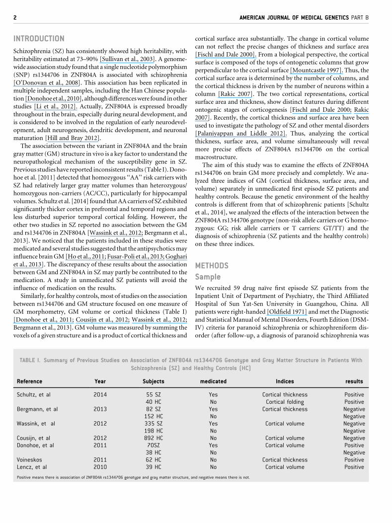
TABLE I. Summary of Previous Studies on Association of ZNF804A
Schizophrenia (SZ) and
Reference Year Subjects
Schultz, et al 2014 55 SZ40 HC
Bergmann, et al 2013 82 SZ152 HC
Wassink, et al 2012 335 SZ198 HC
Cousijn, et al 2012 892 HCDonohoe, et al 2011 70SZ
38 HCVoineskos 2011 62 HCLencz, et al 2010 39 HC
Positive means there is association of ZNF804A rs1344706 genotype and gray matter structure, an
cortical surface area substantially. The change in cortical volume
can not reflect the precise changes of thickness and surface area
[Fischl and Dale 2000]. From a biological perspective, the cortical
surface is composed of the tops of ontogenetic columns that grow
perpendicular to the cortical surface [Mountcastle 1997]. Thus, the
cortical surface area is determined by the number of columns, and
the cortical thickness is driven by the number of neurons within a
column [Rakic 2007]. The two cortical representations, cortical
surface area and thickness, show distinct features during different
ontogenic stages of corticogenesis [Fischl and Dale 2000; Rakic
2007]. Recently, the cortical thickness and surface area have been
used to investigate the pathology of SZ and other mental disorders
[Palaniyappan and Liddle 2012]. Thus, analyzing the cortical
thickness, surface area, and volume simultaneously will reveal
more precise effects of ZNF804A rs1344706 on the cortical
macrostructure.
The aim of this study was to examine the effects of ZNF804A
rs1344706 on brain GM more precisely and completely. We ana-
lyzed three indices of GM (cortical thickness, surface area, and
volume) separately in unmedicated first episode SZ patients and
healthy controls. Because the genetic environment of the healthy
controls is different from that of schizophrenic patients [Schultz
et al., 2014], we analyzed the effects of the interaction between the
ZNF804A rs1344706 genotype (non-risk allele carriers or G homo-
zygous: GG; risk allele carriers or T carriers: GT/TT) and the
diagnosis of schizophrenia (SZ patients and the healthy controls)
on these three indices.
METHODS
SampleWe recruited 59 drug naı̈ve first episode SZ patients from the
Inpatient Unit of Department of Psychiatry, the Third Affiliated
Hospital of Sun Yat-Sen University in Guangzhou, China. All
patients were right-handed [Oldfield 1971] andmet the Diagnostic
and Statistical Manual ofMental Disorders, Fourth Edition (DSM-
IV) criteria for paranoid schizophrenia or schizophreniform dis-
order (after follow-up, a diagnosis of paranoid schizophrenia was
rs1344706 Genotype and Gray Matter Structure in Patients With
Healthy Controls (HC)
medicated Indices results
Yes Cortical thickness PositiveNo Cortical folding PositiveYes Cortical thickness NegativeNo NegativeYes Cortical volume NegativeNo NegativeNo Cortical volume NegativeYes Cortical volume PositiveNo NegativeNo Cortical thickness PositiveNo Cortical volume Positive
d negative means there is not.
WEI ET AL. 3
established; n¼ 22). The DSM-IV diagnosis was performed by an
experienced psychiatrist (QW, 14 years clinical experience) using
the patient version of the StructuredClinical Interview forDSM-IV
Axis IDisorders (SCID-I/P). The severity of psychopathology at the
time of MRI scanning was rated for each patient with SZ according
to the Positive and Negative Syndrome Scale (PANSS) [Kay et al.,
1987]. Family psychiatric history was obtained by interviewing the
patients and their relatives when possible, who provided informa-
tion on family history in details during the clinical interview. This
study adopted the definition of family history as described by Xu
et al. [2008]. Patients with positive family history (PFH) were
defined as having at least one relative with schizophrenia in their
first-degreeor second-degree relatives; otherwise, theywere defined
as patientswithnegative family history (NFH).Wealso recruited 60
age- and gender-matched healthy subjects as the control group.
They were screened for major medical, neurological, or psychiatric
history. None of them had a current or history of mental illness or
first-degree relatives with a psychotic disorder, according to the
non-patient version of SCID. For all participants in this study, the
exclusion criteria were as follows: (i) chronic neurological disor-
ders; (ii) history of alcohol or substance abuse; (iii) history of
electroconvulsive therapy; and (iv) contraindication to MRI scan-
ning. This research was conducted in accordance with the Helsinki
Declaration as revised 1989 and was approved by the Clinical
Research Ethics Committee of the Third Affiliated Hospital of
Sun Yat-Sen University. Informed consent was obtained from
each subject.
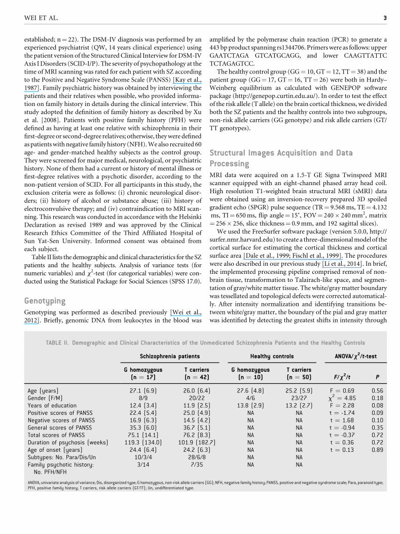
Table II lists the demographic and clinical characteristics for the SZ
patients and the healthy subjects. Analysis of variance tests (for
numeric variables) and x2-test (for categorical variables) were con-
ducted using the Statistical Package for Social Sciences (SPSS 17.0).
GenotypingGenotyping was performed as described previously [Wei et al.,
2012]. Briefly, genomic DNA from leukocytes in the blood was
TABLE II. Demographic and Clinical Characteristics of the Unm
Schizophrenia patients
G homozygous(n ¼ 17)
T carriers(n ¼ 42)
Age (years) 27.1 (6.9) 26.0 (6.4Gender (F/M) 8/9 20/22Years of education 12.4 (3.4) 11.9 (2.5Positive scores of PANSS 22.4 (5.4) 25.0 (4.9Negative scores of PANSS 16.9 (6.3) 14.5 (4.2General scores of PANSS 35.3 (6.0) 36.7 (5.1Total scores of PANSS 75.1 (14.1) 76.2 (8.3Duration of psychosis (weeks) 119.3 (134.0) 101.9 (182Age of onset (years) 24.4 (6.4) 24.2 (6.3Subtypes: No. Para/Dis/Un 10/3/4 28/6/8Family psychotic history:No. PFH/NFH
3/14 7/35
ANOVA, univariate analysis of variance; Dis, disorganized type; G homozygous, non-risk allele carriers (GPFH, positive family history; T carriers, risk allele carriers (GT/TT); Un, undifferentiated type.
amplified by the polymerase chain reaction (PCR) to generate a
443 bpproduct spanning rs1344706. Primerswere as follows: upper
GAATCTAGA GTCATGCAGG, and lower CAAGTTATTC
TCTAGAGTCC.
The healthy control group (GG¼ 10, GT¼ 12, TT¼ 38) and the
patient group (GG¼ 17, GT¼ 16, TT¼ 26) were both in Hardy–
Weinberg equilibrium as calculated with GENEPOP software
package (http://genepop.curtin.edu.au/). In order to test the effect
of the risk allele (T allele) on the brain cortical thickness, we divided
both the SZ patients and the healthy controls into two subgroups,
non-risk allele carriers (GG genotype) and risk allele carriers (GT/
TT genotypes).
Structural Images Acquisition and DataProcessingMRI data were acquired on a 1.5-T GE Signa Twinspeed MRI
scanner equipped with an eight-channel phased array head coil.
High resolution T1-weighted brain structural MRI (sMRI) data
were obtained using an inversion-recovery prepared 3D spoiled
gradient echo (SPGR) pulse sequence (TR¼ 9.568ms, TE¼ 4.132
ms, TI¼ 650ms, flip angle¼ 15˚, FOV¼ 240� 240mm2, matrix
¼ 256� 256, slice thickness¼ 0.9mm, and 192 sagittal slices).
We used the FreeSurfer software package (version 5.0.0, http://
surfer.nmr.harvard.edu) to create a three-dimensionalmodel of the
cortical surface for estimating the cortical thickness and cortical
surface area [Dale et al., 1999; Fischl et al., 1999]. The procedures
were also described in our previous study [Li et al., 2014]. In brief,
the implemented processing pipeline comprised removal of non-
brain tissue, transformation to Talairach-like space, and segmen-
tation of gray/whitematter tissue. The white/graymatter boundary
was tessellated and topological defects were corrected automatical-
ly. After intensity normalization and identifying transitions be-
tween white/gray matter, the boundary of the pial and gray matter
was identified by detecting the greatest shifts in intensity through
edicated Schizophrenia Patients and the Healthy Controls
Healthy controls ANOVA/x2/t-test
G homozygous(n ¼ 10)
T carriers(n ¼ 50) F/x2/t P
) 27.6 (4.8) 25.2 (5.9) F ¼ 0.69 0.564/6 23/27 x2 ¼ 4.85 0.18
) 13.8 (2.9) 13.2 (2.7) F ¼ 2.28 0.08) NA NA t ¼ -1.74 0.09) NA NA t ¼ 1.68 0.10) NA NA t ¼ -0.94 0.35) NA NA t ¼ -0.37 0.72.7) NA NA t ¼ 0.36 0.72) NA NA t ¼ 0.13 0.89
NA NANA NA
G); NFH, negative family history; PANSS, positive and negative syndrome scale; Para, paranoid type;
4 AMERICAN JOURNAL OF MEDICAL GENETICS PART B
surface deformation. The entire cortex for each subject was then
visually inspected and any inaccuracies in segmentation were
manually edited. After creation of the cortical representations,
we estimated cortical measures (cortical thickness, surface area,
and volume) for each of these regions [Fischl et al., 2004]. All of the
reconstructed cortical surfaces were mapped on a common spheri-
cal coordinate system using a spherical transformation. Surface
maps were smoothed with a Gaussian kernel with a full-width-at-
half-maximum (FWHM) of 10mm. Cortical thickness was mea-
sured by calculating the shortest distance from the gray/white
boundary to the gray/CSF boundary and vice versa at each vertex,
and averaging these two values [Fischl andDale 2000]. Estimates of
the cortical surface area were obtained by computing the area
surrounding each vertex on a triangular grid. The cortical volume
was obtained by multiplying the surface area and thickness in each
vertex across the cortical mantle.
Statistical AnalysisAgeneral linearmodel (GLM) controlling for age, gender, and years
of education was used to perform a 2� 2 ANOVA with cortical
thickness, area, and volume as dependent variables. The first factor
was diagnosis (schizophrenia and healthy control), and the second
was the presence/absence of a genotype of ZNF804A rs1344706
(non-risk and risk allele carriers). The right and left hemispheres
were tested separately. A Monte Carlo simulation cluster
analysis with 10,000 iterations and a cluster threshold of
P< 0.05 was adopted to correct for multiple comparisons.
In order to further verify and analyze the influence of inter-
actions between the genotype and a diagnosis of schizophrenia on
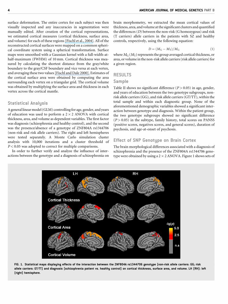
FIG. 1. Statistical maps displaying effects of the interaction between the
allele carriers: GT/TT) and diagnosis (schizophrenia patient vs. healthy c
(right) hemisphere.
brain morphometry, we extracted the mean cortical values of
thickness, area, andvolumeat the significant clusters andquantified
the differences (D) between the non-risk (G homozygous) and risk
(T carriers) allele carriers in the patients with SZ and healthy
controls, respectively, using the following equation:
D ¼ ðMG �MTÞ=MG ð1ÞwhereMG (MT) represents the group averaged cortical thickness, or
area, or volume in thenon-risk allele carriers (risk allele carriers) for
a given region.
RESULTS
SampleTable II shows no significant difference (P> 0.05) in age, gender,
and years of education between the two genotype subgroups, non-
risk allele carriers (GG), and risk allele carriers (GT/TT), within the
total sample and within each diagnostic group. None of the
aforementioned demographic variables showed a significant inter-
action between genotype and diagnosis. Within the patient group,
the two genotype subgroups showed no significant difference
(P> 0.05) in the subtype, family history, total scores on PANSS
(positive scores, negatives scores, and general scores), duration of
psychosis, and age-at-onset of psychosis.
Effect of SNP Genotype on Brain CortexThe brain morphological differences associated with a diagnosis of
schizophrenia and the presence of the ZNF804A rs1344706 geno-
type were obtained by using a 2� 2 ANOVA. Figure 1 shows sets of
ZNF804A rs1344706 genotype (non-risk allele carriers: GG; risk
ontrol) on cortical thickness, surface area, and volume. LH (RH): left
WEI ET AL. 5
p-maps to indicate a significant interaction between diagnosis and
genotype on the cortical thickness, surface area, and volume,
separately.
Cortical thickness. We detected a significant effect of the
interaction between the genotype and the diagnosis of schizophre-
nia on cortical thickness in the following four clusters with their
corresponding cluster-wise probabilities (CWP) (Fig. 1 and
Table III), the left precuneus, superior parietal (CWP¼ 0.0001);
the left precentral cortex (CWP¼ 0.0001); the left superior tem-
poral, inferior parietal cortex (CWP¼ 0.0040); and the right pre-
cuneus, superior parietal and paracentral gyrus (CWP¼ 0.0001).
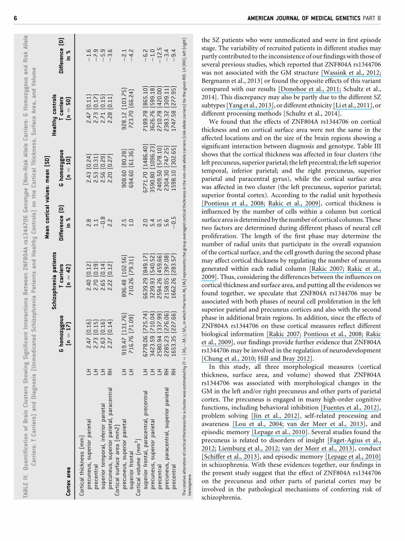
Table IV lists the extracted mean cortical thickness in the four
aforementioned clusters, in which the interaction between diagno-
sis and genotype was significant. The results showed that compared
to non-risk allele carriers, the risk allele carriers had thinner cortical
thickness in the bilateral precuneus (left: D¼ 2.8%; right:
D¼ 2.2%) and left precentral gyrus (D¼ 1.1%) in the patients
with SZ. But the healthy controls with risk allele carriers (GT/TT)
showed a thicker cortical thickness in those clusters (left precuneus:
D¼�1.6%; right precuneus: D¼�3.6%; left precentral: D¼�7.9%) than did the healthy non-risk allele carriers (GG).
Cortical surface area. As for cortical surface areas, we detected
significant interactions between genotype andadiagnosis of schizo-
phrenia in these clusters, the left precuneus and superior parietal
cortex (CWP¼ 0.0020), the left superior frontal cortex (CWP
¼ 0.0124) (Fig. 1, Table III). From Table IV, we can see that in
the SZ patients, the risk allele carriers had a decreased cortical
surface area in these two clusters (left precuneus: D¼ 2.5%; left
superior frontal: D¼ 1.0%), but in the healthy controls, the risk
allele carriers had an increased cortical surface area,whencompared
to the non-risk allele carriers.
Cortical volume. Figure 1 also shows an interaction effect
between genotype and a diagnosis of schizophrenia on the cortical
volume in five clusters. They were located in the left superior
TABLE III. Clusters Showing Significant Interaction Between the ZNF8
Carriers) and Diagnosis (Unmedicated Schizophrenia Patients and HSepara
Cortex areaTa
Cortical thickness (mm)Precuneus, superior parietal LHPrecentral LHSuperior temporal, inferior parietal LHPrecuneus, superior parietal, paracentral RH
Cortical surface area (mm2)Precuneus, superior parietal LHSuperior frontal LH
Cortical volume (mm3)Superior frontal, paracentral, precentral LHPrecuneus, superior parietal LHPrecentral LHPrecuneus, paracentral, superior parietal RHPrecuneus, superior parietal RH
LH (RH), left (right) hemisphere; CWP, cluster-wise probability.
frontal, paracentral, and precentral gyrus (CWP¼ 0.0001); the
left precuneus and superior parietal gyrus (CWP¼ 0.0010); the
left precentral gyrus (CWP¼ 0.0068); the right precuneus, superior
parietal, and paracentral gyrus (CWP¼ 0.0208); and the right
precuneus and superior parietal cortex (CWP¼ 0.0385). The
details of these five clusters are listed in Table III.
An ROI-wise analysis was performed to determine the differ-
ences between the risk allele carriers and non-risk allele carriers for
the five clusters (Table IV). In the patients, we found that the risk
allele carriers had a smaller cortical volume in four clusters, the left
superior frontal (D¼ 2.0%), left precuneus (D¼ 5.4%), left pre-
central (D¼ 0.5%), and right precuneus (D¼ 5.6%), compared to
non-risk allele carriers. However, in the healthy controls, we
detected an increased cortical volume for these four brain regions
in the risk allele carriers compared to non-risk allele carriers (see
Table IV for details).
DISCUSSION
The present study used sMRI to explore the potential association of
the schizophrenia risk gene variant ZNF804A rs1344706 with the
cortical thickness, surface area, and volume in unmedicated first
episode SZ patients and a healthy population. We found that the
risk allele (T) was associated with thinner cortical thickness, less
cortical surface area, and a smaller cortical volume in multiple
regions in the SZ patients, whereas the opposite effect was observed
in the healthy controls, i.e., the risk allele (T) was associated with
thicker cortical thickness, larger cortical surface area, and a bigger
cortical volume in these regions.
It is important to note that antipsychoticsmay cause the increase
or decrease of brain GM as several studies suggested antipsychotics
is an important confounding factor in neuroimaging studies of SZ
[Ho et al., 2011; Fusar-Poli et al., 2013; Goghari et al., 2013].
Different from previous studies, this case-controls study recruited
04A rs1344706 Genotype (Non-Risk Allele Carriers and Risk Allele
ealthy Controls) on Cortical Thickness, Surface Area, and Volume,tely
lairach coordinates(x, y, z) Cluster size (mm2) CWP
(�17, �45, 55) 2742.3 0.0001(�53, �7, 39) 1445.8 0.0001(�44, �54, 22) 1053.8 0.0040(8, �60, 57) 1968.0 0.0001
(�16, �67, 59) 931.9 0.0020(�7, 34, 50) 736.0 0.0124
(�7, 34, 50) 2201.1 0.0001(�8, �73, 46) 1133.3 0.0010(�53, �7, 39) 902.5 0.0068(8, �59, 57) 784.6 0.0208(39, �10, 61) 699.2 0.0385
TABLE
IV.Quantification
ofBrain
Clusters
Show
ingSignificantInteractionsBetweenZN
F804Ars1344706Genotype(Non-RiskAllele
Carriers:
GHom
ozygousandRiskAllele
Carriers:TCarriers)andDiagnosis
(Unmedicated
Schizophrenia
Patients
andHealthy
Controls)
ontheCortical
Thickness,
SurfaceArea,andVolume
Meancortical
values:mean(SD)
Schizophrenia
patients
Healthycontrols
Cortex
area
Ghomozygous
(n¼
17)
Tcarriers
(n¼
42)
Difference
(D)
in%
Ghom
ozygous
(n¼
10)
Tcarriers
(n¼
50)
Difference
(D)
in%
Cortical
thickness(m
m)
precuneus,superior
parietal
LH2.47(0.16)
2.40(0.12)
2.8
2.43(0.24)
2.47(0.11)
�1.6
precentral
LH2.73(0.15)
2.70(0.19)
1.1
2.53(0.31)
2.73(0.17)
�7.9
superior
temporal,inferior
parietal
LH2.63(0.16)
2.65(0.14)
�0.8
2.56(0.29)
2.71(0.15)
�5.9
precuneus,superior
parietal,paracentral
RH
2.27(0.14)
2.22(0.12)
2.2
2.20(0.27)
2.28(0.11)
�3.6
Cortical
surfacearea
(mm2)
precuneus,superior
parietal
LH919.47(131.76)
896.48(102.56)
2.5
908.60(80.28)
928.12(103.75)
�2.1
superior
frontal
LH716.76(71.09)
710.26(79.31)
1.0
694.60(61.36)
723.70(66.24)
�4.2
Cortical
volume(m
m3)
superior
frontal,paracentral,precentral
LH6778.06(725.74)
6639.29(849.17)
2.0
6771.70(1486.40)
7189.78(865.10)
�6.2
precuneus,superior
parietal
LH3423.59(710.04)
3239.93(540.52)
5.4
3590.80(1096.23)
3626.76(599.18)
�1.0
precentral
LH2580.94(337.99)
2594.26(459.66)
0.5
2409.50(578.10)
2710.78(420.00)
�12.5
precuneus,paracentral,superior
parietal
RH
2285.23(276.06)
2158.05(397.08)
5.6
2304.30(747.25)
2383.32(309.11)
�3.4
precentral
RH
1653.35(227.66)
1662.26(283.57)
�0.5
1598.10(302.65)
1747.58(277.95)
�9.4
Therelative
alteration
ofcorticalthicknessfora
clusterw
asestimated
byD
¼ðM
G�M
TÞ=M
G,inwhich
theterm
MG(M
T)representsthegroupaveraged
corticalthicknessinthenon-riskallelecarriers(riskallelecarriers)forthe
givenROI.LH
(RH),left(right)
hemisphere.
6 AMERICAN JOURNAL OF MEDICAL GENETICS PART B
the SZ patients who were unmedicated and were in first episode
stage. The variability of recruited patients in different studies may
partly contributed to the inconsistence of our findingswith those of
several previous studies, which reported that ZNF804A rs1344706
was not associated with the GM structure [Wassink et al., 2012;
Bergmann et al., 2013] or found the opposite effects of this variant
compared with our results [Donohoe et al., 2011; Schultz et al.,
2014]. This discrepancy may also be partly due to the different SZ
subtypes [Yang et al., 2013], or different ethnicity [Li et al., 2011], or
different processing methods [Schultz et al., 2014].
We found that the effects of ZNF804A rs1344706 on cortical
thickness and on cortical surface area were not the same in the
affected locations and on the size of the brain regions showing a
significant interaction between diagnosis and genotype. Table III
shows that the cortical thickness was affected in four clusters (the
left precuneus, superior parietal; the left precentral; the left superior
temporal, inferior parietal; and the right precuneus, superior
parietal and paracentral gyrus), while the cortical surface area
was affected in two cluster (the left precuneus, superior parietal;
superior frontal cortex). According to the radial unit hypothesis
[Pontious et al., 2008; Rakic et al., 2009], cortical thickness is
influenced by the number of cells within a column but cortical
surface area is determinedby thenumber of cortical columns.These
two factors are determined during different phases of neural cell
proliferation. The length of the first phase may determine the
number of radial units that participate in the overall expansion
of the cortical surface, and the cell growth during the second phase
may affect cortical thickness by regulating the number of neurons
generated within each radial column [Rakic 2007; Rakic et al.,
2009]. Thus, considering the differences between the influences on
cortical thickness and surface area, and putting all the evidences we
found together, we speculate that ZNF804A rs1344706 may be
associated with both phases of neural cell proliferation in the left
superior parietal and precuneus cortices and also with the second
phase in additional brain regions. In addition, since the effects of
ZNF804A rs1344706 on these cortical measures reflect different
biological information [Rakic 2007; Pontious et al., 2008; Rakic
et al., 2009], our findings provide further evidence that ZNF804A
rs1344706 may be involved in the regulation of neurodevelopment
[Chung et al., 2010; Hill and Bray 2012].
In this study, all three morphological measures (cortical
thickness, surface area, and volume) showed that ZNF804A
rs1344706 was associated with morphological changes in the
GM in the left and/or right precuneus and other parts of parietal
cortex. The precuneus is engaged in many high-order cognitive
functions, including behavioral inhibition [Fuentes et al., 2012],
problem solving [Jin et al., 2012], self-related processing and
awareness [Lou et al., 2004; van der Meer et al., 2013], and
episodic memory [Lepage et al., 2010]. Several studies found the
precuneus is related to disorders of insight [Faget-Agius et al.,
2012; Liemburg et al., 2012; van der Meer et al., 2013], conduct
[Schiffer et al., 2013], and episodic memory [Lepage et al., 2010]
in schizophrenia. With these evidences together, our findings in
the present study suggest that the effect of ZNF804A rs1344706
on the precuneus and other parts of parietal cortex may be
involved in the pathological mechanisms of conferring risk of
schizophrenia.

WEI ET AL. 7
We also found that the risk allele (T) in ZNF804A rs1344706,
compared to the non-risk allele (G), was associated with a smaller
cortical surface area in the left superior frontal, and with a lower
cortical volume in the left superior frontal in schizophrenia.
Previous studies also showed that ZNF804A s1344706 was associ-
ated with the prefrontal cortex [Esslinger et al., 2009; Nenadic et al.,
2015; Schultz et al., 2014]. Many studies [Glahn et al., 2008] have
demonstrated that the frontal cortex is associated with the neuro-
pathological mechanism of schizophrenia. About the temporal
cortex, we found that ZNF804A rs1344706 was associated with
the cortical thickness in the left superior temporal in theSZpatients.
This result is consistentwith several previous studies,which showed
that ZNF804A s1344706 was associated with the temporal cortex
[Mohnke et al., 2014; Nenadic et al., 2015]. Temporal cortex also
has been showed to be involved in the neuropathological mecha-
nismof SZ[Sun et al., 2009].Thus, the effect ofZNF804Ars1344706
on the prefrontal and temporal cortices may be also involved in the
pathological mechanisms of conferring risk of schizophrenia.
In addition, we found that the effect of ZNF804A rs1344706 on
the brain GM in the healthy controls was opposite to that in SZ
patients. That is, the risk allele was associated with significantly
decreased brain structural measures (thickness, surface area, and
volume) in the SZ patients, but with the significantly increased
brain structural measures in the healthy controls, in the precuneus,
parietal, frontal and temporal cortices (Table III). This results are
partly consistent with the previous finding about the differential
effects of rs1344706 on gray matter [Schultz et al., 2014] or white
matter [Wei et al., 2012] in healthy controls and SZ patients. This
different effect of ZNF804A rs1344706 on the brain GM in the SZ
and in the controls may be caused from variability of subjects or
their different risk genetic environments [Prata et al., 2009; Nic-
odemus et al., 2010]. For example, an exonic SNP rs12476147,
located in exon four of ZNF804A, was shown significantly associa-
tionwith SZ and allelic expression imbalance in theDLPFC, using a
powerful within-subject design [Guella et al., 2014]. In the same
study [Guella et al., 2014], the rs1344706 SZ risk allelewas found the
cis-regulatory variant directly responsible for this allelic expression
imbalance in adult cortex. Taken together, the different effects of
rs1344706 on the GM in SZ and control groups may be involved in
neuropathological mechanisms of this risk variant rs1344706 con-
ferring risk to schizophrenia.
There are several limitations to be considered in the present
study. First, to avoid the confounding factors of antipsychotics, we
included the unmedicated SZ patients only, and the sample size was
relative smaller. Second, we analyzed rs1344706 only, while other
SNPs or haplotypes in the same gene or in other genes interacting
with ZNF804A may also be relevant. Third, the brain structural
imageswere acquiredon a1.5GEMRI scanner and the SNRwas low
which may affect the accuracy of our findings. To minimize the
effect of low SNR on the calculations, we have screened the brain
structural images, and selected these images had a high gray/white
matter contrast into the data analysis. Thismay ensure the accuracy
of segmentation of gray and white matter. No doubt, the future
studies need to consider a larger sample size, to genotype other
SNPsorhaplotypes in the samegeneor inother related genes, and to
acquire relative higher SNR images by using a high- strength MRI
scanner or by repeating MRI scans. Therefore, the future studies
need to consider a larger sample size, to genotype other SNPs or
haplotypes in the same gene or in other related genes, and to acquire
relative higher SNR images byusing ahigh-strengthMRI scanner or
by repeating MRI scans.
In summary, for the first time, we investigated the effects of
ZNF804A rs1344706 on the cortical morphometry in unmedicated
SZ patients and healthy controls and found that the effects of the
risk genetic variants (T) on the microstructure of the GM in
precuneus, parietal, prefrontal, and temporal cortex in the schizo-
phrenic patients were opposite to those found in the healthy
controls. Our findings suggest that ZNF804A rs1344706 may
aggravate the risk for schizophrenia by exerting its effects on cortical
thickness, surface area, and cortical volume in these brain regions.
ACKNOWLEDGMENTS
The research was supported by grants from the Natural Science
Foundation of China (grant Nos. 81071093, 81101028, 81271548,
81371535, 81428013, and 81471654), The Project Supported by
Guangdong Natural Science Foundation (grant Nos.
S2012010009027), Science and Technology Planning Project of
Guangdong Province (grant Nos. 2011B031800073,
2011B031800101, 2012B031800054, 2013B021800085), the Funda-
mental Research Funds for the Central University (14ykpy28).
We thank Dr. Haihong Liu, from the Institute of Mental Health
of Second Xiangya Hospital, Central South University, for his
assistance in MRI data collection.
REFERENCES
BergmannO,HaukvikUK, BrownAA, Rimol LM,HartbergCB,AthanasiuL,Melle I,Djurovic S,AndreassenOA,DaleAM,Agartz I. 2013.ZNF804Aand cortical thickness in schizophrenia and bipolar disorder. PsychiatryRes 212(2):154–157.
Chung HJ, Lee JY, Deocaris CC, Min H, Kim SH, Kim MH. 2010. Mousehomologue of the schizophrenia susceptibility gene ZNF804A as a targetof hoxc8. J Biomed Biotechnol 2010:231708.
CousijnH, RijpkemaM,Harteveld A,Harrison PJ, Fernandez G, Franke B,Arias-Vasquez A. 2012. Schizophrenia risk gene ZNF804A does notinfluence macroscopic brain structure: An MRI study in 892 volunteers.Mol Psychiatry 17(12):1155–1157.
Dale AM, Fischl B, Sereno MI. 1999. Cortical surface-based analysis. I.Segmentation and surface reconstruction. Neuroimage 9(2):179–194.
DonohoeG,Morris DW,CorvinA. 2010. The psychosis susceptibility geneZNF804A: Associations, functions, and phenotypes. Schizophr Bull 36-(5):904–909.
DonohoeG,RoseE, FrodlT,MorrisD, Spoletini I, Adriano F,Bernardini S,Caltagirone C, Bossu P, Gill M, Corvin AP, Spalletta G. 2011. ZNF804Arisk allele is associated with relatively intact gray matter volume inpatients with schizophrenia. Neuroimage 54(3):2132–2137.
EsslingerC,WalterH,Kirsch P, Erk S, Schnell K, ArnoldC,HaddadL,MierD, Opitz Von Boberfeld C, Raab K, Witt SH, Rietschel M, Cichon S,Meyer-Lindenberg A. 2009. Neural mechanisms of a genome-widesupported psychosis variant. Science 324(5927):605.
Faget-Agius C, Boyer L, Padovani R, Richieri R, Mundler O, Lancon C,Guedj E. 2012. Schizophrenia with preserved insight is associated withincreased perfusion of the precuneus. J Psychiatry Neurosci 37(5):297–304.

8 AMERICAN JOURNAL OF MEDICAL GENETICS PART B
Fischl B, Dale AM. 2000. Measuring the thickness of the human cerebralcortex from magnetic resonance images. Proc Natl Acad Sci USA 97-(20):11050–11055.
Fischl B, Sereno MI, Dale AM. 1999. Cortical surface-based analysis. II:Inflation, flattening, and a surface-based coordinate system.Neuroimage9(2):195–207.
Fischl B, van der Kouwe A, Destrieux C, Halgren E, Segonne F, Salat DH,BusaE, SeidmanLJ,Goldstein J,KennedyD,CavinessV,MakrisN,RosenB, Dale AM. 2004. Automatically parcellating the human cerebral cortex.Cereb Cortex 14(1):11–22.
Fuentes P, Barros-Loscertales A, Bustamante JC, Rosell P, Costumero V,Avila C. 2012. Individual differences in the Behavioral Inhibition Systemare associated with orbitofrontal cortex and precuneus gray mattervolume. Cogn Affect Behav Neurosci 12(3):491–498.
Fusar-Poli P, Smieskova R, Kempton MJ, Ho BC, Andreasen NC, Borg-wardt S. 2013. Progressive brain changes in schizophrenia related toantipsychotic treatment? A meta-analysis of longitudinal MRI studies.Neurosci Biobehav Rev 37(8):1680–1691.
Glahn DC, Laird AR, Ellison-Wright I, Thelen SM, Robinson JL, LancasterJL, Bullmore E, Fox PT. 2008. Meta-analysis of gray matter anomalies inschizophrenia: Application of anatomic likelihood estimation and net-work analysis. Biol Psychiatry 64(9):774–781.
Goghari VM, Smith GN, Honer WG, Kopala LC, Thornton AE, Su W,Macewan GW, Lang DJ. 2013. Effects of eight weeks of atypical antipsy-chotic treatment on middle frontal thickness in drug-naive first-episodepsychosis patients. Schizophr Res 149(1–3):149–155.
Guella I, Sequeira A, Rollins B, Morgan L, Myers RM, Watson SJ, Akil H,Bunney WE, Delisi LE, Byerley W, Vawter MP. 2014. Evidence of allelicimbalance in the schizophrenia susceptibility gene ZNF804A in humandorsolateral prefrontal cortex. Schizophr Res 152(1):111–116.
Hill MJ, Bray NJ. 2012. Evidence that schizophrenia risk variation in theZNF804A gene exerts its effects during fetal brain development. Am JPsychiatry 169(12):1301–1308.
Ho BC, Andreasen NC, Ziebell S, Pierson R,Magnotta V. 2011. Long-termantipsychotic treatment and brain volumes: A longitudinal study of first-episode schizophrenia. Arch Gen Psychiatry 68(2):128–137.
JinG,LiK,QinY,ZhongN,ZhouH,WangZ,Xiang J,HuY,WangM,ZengQ. 2012. FMRI study in posterior cingulate and adjacent precuneuscortex in healthy elderly adults using problem solving task. J Neurol Sci318(1–2):135–139.
Kay SR, Fiszbein A, Opler LA. 1987. The positive and negative syndromescale (PANSS) for schizophrenia. Schizophr Bull 13(2):261–276.
Lepage M, Pelletier M, Achim A, Montoya A, Menear M, Lal S. 2010.Parietal cortex and episodic memory retrieval in schizophrenia. Psychi-atry Res 182(3):191–199.
Li M, Luo XJ, Xiao X, Shi L, Liu XY, Yin LD, Diao HB, Su B. 2011. Allelicdifferences between Han Chinese and Europeans for functional variantsin ZNF804A and their association with schizophrenia. Am J Psychiatry168(12):1318–1325.
Li M, Shi CJ, Shi YY, Luo XJ, Zheng XB, Li ZQ, Liu JJ, Chong SA, Lee J,Wang Y, Liu XY, Yin LD, Pu XF, Diao HB, Xu Q, Su B. 2012. ZNF804Aand schizophrenia susceptibility inAsianpopulations. AmJMedGenet BNeuropsychiatr Genet 159B(7) 794–802.
LiM,Tian J,ZhangR,QiuY,WenX,MaX,Wang J,XuY, JiangG,HuangR.2014. Abnormal cortical thickness in heroin-dependent individuals.Neuroimage 88:295–307.
Liemburg EJ, van der Meer L, Swart M, Curcic-Blake B, Bruggeman R,Knegtering H, Aleman A. 2012. Reduced connectivity in the self-proc-essing network of schizophrenia patients with poor insight. PLoS ONE7(8):e42707.
Lou HC, Luber B, Crupain M, Keenan JP, Nowak M, Kjaer TW, SackeimHA, Lisanby SH. 2004. Parietal cortex and representation of the mentalSelf. Proc Natl Acad Sci USA 101(17):6827–6832.
Mohnke S, Erk S, Schnell K, Schutz C, Romanczuk-Seiferth N, Grimm O,Haddad L, Pohland L, GarbusowM, SchmitgenMM, Kirsch P, EsslingerC, Rietschel M, Witt SH, Nothen MM, Cichon S, Mattheisen M,Muhleisen T, Jensen J, Schott BH,MaierW,Heinz A,Meyer-LindenbergA, Walter H. 2014. Further evidence for the impact of a genome-wide-supported psychosis risk variant in ZNF804A on the Theory of MindNetwork. Neuropsychopharmacology 39(5):1196–1205.
Mountcastle VB. 1997. The columnar organization of the neocortex. Brain120(Pt 4) 701–722.
Nenadic I, Maitra R, Basmanav FB, Schultz CC, Lorenz C, Schachtzabel C,Smesny S, Nothen MM, Cichon S, Reichenbach JR, Sauer H,Schlosser RG, Gaser C. 2015. ZNF804A genetic variation (rs1344706)affects brain grey but not white matter in schizophrenia and healthysubjects. Psychol Med 45(1):143–152.
Nicodemus KK, LawAJ, Radulescu E, Luna A, Kolachana B, Vakkalanka R,Rujescu D, Giegling I, Straub RE, Mcgee K, Gold B, Dean M, Muglia P,Callicott JH, Tan HY, Weinberger DR. 2010. Biological validation ofincreased schizophrenia risk with NRG1, ERBB4, and AKT1 epistasis viafunctional neuroimaging in healthy controls. Arch Gen Psychiatry 67-(10):991–1001.
O’DonovanMC, Craddock N, Norton N,Williams H, Peirce T, MoskvinaV, Nikolov I, HamshereM, Carroll L, Georgieva L, Dwyer S, Holmans P,Marchini JL, Spencer CC, Howie B, Leung HT, Hartmann AM, MollerHJ, Morris DW, Shi Y, Feng G, Hoffmann P, Propping P, Vasilescu C,MaierW, RietschelM, Zammit S, Schumacher J, Quinn EM, Schulze TG,Williams NM, Giegling I, Iwata N, Ikeda M, Darvasi A, Shifman S, He L,Duan J, Sanders AR, Levinson DF, Gejman PV, Cichon S, Nothen MM,GillM, Corvin A, RujescuD, KirovG,OwenMJ, BuccolaNG,Mowry BJ,Freedman R, Amin F, Black DW, Silverman JM, Byerley WF, CloningerCR. 2008. Identificationof loci associatedwith schizophrenia by genome-wide association and follow-up. Nat Genet 40(9):1053–1055.
Oldfield RC. 1971. The assessment and analysis of handedness: TheEdinburgh inventory. Neuropsychologia 9(1):97–113.
Palaniyappan L, Liddle PF. 2012. Differential effects of surface area,gyrification and cortical thickness on voxel based morphometric deficitsin schizophrenia. Neuroimage 60(1):693–699.
Pontious A, Kowalczyk T, Englund C, Hevner RF. 2008. Role of interme-diate progenitor cells in cerebral cortex development. Dev Neurosci30(1–3):24–32.
Prata DP, Mechelli A, Fu CH, Picchioni M, Toulopoulou T, Bramon E,Walshe M, Murray RM, Collier DA, Mcguire P. 2009. Epistasis betweenthe DAT 3’ UTR VNTR and the COMT Val158Met SNP on corticalfunction in healthy subjects and patients with schizophrenia. Proc NatlAcad Sci USA 106(32):13600–13605.
Rakic P. 2007. The radial edifice of cortical architecture: Fromneuronal silhouettes to genetic engineering. Brain Res Rev 55-(2):204–219.
Rakic P, Ayoub AE, Breunig JJ, Dominguez MH. 2009. Decision bydivision: Making cortical maps. Trends Neurosci 32(5):291–301.
Schiffer B, Leygraf N, Muller BW, Scherbaum N, Forsting M, Wiltfang J,Gizewski ER, Hodgins S. 2013. Structural brain alterations associatedwith schizophrenia preceded by conduct disorder: A common anddistinct subtype of schizophrenia? Schizophr Bull 39(5):1115–1128.
Schultz CC, Nenadic I, Riley B, Vladimirov VI, Wagner G, Koch K,Schachtzabel C, Muhleisen TW, Basmanav B, Nothen MM, Deufel T,Kiehntopf M, Rietschel M, Reichenbach JR, Cichon S, Schlosser RG,Sauer H. 2014. ZNF804A and cortical structure in schizophrenia: In vivoand postmortem studies. Schizophr Bull 40(3):532–541.

WEI ET AL. 9
Sullivan PF, Kendler KS,NealeMC. 2003. Schizophrenia as a complex trait:Evidence from a meta-analysis of twin studies. Arch Gen Psychiatry60(12):1187–1192.
Sun J, Maller JJ, Guo L, Fitzgerald PB. 2009. Superior temporal gyrusvolume change in schizophrenia: A review on region of interest volu-metric studies. Brain Res Rev 61(1):14–32.
van der Meer L, de Vos AE, Stiekema AP, Pijnenborg GH, van Tol MJ,Nolen WA, David AS, Aleman A. 2013. Insight in schizophrenia: In-volvement of self-reflection networks? Schizophr Bull 39(6):1288–1295.
Wassink TH, Epping EA, Rudd D, Axelsen M, Ziebell S, Fleming FW,Monson E, Ho BC, Andreasen NC. 2012. Influence of ZNF804a on brainstructure volumes and symptom severity in individuals with schizophre-nia. Arch Gen Psychiatry 69(9):885–892.
Wei Q, Kang Z, Diao F, Shan B, Li L, Zheng L, Guo X, Liu C, Zhang J,Zhao J. 2012. Association of the ZNF804A gene polymorphismrs1344706 with white matter density changes in Chinese schizo-phrenia. Prog Neuropsychopharmacol Biol Psychiatry 36(1):122–127.
Xu B, Roos JL, Levy S, van Rensburg EJ, Gogos JA, Karayiorgou M. 2008.Strong association of de novo copy number mutations with sporadicschizophrenia. Nat Genet 40(7):880–885.
Yang Y, Li W, Yang G, Xiao B, Wang X, Ding M, Zhao J, Song X, Yue W,ZhangD,ZhangH, LvL. 2013. Evaluation of the relationship between theZNF804A single nucleotide polymorphism rs1344706 A/C variant andschizophrenia subtype in Han Chinese patients. Int J Psychiatry Med45(3):269–278.