Toward a brain-based theory of beauty
10
Toward A Brain-Based Theory of Beauty Tomohiro Ishizu, Semir Zeki* Wellcome Laboratory of Neurobiology and Wellcome Department of Imaging Neuroscience, University College London, London, United Kingdom Abstract We wanted to learn whether activity in the same area(s) of the brain correlate with the experience of beauty derived from different sources. 21 subjects took part in a brain-scanning experiment using functional magnetic resonance imaging. Prior to the experiment, they viewed pictures of paintings and listened to musical excerpts, both of which they rated on a scale of 1–9, with 9 being the most beautiful. This allowed us to select three sets of stimuli–beautiful, indifferent and ugly–which subjects viewed and heard in the scanner, and rated at the end of each presentation. The results of a conjunction analysis of brain activity showed that, of the several areas that were active with each type of stimulus, only one cortical area, located in the medial orbito-frontal cortex (mOFC), was active during the experience of musical and visual beauty, with the activity produced by the experience of beauty derived from either source overlapping almost completely within it. The strength of activation in this part of the mOFC was proportional to the strength of the declared intensity of the experience of beauty. We conclude that, as far as activity in the brain is concerned, there is a faculty of beauty that is not dependent on the modality through which it is conveyed but which can be activated by at least two sources–musical and visual–and probably by other sources as well. This has led us to formulate a brain-based theory of beauty. Citation: Ishizu T, Zeki S (2011) Toward A Brain-Based Theory of Beauty. PLoS ONE 6(7): e21852. doi:10.1371/journal.pone.0021852 Editor: Eric James Warrant, Lund University, Sweden Received March 17, 2011; Accepted June 7, 2011; Published July 6, 2011 Copyright: ß 2011 Ishizu, Zeki. This is an open-access article distributed under the terms of the Creative Commons Attribution License, which permits unrestricted use, distribution, and reproduction in any medium, provided the original author and source are credited. Funding: This work was supported by the Wellcome Trust, London. The funders had no role in study design, data collection and analysis, decision to publish, or preparation of the manuscript. Competing Interests: The authors have declared that no competing interests exist. * E-mail: [email protected] Introduction In the work reported here, we address a question that has been addressed many times over past centuries, namely what constitutes beauty. The question was especially well formulated, in a neurobiologically accessible way, by Edmund Burke. In his Philosophical Enquiry into the Origin of Our Ideas of the Sublime and Beautiful, Burke wrote that ‘‘Beauty is, for the greater part, some quality in bodies acting mechanically upon the human mind by the intervention of the senses’’ [1]. That definition suggests that there is a unique faculty of beauty that can be stimulated by any and all the senses. It thus raises an important question: would the experience of beauty derived from different senses, say the visual and auditory, correlate with activity in the same or different brain areas? If the latter, then the clear implication would be that brain systems that correlate with the experience of beauty are functionally specialized, the experience of visual beauty correlating with activity in one area or set of areas and that of auditory beauty correlating with another. But our reading of the relevant humanistic literature, too numerous to mention, suggests that the first alternative has been more favored by those who have discoursed on the subject, namely that there is a single faculty of beauty into which different senses feed. This alternative is reflected in Burke’s definition. We thus sought to learn something, however small, about how brain activity might be organized during the experience of beauty. Burke and many others, including Immanuel Kant, Anthony Ashley-Cooper (3 rd Earl of Shaftesbury) and Joseph Addison, distinguished between the beautiful and the sublime, the latter having for them characteristics such as ‘‘awe’’, ‘‘horror’’, ‘‘disgust’’ and ‘‘fear’’. In this work we are concerned with the beautiful alone, not the sublime. We undertook a human brain imaging experiment, using functional magnetic resonance imaging (fMRI), in which we asked subjects to view pictures of paintings and listen to brief musical excerpts and rate them according to how beautiful they seemed, while we imaged the activity in their brains. As a working hypothesis, we inclined more towards our neurobiological understanding of Burke’s definition and supposed that there would be a single area or set of areas whose activity would correlate with the experience of beauty, regardless of whether it was derived from an auditory or visual source. Previous work from this and other laboratories [2–6] has implicated activity in the mOFC-an acknowledged pleasure and reward center in the brain [7]-during the experience of visual or musical beauty but no equivalent study for the experience of beauty derived from two different senses in the same subjects has been reported. This is important, since the mOFC is a large expanse of cortex and different but sometimes overlapping parts of it have been activated by different tasks [5,8]. We hypothesized that activity in the same part of mOFC would correlate with beauty in the more abstract sense, that is to say, regardless of whether it is derived from the auditory or visual sense. This turned out to be so and led us to formulate a brain-based theory of beauty. Materials and Methods Subjects 21 healthy right-handed volunteers (9 male, 12 female, mean age 27.5 years) participated in this study. All had normal or corrected-to-normal vision, and none had a history of neurological or psychiatric disorder. Written informed consent was obtained from all, and the study was approved by the Ethics Committee of the Institute of Neurology. All data was anonymized. Subjects PLoS ONE | www.plosone.org 1 July 2011 | Volume 6 | Issue 7 | e21852
-
Upload
independent -
Category
Documents
-
view
3 -
download
0
Transcript of Toward a brain-based theory of beauty

Toward A Brain-Based Theory of BeautyTomohiro Ishizu, Semir Zeki*
Wellcome Laboratory of Neurobiology and Wellcome Department of Imaging Neuroscience, University College London, London, United Kingdom
Abstract
We wanted to learn whether activity in the same area(s) of the brain correlate with the experience of beauty derived fromdifferent sources. 21 subjects took part in a brain-scanning experiment using functional magnetic resonance imaging. Priorto the experiment, they viewed pictures of paintings and listened to musical excerpts, both of which they rated on a scale of1–9, with 9 being the most beautiful. This allowed us to select three sets of stimuli–beautiful, indifferent and ugly–whichsubjects viewed and heard in the scanner, and rated at the end of each presentation. The results of a conjunction analysis ofbrain activity showed that, of the several areas that were active with each type of stimulus, only one cortical area, located inthe medial orbito-frontal cortex (mOFC), was active during the experience of musical and visual beauty, with the activityproduced by the experience of beauty derived from either source overlapping almost completely within it. The strength ofactivation in this part of the mOFC was proportional to the strength of the declared intensity of the experience of beauty.We conclude that, as far as activity in the brain is concerned, there is a faculty of beauty that is not dependent on themodality through which it is conveyed but which can be activated by at least two sources–musical and visual–and probablyby other sources as well. This has led us to formulate a brain-based theory of beauty.
Citation: Ishizu T, Zeki S (2011) Toward A Brain-Based Theory of Beauty. PLoS ONE 6(7): e21852. doi:10.1371/journal.pone.0021852
Editor: Eric James Warrant, Lund University, Sweden
Received March 17, 2011; Accepted June 7, 2011; Published July 6, 2011
Copyright: � 2011 Ishizu, Zeki. This is an open-access article distributed under the terms of the Creative Commons Attribution License, which permitsunrestricted use, distribution, and reproduction in any medium, provided the original author and source are credited.
Funding: This work was supported by the Wellcome Trust, London. The funders had no role in study design, data collection and analysis, decision to publish, orpreparation of the manuscript.
Competing Interests: The authors have declared that no competing interests exist.
* E-mail: [email protected]
Introduction
In the work reported here, we address a question that has been
addressed many times over past centuries, namely what constitutes
beauty. The question was especially well formulated, in a
neurobiologically accessible way, by Edmund Burke. In his
Philosophical Enquiry into the Origin of Our Ideas of the Sublime and
Beautiful, Burke wrote that ‘‘Beauty is, for the greater part, some
quality in bodies acting mechanically upon the human mind by
the intervention of the senses’’ [1]. That definition suggests that
there is a unique faculty of beauty that can be stimulated by any
and all the senses. It thus raises an important question: would the
experience of beauty derived from different senses, say the visual
and auditory, correlate with activity in the same or different brain
areas? If the latter, then the clear implication would be that brain
systems that correlate with the experience of beauty are
functionally specialized, the experience of visual beauty correlating
with activity in one area or set of areas and that of auditory beauty
correlating with another. But our reading of the relevant
humanistic literature, too numerous to mention, suggests that
the first alternative has been more favored by those who have
discoursed on the subject, namely that there is a single faculty of
beauty into which different senses feed. This alternative is reflected
in Burke’s definition.
We thus sought to learn something, however small, about how
brain activity might be organized during the experience of beauty.
Burke and many others, including Immanuel Kant, Anthony
Ashley-Cooper (3rd Earl of Shaftesbury) and Joseph Addison,
distinguished between the beautiful and the sublime, the latter
having for them characteristics such as ‘‘awe’’, ‘‘horror’’, ‘‘disgust’’
and ‘‘fear’’. In this work we are concerned with the beautiful
alone, not the sublime. We undertook a human brain imaging
experiment, using functional magnetic resonance imaging (fMRI),
in which we asked subjects to view pictures of paintings and listen
to brief musical excerpts and rate them according to how beautiful
they seemed, while we imaged the activity in their brains. As a
working hypothesis, we inclined more towards our neurobiological
understanding of Burke’s definition and supposed that there would
be a single area or set of areas whose activity would correlate with
the experience of beauty, regardless of whether it was derived from
an auditory or visual source.
Previous work from this and other laboratories [2–6] has
implicated activity in the mOFC-an acknowledged pleasure and
reward center in the brain [7]-during the experience of visual or
musical beauty but no equivalent study for the experience of
beauty derived from two different senses in the same subjects has
been reported. This is important, since the mOFC is a large
expanse of cortex and different but sometimes overlapping parts of
it have been activated by different tasks [5,8]. We hypothesized
that activity in the same part of mOFC would correlate with
beauty in the more abstract sense, that is to say, regardless of
whether it is derived from the auditory or visual sense. This turned
out to be so and led us to formulate a brain-based theory of
beauty.
Materials and Methods
Subjects21 healthy right-handed volunteers (9 male, 12 female, mean
age 27.5 years) participated in this study. All had normal or
corrected-to-normal vision, and none had a history of neurological
or psychiatric disorder. Written informed consent was obtained
from all, and the study was approved by the Ethics Committee of
the Institute of Neurology. All data was anonymized. Subjects
PLoS ONE | www.plosone.org 1 July 2011 | Volume 6 | Issue 7 | e21852

were drawn from the following cultural groups: 10 West
Europeans, 2 Americans, 4 Japanese, 3 Chinese and 2 Indian.
Except for one subject, none was an artist or a musician.
Psychophysical testingPrior to scanning, psychophysical tests were used to select
stimuli; this allowed subjects to classify stimuli into three groups-
‘beautiful’, ‘indifferent’ and ‘ugly’-which were subsequently shown
in the scanning sessions. We first tested 30 subjects (15 male, 15
female, mean age 25.8 years) who did not participate in the
scanning. Each viewed 60 paintings and listened to 60 musical
excerpts. The visual stimuli included paintings of portraits,
landscapes and still lifes, most of them from Western art but
three from Oriental art. The auditory stimuli included classical
and modern excerpts of mainly Western music with two Japanese
excerpts. All stimuli were presented for 16 s with an inter-trial
interval of 2 s. Each stimulus was given a score from 1 to 9. Those
given scores of 1–3 were classified as ‘ugly’, 4–6 were classified as
‘indifferent’ and 7–9 as ‘beautiful’. Based on the psychophysical
testing, we selected 10 ‘beautiful’, 10 ‘indifferent’ and 10 ‘ugly’
stimuli in the visual and musical categories to be used for the
scanning sessions, resulting in total of 60 stimuli (30 each for
painting and music).
During a first visit to the laboratory, between one and two
weeks prior to scanning, each subject was instructed about the
experiment and rated the stimuli as described above. Only subjects
classifying the stimuli into the three categories in roughly equal
proportions were selected for the scanning experiment (see
Supporting Information: Table S1. Behavioral data collected in
preliminary behavioral test). One subject, who classified all visual
stimuli as ugly or neutral, was excluded.
StimuliStimuli were generated using Cogent 2000 (http://www.vislab.
ucl.ac.uk/cogent_2000) running in MATLAB (MathWorks, Na-
tick, MA, USA). As a counterpart to the evolving and therefore
dynamic nature of the musical stimuli, each visual stimulus was
made to zoom continuously at the rate of 3u sec21, using image-
editing programs (AdobeH Photoshop CS3H, Premiere Pro CS3H).
The visual stimuli were back-projected onto a screen using a LCD
projector through an angled mirror. The resolution of the screen
was 1,40061,050 pixels. Participants listened to the auditory
stimuli through headphones (MR Confon, Magdeburg, Germany).
The session began with subjects viewing a flat black screen for
20 s to allow for T1 equilibration effects to subside (and the
corresponding first six brain volumes were discarded). A fixation
point was then presented at the center of the screen against a black
background for 1 s. This was followed by the presentation of visual
or auditory stimuli, in random order, for 16 s each, followed by an
interval of 1 s. When musical stimuli were presented, a fixation
point appeared at the center of the black screen and participants
were asked to fixate it. After each stimulus presentation,
participants were asked to rate them into one of three
categories–of ‘‘beautiful’’, ‘‘indifferent’’, or ‘‘ugly’’-using button
presses with their right hand. As with the pre-scanning
classification, we expressly asked subjects to rate their experience
of the entire 16 s period during which they were exposed to the
stimulus. The response period lasted for 5 s and participants could
make their rating at any time during that period. The session
ended with a blank period of 5 s, during which the scanner
continued to acquire blood oxygen level-dependent (BOLD)
signals. The stimuli were presented in 5 sessions, each consisting
of 12 stimuli, half of which were auditory and the other half
visual–presented in pseudo-random order. Each session contained
three visual and three auditory stimuli. Prior to the scanning,
participants had a short practice session using different visual and
auditory stimuli to those used in the scanning session.
Scanning detailsScanning data were acquired with a 3-T Siemens Magnetom
Trio MRI scanner (Siemens, Erlangen, Germany) fitted with a 12-
channel head-coil. An echo-planar imaging (EPI) sequence was
applied for functional scans to obtain BOLD signal (echo time
TE = 30 ms, repeat time TR = 70 ms, volume time 3.36 s) using
48 slices to cover the whole brain. The voxel resolution was
3 mm63 mm in-plane resolution, with a 2 mm slice thickness and
1 mm inter-slice gap. T1-weighted anatomical images were
acquired at the end of experimental sessions for each subject
(176 slices, resolution 16161 mm, TE = 2.48 ms, TR = 7.92 ms).
We also recorded physiological responses, heart rate and
breathing, for each subject.
AnalysisAll data were analysed using SPM8 (Statistical Parametric
Mapping http://www.fil.ion.ucl.ac.uk/spm/software/spm8/). The
EPI images for each subject were realigned and normalized into
Montreal Neurological Institute (MNI) space, smoothed using
Gaussian smoothing kernel of 96969 mm, and filtered with a high-
pass cutoff (128 s) to remove drift terms.
The stimulus for each subject was modelled as a set of regressors
in a general linear model (GLM) first-level (within subject) analysis.
The stimulus was a block design and boxcar functions were used to
define stimulus functions, which modelled the onsets and durations
of the appearances of each of the visual and musical stimuli. Key
presses, modelled as delta functions, constituted an additional
variable. Head movement parameters calculated from the
realignment pre-processing step and physiological recordings were
included as regressors of no interest. Stimulus functions were
convolved with a canonical Hemodynamic Response Function
(HRF) to provide regressors for the GLM. We carried out two
separate analyses, categorical and parametric, encoding the same
data in two different ways. For the categorical analysis, separate
stimulus functions for beautiful, indifferent and ugly stimuli (based
on the subject-specific responses) in each modality (visual and
musical) were used. Contrast images for both musical and visual
presentations and beauty ratings were taken to second-level
(between subject) t-tests to produce statistical maps at the group
level. We also analyzed our data for parametric modulation, for
which visual and musical stimuli given a beauty rating were used
as regressors, with beauty rating as the parametric modulator.
Ratings were coded as -1, 0 and 1 for ‘ugly’, ‘indifferent’ and
‘beautiful’, and a 1st order polynomial expansion was included.
Conjunction analyses [9] were used to characterize brain
activations common to visual and musical experiences designated
as beautiful or otherwise.
We report cluster level activations that were significant at
p,0.05 corrected, although some of these were also significant at
the voxel level at p,0.05 FWE (family wise error) corrected. In
cases where we had a priori knowledge of an area’s involvement,
we used a small volume correction (SVC) of 16 mm, p,0.01
corrected at voxel level, using co-ordinates given in a previous
study [2].
Results
Behavioral dataTable 1 shows behavioral data collected in the scanning
experiment. The proportion of stimuli which participants
Brain and Beauty
PLoS ONE | www.plosone.org 2 July 2011 | Volume 6 | Issue 7 | e21852

responded to as beautiful, indifferent or ugly in both visual and
musical conditions is presented. Since we were interested in the
experience subjects had over the entire 16 s of exposure to the
stimuli, we specifically instructed them to respond only after the
stimulus period ended. Subjects could respond anytime within the
5 s response period.
Beautiful . UglyOur chief interest was to determine the cortical activity that
correlates with experiences that were qualified as beautiful or
ugly by the subjects. We therefore used the following contrasts:
(a) Beautiful . Ugly for visual and musical stimuli; (b) Beautiful
. Not Beautiful, that is, Beautiful . Indifferent + Ugly for visual
and musical stimuli; (c) Beautiful . Indifferent for visual and
musical stimuli. The results of these contrasts are given in
Table 2, which shows that (a) the contrasts Visually Beautiful .
Visually Ugly led to activation in the mOFC, at 26 41–11, while
Musically Beautiful vs. Musically Ugly led to activation in
mOFC at 23 41–8 (Figure 1); (b) the contrasts Visually Beautiful
. Visually Indifferent + Visually Ugly led to activation in
the mOFC, at 3 35–11 and 23 38–11 (with the application of
a 16 mm SVC), while the contrast Musically Beautiful .
Musically Indifferent + Musically Ugly led to activation in the
OFC at 0 38–5 (with the application of a 16 mm SVC); (c) the
contrasts Visually Beautiful . Visually Indifferent led to
activation in the mOFC at 6 32–5 (with the application of a
16 mm SVC), while the contrast Musically Beautiful .
Musically Indifferent led to activation in the mOFC at 3 38–5
(with the application of a 16 mm SVC).
The mOFC was the only cortical area that was commonly
activated by all these contrasts, although each contrast also
showed other activations (summarized in Table 2). This
naturally led us to the heart of our enquiry, which was to
learn, through the application of a conjunction analysis,
whether activity in the same part of the mOFC correlated
with the experience of visual and musical beauty. We used the
following contrast for our conjunction analysis: [Visually
Beautiful . Visually Ugly] and [Musically Beautiful .
Musically Ugly]. This led to a significant conjunction in the
mOFC at 23 41–8 (p,0.05, corrected) using an SVC of
16 mm. The results are given in Table 3. Superimposing the
activations derived from the contrast Visually Beautiful .
Visually Ugly and Musically Beautiful . Musically Ugly (using
MRIcron: http://www.cabiatl.com/mricro), showed that the
areas of activation derived from the two contrasts overlap
substantially, if not totally.
Thus the only common area activated by stimuli that were
judged to be beautiful, regardless of whether they were visual or
musical, was located in the mOFC. We refer to this area as
subdivision A1 of the mOFC (see Figure 1 and Discussion).
To check for the possibility that it may take longer to
comprehend or experience beauty derived from one source
compared to the other, we analyzed the data from one
representative subject further with respect to different times within
the viewing period. The data from this subject was divided into 4
periods, corresponding to 4, 8, 12, and 16 s after stimulus onset.
Analyzing data from each time segment separately using boxcar
functions, we found activity in mOFC with musical stimuli in the
first 3 segments while with visual stimuli the activity in mOFC was
detected with the last three segments (p,0.001 uncorrected).
Hence the mOFC was active during most of the period of stimulus
presentation for both visual and musical stimuli even if the
activation induced by musical beauty was slightly earlier (4 s) than
that induced by visual beauty. We conclude that the 16 s model
that we have used with boxcar function is an appropriate model in
this study.
Table 1. Behavioral data collected in fMRI study.
Stimulusmodality Beautiful Indifferent Ugly
Visual 40.00% 25.09% 34.91%
(range) (56.7–30.0) (46.7–16.7) (50.0–26.7)
Musical 42.24% 25.44% 32.32%
(range) (58.7–30.0) (43.3–13.3) (43.7–23.3)
Distribution of behavioral ratings during the scanning experiment by stimulusmodality, averaged over all subjects. Range shows maximum and minimumpercentages among subjects.doi:10.1371/journal.pone.0021852.t001
Table 2. Activated areas correlating with the experience ofbeauty.
Brain regions L/R x y z T kE
Visually Beautiful. Visually Ugly
Caudate nucleus L 29 21 25 6.33 208
Medial OFC L 26 41 211 5.42 178
Musically Beautiful. Musically Ugly
Medial OFC L 23 41 28 5.32 83
Visually Beautiful. Indifferent + Ugly
Medial OFC (SVC) R 3 35 211 5.13 102
Medial OFC (SVC) L 23 38 211 4.89 102
Caudate nucleus L 212 21 28 5.27 92
Caudate nucleus L 26 20 25 5.11 92
Musically Beautiful. Indifferent + Ugly
Medial OFC 0 38 25 5.12 11
Visually Beautiful. Indifferent
Medial OFC (SVC) R 6 32 25 3.70 26
Visually Ugly. Indifferent
No suprathreshold clusters
Musically Beautiful. Indifferent
Medial OFC (SVC) R 3 38 25 3.17 21
Musically Ugly. Indifferent
Supra marginal gyrus R 66 234 34 6.72 101
Location, MNI co-ordinates, cluster size and values for the activations producedby the contrasts: Visually Beautiful . Visually Ugly, Musically Beautiful .
Musically Ugly, Visually Beautiful . Visually Indifferent + Ugly, Musically Beautiful. Musically Indifferent + Ugly, Visually Beautiful . Visually Indifferent, VisuallyUgly . Visually Indifferent, Musically Beautiful . Musically Indifferent andMusically Ugly . Musically Indifferent. In this and subsequent tables, allactivations are cluster level significant at p,0.05 (corrected), although some ofthese were also significant at voxel level. Where we had a priori knowledge ofan area’s involvement, we applied a small volume correction (SVC) of 16 mmindicated as SVC.doi:10.1371/journal.pone.0021852.t002
Brain and Beauty
PLoS ONE | www.plosone.org 3 July 2011 | Volume 6 | Issue 7 | e21852

Strength of activation in mOFCWe wanted to learn whether the strength of activation in the
mOFC is proportional to the strength of the declared experience
of beauty, when viewing or listening to visual and musical
stimuli. A parametric analysis of the relationship between
intensity of experience and the BOLD signal showed that the
activity was parametrically modulated within the mOFC, for
visual stimuli at 26 41–11 (p = 0.03) and for musical stimuli,
with the application of a 16 mm SVC, at 23 41–11 (p = 0.003)
(Figure 2).
Other activations in the contrast Beautiful . UglyBesides the mOFC, the body of the caudate nucleus, which has
been shown to be active in a variety of emotional states, including
the viewing of a loved romantic partner [10] and the experience of
beauty [3], was also significantly active (Table 2).
Ugly . BeautifulThe inverse contrast, Ugly . Beautiful, led to activation in a
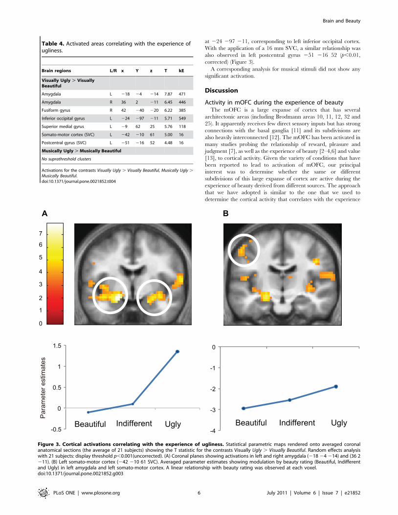
number of areas, summarized in Table 4. The contrast Visually
Ugly . Visually Beautiful led to activation in left and right
Figure 1. Cortical activation correlating with the experience of beauty. Statistical parametric maps rendered onto averaged anatomicalsections (average of 21 subjects) showing the T statistic for the contrasts (A) Visually Beautiful . Visually Ugly, (B) Musically Beautiful . Musically Uglyand (C) the results of a conjunction analysis for Visually Beautiful . Visually Ugly and Musically Beautiful . and Musically Ugly. Upper row showsactivity in mid-saggital sections and the middle row in horizontal sections of the brain. (D) shows the overlap in zones within the medial orbito-frontalcortex (mOFC) activated by visually beautiful (red), musically beautiful (green) stimuli, and the overlap between the two activations (yellow). Randomeffects analysis with 21 subjects. Display threshold p,0.001 (uncorrected). MNI co-ordinates of activation: A: at (26 41–11). B: at (23 41–8). And C: at(23 41–8). The co-ordinates in D are the same as in C.doi:10.1371/journal.pone.0021852.g001
Brain and Beauty
PLoS ONE | www.plosone.org 4 July 2011 | Volume 6 | Issue 7 | e21852

amygdala at 218–4 214 and 36 2–11; in visual cortex at
42-40-20, corresponding to the right fusiform gyrus; at -24-97-11,
corresponding to the left inferior occipital gyrus and in the left
superior medial frontal gyrus at -9 62 25. With the application of a
16 mm SVC, left somato-motor cortex at -42 -10 61 and left
postcentral gyrus at -51-16 52 showed significant activation. The
former was close to the activation site for ugly stimuli in the study
by Kawabata and Zeki (2004) [2].
There was no activity at the corrected significance level in the
contrast of Musically Ugly . Musically Beautiful. The application
of a conjunction analysis using the contrast Visually Ugly .
Visually Beautiful vs. Musically Ugly . Musically Beautiful did
not give any significant activation.
Quantitative relationship between experience of uglinessand cortical activation
Parametric analysis for visual stimuli showed a negative linear
relationship between BOLD signal and declared intensity of
experiences at the most significant voxels in left and right
amygdala at 224 24 217 and 27 24 214 and in visual cortex
Table 3. Activation area in the conjunction analysis.
Brain regions L/R x y z T kE
Visually Beautiful + Musically Beautiful . Visually Ugly + Musically Ugly
Medial OFC L 26 41 211 7.17 1153
Medial OFC L 23 26 4 5.38 1153
Caudate nucleus L 212 21 25 5.30 126
[Visually Beautiful . Visually Ugly] and [Musically Beautiful . Musically Ugly]
Medial OFC (SVC) L 23 41 28 4.81 54
Table 3. Activations for the contrast Visually Beautiful + Musically Beautiful . Visually Ugly + Musically Ugly and conjunction analysis for Visually and Musically Beautiful .
Visually and Musically Ugly.doi:10.1371/journal.pone.0021852.t003
Figure 2. Modulation of cortical activity by aesthetic rating. Averaged parameter estimates showing modulation by beauty rating (Beautiful,Indifferent and Ugly) in mOFC for (A) visual stimuli (at 26 41–11) and (B) musical stimuli at 23 41–8. A linear relationship with beauty rating wasobserved in both conditions.doi:10.1371/journal.pone.0021852.g002
Brain and Beauty
PLoS ONE | www.plosone.org 5 July 2011 | Volume 6 | Issue 7 | e21852
at 224 297 211, corresponding to left inferior occipital cortex.
With the application of a 16 mm SVC, a similar relationship was
also observed in left postcentral gyrus 251 216 52 (p,0.01,
corrected) (Figure 3).
A corresponding analysis for musical stimuli did not show any
significant activation.
Discussion
Activity in mOFC during the experience of beautyThe mOFC is a large expanse of cortex that has several
architectonic areas (including Brodmann areas 10, 11, 12, 32 and
25). It apparently receives few direct sensory inputs but has strong
connections with the basal ganglia [11] and its subdivisions are
also heavily interconnected [12]. The mOFC has been activated in
many studies probing the relationship of reward, pleasure and
judgment [7], as well as the experience of beauty [2–4,6] and value
[13], to cortical activity. Given the variety of conditions that have
been reported to lead to activation of mOFC, our principal
interest was to determine whether the same or different
subdivisions of this large expanse of cortex are active during the
experience of beauty derived from different sources. The approach
that we have adopted is similar to the one that we used to
determine the cortical activity that correlates with the experience
Table 4. Activated areas correlating with the experience ofugliness.
Brain regions L/R x Y z T kE
Visually Ugly . VisuallyBeautiful
Amygdala L 218 24 214 7.87 471
Amygdala R 36 2 211 6.45 446
Fusiform gyrus R 42 240 220 6.22 385
Inferior occipital gyrus L 224 297 211 5.71 549
Superior medial gyrus L 29 62 25 5.76 118
Somato-motor cortex (SVC) L 242 210 61 5.00 16
Postcentral gyrus (SVC) L 251 216 52 4.48 16
Musically Ugly . Musically Beautiful
No suprathreshold clusters
Activations for the contrasts Visually Ugly . Visually Beautiful, Musically Ugly .
Musically Beautiful.doi:10.1371/journal.pone.0021852.t004
Figure 3. Cortical activations correlating with the experience of ugliness. Statistical parametric maps rendered onto averaged coronalanatomical sections (the average of 21 subjects) showing the T statistic for the contrasts Visually Ugly . Visually Beautiful. Random effects analysiswith 21 subjects: display threshold p,0.001(uncorrected). (A) Coronal planes showing activations in left and right amygdala (218 24 214) and (36 2211). (B) Left somato-motor cortex (242 210 61 SVC). Averaged parameter estimates showing modulation by beauty rating (Beautiful, Indifferentand Ugly) in left amygdala and left somato-motor cortex. A linear relationship with beauty rating was observed at each voxel.doi:10.1371/journal.pone.0021852.g003
Brain and Beauty
PLoS ONE | www.plosone.org 6 July 2011 | Volume 6 | Issue 7 | e21852

of temporally asynchronous patterns derived from different
sources [14]. That result showed that each of the sources for
temporal asynchrony activates a different zone of frontal cortex,
with an additional, common, zone activated by all. In this study,
the experience of beauty derived from visual and musical sources
correlated with much the same part of the mOFC, the overlap
between the two being extensive and possibly total.
Relationship to previous studiesMany, if not all, studies that have addressed the neural
correlates of the experience of beauty have found activity in
mOFC, although sometimes the region is referred to otherwise.
For example, Vartanian and Goel 2003 [3] refer to their site of
activation as being in the anterior cingulate or the sub-genual
anterior cingulate although their locus of activation, at 210 42
26, is close to the locus in the study of Kawabata and Zeki (2004)
[2], that of Kirk et al. (2009) [15] and this one. Similarly, Tsukiura
and Cabeza (2011) attribute their locus of activation in response to
facial attractiveness and moral goodness to anterior cingulate but
the site of activation, at 24 44 1 [6], is very close to the one
reported in this study and, in our view, belongs more appropriately
to mOFC. The activation site reported by Di Dio et al (2009) in
the supplementary material to their study of beauty is in the
mOFC at (26 36 26 and 8 52 26) [4] as is the activity reported
by Kranz and Ishai (2006) [16], Cloutier et al. (2008) [17] and
O’Doherty et al. (2003) [18] for facial attractiveness. The
activations in all these studies fall well within field A1 of mOFC
as outlined in the Results section. As well, studies of the
relationship of value to cortical activity have also implicated the
mOFC [13]. Even the study of Jacobsen et al. (2006), which
differed somewhat from the studies mentioned above in that it
involved judgments of beauty vs. symmetry, reported activation in
the mOFC, though at a somewhat more dorsal level (at 1 23 32
and 1 54 26) [19]. There is one exception to this list which is the
result derived from use of magnetoencephalography (MEG) (e.g.
[20]). This may possibly have been due to the fact that activity in
medially situated cortex is not easily detectable by MEG.
In sum, a great many results are in agreement that the
experience of beauty correlates with activity in mOFC. To avoid
any ambiguity and to relate the area demarcated here to areas of
mOFC implicated in other studies, especially those related to
judgment, evaluation, reward and desire, we tentatively refer to
the area we have described as field A1 of mOFC. It is because of
this apparent agreement, that field A1 of mOFC is active in most
studies that have explored the relationship between cortical
activity and the experience of beauty, that we concentrate on it
in this discussion. The extent and boundaries of A1 must at
present be tentative. We place its center at 23 41 28 and estimate
it to have a diameter of between 15–17 mm. There may be further
functional subdivisions within it.
Taking our current results, as well as all the above studies, into
account, we conclude (a) that the experience of beauty derived
from visual and musical sources correlates with activity in the
mOFC; (b) that, within the mOFC it correlates more specifically
with activity in field A1; and (c) that the experience of beauty
derived from at least two modalities, visual and musical, shares a
common cortical locus in field A1 of mOFC. We therefore modify
Burke’s 1757 definition given above and say that ‘Beauty is, for the
greater part, some quality in bodies that correlates with activity in the mOFC
by the intervention of the senses’.
Field A1 of mOFC, value and judgmentThe paradigm that we used in this study is, inevitably, both
judgmental and evaluative and it therefore makes it interesting to
discuss our results in relation to axiology and to previous results
that have explored the relationship of value to brain activity. We
agree with DW Gotshalk [21] that ‘‘beauty is a value’’, that it
commonly evokes desire and that whatever is desired has value,
although we tend to place beauty more in the perceiver than in the
object, without denying that objects may have characteristics that
qualify them as beautiful to one or many subjects. This essentially
implies that there must be an intimate link in the cortical
processing that is linked to value, desire and beauty. It is therefore
interesting to note that the activity in A1 of mOFC that we report
here is almost co-terminous with the activity reported in previous
studies of the neural correlates of desire [22] and of value
judgments [13]. This in turn not only reflects what is well known
about the relationship of value, judgment, beauty and desire in
axiology and philosophical discourse generally but also implies
that there might be a value assigning system in the brain that is
either supra-modal, that is to say not linked to value within any
particular domain, or has specializations within it related to
different values (see below).
It is interesting to note in this context that the judgments that
we speak of above relate to positive judgments, strongly linked to
reward and pleasure. We did not find activity in A1 of mOFC that
correlates positively with the experience of ugly stimuli, although
ugliness, too, involves a judgment. Instead, the parametrically
modulated activity with the experience of ugliness was confined to
the amygdala and left somato-motor cortex. This implies that there
may be a functional specialization within the brain for at least two
different kinds of judgment, those related to positive, rewarding,
experiences and those related to negative ones. Future studies may
yet reveal further specializations for judgments in different domains.
Other activationsA: Visual and auditory cortex. The contrasts Visually
Beautiful . Musically Beautiful led to widespread activity within
visual cortex, while the contrast Musically Beautiful . Visually
Beautiful led to widespread activity within auditory cortex. That
such a large expanse of visual or auditory cortex should have been
active is not surprising because the stimuli, whether visual or
auditory, had many different characteristics; for example, the
visual stimuli consisted of portraits, landscapes, still lifes and were
in color while the musical stimuli had different degrees of melody,
and harmony, and some were derived from large-scale orchestral
performances while others from smaller ones. The activation of
these sensory areas in conjunction with activation of mOFC is
important for the theory we advance below.
B: Caudate nucleus. One of the more interesting activations
was in the caudate nucleus, which was also activated in previous
studies charting the neural correlates of emotional states [23,24].
The caudate activations reported here have two features: (a) their
location is similar to the location of the activity observed in previous
studies of beauty [3] and in studies of the neural correlates of
romantic love [10,24,25], and (b) the activation in it is proportional
to the intensity of the declared experience of beauty. This close
juxtaposition constitutes an interesting neural commentary on the
traditional emphasis made in world literature on the relationship
between love and beauty. Another interesting point about caudate
activity is that it is evident only during the experience of visual
beauty, with no parallel activation during the experience of musical
beauty. We have no current explanation for this.
Linear relationship between strength of cortical activityand strength of declared experience of beauty
Confirming previous studies from this and other laboratories
[2,3,18], the activity in the mOFC was parametrically modulated,
Brain and Beauty
PLoS ONE | www.plosone.org 7 July 2011 | Volume 6 | Issue 7 | e21852

the BOLD signal being higher for stimuli rated as beautiful than
those rated neutral or ugly. This was also true for the caudate
nucleus, though only during the experience of visual beauty. A
conjunction analysis using results derived from both auditory and
visual scans once again showed that the same region (A1) of
mOFC was parametrically modulated by both visual and musical
stimuli, thus adding further to the conclusion that activity in one
and the same brain area correlates in the same way with the
experience of beauty derived from these two different sources. The
experience of visual stimuli as ugly, on the other hand, correlated
with activity in the amygdala and (with the application of an SVC)
in left somato-motor cortex, among other areas (see Table 4). This
activity, too, was proportional to the declared intensity of the
experience. When we searched for quadratic modulation, we
could not find increased activity in amygdala during the
experience of both beauty and ugliness. Indeed, we could not
detect any areas that had a quadratic relationship with the stimuli
(i.e. were active during the experience of beautiful and ugly, but
not indifferent, stimuli). In this, our results differ from those of
Winston et al. (2007) who found that attractive and unattractive
faces, but not ones judged to be neutral, lead to amygdala
activation [26]. The reason for this difference is not known.
Taken together, these results imply that the subjective
experience of beauty and of ugliness can be objectively ascertained
and measured.
Toward a brain-based definition of beautyTaking the two principal results of this study, namely that
activity in a single region (field A1) of mOFC correlates with
experience of both visual and musical beauty and that there is a
linear relationship in it between the BOLD signal and the declared
intensity of the experience of beauty, leads us towards the
formulation of a brain based definition of beauty.
The question of what beauty is has resisted adequate definition
for centuries. Some, such as Vitruvius, Alberti and Leonardo Da
Vinci, have sought to understand beauty in terms of the
characteristics of the apprehended object. In visual art and
architecture this may be reduced to symmetry, proportion,
harmony and so on, while in music it may be beat, harmony
and rhythm. But what are the characteristics that confer beauty on
a more complex scene, such as a theatrical, operatic or cinematic
one? And what would the characteristics of moral beauty be?
An issue that has much exercised philosophers of art and
aesthetics and intrudes into any discussion of beauty is the
relationship of beauty to art. While art has been traditionally
associated with beauty in the popular mind as well as in past
philosophical and artistic speculation, the notion that art and
beauty can be equated has of course been questioned in the past
and received a fatal blow when Marcel Duchamp presented his
urinal, which he euphemistically named The Fountain, to an art
exhibition; it then received a further blow with his Readymades,
which Duchamp considered to constitute ‘‘art without an artist’’.
Notions of art have since changed and many will today
acknowledge that something considered to be a work of art need
not be perceived as beautiful, good examples being some of the
paintings of Francis Bacon, or the nudes of Lucian Freud, which is
not to say that these works do not have considerable artistic merit
both in their painterly style and in projecting truths, including
truths about decay and ugliness. But any work, be it considered art
or not, may be subjectively experienced as being beautiful by an
individual. This leads us to divorce art from beauty in this
discussion and concentrate on beauty alone. In our study, we were
essentially indifferent to whether a stimulus, be it visual or
auditory, constituted a work of art, our only concern being with
whether the individual subject, in the scanner, experienced the
work as being beautiful or not.
In trying to provide an answer, we have been inspired by a
critical question asked by the English art historian, Clive Bell, in
his book entitled Art [27], though less so by the answer he gave.
Bell was concerned in the main with visual beauty but we extend
our argument to beauty in general. He wrote, ‘‘If we can discover
some quality common and peculiar to all the objects that provoke
it [beauty], we shall have solved what I take to be the central
problem of aesthetics’’. Unlike Hume, who placed beauty entirely
in the perceiver, Bell searched for that ‘‘peculiar quality’’ in the
apprehended objects while also giving primacy to the perceiver.
He wrote: ‘‘All systems of aesthetics must be based on personal
experience–that is to say, they must be subjective’’ [27]. What
quality, he asked, ‘‘is common to Sta Sophia and the windows at
Chartres, Mexican sculpture, a Persian bowl, Chinese carpets,
Giotto’s frescoes at Padua and the masterpieces of Poussin, Piero
della Francesca, and Cezanne?’’, a list that excludes music. We
modify his question slightly by adding music and asking: what was
common to all the beauty experiences that each of our subjects
had when viewing the different visual and musical stimuli? Our
results inspire us to provide, speculatively and tentatively, and
perhaps even provocatively, a new, and neurobiological, answer to
Bell’s question as modified by us, an answer based exclusively on
the perceiver rather than on the object, which is not to say that
objects may not have characteristics that qualify them as beautiful.
The answer Bell gave is that the single characteristic that defines
all works of art is ‘‘significant form’’. Such a definition has many
drawbacks, chief of which is defining what significant form might
be in painting, music, fashion, design, film, opera and the many
other areas in which we experience beauty, including moral
beauty. Indeed, Bell himself was vague about what ‘‘significant
form’’ might be in terms of even elementary visual attributes such
as color and line. The term, being resistant to a definition that
applies to all areas in which we experience beauty, thus also
becomes impossible to measure and quantify. We therefore
propose instead a neurobiological definition that makes it un-
necessary to define ‘‘significant form’’ or indeed any other
characteristic of the work being apprehended, a definition that is
amenable to measurement and quantification and which relies on
the perceiver alone. We propose that all works that appear
beautiful to a subject have a single brain-based characteristic,
which is that they have as a correlate of experiencing them a
change in strength of activity within the mOFC and, more
specifically, within field A1 in it. Our proposal shifts the definition
of beauty very much in favor of the perceiving subject and away
from the characteristics of the apprehended object and gives added
strength to the Latin proverb that ‘‘De gustibus non est disputandum’’
(in matters of taste there is no dispute). We emphasize again that
we do not wish to imply that objects that are classified as beautiful
do not have certain characteristics that aid in this classification,
although what these characteristics are has been, and continues to
be, a subject of debate.
Our definition thus not only distinguishes sharply between
artistic merit and aesthetic value but is also indifferent to what is
art and what is not art. Almost anything can be considered to be
art, but only creations whose experience has, as a correlate,
activity in mOFC would fall into the classification of beautiful art.
That the activity in the mOFC is proportional to the intensity of
beauty experienced gives added strength to our theory, since the
strength of activation is related to the intensity of the experience
alone, regardless of the extent to which the work can be classified
as a work of art or not. A painting by Francis Bacon may be
executed in a painterly style and have great artistic merit but may
Brain and Beauty
PLoS ONE | www.plosone.org 8 July 2011 | Volume 6 | Issue 7 | e21852

not qualify as beautiful to a subject, because the experience of
viewing it does not correlate with activity in his or her mOFC.
The definition we propose takes aesthetics very much into the
subjective, though quantifiable, arena: it applies only to an
individual at a specific time and place since what is judged and
experienced as beautiful at one moment and in one context by one
subject may not be so experienced by another in a different
context. Put differently, for an individual who experiences beauty
in a Francis Bacon painting, with a concomitant change in activity
within mOFC, the work can be qualified as beautiful to that
individual. Our definition thus makes it un-necessary to consider
other factors such as up-bringing, culture, context, connoisseurship
and monetary value in the definition of what constitutes the
aesthetic appeal of a work of art, although all these factors may
contribute to the experience of beauty. Indeed, it is for this very
reason that we included people from different cultures and ethnic
backgrounds in our pool of subjects. There are of course many
iconic works of art, such as the music of Beethoven or the Pieta of
Michelangelo, which are experienced as beautiful by those who
belong to different cultures, backgrounds and ethnic groups. This
may be accounted for, as Immanuel Kant did in his Critique of
Judgment [28], by supposing the existence of a sensus communis, that
is to say a brain organization that is similar across individuals and
cultures, which such works stimulate. We are currently addressing
this in greater detail.
We are of course aware that activity in mOFC correlates with
the experience of pleasure and reward, whether real or imagined,
and its expectation [29–31]. This naturally raises, at a neurobi-
ological level, an issue long discussed in the humanities, namely
the relationship of aesthetic experience to pleasure (see Graham
Gordon, The Philosophy of Art [32]). It can be argued that Wagner’s
Prelude to Tristan und Isolde is infinitely more subtle and beautiful
than a composition by, for example, a rock artist. But this
argument has more to do with what is art and what is not art than
with what is perceived as beautiful and rewarding and what is not.
Many who admire and are rewarded by listening to rock music,
which they find beautiful, will probably have little time for
Wagner, and vice versa. We would expect that, in subjects who
find rock music rewarding and beautiful, their experience of the
beauty of rock music will correlate with activity in their mOFC.
Our definition is concerned with what an individual subject
experiences as beautiful at a given moment, nothing else.
It is interesting to note that, contrary to the experience of
beauty, we could not locate, through our conjunction analyses, a
common area in which activity correlated with the experience of
musical and visual ugliness, a negative finding for which we have
no current explanation.
Co-activation of mOFC and perceptive areasOne objection to our hypothesis is that, currently, activity in
mOFC may be related to other experiences, such as judgment,
evaluation, decision-making and reward in other domains, ones
that are not directly related exclusively to beauty. For the sake of
clarity and because of the complex architectonic configuration of
mOFC, we designate the area that was active in this study as
division A1 of mOFC. Activation of mOFC in other reward-
related tasks, such as monetary reward, involves a different overall
pattern of brain activation than the one we report here. Moreover,
such reward tasks may or may not activate field A1 of mOFC. A
recent study [33] reported overlapping activation with juice and
monetary rewards in a region corresponding to A1 of mOFC,
although the results of that study, being based on either
uncorrected statistics at p,0.005 or corrected statistics at
p,0.05 but with the use of an 8 mm SVC, are somewhat weak
and require further study. This is especially so, since another study
based on money rewards puts activity in the orbito-frontal cortex
outside A1 [34] and at a significantly more anterior position than
in the study of Kim et al. Hence the need for a more precise
definition of the relationship of activations derived from different
kinds of reward tasks to the extent of field A1 of mOFC.
In fact a specialization within mOFC may be conferred on it by
the cortical route taken to it. In our study, although only activity in
one cortical area, A1 of mOFC, correlated with the experience of
musical and visual beauty, the path to mOFC through the two
domains was different. With musical experience of beauty,
auditory areas of the brain were co-active with A1 of mOFC
while we could not detect any activity in the caudate nucleus. With
experience of visual beauty, the caudate nucleus was very much
co-active with A1 of mOFC as were the visual areas (we use the
term co-active because the temporal limitations of the fMRI
method do not allow us to isolate the sequence of activity in these
areas). Hence, basing ourselves more on Burke’s definition of
beauty given above, as one mediated by the senses, we consider
that it is not activation of mOFC alone that is a determinant of
beauty; it is rather the co-activation of field A1 of mOFC with the
specialized sensory and perceptive area, or areas, and possibly (in
the case of visual stimuli) with the caudate nucleus as well. Hence
we broaden our neurobiological definition of beauty given above
to include not only activation of mOFC but also its co-activation
with sensory areas that feed it. The interaction between these
sensory areas, and other regions such as the caudate, and A1 of
mOFC, and how activity in the latter is modulated by activity in
the former remains a very interesting puzzle for the future.
We emphasize that our theory is tentative; there are many other
experiences that may be deemed to be beautiful besides the visual
and musical. Our theory will stand or fall depending upon whether
future studies of the experience of beauty in other domains show
that, in these too, the experience correlates with activity in field A1
of mOFC.
Supporting Information
Table S1 Behavioral data collected in preliminarybehavioral test. Distribution of behavioral ratings during
preliminary test by stimulus modality, averaged over all subjects.
Range shows maximum and minimum percentages among
subjects.
(DOCX)
Acknowledgments
We thank John Romaya for his help at all stages and Masamichi Hayashi
for useful suggestions. We also thank Karl Friston, Ray Dolan and Anton
Burdakov for their critical reading of this manuscript.
Author Contributions
Conceived and designed the experiments: SZ TI. Performed the
experiments: TI. Analyzed the data: TI SZ. Contributed reagents/
materials/analysis tools: SZ TI. Wrote the paper: SZ TI.
References
1. Burke E (1757) A philosophical enquiry into the origin of our ideas of the
sublime and beautiful. London: R. and J. Dodsley. 175 p.
2. Kawabata H, Zeki S (2004) Neural correlates of beauty. J Neurophysiol 91:
1699–1705.
Brain and Beauty
PLoS ONE | www.plosone.org 9 July 2011 | Volume 6 | Issue 7 | e21852

3. Vartanian O, Goel V (2004) Neuroanatomical correlates of aesthetic preference
for paintings. Neuroreport 15: 893–897.4. Di Dio C, Macaluso E, Rizzolatti G (2007) The golden beauty: brain response to
classical and renaissance sculptures. PLoS One 2: e1201.
5. Blood AJ, Zatorre RJ, Bermudez P, Evans AC (1999) Emotional responses topleasant and unpleasant music correlate with activity in paralimbic brain
regions. Nat Neurosci 2: 382–387.6. Tsukiura T, Cabeza R (2011) Shared brain activity for aesthetic and moral
judgments: implications for the Beauty-is-Good stereotype. Soc Cogn Affect
Neurosci.7. Grabenhorst F, Rolls ET (2011) Value, pleasure and choice in the ventral
prefrontal cortex. Trends Cogn Sci 15: 56–67.8. Kornysheva K, von Cramon DY, Jacobsen T, Schubotz RI (2010) Tuning-in to
the beat: Aesthetic appreciation of musical rhythms correlates with a premotoractivity boost. Hum Brain Mapp 31: 48–64.
9. Price CJ, Friston KJ (1997) Cognitive conjunction: a new approach to brain
activation experiments. Neuroimage 5: 261–270.10. Zeki S, Romaya JP (2010) The brain reaction to viewing faces of opposite- and
same-sex romantic partners. PLoS One 5: e15802.11. Bechara A, Damasio H, Damasio AR (2000) Emotion, decision making and the
orbitofrontal cortex. Cereb Cortex 10: 295–307.
12. Margulies DS, Kelly AM, Uddin LQ, Biswal BB, Castellanos FX, et al. (2007)Mapping the functional connectivity of anterior cingulate cortex. Neuroimage
37: 579–588.13. FitzGerald TH, Seymour B, Dolan RJ (2009) The role of human orbitofrontal
cortex in value comparison for incommensurable objects. J Neurosci 29:8388–8395.
14. Zeki S, Hulme OJ, Roulston B, Atiyah M (2008) The encoding of temporally
irregular and regular visual patterns in the human brain. PLoS One 3: e2180.15. Kirk U, Skov M, Hulme O, Christensen MS, Zeki S (2009) Modulation of
aesthetic value by semantic context: an fMRI study. Neuroimage 44:1125–1132.
16. Kranz F, Ishai A (2006) Face perception is modulated by sexual preference. Curr
Biol 16: 63–68.17. Cloutier J, Heatherton TF, Whalen PJ, Kelley WM (2008) Are attractive people
rewarding? Sex differences in the neural substrates of facial attractiveness. J CognNeurosci 20: 941–951.
18. O’Doherty J, Winston J, Critchley H, Perrett D, Burt DM, et al. (2003) Beauty
in a smile: the role of medial orbitofrontal cortex in facial attractiveness.
Neuropsychologia 41: 147–155.
19. Jacobsen T, Schubotz RI, Hofel L, Cramon DY (2006) Brain correlates of
aesthetic judgment of beauty. Neuroimage 29: 276–285.
20. Cela-Conde CJ, Ayala FJ, Munar E, Maestu F, Nadal M, et al. (2009) Sex-
related similarities and differences in the neural correlates of beauty. Proc Natl
Acad Sci U S A 106: 3847–3852.
21. Gotshalk DW (1935) Beauty and Vaue. The Journal of Philosophy 32: 604–610.
22. Kawabata H, Zeki S (2008) The neural correlates of desire. PLoS One 3: e3027.
23. Carretie L, Rios M, de la Gandara BS, Tapia M, Albert J, et al. (2009) The
striatum beyond reward: caudate responds intensely to unpleasant pictures.
Neuroscience 164: 1615–1622.
24. Bartels A, Zeki S (2000) The neural basis of romantic love. Neuroreport 11:
3829–3834.
25. Aron A, Fisher H, Mashek DJ, Strong G, Li H, et al. (2005) Reward, motivation,
and emotion systems associated with early-stage intense romantic love.
J Neurophysiol 94: 327–337.
26. Winston JS, O’Doherty J, Kilner JM, Perrett DI, Dolan RJ (2007) Brain systems
for assessing facial attractiveness. Neuropsychologia 45: 195–206.
27. Bell C (1921) Art. London: Chatto and Windus. 292 p.
28. Kant I (1952) The Critique of Judgment. Oxford: Clarendon. 180 p.
29. Bray S, Shimojo S, O’Doherty JP (2010) Human medial orbitofrontal cortex is
recruited during experience of imagined and real rewards. J Neurophysiol 103:
2506–2512.
30. Peters J, Buchel C (2010) Neural representations of subjective reward value.
Behav Brain Res 213: 135–141.
31. Schultz W (2010) Subjective neuronal coding of reward: temporal value
discounting and risk. Eur J Neurosci 31: 2124–2135.
32. Gordon G (1997) Philosophy of the Arts. London: Routledge. 193 p.
33. Kim H, Shimojo S, O’Doherty JP (2011) Overlapping responses for the
expectation of juice and money rewards in human ventromedial prefrontal
cortex. Cereb Cortex 21: 769–776.
34. Sescousse G, Redoute J, Dreher JC (2010) The architecture of reward value
coding in the human orbitofrontal cortex. J Neurosci 30: 13095–13104.
Brain and Beauty
PLoS ONE | www.plosone.org 10 July 2011 | Volume 6 | Issue 7 | e21852