Robust γδ + T cell expansion in infants immunized at birth with BCG vaccine
Upload
independentCategory
view
0download
0
The Ity/Lsh/Bcg Locus: Natural Resistance to Infection with Intracellular Parasites Is Abrogated by Disruption of the Nrampl Gene By Silvia Vidal,*~ Michel L. Tremblay,* Gregory Govoni,*~ Susan Gauthier,* Giovanna Sebastiani,*~ Danielle Malo,~ Emil Skamene,~ Martin Olivier, II Serge Jothy,$ and Philippe Gros*~
From the "Department of Biochemistry, *Center for the Study of Host Resistance, SDepartment of Pathology, McGill University, Montreal, Qu~be6 Canada H3G IY6; and IIService d'Infectiologie, Centre Hospitalier de l'Universitd de Laval, Ste-Foy, Qudbec, Canada G1K 7P4
Summary In mice, natural resistance or susceptibility to infection with intracellular parasites is determined by a locus or group of loci on chromosome 1, designated Bcg, Lsh, and Ity, which controls early microbial replication in reticuloendothelial organs. We have identified by positional cloning a candidate gene for Beg, Nrampl, which codes for a novel macrophage-specific membrane trans- port protein. We have created a mouse mutant bearing a null allele at Nrampl, and we have analyzed the effect of such a mutation on natural resistance to infection. Targeted disruption of Nrampl has pleiotropic effects on natural resistance to infection with intracellular parasites, as it eliminated resistance to Mycobacterium boris, Leishmania donovani, and lethal Salmonella typhimu- rium infection, establishing that Nrampl, Bcg, Lsh, and It), are the same locus. Comparing the profiles of parasite replication in control and Nrampl -/- mice indicated that the NramplAse 169 allele of Beg s inbred strains is a null allele, pointing to a critical role of this residue in the mech- anism of action of the protein. Despite their inability to control parasite growth in the early nonimmune phase of the infection, Nrampl - / - mutants can overcome the infection in the late immune phase, suggesting that Nrampl plays a key role only in the early part of the macro- phage-parasite interaction and may function by a cytocidal or cytostatic mechanism distinct from those expressed by activated macrophages.
A better understanding at the molecular level of the host / ]k mechanisms of defense against infection may open new therapeutic avenues for intervention in diseases in which drug resistance is a major impediment to treatment. The genetic analysis in mouse models of natural variations in innate sus- ceptibility or resistance to various parasitic, bacterial, or viral infections provides a rational approach to the identification and characterization of such mechanisms (for review see ref- erence 1).
One of the best studied genetic determinants of natural resistance or susceptibility to infectious diseases is the Bcg gene (2). Bcg is a single gene that is present in two allelic forms in the mouse (resistant, Bcg r, and susceptible, Beg ~) and determines innate resistance/susceptibility of mice to infec- tion with several mycobacterial species such as Mycobacteriurn bov/s (bacille Calmette-Guerin [BCG]I), M. lepraemurium,
1 Abbreviations used in this paper: BCG, baciUe Calmette-Guerin; ES, em- bryonic stem; LDU, Leishman-Donovan units; mRNA, messenger RNA; P/L, phagosome/lysosome; TM, transmembrane.
S. Vidal and M. L. Tremblay contributed equally to this work.
and M. intracellulare (2-4). In inbred mouse strains, infection with M. bovis (BCG) is biphasic, with an early nonimmune phase (0-3 wk) characterized either by rapid proliferation of the bacteria in reticuloendothelial (RE) organs (liver, spleen) of Beg s, or by absence of bacterial growth in Bcg r mouse strains (2). The late phase (3-6 wk), characterized either by complete clearance of the bacterial load or by persistent in- fection in the RE organs of the susceptible strains, is con- trolled by H-2-1inked immune response genes (5, 6). Genetic linkage studies have shown that Bcg is either aUelic or very tightly linked to two other host resistance loci, Lsh (7) and Ity (8). Similar to Bcg, Lsh controls the replication of Leish- mania donovani in the RE organs, early in the nonimmune phase of infection (7). Ity also influences the early replication of Salmonella typhimurium in RE organs, but in this case animals either survive (resistant; Ity') or succumb rapidly (susceptible; Ity ~) to an infectious inoculum (8), In vitro (9-11) and in vivo (12-14) studies have shown that Beg, Ity, and Lsh affect the capacity of the macrophage to restrict the intracellu- lar replication of these antigenically unrelated intracellular parasites.
655 J. Exp. Med. �9 The Rockefeller University Press �9 0655-1007/95/09/0655/12 $2.00 Volume 182 September 1995 655-666
on June 4, 2014jem
.rupress.orgD
ownloaded from
Published September 1, 1995
We have used positional cloning to isolate candidate genes for Bcg. This approach was based on the construction of high resolution linkage (15) and physical maps (16) of the Bcg re- gion on mouse chromosome 1, and on the isolation and char- acterization of cloned segments of the minimal physical in- terval in YAC clones (17). Briefly, exon trapping was used to identify seven candidate transcription units for Bcg. Of these genes, two were ubiquitously expressed in all tissues including RE organs, while one encoded a messenger RNA (mRNA) exclusively expressed in RE organs and, most importantly, in macrophages derived from them (17). This gene, desig- nated Nramp (natural resistance-associated macrophage pro- tein) encoded a novel integral membrane protein with 12 puta- tive transmembrane (TM) domains, a glycosylated extraceUular loop, and several phosphorylation sites (17). In addition, Nramp shares, through the presence of a consensus sequence signa- ture, homology to a number of eukaryotic and prokaryotic membrane transport proteins, including a nitrate/nitrite con- centrator of the fungus Aspergillus nidulans (18). Therefore, Nramp seems to be a novel macrophage-specific transport pro- tein. Recently, we have determined that Nramp is part of a small gene family of at least two genes in the mouse, with a second gene Nramp2 on chromosome 15 (19), and we have consequently renamed the chromosome 1 locus Nrampl (19).
Since the Bcgs and Beg' genotypes and phenotypic char- acteristics are not unique to a specific mutant strain, but rather separate inbred strains in two nonoverlapping groups, we have analyzed a total of 27 Bcg' and Bcg' mouse strains for nucleo- tide sequence variations within the coding portion of Nrampl (20). One G to A substitution at nucleotide position 596 resulting in the nonconservative replacement of Gly 169 to Asp 169 within TM4 was found to be in absolute association with the Beg phenotype in 20 Bcg r strains (Gly 169) and 7 Bcg' strains (Asp 169) tested (20). Haplotype mapping using se- quence polymorphism identified within Nrampl and 11 ad- ditional polymorphic markers from the region revealed that the 7 Bcg ~ strains tested shared a conserved core haplotype of 2.2 Mb overlapping Nrampl (20).
Although the replacement of a small neutral residue (gly- cine) by a bulkier, negatively charged one (aspartate) within one of the predicted TM domains of Nramp1 would be likely to affect function, it does not per se constitute an obvious loss of function, and a full-length polypeptide is expected to be synthesized in Bcgs mice. Therefore, we have used gene targeting to create a null allele at the Nramp1 locus with the intent (a) to determine if Nramp1 and Bcg are indeed allelic; (b) to determine if Bcg, Lsh, and Ity are allelic; and (c) to compare the phenotypic expression of NrarnplAsp 169 and NrampI null mutations.
Materials and Methods Design of Targeting Vector. The targeting vector pMC.NR was
derived from the plasmid pMClneo-polyA and the Nrampl genomic clone Xnrp1. pMClneo-polyA (Stratagene, La Jolla, CA) contains a 1-kb cassette with the structural neomycin resistance gene (neo) under the control of the Herpes simplex thymidine kinase (HSV- tle) gene promoter and followed at its 3' end by a polyadenylation
signal. The genomic DNA clone Xnrpl (Gros, P., and G. Govoni, unpublished data) was isolated from a 129sv/J mouse genomic li- brary constructed in bacteriophage vector XDASH (Stratagene). It contains a 17-kb DNA insert overlapping exons 3-15 from the Nramp1 gene. The longer genomic arm of pMC.NR was a 5-kb Nrampl fragment from intron 8 to within exon 15 cloned into the BamHI site of pMClneo-poly(A). The shorter genomic arm of pMC.NR was a 1.1-kb SalI to HindlII Nramp1 fragment extending from intron 4 to within exon 7. The cohesive ends of the 1.1-kb fragment were repaired with T4 DNA polymerase before ligation into the blunted XhoI site of the vector. The short Nramt,1 genomic arm provides a unique NotI site that originates from the polylinker of the hi)ASH vector. The two Nramp1 genomic fragments were cloned so that their transcriptional orientation was the same as that of the neo cassette. Before transfection into embryonic stem (ES) cells, the construct was linearized by Nod digestion and dissolved in sterile water.
ES Cell Culture and Electroporation. The 129sv ES cell line J1 (21) was seeded onto a mitomycin C (Sigma Chemical Co., St. Louis, MO)-inactivated embryonic fibroblast feeder cell layer (2 h at 37~ in DMEM (GIBCO BILL, Gaithersburg, MD) containing 500 U/rnl leukemia inhibitory factor (GIBCO BILL) and 15% heat-inactivated FCS (GIBCO BILL). For electroporation, ES cells were trypsinized and resuspended (107 cells/ml) in PBS. 1 ml of the cell suspension was mixed with pMC.NR-linearized DNA at a concentration of 50/~g/ml and electroporated using a Gene Pulser (Bio-Rad Labora- tories, Richmond, CA) with a setting of 500/~F capacitance, 240 mV (22). ES cells were then plated at a density of 2 • 105 cells per 60-mm dish onto a monolayer of neomycin-resistant, mitoti- cally inactive fibroblast feeder cells. Selection for neo + clones was initiated 24 h later by addition of geneticin (G418; GIBCO BRL; final concentration 500/~g/ml). 5-10 G418 R colonies per plate were picked 10 d after electroporation and sequentially expanded in 96- and 24-well titer plates. Each clone was recovered by trypsin treatment and then frozen at -80~ in culture medium sup- plemented with 10% DMSO and 20% FCS.
Genotyping of ES Cell Lines and Mutant Mice. Genomic DNA from individual ES clones was directly isolated onto replica 96-well plates according to the procedure of Ramirez-Solis et al. (23), dis- solved in 30/A of restriction enzyme buffer (recommended by the supplier of enzyme) containing 1 mM spermidine and 100/~g/ml BSA, and digested with 20 U of either BamHI or XbaI (incuba- tion 16 h, 37~ Genomic DNA was isolated from mouse tails by a standard protocol (24) using proteinase K digestion, phenol extraction, and ethanol precipitation. Restricted DNA from ES cell clones or mouse tails was resolved by electrophoresis in 1% agarose gels containing TAE buffer (40 mM Tris-acetate, 20 mM sodium acetate, 20 mM EDTA, pH 7.6), and transferred onto nylon membranes (Hybond-N; Amersham International, Little Chalfont, UK) by capillary blotting in 20x SSC (lx SSC is 0.15 M NaC1, 0.015 M sodium citrate). Southern blots were prehybridized for 16 h at 42~ and then hybridized for 16 h at 42~ in the same mixture composed of 50% formamide, 5x SSC, 1% SDS, 10% dextran sulfate, 20 mM Tris-HC1, pH 7.5, lx Denhardt's solution (0.1% BSA, 0.1% Ficoll, 0.1% polyvinylpyrrolidone), and heat- denatured salmon sperm DNA at a final concentration of 200/~g/ml. The Nrampl hybridization probe was a cDNA fragment (nucleo- tide 300 to 550; 17) labeled to high specific activity (1 x 10⁹ cpm//xg DNA) by random priming (25) with cr (specific activity 3,000 Ci/mmole), and used at 1 x 10⁶ cpm/ml of hybridization solution. The membranes were washed to a final stringency of 0.5x SSC, 0.1% SDS at 65~ for 30 min.
Blastocyst Injection and Animal Breeding. Blastocysts were obtained
656 The Ity/Lsh/Bcg Locus
on June 4, 2014jem
.rupress.orgD
ownloaded from
Published September 1, 1995
3.5 d after coitus from pregnant 6-wk-old BALB/c females obtained by natural mating with syngeneic males. 10--15 ES cells from two independent clones, EPl-26 and EP2-173, were microinjected into each blastocoel cavity (26). The blastocysts were then reimplanted into the uterine horn of pseudopregnant CD1 mice. Chimeric offspring were identified by the agouti contribution of the ES cells to the coat color, and chimeric males were crossed with BALB/c females to produce a (129sv x BALB/c) F1 generation bearing one Nrarnpl null allele (NrarnpI -/A~16~). Germline transmission was de- termined by the presence of agouti mice in the offspring. The geno- type of this offspring was determined by tail blot, and Nrampl -/Asp~s~ carrier animals were either backcrossed to BALB/c animals to expand the heterozygous Nrampl-/A,r16~ population or intercrossed to generate homozygous F2 null mutant mice (Nrampl-/-). Mutant F1 (Nrampl-/A~r 16~) and F2 (Nrampl -/-) animals were genotyped and then tested for susceptibility to infec- tion with intracellular parasites.
RNA Hybn'dization Studies. Total cellular KNA was isolated from frozen spleens after homogenization with a mortar and pestle precooled with liquid nitrogen. The tissue powder was dissolved in guanidinium hydrochloride (6 M), and KNA was recovered by differential ethanol precipitation and phenol and chloroform ex- tractions (27). KNA was separated by electrophoresis in a dena- turing formaldehyde agarose gel and transferred to a nylon mem- brane (GeneScreen Plus, E. I. du Pont de Nemours & Co., Inc., Wilmington, DE), and the blot was prehybridized (16 h, 65~ and hybridized (16 h, 65~ in the same solution consisting of 1 M NaC1, 1% SDS, and 10% dextran sulfate. The hybridization probe was a mouse Nrampl cDNA subfragment overlapping exons 7 and 8 (nudeotide 855-1064; 17), was labeled to high specific activity with oI-[3zp]dATP, and was used at 2 x 10⁶ cpm/ml hybridiza- tion solution. The blot was washed under conditions of increasing stringency up to 0.5x SSC, 0.1% SDS at 65~ for 45 min.
Infection of Mice with BCG, L. donovani, andS. typhimurium. The BCG infectious inoculum consisted of a single-ceLl suspension of M. boris (BCG, strain Montreal) prepared by microfiltration from two serial 7-d cultures in Dubos oleic acid medium, as previously described (2). Mice were inoculated in the caudal tail vein with 0.25 ml of a BCG suspension containing 1.4 x 10⁴ CFU/ml in physiological saline. The degree of infection was assessed by deter- mining the number of spleen colony-forming units at predetermined time intervals (2). Geometric means plus or minus standard devia- tion of spleen BCG CFUs were calcuhted from five infected animals per time point.
The virulent strain L. donovani infantum Ethiopian LV9 was pas- saged continuously by intraperitoneal infection of Syrian hamsters and was generously donated by Dr. G. Matlashewski (Institute of Parasitology, McGill University, Montreal, Canada). The infectious inoculum was prepared from spleen homogenates of infected ham- sters according to the procedure of Murray et al. (28) and used immediately to infect mice intravenously (caudal tail vein, 0.2 ml volume of a suspension of 5 x 10⁷/ml infectious L. donovani amastigotes). At different times (1, 3, and 6 wk) after infection, livers were removed, weighed, and sectioned for impression smears. The extent of disease was quantitated by microscopic determina- tion of the number of parasites in Giemsa-stained liver impression smears. Hepatic parasite burden was expressed in Leishman-Donovan units (LDU, the number of parasites per 1,000 nucleated cells x liver weight in milligrams; 28). A minimum of four mice were used for each infection time point and for each experimental group.
The highly virulent S. typhimurium strain KeUer (29) was origi- nally obtained from Dr. Hugh Robson (Royal Victoria Hospital, Montreal, Canada). Mice were injected intravenously (caudal tail
vein) with a 0.2-ml inoculum containing 5 x 10⁴ CFU/ml S. ty- phimurium. Animals were examined twice a day for a period of up to 4 wk, and deaths were recorded and moribund animals were kiUed. Resistance to infection was assessed by the percentage of surviving animals among groups of 5-15 mice. It)~ animals typi- cally succumbed to infection within 4-6 d, while lty' animals typi- cally survived at least 14 d after infection.
Histopathology. Mice from various groups were killed by cer- vical dislocation and autopsied. The major organs, including spleen, liver, lung, intestine, colon, and thymus, were removed, immedi- ately fixed in 10% formalin, and embedded in paraffin. Sections (5 #m) from paraffin blocks containing these organs were stained with hematoxylin and eosin for routine histological examination. Randomly selected (n = 10) high power fields (x 100 and x 160) of tissue sections were examined from each animal. In addition, 5-#m sections of the spleen and liver were stained for the presence of mycobacteria by the Ziehl-Neelsen acid-fast stain.
Results
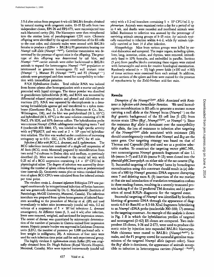
Disruption of the NrarnpICIr 169 Allele Associated with Resis- tance to Infection with lntracellular Parasites. We used homol- ogous recombination in ES calls to generate a mutant mouse carrying a null allele at the Nrarapl locus (NrampI -) on the Bcg r genetic background of the ES cell line J1 (21) from mouse strain 129sv (Beg r, NramplClY 169, or Nrampl +). Since the resistance Beg' aUele is dominant over the susceptibility Bcg s allele, the loss of resistance to infection after targeting of the NramplClY 169 aUele associated with resistance (20) should unambiguously confirm that Nrampl and Bcg are the same gen~ Our targeting strategy was based on that of Thomas and Capecchi (30) and used neo as a positive selec- table marker. To construct the targeting vector pMC.NR, two noncontiguous Nrampl genomic fragments of size 1.1 kb (exons 5-7) and 5.0 kb (exons 9-15) were cloned into the plasmid pMClneo-polyA on either side of the neo cassette (Fig. 1). Successful targeting of the NrarapI locus by homologous recombination using this construct should result in (a) dele- tion of a 500-bp Nrarapl genomic DNA segment disrupting exon 7 and ddeting exon 8; (b) insertion of the neo marker at that site and introduction of translation termination codons in three reading frames, resulting in a severely truncated pro- rein lacking 8 of the 12 predicted TM domains; and (c) gener- ation of novel RFLPs diagnostic for the targeted event.
Successful targeting of Nrampl was identified by Southern blotting of genomic DNA through the appearance of diag- nostic 6.0-kb BamHI or 8.5-kb XbaI fragments hybridizing to an Nrampl cDNA probe (nucleotide 300-550; 17) external to the targeting construct. An example of this analysis is shown in Fig. 1 B in which the hybridization profiles of targeted and nontargeted (2-62) ES clones are compared. Two inde- pendent ES clones, 1-26 and 2-173, were used to produce chi- meric mice by injection into expanded BALB/c blastocysts. Male chimeras were mated to BALB/c (NrampIAsr 169) fe- males, and the F1 progeny scored for successful germline trans- mission of the targeted Nrarapl allele (agouti color). Since the Beg' alhle is dominant, the appearance of animals suscep- tible to infection in heterozygous carriers (Nrampl-/Asp169)
657 Vidal et al.
on June 4, 2014jem
.rupress.orgD
ownloaded from
Published September 1, 1995
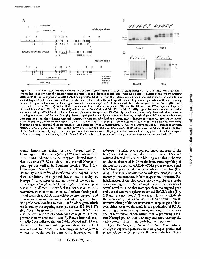
Figure 1. Creation of a null allele at the Nrampl locus by homologous recombination. (A) Targeting strategy. The genomic structure of the mouse Nramp1 locus is shown with the genomic exons numbered 1-15 and identified as dark boxes (wild-type allele). A diagram of the Nrampl-targeting vector showing the neo expression cassette flanked by a proximal 1.1-kb fragment that includes exons 5 and 6 and part of exon 7 on one side, and a 5.0-kb fragment that includes exons 9-15 on the other side, is shown below the wild-type allele map. The genomic organization of the corresponding mutant allele generated by successful homologous recombination at Nramp1 in ES cells is presented. Restriction enzymes sites for BamHI (B), EcoRI (E), HindlII (H), and Xbal (X) are identified in both alleles. The position of key genomic XbaI and BamHl restriction DNA fragments diagnostic of the wild-type (7.0-kb XbaI, 5.5-kb BamHI) and the mutant Nramlsl allele (8.5-kb Xbal, 6.0-kb BamHI) targeted by homologous recombination and recognized by a cDNA hybridization probe overlapping exons 3-4 (positions 300-550; 17) are indicated immediately above and below the corre- sponding genomic maps of the two alleles. (13) Nramp1 targeting in ES cells. Results of Southern blotting analysis of genomic DNA from independent G418-resistant ES cell clones digested with either BamHI or Xbal and hybridized to a Nrampl cDNA fragment (positions 300-550; 17) are shown. Successful targeting is evidenced for clones 1-25, 2-65, 2-110, 2-161, and 2-172 by the presence of diagnostic 6-kb BamHI- and 8.5-kb Xbal-hybridizing fragments on the background of the endogenous 5.5-kb BamHI and 7.0-kb XbaI fragments. (C) Creation Nrampl mutant mice. Results of Southern blotting analysis of genomic DNA from control 129sv mouse strain and individuals from a (129sv x BALB/c) F2 cross in which the wild-type allele of 129sv had been successfully targeted by homologous recombination are shown. Offspring from this cross include heterozygotes (- /+) and homozygotes ( - / - ) for the targeted allele Nramp1-. The Nrampl cDNA probe and diagnostic hybridizing restriction fragments are as described in/~
would demonstrate allelism between Nrampl and Beg. Homozygous null mutants (Nrampl-/-) were obtained by intercrossing independently heterozygotes derived from ei- ther 1-26 or 2-173 ES cell clones, and the null Nrampl-/- genotype was verified by Southern blotting (Fig. 1 C). Homozygous Nrampl-/- null mice were housed in a bar- rier facility and were free of specific mouse pathogens. Under these conditions, the general health and viability of Nrampl -/- mice appeared normal up to 14 mo of age.
Wild-type Nramp1 mRNA Transcripts Are Absent from Nrampl -/- Null Mice. To verify that intact Nrampl mRNA was indeed absent from mutant mice, Northern blotting anal- ysis of total spleen R N A from wild-type, heterozygous, and homozygous mutant mice was carried out using a hybridiza- tion probe corresponding to exons 7 and 8 of the gene, which are deleted by the targeting event (nucleotide 855-1064; 17) (Fig. 2 A). The spleen was chosen as a source of R N A since it is the strongest site of endogenous Nrampl m R N A ex- pression in normal mouse tissues (17). Results from this anal- ysis (Fig. 2 A) indicated that the 2.4-kb Nrampl m R N A was abundant in spleen from wild-type animals and that its level was reduced by ~ 5 0 % in heterozygotes (Nrampl -/+ ), whereas it could not be detected in homozygous null
(Nrampl-/-) mice, even upon prolonged exposure of the blot (data not shown). The reduction in or absence of Nrampl m R N A detected by Northern blotting with this probe was not due to absence of R N A in the lanes, since reprobing of the blot with a control GAPDH cDNA probe revealed equal R N A loading and transfer to the membrane in each lane (Fig. 2 C). These results indicate that no wild-type Nrampl m R N A transcripts are produced in homozygous null mutants. Re- hybridization of the blot with a neo gene probe or a probe corresponding to exon 5 of Nrampl revealed the presence of several novel mRNAs that were specific to the targeted gene and were absent from spleens of control BALB/c mice (Fig. 2 B and data not shown). These transcripts most likely ei- ther represent hybrid neo-Nrampl mRNAs or result from al- ternative splicing of the neo cassette in the targeted gene. How- ever, either event would result in the production of RNAs recruiting different reading frames, resulting in the appear- ance of termination codon within exon 9, producing a ma- ture Nrampl protein that is severely truncated (lacking the carboxy-terminal half) and probably nonfunctional.
Organ Morphology of Nrampl -/- Null Mice. Mouse Nrampl is expressed primarily in macrophages, professional phagocytic cells which populate all tissues of the host. These
658 The Ity/Lsh/Bcg Locus
on June 4, 2014jem
.rupress.orgD
ownloaded from
Published September 1, 1995
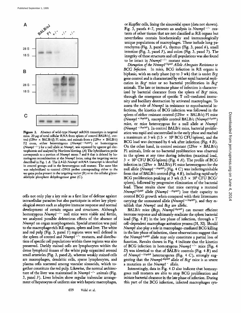
Figure 2. Absence of wild-type Nramp1 mRNA transcripts in targeted mice. 20/zg of total cellular RNA from spleen of control BALB/cJ, con- tro! (129sv x BALB/cJ) F1 mice, and animals from a (129sv x BALB/c) F2 cross, either heterozygous (Nrarnlol-/A'p169) or homozygous (Nrarapl-/-) for a null allele at Nrarapl, was separated by agarose gel elec- trophoresis and analyzed by Northern blotting. (A) The hybridization probe corresponds to a portion of Nrara_pl exons 7 and 8 that is deleted by ho- mologous recombination at the Nrarapl locus, using the targeting vector described in Fig. 1 A. The 2.4-kb Nrampl mtLNA transcript is identified in control groups and in the heterozygous null mutant. The same blot was rehybridized to control cDNA probes corresponding either to the neo gene probe present in the targeting vector (B) or to the cellular glycer- aldehyde phosphate dehydrogenase gene (C).
cells not only play a key role as a first line of defense against intracellular parasites but also participate in other key physi- ological events such as adaptive immune response and normal development of certain organs and structures. Although homozygous Nramlol-/- null mice were viable and fertile, we analyzed possible deleterious effects of the absence of Nrampl on organ morphogenesis. Special attention was paid to the macrophage-rich RE organs, spleen and liver. The white and red pulp (Fig. 3, panel I ) regions were well defined in the spleen of control and Nramp1 -/- mutants, and distribu- tion of specific cell populations within these regions was also preserved. Darkly stained cells are lymphocytes within the dense lymphoid tissues of the white pulp organized around small arterioles (Fig. 3, panel 2), whereas weakly stained cells are macrophages, dendritic cells, sparse lymphocytes, and plasma cells scattered among vascular sinusoids, which to- gether constitute the red pulp. Likewise, the normal architec- ture of the liver was maintained in Nrampl -/- animals (Fig. 3, panel 3). Liver lobules show regular trabecular arrange- ment of hepatocytes of uniform size with hepatic macrophages,
659 Vidal et al.
or Kupffer cells, lining the sinusoidal space (data not shown). Fig. 3, panels 4-7, presents an analysis in Nrampl-/- mu- tants of other tissues that are not classified as RE organs but nevertheless contain biochemically and immunologically unique populations of macrophages. These include lung pa- renchyma (Fig. 3, panel 4), thymus (Fig. 3, panel 6), small intestine (Fig. 3, panel 5), and colon (Fig. 3, panel 7). The integrity of these structures and cell populations was also found to be intact in Nrampl-/- mutant mice.
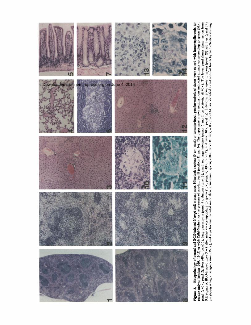
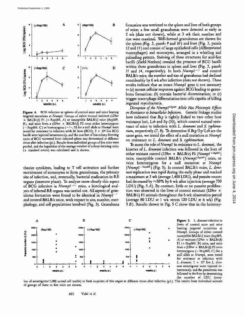
Disruption of the NramlolCly 169 Allele Abrogates Resistance to BCG Infection. In mice, BCG infection in RE organs is biphasic, with an early phase (up to 3 wk) that is under Bcg gene control and is characterized by either rapid bacterial repli- cation in Bcg s mice or no bacterial proliferation in Bcg r animals. The late or immune phase of infection is character- ized by bacterial clearance from the spleen of Bcg ~ mice, through the emergence of specific T cell-mediated immu- nity and bacillary destruction by activated macrophages. To assess the role of Nrampl in resistance to mycobacterial in- fections, the kinetics of BCG infection was followed in the spleen of either resistant control (129sv x BALB/c) F1 mice (Nrampl +/a~v169), susceptible control BALB/c (NramplA~p 169) mice, or mice heterozygous for a null allele at Nrampl (Nrampl -/A,r169). In control BALB/c mice, bacterial prolifer- ation was rapid and uncontrolled in the early phase and reached a maximum at 3 wk (1.5 x 10 s BCG CFU/spleen), and the BCG load was decreased by 6 wk after infection (Fig. 4 B). On the other hand, in control resistant (129sv x BALB/c) F1 animals, little or no bacterial proliferation was observed in the spleen at any time during infection (maximal load, 3 x 10³ CFU BCGApleen) (Fig. 4 A). The profile of BCG infection in (129sv x BALB/c) F1 mice heterozygous for the null allele (Nrampl-/.~t,169) (Fig. 4 C) was indistinguishable from that of BALB/c control (Fig. 4 B), including rapid early BCG proliferation peaking at 3 wk (1.5 x l0 s CFU BCG/ spleen), followed by progressive elimination of the bacterial load. These results show that mice carrying a mutated NramplCtr 169 allele (Nrampl-/Asei69) lose their capacity to control BCG growth when compared with their littermates carrying the unmutated allele (Nrampl +/A,p169), and they es- tablish that Nrampl and Beg are allelic.
BALB/c mice (Bcgs, NrampIA'e 169) can mount efficient immune response and ultimately eradicate the spleen bacterial load (Fig. 4 B) in the late phase of infection, through a T cell--dependent macrophage activation process (31, 32). Should NrampI also play a role in macrophage--mediated BCG killing in the late phase of infection, these observations suggest that the NramplAsp 169 allele may only constitute a partial loss of function. Results shown in Fig. 4 indicate that the kinetics of BCG infection in homozygous Nrampl-/- mice (Fig. 4 D) was identical to that of BALB/c controls (Fig. 4 B) and of Nrampl-/Ase 169 heterozygotes (Fig. 4 C), strongly sug- gesting that the NrampIA'e ~69 allele of Bcgs mice is as severe a mutation as the Nrampl- allele.
Interestingly, data in Fig. 4 D also indicate that homozy- gous null mutants are able to stop BCG proliferation and initiate bacterial clearance in the late phase of infection. During this part of the BCG infection, infected macrophages syn-
on June 4, 2014jem
.rupress.orgD
ownloaded from
Published September 1, 1995
Figu
re 3
. H
isto
path
olog
y of
nor
mal
and
BC
G-in
fect
ed N
ram
lsl n
ull
mut
ant
mic
e. H
isto
logi
c se
ctio
ns (
5/zm
thi
ck)
of fo
rmal
in-f
ixed
, pa
raff
in-e
mbe
dded
tiss
ues
wer
e st
aine
d w
ith h
emat
oxyl
in-e
osin
for
ro
utin
e an
alys
is (s
ectio
ns 1
-10,
12-
13) o
r w
ith Z
eihl
-Nee
lsen
for
the
pres
ence
of a
cid-
fast
bac
illi
(sec
tions
11
and
14).
The
upp
er p
anel
sho
ws
sect
ions
fro
m u
ninf
ecte
d an
imal
s co
rres
pond
ing
to s
plee
n (1
0x,
pane
l 1;
40x
, pa
nel 2
), liv
er (
40x,
pan
el 3
), l
ung
pare
nchy
ma
(pan
el 4
), t
hym
us (
pane
l 6),
sm
all
and
larg
e in
test
ine
(pan
els
5 an
d 7,
res
pect
ivel
y; a
ll 40
• T
he l
ower
pan
el s
how
s tis
sue
sect
ion
from
R
E o
rgan
s of
BC
G-in
fect
ed m
ice
3 w
k af
ter
infe
ctio
n co
rres
pond
ing
to s
plee
n (1
0x,
pane
l 8;
40x
, pa
nel 9
), a
nd l
iver
(40
x, p
anel
12)
. In
divi
dual
gra
nulo
mas
in
sple
en (
pane
l 10
) an
d liv
er (p
anel
13)
ar
e sh
own
at h
ighe
r m
agni
ficat
ion
(160
x ),
and
myc
obac
teria
incl
uded
ins
ide
thes
e gr
anul
omas
(sp
leen
, 200
x, p
anel
11;
live
r, 40
0 x,
pan
el 1
4 ) a
re id
entif
ied
as r
ed a
cid-
fast
bac
illi b
y Ze
ihl-N
eels
en s
tain
ing.
on June 4, 2014jem.rupress.orgDownloaded from
Pub
lishe
d S
epte
mbe
r 1,
199
5
2
( + / A s p 1 6 9 ) A s "
1 2 3 4 5 6
4 "
3 ,
(Asp169) B
1 2 3 4 5 6
e-
e~
b 8
( - / A s p 1 6 9 ) C e
4
�9 3 1 2 3 4 5 6
weeks p.L
(-/-) D
I 2 3 4 5 6
weeks p.i.
Figure 4. BCG infection in spleens of control mice and mice bearing targeted mutations at NrampI. Groups of either control resistant (1295v x BALB/cJ) F1 (+/Aspt69, A) or susceptible BALB/cJ mice (Asp169, B), and mice from a (1295v x BALB/cJ) F2 cross either heterozygous (-/Asp169, C) or homozygous (-/-, D) for a null allele at Nrampl were tested for resistance to infection with M. bovis (BCG). 2 x 10⁴ live BCG bacilli were injected intravenously, and the number of live colony-forming units of BCG recovered from infected spleen was determined at different times after infection ~i.). Results from individual groups of five mice were pooled, and the logarithm of the average number of colony-forming units (+_ standard errors) was calculated and is shown.
thesize cytokines, leading to T cell activation and further recruitment of monocytes to form granulomas, the primary site of infection, and, eventually, bacterial eradication in RE organs (immune phase). To analyze more closely this aspect of BCG infection in Nrarnp1-/- mice, a histological anal- ysis of infected RE organs was carried out. All aspects of gran- uloma formation were found to be identical in Nramp1-l- and control BALB/c mice, with respect to site, number, mor- phology, and cell populations involved (Fig. 3). Granuloma
formation was restricted to the spleen and liver of both groups of mice; a few small granulomas were detected as early as 2 wk (data not shown), while at 3 wk their number and size were maximal. Well-formed granulomas are shown for the spleen (Fig. 3, panels 9 and 10) and liver (Fig. 3, panels 12 and 13) and consist of large epitheloid cells (differentiated macrophages) and monocytes, arranged in a whirling and palisading pattern. Staining of these structures for acid-fast bacilli (Ziehl-Neelsen) revealed the presence of BCG bacilli within these granulomas in spleen and liver (Fig. 3, panels 11 and 14, respectively). In both Nrampl-/- and control BALB/c mice, the number and size of granulomas had declined considerably by 6 wk after infection (data not shown). These results indicate that an intact Nrampl gene is not necessary to (a) mount cellular responses against BCG leading to granu- loma formation; (b) contain bacterial dissemination; or (c) trigger macrophage differentiation into cells capable of killing ingested mycobacteria.
Disruption of the NramplCly 169 Allele Has Pleiotropic Effects on Resistance to Intracellular Infections. Genetic linkage studies have indicated that Beg is tightly linked to two other host resistance loci, Lsh and It), (33), which control natural resis- tance of mice to infection with L. donovani and S. typhimu- rium, respectively (7, 8). To determine ifBcg/Ity/Lsh are the same gene, we tested the effect of a null mutation at Nrampl on resistance to L. donovani and S. typhimurium.
To assess the role of Nrampl in resistance to L. donovani, the kinetics of L. donovani infection was followed in the liver of either resistant control (1295v x BALB/c) F1 (Nrampl +/.~.~169) mice, susceptible control BALB/c (NramplA~P 169) mice, or mice heterozygous for a null mutation at Nrampl (Nrampl-/A~p169) (Fig. 5). In control BALB/c mice, L. dono- vani replication was rapid during the early phase and reached a maximum at 3 wk (average 1,400 LDU), and parasite counts had decreased by '~50% by 6 wk after injection (average 750 LDU) (Fig. 5 A). By contrast, little or no parasite prolifera- tion was observed in the liver of control resistant (129sv x BALB/c) F1 animals throughout the 6-wk observation period (average 80 LDU at 1 wk versus 120 LDU at 6 wk) (Fig. 5 B). Results shown in Fig. 5 C show that in the heterozy-
i
i n
A
25OO
2OOO
1500
1000
5OO
0
( A s p 1 6 9 )
| .
IIi I I I I I
I 2 3 4 5 6
w e e k s p.i.
B
2500"
2000.
1500'
1000,
5 0 0
0
( + / A s p 169)
c
25OO
2OOO
1500
1000,
S00. | I
I �9 l I ! ! : : = 0 ! I l I I I 2 3 4 s e o ~ 2 3 4 s e
w e e k s p. i . w e e k s p. i .
( - / A s p 169) �9
Figure 5. L. donovani infection in livers of control mice and mice bearing targeted mutations at Nrampl. Groups of either control susceptible BALB/cJ mice (Asp169, A) or resistant (129sv x BALB/cJ) F1 (+/Asp169, B) mice, and mice from a (1295v x BALB/cJ) F2 cross heterozygous (-/Asp169, C) for a null allele at Nrarap1, were tested for resistance to infection with L. donovani. 1 x 10⁷ live L. dono- vani amastigotes were injected in- travenously, and the parasitemia was followed in the liver by determining the number of LDU (num-
ber of amastigotes/1,000 scored cell nuclei) in fresh imprints of this organ at different times after infection (Fi.). The results from individual animals of groups of three to five mice are shown.
661 Vidal et al.
on June 4, 2014jem
.rupress.orgD
ownloaded from
Published September 1, 1995
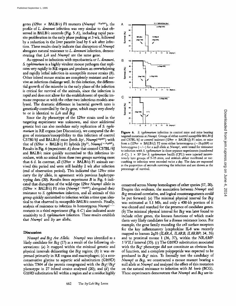
gotes (129sv x BALB/c) F1 mutants (Nrampl-/A,p169), the A profile of L. donovani infection was very similar to that ob- served in BALB/c controls (Fig. 5 A), including rapid para- site proliferation in the early phase peaking at 3 wk, followed by a reduction in the liver parasite load by 6 wk after infec- 0~ tion. These results clearly indicate that disruption of Nrampl abrogates natural resistance to L. donovani infection, demon- strating that Lsh and Nrampl are the same gene.
As opposed to infections with mycobacteria or L. donovani, , o o
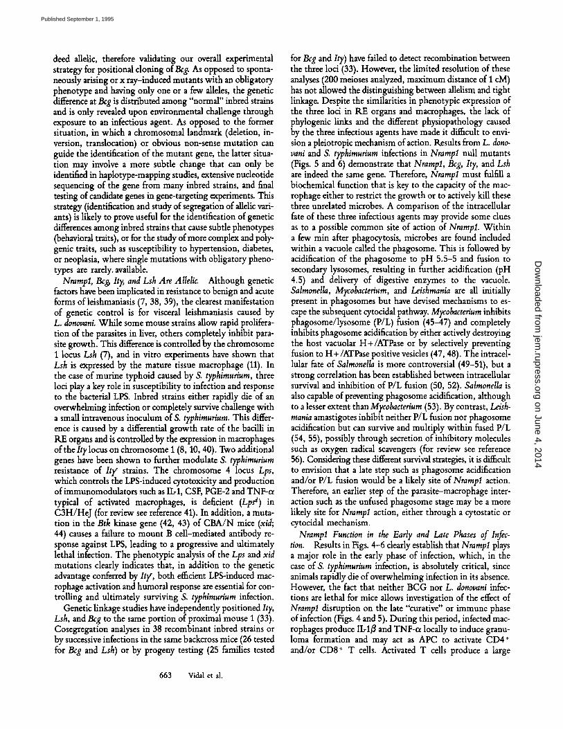
S. typhimurium is a highly virulent mouse pathogen that repli- 8o cates very rapidly in RE organs and produces an overwhelming and rapidly lethal infection in susceptible mouse strains (8). ~0 Other inbred mouse strains are completely resistant and sur- ,o vive an infectious challenge well. In this infection, the differen- 2o tial growth of the microbe in the early phase of the infection 0 is critical for survival of the animals, since the infection is rapid and does not allow for the establishment of specific im- c 100 mune response as with the other two infectious models ana- lyzed. The dramatic difference in bacterial growth rates is ~ ,0 genetically controlled by the Ity gene, which maps very closely to or is identical to Lsh and Bcg.
Since the Ity phenotype of the 129sv strain used in the targeting experiment was unknown, and since additional 0 genetic loci can also modulate early replication of S. typhi- murium in RE organs (see Discussion), we compared the de- gree of resistance/susceptibility to this infection of control C57BI/6J and BALB/cJ mice (both Ity ~, NramlolA~ 169) with that of (129sv x BALB/c) F1 hybrids (Itff s, Nrampl +/A~169). Results in Fig. 6 (experiment A) show that control C57BL/6J and BALB/c mice quickly succumbed to this infectious in- oculum, with no animal from these two groups surviving more than 6 d. In contrast, all (129sv x BALB/c) F1 animals sur- vived this period and were still healthy 2 wk after infection (end of observation period). This indicated that 129sv mice carry the Ity ~ allele, in agreement with previous haplotype- typing data (20). Results from experiment B in Fig. 6 indi- cated that disruption of the wild-type 129sv Nrampl allele in (129sv x BALB/c) F1 mice (Nramp1-/~169) abrogated their resistance to S. typhimurium infection, and all animals of this group quickly succumbed to infection within a time span iden- tical to that observed in susceptible BALB/c controls. Finally, analysis of resistance to infection in homozygous Nrampl -l- mutants in a third experiment (Fig. 6 C) also indicated acute sensitivity to S. typhimurium infection. These results establish that Nrampl and It), are allelic.
Discussion
Nrampl and Bcg Are Allelic. Nrampl was identified as a likely candidate for Bcg (17) as a result of the following ob- servations: (a) It mapped within the minimal genetic and physical intervals delineating the Beg region; (b) it was ex- pressed primarily in RE organs and macrophages; (c) a non- conservative glycine to aspartic acid substitution (G169D) within TM4 of the protein cosegregated with the Bcg'/Bcg' phenotype in 27 inbred strains analyzed (20); and (d) the G169D substitution fell within a region and at a residue highly
i I o . . . . 1 3
F I (129SvXBALB/c) (+/Asp169)
I C67BL~ (.,~ple9) /
7 9 11 13 r p.I.
(§
A~olo9)
I 3 5 7 9 11 13 days p.l.
(+/Aaole0)
(-I-)
J
�9 ~ ' ~ ' f l ;3 ~oy, p.~.
Figure 6. S. ty?himurium infection in control mice and mice bearing targeted mutations at Nrara?l. Groups of either control susceptible BALB/cJ and C57BL/6J or control resistant (129sv x BALB/cJ) F1 mice, or mice from a (129sv x BALB/cJ) F2 cross either heterozygous (-/Asp169) or homozygous ( - / - ) for a null allele at Nrampl, were tested for resistance to infection with S. typhimurium in three separate experiments (numbered A-C). 1 • 104 live S. typhimurium bacilli (CFU) were injected intrave- nously into groups of 5-15 mice, and animals either moribund or suc- cumbing to infection were recorded twice a day. The data are expressed as the proportion of animals surviving the infection and are shown as the percentage of survival.
conserved across Nramp homologues of other species (17, 20). Despite this evidence, the association between Nramp1 and Beg remained correlative, and logical counterarguments could be put forward: (a) The minimal physical interval for Beg was estimated at 1.1 Mb, and only a 400-kb portion of it was cloned and searched for the presence of candidate genes. (b) The minimal physical interval for Bcg was later found to include other genes, the known functions of which made them very likely candidates for a disease resistance locus. For example, the gene family encoding the cell surface receptors for the key inflammatory lymphokine Ib8 was recently mapped to human 2q35 (IL8RA, IL8RB, IL8RBP; 34, 35) and to proximal mouse 1 (36, 37), within the NRAMP- I/VILI interval (35). (c) The G169D substitution associated with the Bcg' phenotype did not constitute an obvious loss of function, and a complete polypeptide was expected to be produced in Bcgs mice. To formally test the candidacy of Nramp1 as Bcg, we constructed a mouse mutant bearing a null allele at Nrampl and analyzed the effect of this mutation on the natural resistance to infection with M. b0v/s (BCG). These experiments demonstrate that Nramp1 and Bcg are in-
662 The Ity/Lsh/Bcg Locus
on June 4, 2014jem
.rupress.orgD
ownloaded from
Published September 1, 1995
deed allelic, therefore validating our overall experimental strategy for positional cloning of Beg. As opposed to sponta- neously arising or x ray-induced mutants with an obligatory phenotype and having only one or a few alleles, the genetic difference at Beg is distributed among "normal" inbred strains and is only revealed upon environmental challenge through exposure to an infectious agent. As opposed to the former situation, in which a chromosomal landmark (deletion, in- version, translocation) or obvious non-sense mutation can guide the identification of the mutant gene, the latter situa- tion may involve a more subtle change that can only be identified in haplotype-mapping studies, extensive nucleotide sequencing of the gene from many inbred strains, and final testing of candidate genes in gene-targeting experiments. This strategy (identification and study of segregation of allelic vari- ants) is likely to prove useful for the identification of genetic differences among inbred strains that cause subtle phenotypes (behavioral traits), or for the study of more complex and poly- genic traits, such as susceptibility to hypertension, diabetes, or neoplasia, where single mutations with obligatory pheno- types are rarely, available.
Nrampl, Beb It),, and Lsh Are Allelic. Although genetic factors have been implicated in resistance to benign and acute forms of leishmaniasis (7, 38, 39), the clearest manifestation of genetic control is for visceral leishmaniasis caused by L. donovani. While some mouse strains allow rapid prolifera- tion of the parasites in liver, others completely inhibit para- site growth. This difference is controlled by the chromosome 1 locus Lsh (7), and in vitro experiments have shown that Lsh is expressed by the mature tissue macrophage (11). In the case of murine typhoid caused by S. typhimurium, three loci play a key role in susceptibility to infection and response to the bacterial LPS. Inbred strains either rapidly die of an overwhelming infection or completely survive challenge with a small intravenous inoculum of S. typhimurium. This differ- ence is caused by a differential growth rate of the bacilli in RE organs and is controlled by the expression in macrophages of the It), locus on chromosome 1 (8, 10, 40). Two additional genes have been shown to further modulate S. typhimurium resistance of Ity r strains. The chromosome 4 locus Lps, which controls the LPS-induced cytotoxicity and production of immunomodulators such as IL-1, CSF, PGE-2 and TNF-cx typical of activated macrophages, is deficient (Lps d) in C3H/HeJ (for review see reference 41). In addition, a muta- tion in the Btk kinase gene (42, 43) of CBA/N mice (xid; 44) causes a failure to mount B cell-mediated antibody re- sponse against LPS, leading to a progressive and ultimately lethal infection. The phenotypic analysis of the Lps and xid mutations clearly indicates that, in addition to the genetic advantage conferred by Ity ~, both efficient LPS-induced mac- rophage activation and humoral response are essential for con- trolling and ultimately surviving S. typhimurium infection.
Genetic linkage studies have independently positioned Ity, Lsh, and Beg to the same portion of proximal mouse 1 (33). Cosegregation analyses in 38 recombinant inbred strains or by successive infections in the same backcross mice (26 tested for Beg and Lsh) or by progeny testing (25 families tested
for Beg and Ity) have failed to detect recombination between the three loci (33). However, the limited resolution of these analyses (200 meioses analyzed, maximum distance of 1 cM) has not allowed the distinguishing between allelism and tight linkage. Despite the similarities in phenotypic expression of the three loci in RE organs and macrophages, the lack of phylogenic links and the different physiopathology caused by the three infectious agents have made it difficult to envi- sion a pleiotropic mechanism of action. Results from L. dono- vani and S. typhimurium infections in Nrampl null mutants (Figs. 5 and 6) demonstrate that Nrampl, Beg, It),, and Lsh are indeed the same gene. Therefore, Nrampl must fulfill a biochemical function that is key to the capacity of the mac- rophage either to restrict the growth or to actively kill these three unrelated microbes. A comparison of the intraceUular fate of these three infectious agents may provide some clues as to a possible common site of action of Nrampl. Within a few min after phagocytosis, microbes are found included within a vacuole called the phagosome. This is followed by acidification of the phagosome to pH 5.5-5 and fusion to secondary lysosomes, resulting in further acidification (pH 4.5) and delivery of digestive enzymes to the vacuole. Salmonella, Mycobacterium, and Leishmania are all initially present in phagosomes but have devised mechanisms to es- cape the subsequent cytocidal pathway. Mycobacterium inhibits phagosome/lysosome (P/L) fusion (45-47) and completely inhibits phagosome acidification by either actively destroying the host vacuolar H+/ATPase or by selectively preventing fusion to H +/ATPase positive vesicles (47, 48). The intracel- lular fate of Salmonella is more controversial (49-51), but a strong correlation has been established between intracellular survival and inhibition of P/L fusion (50, 52). Salmonella is also capable of preventing phagosome acidification, although to a lesser extent than Mycobacterium (53). By contrast, Le/sh- mania amastigotes inhibit neither P/L fusion nor phagosome acidification but can survive and multiply within fused P/L (54, 55), possibly through secretion of inhibitory molecules such as oxygen radical scavengers (for review see reference 56). Considering these different survival strategies, it is difficult to envision that a late step such as phagosome acidification and/or P/L fusion would be a likely site of Nrampl action. Therefore, an earlier step of the parasite-macrophage inter- action such as the unfused phagosome stage may be a more likely site for Nrampl action, either through a cytostatic or cytocidal mechanism.
Nrampl Function in the Early and Late Phases of Infec- tion. Results in Figs. 4-6 dearly establish that Nrampl plays a major role in the early phase of infection, which, in the case of S. typhimurium infection, is absolutely critical, since animals rapidly die of overwhelming infection in its absence. However, the fact that neither BCG nor L. donovani infec- tions are lethal for mice allows investigation of the effect of Nrampl disruption on the late "curative" or immune phase of infection (Figs. 4 and 5). During this period, infected mac- rophages produce II,-1/3 and TNF-cx locally to induce granu- loma formation and may act as APC to activate CD4 § and/or CD8 + T cells. Activated T cells produce a large
663 Vidal et al.
on June 4, 2014jem
.rupress.orgD
ownloaded from
Published September 1, 1995
number of cytokines, most importantly IFN-~ and IL-2, which mediate the activation of macrophage bactericidal functions such as respiratory burst, nitric oxide synthesis, and produc- tion of a large number of proteases and other lysosomal en- zymes. The key role of IFN-~,-mediated macrophage activa- tion in eradication of these infections has been established in vivo and in vitro (57-60, 28). Our results indicate that Nrampl ablation impairs neither granuloma formation (Fig. 3) nor successful microbicidal response in this part of the in- fection (Figs. 4 and 5). Since activated macrophages are the key effector ceUs of this response, these results suggest that Nrarap1 does not play a major role in the elimination of in- tracdhilar parasites by activated macrophages. What biochem- ical mechanism of Nrarapl action can be proposed to account for a key role during the early but not the late phase of infec- tion can only be speculated upon. A possible cytocidal func- tion mediated by NrampI could play a major role early in infection, but may be redundant and may only form part of a number of additional cytocidal mechanisms expressed in the late phase after macrophage activation. The observation that Nrarapl is constitutively expressed in macrophages but is dramatically induced by IFN-qr and LPS treatments is in agreement with a function for Nrampl in both resting and activated macrophages (Gros, P., and G. Govoni, unpublished observations). Alternatively, Nrarapl could have a cytostatic rather than cytocidal function discernible only early during infection, and perhaps creating an intraphagosomal environ- ment unfavorable to parasite replication. In support of a cytostatic function for Nrampl are metabolic labding studies of Leiskraania amastigotes in infected livers, where differences in rates of replication rather than of active elimination were associated with the genetic advantage of Lsh r macrophages (61). Irrespective of the model, the study of Nramp1 mutant mice clearly suggests a novel mechanism by which macro- phages can control replication of taxonomically diverse microorganisms independently of the immune system.
Nramplmp 169 Is a Null Allele. The observation that Nrampl a'P ~69 homozygotes, Nrarapl -/A~p~69 heterozygotes, or Nrampl-/- homozygotes are equally susceptible to BCG, Salmonella, and L. donovani infections (Figs. 4-6) indicates that the G169D substitution is not a partial loss of function but is rather a null mutation at Nrampl. Nrampl is a novel integral membrane protein, the salient predicted features of which include I2 TM domains, with 7 of them containing at least one charge, an extracellular glycosylated loop, and an intracellular consensus transport motif previously described in a large number of prokaryotic and eukaryotic membrane transporters. This suggests that Nrampl performs a trans-
port function in macrophages. We have recently determined that (a) Nrarapl is a member of a family of three genes in rodents (19, 62) that encode highly conserved polypeptides (85% homology) (19); and (b) the Nramp family is extremdy weU conserved through evolution, with members identified in all animal species tested (75-90% homology), but also in distant eukaryotes such as plants, flies, and yeast (50-70% homology) (Gros, P., unpublished results). The predicted TM domains are the most conserved domains (up to 90% for homologues and 75% for orthologues), including the charged amino acids within them, which together suggest a key struc- tural and/or functional role for these segments and residues in a common mechanism of action of these proteins (e.g., stabilization of unstable charges in the lipid bilayer; 63). The G169D substitution would replace a small neutral amino add by a large residue with a charged side chain within predicted TM4 of Nrampl. Considering the high degree of evolutionary conservation of both the sequence and charges within TM domains, the introduction of an additional charged residue within a TM domain would be expected to have severe dele- terious effects on protein function. This mutation could affect either proper targeting, insertion, or folding of Nrampl within the membrane or may interfere with the intimate mechanism of transport of Nrampl.
Haplotype-mapping studies of genealogically and phyloge- nedcaUy distant inbred mouse strains have indicated that Beg s strains share a common chromosome 1 haplotype around Nrarapl. This suggests that the NramplasP 169 allele may have been present in several founders of the different inbred strains and had become randomly fixed in some strains. The persis- tence of the potentially deleterious Asp169 aUele in inbred mouse strains or even in wild mice can be explained in two ways. First, although it is dear that the uncontrolled replica- tion of intracellular parasites in RE organs typical of the Asp169 allele may have a dramatic deleterious effect on sur- vival of Salmonella infection, it may be counterbalanced by positive secondary effects for other types of infection. Indeed, Asp169 animals may be more prone to mounting a vigorous immune response against certain benign infections, which in turn may advantageously cross-protect them against more virulent related organisms that may prove lethal to G1y169- bearing mice. Alternatively, it is possible that maintaining heterozygosity for one of the markers tightly linked to Nrarapl and preserved in the haplotype-defining Bcg' strains may confer a yet to be identified advantage over strains homozy- gous for the Gly169 allele. This advantage may have helped preserve the Asp169 in the breeding of inbred strains and may even have enabled its survival in the wild.
We wish to thank Dr. J. Rossant (Mount Sinai Hospital, Toronto, Canada) for the generous gift of the 129sv/J genomic I phage library, Dr. E. Li and Dr. R. Jaenisch (Whitehead Institute for Biomedical Re- search, Cambridge, MA) for the J1 ES cell line, Dr. G. Matlashewski (Institute of Parasitology, McGiU University) for providing Syrian hamsters infected with L. donouani, and Dr. H. Robson (Royal Victoria Hospital, McGill University) for the gift of an S. typhirauriura isolate. We would like to acknowledge M. Boulr, Eva Michaliszyn, and P. Brinkley for technical assistance.
664 The Ity/Lsh/Bcg Locus
on June 4, 2014jem
.rupress.orgD
ownloaded from
Published September 1, 1995
This work was supported by research grants to P. Gros, E. Skamene, and M. L. Tremblay from the Na- tional Institutes of Health (5R01 AI35237.02). P. Gros is supported by an E. W. R. Steacie fellowship from the National Sciences and Engineering Research Council of Canada and is an International Research Scholar of the Howard Hughes Medical Institute. S. Vidal is supported by a fellowship from Canadian Genetic Diseases Network-Merck Frosst. M. L. Tremblay and D. Malo are recipients of career awards from the Fonds de la Recherche en Sant6 du Qu6bec.
Address correspondence to Dr. P. Gros, Department of Biochemistry, McGill University, 3655 Drum- mond, Room 907, Montreal, Quebec, Canada H3G 1Y6.
Received for publication 3 April 1995.
References
1. Malo, D., and E. Skamene. 1994. Genetic control of host resis- tance to infection. Trends Genet. 10:365-371.
2. Gros, P., E. Skamene, and A. Forget. 1981. Genetic control of natural resistance to Mycobacterium bovis BCG. J. Immunol. 12:2417-2421.
3. Skamene, E., P. Gros, A. Forget, p.J. Patel, and M. Nesbitt. 1984. Regulation of resistance to leprosy by chromosome 1 locus in the mouse. Immunogenetics. 19:117-120.
4. Goto, Y., E. Buschman, and E. Skamene. 1989. Regulation of host resistance to Mycobacterium intracellulare in vivo and in vitro by the Bcg gene. Immunogenetics. 30:218-221.
5. PeUetier, M., A. Forget, D. Bourassa, P. Gros, and E. Skamene. 1982. Immunohistopathology study of Mycobacterium bovis (BCG) infection in innately resistant and susceptible mice. J. Immunol. 129:2179-2182.
6. Brett, S., J.M. Orrell, J. Swanson Beck, and J. Ivanyi. 1992. Influence of H-2 genes on growth of MFobacterium tuberculosis in the lungs of chronically infected mice. Immunology. 76: 129-132.
7. Bradley, D.J. 1977. Genetic control of Leishmania populations within the host. II. Genetic control of acute susceptibility of mice to L. donovani infection. Clin. Exp Immunol. 30:130-140.
8. Plant, J.E., and A. Glynn. 1976. Genetics of resistance to in- fection with Salmonella typkimurium in mice. J. Infect. Dis. 133:72-78.
9. Stach, J.L., P. Gros, A. Forget, and E. Skamene. 1984. Pheno- typic expression of genetically controlled natural resistance to MFobacterium bovis (BCG). J. Immunol. 132:888-892.
10. Lissner, C.R., R.N. Swanson, and A.D. O'Brien. 1983. Genetic control of the innate resistance of mice to Salmonella typkimur- ium: expression of the Ity gene in peritoneal and splenic mac- rophages isolated in vitro f Immunol. 131:3006-3013.
11. Olivier, M., and C.E. Tanner. 1987. Susceptibilities of macro- phage populations to infection in vitro by Leiskmania donovani. Infect. Immun. 55:467-471.
12. Gros, P., E. Skamene, and A. Forget. 1983. Cellular mecha- nisms of genetically-controlled host resistance to Mycobacterium bov/s (BCG). f Immunol. 131:1966-1973.
13. O'Brien, A.D., and E.S. Metcalf. 1982. Control of early Salmonella typhimurium growth in innately Salmonella-resistant mice does not require functional T lymphocytes.J. Immunol. 129:1349-1351.
14. Crocker, P.R.,J.M. Blackwell, and D.J. Bradley. 1984. Transfer of innate resistance and susceptibility to Leishmania donovani infection in mouse radiation bone marrow chimaeras. Immu. nology. 52:417-422.
665 Vidal et al.
15. Malo, D., S.M. Vidal, J. Hu, E. Skamene, and P. Gros. 1993. High resolution linkage map in the vicinity of the host resis- tance locus Beg. Genomics. 16:655-663.
16. Malo, D., S. Vidal, J.H. Lieman, D.C. Ward, andP. Gros. 1993. Physical delineation of the minimal chromosomal segment en- compassing the murine host resistance locus Beg. Genomics. 17:667-675.
17. Vidal, S.M., D. Malo, K. Vogan, E. Skamene, and P. Gros. 1993. Natural resistance to infection with intraceUular para- sites: identification of a candidate gene for Bcg. Cell. 73:469-485.
18. Unkles, S.E., K.L. Hawker, C. Grieve, E.I. Campbell, P. Mon- tague, andJ.R. Kinghorn. 1991. crnA encodes a nitrate trans- porter in Aspergillus nidulans. Pro~ Natl. Acad. Sci. USA. 88:204-208.
19. Gruenheid, S., M. CeUier, S. Vidal, and P. Gros. 1995. Identification and characterization of a second mouse Nramp gene. Genomics. 25:514-525.
20. Malo, D., K. Vogan, S. Vidal, J. Hu, M. Cellier, E. Schurr, A. Fuks, N. Bumstead, K. Morgan, and P. Gros. 1994. Haplo- type mapping and sequence analysis of the mouse Nramp gene predicts susceptibility to infection with intracellular parasites. Genomics. 23:51-61.
21. Li, E., T.H. Bestor, and R. Jaenisch. 1992. Targeted mutation of the DNA methyltransferase gene results in embryonic lethality. Cell. 69:915-926.
22. Love, P.E., M.L. Tremblay, and R. Westphal. 1992. Targeting of the T-cell receptor z-chain in embryonic stem cells: strate- gies for generating multiple mutations in a single gene. Proc. Natl. Acad. Sci. USA. 89:9929-9933.
23. Ramirez-Solis, R., J. Perez-Rivera, J.D. Wallace, M. Wims, H. Zheng, and A. Bradley. 1992. Genomic DNA microextrac- tion: a method to screen numerous samples. Anal. Biochem. 201:331-335.
24. Sambrook, J., E.F. Fritsch, and T. Maniatis. 1989. Molecular Cloning: A Laboratory Manual. 2nd ed. C. Nolan, editor. Cold Spring Harbor Laboratory, Cold Spring Harbor, NY.
25. Feinberg, A.P., and B. Vogelstein. 1984. A technique for ra- diolabeling DNA restriction endonucleases fragments to high specific activity. Anal. Biochem. 132:6-13.
26. Tybulewicz, V.L.J., M.L. Tremblay, M.E. IaMarca, R. Wil- lemsen, B.K. Stubblefields, S. Winfield, B. Zabloka, E. Sidran- sky, B.M. Martin, S.P. Huang, K.A. Mintzer, H. Westphal, R.C. Mulligan, and E.I. Ginns. 1992. Animal model of Gancher's disease from targeted disruption of the mouse glu- cocerebrosidase gene. Nature (Lond.). 357:407-410.
27. Chirgwin, J.M., A.A. Przybyla, R.J. MacDonald, and W.J.
on June 4, 2014jem
.rupress.orgD
ownloaded from
Published September 1, 1995
R.utter. 1979. Isolation of biologically active ribonucleic acid from sources enriched in ribonuclease. Biochemistry. 18:5294- 5306.
28. Murray, H.W., J.J. Stern, K. Welte, B.Y. Rubin, S. Carriero, and C.F. Nathan. 1987. Experimental visceral leishmaniasis: production of interleukin 2 and interferon-gamma, tissue im- mune reaction, and response to treatment with interleukin 2 and interferon-gamma. J. Immunol. 138:2290-2297.
29. Robson, H.G., and S.I. Vas. 1972. Resistance of inbred mice strains to Salmonella typhimurium, j . Infect. Dis. 126:378-386.
30. Thomas, K.R., and M.R. Capecchi. 1987. Site-directed muta- genesis by gene targeting in mouse embryo-derived stem cells. Cell. 51:503-512.
31. Rook, G.A.W. 1990. The role of activated macrophages in pro- tection and immunopathology in tuberculosis. Res. Microbiol. 141:253-256.
32. Wallis, R.S., andJ.J. Ellner. 1994. Cytokines and tuberculosis. J. Leukocyte Biol. 55:676-681.
33. Skamene, E., P. Gros, A. Forget, P.A.L. Kongshavn, C. St.- Charles, and B.A. Taylor. 1982. Genetic regulation of resis- tance to intracellular pathogenes. Nature (Lond.). 297:506-509.
34. Morris, S.W., N. Nelson, M.B. Valentine, N.D. Shapiro, A.T. Look, C.J. Kozlosky, P.M. Beckmann, and D.P. Cerretti. 1992. Assignment of the genes encoding human interleukin-8 recep- tor types 1 and 2 and an interleukin-8 receptor pseudogene to chromosome 2q35. Genomics. 14:685-691.
35. White, J.K., M.-A. Shaw, C.H. Barton, D.P. Cerretti, H. Wil- liams, B.A. Mock, N.P. Carter, C.S. Peacock, andJ.M. Black- well. 1994. Genetic and physical mapping of 2q35 in the re- gion of the NRAMP and ILSR genes: identification of a polymorphic repeat in exon 2 ofNRAMP. Genomics. 24:295- 302.
36. Wilkie, T.M., Y. Chen, D.J. Gilbert, K.J. Moore, L. Yu, M.I. Simon, N.G. Copeland, and N.A. Jenkins. 1993. Identification, chromosomal location, and genome organization of mammalian G-protein-coupled receptors. Genomics. 18:175-184.
37. Cerretti, D.P., N. Nelson, C.J. Kozlosky, P.J. Morrissey, N.G. Copeland, D.J. Gilbert, N.A. Jenkins, J.K. Dosik, and B.A. Mock. 1993. The murine homologue of the human interleu- kin-8 receptor type B maps near the Ity-Lsh-Bcg disease resis- tance locus. Genomics. 18:410-413.
38. Blackwell, J.M., J. Freeman, and D. Bradley. 1980. Influence of H-2 complex on acquired resistance to Leishmania donovani infection in mice. Nature (Lond.). 283:72-74.
39. Roberts, M., B.A. Mock, and M.J. Blackwell. 1993. Mapping of genes controlling Leishmania major infection in CXS recom- binant mice. Eur. J. Immunogenet. 20:349-362.
40. Plant, J., and A.A. Glynn. 1979. Locating Salmonella resis- tance gene on mouse chromosome 1. Clin. Exp. Immunol. 37:1-6.
41. Sultzer, B.M., R. Castagna, J. Bandekar, and P. Wong. 1993. Lipopolysaccharide nonresponder cells: the C3H/HeJ defect. Immunobiology. 187:257-271.
42. Thomas, J.D., P. Sideras, C.I.E. Smith, I. Vorechovsky, V. Chapman, and W.E. Paul. 1993. Colocalization of X-linked agammaglobulinemia and X-linked immunodeficiency genes. Science (Wash. DC). 261:355-358.
43. Rawlings, D.J., D.C. Saffran, S. Tsukada, D.A. Largaespada, and J.C. Grimaldi. 1993. Mutation of unique region of Bruton's tyrosine kinase in immunodeficient XID mice. Science (Wash. DC). 261:358-361.
44. O'Brien, A.D., I. Scher, and E.S. Metcalf. 1981. Genetically conferred defect in anti-Salmonella antibody formation renders CBA/N mice innately susceptible to Salmonella typhimurium infection. J. Immunol. 126:1368-1372.
45. Frehel, C., C. de Chastellier, T. Lang, and N. Rastogi. 1986.
Evidence for inhibition of fusion of lysosomal and prelysosomal compartments with phagosomes in macrophages infected with pathogenic Mycobacterium avium. Infect. Immun. 52:252-262.
46. Sibley, L.D., S.G. Franzblau, and J.L. Krahenbuhl. 1987. In- tracellular fate ofMycobacterium leprae in normal and activated mouse macrophages. Infect. lmmun. 51:461-469.
47. Sturgill-Koszycki, S., P.H. Schlesinger, P. Chakraborty, L.H. Pryce, H.L. Collins, A.K. Fok, R.D. Allen, S.L. Gluck, J. Heuser, and D.G. Russell. 1994. Lack of acidification in Mycobac- terium phagosomes produced by exclusion of the vesicular proton-ATPase. Science (Wash. DC). 263:678-681.
48. Xu, S., A. Cooper, S. Sturgill-Koszycki, T. van Heyninger, D. Chatterjee, I. Orme, P. Allen, and D.G. Russell. 1994. In- tracellular trafficking in Mycobacterium tuberculosis and Mycobac- ten'urn avium-infected macrophages.J. Immunol. 153:2568-2578.
49. Carrol, M.E.W., P.S. Jackett, V.R. Abet, and D.B. Lowrie. 1979. Phagolysosome formation, cyclic adenosine Y:5'-mono- phosphate and the fate of Salmonella t~himurium within mouse peritoneal macrophages. J. Gen. Microbiol. 110:421-429.
50. Buchmeier, N.A., and F. Heffron. 1991. Inhibition of macro- phage phagosome-lysosome fusion by Salmonella typhimurium. Infect. Immun. 59:2232-2238.
51. Isberg, R.R., and G.T. Van Nhieu. 1994. Two mammalian cell internalization strategies used by pathogenic bacteria. Annu. Rev. Genet. 27:395-442.
52. Ishibashi, Y., and T. Arai. 1990. Specific inhibition ofphago- some-lysosome fusion in murine macrophages mediated by Salmonella typhimurium infection. FEMS (Fed. Eur. Microbiol. Soc.) MicrobioL ImmunoL 64:35-44.
53. Alpuche Aranda, C.M., J.A. Swanson, W.P. Loomis, and S.I. Miller. 1992. Salmonella typhimurium activates virulence gene transcription within acidified macrophage phagosomes. Proc. Natl. Acad. Sci. USA. 89:10079-10083.
54. Antoine, J.C., E. Prina, C. Jouanne, and P. Bongrand. 1990. Parasitophorous vacuoles of Leishmania amazonensis-infected macrophages maintain an acidic pH. Infect. Immun. 58:779-787.
55. Russell, D.G., S. Xu, and P. Chakraborty. 1992. Intracellular trafficking and the parasitophorous vacuole of Leishmania mexicana-infected macrophages. J. Celt Sci. 103:1193-1210.
56. Mauel, J. 1990. Macrophage-parasite interactions in Leishmania infections. J. Leukocyte Biol. 47:187-193.
57. Flesch, I.E.A., and S.H.E. Kaufman. 1990. Activation oftuber- culostatic macrophage functions by gamma interferon, Ib4, and tumor necrosis factor. Infect. Immun. 58:2675-2677.
58. Cooper, A.M., D.K. Dalton, T.A. Stewart, J.P. Griffin, D.G. Russell, and I.M. Orme. 1993. Disseminated tuberculosis in interferon g gene-disrupted mice.J. Ext~ Med. 178:2243-2247.
59. Flynn, J.-A.L., J. Chan, K.J. Triebold, D.K. Dalton, T.A. Stewart, and B.R. Bloom. 1993. An essential role for inter- feron g in resistance to MFobacterium tuberculosis infection. J. Exp. Med. 178:2249-2254.
60. Murray, H.W., B.Y. Rubin, and C.D. Rothermel. 1983. Killing of intracellular Leishmania donovani by lymphokine-stimulated human mononuclear phagocytes. Evidence that interferon g is the activating lymphokine. J. Clin. Invest. 72:1506-1510.
61. Bradley, D.J.R. 1979. Regulation of Leishmania populations within the host. IV. Parasite and host cell kinetics studied by radioisotope labeling. Acta Trol~ 36:171-179.
62. Dosik, J.K., C.H. Barton, D.L. Holiday, M.M. Krall, J.M. Blackwell, and B.A. Mock. 1994. An Nramp-related sequence maps to mouse chromosome 17. Mature. Genome. 5:458-460.
63. Sahin-Toth, M., R.L. Dunten, A. Gonzalez, and H.R. Ka- back. 1992. Functional interactions between putative intramem- brane charged residues in the lactose permease of Escherichia coli. Proc. Natl. Acad. Sci, USA. 89:10547-10551.
on June 4, 2014jem
.rupress.orgD
ownloaded from
Published September 1, 1995
Copyright © 2022 FDOKUMEN