Characterization and identification of hidden rare variants in the human genome

Chapter 7
THE HUMAN GENOME
Although the clockwork of life is similar in prokaryotesand eukaryotes, eukaryotes are more complex. Prokary-otes must be mean and lean to ensure fast reproduc-tion. Therefore they keep their genomes small andregulate gene expression in simple yet efficient ways.Eukaryotes, however, require genetic complexity tocontrol their complex cellular and organismal struc-tures and sophisticated lifecycles.
Humans, for example, have 700 times more DNAthan Escherichia coli, but they have only six times moregenes (Table 7.1). This disparity comes from the factthat 90% of E. coli DNA, but only 1.3% of humanDNA, codes for proteins.
Gene regulation is more important than genenumber. Multicellular eukaryotes have (almost) thesame genes in every cell of the body, but differentgenes are expressed in different cell types and at differ-ent stages during the development of the organism.This requires control mechanisms of extraordinarycomplexity.
CHROMATIN CONSISTS OF DNA AND HISTONES
The prokaryotic chromosome is a naked circular DNAdouble helix with a length of about 1 mm. Eukaryoticchromosomes consists of a single linear DNA doublehelix with a length of several centimeters, which istightly packaged with a set of histone proteins.
The histones are small basic proteins, with numerouspositive charges on the side chains of lysine and argi-nine residues. These positive charges bind to the nega-tively charged phosphate groups of the DNA, andthey neutralize at least 60% of the negative chargeson DNA.
Eukaryotic cells have five major types of histones(Table 7.2). With the exception of histone H1, whosestructure varies in different species and even in differenttissues of the same organism, the histones are well con-served throughout the phylogenetic tree. For example,histones H3 and H4 from pea seedlings and calf thymusdiffer in only four and two amino acid positions,respectively. Presumably, the histones were inventedby the very first eukaryotes, perhaps as early as 2 billion
years ago, and have served the same essential functionsever since.
Chromatin, named for its affinity for basic dyes suchas hematoxylin and fuchsin, contains roughly equalamounts of DNA and histones. Euchromatin has aloose structure, whereas heterochromatin is moretightly condensed and deeper staining. Genes areactively transcribed in euchromatin but are repressedin heterochromatin.
THE NUCLEOSOME IS THE STRUCTURALUNIT OF CHROMATIN
Under the electron microscope, euchromatin looks likebeads on a string. The “string” is the DNA doublehelix, and the “beads” are nucleosomes, which are littledisks formed from two copies each of histones H2A,H2B, H3, and H4. One hundred forty-six base pairs ofDNA are wound around the histone core in a left-handedorientation. The DNA between the nucleosomes, typi-cally 50 to 60 base pairs in length, can associate witha molecule of histone H1 (Fig. 7.1). This happens espe-cially during the formation of higher-order chromatinstructures.
Beads on a string are typical for euchromatin, buttranscriptionally silent heterochromatin is presentmainly in the form of the 30-nm fiber (Fig. 7.1, B).The 30-nm fiber is about 40 times more compact thanthe stretched-out DNA double helix. Further compac-tion occurs during the formation of mitotic and meioticchromosomes, when the 30-nm fiber attaches to chro-mosomal scaffold proteins, forming long loops(Fig. 7.1, C). Metaphase chromosomes are about 200times more compact than the 30-nm fiber.
COVALENT HISTONE MODIFICATIONS REGULATEDNA REPLICATION AND TRANSCRIPTION
Transcription can take place only when the 30-nm fiberhas disintegrated into the loose structure of euchroma-tin, and histones have been displaced from the DNA.Whereas prokaryotic genes are transcribed unlesstranscription is prevented by a repressor, eukaryotic
93

genes are silent unless the histones are removed fromthe DNA. The association between histones and DNAis regulated by covalent modifications of the histones:
1. Acetylation of lysine side chains in histones destabi-lizes chromatin structures and favors transcription.Acetylation eliminates the positive charges on thelysine side chains, thereby weakening the binding ofthe histones to DNA.
2. Methylation of some lysine side chains in histonesfavors the formation of tightly condensed hetero-chromatin and reduces transcription, whereasmethylations on other lysine and arginine side chainshave the opposite effect. These effects probably aremediated by nonhistone proteins that bind to themethylated histone.
3. Phosphorylation of a threonine side chain in histoneH2A is characteristic of mitosis and meiosis, althoughits role in these processes is not well understood.
4. ATP-dependent chromatin remodeling complexescan loosen the nucleosome structure temporarily tofacilitate transcription. Little is known about theconstituents, regulation, and biological functions ofthese complexes.
These histone modifications are controlled by sequence-specific DNA binding proteins that regulate transcription(“transcription factors”) and by DNA methylation.
DNA METHYLATION SILENCES GENES
About 3% of the cytosine in human DNA is methylated:
Cytosine
NH2
O
R
CCH
NCH
N
C
5-Methylcytosine
NH2
CH3
O
R
CC
NCH
N
C
Methylcytosine is found in palindromic 50-CG-30
sequences, which carry the methyl mark on the cyto-sines of both strands. 5-Methylcytosine causes chroma-tin condensation and gene silencing, most likely byrecruiting histone deacetylases. The methyl groups areintroduced by two types of DNA methyltransferase:de novo DNA methyltransferases, which attach methylgroups to previously unmethylated CG sequences;and maintenance DNA methyltransferases, whichmethylate the new strand after DNA replication tocomplement a methyl mark on the old strand. Becauseof the maintenance DNA methyltransferases, DNAmethylation is heritable through the cell generations.The term epigenetic inheritance is used to describe thetransmission of DNA methylation patterns and histonemodifications.
DNA methylation has several functions:
1. Gene regulation: About 60% of human genes pos-sess CG islands near their promoters, whose methyl-ation state is different in different tissues. The poorhealth of many cloned animals is attributed to theincomplete erasure of epigenetic marks when asomatic cell nucleus is introduced into an oocyte.
Table 7.2 Five Types of Histones
Type Size (Amino Acids) Location
H1 215 Linker
H2A 129 Nucleosome core
H2B 125 Nucleosome core
H3 135 Nucleosome core
H4 102 Nucleosome core
Table 7.1 Genomes of Various Organisms
Species Type of Organism Genome Size (Mega Base Pairs) Gene Number
Prokaryotes
Escherichia coli Intestinal bacterium 4.639 4289
Mycoplasma genitalium Genitourinary pathogen 0.58 468
Mycobacterium tuberculosis Tubercle bacillus 4.447 4402
Rickettsia prowazekii Typhus bacillus 1.111 834
Treponema pallidum Syphilis spirochete 1.138 1041
Helicobacter pylori Stomach ulcer bacterium 1.667 1590
Eukaryotes
Saccharomyces cerevisiae Baker’s yeast 12.069 6300
Caenorhabditis elegans Roundworm 97 19000
Drosophila melanogaster Fruit fly 137 14000
Homo sapiens Pride of creation 3000 25000
94 GENETIC INFORMATION: DNA, RNA, AND PROTEIN SYNTHESIS

2. Suppression of mobile elements: CG sequences nearmobile elements are kept in the methylated state inorder to prevent transcription of the mobile elements.
3. X inactivation: The inactive second X chromo-some of females (“Barr body”) is kept in thecondensed, heterochromatic state by widespreadDNA methylation.
4. Imprinting: A few dozen human genes becomemethylated selectively in the male or female germ line.The embryo and fetus express these genes only fromthe maternally or paternally inherited chromosome,respectively. Because of imprinting, creation of a via-ble human being by fusing two oocytes in a test tube isnot possible, which is bad news for lesbian couples.
CLINICAL EXAMPLE 7.1: Rett Syndrome
Rett syndrome is a severe neurological disease of
females. Affected girls develop normally for the first 1 to
2 years after birth. After this age they lose the motor
and cognitive skills they had already acquired and
develop mental deficiency, seizures, autism, repetitive
hand movements, and/or autonomic dysfunction.
Death occurs between the ages of 12 and 40 years.
Rett syndrome is caused by a mutation of the gene
encoding MeCP2 (methyl-cytosine binding protein-2),
one of several proteins that repress transcription after
binding to methylcytosine in DNA. However, MeCP2 has
Continued
110 Å
55 Å
Linker DNA
Histone 1 Linker DNA
Histones2A, 2B, 3, 4
CoreDNA
A
30-nm fiberor solenoid
DNAH1
10-nm fiber
30-nm fiber
Protein scaffoldC
B
Figure 7.1 Structure of chromatin. A, The nucleosome. B, Formation of the 30-nm fiber. C, Attachment of the 30-nm fiber
to the central protein scaffold of the chromosome. Each loop of the 30-nm fiber from one scaffold attachment to the next
measures approximately 0.4 to 0.8 mm and contains 45,000 to 90,000 base pairs.
95The Human Genome

CLINICAL EXAMPLE 7.1: Rett Syndrome—cont’d
other unrelated effects as well, including stimulatory
effects on the transcription of many genes. The
neurological aberrations are attributed to deranged
gene expression in neurons and glial cells.
Classic Rett syndrome is limited to females because
the mutated gene is located on the X chromosome.
Heterozygous females have Rett syndrome, whereas
males who carry the mutation on their single X
chromosome die before birth. Rett syndrome usually is
caused by a new mutation because affected females are
too disabled to reproduce.
Milder MeCP2 mutations do occur in males, with
symptoms ranging from mild mental deficiency to fatal
neonatal encephalopathy. Such mutations have been
found in 1.5% of mentally retarded males.
ALL EUKARYOTIC CHROMOSOMES HAVE ACENTROMERE, TELOMERES, AND REPLICATIONORIGINS
Chromosomes need specialized structures to ensuretheir structural integrity, replication, and transmissionduring mitosis (Fig. 7.2).
Replication origins are spaced about 100,000 basepairs apart. Multiple origins are needed because eukary-otic chromosomes are 10 to 100 times longer than bacte-rial chromosomes and because eukaryotic replicationforks move at a rate of only 50 nucleotides per second,which is 6% of the speed of bacterial replication forks.With a single replication origin, replication of the largesthuman chromosome would take at least 1 month.
The centromere consists of several hundred thousandbase pairs of highly repetitive, gene-free, tightlycondensed DNA. Proteins attach to the centromericheterochromatin to form a kinetochore, the immediate
attachment point for the spindle fibers during mitosisand meiosis.
Telomeres form the ends of the chromosomes. Theyconsist of the repeat sequence TTAGGG repeated intandem between 500 and 5000 times. The telomericrepeats bind proteins to cap the chromosome end andprotect it from enzymatic attack.
TELOMERASE IS REQUIRED (BUT NOTSUFFICIENT) FOR IMMORTALITY
Replication of linear DNA in eukaryotic chromosomesposes a special problem. At the end of the chromosome,the leading strand can be extended to the very end ofthe template. The lagging strand, however, is synthe-sized in the opposite direction from small RNA pri-mers. Even in the unlikely case that the last primer isat the very end of the template strand, its removalwould leave a gap that cannot be filled by DNA poly-merase (Fig. 7.3, A).
The enzyme telomerase solves this problem by add-ing the telomeric TTAGGG sequence to the overhang-ing 30 end. No DNA template is available for thisreaction; therefore, telomerase contains an RNA tem-plate. One section of this 150-nucleotide RNA is com-plementary to the telomeric repeat sequence. By basepairing with the DNA, it serves as a template for theelongation of the overhanging 30 terminus. Thisextended 30 end is then used as a template for the exten-sion of the opposite strand (see Fig. 7.3, B and C). Tel-omerase qualifies as a reverse transcriptase, which is anenzyme that uses an RNA template for the synthesis ofa complementary DNA.
Telomerase is required for immortality. The Olympicgods were immortal, so presumably they expressed tel-omerase in all their cells. However, in the human body,only the cells of the germ line are immortal. They have
Telomere
Replicationorigins
Centromere
G1 phase:resting
S phase:DNA replication
Mitotic metaphase:DNA replicated
Mitotic anaphase:daughter chromosomes
separated
Spindlefibers
Figure 7.2 Maintenance structures
of eukaryotic chromosomes.
96 GENETIC INFORMATION: DNA, RNA, AND PROTEIN SYNTHESIS

telomerase; therefore, egg and sperm have long telo-meres. The expression of telomerase tapers off duringfetal development, and from that time on, the cells lose50 to 100 base pairs of DNA from the telomeres withevery round of DNA replication.
For example, fibroblasts can be grown in cell culturebut eventually die after a few dozen mitotic divisions.Fibroblasts taken from an infant survive longer in cellculture than those taken from a senior citizen. How-ever, the best predictor of fibroblast lifespan is not the
CUAACCCUAAC CUAACCCUAAC
3′5′
3′5′ 3′5′
CUAACCCUAAC CUAACCCUAAC
3′5′
5′
3′
3′
5′3′
Leadingstrand
Replication fork
5′
5′
3′
3′
5′
Completely replicated telomere1
Incompletely replicated telomere
3′5′ 3′
5′
Lagging strand
RNA primer
Site of lastRNA primer
3′
5′3′- GGGATT5′- C
GCG
CG
TA
AT
AT(GGGATT)n
3′
5′GGGATT5′- C
GCG
CG
TA
AT
AT(GGGATT)n3′-GGGATT(GGGATT)x
3′
5′
5′- CG
CG
CG
TA
AT
AT
CG
CG
CG
TA
AT
AT
((CG
CG
CG
TA
AT
A)n T)n3′- GGGATT(GGGATT)x
A
B
C
Telomerase elongatesthe 3′ end
Reverse transcriptionof internal RNA template
Reverse transcription
Synthesis of the complementary strand(primase + DNA polymerase?)
Internal RNAtemplate
3′- •••5′GATTGGGATTG•••5′
•••5′ •••5′
3′-
End of telomere
GATTG
3′- GATTGGGATTGGGATTG 3′-GATTG
Positional shift
GGATTG
Figure 7.3 Terminal replication problem of telomeric DNA in eukaryotic chromosomes. A, The problem: One of the daughter
chromosomes is incompletely replicated because DNA replication proceeds only in the 50!30 direction, and replication of the
lagging strand ends at the site of the last RNA primer. B, The solution: Telomerase elongates the overhanging 30 end of the
incompletely replicated telomere. This is followed by synthesis of the complementary strand. Complementary strand synthesis
most likely occurs by the regular mechanism of DNA replication. C, The hypothetical mechanism of telomere extension by
telomerase.
97The Human Genome
chronological age of the donor but the length of thetelomeric DNA. Fibroblasts with long telomeres livelong, and those with short telomeres die fast.
The telomeres bind protective proteins that hide theends of the DNA. Without telomeres, the chromosomeends are recognized as broken DNA in need of repair,and misguided DNA repair systems produce haphazardchromosomal rearrangements. Usually, however, agedcells respond to undersized telomeres with growtharrest and programmed cell death long before the telo-meres have disappeared altogether.
Cancer cells express telomerase and are immortal. Inorder to become malignant, a somatic cell not only hasto escape the controls that normally limit its growth butalso has to find ways to derepress its telomerase. Thissuggests that the lack of telomerase in somatic cells isnot only a curse that seals humans’ earthly fate but alsoa protective mechanism to reduce the cancer risk.
EUKARYOTIC DNA REPLICATION REQUIRESTHREE DNA POLYMERASES
The mechanism of eukaryotic DNA replication isincompletely understood. Although eukaryotes use thesame types of protein as E. coli, the details are different.For example, eukaryotes have a far greater number ofDNA polymerases. The human genome encodes at least14 DNA-dependent DNA polymerases. At least threeof them participate routinely in DNA replication. Theothers are concerned with DNA repair or with DNAreplication across sites of DNA damage.
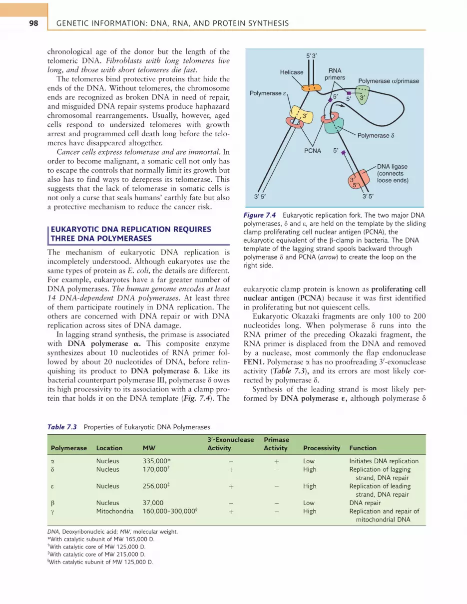
In lagging strand synthesis, the primase is associatedwith DNA polymerase a. This composite enzymesynthesizes about 10 nucleotides of RNA primer fol-lowed by about 20 nucleotides of DNA, before relin-quishing its product to DNA polymerase d. Like itsbacterial counterpart polymerase III, polymerase d owesits high processivity to its association with a clamp pro-tein that holds it on the DNA template (Fig. 7.4). The
eukaryotic clamp protein is known as proliferating cellnuclear antigen (PCNA) because it was first identifiedin proliferating but not quiescent cells.
Eukaryotic Okazaki fragments are only 100 to 200nucleotides long. When polymerase d runs into theRNA primer of the preceding Okazaki fragment, theRNA primer is displaced from the DNA and removedby a nuclease, most commonly the flap endonucleaseFEN1. Polymerase a has no proofreading 30-exonucleaseactivity (Table 7.3), and its errors are most likely cor-rected by polymerase d.
Synthesis of the leading strand is most likely per-formed by DNA polymerase «, although polymerase d
Helicase
Polymerase ε
5′ 3′
5′ 3′
5′3′ 5′3′
5′3′
5′
5′
3′
PCNA
RNAprimers Polymerase α/primase
Polymerase δ
DNA ligase(connectsloose ends)
Figure 7.4 Eukaryotic replication fork. The two major DNA
polymerases, d and e, are held on the template by the sliding
clamp proliferating cell nuclear antigen (PCNA), the
eukaryotic equivalent of the b-clamp in bacteria. The DNA
template of the lagging strand spools backward through
polymerase d and PCNA (arrow) to create the loop on the
right side.
Table 7.3 Properties of Eukaryotic DNA Polymerases
Polymerase Location MW
30-Exonuclease
Activity
Primase
Activity Processivity Function
a Nucleus 335,000* " þ Low Initiates DNA replication
d Nucleus 170,000{þ " High Replication of lagging
strand, DNA repair
e Nucleus 256,000{þ " High Replication of leading
strand, DNA repair
b Nucleus 37,000 " " Low DNA repair
g Mitochondria 160,000–300,000}þ " High Replication and repair of
mitochondrial DNA
DNA, Deoxyribonucleic acid; MW, molecular weight.
*With catalytic subunit of MW 165,000 D.{With catalytic core of MW 125,000 D.{With catalytic core of MW 215,000 D.}With catalytic subunit of MW 125,000 D.
98 GENETIC INFORMATION: DNA, RNA, AND PROTEIN SYNTHESIS

can synthesize the leading strand and appears to beinvolved in leading strand synthesis in some situations.
MOST HUMAN DNA DOES NOTCODE FOR PROTEINS
Only 1.3% of the human genome codes for proteins.Genes are separated by vast expanses of noncodingDNA, including gene deserts extending over more thanone million base pairs. Noncoding DNA is present evenwithin the genes. Human genes are patchworks ofexons, whose transcripts are processed to a maturemRNA, and introns. Introns are transcribed along withthe exons but are excised from the transcript before themessenger RNA (mRNA) leaves the nucleus.
Human genes have between 1 and 178 exons, withan average of 8.8 exons and 7.8 introns. The averageexon is about 145 base pairs long and codes for 48amino acids, and the average polypeptide has a lengthof 440 amino acids. Introns are generally far longerthan exons, and more than 90% of the DNA withingenes belongs to introns (see Fig. 7.12 for an example).
Why human genes have introns, why they have somany of them, and why the introns are so long are notknown. Except for some intronic sequences that contrib-ute to the regulation of gene expression by binding regu-latory proteins, introns appear to be useless junk DNA.
However, the intron-exon structure of human genesis important for evolution. Different structural andfunctional domains of a polypeptide are often encodedby separate exons. For example, the immunoglobulinchains consist of several globular domains with similaramino acid sequence and tertiary structure, eachencoded by its own exon (see Chapter 15). Immuno-globulin genes most likely arose by repeated exonduplication from a single-exon gene.
In other cases, exons from different genes appear tohave combined to form a new functional gene. This iscalled exon shuffling. The exons are the building blocksfrom which the multitude of eukaryotic genes has beenassembled in the course of evolution.
Figure 7.5 shows an overview of the composition ofthe human genome. One commentator wrote about thehuman genome: “In some ways it may resemble yourgarage/bedroom/refrigerator/life: highly individualistic,but unkempt; little evidence of organization; muchaccumulated clutter (referred to by the uninitiated as‘junk’); virtually nothing ever discarded; and the fewpatently valuable items indiscriminately, apparentlycarelessly, scattered throughout.”
GENE FAMILIES ORIGINATEBY GENE DUPLICATION
Most protein-coding genes are present in only one copyin the haploid genome, but duplicated genes, with twoidentical or near-identical copies close together on the
same chromosome, are seen occasionally. Some genesthat code for very abundant RNAs or proteins arepresent in multiple copies, including the ribosomalRNA (rRNA) genes ($200 copies), the 5S rRNA gene($2000 copies), the histone genes ($20 copies), andmost of the transfer RNA (tRNA) genes. In mostcases, identical or near-identical copies of the gene arearranged in tandem, head to tail over long stretches ofDNA, separated by untranscribed spacers.
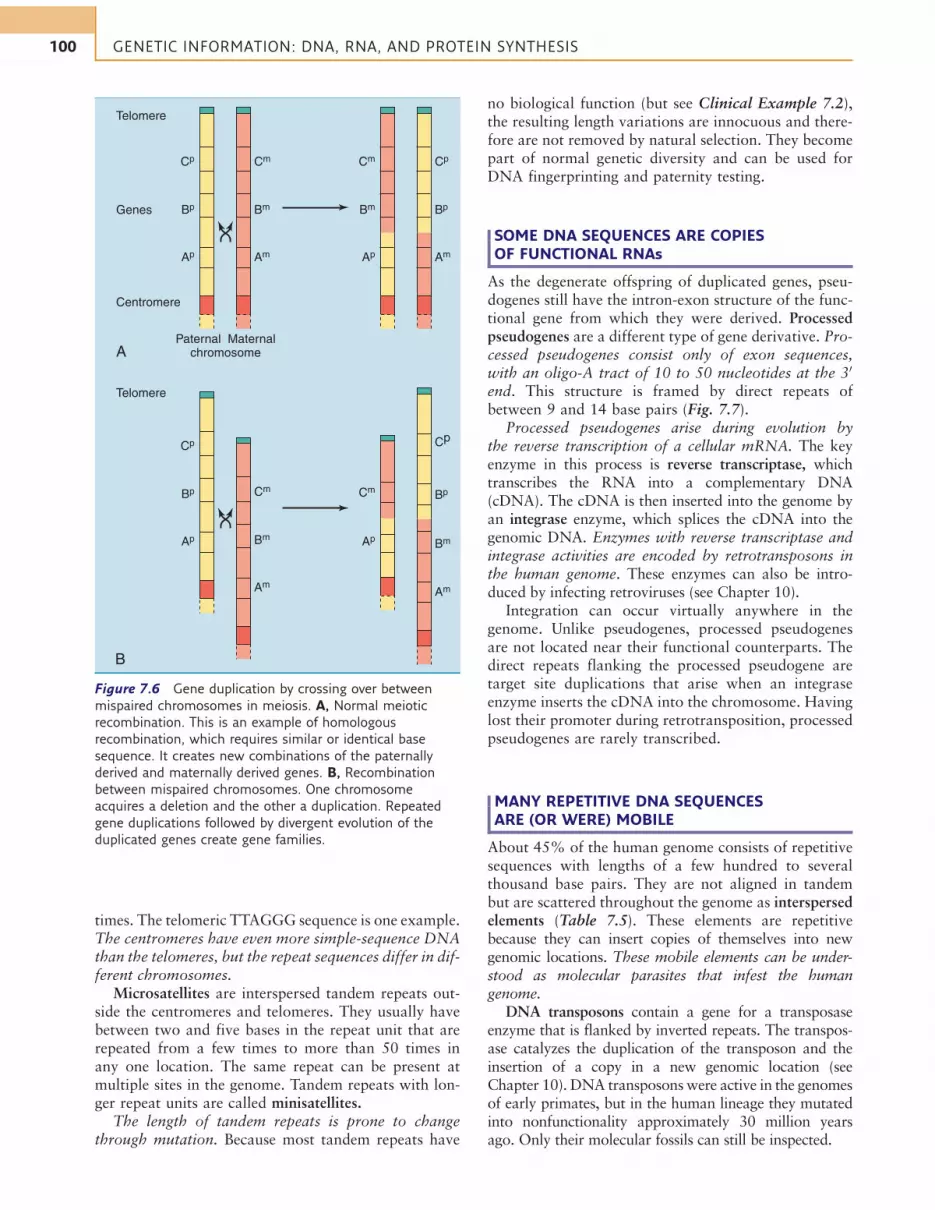
Gene families consist of two or more similar but notidentical genes that, in most cases, are positioned closetogether on the chromosome. They arise during evolu-tion by repeated gene duplications, mostly during cross-ing over in prophase of meiosis I when homologouschromosomes align in parallel and exchange DNA byhomologous recombination. Normal crossing over is astrictly reciprocal process in which the chromosomeneither gains nor loses genes. However, if the chromo-somes are mispaired during crossing over, one chromo-some acquires a deletion and the other a duplication(Fig. 7.6). Through new mutations, a duplicated genecan acquire new biological properties and functions.
Inmany cases, however, one of the duplication productsacquires crippling mutations that prevent its transcriptionor translation. The result is called a pseudogene. Pseudo-genes still have the intron-exon structure of the functionalgene from which they were derived, and they are locatedclose to their functional counterpart on the chromosome.
THE GENOME CONTAINS MANYTANDEM REPEATS
Tandem repeats, also known as simple-sequence DNA,consist of a short DNA sequence of between two and afew dozen base pairs that is repeated head to tail many
21% 21%
13%
8%
12%
12%
6%
3%3%
1.3%
LINEs
SINEs
Retroviralelements
Introns
Exons
Unique DNAoutside genes
Pseudogenes
Tandemrepeats
Segmentalduplications
DNA-onlytransposon
fossils
Figure 7.5 Approximate composition of the human
genome. LINEs, Long interspersed elements; SINEs, short
interspersed elements.
99The Human Genome
times. The telomeric TTAGGG sequence is one example.The centromeres have even more simple-sequence DNAthan the telomeres, but the repeat sequences differ in dif-ferent chromosomes.
Microsatellites are interspersed tandem repeats out-side the centromeres and telomeres. They usually havebetween two and five bases in the repeat unit that arerepeated from a few times to more than 50 times inany one location. The same repeat can be present atmultiple sites in the genome. Tandem repeats with lon-ger repeat units are called minisatellites.
The length of tandem repeats is prone to changethrough mutation. Because most tandem repeats have
no biological function (but see Clinical Example 7.2),the resulting length variations are innocuous and there-fore are not removed by natural selection. They becomepart of normal genetic diversity and can be used forDNA fingerprinting and paternity testing.
SOME DNA SEQUENCES ARE COPIESOF FUNCTIONAL RNAs
As the degenerate offspring of duplicated genes, pseu-dogenes still have the intron-exon structure of the func-tional gene from which they were derived. Processedpseudogenes are a different type of gene derivative. Pro-cessed pseudogenes consist only of exon sequences,with an oligo-A tract of 10 to 50 nucleotides at the 30
end. This structure is framed by direct repeats ofbetween 9 and 14 base pairs (Fig. 7.7).
Processed pseudogenes arise during evolution bythe reverse transcription of a cellular mRNA. The keyenzyme in this process is reverse transcriptase, whichtranscribes the RNA into a complementary DNA(cDNA). The cDNA is then inserted into the genome byan integrase enzyme, which splices the cDNA into thegenomic DNA. Enzymes with reverse transcriptase andintegrase activities are encoded by retrotransposons inthe human genome. These enzymes can also be intro-duced by infecting retroviruses (see Chapter 10).
Integration can occur virtually anywhere in thegenome. Unlike pseudogenes, processed pseudogenesare not located near their functional counterparts. Thedirect repeats flanking the processed pseudogene aretarget site duplications that arise when an integraseenzyme inserts the cDNA into the chromosome. Havinglost their promoter during retrotransposition, processedpseudogenes are rarely transcribed.
MANY REPETITIVE DNA SEQUENCESARE (OR WERE) MOBILE
About 45% of the human genome consists of repetitivesequences with lengths of a few hundred to severalthousand base pairs. They are not aligned in tandembut are scattered throughout the genome as interspersedelements (Table 7.5). These elements are repetitivebecause they can insert copies of themselves into newgenomic locations. These mobile elements can be under-stood as molecular parasites that infest the humangenome.
DNA transposons contain a gene for a transposaseenzyme that is flanked by inverted repeats. The transpos-ase catalyzes the duplication of the transposon and theinsertion of a copy in a new genomic location (seeChapter 10). DNA transposonswere active in the genomesof early primates, but in the human lineage they mutatedinto nonfunctionality approximately 30 million yearsago. Only their molecular fossils can still be inspected.
Genes
A
B
Paternal Maternalchromosome
Centromere
Telomere
Telomere
Cp Cm
Bp
Ap
Bm
Am
Cp
CmBp
Ap Bm
Am
Cm Cp
Bm
Ap
Bp
Am
Cm
Cp
Bp
Ap Bm
Am
Figure 7.6 Gene duplication by crossing over between
mispaired chromosomes in meiosis. A, Normal meiotic
recombination. This is an example of homologous
recombination, which requires similar or identical base
sequence. It creates new combinations of the paternally
derived and maternally derived genes. B, Recombination
between mispaired chromosomes. One chromosome
acquires a deletion and the other a duplication. Repeated
gene duplications followed by divergent evolution of the
duplicated genes create gene families.
100 GENETIC INFORMATION: DNA, RNA, AND PROTEIN SYNTHESIS

Transcription
Polyadenylation site
DNA
Capping,polyadenylation,intron removal
3′ RNA transcript5′
-AAAAAA••••AAA 3′ Mature mRNA
-AA••AA-
5′ CAP-
Integration intochromosomal DNA
-AA••AA 3′ cDNA (only the ‘‘coding’’ strand is shown)
5′
Start of transcription
Promoter
Oligo-Atract
>>
Reversetranscription
Figure 7.7 Origin of a processed pseudogene. , Exons; , introns.
CLINICAL EXAMPLE 7.2: Huntington Disease
Most microsatellites reside in nonessential “junk DNA,”
but some occur in the regulatory or coding sequences
of genes. Huntington disease is caused by expansion of
the trinucleotide repeat CAG in the coding sequence of a
brain-expressed gene. In the normal gene, the CAG is
repeated 6 to 34 times, coding for a polyglutamine tract
in the protein huntingtin.
When the CAG sequence expands to a copy number in
excess of 36, the result is a glutamine-expanded
huntingtin protein that forms abnormal complexes with
other proteins. These abnormal complexes kill cells in
the basal ganglia of the brain and in the cerebral cortex,
leading to an adult-onset disease with personality
changes, a motor disorder, and progressive dementia.
The greater the trinucleotide expansion, the earlier is
the onset of the disease. Because the trinucleotide
repeat tends to expand further during father-to-child
transmission, the disease tends to become more severe in
successive generations. Table 7.4 lists some other
diseases that are caused by trinucleotide expansions.
Table 7.4 Diseases Caused by Expansion of a Trinucleotide Repeat Sequence in a Gene
Repeat Number
Disease Type of Disease Inheritance
Amplified
Repeat* Normal Disease Location in Gene
Huntington disease Neural degeneration AD CAG 6–34 36–120 Coding sequence (Gln)
Myotonic dystrophy Muscle loss, cardiac
arrhythmia
AD CTG 5–37 100–5000 30-Untranslated region
Fragile X Mental retardation XR CGG 6–52 200–3000 50-Untranslated region
Friedreich ataxia Loss of motor
coordination
AR GAA 7–22 200–1000 Intron
AD, Autosomal dominant; AR, autosomal recessive; Gln, glutamine; XR, X-linked recessive.
*A, Adenine; C, cytosine; G, guanine; T, thymine.
101The Human Genome

The other mobile elements are retrotransposons.They are transcribed into RNA, then the RNA is copiedinto a cDNA by reverse transcriptase, and the cDNA isinserted into the genome.
Retroviral retrotransposons are descended from infect-ing retroviruses. This type of virus turns its RNA genomeinto a cDNA, which it inserts into the host cell genome(see Chapter 10).Retroviral elements are integrated retro-viruses that are no longer infectious because they havelost the ability to make the structural proteins of the virusparticle. However, they have multiplied within thegenome because they retained, for some time at least,the ability to move into new genomic locations with thehelp of their reverse transcriptase and integrase. Like theirretroviral ancestors, retroviral retrotransposons areflanked by long terminal repeat (LTR) sequences andtherefore are also called LTR retrotransposons.
Almost all retroviral elements have lost the ability forretroposition. Only one family of these elements has beenactive in the human genome during the past six millionyears, since human ancestors separated from the ancestorsof chimpanzees. Most retroviral retrotransposons are thedead bodies of retroviruses, left to rot in the genomic soil.
L1 ELEMENTS ENCODE A REVERSETRANSCRIPTASE
L1 elements are the most abundant type of long inter-spersed elements (LINEs). A full-length L1 elementhas nearly 6000 base pairs and ends in a poly-A tract.It is framed by short direct repeats that originated dur-ing retrotransposition.
The human genome harbors about 900,000 L1 ele-ments, but only about 5000 are full length. Most ofthe others are badly truncated at the 50 end. Truncationis a common accident during retrotransposition. Differ-ent L1 elements in the human genome are identical inabout 95% of their bases.
Full-length L1 elements contain two genes, butthese genes are intact in only 60 to 100 of the 5000
full-length L1 elements. One of the two genes codesfor an RNA-binding protein of unknown function,and the other for a protein with reverse transcriptase,nuclease, and integrase activities.
The element is transcribed by the cellular RNA poly-merase II from an unusual promoter whose sequencebecomes part of the 50-untranslated region of the L1messenger RNA. Reverse transcription and integrationprobably occur at the same time and are catalyzed bythe same L1-encoded protein (Fig. 7.8). The short directrepeats flanking the element are target site duplicationsthat arise during retrotransposition because the L1 endo-nuclease makes staggered cuts in the target DNA.
The L1 reverse transcriptase acts preferentially onthe transcript of the L1 element. However, on occasionit produces a processed pseudogene by reverse tran-scribing and integrating a cellular mRNA (see Fig. 7.7).
ALU SEQUENCES SPREAD WITH THE HELPOF L1 REVERSE TRANSCRIPTASE
The most populous tribe of short interspersed elements(SINEs) is the Alu sequences, with 1.3 million copies inthe haploid genome. A full-length Alu sequence mea-sures 282 base pairs, contains an adenine-rich tract ofbetween 7 and 50 base pairs at the 30 end, and isflanked by direct repeats of 7 to 21 base pairs.
Alu sequences from different parts of the genome dif-fer on average in about 20% of their bases. Some Alusequences can be transcribed into RNA by RNA poly-merase III, but they do not encode any proteins. How-ever, the RNA transcript of the Alu sequence can bereverse transcribed and integrated into the genome bythe L1-encoded reverse transcriptase/integrase.
L1 sequences might be the descendants of a virus,but Alu sequences are clearly of cellular origin. TheAlu consensus sequence is more than 80% identical tothe sequence of 7SL RNA, a small cytoplasmic RNAthat participates in the targeting of proteins to theendoplasmic reticulum (see Chapter 8).
Table 7.5 Mobile Elements in Human Genome
Class Length
Number in
Genome Encoded Proteins Mode of Movement
Current
Activity
DNA-only transposons Variable, average 220 bp 400,000 Transposase (defunct) Direct transposition Fossils only
Retrovirus-like
retrotransposons
Up to 10,000 bp,
average 350 bp
700,000 Reverse transcriptase
(usually defunct)
Retrotransposition Few are still
active
LINE-1 elements Up to 6000 bp, most are
truncated
900,000 Reverse transcriptase,
RNA-binding protein
Retrotransposition Still active
Alu sequences Up to 300 bp, many
are truncated
1,300,000 None Retrotransposition Still active
bp, Base pair; DNA, deoxyribonucleic acid; LINE, long interspersed element; RNA, ribonucleic acid.
102 GENETIC INFORMATION: DNA, RNA, AND PROTEIN SYNTHESIS

3′
5′ 3′
L1 element (6000bp)
Transcription (RNA polymerase II)Polyadenylation
Target DNA
L1 endonuclease
5′ 3′
5′ 3′
5′
3′
5′
3′ 3′5′
5′
5′
5′ 5′ 3′3′
3′
5′
5′ 3′AAA
AAA
TTT3′
3′
Annealing
L1 reverse transcriptase synthesizes 1st DNA strand
Degradation of L1 RNASynthesis of 2nd DNA strandCleavage of 2nd target DNA strand
DNA synthesis, ligation
New L1 element
5′ 3′
5′5′
TTTAAA3′
3′
5′
5′
3′
5′5′
TTTAAA3′
TTTAAA3′
3′
5′
5′
3′
5′5′
3′
Figure 7.8 Hypothetical mechanism for retroposition of an L1 sequence. Formation of processed pseudogenes and insertions
of Alu sequences use a similar mechanism.
103The Human Genome

MOBILE ELEMENTS ARE DANGEROUS
The human genome is a graveyard for the rotting relicsof 4.3 million copies of largely useless, selfish, parasiticDNA elements. These mobile elements are not entirelyharmless. They cause problems by several mechanisms:
1. Gene disruption during retroposition: Mobile ele-ments can wreak havoc by jumping into a gene. Thisis a rare event that is responsible for a mere 0.3%of pathogenic mutations in humans (but 10% inmice).
2. Chromosome breakage: Even without successful ret-roposition, the L1-encoded nuclease can cause DNAdouble-strand breaks that can lead to large deletionsand other chromosomal rearrangements.
3. Illegitimate crossing-over: Any repetitive sequencecan cause chromosome misalignment during meiosis,when nonhomologous copies of a repetitive elementpair up. This can lead to large duplications and dele-tions (see Fig. 7.6, B) as well as to transfers ofDNA between chromosomes. The latter is calledtranslocation.
HUMANS HAVE APPROXIMATELY 25,000 GENES
Most gene-hunting algorithms detect between 20,000and 35,000 protein-coding genes in the human genome.These computer algorithms locate genes by telltalesequences such as promoter elements, a start codon fol-lowed by a substantial string of potentially amino acidcoding codons, intron-exon junctions, stop codon, andpolyadenylation signal.
Another approach consists of extracting RNA fromcells, copying it into cDNA with the help of reversetranscriptase, and sequencing the cDNA. This methoddefines the transcriptome of the cell, which is the total-ity of transcribed DNA sequences that can be recoveredas RNA. Whereas the genome is the same in each cell ofthe human body, the transcriptomes of different celltypes are different because different genes are tran-scribed in different cells. The transcriptome containsnot only the protein-coding mRNAs but also manynoncoding RNAs of unknown function.
The sum total of expressed proteins is called the pro-teome. Like the transcriptome, the proteome is differentin different cell types. Figure 7.9 shows the presumedfunctions of the proteins that are encoded in the humangenome.
The functional importance of DNA can be inferredfrom comparisons between species. Random mutationsin the junk DNA cause no damage and therefore cansurvive. However, mutations in important coding andregulatory sequences are likely to disrupt gene functionand to cause disease or functional impairments. There-fore they are removed by natural selection. As a result,coding and regulatory sequences of genes show littlevariation among species, and the junk DNA showsmuch variation. Some noncoding RNA transcripts are
conserved and are likely to be functional, but othersappear to be unconstrained and most likely represent“leaky transcription” of nonfunctional DNA.
CLINICAL EXAMPLE 7.3: Heart-Breaking RNA
Acute myocardial infarction is not a genetic disease but
a multifactorial condition that results from
atherosclerosis of the coronary arteries. Risk factors
include both lifestyle and genes. One case-control study
investigated the association of acute myocardial
infarction with more than 52,000 DNA variations
throughout the genome in more than 3400 patients
and 3700 controls. The study found a single-nucleotide
variant, the less common allele of which was associated
with a 35% increased risk of myocardial infarction.
This polymorphism is included in a long RNA transcript
of unknown function that is not translated into
protein. This transcript was named MIAT (myocardial
infarction-associated transcript). Only future studies can
show whether this noncoding RNA is indeed associated
with myocardial infarction and, if so, by which
mechanism it confers disease susceptibility. Because
MIAT is expressed mainly in the brain, its effects on
cardiovascular risk might well be mediated by behavior
and lifestyle.
TRANSCRIPTIONAL INITIATION REQUIRESGENERAL TRANSCRIPTION FACTORS
Eukaryotes use separate enzymes for the synthesis ofrRNA, mRNA, and tRNA (Table 7.6).
RNA polymerase I synthesizes the common pre-cursor of the 5.8S, 18S, and 28S rRNA in the nucleolus,where the ribosomal subunits are assembled. RNApolymerase II synthesizes the mRNA precursors, and
20%
Metabolism
DNA replication/modification
Transcription/translation
Intracellularsignaling
Cell–cellcommunication
Protein foldingand degradationTransport
Cytoskeletal/structural
Defenseand immunity
Other
5%
19%
11%
7.5%
5%7%5.5%
6%
14%
Figure 7.9 Functional categories of proteins encoded by the
human genome.
104 GENETIC INFORMATION: DNA, RNA, AND PROTEIN SYNTHESIS
RNA polymerase III synthesizes small RNAs includingtRNAs and the 5S rRNA.
Transcription requires access of the RNA polymeraseto the promoter at the 50 end of the gene. To this effect,nucleosomes are excluded from the promoter by AT-rich sequences in the promoter and by transcription fac-tors that bind to DNA sequences in the promoter.
Eukaryotic promoters are extremely variable. Thecore promoter includes 30 to 40 base pairs upstreamof the transcriptional start site and serves as an assem-bly point for the general transcription factors, whichare functionally equivalent to the bacterial s subunit.They are named by the acronym TF, followed by thenumber of the RNA polymerase with which they workand an identifying letter. The RNA polymerase mustbind to the promoter-associated transcription factorsbefore it can start transcription.
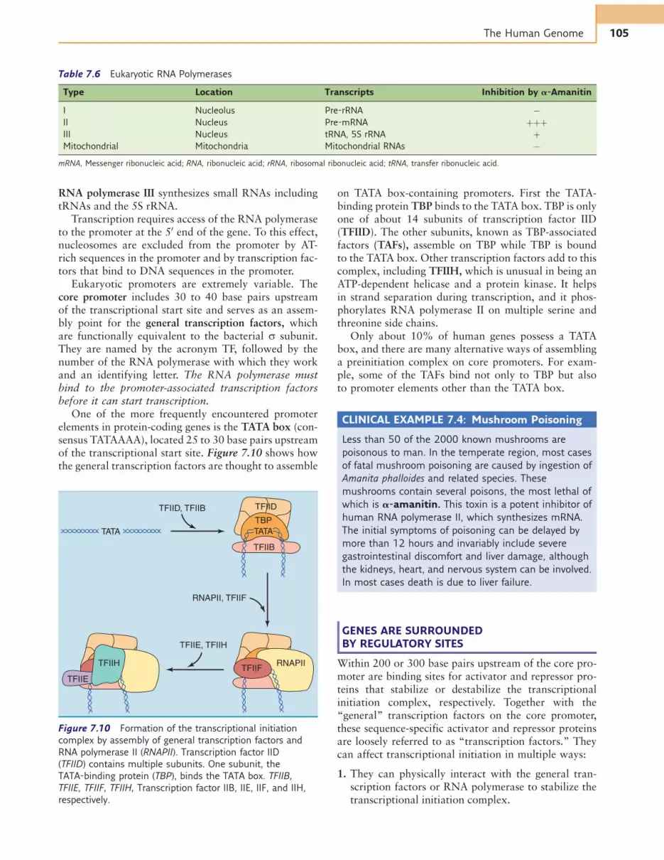
One of the more frequently encountered promoterelements in protein-coding genes is the TATA box (con-sensus TATAAAA), located 25 to 30 base pairs upstreamof the transcriptional start site. Figure 7.10 shows howthe general transcription factors are thought to assemble
on TATA box-containing promoters. First the TATA-binding protein TBP binds to the TATA box. TBP is onlyone of about 14 subunits of transcription factor IID(TFIID). The other subunits, known as TBP-associatedfactors (TAFs), assemble on TBP while TBP is boundto the TATA box. Other transcription factors add to thiscomplex, including TFIIH, which is unusual in being anATP-dependent helicase and a protein kinase. It helpsin strand separation during transcription, and it phos-phorylates RNA polymerase II on multiple serine andthreonine side chains.
Only about 10% of human genes possess a TATAbox, and there are many alternative ways of assemblinga preinitiation complex on core promoters. For exam-ple, some of the TAFs bind not only to TBP but alsoto promoter elements other than the TATA box.
CLINICAL EXAMPLE 7.4: Mushroom Poisoning
Less than 50 of the 2000 known mushrooms are
poisonous to man. In the temperate region, most cases
of fatal mushroom poisoning are caused by ingestion of
Amanita phalloides and related species. These
mushrooms contain several poisons, the most lethal of
which is a-amanitin. This toxin is a potent inhibitor of
human RNA polymerase II, which synthesizes mRNA.
The initial symptoms of poisoning can be delayed by
more than 12 hours and invariably include severe
gastrointestinal discomfort and liver damage, although
the kidneys, heart, and nervous system can be involved.
In most cases death is due to liver failure.
GENES ARE SURROUNDEDBY REGULATORY SITES
Within 200 or 300 base pairs upstream of the core pro-moter are binding sites for activator and repressor pro-teins that stabilize or destabilize the transcriptionalinitiation complex, respectively. Together with the“general” transcription factors on the core promoter,these sequence-specific activator and repressor proteinsare loosely referred to as “transcription factors.” Theycan affect transcriptional initiation in multiple ways:
1. They can physically interact with the general tran-scription factors or RNA polymerase to stabilize thetranscriptional initiation complex.
Table 7.6 Eukaryotic RNA Polymerases
Type Location Transcripts Inhibition by a-Amanitin
I Nucleolus Pre-rRNA "
II Nucleus Pre-mRNA þþþ
III Nucleus tRNA, 5S rRNA þ
Mitochondrial Mitochondria Mitochondrial RNAs "
mRNA, Messenger ribonucleic acid; RNA, ribonucleic acid; rRNA, ribosomal ribonucleic acid; tRNA, transfer ribonucleic acid.
TFIID, TFIIB
TFIIE, TFIIH
RNAPII, TFIIF
TFIID
TBPTATA
TFIIB
TATA
TFIIFTFIIH RNAPII
TFIIE
Figure 7.10 Formation of the transcriptional initiation
complex by assembly of general transcription factors and
RNA polymerase II (RNAPII). Transcription factor IID
(TFIID) contains multiple subunits. One subunit, the
TATA-binding protein (TBP), binds the TATA box. TFIIB,
TFIIE, TFIIF, TFIIH, Transcription factor IIB, IIE, IIF, and IIH,
respectively.
105The Human Genome

2. They can recruit histone-modifying enzymes or ATP-dependent chromatin remodeling complexes.
3. They can interact with the transcriptional initiationcomplex indirectly through a large protein complexwith 20 to 30 subunits known as mediator (Fig. 7.11)
Binding sites for transcription factors are not restrictedto the promoter but can be found thousands and some-times up to one million base pairs away from the tran-scriptional start site in one of the gene’s introns, in the
junk DNA between the genes, or even in an intron of aneighboring gene. These binding sites tend to form clus-ters called enhancers or silencers, depending on whetherthey stimulate or depress transcription.
One gene can have multiple enhancers and silencers,and an enhancer or silencer can regulate a whole set ofneighboring genes. Presumably, the transcription fac-tors bound to these distant regulatory sites can affecttranscription by looping of the DNA, which bringsthem in physical contact with the promoter-bound pro-teins or the mediator complex (see Fig. 7.11).
Typical enhancers look as if an overenthusiastic sor-cerer’s apprentice had stuffed as many binding sites forregulatory proteins as possible into as small a space aspossible. Figure 7.12 shows an example. Most bindingsites are only 15 to 20 base pairs long. Many transcrip-tion factors respond to hormones, second messengers,or nutrients. Therefore their binding sites are calledresponse elements.
GENE EXPRESSION IS REGULATEDBY DNA-BINDING PROTEINS
About 6% of human genes code for proteins withDNA-binding domains and presumed functions in theregulation of gene expression, for a total of about1500 candidate transcription factors. However, theDNA binding and regulatory functions have been veri-fied for fewer than 100 of them.
Enhancer
Activator proteins
Repressor proteins
General transcription factors
RNAPII
Silencer
Mediator
Promoter
Figure 7.11 The mediator is a large protein complex that
mediates the effects of activator and repressor proteins on
transcriptional initiation. RNAP II, Ribonucleic acid
polymerase II.
4
Intron 1
Exon 1
Exon 2
Exon 3
Exon 4
Intron 2
Intron 3
Polyadenylation site
Stop: TGAPolyadenylationsignal: AATAAA
Distalenhancer
Promoter
Start:ATG TATA box
Transcriptional start site
13 3 4
Figure 7.12 Gene for the liver protein
transthyretin (prealbumin), with its
regulatory sites. The gene is drawn to
scale to show its intron-exon structure
and the multiple upstream binding sites
for regulatory proteins. Note that the
introns are far longer than the exons.
Binding sites for regulatory proteins are
clustered in the promoter and a distal
enhancer. With the exception of AP1,
which is present in all nucleated cells,
the regulatory proteins are present in
hepatocytes but not in most other cell
types. , AP1; , C/EBP; 1 , 3 , and
4 , hepatocyte nuclear factors 1, 3,
and 4.
106 GENETIC INFORMATION: DNA, RNA, AND PROTEIN SYNTHESIS

Most transcription factors bind to DNA in a dimericform, either as homodimers of two identical subunits oras heterodimers of two slightly different subunits. Thesymmetry of the dimeric transcription factors ismatched by their response elements, which tend to pos-sess an incomplete dyad symmetry (see Fig. 6.37).
The dimeric transcription factors have a modularstructure (Table 7.7, and Figures 7.13 and 7.14). ADNA-binding module recognizes the specific basesequence of the response element; a dimerization mod-ule forms the active dimeric state; and a transcriptionalactivation (or inhibition) region stimulates or inhibitstranscription. Many transcription factors bind a coacti-vator or corepressor protein, which in turn interactswith the mediator or with RNA polymerase or recruitschromatin-modifying enzymes.
The helix-turn-helix proteins, leucine zipper pro-teins, and helix-loop-helix proteins (see Table 7.7) bindto DNA through an a helix, 20 to 40 amino acids longand with a high content of basic amino acid residues.This a helix fits into the major groove of the DNA dou-ble helix (see Fig. 7.13, C).
The zinc finger proteins contain between two andabout a dozen zinc ions in their DNA-binding region,each complexed to four amino acid side chains: eitherfour cysteines, or two cysteines and two histidines.The zinc finger is a loop of about 12 amino acid resi-dues between two pairs of zinc-complexed amino acids(see Fig. 7.14).
The transcription factors dimerize through an amphi-pathic a helix that forms a two-stranded coiled coil inthe dimeric protein (see Fig. 7.13). In the leucine zipperproteins, the hydrophobic edge of this amphipathic helixis formed by several leucine residues that are spacedexactly seven amino acids apart. Because the a helixhas 3.6 amino acids per turn, these leucine residues allare on the same side of the helix, where they form hydro-phobic interactions with the dimerization partner.
Transcription factors are regulated in several ways:
1. Their own synthesis is regulated in a tissue-specificmanner. In essence, tissue-specific transcription fac-tors regulate the synthesis of other tissue-specifictranscription factors.
2. Some are regulated by the reversible binding of ahormone. The most prominent examples are thereceptors for steroid hormones.
3. Many are regulated by phosphorylation and dephos-phorylation. The protein kinases and protein phos-phatases acting on the transcription factors arethemselves responsive to growth factors, hormones,nutrients, and other external stimuli.
4. Protein-protein interactions are important. For exam-ple, the activation of transcription can be blocked bythe binding of proteins to the transcriptional activatordomain.
EUKARYOTIC MESSENGER RNA IS EXTENSIVELYPROCESSED IN THE NUCLEUS
The introns of human genes are transcribed along withthe exons but are spliced out of the transcript in thenucleus. In addition, the two ends of the mRNAbecome modified.
1. The 50 end of the mRNA receives a cap. The cap is amethylguanosine residue that is linked to the firstnucleotide of the RNA through an unusual 50-50-triphosphate linkage (Fig. 7.15). The cap binds aset of proteins that protect the 50 end of the mRNAfrom 50-exonucleases, help guiding the mRNAthrough the nuclear pore complex into the cytosol,and are needed for the initial interaction betweenthe mRNA and the ribosome.
2. The 30 end of the mRNA receives a poly-A tail ofabout 200 nucleotides. Multiple copies of a poly-Abinding protein (PABP) bind to the poly-A tail. Thisretards the action of 30-exonucleases and allows themRNA to survive for many hours or even a fewdays. Only the histone mRNAs have no poly-A tails,and consequently their half-lives are only a few min-utes. Histones are synthesized only during S phase ofthe cell cycle when the DNA is replicated, and theirsynthesis must be switched off quickly once DNAreplication is completed.
Only fully processed, mature mRNA translocates to thecytoplasm, where it is translated.
Table 7.7 Major Types of DNA-Binding Proteins in Eukaryotes
Structural Motif Structural Features Examples
Helix-turn-helix proteins Two a helices separated by a b turn; “recognition
helix” fitting in major groove of DNA
“Homeodomain” proteins (proteins regulating
embryonic development); most prokaryotic
repressors
Zinc finger proteins Contain zinc bound to Cys and His side chains Receptors for steroid and thyroid hormones
Leucine zipper proteins Two a helices, one with basic residues for DNA
binding, one with regularly spaced Leu for
dimerization
C/EBP (gene activator in liver); c-Myc, c-Fos,
c-Jun (growth regulators, proto-oncogene
products)
Helix-loop-helix proteins DNA-binding a helix and two dimerization helices
separated by a nonhelical loop
Myo D-1, myogenin (proteins that induce muscle
differentiation)
Cys, Cysteine; DNA, deoxyribonucleic acid; His, histidine.
107The Human Genome

mRNA PROCESSING STARTS DURINGTRANSCRIPTION
“Posttranscriptional” processing of mRNA actually iscotranscriptional. It occurs during transcription and isguided by proteins that are recruited by the RNA poly-merase (Fig. 7.16).
The RNA is synthesized in the 50!30 direction, and
50 capping is the first modification of the pre-mRNA.It is done when about 25 nucleotides of the RNA havebeen polymerized.
Next, the introns are removed by spliceosomes. Thespliceosome contains five small RNAs (U1, U2, U4, U5,and U6) with lengths between 106 and 185 nucleotides.These associate with proteins to form small nuclear ribo-nucleoprotein particles (snRNPs [“snurps”]). Overall,about 50 proteins are involved in splicing.
The intron-exon junctions of protein-coding nucleargenes are marked by more or less conserved consensussequences. There is also a conserved “branch site”within the intron, about 30 nucleotides from the 30 end(Fig. 7.17). Splicing releases the intron as a cyclic lariatstructure, with the 50 end bonded with the 20 hydroxylgroup at the branch site.
+
+
Zn2+
orCOO–H3N
Cys
Cys His
His
Zn2+
COO–H3N
Cys
Cys Cys
Cys
Figure 7.14 Zinc finger. This structural motif occurs in
2 to 12 copies in the DNA-binding region of the zinc finger
proteins. The amino acid residues on each side of the zinc are
separated by three or four amino acid residues, and the
intervening loop contains approximately 12 residues. Cys,
Cysteine; His, histidine.
A
B
C
Zipper (α-helix)
Helix
Basic region(α-helix)
Basic region
Dimerizationdomains (α-helices)
Loop
Helix
Figure 7.13 Binding of dimeric transcription factors to their
response elements. The base sequences of the response
elements show a dyad symmetry that matches the symmetry
of the transcription factors. A, Schematic binding of a leucine
zipper protein. The “zipper” is required for dimerization
while the basic region binds to DNA. B, DNA binding by a
helix-loop-helix protein. C, Computer graphic model of
binding of the carboxyl-terminal portion (“basic region”) of
the leucine zipper protein C/EBF to its cognate binding site.
108 GENETIC INFORMATION: DNA, RNA, AND PROTEIN SYNTHESIS

Finally, a polyadenylation signal (consensus AAUAAA)in the last exon recruits an endonuclease that cleaves theRNA about 20 nucleotides downstream. This cut marksthe end of the last exon, and a poly-A tail is added enzy-matically to the newly created 30 end. Transcription canproceed for many hundred nucleotides beyond the polya-denylation site, but the cutoff tail of the transcript isdiscarded.
TRANSLATIONAL INITIATION REQUIRESMANY INITIATION FACTORS
Eukaryotic translation differs from the prokaryotic sys-tem (see Chapter 6) in the usual ways: It is more com-plex, and it is slower.
This is especially obvious in the initiation of transla-tion. Whereas prokaryotic translation initiation requires
only three initiation factors, eukaryotes have at least12 initiation factors consisting of more than twodozen polypeptides. Only some of them are shownin Figure 7.18. Eukaryotic mRNA does not havea ribosome-binding Shine-Dalgarno sequence in the50-untranslated region. The initial binding betweenmRNA and ribosome is mediated by proteins instead.
Figure 7.18 shows that both ends of the mRNA arecoated with proteins. The proteins at the 50 end interactwith the proteins on the poly-A tail, and they serve astranslational initiation factors by interacting with pro-teins on the small (40S) ribosomal subunit.
With the help of these initiation factors, the ribo-some scans the 50-terminal region of the mRNA forthe start codon AUG. Usually the first AUG is chosen,but in some mRNAs the second or third AUG is used.Once the initiator codon has been found, methioninerather than N-formylmethionine is introduced as thefirst amino acid at the N-terminus of the polypeptide.
The steps in the elongation cycle are analogous tothose in bacterial protein synthesis, and the elongationfactors are functionally equivalent (Table 7.8). How-ever, eukaryotes add only two amino acids per secondto the growing polypeptide chain compared to 20 persecond in bacteria.
mRNA PROCESSING AND TRANSLATIONARE OFTEN REGULATED
Control of transcriptional initiation is the most efficientway to regulate gene expression because it avoids theenergetically costly synthesis of unneeded mRNAs.Nevertheless, eukaryotes also use posttranscriptionalcontrols, as follows.
Regulation of Messenger RNA Stability
Only about 5% of the RNA that is synthesized everleaves the nucleus. mRNA must associate with proteinsto guide it through the nuclear pore complexes.Improperly spliced mRNA that lacks the usual post-transcriptional modifications does not associate withthese proteins. It is retained in the nucleus, where it isdegraded by nucleases.
In the cytoplasm, the lifespan of mRNA is regulatedby nucleases that degrade mRNA and by mRNA-bindingproteins that protect mRNA from the nucleases. Thusmutations that change the 30 and 50 untranslated regionsof mRNA can disrupt protein synthesis by interferingwith the binding of protective proteins.
Tissue-Specific Initiation and Terminationof Transcription
Some genes can be transcribed from alternative promo-ters, yielding transcripts with different 50-terminal por-tions. Other genes have alternative polyadenylation
CH2
N
OHOH
HN
N
N+
O
CH3
H2N
O
–O—P O
–O—P O
O
O
O
CH2
–O—P O
O
CH2
Base2
ORO
O
–O—P O
O
O
Base1
OR
O
Figure 7.15 Structure of the cap at the 50 end of eukaryotic
messenger RNAs. Transfer and ribosomal RNAs do not have
caps. R, H or CH3.
109The Human Genome

sites and can produce transcripts with different 30 ends.An example of the tissue-specific initiation of transcrip-tion is the use of alternative promoters in the gene forglucokinase (Fig. 7.19), an enzyme that is expressedonly in the liver and the insulin-producing b-cells ofthe pancreas.
Tissue-Specific Splicing
Recognition of splice sites is regulated by tissue-specificproteins. Therefore an exon that is included in themature mRNA in one cell type can be skipped inanother. About 60% of human genes is thought to besubject to alternative splicing. Therefore the 20,000 to
Capping
E3DNA
5′
RNA polymerase II
E2E1
5′
5′
5′3′
AAUAAAConsensus
Splicing
P
Polyadenylation
Spliced-outintron
Figure 7.16 “Posttranscriptional” processing of mRNA actually takes place while RNA polymerase II is synthesizing
the mRNA. E1, E2, E3, Exons; P, promoter.
5′ bond5′
5′ RNA
3′
3′
2′
Branchsite
10–30 bases
Exon 1 Intron Exon 2
Exon 1 Exon 2
AGG
AGGUXAGU CYXAYY 3′YYYYYYYYNCAGG
C Y X A
Y
YY
Y Y Y Y Y Y YNC
AG
GU X A
GU
+
Figure 7.17 Splicing of introns from mRNA precursors. The consensus sequences base pair with RNA components
of the spliceosomes. A, Adenine; C, cytosine; G, guanine; N, any base; U, uracil; X, purine; Y, pyrimidine.
110 GENETIC INFORMATION: DNA, RNA, AND PROTEIN SYNTHESIS

eIF4E
eIF4AeIF4B
3′
Initiationfactors
40S subunit
Initiator-tRNA
mRNA binds
Met
eIF4G
3′
1.
2.
3.
Initiator-tRNAfinds start codoneIF-2 hydrolyzesGTP, then leavesLarge ribosomalsubunit binds
1. Binding of aminoacyl-tRNA2. Peptide bond formation3. Translocation
GTPGTP
AUG
AUG
poly-Abindingproteins
Met
40S
60S
GDP
eIF3
eIF-2AeIF5B
Met
GTPGTP
Repeat
tRNA
Figure 7.18 Formation of the translational initiation complex in eukaryotes. AUG, Start codon; eIF, eukaryotic initiation factor;
Met, methionine.
Table 7.8 Initiation Factors and Elongation Factors of Eukaryotic Protein Synthesis
Eukaryotic Protein Prokaryotic Equivalent GTP Hydrolysis Function
Initiation Factors
eIF1 IF-3 " Component of initiation complex eIF1A
IF-1 " Component of initiation complex
eIF2 EF-Tu subunit þ Places initiator tRNA on 40S subunit
eIF-3 (10þ subunits) — " Facilitates binding of initiator tRNA and mRNA
eIF4E — " Cap-binding protein
eIF4A — " RNA helicase
eIF4B — " Facilitates scanning
eIF4G — " Scaffold protein
eIF5 — " Activates GTPase activity of eIF2
eIF5B IF-2 þ Stabilizes initiator tRNA binding to ribosome
Elongation Factors
EF-1a EF-Tu þ Places aminoacyl-tRNA in A site of ribosome
EF-1bg EF-Ts " Regenerates GTP-bound form of EF-1a
EF-2 EF-G þ Translocation
eIF, Eukaryotic initiation factor; GTP, guanosine triphosphate; GTPase, guanosine triphosphatase; IF, initiation factor; mRNA, messenger RNA;
tRNA, transfer RNA.
111The Human Genome

30,000 human genes can make an estimated 50,000 dif-ferent polypeptides. Figure 7.20 shows an example.
Translational Repressors
Ribosomal protein synthesis can be regulated by mRNA-binding proteins. For example, the mRNA of the poly-Abinding protein (PABP) has an oligo-A tract in its 50-untranslated region. When PABP is abundant, it binds tothis oligo-A tract to prevent its own continued synthesis.
Messenger RNA Editing
In mRNA editing, a base in the mRNA is altered enzy-matically. If this creates or obliterates a stop codon, thelength of the encoded polypeptide is altered. mRNAediting is rare in mammals, but when it occurs, it canproduce alternative polypeptides from the same genein different cell types.
CLINICAL EXAMPLE 7.5: Diphtheria
Diphtheria is a bacterial infection of the upper
respiratory tract that leads to necrosis (death) of
mucosal cells and airway obstruction. Prior to the
introduction of antibiotics, diphtheria was a major cause
of death in children. The offending bacterium,
Corynebacterium diphtheriae, secretes a toxic protein
that binds to a surface receptor on the mucosal cells
and then is cleaved by a protease. One of the proteolytic
fragments then enters the cell. This active fragment is
an enzyme that inactivates the elongation factor EF-2
(Fig. 7.21). A single toxin molecule is sufficient to
inactivate thousands of EF-2 molecules. Strains of
Corynebacterium that do not produce the toxin are
peaceful members of the normal bacterial flora on skin
and mucous membranes.
Continued
Pancreaticβ-cells
Gene
Primarytranscript
Primarytranscript
1
1 >26 kb 1L 2A 2 3 4 5 6 7 8 9 10
Promoter(pancreas)
Promoter(liver)
Hepatocytes
or
1L
1L 2A
Figure 7.19 Transcription of the glucokinase gene. Alternative promoters are used in hepatocytes and pancreatic b-cells. The
resulting polypeptides differ in the N-terminal region, which is encoded by exon 1 in the pancreas and by exon 1L or exons 1L
and 2A in the liver. , Exon; , intron.
1 4 5 6 8 9 10Sm An
1 4 5 6 8 9
13
10Str An
—3′Gene 5′— 1 4 5 6 7 8 9 10
Poly A(sm)
Poly A(str)
mRNAs
2
2 3 11 12
3 11 12
13
Figure 7.20 The tropomyosin gene is an example of tissue-specific splicing. Only 10 of the 13 exons are used in striated
muscle, and nine are used in smooth muscle. Alternative polyadenylation signals are used in striated muscle and smooth
muscle: poly A (str) and poly A (sm).
112 GENETIC INFORMATION: DNA, RNA, AND PROTEIN SYNTHESIS

CLINICAL EXAMPLE 7.5: Diphtheria—cont’d
SMALL RNA MOLECULES INHIBIT GENEEXPRESSION
Translation can be prevented by RNA interference,which is triggered by the appearance of a double-stranded RNA in the cell. The double-stranded RNAis processed by cellular RNases into small RNAs havinglengths of about 22 nucleotides. The product is calledsmall interfering RNA (siRNA) if it is derived from anexternal source and micro-RNA (miRNA) if it isderived from a cellular gene.
The siRNA or miRNA is targeted to a complemen-tary sequence in an mRNA, whose translation it pre-vents selectively. RNA interference serves two mainfunctions: protection against RNA viruses and posttran-scriptional regulation of gene expression.
For defense against RNA viruses, the nuclease dicercleaves long, double-stranded viral RNA into small piecesof about 22 nucleotides, which are transferred from dicerto an RNA-induced silencing complex (RISC). An RNAhelicase in the RISC converts the RNA into single strands.This single-stranded siRNA binds to an argonaute pro-tein, which forms the active core of the RISC.
The argonaute-bound siRNA guides the RISC tosingle-stranded viral mRNA with complementary base
sequence. Base pairing of the argonaute-bound siRNAprevents the translation of the viral mRNA. In mostcases this is followed by nuclease cleavage of the viralmRNA. One of the four argonaute proteins encodedby the human genome is an active RNase.
This general mechanism has been co-opted for theposttranscriptional regulation of gene expression.Endogenous miRNAs are derived either from noncodingtranscripts or from introns of protein-coding genes. ThemiRNA precursors possess stem-loop structures withbase-paired double-helical stems. The stem loops areprocessed by the RNases drosha and dicer, and theresulting miRNA is loaded on the RISC (Fig. 7.22).The argonaute-bound miRNA is complementary to atarget sequence in a cellular mRNA, most commonlyin its 30-untranslated region.
Prevention of translation rather than mRNA cleavageis the most common outcome of miRNA binding to itstarget mRNA. This effect requires the pairing of onlysix bases in positions 2 to 7 of the miRNA to the mRNA.
Simple eukaryotes have about 100 miRNAs, buthumans and other mammals have about 1000. SomemiRNAs target only one or a few mRNAs, whereasothers regulate hundreds. Between 30% and 80% of allhuman genes are believed to be regulated by at least onemiRNA. Thus a miRNA can coordinate gene expressionby affecting all mRNAs containing a complementarysequence, in the same way that a transcription factorcan coordinate the expression of all genes that have bind-ing sites for the transcription factor in their regulatorysites. The expression of most miRNA precursors is tissuespecific or is restricted to distinct developmental stages.
miRNA appears to be capable of transcriptionalsilencing as well. In this case, a miRNA-loaded RISCrecognizes targets on pre-mRNA while it is producedby RNA polymerase II. Upon target recognition thecomplex recruits histone-modifying enzymes that con-vert the gene into a heterochromatic state.
CLINICAL EXAMPLE 7.6: miRNAs in AlzheimerDisease
Alzheimer disease is caused by the aberrant processing
of amyloid precursor protein (APP) to b-amyloid (see
Chapter 2). When miRNA levels in postmortem brains of
patients with Alzheimer disease were studied, one
micro-RNA (miR-107) was present in reduced amount.
Another study found reduced levels of three other
miRNAs (miR-29a, miR-29b, and miR-9) in Alzheimer
brains. These miRNAs target the mRNA for b-secretase,
the enzyme that produces b-amyloid. We cannot be
certain whether the reduced levels of these miRNAs are
a cause or consequence of the disease, but it is
conceivable that reduced levels of the miRNAs lead to
enhanced expression of b-secretase and thereby to
enhanced formation of b-amyloid.
—
—
—
O
(CH2)2
NH2
CH3
CH3
HC N
C
CH3
CH3
O
(CH2)2
NH2
HC N
C
+
N
NH
P PRibose Ribose
O C
HC—CH2—
NH
Nicotinamide Adenine
NicotinamidePPN Ribose Ribose
Adenine
NAD+
Elongation factor 2
N
O C
HC—CH2—
NH
Inactivated elongation factor
+
Diphtheriatoxin
Figure 7.21 Covalent modification of the eukaryotic
elongation factor 2 (“translocase”) by diphtheria toxin. The
amino acid side chain in the elongation factor is
diphthamide, a posttranslationally modified histidine.
NADþ, Nicotinamide adenine dinucleotide.
113The Human Genome

MITOCHONDRIA HAVE THEIR OWN DNA
Human mitochondria contain 4 to 10 copies of asmall circular chromosome with 16,569 base pairs ofDNA. The genes for 13 polypeptides, 22 tRNAs, and
2 rRNAs (12S and 16S) are transcribed by a mitochon-drial RNA polymerase, and the mRNA is translated bysmall mitochondrial ribosomes that are more similarto bacterial ribosomes than to human cytoplasmicribosomes.
3′
5′
3′
P
3′
3′P
3′P
3′P
P
Pri-miRNA
Pre-miRNA
miRNA duplex
Drosha (nucleus)
Dicer (cytoplasm)
RNA helicase,binding to Ago protein
RNA helicase,binding to Ago 2 protein
Pairing of miRNAwith cellular mRNA
Pairing of siRNAwith viral mRNA
Incomplete match with cellular mRNA:Inhibition of translation
Perfect match with viral mRNA:Degradation of mRNA
Ago
Ago
Cap AAAAA
3′P
Ago 2
3′P
Ago 2
Cap AAAAA
LoopStem
Viral dsRNA
siRNA duplex
Dicer
5′ 3′
3′ 5′
Figure 7.22 Formation of micro-RNA acid (miRNA) from a cellular transcript and small interfering RNA (siRNA) from double-
stranded (ds) viral RNA. Ago, Argonaute protein.
114 GENETIC INFORMATION: DNA, RNA, AND PROTEIN SYNTHESIS

This protein-synthesizing system exists because mito-chondria are the descendants of symbiotic bacteria.More than 1.5 billion years ago, their already aerobicancestors invaded a eukaryotic cell that had not yetlearned the use of oxygen for ATP synthesis. What mayhave started as an attempt at parasitism soon turned intoa peaceful coexistence, and in time the bacteria evolved(or degenerated) into the present-day mitochondria.
One after another, most of the original bacterialgenes relocated into the nucleus, and for good reason.For a gene, the nucleus is a safer place than the mito-chondrion because mitochondrial oxidation pollutesthe environment with DNA-damaging superoxide andhydroxyl radicals. This means that the human nucleargenome is of hybrid origin, being descended in partfrom a primordial eukaryote and in part from a symbi-otic prokaryote.
The reason for the continued existence of the mito-chondrial genome is that once the mitochondrialgenome was very small, it evolved small changes inthe genetic code (Table 7.9). Because today the geneticcode is different in the nucleus and the mitochondria,transfer of the remaining mitochondrial genes into thenucleus is no longer possible.
HUMAN GENOMES ARE VERY DIVERSE
When the genomes of two humans are compared, a dif-ference is encountered about once every 1100 basepairs. For comparison, human DNA and chimpanzeeDNA have about one difference every 80 base pairs.Most differences between humans are single-nucleotidepolymorphisms (SNPs), a replacement of a single base.The human genome contains 11 million SNPs with aminor allele frequency of more than 1%, and seven mil-lion of these have a minor allele frequency greater than5%. The coding sequence of an average protein-codinggene has about four SNPs with a population frequencygreater than 1% for the less common allele. Anothercommon type of variation consists of small insertionsand deletions of one or a few base pairs, collectivelycalled indels.
Table 7.10 lists the density of these variations in dif-ferent functional categories. Generally, there is less var-iation in functional sequences than in presumed junkDNA. For example, the frequencies of SNPs and
especially of insertions or deletions (indels) are lowerin coding sequences of genes than in repetitive elements.The reason is that a new mutation in a coding sequenceis likely to be disruptive and consequently is removedby natural selection. Similar mutations in functionlessDNA, including the large majority of repetitive ele-ments, can be carried through the generations becausethey are harmless. Even differences between humansand chimpanzees are greater in junk DNA than in func-tional sequences.
Indels in a coding sequence tend to be more disrup-tive than SNPs for protein structure and function andtherefore are removed more efficiently. Even amongthe coding SNPs there is a bias in favor of synonymousSNPs, which do not change the amino acid sequence ofthe protein because they change a codon into a differentcodon that still codes for the same amino acid. Nearly50% of the SNPs in coding sequences are synonymous.
Not all genes are equally important. Only 7% ofhuman genes is known to be associated with one oranother genetic disease. These “disease genes” are evenless variable than protein-coding genes in general, pre-sumably because they aremore important.We know fromstudies in knockout mice that many genes can be lostentirely without leading to obvious abnormalities. Alsomany human genes are believed to be rather “unimpor-tant” in the sense that the effects of mutations in thesegenes are too mild to be recognized as a genetic disease.
Some sequences outside protein-coding genes are wellconserved between species and therefore are believed tobe functional. Some are likely to code for functionalRNAs, including miRNA precursors, whereas othersare distal regulatory elements of protein-coding genes.Table 7.10 shows that these conserved elements have lit-tle diversity within the human species as well.
Table 7.9 Differences between the Genetic Code of Human
Mitochondria and the Standard Code Used by Cytoplasmic
Ribosomes
Codon Standard Code Mitochondrial Code
AUA Ile Met
AGA Arg Stop
AGG Arg Stop
UGA Stop Trp
Table 7.10 Genetic Variations in the Genome of an
Individual Human (Craig Venter) Compared to the Human
Reference Genome
Variations per
10,000 Base Pairs
SNPs Indels
Total genome 7.8 0.9
Coding sequences 4.5 0.09
Coding sequences of disease genes 3.6 0.04
50-Untranslated regions 5.5 0.3
30-Untranslated regions 5.9 0.7
Splice sites 5.0 0.6
Promoter (1 kb upstream of start) 6.8 0.8
Introns 7.0 0.9
Conserved elements in introns 4.8 0.5
Conserved elements between genes 5.9 0.5
Alu sequences 9.0 2.6
L1 sequences 8.3 0.6
Tandem repeats 11.0 15
indel, Insertion or deletion; SNP, single-nucleotide polymorphism.
115The Human Genome

HUMAN GENOMES HAVE MANY LOW-FREQUENCY COPY NUMBER VARIATIONS
Whereas SNPs and small indels originate as replicationerrors, most large deletions and duplications arise by cross-ing over between mispaired chromosomes (see Fig. 7.6).
These copy number variations have sizes betweenabout 1000 and more than onemillion base pairs. Mostindividual copy number variants are rare, and only30% to 40% of those that have been observed so farhas a minor allele frequency greater than 1%. Between5% and 10% of individuals have at least one copynumber variant larger than 500,000 base pairs, and1% to 2% of individuals have a variant of more thanone million base pairs.
For any given individual, between 9 and 25million basepairs of DNA are involved in structural variations. Aboutthree times more bases are involved in structural variationsthan in SNPs and small indels. In all, the structural varia-tions that have been observed so far (often in a single indi-vidual or family) include up to 25% of the genome.
Copy number variants can affect a person’s pheno-type and possibly contribute to disease susceptibilitiesthrough gene dosage effects. However, if there are noimpairments, a copy number variant can persist throughthe generations. It can even become a normal feature ofthe genome. Ancient duplications gave rise to genefamilies, and somewhat more recent ones are annotatedas segmental duplications (see Fig. 7.5). Mutations thatproduce copy number variations are quite frequent.Even the genomes of identical twins sometimes differin one or two of them. Most copy number changes seem
to be at least slightly unfavorable, so they remain rareand tend to be selected out of the gene pool slowly whilepossibly contributing to disease on the way. This processis called mutation-selection balance.
SUMMARY
Eukaryotes package their DNA into chromatin withthe help of small basic proteins called histones. Thecondensation state of the chromatin, and with it theaccessibility of the genes for transcription, is regulatedby covalent modifications of both histones and DNA.
Only 1.3% of human nuclear DNA codes for pro-teins. Human genes are interrupted by noncodingintrons, and they are separated from neighboring genesby variable expanses of junk DNA. About 45% of thehuman genome is formed by the remnants of mobileDNA sequences that are best understood as “molecularparasites.” These mobile elements can move into newgenomic locations through an RNA intermediate in aprocess known as retro(trans)position.
Transcription is meticulously regulated by proteinsthat bind to promoters and other regulatory sites inand around the genes. The mRNA transcripts mustbe modified by capping, polyadenylation, and removalof introns before the mature mRNA is allowed toleave the nucleus for translation. Translation can beregulated as well, for example, by micro-RNAs thatsuppress translation by binding to specific mRNAs.A vast amount of variation in the human genome isresponsible for individual differences in disease sus-ceptibilities and other phenotypic traits.
Further Reading
Bandiera S, Hatem E, Lyonnet S, et al: microRNAs in dis-eases: from candidate to modifier genes, Clin Genet 77:306–313, 2010.
Belancio VP, Hedges DJ, Deininger P: Mammalian non-LTRretrotransposons: for better or worse, in sickness andhealth, Genome Res 18:343–358, 2008.
Burgers PMJ: Polymerase dynamics at the eukaryotic DNAreplication fork, J Biol Chem 284:4041–4045, 2009.
Carthew RW, Sontheimer EJ: Origins and mechanisms ofmiRNAs and siRNAs, Cell 136:642–655, 2009.
De Lange T: How telomeres solve the end-protection prob-lem, Science 326:948–952, 2009.
Erson AE, Petty EM: MicroRNAs in development and dis-ease, Clin Genet 74:296–306, 2008.
Frazer KA, Murray SS, Schork NJ, et al: Human genetic vari-ation and its contribution to complex traits, Nat RevGenet 10:241–251, 2009.
Friedman RC, Farh KK-H, Burge CB, et al: Most mammalianmRNAs are conserved targets of microRNAs, Genome Res19:92–105, 2009.
Hernandez G, Altmann M, Lasko P: Origins and evolution ofthe mechanisms regulating translation initiation in eukary-otes, Trends Biochem Sci 35:63–73, 2010.
Ho L, Crabtree GR: Chromatin remodeling during develop-ment, Nature 463:474–484, 2010.
International HapMap Consortium: A second generationhuman haplotype map of over 3.1 million SNPs, Nature449:851–861, 2007.
Ishii N, Ozaki K, Sato H, et al: Identification of a novel non-coding RNA, MIAT, that confers risk of myocardial infarc-tion, J Hum Genet 51:1087–1099, 2006.
Itsara A, Cooper GM, Baker C, et al: Population analysis oflarge copy number variants and hotspots of human geneticdisease, Am J Hum Genet 84:148–161, 2009.
Kapp LD, Lorsch JR: The molecular mechanics of eukaryotictranslation, Annu Rev Biochem 73:657–704, 2004.
Kapranov P, Willingham AT, Gingeras TR: Genome-wide tran-scription and the implications for genomic organization,Nat Rev Genet 8:413–423, 2007.
Khalil AM, Guttman M, Huarte M, et al: Many human largeintergenic noncoding RNAs associate with chromatin-modifying complexes and affect gene expression, ProcNatl Acad Sci U S A 106:11667–11672, 2009.
Kim J-I, Ju YS, Park H, et al: A highly annotated whole-genomesequence of a Korean individual, Nature 460:1011–1015,2009.
Loeb LA, Monnat RJ: DNA polymerases and human disease,Nat Rev Genet 9:594–604, 2008.
Muller F, Demeny MA, Tora L: New problems in RNA poly-merase II transcription initiation: matching the diversity ofcore promoters with a variety of promoter recognition fac-tors, J Biol Chem 282:14685–14689, 2007.
116 GENETIC INFORMATION: DNA, RNA, AND PROTEIN SYNTHESIS

Ng PC, Levy S, Huang J, et al: Genetic variation in an individ-ual human exome, PLoS Genet 4:e1000160, 2008.
Pheasant M, Mattick JS: Raising the estimate of functionalhuman sequences, Genome Res 17:1245–1253, 2007.
Ponting CP, Oliver PL, Reik W: Evolution and functions oflong noncoding RNAs, Cell 136:629–641, 2009.
Pratt AJ, MacRae IJ: The RNA-induced silencing complex: aversatile gene-silencing machine, J Biol Chem 284:17897–17901, 2009.
Sahin E, DePinho RA: Linking functional decline of telo-meres, mitochondria and stem cells during ageing, Nature464:520–528, 2010.
Sonenberg N, Hinnebusch AG: Regulation of translation initi-ation in eukaryotes: mechanisms and biological targets,Cell 136:731–745, 2009.
Vaquerizas JM, Kummerfeld SK, Teichmann SA, et al: A cen-sus of human transcription factors: function, expressionand evolution, Nat Rev Genet 10:252–263, 2009.
Venters BJ, Pugh BF: How eukaryotic genes are transcribed,Crit Rev Biochem Mol Biol 44:117–141, 2009.
Villard L: MECP2 mutations in males, J Med Genet 44:417–423, 2007.
Zoghbi HY: Rett syndrome: what do we know for sure? NatNeurosci 12:239–240, 2009.
QUESTIONS
1. The reason why a fusion of two oocytes in thetest tube cannot produce a viable child is
A. X inactivation would not be possibleB. Some imprinted genes would not be expressedC. Transposons would become activatedD. Female-expressed genes would not be
suppressed by male-derived miRNAsE. Telomeres would shorten excessively, leading to
early senility and death in utero
2. Some pharmaceutical companies are tryingfrantically to find inhibitors of telomerase.A telomerase inhibitor could, in theory, be usedin an attempt to
A. Boost the synthesis of muscle proteinsB. Prevent viral infectionsC. Cure cancerD.Cure acquired immunodeficiency syndrome (AIDS)E. Make people immortal
3. Alu sequences can cause diseases by jumpinginto new genomic locations. Their mobilitydepends on the following enzymatic activities:
A. Transposase and RNA polymeraseB. DNA polymerase and RNA replicaseC. Peptidyl transferase and transposaseD.Primase and integraseE. RNA polymerase and reverse transcriptase
4. The report you have just received from thepaternity testing laboratory states that analysisof polymorphic microsatellites was used. Whatexactly is a polymorphic microsatellite?
A. A sequence in the centromeric DNAB. A piece of chromatin attached to the short arm of
an acrocentric chromosomeC. A tandem repeat of variable lengthD.A nonfunctional copy of a gene
E. A small RNA that regulates gene expression bytargeting mRNAs
5. Eukaryotic enhancers are
A. Regulatory DNA sequences within the codingsequences of genes that affect the rate oftranscriptional elongation
B. Binding sites for general transcription factors inthe promoter
C. Proteins that bind to regulatory base sequencesin DNA
D.DNA sequences outside the promoter region thatcontain multiple binding sites for regulatoryproteins
E. Proteins that enhance the rate of translationalinitiation by binding to either the ribosome or themRNA
6. In the year 2045, the Surgeon Generaldetermines that reverse transcriptase ishazardous to your health because it leads toinsertional mutations. In order to eliminatereverse transcriptase from the human body,genetic engineers would have to excise allfull-length, intact copies of
A. DNA transposons and Alu sequencesB. Retroviral retrotransposons and Alu sequencesC. L1 elements and Alu sequencesD.Pseudogenes and retroviral retrotransposonsE. L1 elements and retroviral retrotransposons
7. Deletion of a gene coding for a miRNAprecursor is likely to result in
A. Increased translation of mRNAsB.Misreading of the genetic codeC. Reduced stability of mRNAsD.Acetylation of histones and enhanced
transcriptionE. Repression of retroposition by mobile elements
117The Human Genome
Copyright © 2022 FDOKUMEN