The Braincase of Chanaresuchus ischigualastensis (Archosauriformes) from the Late Triassic of...
17
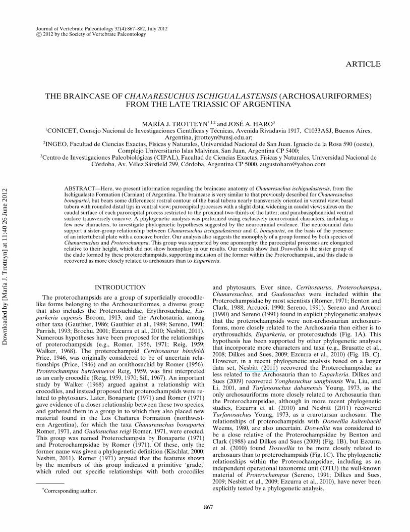
This article was downloaded by: [María J. Trotteyn] On: 26 June 2012, At: 11:40 Publisher: Taylor & Francis Informa Ltd Registered in England and Wales Registered Number: 1072954 Registered office: Mortimer House, 37-41 Mortimer Street, London W1T 3JH, UK Journal of Vertebrate Paleontology Publication details, including instructions for authors and subscription information: http://www.tandfonline.com/loi/ujvp20 The braincase of Chanaresuchus ischigualastensis (Archosauriformes) from the Late Triassic of Argentina María J. Trotteyn a b & José A. Haro c a CONICET, Consejo Nacional de Investigaciones Científicas y Técnicas, Avenida Rivadavia 1917, C1033ASJ, Buenos Aires, Argentina b INGEO, Facultad de Ciencias Exactas, Físicas y Naturales, Universidad Nacional de San Juan. Ignacio de la Rosa 590 (oeste), Complejo Universitario Islas Malvinas, San Juan, Argentina, CP 5400 c Centro de Investigaciones Paleobiológicas (CIPAL), Facultad de Ciencias Exactas, Físicas y Naturales, Universidad Nacional de Córdoba, Av. Vélez Sársfield 299, Córdoba, Argentina, CP 5000 Available online: 26 Jun 2012 To cite this article: María J. Trotteyn & José A. Haro (2012): The braincase of Chanaresuchus ischigualastensis (Archosauriformes) from the Late Triassic of Argentina, Journal of Vertebrate Paleontology, 32:4, 867-882 To link to this article: http://dx.doi.org/10.1080/02724634.2012.670178 PLEASE SCROLL DOWN FOR ARTICLE Full terms and conditions of use: http://www.tandfonline.com/page/terms-and-conditions This article may be used for research, teaching, and private study purposes. Any substantial or systematic reproduction, redistribution, reselling, loan, sub-licensing, systematic supply, or distribution in any form to anyone is expressly forbidden. The publisher does not give any warranty express or implied or make any representation that the contents will be complete or accurate or up to date. The accuracy of any instructions, formulae, and drug doses should be independently verified with primary sources. The publisher shall not be liable for any loss, actions, claims, proceedings, demand, or costs or damages whatsoever or howsoever caused arising directly or indirectly in connection with or arising out of the use of this material.
-
Upload
independent -
Category
Documents
-
view
1 -
download
0
Transcript of The Braincase of Chanaresuchus ischigualastensis (Archosauriformes) from the Late Triassic of...
This article was downloaded by: [María J. Trotteyn]On: 26 June 2012, At: 11:40Publisher: Taylor & FrancisInforma Ltd Registered in England and Wales Registered Number: 1072954 Registered office: Mortimer House,37-41 Mortimer Street, London W1T 3JH, UK
Journal of Vertebrate PaleontologyPublication details, including instructions for authors and subscription information:http://www.tandfonline.com/loi/ujvp20
The braincase of Chanaresuchus ischigualastensis(Archosauriformes) from the Late Triassic of ArgentinaMaría J. Trotteyn a b & José A. Haro ca CONICET, Consejo Nacional de Investigaciones Científicas y Técnicas, Avenida Rivadavia1917, C1033ASJ, Buenos Aires, Argentinab INGEO, Facultad de Ciencias Exactas, Físicas y Naturales, Universidad Nacional de SanJuan. Ignacio de la Rosa 590 (oeste), Complejo Universitario Islas Malvinas, San Juan,Argentina, CP 5400c Centro de Investigaciones Paleobiológicas (CIPAL), Facultad de Ciencias Exactas, Físicas yNaturales, Universidad Nacional de Córdoba, Av. Vélez Sársfield 299, Córdoba, Argentina, CP5000
Available online: 26 Jun 2012
To cite this article: María J. Trotteyn & José A. Haro (2012): The braincase of Chanaresuchus ischigualastensis(Archosauriformes) from the Late Triassic of Argentina, Journal of Vertebrate Paleontology, 32:4, 867-882
To link to this article: http://dx.doi.org/10.1080/02724634.2012.670178
PLEASE SCROLL DOWN FOR ARTICLE
Full terms and conditions of use: http://www.tandfonline.com/page/terms-and-conditions
This article may be used for research, teaching, and private study purposes. Any substantial or systematicreproduction, redistribution, reselling, loan, sub-licensing, systematic supply, or distribution in any form toanyone is expressly forbidden.
The publisher does not give any warranty express or implied or make any representation that the contentswill be complete or accurate or up to date. The accuracy of any instructions, formulae, and drug doses shouldbe independently verified with primary sources. The publisher shall not be liable for any loss, actions, claims,proceedings, demand, or costs or damages whatsoever or howsoever caused arising directly or indirectly inconnection with or arising out of the use of this material.
Journal of Vertebrate Paleontology 32(4):867–882, July 2012© 2012 by the Society of Vertebrate Paleontology
ARTICLE
THE BRAINCASE OF CHANARESUCHUS ISCHIGUALASTENSIS (ARCHOSAURIFORMES)FROM THE LATE TRIASSIC OF ARGENTINA
MARIA J. TROTTEYN*,1,2 and JOSE A. HARO3
1CONICET, Consejo Nacional de Investigaciones Cientıficas y Tecnicas, Avenida Rivadavia 1917, C1033ASJ, Buenos Aires,Argentina, [email protected];
2INGEO, Facultad de Ciencias Exactas, Fısicas y Naturales, Universidad Nacional de San Juan. Ignacio de la Rosa 590 (oeste),Complejo Universitario Islas Malvinas, San Juan, Argentina CP 5400;
3Centro de Investigaciones Paleobiologicas (CIPAL), Facultad de Ciencias Exactas, Fısicas y Naturales, Universidad Nacional deCordoba, Av. Velez Sarsfield 299, Cordoba, Argentina CP 5000, [email protected]
ABSTRACT—Here, we present information regarding the braincase anatomy of Chanaresuchus ischigualastensis, from theIschigualasto Formation (Carnian) of Argentina. The braincase is very similar to that previously described for Chanaresuchusbonapartei, but bears some differences: rostral contour of the basal tubera nearly transversely oriented in ventral view; basaltubera with rounded distal tips in ventral view; paroccipital processes with a slight distal widening in caudal view; sulcus on thecaudal surface of each paroccipital process restricted to the proximal two-thirds of the latter; and parabasisphenoidal ventralsurface transversely concave. A phylogenetic analysis was performed using exclusively neurocranial characters, including afew new characters, to investigate phylogenetic hypotheses suggested by the neurocranial evidence. The neurocranial datasupport a sister-group relationship between Chanaresuchus ischigualastensis and C. bonapartei, on the basis of the presenceof an intertuberal plate with a concave border. Our analysis also suggests the monophyly of a group formed by both species ofChanaresuchus and Proterochampsa. This group was supported by one apomorphy: the paroccipital processes are elongatedrelative to their height, which did not show homoplasy in our results. Our results show that Doswellia is the sister group ofthe clade formed by these proterochampsids, supporting inclusion of the former within the Proterochampsia, and this clade isrecovered as more closely related to archosaurs than to Euparkeria.
INTRODUCTION
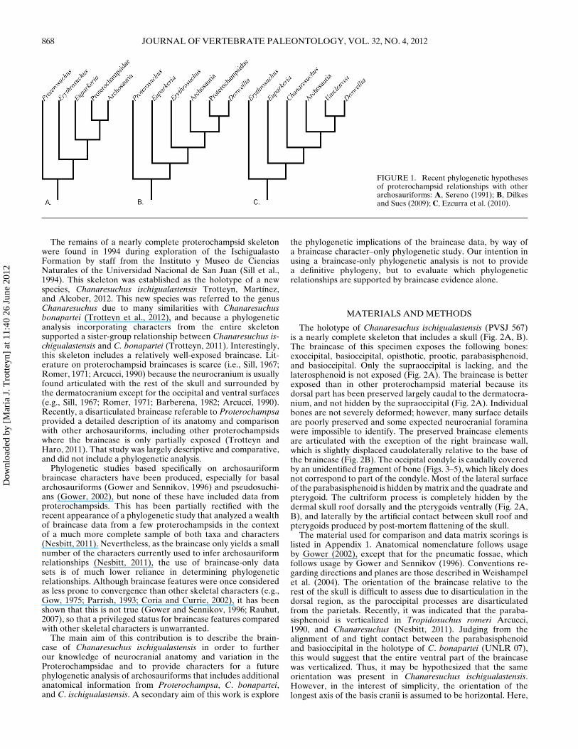
The proterochampsids are a group of superficially crocodile-like forms belonging to the Archosauriformes, a diverse groupthat also includes the Proterosuchidae, Erythrosuchidae, Eu-parkeria capensis Broom, 1913, and the Archosauria, amongother taxa (Gauthier, 1986; Gauthier et al., 1989; Sereno, 1991;Parrish, 1993; Brochu, 2001; Ezcurra et al., 2010; Nesbitt, 2011).Numerous hypotheses have been proposed for the relationshipsof proterochampsids (e.g., Romer, 1956, 1971; Reig, 1959;Walker, 1968). The proterochampsid Cerritosaurus binsfeldiPrice, 1946, was originally considered to be of uncertain rela-tionships (Price, 1946) and an ornithosuchid by Romer (1956).Proterochampsa barrionuevoi Reig, 1959, was first interpretedas an early crocodile (Reig, 1959, 1970; Sill, 1967). An importantstudy by Walker (1968) argued against a relationship withcrocodiles, and instead proposed that proterochampsids were re-lated to phytosaurs. Later, Bonaparte (1971) and Romer (1971)gave evidence of a closer relationship between these two species,and gathered them in a group in to which they also placed newmaterial found in the Los Chanares Formation (northwest-ern Argentina), for which the taxa Chanaresuchus bonaparteiRomer, 1971, and Gualosuchus reigi Romer, 1971, were erected.This group was named Proterochampsia by Bonaparte (1971)and Proterochampsidae by Romer (1971). Of these, only theformer name was given a phylogenetic definition (Kischlat, 2000;Nesbitt, 2011). Romer (1971) argued that the features shownby the members of this group indicated a primitive ‘grade,’which ruled out specific relationships with both crocodiles
*Corresponding author.
and phytosaurs. Ever since, Cerritosaurus, Proterochampsa,Chanaresuchus, and Gualosuchus were included within theProterochampsidae by most scientists (Romer, 1971; Benton andClark, 1988; Arcucci, 1990; Sereno, 1991). Sereno and Arcucci(1990) and Sereno (1991) found in explicit phylogenetic analysesthat the proterochampsids were non-archosaurian archosauri-forms, more closely related to the Archosauria than either is toerythrosuchids, Euparkeria, or proterosuchids (Fig. 1A). Thishypothesis has been supported by other phylogenetic analysesthat incorporate more characters and taxa (e.g., Brusatte et al.,2008; Dilkes and Sues, 2009; Ezcurra et al., 2010) (Fig. 1B, C).However, in a recent phylogenetic analysis based on a largerdata set, Nesbitt (2011) recovered the Proterochampsidae asless related to the Archosauria than to Euparkeria. Dilkes andSues (2009) recovered Yonghesuchus sangbiensis Wu, Liu, andLi, 2001, and Turfanosuchus dabanensis Young, 1973, as theonly archosauriforms more closely related to Archosauria thanthe Proterochampsidae, although in more recent phylogeneticstudies, Ezcurra et al. (2010) and Nesbitt (2011) recoveredTurfanosuchus Young, 1973, as a crurotarsan archosaur. Therelationships of proterochampsids with Doswellia kaltenbachiWeems, 1980, are also uncertain. Doswellia was considered tobe a close relative of the Proterochampsidae by Benton andClark (1988) and Dilkes and Sues (2009) (Fig. 1B), but Ezcurraet al. (2010) found Doswellia to be more closely related toarchosaurs than to proterochampsids (Fig. 1C). The phylogeneticrelationships within the Proterochampsidae, including as anindependent operational taxonomic unit (OTU) the well-knownmaterial of Proterochampsa (Sereno, 1991; Dilkes and Sues,2009; Nesbitt et al., 2009; Ezcurra et al., 2010), have never beenexplicitly tested by a phylogenetic analysis.
867
Dow
nloa
ded
by [
Mar
ía J
. Tro
tteyn
] at
11:
40 2
6 Ju
ne 2
012
868 JOURNAL OF VERTEBRATE PALEONTOLOGY, VOL. 32, NO. 4, 2012
FIGURE 1. Recent phylogenetic hypothesesof proterochampsid relationships with otherarchosauriforms: A, Sereno (1991); B, Dilkesand Sues (2009); C, Ezcurra et al. (2010).
The remains of a nearly complete proterochampsid skeletonwere found in 1994 during exploration of the IschigualastoFormation by staff from the Instituto y Museo de CienciasNaturales of the Universidad Nacional de San Juan (Sill et al.,1994). This skeleton was established as the holotype of a newspecies, Chanaresuchus ischigualastensis Trotteyn, Martınez,and Alcober, 2012. This new species was referred to the genusChanaresuchus due to many similarities with Chanaresuchusbonapartei (Trotteyn et al., 2012), and because a phylogeneticanalysis incorporating characters from the entire skeletonsupported a sister-group relationship between Chanaresuchus is-chigualastensis and C. bonapartei (Trotteyn, 2011). Interestingly,this skeleton includes a relatively well-exposed braincase. Lit-erature on proterochampsid braincases is scarce (i.e., Sill, 1967;Romer, 1971; Arcucci, 1990) because the neurocranium is usuallyfound articulated with the rest of the skull and surrounded bythe dermatocranium except for the occipital and ventral surfaces(e.g., Sill, 1967; Romer, 1971; Barberena, 1982; Arcucci, 1990).Recently, a disarticulated braincase referable to Proterochampsaprovided a detailed description of its anatomy and comparisonwith other archosauriforms, including other proterochampsidswhere the braincase is only partially exposed (Trotteyn andHaro, 2011). That study was largely descriptive and comparative,and did not include a phylogenetic analysis.
Phylogenetic studies based specifically on archosauriformbraincase characters have been produced, especially for basalarchosauriforms (Gower and Sennikov, 1996) and pseudosuchi-ans (Gower, 2002), but none of these have included data fromproterochampsids. This has been partially rectified with therecent appearance of a phylogenetic study that analyzed a wealthof braincase data from a few proterochampsids in the contextof a much more complete sample of both taxa and characters(Nesbitt, 2011). Nevertheless, as the braincase only yields a smallnumber of the characters currently used to infer archosauriformrelationships (Nesbitt, 2011), the use of braincase-only datasets is of much lower reliance in determining phylogeneticrelationships. Although braincase features were once consideredas less prone to convergence than other skeletal characters (e.g.,Gow, 1975; Parrish, 1993; Coria and Currie, 2002), it has beenshown that this is not true (Gower and Sennikov, 1996; Rauhut,2007), so that a privileged status for braincase features comparedwith other skeletal characters is unwarranted.
The main aim of this contribution is to describe the brain-case of Chanaresuchus ischigualastensis in order to furtherour knowledge of neurocranial anatomy and variation in theProterochampsidae and to provide characters for a futurephylogenetic analysis of archosauriforms that includes additionalanatomical information from Proterochampsa, C. bonapartei,and C. ischigualastensis. A secondary aim of this work is explore
the phylogenetic implications of the braincase data, by way ofa braincase character–only phylogenetic study. Our intention inusing a braincase-only phylogenetic analysis is not to providea definitive phylogeny, but to evaluate which phylogeneticrelationships are supported by braincase evidence alone.
MATERIALS AND METHODS
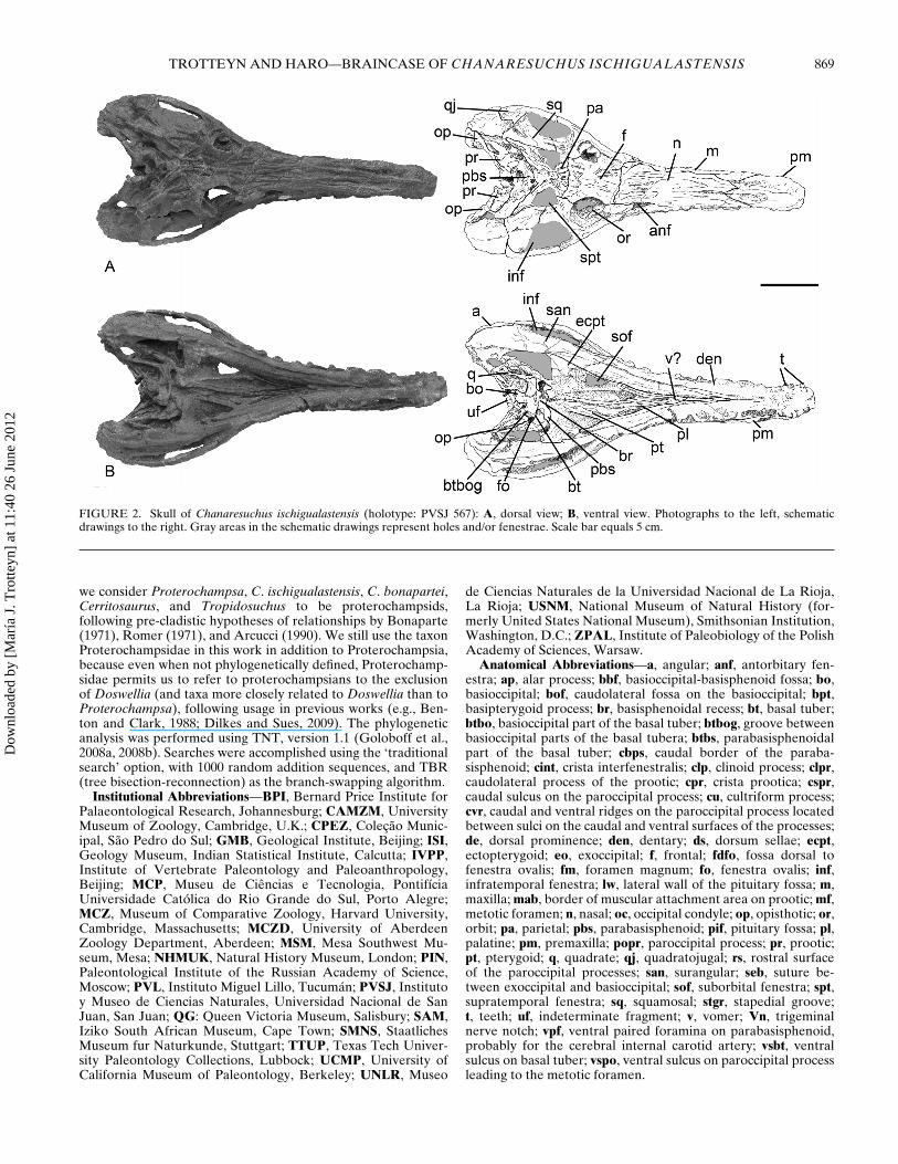
The holotype of Chanaresuchus ischigualastensis (PVSJ 567)is a nearly complete skeleton that includes a skull (Fig. 2A, B).The braincase of this specimen exposes the following bones:exoccipital, basioccipital, opisthotic, prootic, parabasisphenoid,and basioccipital. Only the supraoccipital is lacking, and thelaterosphenoid is not exposed (Fig. 2A). The braincase is betterexposed than in other proterochampsid material because itsdorsal part has been preserved largely caudal to the dermatocra-nium, and not hidden by the supraoccipital (Fig. 2A). Individualbones are not severely deformed; however, many surface detailsare poorly preserved and some expected neurocranial foraminawere impossible to identify. The preserved braincase elementsare articulated with the exception of the right braincase wall,which is slightly displaced caudolaterally relative to the base ofthe braincase (Fig. 2B). The occipital condyle is caudally coveredby an unidentified fragment of bone (Figs. 3–5), which likely doesnot correspond to part of the condyle. Most of the lateral surfaceof the parabasisphenoid is hidden by matrix and the quadrate andpterygoid. The cultriform process is completely hidden by thedermal skull roof dorsally and the pterygoids ventrally (Fig. 2A,B), and laterally by the artificial contact between skull roof andpterygoids produced by post-mortem flattening of the skull.
The material used for comparison and data matrix scorings islisted in Appendix 1. Anatomical nomenclature follows usageby Gower (2002), except that for the pneumatic fossae, whichfollows usage by Gower and Sennikov (1996). Conventions re-garding directions and planes are those described in Weishampelet al. (2004). The orientation of the braincase relative to therest of the skull is difficult to assess due to disarticulation in thedorsal region, as the paroccipital processes are disarticulatedfrom the parietals. Recently, it was indicated that the paraba-sisphenoid is verticalized in Tropidosuchus romeri Arcucci,1990, and Chanaresuchus (Nesbitt, 2011). Judging from thealignment of and tight contact between the parabasisphenoidand basioccipital in the holotype of C. bonapartei (UNLR 07),this would suggest that the entire ventral part of the braincasewas verticalized. Thus, it may be hypothesized that the sameorientation was present in Chanaresuchus ischigualastensis.However, in the interest of simplicity, the orientation of thelongest axis of the basis cranii is assumed to be horizontal. Here,
Dow
nloa
ded
by [
Mar
ía J
. Tro
tteyn
] at
11:
40 2
6 Ju
ne 2
012
TROTTEYN AND HARO—BRAINCASE OF CHANARESUCHUS ISCHIGUALASTENSIS 869
FIGURE 2. Skull of Chanaresuchus ischigualastensis (holotype: PVSJ 567): A, dorsal view; B, ventral view. Photographs to the left, schematicdrawings to the right. Gray areas in the schematic drawings represent holes and/or fenestrae. Scale bar equals 5 cm.
we consider Proterochampsa, C. ischigualastensis, C. bonapartei,Cerritosaurus, and Tropidosuchus to be proterochampsids,following pre-cladistic hypotheses of relationships by Bonaparte(1971), Romer (1971), and Arcucci (1990). We still use the taxonProterochampsidae in this work in addition to Proterochampsia,because even when not phylogenetically defined, Proterochamp-sidae permits us to refer to proterochampsians to the exclusionof Doswellia (and taxa more closely related to Doswellia than toProterochampsa), following usage in previous works (e.g., Ben-ton and Clark, 1988; Dilkes and Sues, 2009). The phylogeneticanalysis was performed using TNT, version 1.1 (Goloboff et al.,2008a, 2008b). Searches were accomplished using the ‘traditionalsearch’ option, with 1000 random addition sequences, and TBR(tree bisection-reconnection) as the branch-swapping algorithm.
Institutional Abbreviations—BPI, Bernard Price Institute forPalaeontological Research, Johannesburg; CAMZM, UniversityMuseum of Zoology, Cambridge, U.K.; CPEZ, Colecao Munic-ipal, Sao Pedro do Sul; GMB, Geological Institute, Beijing; ISI,Geology Museum, Indian Statistical Institute, Calcutta; IVPP,Institute of Vertebrate Paleontology and Paleoanthropology,Beijing; MCP, Museu de Ciencias e Tecnologia, PontifıciaUniversidade Catolica do Rio Grande do Sul, Porto Alegre;MCZ, Museum of Comparative Zoology, Harvard University,Cambridge, Massachusetts; MCZD, University of AberdeenZoology Department, Aberdeen; MSM, Mesa Southwest Mu-seum, Mesa; NHMUK, Natural History Museum, London; PIN,Paleontological Institute of the Russian Academy of Science,Moscow; PVL, Instituto Miguel Lillo, Tucuman; PVSJ, Institutoy Museo de Ciencias Naturales, Universidad Nacional de SanJuan, San Juan; QG: Queen Victoria Museum, Salisbury; SAM,Iziko South African Museum, Cape Town; SMNS, StaatlichesMuseum fur Naturkunde, Stuttgart; TTUP, Texas Tech Univer-sity Paleontology Collections, Lubbock; UCMP, University ofCalifornia Museum of Paleontology, Berkeley; UNLR, Museo
de Ciencias Naturales de la Universidad Nacional de La Rioja,La Rioja; USNM, National Museum of Natural History (for-merly United States National Museum), Smithsonian Institution,Washington, D.C.; ZPAL, Institute of Paleobiology of the PolishAcademy of Sciences, Warsaw.
Anatomical Abbreviations—a, angular; anf, antorbitary fen-estra; ap, alar process; bbf, basioccipital-basisphenoid fossa; bo,basioccipital; bof, caudolateral fossa on the basioccipital; bpt,basipterygoid process; br, basisphenoidal recess; bt, basal tuber;btbo, basioccipital part of the basal tuber; btbog, groove betweenbasioccipital parts of the basal tubera; btbs, parabasisphenoidalpart of the basal tuber; cbps, caudal border of the paraba-sisphenoid; cint, crista interfenestralis; clp, clinoid process; clpr,caudolateral process of the prootic; cpr, crista prootica; cspr,caudal sulcus on the paroccipital process; cu, cultriform process;cvr, caudal and ventral ridges on the paroccipital process locatedbetween sulci on the caudal and ventral surfaces of the processes;de, dorsal prominence; den, dentary; ds, dorsum sellae; ecpt,ectopterygoid; eo, exoccipital; f, frontal; fdfo, fossa dorsal tofenestra ovalis; fm, foramen magnum; fo, fenestra ovalis; inf,infratemporal fenestra; lw, lateral wall of the pituitary fossa; m,maxilla; mab, border of muscular attachment area on prootic; mf,metotic foramen; n, nasal; oc, occipital condyle; op, opisthotic; or,orbit; pa, parietal; pbs, parabasisphenoid; pif, pituitary fossa; pl,palatine; pm, premaxilla; popr, paroccipital process; pr, prootic;pt, pterygoid; q, quadrate; qj, quadratojugal; rs, rostral surfaceof the paroccipital processes; san, surangular; seb, suture be-tween exoccipital and basioccipital; sof, suborbital fenestra; spt,supratemporal fenestra; sq, squamosal; stgr, stapedial groove;t, teeth; uf, indeterminate fragment; v, vomer; Vn, trigeminalnerve notch; vpf, ventral paired foramina on parabasisphenoid,probably for the cerebral internal carotid artery; vsbt, ventralsulcus on basal tuber; vspo, ventral sulcus on paroccipital processleading to the metotic foramen.
Dow
nloa
ded
by [
Mar
ía J
. Tro
tteyn
] at
11:
40 2
6 Ju
ne 2
012
870 JOURNAL OF VERTEBRATE PALEONTOLOGY, VOL. 32, NO. 4, 2012
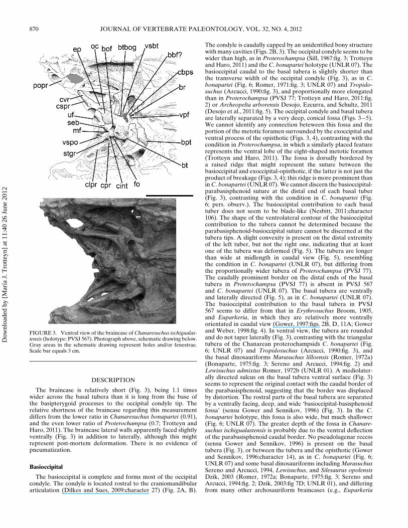
FIGURE 3. Ventral view of the braincase of Chanaresuchus ischigualas-tensis (holotype: PVSJ 567). Photograph above, schematic drawing below.Gray areas in the schematic drawing represent holes and/or fenestrae.Scale bar equals 3 cm.
DESCRIPTION
The braincase is relatively short (Fig. 3), being 1.1 timeswider across the basal tubera than it is long from the base ofthe basipterygoid processes to the occipital condyle tip. Therelative shortness of the braincase regarding this measurementdiffers from the lower ratio in Chanaresuchus bonapartei (0.91),and the even lower ratio of Proterochampsa (0.7; Trotteyn andHaro, 2011). The braincase lateral walls apparently faced slightlyventrally (Fig. 3) in addition to laterally, although this mightrepresent post-mortem deformation. There is no evidence ofpneumatization.
Basioccipital
The basioccipital is complete and forms most of the occipitalcondyle. The condyle is located rostral to the craniomandibulararticulation (Dilkes and Sues, 2009:character 27) (Fig. 2A, B).
The condyle is caudally capped by an unidentified bony structurewith many cavities (Figs. 2B, 3). The occipital condyle seems to bewider than high, as in Proterochampsa (Sill, 1967:fig. 3; Trotteynand Haro, 2011) and the C. bonapartei holotype (UNLR 07). Thebasioccipital caudal to the basal tubera is slightly shorter thanthe transverse width of the occipital condyle (Fig. 3), as in C.bonapartei (Fig. 6; Romer, 1971:fig. 3; UNLR 07) and Tropido-suchus (Arcucci, 1990:fig. 3), and proportionally more elongatedthan in Proterochampsa (PVSJ 77; Trotteyn and Haro, 2011:fig.2) or Archeopelta arborensis Desojo, Ezcurra, and Schultz, 2011(Desojo et al., 2011:fig. 5). The occipital condyle and basal tuberaare laterally separated by a very deep, conical fossa (Figs. 3−5).We cannot identify any connection beteween this fossa and theportion of the metotic foramen surrounded by the exoccipital andventral process of the opisthotic (Figs. 3, 4), contrasting with thecondition in Proterochampsa, in which a similarly placed featurerepresents the ventral lobe of the eight-shaped metotic foramen(Trotteyn and Haro, 2011). The fossa is dorsally bordered bya raised ridge that might represent the suture between thebasioccipital and exoccipital-opisthotic, if the latter is not just theproduct of breakage (Figs. 3, 4); this ridge is more prominent thanin C. bonapartei (UNLR 07). We cannot discern the basioccipital-parabasisphenoid suture at the distal end of each basal tuber(Fig. 3), contrasting with the condition in C. bonapartei (Fig.6; pers. observ.). The basioccipital contribution to each basaltuber does not seem to be blade-like (Nesbitt, 2011:character106). The shape of the ventrolateral contour of the basioccipitalcontribution to the tubera cannot be determined because theparabasisphenoid-basioccipital suture cannot be discerned at thetubera tips. A slight convexity is present on the distal extremityof the left tuber, but not the right one, indicating that at leastone of the tubera was deformed (Fig. 5). The tubera are longerthan wide at midlength in caudal view (Fig. 5), resemblingthe condition in C. bonapartei (UNLR 07), but differing fromthe proportionally wider tubera of Proterochampsa (PVSJ 77).The caudally prominent border on the distal ends of the basaltubera in Proterochampsa (PVSJ 77) is absent in PVSJ 567and C. bonapartei (UNLR 07). The basal tubera are ventrallyand laterally directed (Fig. 5), as in C. bonapartei (UNLR 07).The basioccipital contribution to the basal tubera in PVSJ567 seems to differ from that in Erythrosuchus Broom, 1905,and Euparkeria, in which they are relatively more ventrallyorientated in caudal view (Gower, 1997:figs. 2B, D, 11A; Gowerand Weber, 1998:fig. 4). In ventral view, the tubera are roundedand do not taper laterally (Fig. 3), contrasting with the triangulartubera of the Chanarean proterochampsids C. bonapartei (Fig.6; UNLR 07) and Tropidosuchus (Arcucci, 1990:fig. 3), andthe basal dinosauriforms Marasuchus lilloensis (Romer, 1972a)(Bonaparte, 1975:fig. 3; Sereno and Arcucci, 1994:fig. 2) andLewisuchus admixtus Romer, 1972b (UNLR 01). A mediolater-ally directed sulcus on the basal tubera ventral surface (Fig. 3)seems to represent the original contact with the caudal border ofthe parabasisphenoid, suggesting that the border was displacedby distortion. The rostral parts of the basal tubera are separatedby a ventrally facing, deep, and wide ‘basioccipital-basisphenoidfossa’ (sensu Gower and Sennikov, 1996) (Fig. 3). In the C.bonapartei holotype, this fossa is also wide, but much shallower(Fig. 6; UNLR 07). The greater depth of the fossa in Chanare-suchus ischigualastensis is probably due to the ventral deflectionof the parabasisphenoid caudal border. No pseudolagenar recess(sensu Gower and Sennikov, 1996) is present on the basaltubera (Fig. 3), or between the tubera and the opisthotic (Gowerand Sennikov, 1996:character 14), as in C. bonapartei (Fig. 6;UNLR 07) and some basal dinosauriforms including MarasuchusSereno and Arcucci, 1994, Lewisuchus, and Silesaurus opolensisDzik, 2003 (Romer, 1972a; Bonaparte, 1975:fig. 3; Sereno andArcucci, 1994:fig. 2; Dzik, 2003:fig 7D; UNLR 01), and differingfrom many other archosauriform braincases (e.g., Euparkeria
Dow
nloa
ded
by [
Mar
ía J
. Tro
tteyn
] at
11:
40 2
6 Ju
ne 2
012
TROTTEYN AND HARO—BRAINCASE OF CHANARESUCHUS ISCHIGUALASTENSIS 871
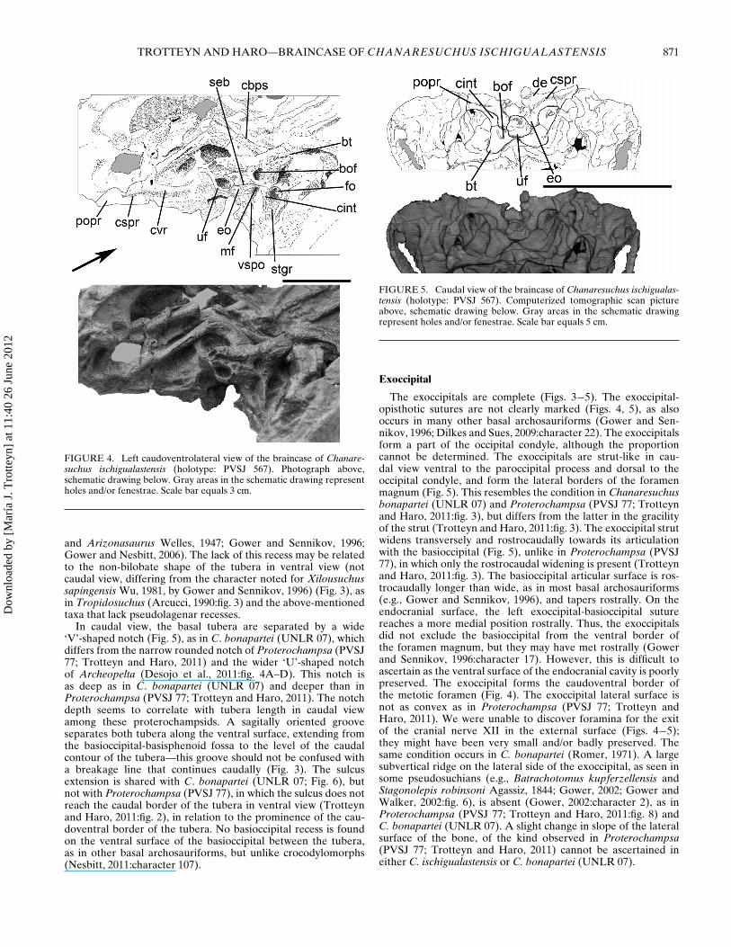
FIGURE 4. Left caudoventrolateral view of the braincase of Chanare-suchus ischigualastensis (holotype: PVSJ 567). Photograph above,schematic drawing below. Gray areas in the schematic drawing representholes and/or fenestrae. Scale bar equals 3 cm.
and Arizonasaurus Welles, 1947; Gower and Sennikov, 1996;Gower and Nesbitt, 2006). The lack of this recess may be relatedto the non-bilobate shape of the tubera in ventral view (notcaudal view, differing from the character noted for Xilousuchussapingensis Wu, 1981, by Gower and Sennikov, 1996) (Fig. 3), asin Tropidosuchus (Arcucci, 1990:fig. 3) and the above-mentionedtaxa that lack pseudolagenar recesses.
In caudal view, the basal tubera are separated by a wide‘V’-shaped notch (Fig. 5), as in C. bonapartei (UNLR 07), whichdiffers from the narrow rounded notch of Proterochampsa (PVSJ77; Trotteyn and Haro, 2011) and the wider ‘U’-shaped notchof Archeopelta (Desojo et al., 2011:fig. 4A–D). This notch isas deep as in C. bonapartei (UNLR 07) and deeper than inProterochampsa (PVSJ 77; Trotteyn and Haro, 2011). The notchdepth seems to correlate with tubera length in caudal viewamong these proterochampsids. A sagitally oriented grooveseparates both tubera along the ventral surface, extending fromthe basioccipital-basisphenoid fossa to the level of the caudalcontour of the tubera—this groove should not be confused witha breakage line that continues caudally (Fig. 3). The sulcusextension is shared with C. bonapartei (UNLR 07; Fig. 6), butnot with Proterochampsa (PVSJ 77), in which the sulcus does notreach the caudal border of the tubera in ventral view (Trotteynand Haro, 2011:fig. 2), in relation to the prominence of the cau-doventral border of the tubera. No basioccipital recess is foundon the ventral surface of the basioccipital between the tubera,as in other basal archosauriforms, but unlike crocodylomorphs(Nesbitt, 2011:character 107).
FIGURE 5. Caudal view of the braincase of Chanaresuchus ischigualas-tensis (holotype: PVSJ 567). Computerized tomographic scan pictureabove, schematic drawing below. Gray areas in the schematic drawingrepresent holes and/or fenestrae. Scale bar equals 5 cm.
Exoccipital
The exoccipitals are complete (Figs. 3−5). The exoccipital-opisthotic sutures are not clearly marked (Figs. 4, 5), as alsooccurs in many other basal archosauriforms (Gower and Sen-nikov, 1996; Dilkes and Sues, 2009:character 22). The exoccipitalsform a part of the occipital condyle, although the proportioncannot be determined. The exoccipitals are strut-like in cau-dal view ventral to the paroccipital process and dorsal to theoccipital condyle, and form the lateral borders of the foramenmagnum (Fig. 5). This resembles the condition in Chanaresuchusbonapartei (UNLR 07) and Proterochampsa (PVSJ 77; Trotteynand Haro, 2011:fig. 3), but differs from the latter in the gracilityof the strut (Trotteyn and Haro, 2011:fig. 3). The exoccipital strutwidens transversely and rostrocaudally towards its articulationwith the basioccipital (Fig. 5), unlike in Proterochampsa (PVSJ77), in which only the rostrocaudal widening is present (Trotteynand Haro, 2011:fig. 3). The basioccipital articular surface is ros-trocaudally longer than wide, as in most basal archosauriforms(e.g., Gower and Sennikov, 1996), and tapers rostrally. On theendocranial surface, the left exoccipital-basioccipital suturereaches a more medial position rostrally. Thus, the exoccipitalsdid not exclude the basioccipital from the ventral border ofthe foramen magnum, but they may have met rostrally (Gowerand Sennikov, 1996:character 17). However, this is difficult toascertain as the ventral surface of the endocranial cavity is poorlypreserved. The exoccipital forms the caudoventral border ofthe metotic foramen (Fig. 4). The exoccipital lateral surface isnot as convex as in Proterochampsa (PVSJ 77; Trotteyn andHaro, 2011). We were unable to discover foramina for the exitof the cranial nerve XII in the external surface (Figs. 4−5);they might have been very small and/or badly preserved. Thesame condition occurs in C. bonapartei (Romer, 1971). A largesubvertical ridge on the lateral side of the exoccipital, as seen insome pseudosuchians (e.g., Batrachotomus kupferzellensis andStagonolepis robinsoni Agassiz, 1844; Gower, 2002; Gower andWalker, 2002:fig. 6), is absent (Gower, 2002:character 2), as inProterochampsa (PVSJ 77; Trotteyn and Haro, 2011:fig. 8) andC. bonapartei (UNLR 07). A slight change in slope of the lateralsurface of the bone, of the kind observed in Proterochampsa(PVSJ 77; Trotteyn and Haro, 2011) cannot be ascertained ineither C. ischigualastensis or C. bonapartei (UNLR 07).
Dow
nloa
ded
by [
Mar
ía J
. Tro
tteyn
] at
11:
40 2
6 Ju
ne 2
012
872 JOURNAL OF VERTEBRATE PALEONTOLOGY, VOL. 32, NO. 4, 2012
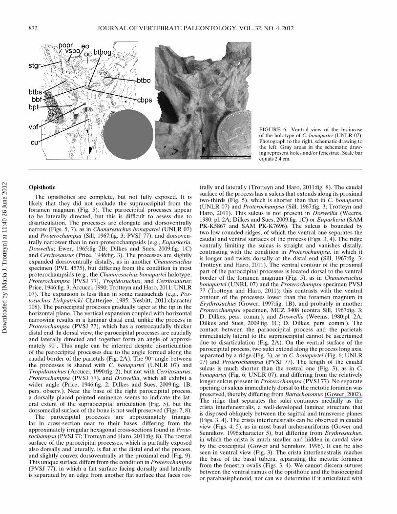
FIGURE 6. Ventral view of the braincaseof the holotype of C. bonapartei (UNLR 07).Photograph to the right, schematic drawing tothe left. Gray areas in the schematic draw-ing represent holes and/or fenestrae. Scale barequals 2.4 cm.
Opisthotic
The opisthotics are complete, but not fully exposed. It islikely that they did not exclude the supraoccipital from theforamen magnum (Fig. 5). The paroccipital processes appearto be laterally directed, but this is difficult to assess due todisarticulation. The processes are elongate and dorsoventrallynarrow (Figs. 5, 7), as in Chanaresuchus bonapartei (UNLR 07)and Proterochampsa (Sill, 1967:fig. 3; PVSJ 77), and dorsoven-trally narrower than in non-proterochampsids (e.g., Euparkeria,Doswellia; Ewer, 1965:fig 2B; Dilkes and Sues, 2009:fig. 1C)and Cerritosaurus (Price, 1946:fig. 3). The processes are slightlyexpanded dorsoventrally distally, as in another Chanaresuchusspecimen (PVL 4575), but differing from the condition in mostproterochampsids (e.g., the Chanaresuchus bonapartei holotype,Proterochampsa [PVSJ 77], Tropidosuchus, and Cerritosaurus;Price, 1946:fig. 3; Arcucci, 1990; Trotteyn and Haro, 2011; UNLR07). The expansion is less than in some rauisuchids (e.g., Pos-tosuchus kirkpatricki Chatterjee, 1985; Nesbitt, 2011:character108). The paroccipital processes gradually taper at the tip in thehorizontal plane. The vertical expansion coupled with horizontalnarrowing results in a laminar distal end, unlike the process inProterochampsa (PVSJ 77), which has a rostrocaudally thickerdistal end. In dorsal view, the paroccipital processes are caudallyand laterally directed and together form an angle of approxi-mately 90◦. This angle can be inferred despite disarticulationof the paroccipital processes due to the angle formed along thecaudal border of the parietals (Fig. 2A). The 90◦ angle betweenthe processes is shared with C. bonapartei (UNLR 07) andTropidosuchus (Arcucci, 1990:fig. 2), but not with Cerritosaurus,Proterochampsa (PVSJ 77), and Doswellia, which all exhibit awider angle (Price, 1946:fig. 2; Dilkes and Sues, 2009:fig. 1B;pers. observ.). Near the base of the right paroccipital process,a dorsally placed pointed eminence seems to indicate the lat-eral extent of the supraoccipital articulation (Fig. 5), but thedorsomedial surface of the bone is not well preserved (Figs. 7, 8).
The paroccipital processes are approximately triangu-lar in cross-section near to their bases, differing from theapproximately irregular hexagonal cross-sections found in Prote-rochampsa (PVSJ 77; Trotteyn and Haro, 2011:fig. 8). The rostralsurface of the paroccipital processes, which is partially exposedalso dorsally and laterally, is flat at the distal end of the process,and slightly convex dorsoventrally at the proximal end (Fig. 9).This unique surface differs from the condition in Proterochampsa(PVSJ 77), in which a flat surface facing dorsally and laterallyis separated by an edge from another flat surface that faces ros-
trally and laterally (Trotteyn and Haro, 2011:fig. 8). The caudalsurface of the process has a sulcus that extends along its proximaltwo-thirds (Fig. 5), which is shorter than that in C. bonapartei(UNLR 07) and Proterochampsa (Sill, 1967:fig. 3; Trotteyn andHaro, 2011). This sulcus is not present in Doswellia (Weems,1980: pl. 2A; Dilkes and Sues, 2009:fig. 1C) or Euparkeria (SAMPK-K5867 and SAM PK-K7696). The sulcus is bounded bytwo low rounded ridges, of which the ventral one separates thecaudal and ventral surfaces of the process (Figs. 3, 4). The ridgeventrally limiting the sulcus is straight and vanishes distally,contrasting with the condition in Proterochampsa, in which itis longer and twists dorsally at the distal end (Sill, 1967:fig. 3;Trotteyn and Haro, 2011). The ventral contour of the proximalpart of the paroccipital processes is located dorsal to the ventralborder of the foramen magnum (Fig. 5), as in Chanaresuchusbonapartei (UNRL 07) and the Proterochampsa specimen PVSJ77 (Trotteyn and Haro, 2011); this contrasts with the ventralcontour of the processes lower than the foramen magnum inErythrosuchus (Gower, 1997:fig. 1B), and probably in anotherProterochampsa specimen, MCZ 3408 (contra Sill, 1967:fig. 3;D. Dilkes, pers. comm.), and Doswellia (Weems, 1980:pl. 2A;Dilkes and Sues, 2009:fig. 1C; D. Dilkes, pers. comm.). Thecontact between the paraoccipital process and the parietalsimmediately lateral to the supraoccipital cannot be ascertaineddue to disarticulation (Fig. 2A). On the ventral surface of theparoccipital process, two sulci extend along the process long axis,separated by a ridge (Fig. 3), as in C. bonapartei (Fig. 6; UNLR07) and Proterochampsa (PVSJ 77). The length of the caudalsulcus is much shorter than the rostral one (Fig. 3), as in C.bonapartei (Fig. 6; UNLR 07), and differing from the relativelylonger sulcus present in Proterochampsa (PVSJ 77). No separateopening or sulcus immediately dorsal to the metotic foramen waspreserved, thereby differing from Batrachotomus (Gower, 2002).The ridge that separates the sulci continues medially as thecrista interfenestralis, a well-developed laminar structure thatis disposed obliquely between the sagittal and transverse planes(Figs. 3, 4). The crista interfenestralis can be observed in caudalview (Figs. 4, 5), as in most basal archosauriforms (Gower andSennikov, 1996:character 5), but differing from Erythrosuchus,in which the crista is much smaller and hidden in caudal viewby the exoccipital (Gower and Sennikov, 1996). It can be alsoseen in ventral view (Fig. 3). The crista interfenestralis reachesthe base of the basal tubera, separating the metotic foramenfrom the fenestra ovalis (Figs. 3, 4). We cannot discern suturesbetween the ventral ramus of the opisthotic and the basioccipitalor parabasisphenoid, nor can we determine if it articulated with
Dow
nloa
ded
by [
Mar
ía J
. Tro
tteyn
] at
11:
40 2
6 Ju
ne 2
012
TROTTEYN AND HARO—BRAINCASE OF CHANARESUCHUS ISCHIGUALASTENSIS 873
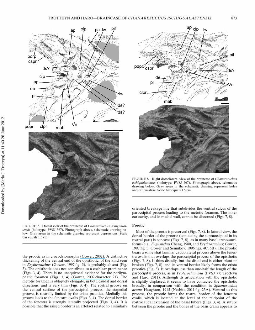
FIGURE 7. Dorsal view of the braincase of Chanaresuchus ischigualas-tensis (holotype: PVSJ 567). Photograph above, schematic drawing be-low. Gray areas in the schematic drawing represent depressions. Scalebar equals 1.5 cm.
the prootic as in crocodylomorphs (Gower, 2002). A distinctivethickening of the ventral end of the opisthotic, of the kind seenin Erythrosuchus (Gower, 1997:fig. 3), is probably absent (Fig.3). The opisthotic does not contribute to a cochlear prominence(Figs. 3, 4). There is no unequivocal evidence for the perilym-phatic foramen (Figs. 3, 4) (Gower, 2002:character 21). Themetotic foramen is obliquely elongate, in both caudal and dorsaldirections, and is very thin (Figs. 3, 4). The rostral groove onthe ventral surface of the paroccipital process, the stapedialgroove, is rostrally limited by the crista prootica. Medially thisgroove leads to the fenestra ovalis (Figs. 3, 4). The dorsal borderof the fenestra is strongly laterally projected (Figs. 3, 4). It ispossible that the raised border is an artefact related to a similarly
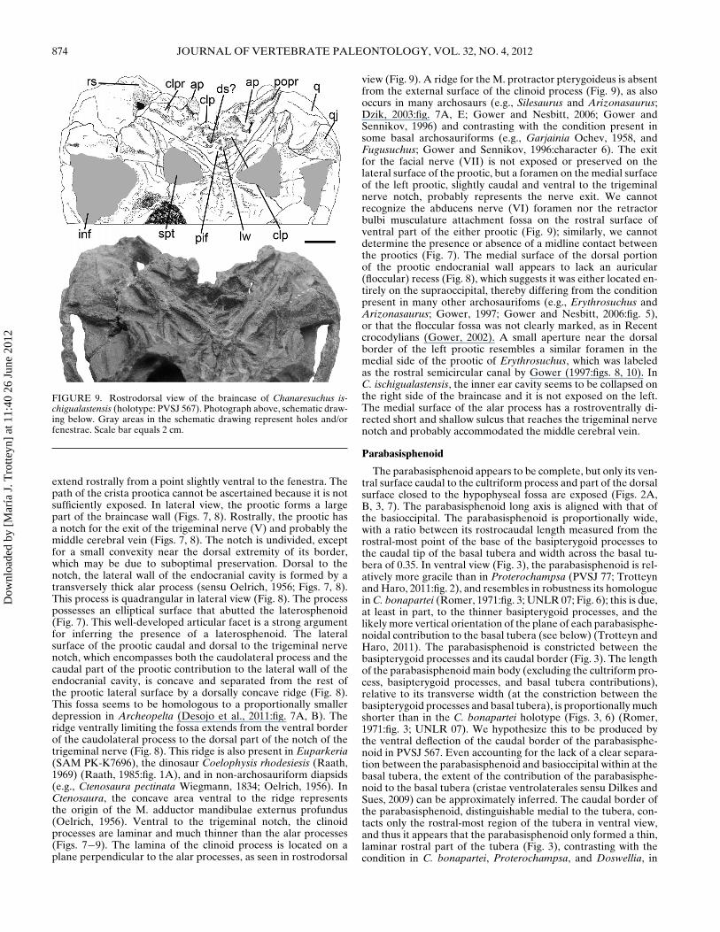
FIGURE 8. Right dorsolateral view of the braincase of Chanaresuchusischigualastensis (holotype: PVSJ 567). Photograph above, schematicdrawing below. Gray areas in the schematic drawing represent holesand/or fenestrae. Scale bar equals 1.5 cm.
oriented breakage line that subdivides the ventral sulcus of theparoccipital process leading to the metotic foramen. The innerear cavity, and its medial wall, cannot be discerned (Figs. 7, 8).
Prootic
Most of the prootic is preserved (Figs. 7, 8). In lateral view, thedorsal border of the prootic (contacting the supraoccipital in itsrostral part) is concave (Figs. 7, 8), as in many basal archosauri-forms (e.g., Fugusuchus Cheng, 1980, and Erythrosuchus; Gower,1997:fig. 3; Gower and Sennikov, 1996:figs. 4C, 6B). The prooticbears a somewhat laminar caudolateral process above the fenes-tra ovalis that overlaps the paroccipital process of the opisthotic(Figs. 7, 8). It thins distally, but the distal end is either blunt orbroken (Figs. 7, 8), and its ventral border likely forms the cristaprootica (Fig. 3). It overlaps less than one-half the length of theparoccipital process, as in Proterochampsa (PVSJ 77; Trotteynand Haro, 2011). Although its articulation with the opisthoticis slightly displaced, it seems to have contacted the opisthoticbroadly, in comparison with the condition in Sphenosuchusacutus Haughton, 1915 (Nesbitt, 2011:fig. 23A). Ventral to thisprocess, the prootic forms the rostral border of the fenestraovalis, which is located at the level of the midpoint of therostrocaudal extension of the basal tubera (Figs. 3, 4). A suturebetween the prootic and the bones of the basis cranii appears to
Dow
nloa
ded
by [
Mar
ía J
. Tro
tteyn
] at
11:
40 2
6 Ju
ne 2
012
874 JOURNAL OF VERTEBRATE PALEONTOLOGY, VOL. 32, NO. 4, 2012
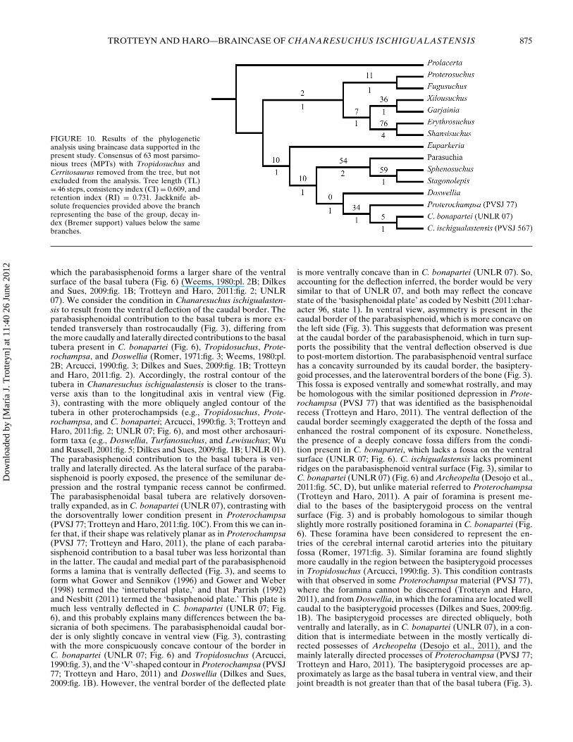
FIGURE 9. Rostrodorsal view of the braincase of Chanaresuchus is-chigualastensis (holotype: PVSJ 567). Photograph above, schematic draw-ing below. Gray areas in the schematic drawing represent holes and/orfenestrae. Scale bar equals 2 cm.
extend rostrally from a point slightly ventral to the fenestra. Thepath of the crista prootica cannot be ascertained because it is notsufficiently exposed. In lateral view, the prootic forms a largepart of the braincase wall (Figs. 7, 8). Rostrally, the prootic hasa notch for the exit of the trigeminal nerve (V) and probably themiddle cerebral vein (Figs. 7, 8). The notch is undivided, exceptfor a small convexity near the dorsal extremity of its border,which may be due to suboptimal preservation. Dorsal to thenotch, the lateral wall of the endocranial cavity is formed by atransversely thick alar process (sensu Oelrich, 1956; Figs. 7, 8).This process is quadrangular in lateral view (Fig. 8). The processpossesses an elliptical surface that abutted the laterosphenoid(Fig. 7). This well-developed articular facet is a strong argumentfor inferring the presence of a laterosphenoid. The lateralsurface of the prootic caudal and dorsal to the trigeminal nervenotch, which encompasses both the caudolateral process and thecaudal part of the prootic contribution to the lateral wall of theendocranial cavity, is concave and separated from the rest ofthe prootic lateral surface by a dorsally concave ridge (Fig. 8).This fossa seems to be homologous to a proportionally smallerdepression in Archeopelta (Desojo et al., 2011:fig. 7A, B). Theridge ventrally limiting the fossa extends from the ventral borderof the caudolateral process to the dorsal part of the notch of thetrigeminal nerve (Fig. 8). This ridge is also present in Euparkeria(SAM PK-K7696), the dinosaur Coelophysis rhodesiesis (Raath,1969) (Raath, 1985:fig. 1A), and in non-archosauriform diapsids(e.g., Ctenosaura pectinata Wiegmann, 1834; Oelrich, 1956). InCtenosaura, the concave area ventral to the ridge representsthe origin of the M. adductor mandibulae externus profundus(Oelrich, 1956). Ventral to the trigeminal notch, the clinoidprocesses are laminar and much thinner than the alar processes(Figs. 7−9). The lamina of the clinoid process is located on aplane perpendicular to the alar processes, as seen in rostrodorsal
view (Fig. 9). A ridge for the M. protractor pterygoideus is absentfrom the external surface of the clinoid process (Fig. 9), as alsooccurs in many archosaurs (e.g., Silesaurus and Arizonasaurus;Dzik, 2003:fig. 7A, E; Gower and Nesbitt, 2006; Gower andSennikov, 1996) and contrasting with the condition present insome basal archosauriforms (e.g., Garjainia Ochev, 1958, andFugusuchus; Gower and Sennikov, 1996:character 6). The exitfor the facial nerve (VII) is not exposed or preserved on thelateral surface of the prootic, but a foramen on the medial surfaceof the left prootic, slightly caudal and ventral to the trigeminalnerve notch, probably represents the nerve exit. We cannotrecognize the abducens nerve (VI) foramen nor the retractorbulbi musculature attachment fossa on the rostral surface ofventral part of the either prootic (Fig. 9); similarly, we cannotdetermine the presence or absence of a midline contact betweenthe prootics (Fig. 7). The medial surface of the dorsal portionof the prootic endocranial wall appears to lack an auricular(floccular) recess (Fig. 8), which suggests it was either located en-tirely on the supraoccipital, thereby differing from the conditionpresent in many other archosaurifoms (e.g., Erythrosuchus andArizonasaurus; Gower, 1997; Gower and Nesbitt, 2006:fig. 5),or that the floccular fossa was not clearly marked, as in Recentcrocodylians (Gower, 2002). A small aperture near the dorsalborder of the left prootic resembles a similar foramen in themedial side of the prootic of Erythrosuchus, which was labeledas the rostral semicircular canal by Gower (1997:figs. 8, 10). InC. ischigualastensis, the inner ear cavity seems to be collapsed onthe right side of the braincase and it is not exposed on the left.The medial surface of the alar process has a rostroventrally di-rected short and shallow sulcus that reaches the trigeminal nervenotch and probably accommodated the middle cerebral vein.
Parabasisphenoid
The parabasisphenoid appears to be complete, but only its ven-tral surface caudal to the cultriform process and part of the dorsalsurface closed to the hypophyseal fossa are exposed (Figs. 2A,B, 3, 7). The parabasisphenoid long axis is aligned with that ofthe basioccipital. The parabasisphenoid is proportionally wide,with a ratio between its rostrocaudal length measured from therostral-most point of the base of the basipterygoid processes tothe caudal tip of the basal tubera and width across the basal tu-bera of 0.35. In ventral view (Fig. 3), the parabasisphenoid is rel-atively more gracile than in Proterochampsa (PVSJ 77; Trotteynand Haro, 2011:fig. 2), and resembles in robustness its homologuein C. bonapartei (Romer, 1971:fig. 3; UNLR 07; Fig. 6); this is due,at least in part, to the thinner basipterygoid processes, and thelikely more vertical orientation of the plane of each parabasisphe-noidal contribution to the basal tubera (see below) (Trotteyn andHaro, 2011). The parabasisphenoid is constricted between thebasipterygoid processes and its caudal border (Fig. 3). The lengthof the parabasisphenoid main body (excluding the cultriform pro-cess, basipterygoid processes, and basal tubera contributions),relative to its transverse width (at the constriction between thebasipterygoid processes and basal tubera), is proportionally muchshorter than in the C. bonapartei holotype (Figs. 3, 6) (Romer,1971:fig. 3; UNLR 07). We hypothesize this to be produced bythe ventral deflection of the caudal border of the parabasisphe-noid in PVSJ 567. Even accounting for the lack of a clear separa-tion between the parabasisphenoid and basioccipital within at thebasal tubera, the extent of the contribution of the parabasisphe-noid to the basal tubera (cristae ventrolaterales sensu Dilkes andSues, 2009) can be approximately inferred. The caudal border ofthe parabasisphenoid, distinguishable medial to the tubera, con-tacts only the rostral-most region of the tubera in ventral view,and thus it appears that the parabasisphenoid only formed a thin,laminar rostral part of the tubera (Fig. 3), contrasting with thecondition in C. bonapartei, Proterochampsa, and Doswellia, in
Dow
nloa
ded
by [
Mar
ía J
. Tro
tteyn
] at
11:
40 2
6 Ju
ne 2
012
TROTTEYN AND HARO—BRAINCASE OF CHANARESUCHUS ISCHIGUALASTENSIS 875
FIGURE 10. Results of the phylogeneticanalysis using braincase data supported in thepresent study. Consensus of 63 most parsimo-nious trees (MPTs) with Tropidosuchus andCerritosaurus removed from the tree, but notexcluded from the analysis. Tree length (TL)= 46 steps, consistency index (CI) = 0.609, andretention index (RI) = 0.731. Jackknife ab-solute frequencies provided above the branchrepresenting the base of the group, decay in-dex (Bremer support) values below the samebranches.
which the parabasisphenoid forms a larger share of the ventralsurface of the basal tubera (Fig. 6) (Weems, 1980:pl. 2B; Dilkesand Sues, 2009:fig. 1B; Trotteyn and Haro, 2011:fig. 2; UNLR07). We consider the condition in Chanaresuchus ischigualasten-sis to result from the ventral deflection of the caudal border. Theparabasisphenoidal contribution to the basal tubera is more ex-tended transversely than rostrocaudally (Fig. 3), differing fromthe more caudally and laterally directed contributions to the basaltubera present in C. bonapartei (Fig. 6), Tropidosuchus, Prote-rochampsa, and Doswellia (Romer, 1971:fig. 3; Weems, 1980:pl.2B; Arcucci, 1990:fig. 3; Dilkes and Sues, 2009:fig. 1B; Trotteynand Haro, 2011:fig. 2). Accordingly, the rostral contour of thetubera in Chanaresuchus ischigualastensis is closer to the trans-verse axis than to the longitudinal axis in ventral view (Fig.3), contrasting with the more obliquely angled contour of thetubera in other proterochampsids (e.g., Tropidosuchus, Prote-rochampsa, and C. bonapartei; Arcucci, 1990:fig. 3; Trotteyn andHaro, 2011:fig. 2; UNLR 07; Fig. 6), and most other archosauri-form taxa (e.g., Doswellia, Turfanosuchus, and Lewisuchus; Wuand Russell, 2001:fig. 5; Dilkes and Sues, 2009:fig. 1B; UNLR 01).The parabasisphenoid contribution to the basal tubera is ven-trally and laterally directed. As the lateral surface of the paraba-sisphenoid is poorly exposed, the presence of the semilunar de-pression and the rostral tympanic recess cannot be confirmed.The parabasisphenoidal basal tubera are relatively dorsoven-trally expanded, as in C. bonapartei (UNLR 07), contrasting withthe dorsoventrally lower condition present in Proterochampsa(PVSJ 77; Trotteyn and Haro, 2011:fig. 10C). From this we can in-fer that, if their shape was relatively planar as in Proterochampsa(PVSJ 77; Trotteyn and Haro, 2011), the plane of each paraba-sisphenoid contribution to a basal tuber was less horizontal thanin the latter. The caudal and medial part of the parabasisphenoidforms a lamina that is ventrally deflected (Fig. 3), and seems toform what Gower and Sennikov (1996) and Gower and Weber(1998) termed the ‘intertuberal plate,’ and that Parrish (1992)and Nesbitt (2011) termed the ‘basisphenoid plate.’ This plate ismuch less ventrally deflected in C. bonapartei (UNLR 07; Fig.6), and this probably explains many differences between the ba-sicrania of both specimens. The parabasisphenoidal caudal bor-der is only slightly concave in ventral view (Fig. 3), contrastingwith the more conspicuously concave contour of the border inC. bonapartei (UNLR 07; Fig. 6) and Tropidosuchus (Arcucci,1990:fig. 3), and the ‘V’-shaped contour in Proterochampsa (PVSJ77; Trotteyn and Haro, 2011) and Doswellia (Dilkes and Sues,2009:fig. 1B). However, the ventral border of the deflected plate
is more ventrally concave than in C. bonapartei (UNLR 07). So,accounting for the deflection inferred, the border would be verysimilar to that of UNLR 07, and both may reflect the concavestate of the ‘basisphenoidal plate’ as coded by Nesbitt (2011:char-acter 96, state 1). In ventral view, asymmetry is present in thecaudal border of the parabasisphenoid, which is more concave onthe left side (Fig. 3). This suggests that deformation was presentat the caudal border of the parabasisphenoid, which in turn sup-ports the possibility that the ventral deflection observed is dueto post-mortem distortion. The parabasisphenoid ventral surfacehas a concavity surrounded by its caudal border, the basiptery-goid processes, and the lateroventral borders of the bone (Fig. 3).This fossa is exposed ventrally and somewhat rostrally, and maybe homologous with the similar positioned depression in Prote-rochampsa (PVSJ 77) that was identified as the basisphenoidalrecess (Trotteyn and Haro, 2011). The ventral deflection of thecaudal border seemingly exaggerated the depth of the fossa andenhanced the rostral component of its exposure. Nonetheless,the presence of a deeply concave fossa differs from the condi-tion present in C. bonapartei, which lacks a fossa on the ventralsurface (UNLR 07; Fig. 6). C. ischigualastensis lacks prominentridges on the parabasisphenoid ventral surface (Fig. 3), similar toC. bonapartei (UNLR 07) (Fig. 6) and Archeopelta (Desojo et al.,2011:fig. 5C, D), but unlike material referred to Proterochampsa(Trotteyn and Haro, 2011). A pair of foramina is present me-dial to the bases of the basipterygoid process on the ventralsurface (Fig. 3) and is probably homologous to similar thoughslightly more rostrally positioned foramina in C. bonapartei (Fig.6). These foramina have been considered to represent the en-tries of the cerebral internal carotid arteries into the pituitaryfossa (Romer, 1971:fig. 3). Similar foramina are found slightlymore caudally in the region between the basipterygoid processesin Tropidosuchus (Arcucci, 1990:fig. 3). This condition contrastswith that observed in some Proterochampsa material (PVSJ 77),where the foramina cannot be discerned (Trotteyn and Haro,2011), and from Doswellia, in which the foramina are located wellcaudal to the basipterygoid processes (Dilkes and Sues, 2009:fig.1B). The basipterygoid processes are directed obliquely, bothventrally and laterally, as in C. bonapartei (UNLR 07), in a con-dition that is intermediate between in the mostly vertically di-rected possesses of Archeopelta (Desojo et al., 2011), and themainly laterally directed processes of Proterochampsa (PVSJ 77;Trotteyn and Haro, 2011). The basipterygoid processes are ap-proximately as large as the basal tubera in ventral view, and theirjoint breadth is not greater than that of the basal tubera (Fig. 3).
Dow
nloa
ded
by [
Mar
ía J
. Tro
tteyn
] at
11:
40 2
6 Ju
ne 2
012
876 JOURNAL OF VERTEBRATE PALEONTOLOGY, VOL. 32, NO. 4, 2012
The relative rostrocaudal thickness of the base of the processesis also much less than in Proterochampsa (PVSJ 77; Trotteynand Haro, 2011:fig. 10A), and is probably more similar to thecondition in Chanaresuchus bonapartei (UNLR 07; Fig. 6). Thebasipterygoid processes size is proportionally much smaller thanin Proterochampsa (PVSJ 77; Trotteyn and Haro, 2011; Nesbitt,2011:character 124). The ventral surfaces of the processes arenot connected by a transverse buttress (Fig. 3), which is also ab-sent in Proterochampsa (PVSJ 77) and C. bonapartei (Trotteynand Haro, 2011; UNLR 07; Fig. 6), contrasting with the condi-tion in some archosaurs (e.g., Saurosuchus galilei Reig, 1959, andHerrerasaurus ischigualastensis Reig, 1963; Sereno and Novas,1994:figs. 7D, 8D; Alcober, 2000:fig. 9B; PVSJ 32, PVSJ 407). Thedistal ends of the basipterygoid processes lack a caudal promi-nence (Fig. 3), as in Chanaresuchus bonapartei (UNLR 07; Fig. 6)and Tropidosuchus (Arcucci, 1990:fig. 3), but differing from theprominent caudal projection present in Proterochampsa (PVSJ77; Trotteyn and Haro, 2011). The articulated pterygoid likelycovers only a portion of the ventral surface of the basipterygoidprocess in ventral view (Fig. 3). On the dorsal surface, sutureswith the other bones cannot be discerned (Figs. 7, 8), and a deeppituitary fossa is exposed (Fig. 7). The fossa is approximately tri-angular in this view, and its sides caudally diverge (Fig. 7), as inseveral archosaurifoms (e.g., Osmolskina czatkowicensis Borsuk-Białynicka and Evans, 2003, Silesaurus, and Arizonasaurus; Dzik,2003:fig. 7E; Gower and Nesbitt, 2006:fig. 1B; Borsuk-Białynickaand Evans, 2009:fig. 20C2). The lateral walls of the pituitary fossaare relatively thin (Fig. 7), as in several archosauriforms (e.g.,Osmolskina, Silesaurus, and Arizonasaurus; Dzik, 2003:fig. 7E;Gower and Nesbitt, 2006:fig. 1B; Borsuk-Białynicka and Evans,2009:fig. 20C2). The dorsal borders of the lateral walls of the fossahypophyseos are concave (Fig. 8), as in Osmolskina (Borsuk-Białynicka and Evans, 2009:fig. 20C2). This concavity probablyrepresents the ventral part of the pituitary vein foramen. The cau-dal wall of the fossa, the dorsum sellae, is damaged as a resultof the detachment and displacement of the right side wall of thebraincase from the basicranium, but portions of it seem to contactthe medial borders of the clinoid processes (Fig. 7). In relation tothis, we cannot discern the foramen of the abducens nerve (VI)on the possible remnants of the dorsum sellae.
RESULTS AND DISCUSSION
General Similarities and Distinctions
Chanaresuchus ischigualastensis possesses one unique neu-rocranial feature when compared with other proterochampsids,namely the nearly transversely oriented rostral contour of thebasal tubera in ventral view. A more oblique, caudally and later-ally oriented contour is observed in other proterochampsid andnon-proterochampsid basal archosauriforms: consequently, thisfeature of Chanaresuchus ischigualastensis seems to representan autapomorphy. Chanaresuchus ischigualastensis differs frommost other proterochampsids, except for the C. bonapartei speci-men PVL 4575, in the presence of distally expanded paroccipitalprocesses, but not from other archosauriforms.
Chanaresuchus ischigualastensis shares a large number ofsimilarities with the holotype of C. bonapartei that are lackingin the Proterochampsa specimen PVSJ 77. These include (1) therelatively longer (compared with its width) main basioccipitalbody caudal to the basal tubera; (2) greater proximodistal lengththan width at midlength of the basal tubera in caudal view,with widely separated distal tips; (3) the lack of a prominentcaudal border on the basioccipital basal tubera; (4) the caudalsulcus on the ventral surface of the paroccipital process is muchshorter than the rostral one; (5) the bases of the basipterygoidprocesses are not as strongly expanded rostrocaudally; (6) theparabasisphenoidal component of the basal tubera sheathsthe basioccipital component mostly rostrolaterally rather than
ventrally; (7) the caudal border of the parabasisphenoid is evenlyconcave; (8) a prominent ‘V’-shaped ridge on the ventral surfaceof the parabasisphenoid is absent; (9) a caudal projection of thebasipterygoid processes is absent; (10) a pair of well-exposedforamina, probably for the cerebral internal carotid arteries, ispresent on the ventral surface of the parabasisphenoid betweenthe bases of the basipterygoid processes; (11) the strut of theexoccipital, ventral to the paroccipital processes and dorsal tothe occipital condyle, is relatively transversely gracile, as can beobserved in caudal view; (12) the angle between the paroccipitalprocesses is not larger than 90◦ in dorsal view; (13) the basicra-nium is shorter from the rostral-most point of the basipterygoidprocesses to the caudal extremity of the occipital condylerelative to the width comprised by the basal tubera; and (14)the possession of ventrally and laterally directed basipterygoidprocesses. However, the last of these features is not systemat-ically useful for distinguishing Chanaresuchus ischigualastensisfrom Proterochampsa because the basipterygoid processes in atleast one Proterochampsa specimen (MCZ 3408) also projectventrally and laterally, in addition to slightly caudally (D. Dilkes,pers. comm.). Chanaresuchus ischigualastensis shares at least onesimilarity with Proterochampsa (PVSJ 77) that is not found in C.bonapartei, namely the transverse concavity in the caudal portionof the ventral surface of the main body of the parabasisphenoid.A second possible similarity between C. ischigualastensis andProterochampsa that is lacking in C. bonapartei might be thepresence of the prominent ridge-like structure at the exoccipital-basioccipital suture on the lateral surface of the braincasewall.
In addition to those previously mentioned in this section, thedifferences observed between Chanaresuchus ischigualastensisand the holotype of C. bonapartei are two, namely: (1) therounded, instead of pointed, distal extremity of the basal tubera;and (2) the basis cranii caudal to the cultriform process shorterrelative to the width constituted by the basal tubera.
The number of differences with the Proterochampsa specimenPVSJ 77 is larger. In addition to those previously mentioned inthis section, they include the following five differences: (1) thelateral surface of the exoccipitals not as convex rostrocaudally;(2) the cross-section of the paroccipital processes approximatelytriangular near the base, instead of approximately irregularhexagonal (related to the presence of a continuously convexrostrodorsal surface instead of distinct dorsal and rostral flat sur-faces connected at a ridge); (3) the distal end of the paroccipitalprocesses with a laminar shape; (4) the sulcus located on thecaudal surface of the paroccipital process extending only alongthe proximal two-thirds of the process; and (5) the ridge ventrallylimiting the sulcus on the caudal surface of each paroccipitalprocess distally vanishing and not twisted dorsalwards.
Comparisons with other proterochampsids are less fruitfulbecause less information has been published. Chanaresuchusischigualastensis resembles Tropidosuchus in a number of fea-tures in which it also resembles the holotype of C. bonapartei,namely: (1) the relatively elongated occipital condyle in ventralview; (2) the presence of a pair of well-exposed foramina on theventral surface of the parabasisphenoid almost directly medial tothe bases of the basipterygoid processes; (3) the concave caudalborder of the parabasisphenoid; and (4) the lack of a caudalprominence in the distal extremity of the basipterygoid process.Chanaresuchus ischigualastensis differs from Tropidosuchus inthe rounded, instead of pointed, distal extremities of the basaltubera, as well as the distally widening paroccipital processes,and the nearly transversely oriented rostral contour of the basaltubera in ventral view. There are three differences betweenChanaresuchus ischigualastensis and the paroccipital processes ofCerritosaurus, the only part of the braincase shared with the spec-imen PVSJ 567 (Price, 1946:fig. 3). The gradual distal divergenceof the dorsal and ventral borders in caudal view of the specimen
Dow
nloa
ded
by [
Mar
ía J
. Tro
tteyn
] at
11:
40 2
6 Ju
ne 2
012
TROTTEYN AND HARO—BRAINCASE OF CHANARESUCHUS ISCHIGUALASTENSIS 877
PVSJ 567 is not observed in Cerritosaurus (Price, 1946:fig. 3). Inaddition, the processes seem to be dorsoventrally thinner than inCerritosaurus (Price, 1946:fig. 3) and form a near 90◦ angle in dor-sal view, contrasting with the larger angle found in Cerritosaurus(Price, 1946:fig. 2). Thus, the data support the conclusion thatPVSJ 567 is a different taxon sharing greater similarities with C.bonapartei and Tropidosuchus among known proterochampsids.
Some similarities previously observed between Prote-rochampsa (PVSJ 77) and C. bonapartei (Trotteyn and Haro,2011) are also shared by Chanaresuchus ischigualastensis. Oneof these is the dorsoventrally thin paroccipital process, which, asmentioned, is apparently thicker in Cerritosaurus. The stronglydeveloped caudal sulcus on the paroccipital process representsthe other.
In addition, we notice a unique feature that is not sharedwith other archosauriforms, except crocodylians: the lack ofindication of a floccular recess on the alar process of the prootic.Indeed, Gower (2002) indicated that this character variedbetween being located entirely on the prootic, which is commonin basal archosauriforms, to being shared between the prooticand supraoccipital, which is present in archosaurs. If we considerthe absence of the floccular recess in the prootic as an indicationof complete absence of the recess from the neurocranium, thenthe feature would represent, on the basis of currently supportedphylogenies, a convergence with crocodylians. If, however, itrepresents a shift of the recess into the supraoccipital, we maysee it as a trait more related to the archosaurian characterstate than to that of basal archosauriforms, because the stateof archosaurs would actually be intermediate between theproposed one for Chanaresuchus ischigualastensis and that ofmore basal archosauriforms. In either case, the condition presentwould be more similar to that in archosaurs than to that in non-archosaurian archosauriforms. If the recess was completely lack-ing, the similarity with crocodylians may be related with similarlife habits, given that amphibious habits have been suggested forproterochampsids by both Bonaparte (1971) and Romer (1971).
Some of the differences between Chanaresuchus ischigualas-tensis and Proterochampsa appear to be associated in part withthe greater robustness of the skull in the latter, which may inturn be related to its larger size. This might account for the pro-portionally thicker basal tubera, the position of the basioccipitalmain body caudal to the tubera, differences in the robustenessof the exoccipital struts, and basipterygoid processes, as well asfor the presence of rostrocaudally thicker paroccipital processesin the latter taxon. This consideration may also explain some ofthe differences between C. bonapartei and Tropidosuchus, onone side, and Proterochampsa, on the other. Other differencesbetween the Chanaresuchus species and Proterochampsa, such asthe proportionally proximodistally longer basal tubera and moreventrally oriented basipterygoid process (in Chanaresuchus),seem to be at least partially related to the less dorsoventrallycompressed skull, which was previously noted by Bonaparte(1971) and Romer (1971) in the case of Chanaresuchus bona-partei. We think caution is required regarding the independenceof these features in future phylogenetic analyses.
Phylogenetic Analysis
Our phylogenetic analysis is based primarily on the data matrixof Gower and Sennikov (1996), which included only neurocra-nial characters. To this matrix were added several proterochamp-sids: Chanaresuchus ischigualastensis, C. bonapartei (scored onthe basis of the holotype), Proterochampsa (scored on the basis ofPVSJ 77), Tropidosuchus (scored from Arcucci, 1990), and Cerri-tosaurus (scored from Price, 1946), as well as the purported pro-terochampsid relative Doswellia (scored from Weems, 1980, andDilkes and Sues, 2009), comprising a total of 17 taxa. We addedthree braincase characters from the analysis in Dilkes and Sues
(2009:characters 20, 22, and 31) and four new braincase charac-ters, proposed in this study, providing a total of 24 characters. Inaddition, some characters in Gower and Sennikov (1996) weremodified. The details of the character list used can be found inAppendix 2 and the data matrix in Appendix 3. Codifications forXilousuchus were modified from Gower and Sennikov (1996), us-ing new data provided by Nesbitt et al. (2011). The details of thesearch are given in the Materials and Methods section. The anal-ysis yielded 63 most parsimonious trees (MPTs) with tree length(TL) of 46 steps, consistency index (CI) of 0.609, and retention in-dex (RI) of 0.731. The consensus tree was nearly completely un-resolved, except for a group formed by Parasuchia, Stagonolepis,and Sphenosuchus, which is fully resolved (also present in Gowerand Sennikov, 1996). This lead us to use the pruning option inTNT, which identifies those OTUs that fall in the greatest num-ber of alternative positions in the trees and removes them fromthe consensus tree without excluding the data from these OTUsfrom the analysis. In this way, the results are based in all thedata available. Pruning revealed that resolution of seven addi-tonal nodes could be gained by excluding Cerritosaurus from theconsensus, and another three can be gained by excluding Tropi-dosuchus. When both Cerritosaurus and Tropidosuchus are ex-cluded, 10 nodes are gained in the consensus, and the consen-sus tree becomes completely resolved (Fig. 10). Bremer supportand probabilistic support values were generally low (see Fig. 10).Our analysis supported the monophyly of the Proterochampsidae(when Tropidosuchus and Cerritosaurus were not considered).This group was supported by only one apomorphy, the paroccipi-tal processes elongated relative to their height (character 21, state1). This character is non-homoplastic, but the condition was notscored for many taxa. Within the Proterochampsidae, Chanare-suchus ischigualastensis was recovered as the sister taxon of C.bonapartei, on the basis of a single character, the acquisition ofa parabasisphenoidal intertuberal plate with a concave border(character 2, state 1). This feature is homoplastic, however, be-cause it appears independently in Euparkeria. This sister-grouprelationship implies that the phylogenetic hypothesis suggestedby the braincase data supports inclusion of Chanaresuchus is-chigualastensis within Chanaresuchus (only when taxa of ambigu-ous relationships are excluded from consideration). This sister-group relationship is also recovered by a phylogenetic analysisencompassing a larger data matrix that includes characters fromother parts of the skeleton (Trotteyn, 2011). Our data agree withthe analyses of Benton and Clark (1988) and Dilkes and Sues(2009), and contrast with the results of Ezcurra et al. (2010) andDesojo et al. (2011), in recovering Doswellia as a proterochamp-sian, more closely related to Proterochampsa and Chanaresuchusthan to archosaurs. The only one apomorphy supporting the Pro-terochampsia is the presence of a parietal-paroccipital processcontact immediately lateral to the supraoccipital (character 20,state 1), which is, however, unknown in the members of thegenus Chanaresuchus scored, and homoplastic, as it is present inmany archosauriforms less related to the Archosauria than prote-rochampsians. Archosauria was recovered as the sister taxon tothe Proterochampsia, and these two groups share the followingthree apomorphies: absence of the ridge on the clinoid process ofthe prootic (character 6, state 1), absence of a pseudolagenar re-cess between the basal tuber and ventral ramus of the opisthotic(character 14, state 1), and parabasisphenoidal contribution tothe basal tubera not wider than the basioccipital contributionto the basal tubera (character 24, state 1). The two first fea-tures are homoplastic: notably, they also appear apomorphicallyin the Shansisuchus Young, 1964 + Erythrosuchus clade. The fi-nal feature of these three is not homoplastic, but we were un-able to score it for members of the Shansisuchus + Erythrosuchusclade. Euparkeria is the sister taxon to the Proterochampsia +Archosauria group, and the clade formed by all these is diagnosedby the following character states: the position of the abducens
Dow
nloa
ded
by [
Mar
ía J
. Tro
tteyn
] at
11:
40 2
6 Ju
ne 2
012
878 JOURNAL OF VERTEBRATE PALEONTOLOGY, VOL. 32, NO. 4, 2012
foramina on the ventral surface of the dorsum sellae (character4, state 1; although scoring for this character is lacking for pro-terochampsians and most archosaurs), and the convex ventrolat-eral contour of the basioccipital part of the basal tubera in caudalview (character 23, states 1−2). Character 4 is homoplastic in thisstudy, appearing independently in the group formed by Erythro-suchus + Shansisuchus. Character 23 is homoplastically acquiredby at least one specimen of Erythrosuchus (BPI 3893; Gower,1997:fig. 2D), but not all (NHMUK R3592; Gower, 1997:fig.2B). Our results resemble those of Gower and Sennikov (1996),Ezcurra et al. (2010), and Nesbitt (2011), but differ from those ofDilkes and Sues (2009), in showing a closer relationship betweenEuparkeria and archosaurs than either of these taxa has with Ery-throsuchus. The large amount of homoplasies observed betweenthe Erythrosuchus + Shansisuchus clade and some subsets ofmembers of the sister group to Euparkeria previously mentionedsuggests widespread convergent evolution. Our results agree withthe results of Gower and Sennikov (1996) in suggesting a mono-phyletic Proterosuchia, with Erythrosuchus most closely relatedto Proterosuchus Broom, 1903, than to Archosauria, but disagreewith the results obtained from the more comprehensive data setsof Sereno (1991), Dilkes and Sues (2009), Ezcurra et al. (2010),and Nesbitt (2011). Our results also agree with Gower and Sen-nikov’s (1996) hypothesis that Xilousuchus is an erythrosuchid,although this was falsified by more recent analyses based onlarger data sets, which recover it as an archosaur (Nesbitt, 2011;Nesbitt et al., 2011). These relationships seem to result from theproportionally greater reliance on data from the Gower and Sen-nikov (1996) study than from the other works mentioned, andthe phylogenetic hypothesis presented herein should not be pre-ferred over more complete phylogenies generated on the basisof more evidence. Our results show lower values for both theCI and RI than those obtained by Gower and Sennikov (1996)(compare our CI of 0.609 with their value of 692, and our RI of0.731 with their value of 0.81). This is expected because our datamatrix incorporates a larger number of taxa and characters thanthat offered by Gower and Sennikov (1996), and matrices withmore taxa and characters generally show more homoplasy, orless consistency, as measured by the consistency index (Sander-son and Donoghue, 1989; Hauser and Boyajian, 1997). In rela-tion with this, our results further support the suggestion of Gowerand Sennikov (1996) and Gower (2002) that homoplasy is poten-tially common in the braincases of basal archosauriforms, a resultechoed by work on neotheropods by Rauhut (2007).
ACKNOWLEDGMENTS
We thank the following for their help in this study: R. Martınez(Instituto y Museo de Ciencias Naturales, Universidad Nacionalde San Juan) for the loan of PVSJ 567; G. Heredia for the draw-ings used here; D. Abelın (Instituto y Museo de Ciencias Natu-rales, Universidad Nacional de San Juan) for preparation of thematerial; I. Zabrodski for photography of PVSJ 567; J. Gauthier(Department of Geology and Geophysics, Yale University) foraccess to his photographs of Euparkeria; S. Martın (UniversidadNacional de La Rioja) for permission to visit the collection un-der his care in order to examine the holotype of C. bonapartei; D.Dilkes for his revision of an earlier draft and for providing sug-gestions; and to the editor, R. Irmis. We thank the Willi HennigSociety for free access to use the TNT softare. Finally, we thankCONICET for financing this work with the fellowships extendedto both authors.
LITERATURE CITED
Agassiz, L. 1844. Monographie des Poissons Fossiles du Vieux GresRouge ou Systeme Devonien (Old Red Sandstone) des Iles Britan-niques et de Russie. Jent et Gassman, Neuchatel, 171 pp.
Alcober, O. 2000. Redescription of the skull of Saurosuchus galilei(Archosauria: Rauisuchidae). Journal of Vertebrate Paleontology20:302–316.
Arcucci, A. 1990. Un nuevo Proterochampsidae (Reptilia-Archosauriformes) de la fauna local de Los Chanares (TriasicoMedio), La Rioja, Argentina. Ameghiniana 27:365–378.
Barberena, M. C. 1982. Uma nova especie de Proterochampsa, P. nodosasp. nov. do Triassico do Brasil. Anais da Academia Brasileira deCiencias 54:127–141.
Benton, M. J., and J. M. Clark. 1988. Archosaur phylogeny and the rela-tionships of the Crocodilia; pp. 295–338 in M. J. Benton (ed.), ThePhylogeny and Classification of the Tetrapods, Volume 1. Claren-don Press, Oxford, U.K.
Bonaparte, J. F. 1971. Cerritosaurus binsfeldi Price, tipo de una nuevafamilia de tecodontes (Pseudosuchia-Proterochampsia). Anais daAcademia Brasileira de Ciencias, suplemento 43:417–422.
Bonaparte, J. F. 1975. Nuevos materiales de Lagosuchus talampayensisRomer (Thecodontia-Pseudolagosuchia) y su significado en el ori-gen de los Saurisquia. Chanarense inferior. Triasico medio de Ar-gentina. Acta Geologica Lilloana 13:1–90.
Borsuk-Białynicka, M., and S. E. Evans. 2003. A basal archosauriformfrom the Early Triassic of Poland. Acta Palaeontologica Polonica48:649–652.
Borsuk-Białynicka, M., and S. E. Evans. 2009. Cranial and mandibularosteology of the Early Triassic archosauriform Osmolskina czatkow-icensis from Poland. Palaeontologia Polonica 65:235–281.
Brochu, C. A. 2001. Progress and future in archosaur phylogenetics. Jour-nal of Paleontology 75:1185–1201.
Broom, R. 1903. On a new reptile (Proterosuchus fergusi) from the Ka-roo beds of Tarkastad, South Africa. Annals of the South AfricanMuseum 4:158–164.
Broom, R. 1905. Notice of some new fossil reptiles from the Karoo Bedsof South Africa. Records of the Albany Museum 1:331–337.
Broom, R. 1913. Note on Mesosuchus browni Watson and on a new SouthAfrican Triassic pseudosuchian (Euparkeria capensis). Records ofthe Albany Museum 2:394–396.
Brusatte, S. L., M. J. Benton, M. Ruta, and G. T. Lloyd. 2008. Superior-ity, competition, and opportunism in the evolutionary radiation ofdinosaurs. Science 321:1485–1488.
Camp, C. L. 1930. A study of the phytosaurs, with description of new ma-terial from North America. Memoirs of the University of California10:1–174.
Chatterjee, S. 1978. A primitive parasuchid (phytosaur) reptile fromthe Upper Triassic Maleri Formation of India. Palaeontology 21:83–127.
Chatterjee, S. 1985. Postosuchus, a new thecodontian reptile from the Tri-assic of Texas and the origin of tyrannosaurs. Philosophical Trans-actions of the Royal Society of London, Series B 309:395–460.
Cheng, Z.-W. 1980. [Vertebrate fossils]; pp. 115–171 in [Mesozoic stratig-raphy and paleontology of the Shanxi-Gansu-Ningxia Basin, Vol-umee 2]. Publishing House of Geology, Beijing. [Chinese]
Coria, R. A., and P. J. Currie. 2002. The braincase of Giganotosaurus car-olinii (Dinosauria: Theropoda) from the Upper Cretaceous of Ar-gentina. Journal of Vertebrate Paleontology 22:802–811.
Desojo, J. B., M. D. Ezcurra, and C. L. Schultz. 2011. An unusual new ar-chosauriform from the Middle–Late Triassic of southern Brazil andthe monophyly of Doswelliidae. Zoological Journal of the LinneanSociety 161:839–871.
Dilkes, D. W. 1998. The Early Triassic rhynchosaur Mesosuchus browniand the interrelationships of basal archosauromorph reptiles. Philo-sophical Transactions of the Royal Society of London, Series B353:501–541.
Dilkes, D. W., and H.-D. Sues. 2009. Redescription and phylogenetic re-lationships of Doswellia kaltenbachi (Diapsida: Archosauriformes)from the Upper Triassic of Virginia. Journal of Vertebrate Paleon-tology 29:58–79.
Dzik, J. 2003. A beaked herbivorous archosaur with dinosaur affinitiesfrom the early Late Triassic of Poland. Journal of Vertebrate Pale-ontology 23:556–574.
Evans, S. E. 1986. The braincase of Prolacerta broomi (Reptilia, Trias-sic). Neues Jahrbuch fur Geologie und Palaontologie, Abhandlun-gen 173:181–200.
Ewer, R. F. 1965. The anatomy of the thecodont reptile Euparkeriacapensis Broom. Philosophical Transactions of the Royal Society ofLondon, Series B 248:379–435.
Dow
nloa
ded
by [
Mar
ía J
. Tro
tteyn
] at
11:
40 2
6 Ju
ne 2
012
TROTTEYN AND HARO—BRAINCASE OF CHANARESUCHUS ISCHIGUALASTENSIS 879
Ezcurra, M. D., A. Lecuona, and A. Martinelli. 2010. A new basal ar-chosauriform diapsid from the Lower Triassic of Argentina. Journalof Vertebrate Paleontology 30:1433–1450.
Gauthier, J. A. 1986. Saurischian monophyly and the origin of birds.Memoirs of the Californian Academy of Science 8:1–55.
Gauthier, J. A., D. Cannatella, K. De Queiroz, A. G. Kluge, and T. Rowe.1989. Tetrapod phylogeny; pp. 337–353 in B. Fernholm, K. Bremer,and H. Jornvall (eds.), The Hierarchy of Life. Elsevier Science Pub-lishers, New York.
Goloboff, P. A., J. S. Farris, and K. C. Nixon. 2008a. TNT, a free programfor phylogenetic analysis. Cladistics 24:1–13.
Goloboff, P. A., J. S. Farris, and K. C. Nixon. 2008b. TNT (Tree Anal-ysis Using New Technology). Published by the authors, Tucuman,Argentina.
Gow, C. E. 1975. The morphology and relationships of Youngina capensisBroom and Prolacerta broomi Parrington. Palaeontologia africana18:89–131.
Gower, D. J. 1997. The braincase of the early archosaurian reptile Ery-throsuchus africanus. Journal of Zoology 242:557–576.
Gower, D. J. 2002. Braincase evolution in suchian archosaurs (Reptilia:Diapsida): evidence from the rauisuchian Batrachotomus kupferzel-lensis. Zoological Journal of the Linnean Society 136:49–76.
Gower, D. J., and S. J. Nesbitt. 2006. The braincase of Arizonasaurus bab-bitti—further evidence of the non-monophyly of Rauisuchia. Jour-nal of Vertebrate Paleontology 26:79–87.
Gower, D. J., and A. G. Sennikov. 1996. Morphology and phyloge-netic informativeness of early archosaur braincases. Palaeontology39:883–906.
Gower, D. J., and A. D. Walker. 2002. New data on the braincase of theaetosaurian archosaur (Reptilia: Diapsida) Stagonolepis robertsoniAgassiz. Zoological Journal of the Linnean Society 136:7–23.
Gower, D. J., and E. Weber. 1998. The braincase of Euparkeria, and theevolutionary relationships of birds and crocodilians. Biological Re-views 73:367–411.
Haughton, S. H. 1915. A new thecodont from the Stormberg Beds. An-nals of the South African Museum 12:98–105.
Hauser, D. L., and G. Boyajian. 1997. Proportional change and pat-terns of homoplasy: Sanderson and Donoghue revisited. Cladistics13:97–100.
Juul, L. 1994. The phylogeny of basal archosaurs. Palaeontologia africana31:1–38.
Kischlat, E. E. 2000. Tecodoncios: a aurora dos arcosaurios no Triassico;pp. 273–316 in M. Holtz and L. F. de Ross (eds.), Paleontologia doRio Grande do Sul. Edicao CIGO/UFRGS, Porto Alegre.
Long, J. A., and P. A. Murry. 1995. Late Triassic (Carnian and Norian)tetrapods from the Southwestern United States. Bulletin of the NewMexico Museum of Natural History and Science 4:1–254.
Nesbitt, S. J. 2011. The early evolution of archosaurs—relationships andthe origin of major clades. Bulletin of the American Museum of Nat-ural History 352:1–292.
Nesbitt, S. J., L. Jun, and L. Chun. 2011. A sail-backed suchian from theHeshanggou Formation (Early Triassic: Olenekian) of China. Earthand Environmental Science Transactions of the Royal Society ofEdinburgh 101:271–284.
Nesbitt, S. J., M. R. Stocker, B. J. Small, and A. Downs. 2009. The oste-ology and relationships of Vancleavea campi (Reptilia: Archosauri-formes). Zoological Journal of the Linnean Society 157:814–864.
Ochev, V. G. 1958. [New data on the Triassic vertebrate faunafrom the Orenburg Cis-Urals]. Doklady Akademii Nauk SSSRRR22:749–752. [Russian]
Oelrich, T. M. 1956. The anatomy of the head of Ctenosaura pectinata(Iguanidae). Miscellaneous Publications of the Museum of Zoology,University of Michigan 94:1–122.
Parrish, J. M. 1992. Phylogeny of the Erythrosuchidae (Reptilia: Ar-chosauriformes). Journal of Vertebrate Paleontology 12:93–102.
Parrish, J. M. 1993. Phylogeny of the Crocodylotarsi, with special refer-ence to archosaurian and crurotarsan monophyly. Journal of Verte-brate Paleontology 13:287–308.
Price, L. I. 1946. Sobre um novo pseudosuquio do Triassico Superior doRio Grande do Sul. Boletim Divisao de Geologıa e Mineralogıa120:1–38.
Raath, M. A. 1969. A new coelurosaurian dinosaur from the Forest Sand-stone of Rhodesia. Arnoldia 4(28):1–25.
Raath, M. A. 1985. The theropod Syntarsus and its bearings on the originof birds; pp. 219–227 in M. K. Hecht, J. H. Ostrom, G. Viohl, and P.Wellnhofer (eds.), The Beginning of Birds: Proceedings of the Inter-
national Archaeopteryx Conference, Eichstatt. Freunde des Jura-Museums, Eichstatt.
Rauhut, O. W. M. 2007. The myth of the conservative character: brain-case characters in theropod phylogenies. Hallesches Jahrbuch furGeowissenschaften, Beiheft 23:51–54.
Reig, O. A. 1959. Primeros datos descriptivos sobre nuevos rep-tiles arcosaurios del Triasico de Ischigualasto (San Juan, Ar-gentina). Revista de la Asociacion Geologica Argentina 13:257–270.
Reig, O. A. 1963. La presencia de dinosaurios saurisquios en los “Estratosde Ischigualasto” (Mesotriasico Superior) de las provincias de SanJuan y La Rioja (Republica Argentina). Ameghiniana 3:3–20.
Reig, O. A. 1970. The Proterosuchia and the early evolution of the ar-chosaurs; an essay about the origin of a major taxon. Bulletin of theMuseum of Comparative Zoology 139:229–292.
Romer, A. S. 1956. Osteology of the Reptiles. University of ChicagoPress, Chicago, Illinois, 772 pp.
Romer, A. S. 1971. The Chanares (Argentina) Triassic reptile fauna.XI: two new long-snouted thecodonts, Chanaresuchus and Gualo-suchus. Breviora 379:1–22.
Romer, A. S. 1972a. The Chanares (Argentina) Triassic reptile fauna.XV. Further remains of the thecodonts Lagerpeton and Lagosuchus.Breviora 394:1–7.
Romer, A. S. 1972b. The Chanares (Argentina) Triassic reptile fauna.XIV. Lewisuchus admixtus, gen. et sp. nov., a further thecodontfrom the Chanares beds. Breviora 390:1–13.
Sanderson, M. J., and M. J. Donoghue. 1989. Patterns of variation in lev-els of homoplasy. Evolution 43:1781–1795.
Sereno, P. C. 1991. Basal archosaurs: phylogenetic relationships and func-tional implications. Memoir of the Society of Vertebrate Paleontol-ogy 2:1–53.
Sereno, P. C., and A. B. Arcucci. 1990. The monophyly of the cruro-tarsal archosaurs and the origin of birds and crocodile ankle joints.Neues Jahrbuch fur Geologie und Palaontologie, Abhandlungen180:21–52.
Sereno, P. C., and A. B. Arcucci. 1994. Dinosaurian precursors from theMiddle Triassic of Argentina: Marasuchus lilloensis, gen. nov. Jour-nal of Vertebrate Paleontology, 14:53–73.
Sereno, P. C., and F. E. Novas. 1994. The skull and neck of the basaltheropod Herrerasaurus ischigualastensis. Journal of Vertebrate Pa-leontology 13:451–476.
Sill, W. D. 1967. Proterochampsa barrionuevoi and the early evolutionof the Crocodilia. Bulletin of the Museum of Comparative Zoo1ogy135:416–446.
Sill, W. D., O. A. Alcober, and V. H. Contreras. 1994. A complete skele-ton of a new archosauriform (?Proterochampsidae) from the Is-chigualasto Formation (Triassic, San Juan, Argentina). Journal ofVertebrate Paleontology 14(3, Supplement):9A.
Trotteyn, M. J. 2011. The phylogenetic relationships and monophylyof Proterochampsidae. IV Congreso Latinoamericano de Paleon-tologıa de Vertebrados, San Juan, Argentina. Ameghiniana. Inpress.
Trotteyn, M. J., and J. A. Haro. 2011. The braincase of a specimenof Proterochampsa Reig (Archosauriformes: Proterochampsidae)from the Late Triassic of Argentina. Palaontologische Zeitschrift85:1–17.
Trotteyn, M. J., R. N. Martınez, and O. A. Alcober. 2012. A newproterochampsid, Chanaresuchus ischigualastensis (Diapsida, Ar-chosauriformes), in the early Late Triassic Ischigualasto Formation,Argentina. Journal of Vertebrate Paleontology 32:485–489.
Walker, A. D. 1968. Protosuchus, Proterochampsa, and the origin of phy-tosaurs and crocodiles. Geological Magazine 105:1–14.
Walker, A. D. 1990. A revision of Sphenosuchus acutus Haughton, acrocodylomorph reptile from the Elliot Formation (Late Triassic orEarly Jurassic) of South Africa. Philosophical Transactions of theRoyal Society, Series B 330:1–120.
Weems, R. E. 1980. An unusual newly discovered archosaur from the Up-per Triassic of Virginia. U.S.A. Transactions of the American Philo-sophical Society 70:1–53.
Weishampel, D. B., P. Dodson, and H. Osmolska. 2004. Introduction; pp.1–3 in D. B. Weishampel, P. Dodson, and H. Osmolska (eds.), TheDinosauria, Second Edition. University of California Press, Berke-ley, California.
Welles, S. P. 1947. Vertebrates from the upper Moenkopi Formation ofNorthern Arizona. University of California, Publications in Geolog-ical Science 27:241–294.
Dow
nloa
ded
by [
Mar
ía J
. Tro
tteyn
] at
11:
40 2
6 Ju
ne 2
012
880 JOURNAL OF VERTEBRATE PALEONTOLOGY, VOL. 32, NO. 4, 2012
Wiegmann, A. F. A. 1834. Herpetologıa Mexicana, seu Descriptio Am-phibiorum Novae Hispaniae, quae Itineribus Comitis de Sack, Fer-dinandi Deppe et Chr. Guil. Schiede in Museum Zoologicum Bero-linense Pervenerunt. C. G. Luderitz, Berlin, iv+54 pp.
Wu, X.-C. 1981. [The discovery of a new thecodont from northeastShanxi]. Vertebrata PalAsiatica 19:122–132. [Chinese]
Wu, X.-C., and A. P. Russell. 2001. Redescription of Turfanosuchus daba-nensis (Archosauriformes) and new information on its phylogeneticrelationships. Journal of Vertebrate Paleontology 21:40–50.
Wu, X.-C., J. Liu, and J. Li. 2001. The anatomy of the first archosauriform(Diapsida) from the terrestrial Upper Triassic of China. VertebrataPalAsiatica 39:251–265.
Young, C. C. 1964. The pseudosuchians in China. Palaeontologia Sinica151:1–205.
Young, C. C. 1973. On a new pseudosuchian from Turfan, Sinkiang (Xin-jiang). Memoirs of the Institute of Vertebrate Paleontology and Pa-leoanthropology of the Academia Sinica, Series B 10:15–37. [Chi-nese]
Submitted June 6, 2011; revisions received December 27, 2011; acceptedFebruary 23, 2012.Handling editor: Randall Irmis.
APPENDIX 1. Sources of information for taxa used incomparisons and character scorings.
Archeopelta arborensis, CPEZ-239a; Desojo et al. (2011).Arizonasaurus babbitti, MSM P4590, MSM P4647; Gower and
Nesbitt (2006); Nesbitt (2011).Batrachotomus kupferzellensis, SMNS 80260; Gower (2002);
Nesbitt (2011).Cerritosaurus binsfeldi, MCP 1694 PV; Price (1946).Chanaresuchus bonapartei, PVL 4575, PVL 4647, UNLR 07;
Romer (1971); Nesbitt (2011); personal observation.Coelophysis rhodesiensis, QG 193, QG 194, QG 196; Raath
(1985).Ctenosaura pectinata; Oelrich (1956).Doswellia kaltenbachi, USNM 214823; Weems (1980); Dilkes
and Sues (2009).Erythrosuchus africanus, NHMUK R3592, BPI 3893,
CAMZM T700; Gower and Sennikov (1996); Gower(1997).
Euparkeria capensis, SAM PK- K5867, SAM PK- K7696,CAMZM T692; Ewer (1965); Gower and Sennikov (1996);Gower and Weber (1998); personal observation from pho-tographs.
Fugusuchus hejiapanensis, GMB V 313; Gower and Sennikov(1996).
Garjainia prima, PIN 951-60; Gower and Sennikov (1996).Herrerasaurus ischigualastensis, PVSJ 407; Sereno and Novas
(1994); personal observation.Lewisuchus admixtus, UNLR 01; Romer (1972b).Marasuchus lilloensis, PVL 3872; Bonaparte (1975); Sereno
and Arcucci (1994).Osmolskina czatkowicensis, ZPAL RV /412; Borsuk-
Białynicka and Evans (2009).Parasuchia, ISI R 42, ISI R 44, ISI R 45, ISI R 47, ISI R
160, UCMP A272/27200; Camp (1930); Chatterjee (1978);Gower and Sennikov (1996).
Postosuchus kirkpatricki, TTUP 9000; Chatterjee (1985); Longand Murry (1995).
Prolacerta broomi, BPI 2675, CAMZM 2003.40, UCMP 37151;Gow (1975); Evans (1986).
Proterochampsa sp., MCZ 3408, PVSJ 77; Sill (1967); Trotteynand Haro (2011); personal observation.
Proterosuchus fergusi, BPI 3993; Gow (1975).Saurosuchus galilei, PVSJ 32; Alcober (2000); personal obser-
vation.Shansisuchus shansisuchus, IVPP V 2511; Gower and Sen-
nikov (1996).
Silesaurus opolensis, ZPAL Ab III/361, ZPAL Ab III/362,ZPAL Ab III/364; Dzik (2003).
Sphenosuchus acutus, SAM PK-K3014; Walker (1990); Gower(2002); Gower and Walker (2002); Nesbitt (2011).
Stagonolepis robinsoni, MCZD 2-4, MCZD 2-5; Gower andWalker (2002).
Tropidosuchus romeri, PVL 4601, PVL 4606; Arcucci (1990).Turfanosuchus dabanensis, IVPP V3237; Wu and Russell
(2001).Xilousuchus sapingensis, IVPP V6026; Wu (1981); Gower and
Sennikov (1996); Nesbitt et al. (2011).
APPENDIX 2. Characters used in the phylogenetic analysis.
(1) Position on parabasisphenoid of foramina for the cerebralbranches of the internal carotid arteries leading to the pitu-itary fossa: caudal (0); caudolateral (1); or rostrolateral (2)(additive) (Gower, 2002:character 1).
(2) Parabasisphenoid intertuberal plate: present (0); absent (1)(Parrish, 1992:character 10).
(3) Elements enclosing abducens foramen: parabasisphenoidand prootic (0); prootic only (1) (Gower and Sennikov,1996:character 3).
(4) Position of external abducens foramina: on ventral (0); orrostral (1) surface (Gower, 2002; modified from Gower andSennikov, 1996:character 4).
(5) Ventral ramus of the opisthotic: prominent (0); recessed (1)(Gower and Sennikov, 1996:character 5).
(6) Ridge on lateral surface of clinoid process: present (0); ab-sent (1) (Gower and Sennikov, 1996:character 6).
(7) Parabasisphenoid orientation: horizontally (0); or more ver-tically (1) oriented (Gower and Sennikov, 1996:character 7).
(8) Crista prootica outline: simply curved (0); sinusoidal (1)(Gower and Sennikov, 1996:character 8).
(9) Prootic midline contact on endocranial cavity floor: absent(0); present (1) (Gower and Sennikov, 1996:character 9).
(10) Parabasisphenoid midline exposure on endocranial cav-ity floor: present (0); absent (1) (Gower and Sennikov,1996:character 10).
(11) Semilunar depression: present (0); absent (1) (Gower andSennikov, 1996:character 11).
(12) Laterosphenoid rostrodorsal channel: absent (0); present(1) (Gower and Sennikov, 1996:character 12).
(13) Parasphenoid cultriform process: simple (0); dorsoventrallyconstricted towards the base (1) (Gower and Sennikov,1996:character 13).
(14) ‘Pseudolagenar recess’ between ventral surface of the ven-tral ramus of the opisthotic and the basal tubera: present(0); absent (1) (Gower and Sennikov, 1996:character 14).
(15) Base of cultriform process of parabasisphenoid: relativelyshort dorsoventrally (0); tall, with the dorsal edge extend-ing dorsally between clinoid processes and ventral part ofcristae prootica (1) (Gower and Sennikov, 1996:character15).
(16) Number of hypoglossal foramina: two (0); one (1) (Gowerand Sennikov, 1996:character 16).
(17) Medial margin of exoccipitals: do not make contact (0);meet rostrally, but diverge caudally (1); make contact formajority of their length (2) (additive) (modified from char-acter 17 in Gower and Sennikov, 1996).
(18) Orientation of basipterygoid processes: rostrolateral (0);lateral (1) (Dilkes, 1998:character 43).
(19) Exoccipitals and opisthotics: discrete (0); fused (1) (Juul,1994).
(20) Association between paroccipital process and parietal: nocontact (0); contact present immediately lateral to supraoc-cipital (1) (Dilkes and Sues, 2009:character 31).
Dow
nloa
ded
by [
Mar
ía J
. Tro
tteyn
] at
11:
40 2
6 Ju
ne 2
012
TROTTEYN AND HARO—BRAINCASE OF CHANARESUCHUS ISCHIGUALASTENSIS 881
(21) Proximodistal length of the paroccipital processes relativeto their dorsoventral thickness as measured by a ratio be-tween length and height: ratio lower than 2.5 (0); ratiohigher than 2.5 (1).
(22) Caudal surface of the paroccipital process: convex, flat, orwith a sulcus on its medial section (0); or with a stronglydeveloped caudal sulcus (1).
(23) Ventrolateral contour of the basioccipital basal tubera incaudal view: not convex (0); slightly convex (1); stronglyconvex (2) (additive).
(24) Relative transverse extent of the basal tubera components:parabasisphenoidal element more laterally prominent (0);basioccipital element more prominent or as laterally promi-nent as the parabasisphenoidal (1).
Dow
nloa
ded
by [
Mar
ía J
. Tro
tteyn
] at
11:
40 2
6 Ju
ne 2
012
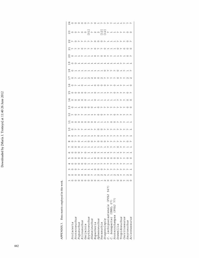
AP
PE
ND
IX3.
Dat
am
atri
xem
ploy
edin
this
wor
k. 12
34
56
78
910
11
12
13
14
15
16
17
18
19
20
21
22
23
24
Prolacerta
00
00
00
00
00
0?
00
00
00
00
??
00
Proterosuchus
00
00
00
0?
??
00
10
0?
?1
01
?0
00
Fugusuchus
00
?0
00
00
??
0?
10
01
11
11
??
?0
Xilousuchus
00
10
0?
11
1?
0?
00
00
11
1?
0?
00
Garjainia
00
10
00
11
11
01
00
01
11
11
??
00
Erythrosuchus
00
11
11
10
11
01
01
11
21
11
??
[01]
?Shansisuchus
0?
11
11
10
1?
01
01
11
21
11
??
??
Euparkeria
01
01
00
1?
??
0?
10
00
01
10
00
20
Sphenosuchus
22
01
01
10
00
10
01
10
01
1?
?0
21
Parasuchia
12
0?
01
?0
10
10
?1
10
21
10
?0
[12]
?Stagonolepis
22
??
01
10
??
1?
?1
10
?0
10
0?
[12]
?C.ischigualastensis(PVSJ567)
01
??
01
??
??
??
?1
??
??
1?
11
??
C.bonapartei(UNRL07)
01
??
0?
??
??
0?
?1
??
??
1?
11
1?
Proterochampsa(PVSJ77)
?2
??
0?
??
??
00
?1
00
??
11
11
11
Doswellia
02
??
01
0?
??
??
??
?1
00
11
0?
?1
Tropidosuchus
0?
??
??
??
??
??
??
??
?1
??
??
?1
Cerritosaurus
??
??
??
??
??
??
??
??
??
?1
0?
??
Saurosuchus
22
?1
01
10
??
1?
00
01
21
10
00
0?
Arizonasaurus
00
11
01
1?
11
1?
00
?0
2?
1?
0?
0?
882
Dow
nloa
ded
by [
Mar
ía J
. Tro
tteyn
] at
11:
40 2
6 Ju
ne 2
012