Artificial Intelligence Methods Based Hierarchical ... - arXiv
Upload
khangminh22Category
view
0download
0
�����������������
Citation: Di Basilio, F.; Esposisto, G.;
Monoscalco, L.; Giansanti, D. The
Artificial Intelligence in Digital
Radiology: Part 2: Towards an
Investigation of acceptance and
consensus on the Insiders. Healthcare
2022, 10, 153. https://doi.org/
10.3390/healthcare10010153
Academic Editor: Norbert Hosten
Received: 9 November 2021
Accepted: 10 January 2022
Published: 14 January 2022
Publisher’s Note: MDPI stays neutral
with regard to jurisdictional claims in
published maps and institutional affil-
iations.
Copyright: © 2022 by the authors.
Licensee MDPI, Basel, Switzerland.
This article is an open access article
distributed under the terms and
conditions of the Creative Commons
Attribution (CC BY) license (https://
creativecommons.org/licenses/by/
4.0/).
healthcare
Article
The Artificial Intelligence in Digital Radiology: Part 2: Towardsan Investigation of acceptance and consensus on the InsidersFrancesco Di Basilio 1, Gianluca Esposisto 1, Lisa Monoscalco 2 and Daniele Giansanti 3,*
1 Facoltà di Medicina e Psicologia, Sapienza University, Piazzale Aldo Moro, 00185 Rome, Italy;[email protected] (F.D.B.); [email protected] (G.E.)
2 Faculty of Engineering, Tor Vergata University, 00133 Rome, Italy; [email protected] Centre Tisp, Istituto Superiore di Sanità, 00161 Rome, Italy* Correspondence: [email protected]; Tel.: +39-06-49902701
Abstract: Background. The study deals with the introduction of the artificial intelligence in digitalradiology. There is a growing interest in this area of scientific research in acceptance and consensusstudies involving both insiders and the public, based on surveys focused mainly on single profession-als. Purpose. The goal of the study is to perform a contemporary investigation on the acceptance andthe consensus of the three key professional figures approaching in this field of application: (1) Medicalspecialists in image diagnostics: the medical specialists (MS)s; (2) experts in physical imaging pro-cesses: the medical physicists (MP)s; (3) AI designers: specialists of applied sciences (SAS)s. Methods.Participants (MSs = 92: 48 males/44 females, averaged age 37.9; MPs = 91: 43 males/48 females,averaged age 36.1; SAS = 90: 47 males/43 females, averaged age 37.3) were properly recruited basedon specific training. An electronic survey was designed and submitted to the participants with a widerange questions starting from the training and background up to the different applications of the AIand the environment of application. Results. The results show that generally, the three professionalsshow (a) a high degree of encouraging agreement on the introduction of AI both in imaging andin non-imaging applications using both standalone applications and/or mHealth/eHealth, and (b) adifferent consent on AI use depending on the training background. Conclusions. The study highlightsthe usefulness of focusing on both the three key professionals and the usefulness of the investigationschemes facing a wide range of issues. The study also suggests the importance of different methodsof administration to improve the adhesion and the need to continue these investigations both withfederated and specific initiatives.
Keywords: e-health; medical devices; m-health; digital-radiology; picture archive and communicationsystem; artificial-intelligence; electronic surveys; chest CT; chest radiography; acceptance; consensus
1. IntroductionArtificial Intelligence and Digital Radiology
The standardization of digital radiology caused important changes in the field oforgan and functional diagnostics. This regards both the diagnostics and the interventionalradiology [1,2]. It has led to exceptional changes in the organization of work and reportingprocesses. Furthermore, it pushed the digitization and computerization [3,4]. This solvedand simplified many organizational problems, such as the organization of the archives, evenif new ones appeared, such as those related to cybersecurity [5,6]. Today, digital radiology(DR) embraces a wide sector of diagnostic scenarios, also including sectors not directlyrelated with the ionizing radiation, such as magnetic resonance and echography [7–9].Those imaging sectors using DICOM are united under the hat of digital radiology [10–13].Now, we are facing the possible impact of research on the health domain [14]. An importantengine in this context is represented by the research efforts during the COVID-19 pandemic.For example, research on chest CT/radiography has opened important discussions andscenarios [15–18].
Healthcare 2022, 10, 153. https://doi.org/10.3390/healthcare10010153 https://www.mdpi.com/journal/healthcare

Healthcare 2022, 10, 153 2 of 15
AI, a field of computer science [19], when used in the health domain is considered a toolable to perform tasks normally requiring human intelligence [20–23] that in recent yearshave been applied in various health-related areas, such as cancer detection [24], dementiaclassification [25], and drug design [26], to name a few.
If we consider the potential of AI in DR, the applications are multiple.We need to consider four important points of view when we enter the field of DR [27,28]:
1. A first point of view is that DR includes different imaging sectors where it can po-tentially be applied. If we exclude imaging processes that do not involve ionizingradiation, we can identify the following sectors, both with reference to organ and totalbody diagnostics:
a. Interventional radiologyb. Diagnostic radiology (radiology, CT)c. Nuclear magnetic resonanced. Positron emission tomographye. amma chamber
2. A second point of view is represented by the transversal sectors that embrace thesedisciplines in which AI can play an important role:
a. Therapyb. Preventionc. Quality controld. Risk assessment
3. A third point of view is represented by the AI app distribution methods. In fact, wemust not forget that AI, in the context of DR, has a future of standardization relatedto software for medical devices [29]. This software has different implications if it isused standalone or on the network, and if it is networked through eHealth or mHealthsolutions. The implications also concern important aspects of cybersecurity [30].
4. A fourth point of view is represented by the specific training that must include AI andalso the related disciplines such as informatics, medical imaging and the technologiesfor biomedical app.
The passage of the AI into the routine of the DR (including the above listed points ofview) must take place through an approach that provides for the transfer of evidence-basedmedicine (EBM) to the operational processes of the health domain, using all the availableagreement tools, which include guidelines [31], technology assessment (TA) such as HTAand CER [32], and consensus conferences [33]. The latest definition of EBM, by Eddy [34],also considers the development of evidence-based policies in a multi-dimensional space ofthe health domain, involving quality, acceptance, consensus, and cost-effectiveness analysis.All the agreement tools will therefore also be based, as in other disciplines, on the acceptanceand consensus of both the insiders and the public who will help to express importantpositions. A PubMed search in this area with the two keys [35,36]:
(acceptance) AND (artificial intelligence [Title/Abstract]) AND Radiology)
(consensus) AND (artificial intelligence [Title/Abstract]) AND Radiology)
shows (Figure 1) 83 results, of which 77 from 2019 to today for the acceptance and 23 resultsfor the consensus, all comprised from 2019 to today.
This means that acceptance and consensus have become a priority on this issue overthe past two years. Among the emerging tools in this area, we find the surveys usefulas sensors for stakeholders and managers in general. These surveys [37–47] focused onsome of the actors that revolve around this area: radiologists, radiographers, primary careproviders (PCP), students, and patients, that is, both on service providers and users, butalso on subjects in training. The studies on patients [37–39] have highlighted the curiosityand non-opposition to these techniques, together with the need to create culture, the need toeducate on the issue and the fear for the aspects of cybersecurity in integration with eHealth

Healthcare 2022, 10, 153 3 of 15
and mHealth. The students [47] showed curiosity and optimism but complained abouta lack of adequate training and the need to integrate specific modules into the trainingprograms. Openings towards these solutions have emerged from studies on radiologistsand radiographers [41–46] accompanied by the strong desire to have an important role infuture work-flow modification processes and adequate training. In almost all studies, withrare exceptions such as [39], free and non-standardized questionnaires were used throughvalidation processes, indicating that scholars, at this historical moment, are relying on theircreativity to create increasingly innovative and adaptive questionnaires. Instruments, suchas the TAM, widely used in radiology were not used [48].
Healthcare 2022, 10, x FOR PEER REVIEW 3 of 16
Figure 1. Output of the search on PubMed on acceptance and consensus on AI in radiology.
This means that acceptance and consensus have become a priority on this issue over the past two years. Among the emerging tools in this area, we find the surveys useful as sensors for stakeholders and managers in general. These surveys [37–47] focused on some of the actors that revolve around this area: radiologists, radiographers, primary care providers (PCP), students, and patients, that is, both on service providers and users, but also on subjects in training. The studies on patients [37–39] have highlighted the curiosity and non-opposition to these techniques, together with the need to create culture, the need to educate on the issue and the fear for the aspects of cybersecurity in integration with eHealth and mHealth. The students [47] showed curiosity and optimism but complained about a lack of adequate training and the need to integrate specific modules into the training programs. Openings towards these solutions have emerged from studies on radiologists and radiographers [41–46] accompanied by the strong desire to have an important role in future work-flow modification processes and adequate training. In almost all studies, with rare exceptions such as [39], free and non-standardized questionnaires were used through validation processes, indicating that scholars, at this historical moment, are relying on their creativity to create increasingly innovative and adaptive questionnaires. Instruments, such as the TAM, widely used in radiology were not used [48].
What emerges from these studies are the following needs of deepening for further study in the surveys. Many figures have been thought of, such as the PCP [40], but others have been neglected. No studies have been identified on the specialists of applied sciences of artificial intelligence systems. In rare cases, surveys have been carried out which involved several professional figures, such as in [44,45], which involved both radiologists and radiographers. Our hypothesis is that the AI acceptance survey in radiology: • must consider the above-listed (1–4) points of views, not limited to imaging and
including the integration into eHealth and mHealth [49]; • must consider all the involved professionals who have different training and a different
work-flow and therefore different expectations from AI. Some studies on the design and test of AI solutions are clearly highlighting the
importance of the team [50]. This team comprehends (with a natural osmosis of skills): • the medical physics; • the medical specialist; • the specialist in applied sciences.
Figure 1. Output of the search on PubMed on acceptance and consensus on AI in radiology.
What emerges from these studies are the following needs of deepening for furtherstudy in the surveys. Many figures have been thought of, such as the PCP [40], butothers have been neglected. No studies have been identified on the specialists of appliedsciences of artificial intelligence systems. In rare cases, surveys have been carried out whichinvolved several professional figures, such as in [44,45], which involved both radiologistsand radiographers. Our hypothesis is that the AI acceptance survey in radiology:
• must consider the above-listed (1–4) points of views, not limited to imaging andincluding the integration into eHealth and mHealth [49];
• must consider all the involved professionals who have different training and a differentwork-flow and therefore different expectations from AI.
Some studies on the design and test of AI solutions are clearly highlighting theimportance of the team [50]. This team comprehends (with a natural osmosis of skills):
• the medical physics;• the medical specialist;• the specialist in applied sciences.
A preparatory and preliminary step to the introduction of the AI in the clinical practiceshould directly face the consensus/acceptance. It emerges, based on the above, that importantactors are undoubtedly (Figure 2): medical specialists (MS)s, medical physicists (MP)s, andspecialists of applied sciences (SAS)s. MSs are a strategic role in the decision flow. MPscontrol the physical process. SASs design and maintain the AI tools (such as the biomedicalengineers and technicians/technologists of radiology). The purpose of the study was: (a) tofocus on these three professionals to investigate their acceptance and consensus; (b) to designand submit them a properly electronic survey for the investigation, with a wide range offeatures considering the highlighted needs of deepening the points listed above.

Healthcare 2022, 10, 153 4 of 15
Healthcare 2022, 10, x FOR PEER REVIEW 4 of 16
A preparatory and preliminary step to the introduction of the AI in the clinical practice should directly face the consensus/acceptance. It emerges, based on the above, that important actors are undoubtedly (Figure 2): medical specialists (MS)s, medical physicists (MP)s, and specialists of applied sciences (SAS)s. MSs are a strategic role in the decision flow. MPs control the physical process. SASs design and maintain the AI tools (such as the biomedical engineers and technicians/technologists of radiology). The purpose of the study was: (a) to focus on these three professionals to investigate their acceptance and consensus; (b) to design and submit them a properly electronic survey for the investigation, with a wide range of features considering the highlighted needs of deepening the points listed above.
Figure 2. Interconnection among experts and AI.
2. Methods In line with the aim of the study, we decided to develop a survey. The methodology comprehended: (I) the choice of the tool for the design of the
electronic survey and (II) the adequacy to regulations; (III) the design of the survey based on the chosen tool respecting the wide range features to investigate; (IV) the dissemination on a population; (V) the data analysis based on an effective statistical approach. The questionnaire was developed using Microsoft Forms. It adhered to the SURGE Checklist [51] for the development and administration of the survey. The statistics followed two steps: • Verification of data normality; • Application of the ANOVA with a P lower than 0.01 for the significance of
differences. For the statistical confidence interval, we set a goal of 95%. We considered that, among the most used tests to verify the normality, there are: (a)
the Shapiro–Wilk test, which is preferable for a small sample; (b) the Kolmogorov–Smirnov test, which is instead used for more numerous samples. The samples in this study are small; therefore, we used the normality test of Shapiro–Wilk. We focused on the key figures (Figure 2) for the investigation.
The electronic survey was designed to face a wide range of features (starting from the training and the background, up to the application of the AI and the environment of application) using: choice questions, open questions, graded questions, and Likert (Figure 3).
Figure 2. Interconnection among experts and AI.
2. Methods
In line with the aim of the study, we decided to develop a survey.The methodology comprehended: (I) the choice of the tool for the design of the elec-
tronic survey and (II) the adequacy to regulations; (III) the design of the survey based onthe chosen tool respecting the wide range features to investigate; (IV) the dissemination ona population; (V) the data analysis based on an effective statistical approach. The question-naire was developed using Microsoft Forms. It adhered to the SURGE Checklist [51] for thedevelopment and administration of the survey. The statistics followed two steps:
• Verification of data normality;• Application of the ANOVA with a P lower than 0.01 for the significance of differences.
For the statistical confidence interval, we set a goal of 95%.We considered that, among the most used tests to verify the normality, there are: (a) the
Shapiro–Wilk test, which is preferable for a small sample; (b) the Kolmogorov–Smirnovtest, which is instead used for more numerous samples. The samples in this study aresmall; therefore, we used the normality test of Shapiro–Wilk. We focused on the key figures(Figure 2) for the investigation.
The electronic survey was designed to face a wide range of features (starting fromthe training and the background, up to the application of the AI and the environment ofapplication) using: choice questions, open questions, graded questions, and Likert (Figure 3).
Healthcare 2022, 10, x FOR PEER REVIEW 5 of 16
Figure 3. Features investigated by means of the electronic survey.
Both in the graded questions and in the Likert questions we fixed a six-level psychometric scale; it was therefore possible to assign a minimum score of one and a maximum score of six with a theoretical mean value (TMV) of 3.5. The TMV can be referred to for comparison in the analysis of the answers. An average value of the answers below TMV indicates a more negative than positive response. An average value above TMV indicates a more positive than negative response. The survey was accompanied by a brief description of the topic that would be addressed, clearly illustrating that the focus was related to the introduction of AI in digital radiology.
For the recruitment, we considered the three figures as planned, who, we remember, are medical specialists (MS), medical physicists (MP), specialists of applied sciences (SAS). All figures have a different role with AI in DR; this implies a different vision/opinion/consensus. The recruitment of these figures was very complex given that they belong to very different sectors, to different scientific societies. Currently, in Italy, there are 334 scientific societies [52]. We followed two paths that we have traced:
First way In Italy, there are also federations of scientific societies that favor a scientific osmosis
between the various scientific societies. As regards the three professionals, we referred to:
• FEDERATION OF ITALIAN MEDICAL-SCIENTIFIC SOCIETIES [53] (includes associations such as the Italian association of medical and health physics and other relevant scientific societies and other societies operating in the Medical Diagnostics and in related fields) mainly for the first two professionals MPs and MSs but also for the SASs.
• FEDERATION OF SCIENTIFIC AND TECHNICAL ASSOCIATIONS [54] (contains the National Group of Bioengineering and other relevant scientific societies) and FEDERATION OF SCIENTIFIC ASSOCIATIONS OF RADIOLOGY TECHNICIANS [55] (contains for example the Italian association of system administrators and telemedicine, association of interventional radiology technicians, Health Imaging Sciences Association, and other relevant societies) mainly for the SASs but also for the other professionals. It was possible for us to have lists of congresses in which to collect preliminary
adhesions of interest for the project, in the presence, with contacts, encounters,
Figure 3. Features investigated by means of the electronic survey.

Healthcare 2022, 10, 153 5 of 15
Both in the graded questions and in the Likert questions we fixed a six-level psycho-metric scale; it was therefore possible to assign a minimum score of one and a maximumscore of six with a theoretical mean value (TMV) of 3.5. The TMV can be referred to forcomparison in the analysis of the answers. An average value of the answers below TMVindicates a more negative than positive response. An average value above TMV indicates amore positive than negative response. The survey was accompanied by a brief descriptionof the topic that would be addressed, clearly illustrating that the focus was related to theintroduction of AI in digital radiology.
For the recruitment, we considered the three figures as planned, who, we remember,are medical specialists (MS), medical physicists (MP), specialists of applied sciences (SAS).All figures have a different role with AI in DR; this implies a different vision/opinion/cons-ensus. The recruitment of these figures was very complex given that they belong to verydifferent sectors, to different scientific societies. Currently, in Italy, there are 334 scientificsocieties [52]. We followed two paths that we have traced:
First wayIn Italy, there are also federations of scientific societies that favor a scientific osmosis
between the various scientific societies.As regards the three professionals, we referred to:
• FEDERATION OF ITALIAN MEDICAL-SCIENTIFIC SOCIETIES [53] (includes as-sociations such as the Italian association of medical and health physics and otherrelevant scientific societies and other societies operating in the Medical Diagnosticsand in related fields) mainly for the first two professionals MPs and MSs but also forthe SASs.
• FEDERATION OF SCIENTIFIC AND TECHNICAL ASSOCIATIONS [54] (containsthe National Group of Bioengineering and other relevant scientific societies) and FED-ERATION OF SCIENTIFIC ASSOCIATIONS OF RADIOLOGY TECHNICIANS [55](contains for example the Italian association of system administrators and telemedicine,association of interventional radiology technicians, Health Imaging Sciences Association,and other relevant societies) mainly for the SASs but also for the other professionals.
It was possible for us to have lists of congresses in which to collect preliminaryadhesions of interest for the project, in the presence, with contacts, encounters, discussions.A WhatsApp group was created to which the invitation and the anonymous questionnairewere sent, with a brief description and a recall of the discussion. In this way, it was possibleto send the survey anonymously.
Second waySending was also carried out through our networks of WhatsApp, also following a
peer-to-peer mechanism.Table 1 reports the participants, the participants agreeing to continue after opening
the questionnaire, and the related demographic characteristics. The average age of thosewho filled out the survey was not high. This depends on the very innovative and recenttypology of the proposed theme, which was more attractive and inclusive (due to thetraining received) for the younger population.
Table 1. Characteristics of the participants in the study and the final involvement.
ParticipantsParticipants Agreeingto Continue/Passing
the RequirementMales/Females Min Age/Max
Age Mean Age
MSs 111 108/92 48/44 32/43 37.9MPs 105 97/91 43/48 31/41 36.1SASs 99 93/90 47/43 33/40 37.3
Figures A1 and A2 in the Appendix A show a sample of the questionnaire. It wasconverted from the Italian language into the English language.

Healthcare 2022, 10, 153 6 of 15
3. Results3.1. Outcome of the Closed Questions from the Survey
The eS contained a specific question relating to an adequate level of knowledge onAI to participate (through the attendance, for example, of specific academic and/or post-academic training). Only those who passed this requirement were admitted to the study.The results are organized into five tables. The first table (Table 2) concerns the training onAI aspects.
Table 2. Specific outcome of the perceived training.
Knowledge MSsScore
MPsScore
SASsScore
ANOVAp
AI (general) 4.56 4.38 4.51 p > 0.1AI (informatics) 4.33 4.24 5.22 p < 0.01
AI (medical imaging) 4.98 5.07 5.02 p > 0.1Technologies for biomedical Apps 4.32 5.03 5.11 p < 0.01
The second table (Table 3) concerns the consent/opinion on the application of AIspecifically related to medical imaging.
Table 3. Specific outcome of the opinion on the application on the medical imaging.
Application of AI in: MSsScore
MPsScore
SASsScore
ANOVAp
4.26 4.18 4.11 p > 0.1Interventional radiology 4.54 4.39 4.41 p > 0.1
Diagnostic radiology (radiology, CT, etc.) 4.26 4.28 4.31 p > 0.1Nuclear magnetic resonance 4.61 4.69 4.72 p > 0.1
Positron emission tomography 4.53 4.38 4.52 p > 0.1Gamma chamber 4.44 4.39 4.43 p > 0.1
The third table (Table 4) concerns the consent/opinion on the application on other medicalaspects not directly related to medical imaging (therapy, risk analysis, quality control, prevention).
Table 4. Specific outcome of the opinion on the application of AI different from imaging.
Application of AI (NonImaging)
MSsScore
MPsScore
SASsScore
ANOVAp
Risk assessment 4.82 4.21 4.13 p < 0.01Therapy 5.21 4.65 4.52 p < 0.01
Prevention 5.11 4.02 4.11 p < 0.01Quality Control 4.12 5.07 5.12 p < 0.01
The fourth table (Table 5) concerns aspects on how it is considered convenient toapproach AI regarding the information available (eHealth, mHealth, Standalone, both eHealthand mHealth) [43].
Table 5. Specific outcome of the opinion on the use/delivery of the AI.
Scheme MSsScore
MPsScore
SASsScore
ANOVAp
eHealth 4.72 4.66 3.93 p < 0.01mHealth 4.55 4.62 3.89 p < 0.01
Both eHealth and mHealth 4.58 4.62 3.86 p < 0.01Standalone 5.33 5,24 5.17 p > 0.1
Healthcare 2022, 10, 153 7 of 15
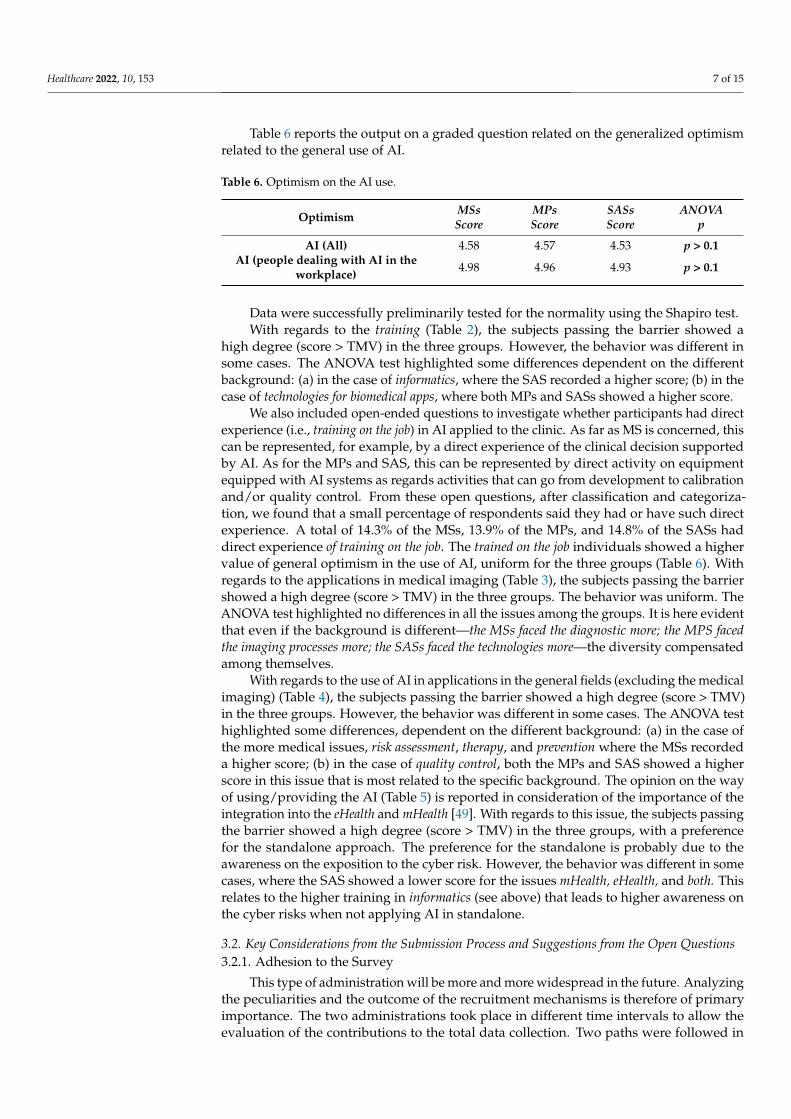
Table 6 reports the output on a graded question related on the generalized optimismrelated to the general use of AI.
Table 6. Optimism on the AI use.
Optimism MSsScore
MPsScore
SASsScore
ANOVAp
AI (All) 4.58 4.57 4.53 p > 0.1AI (people dealing with AI in the
workplace) 4.98 4.96 4.93 p > 0.1
Data were successfully preliminarily tested for the normality using the Shapiro test.With regards to the training (Table 2), the subjects passing the barrier showed a
high degree (score > TMV) in the three groups. However, the behavior was different insome cases. The ANOVA test highlighted some differences dependent on the differentbackground: (a) in the case of informatics, where the SAS recorded a higher score; (b) in thecase of technologies for biomedical apps, where both MPs and SASs showed a higher score.
We also included open-ended questions to investigate whether participants had directexperience (i.e., training on the job) in AI applied to the clinic. As far as MS is concerned, thiscan be represented, for example, by a direct experience of the clinical decision supportedby AI. As for the MPs and SAS, this can be represented by direct activity on equipmentequipped with AI systems as regards activities that can go from development to calibrationand/or quality control. From these open questions, after classification and categoriza-tion, we found that a small percentage of respondents said they had or have such directexperience. A total of 14.3% of the MSs, 13.9% of the MPs, and 14.8% of the SASs haddirect experience of training on the job. The trained on the job individuals showed a highervalue of general optimism in the use of AI, uniform for the three groups (Table 6). Withregards to the applications in medical imaging (Table 3), the subjects passing the barriershowed a high degree (score > TMV) in the three groups. The behavior was uniform. TheANOVA test highlighted no differences in all the issues among the groups. It is here evidentthat even if the background is different—the MSs faced the diagnostic more; the MPS facedthe imaging processes more; the SASs faced the technologies more—the diversity compensatedamong themselves.
With regards to the use of AI in applications in the general fields (excluding the medicalimaging) (Table 4), the subjects passing the barrier showed a high degree (score > TMV)in the three groups. However, the behavior was different in some cases. The ANOVA testhighlighted some differences, dependent on the different background: (a) in the case ofthe more medical issues, risk assessment, therapy, and prevention where the MSs recordeda higher score; (b) in the case of quality control, both the MPs and SAS showed a higherscore in this issue that is most related to the specific background. The opinion on the wayof using/providing the AI (Table 5) is reported in consideration of the importance of theintegration into the eHealth and mHealth [49]. With regards to this issue, the subjects passingthe barrier showed a high degree (score > TMV) in the three groups, with a preferencefor the standalone approach. The preference for the standalone is probably due to theawareness on the exposition to the cyber risk. However, the behavior was different in somecases, where the SAS showed a lower score for the issues mHealth, eHealth, and both. Thisrelates to the higher training in informatics (see above) that leads to higher awareness onthe cyber risks when not applying AI in standalone.
3.2. Key Considerations from the Submission Process and Suggestions from the Open Questions3.2.1. Adhesion to the Survey
This type of administration will be more and more widespread in the future. Analyzingthe peculiarities and the outcome of the recruitment mechanisms is therefore of primaryimportance. The two administrations took place in different time intervals to allow theevaluation of the contributions to the total data collection. Two paths were followed in

Healthcare 2022, 10, 153 8 of 15
our study. The first one began in 2019 with the collection of availability in presence atcongresses with the possibility of an oral interaction/discussion and subsequent sendingwith WhatsApp.
The second was without oral discussion and was based on peer-to-peer sending viaWhatsApp. Figure 4 highlights how the greatest contribution to data collection camethrough the first method based on (traditional) oral communication. Figure 5 shows thepercentages of adhesions with respect to each method. The results show that the firstmethod had a surprisingly higher percentage of adhesion. This demonstrates how theoral communication made of the three verbal, para-verbal, and non-verbal componentscontinues to maintain a greater grip than a communication made with chat only.
Healthcare 2022, 10, x FOR PEER REVIEW 8 of 16
among the groups. It is here evident that even if the background is different—the MSs faced the diagnostic more; the MPS faced the imaging processes more; the SASs faced the technologies more—the diversity compensated among themselves.
With regards to the use of AI in applications in the general fields (excluding the medical imaging) (Table 4), the subjects passing the barrier showed a high degree (score > TMV) in the three groups. However, the behavior was different in some cases. The ANOVA test highlighted some differences, dependent on the different background: a) in the case of the more medical issues, risk assessment, therapy, and prevention where the MSs recorded a higher score; b) in the case of quality control, both the MPs and SAS showed a higher score in this issue that is most related to the specific background. The opinion on the way of using/providing the AI (Table 5) is reported in consideration of the importance of the integration into the eHealth and mHealth [49]. With regards to this issue, the subjects passing the barrier showed a high degree (score > TMV) in the three groups, with a preference for the standalone approach. The preference for the standalone is probably due to the awareness on the exposition to the cyber risk. However, the behavior was different in some cases, where the SAS showed a lower score for the issues mHealth, eHealth, and both. This relates to the higher training in informatics (see above) that leads to higher awareness on the cyber risks when not applying AI in standalone.
3.2. Key Considerations from the Submission Process and Suggestions from the Open Questions 3.2.1. Adhesion to the Survey
This type of administration will be more and more widespread in the future. Analyzing the peculiarities and the outcome of the recruitment mechanisms is therefore of primary importance. The two administrations took place in different time intervals to allow the evaluation of the contributions to the total data collection. Two paths were followed in our study. The first one began in 2019 with the collection of availability in presence at congresses with the possibility of an oral interaction/discussion and subsequent sending with WhatsApp.
The second was without oral discussion and was based on peer-to-peer sending via WhatsApp. Figure 4 highlights how the greatest contribution to data collection came through the first method based on (traditional) oral communication. Figure 5 shows the percentages of adhesions with respect to each method. The results show that the first method had a surprisingly higher percentage of adhesion. This demonstrates how the oral communication made of the three verbal, para-verbal, and non-verbal components continues to maintain a greater grip than a communication made with chat only.
Figure 4. Contributions to the survey by the two different methods. Figure 4. Contributions to the survey by the two different methods.
Healthcare 2022, 10, x FOR PEER REVIEW 9 of 16
Figure 4. Contributions to the survey by the two different methods.
Figure 5. The percentage of adhesion to the survey by the two different methods.
3.2.2. Outcome from the Open Question In the survey to question No. 13, we optionally offered the possibility of reporting
comments and observations. Twenty-one interviewed people reported an observation or comment. We analyzed
the comments that highlighted critical issues and suggestions for improvement, and we carried out datamining, which was followed by categorization.
Figure 6 reports the following points as important suggestions for improvement based on the order of the frequency of occurrence: o to request the CV in a subsection with a series of targeted questions; o to prepare a survey for each type of professional; o to refine the survey within scientific societies; o to offer a question/answer grid with very specific training aspects of AI.
Figure 6. Suggestions for improvement with the obtained frequency of occurrence.
Figure 5. The percentage of adhesion to the survey by the two different methods.
3.2.2. Outcome from the Open Question
In the survey to question No. 13, we optionally offered the possibility of reportingcomments and observations.
Twenty-one interviewed people reported an observation or comment. We analyzedthe comments that highlighted critical issues and suggestions for improvement, and wecarried out datamining, which was followed by categorization.

Healthcare 2022, 10, 153 9 of 15
Figure 6 reports the following points as important suggestions for improvement basedon the order of the frequency of occurrence:
o to request the CV in a subsection with a series of targeted questions;o to prepare a survey for each type of professional;o to refine the survey within scientific societies;o to offer a question/answer grid with very specific training aspects of AI.
Healthcare 2022, 10, x FOR PEER REVIEW 9 of 16
Figure 5. The percentage of adhesion to the survey by the two different methods.
3.2.2. Outcome from the Open Question In the survey to question No. 13, we optionally offered the possibility of reporting
comments and observations. Twenty-one interviewed people reported an observation or comment. We analyzed
the comments that highlighted critical issues and suggestions for improvement, and we carried out datamining, which was followed by categorization.
Figure 6 reports the following points as important suggestions for improvement based on the order of the frequency of occurrence: o to request the CV in a subsection with a series of targeted questions; o to prepare a survey for each type of professional; o to refine the survey within scientific societies; o to offer a question/answer grid with very specific training aspects of AI.
Figure 6. Suggestions for improvement with the obtained frequency of occurrence.
Figure 6. Suggestions for improvement with the obtained frequency of occurrence.
4. Discussion
We are undoubtedly about to face another important change in the world of digitalradiology [14]: the introduction of AI in clinical practice. During the pandemic, theimportance and potential of AI clearly emerged in two sectors of digital radiology: chest CTand chest radiography [15–18,43]. However, even before the pandemic, we were alreadytalking about this phenomenon affecting the health domain, especially the sectors where theconversion to digital health has been heavy, such as the DR [27,28], thanks to the DICOMstandardization process. DICOM is the container of the information arranged into pixelsand/or voxels after the process of image acquisition. The pixels and/or voxels used as AIinput carry different information of the investigated biomedical target. The informationin those elements is related to the physical process (X-rays, gamma rays, magnetic fields,ultrasounds, etc.). Three elements play an important role: (1) the physical process (PP),which depends on the physical fields used (X-rays, magnetic field, ultrasound, etc.); (2) thetechnological process (TP), which concerns both the technologies for capturing informationstarting from the physical process, and the software implementation of AI-based algorithms;(3) the decision-making process (DP), which must consider the outcome from the TP based ona PP and the human decision based on medical knowledge functionally related to both the TPand PP.
It is for this reason that it is important that the experts of the DP, TP, and PP work areconnected in the process of AI introduction and in the related investigations.
It should also be borne in mind that in addition to diagnostic imaging, other AI appli-cations used for categorization into non-imaging problems [9,27] (non-imaging categorization)were considered in the study. These range from risk analysis up to quality control. We alsofound it important to consider in the study how AI is delivered, whether it is delivered instandalone mode, or based on mHealth or eHealth [49]. In light of what has been illustratedabove, we have decided to consider in the study the three figures of MSs, MPs, and SASsconnected to (1,2) to investigate the consensus and acceptance by means of an eS. From ageneral point of view, these three professional figures showed a high degree of acceptance

Healthcare 2022, 10, 153 10 of 15
of the introduction of AI both in imaging and in non-imaging applications, using bothstandalone and network modes (mHealth or eHealth). Specifically, through a statisticalassessment based on ANOVA, we were able to see a different way of approaching AI. Thisapproach was uniform when considering AI applied to imaging. The approach was notuniform when considering the non-imaging applications and the delivery methods. Sub-jects with a background comprehending direct training on the job focused on AI showedthe highest optimism. From a general point of view, the study highlights the usefulness ofinvestigating the inclusion of AI through an eS, the usefulness of doing so based on threecategories of experts (MSs; MPs; SASs), and the general optimism in the introduction of AIin digital radiology.
The background plays an important role in relation to the approach to AI. In thescientific literature, various studies already involved radiologists (key figures in the clinicaldecision) to perform reader studies. In a certain sense, if we look at the study proposed ona direct application of AI [50] in its entirety, we realize that regarding the enhancement ofAI, the study we have proposed is in a complementary position. Our study directly focusesto the three involved professionals, having an active role in the flow from the tool designup to the decision [50]. Our study is in line with the studies based on surveys [37–47];the submission of original surveys allows to obtain strategic information. In additionto similar studies, our study addressed the innovation of submitting the same survey tothree key figures operating in the TP, PP, and DP. Furthermore, considering the needs thatemerged from previous studies, our study proposed different survey schemes based onLikert/graded questions at six psychometric levels to have different quantitative outcomes,useful for categorizations.
A first scheme dealt with the educational, academic, and post-academic trainingaspects on modules relevant to the knowledge bases useful in this field.
A second scheme addressed the imaging aspects in detail, focusing on the differentcompatible DICOM tools used in DR.
The third scheme addressed the aspects of AI external to imaging but always relevantto the work flow (quality control, risk assessment, therapy and prevention) [27,28].
A fourth scheme was dedicated to integration with eHealth and mHealth [49], strategicfor addressing important aspects such as cybersecurity.
From a general point of view, the study differs from other initiatives in this direc-tion [56–59]. Furthermore, it offers to the scholars a complementary contribution andtherefore complementary results if compared to study based on surveys [58,59]. Our pro-posed survey (see Appendix A) comprehends 13 questions (23 if we consider that the Likerthas submodules): (a) it is oriented to all three professions potentially involved, (b) it goesinto detail in the application of AI in the different sectors of imaging with a specific Likertand by means of another Likert in the application of AI in the translational sectors of thehealth domain, and (c) it addresses aspects of network integration (standalone, mHealth,eHealth) important for the impact on software medical device and cybersecurity. We haveused several modules detailing the choice questions, the open questions, the open large questions,and two modules used to give a psycho/sociometric assessment (now currently used in the lifesciences): the graded questions and the Likert. In addition, in our survey, there was also thepossibility of supplementing the demographic information (including training) and workactivity with two specific open questions, one open large question dedicated to the insertionof the CV, and one open question dedicated to the description of one’s own working activity.
The two surveys in [58,59] are in turn complementary; they are each dedicated to aspecific professional figure and with different focuses.
The survey reported in [58] concerns a national audience, is focused on the MSs, andis made up of 13 questions: 4 dedicated to demographic aspects (age, region, activity,position and job site), 3 dedicated to interaction with AI (tasks by AI, advantages, issues),3 dedicated to implications (ethical problems, risk of job loss, needs of policies), and otherquestions in complement, such as the opinion on the definition of AI.

Healthcare 2022, 10, 153 11 of 15
The other survey [59] concerns an international audience, is dedicated to the figureof the MP, and consists of 25 questions. The first eight deal with the training aspects, theinvolvement in AI projects and with the activities and the opinion on the introduction ofAI. Questions 9 to 17 all concern the collection of educational interests in a specific way andthe opinions on the integration of the CV in future activities. An open question (number18) is free. The final questions are all focused on demographics.
Our survey was submitted through two channels, both electronic (one of which,however, was also based on a preliminary in-person presentation of the initiative), whichwere evaluated. Part of the analysis was dedicated to the observations and criticalities thatemerged, as well as specifically collected.
Both the surveys reported in [58,59] were administered with purely electronic methods,and there was no comparison between different modalities. They did not use gradedquestions and Likert questions. Furthermore, the critical issues to be addressed to improvethese initiatives were not collected from both the surveys.
As regards the dissemination of the survey, our study shows that a preliminary contactin presence (followed by an electronic transmission) improves the participation rate. Thissuggests for the future to address these initiatives by preceding them by preliminary face-to-face meetings (for example, in focus groups or congresses). Regarding suggestions forimprovement and development, it should be noted that those proposals that have had a fre-quency greater than 1 push towards a structured request in a grid of the CV, a specializationof the survey for the different professionals, and a refinement in scientific societies.
Considering these observations and what has emerged, the continuation of theseinitiatives in both a specialized and federated way is certainly desirable. It is hoped thatthe AI will be an opportunity to give birth to scientific federations that allow for in-depthinitiatives in both a specific and confederate way.
5. Conclusions
The introduction of AI into clinical practice is now an unstoppable process that willtake this discipline from research to routine use. Many professionals from now to the futurewill be involved, and it will be necessary to provide for targeted consensus actions to issueappropriate recommendations. Guidelines, TA reports, and consensus conferences, spreadby scientific societies in the sector, for example, will in the future also use approaches basedon surveys that scholars are currently developing.
Initiatives aimed at creating position papers in this area will be more and more frequentand will involve more and more teams of professionals, as in [56], where medical physicsand radiologists have worked. Both national [57,58] and international [59] scientific societiescould play an important role in the improvement and dissemination of these surveys,which could play a strategic role in monitoring the topic. It will also be important thatscientific societies representing the different actors work as a team in initiatives that couldpossibly lead to stable and standardized international monitoring actions.
Author Contributions: Conceptualization, D.G.; methodology, D.G. and F.D.B.; software, D.G. andL.M.; validation, D.G., G.E. and F.D.B.; formal analysis, All; investigation, All; resources, not applica-ble; data curation, D.G. and L.M.; writing—original draft preparation, D.G.; writing—review andediting, All; visualization, All; supervision, D.G.; project administration, D.G. and L.M.; funding ac-quisition, not applicable. All authors have read and agreed to the published version of the manuscript.
Funding: This research received no external funding.
Institutional Review Board Statement: Not applicable.
Informed Consent Statement: Not applicable.
Data Availability Statement: Not applicable.
Conflicts of Interest: The authors declare no conflict of interest.

Healthcare 2022, 10, 153 12 of 15
Abbreviations
Acronym DescriptionAI Artificial intelligenceCT Computerized tomographyMP Medical physicistSAS Specialists of applied sciencesMS Medical specialistDICOM Digital imaging and communications in medicineDR Digital radiologyTA Technology assessmentHTA Health technology assessmentCER Comparative effectiveness researchPCP Primary care providerTP Technological processTMV Theorical mean valuePP Physical processDP Decision-making processANOVA Analysis of varianceeHealth Electronic healthmHealth Mobile health
Appendix A
Healthcare 2022, 10, x FOR PEER REVIEW 13 of 16
Appendix A
Figure A1. An example of the survey (first print screen). Figure A1. An example of the survey (first print screen).

Healthcare 2022, 10, 153 13 of 15Healthcare 2022, 10, x FOR PEER REVIEW 14 of 16
Figure A2. An example of the survey (second print screen).
References 1. Thrall, J.H. Teleradiology. Part I. History and clinical applications. Radiology 2007, 243, 613–617. 2. Thrall, J.H. Teleradiology. Part II. Limitations, risks, and opportunities. Radiology 2007, 244, 325–328. 3. Reponen, J. Teleradiology—Changing Radiological Service Processes from Local to Regional, International and Mobile Environment;
University of Oulu: Oulu, Finland, 2010. 4. Wootton, R. Telemedicine: A cautious welcome. BMJ 1996, 313, 1375–1377. 5. Giansanti, D. Teleradiology Today: The Quality Concept and the Italian Point of View. Telemed. E-Health 2017, 23, 453–455. 6. Orlacchio, A.; Romeo, P.; Inserra, M.C.; Grigioni, M.; Giansanti, D. Guidelines for Quality Assurance and Technical Requirements in
Teleradiology; English Translation and Revision of Rapporti ISTISAN 10/44, Rapporti ISTISAN 13/38; Istituto Superiore di Sanità: Roma, Italy, 2013; pp. 1–33.
7. Ruotsalainen, P. Privacy and security in teleradiology. Eur. J. Radiol. 2010, 73, 31–35. 8. Giansanti, D. Diagnostic Imaging and E-Health: Standardization, Experiences and New Opportunities; Rapporti ISTISAN 17/10;
Istituto Superiore di Sanità: Roma, Italy, 2017; pp. 1–60. 9. Giansanti, D. Diagnostics Imaging and M-Health: Investigations on the Prospects of Integration in Cytological and Organ Diagnostics;
Rapporti ISTISAN 20/1; Istituto Superiore di Sanità: Roma, Italy, 2019; pp. 1–66. 10. Canadian Association of Radiologists. CAR Standards for Teleradiology; Canadian Association of Radiologists: Ottawa, ON,
Canada, 2008. 11. American College of Radiology. ACR Standard for Teleradiology; ACR: Reston, VA, USA, 2002. 12. Teleradiology. Merrian-Webster Medical Dictionary Online. Available online: www.merriamwebster
com/medical/teleradiology (accessed on 30 September 2013). 13. Dicom, Digital Imaging and Communication in Medicine. Available online: https://www.dicomstandard.org/ (accessed on 9
January 2022). 14. Giansanti, D. The Artificial Intelligence in Digital Pathology and Digital Radiology: Where Are We? Healthcare 2020, 9, 30.
https://doi.org/10.3390/healthcare9010030.
Figure A2. An example of the survey (second print screen).
References1. Thrall, J.H. Teleradiology. Part I. History and clinical applications. Radiology 2007, 243, 613–617. [CrossRef] [PubMed]2. Thrall, J.H. Teleradiology. Part II. Limitations, risks, and opportunities. Radiology 2007, 244, 325–328. [CrossRef] [PubMed]3. Reponen, J. Teleradiology—Changing Radiological Service Processes from Local to Regional, International and Mobile Environment;
University of Oulu: Oulu, Finland, 2010.4. Wootton, R. Telemedicine: A cautious welcome. BMJ 1996, 313, 1375–1377. [CrossRef]5. Giansanti, D. Teleradiology Today: The Quality Concept and the Italian Point of View. Telemed. E-Health 2017, 23, 453–455.
[CrossRef] [PubMed]6. Orlacchio, A.; Romeo, P.; Inserra, M.C.; Grigioni, M.; Giansanti, D. Guidelines for Quality Assurance and Technical Requirements in
Teleradiology; English Translation and Revision of Rapporti ISTISAN 10/44, Rapporti ISTISAN 13/38; Istituto Superiore di Sanità:Roma, Italy, 2013; pp. 1–33.
7. Ruotsalainen, P. Privacy and security in teleradiology. Eur. J. Radiol. 2010, 73, 31–35. [CrossRef] [PubMed]8. Giansanti, D. Diagnostic Imaging and E-Health: Standardization, Experiences and New Opportunities; Rapporti ISTISAN 17/10; Istituto
Superiore di Sanità: Roma, Italy, 2017; pp. 1–60.9. Giansanti, D. Diagnostics Imaging and M-Health: Investigations on the Prospects of Integration in Cytological and Organ Diagnostics;
Rapporti ISTISAN 20/1; Istituto Superiore di Sanità: Roma, Italy, 2019; pp. 1–66.10. Canadian Association of Radiologists. CAR Standards for Teleradiology; Canadian Association of Radiologists: Ottawa, ON, Canada, 2008.11. American College of Radiology. ACR Standard for Teleradiology; ACR: Reston, VA, USA, 2002.12. Teleradiology. Merrian-Webster Medical Dictionary Online. Available online: www.merriamwebster.com/medical/teleradiology
(accessed on 30 September 2013).13. Dicom, Digital Imaging and Communication in Medicine. Available online: https://www.dicomstandard.org/ (accessed on
9 January 2022).
Healthcare 2022, 10, 153 14 of 15
14. Giansanti, D. The Artificial Intelligence in Digital Pathology and Digital Radiology: Where Are We? Healthcare 2020, 9, 30.[CrossRef]
15. Alsharif, M.H.; Alsharif, Y.H.; Yahya, K.; Alomari, O.A.; Albreem, M.A.; Jahid, A. Deep learning applications to combat thedissemination of COVID-19 disease: A review. Eur. Rev. Med. Pharmacol. Sci. 2020, 24, 11455–11460.
16. Ozsahin, I.; Sekeroglu, B.; Musa, M.S.; Mustapha, M.T.; Uzun Ozsahin, D. Review on Diagnosis of COVID-19 from Chest CTImages Using Artificial Intelligence. Comput. Math. Methods Med. 2020, 2020, 9756518. [CrossRef]
17. Pham, T.D. Classification of COVID-19 chest X-rays with deep learning: New models or fine tuning? Health Inf. Sci. Syst. 2020, 9, 2.[CrossRef]
18. Liang, H.; Guo, Y.; Chen, X.; Ang, K.L.; He, Y.; Jiang, N.; Du, Q.; Zeng, Q.; Lu, L.; Gao, Z.; et al. Artificial intelligence for stepwisediagnosis and monitoring of COVID-19. Eur. Radiol. 2022, 1–11, Epub ahead of print. [CrossRef]
19. Stevenson, A. Oxford Dictionary of English, 3rd ed.; Oxford University Press: Oxford, UK, 2010.20. Hsiang, C.W.; Lin, C.; Liu, W.C.; Lin, C.S.; Chang, W.C.; Hsu, H.H.; Huang, G.S.; Lou, Y.S.; Lee, C.C.; Wang, C.H.; et al. Detection of
left ventricular systolic dysfunction using an artificial intelligence-enabled chest X-ray. Can. J. Cardiol. 2022, Epub ahead of print.[CrossRef]
21. Tajik, A.J. Machine Learning for Echocardiographic imaging: Embarking on another incredible journey. J. Am. Coll. Cardiol. 2016,68, 2296–2298. [CrossRef]
22. Krittanawong, C.; Zhang, H.; Wang, Z.; Aydar, M.; Kitai, T. Artificial intelligence in precision cardiovascular medicine. J. Am. Coll.Cardiol. 2017, 69, 2657–2664. [CrossRef] [PubMed]
23. Zhang, J.; Gajjala, S.; Agrawal, P.; Tison, G.H.; Hallock, L.A.; Beussink-Nelson, L.; Deo, R.C. Fully automated echocardiograminterpretation in clinical practice. Circulation 2018, 138, 1623–1635. [CrossRef] [PubMed]
24. Rodriguez-Ruiz, A.; Lång, K.; Gubern-Merida, A.; Broeders, M.; Gennaro, G.; Clauser, P.; Helbich, T.H.; Chevalier, M.; Tan, T.;Mertelmeier, T.; et al. Stand-Alone Artificial Intelligence for Breast Cancer Detection in Mammography: Comparison With 101Radiologists. J. Natl. Cancer Inst. 2019, 111, 916–922. [CrossRef] [PubMed]
25. Bertini, F.; Allevi, D.; Lutero, G.; Montesi, D.; Calzà, L. Automatic Speech Classifier for Mild Cognitive Impairment and EarlyDementia. ACM Trans. Comput. Healthc. 2022, 3, 1–11. [CrossRef]
26. Mak, K.K.; Pichika, M.R. Artificial intelligence in drug development: Present status and future prospects. Drug Discov. Today 2019,24, 773–780. [CrossRef]
27. Jalal, S.; Nicolaou, S.; Parker, W. Artificial Intelligence, Radiology, and the Way Forward. Can. Assoc. Radiol. J. 2019, 70, 10–12.[CrossRef]
28. European Society of Radiology (ESR). What the radiologist should know about artificial intelligence—An ESR white paper.Insights Imaging 2019, 10, 44. [CrossRef]
29. Regulation (EU) 2017/745 of the European Parliament and of the Council of 5 April 2017 on Medical Devices, AmendingDirective 2001/83/EC, Regulation (EC) No 178/2002 and Regulation (EC) No 1223/2009 and Repealing Council Directives90/385/EEC and 93/42/EEC.2017. Available online: https://eur-lex.europa.eu/legal-content/EN/TXT/HTML/?uri=CELEX:32017R0745&from=IT (accessed on 25 November 2021).
30. Giansanti, D. Cybersecurity and the Digital-Health: The Challenge of This Millennium. Healthcare 2021, 9, 62. [CrossRef]31. Evidence-Based Medicine Guidelines. Available online: https://www.ebm-guidelines.com/dtk/ebmg/home (accessed on
9 January 2022).32. Luce, B.R.; Drummond, M.; Jönsson, B.; Neumann, P.J.; Schwartz, J.S.; Siebert, U.; Sullivan, S.D. EBM, HTA, and CER: Clearing
the confusion. Milbank Q. 2010, 88, 256–276. [CrossRef]33. McGlynn, E.A.; Kosecoff, J.; Brook, R.H. Format and conduct of consensus development conferences. Multi-nation comparison.
Int. J. Technol. Assess. Health Care 1990, 6, 450–469. [CrossRef]34. Eddy, D.M. Evidence-Based Medicine: A Unified Approach. Health Affairs 2005, 24, 9–17. [CrossRef] [PubMed]35. National Library of Medicine. Available online: https://pubmed.ncbi.nlm.nih.gov/?term=%28acceptance%29+AND+%2
8artificial+intelligence%5BTitle%2FAbstract%5D%29+AND+Radiology&sort=date&size=200 (accessed on 9 January 2022).36. National Library of Medicine. Available online: https://pubmed.ncbi.nlm.nih.gov/?term=%28%28consensus%29+AND+
%28artificial+intelligence%5BTitle%2FAbstract%5D%29%29+AND+%28radiology%5BTitle%2FAbstract%5D%29&sort=date&size=200 (accessed on 9 January 2022).
37. Lennartz, S.; Dratsch, T.; Zopfs, D.; Persigehl, T.; Maintz, D.; Hokamp, N.G.; Dos Santos, D.P. Use and Control of ArtificialIntelligence in Patients Across the Medical Workflow: Single-Center Questionnaire Study of Patient Perspectives. J. Med. InternetRes. 2021, 23, e24221. [CrossRef] [PubMed]
38. Zhang, Z.; Citardi, D.; Wang, D.; Genc, Y.; Shan, J.; Fan, X. Patients’ perceptions of using artificial intelligence (AI)-basedtechnology to comprehend radiology imaging data. Health Inform. J. 2021, 27, 14604582211011215. [CrossRef]
39. Ongena, Y.P.; Haan, M.; Yakar, D.; Kwee, T.C. Patients’ views on the implementation of artificial intelligence in radiology:Development and validation of a standardized questionnaire. Eur. Radiol. 2020, 30, 1033–1040. [CrossRef] [PubMed]
40. Hendrix, N.; Hauber, B.; Lee, C.I.; Bansal, A.; Veenstra, D.L. Artificial intelligence in breast cancer screening: Primary careprovider preferences. J. Am. Med. Inform. Assoc. 2021, 28, 1117–1124. [CrossRef]
41. Abuzaid, M.M.; Elshami, W.; McConnell, J.; Tekin, H.O. An extensive survey on radiographers from the Middle East and India onartificial intelligence integration in radiology practice. Health Technol. 2021, 11, 1045–1050. [CrossRef]

Healthcare 2022, 10, 153 15 of 15
42. Abuzaid, M.M.; Tekin, H.O.; Reza, M.; Elhag, I.R.; Elshami, W. Assessment of MRI technologists in acceptance and willingness tointegrate artificial intelligence into practice. Radiography 2021, 27, S83–S87. [CrossRef]
43. Giansanti, D.; Rossi, I.; Monoscalco, L. Lessons from the COVID-19 Pandemic on the Use of Artificial Intelligence in DigitalRadiology: The Submission of a Survey to Investigate the Opinion of Insiders. Healthcare 2021, 9, 331. [CrossRef] [PubMed]
44. Abuzaid, M.M.; Elshami, W.; Tekin, H.; Issa, B. Assessment of the Willingness of Radiologists and Radiographers to Accept theIntegration of Artificial Intelligence into Radiology Practice. Acad. Radiol. 2020, 29, 87–94. [CrossRef] [PubMed]
45. Alelyani, M.; Alamri, S.; Alqahtani, M.S.; Musa, A.; Almater, H.; Alqahtani, N.; Alshahrani, F.; Alelyani, S. Radiology CommunityAttitude in Saudi Arabia about the Applications of Artificial Intelligence in Radiology. Healthcare 2021, 9, 834. [CrossRef][PubMed]
46. European Society of Radiology (ESR). Impact of artificial intelligence on radiology: A EuroAIM survey among members of theEuropean Society of Radiology. Insights Imaging 2019, 10, 105. [CrossRef] [PubMed]
47. Galán, G.C.; Portero, F.S. Medical students’ perceptions of the impact of artificial intelligence in Radiology. Radiologia 2021, in press.[CrossRef]
48. Aldosari, B. User acceptance of a picture archiving and communication system (PACS) in a Saudi Arabian hospital radiologydepartment. BMC Med. Inform. Decis. Mak. 2012, 12, 44. [CrossRef]
49. Moss, R.J.; Süle, A.; Kohl, S. eHealth and mHealth. Eur. J. Hosp. Pharm. 2019, 26, 57–58. [CrossRef] [PubMed]50. Shan, H.; Padole, A.; Homayounieh, F.; Kruger, U.; Khera, R.D.; Nitiwarangkul, C.; Kalra, M.K.; Wang, G. Competitive
performance of a modularized deep neural network compared to commercial algorithms for low-dose CT image reconstruction.Nat. Mach. Intell. 2019, 1, 269–276. [CrossRef]
51. Moher, D.; Altman, D.G.; Schulz, K.F.; Simera, I.; Wager, E. (Eds.) Guidelines for Reporting Health Research: A User’s Manual.Available online: https://onlinelibrary.wiley.com/doi/abs/10.1002/9781118715598.ch20 (accessed on 9 January 2022).
52. Ministero Della Salute Rivede Elenco Società Scientifiche per Stesura Linee Guida. 41 Società in Più. Available online:http://www.aiponet.it/news/104-ufficio-stampa/2149-ministero-della-salute-rivede-elenco-societa-scientifiche-per-stesura-linee-guida-41-societa-in-piu.html (accessed on 9 January 2022).
53. Federazione Delle Società Medico-Scientifiche Italiane. Available online: https://portale.fism.it/ (accessed on 9 January 2022).54. Federazione Delle Associazioni Scientifiche dei Tecnici di Radiologia. Available online: https://www.associazionefaster.org/
(accessed on 9 January 2022).55. Federazione Delle Associazioni Scientifiche e Tecniche. Available online: https://fast.mi.it/chi-siamo/ (accessed on 9 January 2022).56. Thomassin-Naggara, I.; Balleyguier, C.; Ceugnart, L.; Heid, P.; Lenczner, G.; Maire, A.; Séradour, B.; Verzaux, L.; Taourel, P.
Conseil National Professionnel de la Radiologie et Imagerie Médicale (G4). Artificial intelligence and breast screening: FrenchRadiology Community position paper. Diagn. Interv. Imaging 2019, 100, 553–566. [CrossRef]
57. Avanzo, M.; Trianni, A.; Botta, F.; Talamonti, C.; Stasi, M.; Iori, M. Artificial Intelligence and the Medical Physicist: Welcome tothe Machine. Appl. Sci. 2021, 11, 1691. [CrossRef]
58. Coppola, F.; Faggioni, L.; Regge, D.; Giovagnoni, A.; Golfieri, R.; Bibbolino, C.; Miele, V.; Neri, E.; Grassi, R. Artificial intelligence:Radiologists’ expectations and opinions gleaned from a nationwide online survey. Radiol. Med. 2021, 126, 63–71. [CrossRef]
59. Diaz, O.; Guidi, G.; Ivashchenko, O.; Colgan, N.; Zanca, F. Artificial intelligence in the medical physics community: An interna-tional survey. Phys. Med. 2021, 81, 141–146. [CrossRef]
Copyright © 2022 FDOKUMEN