The 4th ANNUAL RESEARCH DAY - CMC Vellore
166
Abstracts of Annual Research Day 2013 1 The 4 th Annual Research Day 15 th November 2013 SCUDDER AUDITORIUM CHRISTIAN MEDICAL COLLEGE VELLORE
-
Upload
khangminh22 -
Category
Documents
-
view
0 -
download
0
Transcript of The 4th ANNUAL RESEARCH DAY - CMC Vellore
Abstracts of Annual Research Day 2013
1
The 4th Annual Research Day
15th November 2013
SCUDDER AUDITORIUM
CHRISTIAN MEDICAL COLLEGE
VELLORE

Abstracts of Annual Research Day 2013
2
Schedule for 4th Annual Research Day
Poster Installation 11.00 am Categories: Basic Sciences
Medicine Specialties Surgery Specialties Field Work and Epidemiology Nursing
Poster Session and 01.00 - 03.30 pm Judging: Lunch 12.30 pm Oral Presentation 2.55 pm – 6.12 pm Special Addresses Vice Principal - Prof. Nihal Thomas Director - Prof. Sunil Chandy Principal - Prof. Alfred Job Daniel Special Oration by Chief Guest - Prof. V.I Mathan (Retired Distinguished Scientist) Prize Distribution and Valediction 6.50 pm

Abstracts of Annual Research Day 2013
3
ANNUAL RESEARCH DAY – 2013 MEDAL AND ORATION FOR DISTINGUISHED RETIRED FACULTY
Prof. V.I. Mathan graduated MBBS from the then Madras University in 1960 and obtained his MD in General Medicine (1965) and PhD (1973) in Epidemiology from the same University through the Christian Medical College, Vellore. He joined the faculty of the Christian Medical College, Vellore in 1965 in the Department of Medicine and was appointed as Professor of Medicine and Gastroenterology in 1973. He started the Department of Gastroenterology at Vellore in 1972 and by the time of his retirement it had grown to one of the largest departments in the country. Prof. Mathan was actively involved in research from his registrar days and has published over 140 papers. He became the Head of The Wellcome Trust Tropical Disease Research Unit at Vellore from 1975. In 1992 this unit was recognized as the Indian Council of Medical Research Centre of Advanced Research on Enteric Diseases. Prof. Mathan’s research work has been recognized by a variety awards and academic distinctions including Honorary Membership and Fellowship of the Royal College of Physicians of London, Honorary Memberships of the British Society of Gastroenterology and The Royal Society of Tropical Medicine and Hygiene. He was elected a Fellow of the Indian National Science Academy in 1991 and awarded the Dr. Ambedkar Medal for lifetime Research Contributions in 1996 by the Indian Council of Medical Research. In addition to his clinical and research activities, Prof. Mathan has been a distinguished Medical Administrator. He headed the department of Gastroenterology at Vellore from its start in 1972 to 1993. He was Medical Superintendent of the Christian Medical College Hospital from 1988 to 1993 and it’s Director from 1993 to 1997 when he retired on superannuation. Subsequent to that Prof. Mathan was the Associate Director of the International Centre for Diarrhoeal Disease Research in Dhaka, Bangladesh for three years and a Senior Consultant to UNAIDS working with the National AIDS Control Organization in Delhi for a year. He occupied the Indian Council of Medical Research Chair of Epidemiology at the National Institute of Epidemiology, Chennai till Sep. 2007. Currently he is Hon. ICMR Consultant, and is the Chair of the Project Advisory Committee on Health Sciences of the Department of Science and Technology, the Scientific Advisory Group of the Division of Epidemiology and Communicable Diseases of the Indian Council of Medical Research and the Technical Committee for the Establishment of Model Rural Health Research Units of the Department of Health Research, Government of India. For his pioneering research work on Tropical Enteropathy, Tropical Sprue, Epidemiology of Enteric Infections and Acute Diarrhoea, Vitamin B12 Absorption. The role of Luminal Endotoxin in determining the Severity of Diarrhoea and the identification of potential antagonists of Endotoxin and developing a research team which has continued to be productive for nearly two decades after his re tirement. Prof. V I. Mathan is awarded the Annual Research Day distinguished retirted faculty medal and oration for the year 2013.
Abstracts of Annual Research Day 2013
4
ABSTRACTS FOR ORAL PRESENTATION

Abstracts of Annual Research Day 2013
5
“DIVING INTO THE UNKNOWN REALM: STRENGTHENING EMERGENCY CARE AT PRIMARY HEALTHCARE LEVEL WITH A HANDY TOOL KIT.”
(A proposal to the Indian Government)
Alen Thomas, MBBS 2010, CMC Vellore Dr. Suresh David, Professor, EM Dept, CMC Vellore Dr. Moses Kirubairaj, Assistant Professor, Department of Family Medicine
Aim:
Trauma or Emergency situations can strike anytime in this fast moving world and people lose their lives much before they reach the Taluk level hospital. There are numerous published papers that emphasize on the fact that early intervention can significantly bring down the mortality rate.
This concept note paper focuses on training, equipping and empowering enthusiastic health professionals at primary healthcare level to provide first aid and to expedite rapid transfer to hospitals.
Methodology:
PART 1 Training of EM Commando
PART 2: Putting into practice
Discussion: Organised positioning will help the health care professional to be more alert and effective. Handy-kit would be helpful and effective for early intervention purposes. Essential Emergency drugs are available for on-site stabilisation. The provision of a Global Positioning Service device would be useful to provide pin-point location of the emergency as informed by the call centre team. It is also useful for identifying potential accident-prone areas in their respective areas. However, recruiting people for this purpose may be initially difficult as this is a whole new approach at the primary health care level. Budget: BUDGET for entire kit is approximately. Rs 5000/-

Abstracts of Annual Research Day 2013
6
Results: The results have not yet been released as the concept is under study and pilot study would be conducted soon with the help of the government. Conclusion: I strongly urge that Emergency medicine should be strengthened at the Primary care centre and followed effectively with the training of EM commandos and utilization of the recommended handy kit. This would significantly reduce mortality and morbidity and together we can build a better world. “EVALUATION OF THE PERFORMANCE OF SERODIAGNOSTIC KITS FOR DENGUE INFECTION IN A TERTIARY CARE CENTRE OF SOUTH INDIA. “ Rajat Choudhari1, Harshita Nori1, Shubhanker Mitra2 K.P.P Abhilash2, Vishali Jeyaseelan3, Asha Mary Abraham4, O.C Abraham5, John A Jude6, Jayaprakash Muliyil7
1. Second year medical student (batch of 2011), MBBS, Christian Medical College, Vellore 2. Assistant Professor, Dept of Medicine, Christian Medical College, Vellore 3. Assistant Professor, Dept of Biostatistics, Christian Medical College, Vellore 4. Professor and Head, Dept of Virology, Christian Medical College, Vellore 5. Professor, Dept of Medicine, Christian Medical College, Vellore 6. Professor, Dept of Microbiology, Christian Medical College, Vellore
7. Retd. Professor, Dept of Clinical Epidemiology, Christian Medical College, Vellore Background and objectives: Rapid Diagnostic tests (RDTs) are used in most dengue endemic resource limited centres for early and economical diagnosis. Hence, we evaluated the performance of four commercially available RDTs during an outbreak season in a tertiary hospital in south India against a consensus clinical criterion as reference standard and establish their ability to differentiate between acute primary and secondary infection.
Methods:
281 adult patients presenting with community acquired acute febrile illness (< 14 days duration) during the outbreak season were recruited for the study. The reference standard, to group patients into dengue or non dengue, was based on the above clinical criteria with any of the constitutional symptoms - myalgia, headache and rash and supportive laboratory findings - thrombocytopenia and leucopoenia with other etiologies like scrub typhus, leptospirosis, malaria and enteric fever proven negative by reliable tests. The samples were tested using Panbio, SD, J. Mitra and Reckon to compare their performance. SPSS version 16.0.1 was used for statistical analysis.
Results:
132 cases were classified as dengue and 149 as non-dengue. Comparing the IgM results against the reference standard, the sensitivities of Panbio, SD, Reckon and J.Mitra were 97.7%, 64.3%, 14% and 36.4% and specificities were 87.8%, 96.6%, 99.3% and 68.7% respectively.

Abstracts of Annual Research Day 2013
7
NS1 detection sensitivities of the SD, Reckon, and J.Mitra (Panbio doesn’t detect NS1) were 20.9%, 18.6% and 27.1% while specificities were 97.3%, 96.6% and 92.5%, respectively. The RDTs could not adequately differentiate between acute primary and secondary dengue. In series combination, Reckon with Panbio and SD gave a specificity of 99.9 % and 100 % respectively while, in parallel Panbio and SD gave the best sensitivity (99.2%) Conclusion: In a dengue endemic resource limited set up, IgM assay of Panbio RDT is a reliable, easily available, economical test whose specificity can be increased by series combination with Reckon or SD.
“PHARMACOKINETICS OF MEROPENEM IN CRITICALLY ILL PATIENTS: COMPARISON OF EXTENDED AND SHORT INFUSION REGIMEN” 1 Mathew SK, 1 Mathew BS, 1 Naik GS,1 Gupta RP., 2 Subramani K.,2 Jacob GG, 1 Fleming D.H., 1Department of Pharmacology and Clinical Pharmacology, 2 Surgical Intensive Care Unit, division of Critical Care, Christian Medical College, Vellore 632 004, Tamil Nadu, India Background: Meropenem is frequently used in intensive care units for treating serious gram negative infections. Inter-individual variation of pharmacokinetic parameters is high which can result in inadequate
plasma levels due to variability in volume of distribution and clearance among these patients. Since meropenem is a time dependant antibiotic, extended infusion seems be a better alternative to short infusion in achieving adequate plasma concentrations. Methods: This was an observational study in critically ill patients who were prescribed 1 gm meropenem thrice daily. Dose and duration of infusion were according to treating doctor’s discretion. Meropenem plasma concentrations were assessed using high performance liquid chromatography and analyzed. The percentage of time (60% and 100% of inter-dose interval), the plasma concentrations were above 4mg/dl (MIC; CLSI guidelines) was compared between extended (3 hour) infusion and short (30 minute) infusion. AUC and other pharmacokinetic parameters were also studied. Results: A total of 31 patients (3 hr, n = 15; 30 min, n = 16) were compared. All 15 patients (3 hour) achieved plasma concentration above 4 mg/dl 60% of inter-dose interval compared to only 69% patients in the short infusion group. 87% of patients in extended infusion group and 25% of patients in short infusion achieved the target concentrations for 100% of inter-dose interval time. Mean AUCs in the extended infusion group was significantly higher compared to short infusion group (193.34 vs 118.94; p = 0.006). Clearance of meropenem was 73.2 (median) (range: 18.6 – 210.8) in the extended infusion group and was significantly higher in

Abstracts of Annual Research Day 2013
8
short infusion group (median:141.84, range: 21.39 – 266.85) (p = 0.01112). High inter-individual variations in PK parameters were noted. Correlation between meropenem clearance and creatinine clearance was poor in both groups. Conclusions: In critically ill patients achievement of adequate plasma concentration is crucial. Extended infusion regimen appears to provide adequate exposure of meropenem in spite of high inter-individual variability in these patients. CLSI: Clinical and Laboratory Standards Institute guidelines Keywords: Meropenem, pharmacokinetics, extended infusion, critically ill patients.
“ROLE OF MAGNESIUM IN POST THYROIDECTOMY HYPOCALCEMIA.”
Anish J C1, Pooja Ramakant1, Thomas Paul2, Abraham D T1, M J Paul1Department of Endocrine surgery1, Department of Endocrinology2 Christian Medical College, Vellore
Background: Transient hypocalcemia is the most common complication following thyroidectomy. A subgroup of these patients have “persistent hypocalcemia” – unresponsive to calcium and vitamin D supplements. Its cause has not been well studied. There are a few conflicting reports in literature on the role of
magnesium in postthyroidectomy hypocalcemia. Aim: Prospectively study the incidence of hypomagnesemia in thyroidectomy patients and its relationship with hypocalcemia. To evaluate the role of other risk factors for postthyroidectomy hypocalcemia and their relationship with hypomagnesemia. Methods: Fiftypatientsundergoing thyroidectomy between October 2012 and September 2013 at the department of Endocrine surgery, Christian Medical College, Vellore, were prospectivelyanalyzed. Results: A female preponderance was observed. The most common procedure performed was total thyroidectomy (72%). Seventy four percent were malignant. Preoperatively sixty percent were vitamin D deficient, though this did not translate to hypocalcemia post operatively. The preoperative prevalence of hypomagnesemia was 24%. There were no patients with preoperative hypocalcemia.The incidence of hypocalcemia and hypomagnesemia on the first postoperative day was28% and 70% respectively. Both hypocalcemia and hypomagnesemia normalizedwithout intravenous correction for all except three patients. Persistent hypocalcemia was seen in three patients, all had low parathormone (PTH) postoperatively. Among these, twopatients had hypomagnesemia as well. Both these patients received intravenous magnesium
Abstracts of Annual Research Day 2013
9
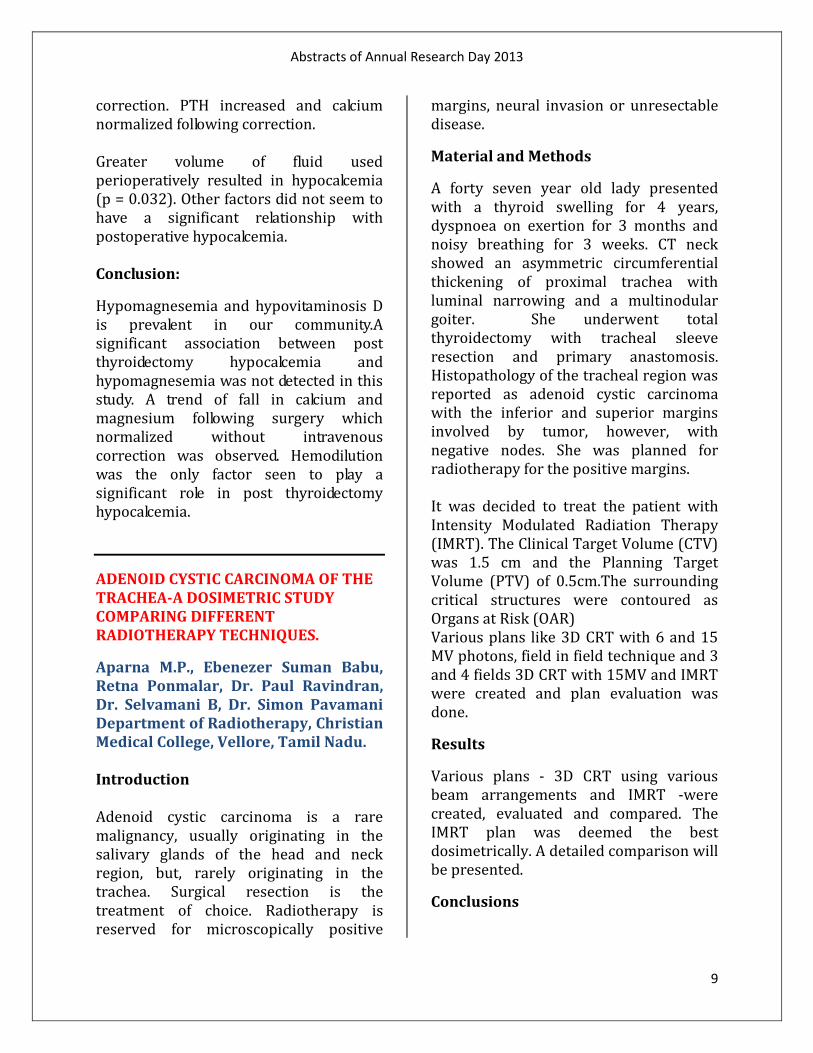
correction. PTH increased and calcium normalized following correction. Greater volume of fluid used perioperatively resulted in hypocalcemia (p = 0.032). Other factors did not seem to have a significant relationship with postoperative hypocalcemia. Conclusion:
Hypomagnesemia and hypovitaminosis D is prevalent in our community.A significant association between post thyroidectomy hypocalcemia and hypomagnesemia was not detected in this study. A trend of fall in calcium and magnesium following surgery which normalized without intravenous correction was observed. Hemodilution was the only factor seen to play a significant role in post thyroidectomy hypocalcemia.
ADENOID CYSTIC CARCINOMA OF THE TRACHEA-A DOSIMETRIC STUDY COMPARING DIFFERENT RADIOTHERAPY TECHNIQUES.
Aparna M.P., Ebenezer Suman Babu, Retna Ponmalar, Dr. Paul Ravindran, Dr. Selvamani B, Dr. Simon Pavamani Department of Radiotherapy, Christian Medical College, Vellore, Tamil Nadu. Introduction Adenoid cystic carcinoma is a rare malignancy, usually originating in the salivary glands of the head and neck region, but, rarely originating in the trachea. Surgical resection is the treatment of choice. Radiotherapy is reserved for microscopically positive
margins, neural invasion or unresectable disease.
Material and Methods
A forty seven year old lady presented with a thyroid swelling for 4 years, dyspnoea on exertion for 3 months and noisy breathing for 3 weeks. CT neck showed an asymmetric circumferential thickening of proximal trachea with luminal narrowing and a multinodular goiter. She underwent total thyroidectomy with tracheal sleeve resection and primary anastomosis. Histopathology of the tracheal region was reported as adenoid cystic carcinoma with the inferior and superior margins involved by tumor, however, with negative nodes. She was planned for radiotherapy for the positive margins. It was decided to treat the patient with Intensity Modulated Radiation Therapy (IMRT). The Clinical Target Volume (CTV) was 1.5 cm and the Planning Target Volume (PTV) of 0.5cm.The surrounding critical structures were contoured as Organs at Risk (OAR) Various plans like 3D CRT with 6 and 15 MV photons, field in field technique and 3 and 4 fields 3D CRT with 15MV and IMRT were created and plan evaluation was done.
Results
Various plans - 3D CRT using various beam arrangements and IMRT -were created, evaluated and compared. The IMRT plan was deemed the best dosimetrically. A detailed comparison will be presented.
Conclusions

Abstracts of Annual Research Day 2013
10
Adenoid cystic carcinoma of the trachea is a rare malignancy and there are no definite guidelines to treat these tumours. IMRT may be the preferred modality of radiotherapy for these tumours due their location & proximity to critical structures.
“ASSOCIATION OF HLA-B*1502 AND SEVERE CUTANEOUS ADVERSE DRUG REACTIONS INDUCED BY AROMATIC AMINE ANTI-EPILEPTIC DRUGS IN INDIAN PATIENTS.”
Girish S Naik, 1T. S. Vijayakumar,2 Mahendran K, 2Sumith K Mathew, 1Denise Helen Fleming, 1Kalpana Ernest, 1Maya Thomas,3 Renu George4
Department of Pharmacology and Clinical Pharmacology 1 Molecular Laboratory, Department of Nephrology 2 Department of Neurology 3 Department of Dermatology 4 Christian Medical College, Vellore, 632 004, Tamil Nadu, India1-4
Background:
Aromatic amine anti-epileptic drugs are one of the most common drugs implicated in producing severe cutaneous ADR like SJS, TEN, SJS/TEN and DIHS/DRESS. Mortality is highest with TEN (20-30%) followed by SJS (1-5%).1 Carbamazepine induced SJS has been shown to have the strongest association with risk allele HLA-B*1502 in South East Asians while other aromatic anti-epileptics have also shown association. The study aimed to evaluate if HLA-B*1502 genotyping can be of clinical utility in identifying Indian patients at risk of developing severe cutaneous ADR to aromatic amine anti-epileptic drugs.
Objective:
To study the association between HLA-B*1502 and SJS, TEN and DRESS induced by aromatic amine AEDs.
Methods:
In this case-control study, patients who developed SJS, TEN or DRESS to one of the aromatic AEDs (cases) and patients on aromatic AED therapy for at least 3 months who did not develop SCAR (controls) were recruited. Controls were matched by drug group and origin (North/South). HLA-B*1502 genotyping was performed using a PCR based assay and the phenotype-genotype association assessed.
Results:
Genotyping was performed for 84 patients (30 cases and 54 controls). Significant association between SJS/TEN induced aromatic AED and HLA-B*1502 was seen (4/9 vs. 4/54; 44% vs. 7.4%; 0.011). CBZ induced SJS/TEN alone compared with CBZ tolerant, origin matched controls also showed significant association (3/4 vs. 0/16; 75% vs. 0%; 0.004). None of the patients who developed DRESS were positive for the allele (0/21 vs. 4/54; 0.569).
Conclusions:
There was no association between aromatic AED induced DRESS and HLA-B*1502 among Indian patients. Although association between SJS, TEN due to all aromatic AEDs studied was statistically significant, 75% was accounted by CBZ. The association was seen amongst south Indians in our study similar to an earlier study from India which reported 75% prevalence among north Indian CBZ induced SJS/TEN cases. Screening for

Abstracts of Annual Research Day 2013
11
HLA-B*1502 seems to be prudent before initiating Indian patients on CBZ to prevent SJS/TEN. Studies with larger sample size showing association of other aromatic amine AED induced SJS/TEN to HLA-B*1502 among Indians are needed before recommending screening prior to initiating other aromatic AEDs. Abbreviations: AED – Anti-Epileptic Drug; CBZ – Carbamazepine; DRESS – Drug Rash with Eosinophilia and Systemic Symptoms; SJS – Stevens-Johnson Syndrome; PHT – Phenytoin; SCAR- Severe Cutaneous Adverse Reaction; TEN – Toxic Epidermal Necrolysis
“PROSPECTIVE STUDY TO DETERMINE THE USEFULNESS OF URINARY NEUTROPHIL GELATINASE ASSOCIATED LIPOCALIN (NGAL) AS AN EARLY AND SENSITIVE MARKER OF URINARY TRACT INFECTION (UTI) IN CHILDREN”
Dr. J. Iswarya, Dr. Indira Agarwal, Dr. Joe Fleming*, Dr. Swasti Chaturvedi, Dr. Rani Diana Sahni**, Department of Pediatrics, Clinical Biochemistry* and Microbiology**, Christian Medical College and Hospital, Vellore, India
Introduction:
Urinary tract infections (UTI) are one of the most common infectious diseases encountered in pediatric practice.(1) In some young children, UTI may lead to renal scarring, leading to poor renal growth, recurrent pyelonephritis, impaired glomerular function, early hypertension and end stage renal disease.(2) Early, aggressive antibiotic treatment has been recommended for symptoms suggestive of UTI in young
children to prevent renal scarring.(3) Although urine culture is the gold standard for diagnosing UTI, the results may not be available for 2–3 days.(1) Hence, urinalysis is commonly used to make an early diagnosis of UTI and to start treatment. The sensitivity of pyuria and positive urine nitrite test as indicators of UTI are low as shown by several studies. Neutrophil Gelatinase Associated Lipocalin (NGAL), also known as Lipocalin 2, is an iron carrier protein that is abundantly expressed in human neutrophils(4). Its transcription is induced by bacterial lipopolysaccharides.(4). There are some animal studies which had shown that NGAL is an useful marker to detect UTI.(4) There are no studies done using NGAL for detection of UTI in Indian population and in children less than five years of age. Hence, we proposed to study the usefulness of uNGAL as an early, sensitive predictor of UTI in Indian population and in the age group of less than 5 years.
Aims and objectives:
Primary objective:
1. To determine the usefulness of urinary Neutrophil Gelatinase Associated Lipocalin (NGAL) as an early and sensitive marker of urinary tract infection (UTI) in children less than 5 years of age
2. To determine the optimal cut-off level for uNGAL to predict UTI in this group of children
Secondary objective:

Abstracts of Annual Research Day 2013
12
3. To compare the sensitivity and specificity of uNGAL estimation with urine WBC estimation in children with culture positivity.
Patients and methods: This prospective study was conducted in the Department of Pediatrics, Christian Medical College, Vellore, India between January 2012 and June 2013.
One hundred children aged between three months and five years, who presented to either Pediatric Casualty or Pediatric Out-patient services, and fulfilled the inclusion criteria and had none of the exclusion criteria were enrolled in the study. The inclusion criteria and exclusion criteria were as follows:
Inclusion Criteria:
All children between the age of three months and five years with symptoms suggestive of UTI as listed below and urine WBC > 5/cu.mm or urine WBC > 11/cu.mm alone.
Fever, nausea, vomiting, abdominal pain/ back pain, straining/ poor urinary stream, foul smelling urine, dysuria, frequency, urgency of micturition and diarrhoea.
Exclusion Criteria:
1. Children with renal anomalies like polycystic kidney disease, multicystic dysplastic kidney
2. Children with lupus nephritis/ glomerulonephritis/ nephritic syndrome
3. Chronic Kidney Disease (CKD) 4. Renal Replacement Therapy (RRT) 5. Contrast Induced Nephropathy
(CIN) 6. Nephrotoxicity
7. Transplantation and delayed graft function
8. Cardiac Surgery (CS) 9. Cancers 10. Prior use of antibiotics for treatment
of UTI
Methodology:
Midstream clean catch urine samples were sent for urine WBC analysis. If urine WBC >11/cu.mm alone or if the urine WBC >5/cu.mm with symptoms suggestive of UTI, the child was included in the study after obtaining informed consent from the parents. Demographic details of the patient including name, age, sex, address and contact phone number were collected. General examination was done for all patients.
Supra pubic aspirate (SPA) urine samples for urinary NGAL levels and urine culture were collected for children aged between three months and three years and midstream clean void urine samples were obtained for children aged between four and five years, before the child was started on antibiotics. A diagnosis of UTI was made if there is significant bacteriuria (≥10,000 cfu /ml of a single pathogen in mid stream clean void urine samples or >1000 cfu /ml in mid stream clean void urine samples, if suggested by our microbiologist as significant or even 1 single bacterium in SPA urine samples) in the urine culture.
Urine NGAL was determined using human NGAL/lipocalin-2 immunoturbidimetric assay, supplied from Bioporto diagnostics. The urine samples were stored at -70 degrees Celsius. Before processing, the sample was thawed for one hour and centrifuged at 2000 rotations per minute for ten minutes. It

Abstracts of Annual Research Day 2013
13
was then processed using Roche molecular P-800 analyser.
Statistical analysis was done using SPSS 18 software. The sensitivity and specificity of uNGAL estimation as a marker of UTI was compared with urine WBC estimation in children with culture positivity. Receiver operating curve (ROC) analysis was performed to determine the sensitivity and specificity of different cut-off points for uNGAL and urine WBC estimation to predict UTI. The most appropriate cut-off point was chosen according to ROC analysis, and the area under the curve (AUC) was calculated.
The positive and negative predictive values and likelihood ratios were also calculated. Results and Discussion: Majority of children were between 3 and 12 months of age (51%) followed by 36% between 25 and 60 months and 13% between 13 and 24 months. The mean age of the children was 22.09 months with a range of 3-60 months. The female: male ratio was 1.3: 1. The presenting complaints and symptoms were recorded in all 100 children. Fever was the most common symptom and was seen in 85% of patients. Fever was the only presenting symptom in 36% of children. Vomiting was the next most common symptom, seen in 26% of children. Dysuria was seen in 18 children. The mean urine WBC value was 509.38 (IQR 22- 143.75). The mean urine NGAL value was 221.28 (IQR 2.73-193.58). Blood culture was done in 58 of 100 children, whenever there was suspicion of
associated urosepsis. Blood culture was negative in all cases.
Among the 100 patients, 34 were urine culture positive and 66 were culture negative. The male: female ratio in the culture positive group was 1.1: 1. E. coli (82%) was the most common organism grown in urine culture followed by Enterococcus (9%), Klebsiella (6%) and Citrobacter (3%). 18 of 25 patients (72%) had Extended-Spectrum Beta-Lactamase (ESBL) producing strains of E. coli. ESBL producing strains of E. coli are more resistant to cephalosporin antibiotics. This makes the infections harder to treat. Receiver Operating Characteristic (ROC) curve analysis was performed for the urine NGAL values. According to ROC analysis, the optimal uNGAL value to predict urinary tract infection was 27ng/ml with a sensitivity of 79.4% and specificity of 68.2%. (Area under curve: 0.765). The positive predictive value (PPV) was 56% and negative predictive value (NPV) was 87%. The positive likelihood ratio (LR+ve) was 2.47 and negative likelihood ratio (LR –ve) was 0.31.

Abstracts of Annual Research Day 2013
14
ROC analysis showed that the optimal urine WBC value to predict urinary tract infection was 38/cu.mm with a sensitivity of 70.6% and specificity of 53%, which were less than that of uNGAL. The positive predictive value (PPV) was 44% and negative predictive value (NPV) was 78%. The positive likelihood ratio (LR+ve) was 1.49 and negative likelihood ratio (LR –ve) was 0.57. With a urine WBC cut-off of 10/cu.mm (routinely followed in clinical practice), the sensitivity and specificity were 94.1% and 7.6% respectively. The positive predictive value (PPV) was 34% and negative predictive value (NPV) was 71%. The positive likelihood ratio (LR+ve) was 1.01 and negative likelihood ratio (LR –ve) was 0.86. The specificity was very low, leading to high false positive rates and the unnecessary use of antibiotics in children with suspected UTI, thus increasing the emerging antibiotic resistance. The correlation of urine NGAL and urine WBC with urine culture in various age groups was looked at in our study. The sensitivity of urine NGAL for predicting UTI in children < 1 year of age was 84.2% and specificity was 71.9% (p<0.0001). The sensitivity of urine NGAL for predicting UTI in children > 1 year was 73.3% and specificity was 64.4% (p<0.014). The area under the curve for urine NGAL <1 year was 0.79 and that for >1 year of age was 0.72. The sensitivity and specificity of urine NGAL for predicting UTI was more in children <1 year of age, thus making it a good test to predict UTI in infants, in whom the incidence of urinary tract
abnormalities is higher and hence higher chance of getting UTI. The odds of getting a urinary tract infection in children <1 year was 13 times greater if their urine NGAL levels are >27ng/ml and in children > 1 year of age, it was 5 times higher than the normal population. We found that urine NGAL was statistically significant in predicting urinary tract infection in both children <1 year and >1 year of age. We compared the sensitivity and specificity of urine NGAL and urine WBC in predicting UTI in males and females. ROC analysis showed that the sensitivity of urine NGAL in females was 87.5% and specificity was 52.5%. The sensitivity of NGAL in males was 77.8% and specificity was 57.7. The area under the curve for males was 0.77 and for females was 0.78. The sensitivity of urine NGAL was higher in females than in males. The mean urine NGAL value to predict UTI was higher in females (19.25) compared to males (9.25). In females, because of the risk of contamination, the urine NGAL values were higher than males as expected. Urine NGAL levels were compared between the culture positive and culture negative groups. The mean urine NGAL values were higher in patients with culture positivity (p value = 0.0001) and lower in patients with culture negativity (p value = 0.001). This was statistically significant. We also compared urine NGAL values between patients who grew E. coli in urine culture with those who grew other organisms. There was no significant difference in the mean urine NGAL values between E. coli and other organisms. Conclusion:

Abstracts of Annual Research Day 2013
15
Urine NGAL proved to be a sensitive and specific marker to predict simple uncomplicated UTI in young Indian children, when compared to urine WBC which is the routine screening test, with an optimal cut-off level of 27ng/ml. Urine WBC cut-off with best sensitivity and specificity to predict UTI was 38/cu.mm, which is higher than the cut-off of 10/cu.mm, which is routinely used. References: 1. Yilmaz A, Sevketoglu E, Gedikbasi A, Karyagar S, Kiyak A, Mulazimoglu M, et al. Early prediction of urinary tract infection with urinary neutrophil gelatinase associated lipocalin. Pediatr Nephrol. 2009;24(12):2387–92.
2. Downing H, Thomas-Jones E, Gal M, Waldron C-A, Sterne J, Hollingworth W, et al. The diagnosis of urinary tract infections in young children (DUTY): protocol for a diagnostic and prospective observational study to derive and validate a clinical algorithm for the diagnosis of UTI in children presenting to primary care with an acute illness. BMC Infect Dis. 2012;12(1):158.
3. Coulthard MG. Is reflux nephropathy preventable, and will the NICE childhood UTI guidelines help? Arch Dis Child. 2008 Mar 1;93(3):196–9.
4. Ichino M, Kuroyanagi Y, Kusaka M, Mori T, Ishikawa K, Shiroki R, et al. Increased urinary neutrophil gelatinase associated lipocalin levels in a rat model of upper urinary tract infection. J Urol. 2009 May;181(5):2326–31.
“ARE WE TALKING ABOUT THE SAME POINT?” POSSIBILITY OF INTER INSTITUTIONAL VARIATION AND ITS
IMPACT IN REPORTING RECTAL DOSE IN HDR BRACHYTHERAPY OF CANCER CERVIX.”
Somnath Dey, Balukrishna S, V.K.Reddy, Sunitha Susan Varghese, Ebenezer Suman Babu, Rabi Raja Singh, Selvamani B Department of Radiotherapy, Christian Medical College, Vellore, Tamil Nadu
Aim:
The ICRU through its report no.38 has tried to bring in uniformity in dose reporting in brachytherapy for cancer cervix. But there is ambiguity and non-uniformity in defining and dose reporting of rectal and bladder point in HDR brachytherapy for carcinoma cervix using orthogonal radiography. This work aims at studying the extent of dose reporting error that exists in Institutions across India.
Materials and methods:
Through personal communication with Radiation Oncologists across the teaching centres in India, we listed out three main methods that are followed for rectal point marking. The response from the oncologists also revealed that all of them adhered to same method in bladder point marking.10 cases were selected by systematic sampling and the rectal points were marked as R1, R2 and R3 and based on technique 1, 2 and 3 respectively. The mean shift between the various points was measured along with the difference in dose at these points.
Results:
The rectal points marked by the three methods were found to have a mean shift

Abstracts of Annual Research Day 2013
16
in the anterior, posterior, cranial and caudal directions between 5mm and 28mm. There were no observed differences in the lateral direction. The difference in percentage rectal dose reported was as high as 25% and mean of 11.2%. The cumulative percentage dose difference at the rectal point as high as 22.67% .There was no difference noticed in marking or dose reporting bladder point.
Conclusion:
Although in the era of Image Guided Brachytherapy, CT planning and volume based optimization may remove the uncertainty, in our country most centres rely on orthogonal films for HDR brachytherapy. The intent of ICRU 38 in helping centers to report uniformly is not served here and hence there is a need for revision for better definition of rectal point and strict adherence to the protocol across the country.
“CHANGING PRACTICE OF INPATIENT HDR BRACHYTHERAPY IN CARCINOMA CERVIX TO AN OUTPATIENT PROCEDURE – A COST MINIMIZATION EXERCISE.”
Judith Aaron, Balukrishna S, Sunitha S, Selvamani B, Department of Radiotherapy, Christian Medical College, Vellore, Tamil Nadu Background: Intracavitory Brachytherapy is an essential component of radical treatment of cancer cervix. High dose rate brachytherapy is preferred for its convenience. Though many centers practice an outpatient procedure, at CMC we still prefer inpatient application under
spinal anaesthesia. This exercise is a lead to changing the Institutional practice and introducing cheaper and convenient outpatient procedure.
Objectives:
1. To test the feasibility of fixing a sleeve at the cervical os
2. To do cost effective analysis of treatment as inpatient basis versus predominantly outpatient basis.
Methods:
Patients with carcinoma cervix suitable for HDR brachytherapy at the end of external beam irradiation were identified. A subject chosen underwent fixation of the sleeve at the cervical os in theatre during the first application followed by second and third applications as outpatient. Three other patients underwent all three applications under spinal anesthesia were also selected. Direct medical and direct non-medical costs for each patient were computed.
Results:
It was possible to suture the sleeve to the cervical os and it stayed in place during the course of treatment (2 weeks). The total treatment cost for the study patient (3 applications) amounted to Rs. 29373 while the mean cost per patient who undergoes all three applications under anesthesia is Rs.39243. Outpatient treatment benefited the patient and society monetarily and in terms of shorter stay in hospital. It was advantageous to the hospital as the

Abstracts of Annual Research Day 2013
17
reduced theatre time and admissions can be best utilized to serve other patients. The effectiveness of treatment was assessed by comparing the dose to point A, rectal dose and bladder dose. There was no significant under dosage to point A or over dosage to critical organs.
Conclusion:
An outpatient application and delivery of HDR brachytherapy was found to be feasible and cost effective as compared to existing practice.
“A RANDOMIZED DOUBLE BLIND CONTROLLED TRIAL COMPARING TWO REGIMENS OF VITAMIN D SUPPLEMENTATION IN PRETERM NEONATES.”
Mintoo Tergestina, Sridhar Santhanam, Anil Kuruvilla, Anna Simon, Niranjan Thomas, Department of Neonatology, Christian Medical College, Vellore
Background:
Preterm babies are prone to metabolic bone disease and are usually supplemented with calcium, phosphorus and vitamin D. While the American Academy of Pediatrics recommends 400 IU of Vitamin D, the European Society for Paediatric Gastroenterology, Hepatology and Nutrition recommends 800 -1000 IU/day. In a previous study we had demonstrated that > 50% of preterms supplemented with 400 IU/day of vitamin D were insufficient at 6 weeks of age. Hence the need for randomized controlled trials to study higher doses of vitamin D supplementation in preterm babies. Objective:
To study the efficacy of two regimens (400 IU/day vs 1000 IU/day) of Vitamin D supplementation in preterm neonates. Setting:
Tertiary care perinatal centre in south India.
Methods:
This was a double blind randomized controlled trial conducted recruiting inborn preterm babies between 28-34 weeks of age. Baseline levels of serum calcium, phosphorus, alkaline phosphatase and Vitamin D were sent at 48 hours of age. When babies reached enteral feeds of 100 ml/kg/day, they were randomised into the trial. All babies were fed unfortified breast milk and once on full feeds, supplemented daily with calcium, and phosphate. One arm of the trial received 400 IU of Vitamin D while the other arm received 1000 IU of Vitamin D. At term corrected gestational age serum calcium, phosphate, alkaline phosphatase, parathyroid hormone (PTH) and 25(OH)D levels were estimated. Samples for Serum Calcium and Urine spot Calcium Creatinine ratio were taken at intervals to evaluate for any evidence of hypervitaminosis D. Results: Ninety babies were recruited into the trial and followed up to term corrected gestational age. The mean Vitamin D level at 48 hours for the 2 groups – 400 v/s 1000 IU was 20.05±9.8 and 24.3±14.8 ng/ml. At term corrected gestational age, mean Vitamin D level in the group that received 400 IU was 18.59±10 and 1000 IU was 47.63±13.78 ng/ml. None of the

Abstracts of Annual Research Day 2013
18
babies were insufficient in the 1000 IU group, as against 53.78% in the group that received 400 IU. Conclusion:
The recommended supplementation of preterm babies with 400 IU/day of vitamin D is insufficient. Preterm babies supplemented with 1000 IU/day of vitamin D maintain normal vitamin D levels without any side effects. Key Words: Preterm; Vitamin D; Supplementation. “STUDY OF THE RELATIONSHIP BETWEEN INITIAL AUTONOMIC FUNCTIONS AND SUBSEQUENT MOTOR AND SENSORY RECOVERY, IN TRAUMATIC CERVICAL SPINAL CORD INJURY PATIENTS.”
Latha N. and Elizabeth Tharion, Department of Physiology, Christian Medical College, Vellore. Aim: To study if assessment of autonomic pathway integrity in traumatic cervical spinal cord injury quadriplegic patients is useful for prognostication. Objectives: To study the (1) correlation between short-term Heart rate Variability (HRV) indices at the time of admission of quadriplegic patients and the change in motor and sensory score at the end of the rehabilitation programme and (2) association between presence or absence
of SSR at the time of admission and subsequent recovery. Methods: HRV indices computed in 20 ASIA grade A/B patients at the time of admission were correlated with the change in motor and sensory scores at the end of the rehabilitation programme using Spearman’s correlation coefficient. Chi square test was used to study the association between the presence or absence of SSR at the time of admission and the change in ASIA grade at the end of the rehabilitation programme.
Results: There was no statistically significant correlation between the HRV indices and the change in motor and sensory scores. Neither was there any statistically significant association between the presence or absence of SSR at the time of admission and the subsequent improvement in ASIA grade.
Conclusion:
The findings of the present study indicate that HRV indices and SSR are not useful parameters to prognosticate recovery in traumatic cervical cord injury quadriplegic patients.
Authors:
Dr. N. Latha, Final year PG, Department of physiology, Christian Medical College, Bagayam, Vellore – 632 002.

Abstracts of Annual Research Day 2013
19
Contact No. – 09965614312 E mail - [email protected]
Dr. Elizabeth Tharion, Professor of Physiology, Department of Physiology, Christian Medical College, Bagayam, Vellore – 632 002. Contact No. +91416 - 2284268 E mail – [email protected]
“CHROMOSOMAL ABERRATIONS IN COUPLES WITH RECURRENT MISCARRIAGES – AN EXPERIENCE OF 479 COUPLES AT A TERTIARY CARE CENTRE IN INDIA.”
Thomas S, Aleyamma T K, Kamath M S, Chandy A, Mangalraj AM, Muthukumar K, Department of Reproductive Medicine, Christian Medical College Hospital, Vellore. Background:
Recurrent miscarriage continues to be a challenging problem for the couple as well as the clinician and determining the cause can be extremely difficult.In couple with recurrent miscarriages there is an increased risk of one of the partners carring a structural chromosomal aberration. Balanced chromosomal rearrangements is an important cause for recurrent pregnancy loss.
Aim:
To evaluate the contribution of chromosomal abnormalities in causing recurrent miscarriages.
Materials and methods:
Retrospective study of 479 couples who attended the Reproductive Medicine unit
in CMC Vellore with history of recurrent miscarriage from 2002 to May 2013.
Results:
There were totally 479 couples (958 individuals) out of whom 69 (7.2%) individuals were detected to have chromosomal aberrations. 21 (2.1%) individuals had translocations, 34 (3.9%) had numerical abnormalities, 4 (0.4%)with inversions and 6 (0.6%) with other chromosomal abnormalities
Conclusion:
Cytogenetic analysis is an important investigation in the workup of couples with recurrent miscarriages.
“EFFECTIVENESS OF AN ARTICULATED KNEE HYPER EXTENSION ORTHOSIS IN GENU RECURVATUM: A CASE REPORT”
Aravind Rahul S.R.M, Student, DPO IInd Year, Rajesh Kumar Mohanty, Lecturer (P&O), Prosthetics & Orthotics Services, Department of PMR, Christian Medical College, Vellore-632004 Background:
Genu Recurvatum is a defomity in the knee joint that tends to push knee backwards by excessive extension in tibio- femoral joints. Commonly used orthoses include heel wedges, ankle foot orthoses (AFO), knee ankle foot orthoses (KAFO) and knee orthoses (K.O) etc. However, technically controlling hyperextension of knee by an orthosis is a difficult task. Therefore, most of the orthoses commonly prescribed for this condition have not been successfully given good results with strong evidences.
Aim:

Abstracts of Annual Research Day 2013
20
To check the effectiveness of an articulated knee hyperextension orthosis in genu recurvatum in terms of gait and energy expenditure.
Case Description & Method: A Subject of 63 years old diagnosed as post polio residual paralysis showed excessive hyperextension of his left knee during long standing and walking. The tibio-femoral angle during full weight bearing was found to be approximately 40 degrees. An orthotic design was proposed, fabricated and fitted to check the effectiveness of the design which incorporated design features like articulated, pre-flexed KAFO based on couple force system. Observational gait analysis (OGA) by video recording, force plate analysis and Energy expenditure using physiological cost index (PCI) was calculated with and without orthosis.
Results:
The orthosis controlled knee hyperextension by not allowing the knee to go beyond neutral position. Advantages including sitting cosmesis, assistance to weaker quadriceps, M-L stability, unlocked but stable gait was observed during trial. Gait deviations like excessive lateral trunk bending, use of ipsilateral hand to support and instability was controlled with the use of orthosis. The gait was more energy efficient and natural as revealed from PCI and force plate studies.
Conclusion:
The proposed design was found to be effective in controlling excessive knee hyperextension and resulted in stable, natural and energy efficient gait.
“MATURITY ONSET DIABETES OF THE YOUNG (MODY) IN INDIA- NOVEL INSIGHTS DERIVED FROM NEXT GENERATION SEQUENCING.”
Aaron Chapla, Mahesh DM, Denny V, HS Asha, V Padmanabhan, Mercy Inbakumari, Sarah Mathai*, Thomas.V.Paul, Nihal Thomas, Department of Endocrinology, Diabetes and Metabolism, Christian Medical College, Vellore, India.
CORRESPONDING AUTHOR:
Dr. Nihal Thomas, MD, MNAMS, DNB (Endo), FRACP (Endo),FRCP(EDIN),FRCP(Glasg) Professor & Head, Department of Endocrinology, Diabetes and Metabolism, Vice Principal (Research), Christian Medical College Hospital, Vellore-632004, India. Email: [email protected]
Background:
Maturity Onset Diabetes of the Young (MODY) has traditionally presented as a monogenic disorder with an autosomal dominant inheritance pattern, characterized by β-Cell dysfunction. An overlap of clinical features with the more common polygenic Type 2 diabetes mellitus makes differentiation of MODY a diagnostic challenge. The current genetic diagnostic protocol involves phenotype guided sequential screening of only a few of the identified MODY genes. However, limitations in the scalability and prohibitive cost of Sanger sequencing has hindered simultaneous genetic screening of all known comprehensive panel of MODY genes.
Abstracts of Annual Research Day 2013
21
Method:
We evaluated 74 patients with young onset diabetes of whom, 51 met the clinical criteria of MODY. A panel of 10 established MODY genes was selected to screen for mutations. A novel multiplex PCR protocol was developed to enrich the target genes and further processed on a semiconductor based next-generation sequencing (NGS). All the identified rare variants were confirmed by Sanger sequencing. Results: Using this approach we have identified MODY mutations in thirteen patients of whom nine mutations were novel. These mutations include four patients with NEUROD1, two with IPF1, and one each with HNF1α, HNF4α, GCK, HNF1β, KLF11, CEL and PAX4. Two patients with NEUROD1 mutation coseggregated with a common PDX1 rare variant.
Conclusion:
NGS is a rapid and cost-effective method for performing comprehensive parallelized genetic screening of the genes implicated in monogenic disorders like MODY. We have identified a higher frequency of NeuroD1 and PDX1 mutations in South Asian Indians, a pattern that differs from populations in the West.
“AN ENHANCED RECOVERY PROTOCOL AFTER RECTAL CANCER SURGERY: CHALLENGING SURGICAL DOGMA”
Rohin Mittal, Mark Ranjan Jesudason, Benjamin Perakath, Christian Medical College, Vellore
Background:
Enhanced recoveries after surgery (ERAS) protocols are increasingly being used in colorectal surgery to improve outcomes and reduce patient discomfort. We analyzed the effect of an enhanced recovery protocol on outcomes following rectal cancer surgery in a tertiary care teaching hospital in South India.
Methods:
This retrospective study compared two cohorts of patients undergoing rectal resection for cancer. An ERAS protocol was introduced in our unit in May 2012.The first cohort of patients (June 2010 to May 2011) underwent standard care and the second (June 2012 to May 2013) an ERAS protocol. The ERAS protocol consisted of pre operative patient education, avoidance of bowel preparation, use of non narcotic analgesia, avoidance of post operative drains and NG tubes, starting of early post operative feeding, early discontinuation of IV fluids, early urinary catheter removal and early discharge. Data was collected from computerised hospital records and a prospectively maintained database.
Results:
One hundred and twenty two patients were included, 51 in the standard arm and 71 in the ERAS arm. Patient in the ERAS group were found to be younger (47.8 vs. 55.3 years, p=0.008), had more frequent metastatic disease (0 vs. 8, p=0.013) and had a higher proportion underwent laparoscopic surgery (p<0.001). Patients were similar in terms of sex distribution, neoadjuvant therapy, type of surgery and the T and N stage.
Median duration of post operative stay was 8 days in the standard care group

Abstracts of Annual Research Day 2013
22
and 6 days in the ERAS group (p=0.69). In patients without any post operative complications, duration of post operative stay was significantly lower in the ERAS group (4.79 vs. 6.96 days, p=0.006). However, in patients with post operative complications, there was no difference among both groups (12.1 vs. 13.1days, p=0.611). Both groups were similar in terms of anastomotic leak, pelvic abscess, wound infection, post operative ileus, bleeding and overall complication rates. There was no difference in readmissions and reoperation rates in both groups.
Conclusion:
An ERAS protocol after rectal resection is safe and helps decrease duration of hospital stay in uncomplicated cases. There is no difference in complication rates, readmission rates and reoperations rates in patients managed under an ERAS protocol. Further objective studies need to be done to quantify improvement of patient comfort in an ERAS programme.
“MINIMALLY INVASIVE OESOPHAGECTOMY VERSUS CONVENTIONAL OPEN SURGERY FOR OESOPHAGEAL CANCER: A SINGLE CENTRE EXPERIENCE.” Deshpande G, Abraham V. ,George S.V., Yacob M., Chandran B.S., Samarasam I. Department of General Surgery, Upper GI surgery unit, Christian Medical College and Hospital, Vellore, Tamil Nadu, India Background: Oesophagectomy is the mainstay of curative treatment for cancer of the oesophagus. The use of minimally invasive techniques in oesophageal
surgery offers hope of reduced morbidity associated with the surgical trauma. Although, the concept of minimally invasive oesophagectomy (MIE) emerged two decades ago, there is still doubt regarding the superiority of outcomes, compared to conventional open surgery. The aim of this study to assess the safety and efficacy of MIE and whether it reduces the morbidity compared with open oesophagectomy, without compromising the oncological principles. Materials and Methods: The clinical and surgical details of patients who underwent curative surgery for oesophageal cancer, from January 2012 to October 2013, at the Upper GI surgical unit, Christian Medical College and Hospital, Vellore was prospectively entered into a computerized database and used for analysis. The patients were grouped into those who underwent conventional open oesophagectomy (Group A) and those who underwent MIE (Group B). The outcome of the surgery between the two groups was assessed based on intra operative and post operative complications, operative time, hospital stay and 30 day mortality. The oncological safety was assessed based on the completeness of resection, extent of lymphadenectomy and the overall survival of the patient. Results: A total of 37 patients underwent oesophagectomy for oesophageal cancer. Out of these, 18 patients had conventional open surgery (Group A) and 19 patients had MIE (Group B). Radical resection (R0) was achieved in a 78 % of patients from Group A and 84 % patients from Group B. The mean operative time taken in Group A was 413 minutes compared to 467 minutes in Group B (p=0.941). The mean

Abstracts of Annual Research Day 2013
23
lymph node harvest in Group A was 11 nodes compared to 16 nodes in Group B (p=0.200). Post operative major surgical morbidity (Clavien Dindo class 3 or 4) of Group A was 5.5% and that of Group B was 5.2%. Post op ventilation was required in 11% patients in group A compare to 10% patients in Group B. The mean ICU stay in Group A was 5 days compared to 2.6 days in Group B (p=0.05). The mean duration of hospital stay was 19.5 days and 16.3 days in Group A and Group B respectively (p=0.162). The mean follow up of the two groups was 10.7 months and 12 months respectively. There was only one 30 day mortality in Group A and none in Group B. Short term follow up during the study period spanning 19 months, revealed an overall survival of 9.8 months in the study group A compared to 12.1 months in Group B (p=0.638). The MIE was performed in the semiprone position, which is a novel technique and has distinct advantages over the classical prone or lateral positions. A short operative video illustrating the operative procedure is also included. Conclusion: Our study suggests that minimally invasive oesophagectomy is a safe procedure, with comparable procedural complications, and possible advantages in relation to post- operative recovery, length of ICU and hospital stay. More importantly, all this can be achieved with no detrimental oncological outcomes. “GENERATION OF DVH-BAND IN CONFORMAL RADIOTHERAPY FOR USE AS A PLAN EVALUATION TOOL.”
Santanu Samanta, Balukrishna S, Sathish Kumar A, Rabi Raja Singh I, Selvamani B, Dept of Radiation Oncology, Ida B Scudder Cancer Centre, Christian Medical College, Vellore. Purpose:
Geometric verification for treatment position is essential for accurate treatment with IMRT. However treatment delivery uncertainties like set up error often fail to deliver intended dose. Current planning systems give a conformal plan but leave the oncologist to compute and analyse the influence of setup errors on the plan. Though targeted therapy with daily imaging will minimise setup errors on treatment delivery, this is not often employed. Here, we propose DVH band as a good visualization tool to select between competing plans to minimise influence of random errors on treatment delivery. Method: Three IGRT plans and their observed couch shift during treatment were analysed retrospectively. From the scatter plot of their shifts the maximum random shift through the course of therapy was found. The shift calculated using Euclidean distance formula was used to generate rival plans for the same planning CT and leaf sequence but with isocentric shift applied to calculated magnitude. The isocentre was shifted to same magnitude in +x, -x, +y, -y, +z & -z directions respectively and plans generated using the same leaf sequences used for treatment delivery. The DVH for critical organs and targets were overlapped to create DVH band.

Abstracts of Annual Research Day 2013
24
Results:
The DVH band gave good visual interpretation of possible influence of random error on the organ or target for that particular plan.
Conclusion:
The wider the DVH band the greater the influence of setup errors on that plan. Hence this can be studied prospectively as a plan evaluation tool for conformal therapy where less imaging is employed at treatment.
“INSULIN SIGNALING IS IMPAIRED IN IRON-LOADED PRIMARY MOUSE HEPATOCYTES FROM HEPCIDIN KNOCK-OUT MICE.”
Joe Varghese1, Andrew McKie2, Molly Jacob1., 1 Department of Biochemistry, Christian Medical College, Vellore, INDIA., 2 Division of Diabetes and Nutritional Sciences, King’s College, London, UK.
Mild to moderate increase in body iron stores is associated with insulin resistance. However, the relationship betweeninsulin andiron homeostasis is not clear. To study the effects of intracellular iron on insulin signalling, we treated wild-type and iron-loaded primary mouse hepatocytes from hepcidin knockout (hepc KO) mice with insulin (10 nM or 100 nM for 10 min). Phosphorylation of Akt (protein kinase B or PKB), a marker of insulin signalling, was significantly reduced in hepc KO hepatocytes. Treatment of these cells with desferroxamine (DFO), an iron chelator, prior to insulin treatment tended to improve Akt phosphorylation in a dose-dependentmanner, indicating improved insulin signalling with reduction in
intracellular iron. Hepcidin, the master regulator of systemic iron homeostasis, was highly expressed in wild-type hepatocytes. Insulin treatment (10 or 100 nM for 1, 3 or 6 hrs) did not affect hepcidin expression significantly. In wild-type hepatocytes,insulin (100 nM for 6 hrs)significantly up-regulated the gene expression of transferrin receptor 1 (TfR1) but did not have significant effects on expression of TfR2, divalent metal transporter-1 (DMT1), hereditary hemochromatosis gene (HFE), hemojuvelin (HJV) or matriptase 2 (TMPRSS6).In summary, iron-loaded primary hepatocytes isolated from hepc KO mice showed impaired insulin signalling when compared to wild-type hepactocytes;pre-treament with DFO tended to reverse this effect.Induction of TfR1 by insulin may explain increased hepatic iron stores that are associated with insulin resistance, a condition characterized by hyperinsulinemia. Further studies are required to confirm this.
“COGNITIVE SYMPTOMS AND DISABILITY” Paul Prabhu K.G., College of Nursing, Christian Medical College Background: Schizophenia is an enipatic disorder with an indisputable clinical heterogoncity. Until recently,t eahetrts for schizophreniah avef ocusedm ainly on reducingp ositive symptoms,o ftenleaving patients \r'ith numerous residual difficulties, including impairment in cagnition, everyday living skills, and social/ocrlpafional ftnctioning. Owhg to the limited litelature fiom the counhy in this area, the researchq uestion
Abstracts of Annual Research Day 2013
25
of assessi-ocgo gtritive slmptoms in patients with schizopb.reoian d its relatiooshipw ith furctional disability was fomulated. Objective: The main objective of the study was to assess the cognitive slmptoms in patieDts with Schizophenia, its relationship srith futrctioMl disability aod its association with selected socio demographic and clinical vadables. Methods: The study had a descdptive cross sectional dosign. 110 subjects who tulfilled the inclusion cdteria cosented for the study, recruiied using coNecutive samplhg tecbnique. SCoRs and WHO-DAS II atrd WHO-DAS II were used to assess the cognitive symptoms and disability respectively,P ANSS was uscd to assessth e psychopathologyo f patieotsw ith SchizophreDiaI.twas conducted in the in the Departmo of Psychiaty, Cbdstiatr Medical College, Vellore Results: It was found that cognitive slmptoms positively cnlrelated with disability (p=0.00). This relationship was also significant i.! atl the domaitrs of disability (p=0.00). Duratioo of iltoess and medicatiotr adheretrce also showed statistically significaot association with both cognitive symptoms aod disability (p=0.00). Conclusion: The subjectsw ere on antipsychotics.T he effect of the drugs causingc lgnitive
deficits was not studiod. Paucity of literature in this area especially from India, and findings of this study highlights the needf or further researchin this field. Key words: SC2RS- Schizapbmia CognitionR oting Scale WHO-DASI I - World.H ealth Organization- Disobility assessmenSt cheduleII PANSS- Positive and NegativeS yndiomeS cale

Abstracts of Annual Research Day 2013
26
ABSTRACTS FOR POSTER PRESENTATION
BASIC SCIENCE

Abstracts of Annual Research Day 2013
27
“LARGE SCALE ISOLATION, PURIFICATION AND CHROMATOGRAPHIC ANALYSIS OF BIO ACTIVE COMPOUNDS FROM CLEISTANTHUS COLLINUS.”
Soosai Manickam Amirtham, Sathya Subramani, Department of Physiology, Christian Medical College, Vellore-2.
Aim:
To isolate active principles of Cleistanthus collinus (C.collinus) by liquid/liquid partition chromatography and enrichment by Thin Layer Chromatography.
Objectives:
C.collinus is a known toxic plant, used for self-harm in rural South India. The mortality is 28%. The exact mechanism of action of this poison is still unknown and there is no promising antidote till date. In this study we screened major active compounds from boiled and room temperature extract.
Methods:
C.collinus leaves were shade dried and hexane delipidated. 100grams of leaves were soaked in 3 liters of distilled water for 24 hrs for room temperature extract. 100 grams of leaves were immersed in 2.5 liters of boiling distilled water for 10 minutes for boiled extract. The supernatant of both extracts were collected and mixed with Chloroform to form two immiscible layers. The bottom chloroform layer, separated with separating funnel sequestered the fluorescent compounds. The top aqueous layer did not have any fluorescent compounds. The fluorescent portion was concentrated and powdered. The active principles Cleistanthin A, Cleistanthin B
and diphyllin in the fluorescent fraction were screened and purified by Thin Layer Chromatography.
Results:
The chromatographic analysis of screened Cleistanthin A, Cleistanthin B and diphyllin were performed by TLC, HPLC, PDA, FTIR and GCMS. The HPLC of Cleistanthin-A and Cleistanthin-B showed 100% purity. Diphyllin was impure.
Conclusion:
The screened phytochemicals can be used to study the action of individual active principles and to find a potential antidote, in the process.
“BACTERIOLOGICAL PROFILE AND ANTIBIOGRAM FROM A NEONATAL UNIT IN SOUTH INDIA.”
Vijay Gupta*, Sridhar S*, Balaji V**, Niranjan Thomas A W*, Anil Kuruvilla K*, Manish Kumar*, Atanu Kumar Jana*., *Department of Neonatology and **Department of Microbiology, Christian Medical College, Vellore Background:
Bacteriological profile in a neonatal unit changes with maternal demographics and local antibiotic policies. There is growing concern about emergence of new organisms in causing early onset sepsis (EOS) (≤ 72 hours) and late-onset sepsis (LOS) (≥ 72 hours) as well as an increase in the incidence of infections caused by multidrug resistant organisms.
Objective: To study the bacteriological profile of newborn sepsis in a tertiary care unit in south India.

Abstracts of Annual Research Day 2013
28
Design: Retrospective review of inpatient charts of newborn babies admitted over a two year period. Setting: Tertiary care perinatal center. Subjects: All babies admitted in the neonatal unit, with blood culture positive sepsis from January 2011 to January 2013 were included in the study. Results: Among 26,107 inborn babies and 815 outborn babies admitted during the study period, there were 360 organisms isolated from 271 newborn infants. Of all the isolates, 184(51.1%) caused early onset sepsis (EOS) while 176(48.9%) caused late onset sepsis (LOS).The incidence among inborn was 8.3 per 1000 live births and sepsis occurred in 7.6% of all nursery admissions. NFGNB (mainly Burkholderia) (47%), Klebsiella(14%) and GBS (12%) were the predominant organisms causing E0S, while Klebsiella (38%), NFGNB (mainly Acinetobacter)(23%) and Enterobacter(12%) were the predominant organisms in LOS. Most of the isolates were sensitive to Amikacin, Cefaperazone sulbactam and Netilmicin. However, resistance to Ciprofloxacin, Gentamicin and cefoxitin was seen in large number of isolates. All strains of GBS were sensitive to Penicillin.
Conclusions: The incidence of neonatal bacterial sepsis was 8.3 per 1000 livebirths. NFGNB and Klebsiella were the most common organisms as the cause for both EOS and LOS. GBS was a common organism causing EOS. Keywords: Neonatal sepsis, antibiogram, South India
“LOW COST NOVEL METHOD OF VISUALLY QUANTIFYING NOISE LEVELS IN THE NICU – THE CMC SOUND METER.”
Sudesh Yadav, Kalyani Kareti, Atanu Kumar Jana, Suresh Devasagayam, Niranjan Thomas., Departments of Neonatology and Bioengineering, Chrisitian Medical College, Vellore, Tamilnadu.
Background:
Exposure to high sound levels in the NICU have been associated with morbidity like hearing impairment and sleep disturbances in preterm babies. The American Academy of Paediatrics recommends maximum safe sound levels in NICU to be 45 dB. A device which can monitor sound in Neonatal Intensive Care Unit with visual display will be useful. Objective and Design: To build a low cost sound level meter with visual display of high sound levels. Methods: A microphone was used as a sensor and sound was converted to electrical signal,

Abstracts of Annual Research Day 2013
29
preamplified and passed through band filters. From the band pass filter the signal goes to the LED driver. Reference voltage of LED drivers was set and connected to a LED Bar Graph for display. (Fig 1) Calibration was done in the Audiometry department where voltage from each band pass filter reaching the LED driver was measured and a plot is made voltage v/s sound loudness dB. Validation was done using the Lutron SL-4013 Sound Level Meter of Spectrum Internationals as the standard and comparison of our instrument and the standard was made by testing it in the NICU during three different timings in the day. Results: The mean sound level detected in the NICU by the CMC sound meter was 63.9 ± 4.5 dB and was similar to that obtained at the same time by the Lutron SL-4013 sound level meter (66.1 ± 4.2 dB). The cost of building the CMC sound meter was Rs 1300 as compared to Rs 15,000 for the Lutron SL-4013. All sounds above 45 dB were visible as a LED glow alerting the health care worker to be careful. At most times, the sound levels in the NICU were more than 45dB. Lower frequencies contribution was less than high frequencies. Sounds > 45dB in the NICU were identified due to handover/rounds, beeping sound of instruments, phone ringing, movement of x-ray machine, trolley and chair. Movement of chairs and trolleys contributed maximally in increasing noise levels to more than 75dB. Conclusion:
The low cost CMC-sound meter was very useful in visually displaying high sound levels in the NICU and helps make immediate changes to reduce noise in the NICU. Keywords: Sound level meter; Visual display Figure 1 CMC sound meter
“HARNESSING GENE EXPRESSION PROFILING IN SEARCH OF NEW CANDIDATE GENES FOR ARA-C RESISTANCE IN ACUTE MYELOID LEUKEMIA.”
Ajay Abraham, Savitha Varatharajan, Sreeja Karathedath, Shaji R Velayudhan, Alok Srivastava, Vikram Mathews, Poonkuzhali Balasubramanian, Department of Haematology, Christian Medical College, Vellore- India. Wide inter-individual variation in terms of treatment outcome and toxic side effects of treatment exist among patients

Abstracts of Annual Research Day 2013
30
with AML receiving chemotherapy with cytarabine (Ara-C) and daunorubicin. We have previously evaluated the expression of the major genes involved in cytarabine transport and metabolism on ex-vivo Ara-C response and compared it with cytogenetic and molecular markers in AML (Blood (ASH Annual Meeting Abstracts) 2011 118: Abstract 3481). Our candidate gene expression data led us to propose Ara-C resistance index (Ara-C RI) (RI = ΔCT (DCK X ENT1)/ ΔCT CDA), which incorporates candidate Ara-C metabolizing genes whose RNA expression are significantly associated with ex-vivo Ara-C cytotoxicity. Ara-C RI values were significantly higher in resistant (IC50 >80 uM) and intermediate (IC50 6.25-80uM) samples when compared to sensitive samples (IC50 <6.25uM) (median 5.459 (1.759- 11.82) and 5.396 (1.89- 11.62) vs. 3.840 (1.89- 9.8); p <0.0001 (Fig 1a). This was then validated in the relapsed AML samples, which showed a significantly higher RI values (median RI 6.312 (2.01- 19.85)) when compared to sensitive ((3.840 (1.895- 9.8)) and resistant samples at diagnosis (5.412 (1.759- 11.82)); p <0.0001 (Fig 1b). Though, the Ara-C RI correlated well with ex-vivo cytotoxicity as well as treatment response in vivo (data not shown), this did not completely explain the variation, suggestive of alternative resistance mechanisms. We undertook a genome-wide gene expression profiling to address possible mechanisms of Ara-C resistance other than the candidate gene approach. Based on ex vivo Ara-C cytotoxicity at diagnosis, Ara-C sensitive (IC50 <3uM AraC) and Ara-C resistant samples (IC50 >80uM) (each 5) were included for microarray analysis. Total RNA was extracted from bone marrow
mononuclear cells using tri reagent, cDNA was synthesized and then subjected to one color 8 X 60K Agilent microarray analysis. Data was normalized, filtered and analyzed using Gene Spring GX (V 12.0) software. Normalization was done using the 75th percentile shift (Percentile shift normalization is a global normalization, where the locations of all the spot intensities in an array are adjusted). Using unpaired t-Test, 4436 genes were identified to be differentially expressed (Fold change expression values were provided as log-base 2) between Ara-C sensitive samples and Ara-C resistant samples. Differentially regulated genes were clustered using hierarchical clustering based on Pearson coefficient correlation algorithm to identify significant gene expression patterns. Genes were classified based on functional category and pathways using GeneSpring GX and Genotypic Biointerpreter-Biological Analysis Software. The differentially expressed genes fell into the following biological processes; transcription (375 genes), transport (364 genes), metabolism (267 genes; Fig 1d), immune (155 genes), cell cycle (129 genes), apoptosis (123 gene; Fig 1c) and so on. Upregulated gene list in Ara-C sensitive group included apoptotic related genes like PMAIP1, NDUFA, BAX, BCL2, TRAF2, transcriptional regulators including SMAD5, SMAD1, ZNF family proteins- ZNF644, ZNF469, ZNF195, ZNF22, ZNF3, ZNF713, ZNF777, ZNF234, CEBP, PARP, ETV6, E2F3 and so on. Down-regulated genes in Ara-C sensitive group are of special interest as they could be potential candidates for targeting Ara-C non-responsive group. They included transcriptional regulators like HDAC4, KLF4, CREB5, CEBP, RARA, E2F4, MNDA, MTA3 and PPAR also apoptotic genes like MCL1, PSEN1, ELMO2, PAK1, APAF1,

Abstracts of Annual Research Day 2013
31
MAPK1, CD40, FAS, CASP1 and CASP8. Interestingly, many of the genes involved in cellular metabolism were found to be down regulated in AraC sensitive group. Major down-regulated metabolic genes include CDA, SLC2A3, SLC2A8, GK, NADK, ACACB, ACSL1, PFKFB4, PFKFB3, PDK4, ME1 and PC. This study has come up with previously unrecognized aspects in Ara-C resistance in AML, and is suggestive of Ara-C sensitive samples having increased expression of anti-Warburg set of genes. Our data as well as growing evidences from various malignancies and altered cellular metabolism propose the possibility of using metabolic inhibitors alone or in combination with Ara-C to
overcome drug resistance.
“ASSOCIATIONBETWEEN APOLIPOPROTEIN E GENOTYPE AND RISK OF NEOPLASMS: A META-ANALYSIS.”
Anand R, Prakash S. S*,
p< 0.0001
n=55 n=55 n=30
p< 0.0001
n=23 N=85 n=55
Fig 1a Fig 1b
Fig 1c Fig 1d

Abstracts of Annual Research Day 2013
32
Veeramanikandan R*., Department of Biochemistry, Christian Medical College, Vellore, Tamilnadu, India. *Equal contribution
Background:
Apolipoprotein E (ApoE), a protein primarily involved in lipoprotein metabolism occurs in 3 isoforms (E2, E3 and E4). The relation between ApoE genotype and serum cholesterol levels is known. Cholesterol levels in turn are associated with increased risk of some malignancies. In another context, ApoE4 is a risk factor for Alzheimer's disease and studies report an inverse association between incidence of malignancies and Alzheimer's disease. While studies evaluating the association between ApoE genotype and incidence of malignancies are available, the results are inconsistent. Aim: To study the association between ApoE genotype and incidence of neoplasms by a meta-analysis. Methods: A literature search of electronic databases revealed 49 studies with information on ApoE polymorphisms and neoplasm risk. After screening and evaluation by a structured questionnaire, 21 studies (18 case-control/3 prospective; 71858 cases/12516 controls) were included for the present meta-analysis. Pooled odds ratios (OR) with 95% confidence intervals (CI) were calculated assuming a random-effect model for all the genotypes and alleles. Subgroup analyses based on ethnicity and type of neoplasm were also performed. Appropriate tests to detect heterogeneity, publication bias and sensitivity were done at all stages.
Results: Overall, the pooled analysis of the data showed no association between ApoE alleles or genotypes with incidence of neoplasms. In the stratified analyses, while, we observed an increased risk of breast neoplasms in E3/E4 genotype (OR: 1.43; 95%CI: 1.01-2.04; P: 0.046) when compared with E3/E3, the risk of neoplasms other than breast and colorectal, were decreased (OR: 0.79; 95%CI: 0.63-0.98; P: 0.0337). We also noted a positive association between E4/E2 genotype and colorectal neoplasms (OR: 1.42; 95%CI: 1.04-1.94; P: 0.029). Stratified analyses based on ethnicity did not show any association. Conclusion: Although subgroup analysis showed associations between specific neoplasms and ApoE genotypes, existing literature does not show a significant association between the genotype and risk for neoplasm in general. “INDOMETHACIN INHIBITS ACTIVATION OF ENDOTHELIAL NITRIC OXIDE SYNTHASE (ENOS) IN THE RAT KIDNEY: POSSIBLE ROLE OF THIS EFFECT IN THE PATHOGENESIS OF INDOMETHACIN-INDUCED RENAL DYSFUNCTION.” Arumugam Suriyam Nagappan1, Joe Varghese1, Gautham Tumkur Pranesh1, Visalakshi Jeyaseelan2 and Molly Jacob1*., 1Department of Biochemistry, Christian Medical College, Vellore – 632002, India., 2Department of Bio-statistics, Christian Medical College, Vellore – 632002, India.

Abstracts of Annual Research Day 2013
33
Corresponding author: Molly Jacob*, Department of Biochemistry, Christian Medical College, Vellore, Tamil Nadu, India. Pin: 632 002. E-mail address: [email protected]. Introduction: Clinical use of non-steroidal anti-inflammatory drugs (NSAIDs) is often associated with adverse effects in the kidney. Indomethacin, an NSAID that has been shown to induce oxidative stress-induced damage in the kidney, was used to study the pathogenesis of such effects in rats. Methods: Male Wistar rats received oral indomethacin (20mg/kg); they were sacrificed 1, 12 or 24 hours (h) later. Various biochemical parameters in the kidneys, affected by oxidative stress, were determined. Results & Conclusion: Indomethacin significantly reduced the ratio of dimeric endothelial nitric oxide synthase (eNOS) (active form) to its monomeric (inactive) form, heme staining in NOS dimers and tissue nitrite levels at 12 and 24h and renal heme levels at 1 and 12h. Heme oxygenase 1 (HO-1) activity was significantly elevated at 1 and 12h after the drug. Evidence of nuclear translocation of Nrf2 (at 12 h) and p38 MAPK signaling (at 12h and 24h), both of which are known to induce HO-1, were found to occur in response to indomethacin. The results show that indomethacin reduced levels of activated eNOS in the kidney possibly by depleting
heme by HO-1 activation that occurred downstream of Nrf2 and p38 MAPK signaling. Reduced renal eNOS activity may result in decreased NO levels and hence reduced renal perfusion. Acknowledgements: This study was funded by the Department of Science and Technology (DST), New Delhi, India and by Fluid Research Grants from the Christian Medical College (CMC), Vellore, Tamil Nadu, India. “AN ADVANCED BENCH TOP CBCT SYSTEM FOR SMALL ANIMAL IMAGING.”
B Paul Ravindran1, Rajdeep Ojha2 and D Devakumar3, 1Department of Radio Therapy (Medical Physics),2
Department of Bioengineering and 3Department of Nuclear Medicine, CMC Vellore,
Aim: To construct an aSi flat panel based bench-top X-ray cone beam CT scanner and to investigate its use for small object / animal imaging Introduction: The viability to generate full 3D volumetric data in a single rotation of source and the detector around the object (or by rotation of the object with source and the detector stationary) has instilled interest in development of the cone beam CT (CBCT). During the annual research day 2012at CMC, Vellore, the feasibility of developing a prototype bench top CBCT was presented using an image intensifier based C-arm X-ray unit. In this work we

Abstracts of Annual Research Day 2013
34
have developed a bench top CBCT using amorphous silicon (aSi) based flat panel detector.We have also implemented improved image reconstruction software developed to run on a Graphic Processing Unit (GPU) using Compute Unified Device Architecture (CUDA). This abstract describes the development and the investigation on the image quality of the CBCT. Materials and Methods: A Pleophos D X-ray machine was used as the X-ray source for the CBCT. A 3030D aSi flat panel detector supplied by Varian of size 30cm x 30 cm was used as the imaging detector. The object was placed on a turntable supplied by Holmac. The turntable was rotated using a customized hardware comprising of microcontrollerand a stepper motor driver. Software was developed in Visual Basic to trigger the microcontroller to rotate the turntable and to acquire the projection images from the flat panel detector at predetermined angular interval (0.50, 10,1.50 and 2 degrees) over 360 degree.Images of the phantoms to study the resolution and the contrast of the CBCT imaging system were obtained.The image reconstruction was performed with FDK based software developed originally in C(1) and then converted to CUDA for parallel processing with the GPU using NVIDIA graphics card. Results and Discussion: The GPU based parallel computing reduced the reconstruction time from 40 minutes to less than a minute for a full reconstruction of more than 300
slices.The bench top CBCT has been developed and found to be suitable for small object and could also be used for small animal imaging. Acknowledgment: The authors thank Mr Kush and MsDhivya, M.Tech. Bioengineering students of CMC, Vellore for CUDA programming. References: 1. Thomas THM, Devakumar D, Purnima S, Ravindran BP. The adaptation of megavoltage cone beam CT for use in standard radiotherapy treatment planning. Phys Med Biol. 2009 Apr 7;54(7):2067–77.
“COMBINED EFFECT OF OLFACTORY ENSHEATHING CELLS AND FIBROBLAST GROWTH FACTOR ON SPINAL CORD REGENERATION.” Basir Ahmed1, Durai Morgan2, George Tharion2,Indirani.K3#, 1Medical student, Christian Medical College, Vellore, TN, India, 2Dept of Physical Medicine and Rehabilitation, Christian Medical College, Vellore, TN, India, 3Dept of Anatomy, Christian Medical College, Vellore, TN, India. Spinal cord injury is one of the most debilitating diseases, leaving more than 99% patients with lifelong neurological complications. Trauma management and rehabilitative measures are the mainstay of SCI with no established therapeutic treatment. As a result several experiments are being carried to look for a feasible therapy. One of the potential options is the transplantation of Olfactory Ensheathing Cells (OECs) into the injured

Abstracts of Annual Research Day 2013
35
spinal cord to promote neuronal regeneration and axonal growth. This study aims at evaluating the combined use of OECs and Fibroblast growth factor (FGF) in rat SCI for neuronal regeneration and functional motor recovery .For this, 24 female Albino Wistar Rats were randomly distributed into 4 groups:6 for control, 6 for OEC, 6 for FGF and 6 for OEC along with FGF. SCI was inflicted by 10 gm drop weight method following T-10 laminectomy. The FGF group was immediately administered 2ul of FGF solution as was the OEC and FGF group. After 9 days, OEC was transplanted in the OEC group and OEC and FGF group. The motor recovery was measured over 8 weeks using the BBB (Basso, Beattie and Bresnahan) Scale. At the end of 8 weeks, EMG was done to obtain the Motor Evoked Potentials. The results show Significant P-Value and 95% Confidence Interval in all the three groups compared individually with the control group for both the BBB scores and the EMG-MEP results. However, there is no significant difference when either the OEC group is compared to OEC+FGF group or FGF group with OEC+FGF group. The significant motor recovery in the three groups (OEC, FGF and OEC and FGF) when compared to the control group indicates neuronal regeneration. Thus concluding, there is significant similar level of locomotor functional recovery indicating neuronal regeneration with the application of OECs and FGF, when applied either individually or in combination. Article submitted for NNF Gold Medal- NEOCON 2013
“COMPARISON OF TRANSCUTANEOUS BILIRUBIN MEASUREMENT WITH SERUM BILIRUBIN MEASUREMENT IN SOUTH INDIAN PRETERM AND TERM BABIES.” Binu Govind, Preethi C, Kurien Anil Kuruvilla, Department of Neonatology, Christian Medical College, Vellore, Tamilnadu. Objective:
To evaluate the efficacy of transcutaneous bilirubinometer in estimating jaundice levels in south Indian neonates. Design: Prospective observational cohort study Setting: Tertiary care perinatal center in south India. Subjects and interventions: Newborn babies admitted from May 2011-July 2012 and who had evidence of clinical jaundice were recruited into the study. The study patients was classified into preterm (<35 wks) and term (≥ 35 wks) babies Methods: Within 1 hour of clinical suspicion of jaundice, transcutaneous bilirubin (TcB) measurement from the forehead was obtained using the Drager Jaundice Meter (JM-103). Serum bilirubin (TSB) levels were sent simultaneously. Results:

Abstracts of Annual Research Day 2013
36
During the one year study period, 188 preterm babies and 381 term babies were evaluated. Correlation between transcutaneous and serum bilirubin in preterm and term babies was good (r=0.77, p<0.01 and 0.7, p <0.01 respectively). Reliability statistics using Cronbachs alpha was 0.857 and 0.821 (p< 0.01) in preterm and term babies respectively. Intraclass Correlation Coefficient was 0.75 (CI 0.67-0.80) in preterm babies and 0.69 (CI 0.63-0.74) in term babies. The sensitivity, specificity, positive predicitive value and negative predicitive value were72.9%, 76.6%, 81.2% and of 67% in preterm and 81.4%, 70%, 93% and 43.3% in term babies respectively. Bland Altman plot showed fair agreement in both term and preterm babies. Conclusion: Transcutaneous bilirubin measurement correlates well with serum bilirubin levels in term and preterm south Indian babies. TcB can be used as a substitute to serum bilirubin in screening babies with clinical jaundice. Keywords: Transcutaneous Bilirubinometry; Neonate; Preterm; Term; Jaundice Introduction: Neonatal jaundice is the commonest condition necessitating observation, investigation, readmission and treatment in the first week of life. Visual assessment of jaundice is often inaccurate, especially when inexperienced personnel are involved in the assessment. In many centers in India, current practice involves a daily visual screening of babies in the nursery and postnatal ward. In those babies who
appear jaundiced, serum bilirubin is determined by obtaining a venous blood sample and analysed in the laboratory using spectrophotometry. The results are obtained usually only after 2-3 hours and decisions are made according to age- specific guidelines for starting phototherapy. Based on this strategy, ~50% of babies have at least one serum bilirubin determination before discharge and only about 30% of these require phototherapy. A study from India showed that need for blood sampling was 34% lower in the TcB group as compared to those with were visual assessment was used (1). Hence, the majority of bilirubin determinations do not result in an immediate decision to start phototherapy and thus could have been avoided. Conversely, in some babies, there is a delay in initiating treatment as the process of collecting a venous sample and analysing it in the lab takes time. The newer generation of bilirubinometers namely the Bilichek and the Drager bilirubinometer JM 103 are less influenced by skin pigmentation and chromogens and have demonstrated good correlation with serum bilirubin values (r=0.8-0.91) in several studies (1,2,3,4). These instruments are now being used routinely in many centres. Evidence obtained from their use indicates that this method of screening has the potential to better select babies who need serum bilirubin determinations (5, 6, and 7). Hence using transcutaneous bilirubinometry (TcB) will reduce the number of invasive serum bilirubin assays thereby reducing pain, waiting time for patients and workload for health care professionals There have been several studies comparing the accuracy of TcB readings with serum bilirubin in western countries (8, 9, 10, and 11). In India, few studies

Abstracts of Annual Research Day 2013
37
have been done in term and late preterm babies, all from north India (13, 14). However, there is no published data on preterm babies (<35 weeks) from India. Also the effect of darker skin pigmentation of south Indian babies on TcB values has not been studied. Hence, though there have been comparative studies from the north India, the impact of relatively darker skin of Indian babies and their effect on TCB needs to be studied (11). The aim of the study was to evaluate TCB measurements as performed by Drager JM-103 jaundice meter, and correlate it with total serum bilirubin levels, measured with standard lab methods in south Indian term and preterm babies.
Methods:
Design: Prospective Observational Cohort study
Setting: Study was done in the Neonatology department of Christian Medical College, Vellore, and a tertiary care perinatal center in south India from May 2011 to July 2012.
Population: Newborn babies admitted either in nursery or the postnatal wards iand who had evidence of clinical jaundice were eligible for the study. The study group was classified into preterm (<35 weeks) and late preterm and term (≥ 35 weeks)
Ethics clearance: The study was started after getting clearance from the Institutional Review Board and ethics committee.
Method Details: Transcutaneous bilirubin measurements were done on babies who underwent total serum bilirubin (TSB) estimations as a part of their routine
neonatal care. When there was clinical suspicion of jaundice and TSB planned, a TcB measurement from the forehead using the Drager Jaundice Meter (JM-103) was done within 1 hour and the value was recorded. The machine was set to take 5 readings and display the mean. The TSB was sent at the same time.
Data including the baby’s age in completed hours at the time of measurement, gender, gestational age in completed weeks, weight in grams at birth, length and the occipitofrontal diameter in centimetres, mode of delivery and hours of life at which jaundice developed was noted. We collected data on skin tones of the babies determined by visual assessment by the investigator aided by Fitzpatrick skin tone scales
The Fitzpatrick Skin Tone Scale:
Type I (scores 0-7) White; very fair; freckles; typical albino skin.
Always burns, never tans
Type II (scores 8-16) White; fair.
Usually burns, tans with difficulty
Type III (scores 17-25) Beige; very common.
Sometimes mild burn, gradually tans

Abstracts of Annual Research Day 2013
38
Type IV (scores 25-30) Beige with a brown tint; typical Mediterranean Caucasian skin.
Rarely burns, tans with ease
Type V (scores over 30) Dark brown.
Very rarely burns, tans very easily
Type VI Black.
Never burns, tans very easily
Sample size: The required sample size to show a sensitivity of 85% for detecting TSB levels of more than phototherapy action values with 5% precision and at an alpha level of 5% was found to be 200 cases.
Formula:
where,
P = sensitivity of the test Q = 1-P d = precision Zα/2 is the 5% level of significances
Statistical Analysis: Statistical analysis was done by the SPSS software version 16.. A Regression analysis was done to compare the two types of measurement. Correlation between the serum bilirubin and transcutaneous bilirubin was done by Pearson correlation. Reliability statistics was done by Cronbachs Alpha and Intraclass correlation coefficient. Sensitivity, specificity, positive predictive value and negative predictive value of transcutaneous bilirubin in comparison with serum bilirubin values were noted at
a cutoff of serum bilirubin of 15 mg% in term and 10 mg% in preterm babies. The data obtained was used to construct Bland Altman plots, comparing the differences between TcB and TSB. Similar plots was also constructed for the different subgroups to derive limits of agreements and to compare it with what is reported in the literature
Results:
During the study period, 188 preterm babies and 381 term babies had clinical suspicion of Jaundice and simultaneous serum and transcutaneous bilirubin readings were done.
Table1 shows the baseline characteristics of the preterm and term babies. The mean gestational age in preterm was 31.4±1.9 weeks and the mean birth weight was 1540±357 g. Among the term babies, the mean birth weight was 2840±567 g and gestational age 38.3± 1.5 weeks. Ninety five (50.5%) of the preterm were males and 93 (49.5%) were females. Among the term babies, 204(53.5%) were male babies and 177(46.5%) were female babies. 46.5% of the preterm babies were delivered by LSCS compared with 33.2% of term babies.
Fitzpatrick skin tone analysis was done and the babies were mostly in the dark skin ranges (Table 2).
The Correlation between transcutaneous and serum bilirubin in preterm showed a correlation coefficient of 0.77 (p <0.01) (Fig 1). The term babies also showed similar findings with Pearson correlation factor of 0.7 (p <0.01) (Fig 2).

Abstracts of Annual Research Day 2013
39
With the help of the Regression analysis, an equation was derived to predict serum bilirubin from transcutaneous values.
In preterm, TSB = 3.487+0.612 x TcB.
In Term babies, TSB = 3.618+0.634 x TcB.
Reliability statistics using Cronbachs alpha of 0.857 and 0.821 respectively in preterm and term babies (p<0.001) (Table 5 ). Intraclass Correlation Coefficient was 0.75 (CI 0.67-0.80) in preterms (p<0.001) and 0.69 (CI O.63-0.74) in term babies (p<0.001) (Table 3).
The diagnostics are shown in Table 4.
Sensitivity and Specificity was 72.9% and 76.6% respectively in preterms with positive predictive values of 81.2% and negative predictive value of 67% to detect bilirubin levels > 10 mg%. In term babies, sensitivity was 81.4%, specificity 70% with positive and negative predictive values of 93% and 43.3% respectively to detect bilirubin levels > 15 mg% (Table 4 ).
Bland Altman plot was done to look at agreement of the two measurements (TcB and TSB). For preterm babies the mean difference was 0.8 with limits of agreement from 5.9 to -4.3 (Fig3). In term babies the mean difference was 1.27 with limits of agreement from 6.7 to -4.1 (Fig 4).
Discussion:
Our study has looked at the correlation of TcB with serum bilirubin levels in term and preterm babies from south Indian babies. The skin tones of the babies were mainly dark brown as per Fitzpatrick’s classification, indicatin that the sample population studied were more pigmented as compared to the previous populations
studied (4, 5, 11). Preterm babies had a lighter skin tone as compared to term babies, probably as pigmentation increases as the fetus matures (5). More number of preterms delivered by LSCS because most of the preterm deliveries were secondary to maternal PIH and eclampsia.
The TcB values correlated well with serum bilirubin levels in our study, both in preterm and term babies (r = 0.77 and 0.7, p<0.01-Figure 1 and 2). Other studies from the west have shown similar good correlation among term and near term babies [Bhutani et al (r=0.91) and Rubatelli etal (r = 0.89, 95%CI O.89-0.91)] (5, 6). Ebbessen et al had estimated correlation of TcB and TSB measurements in both preterm and term babies and had got coefficient of correlation of 0.87 and 0.88 respectively which was slightly better than our estimates (12). Deorari et al in a study on term and late preterm babies showed that TcB values had significant correlation with TSB (r= 0.76, p<0.001), which is very similar to our study (14). The lower values in the Indian studies may reflect the effect of skin pigmentation but the overall correlation is still significantly good.
Regression analysis done showed linear regression between TcB and TSB values, which was in line with almost all other published data (2,3). Based on this we were able to generate a prediction equation to predict TSB from TcB. Ebbessen et al(12) had demonstrated slightly but statistically significant higher TcB in sick infants than healthy infants for the same TSB (p<0.01,95%CI 6.07,17.51); the difference between TcB and TSB varied between 65mmol/l and 120mmol/l in comparison with Rubaltelli et al who had got a higher difference (6).

Abstracts of Annual Research Day 2013
40
De Luca et al had calculated the mean increment rate in TcB for different hours of life based on linear regression analysis and showed that TcB showed increment over the first 72 hours and thereafter plateaued off (10). Maisels et al also showed similar trends in showing similar higher values in the initial days (9). Our study was not powered to make similar normograms. Maisels et al also showed decreased gestational age babies having higher values for TcB (9).
Regresssion and correlation does not address reliability and precision. Hence we did reliability statistics with Cronbach alpha criterion and Intra class correlation coefficient, both of which showed statistically significant results, suggesting good reliability of TcB.
We addressed the issue of precision by using the Bland Altman plot. The mean difference (bias or accuracy) was good (0.8 and 1.27 in preterm and term babies), while the precision was fair (5.9 to -4.3 in preterm and 6.7 to -4.1 in term babies). Thus 95% of future measurements would like between theses limits. This was similar to a study done by Deorari et al (14) were the mean difference was 0.93 and precision was 4.7 to – 2.9.
Rubatelli et al had published data on sensitivity (93%) and specificity (73%) which was higher than our results with similar cut-off (6). Bhutani et al (5) constructed normograms from TcB values and identified that the negative predictive value of TcB measurement to identify the infants with hour specific TSB <75th centile is 100%, positive predictive value is 32.86%, sensitivity is 100%, specificity 88.1% and likelihood ratio 8.43
The limitations of our study was that the number of preterms <30 weeks were small compared to higher gestational ages. Hence subset analysis of preterms was not possible. Also most of our preterm babies did not have values in the high range as they were aggressively treated and so reliability of TcB readings in extreme preterms and higher levels of bilirubin has to be looked into.
Conclusion:
There is good correlation between transcutaneous and serum bilirubin levels in south Indian term and preterm babies. The results attained through the two methods seem to be reliable and accurate. Transcutaneous bilirubin estimation can be used routinely as a reliable screening tool in the management of jaundiced babies.
References:
1. Mishra S, Chawla D, Agarwal R, Deorari AK, Paul VK, Bhutani VK. Transcutaneous bilirubinometry reduced the need for blood sampling in neonates with visible jaundice. Acta Pediatrica 2009;98;1916-1919.
2. Yamanouchi I, Yamauchi Y, Igarashi I. Transcutaneous bilirubinometry: Preliminary studies of noninvasive transcutaneous bilirubin meter in the Okayama National Hospital. Pediatrics. 1980; 65:195-202.
3. Tan KL, Chia HP, Koh BC. Transcutaneous bilrubinometry in Chinese, Malay and Indian infants. Acta Paediatr. 1996; 85:986-90.
4. Bhutani VK, Gourley GR, Adler S, Kreamer B, Dalin C, Johnson L. Noninvasive measurement of total serum bilirubin in a multiracial

Abstracts of Annual Research Day 2013
41
predischarge newborn population to assess the risk of severe hyperbilirubinemia. Pediatrics. 2000;106:17-26e.
5. Bhutani VK, Johnson L, Sivieri EM. Predictive ability of a predischarge hour-specific bilirubin for subsequent significant hyperbilirubinemia in healthy term and near term newborns. Pediatrics. 1999; 103:6-14.
6. Rubaltelli FF, Gourley GR, Loskamp N, Modi N, Roth Kleiner, Sender A, Vert P. Transcutaneous Bilirubin measurement: A multi centre evaluation of a new device. Pediatrics 2001; 107:1264-1271.
7. Ong SK, Doraisamy G, Tsou K, Sethi SK. Transcutaneous bilirubin measurements in Singapore newborns. Abstract Book, 2007 Austalian Association of clinical Biochemists Meeting. Melbourne, Australia.
8. Boo NY, Ishak S. Prediction of severe hyperbilirubinemia using the Bilichek transcutaneous bilirubinometer. J Paed and Child Health 2007; 43:297-302.
9. Maisels MJ, Kring E. Transcutaneous bilirubin levels in the first 96 hours in a normal newborn population of >/= 35 weeks gestation. Pediatrics. 2006;117:1169-1173
10. De Luca D, Romagnoli C, Tiberi E, Zuppa AA, Zecca E. Skin bilirubin nomogram for the first 96h of life in a European normal healthy newborn population obtained with multiwavelength transcutaneous bilirubinometry. Acta Paed. 2008;97:146-150
11. Thompson J et al., Transcutaneous Bilirubinometry and ethnicity.
Arch dis Child Fetal and Neonatal ed Nov 2008 Vol 93 No 6; 474.
12. F Ebbesson Rasmussen LM, Wimberley PD. A new transcutaneous Bilirubinometer, Bilichek, used in the neonatal care unit and the maternity ward. Act Paediatrica 2002;91:203-211.
13. Bhat YR, Rao A. Transcutaneous bilirubin in predicting hyperbilirubinemia in term neonates. Ind J of ped. 2008; 75:119-121.
14. Mishra S, Chawla D, Agarwal R, Deorari AK, Paul VK. Transcutaneous bilirubin levels in healthy term and late preterm Indian neonates. Ind J Pediatrics 2010; 77:45-50.
TABLE – 1 Descriptives
Preterm Term

Abstracts of Annual Research Day 2013
42
n=188
Mean±SD
n=381
Mean±SD
Gestational Age
31.8±1.9 28.3±1.5
Birth Weight
1548±357 2843±567
Length
40.3±3.5 47.4±2.9
APGAR 5’
7.9±1.6 8.7±0.9
APGAR 10’
8.7±0.8 9.5±0.8
LSCS 87 126
Normal 101 255
Male 95 204
Female 93 177

Abstracts of Annual Research Day 2013
43
Table 2 – Skin Colour grading of study patients using Fitzpatrick scale
category Colour grade N (%)
Preterm
3 6 (12.5%)
4 18.1 (70.8%)
5 8 (4.3%)
Term
3 19 (10.8%)
4 188 (67.0%)
5 36 (20.5%)
6 3 (1.7%)
TABLE 3 Reliability Statistics
Preterm
Term
Corbach’s Alpha
0.857
(P<0.01)
0.821
(P<0.01
Intra Class Correlation
0.75 (0.67-0.8)
P<0.01
0.69(0.63-0.74)
P<0.01
TABLE – 4 Diagnostic Indices
Sensitivity
Specificity
PPV NPV
Preterm
72.9% 76.6% 81.2
% 67%
Term
81.4% 70% 93% 43.3
%

Abstracts of Annual Research Day 2013
44
Figure 1- Linear Regression- Preterm babies
Figure 2 Linear Regression – Term Babies
Figure 3 Bland Altman Plot – Preterm babies
Figure 4 Bland Altman Plot- Term babies
-

Abstracts of Annual Research Day 2013
45
“LANTHANUM CAUSES LANTHANUM/CALCIUM EXCHANGE THROUGH SODIUM CALCIUM EXCHANGER.”
Elizabeth Clarence, Anand Bhaskar and Sathya Subramani, Physiology, Christian Medical College.
Background:
The sodium calcium exchanger (Na+/Ca2+
exchanger) plays a central role in regulating contractility in ventricles and automaticity in pace-maker cells. It has a stoichiometry of 3Na+: 1Ca2+ and is capable of working in forward (calcium-extrusive and sodium-acquisitive) and reverse (sodium extrusive and calcium-acquisitive) modes.
Lanthanum is known to inhibit calcium entry in electrically excitable cells, by blocking the reverse mode of sodium–calcium exchanger and voltage gated calcium channels. Action of lanthanum is by competition for Na+/Ca2+ exchanger, which can execute sodium-lanthanum exchange. This study tests our hypothesis that Na+/Ca2+ exchanger is capable of working in lanthanum-calcium exchange mode too.
Aim:
To demonstrate that the Na+/Ca2+
exchanger is capable of Lanthanum/calcium exchange in isolated rat hearts.
Objectives:
1) To induce an increase in cytosolic Ca2+
with caffeine and sodium-free lithium extracellular solution.
2) To test if the contracture by the above procedure can be relaxed by the addition of lanthanum.
Methods and results:
Heart isolated from an anaesthetized rat was perfused through coronary arteries in Langendorff mode with normal extracellular solution and paced at a rate of 280 per minute. Left ventricular pressure was recorded with a balloon connected to a pressure transducer.
Hearts were initially perfused with normal mammalian extracellular solution during the stabilization period. Subsequent perfusion with sodium-free Lithium extracellular solution and addition of caffeine (25mM) induced a contracture (due to calcium release from intracellular stores by caffeine and lack of its clearance due to absence of sodium in the extracellular solution). While the contracture was maintained in control animals, addition of lanthanum to the perfusate in tests, caused relaxation.
Conclusion:
Addition of La3+ to the perfusate relaxed the contracted heart favouring our hypothesis that it can move through the NCX by the La3+/Ca2+ mode. This finding is important because it provides another tool for investigating the activity of NCX. “OXIDATIVE AND NITROSATIVE STRESS IN THE HEART DURING DEVELOPMENT OF LIVER CIRRHOSIS.”
G. Jayakumar Amirtharaj, K. A. Balasubramanian and Anup Ramachandran, The Wellcome Trust Research Laboratory, Division of Gastrointestinal Sciences, Christian

Abstracts of Annual Research Day 2013
46
Medical College, Vellore-632004, INDIA.
Background:
Liver cirrhosis is an advanced stage of progressive fibrosis due to ongoing liver injury. The mortality of liver cirrhosis is due to complications such as portal hypertension, bacterial infection and compromised cardiovascular function. Aim: To examine the role of reactive oxygen and nitrogen species in the heart during development of liver cirrhosis. Methods: Twice-weekly intraperitoneal administration of thioacetamide produced liver cirrhosis after 3 months was confirmed by histology. Oxidative stress parameters and nitrate levels were measured in heart during cirrhosis development. Results: A significant increase in oxidative stress parameters were seen in heart during liver cirrhosis during the time frank cirrhosis was evident by histology. This was accompanied by significant increase in nitrate levels and inducible nitric oxide synthase protein expression. In addition, phosphorylation of stress kinase p38 MAPK also increased during cirrhosis development. Conclusion: These results suggest that during liver cirrhosis reactive oxygen species can cause phosphorylation of p38 MAPK
which in turn could induce nitric oxide synthase to elevate nitric oxide levels. “ASSESSMENT OF DIAGNOSTIC VALIDITY OF PROCALCITONIN IN BURNS SEPSIS PATIENTS- A PROSPECTIVE STUDY.” Dr. Geley Ete, Dr. Elvino Barreto, Naveen Kumar HR, Dr. Kingsly Paul M, Department of Plastic Surgery, Christian Medical College Vellore.
Introduction:
Sepsis is one of the major causes of mortality in burns. Clinically it is very important to identify systemic infection at an early stage to initiate antibiotic therapy and to prevent prolonged morbidity and mortality. This study is undertaken to analyze the procalcitonin value in burns patients and also to determine a cut off value to suspect / diagnose sepsis at its stage.
Aims and objectives:
To establish diagnostic validity (sensitivity and specificity) and best cut off value of procalcitonin in diagnosis of sepsis in burns patients.
Material and Methods:
A total of 36 patients with more than20 % TBSA burns were included in thestudy. To know the diagnostic validity, procalcitonin levels in patients with and without sepsiswere measured. Statistical analysis was done using ROC curve to assess diagnostic performance and Mann-Whitney Utest to know the significance.
Results:

Abstracts of Annual Research Day 2013
47
Data of 36 patients was analyzed with mean age of 27.97 years. 44 % were male and 56 % females. Average TBSA burns were 47.16 %. The diagnostic validity was highest at the cut-off value of 5ng/ml which gives sensitivity of88.9% specificityof 83.3% and positive predictive value of 84.2%.
Conclusion:
PCT has high diagnostic validity to diagnose sepsis in burns.In burns patients the cut off value of PCT to diagnose sepsis is 5ng/ml (sensitivity, specificity 88.9% and 83.3% respectively). Serial measurement of PCT in burns patients has prognostic importance in development of sepsis and recovery.
“FLUPIRTINE RELAXES DISTAL CAPRINE URETER: AN IN-VITRO STUDY”
Girish S Naik, Sumith K Mathew, Kalpana Ernest, Manoj G Tyagi, Jacob Peedicayil, Department of Pharmacology and Clinical Pharmacology, Christian Medical College, Vellore, 632 002, Tamil Nadu, India.
Background:
Ureteric colic secondary to calculi is a common problem worldwide. Medical Expulsive Therapy (MET) is reserved for solitary small single distal ureteric calculus and the standard approved treatment is tamsulosin, an alpha blocker. Recently, Kv7 channels have been demonstrated in the lower urinary tract. Flupirtine maleate, a centrally acting analgesic is an NMDA (N-methyl D-Aspartate) antagonist and Kv7 opener devoid of side effects of opioids and NSAIDs. A drug which can produce both
pain relief and relax distal ureter would be an ideal alternative to tamsulosin.
Objectives:
To study the effect of log concentrations of flupirtine on the spontaneous contractility of distal caprine (goat) ureter, characterize the dose response curve, determine EC50 (95% CI) and study the mechansim of action.
Methods:
Isolated specimens of distal caprine ureter which exhibited spontaneous contractions were subjected to log concentrations of flupirtine maleate for a period of ten minutes (1-90µM) and the effect was reversed with XE991 and 4-Aminopyridine (4-AP), specific Kv7 and nonspecific Kv blockers respectively. Repeated measures ANOVA was used to compare log transformed activity scores following exposure to flupirtine and reversal agents. Nonlinear regression was done to fit the dose response curve. EC 10,
50 and 90 (95% CI) were determined from the model.
Results:
A total of 63 isolated specimens of distal caprine ureter were studied. Flupirtine showed a concentration-dependent inhibition of spontaneous contractions. The EC50for a contact period of 10 minutes (95%CI) was 17.74µM (15.71 to 19.76 µM). XE991 (100µM) and 4-AP (1mM) reversed the effect of 30µMflupirtine significantly.
Conclusion:
Flupirtine relaxes distal caprine ureter and may be useful in conditions requiring distal ureteric relaxation. Action through opening of voltage-gated potassium

Abstracts of Annual Research Day 2013
48
channels (Kv) is the likely mechanism, though involvement of Kv7 channels could not be confirmed conclusively on account of higher doses of XE991 used for reversal. Action through Kv7.5 is possible as IC50 for Kv7.5 has been shown to be about 60µM. However, reversal by 100µM XE 991 through Kv2.1 and ERG channels cannot be excluded and studies exploring further mechanisms are required.
“NEXT GENERATION SEQUENCING APPROACH FOR MOLECULAR GENETIC DIAGNOSIS OF PARTIAL LIPODYSTROPHY”
RUNNING TITLE:
Molecular Genetic Diagnosis of Partial Lipodystrophy
H.S.Asha, Aaron Chapla, Manika V, ShrinathShetty and Nihal Thomas, Department of Endocrinology, Diabetes and Metabolism, Christian Medical College, Vellore, India.
CORRESPONDING AUTHOR:
Dr. H.S. Asha, Associate Professor Department of Endocrinology, Diabetes and Metabolism, Christian Medical College Hospital, Vellore-632004, India.
Background:
Lipodystrophies are a heterogeneous group of diseases characterized by abnormal fat distribution. Familial partial lipodystrophy 2 (FPLD2) is autosomal dominant condition due to mutations in the LMNA gene. This subset of patients present with selectiveloss of subcutaneous adipose tissue from the
limbs and trunk, and accumulation of fat in the neck and face. Furthermore they are usually associated witha variety of metabolic disorders including insulin resistance, diabetes mellitus, dyslipidemia, hepatic steatosis and high blood pressure.
Objective:
To study the clinical and molecular features of a subject with FPLD.
Subjects and methods:
Utilizing Next Generation Sequencing we carried out mutational analysis of LMNA gene in a woman and her daughter with FPLD phenotype.
Results:
The subject aged 23 years was diagnosed to have diabetes mellitus at age 16 years requiring high doses of insulin with suboptimal glycemic control. She had cushingoid appearance of the face, vulval hypertrophy, acanthosisnigricans, prominent muscular contours with minimal fat in the limbs, phlebomegalyand hepatomegaly consistent with partial lipodystrophy. Her mother was diagnosed to have diabetes mellitus at age 30 years, with similar features as the daughter. Her sister aged 17 years also has cushingoid face with prominent muscle contours in the limbs consistent with FPLD. The subject and her mother had elevated C-peptide levels suggestive of insulin resistant diabetes mellitus. Dual energy X-ray absorptiometry in the subject and her mother showed increased truncal fat with decreased limb fat confirming fat redistribution.

Abstracts of Annual Research Day 2013
49
The NGS of LMNA gene showed that mother and daughter were both heterozygous for a reported c.1444C> T missense mutation which causes the substitution of the Arginine at residue 482 by a Tryptophan (Nabrdaliket.al.,Endokrynol Pol. 2013;64(4):306-11). This identified mutation was confirmed by Sanger sequencing. The subject was started on Metformin and insulin therapy was optimized resulting in better glycemic control. Conclusions:
Our subject had a clinical form of FPLD2 due to a mutation affecting lamin A.Clinical suspicion based on the phenotype followed by appropriate biochemical and genetic testing led to the diagnosis in this family. A clear understanding of the disease process is very important to choose the right treatment modalities, predict the natural history of the disease and to determine the prognosis.
“DISTRIBUTION OF CD1A POSITIVE LANGERHANS CELLS IN BENIGN AND MALIGNANT DISEASES OF HUMAN LUNG USING IMMUNOHISTOCHEMISTRY.”
J. Rajkohila, Roy Gnanamuthu, Inbam Indrasingh, J.Suganthy, Department of Anatomy, Christian Medical College, Vellore.
Background:
The dendritic cells are involved in host immunity and immune surveillance against malignancy. Though the presence
of Langerhans cells (LCs) in human lungs had been described earlier, their role in tumour response are still poorly understood. Their presence and number within the tumour tissue is crucial for antitumour immune response. Aim: To study the morphology and distribution of CD1a positive LCs in benign and malignant diseases of the human lung by immunohistochemistry. Methods: After getting informed consent, human lung tissue samples were collected from eleven patients who underwent pulmonary surgery for lung tumours in Christian Medical College, Vellore and were processed for immunohistochemical staining with mouse monoclonal antihuman CD1a antibody. The number of CD1a positive LCs per square area was counted using CellSens Standard image analyzer software. Statistical analysis was done using SPSS version 17. Results: CD1a positive LCs were present in primary and secondary lung malignancy, in the bronchiolar epithelium. In addition, they were present in the interstitium in carcinoid tumor and chondroid hamartoma and in the bronchus associated lymphatic tissue in chondroid hamartoma. The alveolar compartment did not show any CD1a positive LCs. These cells had typical dendritic morphology except in adenocarcinoma in which they were nondendritic. Pleural condensation infiltrated with massive number of CD1a positive LCs was present in a case of

Abstracts of Annual Research Day 2013
50
metastatic thymoma. The mean number of CD1a positive LCs was highest in pleural metastasis of thymoma and lowest in adenocarcinoma. Conclusions: Bronchiolar compartment is better suited to mount an immune response than the alveolar compartment as CD1a positive LCs were predominantly distributed in bronchioles. The number of CD1a positive LCs in adenocarcinoma correlated inversely with the tumour grade. Therefore the distribution of CD1a positive LCs in malignancy could be regarded as an indicator for tumour progression and prognosis in adenocarcinoma. “A ROBUST EX-VIVO ERYTHROPOIESIS SYSTEM FOR STUDYING TRANSCRIPTIONAL REGULATION OF GLOBIN GENES IN THALASSAEMIA.”
Janakiram Rayabaram, MSc*, Thiyagaraj Mayuranathan, MSc*, Divya Jayasree, MSc*, Neeraj Arora, MD*, Eunice Sindhuvi Edison, Ph.D*, Alok Srivastava, MD and Shaji Velayudhan, PhD*, Department of Haematology, Christian Medical College, Vellore, India.
Ex-vivo differentiation of hematopoietic stem cells and progenitors to erythroid cells helped tremendously in our understanding of transcriptional and epigenetic regulation of normal erythroid differentiation as large number of nucleated cells can be generated by these protocols. However, to study the disease mechanisms in haematological diseases, there are practical difficulties in obtaining mobilized CD34+ cells from the patients.
Alternatively, peripheral blood mononuclear cells (PBMNCs), which have a small fraction of haematopoietic progenitors, have also been used to obtain cultured erythroid cells (cErCs). Though PBMNCs is an alternative source, the current protocols using these cells do not generate sufficient number of cErCs, which is a major hurdle for performing transcriptional and epigenetic studies in erythroid cells from patients with β thalassaemia and sickle cell disease. We established a modified two phase culture protocol using a serum free basal medium with IL-3, IL-6, Flt3, SCF in phase I and Epo, SCF, IL-3 and iron compounds in phase II and could generate up to 108 cells from PBMNCs isolated from 5-10ml blood of patients with homozygous β thalassaemia and those who were compound heterozygous for β thalassaemia and HPFH or δβ thalassaemia. Real time quantitative PCR (qPCR) analysis showed steady increase in the expression of α and β globin genes in different stages of erythropoiesis. As we obtained a large number of nucleated cells using this protocol, we could perform chromatin immunoprecipitation (ChIP) to study the binding of RNA Polymerase II (RNAPII), transcription cofactors, CBP and p300, and histone modifications at the regulatory sequences of β globin cluster. High levels of RNAPII was found at the coding sequences of β globin gene in the cErCs from both normal individuals and patients with splice site mutations suggesting that these mutations do not cause significant reduction in the transcription rate. Both CBP and p300 were found to bind at the HS3 and HS2 regions of locus control region in both normal and β thalassaemia cultures though they were not found at the promoter sequences of actively transcribed genes in the β globin cluster.

Abstracts of Annual Research Day 2013
51
In the patients compound heterozygous for β thalassaemia and HPFH-3 or (Aγδβ)° deletion/Inversion, we found increased γ globin gene expression compared to the normal controls and ChIP also showed high levels of RNAPII occupancy at the coding sequences of γ globin gene. As it has been shown earlier that alternative splicing is a transcriptional regulatory mechanism for cell specific expression of genes, we performed experiments to identify the alternatively spliced transcripts of β globin gene and their co-ordinated expression in human erythropoiesis. RT-PCRs and sequencing of β globin cDNA using various sets of primers that bind at the upstream, downstream and coding sequences showed 3 aberrantly spliced transcripts formed by the cryptic splice sites in exon1 and 4 novel transcripts by the activation of splice sites at the 5’UTR. Interestingly, 5’ splice site mutations in intron 1 did not activate cryptic splice sites in the coding sequences and instead, they activated specific cryptic splice sites upstream b globin gene. Q-PCR with chimeric primers for the exact quantitation of the splice variants showed that the splice site mutations also cause significant reduction (> 90%) in expression of normally spliced transcripts suggesting that nonsense mediated decay is not the major mechanism for the reduced β globin production by these mutations. We found high levels of one of the aberrantly spliced transcripts with 5’ splice site in exon 1 (~20%) in normal individuals. All the splice variants showed steady increase in their expression from early to late stages of erythropoiesis. Our study shows that ex-vivo erythropoiesis system is an invaluable tool for studying the mechanism of globin gene regulation and switching. Identification of specific splice variants in cErCs of normal individuals
and in patients with β thalassaemia could define the molecular mechanisms by which splice site mutations cause β-thalassaemia. The presence of abundant splice variants in normal controls suggests that β globin gene is regulated by multiple promoters and other regulatory elements and identification of these sequences will help in understanding the molecular basis of human globin gene regulation.
“THE ROLE OF SODIUM-CALCIUM EXCHANGER IN RHYTHM GENERATION IN RAT HEART.”
Jesi. W, Anand Bhaskar, Sathya Subramani, Department of Physiology, Christian Medical College, Vellore.
Background:
The pacemaker potential which is responsible for rhythm generation in heart involves four currents namely If (funny currents), currents due to T-type calcium channels, L-type calcium channels, and Sodium calcium Exchanger (NCX) in forward mode.
Aim:
To study the contribution of NCX in rhythm generation using Ouabain in isolated rat heart model.
Methods:
Isolated heart preparation of Wistar rats (n=6) perfused with mammalian ringer in Langendorff mode was used for this study. The basal heart rate with mammalian ringer was recorded for the first 15 minutes , then Ouabain (100 μM)was added to the perfusate and the

Abstracts of Annual Research Day 2013
52
heart rate was recorded for 15 minutes, followed by 15 minute recording of heart rate again with normal mammalian ringer solution.
Results:
There was a significant reduction in heart rate following perfusion with 100 μM Ouabain (p 0.03 with Wilcoxon Signed Rank test). And there was partial reversal of heart rate after washout of Ouabain with normal mammalian ringer.
Conclusion:
The significant reduction in heart rate with Ouabain, while the other three currents remained active shows the importance of NCX in rhythm generation.
“TO STUDY THE RELATIONSHIP OF CREATININE AND CYSTATIN C BASED ESTIMATED GFRs WITH MEASURED GFR IN VOLUNTARY KIDNEY DONORS.”
Kakde ST, Alexander S, David VG, Venu M, Varughese S, Basu G, Mohapatra A, Valson AT,Jacob S, Jacob CK, Tamilarasi V, Department of Nephrology unit 1, Christian Medical College, Vellore. Background: Glomerular filtration rate (GFR) is defined as the volume of plasma that can be completely cleared of a particular substance by the kidneys in unit time.
Recently developed CKD EPI equations based on serum creatinine (2009) and cystatitin C ± serum creatinine (2012) have been recommended by KDIGO 2013 as alternatives to overcome the limitations of other estimated GFRs [MDRD, Cockroft- Gault (CG)].
Measured GFR using DTPA (Gates GFR) was developed for instant assessment of GFR.
Aims:
To study the relationship of creatinine and cystatin C based estimated GFRs with measured GFR in voluntary kidney donors (VKD). Methods:
• Consecutive VKDs from Jan 2008 to Dec 2012.
• Data from: Clinical workstation
network
• eGFR Equations used
MDRD Study 2006 Recommended by NKF-KDOQI 2002. CKD-EPI creatinine 2009 Recommended by KDIGO 2013. CKD-EPI cystatin C 2012 Recommended by KDIGO 2013. CKD-EPI creatinine-cystatin C 2012- Recommended by KDIGO 2013.
Statistical methods:
Categorical variables: Chi sq and Fisher exact Continuous variables: Independent t test and ANOVA. Bias was defined as mean difference between estimated and measured GFR. Precision was defined by R2 statistics from linear regression analysis. Pearson’s correlation was performed between CG, MDRD, CKD-EPI- creatinine,

Abstracts of Annual Research Day 2013
53
CKD EPI cystatin C, CKD EPI creatinine-cystatin C and DTPA (Gates GFR). Agreement was assessed by Bland-Altman plots for all estimated GFRs versus measured GFR (DTPA). ROC was constructed for estimated GFRs with measured GFR (DTPA) as reference for detection of GFR ≤ 90ml/min/1.73sq.m. Software: SPSS v 16.0, Level of significance – p<0.05 Inclusion criteria:
All subjects who underwent renal transplant from Jan 2008 to Dec 2012 at Dept of Nephrology ,CMC Vellore.
Results: There were 336 kidney donors with mean age 41.62±11.8 yrs, females 60(65.9%), BMI =24.1±3.8 kg/m2, proteinuria 60.40±27.94 mg/d, sCr=0.9±0.13 mg/dl and cysC=0.8±0.14mg/L. Cocroft Gault GFR (88.1±15.9), MDRD Mean(78±14.7) CKD-EPI Creatinine GFR (88.1±15.5ml/min/1.73m2), CKD EPI Cystatin c (97±19.9),CKD EPI creatinine cyastatin c(92.5+14.1) was significantly higher than CG and MDRD to detect GFR≥60mL/min/1.73m2 (reference DTPA). Correlation of CKD EPI Creatinine cystatin C had best correlation with DTPA.Bland Altman analyses for levels of agreement and precision (95% CI) to DTPA GFR were poor for all estimated equations: Grubb = -84.7 to 53.3mL/min (95%CI=-9.2 to 5.2), CKD-EPI = -23.5 to 56mL/min (95%CI=-6.2 to 2.2), MDRD = -19 to 63.1mL/min (95%CI=-6.3 to 2.3). Among the baseline variables; donor age, BSA and serum creatinine were
significant predictors of measured GFR in multivariate analysis. Conclusion:
1) Among the eGFRs, CKD–EPI creatinine cystatin C (2012) equation had the best correlation with DTPA, lower bias and greatest precision in South Asian voluntary kidney donors.
2) CG did not have significant AUC with reference to DTPA in detecting GFR 90ml/min/1.73m2 or less.
3) None of the eGFRs had good agreement with DTPA Gates GFR. The underestimation was greatest with MDRD and CG eGFRs.
“SLC22A1 (HOCT1) GENETIC VARIANTS INFLUENCE MOLECULAR RESPONSE TO IMATINIB IN PATIENTS WITH CHRONIC MYELOID LEUKEMIA.”
Bharathi M.R, Preetha Markose, Vinodhini K, Sukanya Ganesan, Sreeja K, Senthamizhselvi A, Aby Abraham, Auro Viswabandya, Biju George, Alok Srivastava, Vikram Mathews, Poonkuzhali Balasubramanian, Department of Haematology, Christian Medical College, Vellore.
Targeted therapy in CML with tyrosine kinase inhibitor (TKI) imatinib has resulted in significant improvement in outcome. However, resistance and intolerance to imatinib have seriously limited the success of this therapy, with only a proportion of patients achieving

Abstracts of Annual Research Day 2013
54
major molecular response (MMR). It is important to identify predictors of response to imatinib, since early switch to second generation TKI has been shown to improve outcome in non-responders. Polymorphisms in the imatinib influx and efflux transporter genes and drug metabolizing enzymes have been shown to influence imatinib therapy. We evaluated the role of polymorphisms in imatinib influx transporters hOCT1 (SLC22A1) on molecular response to imatinib therapy in newly diagnosed patients with CML. In addition, we screened hOCT1 SNPs in normal healthy volunteers to compare the genotypic frequencies between CML patients and normal controls. From December 2009 till August 2012, 134 newly diagnosed CML patients with written informed consent and serial follow-up were included in the study. BCR-ABL transcript levels were monitored in CML patients at diagnosis, 3, 6, 9, 12, 18, 24 and 30 months after start of imatinib. Molecular response to imatinib after a minimum of 12 months was calculated using the ratio of BCR-ABL to ABL transcript expressed in International scale. Patients were classified to have MMR (BCR-ABL/ABL <0.1) or no MMR (BCR-ABL/ABL >0.1) at around 12 months from the start of imatinib therapy, and resistant (BCR-ABL/ABL >10; no cytogenetic response even after imatinib dose was increased), or intolerant (severe cytopenia or skin toxicity requiring frequent dose reductions). All coding exons of hOCT1 gene were screened using genomic DNA samples by PCR followed by direct sequencing. SNPs were identified using SeqScape software.
We identified nine coding nonsynonymous variants and one synonymous variant including a novel exon2 variant Gly-165-Cys and 5-intronic variants in SLC22A1 upon sequencing. The genotype frequencies of the polymorphisms in hOCT1 in CML patients and healthy volunteers were comparable and are given in Table. Intron5 (rs77092743) was in complete linkage disequilibrium with exon6 (rs2282143). The 8bp-ins polymorphism in intron7 (rs4646281) was in complete linkage disequilibrium with the exon7 coding variant (rs628031) and results in a splice variant due to aberrant truncation. Intron7 ins polymorphism in the mutant state did not produce full length hOCT1 transcript while the heterozygotes showed both truncated and full length transcripts and the wild type did not show the truncated transcript upon RT-PCR. Out of the 134 patients, 56 achieved MMR, 43 did not achieve MMR; and 19 were intolerant to imatinib; 16 patients progressed. We then compared the influence of polymorphism in OCT1 with molecular response to imatinib. Polymorphism in exon7 (rs72552763), intron 5 (rs7762846) and intron7 (rs9457843) were associated with molecular response and overall survival. Further studies are ongoing to compare the imatinib pharmacokinetics in relation to the hOCT1 genetic variants. Our study suggests that hOCT1 genetic polymorphisms may contribute towards inter-individual variations in response to Imatinib treatment in CML patients.
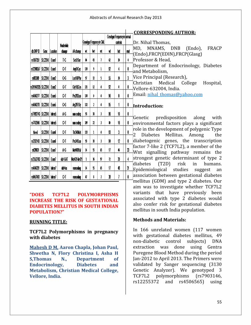
Abstracts of Annual Research Day 2013
55
“DOES TCF7L2 POLYMORPHISMS INCREASE THE RISK OF GESTATIONAL DIABETES MELLITUS IN SOUTH INDIAN POPULATION?”
RUNNING TITLE:
TCF7L2 Polymorphisms in pregnancy with diabetes
Mahesh D M, Aaron Chapla, Johan Paul, Shwetha N, Flory Christina I, Asha H S,Thomas N., Department of Endocrinology, Diabetes and Metabolism, Christian Medical College, Vellore, India.
CORRESPONDING AUTHOR:
Dr. Nihal Thomas, MD, MNAMS, DNB (Endo), FRACP (Endo),FRCP(EDIN),FRCP(Glasg) Professor & Head, Department of Endocrinology, Diabetes and Metabolism, Vice Principal (Research), Christian Medical College Hospital, Vellore-632004, India. Email: [email protected] Introduction: Genetic predisposition along with environmental factors plays a significant role in the development of polygenic Type 2 Diabetes Mellitus. Among the diabetogenic genes, the transcription factor 7-like 2 (TCF7L2), a member of the Wnt signalling pathway remains the strongest genetic determinant of type 2 diabetes (T2D) risk in humans. Epidemiological studies suggest an association between gestational diabetes mellitus (GDM) and type 2 diabetes. Our aim was to investigate whether TCF7L2 variants that have previously been associated with type 2 diabetes would also confer risk for gestational diabetes mellitus in south India population.
Methods and Materials:
In 166 unrelated women (117 women with gestational diabetes mellitus, 49 non-diabetic control subjects) DNA extraction was done using Gentra Puregene Blood Method during the period Jan-2012 to April 2013. The Primers were validated by Sanger sequencing (3130 Genetic Analyzer). We genotyped 3 TCF7L2 polymorphisms (rs7903146, rs12255372 and rs4506565) using

Abstracts of Annual Research Day 2013
56
TaqMan allelic discrimination assay. Data Analysis was done with SPSS 17, R
Results and Discussion:
The Common variants of TCF7L2 (rs7903146,rs12255372 and rs4506565) have been shown to be associated with diabetes mellitus in European (Cauchi et al. 2007) and Asian population (Chandak et al. 2006, Alami et al. 2012) [ OR=1.3-1.9] . The association of GDM with these TCF7L2 variants has varied in different populations, in Mexican Americans (OR=2.49 [95% CI 1.17–5.31]; P = 0.018), Arab women (OR=2.370, (95% of CI 1.010–5.563,p=0.047) and Scandinavian women(OR=1.49 [95% CI 1.28–1.75], p = 4.9 × 10⁻⁷ ). In a small south Indian population we have found the TCF7L2 polymorphism rs4506565 showed a strong trend towards association [O.R=3.75(C.I=0.75-18.53) P=0.08] with increased risk in the occurrence of gestational diabetes when compared to other two polymorphisms rs7903146 [O.R= 1.77 (C.I=0.503-6.263) P=0.37] and rs12255372 [O.R=1.40 (C.I=0.24-8.11) P=0.70].
Conclusion:
The TCF7L2 polymorphism rs4506565 showed a strong trend towards association when compared to other two common polymorphisms in TCF7L2 (rs7903146, rs12255372). This is the first preliminary data of TCF7L2 polymorphisms associated with Gestational diabetes in India. However, to confirm our findings further study needs to be performed in a large population.
References:
• Chandak, G R, A T Hattersley, C S Yajnik. “Common Variants in the
TCF7L2 Gene Are Strongly Associated with Type 2 Diabetes Mellitus in the Indian Population.” 2007;Diabetologia 50 (1) (January): 63–67
• Shaat, N, Lernmark, E. L. Groop et al. “A Variant in the Transcription Factor 7-like 2 (TCF7L2) Gene Is Associated with an Increased Risk of Gestational Diabetes Mellitus.” 2007; Diabetologia 50 (5) (March 7): 972–979
• Nobrega, Marcelo A. “TCF7L2 and Glucose Metabolism: Time to Look Beyond the Pancreas.” Diabetes 62 (3) (March1): 2013; 706–708.
“THE INVERSION 16 IN ACUTE MYELOID LEUKEMIA.”
Manoj M 1, Nancy A1, Sitaram U2,Ganapule A1, Abraham A1, Viswabandya A1, George B1, Mathews V1, Srivastava A1, Srivastava VM.3, Departments of 1Haematology, 2
Transfusion Medicine and Immunohaematology and 3
Cytogenetics Unit, Christian Medical College, Vellore.
Introduction:
Rearrangements of the core binding factor beta (CBFß) gene on chromosome 16, band q22 are associated with a good prognosis in acute myeloid leukemia (AML) 1. The inversion (16)(p13q22) is the typical abnormality in this group and results in the formation of a CBFß/MYH11 fusion gene 2.It accounts for ~ 4% of the AML-associated abnormalities and is usually associated with AML M4 with eosinophilia or M2 3. Trisomy 22 is the most common secondary change 4.

Abstracts of Annual Research Day 2013
57
Materials and methods: G banded karyotypes of all patients with the inversion 16 seen in the Department of Haematology between January 2003 and December 2012 were correlated with blood and bone marrow findings. Results: There were 23 patients with the inversion 16 among 1842 AMLs seen during this period (1.2 %). The median age was 33 years (range 1-58 years). There were 11 males. The median haemoglobin was 8.7g/dl (1.6-14.4 g/dl), the median WBC count, 64.9 × 10⁹/L (range 1-244.3 × 10⁹/L) and the median platelet count, 26×109/L (range 7 -192 × 109/L). The FAB subtypes were AML M4 (11 patients), M2 (six), AML not otherwise specified (four), M1 (two). The bone marrow blast percentage ranged from 5-91%. Abnormal eosinophils were seen in nine patients with AML M4 and M2 accounting for four each. The inversion 16 was solitary in 13 (57%), associated with a single additional abnormality in three (13%) and with two or more additional abnormalities in seven (30%).The common additional abnormalities were trisomy 8 (seven patients), trisomy 22 (four) and trisomy 9 (two). Conclusion: Our data is similar to the literature except for trisomy 8 being the most common secondary change. The inversion 16 can be overlooked unless the morphology of the metaphases is good. FISH analysis or molecular testing is helpful for confirmation of this abnormality,
especially if the bone marrow shows myelomonocytic leukemia or abnormal eosinophils or if a solitary trisomy 22 is present. References: 1.Delaunay, J., et al., Prognosis of inv(16)/t(16;16) acute myeloid leukemia (AML): a survey of 110 cases from the French AML Intergroup. Blood, 2003. 102(2): p. 462-9.
2. Liu P, Tarlé SA, Hajra A, Claxton DF, Marlton P, Freedman M, Siciliano MJ, Collins FS. Fusion between transcription factor CBF beta/PEBP2 beta and a myosin heavy chain in acute myeloid leukemia. Science. 1993 Aug 20;261(5124):1041-4. 3. Le Beau MM, Larson RA, Bitter MA, Vardiman JW, Golomb HM, Rowley JD. Association of an inversion of chromosome 16 with abnormal marrow eosinophils in acute myelomonocytic leukemia. A unique cytogenetic- clinicopathological association. N Engl J Med. 1983 Sep 15;309(11):630-6.
4.Xu, W., et al., Trisomy 22 as the sole abnormality is an important marker for the diagnosis of acute myeloid leukemia with inversion 16. Onkologie, 2008. 31 (8-9): p. 440-4. “PHYSIOLOGICAL COST INDEX DURING AMBULATION IN SPINAL CORD INJURED PERSONS AT DIFFERENT PHASES OF REHABILITATION-A PILOT STUDY.” Maria Stefy, Physical Medicine and Rehabilitation Centre, Christian Medical College, Vellore.
Background:

Abstracts of Annual Research Day 2013
58
Energy expenditure is an important parameter in the assessment of the orthotic gait in persons with locomotor disability. Currently, persons with T9 and below are trained to be ambulant with bilateral Knee ankle foot orthosis and elbow crutches. Macgregor combined the heart rate and speed of the locomotion to produce a single index called the Physiological Cost Index. The conventional method of measuring the Physiological Cost Index (PCI) is done by calculating the oxygen uptake. This method is difficult because of the unavailability of tools in the clinical setup. This study attempts to observe and analyze PCI values of the persons with T9-L1 paraplegia at two different stages of their rehabilitation process, i.e. during the mid-term when the person ambulates using reciprocal walker and bilateral KAFO and secondly at the time of discharge, when the person walks using bilateral KAFO and elbow crutches. Objectives: The objectives of this study are: 1. To find the PCI of persons with T9-LI
paraplegia with walking with reciprocal walker and bilateral KAFO at the midterm of the rehab process.
2. To find the PCI of the same persons walking with bilateral knee ankle foot orthosis and elbow crutch prior to their discharge.
3. To compare the PCI between the two phases of rehabilitation in the same group of participants.
4. To compare the obtained values of the PCI with the established normative data of PCI in non-injured persons as established in the literature.
Methodology:
Study design: Observational study Setting: Rehabilitation Institute CMC, Vellore Subjects: Persons with T9-L1 paraplegia cleared for ambulatory training. Procedure: Persons with T9-L1 paraplegia are selected consecutively. Physiological cost index using standard bed-side method computing the pulse rate and speed of walking is calculated at midterm phase of their rehabilitation. The participants are followed through their rehabilitation process and prior to their discharge a final PCI evaluation is done. These two PCI values are computed and analyzed for any correlation. The PCI values are compared with the established normative data of non injured people to analyze the difference. Variables:
1. Physiological cost index 2. Single neurological level (T9-L1)
Results: This study shows that there is no difference in the Physiological cost index between the orthotic (KAFO) reciprocal walking and orthotic (KAFO) walking with elbow crutches. There was a significant difference in the PCI between the established normative value for non-injured persons with that of the persons with SCI walking with KAFO and elbow crutches. Acronyms: KAFO – Knee Ankle Foot Orthotics PCI – Physiological Cost Index

Abstracts of Annual Research Day 2013
59
SCI – Spinal cord injury Key words: Paraplegia, Spinal cord injury, Physiological Cost Index, reciprocating walker, elbow crutch and knee ankle foot orthosis, energy consumption, ambulation.
“THE T(8;14) AND ITS VARIANTS IN ALL.”
Meenu Angi 3 ,Yuvarani S3 ,Sitaram U2, Nair S2,Ganapule A1, Abraham A1, Viswabandya A1, George B1, Mathews V1, Srivastava A1, Srivastava VM.3., Departments of 1Haematology, 2
Transfusion Medicine and Immunohaematology and 3
Cytogenetics Unit, Christian Medical College, Vellore. Introduction:
The t(8;14)(q24;q32) and its variants – the t(2;8)(p12;q24) and t(8;22)(q24;q11) are associated with B cell neoplasia, notably Burkitt lymphoma /leukemia (ALL-L3). These translocations involve the MYC gene on chromosome 8q24 and one of the immunoglobulin genes – IgH on 14q32, (the most common translocation), the IgK on 2p12 or IgL on 22q11 leading to the formation of a MYC/Ig fusion gene.1,2 Secondary changes (structural abnormalities of chromosome 1q and 13q3, trisomies 7 and 12) are seen in~ 60% of patients. 3, 4
Patients and methods:
G-banded karyotypes of all patients with ALL and the t (8; 14) and its variants seen in the Department of Haematology, Christian Medical College, Vellore,
between January 2003 and December 2012 were correlated with blood and bone marrow findings.
Results:
The translocations were seen in 26/1235 (2.1%) patients with ALL. The t(8;14) was seen in 19 patients(73%), t(8;22) in six (23%) and t(2;8) in one (4 %).
The majority (23, 88.4%) of these patients were males and adults (21, 80.7 %, median age 37 years, range 1-58). The median hemoglobin was 7.9 g/dl (range: 4.3-12.6); median WBC count, 9.6 x 10⁹/L (range: 2.1-245.8); median platelet count, 45x 109/L (range: 1.6- 243), and the median BM blast percentage, 78.5 (range 36-96%). ALL L3 was seen in 16, L2 in one, L1 in two and ALL not otherwise subtyped in seven. Six patients had extra medullary disease with involvement of nodes (four) or stomach or kidney (one each). Four had Burkitt lymphoma and two, high grade B cell lymphoma.
The majority (19/ 26, 73.1 %) of patients had complex karyotypes (two or more additional abnormalities). One had ALL-L3 and nodal high grade B cell lymphoma with both t(8;14 ) and t(14;18) – double- hit lymphoma.
Structural abnormalities of chromosome 1q accounted for 42% of the secondary abnormalities followed by deletions 9q, 11q (11.5% each) and 6q (7.7%), addition 13q34 (7.7%), trisomies 7 and 12 and deletion 17p (3.8% each).
Conclusion: The frequency of these translocations in our series is slightly lower than what has been reported (2% vs 3-5%). We also had

Abstracts of Annual Research Day 2013
60
a higher number of t (8; 22) – 23% vs 10%. The association with 1q abnormalities is similar to the literature while 13q3 abnormalities and trisomies 7 and 12 were seen in fewer patients (7.7 and 3.8 vs 15%). References: 1. Bilhou-Nabera C . t(8;14)(q24;q32); t(2;8)(p12;q24); t(8;22)(q24;q11). Atlas Genet Cytogenet Oncol Haematol. February 1999 . 2. M R Velangi, M M Reid, N Bown, et al. J Clin Pathol 2002 55: 591-595. 3. EG Boerma, R Siebert, PM Kluin and M Baudis. Leukemia (2009) 23, 225–234. 4. Johansson B, Mertens F, Mitelman F. Cytogenetic evolution patterns in non-Hodgkin’s lymphoma. Blood 1995;86:3905 “EXPRESSION OF IRON-RELATED PROTEINS IN THE DUODENUM IS UP-REGULATED IN PATIENTS WITH CHRONIC INFLAMMATORY DISORDERS.” Abitha Sukumarana, Jithu Jamesa, Harish PalletiJanardhana, Anita Amaladasa, Lekshmy Madathilazhikathu Suresha, Debashish Dandab, Visalakshi Jeyeseelanc, Balakrishnan Siddharth Ramakrishnad and Molly Jacoba., Departments of Biochemistrya, Rheumatology and Clinical Immunologyb, Biostatisticsc
andGastrointestinal Sciences, Christian Medical College, Vellore, Tamil Nadu, India. Mechanisms responsible for derangements in iron homeostasis in chronic inflammatory conditions are not
entirely clear. The aim of this study was to test the hypothesis that inflammation affects expression of iron-related proteins in the duodenum and monocytes in patients with chronic inflammatory disorders, thus contributing to dysregulated iron homeostasis. Duodenal mucosal samples and peripheral blood monocytes obtained from patients with chronic inflammatory disorders, viz. ulcerative colitis (UC), Crohn’s disease (CD) and rheumatoid arthritis (RA), were used for gene and protein expression studies. Haemoglobin levels were significantly lower and serum C-reactive protein (CRP) levels significantly higher in those in the disease groups. Gene expression of several iron-related proteins in the duodenum was significantly up-regulated in patients with UC and CD. In those with UC, it was found that protein expression of divalent metal transporter (DMT1) and ferroportin, which are involved in absorption of dietary non-heme iron, was also significantly higher in the duodenal mucosa. Gene expression of the duodenal proteins of interest correlated positively with one another and negatively with haemoglobin. Gene expression of iron-related proteins in monocytes was studied in patients with UC and found to be unaffected. In a separate group of patients with UC, serum hepcidin levels were found to be significantly lower than in control subjects. In conclusion, expression of iron-related proteins was up-regulated in the duodenum of patients with chronic inflammatory conditions in this study. The effects appeared to be secondary to anemia and the consequent erythropoietic drive. “RHYTHM GENERATING MECHANISMS IN ISOLATED RAT HEART.”

Abstracts of Annual Research Day 2013
61
Latha N., Swetha Raman, Kamalakannan.V., Anand Bhaskar and Sathya Subramani, Department of Physiology, Christian Medical College, Vellore. Introduction: In mammalian hearts, automaticity is initiated by the sinoatrial node. Funny current (If ) initiates the diastolic depolarization, which is enhanced by T-type calcium current (ICaT) and sodium calcium exchanger current (I NCX ). L-type calcium current (ICaL) is responsible for the action potential that ensues (1). These currents have been targets for development of anti arrhythmic drugs. Aim: To study the contribution of If , I NCX , ICaL and ICaT to rhythm generation in rat heart. Methods: Heart was isolated from an anaesthetized rat and perfused through the aorta with physiological salt solution in Langendorff mode for 15 minutes to stabilize the heart rate. Then it was perfused with test solutions and again reperfused with normal solution. Using ECG contact electrodes, surface electrocardiogram was recorded using a computerized data acquisition system (CMC Daq). Results:
Cesium (5mM) and Ivabradine (10μM) (blockers of If ), reduced heart rate by 27±5 % and 45±7 % respectively. With low sodium solution, the heart stopped completely. High calcium solution (5 mM) showed equivocal results with increase in
heart rate in some rats and decrease in others (n=3 each). With Verapamil and Diltiazem (both at 10μM, to reverse INCX and block ICaL), percentage reduction in heart rate was 70.14 ± 5.08% and 46.3±28.4% respectively. With Nifedipine (1 μM) (blocker of ICaL), heart rate reduced by 42.86± 5.18% and stopped at a higher dose of 10μM. With Nickel (40μM) (blocker of ICaT), the heart rate reduced only by 3.38±10.8%.
Conclusion:
Both ICaL and INCX seemed to be mandatory for initiation of pacemaker action potential unlike ICaT that might play a minor role. Ivabradine did not stop the heart even at the dose used which causes maximal blockade of If; therefore If is not mandatory for rhythm generation.
Reference:
1. Mangoni ME, Nargeot J. Genesis and regulation of the heart automaticity. Physiol. Rev. 2008 Jul;88(3):919–82.
“CHROMOSOME 14Q FLUORESCENCE IN-SITU HYBRIDISATION (FISH) ANALYSIS IN MENINGIOMAS.”
Noopur Gupta, Geeta Chacko, Ari G. Chacko, V. Rajshekhar, Jayaprakash Muliyil, Departments of Pathology, Community Medicine and Neurological Sciences, Christian Medical College, Vellore, Tamil Nadu. Background: Deletion of 14q has been reported in meningiomas and it is implicated in tumour progression. Aim:
Abstracts of Annual Research Day 2013
62
To correlate histopathological grading of meningiomas segregated into subgroups based on MIB-1 labelling index with deletion of chromosome 14q.
Methods:
46 cases were segregated into five categories based on the MIB-1 labelling indices (MIB-1 LI) of <5%, 5-7.5%, 7.5-9.9%, 10% and >10%. Immunohistochemical staining for MIB-1 was performed using a Ventana Benchmark XT Autostainer. Commercial FISH kit (Abbott Molecular: Vysis TelVysion) was used for detecting 14q deletion. The observer reporting 14q status was blinded to the MIB-1 LI. Subsequently, correlation between 14q deletion and MIB-1 LI was studied.
Results & Conclusion:
There were 21 WHO grade I, 24 grade II and 1 grade III meningiomas. There was a statistically significant difference between the mean duration of symptoms, average maximum dimension and the MIB-1 LI of grade I and grade II meningiomas. 33.3% grade I cases showed 14q deletion, when compared to 84% of grade II and III meningiomas. Amongst the histological parameters for diagnosis of atypical meningioma, hypercellularity, small cell formation, prominent nucleoli and sheet like growth were significantly associated with 14q deletion. All brain invasive meningiomas (n = 6) had 14q deletion. As MIB-1% increased, the prevalence of deletions was significantly higher. The mean MIB-1 of the 7 grade I meningiomas that had 14q deletions was 8.86±1.95% when compared to 4.14±1.35% for those without 14q deletions.
MIB-1 LI correlates with 14q deletion. Meningiomas with grade I histology have
a subgroup stratified by MIB-1 labelling index, with consistent 14q deletions. 14q deletion, being a molecular marker of tumour progression may predict the biological behaviour of histologically benign meningiomas.
“COMPARISON OF THE RADIOLOGICAL TISSUE PROPERTIES OF ANTHROPOMORPHIC PLASTINATED PHANTOM WITH ALDERSON RANDO PHANTOM.”
Pauline Shanthi *, I. Rabi Raja Singh **, Gibikote Sridhar ***, Suganthy Rabi * *Department of Anatomy, ** Department of Radiotherapy, *** Department of Radio diagnosis, Christian Medical College, Vellore.
Background:
Plastination is the art of preserving biological tissues with curable polymers. Phantoms are structures made of one or more tissue substitutes that respond to the radiation as similar as human body, used extensively in external beam radiotherapy. The Alderson Rando Phantom has been widely used for this purpose. Since plastination yields a stable specimen which shows repeatability and reproducibility and are also cost-effective, we propose to make phantoms using the ‘standard S-10 silicone’ plastination technique and study the radiological tissue properties of the plastinated phantom.
Aim:
To make plastinated phantom from cadaver and to validate it by comparing its radiological tissue properties with that of the Alderson Rando phantom.
Methods:
Abstracts of Annual Research Day 2013
63
An above-diaphragm specimen from a male cadaver and a below-diaphragm specimen from a female cadaver were plastinated using the standard S-10 plastination technique. CT imaging was carried out with PHILIPS CT BRILLIANCE 6 slice machine before and after plastination. These images of pre-plastinate, post-plastinate and Rando phantom were compared using an Eclipse 3D Treatment Planning System (VARIAN). The attenuation property of the plastinated head phantom was compared to that of the Alderson Rando head phantom by irradiating them with 200 cGy of cobalt-60 by using telecobalt machine (Th780C).
Results:
Plastinated phantoms obtained were dry, robust and durable. CT imaging of the plastinated phantom showed better anatomic detail of the organs than the pre-plastinate. However the CT numbers were increased in the plastinated phantom. The attenuation of the plastinated phantom was closer to the standard water phantom than that of the Rando phantom.
Conclusion:
Though the plastinated phantom had increased CT numbers because of the silicone, its attenuation property was closer to the standard water phantom than the Rando phantom. Nevertheless a better phantom has to be obtained by light-weight plastination if it has to be used for external beam radiotherapy. ________________________________________________
“THE DER(1;7)(Q10;P10) IN MYELOID NEOPLASIA.”
Poonam Jain1,S Yuvarani1,Usha Sitaram 2, Marie-Therese Manipadam4, Abhijeet Ganapule3,Aby Abraham3, Auro Viswabandya3,Biju George3,Vikram Mathews3,Alok Srivastava3,Vivi M. Srivastava1., Cytogenetics Unit1, Departments of Transfusion Medicine and Immunohaematology2, Haematology3 and Pathology4, Christian Medical College, Vellore, India. Introduction: The der(1;7)(q10;p10) is an unbalanced whole-arm translocation which results in gain of the long(q) arm of chromosome 1 and loss of chromosome 7q. This translocation is seen in myelodysplastic syndrome (MDS), acute myeloid leukemia (AML) and rarely, in myeloproliferative neoplasms and has been reported to be associated with eosinophilia, trilineage dysplasia and a previous history of chemo- or radio-therapy.1It usually occurs as a solitary abnormality. Associated abnormalities include trisomy 8 and, less commonly, trisomies 9 and 21 and deletion 20q.1-3
Patients and Methods:
G-banded karyotypes of all patients with the der(1;7)(q10;p10) seen between 2003 - 2013 in the Department of Hematology, Christian Medical College,Vellore were analysed and correlated with blood and bone marrow findings.
Results:
The der (1; 7) (q10; p10) was seen in nine patients (median age 52 years, range 26 - 68; male: female ratio, 3.5:1). The median haemoglobin was 7.9gm/dl (range: 4.8-13.5), median total leucocyte count, 3.8

Abstracts of Annual Research Day 2013
64
x109/L (range: 1.3-82.9), median platelet count, 40x109/L (range:3-154), and median peripheral blood eosinophil count, 1% (range 0-7%). Five had MDS; three, AML and one, myelofibrosis.The der (1; 7) accounted for 0.67% (5/741) of MDS and 0.16% (3/1842) of AML. All nine had dysmegakaryopoiesis, with bi- or tri-lineage dysplasia in two (22%) each. Reticulin was increased in seven patients.
Seven patients had additional cytogenetic abnormalities, with complex karyotypes (three or more abnormalities) in four. These included trisomies 8 (four cases), 9 and 21 (two each).
Conclusions:
Our findings are similar to the literature with respect to the male preponderance, the frequency of the der (1; 7) in AML and its association with trisomy 8. We noted differences with respect to the median age (52 versus 58-68 yrs), the frequency of the der (1; 7) in MDS (0.67% vs 1.5 – 6%), trilineage dysplasia (22%), additional abnormalities (77% vs 36 – 42%) and complex karyotypes (44% vs 12%) and the absence of deletion 20q and eosinophilia.
“COMPARISON OF TOTAL AND FREE PLASMA CONCENTRATION OF CISPLATIN FOLLOWING LONG AND SHORT DURATION INTRAVENOUS INFUSION : A PILOT STUDY.” Pradeep Rajkumar L A*, Dr.Binu S Mathew*, Dr. Saikat Das, Dr.Rajesh I, Dr. Subashini John, Dr.Ratna Prabha*, Dr.Denny H Fleming*, *Clinical Pharmacology Unit, Department of Radiation Oncology, Christian Medical College, Vellore-632004.
Background: Cisplatin and concurrent radiotherapy has improved survival over treatment with cisplatin or radiotherapy alone. Cisplatin has radiosensitizing properties and the best sensitization to the radiotherapy occurs when the blood drug concentration is higher. However, the optimal time sequence between the chemotherapy and radiation therapy is unclear
Aim:
Determination of total and free plasma concentration of cisplatin following long and short durations of intravenous infusion in patients diagnosed with head and neck or cervical malignancy. Methods: Patients were recruited into one of two groups based on their cisplatin infusion schedule, either the 1 hour or the 3 hour infusion regimen. On the day of cisplatin infusion, blood specimens were collected at 0.5, 1, 1.5, 2, 3 and 5hrs from the start of infusion in the 1 hr infusion group. For the 3hr infusion group specimens were collected at 2, 3, 3.5, 4, 6 and 24hrs from the start of infusion. Total and free cisplatin were measured using HPLC-UV. Results: The highest concentration of total and free cisplatin was achieved at the end of the infusion in both regimens. The decline in total cisplatin concentration at 30mins after the end of infusion in 1h regimen (1.5hr specimen) and in 3h regimen (3.5hr specimen) was 41.4% and 39% respectively, compared to the concentration at the end of the infusion.
Abstracts of Annual Research Day 2013
65
After 1hr in both regimens the free drug concentration declined by73.9% (1hr Infusion) and 70.8% (3hr infusion). However, the total drug, in both regimens had only declined by 41.9% (1hr Infusion) and 44.0% (3hr infusion). Conclusion: In conclusion, the ideal time to administer radiation therapy, based on the maximum concentration of total and free cisplatin, would be immediately at the end of infusion, in both regimens. A delay of 30 minutes causes a significant fall in the cisplatin concentration. “FREQUENCY OF BRAF MUTATION IN THYROID FINE-NEEDLE ASPIRATES AMONG INDIAN PATIENTS.” Prakash Gangadaran1,2, Rekha Pai1, Marie Therese Manipadam1, Deepak Abraham2, Pooja Ramakanth2, Simon Rajaratnam3, Nihal Thomas3, Paul MJ2., 1Department of Pathology, 2Department of Endocrine Surgery, 3Department of Endocrinology, Christian Medical College, Vellore, Tamil Nadu, India. Background: The BRAF V600E mutation is the most common genetic alteration. BRAF mutation activates the mitogen-activated protein kinase (MAPK/ERK) pathway and may confer an aggressive phenotype in PTC. Testing for BRAF mutation is clinically useful in providing prognostic prediction and facilitating accurate diagnosis of PTC in thyroid fine-needle aspirate (FNA) samples. It has also been proposed as a diagnostic adjunctive tool in evaluation of thyroid nodules with
indeterminate cytology findings. However, the frequency of BRAF mutations among Indian patients remains unclear. Aim: The aim of this study was to evaluate the BRAF mutational frequency in fine-needle aspirates obtained from thyroid nodules. Methods: In this study, we enrolled 136 patients from our hospital and BRAF mutation status was examined by direct sequencing on thyroid fine-needle aspirates and correlated with clinico-pathological parameters of the patients. Results: Out of 136 thyroid nodules tested, 12 cases were found to be malignant by routine cytological analysis and 7 cases had papillary thyroid cancer (58 %). Seventy one percent of patients with PTC were females. Out of 7 PTCs, 4 cases were positive for BRAF mutation (57 %); the incidence of the BRAF mutation did not differ between male and female. Three of the 4 PTC patients with BRAF mutation had a smaller tumor (3cm) when compared to those without mutations. Conclusions: The frequency of muations seen at our centre though higher than that reported from many western countries, is similar to that reported from a single other study from the Indian subcontinent which showed 53% mutation positivity.

Abstracts of Annual Research Day 2013
66
“ABSENCE OF G1528C MUTATION IN LONG-CHAIN 3-HYDROXYACYL-COA DEHYDROGENASE IN FOUR INDIAN PATIENTS WITH PREGNANCY-RELATED LIVER DISEASE.”
Raghupathy V, AshishGoel, KavithaThangaraj, Eapen C. E, Balasubramanian K. A; Annie Regi, Ruby Jose, Santosh J Benjamin, Anup Ramachandran
The human placenta is known to use fatty acids as a significant source of metabolic fuel during gestation and expresses enzymes involved in mitochondrial β oxidation. Fetal disorders of mitochondrial fatty acid β oxidation, especially deficiency of long chain 3-hydroxyacyl-coenzyme-A dehydrogenase (LCHAD) has been correlated with hepatic disorders such as pre-eclampsia, Acute Fatty Liver of Pregnancy (AFLP) and Hemolysis, Elevated Liver enzymes, Low Platelets (HELLP). The commonest identified mutation in Western populations is G1528C in exon 15 of the mitochondrial trifunctional protein (MTP) α subunit, which selectively decreases LCHAD activity. Though a few studies have pointed out the lack of this mutation in Chinese and Japanese populations, the status in the Indian population is unknown. As the genetic composition of the placenta is same as the fetus, we used the placental DNA to screen for the G1528C mutation by PCR-RFLP analysis and report its absence in 3 AFLP and 1 HELLP patient but identify a novel mutation in one of the AFLP patients.
“ASSESSMENT OF METABOLIC CHARACTERISTICS AND BODY COMPOSITION IN ASIAN INDIAN SUBJECTS WITH TYPE 1 DIABETES
MELLITUS USING EUGLYCEMICHYPERINSULINEMIC PANCREATIC CLAMP, DUAL ENERGY X-RAY ABSORPTIOMETRY SCAN AND 1-H-NUCLEAR MAGNETIC RESONANCE SPECTROSCOPYTECHNIQUES.” RUNNING TITLE: Metabolic characteristics and body composition in Asian Indian subjects with Type 1 Diabetes Mellitus
Riddhi Das Gupta, Ron Thomas Varghese, Veena Nair, Padmanabhan S, Mercy Inbakumari, Flory Christina, Roshan Livingstone1,H.S. Asha, Sylvia Kehlenbrink2,Sudha Koppaka2, Michelle Carey2, Meredith Hawkins2, Nihal Thomas, Department of Endocrinology, Diabetes and Metabolism, Christian Medical College, Vellore, India, 1Department of Radiodiagnosis, Christian Medical College, Vellore, India, 2Dept. of Endocrinology, Albert Einstein College of Medicine , New York, USA.
CORRESPONDING AUTHOR:
Dr. Nihal Thomas, MD, MNAMS, DNB (Endo), FRACP (Endo),FRCP(EDIN),FRCP(Glasg) Professor & Head, Department of Endocrinology, Diabetes and Metabolism, Vice Principal (Research), Christian Medical College Hospital, Vellore-632004, India. Email: [email protected] Introduction: Type 1 Diabetes Mellitus has a significant contribution to the global diabetes epidemic.However, the physiological mechanisms regulating insulin sensitivity

Abstracts of Annual Research Day 2013
67
and body composition in themremains elusive. Materials and methods:
14 male subjects with Type 1 Diabetes Mellituswere studied. Insulin sensitivity at the hepatic and peripheral tissueswas evaluated using a 6-hour EuglycemicHyperinsulinemicPancreatic Clamp.Body composition assessment included skin fold thickness , DEXA scan for total body fat %and 1H-NMR Spectroscopy forHepatocellular Lipid (HCL) and IntramyocellularLipid (IMCL) and ExtramyocellularLipid (EMCL) at the level of soleus muscle. The results were compared with 11subjects with Type 2 Diabetes Mellitus, 5 lean subjects with diabetes (BMI ≤ 19 kg/m2) and9age and BMI-matched non-diabetic subjects.
Results and analysis: The M-value, an index of whole body insulin sensitivity, was lowerfor subjects with Type 1 Diabetes as compared to the non diabetic group (13.82 vs 22.28µmol/kg/min) .The Rate of Disappearance of glucose (Rd)an estimate of peripheral insulin sensitivity,was higher in Type 1 Diabetes subjects as compared to those with Type 2 Diabetes (8.23 vs 4.60 mg/kg/min). Endogenous Glucose Production (EGP), an index of hepatic insulin sensitivity, was similar in Type 1 and Type 2 Diabetes subjects (0.42 vs 0.43 mg/kg/min). In the Type 1 Diabetes group, M value correlated significantly with Weight, BMI and subscapular skinfold thickness (p < 0.05), Rd correlated with M value, BMI,Waist circumference, subscapular skinfold thickness and Total body fat % (p < 0.05), while EGP correlated significantly with HCL. The IMCL correlated with age,
weight, body surface area, and hip circumference, while EMCL correlated with waist hip ratio and basal metabolic rate (p < 0.05). Conclusions: Subjects with Type 1 Diabetes Mellituswere insulin resistant as compared to matched non- diabetic subjects and as insulin resistant at the hepatic level as subjects with Type 2 Diabetes. The Intramyocellular and ExtramyocellularLipid weremarkers for subcutaneous and visceral fat respectively in these subjects. The lean diabetic group were significantly more resistant than the Type 1 Diabetes group at both the hepatic and peripheral levels.
“ESSENTIALITY OF ARGININE AND HISTIDINE IN HUMANS – A CRITICAL APPRAISAL OF AVAILABLE EVIDENCE.” Prakash S. S, Arthi T. S*, Jagadish R*., Department of Biochemistry, Christian Medical College, Vellore, India. *Contributed equally Background:
Essentiality of amino acids from a nutritional point of view is addressed by its effect on nitrogen balance and other biochemical parameters. Based on this, ten amino acids are classified as essential amino acids which need to be supplied in diet. Of these, arginine and histidine are reported to be semi-essential in some cases.
Aim:
To ascertain the essentiality of arginine and histidine based on available evidence.

Abstracts of Annual Research Day 2013
68
Methods:
We initially collected evidence from all the available latest editions of textbooks of biochemistry in our institutional library on whether arginine and histidine are listed as essential or semi-essential. Besides, we also referred to electronic databases such as PUBMED on the same topic.
Results:
According to the foreign author textbooks, histidine was listed as an essential amino acid. Arginine was listed as conditionally essential in some and essential in others. In all the Indian author textbooks, both histidine and arginine were listed as semi-essential amino acids. The discrepancy was resolved by our findings from the published literature. It was found that arginine can be synthesized in our body as an intermediate in the urea cycle, and is sufficient for adults. However, arginine supplements are found to be useful in some clinical conditions. In children, especially in pre-term infants arginine is an essential nutrient. Histidine cannot be synthesized in humans as they lack the enzymes involved in its biosynthesis. A histidine-deficient diet for longer periods (about 7 days) has shown to alter nitrogen balance. Histidine is thus an essential amino acid in humans.
Conclusion:
Both arginine and histidine should be regarded as essential amino acids. Arginine becomes conditionally essential in some cases. This study emphasizes the need for critically analyzing the existing facts to ascertain the right information.
Acknowledgements:
We would like to thank Dr. Molly Jacob and Dr. Premila Abraham for their valuable inputs. We would also like to appreciate the efforts of some of the students of MBBS batch of 2013 who searched the literature and submitted their reports.
“STUDY OF THE KINETICS OF CYTOMEGALOVIRUS (CMV) MRNA LEVELS IN PATIENTS WITH CMV REACTIVATION FOLLOWING BONE MARROW TRANSPLANTATION.”
Sangeeta Susan Thomas, Rayaz Ahmed, Santhosh Kumar D, Rajesh Kannangai, Biju George, Asha Mary Abraham, Department of Clinical Virology and Department of Hematology, Christian Medical College, Vellore.
Background:
Cytomegalovirus infections form the major viral opportunistic infection in bone marrow transplant patients that manifest in the early post-transplant period. CMV mRNA levels were quantified as a marker of reactivation in the post-transplant period. Aim: To quantitate the CMV mRNA levels in blood samples of bone marrow transplant patients using real time reverse transcriptase PCR (RT-qPCR) in the post-transplant period.
Methods:
One hundred and sixty two bone marrow transplant patients were enrolled in the study and 2467 EDTA blood samples were collected from them. Peripheral blood mononuclear cells were separated from the samples using Ficoll-Hypaque and stored in RNA Later solution at -80ºC.

Abstracts of Annual Research Day 2013
69
Of 162 patients recruited into the study, 35 (21.6%) showed CMV reactivation as determined clinically and by CMV DNA levels. Of the 35, only 25 patients had the complete set of samples collected and they were tested to obtain serial CMV DNA levels in the post transplant period. Of these 25 patients, samples from 15 patients were preliminarily tested for one splice variant of CMV Immediate Early (IE) mRNA using RT-qPCR. Results: Of the 15 patients tested preliminarily, 7(46.6%) tested positive for CMV mRNA.The samples from patients with declining levels of CMV DNA tested negative for CMV mRNA.The mean CMV DNA value of patients who were CMV mRNA positive was higher than those who were CMV mRNA negative. Conclusion: Testing for CMV IE-mRNA may be used as a marker of CMV reactivation. “EXPRESSION PROFILING OF NUCLEAR HORMONE RECEPTORS IN MYELOID LEUKEMIA REVEALS POTENTIAL.” Novel Drug Targets for Combination Therapy Sreeja Karathedath*, Sukanya Ganesan*, Wei Zhang, Ajay Abraham, Savitha Varatharajan, Bharathi, M.R, Shaji R.V, Alok Srivastava, Vikram Mathews, Poonkuzhali Balasubramanian, Department of Haematology, Christian Medical College, Vellore, India & 2Department of Pediatrics,University of Illinois, Chicago, USA.
Nuclear Hormone Receptors (NHRs) are a large superfamily of ligand dependent transcription factors regulating a plethora of genes involved in metabolism, growth and differentiation. Recent studies identified the overwhelming role played by NHR in determining hematopoiesis as well as HSC function and fate, suggesting that defective proliferation and maturation of progenitors in hematopoietic malignancies could be attributed to altered expression of these transcriptional regulators. NHRs are attractive but relatively unexplored drug targets and there is limited data on their expression profile in hematological malignancies. Our preliminary screening of AML cell lines and primary cells revealed differential expression of PPARg and RXRα among AML cell lines and decreased expression of these targets in resistant cell lines vs. sensitive ones (Fig.1a). We extended this study aiming to identify the expression pattern of all NHRs in myeloid malignancies, compare with ex-vivo chemoresistance and prognostic markers.Myeloid leukemia cell lines AML(n=12) carrying AML-ETO, Inv-16, PML-RAR fusion transcript, NPM1 or FLT3 mutation and CML(n=8), pooled primary samples harboring only FLT3 ITD mutation and only NPM1 mutation, CD34+ cells and total cellular RNA samples from normal donors were included in the study. Bone marrow samples from AML patients and peripheral blood samples from healthy volunteers were collected after informed consent. PBSC from normal donors (n=7) were enriched for CD34+ cells by magnetic enrichment. RNA extraction followed by purification and cDNA synthesis were done as per manufacturer’s recommendation. The

Abstracts of Annual Research Day 2013
70
expression of 42 NHRs and 42 co-regulators was profiled by qRT PCR using RT2 PCR Profiler array (SABiosciences, Germany). Expression of each gene was normalized to the average of 5 housekeeping genes (GAPDH, b−actin, b2-microglobulin, HGPRT & RPLP0) and differential expression was calculated by normalizing this with normal total RNA samples to determine fold change. Data normalization and differential gene expression were computed using SABiosciences web-based analysis software. Ex vivo cytotoxicity to cytarabine and daunorubicin was analyzed using MTT assay and IC50 was calculated using Adapt software. Based on Ara-C and Dnr IC50, AML samples were categorized as sensitive or resistant (IC50 <6.25μM and >6.25μM for Ara-C; <0.5μM and >0.5μM for Dnr). FLT3-ITDand NPM1 mutations were screened using PCR followed by GeneScan methods. Upon analysis we observed steroid receptors except PPARG, NR5A1, RXRG, NR1D2, glucocorticoid receptors, androgen receptors and their co-regulatory molecules were expressed across all cell lines. In contrast, expression of certain NHRs such as NR2E3, NR1I2, NR1H4, NR0B1, NR0B2, ESRRG, ESRRB, NR1D1 and the co-regulator HDAC7 were less or at undetectable levels, while estrogen receptors and its co-regulatory molecules except PPARGC1B, steroid receptors HNF4A, NR2F1, NR2F2, RARG and NR3C2 were moderately expressed across the cell lines (Fig.1b). Analysis also revealed significantly reduced expression of ESR2, ESRRG, NR2F2, NR2F6 and THRB and increased PPARG in AML compared to CML cell lines. Comparison between CD34+ progenitor population and well differentiated hematopoietic cells demonstrated >6 fold change in NR4A1 expression. Likewise, expression of
several differentiation markers NR1H3, NR1D2, RORA, RORB, RARA and VDR were elevated in cells with increased degree of maturation. The receptor inducing terminal differentiation of erythrocytes THRA, had lesser expression (<60 Fold) in erythroleukemic cell lines K562, HEL and LAMA-84 compared to normal (Fig.1c). In Ara-C resistant cell lines, expression of NR1I3, AHR, and HNF4A were up-regulated while PPARG and RXRA were down-regulated. NPM1 positive cell lines and patient samples had increased expression of PPARG, PPARGC1A, NOTCH2, NR1H3 and NR2F2 than the FLT3 mutated group (Fig.1d) and similar expression of PPARG and RXRA was validated in NPM mutated primary AML blasts (Fig.1e). Our comprehensive analysis of NHRs and its co regulatory molecules provides us with insights on NHR expression on differentiation and drug resistance across myeloid leukemia cell lines with various genetic and molecular characteristics. This could be further explored to identify potential novel drug targets to be used as combination therapy in myeloid leukemias to overcome drug resistance.
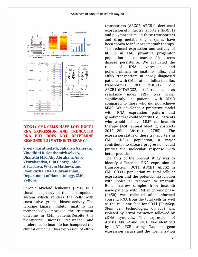
Abstracts of Annual Research Day 2013
71
“CD34+ CML CELLS HAVE LOW HOCT1 RNA EXPRESSION AND TRUNCATED RNA BUT DOES NOT DETERMINE RESPONSE TO IMATINIB THERAPY.”
Sreeja Karathedath, Sukanya Ganesan, Vinodhini K, Senthamizhselvi A, Bharathi M.R, Aby Abraham, Auro Viswabandya, Biju George, Alok Srivastava, Vikram Mathews and Poonkuzhali Balasubramanian, Department of Haematology, CMC, Vellore.
Chronic Myeloid leukemia (CML) is a clonal malignancy of the hematopoietic system which renders the cells with constitutive tyrosine kinase activity. The tyrosine kinase inhibitor imatinib has tremendously improved the treatment outcome in CML patients.Despite this therapeutic success, resistance and intolerance to imatinib has hampered the clinical outcome. Overexpression of efflux
transporters (ABCG2, ABCB1), decreased expression of influx transporters (hOCT1) and polymorphisms in these transporters and drug metabolizing enzymes have been shown to influence imatinib therapy. The reduced expression and activity of hOCT1 in CML primitive progenitor population is also a marker of long term disease persistence. We evaluated the role of RNA expression and polymorphisms in imatinib influx and efflux transporters in newly diagnosed patients with CML; ratio of influx to efflux transporters: dCt hOCT1/ dCt ABCB1*dCTABCG2, referred to as resistance index (RI), was lower significantly in patients with MMR compared to those who did not achieve MMR. We developed a predictive model with RNA expression pattern and genotype that could identify CML patients who would achieve MMR on imatinib therapy (ASH annual Meeting abstracts 2012-120: Abstract 2785). The expression status of these transporters in CML CD34+ population, a major contributor to disease progression, could predict the molecular response with better precision. The aims of the present study was to identify differential RNA expression of transporters hOCT1, ABCB1, ABCG2 in CML CD34+ population vs. total cellular expression and the potential association with molecular response to imatinib. Bone marrow samples from imatinib naïve patients with CML in chronic phase (n=50) was collected after informed consent. RNA from the total cells as well as the cells enriched for CD34 (EasySep, Stem cell technologies, Canada) was isolated by Trizol extraction followed by cDNA synthesis. The expression of ABCB1, ABCG2 and hOCT1 was identified by qRT PCR using Taqman gene expression assays and the normalization

Abstracts of Annual Research Day 2013
72
was done against GAPDH. The expression of these genes in CD34+ and total CML cells was compared. There was a significant increased expression of efflux transporters ABCG2 (p=0.001) and ABCB1 (p=0.007) and decreased expression of hOCT1 (p=<0.001) in CD34+ fraction compared to the total cellular RNA. This reduction in OCT-1 mRNA was also observed in CD34+ cells derived from healthy donors (data not shown). To determinewhether these parameters may be predictive of clinical responsesto IM, RNA expression of these transporters in the CD34+ cells was compared with molecular response to imatinib therapy. There was no significant association between RNA expression of these transporters and molecular response, though the number of samples for which the CD34+ cell fraction was available was very less (n=50). We further tested if the decreased expression of hOCT1 in CD34+ fraction was due to the presence of short transcript of hOCT1 resulting in decreased functional activity and found that it is indeed true. We are currently evaluating the gene expression profiles of CD34+ fractions from patients with good or poor response to imatinib therapy to predict a molecular signature of imatinib response in CML. “ASSOCIATION OF PLASMA AND INTRACELLULAR IMATINIB LEVELS WITH MOLECULAR RESPONSE IN PATIENTS WITH CML.” Sukanya Ganesan, Preetha Markose, Savitha Varatharajan, Ezhilpavai Mohanan, Vinodhini Kumaraswamy, Senthamizhselvi Anandan, Auro Viswabandya, Biju George, Alok
Srivastava, Vikram Mathews, Poonkuzhali Balasubramanian, Department of Haematology, Christian Medical College, Vellore. Targeted therapy with Imatinib mesylate, has revolutionized the treatment response and survival in patients with chronic myeloid leukemia (CML). Imatinib steady-state plasma levels, measured following the first month of treatment with the standard 400-mg/day dose, has been shown to be correlated with long-term complete cytogenetic and molecular responses, as well as long-term event free survival (EFS). There is limited report from India on the association between imatinib levels and molecular response. We aimed to determine correlation of plasma imatinib and its metabolite desmethyl imatinib levels as well as intracellular imatinib levels (after ex-vivo incubation of primary CML cells with imatinib) with molecular response in CML.
Imatinib naïve CML patients (n=135) treated with standard dose of imatinib with serial follow-up post therapy were included in this study [Chronic phase (n=117), Accelerated phase (n=12), Blast crisis (n=6)]. After obtaining informed consent, peripheral blood samples were collected from patients 24 hours post first imatinib dose and on Day 29 (steady state). Imatinib and desmethyl imatinib levels were measured using HPLC with UV detection. Molecular response to imatinib was measured by RQ-PCR for BCR-ABL fusion transcript from peripheral blood samples collected once in three months with a minimum follow-up of 12 months post therapy. The results were expressed in the International Scale (Poonkuzhali et al, Acta Haematologica, 2012).

Abstracts of Annual Research Day 2013
73
The median plasma concentration of imatinib and desmethyl imatinib at 24 hours was 593ng/mL (74-2177) and 86ng/mL (12-609) while on Day 29 it was 1072ng/mL (106-3694) and 206ng/mL (31-613) respectively. Based on therapeutic response after 12 months, patients were divided into complete molecular response [CMR (n=11)], major molecular response [MMR (n=48)], no molecular response (n=39); in addition, patients were classified as intolerant (n=26) and those with disease progression (n=11). The plasma imatinib and Desmethyl imatinib concentration (24 hours) was significantly higher in patients who achieved CMR/MMR when compared to those with who did not. (P=0.02) Intracellular imatinib level did not show significant association with achievement of MMR/CMR. Number of patients who achieved CMR/MMR had significant association with high plasma imatinib concentration suggesting that higher the level of plasma imatinib concentration, better the probability of attaining response. Though the number of patients included is small, this study suggests that monitoring plasma could help predict treatment outcome as well as timely dose increase in imatinib to improve clinical response.
“BENIDIPINE, AN L-TYPE AND T-TYPE CALCIUM CHANNEL BLOCKER, INHIBITS SPONTANEOUS CONTRACTION OF ISOLATED CAPRINE URETER.” Sumith K. Mathew, Girish S Naik, Jacob Peedicayil, Department of Pharmacology and Clinical Pharmacology, Christian Medical College, Vellore.
Objectives: To study the effect of benidipine on the frequency and amplitude of spontaneous contraction of distal caprine (goat) ureter. To determine the EC50 and dose-response relationship of benidipine on distal caprine ureter. Materials and methods: The effect of an L- type and T- type calcium channel blocker benidipine, in inhibiting the spontaneous contractions of distal segments of caprine ureter was studied using a physiograph. The effects of different concentrations of benidipine hydrochloride were assessed in 54 specimens. We analyzed the effect of drug on spontaneous contraction of ureters over time using Repeated measures ANOVA and plotted a dose response curve to determine the EC50 of benidipine hydrochloride in inhibiting spontaneous contractions using DRC package in software R. Results: There was significant inhibition of spontaneous contractions over time when we used 6 × 10 -8 M, 3 × 10 -7M, 10 -6 M, 3 × 10 -6M, and 10 -5 M concentrations of benidipine. There was no significant inhibition of contractions in the control arm. The EC50 of benidipine in inhibiting the spontaneous contraction of distal caprine ureter over 30 minutes was 47.2 × 10 -6 M with 95% confidence interval of 12.12 × 10 -9 M – 82.26 × 10 -6 M. Conclusion: Benidipine inhibits the spontaneous contractions of ureter significantly at clinically achievable plasma

Abstracts of Annual Research Day 2013
74
concentrations. Frequency of contractions were less inhibited compared to amplitude of contractions especially at lower test drug concentrations, suggesting the role of T- type calcium channels in modulating the frequency of spontaneous ureteric contractions. “EFFECT OF POLYMORPHISMS IN CYP1A2*1C, CYP1A2*1F AND TAQ1A ON CLINICAL RESPONSE TO CLOZAPINE IN PATIENTS WITH TREATMENT-RESISTANT SCHIZOPHRENIA (TRS) – A PRELIMINARY REPORT.” Susan Philip1, Veeramanikandan R2, Lakshmikirupa S1, Anto P Rajkumar3, C Chitra1, Anju Kuruvilla1, Alok Srivastava4, B Poonkuzhali4, Molly Jacob2 and K S Jacob1, 1Department of Psychiatry, Christian Medical College, Vellore, India; 2Department of Biochemistry, Christian Medical College, Vellore, India; 3Centre for Psychiatric Research, Aarhus University Hospital, Risskov-8240, Denmark; 4Department of Hematology, Christian Medical College, Vellore, India.
Background and Aim:
Clinical response to clozapine, used in treatment-resistant schizophrenia (TRS), is often variable. The present study evaluated whether polymorphisms in genes that metabolize clozapine (CYP1A2*1F and CYP1A2*1C) and regulate dopamine synthesis (Taq1A) affected clinical response to, serum levels and adverse effects of clozapine in such patients. Methodology:
Patients diagnosed to have TRS by standard criteria were the subjects of the study. Their premorbid functioning, responses to traumatic events, cognitive status and extent of disability were assessed. DNA was isolated from blood from these subjects. Polymorphisms in the genes of interest and serum clozapine levels were determined. Associations between the genotypes and clinical response, serum levels and adverse effects of clozapine were determined using appropriate statistical tests. Results: The 45 patients recruited consisted of 31 males and 14 females, with a mean age of 32.28 years (±10.44). Scores of the subjects, using the Brief Psychiatric Rating Scale (BPRS) for assessment of psychopathology, ranged from 18 to 147 with a mean of 52.44 (±23.35). Subjects were grouped as responders (BPRS score ≤ 35) and non-responders (BPRS score > 35). The Taq1A polymorphism showed significant association with response to clozapine; the heterozygous genotype (A/B) occurred more frequently in responders than wild type and homozygous genotypes (OR 9.0; CI 1.0 – 80.0; p 0.045). However, this association was not seen when age and gender were adjusted for. CYP1A2*1C and CYP1A2*1F polymorphisms did not show significant associations with response to clozapine. None of the polymorphisms influenced serum levels of clozapine or occurrence of adverse effects. Conclusion: Polymorphisms in Taq1A, but not CYP1A2*1C and CYP1A2*1F, showed a tendency to influence clinical response to

Abstracts of Annual Research Day 2013
75
clozapine. None of them affected serum levels or adverse effects of the drug. This is an on-going study and further analysis with larger sample sizes will be necessary before definitive conclusions can be drawn. “IS THE NON FLUORESCENT FRACTION OF CLEISTANTHUS COLLINUS TOXIC?” Swetha Raman, Soosai Manickam, Sathya Subramani, Department of Physiology, Christian Medical College,Vellore. Cleistanthus Collinus (Oduvanthazhai) is a poisonous shrub and our larger aim is to delineate the toxic compounds in it. Excluding non-toxic fractions and focusing on just the toxic fractions is one of our approaches in defining the toxicity of the plant. All fluorescent fractions in the aqueous extract could be sequestered by partitioning it with chloroform. Its known toxins, Cleistanthin A and B which are fluorescent, are toxic as seen in previous studies. The aim here was to assess the toxicity of its non fluorescent fraction (NFF) of the boiled extract. Objectives: (a) To assess if the non-fluorescent fraction of C.collinus causes death in within 8 hours after intraperitoneal administration. (b) To monitor arterial blood gases, electrolytes, respiration, ECG, blood pressure, urine pH in test animals and compare it with controls. Methods: Carotid artery of anaesthetized wistar rats was cannulated. Blood pressure, ECG
and respiratory rate were recorded using CMC data acquisition system. Regular arterial samples were taken to estimate electrolytes and blood gases. If the pre intervention blood parameters satisfied published inclusion criteria, the animals were randomized into test (NFF-Oduvan) or control (Drumstick-processed similarly) groups and received the respective extracts intraperitoneally at a dose of 20 mg/100gBW. Results & Conclusion: 20% mortality in test and 0% in controls (n=5 each) was recorded. Normal electrolyte levels without altered pH was observed (both). Respiration and intra arterial pressure remained normal (both), but associated with a progressive tachycardia in test (p=0.00). Peculiarly, there was hyperoxia (p=0.00) with hypercapnia (both). The blood gases did not seem to satisfy the limits of the alveolar gas equation. To conclude, NFF fraction seemed to be non-toxic, which has to be substantiated by testing for a longer period with a larger sample size. For clinical relevance, oral gavaging must be the mode of administration rather than intraperitoneal. “VARIEGATED EXPRESSION OF IMPRINTED GENES IN MOUSE INDUCED PLURIPOTENT STEM CELLS.” Syed Mohammed Musheer Aalam, Kannan V Manian, Sumitha P Bharathan, Alok Srivastava, Shaji R Velayudhan, Centre for Stem Cell Research and Department of Haematology, Christian Medical College, Vellore, India.
Abstracts of Annual Research Day 2013
76
Most of the generated mouse induced pluripotent stem cells (iPSC) clones show embryonic stem cell (ESC) like features of expression of pluripotency genes and the ability for in vitro and in vivo differentiation. However, many clones do not support the full term development of tetraploid-complemented mice. Recent studies have shown that transcripts present in the imprinted Dlk1-Dio3 gene cluster on chromosome 12qF1 are aberrantly silenced in most of the iPSC clones which causes poor contribution to chimeras. Decreased expression of these imprinted genes has been attributed variously to sub optimal reprogramming factor stoichiometry and loss of active chromatin marks or hypermethylation of this locus. Addition of Vitamin C and downregulation of Dnmt3a/3b have been shown to help in maintaining the normal imprinting of the genes in this locus. It has also been proposed that Klf4 and Oct4 regulate the expression of these imprinted genes and the overexpression of Klf4 and Oct4 causes their normal expression. We estimated the expression levels of Gtl2, Rian and Mirg in the Dlk1-Dio3 imprinted cluster in various mouse iPSC clones in our laboratory to test whether it can be used as an effective and quick method to assess the pluripotency of mouse iPSC clones generated using retroviral transduction of four factors, Oct4, Sox2, Klf4 and c-Myc (OSKM) or three factors, Oct4, Sox2 and Klf4 (OSK). We included mRFP1 for transduction to monitor retroviral transgene silencing, which occurs in pluripotent clones. The transgene silenced (RFP-) clones (n=7) had activated expression of endogenous pluripotency genes Oct4, Sox2, Klf4 and Nanog to the levels similar to those in R1ESCs while RFP+ clones (n=8) maintained high expression levels of all
the transgenes and had no induction of endogenous pluripotency genes except for Oct4. The RFP- Nanog+ clones showed variegated expression of Rian, Gtl2, Mirg; 2 out of 4 OSKM clones had high levels of Gtl2 and Mirg expression but lacked the expression of Rian and the remaining two clones lacked the expression of all three imprinted genes. Out of the 3 OSK clones, 2 clones expressed optimal levels of Rian and Mirg, but they lacked expression of Gtl2 and the other one expressed only Rian. In 7 out of 8 RFP+ clones, the imprinted genes were silenced and in one of them only Gtl2 was expressed. We could not find any correlation between expression of Klf4 and Dlk1-Dio3 cluster genes. We measured the expression levels of Hdac1 to 11 and the results showed that Hdac7 is repressed in R1ES cells and RFP- iPSCs but is highly expressed in RFP+ iPSCs and mouse embryonic fibroblasts (MEFs) and in contrast, Hdac1 was found to be repressed in MEFs and activated at comparable levels in R1ESCs and in both RFP- and RFP+ iPSC colonies. However, there was no correlation between the expression levels of Hdac1 or Hdac7 and the imprinted genes. These results suggest that downregulation of Hdac7 is important for reprogramming, but not sufficient to activate the expression of imprinted genes in iPSC clones. VPA causes increase in acetylation of imprinted genes, but not by directly modulating the levels of Hdac1 and Hdac7. We conclude that there is heterogeneity in the expression of Dlk1-Dio3 cluster genes and they are not transcriptionally co-regulated and different epigenetic mechanisms may control the individual expression of these genes. A thorough understanding of the factors responsible for the activation of these genes will help us tailor strategies

Abstracts of Annual Research Day 2013
77
to obtain fully pluripotent mouse iPSC clones.
“MOLECULAR BASIS OF (ΔΒ)0 THALASSAEMIA AND HPFH IN INDIAN POPULATION AND THE ROLE OF NON-CODING TRANSCRIPTS IN INCREASING FOETAL HAEMOGLOBIN IN ADULTS.” Thiyagaraj Mayuranathan1, Janakiram Rayabaram1, Reena Das,2 Neeraj Arora1, Eunice Sindhuvi Edison1, Mammen Chandy3, Alok Srivastava1, Shaji R Velayudhan1. 1Department of Haematology, Christian Medical College, Vellore, Tamil Nadu., 2Postgraduate Institute of Medical Education and Research, Chandigarh., 3Tata Medical Centre, Kolkata.
Hereditary persistence of foetal haemoglobin (HPFH) and ( ) 0-thalassaemia are conditions caused by large deletions of - and -globin genes and they are characterized by high foetal haemoglobin (HbF) in heterozygotes of these mutations. Complete characterization of these deletions is important for understanding the transcriptional and epigenetic mechanisms involved in increase in HbF levels by these mutations. Previous studies demonstrated that these large deletions remove the regulatory sequences which suppress γ-globin gene expression in adults and increase the interaction of long range enhancers with γ-globin promoter since δ- and β-globin genes are deleted. However, the exact mechanism for elevated HbF in these conditions and differences in HbF levels and phenotypes between ( ) 0-
thalassaemia and HPFH still remains unclear. We screened 90 individuals from 51 families by gap-PCR for previously reported common deletions and by MLPA and quantitative fluorescent multiplex -PCR (QFM-PCR) for new deletions in the β-globin cluster. After identification of the approximate locations of the deletions by MLPA and QFM-PCR, we developed PCR strategies to amplify across the breakpoints and the products were sequenced to characterize the breakpoints. We identified six different deletions in Indian population. The frequencies of the previously reported common mutations, Asian inversion deletion and HPFH-3, were 46.66% and 24.44%, respectively. The rare mutations which were not found earlier in Indian population include 49.3kb deletion (12.22%) and 32.6kb deletion (10%). We found 2 novel deletions, 49.98kb deletion and 86.7kb deletion, which accounted 5.55% and 1.11%, respectively. The breakpoints of the 49.98kb deletion were found to be very close to those of HPFH-3 and this mutation was found in compound heterozygous with β-thalassaemia in a 8 year old boy with thalassaemia intermedia (Hb=8.8g/dL). Four heterozygotes in the family had mean MCV = 79.7fL (range: 76-85.6), mean HbF%= 26.9% (range: 23.3-33.0) and pancellular distribution of HbF suggesting that this mutation causes HPFH. The 86.7kb region encompassing A-, -and - globin genes was found in heterozygous state in one individual and it also had haematological parameters of HPFH (MCV=80.8fL and HbF=32.2%). To develop an experimental model to study the molecular mechanisms for elevated HbF in these conditions we established ex-vivo erythropoiesis system with the peripheral blood mononuclear cells

Abstracts of Annual Research Day 2013
78
obtained from patients with Asian inversion deletion and HPFH-3. Quantitative real time PCR (qPCR) showed increased expression of γ-globin transcripts in the cultured erythroid cells and chromatin immunoprecipitation (ChIP) of RNA Polymerase II (RNAPII) also showed increased transcriptional activation of γ-globin gene in the erythroid cells obtained from the patients with these mutations. To understand the roles of non-coding transcripts at the 5’ and 3’ breakpoints of the deletions in the transcriptional activation of γ-globin, we performed qPCR with the cDNA obtained from cultured erythroid cells using the primers that bind upstream and downstream of these breakpoints. The results showed that the level of BGL3, the noncoding transcript present near the 5’breakpoint, is enhanced by these deletions. We found activation of a novel non-coding transcript at the 3’ breakpoints and they were absent in the erythroid cells of normal individuals. ChIP also showed significant binding of RNAPII at the genomic regions from where these non-coding transcripts are produced. The role of non-coding transcripts has not been reported earlier as a mechanism for increased γ-globin transcription in HPFH and ( ) 0-thalassaemia. It is possible that these deletions remove the genetic elements that cause transcription repression of γ-globin gene in normal adults and they also cause activation of non-coding transcripts which change the epigenetic status of γ-globin gene resulting in its active transcription. To conclude, using QFM-PCRs we could perform a comprehensive study of ( )0-thalassaemia and HPFH in Indian population and detected rare and novel deletions that cause these conditions. Using ex-vivo erythropoiesis system
which mimicked the pattern of globin gene expression in normal individuals and patients with globin gene mutations, we could identify the role of noncoding transcripts in increased HbF expression in the patients with HPFH and ( ) 0-thalassaemia. “AUDIT OF TEXT BOOK INFORMATION ABOUT PACEMAKER MECHANISMS, WITH SPECIAL REFERENCE TO IF AND INCX .” Abirami. V, Sathya Subramani
Background:
Current debate on the fundamentals of pacemaker mechanism of heart revolves around two ionic currents, If and INCX. If
(funny current) is a hyperpolarisation activated depolarising current that plays a vital role in pacemaker mechanism. Recent data suggests a significant role for INCX (sodium calcium exchanger current) as a pacemaker current. Studies from our laboratory also show that INCX is mandatory for pacemaker activity.
In view of this we undertook to review textbook information on pacemaker mechanisms and hence this poster.
Aim:
To catalogue information on pacemaker mechanism of heart from various Physiology text books used by medical students and faculty in our institution
Objective:
To assess specifically whether the text books chosen for the audit mention If and INCX in pacemaker mechanisms
Methods:

Abstracts of Annual Research Day 2013
79
12 Physiology textbooks were chosen based on popular usage by medical students in our institution. Information on pacemaker currents (with special reference to If and INCX currents) was entered into a table.
Results:
50% of the textbooks listed in the table mention the role of If. Only 17% of text books mention about INCX, which is now evolving as an important pacemaker current.
Conclusion:
Majority of Physiology textbooks do not describe the major pacemaker mechanisms in heart.
“MOTORIZED TENODESIS HAND ORTHOSIS FOR CERVICAL LEVEL SPINAL CORD INJURY PATIENTS.” Vinil T.C Spinal Cord injury leads to partial or complete paralysis of di_er-ent motor functions. When partial paralysis of upper limb occurs in patients due cervical level 5 spinal cord injury, their activities of daily living is a_ected. To remedy this problem a light weight, low pro_le motorized tenodesis splint has been designed to restore prehension ability in these patients. The actu-ating mechanism involving a four bar linkage driven by a servo motor. The linkage from the motor is attached to the wrist using a cu_. By _exing wrist around 30 degree, internal _exor tendon will be stretched and thereby the digits will come closer, creating just the right movement to be able to grasp objects without consciously moving _ngers. E_ciency of this action can
be improved by reducing the length of the _exor tendon by surgical correction. PG Trainee ( MS Bioengineering), Dept of Bioengineering, CMC Vellore. E-mail address: [email protected]
“THE EFFECT OF DIFFERENT LOADING DOSES OF PHENOBARBITONE ON SERUM PHENOBARBITONE LEVELS IN NEONATES.”
Vinod Pallaparthy, Atanu Kumar Jana, Santhanam Sridhar, Manish Kumar, Anil Kuruvilla, Niranjan Thomas, Department of Neonatology, Christian Medical College, Vellore.
Background:
Using the current recommendation for phenobarbitone dosing (loading dose at 20-10-10 mg/kg), we have noted that babies are extremely sedated with elevated phenobarbitone levels. We postulate that in asphyxiated babies, damage to the liver impairs drug metabolism and renal damage affects drug elimination, thus elevating serum levels. Hypothermia can also affect the drug levels.
Objective:
To determine the serum levels of the phenobarbitone in babies receiving different loading doses of phenobarbitone for neonatal seizures and to study the effect of asphyxia and therapeutic hypothermia on drug levels.
Design:
Prospective observational Cohort study
Setting:
Tertiary care perinatal center.

Abstracts of Annual Research Day 2013
80
Material and Methods:
Term neonates with seizures of any cause were given phenobarbitone 20mg/kg as a loading dose followed by additional doses of 10mg/kg upto a maximum of 40mg/kg. Maintenance dose of 5mg/kg in two divided doses was started after 24 hours of loading dose. Serum phenobarbitone levels were asessed after 4 hours of the initial loading dose and the subsequent values at 24, 48, and 72 hours from the time when the baby received the maximum loading dose.
Results:
A total of 28 babies completed the study. Fourteen (50%) received 20 mg/kg, 8 (28.6%) received 30 mg/kg and 6 (21.4%) received 40 mg/kg loading dose. Asphyxia was the cause of seizures in 19 (67.9%) of which 7 underwent therapeutic hypothermia. The mean phenobarbitone levels were 30.5±11.4, 44±18.4, 47.4±20.8 and 53.8±23.5 mcg/ml at 4, 24, 48 and 72 hours of age respectively. The mean phenobarbitone level at 72 hours was 45.4±31.5 mcg/ml when 20 mg/k was used as loading dose as compared to 51.2±12.4 and 55.6±15.2 mcg/ml when loading doses of 30 and 40 mg/kg were used. 83.3% of babies who had received 40 mg/kg loading dose had phenobarbitone levels more than the therapeutic range at 72 hours compared to 57.1% of those who received 20 m/kg. The mean phenobarbitone levels at 72 hours were higher in asphyxiated babies who underwent therapeutic hypothermia as compared to those with other causes (61.2±7.4 vs 44.2±3 mcg/ml).
Conclusion:
Using the current recommended loading dose of upto 40 mg/kg may lead to supra
therapeutic phenobarbitone levels in a majority of babies, especially those undergoing therapeutic hypothermia. Loading doses beyond 20 mg/kg should be used with close monitoring of serum levels.
Key words:
Phenobarbitone; Serum levels; Asphyxia; Therapeutic hypothermia.
“PHENOTYPIC AND MOLECULAR CHARACTERIZATION OF VANCOMYCIN RESISTANT ENTEROCOCCI (VRE).” Baby Abirami S¹, Suganya D¹, Dr. Balaji V¹., ¹ Department of Clinical Microbiology. Introduction: Vancomycin-resistant Enterococci (VRE) infections are the cause of nosocomial outbreak which remains an amendable challenge for treatment. Acquired glycopeptide resistance in Enterococcus species is due to the acquisition of vanA, vanB, vanD, vanE, and vanG genes This study highlights the VRE resistance profile which was further confirmed by multiplex PCR detecting VanA and VanB gene to estimate the prevalence rate and to formulate antimicrobial therapy for treatment. Aim: To determine the presence of van A and vanB gene which confers glycopeptide resistance in Enterococcus spp. Materials and methods:

Abstracts of Annual Research Day 2013
81
A total of 70 VRE isolates were included in this study. Screening for vancomycin resistance was done by Kirby Bauer Disc Diffusion method and thereby confirmed with E-test. Molecular identification of VanA and VanB gene were done by PCR. Results: Out of 70 VRE isolates, 87% of the isolates were E.faecalis and remaining 13% of the isolates were E.faecium. The isolates were 0% susceptible to vancomycin and 1% to teicoplanin. 92% to daptomycin and 93% to linezolid .This study shows an overall prevalence of VanA gene (100%) and none of them had VanB gene (0%). Conclusion: Emerging VRE complicate the clinical situations further. Hence implicate the necessity for routine screening and identification of VRE. Molecular methods will provides information on geographical prevalence of resistant strains.
“ANTIBIOTIC SUSCEPTIBILITY PROFILES OF GRAM NEGATIVE BACILLI (GNB) ISOLATED FROM INTRA ABDOMINAL INFECTIONS (IAIS) COLLECTED ACROSS INDIA.” Agila Kumari Pragasam1, Yamuna Devi B1 Balaji V1, Shalini Anandan1, Department of Clinical Microbiology, Christian Medical College, Vellore. Introduction:
A variety of antimicrobial agents are recommended for the treatment of IAIs but utility of many of these has become restricted, over time owing to increasing
resistance rates. The Study for Monitoring Antimicrobial Resistance Trends (SMART) surveillance program monitors the susceptibility of GNB from IAIs to ertapenem and other comparator agents.
Objectives:
To monitor the in vitro susceptibility of GNB to Ertapenem and other antimicrobial agents. Materials and Methods: A total of 703 isolates derived from IAIs were collected across India from a period of 2010 – 2012. All isolates were identified up-to species level and tested by Micro broth dilution method as per CLSI guidelines
Results: A total of 330, 167, 110, 30, 29, 27 and 10 isolates of E.coli, Klebsiella, Pseudomonas, Enterobacter, Proteus, Citrobacter and Morganella were collected during 2010-2012. Ertapenem and Imipenem activity towards E.coli, Klebsiella, Pseudomonas, Enterobacter, Proteus, Citrobacter and Morganella spp shows >80 % susceptibility. Whereas the other comparator agents MIC for 90% of tested strains varied considerably. The remaining data will be included during the presentation.
Conclusion: The regional rates of Extended Spectrum Beta Lactamases (ESBL) positive isolates ranged from 68 %, 56 % in 2010 increasing to 70%, 60% in 2012 for E. coli and K. pneumoniae respectively. Of the nine antimicrobial agents tested, only amikacin, ertapenem and Imipenem consistently exhibited percent susceptibilities of ≥80% for all 3 years of the study. However fluctuations in the

Abstracts of Annual Research Day 2013
82
susceptibility to other antibiotics were also observed.
“UNDERSTANDING THE ANTIBIOGRAM OF MOST FREQUENT RESPIRATORY PATHOGENS.”
Jones Lionel Kumar D1, GEETHA PRIYA D1, DR. BALAJI V1, DR. SHALINI ANANDAN1, Department of Clinical Microbiology, Christian Medical College & Hospital, Vellore.
Introduction:
The empiric management of community-acquired respiratory tract infections has been complicated by the emergence of high rates of antimicrobial resistance. Major causative agents likeStreptococcus pneumoniae,Haemophilusinfluenzae, Moraxella catarrhalis and Streptococcus pyogenes , have developed multiple resistance mechanisms to combat the effects of most commonly used classes of antibiotics, particularly the beta-lactams and macrolides. This study helps to understand the susceptibility profiles of pathogens involved in community-acquired respiratory tract infections that can help in treatment and also reduce the spread of resistance.
Objectives:
To understand the antibiotic susceptibility profile of most frequent pathogens involved in community-acquired respiratory tract infections.
Materials and Methods:
Susceptibility was evaluated against a range of antimicrobial agents using Epsilometrytest andKirby-Bauer disc diffusion method as per the CLSI & EUCAST guidelines and the production of β-lactamase was tested using a nitrocefin-based test.
Results:
A total of 89 isolates of S. pneumoniae, 32 isolates of H. influenzae, 19 isolates of S.pyogenes and (11)M.cattarhalis were collected largely from patients with indications of CA-RTI from January - October 2013. S. pneumoniae isolates were >90% susceptible to Levofloxacin, Penicillin, Amoxicillin/clavulanate and Ofloxacin while 82% were non resistant to Azithromycin and Co-trimoxazole. Similarly >95% S.pyogeneswere susceptible to Penicillin and 5% resistant to Levofloxacin, 100% resistant to Azithromycin, Co-trimoxazole and Clarithromycin(74%). H. influenzae showed the highest percent susceptibility to Cephalosporins(100%), Levofloxacin(94%). 87.5% of these isolates were susceptible to Amoxicillin/clavulanate, Ampicillin and Azithromycin, 84.3% to Ofloxacin, 81% to Ciprofloxacin, while 78% were resistant to Co-trimoxazole and 69% to Clarithromycin. M. cattarhaliswere all susceptible toAzithromycin and Cefixime and completely resistant to cefuroxime. Overall, the percentage of H. influenzae andM. cattarhalis isolates producing β-lactamase was 12.5% and 100%, respectively.
Conclusions:
In our study all these CA-RTI isolates shows highest susceptibility to β-lactams and fluroquinolones when compared to

Abstracts of Annual Research Day 2013
83
macrolides. Β-lactams may continue to be considered options for non complicated CA-RTI.
“SEROTYPE DISTRIBUTION AND ANTIMICROBIAL RESISTANCE IN STREPTOCOCCUS PNEUMONIAE CAUSING INVASIVE DISEASE IN INDIAN CHILDREN.” Ranjith J1, Rosemol Varghese1, Dr. V. Balaji1, Dr. V. P. Verghese2, Dr. Kurien thomas3, 1Department of Microbiology, Christian Medical College and Hospital, Vellore, India, 2Department of Child Health Unit, Christian Medical College and Hospital, Vellore, India, 3Department of General Medicine, Christian Medical College and Hospital, Vellore, India.
Introduction: Streptococcus pneumoniae causes significant morbidity and mortality worldwide; particularly in childrens less than five years. Antibiotic resistance in S.pneumonia is also a growing concern. Surveillance on pneumococcal serotypes and antibiotic resistance is necessary to understand the epidemiology of pneumococcal diseases and formulating appropriate treatment options. Here we present data on S.pneumoniaeinfections in Indian children, with response to its serotype distribution and antimicrobial susceptibility profile. Aim: To determine the serotype distribution and antimicrobial resistance profile for S.pneumoniae in Indian children
Materials and Methods: A prospective analysis of all Invasive S.pneumoniae isolates from children less than five years of age, during 2007 – 2013 June was made. S.pneumoniae were confirmed by traditional methods. Isolates were then serogrouped and typed by Co-agglutination technique and reconfirmed by multiplex PCR. Results: The prevalent serotypes causing invasive infections in children less than five years of age are 14, 19F, 5, 6A and 6B. 96.4% of the isolates were non-susceptible to Cotrimoxazole, and 30% were non-susceptible to Erythromycin. 5.2% of the isolates were non-susceptible to Penicillin; only 0.8% was non-susceptible to Cefotaxime. Conclusion: Streptococcus pneumoniae causes significant morbidity and mortality in Indian children. Understanding the epidemiology of pneumococcal infections in relation to serotypes and antibiotic resistance trends will help to implement pneumococcal disease preventive measures.
“BASELINE MINIMUM INHIBITORY CONCENTRATION (MIC) OF SULBACTAM FOR ACINETOBACTER BAUMANII-CALCOACETICUS COMPLEX (ABCC) AND SYNERGISM WITH CARBAPENEM.”
Sangeetha Rajenderan1, Dr. Shakti Laishram1, Dr. Shalini Anandan1, Dr.
Abstracts of Annual Research Day 2013
84
Balaji. V1, Department of Microbiology, Christian Medical College, Vellore.
Introduction:
Carbepenem are often the drugs of choice for the treatment of infections due to multidrug-resistant ABCC. Due to emerging resistance to carbapenems the treatment options are now limited only to colistin and tigecycline which is not approved for infections other than skin and soft tissue and abdominal infection. Various drug combinations used for these carbapenem isolates often without laboratory verification of efficacy of the combination against the isolate. Aim: To determine MIC for sulbactam and in vitro synergistic activity of sulbactam in combination with meropenem and colistin against ABCC Materials and Methods: Thirty carbapenems resistant ABCC isolates from respiratory and blood sample was included in the study. MICs for sulbactam and meropenem were determined by micro broth dilution method as per CLSI guidelines. Synergism between Sulbactam and meropenem was detected by the checkerboard (CB) and time-kill assay (TKA) at a sub-inhibitory concentration of ½ MIC. Results: All the isolates showing MIC (≥8µg/mL) to either meropenem or sulbactam. The checkerboard method with the combination of meropenem and sulbactam demonstrated40% synergism and 60% indifference (lowest FIC
index).Combination of meropenem and sulbactam may show synergism for ABCC isolates. Conclusion: TKA detected synergism more than the checkerboard assay. High level of synergism (77%), bactericidal effect (73%) and the lack of antagonism seen with combination of meropenem and sulbactam in both CB and TKA are an encouraging outcome with possible use of combination of these drugs for the treatment of carbapenem resistant Acinetobacter infection. “COMPARISON OF PNEUMOCOCCAL SEROTYPES IN ITS DISEASE POTENTIAL IN INDIAN ADULTS.” Suganya D¹, Baby Abirami S¹, Dr.Balaji V¹, ¹ Department of Clinical Microbiology Introduction: Streptococcus pneumoniae is a major cause of bacterial infections with significant morbidity and mortality worldwide. WHO estimates approximately 1.6 million deaths annually, in young children and elderly. Serotype based vaccines are effective in preventing pneumococcal diseases. Since there is epidemiological variation in capsular types, serosurveillance is necessary to determine the impact of available vaccines . In our study we compared the seroprevalence of bacteremic and non bacteremic pneumococcal isolates in adults. Aim: To determine the seroprevalence of Streptococcus .pneumoniae causing diseases in Indian adults.

Abstracts of Annual Research Day 2013
85
Materials and methods: A total of 145 S. pneumoniae invasive (CSF, Blood) and 148 non invasive isolates (sputum) collected during 2011 to 2013 were included in this study. Phenotypic identification were done by conventional methods. Serotyping of isolates were done by co-agglutination method and confirmed by molecular typing (PCR). Result:
Most prevalent serotypes found in invasive isolates are 1(16%), 19F(13%), 23F(5%), 5(5%), 4(3%). Pattern of seroprevalence observed with non-invasive isolates are 19F (30%), 6B (15%) and 14(13%). Hence this study elucidate the most common prevalence of serotypes (1, 6B, 19F, 14 followed by 23F, 5 and 4) encountered in invasive and non-invasive pneumococcal infection.
Conclusion:
The prevalence of serotypes varies between invasive and non-invasive isolates. The epidemiology of Streptococcus pneumoniae varies with potency of disease which is illustrated in our study. Continues surveillance is essential to understand the pneumococcal disease and formulating vaccine strategies.
“EMERGING ANTIBIOTIC RESISTANCE AMONG GRAM NEGATIVE PATHOGENS CAUSING URINARY TRACT INFECTION.”
Yamuna Devi1, Agila Kumari1, Dr. V. Balaji1, 1Department of Clinical Microbiology, Christian Medical College, Vellore.
Introduction: Development of antimicrobial resistance among Gram-negative pathogens is becoming progressively increasing and associate with the dangerous noscomial outbreak. E. coli is the most common etiologic gram-negative organism, which is followed in descending order of frequency by P. aeruginosa, Klebsiella spp, Enterobacter spp, and A. baumannii. Multiresistant strains are particularly problematic. Objectives: To estimate the species prevalence among the pathogens causing urinary tract infection and also to determine antibiotic susceptibility pattern of significant pathogens isolated from urinary tract infection. Materials and methods: Urinary isolates collected during the period (2010-2012) and identified up to species level by conventional methods and its antibiotic susceptibility pattern against 15 different antibiotics were determined by micro-broth dilution method (Pre-coated antibiotic panels from Merck), interpretation was carried out according to CLSI guidelines. Result: ESBL producing E.coli and Klebsiella spp showed promising activity to amikacin ertapenem, imipenem with 90%, 93%, 97% and 80%, 80%, 97% respectively. Indeed ciprofloxacin and levofloxacin showed poor activity against ESBL producing E.coli (6% & 9%) and Klebsiella (13% and 39%). P.aeruginosa exhibits with the susceptibility profile of

Abstracts of Annual Research Day 2013
86
53%, 47%, 50%, and 50% to amikacin, ceftazidime, imipenem and Piperacillin/Tazobactam. 100% of resistance to ertapenem was observed P.aeruginosa isolates. A.baumannii showed a peak resistant profile against all the tested antibiotics Conclusion: Routine screening of isolates for ESBL and Carbapenemase detection is necessary to formulate appropriate antibiotic therapy. “EVALUATION OF THE CAVIDI EXAVIR VERSION 3.0 REVERSE TRANSCRIPTASE(RT) ASSAY FOR THE MEASUREMENT OF HIV-1 AND HIV-2 PLASMA VIRAL LOAD. “ Priyadarshini A Padaki, Jaiprasath Sachithanandham, Veena V Ramalingam, Rita Isaac, O C Abraham, Susanne A Pulimood, Rajesh Kannangai, Departments of Clinical Virology, RUHSA, Internal Medicine and Dermatology, Christian Medical College, Vellore, Tamil Nadu-632004 . Background: There is a need for simpler less expensive HIV viral load (VL) assays for the monitoring of HIV infected individuals. Aim: To evaluate ExaVir version 3.0 assay for the estimation of RT activity(equivalent plasma viral load) in HIV infected individuals. Materials and methods:
Samples from 75 HIV-1, 13 HIV-2, 20 HIV negative and an additional 50 HIV-1 treatment experienced individuals were tested. ExaVir is an ELISA format to quantitate the RT activity in plasma (fg/ml) and gives corresponding VL (copies/ml) with the help of the software provided. The lower detection limit was 200 copies/ml. For HIV-1, values were compared with VL measured by real-time PCR (Artus Qiagen,Hilden,Germany) and discordant samples or the ones with log10 difference of >0.5 were tested with another assay(Abbott,Illinois,U.S.A). For HIV-2 samples, ExaVir VL was correlated with CD4 counts. The ExaVir and Artus real time PCR VL values in HIV-1 infected treatment naïve individuals were compared with CDC staging for HIV. Results: All the HIV negative samples were undetectable. Bland Altman analysis showed a bias of 0.1log between Artus and ExaVir (n=66, r=0.63, p<0.001) and 0.2 log between Abbott and ExaVir (n=32, r=0.81,p<0.001) with samples having detectable VL. The sensitivity of the assay was 77.6% and 82.5% when compared to Artus and Abbott respectively for samples with VL >200copies/ml. The same for samples with VL >1000copies/ml, was 86.8% and 94.1% respectively. There was a significant negative correlation (rho= -0.83, p=0.0039) between ExaVir HIV-2 VL and CD4 count. There was no significant correlation observed between CDC clinical staging and ExaVir as well as Artus real time PCR VL values. Conclusion:
The ExaVir version 3 assay has very good specificity and has a better sensitivity compared to its previous versions and

Abstracts of Annual Research Day 2013
87
hence can be used in resource limited settings for the monitoring of HIV infected individuals including HIV-2. “ANTIMICROBIAL RESISTANCE IN BLOOD CULTURE ISOLATES- A TERTIARY CARE EXPERIENCE OVER 3 YEARS (2010 TO 2012).” Dr. Marilyn Mary Ninan, Dr. Shakti Laishram , Dr. Shalini Anandan, Dr. V. Balaji, Clinical Microbiology, Christian Medical College , Vellore. Introduction: Blood stream infections are an important cause of mortality and morbidity - studying the resistance profile will help treatment. Objectives: To describe the temporal trend of resistant bacterial isolates (MRSA, ESBL, CRO and VRE) in blood cultures. Materials and Methods: All positive blood cultures during 2010–2012 were evaluated retrospectively. Results: Amongst the total Staphylococcus isolates, a fall in isolates of MRSA over 3 years( 36% to 33%)with an upward Vancomycin MIC creep. Of 2259 E. coli isolates over 3 years, a fall in the ESBL isolates from 77% (2010) , to 71%(2011), to 65%(2012). Of 1228 Klebsiella isolates over 3 years, the ESBL isolates have fallen from 61%(2010) to 51% (2011) and 53% (2012). Of 2259 , E. coli isolates over the 3 years, the CRO
isolates have shown a slight rise, from 3%(2010) , to 4% (2011) and 4%(2012). Of 1228 Klebsiella isolates, CRO have peaked , from 25%(2010) to 40% (2011) and fallen to 25% (2012). Of the total 601 enterococci isolates, the VRE have risen from 9% (2010) to 17.6%(2011) and then fallen to 11.8%(2012). Conclusions: This study provides information on antibiotic resistance profile of blood isolates. It clearly illustrates howimproved antimicrobial stewardship and good hospital control practices can help curb drug resistance. “IMPROVED ADENO-ASSOCIATED VIRUS (AAV) VECTORS FOR HEPATIC GENE THERAPY.” Dwaipayan Sen1*, Akshaya Krishnagopal2*,Rupali A Gadkari3, Govindarajan Sudha3,Vaani Roshini1, Ijaz Mahamadulla2, Narayanaswamy Srinivasan3, Alok Srivastava1,2, Giridhara R Jayandharan1,2*., 1Department of Hematology, 2Centre for Stem Cell Research, Christian Medical College, Vellore, Tamil Nadu, 3Molecular Biophysics unit, Indian Institute of Science, Bengaluru, India.
* Both authors contributed equally.
Background:
Recombinant adeno-associated virus (AAV) vectors based on serotype -8 have shown significant promise for hepatic gene therapy of hemophilia B. However, the theme of vector dose dependent immunotoxicity seen with AAV2 vectors earlier has re-emerged with AAV8 vectors as well. We had previously reported that

Abstracts of Annual Research Day 2013
88
novel AAV2 vectors bioengineered at specific (S)/threonine (T) alanine (A)or lysine (K) arginine (R) on its capsidhave enhanced potential for liver directed gene transfer (Gabriel et al, Hum Gene Ther Methods, 2013). Similar proof-of-concept mutagenesis of homologous residues in the AAV8 serotype vectors improved coagulation factor IX expression with concomitant reduction in inflammatory and neutralizing antibody response invivo (Sen et al, Hum Gene Ther Methods, 2013).In the present studies, we investigated if further modifications of capsid motifs identified by computational analysis on AAV8 can improve the efficiency of hepatic gene therapy. Results: Extensive computational analysis was carried out toscrutinize other potential sites on the AAV8 capsid (PDB 2qa0), which would generate efficient novel vectors. The study was done with respect to the inclusion of specific motifs/aminoacidsin phosphodegrons (phosphorylation sites recognized as degradation initiation signals by ubiquitinligases), they being either phosphosites or ubiquitination sites, or their participation in the protein-protein interactions of the capsid proteins. Inclusion of these positions in the antigenic segments on capsid proteins and/or in the receptor binding regions wasalso investigated.Based on data generated from this analysis we strategically mutated the following sites, K143, K652, S149, S156, T138, T654 on AAV8 capsid. To test the efficiency of these novel vectors,we delivered luciferase gene under the control of chicken β-actin promoter (5x10¹⁰ vgs per mouse, i.v) into 8-12 weeks old male
C57BL/6 mice. Hepatic gene expression was analyzed 2 and 5 weeks after gene transfer in a small-animal imaging system.As seen in Fig. 1 A-C, the S156A and double mutant (S671A+K137R) vectors showed increased luciferase expression (2.5-4 fold Vs WT-AAV8) in the liver 2- weeks post gene transfer. Interestingly, at a longer follow-up (5 weeks) a systemic distribution of luciferase gene expression was noted for K143R, K652R, S149A and T138A vectors. This was further corroborated by real time PCR analysis of vector copy numbers in organs like kidney (K143R- 4, K652R- 8, S149A- 3, T138A- 1.6 Vs WT-AAV8- 0.67 vector copies/diploid genome) and spleen (K143R- 1.4, K652R- 14, S149A- 6, T138A- 2.7 Vs WT-AAV8- 0.3 vector genome copies/diploid genome). Conclusions: Our data suggests that alteration of AAV8 phosphosites based on in silico prediction alters the phenotype of this promising gene therapy vector. Further on-going studies with these vectors expressing human coagulation factor IX in murine models, will demonstrate the feasibility of the use of these novel vectors for potential gene therapy of hemophilia B. Figure 1: Hepatic gene expression of various novel AAV8 vectors 2 weeks and 5 weeks post vector administration (A). Quantitation of data from A is presented in photons/sec, 2 weeks (B) and 5 weeks (C) post vector administration.

Abstracts of Annual Research Day 2013
89
WT-AAV8
K143R
2 weeks 5 weeks
K652R
S149A
S156A
T138A
T654A
K137R+S671A
AB
C
02468
101214
To
tal F
lux X
10^6 (
ph
oto
ns/s
ec) 2 weeks
-10
-5
0
5
10
15
20
25
30
To
tal F
lux X
10^6 (
ph
oto
ns/s
ec)
5 weeks*
*
* *
“CLINICAL PROFILE OF 60 CHILDREN WITH CYSTIC FIBROSIS.”
D. Accurate Mercy, Sneha Varkki, Sumita Danda*, Department of Paediatrics and Department of Clinical Genetics*, Christian Medical College and Hospital, Vellore,Tamil Nadu, 632004.
Objectives:
To document the clinical profile of children with cystic fibrosis (CF) attending a tertiary care centre in South India.
Methods:
Retrospective chart review of all patients diagnosed to have cystic fibrosis was done. Diagnosis of cystic fibrosis was made by clinical features and elevated sweat chloride level > 60meq/l. Few children who had borderline sweat chloride level (>50meq/l), and suggestive clinical features of CF or one CFTR mutation with classical CF features were also included in this study.
Sweat collection and analysis was done by WESCOR macroduct sweat collection system by pilocarpine iontophoresis. Clinical features were documented using a structured proforma. Mutation analysis for four common mutations were done on a subgroup of children by ARMS (Amplication Refractory mutation system) and verified by DNA sequencing.
Results:
Total number of children included in the study were sixty of which 36 (60%) were males. 35 (58%) reported symptom onset in infancy, but only 20 (33%) were diagnosed before the age of 5 years. In 4 (7%) diagnosis was made only 10yrs after reported symptom onset. 1/3rd were given empirical anti tuberculosis treatment , based on their symptoms before the diagnosis of CF. There were 3 families with more than one child affected. However prenatal genetic counseling was offered to none of them. Majority 37(62%) of the children in our study were from southern Indian states. Sweat chloride level varied from 50 -120 meq/l. 10(17%) children had values above 100meq/l. Genetic mutation analysis was done on a subgroup of 41(68%) subjects, 3 (5%) subjects had F508del mutation –homozygous and 6(10%) had the same mutation in one copy only. We also looked for G551D,

Abstracts of Annual Research Day 2013
90
G542X, 621G2T mutations but none were present in this population. Common clinical manifestations at the time of presentation: Recurrent or persistent pneumonia in 51 (85%), failure to thrive in 37 (62%), steatorrhoea and malabsorption symptoms in 15 (25%) and history of meconium ileus in 1 (2%) were the symptoms at presentation. History of salty taste on kissing was present in 3(5%). 34 (57%) had bronchiectsis or severe lung disease at the time of presentation. Nasal polyposis was the only symptom in 2 children. 18 (30%) of these children had pseudomonas species isolated from airway secretion cultures. Conclusion:
We report here clinical profile of 60 children diagnosed and followed up in our institution. Majority of children had advanced lung disease at the time of diagnosis. There are differences in the clinical and genetic profile of patients reported from different parts of India. It is important to increase awareness about the condition in order to diagnose and initiate treatment early and to avoid unnecessary anti tuberculosis treatment. It is also important to give prenatal genetic counseling to parents and offer prenatal diagnosis where possible.
“EFFECT OF MEDICINE BALL TRAINING IN IMPROVING SITTING BALANCE IN PERSONS WITH T1 TOT12 SPINAL CORD INJURY.”
Annu T, Physical Medicine and Rehabilitation, Christian Medical College, Vellore.
Study design: Randomized controlled trial
Objectives:
To evaluate the effectiveness of medicine ball training in sitting balance in persons with thoracic level spinal cord injury. Setting:
Rehabilitation Institute, PMR Department, Christian Medical College, Bagayam, Vellore.
Background:
Spinal cord injury is a condition where motor, sensory, and autonomic systems are affected. Unsupported sitting is an important component in spinal cord injury patients to carry out most of their activities of daily living. Sitting balance can be improved by task specific training and goal based exercises etc. Medicine ball exercise is a new intervention which can be used to improve muscle strength, muscle power, co-ordination, balance, speed etc. In this study we are trying to find how effective the medicine ball exercise was in high sitting balance in person with spinal cord injury level T1 to T12. Methodology: Persons with thoracic level spinal cord injury (T1 to T12) satisfying the inclusion criteria are selected and randomly allocated into control and experimental group by computer generated random allocation method. The primary investigator does the pre and post intervention assessment and is blinded to the grouping and interventions. The co-primary investigator does the intervention for persons in control and

Abstracts of Annual Research Day 2013
91
experimental group. The intervention is for 5 days a week for 2 consecutive weeks. The primary outcome measure used in this study is modified functional reach test and secondary outcome measure is duration of high sitting with arms at chest level without any support. The normality of the collected data was analysed and statistical tool was used accordingly for pre vs. post in within the group and post vs. post between two groups. Results: The control group did not show a statistically significant difference in the Modified Functional Reach Test (p value = 0.085). The control group showed statistically significant difference in unsupported sitting duration (p value = 0.012). Experimental group showed statistically significant difference in both MFRT and Unsupported sitting duration (p value = 0.018 and 0.012 respectively). Comparing the post interventional values for both the groups there was statistically significant difference in the secondary outcome measure ie unsupported sitting duration (p value = 0.026),Whereas the primary outcome measure did not show statistically significant difference (p value =0.441). Conclusion: Medicine ball training was found to be an effective method to improve the high sitting balance and thereby improving the functional abilities in persons with spinal cord injury. Key words:
Spinal cord injury (SCI), Functional balance scale (FBS), Modified functional reach test (MFRT). Acronyms SCI - Spinal cord injury FBS - Functional balance scale MFRT - Modified functional reach test
“CONTRALATERAL LIMB APPLICATION OF TRANSCUTANEOUS ELECTRICAL NERVE STIMULATION (TENS) FOR MANAGEMENT OF PHANTOM LIMB PAIN (PLP) IN AMPUTEES – A SINGLE BLINDED RANDOMIZED CONTROLLED TRIAL.” Anumeha Srivastava, Physical Medicine and Rehabilitation, Christian Medical College, Vellore. Objectives: To find out the efficacy of contralateral limb application of TENS in the management of PLP.
Background:
Almost 85% of all amputees experience PLP that hampers rehabilitative progress besides causing immense socio-psychological distress. Residual limb site application of TENS has been the mainstay treatment. In the recent past, Contralateral limb application of TENS has been proposed to treat PLP. However, there are no Randomized control trials to support these findings.
Methodology: A total of 16 amputees (mean age 37.25± 9.103 years) who registered for the Amputee Clinic at Christian Medical

Abstracts of Annual Research Day 2013
92
College, Vellore, were eligible and consented to participate in this study. A baseline assessment was done by a Physical Therapist (PT 1) who was blinded to the treatment given. Patients were then randomly allocated into Group I (Contralateral TENS) and Group II (residual limb TENS). Final assessment was done by PT 1 after 4 days of intervention. Paired t test was used to find individual group efficacy. Independent samples t test was used to compare intergroup efficacy. IRB Approval Minute Number: 8255. Results:
There was a significant reduction in PLP, in both Group1 (p value = 0.000) and Group 2 (p value = 0.001). However, no significant difference was found between both the groups, in the reduction of PLP. (p value = 0.235) Conclusion:
In patients with phantom limb pain, application of TENS to both the sites were found to be effective, however there was no difference between the groups. Implications to Healthcare:
Contralateral TENS application can be started as an early intervention measure for management of PLP even before the residual limb site gets healed. “FUNCTIONAL STATUS AND COMMUNITY REINTEGRATION IN INDIVIDUALS WITH CEREBRAL PALSY AFTER RESIDENTIAL BASED REHABILITATION.”
Sanjeev M Padankatti, Joseph Bedford Thomas Raj, Arputh Martin Sam Background: Around 150 individuals have undergone rehab at a residential school for individuals with cerebral palsy situated in Vellore town. During this period they undergo a holistic rehabilitation process including occupational therapy, physiotherapy, speech therapy, special education and vocational training. Till date the productive stature and functional status of these individuals who have graduated from this center has not been evaluated. This study attempts to explore how these individuals are performing in their respective communities. Aim: To identify the Functional status, community reintegration and the current self-perceived problems of individuals with cerebral palsy after a residential based rehabilitation. Design: Cross sectional explorative study. Participants: Individuals with cerebral palsy residing within 20 KM radius of Vellore, who have undergone rehabilitation in the residential based rehabilitation center in Vellore. 10 males and 7 females were recruited for this study. Main Outcome Measures: Gross motor functional classification system (GMFCS), Manual ability classification system (MACS), Craig

Abstracts of Annual Research Day 2013
93
handicap assessment and reporting technique (CHART), Canadian Occupational Performance Measure (COPM). Results: The functional status at admission versus discharge was significant (p<0.01), but the status from discharge till date remains statuesque. The mean score for community integration according to the CHART was 76.49 ± 19.66. Physical independence (82.24±17.23), cognition (80.29±20.23) were the areas with high community integration and occupation (60.47 ± 60.47) was the least. The prioritized self-perceived need as assessed by COPM was in the domain of productivity; in areas of employment and house hold management. Conclusion: The Functional status after discharge from the Residential based rehabilitation center has neither improved nor worsened.Though individuals have been well trained in the area of Physical independence and cognition, employment and household management stand as an area of concern.
“PAEDIATRIC CIPNM STUDY – A PROSPECTIVE DESCRIPTIVE AND OBSERVATIONAL STUDY ON CRITICAL ILLNESS POLYNEUROPATHY AND MYOPATHY IN CHILDREN.”
Cherryl Tryphena, Maya Thomas, Mathew Alexander, Ebor Jacob, Department: PICU, CMCH, Vellore.
Introduction:
Critical illness polyneuropathy and myopathy (CIPNM) is a distal axonal sensorimotor polyneuropathy, in which distal degeneration of both motor and sensory axons occur without inflammation. It can complicate critical illness and manifests as muscle weakness and failure to wean from the ventilator.
Aim: The aim of our study was to find the incidence of CIPNM in critically ill children and to analyse the risk factors associated with it.
Method: The children who were admitted to PICU, who either had SIRS for more than 5 days and/or mechanically ventilated for more than 5 days and/or who developed MODS were included in the study. These patients underwent neurological examination and electrophysiological studies at first and second week after inclusion in the study. Results: There were 515 patients admitted to PICU during the study period, of which 30 patients were included in the study. Among the 30 children, 27 developed CIPNM. Most of the affected children were less than 5 years of age with male predominance. 70% of children with CIPNM had PICU stay of more than 10 days. Among the 27 children with SIRS, 25 developed CIPNM and 16 out of 17 children with MODS developed CIPNM. 17 children had both SIRS and MODS, out of which 16 developed CIPNM. In children with CIPNM, 24 required ventilation for more than 5 days. 6 out of 30 patients underwent dialysis for acute kidney injury and all 6 children developed

Abstracts of Annual Research Day 2013
94
CIPNM. 59% of children had high CPK levels and 89% had positive CRP.
Conclusions:
Children less than 5 years were more prone for developing CIPNM with male predominance. Risk factors like SIRS, MODS, prolonged PICU stay and sepsis were associated with higher chances of developing CIPNM.
“HYPERFRACTIONATED INTENSITY MODULATED RADIATION THERAPY (HF-IMRT) IN HEAD AND NECK CANCER - A PILOT STUDY TO ASSESS THE FEASIBILITY.”
Balukrishna S, Reddy V.K. , Rajeev Michael , Paul Ravindran, Prasanna Samuel, Simon Pavamani, Selvamani B, Viswanathan P.N.,Dept of Radiation Oncology, Ida B Scudder Cancer Centre, Christian Medical College, Vellore.
Aim:
To study the feasibility of hyper-fractionated radiation therapy in head and neck cancers utilising Intensity Modulation.
Background:
Among various fractionation schedules in head and neck cancers, survival benefit was seen only in hyperfractionation according to Meta-analysis by MARCH collaborative group. The present study was conducted to investigate the benefits of combining hyperfractionation with Intensity Modulated Radiotherapy.
Methods:
Three patients were recruited between December 2010 and May 2012, two had carcinoma oropharynx and the third had carcinoma hypopharynx. The treatment planning was done in two phases. Phase I included High risk volume(HRV) and Intermediate risk volume(IRV) and received 60Gy in 50 fractions at 1.2Gy per fraction, two fractions per day, 6 – 8 hours apart. The Low risk volume(LRV) received 55Gy at 1.1Gy per fraction. In Phase II, HRV received 1960cGy in 20 fractions, to a total of 7960cGy over 7 weeks.
Results:
The three patients were assessed weekly during radiotherapy for radiation induced toxicities. The overall treatment duration ranged from 50 to 60 days. The maximum break during treatment was 11 days. Grade II dermatitis was seen during the last 2 weeks of radiation. Grade II mucositis was seen in two patients and the patient with carcinoma supraglottis had Grade III mucositis during the last 3 weeks of radiation therapy. One patient was disease free at 9 months and another was disease free at 6 months. The third patient is due for his follow up visit. Swallowing function was assessed subjectively and all three patients required diet modification during radiation therapy. The average weight loss was 3Kg during radiation therapy.
Conclusion:
This pilot study demonstrates the feasibility of combining radiobiological benefits of hyperfractionation with tissue sparing IMRT. With the benefit of normal tissue sparing of IMRT, the acute toxicity is expected to be less.

Abstracts of Annual Research Day 2013
95
“INCARCERATED PESSARY: REPORT OF A CASE SERIES FROM CMCH OG DEPARTMENT.”
Catherine. S*, Aruna N Kekre**,
Department of Obstetrics and Gynaecology Unit II, Christian Medical College, Vellore ,TN.
*MS postgraduate trainee, **Professor & Head
Pessaries are used in non-surgical management of pelvic organ prolapse in patients on whom definitive surgery is deferred due to underlying comorbidities or advanced age. They are also used in the management of stress urinary incontinence and in pregnancy complicated by prolapse or incarcerated and retroverted uterus. Pessaries though cost effective and easy to use are not without complications. We report herewith three patients with complications related to use of pessary.
Three women with impacted ring pessary were seen in our department during the period 2009 to 2013. All of them had history of prolapse ranging from 3 to 23 years. All three patients required removal of the impacted pessary under anaesthesia. Two patients required definitive corrective surgical procedure in the form of Vaginal hysterectomy with pelvic floor reconstruction for prolapse. One 90 years old patient had reinsertion of pessary.
Pessary insertion can result in serious complications. We emphasize the importance of patient education on self-care, regular follow up and the need to seek immediate medical advice in the presence of warning symptoms. Maintaining a register of patients who
have had pessary insertion with structured follow up by the clinician may help in the prevention of major complications.
“OCULOFACIOCARDIODENTAL SYNDROME IN SIBLINGS – AN EIGHT YEAR FOLLOW UP. “
Deepa John*, Padma Paul*, , Sumita Danda**, Santosh koshy*** Kerstin Kutsche#, Renu Raju*, *Departments of Ophthalmology, **Clinical Genetics and *** Dental Surgery, Christian Medical College, Vellore, India and Institute of Human Genetics, University Medical Center Hamburg-Eppendorf, Hamburg, Germany.
Contact Information:
Deepa John Department of Ophthalmology, Schell Eye Hospital, Christian Medical College. Introduction:
Two sisters, now aged 8 and 6 years, were brought by their mother at 40 and 13 days of age respectively, as advised by their physician. Examination revealed bilateral cataract and microcornea in both sisters. They underwent bilateral lens matter aspiration, primary posterior capsulotomy and partial anterior vitrectomy, and were given aphakic spectacles. Systemic evaluation showed ventricular septal defect with pulmonary stenosis and facial dysmorphism in the elder child. Based on this a diagnosis of oculofaciocardiodental (OFCD) syndrome was considered and genetic testing requested. Molecular analysis of the BCOR gene revealed a heterozygous nonsense mutation (c.3490C>T / p.R1164*) in exon 8 in both sisters confirming the diagnosis

Abstracts of Annual Research Day 2013
96
OFCD syndrome. On follow up, both children developed raised intraocular pressure in one eye. The elder sibling was started on topical antiglaucoma medications while the younger sibling required trabeculectomy.Discussion:OFCD syndrome is an X-linked genetic disorder that affects ocular, cardiac, facial and dental systems(1,2). It is seen only in females as it is embryonically lethal in males(3). Ocular features such as congenital cataract, microcornea, secondary glaucoma(1)and typical coloboma(4) have also been described.Conclusions:Congenital cataract surgery at an early age along with small cornea carries a high risk for raised intraocular pressure. It is essential to have periodic follow up to assess vision, refraction, intraocular pressure and other co-morbidities in children with OFCD. “A PROSPECTIVE CROSS SECTIONAL STUDY OF THE CARDIOVASCULAR RISKS IN CHILDREN WITH CHRONIC KIDNEY DISEASE.” Dulari Gupta1, Indira Agarwal 1, Swasti Chaturvedi1 and Sunil Chandy 2. 1Pediatric Nephrology, Christian Medical College, Vellore, Tamil Nadu, India and 2Cardiology, Christian Medical College, Vellore, Tamil Nadu, India.
Introduction and Aims:
Cardiovascular consequences in children with chronic kidney disease are an important cause of mortality but are not adequately monitored. We aim to study the cardiovascular risks amongst children with CKD by measuring Left Ventricular
Dysfunction using Echocardiogram and assessing the prevalence of Hypertension by both clinic BP and 24 hour Ambulatory BP monitoring. Methods: In this prospective cross sectional study, 1-18 year old children with CKD stages III to V were recruited from the Pediatric Nephrology OPD after informed parental consent.Cardiovascular risk was assessed by comparing 3 clinic BP readings with 24hour Ambulatory BP (ABPM) using Space Labs Health Care machine. ECHO was done to determine Left Ventricular Hypertrophy (LVH) and Left Ventricular dysfunction.
Results:
Forty six children with CKD were studied. The Male: Female ratio was 4.1:1. Obstructive uropathy was found to be the commonest cause of CKD (48%). Clinic Hypertension was present in 10/46 (21.7%) while ABPM helped to detect masked hypertension in 32/46 (69.5%). Amongst known hypertensives on treatment, Clinic BP was elevated in 8/34 (23.5%), whereas ABPM was elevated in 30/34 (88.2%). Thus ABPM was able to detect an additional 22/30 (73.3%) children with poor BP control. Echocardiography detected LVH in 13 children (28.26 %).Those on dialysis had a significantly higher percentage of LVH (p=0.036). Left Ventricular Mass (LVM) and LVM/Ht were found have linear correlation with systolic and diastolic blood pressure detected by both ABPM and Clinic BP (p = <0.001).
Conclusions:

Abstracts of Annual Research Day 2013
97
Hypertension is an important cardiovascular risk factor amongst children with CKD and may be masked. ABPM is strongly recommended in every child with CKD along with regular ECHO to reduce morbidity and improve [email protected] Cell: 09894145887.
Objective: To find out immediate effect of active leg cycling on functional mobility and risk of fall in individuals with Parkinson’s disease
Design:
Quasi experimental one group pre test post test study
Method:
Patients fulfilling the inclusion criteria have been recruited for the study. Functional mobility and risk of fall were assessed using UPDRS motor subscale, Lindop Parkinson’s assessment tool , Timed up and go test(TUG) and Tinnetti balance gait POMA scale before treatment. Active leg cycling was administered for 3 consecutive days. After three days of treatment the same assessment was repeated. The values obtained during the pre test and post test was calculated and observed the difference before and after cycling.
Outcome measure(s):
Primary Outcome: 1. UPDRS- Subscale III – 26,27,29 2. Lindop Parkinson’s assessment scale 3. Tinnette Balance Gait –POMA
Secondary Outcome: 1. Timed up and go test
Result:
Lindop Parkinson’s assessment scale gait mobility mean pre test score was found to be 24.36 which was increased post cycling to 27.71 ( significance level 0.001 ). .Mean POMA pre test score was 19.50 and post test score was found to be 22.43 (significance level 0.001) which clearly indicates a marked difference following the therapy. TUG test score showed significant improvement from 22.64 to 19.36 following therapy (level of significance 0.02). UPDRS motor pre test mean was 4.14 and post test mean was 3.79. (significance level 0.290).
Conclusion:
This study proves that there is significant improvement in functional mobility and a remarkable reduction in of risk of fall in individuals with Parkinson’s disease, following three days of 30 minutes active leg cycling . UPDRS subscale scores however did not show significant improvement post cycling.
Key Words:
PD-Parkinson’s disease
“POSITIONING FOR COUGH EFFICIENCY IN PERSONS WITH HEMIPARESIS.”
Hephzy Philipose, Samuel Vinod Kumar D, Lenny Vasanthan T, Raji Thomas, Anand Zachariah.
Background:
Person with hemiparesis or hemiplegia followed Cerebro Vasculo Accident (CVA)

Abstracts of Annual Research Day 2013
98
carry a risk of developing respiratory complications which could range from retention of secretions, atelectasis to aspiration pneumonia, this is primarily due to their inability in clearing secretions accumulated in the airways.
Strength of cough varies with position of a person, though this is a physiologically accepted principle there are no studies to elucidate the same. Hence we have attempted to find an efficient position for coughing in persons with hemiparesis which would help health care members who are involved in bronchial hygiene to make evidence based decisions on the use of positioning for effective secretion clearance.
Objectives:
To explore the efficient position for cough in persons with hemiparesis following CVA.
Methodology:
24 subjects with at least grade 2 voluntary control according to Brunnstorm voluntary control scale were recruited from multidisciplinary stroke clinic (PMR OPD) and medicine wards. After obtaining their written consent they were asked to cough on a peak cough flow (PCF) meter three times in each position (supine, both side lying and supported sitting). Best of three measurements were taken for analysis.
Results:
Values obtained were analysed using SPSS version 16.0.Sitting position showed a higher value in PCF as compared to all other positions. Incidentally we found that people who were used to smoking generated a higher expiratory flow rate when compared to non smokers.
Conclusion:
Positioning a person in a more upright position helps in improving cough efficiency. PCF is a safe method which can be used in persons with Hemiparesis.
Key Words:
Secretion, Airway clearance, Forced expiration, Peak cough flow.
“ACUTE MYELOID LEUKEMIA WITH THE T(1;22)(P13;Q13)- REPORT OF THREE CASES.”
Meena J 1, Nancy A1, Sitaram U2,Ganapule A1, Abraham A1, Viswabandya A1, George B1, Mathews V1, Srivastava A1, Srivastava VM.3., Departments of 1Haematology, 2
Transfusion Medicine and Immunohaematology and 3
Cytogenetics Unit, Christian Medical College, Vellore.
Introduction:
The t(1;22) is a rare translocation which is typically associated with AML M7 in infants and young children and an intermediate to poor prognosis 1. This translocation is seen in 0-3% of paediatric AML and results in the formation of a fusion gene, RBM15/MKL12,3. In the WHO classification (2008) of AML with recurrent genetic abnormalities, AML with the t(1;22) has been included as a separate subtype 4.Fewer than 50 cases have been described till date 5.We describe the cytogenetic and morphological features of three children with the t(1;22).
Materials and methods:

Abstracts of Annual Research Day 2013
99
Karyotypes of AML with the t(1;22) seen between January 2003 and July 2012 were correlated with peripheral blood and bone marrow findings. For each patient, at least 20 G-banded metaphases were analysed from unstimulated overnight cultures of bone marrow.
Results:
There were 1694 AML seen during the period of study of which three (0.18%) showed the t(1;22). The age of the patients was 9 months to two years. Two were males. The haemoglobin ranged from 7.5 - 10.3 g/dl, WBC count, 1.3 – 30.9× 10⁹ /L, and platelet count, 5 -129 × 109 /L.
One of the three children had received treatment elsewhere and his marrow showed acute leukemia with 40% blasts; further morphological subtyping was not possible.
The other two patients presented at diagnosis. In one, the bone marrow morphology morphology was typical of AML M7. The marrow of the third patient showed scattered immature cells resembling megakaryoblasts with focal marked reticulin fibrosis.
All three children had hyperdiploid karyotypes (chromosome number 47~56). The additional abnormalities included gain of the derivative chromosome 1 and trisomy 6 in two patients.
Conclusion:
AML M7 may be mistaken for a non-haematopoietic tumour due to the primitive appearance of the blasts 1. Therefore, it is important to be aware of this translocation which is diagnostic of AML M7.
References:
1. Bernstein J, Dastugue N, Haas OA, Harbott J, Heerema NA, Huret JL, Landman-Parker J, LeBeau MM,Leonard C, Mann G, Pages MP, Perot C, Pirc-Danoewinata H, Roitzheim B, Rubin CM, Slociak M, Viguie F.Leukemia. 2000 Jan;14(1):216-8.
2. Carroll A, Civin C, Schneider N, Dahl G, Pappo A, Bowman P, Emami A, Gross S,Alvarado C, Phillips C, et al. Blood. 1991 Aug 1;78(3):748-52.
3. Ma Z, Morris SW, Valentine V, Li M, Herbrick JA, Cui X, Bouman D, Li Y, Mehta PK, Nizetic D, Kaneko Y, Chan GC, Chan LC, Squire J, Scherer SW, Hitzler JK. Nat Genet. 2001 Jul;28(3):220-1.
4. Vardiman JW, Thiele J, Arber DA, Brunning RD, Borowitz MJ, Porwit A, Harris NL, Le Beau MM, Hellström-Lindberg E, Tefferi A, Bloomfield CD. Blood. 2009 Jul 30;114(5):937-51. doi: 10.1182/blood-2009-03-209262. Epub 2009 Apr 8.
5. Huret JL . t(1;22)(p13;q13). Atlas Genet Cytogenet Oncol Haematol. June 2001 .
“A NEW ERA IN THE MANAGEMENT OF RH ISOIMMUNISED PREGNANCY - USE OF MARI’S CURVE.” Jameela Pon Malar A. R, Jiji Elizabeth Mathews, Swathi, Santhosh, Anuja, Department of Obstetrics and Gynecology, Christiab Medical Collrge, Vellore.
Introduction:

Abstracts of Annual Research Day 2013
100
Red blood cell alloimmunization, also known as erythrolastosis fetalis,is the formation of maternal antibodies to fetal RBC antigens.These antibodies can lead to hemolysis and anemia in the fetus.Most recently progress in the field of ultrasonography has improved management of this disease through less invasive means of anemia diagnosis by using mari’s curve. Case Report:
Mrs.X, G4P3L2END1, previous 3 LSCS, booked at 10weeks gestation was detected to be allo-immunised in the second trimester ,and was on frequent follow up .Her indirect coombs test,in dilution was 1.128 at 27+4weeks and was serially monitored with MCAPSV on mari’s curve .Cordocentesis was done twice to check fetal hematocrit as MCA PSV was in zone A.She was admitted in ward at 32+2weeks for close monitoring since there was drop in fetal hematocrit.she was given steroids for lung maturity and at 33+2weeks she went into spontaneous labour and undewent Caesarean section .Baby was found to have a bilirubin of 5.1mg% on the second day was treated with double surface phototherapy and baby was well at discharge. Discussion:
Amniocentesis for detecting fetal anemia become obsolete since the first large trial of doppler sonography of the middlec erebral artery was published by MARI ET AL in 2000.Ultrasound examination of the MCA in fetus has become the standard of care for fetal anemia screening. MCA PSV greater than 1.5MOM have 100%sensitivity
with false positivity of 12% .False positivity increases after 35weeks.
Conclusion:
We have attempted to place the most contemporary advancement ,MCA-PSV for the diagnosis of fetal anemia into its appropriate historical and clinical context while reviewing the current knowledge of this disease.
Abstracts of Annual Research Day 2013
101
MEDICAL SPECIALITY

Abstracts of Annual Research Day 2013
102
“ANTIBIOTIC SUSCEPTIBILITY PATTERN OF ORGANISMS ASSOCIATED WITH TONSILLOPHARYNGITIS IN PAEDIATRIC PATIENTS IN A TERTIARY CARE CENTRE.”
Kent RK1, Abhinav A1, Margaret SFX 2, Winston AB2, Debasis DA3, Shalini A4
1.Undergraduate Students, IInd year, Christian Medical College, Vellore, 2.Department of Pharmacology, Christian Medical College, Vellore, 3.Department of Paediatrics, Christian Medical College, Vellore, 4. Department of Microbiology, Christian Medical College, Vellore Background: Acute respiratory tract infections are the most common illnesses in childhood, of which tonsillopharyngitis is the most
prevalent in children between 4 to 10 years of age. Recently the increase in the rates of antibiotic resistance amongst the major microbial agents involved, has compromised the selection of empirical treatment for some of these infections. This study identifies the micro-organisms associated with acute tonsillopharyngitis and their antimicrobial susceptibility pattern in paediatric patients. Settings & Design: A retrospective study conducted in a tertiary care centre in India. Subjects & Methods: A total of 389 patients below 15 years of age, who presented with acute tonsillopharyngitis during February - June 2013 were included in the study. Their microbiology reports and demography details were analyzed. Results:

Abstracts of Annual Research Day 2013
103
Among the 389 samples, 141 (36.3%) grew normal flora and 7 (1.8%) did not grow any micro-organisms. Among the rest 241 samples, 186 (77.2%) grew a single organism while 55 (22.8%) grew more than one organism. The most common organisms isolated were Haemophilus parainfluenzae 142 (36.2%), β-haemolytic Streptococci (Groups A, C & G) 57 (18.8%), Haemophilus influenzae 30 (9.9%), Streptococcus pneumoniae 26 (8.6%), Staphylococcus aureus 12 (4.0%), Moraxella catarrhalis 12 (4.0%) and MRSA 11 (3.6%). β-haemolytic Streptococci (Groups A, C & G) were susceptible to Penicillin (100%) and Erythromycin (56.1%). Haemophilus influenzae were susceptible to Amoxicillin/clavulanic acid (100.0%), Azithromycin (100.0%), Ceftriaxone (100.0%) and Levofloxacin (96.7%) while they were highly resistant to Sulfamethoxazole (76.7% resistant). Other Haemophilus species (H. parainfluenzae, H. hemolyticus, H. parahemolyticus) were susceptible to Amoxicillin/clavulanic acid (97.3%), Azithromycin (95.9%), Ceftriaxone (93.2%) and Levofloxacin (89.0%) while they were highly resistant to Sulfamethoxazole (78.8% resistant). Streptococcus pneumoniae were susceptible to Levofloxacin (100%), Penicillin (92.3%) while being resistant to Erythromycin (53.8%). Moraxella catarrhalis were susceptible to Levofloxacin, Netilmicin, Amikacin, Ceftazidime, Tobramycin and Piperacillin (100%).
Conclusion: The commonest organisms isolated were Haemophilus species and β-haemolytic Streptococci. Most of these organisms were susceptible to the penicillin group of drugs. The resistance of Streptococcus pneumoniae to Penicillin was also noted.
“PSEUDOACHONDROPLASIA: REHABILITATION CHALLENGES AND PREVENTION OF MISDIAGNOSIS.”
Lahunlang Sohliya, Rohit Bhide, Shiela Mary Verghese, Raji Thomas, Department of PMR, Christian Medical College, Vellore. Pseudoachondroplasia has been mistaken for classical Achondroplasia, which is the most common type of short limbed dwarfism. The two, although genetically different phenotypes, can be mistaken for each other, if the differentiation between their presenting features is not clear. Accuracy of diagnosis is important for genetic counselling and patient management.
In this report, we describe the case of a 37 year old lady diagnosed earlier to have achondroplasia. She presented to us with multiple somatic complaints and inability to meet the daily challenges of her disorder. She had undergone multiple surgeries and procedures prior to presenting to us. While admitted under us, she was diagnosed to have pseudoachondroplasia.
The report touches upon how pseudoachondroplasia can often be misdiagnosed as achondroplasia, and the clinical and roentgenological features

Abstracts of Annual Research Day 2013
104
which differentiate the two are elaborated so that such instances can be prevented. The physical challenges of dwarfism and the medical complications of pseudoachondroplasia are thereafter discussed. The report also describes the role of rehabilitation to overcome these challenges by physical and occupational therapy and the use of orthoses to improve ambulation and activities of daily living.
“KINDLER’S SYNDROME WITH A NOVEL MUTATION OF THE FERMT1 GENE : A CASE REPORT.”
Lydia Mathew 1, Renu George 1, Christina Has 2 Department of Dermatology, Venereology and Leprosy, Christian Medical College1 and the Department of Dermatology, University of Freiburg, Freiburg, Germany 2
Introduction:
Kindler’s syndrome is an autosomal recessive inherited blistering disorder characterised by skin fragility caused by mutation in focal adhesion protein kindlin-1 expressed in keratinocytes especially basal keratinocytes . Kindlin-1 is encoded by the FERMT1(KIND1) gene on chromosome 20p12.3
Case report:
A 5 year old girl born by normal vaginal delivery to parents of non-consanguineous marriage presented with history of recurrent blisters over the trunk and extremities since 5 days of birth. There was a history of photosensitivity. On examination poikilodermatous skin was noted over the
neck. There was diffuse cutaneous atrophy of the trunk and extremities and mottled pigmentation over the posterior trunk and extremities. Haemorrhagic bullae were noted over the palms and erosions with haemorrhagic crusting noted over the extremities especially over the joints. Mild palmoplantar keratoderma was present. Oral mucosa, scalp and nails were normal.
Discussion:
With the above, a possibility of Kindler’s syndrome was suspected. Genetic analysis revealed a novel homozygous mutation c.907G>T, p.E303* in exon 7 of the FERMT1 gene confirming the diagnosis of Kindler syndrome.
Conclusion:
In inherited blistering disorders, a history of photosensitivity and careful cutaneous examination for poikilodermatous changes is essential to suspect Kindler’s syndrome. The report of the novel mutation detected in our patient adds to the evolving database of this rare inherited blistering disorder.
“EFFECTIVENESS OF A PARENT ADMINISTERED PRE-FEEDING STIMULATION PROTOCOL ON PRE-TERM INFANTS.”
Magdalene Grace, Sanjeev Padankatti, Hima J.B, Grace Samuel, Anil Kuruvilla, Department of Neonatology, Department of Physical Medicine And Rehabilitation, Christian Medical College and Hospital, Vellore.
Background:
Preterm babies often have oral-motor difficulties due to which they are started

Abstracts of Annual Research Day 2013
105
on naso-gastric tube feeds. Oral-motor stimulation is required in order to enable optimum suck-swallow-breathe co-ordination which is an important pre-requisite to oral feeds. Complete oral feeds are an important criterion for discharge from the nursery. This study aims at comparing the effectiveness of stimulation given by the therapist to intervention given by the mother on outcomes such as length of hospitalization, weight gain, head circumference, time required for transition from gavage to oral feeds and neonatal breastfeeding behaviors.
Methods:
21 Infants who fulfilled the inclusion criteria were included in the study. They were randomly divided into 2 groups (Group A =10, group B=11). Stimulation was begun once they were on stable feeds. Group A received stimulation from the therapist only and Group B received stimulation from the mother. Neonatal breastfeeding behaviour observations were carried out twice a week once the child started to breastfeed, till discharge. Head circumference was measured twice a week. Other details were obtained from records.
Results:
In all the parameters assessed there was no significant difference between the therapist’s and the mothers’ intervention. The modified behaviour state of quiet alert (p value=0.02) was found to be better in the group in which the therapist intervened (Group A=0.005, Group B=0.003).
Conclusion:
From this study it can be concluded that mothers who are taught the protocol are
as efficient as the therapist in providing oral-motor stimulation. However, further research using a larger study population is needed to generalize the findings.
“FOOD BELIEFS AND PRACTICES DURING PREGNANCY WITH SPECIAL REFERENCE TO THERMAL ATTRIBUTES.” Mahendri. N.V., Department of Dietetics, Christian Medical College, Vellore-632 004.
*Ms. Mahendri .N.V HOD, Department of Dietetics Christian Medical College, Vellore- 632 004 Email ID: [email protected] Background: Maternal diet is an important determinant of outcomes of pregnancy and is strongly influenced by food choice. Every society has its own traditional beliefs and practices regarding food choice during pregnancy. In India, thermal attributes of food play a major role in food selection during pregnancy. Aim: To assess food beliefs and practices with special reference to thermal attributes during pregnancy. Methods: A cross-sectional survey was conducted in an urban antenatal clinic of a tertiary care center in Tamilnadu .100 pregnant women visited the clinic were interviewed after obtaining their consent. A pre-tested questionnaire was used to

Abstracts of Annual Research Day 2013
106
elicit the information. Statistical analysis was done using proportions (%). Results: 55-90% of the subjects believed thatragi, whole grams, milk, curd, green leafy vegetables, beans, roots, apple, banana, guava, orange, pomegranate and non-vegetarian foods (except chicken), were healthy foods and included. 40-90% believed that papaya, pineapple, jamun, horse gram, chicken and beef were hot, harmful foods and can lead to abortion and avoided. 10-55% believed ragi, banana, guava, orange, beans ,cabbage, pumpkin as cold foods and avoided. However, majority of them still included ragi, guava and orange as they considered them to be healthy foods. All respondents were literate except one. Majority of the subjects’ food choice was found to be influenced by mother and mother-in-law. Conclusion: Changing traditional food belief and practices is difficult; however personalized nutrition education would help in improving dietary practices of pregnant women. “THE H SYNDROME: A CASE REPORT”
Mahesh D M, Lydia Mathew1, Aaron Chapla, H S Asha, Renu George1, Thomas Paul, Nihal Thomas, Department of Endocrinology, Diabetes and Metabolism, Christian Medical College, Vellore, India, 1Department of Dermatology, Christian Medical College, Vellore, India.
CORRESPONDING AUTHOR:
Dr. H. S. Asha, Associate Professor, Department of Endocrinology, Diabetes and Metabolism, Christian Medical College Hospital, Vellore-632004, India. Introduction:
H syndrome is a rare autosomal recessive genodermatosis caused by mutation in the SLC29A3 gene which encodes for hENT3. Utilizing Next Generation Sequencing approach this report presents a SLC29A3 mutation ina 19 year old girl withH syndrome.
Case report:
Our subject, born to parents of non-consanguineous marriage presented withprogressively increasing hyperpigmented lesions and generalisedhypertrichosis from 6 years of age.She developed diabetes at the age of 6 years, hypothyroidism and hearing loss at 9 years of age. She had primary amenorrhoea.She was short statured and cutaneous features included symmetrical largehyperpigmented , indurated plaques with terminal hair over the lower limbs with characteristic sparing the buttocks and knees, hyperpigmented patches over the lower abdominal wall,hirsuitism with FerrimanGallwey score of 12/36, ichthyosis of the feet with non-pitting pedal edema. Other features noted werecamptodactyly, arthrogryposis at the ankles, breasts –tanner stage 1. She had non-auto-immune insulin deficient diabetes mellitus. She had anelevated ESR and CRP, audiogram showed bilateral sensorineural hearing loss andbicuspid aortic valveon echocardiogram suggestive of H syndrome.Next Generation Sequencing of SLC29A3 gene revealed a

Abstracts of Annual Research Day 2013
107
previously reported homozygous mutation c.400C>T, p.R134C. These results were further confirmed by Sanger sequencing. Conclusion: The diagnosis of H syndrome was based on confirmedhomozygous SLC29A3 mutationwith spectrum of multisystemic manifestationswhich include hyperpigmentation, hypertrichosis, hyperglycemia, height abnormality (short stature), hypogonadotropichypogonadism, sensorineural hearing loss and involvement of the heart.
“DOES SERUM PROGESTERONE LEVEL ON THE DAY OF HUMAN CHORIONIC GONADOTROPIN (HCG) ADMINISTRATION AFFECT CLINICAL PREGNANCY RATE IN FRESH EMBRYO TRANSFER CYCLES? A RETROSPECTIVE COHORT STUDY.”
Mascarenhas M, Kamath MS, Aleyamma TK, Chandy A, Mangalraj AM, Muthukumar K, Department of Reproductive Medicine, Christian Medical College Hospital, Vellore.
Background:
Elevated serum progesterone levels on the day of hCG administration has been suggested to cause premature endometrial advancement and hence decline in pregnancy rates. However, the evidence is still weak especially with regard to antagonist cycles,
Aim:
To determine whether serum progesterone level on the day of hCG
administration affects clinical pregnancy rate in fresh embryo transfer cycles.
Materials and methods:
Retrospective cohort study of all IVF cycles at the Reproductive Medicine unit in CMC Vellore from January 2011 to December 2012.
Results:
There were 285 fresh embryo transfer antagonist cycles of which 136 cycles (47.1%) had a clinical pregnancy. 17 cycles had a progesterone level > 1.5 ng/ml, out of which 3 (17.6% ) had a clinical pregnancy. 272 cycles had a progesterone level </= 1.5 ng/ml, out of which 133 (48.9% ) had a clinical pregnancy. 24 cycles had a progesterone/estradiol ratio > 1, out of which 5 (20.8% ) had a clinical pregnancy. 265 cycles had a progesterone/estradiol ratio </= 1, out of which 131 (49.4% ) had a clinical pregnancy. Similar calculations were done for the long, ultralong and short protocols and these also showed a significant decline in clinical pregnancy rate with elevated progesterone levels.
Conclusion:
The clinical pregnancy rates are significantly lower in the fresh embryo transfer cycles in which the progesterone levels on the day of hCG trigger are > 1.5 ng/ml, or the progesterone/estradiol ratio is >1.
“EFFECT OF THERAPEUTIC DOSES OF CIPROFLOXACIN ON QTC INTERVALS IN NEONATES.”

Abstracts of Annual Research Day 2013
108
Nelby George Mathew, Devi A, Atanu Kumar Jana, Niranjan Thomas, Department of Neonatology and Cardiology, Christian Medical College, Vellore.
Background:
Ciprofloxacin is used commonly as a second line antibiotic in neonatal infections. Ciprofloxacin is known to cause QT prolongation in adults but the effect in neonates has not been studied. Over the last 3 years, 2 babies admitted in our unit died soon after an intravenous dose of ciprofloxacin. There are also a few case reports that have reported sudden death after ciprofloxacin use in neonates and children.
Objective: To evaluate the effect of therapeutic doses of ciprofloxacin on QTc intervals in neonates. Setting: Tertiary care neonatal unit in south India from April 2013 to September 2013. Methods:
The study was an observational cohort study in which babies in the neonatology unit who were started on ciprofloxacin, were included. Those with complex heart disease, hypothyroidism, electrolyte abnormalities, on drugs known too affect the QT interval and those with baseline QTc > 450 msec were excluded, According to the unit protocol ciprofloxacin was given at a dose of 20 mg/kg/day in two divided doses. Twelve lead ECG was obtained as base line before starting the
antibiotic, 24-48 hours and 72 hours after the first dose of ciprofloxacin. To account for the variation in heart rate the corrected QTc interval was calculated using 3 formula’s- Bazetts’, Fredriks and linear regression (QTc interval was calculated as in five beats using lead II and these five values were then averaged).
Outcome measures:
A QTc interval higher than the arbitrary value of 0.450 msec was regarded as significant and the mean difference in QTc was also evaluated.
Results:
The mean QTc increased from 401.4±23.9 mses at baseline to 405.6±23.38 at 24 hours and 406.3±27.3 at 72 hours. This was not statistically significant. However, though there were no babies with QTc > 450 msec before ciprofloxacin administration, 2 babies developed QTc > 450 msec after ciprofloxacin administration. Of the three methods used to measure QTc, the QTc was longest in the linear regression method and the shortest in the Fredriks method.
Conclusion:
Ciprofloxacin in neonates caused prolongation in mean QTc, though this was not statistically significant. However, two babies developed QTc > 450 msec after ciprofloxacin administration suggesting the possibility of a potential to cause cardiac arrthymias and sudden death.
Keywords:

Abstracts of Annual Research Day 2013
109
Ciprofloxacin; QTc interval, Arrthymias.
“SPONDYLOEPIPHYSEAL DYSPLASIA TARDA WITH PROGRESSIVE ARTHROPATHY MIMICKING JUVENILE RHEUMATOID ARTHRITIS.”
Nicholas Vijay Rao G
Background:
Spondyloepiphyseal dysplasia tarda with progressive arthropathy (SEDT-PA) is a rare autosomal recessive hereditary skeletal disease. WISP3 gene is essential for maintaining cartilage integrity mainly by regulating the expression of collagen II. Mutations of WISP3 gene can compromise this function and lead to cartilage loss leading to SEDT-PA. It is characterised by generalised platyspondyly and epiphyseal involvement, with enlargement of both ends of the short tubular bones of the hands. Clinical features include onset in childhood, a disproportionately short stature and premature osteoarthritis.
In the present case report, we describe a female patient diagnosed with SEDT-PA at the age of 39 years who presented with pain and progressive deforming arthritis of the hip joint and the small joints of the hand since the age of 6 years. She had a short stature with exaggerated thoracic kyphosis and lumbar lordosis. The range of motion was restricted in the spine, hip and the wrist joints. Phalangeal epiphyses and metaphyses were enlarged with multiple joint narrowing and osteoarthritic changes. Radiologic examination also revealed generalized platyspondyly and irregularity of the vertebral endplates and bony ankylosis of the hip joints for which she underwent
bilateral hip replacement. MRI spine revealed multilevel disc bulge, with spinal canal narrowing and C3-4 intervertebral disc prolapse.
Conclusion:
Genetic disorders like SEDT-PA may have a rheumatological involvement, and they are a close mimic of juvenile rheumatoid arthritis. The classic difference being presence of non erosive arthritis and lack of response to Disease modifying anti rheumatic drugs (DMARDs).It’s important to consider this entity in the differential diagnosis of progressive arthropathy.
THORACIC SCOLIOSIS
PLATYSPONDYLY

Abstracts of Annual Research Day 2013
110
HAND XRAY – EPIPHYSEAL ENLARGEMENT WITH JOINT SPACE
NARROWING
Fused atlanto-occipital joints bilaterally
Left hip ankylosed with arthritis and reduced joint space right hip

Abstracts of Annual Research Day 2013
111
“COMPLICATIONS AND OUTCOMES OF CHRONIC PERITONEAL DIALYSIS IN A TERTIARY CARE CENTER - AN OBSERVATIONAL COHORT STUDY.”
Ninoo G, Alexander S, David VG, Jacob S, Mohapatra A, Valson A, Basu G, Varughese S, Jacob CK, Tamilarasi V., Department of Nephrology, Christian Medical College, Vellore.
Key Words:
CAPD, complications, PD-related infections, peritonitis
Introduction and aim:
The success of peritoneal dialysis and patient outcomes are influenced by various infective and non-infective complications occurring during chronic peritoneal dialysis (CPD). Only limited data is available on the immediate complications of the blind, percutaneous technique of CAPD catheter insertion, done by nephrologists. Infective complications remain an important cause of morbidity and mortality on CPD. Therefore, we sought to determine the occurrence of complications related to the percutaneous and open surgical technique of Tenckhoff catheter insertion, PD-related infective complications and patient outcomes in CPD in our center.
Materials and methods:
We conducted an observational study of patients initiated on PD from January 2009 to March 2012. The double-cuffed Tenckhoff PD catheter was inserted using blind, percutaneous or open surgical technique. Data was collected from patient records. Standard definitions were used for peritonitis, tunnel and exit-
site infections. Mortality, catheter loss, transfer to hemodialysis and transplantation were studied as outcomes.
Results:
178 patients with a mean age of 49.8 ± 14.7 years were followed up for a median period of 14.5 months (IQR 0-47). The male-female ratio is 1.8:1. The underlying renal disease was diabetic nephropathy in 52.3% patients. PD catheter was inserted using blind, percutaneous technique in 152 (85.4%) patients. Catheter insertion-related complications occurred in 26.9% in open-surgical group and 15.1% in percutaneous group (p, 0.091). The occurrence of peri-catheter leak and poor outflow occurred significantly less in the percutaneous group compared to the open, surgical group (3.3% Vs 11.5% p, 0.031 and 3.3% Vs 11.5% p, 0.009 respectively). Overall among 178 patients, the peritonitis rate is 1/27 patient-months or 0.44 episode/year-at-risk. Exit-site infection occurred in 2.8% and tunnel infection in 3.9%. Among culture-positive cases (67%), majority were gram-negative (52.9%). Gram-positive, mycobacterial and fungal infections accounted for 17.6%, 8.8% and 20.5% respectively. The incidence of culture negative peritonitis is 33%. Mortality occurred in 37.1% patients. In multivariate logistic regression analysis, age>55 years (p=0.002), female sex (p=0.008), diabetes (0.028) and concomitant HIV (p=0.006) or HCV infection (0.037) were significant predictors of mortality.
Conclusion:
The blind, percutaneous technique of PD-catheter insertion is safe. Peritonitis incidence in our center is 0.44 episode /
Abstracts of Annual Research Day 2013
112
patient-year-at-risk. Most infections occurred due to gram-negative organisms. Age >55 years, female sex, diabetes mellitus and concomitant HIV/HCV infection significantly predicted mortality.
________________________________________________
“CLINICAL SIGNIFICANCE OF INDETERMINATE PULMONARY NODULE IN RT PLANNING CT SCANS OF BREAST CANCER PATIENTS.”
Paul Gopu, V K Reddy, Balukrishna S, Selvamani B, Dept of Radiation Oncology, Ida B Scudder Cancer Centre, Christian Medical College, Vellore.
Background:
Incidental finding of lung nodules on Planning CT scans in breast cancer patients is common. It causes clinical dilemma regarding the plan of further management. It is important to study the course of these lung nodules to make proper clinical judgment in future.
Patients and Methods:
From January 2010 to May 2013, RT planning CT scans done for breast cancer patients were reviewed retrospectively to identify indeterminate pulmonary nodules. Patients with pulmonary nodules were grouped according to number, size, associated benign diseases, stage and were followed up. Increase in the size of the nodules were further evaluated with either biopsy or PET CT scan. The lung nodules which had progressed were correlated with patient’s characteristics. Results:
Out of 134 patients treated with conformal radiation therapy, 30 patients
(23%) were found to have incidental lung nodules in planning CT scans. Fourteen patients had solitary nodules and 16 had multiple nodules. 21 had nodule less than 5mm and 9 had more than 5mm. Median follow up period was 13 months (range 2–37 months). During follow up, 5 patients(3%) progressed to lung metastases at 9 months median follow up. 4/5 patients had multiple nodules and locally advanced breast carcinoma at presentation.
Conclusion:
Incidental lung nodules are common finding on routine imaging. Multiple nodules, size >5mm, initial advanced stage are high risk for progression to lung metastases and should be closely followed up. Majority of incidental lung nodules are benign and unnecessary evaluation should be avoided as it delays treatment of the primary.
“URINARY TRACT INFECTIONS IN KIDNEY TRANSPLANT RECIPIENTS – A STUDY OF RISK FACTORS, COURSE AND CONSEQUENCES.”
Riyaz Ahmed Asad, Gopal Basu, Golla Sudhakar, Anjali Mohapatra, Anna T Valson, Shibu Jacob, Suceena Alexander, Vinoi George David, Santosh Varughese, Balaji Veeraraghavan1, Chakko Korula Jacob, Veerasamy Tamilarasi, Department of Nephrology, Christian Medical College, Vellore., 1 Department of Microbiology, Christian Medical College, Vellore.
Introduction and Aim:

Abstracts of Annual Research Day 2013
113
Urinary tract Infection (UTI) is a common cause of morbidity and post transplant graft dysfunction and contributes to graft loss. We aimed to study the incidence, clinical profile and outcome of UTIs in Renal allograft recipients (RARs) in a tertiary care hospital.
Methodology and materials:
We performed an observational study of consecutive renal allograft recipients who underwent kidney transplantation between January 2005 and December 2011. Renal allograft recipients underwent routine urine culture evaluation before transplantation, 7-10 days after transplantation, monthly thereafter, and ad-hoc when symptomatic of UTI. Data was collected from transplant records and electronic medical records. Standard statistical tests were applied for analysis considering a p <0.05 as significant.
Results:
531 RARs (mean age =35.9±12.3 yr; M:F = 3.4:1) underwent renal transplant at our centre during the study period, from 91.1% living donors (mean age=41.4±11.2 yr; M:F=1:1.7) and 8.9% deceased donors. Urological native kidney disease (NKD) was observed in 6.2% recipients. Pre Transplant UTI was observed in 15.4% recipients, and was significantly associated with urological NKD (69.7% vs. 11.8%; p<0.001) Incidence of asymptomatic bacteruria in RARs was 18.8%, whereas, symptomatic UTIs were seen in 28.2%. The most common pathogens causing symptomatic UTI were E.coli (66.0%), Klebsiella (14.7%), Enterococcus (12.0%), and Pseudomonas (6.1%). ESBL organisms more commonly result in symptomatic UTIs and urosepsis. Symptomatic UTI was
significantly associated with female recipients, higher recipient age, H/o pre transplant UTIs, urological NKD, post transplant CMV disease, NODAT and systemic mycosis. Acute graft pyelonephritis (AGPN) is associated with increased graft loss and urosepsis with poor survival.
Conclusion:
UTIs are common in RARs and significantly associated with NKD due to urologic causes, prior UTIs, NODAT, CMV disease and systemic mycosis. Majority of the symptomatic UTI in RARs are due to ESBL pathogens. AGPN is significantly associated with graft loss and poor patient survival.
“THE DIAGNOSIS OF OSTEOPOROSIS AMONG SOUTH INDIAN MALE AND FEMALE SUBJECTS WITH LOW IMPACT HIP FRACTURE AND COMPARISON BETWEEN INDIAN COUNCIL OF MEDICAL RESEARCH (ICMR) AND CAUCASIAN BONE MINERAL DENSITY DATABASES.” RUNNING TITLE Comparison between Indian Council of Medical Research (ICMR) and Caucasian Bone Mineral Density Databases Sahana Shetty, Kapoor N, Asha HS, Naik D, Visalakshi P1, Cherian VM2, Oommen AT2, Daniel AJ2, Thomas N, Paul TV, Department of Endocrinology, Diabetes and Metabolism, 1Department of Biostatistics., 2Department of Orthopedics., Christian Medical College, Vellore, India.

Abstracts of Annual Research Day 2013
114
CORRESPONDING AUTHOR: Dr Thomas V Paul, MD, DNB(Endo), PhD(Endo) Professor, Department of Endocrinology, Diabetes and Metabolism, Christian Medical College Hospital, Vellore-632004, India. Aims & objectives: This study was undertaken to look at the agreement between the Hologic Database(HD) based on NHANES Bone Mineral Density(BMD)data in Caucasians and the ICMR database(ICMRD) published in 2010 in defining normal and subnormal BMD in subjects with or without hip fracture and to arrive at a BMD cut off which has a high sensitivity of predicting fracture. Materials & methods: A cross sectional study of 3098 subjects (men-341, women-2757) (mean age+SD=60.1+7.6yrs), which included 314 subjects with low impact hip fracture, 2321 from the hospital database and healthy postmenopausal women (n=461) from community who underwent (DXA) scanning between 2007-2012. Recalculated T-scores from ICMRD were used for the diagnosis of osteoporosis and compared with HD. The threshold of hip BMD which best predicts the fracture risk was assessed using ROC curve. Results: Almost a perfect agreement existed between two databases for the diagnosis of osteoporosis at the hip (Kappa-0.82,P<0.0001)in overall subjects and substantial in subjects with hip fracture(Kappa-0.65,P<0.0001). Seventy
three of 314(23.5%) defined as osteoporosis according to HD were classified as osteopenia according to ICMRD. The ROC derived BMD cut-off of 0.681gm/cm2 had a 82% sensitivity and a 71% specificity to detect a fracture which corresponded with a T-score of -2.1 using a HD and -2.0 with ICMRD using hospital database as controls. When healthy postmenopausal women from community were taken as controls, BMD of 0.681gm/cm2 had 82% sensitivity and 71% specificity in predicting fracture risk which corresponded to a T-score of -2.1 using a HD and -2.0 with ICMRD. Conclusions: The threshold of hip BMD T-score for treating osteoporosis may have to be redefined if the ICMR reference database is used. This study also highlights the importance of assessing risk factors other than BMD affecting bone health for making therapeutic decisions. Follow up studies with a larger sample size are needed to further validate our findings. “SWEET’S SYNDROME ASSOCIATED WITH CROHN’S DISEASE – A CASE REPORT.”
Sheena Evelyn Ebenezer, Thambu David, Ravali Hickson, Department of Medicine II, Christian Medical College, Vellore.
Background:
Sweet’s syndrome, also known as acute febrile neutrophilic dermatosis, has been associated with malignancy, autoimmune disease and collagen vascular disease. The

Abstracts of Annual Research Day 2013
115
association of Crohn's disease and Sweet's syndrome is rare. We report a case of Sweet's syndrome in a patient with Crohn's disease.
Case presentation:
A 50 year old lady presented with inflammatory arthritis of the knee and ankle joints for 10 years associated with bilateral gluteal pain. She also had a history of chronic small volume loose stools with blood and mucus for 4 years and episodic painful erythematous nodules in both palms and back for 2 years each episode lasting for one month. The current episode of erythematous skin nodules was associated with high grade feverfor1 week. On examination she had tender violaceous papules and plaques in bilateral hands and back and Schobers test was positive (3 cms). Skin biopsy from the lesion in the palm was suggestive of neutrophilic dermatosis and colonoscopy revealed multiple ulcers in ileocaecal region and entire colon, with skipped areas of normal mucosa. Biopsy from the colonic ulcers revealed chronic activeileitis, pancolitis, proctitis with crypt disarray. Diagnosis of Crohn’s disease with Sweet’s syndrome was made. She was started on Mesalazine for the Crohn’s disease. She showed good response to treatment. Discussion: Differentials for a patient with fever , and violaceous tender nodules with leucocytosis can be infection, vasculitis and neutrophilic dermatosis . In a patient fulfilling criteria, for Sweet’s syndrome detailed work up for underlying malignancy, connective tissue disorder and inflammatory bowel disease has to be done. Sweet’s syndrome can be
an extra- articular manifestation of Crohn’s disease.
“ONCOGENIC OSTEOMALACIA: FIVE YEARS EXPERIENCE IN A TERTIARY CARE CENTRE.”
Shetty S, Asha HS, Naik D, Rajaratnam S, Thomas N, Paul TV, Department of Endocrinology, Diabetes and Metabolism, Christian Medical College, Vellore, India.
CORRESPONDING AUTHOR:
Dr Thomas V Paul, Professor, Department of Endocrinology, Diabetes and Metabolism, Christian Medical College Hospital, Vellore-632004, India. Background: Oncogenic osteomalacia (OO) is a paraneoplastic syndrome seen in tumours of mesenchymalorigin. They secrete 'phosphatonins,' like fibroblast growth factor-23 (FGF-23) that inhibit therenal tubular reabsorption of phosphates. They are characterized by hypophosphatemia andosteomalacia. The patients make remarkable recovery once tumours are localized and excised. Aim: To study retrospectively the clinical, biochemical profile and follow up of the subjects whopresented with the features of OO to our department. Methods:

Abstracts of Annual Research Day 2013
116
Clinical, biochemical and radiological data of all the patients diagnosed to havehypophosphatemicosteomalacia (HO) from 2008-2012 were collected using the computerizeddatabase. The patients with proven OO were analyzed. Results: Among the 43 patients who presented with features of HO, 10 were diagnosed to have OO.The most common presenting symptoms were bone pains and proximal muscle weakness. The median duration of symptoms before initial presentation was 5.5 years. The mostcommon site of tumour was nasal cavity. The median age of presentation was 42.5 years. The average serum corrected calcium, phosphorus, Tubular maximum Phosphate Reabsorptionper Glomerular Filtration Rate (TmP/GFR), Alkaline Phosphatase, 25 hydroxyl vitamin D were9.05 mg%, 1.7 mg%, 1.1, 327 U/L and 36.5 ng/ml respectively. FGF23 was available for only 5patients with a median of 303 RU/ml (Reference interval 21.6-91.0 RU/ml). The tumourwas localized in 5 patients at initial presentation and the rest on follow up. All patients underwentsurgical excision, of which 8 are in complete remission, rest are on medical treatment. Conclusion: Screening for OO should be done in patients with HO. The head and neck region is the mostcommon area of tumor occurrence. A high index of clinical suspicion, selective imaging and followup is needed to recognize this condition. Surgery is the treatment of choice. References:
1. J Assoc Physicians India. 1994 Sep;42(9):692-4.Osteomalacia--fifty five patientsseen in a teaching institution over a 4 year period.Mathew JT, Seshadri MS,Thomas K, Krishnaswami H, Cherian AM.
2. JJ Jacob, P Finny, MeeraThomas,N Thomas, M John; Oncogenic Osteomalacia:JAPI March 2007; VOL. 55:231-233
3. Hendry DS, Wissman R. Case 165: Oncogenic Osteomalacia. Radiology. 2011 Jan1;258(1):320–2.
4. William H Chong. Tumor-induced osteomalacia. Endocrine-Related Cancer 2011June;18(3):R53–R77.
“EFFECT OF IVABRADINE ON HEART-RATE AND HEMODYNAMICS IN CRITICALLY ILL PATIENTS.”
Mitra S, Thomas K, Chacko B, Peter JV. CMC Hospital, Vellore.
Purpose:
We evaluated if ivabradine, a specific funny channel blocker, reduces heart-rate without adverse hemodynamic effects in critically ill patients.
Materials and methods:
In this double-blinded randomized crossover trial, 53 patients admitted in the ICU with persistent sinus tachycardia (heart rate ≥120 beats/min) were randomized to receive either ivabradine or placebo. Patients randomized to ivabradine received 2 doses of 7.5-mg by oral/nasoenteric route 12-hours apart. Placebo consisted of pantoprazole 40-mg. After 24-hours, subjects crossed over to the second treatment if heart rate was

Abstracts of Annual Research Day 2013
117
≥80 beats/min. During the entire observation period of 48-hours, heart-rate and hemodynamic parameters were recorded. The primary endpoint was heart rate reduction and changes in blood pressure. Secondary endpoints included vasopressor score and lactate levels.
CTRI Reference number: REFCTRI-2010 002969, 08-11-2010
Results:
At the end of the first treatment period, a significant reduction in heart-rate was observed with both ivabradine (130.9±8.0 to 103.0±17.9, P<0.001) and placebo (129.7±10.9 to 118.7±14.0, P=0.003). However, a greater reduction in heart rate was observed with ivabradine when compared with placebo (mean difference -15.7, 95%CI -25.3 to -5.2, P<0.001). Using repeated measures ANOVA, there was an overall significant difference between ivabradine and placebo at different time points (P<0.001). There was no significant difference between the groups with respect to blood pressure (P>0.10), need for vasoactive agents (P=0.53) or lactate levels (P=0.28). A significant reduction in heart rate without adverse hemodynamics was observed with ivabradine in the second period of the crossover and on pooled analysis of both treatment periods.
Conclusions:
Ivabradine at the dose of 7.5-mg twice daily produces a substantial reduction in heart rate without any adverse hemodynamic effects in critically ill patients.
NEMAKI STUDY
“URINARY NGAL AS AN EARLY MARKER OF ACUTE KIDNEY INJURY IN CHILDREN WITH SHOCK IN PAEDIATRIC ICU.”
Steve Thomas, Manivachagan, Joe Fleming, Indira Agarwal, J. Ebor Jacob, Paediatric ICU, Clinical Biochemistry, Christian Medical College, Vellore.
Background:
Acute kidney injury (AKI) is a frequent and serious problem ,associated with high mortality and morbidity in the critically ill children.The current diagnostic tools at an early stage of AKI are limited,leading to delay in diagnosis and initiation of renal protective measures in early stages.Several biomarkers such asCystatin C, KIM-1, NGAL, Cytokines (IL-6, IL-8,IL-18), L-FABP, NHE3have been studied. Among these NGAL has been emerging as a promising biomarker. Hence, this study was designed.
Aim:
To analyze urinary NGAL as an early marker of AKI in children presenting withshock .To correlate titres of urinary NGAL with hospital stay, need for renal replacement and mortality.
Method:
Prospective
Period:
10 months
Results:
81 patients were included in the study.31% (25 out of 81) developed AKI.UrinaryNGAL with the cutoff of >120

Abstracts of Annual Research Day 2013
118
, was found to a have a sensitivity of 80% and specificity of 71.4%.The AUC-ROC curve was 0.8175, proves that urinary NGAL is a early marker of acute kidney injury.AKI was associated with high mortality of 84% when compared to children with shockbut no AKI(14.29%).Independently urinary NGAL was a predictor of mortality.(P=0.0014).Duration of PICU stay was significantly increased with children with AKI(P=0.0382).We also found urinary NGAL values were significantly high in children with septic shock(irrespective of AKI) and comparatively low in dengue shock,this was statistically significant.
Conclusion:
Urinary NGAL is a good early marker of AKI in critically ill children presenting with shock with normal creatinine levels.Urinary NGAL rises early (<24 hours) in children with AKI and correlates well with mortality,need for renal replacement therapy and duration of PICU stay.
“EFFECTIVENESS OF A SHORT-TERM EXERCISE TRAINING TO IMPROVE EXERCISE PERFORMANCE IN MODERATE TO SEVERE COPD PATIENTS.”
Subashini. K, Neethu John, Balamugesh.T, ChristopherDJ, Department of Pulmonary Medicine, CMC, Vellore.
Introduction:
Exercise conditioning is a corner stone of pulmonary rehabilitation program and is the best available means of improving
muscle function in COPD. The duration of exercise training in pulmonary rehabilitation is 8 to 12 weeks and the long term rehabilitation needs extensive personal efforts from the patient. Therefore, there exists a need of a shorter cost effective exercise program that benefit the patient to improve their exercise capacity and the functional status.
Aim:
To evaluate the effectiveness of a short term exercise training program in improving the exercise capacity and symptom relief in moderate to severe COPD patients.
Methods:
Twenty five patients with moderate to severe COPD patients were given exercise training for one week which included static cycling, treadmill walking for 30 minutes and peripheral muscle strengthening exercises. VO2 max using Breath by breath analysis (Cardiopulmonary exercise testing), the perceived level of breathlessness was measured using Borg scale and 6 minute walk test were done before and after one week of exercise training to assess physiologic response to exercise. This study was approved by the Institutional Review Board.
Study design:
Prospective, observational study
Data Analysis:
Statistical analysis were done using paired t test and using SPSS for windows version 11
Results:

Abstracts of Annual Research Day 2013
119
A statistically significant increase was seen in in Vo2 max from pre and post with a mean difference of 159.28(95% CI 88.95-241.59) (p<0.0001). There was a significant reduction in Borg pre and post measures with a mean difference of 0.72 (0.22-1.24) (p=0.018) There was no significant increase from pre and post six minute walk distance with a mean reduction of 18.92 (95%CI: – 7.37- 45.21) (p =0.150)
Conclusion:
These findings suggest that short term pulmonary rehabilitation benefits patients with COPD with moderate to severe airflow obstruction, as reflected in improvement in Vo2 max and reduction in Borg score.
“PROFILE OF CYTOMEGALOVIRUS INFECTION IN KIDNEY TRANSPLANT RECIPIENTS IN THE VALGANCICLOVIR PROPHYLAXIS ERA. “
Sudhakar G, Basu G, Valson AT, Mohapatra A, David VG, Alexander S, Jacob S, Varughese S, Jacob CK, Tamilarasi V., Department of Nephrology, Christian Medical College, Vellore.
Aim:
To study the incidence and clinical profile of CMV infection among renal allograft recipients in the era of Valganciclovir (ValG) prophylaxis
Methodology:
Retrospective Observational study of consecutive renal transplant recipients from June 2009 to December 2011
Results:
A total of 155 kidney transplant recipients (mean age =35.6±12.3 years; M:F = 3.4:1) received renal allografts from 92.3% living donors (mean age= 42±11.2 years; M:F=1:2) and 7.7% deceased donors. 83.9% of patients received Basiliximab (2 dose) induction and 12.9% received ATG (immunologically high risk or deceased donor transplants). The most common immunosuppressive regimen was prednisolone + Tacrolimus + Mycophenolate (in 94.8%). All patients were offered ValG prophylaxis; however, only 87.7% were on ValG prophylaxis. ValG was used at 450 mg/day for a period of 3 months and adjusted according to tendency to develop leucopenia.
The incidence of CMV disease among the study cohort was 6.5 % (5.9% vs. 10.5% among those taking vs not taking ValG prophylaxis). The mean time of diagnosis of CMV disease was 5.1 ±2.5 months after transplantation. 80% of the CMV disease in the study was diagnosed by viremia with symptoms. Only 20% of the patients with CMV disease had tissue invasion.
20% of the CMV disease occurred in patients not taking ValG prophylaxis. The mean time of diagnosis of CMV disease among patients not on ValG prophylaxis was significantly earlier (during the first three months of transplantation) compared to those on ValG prophylaxis (2.8 ± 0.0 vs. 5.6 ±2.6 months; p= 0.018). A higher percentage of patients not on ValG prophylaxis had tissue invasive CMV disease. 20% of patients had recurrence / CMV not responding to treatment with ValG.
CMV disease was significantly associated with both death (20.0% vs. 1.4%; p=

Abstracts of Annual Research Day 2013
120
0.021 ) and graft loss (20.0% vs. 2.1%; p=0.035)
Conclusions:
ValG prophylaxis reduces the incidence of CMV disease, delays its onset and appears to reduce tissue invasive CMV disease. CMV disease is significantly associated with death and graft loss among kidney transplant recipient.
“ASYMPTOMATIC BACTERIURIA IN OLDER DIABETIC WOMEN.”
Sushil Thomas Alexander*, K.Prasad Mathews**, Nitin Kekre***, *Assistant Professor of Medicine-UNIT III,**Professor of Medicine & Geriatrics-UNIT III,***Professor of Urology UNIT-I, CMC Hospital Vellore India.
Background:
Diabetic patients are more prone for serious complications of most infections especially urinary tract infections. Symptomatic urinary tract infections can be preceeded by asymptomatic bacteriuria (ASB) which is more prevalent in older diabetics. Whether symptomatic urinary tract infections are preceded by asymptomatic bacteriuria (ASB) has remained a matter of debate.This study was done with a view of looking at potentially modifiable risk factors including cystopathy and bladder outlet obstruction to reduce the overall risk posed by this condition.
Objectives:
To study the prevalence and clinical and laboratory predictors of asymptomatic bacteriuria in older women with Type 2 diabetes.
Methods:
A total of 107 diabetic women , 50 years and above ,who attended the Geriatric Medicine and Endocrinology outpatient clinic in a tertiary teaching care hospital in south India between October 2007 to August 2008 were included in the study .We evaluated risk factors by interviewing and screening for the presence of asymptomatic bacteriuria in these women.
Results:
The prevalence of asymptomatic bacteriuria was 16.7% in Type 2 diabetic women.The most common organism cultured was Escherichia coli. Risk factors for asymptomatic bacteriuria in Type 2 diabetic women included presence of macrovascular complications , the use of insulin and the presence of lower peak urinary flow rates. No significant association was associated with age, BMI, duration of diabetes, HbA1c, dyslipidemia, nephropathy, neuropathy and microvascular diabetic complications.
Conclusion:
Asymptomatic bacteriuria is a commonly prevalent condition in older women with Type 2 diabetes mellitus .The most common causative bacteria was Escherichia coli. The increased association of Diabetic subjects with a lower peak urinary and asymptomatic bacteriuria needs further clarification in future studies.

Abstracts of Annual Research Day 2013
121
“DIFFUSE PRIMARY LEPTOMENINGEAL GLIOMATOSIS- AN UNUSUAL MIMIC OF TUBERCULAR MENINGITIS.”
Swaratika Majumdar, Soumya Sathyendra
Primary diffuse leptomeningeal gliomatosis is a rare, progressive neoplasm involving the CNS arising from nests of glial tissue within the subarachnoid space. It presents with headache, neck stiffness, papilledema and multiple cranial nerve paralysis. The clinical manifestations being similar to chronic meningitis, it is often mistaken for tubercular meningitis. However it differs from tubercular meningitis in having a short, aggressive course and universally fatal outcome with limited treatment options. We describe a case of primary diffuse leptomeningeal gliomatosis with rapid, progressive neurological deterioration and eventual death.
Author’s Name- Dr. Swetha Madhuri. Chikkala
Co- author - Dr. Saumil Dholakia
Address - Department of Psychiatry, Christian Medical College (CMC), Bagayam, Vellore-632002, Tamil Nadu, India
Phone number- 9500519969,
Email – [email protected]
“PSYCHOGENIC NON-EPILEPTIC SEIZURES IN A PATIENT SUFFERING FROM REFRACTORY EPILEPSY AND
ORGANIC PERSONALITY CHANGE: DIAGNOSTIC DILEMMAS.”
Background:
20-30% of patients coming to tertiary care epilepsy clinics for refractory seizures have both epilepsy and psychogenic non-epileptic seizures (PNES)[1].
PNES in patients with epilepsy is hypothesized to be modulated by traumatic life experiences, neuropsychological impairments, symptom modeling, specific environmental triggers and inadequate coping during crisis. [2]
Case report-Mr. K, a 21 years old single male with a childhood history of treated seizures, suffered from recurrence of both complex partial and Generalized tonic clonic seizures following an episode of Varicella Encephalopathy. He showed a distinct change from premorbid personality, memory deficits and seizures remained refractory to treatment.Detailed psychiatric evaluation revealed a third constellation of symptoms along with continuing complex partial seizures without loss of consciousness with intact awareness of surrounding occuring in presence of family members and temporally correlated with situational stressors suggestive of PNES.
Adequate augmentation of antiepileptic drugs with psychopharmacological and non-pharmacological strategies in a psychiatric setting along with close liason with neurology helped achieve adequate symptom control and return to functional recovery.
Discussion:

Abstracts of Annual Research Day 2013
122
This case report highlights the need to revisit dichotomous concepts of disease vs illness, functional origin vs organic origin of illness and categorical vs dimensional methods of assessment of patient’s problems.[3].
Conclusion:
Detailed psychological evaluation for patients with refractory epilepsy and adequate treatment of underlying psychiatric morbidity seems necessary for adequate control of symptoms.
References:
[1]. Turner K, Piazzini A, Chiesa V, Barbieri V, Vignoli A, Gardella E, Tisi G, Scarone S, Canevini MP, Gambini O. Patients with epilepsy and patients with psychogenic non-epileptic seizures:video-EEG, clinical and neuropsychological evaluation. Seizure. 2011 Nov;20(9):706-10. doi: 10.1016/j.seizure.2011.07.001. Epub 2011 Jul26.
[2]. N.M.G. Bodde, J.L. Brooks, G.A. Baker, P.A.J.M. Boon, J.G.M. Hendriksen, O.G. Mulder, A.P. Aldenkamp. Psychogenic non-epileptic seizures-Definition, etiology, treatment and prognostic issues: a critical review. Seizure, 2009 Oct;18(8):543-553.
[3]. Cecil G Helman. Disease versus illness in general practice.Journal of Royal College of General Practioners. 1981;31:548-552.
“INTERDIGITATING DENDRETIC CELL SARCOMA OF CERVICAL AND AXILLARY LYMPH NODE: A CASE REPORT.”
Tirkey A.J;* Rajnikanth J;*Thomas C.T;*Das S.# ;Gaikwad P.;*., *Departemt
of General Surgery & Head and Neck Oncology Christian Medical College, Vellore., # Department of Radiotherapy Christian Medical College , Vellore.
Introduction:
Interdigitating dendritic cell sarcoma are rare tumors which affect the lymphatic system as well the extranodal site. Diagnosis depends on morphological,histological ,electron microscopy and most importantly on immunohistolchemistry. Here we report a rare case of interdigitating dendritic cell sarcoma (IDCS).
Case presentation:
A young 40male presented with multiple swellings of the right side of the neck and axilla for six months, without any constitutional symptoms.
On examination he had multiple large discrete well defined nodes involving the right cervical and right axillary region, the biopsy of the node suggested Kimura's disease. The axillary and the cervical nodes were surgically resected and biopsy came as inter digitating dendritic cell sarcoma. Evaluation by PET scan and bone marrow examination was normal. Patient was planned for adjuvant radiotherapy along withchemotherapy .Patient is on chemotherapy currently.
Conclusion:
IDCS of lymph node is a very rare entity of not many cases reported all over the world. Immunohistochemistery plays a major role in molecular characterization. In a non metastatic disease surgical excision constitutes the main treatment

Abstracts of Annual Research Day 2013
123
modality followed by adjuvant radiotherapy and chemotherapy.
Dr. Amit Jiwan Tirkey, Associate Professor of General Surgery, Christian Medical College Vellore and Dept of General Surgery &Head and Neck Oncology, Christian Medical College & Hospital, Vellore 632 004, Tamil Nadu, India.
Contact Details Postal Address Dept of General Surgery Unit I 2nd floor,Paul Brand Building Christian Medical College & Hospital Vellore 632 004, Tamil Nadu, India Office Telephone Office +91 (416) 2282082 Fax +91 (416) 2232035 / 2232103 / 2232054 E Mail [email protected] Personal Telephone Residence0416 3077102 Mobile No. 09443800353 E Mail [email protected]
“LYMPH NODE HARVEST IN RECTAL RESECTION FOR CANCER.”
Amrit Pipara, Rohin Mittal, Mark Ranjan Jesudason, Benjamin Perakath, Department of surgery unit-2 - Colorectal Surgery, Christian Medical College, Vellore.
Background:
Lymph node harvest in rectal cancer surgery is important for staging, prognostication and to decide on the need for adjuvant chemotherapy. For accurate lymph node staging, a minimum of 12
nodes is considered essential. Various factors affect the adequacy of lymph node harvest in rectal resections. We studied the lymph node harvest after neoadjuvant therapy and rectal resection for cancer, with regard to adequacy, lymph node positivity and factors affecting lymph node yield.
Methods:
All patients undergoing rectal resection for cancer, between June 2012 and May 2013, in a colorectal unit in a tertiary care hospital in South India were included in the study. Data was collected from computerized hospital records and a prospectively maintained database.
Results:
Sixty-nine patients underwent rectal resection for cancer. Forty six (66.7%) were males. Mean age was 47.9 (20-74) years. Sixty eight of the 69 patients received neoadjuvant therapy. Sixty one (88.4%) patients received neoadjuvant long course chemoradiotherapy (LCCRT), 1 (1.4%) short course radiotherapy (SCCRT) alone, 3 (4.3%) chemotherapy followed by SCRT and 3 (4.3%) chemotherapy followed by LCCRT. Twenty-six (37.7%) patients underwent abdominoperineal excision and 43(62.3%) sphincter sparing surgery. Forty (58%) patients underwent laparoscopic resection and 29 (42%) open resection. Circumferential resection margin (CRM) was less than 2mm in 23 (33.3%) patients.
Node negative disease was present in 50(72.5%), while positive disease was found in only 19 (27.5%) patients. The average lymph node harvest was 8.78(0-20). Twenty two (31.9%) patients had more than 12 lymph nodes harvested.

Abstracts of Annual Research Day 2013
124
Open surgery was associated with a higher lymph node harvest (p=0.013). Age, gender, sphincter sparing or sacrificing surgery, pathological T and N stage and number of positive lymph nodes did not affect lymph node yield.
Conclusion:
In patients receiving adequate neoadjuvant therapy, the rate of lymph node positivity is low, with only about a quarter of patients having positive lymph nodes. Only about a third of such patients have an adequate lymph node harvest after rectal resection for cancer.

Abstracts of Annual Research Day 2013
125
SURGICAL SPECIALITY
“MISSED RADIAL HEAD DISLOCATION ASSOCIATED WITH BOTH BONE FOREARM FRACTURES.”
Arun Immanuel Muthiah, Jerry Nesaraj, Thilak Jepegnanam, Department of Orthopaedics Unit III, Christian Medical College Vellore Hospital, Tamil Nadu, India.
* MS Ortho PG registrar ** Assistant Professor, Ortho Unit III *** Professor, Ortho Unit III. Introduction: A Monteggia lesion is a dislocation of the radial head associated with a fracture in the proximal third of the ulna. We present a case report of missed monteggia fracture and the outcome following reduction of the radial head.
Case outline: Our patient is a 32 years old male, who sustained closed left Bado type IV monteggia fracture who was treated with ORIF of radius and ulna elsewhere and the radial head dislocation was missed during the initial surgery. He presented to us with painful restriction of forearm rotations, after 2 years. We did ulnar corrective osteotomy and radial head reduction. He regained full pronation and supination in the forearm and his ulnar osteotomy healed uneventfully.
Conclusion: Eventhough Monteggia fractures are rare, the radial head dislocation is frequently missed and can lead to severe morbidity if not properly addressed. So careful

Abstracts of Annual Research Day 2013
126
attention should be given to all proximal forearm fractures to rule out radial head dislocation.
“POORLY DIFFERENTIATED NEUROENDOCRINE CARCINOMA OF THE ORAL CAVITY – A CASE REPORT.”
Thomas CT, Tirkey AJ, Rajinikanth. J, Gaikwad.P, Department of General Surgery – Unit 1
Introduction:
Neuroendocrine carcinoma is a rare tumor of epithelial and neural differentiation. Primary neuroendocrine carcinomas of the head and neck region have been reported, largely, in the larynx with a few reports of these in the oral cavity.
Case history:
A 63 year old gentleman, who was a smoker, presented with a 3 month history of oral ulcer, difficulty in swallowing and multiple swellings over the left side of the neck. On examination, there was a 2.5x2 cm ulcer over the left retromolar trigone. There were multiple left cervical lymph nodes palpable at levels Ib to V, the largest measuring 4x3 cm and multiple, enlarged left axillary nodes upto 2 cm in diameter. Biopsy of the ulcer was suggestive of neuroendocrine carcinoma. Immunohistochemistry revealed tumour cells positive for pancytoK (paranuclear dot-like), synaptophysin and CD56. Whole body PET scan revealed disease limited to the neck and axilla. He underwent a left composite resection, axillary clearance and reconstruction with a pectoralis major myocutaneous flap. He had an uneventful postoperative period. The postoperative biopsy revealed a poorly
differentiated neuroendocrine carcinoma and multiple cervical and axillary nodal metastases with extranodal extension. He received 5 cycles of adjuvant chemotherapy with carboplatin and etoposide. The 6th cycle was deferred as he developed generalized weakness and bony pains. His investigations revealed hypercalcemia which was thought to be paraneoplastic or due to skeletal metastases. He also had a metastatic node in the left supraclavicular region. At this point, the patient expressed his desire to forgo any further evaluation and return home.
Conclusion:
Neuroendocrine carcinomas of the oral cavity are aggressive tumors and treatment needs to be individualized. The role of debulking surgery in metastatic disease (like our case) needs to be considered.
“VISUAL OUTCOME OF OPEN GLOBE INJURIES IN CHILDREN AT A TERTIARY CARE CENTRE – A 4 ½ YEAR STUDY.”
Shimna C P, Mary Esther John B, Deepa John, Padma Paul, Pushpa Jacob, Renu Raju, Department of Opthalmology, Christian Medical College, Vellore.
Aim:
To study the demography and the factors affecting visual outcome of open globe injuries in children.
Methods:
Observational study including retrospective chart analysis for 4 years and prospective analysis for 6 months of children with open globe injury.

Abstracts of Annual Research Day 2013
127
Results:
Our study included 156 children, of which 72.4% were boys and 27.6% were girls.The mean age among boys was 9.04 years and among girls was 7.66 years.Final visual outcome in the injured eye was better than or equal to 6/12 in 44.5%, 6/18 to 6/36 in 17.5%, 6/60 to 3/60 in 7.7% and <3/60 in 30.3%.Our study has shown statistically significant association between corneal tear involving visual axis, presence of relative afferent pupillary defect (RAPD)at presentation and presence of intraocular foreign body (IOFB) with poor visual outcome.Conclusion: This is the first study showing association between RAPD and presence of IOFB as factors affecting final visual acuity in children presenting with open globe injuries.
“PRE-OPERATIVE PREDICTORS OF LIVER DYSFUNCTION IN PATIENTS WITH HEPATOLITHIASIS: A RETROSPECTIVE ANALYSIS OF 83 PATIENTS.”
Dhiraj John Sonbare, Ravish Sanghi, Fredrick L Vyas, Philip Joseph, Venkatramni Sitaram, Department of HPB Surgery, Christian Medical College, Vellore.
Background:
Hepatolithiasis is common in Asia-Pacific region with prevalence as high as 30-50%. Its prevalence in Indian subcontinent is unknown. The treatment of hepatolithiasis is difficult and marked by treatment failure, recurrences and liver dysfunction. Although the predictors and outcome of liver complications in malignant disease is known, there is
sparse data in patients with hepatolithiasis.
Aims:
To study the predictors of liver dysfunction in patients with hepatolithiasis, who underwent surgical treatment at Christian Medical College, Vellore from 2004 to 2012.
Methods:
From 2004 to 2012, 106 patients were evaluated for hepatolithiasis, of which 83 had surgical treatment. These patients were divided into two groups based on cross sectional imaging and intra-operative findings: those with liver dysfunction and those without. All patients underwent standard surgical management.
Results:
There were 34 (40.96%) male and 49 (59.04%) female patients. The commonest cause of hepatolithiasis was choledochal cyst (36.14%). Twenty seven patients had liver dysfunction (32.53%) whereas fifty six patients did not (67.47%).
On univariate analysis, male sex (P = 0.031), high globulin level (P = 0.032), lower albumin globulin ratio (P = 0.040) and increased INR (P = 0.005) predicted liver dysfunction at 5% significant levels and lower albumin level (P = 0.097) and presence of a biliary stricture, extra hepatic or intrahepatic, (P = 0.083), predicted dysfunction at 10% significance level.
Multivariate logistic regression analysis was performed on factors which were significant on univariate analysis. On multivariate analysis, the male sex (P

Abstracts of Annual Research Day 2013
128
value 0.008) and higher absolute globulin levels (P value 0.035) were predictors of liver dysfunction in patients with hepatolithiasis.
Conclusion:
Male sex and high absolute globulin levels predict liver dysfunction in patients with hepatolithiasis. This can be used for scoring liver dysfunction in these patients in pre-operative setting.
“NEOADJUVANT CHEMORADIOTHERAPY FOLLOWED BY OESOPHAGECTOMY FOR SQUAMOUS CELL CARCINOMA OF OESOPHAGUS : A SINGLE CENTRE EXPERIENCE.”
Deshpande G*., Samarasam I*., Mittal R.*, Abraham V.* ,George S.V*., Chandran B.S*, Subhashini. J.# Mathew G*, *Department of General Surgery, Upper GI surgery unit, # Department of Radiation Oncology, Christian Medical College and Hospital, Vellore, Tamil Nadu, India .
Background:
Neoadjuvant chemoradiotherapy (NACRT) followed by surgery for operable squamous cell carcinoma (SCC) of the oesophagus has shown survival benefit, frequently at the expense of increased morbidity and operative mortality. This study evaluates the effectiveness of a standard NACRT protocol, with regards to tolerance, complete pathological response (pCR) rate, completeness of resection (R0) and long term survival.
Materials and methods:
A retrospective analysis of the case records of all patients with squamous cell carcinoma of the oesophagus, treated with NACRT followed by surgery, was performed. Response to the NACRT was evaluated by the percentage of complete pathological response (pCR or ypT0N0) and the overall survival (OS). The Kaplan Meier survival probability was estimated. Logistic regression analysis was used to evaluate the clinico-pathological variables affecting the survival of these patients.
Results:
From January 1995 to June 2013, 119 patients with squamous cell carcinoma of the oesophagus were treated with NACRT (cisplatin and 5- fluorouracil with 45 Gy of concurrent radiotherapy), at the Christian Medical College and Hospital, Vellore, India. Around 83% of these patients successfully completed the NACRT protocol. The patients with mid third tumors underwent transthoracic resection and patients with lower third tumors underwent either a transthoracic or transhiatal resection. Radical resection and pCR was achieved in a significant number of patients. Post operative mortality was within the acceptable levels. The overall survival of the study group was 39 months.
Conclusion:
This study reinstates the fact that in patients with squamous cell carcinoma of the oesophagus, neoadjuvant chemoradiotherapy followed by surgery is well tolerated and can be performed with acceptable morbidity and mortality. In addition, NACRT helps in achieving R0 resection and high pCR rates. Thus, NACRT improves the overall survival among potentially curable SCC of the oesophagus, as seen in this study.

Abstracts of Annual Research Day 2013
129
“EVALUATION OF A POINT OF CARE DEVICE FOR INR TESTING IN A VASCULAR SURGERY OUTPATIENT CLINIC.”
Indirani Sen, Vascular Surgery, Christian Medical College, Vellore.
Introduction:
Patients presenting with venous and occasionally arterial thrombosis need oral anticoagulation and monitoring of INR. We studied the analytical performance of a coagulation monitoring system INRatio [point of care] for the determination of INR compared with an established laboratory method and its use in shortening patient treatment time in a tertiary care vascular surgery centre.
Methods:
This was a prospective case control study which was carried out in the Department of Vascular surgery from Feb 2012 to Feb 2013. Patients who were on oral anticoagulation alone were included in the study; lupus anticoagulant positive or those on concurrent injectable preparations were excluded. The study was granted IRB approval.
Results:
The point of care device meant for home use can be used in the OPD setting with significant shortening of patient treatment time. The sensitivity of the test is 92.7% for INR’s in the therapeutic range.
Conclusions:
A point of care machine availability in the Vascular Surgery ODP is an useful adjunct
for treatment of patients on oral anticoagulation.
“RIGHT COLON CANCER – 10 YEAR EXPERIENCE FROM A TERTIARY CARE CENTRE IN INDIA”
Joshua Franklyn, Rohin Mittal, Benjamin Perakath, Department of General surgery unit 2, Christian Medical College, Vellore.
Background:
There is sparse Indian data on the demographics and outcomes of surgically treated right sided colon cancer. We reviewed our 10 year experience with right sided colon cancer.
The 5 year survival of colon cancer in India is perceived to be lower compared to the west and the rest of Asia.
Methods:
Data on patients operated for right sided colon cancer between January 2004 and December 2012 in a colorectal surgery unit were studied. The patients were followed up telephonically and using hospital records.
Results:
Two hundred and thirty one patients underwent surgery for right colon cancer. The mean age at presentation was 48 years and 53.6% were 50 years or younger.
On staging, 2(1%) were T1, 24(10.3%) were T2, 150(66%) were T3 and 51(22.5%) were T4. Node positive disease was present in 118(51%) patients and metastatic disease in 31(13%). Tumour was in the caecum in 49(21.2%),

Abstracts of Annual Research Day 2013
130
ascending colon in 105(45.5%) and hepatic flexure in 44(19%) patients and 33(14.2%) in transverse colon. Two hundred and twenty one (95%) patients were anastomosed. The anastomotic leak rate was 8.1%(18).
Follow up was available for 197(85.6%) patients. Mean follow up was 34 months. 5 year disease free survival and overall survival was 79% and 80%. 5 year stage wise survival was 100 % for stage 1, 90% for stage 2, 77% for stage 3 and 16% for stage 4. 5 year disease free survival was 100% for stage 1, 89% for stage 2, 53% for stage 3 and 0 for stage 4.
Stage of disease, presence of lymphovascular invasion and perineural invasion,<12 lymph nodes harvested and anastomotic leak were predictors of poor outcome.
Conclusions:
Right colon cancer presents at a younger age and at a more advanced stage in India.
When colon cancer is treated with curative intent, good long term survival can be achieved in the Indian setting.
“AXILLARY REVERSE MAPPING IN BREAST CANCER USING INDOCYANINE GREEN DYE AND AN IN-HOUSE NEAR-INFRARED FLUORESCENCE IMAGING SYSTEM.”
Jyothsna M, Syrpailyne Wankhar, Pooja Ramakant, Deepak Abraham, M J Paul, Department of Endocrine Surgery and Bioengineering, Christian Medical College, Vellore – 632 004.
Background:
Axillary reverse mapping (ARM) is a novel technique to preserve upper
extremity lymphatics that may reduce the incidence of lymphedema after axillary lymph node dissection (ALND). Lymphedema is a long term, devastating complication of axillary dissection and its incidence has ranged from 6% to 77% in various studies. The concept of ARM is to map the lymphatic drainage of the arm to avoid injuring it during axillary dissection. However concerns have been raised that nodes with metastatic tumour may be left behind in the axilla. The technique uses indocyanine green (ICG) dye and an in-house near infrared (NIR) fluorescent imaging system to visualize the lymphatic pathways and nodes. This study will determine the feasibility and oncologic safety of ARM.
Methods:
The institutional review board (IRB) approved study from March 2013 to August 2014, involves 100 patients with breast cancer, undergoing ALND. In ARM, 2.5ml – 5ml indocyanine green (ICG) dye is injected into the upper inner aspect, along the medial inter-muscular groove of the ipsilateral arm. Fluorescence images will be obtained using a charge couple device (CCD) camera with a cut filter as a detector and light emitting diodes (LED) at 760 nm as the light source. The ARM node is then dissected by fluorescence navigation and sent for histopathological examination to detect metastasis.
Summary:
If ARM, can be confirmed to be both effective in preventing lymphedema and to be safe, this technique could become one of the most important technological advancements since sentinel lymph node biopsy. Fluorescence imaging is used for the visualisation of cells and tissues both in vitro and in vivo. ICG has been used in

Abstracts of Annual Research Day 2013
131
ophthalmic angiography and for determining cardiac output and hepatic function and tumor delineation on the basis of its dark green colour. NIR-ICG has also appeared most promising in the detection of sentinel lymph node and evaluation of lymphedema and assessment of micro vascular circulation of free flaps in reconstructive surgery. NIR-ICG fluorescence imaging may enable new discoveries associated with lymphatic function as it allows non-invasive imaging of lymphatic architecture due to its exquisite sensitivity enabling high spatial and temporal resolution and real-time image guidance during surgery.
Keywords:
Axillary reverse mapping, Breast cancer, Indocyanine green, Fluorescence imaging, and Navigation surgery.
“PERINEURAL INVASION OF VAGUS NERVE IN MUCOEPIDERMOID CARCINOMA OF PAROTID: A RARE PRESENTATION.”
Lurstep Wanshnong, Amit J Tirkey, Pranay Gaikwad, Department of Division of Surgery Unit 1 – Head and Neck Oncosurgery, Christian Medical College, Vellore.
Perineural spread and infiltration is known to occur in salivary gland malignancies. Commonly seen in adenoid cystic carcinomas, it is also known to occur in mucoepidermoid carcinomas. Although this is not associated with a poor prognosis it is usually seen in high grade malignancies and is an indication for adjuvant treatment.
A 58 year old gentleman presented with a swelling over the right side of his face for 45 years with a 6 months history of rapid increase in its size and right sided facial weakness. He had a 10 x 10 cm irregular, firm, nontender swelling seen on the right parotid area of the face with restricted mobility, lifting up the right ear lobe. The overlying skin was stretched, hyper pigmented and ulcerated. He had significant right cervical lymphadenopathy and a right lower motor neuron facial palsy.Trucut biopsy and imaging studies confirmed a parotid malignancy and he underwent a right radical parotidectomy with a modified radical neck dissection. Intraoperatively a large mass involving the parotid gland infiltrating the surrounding structures with multiple ipsilateral neck nodes was seen. The vagus nerve was infiltrated by the tumor. Histopathology revealed a high grade mucoepidermoid carcinoma and the vagus nerve showed features of perineural invasion by the tumor.
Mucoepidermoid carcinoma is the commonest type of malignant neoplasm arising from the parotid gland. This tumor is not encapsulated and is characterized by squamous cells, mucus-secreting cells, and intermediate cells. They can be classified as low grade (well localized) and high grade (nonlocalised). Peri neural involvement is ascribed to myoepithelial cells and is believed to be due to over expression of cell adhesion molecules e.g. N-CAM. Trigeminal and facial nerve are the commonest nerve to be involved with a tendency to extend to skull base.
Perineural invasion is not very common in mucoepidermoid carcinomas and involvement of vagus nerve is rarely reported.

Abstracts of Annual Research Day 2013
132
“REDUCTION IN RANGE OF CERVICAL MOTION ON SERIAL LONG TERM FOLLOW-UP IN PATIENTS UNDERGOING OBLIQUE CORPECTOMY FOR CERVICAL SPONDYLOTIC MYELOPATHY.”
Mazda K. Turel, Sauradeep Sarkar, Krishna Prabhu, Roy T. Daniel, 1KS Jacob, Ari G. Chacko, Section of Neurosurgery, Department of Neurological Sciences and 1Department of Psychiatry, Christian Medical College, Vellore 632 004, Tamil Nadu, India. Background
Long-term effects of the oblique cervical corpectomy (OCC) performed for cervical spondylotic myelopathy (CSM) and its outcome on movement of the cervical spine have not been studied.
Aims
To determine whether motion preservation following OCC for CSM persists with serial follow-up.
Methods
We included 28 patients with preoperative and at least two serial follow-up neutral and dynamic cervical spine radiographs who underwent OCC for CSM. Patients with an ossified posterior longitudinal ligament (OPLL) were excluded. Changes in sagittal curvature, segmental and whole spine range of motion (ROM) were measured. Nathan’s system graded anterior osteophyte formation. Neurological function was measured by Nurick’s grade
and modified Japanese Orthopedic Association (JOA) scores.
Results
The majority (23 patients) had a single or 2-level corpectomy. The average duration of follow-up was 45 months. The Nurick’s grade and the JOA scores showed statistically significant improvements after surgery (p<0.001). 17% of patients with preoperative lordotic spines had a loss of lordosis at last follow-up but with no clinical worsening. 77% of the whole spine ROM and 62% of segmental ROM was preserved at last follow-up. The whole spine and segmental ROM decreased by 11.2 and 10.9 degrees respectively (p≤0.001). Patients with a greater range of segmental movement preoperatively had a statistically greater range of movement at follow up. Analysis of serial radiographs indicated that the range of movement of the whole spine and range of movement at the segmental spine levels significantly reduced during the follow up period. Nathan’s grade showed increase in osteophytosis in more than two-thirds of the patients (p=<0.01). The whole spine range of movement at follow up significantly correlated with Nathan’s grade.
Conclusions
Although the OCC preserves segmental and whole spine ROM, serial measurements show a progressive decrease in ROM albeit without clinical worsening. The reduction in this ROM is probably related to degenerative ossification of spinal ligaments.

Abstracts of Annual Research Day 2013
133
“BENIGN BREAST DISEASES - WIDE CLINICAL SPECTRUM AND MANAGEMENT OPTIONS.”
Mithun Raam, Pooja Ramakant, Abraham DT, Paul MJ, Department of Endocrine Surgery, Christian Medical College, Vellore.
Aim:
To evaluate the various clinical presentations in women with Benign Breast Diseases (BBD) and assess response to various management options.
Background:
Benign breast diseases(BBD)is the most common problem for which a woman presents to the breast clinic, affecting upto 70% women, atleast once in their lifetime. They constitute a heterogeneous group with a wide range of clinical presentations. Due to their varied etiopathogenesis, multiple treatment modalities are available and show variations in terms of efficacy from person to person.
Methodology:
A Retrospective chart analysis was done from 1st January 2012 to 29th Feb 2012 on all patients presenting with complaints of Benign Breast Diseases to the Endocrine surgery OPD.
Results:
A total of 143 women were included in the study with a mean age of 34±10.8 years. Majority of women were premenopausal (86%) and multiparous (65.7%). The most common presenting complaint was mastalgia (60.8%) followed by lump (55.9%) and nipple discharge (12.6%) with the mean
duration of 2 years. About 61.5% of patients presented with unilateral complaints and 37.1% with bilateral involvement. 73 % of women had breast fed and 5 % were lactating at the time of presentation. Family history of BBD was documented in about 3.5% and that of breast cancer in 2.1%. On examination 47.6 % had breast lumps, 29.4 % had nodularity and 25.9% had tenderness. The most common modality of management done elsewhere prior to presentation to our OPD was surgical (14.7%) followed by Evening Primrose oil (EPO) (8.4%) and Danazol (4.2%). Medical management was prescribed in CMCH for 54.5% of women, Surgical management in 10.5% and 23.1% women were kept on observation. EPO was the commonest drug used (39.9%) followed by Ormeloxifene in 11.9% , Vitamin E in 11.2% and Tamoxifen in 1.4%. 38.5 % of patients on medical management came for review out of which 56.6% showed improvement (30% with EPO,45% with Ormeloxifene and 40% with vitamin E). Conclusion: Benign breast disease forms a sizeable part of the patient load at the Endocrine Surgery OPD in CMCH. Patients present with a variety of clinical features. Numerous treatment options are available and majority of patients can be managed medically. Efficacy with Ormeloxifene was found to be higher than others and needs further objective assessment with a prospective RCT.
“MONITORING CAPILLARY GLUCOSE LEVELS OF FLAPS BY GLUCOMETER : A SIMPLE AND OBJECTIVE METHOD OF FLAP MONITORING.”

Abstracts of Annual Research Day 2013
134
Mukesh Sharma, Elvino Barreto, Kingsly Paul M, Department of Plastic Surgery, CMC Vellore.
Introduction:
Flap surgery is one of the most commonly performed surgeries in the field of reconstructive surgery. Early detection of flap failure and rapid re-exploration are important for flap salvage and hence a reliable monitoring method is required. This study is to evaluate flap blood glucose measurement (BGM) as a reliable method for flap monitoring.
Objectives:
To assess the efficacy of flap capillary glucose level and its best cut-off value in the post-operative monitoring of pedicled and free flaps.
Material and Methods:
45 different flaps were monitored. Post-operative examination was done every hour for the first 6 hours, and every 6th hour thereafter till 5 days. Flap capillary glucose levels were measured by using accu check glucometer. Glucose levels of flap with necrosis and without necrosis were compared. Statistical analysis was done by ROC curve to determine the best cutoff value for the flap blood glucose monitoring.
Result:
Out of 45 different flaps 1 flap had complete failure and 11 flaps had partial necrosis. To detect flap ischemia a cut-off value of 63 mg/dL was determined for the flap BGM, at which the sensitivity and specificity were 92% and 92%.
Conclusion:
Flap blood glucose monitoring is an Objective, Easy to do, In-expensive, sensitive and specific test which can be done by any medical professional (Nurses/ Interns/ Paramedics) without need of any specialised setup. From the current study we concluded that flap capillary glucose levels less then 63mg/dl is suggestive of ischemia of flap (sensitivity and specificity 92% and 92%, respectively). This test allows early detection of vascular compromise which helps in early intervention for flap salvage.
“ENDOSCOPIC ULTRASONOGRAPHY--A SENSITIVE TOOL IN THE PREOPERATIVE LOCALIZATION OF INSULINOMA.”
NitinKapoor, Joseph AJ, Simon EG, Chacko A, Thomas EM, Eapen A, Abraham DT, Jacob PM, Paul T, Rajaratnam S, Thomas N, Dept. of Endocrinology, Diabetes and Metabolism, CMC, Vellore.
Objective:
A number of imaging modalities have been used in the preoperative localization of insulinomas. Computed tomography (CT) is the most commonly employed modality. Endoscopic ultrasound (EUS) allows the transducer to be placed in close proximity to the pancreas, thereby yielding higher quality images, which facilitates accurate localization, minimally invasive surgery, and a lower occurrence of residual tumors, all of which contribute to a better clinical outcome. Methods:

Abstracts of Annual Research Day 2013
135
We analyzed the hospital records of all adult patients (age >18 years) diagnosed with insulinomabetween October 2004 and September 2010. The diagnosis was based on the clinical practice guidelines of the American Endocrine Society. We compared the sensitivities of EUS and multidetector computed tomography (MDCT) in lesion. Results: Eighteen patients were seen over a period of 6 years, and all underwent EUS. MDCT scans were carried out in 17 patients. EUS had greater sensitivity (89%) in localizing insulinomas compared to CT (69%). In this series, the lesions that were missed on CT but picked up on EUS were smaller (<12 mm, P<.001). Lesions that were near mesenteric vessels and those located in the head of the pancreas were more likely to be missed on CT. Conclusions: EUS has a greater sensitivity in identifying and localizing insulinomas. As availability increases, EUS should be part of a preoperative insulinoma workup.
“COMPARING MALIGNANCY PROFILE IN PATIENTS WITH THYROTOXICOSIS DUE TO GRAVES’ DISEASE (GD), TOXIC ADENOMAS (TA)/MULTINODULAR GOITERS (TMNG) AND HASHIMOTO’S THYROIDITIS (HT) - A RETROSPECTIVE ANALYSIS.”
Introduction:
The literature gives conflicting views on the incidence and patterns of malignancy in patients with thyrotoxicosis.
Aim: To compare malignancy profiles in GD, TA/TMNG AND HT with hyperthyroidism. Methods: Data of patients with toxic goiters managed during January, 2004- Decemer,2012 were reviewed retrospectively. Data analyzed using Independent Sample T test (SPSS v.17). Results: Out of total 157 patients with thyrotoxicosis, 150 underwent surgery (7 defaulted) and this surgical group included 40 patients with GD, 83 with TA/ TMNG and 27 with HT with thyrotoxicosis. Mean age was in 41.8(+ 1.8,21-64), 51.4(+1.3,25-72) and 41.6(+2.2,23-65) years in the 3 groups respectively. Female gender preponderance was noticed in all 3 groups. Thyroid antibody positivity rates were 43%, 33% and 100% respectively. Ophthalmopathy was present in 63%, 14% and 19% respectively. Histology was benign in 34 with GD, 72 with TA/TMNG and 24 with HT. Malignancy rates were higher in TMNG ( 11/58 malignant) as compared to TA(1/10 malignant).Diffusely nodular(n=15) toxic goiters were all benign. In malignant GD, 2/6 had multiple nodules. In all the 3 groups, most common malignancy was papillary thyroid carcinoma followed by papillary microcarcinoma.
Characteristics
Graves’ disease
Toxic nodules(toxic adenoma/multinodular goiters)
Hashimoto’s thyroiditis

Abstracts of Annual Research Day 2013
136
(n=40)
(n=83)
(n=27)
Malignancy rates (n,%)
(Papillary thyroid cancer
Papillary microcarcinoma
Follicular thyroid cancer)
6( 15)
5
1
0
11( 13.3)
8
2
1
3 (11.1)
3
0
0
Mean tumor size (cm)(range in cm)
1.8 (1-5)
2.95 (0.3-13) 1.1 (0.2-2)
Nodal metastases
Distant metastases
0
0
1
1 lung, 1 skeletal
1
0
Ophthalmopathy
5 2 0
Conclusion:
Compared to literature, malignancy rates overall are higher in our patients with thyrotoxicosis. Toxic multinodular goiters present in older age group, have higher malignancy rates and more aggressive compared to Graves’ disease and Hashimoto’s thyroiditis.
“LASER LITHOTRIPSY - A NOVEL APPROACH TO IMPACTED STONES IN THE TERMINAL END OF COMMON BILE DUCT (CBD). “
Prerit Thomas, Surgery, Christian Meical College, Vellore.
Introduction:
Stones in the CBD are commonly dealt with endoscopically. However when the ampulla cannot be visualized/accessed by an endoscope, it will require a surgical exploration via the CBD to successfully extract these stones.. In spite of this, inability to completely extract all stones occurs, especially those impacted at the terminal end of the CBD. This would require additional procedures such as a duodenotomy and sphincteroplasty to remove these stones. Case report:
A 55 year old female presented with upper abdominal pain and transient fever and jaundice. She was diagnosed to have cholelithiasis and choledocholithiasis and underwent ERCP at another centre but the papilla was edematous and could not be cannulated. She presented to this institution where the diagnosis was confirmed by MRCP. A second ERCP was attempted but failed because the papilla could not be accessed. The patient developed cholangitis with E.coli sepsis while undergoing evaluation and was treated with appropriate antibiotics. She underwent open cholecystectomy and common bile duct exploration. At exploration of the CBD stones were found to be impacted at the terminal end of CBD and could not be extracted mechanically.

Abstracts of Annual Research Day 2013
137
This was confirmed with a flexible choledochoscope inserted through the choledochotomy. Through the choledochoscope a Holmium-YAG laser was introduced and complete lithopexy of the CBD stones was done. Inference: Laser lithopexy adds a new paradigm in the management of impacted terminal bile duct stones. It avoids opening the duodenum as route for extraction of impacted stones and preserves the sphincter of Oddi function.
“IS LAPAROSCOPIC COLONIC RESECTION AN ONCOLOGICALLY SAFE OPTION?”
Rajat Raghunath, Rohin Mittal, Mark Ranjan Jesudason and Benjamin Perakath, Department of Surgery, CMC Vellore.
Background:
Laparoscopic colonic resections are being performed as standard of care for colon cancers in most centers today. Laparoscopic resection has been shown to be superior to open surgery in terms of postoperative pain, duration of stay, post op morbidity and return of bowel function. There is conflicting evidence as to the superiority of one method over the other as far as oncological parameters are concerned, thus leaving this issue unanswered. Comparison of lymph node yield may be one way of assessing oncological adequacy of laparoscopic resection. We therefore examined if lymph node resection was adequate by comparing the number of nodes retrieved in each group and also comparing the
numbers in which more than 12 nodes were harvested. Methods:
A cross sectional study was done where 51 patients who underwent curative resection for operable colonic carcinoma in the past year were included. Patients undergoing palliative resection or patients who had undergone neoadjuvant therapy were excluded. The sample was a universal sample and information was collected from patients’ medical records. Results: Forty patients (78.4%) were male and 11 (21.6%) were female. The average age was 49.67 years (SD = 13.26). Twenty five patients (49%) had open and twenty six (51%) had laparoscopic resection. The tumour was located in the caecum and right colon in 32 (62.7%), in the transverse colon in 4 (7.8%), and in the sigmoid colon and rectosigmoid in 15 (29.4%). The average number of lymph nodes resected in open colonic resections was 16.36 (SD=7.01) and in laparoscopic colonic resections was 17.35 (SD= 8.153). The difference between these 2 means was not significant (p = 0.646). Adequate lymph node resection (more than 12 lymph nodes) was done in 72.2% of the open colonic resections and 92.9% of the laparoscopic colonic resections. The difference between the 2 proportions was also not significant (chi square 2.2, p =0.138). Conclusions: Laparoscopic and open resections in our hands remove an adequate and comparable number of nodes. Thus laparoscopy is not oncologically inferior to open surgery.

Abstracts of Annual Research Day 2013
138
“OSTEOSARCOMA OF EXTREMITIES : THE OUTCOMES OF NEOADJUVANT CHEMOTHERAPY AND SURGERY.” Renitha Miriam Cherian, Selvamani.B, V.T.K Titus*, Department of Radiation Oncology, Department of Orthopaedics* Christian Medical College, Vellore. Objectives: To describe an institutional experience of osteosarcoma of extremities and to determine whether neoadjuvant chemotherapy would allow limb salvage surgery yet maintain local disease control. Methods: Between April 2011 to April 2013, 39 patients were seen in Department of Radiation Oncology with osteosarcoma. They were retrospectively analysed with respect to age, gender, site of tumor, stage, histology, surgery, post surgical histopathology response and condition on last follow up. Results: There were 29 males and 10 females with age ranging from 16 yrs to 60 yrs. The most common site was around the knee joint and uncommon sites were pelvis, L3, scalp and gingiva. The commonest histology was high grade osteosarcoma. Twelve patients were excluded from the study as they had no treatment or had metastatic disease. Out of the other 27 patients, 5 had amputation, 18 underwent limb salvage surgery and 4 had wide local excision. The follow up ranged from 3 to 27 months. Fifteen patients were disease
free at last follow up. The sites of metastases seen were lung, bone, inguinal nodes and right ventricle.
Conclusion: The combination of advances in surgical techniques, improved imaging modalities to accurately document tumor extent and the effect of neoadjuvant chemotherapy has made limb salvage surgery as the standard of care in the management of osteosarcoma. Comprehensive multidisciplinary approach has transformed it from a disease with a modest long term survival in which at least 2/3 of the patients will be cured. Successful management of osteosarcoma with limb salvage surgery is a challenging problem in the developing world. “THE EVOLUTION OF T2-WEIGHTED INTRAMEDULLARY SIGNAL CHANGES FOLLOWING THE OBLIQUE CERVICAL CORPECTOMY FOR CERVICAL SPONDYLOTIC MYELOPATHY.”
Sauradeep Sarkar, Department of Neurological Sciences, Christian Medical College, Vellore.
Object:
T2-weighted intramedullary increased signal intensity (ISI) on MR imaging in patients with cervical spondylotic myelopathy (CSM) appears to represent a wide spectrum of pathological changes that determine reversibility of cord damage. Although sharp T2-weighted ISI on preoperative imaging may correlate with poorer surgical outcomes, there is limited data on how these changes progress following surgery. In this study, we characterized pre- and postoperative ISI changes in patients undergoing

Abstracts of Annual Research Day 2013
139
surgery for CSM and studied their postoperative evolution in an attempt to quantify their clinical significance.
Methods:
The preoperative and postoperative MR images of 56 patients who underwent the oblique cervical corpectomy (OCC) for CSM were reviewed and the ISI classified into 4 subtypes based on their margins and intensity: Type 0 (none), Type 1 (“fuzzy”), Type 2 (“sharp”) and Type 3 (“mixed”). The locations of the ISI were further classified as focal if they were single discrete lesions, multifocal if there were multiple lesions with intervening normal cord and multisegmental when they were continuous over >1 segment. The maximum craniocaudal length of the ISI was measured on each mid-sagittal MR image. The Nurick grade and JOA score were used to assess clinical status. The mean duration of follow-up was 28 months.
Results:
T2-weighted ISI changes were noted preoperatively in 54 patients (96%). Most preoperative ISI changes were Type 1 (41%) or Type 3 (34%), with a significant trend towards Type 2 (71%) changes at follow-up. Multisegmental and Type 3 lesions tended to regress significantly after surgery (p value 0.000), reducing to Type 2 changes at follow-up. Clinical outcomes did not correlate with ISI subtype, however, there was a statistically significant trend towards improvement in postoperative Nurick Grade in patients with a >50% regression in ISI size. In addition, patients with >18 months follow-up showed significant
regression in ISI size compared to patients imaged earlier. On logistic regression analysis, preoperative Nurick Grade and duration of follow-up were the only significant predictors of postoperative improvement in functional status (OR 4.136, p value 0.003 and OR 6.402, p value 0.03 respectively).
Conclusions:
There are a distinct group of patients with multisegmental Type 3 intramedullary changes, who show remarkable radiological regression after surgery, but demonstrate a residual sharp focal ISI at follow-up. A regression of the ISI by >50% predicts better functional outcomes. Patients with a good preoperative functional status remain the most likely to show improvement, and the improvement continues to occur even at remote follow-up. The clinical relevance of the quality of the T2-weighted ISI changes in patients with CSM remains uncertain; however postoperative regression of the ISI change is possibly a more important correlate of patient outcome than the quality of the ISI change alone.
“ONCOLOGICAL OUTCOMES AND FEASIBILITY OF STRAP MUSCLE FLAP IN HEAD AND NECK RECONSTRUCTION.”
Jayanth S T$, Rajinikanth J$ , Gaikwad P$, $ Department of General Surgery including Head and Neck Surgery, Christian Medical College, Vellore.
Corresponding author ([email protected] )
Background:

Abstracts of Annual Research Day 2013
140
It is well accepted that vascularised free flaps is the optimal choice of reconstruction for oral cavity defects. Often, due to time and financial constraints in developing countries, there is a need for cost-effective and simpler local flaps. Local flaps have not gained popularity in head and neck reconstruction, due to concerns about oncological safety. Infrahyoid musculo-cutaneous flap (strap muscle flap) appears to be a suitable alternative. It is easy, quick to harvest and oncologically safe.
Methods:
This is a retrospective chart review of nineteen patients who underwent strap muscle flap as reconstruction for glossectomy and floor of mouth defects from November 2011 to May 2013. A critical analysis of the short term outcomes and oncological safety of the flap was done.
Results: There were nine females and ten males in this study group. The mean age was 47 years (Range: 27 – 67 years). The reconstruction strap muscle flap was done after wide local excision of tongue (16) and floor of mouth (3) lesions. There were three patients with total and one with partial flap necrosis. There were 3 patients with flap infection and one each with a seroma and flap haematoma. Of the four patients who had flap failure (either partial or total) one patient had clinically positive nodes in level II. There were seven patients with close margins and one with positive margin.
On follow up (Range: 2 to 18 months) there were three patients with
local and regional recurrence and one with lung metastases. There were five deaths (Three due to local and regional recurrence, one due to lung metastases and one due to a myocardial infarction). The cost of strap muscle flap (Rupees 60,000) when compared to radial artery forearm free flap (Rupees 2.5 lakhs) is significantly lower. All flaps were performed by the corresponding author. Conclusion: The strap muscle flap is a reliable and convenient flap suitable for repairing small and medium-sized defect that arise after resection of tongue and FOM lesions. It serves as an alternative to microvascular free flaps in select cases. It does not compromise neck dissection and it is easy to harvest and is cost effective. It is oncologically safe but long term results are yet to be established. This flap should be ideally reserved for node negative disease as nodes in level II and III may complicate the harvesting of the flap. Key words: Infrahyoid myocutaneous flap, Alternative to free flap reconstruction, oral cavity defects, oncological safety, cost effective. Introduction: Reconstruction following resection of oral cavity cancers is a challenge. Optimal reconstruction should restore both form and function and should not compromise on the oncological safety. It is important to tailor the reconstruction based on the site of the primary lesion, the size of the defect, nodal status, the associated co-morbidities and others. Currently vacsularised free flaps are the preferred

Abstracts of Annual Research Day 2013
141
method of such reconstruction. There has been data to suggest that it provides good functional and oncological outcomes. The flap that is commonly used is the free radial forearm flap. It is very versatile and reliable and offers good form and function. Radial forearm free flap offers thin pliable skin to suit most of the lining defects in the oral cavity including tongue and the Floor of the mouth defects. Often, not all patients are suitable for free flap reconstruction and there is time and financial constraints in developing countries and hence there is a need for cost-effective simpler local flaps. The flap that is used as a workhorse is the pectoralis major myocutaneous flap. Local flaps are not very popular due to concerns of oncological safety and the short rotation arch. Infrahyoid myocutaneous flaps were described first by Wang and Shen in 1980. It is based on the superior thyoid artery. It has been used in reconstruction of defects in the oral cavity and pharynx following resection of oropharyngeal cancers. The complication rate reported in the literature is extremely variable, ranging from 3% to 47%. The main problems are related to the reliability of the skin paddle for insufficient venous drainage. Materials and Methods: This is a retrospective chart review of nineteen patients who underwent strap muscle flap as reconstruction for glossectomy and floor of mouth defects from November 2011 to May 2013. A critical analysis of the oncological safety of the flap with review of close margins in the resected area and local & regional recurrence, the number of nodes harvested during neck dissection, success rate of the flaps and the size of the defects
was done. There were nine females and ten males in this study group. The mean age was 47 years (Range: 27 – 67 years ). The commonest histological type was squamous cell carcinoma. The reconstruction with infrahyoid musculocutaneous flap was done after wide local excision of tongue(16) and floor of mouth(3) lesions. Results: The number of nodes harvested were between 15 – 20. The mean size of the defect was 5 x 4 cm and the mean harvest time of the flap was one hour. Four of the flaps were horizontally oriented and fifteen were vertically oriented. There were three total necrosis and one partial flap necrosis. There were 3 patients with flap infection and one each with a seroma and flap haematoma. Two Patients underwent flap revision and three patients underwent debridement of the necrosed flap. Of the four patients who had flap failure (either partial or total) two had positive nodes and two had negative nodes on histopathology. In this subgroup of flap necrosis, margin positivity did not seem to have an impact on the failure of the flap (two patients had close and two had clear margins). There were seven close margins and one positive margin. On follow up (Range: 2 to 18 months) there were three patients with local and regional recurrence and one with lung metastases. One patient with a regional recurrence underwent a revisional surgery with total glossectomy, tracheostomy, feeding gastrostomy and a pectoralis major myocutaneous flap. However he succumbed to a loco-regional recurrence within a month. There were

Abstracts of Annual Research Day 2013
142
five deaths (Three due to local and regional recurrence, one due to lung metastases and one due to a myocardial infarction). The cost of strap muscle flap (Rupees 60,000) when compared to radial artery forearm free flap (Rupees 2.5 lakhs) is significantly lower. All flaps were performed by the corresponding author.
Conclusion: The infrahyoid myocutaneous flap is a reliable and convenient flap suitable for repairing small and medium-sized defect that arise after resection of tongue and FOM lesions. It serves as an alternative to microvascular free flaps in select cases. It does not compromise neck dissection and it is oncologically safe and easy to harvest, in less than an hour. This flap should be ideally reserved for node negative disease as nodes in level II and III may complicate the harvesting of the flap. “URINARY TOXICITY IN HIGH RISK PROSTATE CANCER PATIENTS TREATED WITH WHOLE PELVIS INTENSITY MODULATED RADIOTHERAPY (WP-IMRT) WITH CONE BEAM CT (CBCT) FOR IMAGE GUIDANCE.”
Simon Pavamani
Background:
In India, the incidence of prostate cancer is low & patients are usually diagnosed in advanced stages of disease. Patients with localized high risk prostate cancer undergo androgen deprivation therapy(ADT) and whole pelvic radiation. This study aimed to assess the impact of
radiation dose escalation on urinary toxicity in Indian men undergoing treatment for prostate cancer with WP-IMRT utilizing CBCT image guidance. Material and methods: Between 2010 and 2013, patients with localized high risk prostate cancer were treated with ADT and WP-IMRT. WP-IMRT (4680cGy) was followed by an IMRT boost (2520 to 2880cGy) to the prostate, based on tolerance of organs at risk. Image guidance was done by Daily CBCT. Urinary symptoms of patients were assessed using International Prostate Symptom Score (IPSS) prior to starting radiation and during each follow-up. The initial scores were analyzed and compared with the scores on follow up. Results: Twelve patients with localized high risk prostate cancer were treated with WP-IMRT with CBCT image guidance. The median prescription radiation dose was 7380cGy. The planned radiation dose to the urinary bladder was within the accepted tolerance limits. The median follow-up after completion of radiation was 9.5 months with a range of 3 to 23 months. IPSS scores improved in 5 patients at the time of first follow-up and in 9 by second follow-up. Only one patient had a 10 point increase in IPSS at his first follow-up, but the score improved to below his baseline by second follow-up. There was no significant worsening of urinary symptoms by the second follow-up. No patient suffered urinary incontinence or obstruction. Conclusion:

Abstracts of Annual Research Day 2013
143
IMRT delivered with image guidance is a relatively new treatment modality in India. Patients undergoing WP-IMRT experience urinary toxicity. However, this resolves within a year and the majority of patients do not experience any significant long term urinary toxicity.
“A NOVEL AND ACCURATE TECHNIQUE OF PHOTOGRAPHIC WOUND MEASUREMENT.”
Ashish Kumar Gupta, Shashank Lamba, Aashish Shashidharan, Soumya Gupta, Rahul Shetty, Department of Plastic and Reconstructive Surgery, Christian Medical College, Vellore.
Background:
Wound measurement helps in quantitative assessment of healing or progression of wound. This renders the treatment efficacy predictable.
Aim:
To introduce a simple and accurate technique of wound measurement.
Method: The graph method was taken as the control measurement. The extent of deviation in wound measurements with our method was compared with the standard technique. The statistical analysis used was ANOVA. Results: The ruler method overestimated the wound size by nearly 50%.Our technique remained consistent and accurate with the percentage of over or
underestimation being 2-4% in comparison with the graph method. Conclusions:
The photographic standardised wound measurement is a simple, accurate, non-invasive and cost effective technique.
“INCIDENCE AND RISK FACTORS OF POST MASTECTOMY LYMPHEDEMA.”
Dr Soumya Gupta, Dr. Ashish Kumar Gupta, Dr. M J Paul, Department of Plastic and Reconstructive Surgery, Christian Medical College, Vellore.
Background:
The incidence of lymphedema varies from 10-60% depending on the method of estimation. Many risk factors have been attributed to susceptibility of patients for developing lymphedema. Obesity, hypertension, axillary dissection, wound infection, chemotherapy and radiotherapy are most commonly related. In breast cancer patients, lymphedema has been described as an often overlooked and undertreated condition. Early diagnosis, referral and intervention have been shown to reduce the risk of chronic lymphedema and to improve outcomes.
Aim:
To study the incidence and factors influencing post mastectomy lymphedema.
Method: 103 adult women were included in the study. Baseline preoperative measurements were taken and then they

Abstracts of Annual Research Day 2013
144
were followed up for one year with periodic measurement. The probable risk factors were accounted and measured. Results: The incidence of lymphedema in our institute is 25%. No significant risk factors could be pointed out. Conclusions:
Recognition of early onset of lymphedema rehabilitates these patients better. We must aim to lower the incidence rate in an esteemed institute like ours.
“A PILOT DOSIMETRIC STUDY TO COMPARE MICRO-MULTILEAF COLLIMATOR BASED STEREOTACTIC RADIOSURGERY AND INTENSITY MODULATED RADIOSURGERY FOR INTRACRANIAL TARGETS.”
A. Thoke, S. Varghese, S. Backianathan, P. Ravindran, A. Chacko, R. Vedantam, P.N. Viswanathan, Departments of Radiation Oncology, Radiation Physics and Neurosurgery, Christian Medical College, Vellore. Background: Stereotactic radiosurgery (SRS) is an important modality for treatment of intracranial lesions. Linear accelerator based SRS has been in use since 1980s. Intensity Modulated Stereotactic Radiosurgery (IMRS) technique is a recent advancement. A dosimetric comparison of safety and accuracy between SRS and IMRS is required before actual practical application. Aim:
To do a dosimetric comparison between IMRS and SRS to assess feasibility and safety of IMRS.
Materials and methods:
This pilot study was carried out using radiotherapy planning software, on the radiological images of patients with intracranial lesions, who had earlier received SRS treatment with BRW frame in our institution. Twenty such image sets were randomly selected from the database using random number sampling. These image sets were re-contoured to include additional Organs at Risk (OARs) including normal brain. The doses delivered by the previous SRS plans was recalculated and new IMRS plan was created for each data set using Brainlab™ treatment planning system. The various dosimetric parameters concerning Target and OARs were calculated and compared between SRS and IMRS.
Results:
IMRS was able to deliver the desired dose to the target while keeping doses to OARs within the tolerance range. It was able to achieve higher peripheral isodose coverage than SRS in majority of plans. The IMRS plans showed increased dose inhomogenity and integral dose to the normal brain. Conformity index was consistently better in SRS than IMRS. It was observed that for targets situated very close to OARs, IMRS cannot spare OARs better than SRS. The difference was not statistically significant because of lower numbers studied in this pilot study.
Conclusions:
IMRS is a feasible and safe modality of radiation delivery to intracranial lesions.

Abstracts of Annual Research Day 2013
145
“FREQUENCY OF EPSTEIN - BARR VIRUS INFECTION AS DETECTED BY EBNA1 MRNA IN HISTOLOGICALLY PROVEN GASTRIC ADENOCARCINOMA IN PATIENTS PRESENTING TO A TERTIARY CARE CENTER IN SOUTH INDIA.” Manbha L. Rymbai, Veena Vadhini Ramalingam, Jerobin J, Asha Mary Abraham, Inian Samarasan, George Mathew, Rajesh Kannangai, Departments of General Surgery and Clinical Virology, Christian Medical College, Vellore-632004. EBV associated gastric carcinoma is a relatively uncommon entity detected in approximately 10% of gastric adenocarcinoma. The purpose of this study was to estimate the prevalence of EBV associated gastric carcinoma and also to assess the nature of presentation, any significant difference between this subgroup and EBV negative gastric adenocarcinomas with respect to age and sex predilection, lymph nodal status, site of presentation. We prospectively analysed 100 cases of gastric adenocarcinoma which underwent either a partial or total gastrectomy from March 2010 to August 2011. The tumour and the corresponding normal gastric tissue from the same patient were analyzed for the presence of mRNA for EBNA1 protein of EBV by real time PCR. EBV was detected in 6% cases of gastric adenocarcinoma. All the positive patients were males. In majority of cases involved the proximal stomach and there was variable lymph nodal involvement. Our study reiterate the fact that there is an association between EBV infection and gastric adenocarcinoma. There was no
significant difference between this subgroup and EBV negative gastric adenocarcinomas with respect to age and sex predilection, lymph nodal status and site of presentation. Short term follow-up of this subgroup of patients seems to indicate a good overall prognosis after appropriate treatment. However, a larger prospective study with long term follow up is needed to further establish the role of EBV in gastric adenocarcinoma in this study population. “PREVALENCE OF RISK FACTORS FOR CORONARY HEART DISEASE IN RURAL AREAS- A REPEAT CROSS SECTIONAL SURVEY.”
Anu Mary Oommen, Kuryan George, Vinod Joseph Abraham, Jacob Jose, Departments of Community Health and Cardiology, CMC Vellore.
Background:
While it is known that non communicable diseases and their risk factors are increasing in urban areas there are fewer studies documenting such increases in rural areas.
Methods:
A repeat cross sectional survey was done in nine villages of Kaniyambadi block that had been surveyed for risk factors for cardiovascular diseases in 1991-94. All consenting adults aged 30 to 64 years were interviewed by field workers for socio-demographic and behavioural risk factors using the WHO STEPS questionnaire andfurther screened for medical problems at a clinic.

Abstracts of Annual Research Day 2013
146
Results: Of all the eligible respondents 3799 (83%) were interviewed with 82% of these (1323 males and 1772 females) providing all results.There was an increase in prevalence of diabetes (fasting plasma glucose ≥ 126 mg% or on medication) from 2.9% in 1991-94 to 10.4% in 2010-12and hypertension (BP ≥ 140/90 mm Hg or on medication) from 5.6% to 15.3%.The mean total cholesterol and triglyceride values for both sexes showed an increase from the previous survey. The proportion with BMI ≥ 25 kg/m2increased from 8.5% among males to 26.4% and from 11% among females to 34.5%.
The proportion of males who were current smokers decreased from 46% in 1991-94 to 25% in 2010-12 while the proportion of male ever consumers of alcohol increased from 53.2% to 61.5%.The proportion who consumed ≥ five servings of fruits/vegetables daily was currently 0.4% while 43.3% were classified as having low physical activity. Conclusions: The rates of hypertension, diabetes and obesity have tripled since 1991. A large proportion of rural persons are physically inactive and do not consumeadequate fruits and vegetables. Although smoking is decreasing,one in four men smoke while alcohol consumption is on the rise. These findings highlight the need for increased primary prevention efforts to prevent non-communicable diseases in rural areas.
Funding:
The Indian Council of Medical Research.
“KNOWLEDGE ATTITUDE AND PRACTICE AMONG MOTHERS REGARDING POST PARTUM CARE IN A RURAL SETUP OF TAMIL NADU.”
First authors:
Dr Himadri Sinha, Intern, Christian medical college, Vellore Dr Kiruba Vidhyantha, Intern, Christian medical college, Vellore Second author: Dr Chella Sindhu KN, PG registrar, Rural unit for health and social affairs, Christian medical college, Vellore
Third author:
Dr Sherin Susan Paul, Tutor, Rural unit for health and social affairs, Christian medical college, Vellore Corresponding author: Dr Rita Isaac, HOD, Rural unit for health and social affairs, Christian medical college, Vellore
Background:
The rural population and urban Population constitute 68.84% and 31.16% respectively to the total population of India. Infant mortality rate in India is very high (47/1000 live births) with a greater proportion of the mortality emerging from rural areas. Numerous mis-beliefs and food fads centre on the mothers of rural India regarding postpartum care, which as a result influence the infant and neonatal mortality rates considerably. The aim of this study was to understand the knowledge, attitude and practice among

Abstracts of Annual Research Day 2013
147
mothers about post partum care in a rural population of South India.
Method:
Study setting: K.V. Kuppam block, Tamil Nadu
Study design:
Qualitative – Focus group discussion
Quantitative- Cross sectional study
Mothers with children less than six months were included in the study. A sample size of 65 was calculated based on a pilot survey conducted. A two-staged cluster sampling was performed. Three villages were chosen as clusters in the first stage and a simple random sampling was performed within each of the three clusters. 63 women fulfilling the inclusion criteria were interviewed.
Results:
60.3% of the total women interviewed said they had been advised about spacing of births. 98.4% felt personal hygiene was important following delivery. 88.9% mothers said the baby should be breastfed within half to one hour of birth. A small proportion believed in giving the baby honey, vasambu and donkey’s milk as the first feed. 88.9% mothers said specific food items should be avoided following delivery. 34.9% avoided water following delivery as they thought it will cause the baby to develop cold. 23 women said they had to follow sleep restriction immediately following water consumption. Only 7.9% mothers had a bath immediately following delivery. A majority (79.4%) of them washed their hair on the ninth day of delivery which
was considered as an auspicious day. 23.8% mothers clipped their nails on certain specified days. 23.8% mothers did not wash their hands before handling the baby. 85.7% only wiped the baby’s buttocks with cloth and did not wash with water after defecation for the first 3 months following delivery. The overall prevalence of inadequate knowledge regarding postpartum care was 65.1% (95% C.I= +-12). The study found a significant difference in knowledge between primiparous and multiparous mothers with primiparous mothers having a higher score (p value=0.021, odds ratio 3.491 (95%CI 1.181-10.320). Higher maternal education was associated with higher scores (p value = 0.004, odds ratio 6.404 (95% CI 1.683-24.374). Multivariate analysis elicited a significant difference between primiparous and multiparous mothers, and between higher maternal education and lower education.
Key conclusions:
• Prevalence of inadequacy of knowledge about postpartum care= 65.1% (corresponded with the pilot survey of 70%).
• There was a significant difference in the knowledge between primiparous and multiparous mothers, with primiparous mothers having a higher score.
• Higher level of maternal education was associated with an increased awareness regarding postpartum care
• Type of family & socioeconomic status did not show a significant difference with respect to post-partum care.
Implications for practice:

Abstracts of Annual Research Day 2013
148
Understanding cultural beliefs and traditional practices regarding postpartum care of mothers and their babies is important to eliminate harmful practices followed in a vast majority of rural population, with no scientific basis and thereby contributing to the health and the wellbeing of the children in the early stages of growth.
“PREVALENCE OF REFRACTIVE ERRORS AMONG HIGH SCHOOL CHILDREN IN SOUTH INDIA, A CROSS SECTIONAL STUDY.”
Deepika John, Sarada David, Padma Paul, Evon Selina Kujur, Smitha Jasper, Muliyil JP, Ophthalmology
Aim:
To determine the prevalence of uncorrected refractive error among high school children in a rural block in South India and find the Number Needed to Screen (NNS) to find one child with low vision or blindness from uncorrected refractive error in this age group.
Methods: A school based cross-sectional study was carried out on pre-determined days in all those government schools of Kaniyambadi block which had students in the 6th – 9th grades. All children belonging to these grades willing to participate were included. Visual acuity screening was done using a single line 6/12 optotype E chart at 6 meters. Children were categorized into “seeing” or “not seeing” based on their ability to identify the 6/12 optotype. Those with vision worse than
6/12 were referred for a detailed examination at the hospital. Results:
Of the 4739 children on the rolls in the 6th – 9th grades of 22 government schools within the Kaniyambadi block all the 87.3% of the children present on the pre-determined day of examination underwent screening. Absenteeism was significantly more among boys (p<0.0001). Of the children screened, prevalence of children unable to see the 6/12 optotype in either eye (uncorrected) was 2.30 % (95% Confidence Interval 1.834 to 2.766). 40 (40.82 %) of these were boys and 58 (59.18%) were girls. The prevalence of refractive error was 2.19%, (95%CI 1.73 – 2.65). Forty percent of those were myopic and the remaining sixty percent were astigmatic. Amblyopia was the commonest co-morbidity. The NNS to detect one child with low vision or blindness from uncorrected refractive error using the National Program for Control of Blindness definitions was only 147 school children studying in grades 6th to 9th.
Conclusions:
The prevalence of uncorrected refractive error in Kaniyambadi block is similar to some other studies in children within the same age group in different studies around the world and within the country. The low number needed to screen makes screening a worthwhile proposition for school screening programs in developing countries such as ours.

Abstracts of Annual Research Day 2013
149

Abstracts of Annual Research Day 2013
150
FIELD WORK AND EPIDEMIOLOGY
“KNOWLEDGE, PERCEPTION AND AWARENESS ON PREVENTABILITY OF UNINTENTIONAL CHILDHOOD INJURIES AMONG PRIMARY CAREGIVERS OF CHILDREN AGED BETWEEN 0-14 YEARS IN RURAL SOUTHERN INDIA.” Nikhil Muduli, MBBS Student, Christian Medical College, Vellore Context: Globally, about 2270 children die everyday due to unintentional injury and more than 1000 children’s lives everyday could be saved by implementing proven interventions . Experts estimate that up to 90% of childhood injuries are considered both predictable and preventable 2. However, many among the general public believe
that injuries are “accidents” or “acts of fate”. Changing people’s perceptions and beliefs about the nature of childhood injuries play a vital role in injury prevention. Objective: To assess the knowledge, perception and awareness among primary caregivers of children aged between 0-14 years in rural Southern India regarding types of injury and its preventability. Setting: A cross-sectional study was done in 6 villages of Vellore district in Tamil Nadu. Design:

Abstracts of Annual Research Day 2013
151
302 primary caregivers, chosen by cluster sampling were interviewed with a semi-structured questionnaire during which demographic profile was collected and ten pictures of high risk situations were shown and a response elicited to determine perception on injury, knowledge on type of injury, perception on prevention and awareness on prevention methods. Results & Conclusions: 84.5 % (255/302) caregivers had a good perception of injuries that could have occurred in those situations, 81.7 % (247/302) had adequate knowledge of the type of injury that could occur in each situation, 73.5 %(222/302) believed that the injuries were preventable, 60.9 % (184/302) of the caregivers were aware of the correct prevention methods that could be used in those situations. Pre-metric education, illiteracy, low socio-economic score were significantly associated with poor perception on injury, inadequate knowledge on type of injury, poor perception on prevention and inadequate awareness on prevention methods. This study recommends health education to primary caregivers of children about childhood injuries in order to improve their perception and knowledge about injuries and eventually bring down morbidity and mortality due to the same. “VISUAL FUNCTION AND QUALITY OF LIFE AFTER CATARACT SURGERY IN KANIYAMBADI BLOCK, A CROSS SECTIONAL STUDY.”
Padma Paul, Smitha Jasper, Thomas Kuriakose, Renu Raju, Kuryan George, Jacob John, Prem Doss, Muliyil JP.
Purpose:
To determine visual function (VF) and quality of life (QOL) among patients having undergone cataract surgery in one or both eyes and measure association between quality of life and visual outcomes in them. Methods: From the population based cross sectional study of ten villages selected by cluster sampling comprising 5530 individuals 50 years and older, 749 individuals who had undergone cataract surgery in one or both eyes were administered queries regarding cataract surgery, underwent visual acuity assessment using Log MAR E chart and ophthalmic examination after obtaining informed consent. Outcomes were considered ‘good’ if the visual acuity of the operated eye was 6/18 or better, ‘fair’ if worse than 6/18 but better than or equal to 6/60 and ‘poor’ if worse than 6/60. A previously validated Visual Function (VFQ) and Quality of Life (QOL) Questionnaire was administered to all patients with BCVA 6/18 or less in the operated eye and first 10 patients of those with BCVA better than 6/18 in the operated eye in each village by a trained health worker. Results:
There was complete information for QOL in 513 patients and for VFQ in 531 patients. For the VFQ over a third of the respondents reported ‘quite a lot of problem’ (score 3) in recognizing people

Abstracts of Annual Research Day 2013
152
across the street, adjusting to darkness after being in a bright light and seeing with bright lights shining in eyes. The social domain had the lowest mean score of the QOL domains and remained the domain with respondents requiring most help (25%) due to vision. One way ANOVA analysis for the difference in mean for both VFQ and QOL scores revealed p value <0.001 which was statistically significant implying that there was a significant difference between the mean scores of VFQ and QOL in the three different groups of outcomes. Conclusions:
The post operative VF and QOL of individuals studied seems to be very good on the whole. The social domain of the quality of life seems to be most affected in individuals with visual impairment after cataract surgery.
“ELEVATED BLOOD LEAD LEVELS, RISK FACTORS AND ASSOCIATION WITH ANAEMIA AND COGNITION AMONG PRESCHOOL CHILDREN IN URBAN VELLORE.”
Venkata Raghava Mohan, Srujan Sharma, Karthikeyan, Ramanujam, SudhirBabji, Sushil Mathew John and Gagandeep Kang.
Background: Lead poisoning accounts for about 0.6% of the global burden of disease with developing countries having the highest burden. Children are more vulnerable to the toxic effects of lead, with even low levels of exposure potentially causing serious andsometimesirreversible neurological damage.Lead exposure during childhood contributes to 600,000 incident cases of intellectual disabilities among children.Starting with conception, children are at a greater risk of exposure and increased susceptibility to the toxic effects of lead. They may be exposed to lead throughout pregnancy and they consume more food and water and breathe more air per unit of body weight. Their exploratory behaviors, increased hand to mouth contact and tendencies to spend longer periods of time in a single

Abstracts of Annual Research Day 2013
153
environment, places them at higher risk of exposure. Absorption of ingested lead is high in children (50% as compared to 10% in adults) with nutritional deficiencies and pica further enhancing the absorption rate. The Centers for Disease Control (CDC) has defined an elevated blood lead level (BLL) as >10 µg/dL for children under 6 years of age. The burden of lead poisoning, sources and patterns of exposure to lead vary greatly within and between nations. Objectives: This study aims to study the proportion of children with elevated BLLs at 15 and 24 months among the MalED cohort in Vellore, India. We also assessed risk factors for having elevated BLLs at 15 months and studied the effects of elevated BLLs on cognition and anaemia. Methods: Blood samples were collected from the enrolled children at 15and 24 months as per the study protocol and tested for lead
levels by Graphite furnace atomic absorption spectrophotometry method (GFAAS). A value of 10 µg/dL was used as the cut off level to identify elevated BLLs. The mean at 15 and 24 months were calculated and compared using paired samples T-test to identify significant differences. Risk factor analyses were performed to test the association between established risk factors for elevated BLLs among children. The effect of elevated BLLs on the anaemia levels and cognition scores were tested using non-parametric tests for association.
Variables and data sets used for the analyses:
Results:
A total 150 and 70 blood samples were tested for lead levels at 15 and 24 months respectively. The mean (SD) BLLs were 10.4 (5.4) and 11.1 µg/dL (6.5) at 15 and 24 months respectively, with 44.7% (67/150) and 44.3% (31/70) children with BLLs of 10 µg/dL or more at 15 and 24 months. Proportions of children with
S. no. Variable Dataset Form name 1 Lead levels BLL, 15 and 24 months Blood Lead Assay Results
form 2 GIS data Census data Internal data from the site 3 Child characteristics CAF, recruitment Child Assessment Form 4 Maternal MAF, 2 months Maternal Assessment Form 5 Maternal depression SRQ20, 6 months Self reporting questionnaire
20 6 Household FSE, 6 months Follow up Socio-economic
status form 7 Breast feeding data SAF, 0 to 24 months Surveillance Assessment
Form 8 Child haemoglobin levels BCH, 15 months Blood collection and
haemoglobin form 9 Child cognition scores BSD, 24 months Bayley scales of infant and
toddler development

Abstracts of Annual Research Day 2013
154
BLL >15 µg/dL at 15 and 24 months were 17.1% and 16% .The mean difference of 2.7 µg/dL in the BLLs between 15 and 24 months was statistically significant. Independent risk factor analysis showed elevated BLLs to be higher among boys (47.1%) than girls (42.5%). Under educated mothers had a protective effect on the BLLs. Belonging to a lower socioeconomic group, using piped water supply for drinking and having mud or clay as flooring in the house were associated with higher risk of having elevated BLLs. Multivariate logistic regression model identified maternal under education was significantly associated with low BLLs at 15 months of age. 50.7% (76/150) of the children had anaemia at 15 months of age, with more than half of them with mild anaemia (46/76) as estimated by a haemoglobin value between 10 and 10.9g/dl. Children with low BLLs had higher proportions of anaemia across all categories. Cognitive scores at 24 months were correlated with the BLL at 15 months with a Spearman's rho of 0.091, this was not statistically significant (p=0.33). Children with elevated BLLs had 2.6 times higher odds of having poor cognitive scores (OR 2.57; 95% CI 1.04 – 6.47) which was statistically significant (Chi-square value = 4.189, p<0.05).
Discussion and Conclusion:
The proportion of children with elevated BLLs (44%) in our study was higher than the proportions reported from other parts of India (12 to 38%). We did not detect statistical significance for the established independent risk factors
except for the type of flooring. Unlike other studies, we found maternal education levels to be negatively associated with the 15 month blood lead levels among children. We did not find any significant correlation between BLL at 15 months and haemoglobin levels at 7 and 15 months. In this study, cognitive scores at 24 months were inversely correlated with the 15 month BLLs and elevated BLLs were significantly associated with poorer cognitive scores at 24 months. The high proportion of children with elevated BLLs in this study, may have been exposed to lead from a variety of sources. Lead from paints, toys and water piping are now considered to be important sources of exposure in the developing countries. Over 80% of the enamel based paints in India have lead concentrations above 1000 ppm. More research focussing on the built environment and behavioural characteristics of the children and parents is needed to understand the exposure pathways and to establish causality. “BLOOD DONATION AWARENESS IN A COMMUNITY SETUP IN SOUTH INDIA – A KNOWLEDGE, ATTITUDE, PRACTICE STUDY.” (VBAS study – The Vellore Blood donation awareness study)
Akhil Kurup, Chella Sindhu KN, Arun Bhatt, Avanish Jha, Shalini, Anu Oommen.
Background:

Abstracts of Annual Research Day 2013
155
Although there is a growing demand for blood and its products in health care in India, only 4% of the eligible population donates blood. While 52% of available blood is generated from voluntary donors only75% of donors donate at least once a year. This study was designed to assess the knowledge of the people in the community regarding the blood donation process, attitude towards voluntary blood donation and to assess the willingness for voluntary blood donation. Methodology: A cross sectional study was carried out in five randomly selected villages in Kaniyambadi block using a pilot-tested interviewer administered questionnaire. Systematic random sampling was used to identify one eligible individual per household. Results: The number of persons surveyed was 104 of the required sample size of 120. While 90% were aware that blood can be donated on a voluntary basis, the main sources of their awareness being the television or on a visit to hospital only 15% had donated blood in the past. However 44% said they were willing to donate blood and most of the willing persons (83%) said they would donate for anyone in need with serving the society being the most common motivation. Those who were not willing to donate sighted weakness and lack of blood in their body as the reason. Many respondents (74%) thought that blood could be bought with money when
needed, 33% thought that women and 38 % felt that manual
labourers should not donate blood while 22% believed that blood donation would
make them weak. Most respondents (76 %) would allow their family members to donate in future but not at present. Higher educational status was significantly associated with blood donation (OR=3.758, CI=1.54-9.156). Also there was significant difference between occupation and willingness to donate blood (OR=5.333, CI=1.429-19.90). No significant association was found between gender and the willingness to donate blood. Conclusions: The practice of blood donation was uncommon although almost half of the respondents expressed willingness to do so. Widespread false beliefs regarding the blood donation process in the community may be the reason why most of the eligible population have notdonated blood so far and need to be corrected through health education programs. “ARE MANY PATIENTS TELLING UNTRUTHS TO OBTAIN CONCESSIONS? A SOCIAL WORK REPORT FROM PALLIATIVE CARE.” Ramu Kandasamy, Sharon Uniki Lyngdoh, David Mathew, Jenifer Jeba, Reena George
Background: Fear of the cost of medical care can lead to false information about the socioeconomic status on presentation to hospital. We aimed to assess the extent and severity of this problem in the population seen in palliative care. Methodology: The palliative care social worker obtains a detailed family tree and social history for

Abstracts of Annual Research Day 2013
156
patients attending the clinic. The same social worker then does a home visit to ascertain palliative care needs in the domiciliary setting. The discrepancies between the history reported and the actual situation was analyzed in a random sample of 506 cases seen between 2004 and 2011. Results: Families were classified as poor / lower middle class / middle class and / upper middle class + rich. In 78 % of cases there was no change in the socio- economic class between the clinic history and the home visit, in 8.4% there was a major change and in 14 % there was a change by one category (e.g. poor to low middle class). Salaried people were significantly less likely to give false information than business people, landowners, and agricultural labourers. Overall, minor or major discrepancies in information were seen in 49% of patients. Most commonly this was regarding the type of job and the number of rooms in the house. Patients were more likely to lie about the number of sons than the number of daughters. Conclusions: In the majority of cases, falsification of social history was because of genuine financial struggles among the poor and low middle class. It is hoped that the availability of government health schemes will reduce this problem. Social work assessment and support can help improve access to health care for deprived groups.

157
EFFECT OF CHEWING GUM THERAPY ONSELECTED POST OPERATIVE OUTCOMES INPATIENTS UNDERGOING ABDOMINALSURGERY
ABSTRACTBackground: Abdominal surgery and itscomplications are inevitable. Bowel distension is oneof the major complication after abdominal surgeryleading to post operative ileus. Postoperative ileus isa common problem among these patients. Patientsexperience nausea, vomiting, delay in taking oralfeeds and feel dry and thirsty. Chewing gum therapyis an inexpensive therapy having benefits indecreasing ileus in post operative abdominal surgerypatients. Gum chewing also improves oral hygieneand reduces thirst.
Objectives of the study:To assess the effectiveness of chewing gumtherapy in selected postoperative outcomes suchas post operative ileus, oral hygiene, feeling ofthirst and early discharge of patients undergoingabdominal surgery. Secondly, to identify theassociation between selected demographic andclinical variables on the post operative outcomes.
Methodology:A quasi experimental research design wasconducted in surgical wards of CMC, Vellore, TamilNadu. The sample size was 45, chosen by blockrandomization sampling method and divided intocontrol group (24) and experimental group (21).The investigator used a demographic and Clinicalvariable proforma, a bowel motility chart to assessthe effectiveness of chewing gum therapy in regardto the occurance of bowel movement, thirstdistress scale to measure the intensity of thirst andoral hygiene index to assess the oral hygiene fromboth control and experimental group. Theparticipants in control group were given regularpost operative care and the experimental groupwas given one sugarless chewing gum three timesa day along with regular post operative care tillthe participants were discharged.
Results:Experimental group had mean time of 84.11 hoursto pass stool, the control group took mean time of80.79 hours (p value >0.05). Experimental grouphad good oral hygiene than the control group(p<0.001). Thirst experienced by the participantsin the experimental group significantly less thanthe control group (p<0.05).
Conclusion:It was found that there is no significant effect ofchewing gum therapy on ileus, possibly due to theheterogeneity of the subjects with regard to thetype of abdominal surgery. The study showedimproved oral hygiene and reduction in feeling ofthirst.
KNOWLEDGE OF NURSING PERSONNEL ANDTHE ADEQUACY OF CARE PROVIDED TO THENEWBORN ON MECHANICAL VENTILATOR
*Mories sankar M.sc (N)**Ebenezer Ellen Benjamin (M.sc (N)***Christy Simpson (M. sc (N)
ABSTRACTBirth of a Newborn is a special moment of joy withimmense expectation. However the first minuteafter birth is full of anxious moments and rapidphysiological adjustments. Most newborn gothrough the transition successfully as a matter ofroutine process. However, 10% of them may needvarying. A high- tech care is needed when they arecritically ill. A descriptive study was conducted toassess the adequacy of care received by newbornbabies on ventilator admitted in LIII nursery,Christian Medical College, Vellore. The data wascollected from 22 subjects and 112 observationsin LIII Neonatal unit of CMC, Vellore. The data wascollected and analyzed for knowledge and caregiven to the newborn. In this study descriptivestatistics and chi-square were used to analyze thedata using statistical package for social sciences

158
(SPSS) P value of < 0.05 was considered significantand P value of <0.001 was considered highlysignificance. To assess the knowledge of thenursing personnel worked in LIII nursery regardingventilator care, questionnaires were given to thenurses. A total of 22 nursing personnel’sknowledge on ventilator care was assessed whichshowed that (9) 40.9 % had adequate knowledgeand (13) 59.1% had moderately adequateknowledge. The over all care in terms of adequatecare 17.9%, moderately adequate care wasprovided 78.6%, and inadequate care was 3.6%.The care provided by nurses to mechanicallyventilated newborn in the Neonatal Intensive CareUnit was moderately adequate. The nursesperformed very well in those parameters whichassessed basic nursing knowledge and techniques.The nurses performed less well in theparameters specifically related to ventilation andcritical care. Hence training of nurses in NeonatalIntensive Care Unit should be more focused inthese aspects of care.
NURSING STUDENTS’ PERCEPTION OFCLINICAL EXPERIENCE
Mrs. Sophia Lawrence, Professor Dr. JayaraniPremkumar, Professor Mrs. Ilavarasi Jesudoss,Professor Mrs. Amala Rajan, Professor
Abstract:BackgroundThe clinical experience is perceived to be one ofthe anxiety producing components of the nursingprogramme by the students. Nursing students’perception of their clinical experience providesinformation to the nurse educators for designingmore effective strategies to improve the clinicallearning of the nursing students.
ObjectiveThe main objective of this study was to assess thenursing students’ perception about their clinicalexperience in College of Nursing, ChristianMedical College, Vellore.
MethodsQualitative study design was adopted with the useof focus group discussions to assess the students’perception about their clinical experience. Theparticipants were the group of volunteersconsisting of 7 to 10 students from each class ofundergraduate nursing students of both Degreeand Diploma programmes excluding the first year.The focus group interviews were conducted atmidyear month and last month ofthe academicyear. The period of each interview was about 1 to2 hours. Debriefing notes were made by theobserver after each focus group discussion. Thediscussions were tape recorded and latertranscribed.
Data AnalysisThe content of the tape and the notes of theobserver were compared and checked to includethe nonverbal cues of the participants. Theresearcher analyzed the data looking for thesignificant statements and codes and categorizedthem into different themes.
ResultsThe following six themes emerged from the data:Clinical anxiety, clinical supervision, clinicalteaching, clinical requirement, professional roleand clinical environment which were consideredimportant factors in clinical experience
ConclusionThe result of the study showed that the nursingstudents were not completely satisfied with theirclinical experience. They experienced anxiety andstress and brought out suggestions to improvetheir clinical learning.

159
EFFECT OF STRUCTURED TEACHINGPROGRAMME REGARDING PHYSICAL ANDCHEMICAL RESTRAINTS ON THE KNOWLEDGE,ATTITUDE AND PRACTICE OF NURSINGPERSONNEL
*Ms. Tinu S. Kurien, M.Sc.(N)**Dr. (Mrs). Jayarani Premkumar, M.Sc.(N). Ph.D***Mrs. Sheela Durai, M.Sc.(N).****Dr. Nagamani Sen M.B.B.S., M.D.
BackgroundRestraints are commonly used in the critical careunits to control aggressive behaviour amongpatients. Nurses in critical care units considerrestraint related care to be of less prioritycompared to all their other responsibilities in thecare of critically ill patients.
ObjectiveA study to assess the effect of a structured teachingprogramme regarding the use of physical andchemical restraints on the knowledge, attitudeand practice of nursing personnel in critical careunits of Christian Medical College, Vellore.
MethodA pre-experimental study design was undertaken.Eighty five nurses who met inclusion criteria wereselected to assess knowledge and attitude. Fiftynurses were observed for practice related to theuse of physical and chemical restraints. Purposivesampling technique was used. Pre test was doneduring the first 2 weeks during which the practiceof nursing personnel in the use of restraints wasobserved using an observation checklist.Following this, a self administered questionnaireto assess the knowledge and a Likert’s scale toassess the attitude regarding restraints wasadministered simultaneously to all the subjects.Structured teaching programme was conducted atthe end of the pre test. This was followed by posttest.
ResultDuring pre test only 18.8% of nursing personnelhad adequate knowledge regarding restraintswhile, 77.6% had adequate knowledge during thepost test. Only 35.3% of nursing personnel hadfavourable attitude during pre test while 68.2%had favourable attitude during the post test Alsothere was a statistically significant (p<0.05)improvement in the practice of nursing personnelregarding the use of physical and chemicalrestraints after the teaching programme.
ConclusionTeaching programme on restraints can improvethe knowledge,attitude and practice of nurses inthe use of physical and chemical restraints.
PEDIATRIC BUM OUTCOMES- A REVIEW
Dr. Vinitha Ravindran, Professor, College ofNursing, Dr. Gwen Rempel, Associate Professor,University of Alberta, Dr. Linda Ogilvie, Professor,University of Alberta
AbstractBackgroundConsequences of bums in children have beenwidely discussed in the literature. Futuredirections for research, practice, and policy relatedto pediatric bum rehabilitation, however, areunclear.
AimThe aim of this literature review was to examineand synthesise data on pediatric bum outcomesin terms of functional status, comparing data fromHICs and LMICs where possible, so as to identifygaps in the literature that could be addressedthrough further research.
MethodA systematized review of pediatric bum literaturewas done.

160
ResultsPediatric bum mortality remains high in many lowand middle-income countries (LMIC) and there isa dearth of information on long-term bumoutcomes in children and their families. Althoughfamily environment has been identified as a factorfor positive outcomes in children, interventions tosupport families and children have rarely beentested or implemented. Little is known aboutsubjective experiences of a bum child’s recoveryor parents’ caregiving.
ConclusionStudies addressing long term pediatric bumoutcomes in LMICs and contextually relevantfamily and child support interventions are neededto help establish creative and flexible follow-upservices for the pediatric bum population.
Key words -Bums, Child, Outcomes assessment, Review
FAMILY BURDEN AND COPING STRATEGIESAMONG PATIENTS WITH SCHIZOPHRENIAAND BIPOLAR AFFECTIVE DISORDERS
*Jeeva.Sebastian, Junior Lecturer, CollegeOfNursing, Christian Medical College, Vellore.**Sheeba Susan Chandy, Professor, College OfNursing, CMC, Vellore.*** Dr.Suja Kurian, Professor, Department ofpsychiatry, CMC, Vellore.****Mrs.Grace Rebecca., Depar.tment ofBiostatistics, CMC, Vellore.
AbstractBackgroundCaregivers of people with Schizophrenia andBipolar Affective Disorder suffer from having aconsiderable burden as a result of their care givingrole. They develop different kinds of copingstrategies to deal with this burden. This study aimsto assess the burden and coping strategiesemployed by care givers of patients havingschizophrenia and BPAD.
MethodsA descriptive study was done. Consecutivesampling t,chnique was used to recruit 130caregivers of patients with schizophrenia andBPAD. Burden Assessment Scale and JalowiecCoping Scale were used to assess the burden andcoping.
ResultsFindings revealed that the caregivers of both thecondition experience high level of burden in theareas of patient care, finance, physical andemotional burden, family relation and occupation,Also there was a significant relationship betweenthe gender of the family member (p=0.003),relationship with the patient (p=0.011) and thehigh burden. There was significant relationshipbetween coping strategies and number of peopleliving with the patient (p=0.021), educational levelof the primary care giver (p=0.011). High burdenis related to taking responsibilities of the primarycare giver (p=0.010); and using avoidant copingstrategies (p=0.027).
ImplicationsThis study high lights the need for familyinterventional programs in follow up clinics toaddress the specific concern, related to burdenand coping strategies of the family members.
Keywords:Caregivers; Schizopehrenia; Bipolar Affectivedisorder, Burden and copmg strategies.
ABSTRACTContext: The concept of patient autonomy andgood death is gaining importance globally, yetthere is a dearth of national studies to examinethe end-of-life care perspectives of patients withadvanced carcinoma and of health professionalscaring for them. Advance directive is a conflictingissue in Indian health care setting because of theless availability of medical insurance coveragecompared to the western world and hence most

161
of the decisions are taken by the family membersand most of the time putients’ wishes areneglected.
Objectives:This study was aimed to explore the end-of-life care perspectives of patients with advancedcarcinoma and of health care professionals in theDepartment of Radiation Oncology, ChristianMedical College, Vellore.
Methodology :A descriptive approach was undertaken to assessthe end-of lije care perspectives. A total with of140 patients were selected using enumerativesampling technique and 40 health careprofessionals selected by quota samplingtechnique. Data was collected through structuredinterview from the patients and by selfadministered questionnaire from the health careprofessionals.
Results:• Majority of the patients’ perceived that
symptom management (93.6%;) and financialarrangement (90.7%) as extremely importantelement of end-of-life care. Conclusion couldnot be drawn about artificial prolongation oflife; people varied widely in their opinion.
• There was a significant association betweenthe subscale of artificial prolongation of lifewith uge (p=0.03) and religion (p= 0.02 )Patients who belonged to the category of 25-35 preferred arificial prolongation of life themost (83%). Patients who were non-Hindusgave more importance to artificialprolongation of life ( 74.5%) as compared toHindus· (53.6%).
• There was no significant association betweenclinical variables and various dimensions ofend-of-life care
• Health care professionals consideredsymptom management to be of extremeimportance (72.5%).
• There was poor agreement between patientsand health care providers regarding variousaspects of end of life care(Kappa co-efficient<0). Patients considered various aspects to beimportant compared to the health careprofessionals
Conclusion: The findings of this study have severalethical, legal, moral and social implications in thefield of medicine, nursing and social sciences Theresulrs suggest an urgent call for formulating end-of-life. care policy and advance directives whilecaring for patients with terminal illness. Nursesand physicians should be proactive in offering keysupportive services to ensure patient autonomyand facilitate good death
Key words: End-of-life, End-of-life care, End-of lifeperspectives, Patients, Advanced Carcinoma,Health Care Professionals.
EFFECTIVENESS OF INSTRUCTIONAL VIDEOON PRE-OPERATIVE ANXIETY OF PATIENTSPOSTED FOR ORTHOPEDIC SURGERY
Deborah paripoorani. PMrs. Valliammal Babu, Professor, MedicalSurgical Nursing, Ms. Poongodi, Professor,Professor, Medical Surgical Nursing, Dr. Vino.MCherian, Professor, Department of Orthopaedics,Mrs. Visalakshi, Professor, Department ofBiostatistics
ABSTRACT
Introduction:The anxiety placed on patients in the pre-operative period may be Underestimated, andthis burden lessens patients’ ability tocomprehend and contribute to the postsurgicalplan. Nurses are in a key position to provide pre-operative teaching and respond to patient’squestions and concerns. Advancements intechnology have provided nurses the opportunityto improve and intensify pre-operative

162
educational strategies. A structured surgicalorientation video provides and effective andefficient method of distributing important peroperative care information and may enhancepatients’ ability to recall the key aspects of theirpre-operative instruction.
Aim :This study was done to assess the effectiveness ofvideo instruction about per-operative care on pre-operative anxiety of patients posted for orthopedicsurgery in selected orthopedic wards of ChristianMedical College and Hospital, Vellore.
Methods :Pre-test, post-test quasi experimental design wasemployed. An instructional video was developedand validated. Multi-stage sampling was used. 35participants in the experimental group and 35participants in the control group were selected.The anxiety was measured in both the groupsbefore and after the intervention (instructionalvideo) using STAI scale which is a self administeredquestionnaire.
Data Analysis :Data were analyzed using descriptive andinferential statistics. Data was collected from 70subjects with reference to their level of anxiety inregard to the surgery proposed. 35 were in theintervention group and 35 in the control group.Socio-demographic and clinical variables wereanalyzed with appropriate descriptive statistic.Values are expressed as mans + standard error ofmeans. Difference between the surgicalorientation video (intervention group) and theregular teaching groups (control group) on thelevel of anxiety were analyzed by t-test.Differential anxiety level effects on nursinginstruction with video-tape viewing and regularteaching groups were compared before and afterinstruction using Paired test. Results wereanalysed with SPSS for Windows (version 17.0).
Findings:The study revealed that there is a significantreduction of anxiety level in the experimentalgroup after the intervention in the form of videoreduction. About 86% of the participants controlgroup and 88.6% of the participants in the controlgroup had average anxiety. Majority of theparticipants experienced average level of anxietybefore surgery. The control group which had onlythe regular teaching and did not have theintervention showed an increase in the level ofanxiety level on the day of surgery (afterintervention) than the anxiety level on the eveningbefore surgery. On the day of surgery (afterintervention) the experimental group experiencedlower level of anxiety than that of the controlgroup.
Conclusion : Most patients who are undergoingorthopedic surgery are experiencing average levelof anxiety. A certain proportion does undergo highlevels of anxiety which seems to increase as thehours of surgery get closer. Patients undergoinghigher levels of anxiety must be identified andhelped out. This video teaching can beimplemented in the regular schedule of preparingorthopedic patients for surgery which can helpthem prepare psychologically and handle theiranxiety levels.

163
QUALITY OF LIFE AND ITS RELATIONSHIP TOSEVERITY OF SYMPTOMS AM’ONG PATIENTSWITH END STAGE RENAL DISEASEUNDERGOING HAEMODIALYSISbyMs.Vidya.J
RESEARCH GUIDEDR. (MRS). JAYARANI PREMKUMAR,M.Sc. (N), Ph.D.,Professor and HeadDepartment of Medical NursingCollege of Nursing,Christian Medical College, Vellore.
CO -GUIDEMRS. ILAVARASI JESUDOSS,B.Sc. (N)., R.N., R.M., M.Sc. (N), ProfessorMedical Surgical NursingCollege of Nursing,Christian Medical College, Vellore.
MEDICAL GUIDEDR. ANJALI MOHAPATRA, M.D., D.M.,Associate Professor, Department of Nephrology(II),Christian Medical College, Vellore.
STATISTICAL GUIDEMR. PRASSANA SAMUEL,Biostatician, Dept of Biostatistics,Christian Medical College, Vellore.
ABSTRACTBackground:The concept of quality of life for persons with endstage renal disease is a growing concern amongdialysis professionals for two reasons. First,ensuring the highest acceptable quality of lifeconstitutes ethical care and second, quality of lifemeasures may assist health care providers to tractillness progression including identification of endof life.
Objective:This study was aimed to assess the quality of lifeand its relationship to severity of symptomsamong patients with end stage renal diseaseundergoing haemodialysis in dialysis unit ofChristian Medical College, Vellore.Methodology:A descriptive correlational study design was usedand a total of 124 patients were selected usingtotal enumeration sampling technique. Data wascollected using the Quality of Life Index DialysisVersion III (QLI-D) and Dialysis symptom Index(DSI) by self administered questionnaire from thesubjects.
Results:The findings showed that most of the subjects hada good quality of life (70.2%) and experienced lesssevere symptoms(97.6%).There was significantcorrelation between domains of quality of life anddomains of severity of symptoms (p<.Ol). Therewas also a significant negative correlationbetween quality of life and severity of symptoms(p<.Ol). There was a significant associationbetween socio demographic variables like sex,marital status, occupation, monthly family income(p<.Ol) and clinical variables like duration ondialysis (p=.003), haemoglobin levels (p=.014)with quality of life. There was also a significantassociation between socio demographic variableslike sex (p=.002) and occupation (p=.002) withseverity of symptoms.
Conclusion:The findings suggest that there is a need toeducate patients, significant family member anddialysis professionals on quality of life, severity ofsymptoms and factors associated with them.There is a need to use symptom and itsmanagement checklist on a periodic basis toevaluate patient’s response to the treatment.
Key words:Quality of life, Severity of symptoms, End stagerenal disease, Dialysis professionals.

164
ADEQUACY OF NUTRITIONAL SUPPORT ANDTHE FACTORS ASSOCIATED WITH ITSINADEQUACY IN CRITICALLY ILL PATIENTS
Hepsi Raju, *Punitha Ezhilarasu,** RebeccaSumathy Bai**, JV Peter#, Prasanna Samuel##’Mahendri”.*-Junior lecturer; college of Nursing, CMC, Vellore**-Professor, College of Nursing#-Associate Professor, Physician, Medical ICU,CMC, Vellore.##-Lecturer, Department of Biostatistics, CMC,VelloreHead of the Department of Dietetics, CMC,Vellore.
AbstractBack ground:Inadequate nutritional support is found to beassociated with poor outcomes like increasedinfectious morbidity and mortality and yet is oftenoverlooked with underfeeding common incritically ill patients.
Objectives:The objectives of this study werei. To determine the difference in the estimated
energy requirements and the energy received.ii. To assess the adequacy of nutritional support
and the factors associated with the inadequacyiii. To identify the prescribed-delivered gap and
the factors associated with it.
Methods:A Prospective descriptive study was conductedin the Medical, Surgical and Neuro ICUs and HDUsof CMC, Vellore with 176 consecutive critically illpatients with more than 24hr stay in the ICU.The adequacy of nutritional support wasassessed with the energy requirement which wasestimated using the Harris-Benedict Equation.It was also compared against the caloriesprescribed and delivered for seven days. Factorsthat could be associated with the inadequacy andthe prescribed-delivered gap were assessed by
an observation check list.
Results:The energy received was significantly lesser thanthe estimated requirement (P < .001). Subjectswith adequate nutritional support was 1.1% onDay 1 and 15.9% on Day 7. The foremost factorattributed to inadequacy, both on Day I and 7was fluid restriction and difficulty with glycemiccontrol. The prescribed calories also amounted tomuch lower than estimated requirements. Therewas a significant difference in the energyprescribed and received with 37.5% of the subjectsreceiving less than 90% of the prescribed qtlories.Gastrointestinal factors were associated with thePrescribed-Delivered gap (P < .001), along withdilution of feeds (P = .007) and the place ofadmission.
Conclusion:This study underlines the importance of aconscientious approach of nurses and physicianstowards meeting the nutritional needs of thecritically ill. Further studies can be done toidentify the perceived barriers among criticalcare health team and evaluate the impact ofnutritional inadequacy.
KNOWLEDGE, AITITUDE AND PRACTICE ONTEMPORARY METHODS OF CONTRACEPTIONAMONG WOMEN WHO HAD LOWERSEGMENT CESAREAN SECTION.
S. Jebah Terina, Mrs. Rosaline Jayakaran, Dr.Jasmine Helen, Ms. Devika
Background:knowledge influences practice. A lot of studieshave been done on the use of contraception, butthe use of temporary methods of contraceptionafter cesarean section has not yet been studied.

165
Objective:The purpose of the study is to assess theknowledge, attitude and practice of mothersregarding temporary methods of contraceptionafter lower segment cesarean section, todetermine the association between knowledge,attitude and practice, and to develop aninstructional module regarding spacing.
Methods:A self administered questionnaire developed bythe investigator was used to extract data. Data wassynthesized using SPSS versionll.
Result:Data was analyzed from 100 subjects who hadundergone primary cesarean section. Odds ratio(95%CI) was used to find the association betweenknowledge and attitude with practice. 34.4% ofthe study group was using a temporary methodof contraception for spacing. The group withadequate knowledge had 3 times more chance ofpracticing contraception as compared to thosewith inadequate knowledge. The study group withfavorable attitude had 2.7 times more chance ofpracticing as compared to the group withunfavorable attitude with a statistically significantP value of <0.05.
Conclusion:The study findings indicated that knowledgeinfluences practice, so nurses should be preparedto impart knowledge to women of child bearingage on methods of contraception, so that theyhave a positive behavioral change. Theinstructional module developed can be given topostnatal mothers in the hospital and communityto inform them about the various methods ofcontraception for spacing.
Key words:Temporary methods of contraception, LowerSegment Cesarean Section.
COGNITIVE SYMPTOMS AND DISABILITY INSCHIZOPHRENIA
Paul PrabhuManoranjitham SathiyaseelanRajesh GopalakrishnanTunny SebastianDevika
ABSTRACTBackground:Schizophrenia is an enigmatic disorder with anindisputable clinical heterogeneity. Until recently,treatments for schizophrenia have focused mainlyon reducing positive symptoms, often leavingpatients with numerous residual difficulties,including impairment in cognition, everyday livingskills, and social/occupational functioning. Owingto the limited literature from the country in thisarea, the research question of assessing cognitivesymptoms in patients with schizophrenia and itsrelationship with functional disability wasformulated.
Objective:The main objective of the study was to assess thecognitive symptoms in patients withschizophrenia, its relationship with functionaldisability and its association with selected sociodemographic and clinical variables.
Methods:The study had a descriptive cross sectional design.110 consecutive subjects who fulfilled theinclusion criteria consented for the study. SCoRsand WHO-DAS II and WHO-DAS II were used toassess the cognitive symptoms and disabilityrespectively, PANSS was used to assess thepsychopathology of patients with Schizophrenia.Study was conducted in the Department ofPsychiatry, Christian Medical College, Vellore
Results:It was found that cognitive symptoms positivelycorrelated with disability (p=0.00). This

166
relationship was also significant in all the domainsof disability (p=0.00). Duration of illness andmedication adherence also showed statisticallysignificant association with both cognitivesymptoms and disability (p=0.00).
Conclusion:The subjects were on antipsychotics. The effect ofthe drugs causing cognitive deficits was notstudied. Paucity of literature in this area especiallyfrom India, and findings of this study highlightsthe need for further research in this field.
Key words:SCoRS – Schizophrenia Cognition Rating ScaleWHO-DAS II – World Health Organization –Disability assessment Schedule IIPANSS – Positive and Negative Syndrome Scale