Implicit emotion regulation: feeling better without knowing why

Taking one�s time in feeling other-race pain: anevent-related potential investigation on thetime-course of cross-racial empathyPaola Sessa,1 Federica Meconi,1 Luigi Castelli,1 and Roberto Dell’Acqua1,2
1Department of Developmental and Social Psychology, University of Padova, Padova, Italy and 2Centre for Cognitive and Brain Science, University
of Padova, Padova, Italy
Using the event-related potential (ERP) approach, we tracked the time-course of white participants� empathic reactions to white (own-race) and black(other-race) faces displayed in a painful condition (i.e. with a needle penetrating the skin) and in a nonpainful condition (i.e. with Q-tip touching the skin).In a 280–340 ms time-window, neural responses to the pain of own-race individuals under needle penetration conditions were amplified relative to neuralresponses to the pain of other-race individuals displayed under analogous conditions. This ERP reaction to pain, whose source was localized in theinferior frontal gyrus, correlated with the empathic concern ratings of the Interpersonal Reactivity Index questionnaire. In a 400–750 ms time-window,the difference between neural reactions to the pain of own-race individuals, localized in the middle frontal gyrus and other-race individuals, localized inthe temporoparietal junction was reduced to nil. These findings support a functional, neural and temporal distinction between two sequential processingstages underlying empathy, namely, a race-biased stage of pain sharing/mirroring followed by a race-unbiased stage of cognitive evaluation of pain.
Keywords: race; empathy; event-related potentials; empathic concern; inferior frontal gyrus
INTRODUCTION
In the laboratories, a standard practice to study the effects race exerts at
various levels of the human processing architecture is to monitor the
behavioral and neural reflections trailing the onset of a face stimulus in
designs in which the face belongs to a member of the same race as that
of the participant or to a member of a different race. Over the last
decade, a large body of neuroimaging studies adopting variants of this
approach has established that own- and other-race faces are processed
differently, likely at all stages of the identification process (Hart et al.,
2000; Amodio et al., 2003; Cunningham et al., 2004; Wheeler and
Fiske, 2005). Results from studies using event-related potentials
(ERPs) are exemplar in this perspective. Race-driven modulations of
face processing occur as early as the N1 component time-locked to face
onset (Ito and Urland, 2003, 2005). Race, however, continues to
modulate neural activity in cascade even during postsensory stages of
processing, up to and including working memory maintenance of face
stimuli, as reflected in race-dependent effects observed on subsequent
P2, N2, P3 (Dickter and Bartholow, 2007) and sustained posterior
contralateral negativity (Sessa et al., 2012; see also Luria et al., 2010;
Lefebvre et al., 2011; Sessa et al. 2011) ERP components (see Ito and
Bartholow, 2009, for a review).
A recent line of investigation has revealed that race can also bias the
processing of elements other than those strictly necessary to build and
maintain a visual representation of a face. One of these elements is the
ability to empathize with other persons, which appear to be more
natural when facing own-race than other-race members (Xu et al.,
2009; Avenanti et al., 2010). This view seems to dovetail nicely with
functional magnetic resonance imaging (fMRI) evidence suggesting
that, in addition to the fusiform face area, which is assumed to be
implicated in the generation of a visual representation of a face,
race-driven differential activation following the exposure to a face
can be found at the level of the amygdala, a crucial node in the
neural circuitry underpinning the activation and control of emotional
responses (Phelps et al., 2000; Golby et al., 2001; Golarai et al., 2004).
Empathy is a notoriously ambiguous term that has resisted a uni-
tary definition for years, eliciting an active debate about the diverse
components entailed in an empathic reaction. Current theorizing
agrees on the proposal that a definition of empathy must encompass
at least two aspects. One such aspect is neural resonance, which is
envisaged as a mechanism enabling rapid sharing of other people’s
internal states. A different aspect, usually referred to as cognitive em-
pathy, is related to the construction of a mental representation of
other people’ internal states (i.e. mentalizing) and self-regulation/con-
trol of internal emotional states. Interestingly, neuroimaging evidence
suggests that these two components of empathy are subserved by dis-
tinct brain networks (Decety and Jackson, 2006; Decety and Lamm,
2006; Shamay-Tsoory, 2011). Neural resonance, in its various pro-
posed declinations (e.g. experience sharing, affective empathy, emo-
tional contagion and shared self-other representations; Zaki and
Ochsner, 2012), has been framed as reflecting rapid bottom–up acti-
vation of subcortical/cortical circuitries supporting sensory-motor and
affective resonance, with neural underpinnings in the mirror neuron
system (i.e. intra-parietal lobule, inferior frontal gyrus (IFG) and
dorsal premotor cortex) and the limbic system (i.e. amygdala, anterior
cingulate cortex (ACC), anterior insula and ventral striatum; Keysers
et al., 2010; Lamm and Singer, 2010; Rizzolatti and Sinigaglia, 2010).
The cognitive component of empathy, on the other hand, has been
shown to be influenced by higher-level, top–down, signals originating
in prefrontal cortical circuitries (Decety and Jackson, 2006; Decety and
Lamm, 2006; Zelazo et al., 2008), with neural underpinnings in the
dorsomedial, dorsolateral and medial prefrontal cortices, middle
frontal gyrus (MFG), temporoparietal junction (TPJ) and precuneus
(Kanwisher, 2003; Amodio and Frith, 2006; Decety, 2011; Lamm et al.,
2011).
In an attempt at forging a link between empathy and race, Xu et al.
(2009) had Chinese and Caucasian participants watch short video clips
featuring Chinese or Caucasian characters whose faces were stimulated
with tools associated with the sensation of pain (e.g. a face penetrated
by a needle) or with a more neutral sensation (e.g. a face touched by a
Received 27 July 2012; Accepted 3 January 2013
Advance Access publication 12 January 2013
Correspondence should be addressed to Paola Sessa, Department of Developmental and Social Psychology,
University of Padova, Via Venezia 8, 35131 Padova, Italy. E-mail: [email protected]
doi:10.1093/scan/nst003 SCAN (2014) 9, 454^463
� The Author (2013). Published by Oxford University Press. For Permissions, please email: [email protected]
at Universita degli Studi di T
rento on April 17, 2014
http://scan.oxfordjournals.org/D
ownloaded from

Q-tip). The participants’ task was to categorize the video clips based on
whether the characters were feeling pain or not, so-called pain decision
task, disregarding their race. Blood oxygenation level–dependent
(BOLD) responses recorded from the ACC, a region mediating the
perception of self-experienced pain as well as vicarious pain, was
increased when participants watched faces under painful stimulation
relative to faces stimulated with the nonpainful tool. Notably, such
BOLD response increase detected in ACC was of greater magnitude
when the painful stimulation was applied to the faces of own-race
characters compared with faces of other-race characters. Results and
conclusions from this study were compatible with the view proposed in
prior work (Singer et al., 2004) that, among the diverse structures
composing the complex neural network subtended in the perception
of pain, dubbed pain matrix, the selective involvement of the ACC is a
direct reflection of an empathic reaction of emotional/affective nature
(Decety and Jackson, 2006). More specifically, results by Xu et al.
(2009) showed that this modulation depended on race.
An elegant demonstration that the interplay between empathy and
race is however not confined to emotional/affective aspects of social
interactions has been provided by Avenanti et al. (2005, 2010).
Avenanti et al. used transcranial magnetic stimulation (TMS) to
probe the excitability of sensory-motor corticospinal neurons in par-
ticipants exposed to video clips of a hand stimulated painfully (i.e.
needle penetration) or nonpainfully (i.e. Q-tip touch) in distinct
skin regions. The experimental design included one condition in
which the hand belonged to an own-race member and a different
condition in which the hand belonged to an other-race member. The
results were clear-cut in revealing a sizable reduction in excitability of
the monitored neurons�which is typically observed in preparation of a
self-experienced painful event�in participants watching the hand of
own-race members penetrated by a needle, as though the painful
stimulation was applied to their own hands. This reaction, termed
sensory-motor contagion, was nil in participants watching the hand
of other-race members. As further, complementary support, partici-
pants showing greater implicit pro-ingroup preference�as measured
by the implicit association test (or IAT; Greenwald et al., 2003)�were
less affected by sensory-motor contagion when observing the physical
suffering of other-race individuals.
Although both studies reported in the foregoing section appear to
provide unequivocal evidence for the role of race as modulatory factor
of the neural resonance component of empathy, we note that, though
providing fundamental information about where the potential neural
loci of the interplay between racial bias and empathy may be localized
in the brain, these studies are virtually tacit relative to when such
interplay takes place. The interpretation of results of Xu et al. (2009)
hinged on variations in BOLD signal, whose time-scale is suboptimal
to capture effects arising from subtle factorial interactions (Logothetis,
2008; Scarpa et al., 2010; Cutini et al., 2011, 2012, 2014). Second,
in studies by both Xu et al. (2009) and Avenanti et al. (2010), partici-
pants were exposed to face/hand stimuli conveying racial and
empathy-eliciting information for a relatively long interval prior to
recording their joint effects on stimuli processing. The use of long
stimuli exposure makes it plausible that the affective/emotional and
sensory-motor reactions described in those studies were in fact both
mediated by higher level processes linked to the cognitive aspects of
empathy, including attention, emotion regulation and cognitive evalu-
ation of others’ pain. In this vein, the long stimuli exposure provided
participants with the opportunity to create a cognitive representation
of others’ suffering which may have in turn mediated�in a top–down
fashion, as hinted by Decety and Jackson (2004, 2006; see also Decety,
2011)�their emotional/affective and sensory-motor reactivity to the
stimuli used by Xu et al. (2009) and Avenanti et al. (2010).
Mapping out the temporal locus of the interplay between race and
empathy-related subprocesses, which is the primary scope of the pre-
sent investigation, may capitalize on ERP studies describing ERP
responses to pain of different temporal and functional origins. Fan
and Han (2008), for instance, presented participants with a varying
number of hands (i.e. one vs. two) that were displayed in a painful or
nonpainful condition. Participants had to carry out a pain decision
task (disregarding hands’ number) in one condition and a counting
task (disregarding pain) in a different condition. ERP responses to
painful vs. nonpainful stimulation diverged from 140 ms until about
660 ms after stimulus, including the P2, N2 and N3 components of
ERP. This ERP reaction to pain, however, exhibited a different sensi-
tivity to the task manipulation, remaining constant across task condi-
tions in a 140–360 ms time range and decreasing substantially from
380 ms onward (i.e. in a time range typically associated with the P3
component) in the counting task. The dependency on task-set of the
P3 reaction to pain was taken as a clear indication of its cognitive
origin (see also Donchin, 1981; Donchin and Coles, 1988; Verleger,
1988, 1997; Sessa et al., 2007; Li and Han, 2010), whereas the earlier,
pre-P3, portion of the reaction to pain was hypothesized to reflect a
prompt reaction to others’ pain analogous to neural resonance (Decety
et al. 2010).
Having therefore a temporal hallmark of when, during the flow of
processing triggered by stimuli associated with pain, the neural reson-
ance and the cognitive components of a reaction to others’ pain
become manifest in ERP is valuable in our perspective, as it allows
us to make specific predictions concerning an hypothetical ERP scen-
ario coming about by implementing in a single design an orthogonal
variation of race and painful stimulation conditions. More specifically,
white participants in the present ERP study were presented with own-
(i.e. white) and other-race (i.e. black) faces in a painful condition (i.e.
penetrated by a needle) or in a nonpainful condition (i.e. touched by a
Q-tip) under the requirement to perform a pain decision task. Extant
evidence of modulations of ERP responses to faces as a function of race
occurring as early as the N1 component would lead to hypothesize
that, if an interaction between race and pain stimulation conditions
were evident in ERPs, it might surface relatively early in time, within a
pre-P3 onset time window, thereby suggesting that neural resonance is
reduced when facing other-race individuals in pain. An alternative
scenario would be more congruent with our interpretation of the
results by Xu et al. (2008) and Avenanti’s et al. (2010), namely, that
of a cognitive involvement in the reduction of brain responses to the
pain of other-race individuals reflected in interactive effects largely
confined to the P3 time range.
Note, however, there is no a priori theoretical ground to hypothesize
that these two empirical outcomes would be mutually exclusive. In this
vein, to better qualify the meaning of each ERP component modulated
by pain and/or race, participants in this study were also administered,
at the end of the pain decision task, a standard-race IAT and the Italian
version of the Interpersonal Reactivity Index (IRI; Albiero et al., 2006;
original versions by Davis, 1980, 1983), a self-report questionnaire that
measures affective and cognitive components of empathy. These meas-
ures were collected to assess parametrically whether implicit racial bias,
as assessed by the IAT, influenced ERP reactions to pain, and whether
affective and cognitive components of empathy, as assessed by the IRI,
correlated with earlier (i.e. pre-P3) and later (P3 and following) ERP
reactions to pain.
METHOD
Participants
Informed consent was obtained from 12 white students at the
University of Padova [8 females; mean age¼ 26.4 years, standard
ERPs and the time-course of cross-racial empathy SCAN (2014) 455
at Universita degli Studi di T
rento on April 17, 2014
http://scan.oxfordjournals.org/D
ownloaded from
deviation (s.d.)¼ 8.6; 1 left-handed] volunteered to participate in the
present experiment.
Stimuli and procedure
ERP recording session
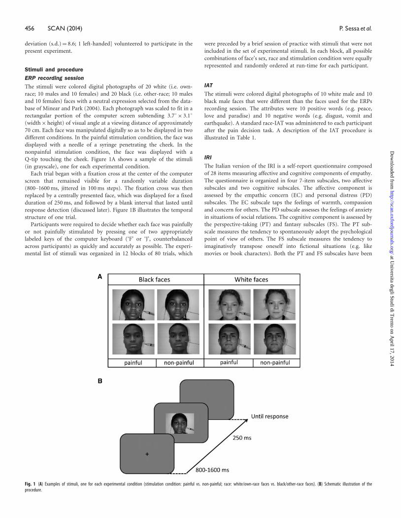
The stimuli were colored digital photographs of 20 white (i.e. own-
race; 10 males and 10 females) and 20 black (i.e. other-race; 10 males
and 10 females) faces with a neutral expression selected from the data-
base of Minear and Park (2004). Each photograph was scaled to fit in a
rectangular portion of the computer screen subtending 3.78� 3.18(width� height) of visual angle at a viewing distance of approximately
70 cm. Each face was manipulated digitally so as to be displayed in two
different conditions. In the painful stimulation condition, the face was
displayed with a needle of a syringe penetrating the cheek. In the
nonpainful stimulation condition, the face was displayed with a
Q-tip touching the cheek. Figure 1A shows a sample of the stimuli
(in grayscale), one for each experimental condition.
Each trial began with a fixation cross at the center of the computer
screen that remained visible for a randomly variable duration
(800–1600 ms, jittered in 100 ms steps). The fixation cross was then
replaced by a centrally presented face, which was displayed for a fixed
duration of 250 ms, and followed by a blank interval that lasted until
response detection (discussed later). Figure 1B illustrates the temporal
structure of one trial.
Participants were required to decide whether each face was painfully
or not painfully stimulated by pressing one of two appropriately
labeled keys of the computer keyboard (‘F’ or ‘J’, counterbalanced
across participants) as quickly and accurately as possible. The experi-
mental list of stimuli was organized in 12 blocks of 80 trials, which
were preceded by a brief session of practice with stimuli that were not
included in the set of experimental stimuli. In each block, all possible
combinations of face’s sex, race and stimulation condition were equally
represented and randomly ordered at run-time for each participant.
IAT
The stimuli were colored digital photographs of 10 white male and 10
black male faces that were different than the faces used for the ERPs
recording session. The attributes were 10 positive words (e.g. peace,
love and paradise) and 10 negative words (e.g. disgust, vomit and
earthquake). A standard race-IAT was administered to each participant
after the pain decision task. A description of the IAT procedure is
illustrated in Table 1.
IRI
The Italian version of the IRI is a self-report questionnaire composed
of 28 items measuring affective and cognitive components of empathy.
The questionnaire is organized in four 7-item subscales, two affective
subscales and two cognitive subscales. The affective component is
assessed by the empathic concern (EC) and personal distress (PD)
subscales. The EC subscale taps the feelings of warmth, compassion
and concern for others. The PD subscale assesses the feelings of anxiety
in situations of social relations. The cognitive component is assessed by
the perspective-taking (PT) and fantasy subscales (FS). The PT sub-
scale measures the tendency to spontaneously adopt the psychological
point of view of others. The FS subscale measures the tendency to
imaginatively transpose oneself into fictional situations (e.g. like
movies or book characters). Both the PT and FS subscales have been
Fig. 1 (A) Examples of stimuli, one for each experimental condition (stimulation condition: painful vs. non-painful; race: white/own-race faces vs. black/other-race faces). (B) Schematic illustration of theprocedure.
456 SCAN (2014) P. Sessa et al.
at Universita degli Studi di T
rento on April 17, 2014
http://scan.oxfordjournals.org/D
ownloaded from

shown to be positively correlated with other validated measures of
cognitive empathy, such as the Hogan (1969) empathy scale.
EEG acquisition and analysis
EEG activity was recorded from 64 electrodes distributed over the scalp
according to an extension of the international 10/20 system referenced
to the left earlobe. The EEG was re-referenced offline to the average of
the left and right earlobes. Trials contaminated by eye blinks, large
horizontal eye movements or incorrect responses in the pain decision
task were discarded from analysis.
Mean N1, P2 and N2 component amplitudes were measured
at frontocentral electrode sites in 80–100 ms, 120–150 ms and
200–240 ms time-windows locked to the onset of the face stimuli,
respectively. The mean P3 amplitude was measured in a 400–750 ms
time-window at Pz, P3 and P4 electrode sites. An additional
280–340 ms window, spanning the trough between N2 and N3 peaks,
was selected based on visual localization of factor effects maximum
values. Statistical analyses were conducted on individual amplitude
estimates of activity recorded at each of frontal (AF3/AF4, AF7/AF8,
Fz, FCz, F1/F2, F3/F4, F5/F6, F7/F8, FC1/FC2, FC3/FC4, FC5/FC6 and
FT7/FT8), central (Cz, C1/C2, C3/C4 and C5/C6) and parietal (Pz and
P3/P4) electrode sites. In all multi-factorial analyses, a
Greenhouse–Geisser correction was used where appropriate.
The standardized Low Resolution Brain Electromagnetic Tomogra-
phy (sLORETA) (Pascual-Marqui, 2002) was used for brain localiza-
tion of the potential sources of ERP reactions to pain. sLORETA
analyses were conducted following the creation of a boundary element
method model, with about 5000 nodes from MRI data, the selection of
a temporal window in which ERP responses differentiated between
painful and nonpainful stimulations, and a location-wise inverse
weighting from the minimum norm least square analysis with esti-
mated variances.
RESULTS
Pain decision task
Reaction times (RTs) exceeding each individual mean RT in a given
condition �2.5 s.d. and RTs associated with incorrect responses were
excluded from the RT analysis. Individual mean proportions of correct
responses and RTs were submitted to separate repeated measure ana-
lyses of variance (ANOVA), both considering stimulation condition
(painful vs. nonpainful) and race (white faces vs. black faces) as
within-subjects factors. Neither ANOVA showed significant factor
effects (max F¼ 2.2; min P¼ 0.16).
IRI
Scores were computed by summing 1–7 scores to each item of the four
subscales. Interindividual mean rating scores were 27.1 (s.d.¼ 4.03) for
the PT subscale, 25.0 (s.d.¼ 4.94) for the FS subscale, 26.8 (s.d.¼ 2.96)
for the EC subscale and 21.4 (s.d.¼ 5.70) for the PD subscale.
IAT
Score calculation followed the improved algorithm proposed by
Greenwald et al. (2003). D positive scores were taken to indicate a
preference towards white people. Mean D scores were significantly
different from zero (mean D� standard error: 0.37� 0.17;
t(11)¼ 2.2, P < 0.05), reflecting a successful detection of a positive
bias toward own-race members.
ERP: N1, P2 and N2
Figure 2 shows mean amplitudes of the subset of face-locked ERP
components recorded at electrode site Fz that previous studies have
indicated as sensitive to race, namely, N1, P2 and N2. We submitted to
ANOVA individual amplitude values of each component considering
stimulation (painful vs. nonpainful), race (white faces vs. black faces)
and electrode site as within-subjects factors. The ANOVAs carried out
on N1, P2 and N2 mean amplitude values revealed main effects of race
over all electrodes of the frontal area (all Ps < 0.05). As expected, black
(other-race) faces elicited N1 and P2 of greater amplitude than white
(own-race) faces, and white faces elicited an N2 of greater amplitude
than black faces. The ANOVAs detected no other main effect or inter-
action (all Ps > 0.05).
ERP: N2–N3
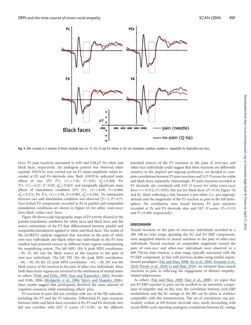
Figures 3A and 4 show face-locked ERPs recorded from a selection of
frontal electrode sites, separately for white and black faces. A visual
inspection of Figures 3A and 4 suggests that participants processed
differently the faces in painful and nonpainful conditions, exhibiting
a positive shift in response to painful stimulation only when applied to
white (own-race) faces relative to black (other-race) faces. This obser-
vation was substantiated by an ANOVA carried out on individual ERP
mean amplitude values recorded in a 280–340 ms time-window con-
sidering recording side (left vs. right hemisphere electrodes) as add-
itional factor, which showed a significant interaction between
stimulation condition and race on all the frontal and central electrode
sites (min F¼ 5.4; max P¼ 0.013, min �2p¼ 0.516). This pattern was
bilaterally distributed, for recording site did not produce significant
effects either as main factor or in interactions with race and stimula-
tion conditions (all Fs < 1; min P > 0.45). Planned comparison indi-
cated that painful stimuli elicited a positive shift relative to nonpainful
stimuli in the N2–N3 time window only when applied to white
(own-race) faces (min t¼ 2.39; max P¼ 0.036). This shift was absent
when black (other-race) faces were painfully stimulated (all Ps < 0.23).
These findings suggest that, in a temporal window of 280–340 ms,
participants were in a state of experience sharing elicited by the pres-
entation of suffering own-race members, but not of suffering
other-race members. A scalp topographic map of N2–N3 activity eli-
cited by white (own-race) faces in the painful stimulation condition is
depicted in Figure 3B (upper panels).
Table 1 Schematic description of the IAT procedure
Block Type of judgment Instructions Number of trials
1. Race discrimination Press ‘D’ to categorize pictures as whites and ‘K’ to categorize pictures as blacks 202. Attribute discrimination Press ‘D’ to categorize positive words and ‘K’ to categorize negative words 203. Prejudice-consistent combination Press ‘D’ to categorize pictures of whites and positive words and ‘K’ to categorize pictures of blacks and negative words 404. Race discrimination reversed Press ‘D’ to categorize pictures as blacks and ‘K’ to categorize pictures as whites 205. Prejudice-inconsistent combination Press ‘D’ to categorize pictures of blacks and positive words and ‘K’ to categorize pictures of whites and negative words 40
IAT consisted of five classification tasks administered in a sequential order on the computer screen. Block 1, 2 and 4 were learning blocks, whereas blocks 3 and 5 were critical to compute IAT scores. The orderof the critical blocks was counterbalanced across participants. In each block, participants were required to classify stimuli by pressing one of two different keys on the computer keyboard (‘D’ and ‘K’).Instructions and key assignments were displayed on the computer screen before each block. Stimuli were presented at the center of the screen and remained visible until response.
ERPs and the time-course of cross-racial empathy SCAN (2014) 457
at Universita degli Studi di T
rento on April 17, 2014
http://scan.oxfordjournals.org/D
ownloaded from
The results of sLORETA analysis revealed that the neural activity in
the N2–N3 time window that differentiated between painful and
nonpainful stimulations applied to white faces was mainly localized
in the left IFG (Brodman area, BA, 45; peak Montreal Neurological
Institute (MNI) coordinates: �50, 25, 20, Figure 3B, bottom), a core
region of the mirror neuron system. This finding provides further
support to the view that the early ERP reaction to pain was a likely
reflection of neural resonance elicited by own-race faces in a painful
condition.
We also correlated individual pain reactions detected in the N2–N3
temporal window for each electrode site over frontal and central areas
with the ratings collected with the IRI subscales. Pain reactions in the
present time-window were isolated by subtracting ERPs elicited in the
nonpainful stimulation condition from ERPs elicited in the painful
condition, separately for white (own-race) faces and black (other-race)
faces. Pain reactions recorded from a subset of frontal electrodes pos-
itioned on the left hemisphere (i.e. AF7, AF3, F7, F5, F3, F1, FT7 and
FC5) correlated with the EC ratings (measuring the affective compo-
nent of empathy) but only when white faces were presented. Spearman
rs ranged from 0.50 at electrode site F7 to 0.73 at electrode site AF7
(all Ps < 0.05). Figure 5A shows the scatter plot of individual EC ratings
and pain effects recorded at electrode site AF7, and a graphical indi-
cation of the additional electrode sites where EC ratings and pain
effects were significantly correlated. Pain reactions in this temporal
window, however, did not correlate with IAT D score (all Ps > 0.05).
Figure 5B shows the scatterplot of individual EC ratings and pain
reactions recorded at the electrode site AF7 for black faces. At all
frontal electrode sites, the correlations between EC ratings and pain
reactions elicited by black faces were not significant (rs ranged from
0.33 to 0.45 all Ps > 0.05).
ERP: P3
An ANOVA on individual P3 amplitude values recorded at Pz elec-
trode site revealed a main effect of race. P3 amplitude was greater for
black (other-race) faces (4.4 mV) than white (own-race) faces [3.3 mV;
F(1, 11)¼ 11.38, P < 0.01, �2p¼ 0.532). The main effect of stimulation
condition was also significant [F(1, 11)¼ 7.05, P < 0.05, �2p¼ 0.414).
Notably, the interaction between these two variables did not reach
significance (F < 1), suggesting no role of race in modulating a
neural reflection of the cognitive component of empathy. P3 pain
reactions were again isolated by subtracting amplitude values in the
nonpainful stimulation condition from amplitude values in the painful
condition, separately for white (own-race) faces and black (other-race)
Fig. 3 (a) ERPs recorded at a selection of frontal electrode sites (i.e. Fz, FCz, F3 and F4) relative to the two stimulation conditions (painful vs. nonpainful) for white/own-race faces. (b) Voltage topography ofthe N2–N3 activity recorded in the painful condition (upper panels) and source estimation of the N2–N3 activity in the painful vs. nonpainful conditions for white/own-race faces (lower panels).
Fig. 2 ERPs time-locked to the presentation of the faces recorded at electrode site Fz in response towhite/own-race and black/other-race faces collapsed across stimulation conditions (painful vs.nonpainful).
458 SCAN (2014) P. Sessa et al.
at Universita degli Studi di T
rento on April 17, 2014
http://scan.oxfordjournals.org/D
ownloaded from
faces. P3 pain reactions amounted to 0.80 and 0.94 mV for white and
black faces, respectively. An analogous pattern was observed when
separate ANOVAs were carried out on P3 mean amplitude values re-
corded at P3 and P4 electrode sites. Both ANOVAs indicated main
effects of race [P3: F(1, 11)¼ 7.45, P < 0.05, �2p¼ 0.404; P4:
F(1, 11)¼ 6.27, P < 0.05, �2p¼ 0.363] and marginally significant main
effects of stimulation condition [P3: F(1, 11)¼ 4.09, P¼ 0.068,
�2p¼ 0.271; P4: F(1, 11)¼ 3.58, P¼ 0.085, �2p¼ 0.246]. No interaction
between race and stimulation condition was observed [F < 1, P > 0.7).
Face-locked P3 components recorded at Pz in painful and nonpainful
stimulation conditions are shown in Figure 6A for white (own-race)
faces black (other-race) faces.
Figure 6B shows scalp topographic maps of P3 activity elicited in the
painful stimulation condition for white faces and black faces and the
source estimations of the P3 that differentiated between painful and
nonpainful stimulations applied to white and black faces. The results of
the sLORETA analysis suggested that reactions to the pain of white
own-race individuals and black other-race individuals in the P3 time
window had potential sources in different brain regions underpinning
the mentalizing system. The left MFG (BA 9; peak MNI coordinates:
�30, 35, 40) was the likely source of the reaction to the pain of
own-race individuals. The left TPJ (BA 40; peak MNI coordinates:
�60, �50, 20; BA 22; peak MNI coordinates: �63, �40, 20) was the
likely source of the reaction to the pain of other-race individuals. Since
both these brain regions are involved in the attribution of mental states
to others (Frith and Frith, 1999; Saxe and Kanwisher, 2003; Amodio
and Frith, 2006; Moriguchi et al., 2006; Spiers and Maguire, 2006),
these results suggest that participants devolved the same amount of
cognitive resources while mentalizing others’ pain.
P3 reactions to pain did not correlate with any of the IRI subscales,
including the PT and the FS subscales. Differential P3 pain reactions
between white and black faces recorded at Pz, P3 and P4 electrode sites
did not correlate with IAT D scores (P > 0.38). As the different
potential sources of the P3 reactions to the pain of own-race and
other-race individuals could suggest that these reactions are differently
sensitive to the implicit pro-ingroup preference, we decided to com-
pute correlations between P3 pain reactions and IAT D scores for white
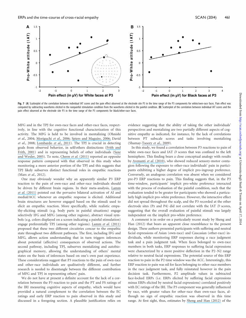
and black faces, separately. Interestingly, P3 pain reactions recorded at
P3 electrode site correlated with IAT D scores for white (own-race)
faces (r¼ 0.512, P < 0.05), but not for black faces (P > 0.10; Figure 7A
and B), likely reflecting a link between a pro-white (i.e. pro-ingroup)
attitude and the magnitude of the P3 reaction to pain in the left hemi-
sphere. No correlations were found between P3 pain reactions
recorded at Pz and P4 electrode sites and IAT D scores (P¼ 0.153
and P¼ 0.499, respectively).
DISCUSSION
Neural reactions to the pain of own-race individuals recorded in a
280–340 ms time range, spanning the N2 and N3 ERP components,
were magnified relative to neural reactions to the pain of other-race
individuals. Neural reactions of comparable magnitude toward the
pain of own-race and other-race individuals were observed in a
400–750 ms time-window, a time range typically associated with the
P3 ERP component. In line with previous studies using similar experi-
mental paradigms (Fan and Han, 2008; Xu et al., 2009; Avenanti et al.,
2010; Decety et al., 2010; Li and Han, 2010), we interpret these neural
reactions to pain as reflecting the engagement of distinct empathy-
related subprocesses.
As others (Fan and Han, 2008; Han et al., 2008), we argue that
pre-P3 ERP reaction to pain can be ascribed to an automatic compo-
nent of empathy and, in this vein, the correlation between such ERP
modulations and the EC ratings of the IRI can be taken as evidence
compatible with this interpretation. The set of correlations was par-
ticularly evident at left-frontal electrode sites, nicely dovetailing with
recent fMRI work reporting analogous correlations between EC ratings
Fig. 4 ERPs recorded at a selection of frontal electrode sites (i.e. Fz, FCz, F3 and F4) relative to the two stimulation conditions (painful vs. nonpainful) for black/other-race faces.
ERPs and the time-course of cross-racial empathy SCAN (2014) 459
at Universita degli Studi di T
rento on April 17, 2014
http://scan.oxfordjournals.org/D
ownloaded from

and BOLD responses recorded from the left insula and ACC (Singer
et al., 2004). The likely source of this ERP response was found in the
left IFG, a key region of the mirror neuron system, thus dovetailing
with prior similar evidence (Shamay-Tsoory et al., 2004, 2009;
Chakrabarti et al., 2006; Minio-Paluello et al., 2006; Schulte-Ruther
et al., 2007; Jabbi and Keysers, 2008; Fruhholz and Grandjean, 2012).
In the P3 time range, the racial bias on neural reactions to pain was
reduced to nil. The source of P3 responses to pain was localized in the
Fig. 6 (A) P3 ERP component recorded at the electrode site Pz relative to the two stimulation conditions (painful vs. nonpainful) for white/own-race faces and for black/other-race faces. (B) Voltagetopographies and source estimation of P3 activity as a function of race and stimulation conditions (painful vs. nonpainful).
Fig. 5 (A) Scatterplot of the correlation between individual EC ratings and the pain effect observed at the electrode site AF7 in the time range of the N2–N3 components for white faces. Pain effect wascomputed by subtracting waveforms elicited in the nonpainful stimulation condition from the waveforms elicited in the painful condition. A schematic illustration of the EEG montage shows the electrode siteson the scalp for which correlations were significant (AF7 in black color, AF3, F7, F5, F3, F1, FT7 and FC5 in gray color). (B) Scatterplot of the correlation between individual EC ratings and the pain effectobserved at the electrode site AF7 in the time range of the N2–N3 components for black/other-race faces.
460 SCAN (2014) P. Sessa et al.
at Universita degli Studi di T
rento on April 17, 2014
http://scan.oxfordjournals.org/D
ownloaded from
MFG and in the TPJ for own-race faces and other-race faces, respect-
ively, in line with the cognitive functional characterization of this
activity. The MFG is held to be involved in mentalizing (Ohnishi
et al., 2004; Moriguchi et al., 2006; Spiers and Maguire, 2006; David
et al., 2008; Lombardo et al., 2011). The TPJ is crucial in detecting
goals from observed behavior, in self/others distinctions (Frith and
Frith, 2001) and in representing beliefs of other individuals (Saxe
and Wexler, 2005). To note, Cheon et al. (2011) reported an opposite
response pattern compared with that observed in this study when
monitoring a more anterior portion of the TPJ and this suggests that
TPJ likely subserves distinct functional roles in empathic reactions
(Mars et al., 2012).
One may obviously wonder why an apparently similar P3 ERP
reaction to the pain of own-race and other-race individuals should
be driven by different brain regions. In their meta-analysis, Lamm
et al. (2011) pointed out the pervasive bilateral activation of AI and
medial/ACC whenever an empathic response is elicited. Additional
brain structures are however engaged based on the stimuli used to
elicit an empathic reaction. More specifically, while realistic empa-
thy-eliciting stimuli (e.g. body parts in painful situations) engage
selectively IFG and MFG (among other regions), abstract visual sym-
bols (e.g. colors displayed on a screen indicating a painful stimulation)
engage preferentially TPJ (among other regions). Lamm et al. (2011)
proposed that these two different circuitries concur to the empathic
state throughout two different pathways. The first, including IFG and
MFG, allows action understanding that in turn triggers inferences
about potential (affective) consequences of observed actions. The
second pathway, including TPJ, subserves mentalizing and autobio-
graphical memory, allowing the understanding of others’ mental
states on the basis of inferences based on one’s own past experience.
These considerations suggest that P3 reactions to the pain of own-race
and other-race individuals are qualitatively different, and further
research is needed to disentangle between the different contribution
of MFG and TPJ in representing others’ pain.
We do not have at present a definite account for the lack of a cor-
relation between the P3 reaction to pain and the PT and FS ratings of
the IRI measuring cognitive aspects of empathy, which would have
provided a natural complement to the correlation between the EC
ratings and early ERP reaction to pain observed in this study and
discussed in a foregoing section. A plausible justification relies on
evidence suggesting that the ability of taking the other individuals’
perspectives and mentalizing are two partially different aspects of cog-
nitive empathy as indicated, for instance, by the lack of correlations
between PT subscale scores and tasks involving mentalizing
(Shamay-Tsoory et al., 2009).
In this study, we found a correlation between P3 reactions to pain of
white own-race faces and IAT D scores that was confined to the left
hemisphere. This finding bears a close conceptual analogy with results
by Avenanti et al. (2010), who showed reduced sensory-motor conta-
gion following the exposure to other-race individuals’ pain in partici-
pants exhibiting a higher degree of implicit pro-ingroup preference.
Conversely, an analogous correlation was absent when we considered
pre-P3 ERP reactions to pain. This finding suggests that, in the P3
time-window, participants’ implicit pro-white preference interacted
with the process of evaluation of the painful condition, such that the
evaluation tended to be greater for participants who showed a particu-
larly high implicit pro-white preference. However, the interactive effect
did not spread throughout the scalp, and the P3 recorded at the other
electrode sites (Pz and P4) did not correlate with the IAT D scores,
suggesting that the overall evaluation of painful stimuli was largely
independent on the implicit pro-white preference.
A comment is in order on a particularly recent study by Sheng and
Han (2012) that bears an apparent close resemblance to the present
design. These authors presented participants with suffering and neutral
facial expressions of Asian (own-race) and Caucasian (other-race) in-
dividuals, while monitoring ERP responses during a race judgment
task and a pain judgment task. When faces belonged to own-race
members in both tasks, ERP responses to suffering facial expressions
were characterized by a more positive deflection in the P2–N2 range
relative to neutral facial expressions. The potential source of this ERP
reaction to pain in the P2 time window was the ACC. Interestingly, this
ERP reaction to pain was nil for faces belonging to other-race members
in the race judgment task, and fully reinstated however in the pain
decision task. Furthermore, P2 amplitude values in subtracted
face-locked ERPs (i.e. ERPs elicited by suffering facial expressions
minus ERPs elicited by neutral facial expressions) correlated positively
with EC ratings of the IRI. The P3 component was generally influenced
by race, with greater values for other-race than own-race faces, al-
though no sign of empathic reaction was observed in this time
range. At first sight, thus, estimates by Sheng and Han (2012) of the
Fig. 7 (A) Scatterplot of the correlation between individual IAT scores and the pain effect observed at the electrode site P3 in the time range of the P3 components for white/own-race faces. Pain effect wascomputed by subtracting waveforms elicited in the nonpainful stimulation condition from the waveforms elicited in the painful condition. (B) Scatterplot of the correlation between individual IAT scores and thepain effect observed at the electrode site P3 in the time range of the P3 components for black/other-race faces.
ERPs and the time-course of cross-racial empathy SCAN (2014) 461
at Universita degli Studi di T
rento on April 17, 2014
http://scan.oxfordjournals.org/D
ownloaded from

time-course of the interplay between race and pain reactions are
slightly different from the estimates proposed in the present context,
posing the question of which are the possible sources of such discrep-
ancy. In our view, a possible explanation can be traced back to the type
of stimuli used in the two studies, which were substantially different.
To elicit the reaction to pain, Sheng and Han (2012) manipulated the
facial expression (suffering vs. neutral) of members of distinct races,
whereas we arbitrarily chose faces of members of distinct races with
neutral facial expressions in combination with tools sharing a strong
conceptual association with distinct feelings (pain vs. no pain). Our
choice was motivated by the need to provide the most stringent control
for low-level�sensory-driven�factors potentially influencing ERP esti-
mates. Using physically identical faces with a neutral facial expression
and tools which did not differ macroscopically (being both fusiform,
equally oriented and globally of similar color) was compelling in this
perspective. The physical difference between differing facial expressions
is more evident. Besides, we avoided manipulating facial expressions to
not induce ‘spurious’ automatic reactions in participants like mimicry
(Hatfield et al., 2009), which cannot be taken, in and of itself, as
symptomatic of an empathic state in the observers, although it clearly
concurs to a thorough manifestation of empathy (Decety and Jackson,
2006). Differing facial expressions may also be related to possible vari-
ations in the degree of difficulty in decoding others’ feelings from faces
belonging to distinct races (Anthony et al., 1992; Elfenbein and
Ambidi, 2002). To note, ERP studies investigating the perception of
others’ pain have reported different estimates of the time at which
ERPs start to differentiate between painful and nonpainful conditions,
ranging from 90 ms (Decety et al., 2010) to 230 ms (Li and Han, 2010).
These different estimates are likely related to differences in the type of
stimuli and task requirements that mediate the relative contribution of
neural resonance and cognitive components of empathy (Zaki and
Ochsner, 2012; see also Lamm et al., 2011). An experimental manipu-
lation implemented in the design of Fan and Han (2008) is illustrative
in this regard. As described in the Introduction, these authors used as
stimuli images of hands in painful or neutral situations. The images
were either pictures or cartoons, and, interestingly, the time at which
ERPs started to differentiate between painful and neutral situations was
postponed by 80 ms for cartoons relative to pictures. Fan and Han
(2008) interpreted their findings as indicating that a lack of contextual
reality of stimuli reduced the affective empathic responses, delaying the
starting time of ERP reactions to pain. Therefore, one plausible way to
reconcile the study by Sheng and Han (2012) and this study on the
interplay between race and empathy is to ascribe the slightly dissimilar
temporal maps to the use of suffering facial expressions in the absence
of a context indicating the source of pain in the study by Sheng and
Han (2012) that may have enhanced the involvement of neural reson-
ance and reduced the contribution of cognitive empathy vis-a-vis the
use of neutral facial expressions in this study in the context of a painful
stimulation, that may have triggered neural resonance to a lesser
extent, whereupon mentalizing was instead necessary to infer others’
painful states on the basis of the stimulation condition, as proposed by
Zaki and Ochsner (2012).
In conclusion, our findings provide insights into the nature of
cross-racial empathy. The present results complement and extend pre-
vious fMRI (Xu et al., 2009) and TMS (Avenanti et al., 2010) work by
mapping out the time-course of the temporally asynchronous engage-
ment of an early neural resonance component of empathy, which
amplifies responses to the pain of own-race members, and of a cogni-
tive component likely related to mentalizing which magnitude appears
not to be influenced by their race, although the underlying different
potential source estimates suggest that this later reaction qualitatively
differs between own-race and other-race conditions.
Conflict of InterestNone declared.
REFERENCES
Albiero, P., Ingoglia, S., Lo Coco, A. (2006). Contributo all’adattamento italiano
dell’Interpersonal Reactivity Index [A contribution to the Italian validation of the
Interpersonal Reactivity Index]. Testing Psicometria Metodologia, 13, 107–25.
Amodio, D.M., Frith, C.D. (2006). Meeting of minds: the medial frontal cortex and social
cognition. Nature Reviews Neuroscience, 7, 268–77.
Amodio, D.M., Harmon-Jones, E., Devine, P.G. (2003). Individual differences in the acti-
vation and control of affective race bias as assessed by startle eyeblink responses and
self-report. Journal of Personality and Social Psychology, 84, 738–53.
Anthony, T., Cooper, C., Mullen, B. (1992). Cross-racial facial identification: a social
cognitive integration. Personality and Social Psychology Bulletin, 18, 296–301.
Avenanti, A., Bueti, D., Galati, G., Aglioti, S.M. (2005). Transcranial magnetic stimulation
highlights the sensorimotor side of empathy for pain. Nature Neuroscience, 8, 955–60.
Avenanti, A., Sirigu, A., Aglioti, S.M. (2010). Racial bias reduces empathic sensory-motor
resonance with other-race pain. Current Biology, 20, 1018–22.
Chakrabarti, B., Bullmore, E., Baron-Cohen, S. (2006). Empathizing with basic emotions:
common and discrete neural substrates. Social Neuroscience, 1, 364–84.
Cheon, B.K., Im, D., Harada, T., et al. (2011). Cultural influences on neural basis of
intergroup empathy. NeuroImage, 57, 642–50.
Cunningham, W.A., Nezlek, J.B., Banaji, M.R. (2004). Implicit and explicit ethnocentrism:
revisiting the ideologies of prejudice. Personality and Social Psychology Bulletin, 30,
1332–46.
Cutini, S., Basso Moro, S., Bisconti, S. (2012a). Functional near infrared optical imaging in
cognitive neuroscience: an introductory review. Journal of Near Infrared Spectroscopy, 20,
75–92.
Cutini, S., Scarpa, F., Scatturin, P., Dell’Acqua, R., Zorzi, M. (2014). Number-space inter-
actions in the human parietal cortex: enlightening the SNARC effect with functional
near-infrared spectroscopy. Cerebral Cortex, 24(2), 444–51.
Cutini, S., Scarpa, F., Scatturin, P., et al. (2011). A hemodynamic correlate of lateralized
visual short-term memories. Neuropsychologia, 49, 1611–21.
David, N., Aumann, C., Santos, N.S., et al. (2008). Differential involvement of the posterior
temporal cortex in mentalizing but not perspective taking. Social Cognitive and Affective
Neuroscience, 3, 279–89.
Davis, M.H. (1980). A multidimensional approach to individual differences in empathy.
JSAS Catalog of Selected Documents in Psychology, 10, 85.
Davis, M.H. (1983). Measuring individual differences in empathy: evidence for a multidi-
mensional approach. Journal of Personality and Social Psychology, 44, 113–26.
Decety, J. (2011). The neuroevolution of empathy. Annals of the New York Academy of
Sciences, 1231, 35–45.
Decety, J., Jackson, P. (2006). A social-neuroscience perspective on empathy. Current
Directions in Psychological Science, 15, 54–8.
Decety, J., Jackson, P.L. (2004). The functional architecture of human empathy. Behavioral
and Cognitive Neuroscience Reviews, 3, 71–100.
Decety, J., Lamm, C. (2006). Human empathy through the lens of social neuroscience.
Scientific World Journal, 6, 1146–63.
Decety, J., Yang, C., Cheng, Y. (2010). Physicians down-regulate their pain empathy re-
sponse: an event-related brain potential study. NeuroImage, 50, 1676–82.
Dickter, C.L., Bartholow, B.D. (2007). Event-related brain potential evidence of ingroup
and outgroup attention biases. Social Cognitive and Affective Neuroscience, 2, 189–98.
Donchin, E. (1981). Presidential address, 1980. Surprise!. . .Surprise? Psychophysiology, 18,
493–513.
Donchin, E., Coles, M.G.H. (1988). Is the P300 component a manifestation of context
updating? Behavioral and Brain Sciences, 11, 357–74.
Elfenbein, H.A., Ambidi, N. (2002). On the universality and cultural specificity of emotion
recognition: a meta-analysis. Psychological Bulletin, 128, 203–35.
Fan, Y., Han, S. (2008). Temporal dynamic of neural mechanisms involved in empathy for
pain: an event-related brain potential study. Neuropsychologia, 46, 160–73.
Frith, C.D., Frith, U. (1999). Interacting minds: a biological basis. Science, 286, 1692–95.
Frith, U., Frith, C. (2001). The biological basis of social interaction. Current Directions in
Psychological Science, 10, 151–5.
Fruuhholz, S., Grandjean, D. (2012). Towards a fronto-temporal neural network for the
decoding of angry vocal expressions. NeuroImage, 62, 1658–66.
Golarai, G., Ghahremani, D.G., Eberhardt, J.L., Grill-Spector, K., Gabrieli, G.D.E. (2004).
Representation of parts and canonical face configuration in the amygdala, superior
temporal sulcus (STS) and the fusiform “face area” (FFA). Journal of Vision, 4, a131.
Golby, A., Gabrieli, J., Chiao, J., Eberhardt, J. (2001). Differential responses in the fusiform
region to same-race and other-race faces. Nature Neuroscience, 4, 845–50.
Greenwald, A.G., Nosek, B.A., Banaji, M.R. (2003). Understanding and using the implicit
association test. I. An improved scoring algorithm. Journal of Personality and Social
Psychology, 85, 197–216.
Han, S., Fan, Y., Mao, L. (2008). Gender difference in empathy for pain: an electrophysio-
logical investigation. Brain Research, 1196, 85–93.
462 SCAN (2014) P. Sessa et al.
at Universita degli Studi di T
rento on April 17, 2014
http://scan.oxfordjournals.org/D
ownloaded from

Hart, A.J., Whalen, P.J., Shin, L.M., McInerney, S.C., Fischer, H., Rauch, S.L. (2000).
Differential response in the human amygdala to racial outgroup vs. ingroup face stimuli.
NeuroReport, 11, 2351–55.
Hatfield, E., Rapson, R.L., Le, Y.C. (2009). Emotional contagion and empathy.
In: Decety, J., Ickes, W., editors. The Social Neuroscience of Empathy. Cambridge, MA:
MIT Press, pp. 19–30.
Hogan, R. (1969). Development of an empathy scale. Journal of Consulting and Clinical
Psychology, 33, 307–16.
Ito, T.A., Bartholow, B.D. (2009). The neural correlates of race. Trends in Cognitive Sciences,
13, 524–31.
Ito, T.A., Urland, G.R. (2003). Race and gender on the brain: electrocortical measures of
attention to race and gender of multiply categorizable individuals. Journal of Personality
and Social Psychology, 85, 616–26.
Ito, T.A., Urland, G.R. (2005). The influence of processing objectives on the perception of
faces: an ERP study of race and gender perception. Cognitive, Affective, & Behavioral
Neuroscience, 5, 21–36.
Jabbi, M., Keysers, C. (2008). Inferior frontal gyrus activity triggers anterior insula response
to emotional facial expressions. Emotion, 8, 775–80.
Keysers, C., Kaas, J.H., Gazzola, V. (2010). Somatosensation in social perception. Nature
Reviews Neuroscience, 11, 417–28.
Lamm, C., Decety, J., Singer, T. (2011). Meta-analytic evidence for common and distinct
neural networks associated with directly experienced pain and empathy for pain.
NeuroImage, 54, 2492–502.
Lamm, C., Singer, T. (2010). The role of anterior insular cortex in social emotions. Brain
Structure and Function, 214, 579–91.
Lefebvre, C., Dell’Acqua, R., Roelfsema, P., Jolicœur, P. (2011). Surfing the attentional
waves during visual curve tracing: evidence from the sustained posterior contralateral
negativity. Psychophysiology, 48, 1509–15.
Li, W., Han, S. (2010). Perspective taking modulates event-related potentials to perceived
pain. Neuroscience Letters, 469, 328–32.
Logothetis, N.K. (2008). What we can do and what we cannot do with fMRI. Nature, 453,
869–78.
Lombardo, M.V., Chakrabarti, B., Bullmore, E.T., Baron-Cohen, S. (2011). Specialization
of right temporo-parietal junction for mentalizing and its relation to social impairments
in autism. NeuroImage, 56, 1832–8.
Luria, R., Sessa, P., Gotler, A., Jolicœur, P., Dell’Acqua, R. (2010). Visual short-term
memory capacity for simple and complex objects. Journal of Cognitive Neuroscience,
22, 496–512.
Mars, R.B., Sallet, J., Schuffelgen, U., Jbabdi, S., Toni, I., Rushworth, M.F. (2012).
Connectivity-based subdivisions of the human right “temporoparietal junction area”:
evidence for different areas participating in different cortical networks. Cerebral Cortex,
22, 1894�903.
Minear, M., Park, D.C. (2004). A lifespan database of adult facial stimuli. Behavior Research
Methods, Instruments, & Computers, 36, 630–3.
Minio-Paluello, I., Avenanti, A., Aglioti, S.M. (2006). Left hemisphere dominance in read-
ing the sensory qualities of others’ pain? Social Neuroscience, 1, 320–33.
Moriguchi, Y., Ohnishi, T., Lane, R.D., et al. (2006). Impaired self-awareness and theory of
mind: an fMRI study of mentalizing in alexithymia. NeuroImage, 32, 1472–82.
Ohnishi, T., Moriguchi, Y., Matsuda, H., et al. (2004). The neural network for the mirror
system and mentalizing in normally developed children: an fMRI study. NeuroReport,
15, 1483–7.
Pascual-Marqui, R.D. (2002). Standardized low-resolution brain electromagnetic tomog-
raphy (sLORETA): technical details. Methods and Findings in Experimental and Clinical
Pharmacology, 24(Suppl. D), 5–12.
Phelps, E.A., O’Connor, K.J., Cunningham, W.A., et al. (2000). Performance on indirect
measures of race evaluation predicts amygdala activation. Journal of Cognitive
Neuroscience, 12, 729–38.
Rizzolatti, G., Sinigaglia, C. (2010). The functional role of the parieto-frontal mirror cir-
cuit: interpretations and misinterpretations. Nature Reviews Neuroscience, 11, 264–74.
Saxe, R., Kanwisher, N. (2003). People thinking about thinking people: The role of the
temporo-parietal junction in theory of mind. NeuroImage, 19, 1835–42.
Saxe, R., Wexler, A. (2005). Making sense of another mind: the role of the right
temporo-parietal junction. Neuropsychologia, 43, 1391–9.
Scarpa, F., Cutini, S., Scatturin, P., Dell’Acqua, R., Sparacino, G. (2010). Bayesian filtering
of human brain hemodynamic activity elicited by visual short-term maintenance re-
corded through functional near-infrared spectroscopy (fNIRS). Optics Express, 18,
26550–68.
Schulte-Ruther, M., Markowitsch, H.J., Fink, G.R., Piefke, M. (2007). Mirror neuron and
theory of mind mechanisms involved in face-to-face interactions: a functional magnetic
resonance imaging approach to empathy. Journal of Cognitive Neuroscience, 19, 1354–72.
Sessa, P., Luria, R., Gotler, A., Jolicœur, P., Dell’Acqua, R. (2011). Inter-hemispheric ERP
asymmetries over inferior parietal cortex reveal differential visual working memory
maintenance for fearful versus neutral facial identities. Psychophysiology, 48, 187–97.
Sessa, P., Luria, R., Verleger, R., Dell’Acqua, R. (2007). P3 latency shifts in the attentional
blink: further evidence for second target processing postponement. Brain Research, 1137,
131–9.
Sessa, P., Tomelleri, S., Luria, R., Castelli, L., Reynolds, M., Dell’Acqua, R. (2012). Look out
for strangers! Sustained neural activity during visual working memory maintenance of
other-race faces is modulated by implicit racial prejudice. Social Cognitive and Affective
Neuroscience, 7, 314–21.
Shamay-Tsoory, S.G. (2011). The neural bases for empathy. Neuroscientist, 17, 18–24.
Shamay-Tsoory, S.G., Tomer, R., Goldsher, D., Berger, B.D., Aharon-Peretz, J. (2004).
Impairment in cognitive and affective empathy in patients with brain lesions: anatomical
and cognitive correlates. Journal of Clinical and Experimental Neuropsychology, 26,
1113–27.
Shamay-Tsoory, S.G., Aharon-Peretz, J., Perry, D. (2009). Two systems for empathy: a
double dissociation between emotional and cognitive empathy in inferior frontal
gyrus versus ventromedial prefrontal lesions. Brain, 132, 617–27.
Sheng, F., Han, S. (2012). Manipulations of cognitive strategies and intergroup relation-
ships reduce the racial bias in empathic neural responses. NeuroImage, 61, 786–97.
Singer, T., Seymour, B., O’Doherty, J., Kaube, H., Dolan, R.J., Frith, C.D. (2004). Empathy
for pain involves the affective but not sensory components of pain. Science, 303,
1157–62.
Spiers, H.J., Maguire, E.A. (2006). Spontaneous mentalizing during an interactive real
world task: an fMRI study. Neuropsychologia, 44, 1674–82.
Verleger, R. (1988). Event-related potentials and cognition: a critique of the context updat-
ing hypothesis and an alternative interpretation of the P3. Behavioral and Brain Sciences,
11, 343–427.
Verleger, R. (1997). On the utility of P3 latency as a measure of mental chronometry.
Psychophysiology, 34, 131–56.
Wheeler, M.E., Fiske, S.T. (2005). Controlling racial prejudice and stereotyping: social cog-
nitive goals affect amygdala and stereotype activation. Psychological Science, 16, 56–63.
Xu, X., Zuo, X., Wang, X., Han, S. (2009). Do you feel my pain? Racial group membership
modulates empathic neural responses. Journal of Neuroscience, 29, 8525–9.
Zaki, J., Ochsner, K.N. (2012). The neuroscience of empathy: progress, pitfalls, and prom-
ise. Nature Neuroscience, 15, 675–80.
Zelazo, P.D., Carlson, S.M., Kesek, A. (2008). The development of executive function in
childhood. In: Nelson, C., Luciana, M., editors. Handbook of Developmental Cognitive
Neuroscience, 2nd edn. Cambridge, MA: MIT Press, pp. 553–74.
ERPs and the time-course of cross-racial empathy SCAN (2014) 463
at Universita degli Studi di T
rento on April 17, 2014
http://scan.oxfordjournals.org/D
ownloaded from
Copyright © 2022 FDOKUMEN