
Synthesis and spectroscopic studies on the Schiff base ligand derived from condensation of...
7
Spectrochimica Acta Part A 78 (2011) 29–35 Contents lists available at ScienceDirect Spectrochimica Acta Part A: Molecular and Biomolecular Spectroscopy journal homepage: www.elsevier.com/locate/saa Synthesis and spectroscopic studies on the Schiff base ligand derived from condensation of 2-furaldehyde and 3,3 -diaminobenzidene, L and its complexes with Co(II), Ni(II), Cu(II) and Zn(II): Comparative DNA binding studies of L and its Cu(II) and Zn(II) complexes Mohammad Shakir a,∗ , Ambreen Abbasi a , Asad U. Khan b , Shahper N. Khan b a Division of Inorganic Chemistry, Department of Chemistry, Aligarh Muslim University, Aligarh 202002, India b Interdisciplinary Biotechnology Unit, Aligarh Muslim University, Aligarh 202002, India article info Article history: Received 4 September 2009 Received in revised form 22 February 2010 Accepted 26 February 2010 Keywords: Schiff base condensation Spectroscopic studies Square planar geometry Calf thymus DNA abstract The Schiff base ligand, N,N -bis-(2-furancarboxaldimine)-3,3 -diaminobenzidene (L) obtained by conden- sation of 2-furaldehyde and 3,3 -diaminobenzidene, was used to synthesize the mononuclear complexes of the type, [M(L)](NO 3 ) 2 [M = Co(II), Ni(II), Cu(II) and Zn(II)]. The newly synthesized ligand, (L) and its complexes have been characterized on the basis of the results of the elemental analysis, molar con- ductance, magnetic susceptibility measurements and spectroscopic studies viz, FT-IR, 1 H and 13 C NMR, mass, UV–vis and EPR. EPR, UV–vis and magnetic moment data revealed a square planar geometry for the complexes with distortion in Cu(II) complex and conductivity data show a 1:2 electrolytic nature of the complexes. Absorption and fluorescence spectroscopic studies support that Schiff base ligand, L and its Cu(II) and Zn(II) complex exhibit significant binding to calf thymus DNA. The highest binding affinity in case of L may be due to the more open structure as compared to the metal coordinated complexes. © 2010 Elsevier B.V. All rights reserved. 1. Introduction Active and well-defined Schiff base ligands are considered as ‘privileged ligands’ because they are easily prepared by con- densation of aldehydes or ketones with amines and are able to stabilize different metals in various oxidation states [1]. Schiff base complexes are extensively studied due to synthetic flexibil- ity, selectivity and sensitivity towards variety of metal ions [2]. Schiff bases have application towards, degradation of organic com- pounds [3], radiopharmaceuticals [4] and as corrosion inhibitors in especially acidic environments for various alloys and metals like steel, aluminium and copper [5]. Complexes of transition and non-transition metals with Schiff base ligands are promising mate- rials for optoelectronic applications due to their outstanding photo- and electroluminescent properties, and the ease of synthesis that readily allows structural modification for optimization of material properties [6]. Bidentate and tetradentate Schiff base ligands involving N,O donor sites possess many advantages such as facile approach for synthesis, relative tolerance, readily adjusted ancillary ligands, and tunable steric and electronic coordination environments on the ∗ Corresponding author. Tel.: +91 5712703515. E-mail addresses: [email protected], [email protected] (M. Shakir). metal centre. In view of above, tetradentate N 2 O 2 ligands and their transition metal complexes, act as catalyst [7]. Various transition and inner transition metals complexes with bi-, tri- and tetraden- tate Schiff bases containing nitrogen and oxygen donor atoms play important role in biological systems and represent interesting mod- els for metalloenzymes, which efficiently catalyze the reduction of dinitrogen and dioxygen [8]. Di- and tetra-Schiff base precursors based on 3,3 - diaminobenzidene have been reported in literature [9,10]. Bolos et al. have reported biological studies of Cu(II) coordination compounds with Schiff bases of polyamines with heterocyclic aldehydes as ligands [11,12]. Polyamines and heterocyclic alde- hydes (2-thiophene-carboxaldehyde and 2-furaldehyde) were selected, the former due to its coordinative, conformational and physicochemical properties, while the latter in order to mimic biological systems and mechanisms in the process of drug design. The five- or six-membered chelate ring Schiff bases stabilize, in thermodynamic terms of entropy, the compounds synthesized [13]. In view of aforesaid importance of Schiff bases and their complexes, we have synthesized a novel Schiff base ligand, N,N -bis-(2-furancarboxaldimine)-3,3 -diaminobenzidene derived from condensation reaction of 3,3 -diaminobenzidene with 2- furaldehyde and its complexes with Co(II), Ni(II), Cu(II), Zn(II) and their DNA binding studies. 1386-1425/$ – see front matter © 2010 Elsevier B.V. All rights reserved. doi:10.1016/j.saa.2010.02.034
-
Upload
lahoreschoolofeconomics -
Category
Documents
-
view
10 -
download
0
Transcript of Synthesis and spectroscopic studies on the Schiff base ligand derived from condensation of...
ScwC
Ma
b
a
ARRA
KSSSC
1
adsbiSpilnrarp
dst
1d
Spectrochimica Acta Part A 78 (2011) 29–35
Contents lists available at ScienceDirect
Spectrochimica Acta Part A: Molecular andBiomolecular Spectroscopy
journa l homepage: www.e lsev ier .com/ locate /saa
ynthesis and spectroscopic studies on the Schiff base ligand derived fromondensation of 2-furaldehyde and 3,3′-diaminobenzidene, L and its complexesith Co(II), Ni(II), Cu(II) and Zn(II): Comparative DNA binding studies of L and itsu(II) and Zn(II) complexes
ohammad Shakira,∗, Ambreen Abbasia, Asad U. Khanb, Shahper N. Khanb
Division of Inorganic Chemistry, Department of Chemistry, Aligarh Muslim University, Aligarh 202002, IndiaInterdisciplinary Biotechnology Unit, Aligarh Muslim University, Aligarh 202002, India
r t i c l e i n f o
rticle history:eceived 4 September 2009eceived in revised form 22 February 2010ccepted 26 February 2010
a b s t r a c t
The Schiff base ligand, N,N′-bis-(2-furancarboxaldimine)-3,3′-diaminobenzidene (L) obtained by conden-sation of 2-furaldehyde and 3,3′-diaminobenzidene, was used to synthesize the mononuclear complexesof the type, [M(L)](NO3)2 [M = Co(II), Ni(II), Cu(II) and Zn(II)]. The newly synthesized ligand, (L) and its
eywords:chiff base condensationpectroscopic studiesquare planar geometryalf thymus DNA
complexes have been characterized on the basis of the results of the elemental analysis, molar con-ductance, magnetic susceptibility measurements and spectroscopic studies viz, FT-IR, 1H and 13C NMR,mass, UV–vis and EPR. EPR, UV–vis and magnetic moment data revealed a square planar geometry forthe complexes with distortion in Cu(II) complex and conductivity data show a 1:2 electrolytic nature ofthe complexes. Absorption and fluorescence spectroscopic studies support that Schiff base ligand, L andits Cu(II) and Zn(II) complex exhibit significant binding to calf thymus DNA. The highest binding affinity
the
in case of L may be due to. Introduction
Active and well-defined Schiff base ligands are considereds ‘privileged ligands’ because they are easily prepared by con-ensation of aldehydes or ketones with amines and are able totabilize different metals in various oxidation states [1]. Schiffase complexes are extensively studied due to synthetic flexibil-
ty, selectivity and sensitivity towards variety of metal ions [2].chiff bases have application towards, degradation of organic com-ounds [3], radiopharmaceuticals [4] and as corrosion inhibitors
n especially acidic environments for various alloys and metalsike steel, aluminium and copper [5]. Complexes of transition andon-transition metals with Schiff base ligands are promising mate-ials for optoelectronic applications due to their outstanding photo-nd electroluminescent properties, and the ease of synthesis thateadily allows structural modification for optimization of materialroperties [6].
Bidentate and tetradentate Schiff base ligands involving N,Oonor sites possess many advantages such as facile approach forynthesis, relative tolerance, readily adjusted ancillary ligands, andunable steric and electronic coordination environments on the
∗ Corresponding author. Tel.: +91 5712703515.E-mail addresses: [email protected], [email protected] (M. Shakir).
386-1425/$ – see front matter © 2010 Elsevier B.V. All rights reserved.oi:10.1016/j.saa.2010.02.034
more open structure as compared to the metal coordinated complexes.© 2010 Elsevier B.V. All rights reserved.
metal centre. In view of above, tetradentate N2O2 ligands and theirtransition metal complexes, act as catalyst [7]. Various transitionand inner transition metals complexes with bi-, tri- and tetraden-tate Schiff bases containing nitrogen and oxygen donor atoms playimportant role in biological systems and represent interesting mod-els for metalloenzymes, which efficiently catalyze the reduction ofdinitrogen and dioxygen [8].
Di- and tetra-Schiff base precursors based on 3,3′-diaminobenzidene have been reported in literature [9,10].Bolos et al. have reported biological studies of Cu(II) coordinationcompounds with Schiff bases of polyamines with heterocyclicaldehydes as ligands [11,12]. Polyamines and heterocyclic alde-hydes (2-thiophene-carboxaldehyde and 2-furaldehyde) wereselected, the former due to its coordinative, conformational andphysicochemical properties, while the latter in order to mimicbiological systems and mechanisms in the process of drug design.The five- or six-membered chelate ring Schiff bases stabilize, inthermodynamic terms of entropy, the compounds synthesized[13].
In view of aforesaid importance of Schiff bases and their
complexes, we have synthesized a novel Schiff base ligand,N,N′-bis-(2-furancarboxaldimine)-3,3′-diaminobenzidene derivedfrom condensation reaction of 3,3′-diaminobenzidene with 2-furaldehyde and its complexes with Co(II), Ni(II), Cu(II), Zn(II) andtheir DNA binding studies.
3 ica Acta Part A 78 (2011) 29–35
2
2
(fapdDtSuD(2TtNCTrAKNuoTswsXBrr13nbpJ3dtDatttApwc
2N
itistac
0 M. Shakir et al. / Spectrochim
. Experimental
.1. Materials and instrumentation
The metal salts, M(NO3)2·xH2O [M = Co, Ni, Zn, x = 6; Cu, x = 3]all E. Merck) were commercially pure samples. The chemicals 2-uraldehyde and 3,3′-diaminobenzidene (both Acros) were useds received. Solvent acetonitrile (AR) was used without furtherurification. The DMSO was dried before use by standard proce-ure in order to avoid the effect of water on conductivity [14].eionized water was used in synthesis. Highly polymerized calf
hymus DNA sodium salt (7% Na content) was purchased fromigma Chemical Co. Other chemicals were of reagent grade andsed without further purification. The stock solution of 12.5 mMNA/phosphate calf thymus DNA was prepared by dissolving 0.5%
w/w) in 0.1 M sodium phosphate buffer (pH 7.40) at 310 K for4 h with occasional stirring to ensure homogeneity of solution.he absorption ratio A260/A280 in the range of 1.8–1.9 indicatedhat DNA was sufficiently free from protein. The stock solution of,N′ bis-(2-furancarboxaldimine)-3,3′-diaminobenzidene and itsu and Zn complexes with 5 mg/ml concentration were prepared.he elemental analysis was obtained from micro-analytical labo-atory of CDRI, Lucknow using PerkinElmer 2400 CHN Elementalnalyzer. The FT-IR spectra (4000–400 cm−1) were recorded asBr pellets on Shimadzu 8201 PC spectrophotometer. 1H and 13CMR spectra in DMSO-d6 at room temperature were recordedsing Bruker Avance II 400 NMR spectrometer. The DART-MSf the Schiff base ligand was recorded on JEOL-AccuTOF JMS-100LC Mass spectrometer having a DART source. The givenample was subjected as such in front of DART source. Dry heliumas used with 4 LPM flow rate for ionization at 3500 ◦C. EPR
pectrum at room temperature was recorded on Varian E-112-band EPR spectrometer using TCNE as standard at RSIC, IIT,ombay, India. The electronic spectra at room temperature wereecorded with Varian Cary 5E spectrophotometer in 1200–185 nmange using DMSO as solvent. The electrical conductivities of0−3 M solution in DMSO were obtained on a Systronic type02 conductivity bridge equilibrated at 25 ± 0.01 ◦C. The mag-etic susceptibility measurements were carried out using Faradayalance at room temperature. Fluorescence measurements wereerformed on a spectrofluorimeter Model RF-5301PC (Shimadzu,
apan) equipped with a 150 W Xenon lamp and a slit width ofnm. A 1.00 cm quartz cell was used for measurements. For theetermination of binding parameters, 50 �M of complex solu-ion was taken in a quartz cell and increasing amounts of ctNA solution was titrated. Fluorescence spectra were recordedt 310 K temperatures in the range of 740–880 nm upon exci-ation at 280 (�em was 772 nm). The UV measurements of calfhymus DNA were recorded on a Shimadzu double beam spec-rophotometer model-UV1700 using a cuvette of 1 cm path length.bsorbance value of DNA in the absence and presence of com-lex were made in the range of 220–300 nm. DNA concentrationas fixed at 0.1 mM, while the compound was added in increasing
oncentration.
.2. Synthesis of,N′-bis-(2-furancarboxaldiamine)-3,3′-diaminobenzidene (L)
A hot solution of 3,3′-diaminobenzidene (1 mmol, 0.214 g)n 1:1 acetonitrile to water mixture (25 ml) was slowly addedo a solution of 2-furaldehyde (2 mmol, 0.167 ml) dissolved
n acetonitrile (5 ml). The reaction mixture was magneticallytirred for 24 h at room temperature leading to the isola-ion of yellow solid product which was washed with waternd acetonitrile and dried in vaccum over anhydrous calciumhloride.Fig. 1. Fluorescence emission spectra of L in the absence and presence of increasingamount of DNA from (a) to (i): pH 7.4, T = 298 K and (x) represents the DNA alone.
2.3. Synthesis of metal complexes
A solution of hydrated metal nitrate (.085 mmol) in acetonitrile(10 ml) was added slowly to a hot solution of L (.085 mmol) in ace-tonitrile (60 ml). The resulting solution was stirred under refluxfor 6 h resulting in the isolation of solid product. The product thusformed was filtered, washed with acetonitrile and dried in vaccumover anhydrous calcium chloride.
2.4. Binding data analysis of L and its Cu and Zn complex
To elaborate the fluorescence quenching mechanism theStern–Volmer equation (1) was used for data analysis [15].
F0
F= 1 + KSV[Q ] (1)
where F0 and F are the steady-state fluorescence intensities inthe absence and presence of quencher, respectively. KSV theStern–Volmer quenching constant and [Q] is the concentration ofquencher (DNA). The KSV for L and its Cu and Zn complex werecalculated to be 1.79 × 10⁴, 1.42 × 10⁴ and 1.36 × 10⁴ LM−1, respec-tively. A higher KSV value of L as compared to its Cu(II) and Zn(II)complexes suggests its stronger quenching ability. Further impli-cates its higher binding affinity toward the DNA then its complexes.The linearity of the F0/F versus [Q] (Stern–Volmer) plots for DNA-Land its Cu(II) and Zn(II) complexes (Fig. 1) depicts that the quench-ing may be static or dynamic, since the characteristic Stern–Volmerplot of combined quenching (both static and dynamic) is an upwardcurvature. When ligand molecules bind independently to a set ofequivalent sites on a macromolecule, the equilibrium between freeand bound molecules is given by the equation [16]:
log[
F0 − F
F
]= log K + n log[Q ] (2)
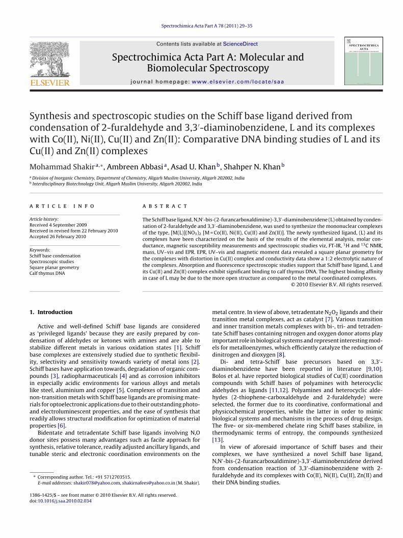
where K and n are the binding constant and the number of bind-ing sites, respectively. Thus, a plot of log(F0 − F)/F versus log[Q](Fig. 2) can be used to determine K as well as n. The bindingconstant and the number of binding sites for L and its Cu andZn complex were found to be K = 2.75 ± 0.22 × 10⁴ M−1; n = 1.07,
4 −1 4 −1
K = 0.56 ± 0.41 × 10 M ; n = 0.91 and K = 0.91 ± 0.34 × 10 M ;n = 0.94 respectively. The results suggest the compounds exhibitdifferent degree of affinity toward the DNA molecule. The highestbinding affinity in case of L may be due to the more open structureas compared to the metal coordinated complexes.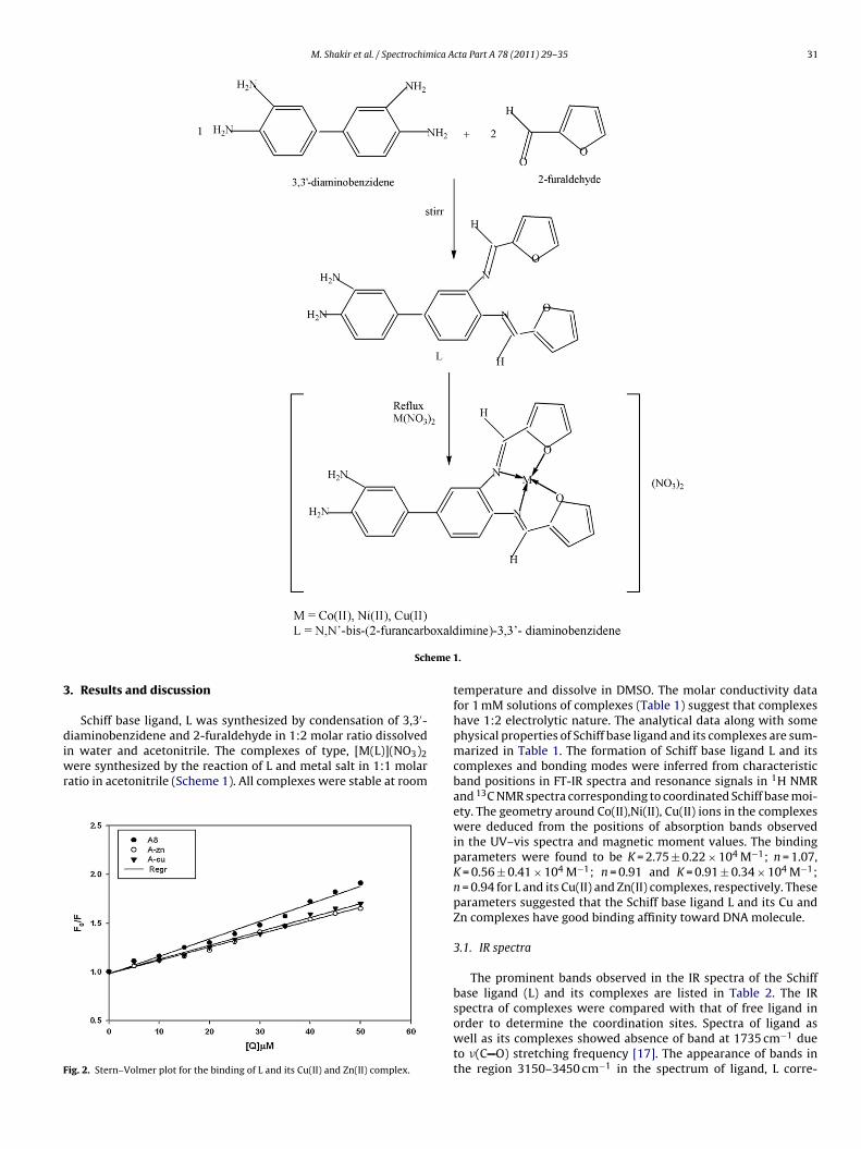
M. Shakir et al. / Spectrochimica Acta Part A 78 (2011) 29–35 31
eme 1
3
diwr
F
Sch
. Results and discussion
Schiff base ligand, L was synthesized by condensation of 3,3′-
iaminobenzidene and 2-furaldehyde in 1:2 molar ratio dissolvedn water and acetonitrile. The complexes of type, [M(L)](NO3)2ere synthesized by the reaction of L and metal salt in 1:1 molar
atio in acetonitrile (Scheme 1). All complexes were stable at room
ig. 2. Stern–Volmer plot for the binding of L and its Cu(II) and Zn(II) complex.
.
temperature and dissolve in DMSO. The molar conductivity datafor 1 mM solutions of complexes (Table 1) suggest that complexeshave 1:2 electrolytic nature. The analytical data along with somephysical properties of Schiff base ligand and its complexes are sum-marized in Table 1. The formation of Schiff base ligand L and itscomplexes and bonding modes were inferred from characteristicband positions in FT-IR spectra and resonance signals in 1H NMRand 13C NMR spectra corresponding to coordinated Schiff base moi-ety. The geometry around Co(II),Ni(II), Cu(II) ions in the complexeswere deduced from the positions of absorption bands observedin the UV–vis spectra and magnetic moment values. The bindingparameters were found to be K = 2.75 ± 0.22 × 10⁴ M−1; n = 1.07,K = 0.56 ± 0.41 × 10⁴ M−1; n = 0.91 and K = 0.91 ± 0.34 × 10⁴ M−1;n = 0.94 for L and its Cu(II) and Zn(II) complexes, respectively. Theseparameters suggested that the Schiff base ligand L and its Cu andZn complexes have good binding affinity toward DNA molecule.
3.1. IR spectra
The prominent bands observed in the IR spectra of the Schiffbase ligand (L) and its complexes are listed in Table 2. The IR
spectra of complexes were compared with that of free ligand inorder to determine the coordination sites. Spectra of ligand aswell as its complexes showed absence of band at 1735 cm−1 dueto �(C O) stretching frequency [17]. The appearance of bands inthe region 3150–3450 cm−1 in the spectrum of ligand, L corre-
32 M. Shakir et al. / Spectrochimica Acta Part A 78 (2011) 29–35
Table 1Analytical data, % yield, color, melting point and molar conductivity of Schiff base ligand L and its metal complexes.
Compound Analysis found (calcd)% Yield (%) Color mp (◦C) Molar conductivity (�−1 cm2 mol−1)
C H N
L 69.05 4.96 15.43 50 Yellow 220–224 –C22H18N4O2 (68.93) (4.89) (15.12)
[Co(L)](NO3)2 48.99 3.42 15.37 15 Green >300 120C22H18CoN6O8 (48.96) (3.38) (15.34)
[Ni(L)](NO3)2 47.97 3.30 15.20 30 Red brown >300 113C22H18NiN6O8 (47.77) (3.28) (15.20)
[Cu(L)](NO3)2 52.07 3.74 15.48 35 Dark brown >300 117
sggga[(t[td1motwbaan
3
i8usTwatZoms
dao
TI
C22H18CuN6O8 (51.94) (3.85) (15.30)
[Zn(L)](NO3)2 42.20 2.86 14.63 20C22H18ZnN6O8 (42.37) (3.24) (15.01)
ponded to N–H stretching frequency of the uncondensed 1◦ aminoroup of diaminobenzidene [18]. However, the position of theseroups remained unchanged in the complexes indicating that –NH2roups were not involved in coordination to metal ion. The bandt 1621 cm−1 in the free ligand was assigned to �(C O) stretching19]. However in the complexes this band was shifted to higher2–27 cm−1) or lower (5 cm−1) wavenumbers indicating the par-icipation of azomethine nitrogen in the coordination to metal ion20]. A medium intensity band due to �(C–O–C) stretching vibra-ion of furan appeared at 1247 cm−1 in the ligand L [21]. This bandisappeared in Zn(II) complex while shifted to lower frequency at231 cm−1, 1230 cm−1 and 1236 cm−1 in Co(II), Ni(II) and Cu(II)etal complexes, respectively suggesting a coordination through
xygen of furan moiety [22]. Appearance of new bands in the spec-ra of complexes in the regions 552–596 cm−1 and 424–468 cm−1
ere assigned to �(M–O) and �(M–N) stretching vibration [21]. Theands appearing in the region 1480–1440 cm−1, 1100–1064 cm−1
nd 805–740 cm−1 were usual modes of phenyl ring vibration. Inll complexes a very strong band corresponding to �(N–O) of freeitrate anion was observed around 1383 cm−1 [23].
.2. 1H NMR and 13C NMR spectra
The 1H NMR spectra of the ligand L and its Zn(II) complex shownn Figs. 3 and 4. The spectrum of the ligand shows a singlet at.5 ppm [24] assigned to azomethine protons (2H) which undergopfield shift in the Zn(II) complex and appears at 7.98 ppm [21]uggesting the coordination of azomethine nitrogen to Zn(II) ion.he upfield shift in the chemical shift values of aromatic protons asell as furan ring protons in complex 5.69–7.74 ppm (m,12H,6Ar
nd 6 furan H) against the free ligand 6.61–7.88 ppm again indicatehe involvement of azomethine nitrogen in the coordination withn(II) ion [21]. However a broad singlet around 4 ppm is indicativef uncondensed NH2 protons (4H) on the other side of benzideneoiety [9] in the ligand which remain unchanged in the 1H NMR
pectrum of Zn(II) complex.13C NMR spectral data were consistent with 1H NMR spectral
ata. A strong NMR signal appearing at 168 ppm may reason-bly be assigned to azomethine carbon [25]. The resonance signalsbserved in the region 119–141.8 ppm were assigned to phenyl and
able 2R spectral data of Schiff base ligand (L) and its metal complexes (cm−1).
Compound �(C N) �(N–H)(str) �(C–O–C)
L 1621 3474 1247[Co(L)](NO3)2 1616 3470 1231[Ni(L)](NO3)2 1648 3471 1228[Cu(L)](NO3)2 1646 3472 1236[Zn(L)](N03)2 1623 3470 –
Light brown >300 114
furanyl moiety of the ligand [25,26]. These signals showed down-field shift (1–2 ppm) in the Zn(II) complex invoking coordination ofthe ligand to Zn(II) ion through its azomethine N and furan O.
3.3. Electronic spectra and magnetic moments
The electronic spectrum of Co(II) complex showed two bandsat 17,055 cm−1 and 14,960 cm−1 assignable to 4A2g → 4T1g (P)and 4A2g → 4T1g (F) transitions, respectively. However, a band at22,730 cm−1 may be assigned to �–�* transition supporting asquare planar geometry [27]. The observed magnetic moment of3.12 B.M. further corroborated the electronic spectral finding.
The electronic spectrum of Ni(II) complex exhibited threeabsorption bands at 13,720 cm−1, 21,207 cm−1 and 25,070 cm−1
which may be assigned to three spin allowed transitions,1A1g → 1A2g, 1A1g → 1B2g and 1A1g → 1Eg, respectively character-istic of square planar geometry around Ni(II) ion. The diamagneticnature revealed by magnetic moment studies further confirm thesquare planar environment around the Ni(II) ion [28].
However, the electronic spectrum of Cu(II) complex showedtwo bands at 17,827 and 22,275 cm−1 assignable to 2B1g → 2A1gand 2B1g → 2Eg transitions, respectively which corresponded to asquare planar geometry around Cu(II) ion [27]. The observed mag-netic moment of 1.72 B.M. further supported the electronic results.
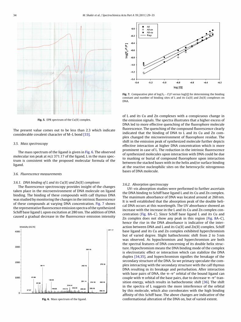
3.4. EPR spectra
EPR spectrum of Cu(II) complex (Fig. 5) shows two regions ofabsorption centered at g|| and g⊥with well resolved nuclear hyper-fine splitting of g|| signal due to copper nuclei in parallel orientation.There is no observable splitting of g⊥ signal. The analysis of spec-trum gives g|| = 2.23, g⊥ = 2.05, A|| = 167 × 10−4 cm−1 which agreewell with the values reported for distorted square planar geom-etry around Cu(II) ion [29,30]. The spectrum shows the relationg|| > g⊥ > 2.0023 which is typical of axially symmetric d9 Cu(II) hav-
ing one unpaired electron in dx2 − y2 orbital [31]. The absence ofsignal corresponding to (�Ms = ±2) in the half field indicates theabsence of any Cu–Cu interaction thus ruling out possibility ofdimeric structure. In axial symmetry, the G-parameter defined asG = g|| − 2/g⊥ − 2, reflects the spin interaction between Cu(II) cen-�(M–O) �(M–N) v(N–O) Phenyl ring vibration
– – – 1480, 1146, 753585 430 1384 1461, 1069, 668591 434 1384 1420, 1073, 745590 436 1383 1440, 1078, 765590 438 1383 1471, 1105, 750

M. Shakir et al. / Spectrochimica Acta Part A 78 (2011) 29–35 33
pectru
tabp
Fig. 3. 1H NMR s
ers in solid polycrystalline complexes. According to Hathawaynd Billing [32], if G > 4, the spin-exchange interaction is negligi-le and if it is less than 4 considerable spin-exchange interactionrevails. In the present case this value comes out to be 4.6 which
Fig. 4. 1H NMR spectrum o
m of the ligand.
again indicates absence of Cu–Cu interaction, thus supporting pro-posed monomeric structure. Kivelson and Neiman have reportedthat g|| values less than 2.3 indicates considerable covalent char-acter of M–L bond and greater than 2.3 indicates ionic character.
f the Zn(II) complex.
34 M. Shakir et al. / Spectrochimica Acta Part A 78 (2011) 29–35
Tc
3
mtl
3
3
tbwotSc
Fig. 7. Comparative plot of log(F − F)/F versus log[Q] for determining the binding
Fig. 5. EPR spectrum of the Cu(II) complex.
he present value comes out to be less than 2.3 which indicateonsiderable covalent character of M–L bond [33].
.5. Mass spectroscopy
The mass spectrum of the ligand is given in Fig. 6. The observedolecular ion peak at m/z 371.17 of the ligand, L in the mass spec-
rum is consistent with the proposed molecular formula of theigand.
.6. Fluorescence measurements
.6.1. DNA binding of L and its Cu(II) and Zn(II) complexesThe fluorescence spectroscopy provides insight of the changes
aken place in the microenvironment of DNA molecule on ligandinding. The binding of these compounds with calf thymus DNAas studied by monitoring the changes in the intrinsic fluorescence
f these compounds at varying DNA concentration. Fig. 7 showshe representative fluorescence emission spectra of the synthesizedchiff base ligand L upon excitation at 280 nm. The addition of DNAaused a gradual decrease in the fluorescence emission intensity
Fig. 6. Mass spectrum of the ligand.
0
constant and number of binding sites of L and its Cu(II) and Zn(II) complexes onDNA.
of L and its Cu and Zn complexes with a conspicuous change inthe emission signals. The spectra illustrates that a higher excess ofDNA led to more effective quenching of the fluorophore moleculefluorescence. The quenching of the compound fluorescence clearlyindicated that the binding of DNA to L and its Cu and Zn com-plex changed the microenvironment of fluorophore residue. Theshift in the emission peak of synthesized molecule further depictseffective interaction at higher DNA concentration which is moreprominent in case of L. The reduction in the intrinsic fluorescenceof synthesized molecules upon interaction with DNA could be dueto masking or burial of compound fluorophore upon interactionbetween the stacked bases with in the helix and/or surface bindingat the reactive nucleophilic sites on the heterocyclic nitrogenousbases of DNA molecule.
3.6.2. Absorption spectroscopyUV–vis absorption studies were performed to further ascertain
the DNA binding to Schiff base ligand L and its Cu and Zn complex.The maximum absorbance of DNA was located around at 260 nm.It is well established that the absorption peak of the double heli-cal DNA occurs at this wavelength. The UV absorbance showed anincrease with the increase in the L and its Cu and Zn complex con-centration (Fig. 8A–C). Since Schiff base ligand L and its Cu andZn complex does not show any peak in this region (Fig. 8A–C),hence the rise in the DNA absorbance is indicative of the inter-action between DNA and L and its Cu(II) and Zn(II) complex. Schiffbase ligand and its Cu and Zn complex exhibited hyperchromismbut of varied degree. Slight bathochromic shift from 2 to 5 nmwas observed. As hypochromism and hyperchromism are boththe spectral features of DNA concerning of its double helix struc-ture. Hypochromism means the DNA binding mode of the complexis electrostatic effect or interaction which can stabilize the DNAduplex [34,35], and hyperchromism signifies the breakage of thesecondary structure of the DNA. So we primary speculate the com-plex interacting with the secondary structure with the calf thymusDNA resulting in its breakage and perturbation. After interactionwith base pairs of DNA, the �–�* orbital of the bound ligand cancouple with � orbital of the base pairs, due to decrease �–�* tran-sition energy, which results in bathochromic shift [36]. The shiftin the spectra of L suggests the more interference of the orbital
by this molecule, which also corroborates with the high bindingaffinity of this Schiff base. The above changes are indicative of theconformational alteration of the DNA on, but of varied extent.
M. Shakir et al. / Spectrochimica A
Ft0s
4
d
[
[
[
[
[
[
[[[[
[[
[[[[
[[[[[
[31] B.A. Goodman, J.B. Rayner, Adv. Inorg. Chem. Radiochim. 35 (1978) 701.[32] B.J. Hathaway, D.E. Billing, Coord. Chem. Rev. 5 (1970) 143.
ig. 8. Absorbance spectra of DNA in the absence and presence of different concen-ration of (A) L, (B) Cu(II) complex and (C) Zn(II) complex. DNA concentration was.10 mM (a). L, Cu(II) complex, Zn(II) complex concentration for DNA-compoundystem was at 12.5 �M (b), 25 �M (c) and 50 �M (d).
. Conclusion
A novel tetradentate Schiff base ligand (L) containing N2O2onor set and its corresponding Co(II), Ni(II), Cu(II) and Zn(II)
[[[[
cta Part A 78 (2011) 29–35 35
complexes have been synthesized and spectrally characterized.Comparative DNA binding studies of L with its Cu(II) and Zn(II)complexes reveals that the Schiff base ligand exhibits highest bind-ing affinity towards DNA molecule due to more open structure ascompared to metal coordinated complexes.
Acknowledgements
The Chairman, Department of Chemistry, Aligarh Muslim Uni-versity, Aligarh, India is acknowledged for providing necessaryresearch facilities. Miss Ambreen Abbasi is grateful to UGC for finan-cial support.
References
[1] D.M.C. Maria, R. da silva, M.G. Jorge, L.R.S. Ana, C.F.C. Paula, B. Schroder, A.V.Manual, J. Mol. Catal. A: Chem. 224 (2004) 207–212.
[2] C. Spinu, A. Kriza, Acta Chem. Slov. 47 (2000) 179.[3] H. Yoon, T.R. Wagler, K.J. O’Connor, C.J. Burrows, J. Am. Chem. Soc. 122 (1990)
4568.[4] M.A. Green, H. Luo, P.E. Fanwick, Inorg. Chem. 37 (1998) 1127.[5] K.C. Emregul, E. Duzgun, O. Atakol, Corros. Sci. 48 (2006) 3243.[6] T. Yu, K. Zhang, Y. Zhao, C. Yang, H. Zhang, L. Qian, D. Fan, W. Dong, L. Chen, Y.
Qiu, Inorg. Chim. Acta 361 (2008) 233.[7] M. Wang, H. Zhu, K. Jin, D. Dai, L. Sun, J. Catal. 220 (2003) 392.[8] A.S. Al-Shiri, Spectrochim. Acta (A) 60 (2004) 118.[9] S. Curreli, E.C. Escudero-Adan, J. Benet-Buchholz, A.W. Kleij, J. Org. Chem. 72
(2007) 7018.10] G. Venkatachalam, N. Raja, D. Pandiarajan, R. Ramesh, Spectrochim. Acta A 71
(2008) 884.11] C.A. Bolos, K.T. Papazisis, A.H. Kortsaris, S. Voyatzi, D. Zambouli, D.A. Kyriakidis,
J. Inorg. Biochem. 88 (2002) 25.12] C.A. Bolos, G.St. Nikolov, L. Ekateriniadou, A. Kortsaris, D.A. Kyrikidis, Metal-
Based Drugs 5 (6) (1998) 323.13] R.L. Fanshawe, A. Mobinikhaledi, C.R. Clark, A.G. Blackman, Inorg. Chim. Acta
307 (2000) 27.14] B.S. Furniss, A.J. Hannaford, P.W.G. Smith, A.R. Tatchell, Vogel’s Textbook of
Practical Organic Chemistry, 5th ed., p. 412.15] A.M. Pyle, J.P. Rehmann, R. Meshoyrer, C.V. Kumar, N.J. Turro, J.K. Barton, J. Am.
Chem. Soc. 111 (1989) 3051.16] X.-Z. Feng, Z. Lin, L.-J. Yang, C. Wang, C.-L. Bai, Talanta 47 (1998) 1223.17] Z.H. Chohan, S. Kausar, Metal-Based Drugs 1 (2000) 17.18] H. Keypour, S. Salehzadeh, R.V. Parish, Molecules 7 (2002) 140.19] S.M. Annigeri, A.D. Naik, U.B. Gangadharmath, V.K. Revankar, V.B. Mahale,
Trans. Met. Chem. 27 (2002) 316.20] G.G. Mohamed, M.M. Omar, A.M.M. Hindy, Spectrochim. Acta A 62 (2005) 1140.21] M.M. Omar, G.G. Mohamed, A.M.M. Hindy, J. Therm. Anal. Calorim. 86 (2006)
315.22] G.G. Mohamed, M.M. Omar, A.M. Hindy, Turk. J. Chem. 30 (2006) 361.23] G. Singh, C.P. Singh, S.M. Mannan, J. Hazard. Mater. 123 (2006) 10.24] P.E. Aranha, M.P. dos Santos, S. Romera, E.R. Dockal, Polyhedron 26 (2007) 1373.25] N. Padma Priya, S. Arunachalam, A. Manimaran, D. Muthupriya, C. Jayabalakr-
ishnan, Spectrochim. Acta A 72 (2009) 670.26] H.L. Singh, A.K. Varshney, Bioinorg. Chem. Appl. (2006) 1.27] N. Raman, C. Thangaraja, Trans. Met. Chem. 30 (2005) 317.28] S. Chandra, L.K. Gupta, D. Jain, Spectrochim. Acta 60A (2004) 2411.29] N. Gupta, R. Gupta, S. Chandra, S.S. Bawa, Spectrochim. Acta A 61 (2005) 1175.30] V.T. Kasumov, A. Bulut, F. Koksal, M. Aslanoglu, I. Ucar, C. Kazak, Polyhedron 25
(2006) 1133.
33] D. Kivelson, R. Neiman, J. Chem. Phys. 35 (1961) 149.34] P. Yang, M.-L. Guo, B.-S. Yang, Chin. Sci. Bull. 39 (1994) 997.35] E.C. Long, J.K. Barton, Acc. Chem. Res. 23 (1990) 271.36] X.F. He, H. Chen, L. Xu, L.-N. Ji, Polyhedron 17 (1998) 316.


![Synthesis, characterization and antimicrobial activities of [Fe(II), Co(II), Ni(II),Cu(II) and Zn(II)] mixed ligand complexes schiff base derived from amoxicillin drug and 4-(dimethylamino)benzaldehyde](https://static.fdokumen.com/doc/165x107/631d3846a906b217b9075674/synthesis-characterization-and-antimicrobial-activities-of-feii-coii-niiicuii.jpg)


