syl741.pdf - University of Calicut
34
U.O.No. 9546/2019/Admn Dated, Calicut University.P.O, 19.07.2019 Biju George K Assistant Registrar Forwarded / By Order Section Officer File Ref.No.82320/GA - IV - J3/2019/Admn UNIVERSITY OF CALICUT Abstract General and Academic - Faculty of Science - Syllabus of MSc Radiation Physics for University Teaching Department under CCSS PG Regulations 2019 with effect from 2019 Admission onwards -Implemented- Orders Issued. G & A - IV - J Read:-1. U.O.No. 4500/2019/Admn dated 26.03.2019 2. Minutes of the meeting of the Board of Studies in Radiation Physics held on 22.05.2019 3. Item No. I.15 in the minutes of the meeting of Faculty of Science held on 27.06.2019 ORDER The Regulations under Choice-based Credit Semester System for Post Graduate Programmes (CCSS-PG -2019) of all Teaching Departments / Schools of the University of Calicut w.e.f 2019 admissions has been implemented vide paper read first above. The meeting of the Board of Studies in Radiation Physics held on 22.05.2019 has approved the Syllabus of MSc Programme in tune with new CCSS PG Regulation implemented with effect from 2019 Admission onwards, vide paper read second above. The Faculty of Science at its meeting held on 27.06.2019 has approved the minutes of the meeting of the Board of Studies in Radiation Physics held on 22.05.2019 vide paper read third above. Under these circumstances, considering the urgency, the Vice Chancellor has accorded sanction to implement the Scheme and Syllabus of MSc Radiation Physics Programme in accordance with the new CCSS PG Regulations 2019, in the University of Calicut with effect from 2019 Admission onwards, subject to ratification by the Academic Council. The Scheme and Syllabus of MSc Radiation Physics Programme in accordance with CCSS PG Regulations 2019 is therefore implemented in the University with effect from 2019 Admission onwards. Orders are issued accordingly. ( Syllabus appended ) To The HoD, Department of Physics Copy to: PS to VC/PA to PVC/ PA to Registrar/PA to CE/JCE I/JCE V/DoA/EX and EGSections/GA I F/CHMK Library/Information Centres/SF/DF/FC
-
Upload
khangminh22 -
Category
Documents
-
view
2 -
download
0
Transcript of syl741.pdf - University of Calicut

U.O.No. 9546/2019/Admn Dated, Calicut University.P.O, 19.07.2019
Biju George K
Assistant Registrar
Forwarded / By Order
Section Officer
File Ref.No.82320/GA - IV - J3/2019/Admn
UNIVERSITY OF CALICUT
AbstractGeneral and Academic - Faculty of Science - Syllabus of MSc Radiation Physics for University Teaching Departmentunder CCSS PG Regulations 2019 with effect from 2019 Admission onwards -Implemented- Orders Issued.
G & A - IV - J
Read:-1. U.O.No. 4500/2019/Admn dated 26.03.20192. Minutes of the meeting of the Board of Studies in Radiation Physics held on22.05.20193. Item No. I.15 in the minutes of the meeting of Faculty of Science held on 27.06.2019
ORDER
The Regulations under Choice-based Credit Semester System for Post GraduateProgrammes (CCSS-PG -2019) of all Teaching Departments / Schools of the University of Calicut w.e.f2019 admissions has been implemented vide paper read first above. The meeting of the Board of Studies in Radiation Physics held on 22.05.2019 has approvedthe Syllabus of MSc Programme in tune with new CCSS PG Regulation implemented with effectfrom 2019 Admission onwards, vide paper read second above. The Faculty of Science at its meeting held on 27.06.2019 has approved the minutes of the meetingof the Board of Studies in Radiation Physics held on 22.05.2019 vide paper read third above. Under these circumstances, considering the urgency, the Vice Chancellor has accorded sanctionto implement the Scheme and Syllabus of MSc Radiation Physics Programme in accordance with thenew CCSS PG Regulations 2019, in the University of Calicut with effect from 2019 Admissiononwards, subject to ratification by the Academic Council. The Scheme and Syllabus of MSc Radiation Physics Programme in accordance with CCSSPG Regulations 2019 is therefore implemented in the University with effect from 2019Admission onwards. Orders are issued accordingly. ( Syllabus appended )
ToThe HoD, Department of Physics Copy to: PS to VC/PA to PVC/ PA to Registrar/PA to CE/JCE I/JCE V/DoA/EX andEGSections/GA I F/CHMK Library/Information Centres/SF/DF/FC

SCHEME, REGULATIONS AND SYLLABUS
FOR THE
M.Sc. RADIATION PHYSICS COURSE(Revised for 2019 Admission)
UNIVERSITY OF CALICUT
1

UNIVERSITY OF CALICUT
SCHEME AND REGULATIONS FOR M.Sc. RADIATION PHYSICS COURSE.(REVISED FOR 2019 ADMISSION)
I. TITLE OF THE PROGRAMME: The program shall be called Master of Science (M.Sc) Degree in Radiation Physics.
II. ABOUT THE COURSE: M.Sc. Radiation Physics course is a highly specializedmultidisciplinary course in Applied Physics. The course will emphasis on the interactionof radiation with human body, application in radiotherapy and the safety measures. Thecourse has immense job potential as highly demanded Medical Physicists andRadiological Safety Officers in Advanced Hospitals, Industrial and ResearchOrganizations in India and abroad as well as faculty, researchers, dosimetrist etc
III. ELIGIBILITY FOR ADMISSION: A pass in B.Sc. Physics as core subject, withMathematics as one of the subjects, from University of Calicut or equivalent with 60%marks in aggregate of the subjects or equivalent grade.
IV. ADMISSION CRITERIA: The admission is made on the basis of the performance in the entrance test of the objective type/ short answer questions of 2 hours duration with the syllabus of B.Sc. Physics of the University of Calicut
V. DURATION OF THE COURSE : 3 years T-wo years course work + One year Internship/Field training– Four semesters each of 6 months followed by internship/clinical training of 12 months- A project work is to be submitted during the period.
VI. MEDIUM OF INSTRUCTION – English
VII. PROGRAMME STRUCTURE
1. The programme shall include three types of courses, viz. Core courses, Elective
courses and Audited Courses.
2. There shall be a field training/ internship for one year for those who opt for jobs asRadiological Safety Officer (RSO) and Medical Physicists. A project work is alsois to be carried out during this period.
3. Total credit for the program shall be 100 (eighty). The audit courses carry a total of4 credits and it is over and above 82. The pattern of distribution of the 82 credits isas detailed below :
i)Total credits for the core courses (theory, practicals, Viva-voce andproject) shall be 74. Out of this the total credits for comprehensiveviva-voce and project work combined together shall be 10 (eight)subject to a minimum of 4 (four) credit for project work.
ii)Total credits for Elective courses shall be 8.
2

Table 1. Structure of the Programme
Programme Duration M.Sc Radiation Physics
Accumulated minimum credits required for successful completion of programme
82
Minimum credits required fromCore courses (including viva-voce)
74
Minimum credits required fromElective courses
8
Total number of credits to beacquired for for fulfilling theprofession (excluding auditcourse)
100
5. Audit courses :
In addition to the above courses for the mandatory requirement of aprogramme there will be two compulsory courses - Ability Enhancement Course(AEC) & Professional Competency Course (PCC) each with 2 credits, and thesecourses are to be done within the first two semesters. The credits will not be countedfor computing the overall SGPA/CGPA of the student. The concerned departmentshall conduct examination for these courses and shall intimate /upload the results ofthe same to the University on the stipulated date during the III Semester. The studenthas to obtain only minimum pass requirements in these two courses. The broadframework of the compulsory audited courses are given hereunder.
Audit course 1 - Presentation of seminar in the related topic is to be done
Audit course 2- developing a computer program in the specific topics in radiologymay be done as
VIII. ATTENDANCE – A candidate is required to put in at least 80% attendance intheory and practical subjects separately in the recognized institution approved for thesame or affiliated to the University of Calicut. This has to be determined on a semesterbasis.
IX. SCHEME OF CLASSES:Every semester will have the course distribution with appropriate number of theory andpracticals. The theory subjects shall have lectures for a total duration of around 80 hourseach and the practical classes will be of about 70 hours each. This works out to be about500 hours teaching per semester including tutorial. It shall be split suitably at the rate ofsix days per week. The fourth semester will accommodate the project work also. The
3

classes per day shall work out as 4 hours for theory and 3 hours for practical. Workingdays per week – 6.
Failure in Semester:A candidate who has failed in the I semester shall be promoted to II and III semester butwill not be allowed to attend the IV semester classes until he/she cleared the 1st semestersubjects. Candidate failed in any semester will not be allowed to do the internship/fieldltraining until the backlog papers are cleared.
Discontinuation: No discontinuation is allowed in normal basis. However if a student hasto discontinue the course in any semester due to the reasons of not his own, and he haspaid the fee for the semester, he can be re-admitted to the semester in later time, if thecoordinator is fully satisfied with the reason. In such cases he has to complete the coursework as per the regulations of the newly admitted batch he is re-admitted and appear forthe examinations accordingly. This provision is conditional on the availability of seatsand facility.
X. PROJECT WORK:Every candidate must do a project work under an approved supervisor (approved by theCoordinator) in a topic having relevance to the application of radiation in medicine,industry, agriculture and research in the 5/6th semester. The project thesis should besubmitted to the University. The supervisor should certify about the satisfactorycompletion of the project. Students must present their project work before a committeeconstituted by the course coordinator. Project Report must be submitted within twomonths from the last working day of the final semester
XI. INTERNSHIP/ FIELD TRAINING:Total duration of the internship/clinical training will be 1 year (as prescribed by theAERB). It should be done under the supervision of a designated academic staff memberof recognized institute. The supervisor must certify to the adequacy of the field trainingon the basis of the thesis report submitted by the candidate. The students shouldnecessarily present at least one seminar on the basis of the field training and the record ofthe field training must be duly certified by the designated officer in the centre and theCourse Coordinator. (The students should pay the charges for clinical training as required by the institution).
XII. RADIOLOGICAL SAFETY OFFICER (RSO) approval by AERB:The University shall initiate steps for registering for the examination for RadiologicalSafety Officer (Level III Medical) certification, for all candidates. The examination for thesame shall be conducted by Radiological Physics and Advisory Division (RPAD) of DAEas per the regulations of the Atomic Energy Regulator Board (AERB). Students qualifyingthis examination will be eligible for RSO. Candidate completing one year clinical trainingare eligible for this examination. Student should attend and qualify the RSO examinationat their own capacity.
4

XIII. SCHEME OF EXAMINATION:Theory papers: Each paper is of three hours duration – Maximum marks 100 End semester examination 70 marks Continuous valuation 30 marks* Practical Examination:Three hours duration – Maximum marks 100 End semester: 70 marks
Continuous evaluation: 30 marks* * Continuous evaluation is based on the regular performance in attendance -4 marks,internal tests (best 2 out of 3)- 12 marks, Assignments 8 marks, seminars (6 marks). Vivavoce: There will be an end semester viva-voce, distributed on all papers of thesemester, for all the four semester. Maximum marks for the viva voce will be 50 marks ineach semester.
Project work: Total marks: 200Project Record : 100
Presentation and viva: 100
Total Marks: 2800 (650+650+750+750)
Mark Distribution: Semester I-IV: 2800 (750+750+650+650) Project Report : 120* Presentation and viva: 80** Project marks are for qualitative evaluation only and not to be included in consolidation.(Viva may include project work and clinical training) Total Marks: 2800 Total credits 100 Minimum credits required to pass 82
Classification of resultsMinimum marks for a pass: Theory 40% (equivalent grade of C) minimum per paper andan aggregate of 50% (equivalent grade of B) - separately for theory and practical. There isno paper minimum for practicals. Evaluation of internship and project work is only forqualitative purpose only. Minimum grade required will be 50% or equivalent. Project andinternship will not be accounted for grading and consolidation. University will issuecertification to the effect as,
1. The candidate has successfully undertaken project work carrying 6 credits2. The candidate has successfully completed one year internship/ clinical training carrying 12 credits
The Semester grade point Average (SGPA) and final grade(Cumulative Grade Point Average CGPA) will be calculated as follows. SGPA = (C1*G1 + C2*G2 +....Cn*Gn)/(C1 + C2 +....Cn)
Where Ci, C2.. Cn are the credits of each paper and G1,G2 ..Gn are the grade scored inthe respective papers. Same procedure will be followed for CGPA for the entire coursework.
5

Presently the Following mark based grade will be followedMark range (in %) Grade point Letter grade Class
80 - 100 8.0-10.0 O Outstanding
70 - 79 7.0-7.9 A+ Excellent
60 - 69 6.0-6.9 A Very Good
55-59 5.5 – 5.9 B + Good
50 - 54 5.0-5.4 B Average (SGPA pass minimum)
40 – 50 4.0-4.9 C F in CGPA)(Paper pass minimum)
< 40 < 4.0 F F in paper
Course incomplete
I Course incomplete
* Fractional percentages should to be rounded off to the next whole number)
XIII. Award of the Certificate:M.Sc Radiation Physics Degree certificate will be awarded to the successful candidatesonly after successful completion of the course as detailed above. However those who arenot interested in Medical Physicists/RSO they can be issued certificates for four semesterM.Sc course without undergoing field training/internship and project work.
-----------------
Semester-vise break-up.......XIV. SEMESTER VISE BREAKE-UP OF COURSE CONTENT:
SEMESTER I: 21 credits RPH1C01 Mathematical Methods in Physics 4 creditsRPH1C02 Classical Mechanics 2 creditsRPH1C03 Basic Electronics 4 creditsRPH1C04 Introductory Nuclear Physics 4 creditsRPH1C05. Basics of Electrodynamics 2 creditsRPH1C06 Interaction of Radiations with Matter 2 creditsRPH1C07 Electronics Practical 2 creditsRPH1C08 Comprehensive semester viva voce 1 creditRPH1A01 Audit course 1-Ability enhancement- Presentation skill 2 credits
SEMESTER II 21 credits RPH2C09 Quantum mechanics 4 creditsRPH2C10 Anatomy, Physiology and Radiobiolgy 4 creditsRPH2C11 Radiation Detection, Measurement and Instruments 4 credits
6
RPH2C12 Numerical Techniques and Computer programming 2 credits RPH2C13 Radiation Physics Fundamental 2 creditsRPH2C14 Practicals in Computer applications 2 creditsRPH2C15 Practicals in Instrumentation in Radiology 2 creditsRPH2C16 Comprehensive viva voce 1 creditRPH2A02 Audit course 2-Professional competency course- 2 credits
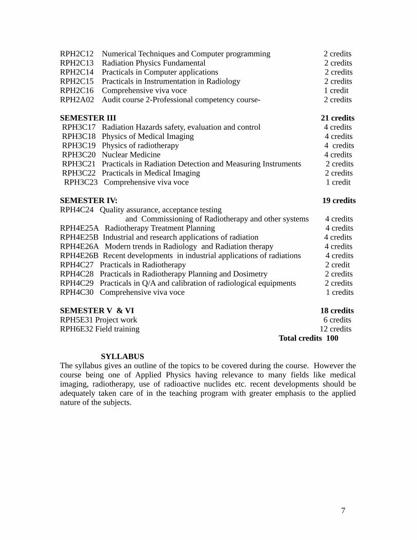
SEMESTER III 21 credits RPH3C17 Radiation Hazards safety, evaluation and control 4 credits RPH3C18 Physics of Medical Imaging 4 credits RPH3C19 Physics of radiotherapy 4 credits RPH3C20 Nuclear Medicine 4 credits RPH3C21 Practicals in Radiation Detection and Measuring Instruments 2 credits RPH3C22 Practicals in Medical Imaging 2 credits RPH3C23 Comprehensive viva voce 1 credit
SEMESTER IV: 19 creditsRPH4C24 Quality assurance, acceptance testing and Commissioning of Radiotherapy and other systems 4 creditsRPH4E25A Radiotherapy Treatment Planning 4 creditsRPH4E25B Industrial and research applications of radiation 4 creditsRPH4E26A Modern trends in Radiology and Radiation therapy 4 creditsRPH4E26B Recent developments in industrial applications of radiations 4 credits RPH4C27 Practicals in Radiotherapy 2 creditRPH4C28 Practicals in Radiotherapy Planning and Dosimetry 2 creditsRPH4C29 Practicals in Q/A and calibration of radiological equipments 2 creditsRPH4C30 Comprehensive viva voce 1 credits
SEMESTER V & VI 18 creditsRPH5E31 Project work 6 creditsRPH6E32 Field training 12 credits Total credits 100
SYLLABUSThe syllabus gives an outline of the topics to be covered during the course. However thecourse being one of Applied Physics having relevance to many fields like medicalimaging, radiotherapy, use of radioactive nuclides etc. recent developments should beadequately taken care of in the teaching program with greater emphasis to the appliednature of the subjects.
7
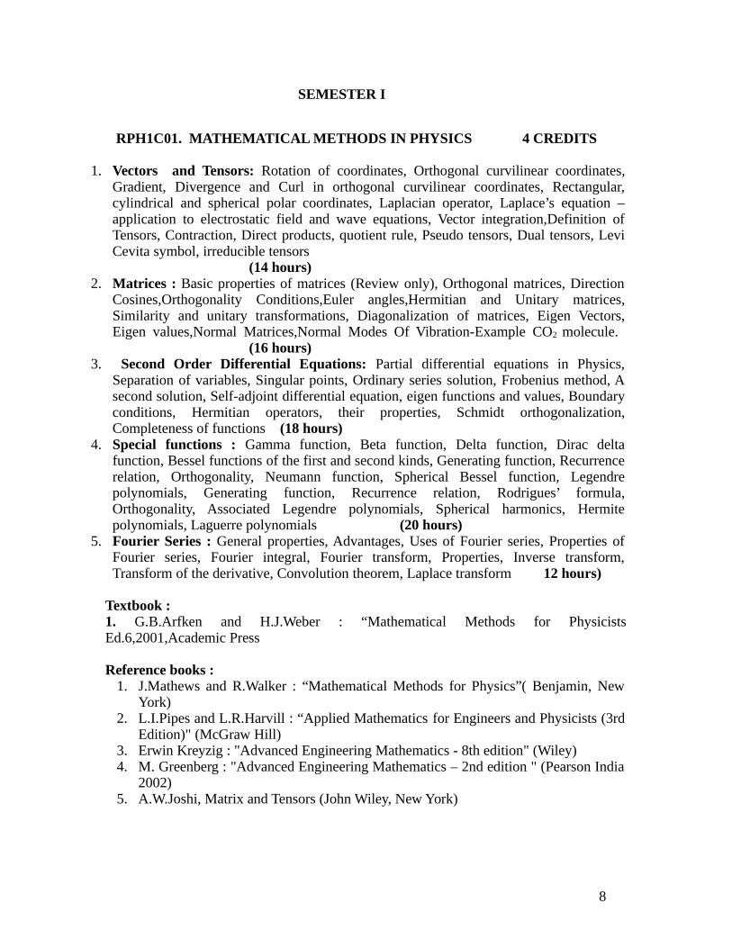
SEMESTER I
RPH1C01. MATHEMATICAL METHODS IN PHYSICS 4 CREDITS
1. Vectors and Tensors: Rotation of coordinates, Orthogonal curvilinear coordinates,Gradient, Divergence and Curl in orthogonal curvilinear coordinates, Rectangular,cylindrical and spherical polar coordinates, Laplacian operator, Laplace’s equation –application to electrostatic field and wave equations, Vector integration,Definition ofTensors, Contraction, Direct products, quotient rule, Pseudo tensors, Dual tensors, LeviCevita symbol, irreducible tensors
(14 hours) 2. Matrices : Basic properties of matrices (Review only), Orthogonal matrices, Direction
Cosines,Orthogonality Conditions,Euler angles,Hermitian and Unitary matrices,Similarity and unitary transformations, Diagonalization of matrices, Eigen Vectors,Eigen values,Normal Matrices,Normal Modes Of Vibration-Example CO2 molecule.
(16 hours)3. Second Order Differential Equations: Partial differential equations in Physics,
Separation of variables, Singular points, Ordinary series solution, Frobenius method, Asecond solution, Self-adjoint differential equation, eigen functions and values, Boundaryconditions, Hermitian operators, their properties, Schmidt orthogonalization,Completeness of functions (18 hours)
4. Special functions : Gamma function, Beta function, Delta function, Dirac deltafunction, Bessel functions of the first and second kinds, Generating function, Recurrencerelation, Orthogonality, Neumann function, Spherical Bessel function, Legendrepolynomials, Generating function, Recurrence relation, Rodrigues’ formula,Orthogonality, Associated Legendre polynomials, Spherical harmonics, Hermitepolynomials, Laguerre polynomials (20 hours)
5. Fourier Series : General properties, Advantages, Uses of Fourier series, Properties ofFourier series, Fourier integral, Fourier transform, Properties, Inverse transform,Transform of the derivative, Convolution theorem, Laplace transform 12 hours)
Textbook : 1. G.B.Arfken and H.J.Weber : “Mathematical Methods for PhysicistsEd.6,2001,Academic Press
Reference books : 1. J.Mathews and R.Walker : “Mathematical Methods for Physics”( Benjamin, New
York)2. L.I.Pipes and L.R.Harvill : “Applied Mathematics for Engineers and Physicists (3rd
Edition)" (McGraw Hill) 3. Erwin Kreyzig : "Advanced Engineering Mathematics - 8th edition" (Wiley) 4. M. Greenberg : "Advanced Engineering Mathematics – 2nd edition " (Pearson India
2002)5. A.W.Joshi, Matrix and Tensors (John Wiley, New York)
8

RPH1C02 : CLASSICAL MECHANICS (2 CREDITS)
1.Lagrangian and Hamiltonian Formulation : Preliminary ideas about Constraints and Generalized coordinates, D'Alemberts principle and Lagrange’s equation, Velocity dependent potentials, Simple applications of Lagrangian formulation,Hamilton’s Principle, Conservation theorems and symmetries, Lagrange’s equation from Hamilton’s principle, Two- body central problems, Equivalent one -body and one dimensional problem, Kepler problem, Inverse square law of force, Laplace-Lenz vector, Scattering in a central force field, Transformation to lab coordinates. Exercises (18 hours)
2.Hamiltonian Formulations: Legendre Transformation and Hamilton’s equations, Cyclic co-ordinates and conservation theorems, Principle of least action, Canonical transformations and examples, Infinitesimal canonical transformations, Poisson brackets and other canonical invariants, Equation of motion in Poisson bracket form, Angular momentum Poisson brackets, Hamilton-Jacobi equation, Hamilton’s principal and characteristic function, H-J equation for the linear harmonic oscillator, Separation of variables, Action-angle variables, H-J formulation of the Kepler problem, H-J equation and the Schrödinger equation. (19 hours)
Books for study : Goldstein, Classical Mechanics, 3rd Edition. Addison Weley
References: 1. V.B.Bhatia : “Classical Mechanics” (Narosa Publications, 1997)2. N.C.Rana and P.S.Joag : “Classical Mechanics” (Tata McGraw Hill, 2011) 3. R.G.Takwale and P.S.Puranik : “Introduction to Classical Mechanics” (Tata
McGraw Hill, 1978 ) 4. Atam P. Arya : "Introduction to Classical Mechanics, " ( 2nd Edition, Addison
Wesley, 1998)
RPH1C03. BASIC ELECTRONICS – 4 CREDITS
1. Transistor Amplifier :BJT: Biasing and ac models, voltage amplifiers, power amplifiers (EP 11:3 – 11-5), emitter follower , differential amplifier, FET: h-parameters, FET small signal model, biasing the FET, analysis of common source and common drain amplifiers and the high frequency response, FET as VVR and its applications. MOSFET: circuit symbol and equations, small signal model Digital MOSFET circuits. (16 hours) Texts: 1.Electronic principles, Malvino 6th Edition, Tata McGrows Hills India 2.Integrated Electronics, Millman and Halkias, Tata McGrows Hills India
2. Microwave and Photonic Devices: Tunnel diode, Transferred electron devices,Negative differential resistance and devise operation, Radiative transitions and opticalabsorption, Light emitting diodes (LED) –Visible and IR, Semiconductor lasers -materials, operation (population inversion, carrier and optical confinement, opticalcavity and feedback, threshold current density),Photo-detectors, Photoconductor (Lightdependent resistor- LDR) and photodiode, p-n junction solar cells - short circuitcurrent, fill factor and efficiency 12 hours)
Books for study:1. S. M. Sze, “Semiconductor Devices- Physics and Technology”, John Wiley &Sons,
9

3. Operational Amplifier: Dual input differential amplifier DC and AC analysis, Op-Amp, block diagram representation, analysis of a typical Op-Amp equivalent circuit idealOp-Amp characteristics, equivalent circuit, open loop configurations, Op-Ampparameters input offset voltage & current, input bias current, output offset voltage,CMRR , Op-Amp with negative feedback: voltage series feedback amplifier: gain,input & output impedances , Frequency response, compensating networks (14 hours)
Text: Op-Amps and Linear Integrated Circuits: 3rd Edition, R. A. Gayakwad, PHI
4. OPAMP Applications: Summing, scaling and averaging amplifiers , Analog integrator and differentiator Electronic analog computation, Active filters: Butterworth filters (low & high orders), Low pass, High pass, band pass (wide & narrow band) and band reject filters , Oscillators: Phase shift,Wein bridge, Quadrature oscillators, Square, triangular and saw-tooth wave generators , comparators, zero crossing detectors, Schmitt trigger (12 hours) Texts: 1. Op-Amps and Linear Integrated Circuits: 3rd Edition, R. A. Gayakwad, PHI
2. Integrated Electronics, Millman and Halkias, TMH India.
5. Digital Electronics: Arithmetic circuits: adder, adder/subtracter, ALU, RS, JK and JKMS flip-flops, Registers: types of registers, SISO & 7491, SIPO &74164, PIPO, 74198,applications of shift registers. Counters:asynchronous counter & 7493A, decoding gates,synchronous counters, 7490A, decade counters . D/A-A/D converters , Memory &memory addressingMicroprocessors and Microcontrollers: Microprocessor, architecture of 8085: Busorganization, Registers, memory, block diagram of 4 bit register, memory map, tri-statebuffer , 8085 functional pin diagram, control & status signals, microprocessorcommunication and bus timing (memory read/write operations), address data de-multiplexing , microcontrollers, architectural overview and block diagram ofmicrocontrollers , functional pin diagram of Atmega16 microcontroller. ( 16 hours)
Texts: 1. Digital Principles and Applications: 6th Edition, Leach, Malvino and Saha, (Tata McGraw Hill).2.Microprocessor Architecture and Programming and Application, Ramesh S.Gaonkar, (New Age Publishers.)3. The 8051 Microcontroller: 2Ed, Kenneth J. Ayala, Thomson, (Delmar)4. Atmega16 microcontroller data sheet available (from Atmel website.)
General references:1. Electronic devices and circuit theory, Robert L. Boylstead & L. Nashelsky –
Pearson Education.2. Electronic devices, 5th Edition, Floyd, Pearson Education. 3. Alen Motorshed4. Microelectronic Circuits: Analysis & Design, M. H. Rashid, PWS Publishing 5. Linear Integrated circuits, D. R. Choudhuri, S. Jain, (New Age International) 6. Fundamentals of Microprocessors and Microcomputers, 2nd Edition, B.Ram,
Dhanapathi Rai & Sons. 7. Embedded C Programming and the Atmel AVR, Barnett, O’cull, Cox, Cengate
Learning.
10

RPH1C04. INTRODUCTORY NUCLEAR PHYSICS 4 CREDITS
1. Nuclear Forces: Four basic forces - Gravitational, Electromagnetic, Weak and Strong -Relative strengths, Properties of the nucleus, size, binding energy, angular momentum,The deuteron and two-nucleon scattering experimental data, Simple theory of thedeuteron structure, Low energy n-p scattering, characteristics of nuclear forces, Spindependence, Tensor force, Scattering cross sections, Partial waves, Phase shift, Singletand triplet potentials, Effective range theory, p-p scattering. (14 hours)
2. Nuclear Decay: Basics of alpha decay and theory of alpha emission. Beta decay,Energetics of beta decay, Fermi theory of beta decay, Comparative half-life, Allowedand forbidden transitions, Selection rules, Parity violation in beta decay. Neutrino.Energetics of Gamma Decay, Multipole moments, Decay rate, Angular momentum andparity selection rules, Internal conversion, Lifetimes. (18 hours)
3. Nuclear Models, Fission and Fusion: Shell model potential, Spin-orbit potential,Magnetic dipole moments, Electric quadruple moments, Valence Nucleons, Collectivestructure, Nuclear vibrations, Nuclear rotations, Liquid drop Model, Semi-empiricalMass formula, Energetics of Fission process, Controlled Fission reactions. Fusionprocess, Characteristics of fusion, solar fusion, Controlled fusion reactors. Criticalconditions, four factor formula (16 hours)
4. Nuclear reactions: Nuclear Reactions, Energetics of nuclear reactions, conservationlaws, scattering and reaction cross- sections, theoretical calculation, measurement ofcross-section, Nuclear reaction models-compound nucleus reactions, Direct reactions,resonance reactons, statistical theory, optical potential, heavy ion reactions. (16 hours)
5. Reactor Physics: Interaction of neutrons with mater, Nuclear Fission, Neutron ChainReacting Systems, Criticality, Multiplication Factor, Neutron balance conditions forcryticality, Conversion and Breeding, Types of nuclear reactors, Reactor Power, FuelBurnup, Neutron transport in reactors, Neutron current density, Equation of Condinuity,Fricks law, Diffusion Equation, Neutron moderation, Leathergy, Multiscatteredneutrons, Fermi Age theory, Age equation, Solusions to the age equation, Elasticmoderation time, Slowing down kernals, Neutron absorption with moderation andfission, Weak absorption, Resonance escape, Thermal neutron spectra, Reactor Power,Criticality for reactor geometries. (16 hours)
Books for study 1. H.Enge : Introduction to Nuclear Physics” (Addison Wesley)2. John. R. Lamarsh, Introduction to Nuclear Reactor Theory, Addison Wesley, USA
Reference:2. H.S.Hans: Nuclear Physics – Experimental & theoretical (New Age International 2001)3. Kenneth S Krance, Introductory nuclear physics, (Wiley india, 2012)4. S.B.Patel, An introduction to nulcear Physics, (New Age International)5. George I. Bell and Samuel Glasstone, Nuclear Reactor Theoy, Van Nostrand Reinhold Company, USA
11

RPH1C05. BASICS OF ELECTRODYNAMICS 2 CREDITS
1. Electrodynamics: Review of Electrostatics and Magnetostatics, Time varying fieldsand Maxwell’s equations, Potential formulations, Gauge transformations, boundaryconditions, wave equations and their solutions, Poynting theorem, Maxwell’s stresstensor. (14 hours)
2. Transmission lines, Wave guides and cavity resonators: Transverse electromagneticwaves along a parallel plate transmission line, General transmission line equations, Wavecharacteristics on finite transmission lines, General wave behavior along uniform guidingstructures, Rectangular wave guides, Cavity resonators (10 hours)
3. Electromagnetic Radiation: Retarded potentials, Jefimenkos equations, Point charges,Lienard-Wiechert potential, Fields of a moving point charge, Electric dipole radiation,Magnetic dipole radiation, Power radiated by point charge in motion. Radiation reaction,Physical basis of radiation reaction (16 hours)
Text:1. David K. Cheng : “ Field and Wave Electromagnetics” (Addisson Wesley)
Reference books :
1. J.D.Jackson : “Classical Electrodynamics” (3rd Ed.) (Wiley,1999)2. David Griffiths : “ Introductory Electrodynamics” (Prentice Hall of India, 1989)3. Podgorsak, Ervin B: “Radiation Physics for Medical Physicists” (Springer, 2010)4. Helmut Wiedemann: “Particle Accelerator Physics”(3rd Ed.)(Springer, 2007)
RPH1C06. INTERACTION OF RADIATIONS WITH MATTER 2 CREDITS
1. Interaction of electromagnetic radiation with matter (14 hours)Thomson scattering - Photoelectric absorption – Angular distribution of photoelectrons–Compton effect, Compton process – Klein Nishina cross-section – Scatteringcoefficients–angular distribution of Compton electrons – Pair production – Annihilation radiation,electrons – energy momentum conservation, Photo nuclear reactions,– Attenuation –Linear, mass attenuation coefficients- Total absorption coefficients. Absorption andscattering coefficients and cross sections.
2. Interaction of electrons and heavy charged particles with Matter (16 hours) Classical theory of inelastic collisions with atomic electrons, energy loss per ion pairby primary and secondary ionization, Cerenkov radiation, Electron, Absorption,Scattering, Excitation and Ionization. Values of w in different media, radioactivecollision– Radiation energy loss (bremsstrahlung)– Range of beta particles, Rangestraggling, absorption of beta particles and back scattering, self absorption . Interactionof heavy charged particles with matter –Energy loss by collision, maximum energyloss in a single collision, range energy relation- Bragg curve, specific ionization,Bethe-Bloch formula for collision, stopping power and radiation stopping power
12

3. Interaction of Neutrons with Matter (10 hours) Neutron capture Neutron sources, properties, energy classification, Elastic andinelastic scattering coefficients and cross sections– Energy transfer and logarithmicenergy decrement, Inelastic scattering, Nuclear reaction, Dependence on E and Z, (n,p), (n, 2n), (n, f) and other reaction, Neutron activation, Radio- isotope production.Familiarization of standard data libraries
STANDARD BOOKS FOR STUDY
1. E.B Podgorsak : "Radiation Physics for Medical Physicists". Springer, USA2. F.H.Attix “Radiation Dosimetry” Vol I-III, Academic press New York, 1985.
REFERENCES1. R.D Evans : "The Atomic Nucleus" (Tata -McGraw Hill Publishing Company)2. G.F.Knoll : “Nuclear Radiation Detectors” (Willy international, New york)3. H.E.Jones, J.R.Cunnigham, “The Physics of Radiology” Charles C.Thomas, NY,
1980.4. J.R Greening, Medical Physics, North Holland publishing Co, New York, 1981.5. W.J.Meredith and J.B.Massey “Fundamental Physics of Radiology” John Wright
and sons, UK, 1989.6. W.R.Hendee, “Medical Radiation Physics”, Year Book – Medical Publishers Inc.
London, 1981.7. F.M Khan : “Physics of Radiation Therapy”- Fourth Edition8. E.J.Hall Radiobiology for Radiologists J.B.Lippincott Company, Philadelphia
1987.
PRACTICALS:-RPH1C07. ELECTRONICS PRACTICALS – (70 Hours) 2 CREDITS
( Minimum 12 expts. are to be carried out)1. Measurement of L, C and R by Universal bridge2. Series resonance and Q of a coil3. Two stage RC coupled amplifier – frequency response4. Construction of a voltage multiplier – Measurement of Ripple5. Characteristics of a regulated power pack using 2N30556. DC voltage regulator using OPAMP7. Feedback amplifier8. Construction of an oscillator9. Multivibrator- Monostable and astable 10. Low pass and high pass filter -first and second order11. OPAMP circuits – Inverting and non inverting amplifiers12. Integrator and differentiator circuit using OPAMP13. Simple D/A converter – Ladder type14. Coincidence and anti-coincidence timed circuits15. Pulse shaping circuits16. Microprocessor experiments (Addition, subtraction, division and multiplication –
8 bit using 8085)
13

SEMESTER II
RPH2C09. QUANTUM MECHANICS– 4 CREDITS
1. Formulation of Quantum Mechanics: Vector spaces, The Hilbert space,Dimensionsand basis, Operators and properties, Representation of vectors and operators,Commutator,Functions of operators, Eigen values and eigen vectors, Matrix representation of bras, ketsand operators, Coordinate and momentum representations and their connection, Thefundamental postulates Probability density, Superposition principle, Observables andoperators, The uncertainty principle (16 Hours)
2. Quantum Dynamics : The equation of motion, Schrödinger, Heisenberg and theInteraction pictures of time development, The linear harmonic oscillator in theSchroedinger and Heisenberg pictures, Hydrogen atom problem (24 hours)3. Theory of Angular Momentum: Angular momentum operators, Matrix representationof angular momentum operators, Pauli spin matrices, Orbital angular momentum, Thehydrogen atom, Addition of angular momenta, Clebsh-Gordon coefficients, Simpleexamples (12 hours)
4. Symmetry and Conservation Laws : Space-time symmetries, Space translation andconservation of linear momentum, Time translation and conservation of energy, Spacerotation and conservation of angular momentum, Space inversion and time reversal,Identical particles, Construction of symmetric and anti symmetric wave functions, Slaterdeterminant, Pauli exclusion principle, Bosons and Fermions, Spin wave functions fortwo electrons, The ground state of He atom, Scattering of identical particles (12 hours)
5. Scattering : Classical approach for Scattering in a central force field- alpha scattering,Transformation to lab coordinates. Quantum mechanical approach -Scattering crosssection and scattering amplitude, Low energy scattering by a central potential, Themethod of partial waves, Phase shifts, Optical theorem, Convergence of partial waveseries, Scattering by a rigid sphere, Scattering by a square well potential, High energyscattering, Scattering integral equation and Born approximation (14 hours) Books for study: N.Zettili, Quantum Mechanics – Concepts and applications (John
Wiley&Sons, 2004)
Reference books :1. V.K.Thankappan : “Quantum Mechanics” (Wiley Eastern)2. L.I. Schiff : “Quantum Mechanics” (McGraw Hill)3. P.M.Mathews and K.Venkatesan : “A Textbook of Quantum Mechanics" (TataMG Hill)5. J.J.Sakurai : “Modern Quantum Mechanics” (Addison Wesley)6. A.Ghatak and S.Lokanathan : “Quantum Mechanics” (Macmillan)
RPH2C10. ANATOMY, PHYSIOLOGY AND RADIOBIOLGY 4 CREDITS
1. Basic Anatomy : Introduction to the Human Body, Skeletal System, Muscular System,Nervous Tissue, Central Nervous System, Peripheral and Autonomic Nervous Systems,Cardiovascular System, Lymphatic System and Body Immunity, Respiratory System,Digestive System, Urinary System, Water and Electrolyte Balance, Reproductive System,Xray anatomy, CT/MRI anatomy surface anatomy applied to RD and RT (16 hours)
14

2. Clinical Aspects of Radiation Oncology: Biology of cancer and tumor, catagories ofdisease, treatment modalites-Radiation therapy, Surgery, Chemotherapy, HormoneTherapy, Immunotherapy & radionuclide therapy, Benign and malignant disease, Methodsof spread of malignant disease, Treatment intent, Curative & Palliative (12 hours)
3. Radiation Chemistry and Cell Kinetics: Elements of cell biology, Action of ionizingradiation on living cells –Direct and indirect actions, effects at Molecular level, RadiationChemistry, Stochastic Nature Of Energy Transfers CellularLevels, Reactions Of TheProducts of Water Radiolysis, G Value, Expression Of Yield In Radiation Chemistry,Products Of Radiolysis, Fricke Dosimeter Direct And Indirect Action, Recombination,Restitution and Repair, DNA Structure And Radiation Damage, Theories And Models ForCell Survival, Clonogenic Survival, Biological Survival Curves, Development Of TheTarget Theory Model, MultitargetSingleHit Survival, Molecular Models For Cell Death,Molecular Theory Of Radiation Action, Survival Curve And Its Significance, SignificanceOf Shoulder On The Survival Curve, Repair Of Sublethal Damage, Repair Of PotentiallyLethal Damage, (18 hours)
4. Radiobiological Concepts: Linear energy transfer and its effect, Tumor lethal dose,tissue tolerance dose and therapeutic ratio, Radiobiological effectiveness (RBE), Oxygeneffect, Oxygen enhancement ratio(OER), Five R's of Radiobiology, TCP/NTCP Time dosefraction(TDF) basis for dose fractionation in radiotherapy Concept of nominal standarddose (NSD), Linear Quadratic models, AlphaBeta concepts. BED, Rate equationsSensitizers and Protectors, Reduction of side effects. Somatic effects of radiation –Acuteradiation sickness –LD50 dose –Effect of radiation on skin –Blood changes –Sterility –Cataract formation –Effects of chronic exposure to radiation. Doubling dose and its effecton genitic equilibrium, Tissue structure and radiation effect, Radiation effect on fetus,Fractionation and its effect, effect on different systems dependence on dose and dose rate,tolerance limits for various systems, acute radiation syndrome, effects of low levelirradiation, effects relevant to women, fetus and children (22 hours)
5. Modifiers and Dose limits: Modification Of The Radiation Response, Role of WaterTemperature And Radiation Damage, Oxygen Effect, Modification of The RadiationResponse, Hypoxia And Radiosensitivity in Tumor Cells, Late Effects of Radiation onNormal Tissues, Nonstochastic/ Deterministic Effects Stochastic Effects, Fractionationand protraction of exposure in the modification of late radiation injury, Stochastic EffectsRadiation Carcinogenesis, Biological Modifiers, Cell Kinetics, Cell Cycle ControlMechanisms, RTOGs, IMAMI and CONTEC recommendations (14 hours) Books for study1. Radiobiology- Hand book for teachers and students, TC-42, IAEA,2010
Reference:1. Delmar’s “Fundamentals of Anatomy & Physiology”, Thomson LearningUSA, 20012. A LANGE medical book “Basic Radiology” 2nd Edition, The McGrawHill20113. Edward L. Alphen, “Radiation Biophysics” Academic Press, Second Edition.4. EJ Hall and Amato J Glaccia, “ Radiobiology for the Radiologist”, 7 th Edn., LippincottWilliams &Wikins, USA5. IAEA, AERB , NCRP PUBLICATIONS ON DOSE LIMITS.
15

RPH2C11. RADIATION DETECTION, MEASUREMENT AND INSTRUMENTS 4 CREDITS1. Gas filled , Scintillation Detectors: Relationship Between Voltage and ChargeCollected -characteristic curve, Different Types of Gas-Filled Detectors, IonizationChambers, Proportional Counters, Geiger-Muiller Counters, Gas-Flow Counters, RateMeters Scintillation detectors, Inorganic (Crystal) Scintillators , Organic Scintillators ,Gaseous Scintillators , The Relationship Between Pulse Height and Energy and Type ofIncident Particle , The Photomultiplier Tube, Assembly of a Scintillation Counter and theRole of Light Pipes, Dead Time of Scintillation Counters, Sources of Background in aScintillation Counter, Resolving time and resolving power of detectors. (14 hours)
2. Semiconductor, Thermo luminescent Dosimetry Miscellaneous Detectors::Different Types of Semiconductor Detectors - Surface-Barrier Detectors - Diffused-Junction Detectors - Silicon Lithium-Drifted [Si(Li)] Detectors - Germanium Lithium-Drifted [Ge(Li)] Detectors - germanium (Ge) Detectors -CdTe and HgI Detectors -Radiation Damage to Semiconductor Detectors , Detector Telescopes (E-dE Detectors),Position-Sensitive Detectors. Thermo luminescent dosimetry; process and properties,photon energy dependence, fading, residual TL and annealing for reuse, repeated read outof TLDs, TL- instrumentation, beta, gamma extremity dosimetry, ultra thin TLDs,graphite/boron carbide mixed TLDs, glow curve analysis, -common TLD materials, theircharacteristics, energy dependence and method of use. Chemical dosimetery, Organic andinorganic systems, Fricke dosimeter, FBX dosimeter, Free radical dosimeter – Cericsulphate dosimeter, Other high and low level dosimeters, Applications in Radiotherapyand industrial irradiators, Glass dosimetry, Calorimetry, Instruments of personalmonitoring, films, digital pocket dosimeters ,Radiation survey meter, Contaminationmonitors ,gamma ray spectrometer, whole body monitor etc. , Thermal and fast neutronsurvey meters (20 hours)
3. Neutron Detectors, Spectroscopy: The BF3 Counter, 6Li- Counters, 3He proportionalcounter, Fission Chambers, Neutron Detection by Foil Activation (Activation counter),Detection of Fast Neutrons using Threshold Activation Reactions (Threshold Detector),The Time-of-Flight Method, Bubble chambers, Sources of Radiation, Irradiation of theSample, Counting of the Sample, Analysis of the Results, Advantages and Disadvantagesof the Activation Analysis Method ,Measurement of neutron flux -Activation andabsorption methods, CR -39 films, SSNTD, Albedo Dosimeter, Manganese Bath,Precision long Counter. (16 hours)
4. Nuclear Instrumentation: NIM Concept, High-Voltage Power Supply, Preamplifier,Amplifier, Oscilloscope, -Differentiating Circuit- Integrating Circuit- Delay Lines- PulseShaping- Timing- Coincidence -Anti coincidence Measurements, Pulse-ShapeDiscrimination, Discriminator- Single-Channel Analyzer (SCA). energy measurements,introduction to spectroscopy: Definition of Energy Spectra, Measurement of an IntegralSpectrum and Differential Spectrum, Energy Resolution of a Detection System,Multichannel Analyzer (MCA) -calibration, Timer, Analog-to-Digital Converters (ADC).Time of flight spectrometer, charged particle spectroscopy, Measurement of energystraggling, (16 hours)
16

4. Counting Statistics and error analysis: Characterization of data, Statistical models-binomial - poisson -normal distributions, Application of statistical models -checkout forconsistency -precision of single measurement, Error propagation- sum -difference-multiplication- division -mean value of combination, Optimization of countingexperiments, Limits of detectability, Distribution of time intervals -successive events-time to next event -scaled event -Error analysis- Accidental and systematic errors, errordistribution and error function, error in numerical computations and propagation oferrors, applications in calibration of instruments, standards in overall error estimation,applications to evaluation of noise in amplifier system, examples of medical statisticsfor follow up studies (14 hours)
BOOKS FOR STUDY 1. Nicholas Tsoulfanidis - Measurement and Detection of Radiation, second edition2. G.F.Knoll, Radiation detection and measurements, 3rd edn, John Wiley, New York
AND REFERENCES
1. W.E. Burcham & M. Jobes – Nuclear and Particle Physics – Longman (1995)2. Mcknlay, A.F., Bristol, Adam Hilger , Thermoluninescense Dosimetry-Medical
Physics Handbook 5,,CRC Press, USA3. W.J.Meredith and J.B.Massey “Fundamental Physics of Radiology” John Wright
and sons, UK, 1989.4. J.R.Greening “Fundamentals of Radiation Dosimetry”, Medical Physics Hand
Book Series Adam Hilger Ltd., USA
RPH2C12. NUMERICAL TECHNIQUES AND COMPUTER PROGRAMMING 2 CREDITS
1. Roots of transcendental equations : Location theorem, Bisection (half interval)method- Method of false position (Regula Falsi), Graphical Method, Newton-Raphsonmethod, Geometric significance, inherent error, convergence of Newton Raphsonmethod, Special procedure for Algebraic equations, Iteration Method, Geometry andconvergence of iteration process. (10 hours) 2. Interpolation and curve fitting : Errors in polynomial interpolation, Detection oferrors, Linear interpolation, Interpolating polynomials, Lagrange interpolatingpolynomial, Difference calculus, Detection of errors, Newton forward and backwarddifference formulae, Least squares curve fitting( linear and nonlinear), (9 hours)
3. Numerical integration and Ordinary differential equations : Numericaldifferentiation, Maximum and minimum value, Numerical integration, Trapezoidal andSimpson’s methods, Newton Cote’s method, Gauss quadrature, Solution of ordinarydifferential equations – Euler’s Maclaurin method, Runge-Kutta methods, (9 hours.) 4. Determinants and matrices: Evaluation of numerical determinants, Cramer's rule,Successive elimination of unknowns-division by leading co-efficients, Gaussmethod,solution by inversion of matrices:Solution of equation by matrix methods,Systems soluble by Iteration and condition for convergence. The Eigenvalue problem-Eigen values of asymmetric tridiagonal matrix-Householder's method-QR method,Enough Exercises (12 hours)
17

5. C Programming fundamentals (to be taught as a part of practicals) : Constantsand variables, Data types, Type declaration of variables, Symbolic constants, Arithmeticoperators, Increment and decrement operators, Conditional operator, Bitwise operators,Hierarchy, Arithmetic expressions, Logical operators and expressions, Assignmentoperators, Arithmetical and assignment statements, Mathematical functions, Input/outputstatements, Formatted I/O, Relational operators, Decision making and branching, Go to,if, if…else, switch statements, Looping, While, do and for, Arrays, Handling charactersand strings, Functions and voids, Structures, Pointers(elementary ideas only), Fileoperations (defining, opening, reading, writing, updating and closing of files (20hours)
Books for study :1. S.S.Shastry : “Introductory methods of Unumerical analysis” (Prentice Hall ofIndia,1983)2. E.Balaguruswamy : “Programming in ANSI C” (Tata-McGraw Hill, 1992)
Reference Books :1. V. Rajaraman : “Programming in C”, PHI2. J.H. Rice : Numerical methods-software and analysis (McGraw Hill, 1983)3. J.B. Scarborough: Numerical mathematical analysis (Oxford and IBH, 6th Edn)
RPH2C13. RADIATON PHYSICS FUNDAMENTAL 2 CREDITS
1.Radiation Quantities and Units: Radiation quantities and units – Radiometry ,Particle flux and fluence, Energy flux and fluence, Cross section, Mass energy transferand mass absorption coefficients, LET - Radiation chemical yield – W value – Dosimetry– Energy imparted –Absorbed dose- Radiation and tissue weighting factors, equivalentdose, effective dose, committed equivalent dose, committed effective dose, Concepts ofcollective dose – KERMA- CEMA, XEMA, TERMA, Exposure, Air kerma rate constant– Charged particle equilibrium (CPE) – Relationship between kerma, absorbed dose andexposure under CPE, Dose equivalent, Ambient and directional dose equivalents [(H*(d)and H’(d)], individual dose equivalent penetrating Hp(d), Individual dose equivalentsuperficial Hs(d). (20 hours)
2. Standards and Measurement of Ionizing Radiation: Standards – Primary andSecondary Standards, Traceability, Uncertainly in measurement. Bragg-gray principleand air wall chamber, Standardization of X-ray, electrons and gamma ray beams:Determination of exposure and air kerma, conditions for the realization of exposure,ionization chamber for low, medium and high energy x-rays and gamma rays,determination of absorbed dose, -Bragg Gray theory and its validity, Burlin’s theory formeasurement for radiation quantities, design of free air chambers(FAIC), thimblechamber, chamber calibration, Electrometers, Parallel plate chambers, Ion collection,Polarity effect, Measurement of exposure, Radiation absorbed Dose, Kerma, Relationbetween Dose, Kerma and exposure. Calculation of Absorbed Dose from Exposure.Effective point of measurement. Calibration of secondary standards-Calibration factors-Concepts of protocols (20 hours)
BOOKS FOR STUDY 1. F.A.Attix “Radiation Dosimetry” Vol I-III, Academic press New York, 1985.2. ICRU Report No. 85, Journal of ICRU Vol. 11 No1.(2011) Oxford University Press3. H.E.Jones, J.R.Cunnigham, “The Physics of Radiology” Charles C.Thomas, NY, 1980
18

REFERENCES1. W.J.Meredith and J.B.Massey “Fundamental Physics of Radiology” John Wright andsons, UK, 1989.2. W.R.Hendee, Medical Radiation Physics, Year Book – Medical Publishers Inc.London, 1981.3. E.J.Hall Radiobiology for Radiologists J.B.Lippincott Company, Philadelphia 1987.4. J.R Greening, Medical Physics, North Holland publishing Co, New York, 1981
RPH2C14- PRACTICALS IN COMPUTER APPLICATIONS Minimum experiments. to be carried out 12. (70 Hours) 2CREDITS
1. Solution of quadratic equation2. Curve fitting methods- Least squares, chi square, 3. Error estimation in curve fitting with - in Gausian, exponential, polynomial4. Numerical interpolation5. Numerical Integration(Simpson’s method)6. Numerical solution of first order differential equation by Runge_Kutta method
Simulation (BASIC / C)7. Quantum mechanical particle in a box8. Bouncing ball9. Phase space plots for damped and undamped oscillator10. Transmission coefficient for a potential barrier11. Evaluation of pi (p) using montecarlo12. Simulation detector response13. Alpha scattering14. Attenuation of radiations15. Dose evaluation using stopping power table
RPH2C15. PRACTICALS IN INSTRUMENTATION IN RADIOLOGY (70 Hours)2 CREDITS (Minimum 12 expts. to be carried out )
1. GM counter – characteristics – plateau and variation of pulse height with appliedvoltage and resolving time
2. GM counter – Statistics of counting 3. GM counter – Inverse square law properties 4. Gamma ray spectroscopy using NaI(Tl) -characteristics – plateau and variation of
pulse height with applied voltage5. Scintillation spectrometer – Calibration and determination of unknown energy6. Measurement of linear and mass attenuation coefficients for a gamma ray beam
using GM counter7. Measurement of range of Beta rays (1) in air (2) in material like aluminum and
calculation of absorption coefficients- GM counter – Feather Analysis – end pointenergy
8. Absorption of Gamma rays from different isotopes -energy and Z dependence9. Alpha spectroscopy using solid state detectors- determination of energy10. Experiments with solid state nuclear track detector(SSNTD)11. Resolution of Scintillation counter at various energies12. Resolution of Ge/Si detector at various energies
19

13. Gamma Spectrometer - Compton scattering- angle -energy relation14. Determination of half life of a short lived isotope15. Characteristics of a flow type proportional counter16. Measurement of radioactivity using an isotope calibrator17. Measurements using radiation monitor
SEMESTER III
RPH3C17. RADIATION HAZARD, SAFETY, EVALUATION AND CONTROL 4 CREDITS1. Radiation Hazard : Radiation Hazard- external, internal hazard, Radiation HazardEvaluation by Calculation and measurement. Calculation of specific gamma constant.RHM, RMM, Area monitoring, personal monitoring Internal Hazard Evaluation byCalculation and measurement – inhalation, ingestion, and Absorption, Physical Decay,Biological Decay. Bioassay, Whole Body counter. Internal Radiation hazard Evaluationand Control, contamination on work surfaces, person and samples – Internal radiationhazards – Radio toxicity of different radio nuclides and the classifications of laboratories– General requirements of class A, class B and class C laboratories – Basic Principles forcontrol of contamination, -Methods of decontamination.
Effects of distance, time and shielding – Shielding calculations, Alpha, Beta, NeutronShielding, Shielding thickness calculation, Narrow Beam/ good geometry, Broad beamgeometry, HVT, TVT , relation between TVT and HVT (18 hours)
2. Transport of Radioactive Material: Introduction, Regulatory aspects, Objective of theregulations, Radioactive Material, Special form Radioactive Material, A1, A2 values,Determination of A1/ A2 values of radionuclides, Contamination, Exclusive Use, Lowspecific activity material, Surface Contaminated object, Shipment under specialarrangement, Package- Excepted package, Industrial (IP-1, IP-2, IP-3) package, Type Apackage, Type B package, type B(U) /(M), Type C package. Contents limit for package,General requirements for all types of packages, Additional requirements for packagestransported by Air, Requirements for Type A , B(U), B(M), C packages, Test Procedures:Test for special form radioactive material, Tests for different types of packages- Type A,Type B(U),B(M) and Type C. Approval and Administrative Requirements, Contaminationlevel for packages, Categories of packages, Transport Index, Radiation level on Surface,Marking , Labeling and Placarding. Consignor's Responsibility, Emergency ResponseRequirements on transport accidents. (16 hours)3. Radiation waste Disposal:Disposal of radioactive wastes,Sources of radioactive waste,Classification of wastes, Permissible levels and authorization, Disposal of liquid wastes,Treatment techniques, for solid, liquid and gaseous effluents, permissible limits fordisposal of wastes, Sampling technique for water, air and solid, ecological considerations,general methods of disposal, management of radioactive waste in hospital and researchestablishments, Meteorological parameters. Emergency preparedness, emergencyhandling, graded approach, site emergency. Safe custody of sources- procedures for issuefor applications - methods of eventual disposal. (14 hours)
4. Administrative and legislative aspects of radiation protection: Aims of RadiologicalProtection, need for protection, System of Radiological Protection, - Justification,
20

Optimization, Dose Limit,Types Of Radiation Exposure- Fetus Dose, Radiation traineeDose limit, external and internal exposure, additive risk model and multiplicative riskmodel, risk coefficients, Emergency/ Interventions, ICRP and AERB recommendations,Atomic Energy Act, Radiation Protection Rules (RPR). Applicable Safety Codes,Standards, Guides and Manuals. Regulatory Control – Licensing, Inspection AndEnforcement. Responsibilities of Employers, Licensees, Radiological Safety Officers AndRadiation Workers (16 hours)
5. Safety Concern on Therapy/Diagnostic/Brachytherpay Room Planning: Shieldingmaterials, Site selection, Area requirements, Parameters used for shielding calculations,Use factor, work load, Occupancy Factor, TVT, HVT, Radiation dose- Permissible limits,Calculation of Shielding thickness for the walls and ceiling- primary wall, secondary wall,Maze wall and its importance, Width of Primary barrier, Calculation of secondarythickness, scattered radiation, leakage radiation, Radiation at Door level. Neutron doseshielding in high energy Linac. Workload of x-ray machine, Shielding calculation fordiagnostic X-ray rooms, Dose due to primary, leakage, scattered radiations, Lead lining ofthe Door. Brachytherapy Room calculation for Manual after loading, Remote Afterloading and HDR. (16 hours)
STANDARD BOOKS FOR STUDY AND REFERENCES1. NCRP, ICRP, ICRU, IAEA, AERB Publications.2. S.P.Yaremonenko, “Radiobiology of Humans and Animals”, MIR Publishers,Moscow, 1988.3. R.F. Mold “Radiation Protection in Hospitals” Adam Hilger Ltd. Bristol, 1985.4. A.Martin and S.A.Harbisor, An Introduction to Radiation Protection, John Willey&Sons, Inc. New York, 1981.5.Herman Cember. “Introduction to Health Physics”
RPH3C18. PHYSICS OF MEDICAL IMAGING 4 CREDITS
1. X-Ray Production, X-Ray Tubes and Generators: Discovery of X-rays, Productionand properties of x-rays ,X-ray tubes, X-ray tube insert, tube housing, filtration andcollimation, X-ray generator -function and components, -circuit design, Timers inradiography. Factors affecting x-ray emission, Power rating and heat loading, x-rayexposure rating charts. Nature of Cooling, Safety devices in X-ray tubes, Mammography -X-ray tube design, X-ray generator and phototimer system, compression scatteredradiation and magnification, screen-film cassettes and film processing, Ancillaryprocedures, radiation dosimetry. (16 hours)
2. Screen-Film Radiography and Film Processing: Basic geometric principles ofradiographic image, Latent image, screen-film system, construction and Characteristics,optical density, contrast, speed and latitude, Types of films, intensifying screens –construction and action, Types of screens-rare earth, Fluoroscopic, Film exposure,Radiographic grids. Film processing, Automatic Film Processing, artifacts, Processor QA,Contrast and dose in radiography, scattered radiation in projection Radiography, reductionof patient dose, patient dose measurement, dose level for diagnostic procedures, methodsto reduce patient dose. Image Quality –Unsharpness, Spatial resolution, Contrast, contrastagents, Image Noise, Image distortion and artifacts, detective quantity efficiency,sampling and aliasing in digital images, contrast-detail curves
(16 hours)
21

3. Computed Tomography and Other X-ray techniques: Basic principles, Historicaldevelopment, Detectors and detector arrays , Details of acquisition, Reconstructionalgorithms, Radon Transform, Back Projection, Filtered Backprojection, IterativeReconstruction, ML-EM, Digital image display, scan motions, x-ray sources, collimation,X-ray detectors, viewing system, Radiation Dose, Image quality, Artifacts, Fluoroscopy,image intensification, Digital fluoroscopy, Automatic Brightness Control, Cinefluorography, Xeroradiography-. Digital Radiography- Thermography-Basic principles,scanning techniques, radiation dose to patients, Radiography of welds-casting andforgings, Microradiography, Autoradiography, Flash radiography, X-ray diffractionanalysis. (16 hours)
4. Ultrasound: Basic principles, Characteristics of sound, nature and production ofultrasound, interaction of ultrasound with matter, Transducers and their design,Piezoelectric effect, frequency response of transducers, various types of transducers,Ultrasound beam properties, Image data acquisition, Dynamic range, Different scanmodes-A,B,M modes, Two-Dimensional image display and storage Real time scanning,Principles of Gray-scale imaging, significance of gain and gain compensation, pulse rateand its significance, Resolution and frequency, depth and frequency, Image quality,artifacts, Doppler techniques and principles of colour Doppler , System performance andQA, Acoustic power and biological effect of ultrasound (16 hours)
5. Nuclear Magnetic Resonance (NMR) and MRI: Magnetisation properties,Generation and detection of magnetic resonance signals, Interaction of nuclei with a staticmagnetic field, Rotation and precession, Interaction of nuclei with radiofrequency wave,induction of a magnetic resonance signal in a coil, Quantum mechanical interpretation,Bulk magnetisation, relaxation processes:T1 and T2, Relaxation times (T1 and T2) qualityassurance, acceptance testing and commissioning of radiation system for biologicmaterials. Pulse sequences, spin echo, Inversion recovery, Gradient recalled echo, signalfrom flow, perfusion and diffusion contrast, Magnetisation transfer contrast, Principles ofMRI, Localisation of MR signal, k-space data acquisition and image reconstruction, 3dFourier transform image acquisition, image characteristics, angiography andmagnetisation transfer contrast, artifacts, instrumentation, safety and bioeffets. (16 hours)
STANDARD BOOKS FOR STUDY AND REFERENCES1. Diagnostic radiology for teachers and students, IAEA Publication, Pub15642. N. Smith and A. Webb , Introduction to Medical Imaging Physics, Engineering
and Clinical Applications 2011,Cambridge University Press3. W.J. Meredith and J.B. Massey “Fundamental Physics of Radiology” John Wright
and Sons, UK, 19894. Christensen ‘Physics of Diagnostic Radiology’ Lea and Febiger – Philadelphia
(1990).5. W.R. Hendee, “Medical Radiation Physics”, Year Book – Medical Publishers Inc.
London, 19816. P. Sprawls, Magnetic Resonance Imaging: Principles, Methods and Techniques,
Medical Physics Publishing, Madison (2000)
22

RPH3C19. PHYSICS OF RADIOTHERAPY 4 CREDITS
Unit 1: Basic Therapy Physics: Electromagnetic Radiation, Nuclear Transformation,Decay Process, Radioactive Equilibrium, Modes of Radioactive Decay, NuclearReactions, Activation of Nuclides, Nuclear reactors, physics of X-ray Production-Bremsstrahlung, Characteristic, X-ray Spectra, Clinical Radiation generators- Kilovoltageunits, Grenz Ray, Superficial therapy, Deep therapy, Megavoltage, Vande Graff, Medicallinear accelerators and its components, Magnetron, Klystron, Betatron, Microtron,Cyclotron, Co-60 teletherapy unit, Source description, Source Housing, Penumbra, Heavyparticle beam, (16 hours)
Unit 2: Dose Distribution and Dosimetric Calculations: Phantom, Depth DoseDistribution, PDD, factors effecting PDD, Mayneord factor, TAR, Dose Calculation,SAR, TPR, Collimator factor(Output factor) , Phantom scatter factor, TMR, Acceleratorcalculations- SSD technique and SAD technique. SPR, SMR, Isodose chart,Measurements of Isodose curves, Parameters of Isodose curves, Wedges Filters,Combination of Radiation fields, Parallel opposed Field, Integral Dose, Multiple Fields,Isocentric, Rotation Therapy, Wedged field technique, Wedge Angle, ICRU 23/ 60 targetvolumes, ICRU reference Points. (16 hours)
Unit 3: Treatment Planning Acquisition of patient data, Body Contours, Localization of Internal Structures, Treatmentsimulation, Treatment Verification, Electronic Portal Imaging, Correction for contourirregularities, Corrections for Tissue Inhomgenities, Absorbed Dose withinInhomogenities, Tissue Compensators, 2D Compensators, 3D Compensators, Patientpositioning, XYZ isocenter Setup. Field Blocking, Custom Blocking, Multi LeafCollimators, Skin Dose, Separation of Adjacent Fields, Guidelines for field matching.(16 hours)
Unit 4: Electron therapy: Electron Interaction, Rate of energy loss, Electron Scattering,Depth- Dose Curve, Absorbed Dose determination, Use of Films, Solid Phantoms, CentralAxis Depth- Dose Curves, Isodose Carves, Field Flatness and symmetry, Electron source,X-ray Contamination, Choice of Energy- Treatment Planning, Use of bolus, Externalshielding, Total Skin Irradiation, Large Field Techniques, Treatment Planning Algorithms- Fermi-eyes principle, (12 hours)
Unit 5:Brachytherapy: Radiative Sources in Brachytherapy, criteria for source selectionUse of Radium and radiumsubstitutes, Co-60, Ta-82 ,Cs-137 Ir-192, I-125 and Au-198,specification of source strength-activity,exposure rate equivalent mass of radium, apparentactivity,airkerma strength, Calibration of exposure rate constants- open airmeasurements,well type ion chamber, calculation of dose distribution-sievert integral-effect of inverse square law absorbed dose in tissue, TG-43, isodose curves,Systems ofImplant dosimetry-peterson parker system,quimbi system,paris system,computer systemComputer Dosimetry-localization of sources, implantation techniques- surface mould,interstitial, Intracavity,-Dose specification : Cancer of Uterine Cervix-Manchester systemDose rate considerations and classification of brachytherapy techniques –– Low dose rate(LDR), high dose rate (HDR) and pulsed dose rate (PDR), Afterloading techniquesAdvantages and disadvantages of manual and remote afterloading techniques- temporaryand permanent implants (20 hours)
23

STANDARD BOOK FOR STUDY Radiotherapy handbook for teachers and students, IAEA publication REFERENCES
1. F.M. Khan “ Physics of Radiation Therapy” 2010- Fourth edition.2. F.A.Attix “Radiation Dosimetry” Vol I-III, Academic press New York, 1985.3. F.M. Khan “ Physics of Radiation Therapy” 2010- Third edition4. H.E.Jones, J.R.Cunnigham, “The Physics of Radiology” Charles C.Thomas, NY,
1980.5. W.R.Hendee, “Medical Radiation Physics”, Year Book – Medical Publishers Inc
London, 1981. 5. R.F.Mould, “Radiotherapy Treatment Planning Medical Physics Hand book seriesNo.7, Adam Hilger Ltd, Bristol, 1981. 6..S.C.Klevenhagen “Physics of Electron Beam Therapy” Medical Physics Hand Book Series No.6 Adam Hilger Ltd, Bristol, 1981.
7. J.R.Greening “Fundamentals of Radiation Dosimetry”, Medical Physics HandBook, ADAM Hildre, 1981
RPH3C20. NUCLEAR MEDICINE 4 CREDITS)
1: Introduction to Nuclear Medicine:Nuclides and Radioactive Processes, Nuclides andTheir Classification, Nuclear Structure and Excited States of a Nuclide, Radionuclide andStability of Nuclides, Radioactive Processes and Conservation Laws, Radioactivity: Lawof Decay, Calculation of the Mass of a Radioactive sample, Specific Activity, Problemson Radioactive Decay, Average Life, Biological Half-Life, Effective Half-Life, Statisticsof Radioactive Decay , Mean, Standard Deviation, Random error, Systematic error,Accuracy, Variance, Poisson distribution, standard deviation, probable error, resolvingtime and loss of counts, sample counting procedures. (16 hours)
2: Production Of Radio Nuclides and Detectors used: Use of unsealed sources indiagnosis and treatment, details of radionuclide including decay schemes, method ofpreparation, storage and handling, nature of pharmaceutical preparations. Isotopes,Selection of Isotopes, career free, Radiopharmaceuticals , Production of Radionuclide,Radionuclide generator. Instruments used in radiation detection and measurement innuclear medicine, GM systems, liquid scintillators, solid scintillators, and electroniccircuits for a scintillation detector, single and multi channel anlysers, Pulse heightspectroscopy. In Vitro radiation detection-Well type NaI(Tl) Scintillation detectors, 4π β-γ coincidence counting , Routine sample measurements with radioisotopes – re-entrantchamber methods, Liquid counters – Window-less counting of liquid samples ,statistics ofisotopes counting, (16 hours)
3: In vitro- In vivo Procedures: Uptake studies, thyroid uptake, details of instrumentsused, method of uptake measurement, determination of plasma volume using a wellcounter, time dependence studies like life of erythrocytes, Studies with radioactivetracers, uses of isotopes like C14, P32, Cr51, Co57, Co58,Ga67, Tc99m, I123, I131,Xe133, 111In, Au198, Tl 201. uses of whole body counters, – Circulation studies with Na-24 iron physical principles of isotopes, dilution analysis, multiple compartment system,measurement of circulation time, renal, liver, lung, cerebral function studies, In-vitro
24

procedures, RIA kit, Treatment of thyrotoxicosis, thyroid cancer with Iodine, use ofphosphorus -32 for therapy, Treatment of Polythaemia Vera and leukemia with P-32,patient doses. Use of colloidal gold and chromic phosphate in the treatment of malignanteffusions – Calculation of treatment doses. (16 hours)
4: Emission Tomography: Imaging using radio nuclides, rectilinear scanner, the AngerCamera – Principles of construction, use and maintenance. Different types of Collimators.Basic principles and Problems, Focal planeTomography, Emission Computed Tomography, Single Photon Emission ComputedTomography. Various Image Reconstruction Techniques SPECT, Positron emissiontomography (PET), principles of PET imaging, clinical applications. Working of MedicalCyclotron, Radioisotopes produced and their characteristic. (16 hours)
5: QA of Nuclear Medicine equipments, Room Design and Safety concern QA in the preparation of radio- pharmaceuticals, QA in imaging, flood phantom. QA ofGamma Camera- Spatial Resolution (intrinsic resolution, collimator resolution, Scatterresolution), geometric efficiency. Handling of radioactive materials, radiation units,permissible radiation exposures, ALARA, radiation protection measures, nuclear medicinespecial laboratory procedures. Planning of nuclear laboratories for diagnostic andtherapeutic procedures- Categories of Nuclear Medicine laboratories (category 1, 2, 3, 4),Equipments and accessories, Staff requirements, shielding requirements in diagnostic andtherapy nuclear medicine laboratories. Delay tank system. Site planning for cyclotron –PET/CT facility. (16 hours)
STANDARD BOOKS FOR STUDY AND REFERENCES1. Nuclear Medicine handbook for teachers and students, IAEA Publications2. W.H.Blahd, “Nuclear Medicine”, McGraw Hill Co., New Delhi, 1980.3. H.N.Wagner, “Principles of Nuclear Medicine”, W.B.Saunders Co, London, 1970.4. Herbert (John) & D.A.Rocha, Text Book of Nuclear Medicine, Vol 2 & 6, Lea and
Febiger, Philadelphia, 1984.5. Ramesh Chandra, “ Nuclear Medicine Physics- The Basics Nuclear Medicine
Physics: The Basics, 6th Edition” ©2004 6. 6. Safety Report Series No. 40 “ Applying Radiation Safety Standards in Nuclear
Medicine “ – IAEA7. “ Nuclear Medicine Resources Manual” INTERNATIONAL ATOMIC ENERGY
AGENCY VIENNA, 2006
RPH3C21 – PRACTICALS IN RADIATION DETECTION AND MEASURINGINSTRUMENTS ( Minimum 12 expts. to be carried out) (70 HOURS) 2 CREDITS
1. Efficiency of G.M. Counter2. Efficiency of NaI(Tl) and semiconductor detectors3. Measurement of exposure time of X-ray units using spinning top.4. Study of dependence of exposure on factors like kV, mA, time and distance.5. Measurement of HVL and TVT of an X-ray beam6. Energy dependence of HVT and TVT of an Xray beam7. Liquid scintillation counter8. Use of a large volume ion chamber for monitoring9. Thermo luminescent dosimeter10. Measurement of contamination level and methods of decontamination.11. Auto radiography of discrete sources
25

12. Contamination monitoring of discrete sources13. Use of isotope calibrator14. Film dosimetry15. Fricke dosimetry
RPH3C22 -PRACTICALS IN MEDICAL IMAGING Minimum 12 expts. to be carried . (70 Hours)2 CREDITS
1. Standard procedures for processing of an exposed film2. Study of safe light and light proof nature of dark room3. Study of speed of an intensifying screen4. Latitude of a film screen combination5. Testing of collimator and field congruence6. Measurements of KVp, mAS for an X-ray unit7. Study of radiation level around an X-ray tube head8. Working of automatic processing systems9. Patient dose measurements in diagnostic radiology10. Study of effectiveness of filters11. Preparation of processing chemicals12. Use of a sensitometer13. Measurement of point-spread function14. Measurement of edge-spread function15. Measurement of Line-spread function
SEMESTER IV
RPH4C24. QUALITY ASSURANCE, ACCEPTANCE TESTING AND COMMISSIONING OF RADIATION SYSTEM 4 CREDITS
1: Need for Quality Assurance: Need for quality assurance, Goals of QA, PhysicsStaffing, Personnel Requirements for Clinical Radiation Therapy, Roles andResponsibilities. Documentation And Quality Assurance, Definition Of Terms - QualityControl, Quality Assurance, advantages of a Code of Practice based on standards ofabsorbed dose to water, Expression of uncertainties, The International MeasurementSystem, The IAEA/WHO network of SSDLs, Standards of absorbed dose to water,
(14 hours)
2: Dosimetric Protocols and QA for Radiation Therapy: Different Protocols ForDosimetry- TRS 277, TRS-398, TG51, TG-43 ND,W-BASED FORMALISM, CorrectionFor The Radiation Quality Of The Beam, Kq,Qo Ionization Chambers, Phantoms,Calibration Of Ionization Chambers, Determination Of Absorbed Dose To Water, Electriccheck, Mechanical Checks, Dosimetric Checks, Protection Checks for Co- 60 and Linacs,- field size, alignment of radiation and optical fields, safety system and warning lights,symmetry and parallelism of collimation jaws, energy stability, Daily Checks in C0 60and LINAC. Brachy therapy – QA of sources, Leakage and Contamination, SourceStrength Verification, Uniformity of Activity, QC of Applicator, Dwell PositionVerification, QC of treatment Unit, Radiation Safety, HDR Source Transport, Type Apackage, Source Transfer Process and safety Concern in HDR. (16 hours)
26

3: Quality Assurance tests in Diagnostic Radiology: Details of medical diagnostic X-ray equipment and manufacturer, Mechanical Characteristics and Display Indicators, QAtests for diagnostic X-ray machine, Air kerma rate at table top, Resolution of the imagingsystem, Radiation leakage levels from X-Ray tube housing & Collimator at 1m from thefocus, Radiation protection survey. Darkroom QC , Intensifying Screen CleaningProcedure, Darkroom Integrity or Fog Test, Mechanical tests for CT - Alignment of tableto gantry, Gantry tilt, Positioning of the patient support, patient positioning accuracy,Collimation test, Accuracy of kV, mA Irradiation Time (t), Reproducibility, RadiationDose test, Noise, Mean CT number and Uniformity, Low contrast resolution, Calibrationmethods, duration of dose delivery, documentation of physical parameters, quality indices,Magnetic resonance imaging (MRI), Phantom materials, resonance frequency, signal tonoise ratio, image uniformity, spatial linearity, high contract spatial resolution, slicethickness, slice position separation, image artifacts, (18 hours)
4: Quality Assurance tests for TPS: AAPM Task Group 53 Quality Assurance for Clinical Radiotherapy Treatment Planning (1998) and IAEA Technical Report Series No. 430 Commissioning and Quality Assurance of Computerized Treatment Planning Systemsfor Radiation Treatment of Cancer (2005). Digitizer Accuracy, Image Acquisition and Display, Hard copy Output Accuracy, Monitor Unit Check – Open and Wedge Fields, Isodose Checks, Clinical Isodose/Monitor Unit Check, Electron Monitor Unit and PDD Check, Operating Consistency of IMRT Dose Optimization Software, HDR Treatment Planning QA, Prostate Seed Treatment Planning QA (14 hours)
5: Commissioning, Acceptance tests and Decommissioning Procedures:Acceptance /QA tests for Co-60 Teletherapy, Medical linear Accelerators andBrachytherapy, Mechanical Tests, Electrical Tests, Photon Beam Characteristics, ElectronBeam Characteristic, Dose Monitoring System, Treatment table, Radiation Leakage,Protection Survey, essential Equipments for Commission and Decommission.Decommissioning Process for Radioactive Sources, Depleted Uranium, Medical LinacsBrachytherpay , Equipments required, Pocket dosimeters and others , Transport ofSources, Survey for Contamination. (18 hours)
STANDARD BOOKS FOR STUDY1. NCRP, ICRP, ICRU, IAEA, AERB Publications on QA.2. TRS-398, TRS 277, TRS 430 IAEA Technical Series3. TG 51, TG 21, TG 43, TG 53 AAPM Task Group4. Treatment Planning in Radiation Oncology, Faiz M.Khan Roger A.Potish
RPH4E25A. RADIOTHERAPY TREATMENT PLANNING 4 CREDITS1. Dosimetric Parameters, Dose Distribution and Scatter AnalysisTarget Volume Definition and Dose Prescription Criteria (ICRU 50, ICRU 62 and ICRU83), Gross tumor volume (GTV),Clinical target volume (CTV), Planning target volume(PTV) etc Dose prescription point, isodose line, or isodose surface, Photon Beams: DoseModeling and Treatment Planning , Single-field dose distribution, Parameters influencingisodose curves and isodose surfaces, Combination of fields, Wedged and angled fields,Corrections for SSD, missing tissue, and inhomogeneities, Dose specification andnormalization. (14 hours)
27

2. Manual and Computerized Treatment Planning MethodsPhoton Beams: Treatment Planning, Acquisition of isodose data, Computer hardwareCommon algorithms: Convolution, superposition, pencil beam, Dimensionality (2D,2.5D, and 3D treatment plans), Non-coplanar plans, Treatment planning with asymmetriccollimators, Treatment planning with wedges (hard, dynamic, and virtual), Treatmentplanning with multileaf collimators (MLCs), Compensator design, 3-D treatmentplanning, Inverse planning objectives and techniques. Optimization methods, Treatmentplanning with Monte Carlo techniques, Biological modifiers/optimization, ClinicalPhoton Beams: Patient Application, Patient data acquisition, Contours, ContouringImages from CR, CT, MRI, US, PET, Fusion Techniques Conventional simulatortechniques, Positioning/immobilization, Use of contrast, markers, Imageparameters/optimization, Block cutting, Compensators, Bolus, CT-simulator techniques,Scout view images, Virtual simulation Digitally reconstructed radiographs (DRRs), CTnumber and (electron) density relation and calibration, Special considerations, Skin dose,Field matching, Integral dose, Dose-volume histograms (DVHs): Differential and integral(18 hours)
3. Electron Beam Therapy-Treatment Planning and Dose CalculationEffects of patient and beam geometry - Air gap, Beam obliquity, Irregular patient surface,Internal heterogeneities: bone, fat, lung, air, Dose algorithms, Analytical algorithms (e.g.,Fermi-Eyges based pencil beam), Monte Carlo algorithms, Clinical commissioning,Quality assurance of treatment plans, Treatment planning techniques- Energy and fieldsize selection, Bolus: Constant thickness and shape, Collimation: Inserts, skin, internal,Field abutment techniques Photon-electron mixed beams, Special electron treatmenttechniques, Total skin irradiation, Total limb irradiation, Electron arc therapy,Intraoperative electron therapy, Total scalp irradiation, Craniospinal irradiation,Conformal therapy (16 hours)
4. Techniques of Radiotherapy Planning for all The Malignancies in The BodyConcept of therapeutic ratio, TCP, NTCP, organs at risk, TD50/5 table and doses,Individual organ tolerances, Patient care before and during radiotherapy Indications andRT techniques for tumors Skin, Head and Neck, Oral cavity, Pharynx, CNS, Thyroid,Lung, Breast, lymphomas, Esophagus and stomach, Pancreas and liver, Rectum, Anus,Prostate, pediatric tumors, Indications for systemic irradiation, Curative /Palliative RT,Nodular treatment, Dose-fractionation, Hypo and hyper, Accelerated radiotherapy,Concomitant radiotherapy and chemotherapy, Concomitant radiotherapy and hypoxicsensitizers, Gaps in treatment delivery, Inter-fractional motion, (16 hours)
5. Brachytherpay- Treatment Planning and Dose CalculationPurposes of Brachytherapy Treatment Planning, Prescription points for vaginal cylinder,T&O, esophagus, endobronchial , and bile duct treatments, Treatment site, disease,prescribed doses, isodose line /prescription points, isotopes, applicators used , Contrast,markers, skin wires, Target and critical organs , Applicator insertion , T&O implants ,totalsource strength and exposure time (or dose), Seeds-alone vs. boost prostate implants,indexer lengths , Catheter numbering in interstitial implants, Use of spacers, ICRU Report58 Quantities, RTOG 95-17 , Accelerated Partial Breast Brachytherapy, Differential DVHfor Optimized Plan, Simplified analytical solutions(unfiltered line source Sievers
28

integral), Use of classical implant systems(Manchester, Quimby, Paris) for interstitialimplants (16 hours)
STANDARD BOOKS FOR STUDY AND REFERENCES1. Faiz M.Khan, Treatment Planning in Radiation Oncology, LWW publication, 2nd
ed.2. S.C.Klevenhagen “Physics of Electron Beam Therapy” Medical Physics Hand
BookSeries No.6 Adam Hilger Ltd, Bristol, 1981.
3. Ann Barrett, Jane Dobbs, Stephen Morris and Tom Roques. “Practical Radiotherapy Planning” Fourth Edition- 2009
4. R.F.Mould, “Radiotherapy Treatment Planning Medical Physics Hand book series No.7, Adam Hilger Ltd, Bristol, 1981.
RPH4E25B.- INDUSTRIAL AND RESEARCH APPLICATIONS OF RADIATIONS
UNIT 1. Industrial application of radiation: Principles of industrial radiography with x rayand gamma ray, radiographic exposure devices, photographic film technique, radiographiccontrast, definition and sensitivity, intensifying screens and pentameters,
UNIT 2. Principle and measurement of thickness and level in different application,density and moisture hydrogen in hydrocarbons, well logging, composition analysis.Principe of operation of consumer products using radiation sources fire detector, baggageinspection systems, static eliminator, luminous paints and gas mantles.
UNIT 3. Industrial radiation processing, gamma chambers, radiation sterilization,irradiation of food and medical products. Ion exchange techniques – Physical basis ofseparation – Cation and anion exchangers – Removal of impurities of interferingsubstance – Concentration of trace constituents – Applications.
UNIT 4 :Autoradiography – General considerations – Laboratory requirements – Types ofphotographic emulsions used – Contact and stripping film methods – Interpretation ofresults – Chromatography – Partition, paper and gas chromatographic methods –Mounting of samples – Suitable Solvents – Quantitative aspects – Applications.
UNIT 5. Attenuation of beta particles and electrons in paper manufacturing. Multielementanalysis, thin layer activation, smoke detectors, mineral processing, coastal engineering,flow rate measurement, - gas, liquid, river flow- erosion studies-use of Cesium-, groundwater management, Oceanography, effect of high energy radiation on polymers,Accelerator mass Spectroscopy.
STANDARD BOOKD FOR STUDY
4. Practical Applications of Radioactivity and Nuclear Radiations, G.C.Lowental andP.L.Airey, Cambridge Universiyt Press, U.K., 2001
5. J.R.Greening “Fundamentals of Radiation Dosimetry”, Medical Physics HandBook Series No.6 Adam Hilger Ltd., Bristol 1981.
6. U.B.Thripathi Quality assurance of Radiation therapy equipment and practice –lecture note
29

7. A.Martin and S.A.Harbisor, An Introduction to Radiation Protection, John Willey& Sons, Inc. New York, 1981.
8. NCRP, ICRP, ICRU, IAEA, AERB Publications
RPH4E26A. MODERN TRENDS IN RADIOLOGY AND RADIOTHERAPY 4 CREDITS
1: Imaging in RadiotherapyRecent advances in Diagnostic Radiology, Digital Radiography, Digitally SubtractedRadio Graph (DRR), Xero Radiography, Mammography, New Generation Of CT-Scanners and Their Applications – CVCT, Spiral CT, CBCT, 4DCT Etc , ImageReconstruction, CT Simulation and Simulator CT, MRI and PET Imaging, Image FusionIn Radiotherapy, Portal Imaging With Gas Filled And Solid State Detectors, KV and MVImaging. (16 hours)
Unit 2: Introduction to Treatment planning system and Dose Calculation algorithmTreatment Planning System (TPS), Computers In Radiotherapy, Hard Ware and SoftwareRequirements, Development Of 3D TPS, Physics Of Treatment Planning and VariousSteps In Treatment Planning Dose Calculation Algorithms- Correction Based and ModelBased Algorithms, Convolution, Super Position and Montecarlo Methods, PBC, AAAAnd Collapsed Cone Algorithms and Dose Calculation. QA of Treatment PlanningSystem. Gamma verification and CAP for TPS (16 hours)
Unit 3: Multileaf collimators and Conformal Radiotherapy : Physical and ClinicalAspects of Multileaf Collimators, Quality Assurance for Multi Leaf Collimators, DoseVolume Specifications In Radiotherapy, Concepts Of GTV, CTV, ITV and PTV, ICRU-50And ICRU-62 Guidelines, Beams Eye View, Planning Optimization Methods, PlanEvaluation, Dose Volume Histogram, DMLC, MMLC, QA Of 3DCRT Plans (14 hours)
Unit 4: Advancements in Conformal Radiotherapy: Intensity Modulated Radiotherapy(IMRT)-Physical and Clinical Aspects, Treatment Planning, Optimization and DeliveryMethods, Quality Assurance Of IMRT- Machine Specific And Patient Specific QA inIMRT. Gamma Analysis. Concept of energy modulated therapy, Image GuidedRadiotherapy (IGRT)-Physical and Clinical Aspects, Gated IGRT, TechnicalRequirements for IGRT. Volumetric Arc Therapy-Physical and Clinical Aspects andQuality Assurance. Tomotherapy, Particle Beam-Proton, Neutron and Heavy Ion Therapyand Adaptive Radiotherapy,Stereotactic Radio Surgery(SRS) and StereotacticRadiotherapy ( SRT)- Cranial and Extra Cranial Surgery Systems-Gamma Knife , X-Knife and Cyber knife Systems, Clinical Applications Of Cranial and Extra Cranial RadioSurgery, Concept Of SBRT-Stereotactic Body Radiotherapy. (18 hours)
Unit 5: Advancements in Brachytherapy: CT Based 3D Planning Systems InBrachytherapy, Use Of New Radionuclides in Brachytherapy, LDR, HDR and PDRSystems, Intra-Operative Radiotherapy, Intraluminal Radiotherapy, Prostate Implants andUltra Sounded Guided Implants, Perineal Templates, Iodine Seeds For Prostate Cancer,Intra Vascular Brachytherapy, Dose Prescription and Plan Evaluation In Brachytherapy,QA In Brachytherapy. (16 hours)
30

REFERENCES:1. James A Purdy, Walter H. Grant , 3D Conformal and Intensity Modulated
Radiation Therapy- Physics and Clinical Applications” ( Perez Publisher)2. S. Webb, Contemporary IMRT Developing Physics and Clinical Implementation”,
9780750310048 - Book Depository3. New Technologies in Radiation Oncology, W. Schlegel, T. Bortfeld, A.L. Grosu,
LLW 4. “The Physics of Conformal Therapy - Advances in Technology” by S.Webb5. A Practial Guide to CT simulation”, by Lowrence Coy6. 5. The Physics of Medical Imaging, S.Webb, Medical Science Series, Adam
Hilger, Bristol,1984.7. Therapeutic Applications of Monte Carlo Calculations in Nuclear Medicine”
Habib Zaidi, George Sgouros- IOP, Institute of Physics Publishing, Bristol and Philadelphia
PAPER RPH4E26B- RECENT DEVELOPMENTS IN INDUSTRIALAPPLICATIONS OF RADIATIONS (70 hours)
UNIT 1: Radioisotope tracer applications – Measurement of flow rate – Determination ofphosphorous and sulphur content in shell – Diffusion and vapour pressure measurements– Detection of animal and plant diseases by tracer methods – Study of photosynthesis anduptake of nutrients – Silt movement studies – Go-devil detection – Control of catalystflow rate – UNIT 2: Activation analysis – Wear and tear studies – Chemical analysis by activationmethods – Induction of polymerization, halogenation and oxidation by radiation –Luminous dial painting – Static eliminators – Sterilization of foods and drugs –Preservation of foods, drugs, and vegetables – Radiation induced genetic changes andcrop improvement – Specialized applications of radioisotopes in industry and the lifesciences.
UNIT 3: Radioisotope gauges – Use of transmission gauges for measurement ofthickness, density and composition – Level indicators – Use of back scatter gauges formeasurements of the thicknesses of coatings etc. – Bremsstrahlung gauges – Neutron andgamma scatterings gauges. Details about current suppliers and procedures forprocurement of sources
UNIT 4: Planning of radioisotope laboratories for agricultural and research institutions –Design of gamma gardens for agriculture, Desing of gamma chamber, Irradiation hall,control hall, hot room- planning and management, post irradiation storing, Food irradiation: Room desing- management and storing, testing and verificationBlood irradiation: Room design- management and storing, testing and verification
UNIT5: Specific Application: Leakage test, oil gas mining, smoke detection, applicationof AM-241 and Cf-252. Thickness measurement, container, transmission pipes, valve test,forensic and security test, DNA testing, pest control, defense applications, air traffic andmarine safety.
31

STANDARD BOOKS FOR STUDY1. Practical Applications of Radioactivity and Nuclear Radiations, G.C.Lowental andP.L.Airey, Cambridge Universiyt Press, U.K., 20012. J.R.Greening “Fundamentals of Radiation Dosimetry”, Medical Physics HandBook Series No.6 Adam Hilger Ltd., Bristol 1981.3. A.Martin and S.A.Harbisor, An Introduction to Radiation Protection, John Willey& Sons, Inc. New York, 1981.4. NCRP, ICRP, ICRU, IAEA, AERB Publications
RPH3C27 -PRACTICALS IN RADIOTHERAPY (Minimum 12 expts.) (70 Hours)2 CREDITS
1. Calibration of a cobalt therapy unit2. Acceptance testing of a cobalt therapy unit3. Measurement of central axis percent depth dose4. Measurement of TAR and BSF5. Measurement of TPR and TMR6. Use of a large volume ion chamber as an isotope calibrator7. Penumbra trimmers8. Plotting of combined isodose curves for parallel pair fields with various IFDs9. Plotting of combined isodose curves for parallel pair fields for various energies
photon beams10. Combined isodose curves for oblique fields11. Wedge fields planning12. Dosimetry of brachytherapy / Conventional13. Orthogonal films and calculations14. Use of optical densitometer for field profile determination15. Invivo dosimetry
RPH4C28 – PRACTICALS IN RADIOTHERAPY PLANNING AND DOSIMETRY Minimum expts. to be carried out 12. (70 Hours) 2CREDITS
1. Planning and dosimetry for single field photon irradiation2. Planning and dosimetry for multiple fields3. Planning of a three field isocentric treatment4. Use of TAR and TPR in practical situations5. Study of advantages of longer SSD using isodose curves in parallel pair irradiation6. Dosimetry for rotational treatment7. Dosimetry for four field isocentric irradiation8. Determination of critical organ doses in typical multi field techniques9. Measurement of entrance and exit doses and evaluation10. Exit dose measurement and evaluation of deep tissues in homogeneities11. Dosimetry for a case of irradiation of maxillary antrum12. Use of computerized treatment planning system13. Preparation of a surface applicator and its dosimetry14. Dosimetry of irregular fields15. Dosimetry of a linear arrangement of brachytherapy sources
32

16. Dosimetry for single plane and double plane implants17. Dosimetry for a cylindrical mould
RPH4C29- PRACTICALS IN Q/A AND CALIBRATION OF RADIOLOGICAL EQUIPMENTS (Minimum expts. to be carried out 12). (70 Hours) 2
CREDITS1. Calibration of radiation monitor 2. Calibration of a survey meter using a standard source3. Q.A testing of a simple diagnostic X-ray unit I4. Q.A testing of a simple diagnostic X-ray unit II5. Q.A testing of brachytherapy systems6. Q.A testing of C.T units7. Q.A testing of telecoblat therapy units8. Routine testing of a Linac system9. Room planning of a radiotherapy installation10. Radiation monitoring around a teletherapy installation11. Study of the effectiveness of protective systems in diagnostic radiology12. Study of the effectiveness of shielding blocks in radiotherapy13. Tracing a missing source14. Management of emergencies in a cobalt therapy unit15. Management of emergencies in a brachytherapy unit
RPH4C30 Comprehensive viva voce 1 credit
Chairman PG Board of Studies in Radiation Physics
University of Calicut
33