Joint Watermarking and Encryption of Color Images in the Fibonacci-Haar Domain
Abstract The landscape of healthcare delivery and
medical data management has significantly changed
over the last years, as a result of the significant
advancements in information and communication
technologies. Complementary and/or alternative solu-
tions are needed to meet the new challenges, especially
regarding security of the widely distributed sensitive
medical information. Digital watermarking is a re-
cently established research area with many applica-
tions; nevertheless, the potential of this technology to
contribute value-added services to medical information
management systems has only recently started to be
realized by the research community. The paper
presents a review of research efforts in the area of
medical-oriented watermarking and proposes a wave-
let-based multiple watermarking scheme; this scheme
aims to address critical health information manage-
ment issues, including origin and data authentication,
protection of sensitive data, and image archiving and
retrieval. In accordance with the strict limitations
applying to medical images, the scheme allows the
definition of a region of interest (ROI) whose diag-
nostic value is protected, since the only additional
information embedded therein aims at integrity con-
trol. The robustness of the method is enhanced through
a form of hybrid coding, which includes repetitive
embedding of BCH encoded watermarks. The experi-
mental results on different medical imaging modalities
demonstrate the efficiency and transparency of the
watermarking scheme.
Keywords Multiple watermarking Æ Repetitive BCH
encoding Æ Medical data protection Æ Authentication ÆIntegrity control Æ Image retrieval
1 Introduction
Recent innovations in information and communication
technologies have led to a new era in healthcare
delivery and medical data management. New chal-
lenges have arisen as a result of easier access and dis-
tribution of digital data, especially regarding security
of sensitive medical information. The research com-
munity seeks complementary and/or alternative solu-
tions to confront these challenges and to effectively
deal with a range of substantial healthcare information
management issues.
Digital watermarking is a recently established re-
search area with a plethora of applications [4, 10, 11,
25, 30]; however, its perspectives in the health infor-
mation management field have just begun to be ex-
plored. Digital watermarking has the potential to act as
a value-added tool for a range of issues including
enhancement of medical confidentiality protection,
origin and data authentication, highlighting of diag-
nostically significant regions, and efficient information
retrieval, as discussed in detail in [12]. The paper pre-
sents an overview of research efforts in the area of
medical data watermarking and describes a medical-
oriented wavelet-based watermarking scheme. This
A. Giakoumaki (&) Æ D. KoutsourisBiomedical Engineering Laboratory, School of Electricaland Computer Engineering, National Technical Universityof Athens, Athens, Greecee-mail: [email protected]
S. PavlopoulosInstitute of Communication and Computer Systems,National Technical University of Athens, Athens, Greece
Med Bio Eng Comput (2006) 44:619–631
DOI 10.1007/s11517-006-0081-x
123
ORIGINAL ARTICLE
Secure and efficient health data management through multiplewatermarking on medical images
A. Giakoumaki Æ S. Pavlopoulos Æ D. Koutsouris
Received: 15 November 2005 / Accepted: 2 June 2006 / Published online: 19 July 2006� International Federation for Medical and Biological Engineering 2006
scheme embeds in medical images multiple water-
marks conveying the physician’s digital signature for
origin authentication, patient’s personal and examina-
tion data, and keywords for image retrieval through
indexing. Finally, a reference watermark, which is a
priori known at the receiver’s side, allows an overall
image integrity control. The scheme conforms to the
strict limitations concerning the integrity of medical
images, by allowing the definition of a diagnostically
significant region of interest (ROI), wherein the only
additional information inserted is the reference
watermark for integrity checking purposes.
2 Methods
2.1 Description of the scheme
The proposed scheme applies multiple watermarking
in medical images, aiming to provide a unified ap-
proach towards enhancement of medical confidential-
ity protection, origin and data authentication, and
efficient information retrieval. The multiple water-
marks are embedded in the image by applying 4-level
discrete wavelet transform (DWT) and a proper
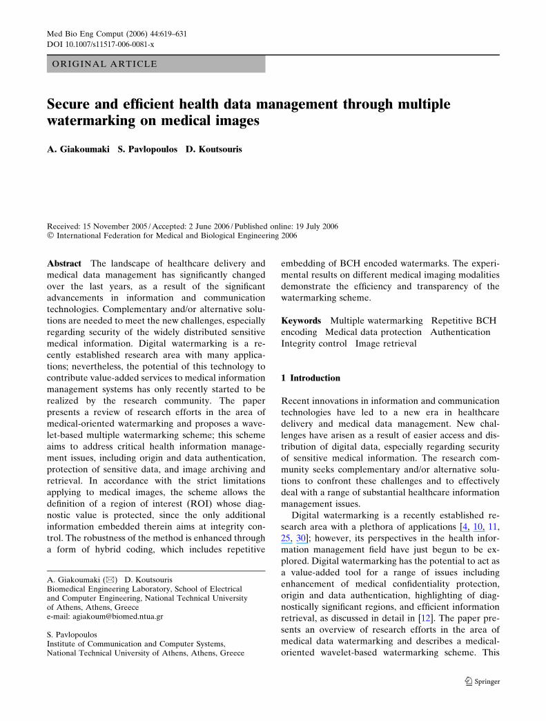
quantization of coefficients. Figure 1 illustrates the
pyramid structure of a 4-level wavelet decomposition
of an image, including a coarse scale image approxi-
mation at the highest decomposition level (LL4), and
12 detail images corresponding to the horizontal (HL),
vertical (LH), and diagonal (HH) details at each of the
four levels. It is well known that transform domain
watermarking schemes outperform compared to spatial
ones, since they exploit perceptual properties of the
human visual system (HVS) [33] in order to achieve an
optimal trade-off between robustness and impercepti-
bility. Wavelet-based schemes particularly have gained
great attention in recent years, due to their ability to
provide both spatial and frequency resolution [14, 35];
their main advantage, however, is the fact that the
dyadic scaling decomposition of the wavelet transform
resembles the signal processing of the HVS, thus
allowing the distortion induced by quantization and/or
watermarking to be adapted to the masking properties
of the human eye [22].
In order to enhance medical confidentiality protec-
tion and to allow efficient data management, retrieval
and integrity control, the scheme simultaneously in-
serts the following different purpose-specific water-
marks:
1. A signature watermark comprising the physician’s
digital signature or identification code for the
purpose of origin authentication.
2. An index watermark that contains keywords (e.g.,
ICD-10 diagnostic codes, image acquisition char-
acteristics, etc.) and facilitates image retrieval by
database querying mechanisms. The insertion of
indices into the images provides an alternative for
Fig. 1 Multiresolution wavelet decomposition of an image: a pyramid structure of 4-level DWT, b 4-level Haar DWT of a CT testimage
620 Med Bio Eng Comput (2006) 44:619–631
123
efficient indexing and archiving of digital medical
data in hospital information systems, which elimi-
nates storage and transmission bandwidth
requirements.
3. A caption watermark containing patient’s personal
and examination data (e.g., demographics, health
history, and diagnostic reports), which grants a
permanent link between the patient and the med-
ical data, and an additional level of protection; this
descriptive watermark provides information con-
tributing to a thorough patient status evaluation
and also allows the highlighting of diagnostically
significant regions, preventing from separate
transmission and storage of metadata that would
increase memory and transmission bandwidth
requirements.
4. A reference watermark is embedded throughout
the image for the purpose of data integrity control
[18]; the comparison of the extracted reference
watermark bits with the originally embedded ones
not only provides information on whether the im-
age has been modified, but also indicates the pos-
sibly tampered image regions.
The scheme takes into consideration the ethical and
legal limitations that apply to medical image manipu-
lation and maintains the integrity of diagnostically
significant parts; specifically, it allows the definition of
a region of interest whose quality is perceptually pre-
served throughout the watermarking process, since the
only information embedded therein is the integrity-
checking reference watermark, which does not affect
its diagnostic value.
Different medical applications, ranging from tele-
medicine to data archiving, storage, and retrieval in
picture archiving and communication systems (PACS),
could be benefited by integrating the proposed scheme
with image acquisition devices, PDAs, PACS viewing
stations, etc. Indicatively, in a telemedicine application,
the acquired image can be watermarked with the
identification code of the mobile unit; also, a mobile
healthcare provider can embed patient’s personal data
and additional information in the image and transmit
them with an additional level of security to a base
station (e.g., hospital). There, the expert retrieves the
watermarks, makes a preliminary diagnosis based on
both the image and the extracted information, and
provides directions to the mobile paramedics. Another
use case involves acquiring an image in a hospital
laboratory and watermarking it with information
including the physician’s identification code, patient’s
personal and examination data, and keywords to be
used for image indexing. The watermarked image is
stored in the hospital database and can be retrieved
through querying mechanisms. Any authorized medi-
cal staff member can extract the embedded water-
marks, thus gaining access to information including the
primary physician’s identity and diagnosis, data con-
cerning the patient and the examination, as well as
additional comments for healthcare providers’ guid-
ance. A variety of other medical applications could
also be addressed by the proposed watermarking
scheme, towards secure and efficient health data
management.
2.2 The algorithm
As mentioned above, the embedding procedure is
based on image decomposition through DWT. The
Haar wavelet is selected as the mother wavelet for the
image decomposition, in order to exploit the dyadic
rationality of the resulting coefficients [31] for in-
creased watermark robustness. Specifically, the Haar
wavelet coefficients have the attribute that, when
modified by addition or subtraction of a multiple of 2l
(where l is the decomposition level), their inverse
wavelet transform produces an image with integer
pixel values; thus any rounding operation, which could
distort the values of certain watermark bits, is avoided
[17]. The proposed method exploits this attribute in the
quantization scheme used to insert the multiple
watermarks in embeddable coefficients. According to
the algorithm, any coefficient f selected to cast a
watermark bit, is assigned a binary value through the
following quantization function:
Qðf Þ ¼ 0; if 2k � D þ s � f\ ð2kþ 1Þ � Dþ s1; if ð2kþ 1Þ � D þ s � f\ ð2kþ 2Þ � Dþ s
�
ð1Þ
where k is an integer, s is a user defined offset for
increased security, and D, the quantization parameter,
is chosen to be equal to 2l in order to exploit the dyadic
rationality of Haar coefficients. The above quantiza-
tion function can be equivalently rewritten as follows:
Qðf Þ ¼0; if ðf � sÞ=Db c is even
1; if ðf � sÞ=Db c is odd
(ð2Þ
where :b c is the floor function.
The multiple watermarks embedding procedure in-
cludes the following steps:
Step 1: The image is decomposed through 4-level Haar
wavelet transform in a coarse scale image approxima-
tion at the highest decomposition level and a sequence
Med Bio Eng Comput (2006) 44:619–631 621
123
of detail images (horizontal, vertical, and diagonal) at
each of the four levels.
Step 2: The above quantization function is applied to
each coefficient f that is to be watermarked. If the
resulting binary value is equal to the value of the
watermark bit to be embedded, the coefficient is left
intact; otherwise, it is modified as follows in order to
cast the watermark bit value:
f ¼f þ D; if f � 0
f � D; if f > 0
(ð3Þ
Step 3: The 4-level inverse wavelet transform is
implemented to produce the watermarked image.
The extraction of the multiple watermarks is per-
formed through decomposition of the watermarked
image using 4-level Haar wavelet transform and key-
based detection of the watermarked coefficients. The
multiple watermark bits are subsequently extracted by
applying the quantization function to each of these
coefficients.
2.3 Selection criteria for watermark embedding
locations
The wavelet coefficients to be watermarked are speci-
fied based on a random key and the ROI map. Initially,
the key selects the embeddable coefficients of all levels
and subbands; in the cases of the data watermarks
(signature, index, caption) however, the wavelet do-
main ROI map determines which of the key-selected
coefficients will finally be used for embedding, by not
belonging to the ROI. In this way, the reference
watermark is inserted in coefficients corresponding to
the whole image, hence allowing an overall image
integrity control, whereas the signature, index, and
caption watermarks are cast into parts of no diagnostic
significance. Thus, the diagnostically important regions
of interest are protected against any compromise on
their quality, by being burdened only with the infor-
mation needed to enable integrity control. The wavelet
domain ROI map is produced based on the spatial self-
similarity between subbands [29] through the procedure
described in [13]. The correspondence among the ROI
maps of a wavelet-transformed CT test image in four
decomposition levels is illustrated in Fig. 1b.
The multiple watermarks are distributed in different
decomposition levels and subbands according to their
individual characteristics and requirements. In the case
of the signature watermark, robustness is of critical
importance, due to the fact that even one error bit
could result in authentication failure; on the contrary,
the capacity requirements for the specific watermark
are quite limited, since its length is restricted to the
minimum needed to grant uniqueness of the conveyed
identification code. The index and the caption water-
marks on the other hand, demand also robustness but
mainly increased capacity, since they convey many bits
of additional information; as they comprise keywords
for image retrieval and patient’s personal/examination
data respectively, it is evident that especially in the
latter case the capacity requirements are even greater.
Given the decreasing number of coefficients and con-
sequently the reduced available capacity in ascending
decomposition levels, the signature, index, and caption
watermarks are embedded in non-ROI coefficients of
the fourth, third, and second levels, respectively. The
specific distribution of the watermarks is also in
accordance with the robustness requirements; due to
the fact that most of the energy is concentrated in the
high decomposition levels, it is expected that more
watermark robustness is achieved in ascending levels.
Table 1 illustrates the energy distribution of a CT test
image in its approximation and detail images, produced
by 4-level Haar DWT. The energy is calculated using
the following equation:
ek ¼1
Nk �Mk
Xi
Xj
Ikði; jÞj j ð4Þ
where k denotes the approximation and the detail
images at each of the decomposition levels, Ik are the
coefficients of the subband images, and Nk, Mk are
their corresponding dimensions. The table shows the
increasing energy concentration in ascending decom-
position levels, with the coarse scale image approxi-
mation accumulating the major energy proportion. As
also evident from the table, the horizontal detail sub-
bands concentrate more energy than the vertical and
much more so than the diagonal subbands, a fact that
indicates their minor vulnerability to attacks and
motivates their use for embedding of the signature,
index, and caption watermarks.
Due to the generally resembling behavior of hori-
zontal and vertical subbands [15], an image modifica-
tion is very likely to affect them in a similar way;
therefore, the selection of vertical subbands for refer-
ence watermark embedding provides a reflection of the
potential tampering of the image. For imperceptibility
reasons, the coarse scale image approximation is left
intact by the embedding procedure, because of its
crucial effect on image quality resulting from the large
energy concentration. Besides, the first decomposition
level coefficients are used exclusively for reference
watermarking and not for signature, index, and caption
622 Med Bio Eng Comput (2006) 44:619–631
123
watermarks embedding, due to their minor effect on
image quality that makes them susceptible to common
image processing, compression, or attacks. In order to
enable a comprehensive image distortion report, the
reference watermark is embedded in selected coeffi-
cients of the other three decomposition levels as well;
in this way, it can be extracted from specific frequen-
cies and/or spatial regions, in order to reflect their
potential tampering.
It should be noted that, despite the fact that the
energy allocation in subbands and decomposition
levels depends on the characteristics of the image,
some attributes are common in different imaging
modalities; based on these attributes, as well as the
individual robustness and capacity requirements of the
multiple watermarks, their distribution in the subbands
was devised as illustrated in Table 2. The particular
allocation of the watermarks optimizes the trade-off
among robustness, capacity, and imperceptibility,
being efficient and applicable to different imaging
modalities without adaptation. The table presents the
maximum available capacity in the selected subbands
for images of size 512 · 512 pixels, which corresponds
to the amount of embeddable coefficients when no
ROI is defined; in any other case, the available
capacity in the horizontal subbands is determined by
the extent of the ROI, since only horizontal coeffi-
cients belonging to the non-ROI are allowed to carry
watermark bits.
3 Results
The algorithm was tested on different medical imaging
modalities (CT, MRI, MRA, PET), with each test set
containing 20 images of size 512 · 512 pixels. The
algorithm embeds watermarks that are binary arrays
from the set {0, 1}; in our simulations, the signature and
the reference watermarks were produced by a uniform
random number generator, whereas the index and
caption ones were derived by ASCII coding of text files
containing keywords and patient’s data, respectively.
The length of the signature watermark was selected to
be 128 bits, which is sufficient to grant uniqueness to
the conveyed identification code [27]. The sets of
keywords and patient’s data used for index and caption
watermarks generation, were arbitrarily selected to
comprise 52 and 208 characters respectively; this cor-
responds to index and caption watermarks of length
364 and 1,456 bits, respectively, by assigning seven bits
per character through ASCII coding.
In order to increase robustness of the watermarks
carrying additional data (signature, index, caption), a
form of hybrid coding including repetitive embedding
of BCH encoded watermarks was implemented; spe-
cifically, each of the three watermarks was split into
parts of equal length, which were then incorporated
into suitably selected BCH codes. Afterwards, the
BCH encoded watermarks were embedded three times
each; this hybrid coding provides the possibility to
correct some errors using repetition decoding, before
BCH decoding is performed, thus increasing the
robustness of the watermarks [38]. Repetition decod-
ing is actually a majority vote process and refers to
forming the output watermark based on the most
common bit values extracted from the three embedded
watermark copies. Table 3 presents the BCH encoding
schemes used in these simulations, as well as the
number of BCH codes needed in order to comprehend
the whole watermarks. In general, a binary BCH code
with parameters (n, k, l) represents a codeword of
length n, which includes k bits of the watermark array,
and can correct up to l bit errors. For instance, BCH
(255, 91, 25) comprises a codeword of 255 bits, which
includes 91 bits of the watermark to be embedded, and
has an error correction capability of 25 bits. In order
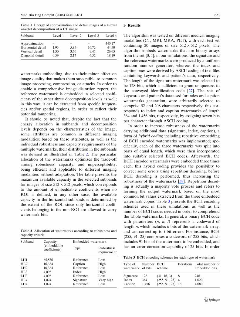
Table 1 Energy of approximation and detail images of a 4-levelwavelet decomposition of a CT image
Subband Level 1 Level 2 Level 3 Level 4
Approximation – – – 440.17Horizontal detail 1.93 5.95 16.72 44.50Vertical detail 1.30 3.60 9.45 26.63Diagonal detail 0.59 2.17 6.52 18.19
Table 2 Allocation of watermarks according to robustness andcapacity criteria
Subband Capacity(embeddablecoefficients)
Embedded watermark
Type Robustnessrequirement
LH1 65,536 Reference LowHL2 16,384 Caption HighLH2 16,384 Reference LowHL3 4,096 Index HighLH3 4,096 Reference LowHL4 1,024 Signature Very highLH4 1,024 Reference Low
Table 3 BCH encoding schemes for each type of watermark
Type ofwatermark
Numberof bits
BCHscheme
Iterations Total number ofembedded bits
Signature 128 (31, 16, 3) 8 248Index 364 (255, 91, 25) 4 1,020Caption 1,456 (255, 91, 25) 16 4,080
Med Bio Eng Comput (2006) 44:619–631 623
123

for example to encode the 1,456-bits caption water-
mark using BCH (255, 91, 25), the watermark is split
into 16 equal parts, and a separate BCH code is used
for each part; this results in a total number of 4,080
codeword bits that need to be embedded.
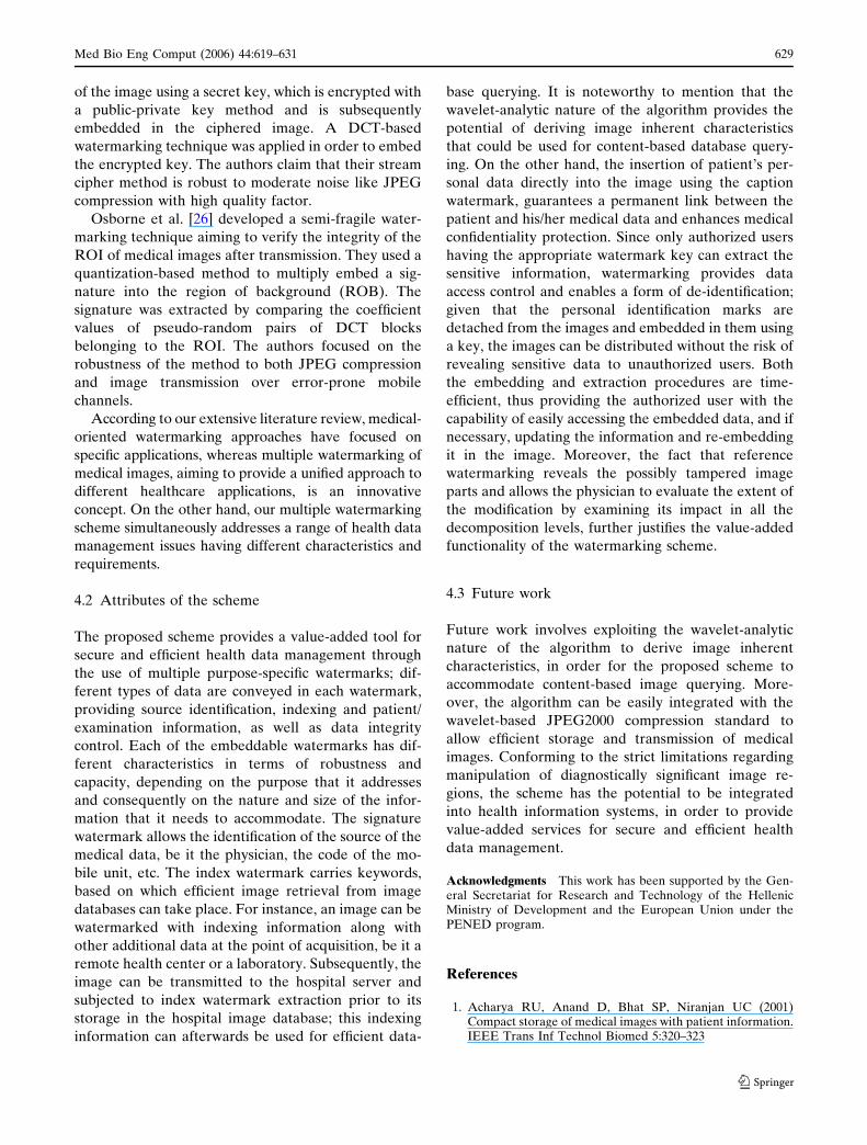
Given the strict limitations regarding the acceptable
modifications of medical images, a thorough evaluation
of the scheme performance in terms of transparency is
necessary; therefore, both perceptual and signal qual-
ities need to be assessed. Peak signal-to-noise-ratio
(PSNR), although not well correlated with perceptual
quality, provides an efficient measure of image distor-
tion in terms of numerical values [5, 19, 20], which
convey important information in medical applications,
for instance in diagnosis support systems. Table 4
presents the obtained average PSNR values of the
watermarked images, for each of the four imaging
modalities tested.
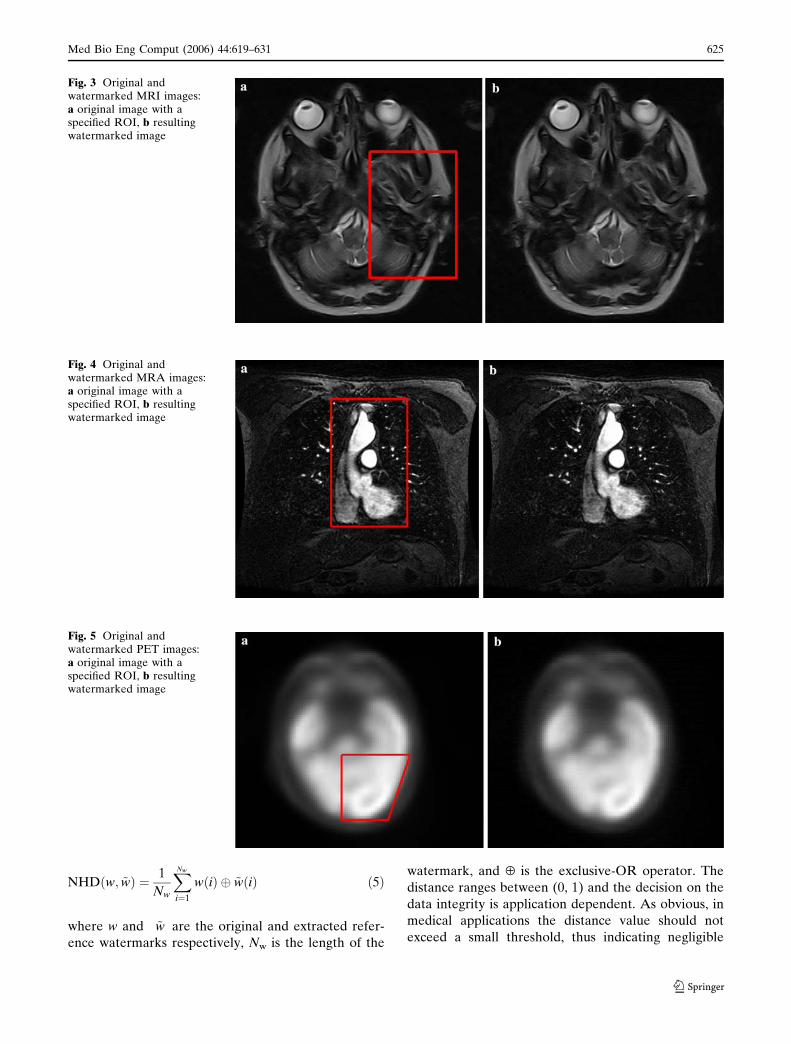
The watermarked images were compared with the
original ones by a physician who concluded that the
visual difference between them was negligible; special
attention was paid to the evaluation of the ROI, whose
quality was found to be preserved, as the minor dis-
tortion due to reference watermark embedding in
sporadic ROI coefficients did not cause any image
degradation that could affect its fidelity and diagnostic
value. Figures 2, 3, 4, and 5 illustrate four test images
(CT, MRI, MRA, PET, respectively), each with a
specified ROI, and the corresponding watermarked
images resulting from the proposed scheme. The high
PSNR values obtained, combined with the adequate
perceptual quality of the watermarked images, illus-
trate the transparency of the scheme.
The performance of the scheme in terms of robust-
ness of the data watermarks (signature, index, caption)
was evaluated through JPEG compression of the
watermarked images; JPEG compression was selected
as an indicative attack applied to the whole image and
with varying quality factors, thus reflecting the toler-
ance of the watermarks in different levels of image
distortion. Tables 5, 6, 7, and 8 demonstrate the per-
formance of the scheme for each of the imaging
modalities tested. The watermarked images were sub-
jected to JPEG compression with different quality
factors and the three watermarks carrying the addi-
tional data were extracted and subsequently compared
with the originally embedded ones; the tables show the
percentage of bit errors in the extracted watermarks.
As evident from these tables, the results in terms of
robustness were satisfactory; indicatively, the water-
mark conveying the signature was extracted intact
from all imaging modalities tested, even when the
watermarked images were subjected to JPEG75 com-
pression. The index watermark resisted JPEG com-
pression with quality factors of at least 80, and
particularly in the cases of MRA and PET images, it
was extracted intact even after JPEG75 compression.
As expected, the tolerance of the caption watermark to
JPEG compression was less, due to the decreasing
robustness in descending decomposition levels.
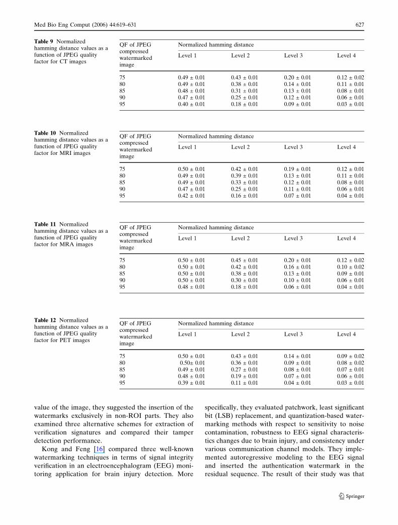
Tables 9, 10, 11, and 12 demonstrate the effects of
applying JPEG compression with different quality
factors to the watermarked images of CT, MRI, MRA,
and PET modalities, respectively. In order to evaluate
the degree of distortion, we selected the normalized
hamming distance (NHD) as the similarity measure:
Table 4 Performance of thescheme in terms of PSNR
Imagemodality
PSNR (dB)
CT 46.47 ± 0.06MRI 46.37 ± 0.05MRA 45.96 ± 0.04PET 46.66 ± 0.20
Fig. 2 Original and watermarked CT images: a original image with a specified ROI, b resulting watermarked image
624 Med Bio Eng Comput (2006) 44:619–631
123
NHDðw; ~wÞ ¼ 1
Nw
XNw
i¼1
wðiÞ � ~wðiÞ ð5Þ
where w and ~w are the original and extracted refer-
ence watermarks respectively, Nw is the length of the
watermark, and ¯ is the exclusive-OR operator. The
distance ranges between (0, 1) and the decision on the
data integrity is application dependent. As obvious, in
medical applications the distance value should not
exceed a small threshold, thus indicating negligible
Fig. 3 Original andwatermarked MRI images:a original image with aspecified ROI, b resultingwatermarked image
Fig. 4 Original andwatermarked MRA images:a original image with aspecified ROI, b resultingwatermarked image
Fig. 5 Original andwatermarked PET images:a original image with aspecified ROI, b resultingwatermarked image
Med Bio Eng Comput (2006) 44:619–631 625
123

image modifications. As expected, JPEG compression
with decreasing quality results in increasing NHD
values. The tables also illustrate the decrease of the
NHD value in ascending decomposition levels, which is
due to the increased robustness.
The above presented experimental results demon-
strate the efficiency of the scheme in terms of robust-
ness, imperceptibility, and integrity control capability,
and its potential to provide value-added services in
health data management systems.
4 Discussion
The paper discusses the perspectives of digital water-
marking in health information management and pro-
poses a medical-oriented wavelet-based multiple
watermarking scheme; this scheme simultaneously
embeds four types of watermarks into medical images,
intending to enhance protection of sensitive data,
provide origin and data authentication capability, and
allow efficient image archiving and retrieval. In order
to increase robustness of the watermarks conveying
signature, index, and caption data, a combination of
BCH encoding and repetitive embedding is performed.
The experimental results demonstrate the efficiency of
the scheme in terms of robustness, imperceptibility,
and integrity control capability.
4.1 Comparison with literature
A plethora of studies regarding digital watermarking
techniques and applications have enriched the litera-
ture over the last decade; nevertheless, the exploitation
of digital watermarking perspectives in medical appli-
cations is still in its infancy. A brief overview of the
main medical-oriented watermarking studies con-
ducted so far is presented below:
Macq and Dewey [21] focused on the issue of the
‘‘trusted header’’ and proposed a reversible water-
marking technique for the purpose of DICOM header
verification through insertion of the header hash in the
medical image. The method uses ‘‘pseudo-sums’’ and
‘‘pseudo-differences’’ of adjacent image pixels for the
insertion of the hash, the comparison of which with the
hash corresponding to the received image allows the
reliability control of the link between the header and
the image.
Coatrieux et al. [9] proposed the use of water-
marking techniques as a complementary measure to
the existing ones for protecting medical images; they
pinpointed the necessary requirements a watermarking
system has to conform to in order to be accepted in a
medical environment, as well as its complementary
role to the existing security systems. Two different
application scenarios were presented, which involved
image authentication and tracing, and health record
integrity control, respectively. In a latter study, Coat-
rieux et al. [8] discussed again the role of watermarking
in the context of security of health information systems
and focused on the issue of medical image integrity
control; they proposed to separate the images into two
parts, the one comprising the regions of diagnostic
significance (ROI), and the other representing a region
of non-interest. In order to preserve the diagnostic
Table 5 Percentage of bit errors in watermarks extracted fromCT images (%)
Type of watermark Signature Index Caption
JPEG quality factor95 0 0 2.090 0 0 13.585 0 0 20.480 0 0 30.975 0 5.5 37.8
Table 6 Percentage of bit errors in watermarks extracted fromMRI images (%)
Type of watermark Signature Index Caption
JPEG quality factor95 0 0 090 0 0 13.885 0 0 23.480 0 0 30.575 0 4.4 39.3
Table 7 Percentage of bit errors in watermarks extracted fromMRA images (%)
Type of watermark Signature Index Caption
JPEG quality factor95 0 0 090 0 0 15.985 0 0 25.380 0 0 35.875 0 0 43.6
Table 8 Percentage of bit errors in watermarks extracted fromPET images (%)
Type of watermark Signature Index Caption
JPEG quality factor95 0 0 090 0 0 12.685 0 0 20.980 0 0 30.875 0 0 36.0
626 Med Bio Eng Comput (2006) 44:619–631
123
value of the image, they suggested the insertion of the
watermarks exclusively in non-ROI parts. They also
examined three alternative schemes for extraction of
verification signatures and compared their tamper
detection performance.
Kong and Feng [16] compared three well-known
watermarking techniques in terms of signal integrity
verification in an electroencephalogram (EEG) moni-
toring application for brain injury detection. More
specifically, they evaluated patchwork, least significant
bit (LSB) replacement, and quantization-based water-
marking methods with respect to sensitivity to noise
contamination, robustness to EEG signal characteris-
tics changes due to brain injury, and consistency under
various communication channel models. They imple-
mented autoregressive modeling to the EEG signal
and inserted the authentication watermark in the
residual sequence. The result of their study was that
Table 9 Normalizedhamming distance values as afunction of JPEG qualityfactor for CT images
QF of JPEGcompressedwatermarkedimage
Normalized hamming distance
Level 1 Level 2 Level 3 Level 4
75 0.49 ± 0.01 0.43 ± 0.01 0.20 ± 0.01 0.12 ± 0.0280 0.49 ± 0.01 0.38 ± 0.01 0.14 ± 0.01 0.11 ± 0.0185 0.48 ± 0.01 0.31 ± 0.01 0.13 ± 0.01 0.08 ± 0.0190 0.47 ± 0.01 0.25 ± 0.01 0.12 ± 0.01 0.06 ± 0.0195 0.40 ± 0.01 0.18 ± 0.01 0.09 ± 0.01 0.03 ± 0.01
Table 10 Normalizedhamming distance values as afunction of JPEG qualityfactor for MRI images
QF of JPEGcompressedwatermarkedimage
Normalized hamming distance
Level 1 Level 2 Level 3 Level 4
75 0.50 ± 0.01 0.42 ± 0.01 0.19 ± 0.01 0.12 ± 0.0180 0.49 ± 0.01 0.39 ± 0.01 0.13 ± 0.01 0.11 ± 0.0185 0.49 ± 0.01 0.33 ± 0.01 0.12 ± 0.01 0.08 ± 0.0190 0.47 ± 0.01 0.25 ± 0.01 0.11 ± 0.01 0.06 ± 0.0195 0.42 ± 0.01 0.16 ± 0.01 0.07 ± 0.01 0.04 ± 0.01
Table 11 Normalizedhamming distance values as afunction of JPEG qualityfactor for MRA images
QF of JPEGcompressedwatermarkedimage
Normalized hamming distance
Level 1 Level 2 Level 3 Level 4
75 0.50 ± 0.01 0.45 ± 0.01 0.20 ± 0.01 0.12 ± 0.0280 0.50 ± 0.01 0.42 ± 0.01 0.16 ± 0.01 0.10 ± 0.0285 0.50 ± 0.01 0.38 ± 0.01 0.13 ± 0.01 0.09 ± 0.0190 0.50 ± 0.01 0.30 ± 0.01 0.10 ± 0.01 0.06 ± 0.0195 0.48 ± 0.01 0.18 ± 0.01 0.06 ± 0.01 0.04 ± 0.01
Table 12 Normalizedhamming distance values as afunction of JPEG qualityfactor for PET images
QF of JPEGcompressedwatermarkedimage
Normalized hamming distance
Level 1 Level 2 Level 3 Level 4
75 0.50 ± 0.01 0.43 ± 0.01 0.14 ± 0.01 0.09 ± 0.0280 0.50± 0.01 0.36 ± 0.01 0.09 ± 0.01 0.08 ± 0.0285 0.49 ± 0.01 0.27 ± 0.01 0.08 ± 0.01 0.07 ± 0.0190 0.48 ± 0.01 0.19 ± 0.01 0.07 ± 0.01 0.06 ± 0.0195 0.39 ± 0.01 0.11 ± 0.01 0.04 ± 0.01 0.03 ± 0.01
Med Bio Eng Comput (2006) 44:619–631 627
123

the patchwork method outperforms compared with the
other two techniques, providing lower error bit passing
probability and higher signal-to-noise-ratio (SNR) in
conditions of moderate bit error rate in the commu-
nication channel.
Acharya et al. [1] proposed the embedding of an
encrypted version of the electronic patient record
(EPR) in medical images, by replacing the LSB of the
gray levels of pixels. The text file containing patient’s
data was encrypted using the logarithm of the ASCII
codes of the text, whereas the encryption of the heart
rate signal was based on predictive coding techniques,
such as differential pulse code modulation (DPCM)
and adaptive delta modulation (ADM). The authors
pointed out the reduction of storage and transmission
overheads as a result of inserting patient information in
medical images. In a latter study, Acharya et al. [2]
repeated their tests using error control coding, in order
to enhance the reliability of image transmission and
storage in the presence of noise or other interference.
Subsequently, Acharya et al. [3] performed watermark
embedding by LSB replacement in the frequency do-
main. More specifically, they applied discrete cosine
transform (DCT) into image blocks of size 8 · 8 pixels
and replaced the LSBs of DCT coefficients belonging
to a mid-frequency range with the bits of the array
which resulted from the encryption of EPR data. Na-
yak et al. [24] presented a comparative evaluation of
the results deriving from applying the LSB method to
the spatial and frequency domains, the latter including
not only DCT, but also discrete fourier transform
(DFT) and DWT. It is worth mentioning though that,
despite the minimal degradation of image quality and
the low complexity, the LSB watermarking method is
not applicable in practice due to its well-known fra-
gility.
Miaou et al. [23] and Chao et al. [7] also proposed a
watermarking technique based on LSB replacement,
aiming to provide origin authentication and protection
of the patient’s health record. They applied an algo-
rithm using a bipolar multiple-number base in order to
embed a watermark consisting of an ECG signal, a
diagnostic report, and the physician’s seal. It is note-
worthy however to mention that the extraction of the
watermark requires the knowledge of the original,
unwatermarked image, a fact that invalidates the worth
of the system in practice.
The lack of robustness of LSB-replacement water-
marking techniques to both malicious attacks and
common image processing operations has not deterred
other researchers to also implement them in medical
applications; Zhou et al. [37] used an LSB water-
marking scheme in order to verify the authenticity and
the integrity of digital mammography images. They
embedded the health record in the image by replacing
the LSBs of randomly selected pixels with the bits of
the array comprising the record. This array included
patient’s data, as well as the digital signature resulting
from the encrypted image digest, thus allowing the data
authenticity and integrity control. In order to avoid any
distortion of the image digest due to watermarking, the
LSBs of the image pixels were excluded from the
procedure of digest calculation. Cao et al. [6] presented
the existing context of medical data protection in
PACS based on the DICOM standard and discussed
the advantages and disadvantages of the LSB water-
marking technique with respect to the existing security
measures.
Wakatani [34] proposed the embedding of a signa-
ture image in non-ROI regions of the original image in
order to avoid any compromise on its diagnostic value.
The signature was encoded using embedded zerotree
wavelet (EZW) progressive coding and the resulting
bit array was embedded in the image by replacing the
bits of a randomly selected bit plane of the pixels. The
watermark was inserted in a spiral way around the
ROI, with the most significant information of the sig-
nature embedded in the nearest to the ROI area. By
dividing the contour of the ROI in several regions and
inserting the signature in each region, the signature
image could be extracted from a clipped image
including only a part of the ROI.
Trichili et al. [32] suggested the addition of a virtual
border to the image by mirror effect, in order to embed
the watermark in pixels not belonging to the image
itself, thus guaranteeing its integrity. The watermark
included the patient’s name, age, and the image
acquisition laboratory, and was embedded in the LSBs
of the virtual border. The authors claim that the ori-
ginal image can be extracted from the watermarked
one due to the reversibility of the method; still, the
disadvantages of LSB embedding are applied to this
approach as well.
Yang and Bao [36] presented a brief overview of
reversible watermarking methods that have been pro-
posed in the literature and proposed a method which is
exclusively applicable to the authentication of an
Electronic Clinical Brain Atlas. Their method uses for
embedding the border between different structures of
the atlas, and modifies in a reversible way the color of
specific pixels of the border in order to embed the
binary sequence.
Recently, Puech and Rodrigues [28] proposed the
combination of encryption and watermarking tech-
niques for the purpose of safe transmission of medical
images. The suggested methodology includes ciphering
628 Med Bio Eng Comput (2006) 44:619–631
123
of the image using a secret key, which is encrypted with
a public-private key method and is subsequently
embedded in the ciphered image. A DCT-based
watermarking technique was applied in order to embed
the encrypted key. The authors claim that their stream
cipher method is robust to moderate noise like JPEG
compression with high quality factor.
Osborne et al. [26] developed a semi-fragile water-
marking technique aiming to verify the integrity of the
ROI of medical images after transmission. They used a
quantization-based method to multiply embed a sig-
nature into the region of background (ROB). The
signature was extracted by comparing the coefficient
values of pseudo-random pairs of DCT blocks
belonging to the ROI. The authors focused on the
robustness of the method to both JPEG compression
and image transmission over error-prone mobile
channels.
According to our extensive literature review, medical-
oriented watermarking approaches have focused on
specific applications, whereas multiple watermarking of
medical images, aiming to provide a unified approach to
different healthcare applications, is an innovative
concept. On the other hand, our multiple watermarking
scheme simultaneously addresses a range of health data
management issues having different characteristics and
requirements.
4.2 Attributes of the scheme
The proposed scheme provides a value-added tool for
secure and efficient health data management through
the use of multiple purpose-specific watermarks; dif-
ferent types of data are conveyed in each watermark,
providing source identification, indexing and patient/
examination information, as well as data integrity
control. Each of the embeddable watermarks has dif-
ferent characteristics in terms of robustness and
capacity, depending on the purpose that it addresses
and consequently on the nature and size of the infor-
mation that it needs to accommodate. The signature
watermark allows the identification of the source of the
medical data, be it the physician, the code of the mo-
bile unit, etc. The index watermark carries keywords,
based on which efficient image retrieval from image
databases can take place. For instance, an image can be
watermarked with indexing information along with
other additional data at the point of acquisition, be it a
remote health center or a laboratory. Subsequently, the
image can be transmitted to the hospital server and
subjected to index watermark extraction prior to its
storage in the hospital image database; this indexing
information can afterwards be used for efficient data-
base querying. It is noteworthy to mention that the
wavelet-analytic nature of the algorithm provides the
potential of deriving image inherent characteristics
that could be used for content-based database query-
ing. On the other hand, the insertion of patient’s per-
sonal data directly into the image using the caption
watermark, guarantees a permanent link between the
patient and his/her medical data and enhances medical
confidentiality protection. Since only authorized users
having the appropriate watermark key can extract the
sensitive information, watermarking provides data
access control and enables a form of de-identification;
given that the personal identification marks are
detached from the images and embedded in them using
a key, the images can be distributed without the risk of
revealing sensitive data to unauthorized users. Both
the embedding and extraction procedures are time-
efficient, thus providing the authorized user with the
capability of easily accessing the embedded data, and if
necessary, updating the information and re-embedding
it in the image. Moreover, the fact that reference
watermarking reveals the possibly tampered image
parts and allows the physician to evaluate the extent of
the modification by examining its impact in all the
decomposition levels, further justifies the value-added
functionality of the watermarking scheme.
4.3 Future work
Future work involves exploiting the wavelet-analytic
nature of the algorithm to derive image inherent
characteristics, in order for the proposed scheme to
accommodate content-based image querying. More-
over, the algorithm can be easily integrated with the
wavelet-based JPEG2000 compression standard to
allow efficient storage and transmission of medical
images. Conforming to the strict limitations regarding
manipulation of diagnostically significant image re-
gions, the scheme has the potential to be integrated
into health information systems, in order to provide
value-added services for secure and efficient health
data management.
Acknowledgments This work has been supported by the Gen-eral Secretariat for Research and Technology of the HellenicMinistry of Development and the European Union under thePENED program.
References
1. Acharya RU, Anand D, Bhat SP, Niranjan UC (2001)Compact storage of medical images with patient information.IEEE Trans Inf Technol Biomed 5:320–323
Med Bio Eng Comput (2006) 44:619–631 629
123
2. Acharya RU, Bhat SP, Kumar S, Min LC (2003) Transmis-sion and storage of medical images with patient data. ComputBiol Med 33:303–310
3. Acharya RU, Niranjan UC, Iyengar SS, Kannathal N, MinLC (2004) Simultaneous storage of patient information withmedical images in the frequency domain. Comput MethodsPrograms Biomed 76:13–19
4. Bartolini F, Tefas A, Barni M, Pitas I (2001) Imageauthentication techniques for surveillance applications. ProcIEEE 89:1403–1418
5. Bruckmann A, Uhl A (1998) Selective medical image com-pression using wavelet techniques. J Comput Inform TechnolSpecial Issue on Biomed Image Processing and Analysis6:203–213
6. Cao F, Huang HK, Zhou XQ (2003) Medical image securityin a HIPAA mandated PACS environment. Comput MedImaging Graph 27:185–196
7. Chao HM, Hsu CM, Miaou SG (2002) A data-hiding tech-nique with authentication, integration, and confidentiality forelectronic patient records. IEEE Trans Inf Technol Biomed6:46–53
8. Coatrieux G, Maitre H, Sankur B (2001) Strict integritycontrol of biomedical images. Proceedings of the SPIEsecurity and watermarking of multimedia contents III, vol4314. San Jose, pp 229–240
9. Coatrieux G, Maitre H, Sankur B, Rolland Y, CollorecR (2000) Relevance of watermarking in medical imaging.Proceedings of the 3rd conference on informationtechnology application in biomedicine. Arlington, pp 250–255
10. Cox IJ, Miller ML (2002) The first 50 years of electronicwatermarking. EURASIP J Appl Signal Process 2002:126–132
11. Eggers JJ, Bauml R, Tzschoppe R, Huber J (2001) Appli-cations of information hiding and digital watermarking.Proceedings of the ECDL workshop on generalized docu-ments. Darmstadt, Germany
12. Giakoumaki A, Pavlopoulos S, Koutsouris D (2004) Amultiple watermarking scheme applied to medical imagemanagement. Proceedings of the 26th IEEE-EMBS annualinternational conference on engineering in medicine andbiology. San Francisco, pp 3241–3244
13. Giakoumaki A, Pavlopoulos S, Koutsouris D (2005) Multipledigital watermarking applied to medical imaging. Proceed-ings of the 27th IEEE-EMBS annual international confer-ence on engineering in medicine and biology. Shanghai,China, pp 3444–3447
14. Hartung F, Kutter M (1999) Multimedia watermarkingtechniques. Proc IEEE Special Issue on Identification andProtection of Multimedia Information 87:1069–1107
15. Kim BS, Kwon KK, Kwon SG, Park KN, Song KI, Lee KI(2002) A robust wavelet-based digital watermarking usingstatistical characteristic of image and human visual system.Proceedings of the international conference on circuits sys-tems computers and communications. Phuket, Thailand,pp 1019–1022
16. Kong X, Feng R (2001) Watermarking medical signalsfor telemedicine. IEEE Trans Inf Technol Biomed 5:195–201
17. Kundur D, Hatzinakos D (1999) Digital watermarking fortelltale tamper proofing and authentication. Proc IEEE87:1167–1180
18. Kundur D, Hatzinakos D (2001) Diversity and attack char-acterization for improved robust watermarking. IEEE TransSignal Process 49:2383–2396
19. Kutter M, Hartung F (2000) Introduction to watermarkingtechniques. In: Katzenbeisser S, Petitcolas FAP (eds) Infor-mation hiding techniques for steganography and digitalwatermarking. Artech House, Norwood pp 97–120
20. Kutter M, Petitcolas FAP (1999) A fair benchmark forimage watermarking systems. Proceedings of the SPIEelectronic imaging, security and watermarking of multi-media contents, vol 3657. San Jose, California, pp 226–239
21. Macq B, Dewey F (1999) Trusted headers for medical ima-ges. Proceedings of the DFG VIII-DII watermarking work-shop. Erlangen, Germany
22. Meerwald P, Uhl A (2001) A survey of wavelet-domainwatermarking algorithms. Proceedings of the SPIE securityand watermarking of multimedia contents, vol 4314. SanJose, pp 505–516
23. Miaou SG, Hsu CM, Tsai YS, Chao HM (2000) A securedata hiding technique with heterogeneous data combiningcapability for electronic patient records. Proceedings of the22nd annual international conference on IEEE engineeringin medicine and biology society. Chicago, pp 280–283
24. Nayak J, Bhat SP, Acharya RU, Niranjan UC (2004)Simultaneous storage of medical images in the spatial andfrequency domain: a comparative study. Biomed Eng3(1):17. Available via http://www.biomedical-engineering-online.com/content/3/1/17. Accessed 16 June 2004
25. Nikolaidis N, Pitas I (1999) Digital image watermarking: anoverview. Proceedings of the international conference onmultimedia computing and systems, vol 1. Florence, Italy,pp 1–6
26. Osborne D, Abbott D, Sorell M, Rogers D (2004) Multipleembedding using robust watermarks for wireless medicalimages. Proceedings of the 3rd international conference onmobile and ubiquitous multimedia. College Park, Maryland,pp 245–250
27. Paquet AH, Ward RK (2002) Wavelet-based digital water-marking for image authentication. Proceedings of the IEEECanadian conference on electrical and computer engineer-ing, vol 2. Winnipeg, Canada, pp 879–884
28. Puech W, Rodrigues JM (2004) A new crypto-watermarkingmethod for medical images safe transfer. Proceedings of the12th European signal processing conference. Vienna,Austria, pp 1481–1484
29. Su PC, Wang HJ, Kuo CCJ (1999) Digital image water-marking in regions of interest. Proceedings of the IS&Tconference on image processing, image quality, image cap-ture systems. Savannah, Georgia, pp 295–300
30. Swanson M, Kobayashi M, Tewfik A (1998) Multimediadata-embedding and watermarking technologies. Proc IEEE86:1064–1987
31. Tian J (2002) Wavelet based reversible watermarking forauthentication. Proceedings of the SPIE security andwatermarking of multimedia contents, vol 4675. San Jose,pp 679–690
32. Trichili H, Bouhlel M, Derbel N, Kamoun L (2002) A newmedical image watermarking scheme for a better telediag-nosis. Proceedings of the IEEE international conference onsystems, man and cybernetics, vol 1. Hammamet, Tunisia,pp 556–559
33. Unser M, Aldroubi A (1996) A review of wavelets in bio-medical applications. Proc IEEE 84:626–638
34. Wakatani A (2002) Digital watermarking for ROI medicalimages by using compressed signature image. Proceedings ofthe 35th annual Hawaii international conference on systemsciences. Big Island, Hawaii.
630 Med Bio Eng Comput (2006) 44:619–631
123
35. Wang HJM, Su PC, Kuo CCJ (1998) Wavelet-based digitalimage watermarking. Opt Express 3:491–496
36. Yang Y, Bao F (2003) An invertible watermarking scheme forauthentication of Electronic Clinical Brain Atlas. Proceedingsof the IEEE international conference on acoustics, speech, andsignal processing. Hong Kong, pp 533–536
37. Zhou XQ, Huang HK, Lou SL (2001) Authenticity andintegrity of digital mammography images. IEEE Trans MedImaging 20:784–791
38. Zinger S, Jin Z, Maitre H, Sankur B (2001) Optimization ofwatermarking performances using error correcting codes andrepetition. Proceedings of the joint conference on commu-nications and multimedia security issues of the new century.Darmstadt, Germany, pp 229–240
Med Bio Eng Comput (2006) 44:619–631 631
123
Copyright © 2022 FDOKUMEN