
Scheme of Work Cambridge IGCSE ... - GCE Guide
90
Version 1 Scheme of Work Cambridge IGCSE™ / Cambridge IGCSE (9–1) Biology 0610 / 0970 For examination from 2023
-
Upload
khangminh22 -
Category
Documents
-
view
8 -
download
0
Transcript of Scheme of Work Cambridge IGCSE ... - GCE Guide

Version 1
Scheme of Work
Cambridge IGCSE™ / Cambridge IGCSE (9–1) Biology 0610 / 0970 For examination from 2023

In order to help us develop the highest quality resources, we are undertaking a continuous programme of review; not only to measure the success of our resources but also to highlight areas for improvement and to identify new development needs. We invite you to complete our survey by visiting the website below. Your comments on the quality and relevance of our resources are very important to us. www.surveymonkey.co.uk/r/GL6ZNJB
Would you like to become a Cambridge consultant and help us develop support materials? Please follow the link below to register your interest. www.cambridgeinternational.org/cambridge-for/teachers/teacherconsultants/
Copyright © UCLES January 2020 Cambridge Assessment International Education is part of the Cambridge Assessment Group. Cambridge Assessment is the brand name of the University of Cambridge Local Examinations Syndicate (UCLES), which itself is a department of the University of Cambridge. UCLES retains the copyright on all its publications. Registered Centres are permitted to copy material from this booklet for their own internal use. However, we cannot give permission to Centres to photocopy any material that is acknowledged to a third party, even for internal use within a Centre.
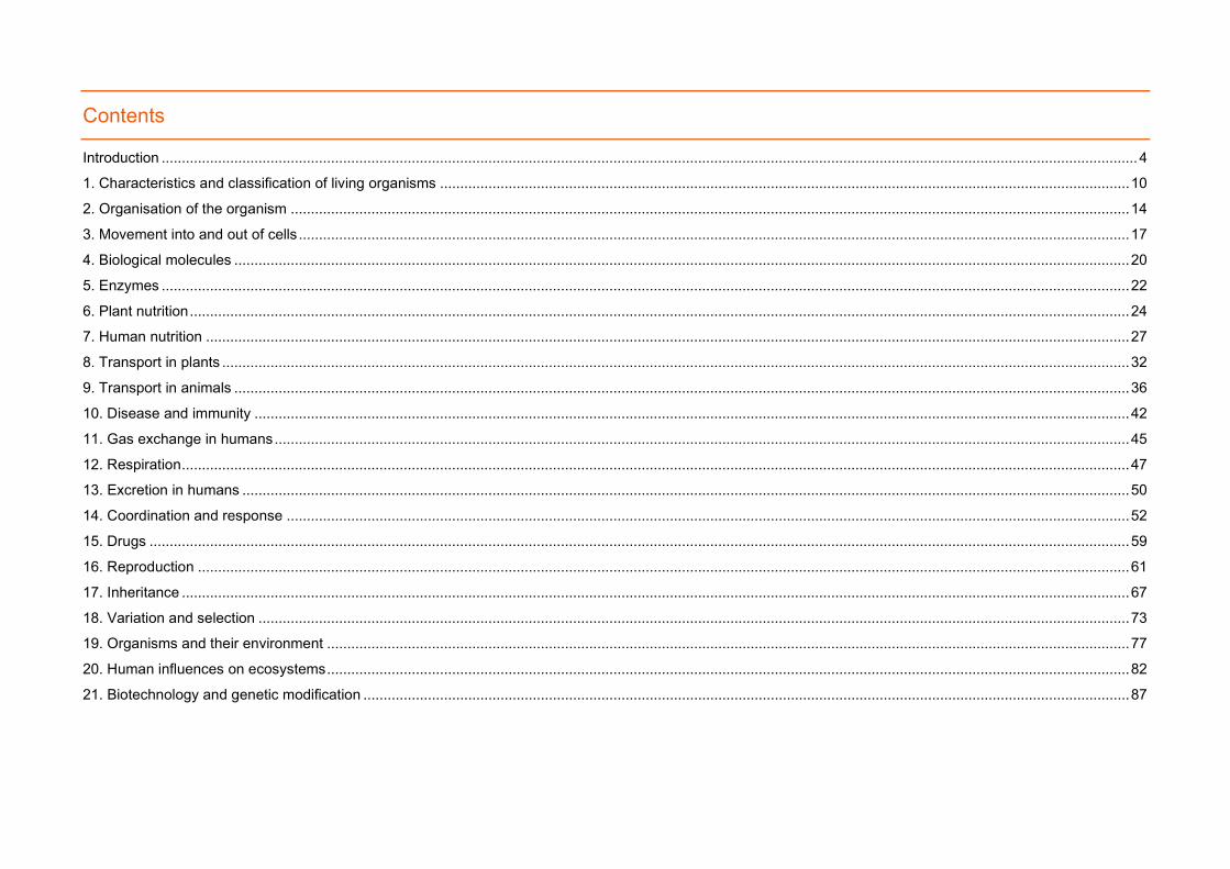
Contents
Introduction .................................................................................................................................................................................................................................................. 4 1. Characteristics and classification of living organisms ........................................................................................................................................................................... 10
2. Organisation of the organism ................................................................................................................................................................................................................ 14
3. Movement into and out of cells .............................................................................................................................................................................................................. 17
4. Biological molecules .............................................................................................................................................................................................................................. 20
5. Enzymes ................................................................................................................................................................................................................................................ 22
6. Plant nutrition ......................................................................................................................................................................................................................................... 24 7. Human nutrition ..................................................................................................................................................................................................................................... 27
8. Transport in plants ................................................................................................................................................................................................................................. 32
9. Transport in animals .............................................................................................................................................................................................................................. 36
10. Disease and immunity ......................................................................................................................................................................................................................... 42
11. Gas exchange in humans .................................................................................................................................................................................................................... 45
12. Respiration ........................................................................................................................................................................................................................................... 47 13. Excretion in humans ............................................................................................................................................................................................................................ 50
14. Coordination and response ................................................................................................................................................................................................................. 52
15. Drugs ................................................................................................................................................................................................................................................... 59
16. Reproduction ....................................................................................................................................................................................................................................... 61
17. Inheritance ........................................................................................................................................................................................................................................... 67
18. Variation and selection ........................................................................................................................................................................................................................ 73 19. Organisms and their environment ....................................................................................................................................................................................................... 77
20. Human influences on ecosystems ....................................................................................................................................................................................................... 82
21. Biotechnology and genetic modification .............................................................................................................................................................................................. 87

Scheme of Work
4
Introduction
This scheme of work has been designed to support you in your teaching and lesson planning. Making full use of this scheme of work will help you to improve both your teaching and your learners’ potential. It is important to have a scheme of work in place in order for you to guarantee that the syllabus is covered fully. You can choose what approach to take and you know the nature of your institution and the levels of ability of your learners. What follows is just one possible approach you could take, and you should always check the syllabus for the content of your course.
Suggestions for independent study (I) and formative assessment (F) are also included. Opportunities for differentiation are indicated as Extension activities; there is the potential for differentiation by resource, grouping, expected level of outcome, and degree of support by teacher, throughout the scheme of work. Timings for activities and feedback are left to the judgement of the teacher, according to the level of the learners and size of the class. Length of time allocated to a task is another possible area for differentiation.
Guided learning hours Guided learning hours give an indication of the amount of contact time you need to have with your learners to deliver a course. Our syllabuses are designed around 130 hours for Cambridge IGCSETM courses. The number of hours may vary depending on local practice and your learners’ previous experience of the subject. The table below give some guidance about how many hours we recommend you spend on each topic area.
Topic
Suggested teaching time (hours / % of the course) Suggested teaching order
1: Characteristics and classification of living organisms
It is recommended that this unit should take about X hours/ X% of the course. 1.1.1, 1.2.1, 1.2.2, 1.2.3, 1.2.4, 1.2.5, 1.2.6, 1.2.7, 1.3.1, 1.3.2, 1.3.3, 1.3.4, 1.3.5, 1.3.6, 1.3.7.
2: Organisation of the organism
It is recommended that this unit should take about X hours/ X% of the course. 2.1.1, 2.1.2, 2.1.3, 2.1.4, 2.1.5, 2.1.6, 2.1.7, 2.2.1, 2.2.2, 2.2.3.
3: Movement into and out of cells
It is recommended that this unit should take about X hours/ X% of the course. 3.1.1, 3.1.2, 3.1.3, 3.1.4, 3.1.5, 3.2.1, 3.2.2, 3.2.3, 3.2.4, 3.2.5, 3.2.6, 3.2.7, 3.2.8, 3.2.9, 3.3.1, 3.3.2, 3.3.3.
4: Biological molecules
It is recommended that this unit should take about X hours/ X% of the course. 4.1.1, 4.1.2, 4.1.3, 4.1.4.

Scheme of Work
5
Topic
Suggested teaching time (hours / % of the course) Suggested teaching order
5: Enzymes It is recommended that this unit should take about X hours/ X% of the course. 5.1.1, 5.1.2, 5.1.3, 5.1.4, 5.1.5, 5.1.6, 5.1.7, 5.1.8, 5.1.9.
6: Plant nutrition It is recommended that this unit should take about X hours/ X% of the course. 6.1.1, 6.1.2, 6.1.3, 6.1.4, 6.1.5, 6.1.6, 6.1.7, 6.1.8, 6.1.9, 6.1.10, 6.1.11, 6.2.1, 6.2.2, 6.2.3.
7: Human nutrition It is recommended that this unit should take about X hours/ X% of the course. 7.1.1, 7.1.2, 7.1.3, 7.2.1, 7.2.2, 7.3.1, 7.3.2, 7.3.3, 7.3.4, 7.3.5, 7.3.6, 7.3.7, 7.4.1, 7.4.2, 7.4.3, 7.4.4, 7.4.5, 7.4.6, 7.4.7, 7.4.8, 7.5.1, 7.5.2, 7.5.3, 7.5.4, 7.5.5.
8: Transport in plants It is recommended that this unit should take about X hours/ X% of the course. 8.1.1, 8.1.2, 8.1.3, 8.2.1, 8.2.2, 8.2.3, 8.2.4, 8.3.1, 8.3.2, 8.3.3, 8.3.4, 8.3.5, 8.3.6, 8.3.7, 8.4.1, 8.4.2, 8.4.3.
9: Transport in animals
It is recommended that this unit should take about X hours/ X% of the course. 9.1.1, 9.1.2, 9.1.3, 9.1.4, 9.2.1, 9.2.2, 9.2.3, 9.2.4, 9.2.5, 9.2.6, 9.2.7, 9.2.8, 9.2.9, 9.2.10, 9.2.11, 9.3.1, 9.3.2, 9.3.3, 9.3.4, 9.3.5, 9.3.6, 9.4.1, 9.4.2, 9.4.3, 9.4.4, 9.4.5, 9.4.6, 9.4.7.
10: Disease and immunity
It is recommended that this unit should take about X hours/ X% of the course. 10.1.1, 10.1.2, 10.1.3, 10.1.4, 10.1.5, 10.1.6, 10.1.7, 10.1.8, 10.1.9, 10.1.10, 10.1.11, 10.1.12, 10.1.13, 10.1.14, 10.1.15, 10.1.16, 10.1.17.
11: Gas exchange in humans
It is recommended that this unit should take about X hours/ X% of the course. 11.1.1, 11.1.2, 11.1.3, 11.1.4, 11.1.5, 11.1.6, 11.1.7, 11.1.8,

Scheme of Work
6
Topic
Suggested teaching time (hours / % of the course) Suggested teaching order
11.1.9, 11.1.10, 11.1.11.
12: Respiration It is recommended that this unit should take about X hours/ X% of the course. 12.1.1, 12.1.2, 12.2.1, 12.2.2, 12.2.3, 12.3.1, 12.3.2, 12.3.3, 12.3.4, 12.3.5, 12.3.6, 12.3.7.
13: Excretion in humans
It is recommended that this unit should take about X hours/ X% of the course. 13.1.1, 13.1.2, 13.1.3, 13.1.4, 13.1.5, 13.1.6, 13.1.7, 13.1.8, 13.1.9.
14: Coordination and response
It is recommended that this unit should take about X hours/ X% of the course. 14.1.1, 14.1.2, 14.1.3, 14.1.4, 14.1.5, 14.1.6, 14.1.7, 14.1.8, 14.1.9, 14.1.10, 14.2.1, 14.2.2, 14.2.3, 14.2.4, 14.2.5, 14.2.6, 14.2.7, 14.2.8, 14.2.9, 14.3.1, 14.3.2, 14.3.3, 14.3.4, 14.3.5, 14.3.6, 14.4.1, 14.4.2, 14.4.3, 14.4.4, 14.4.5, 14.4.6, 14.4.7, 14.4.8, 14.5.1, 14.5.2, 14.5.3, 14.5.4, 14.5.5.
15: Drugs It is recommended that this unit should take about X hours/ X% of the course. 15.1.1, 15.1.2, 15.1.3, 15.1.4, 15.1.5.
16: Reproduction It is recommended that this unit should take about X hours/ X% of the course. 16.1.1, 16.1.2, 16.1.3, 16.2.1, 16.2.2, 16.2.3, 16.2.4, 16.3.1, 16.3.2, 16.3.3, 16.3.4, 16.3.5, 16.3.6, 16.3.7, 16.3.8, 16.3.9, 16.3.10, 16.3.11, 16.3.12, 16.4.1, 16.4.2, 16.4.3, 16.4.4, 16.4.5, 16.4.6, 16.4.7, 16.4.8, 16.4.9, 16.4.10, 16.5.1, 16.5.2, 16.5.3, 16.5.4, 16.6.1, 16.6.2, 16.6.3, 16.6.4, 16.6.5.
17: Inheritance It is recommended that this unit should take about X hours/ X% of the course. 17.1.1, 17.1.2, 17.1.3, 17.1.4, 17.1.5, 17.1.6, 17.1.7, 17.1.8, 17.1.9, 17.1.10, 17.1.11, 17.1.12, 17.2.1, 17.2.2,

Scheme of Work
7
Topic
Suggested teaching time (hours / % of the course) Suggested teaching order
17.2.3, 17.2.4, 17.2.5, 17.3.1, 17.3.2, 17.4.1, 17.4.2, 17.4.3, 17.4.4, 17.4.5, 17.4.6, 17.4.7, 17.4.8, 17.4.9, 17.4.10, 17.4.11, 17.4.12, 17.4.13, 17.4.14, 17.4.15, 17.4.16, 17.4.17, 17.4.18.
18: Variation and selection
It is recommended that this unit should take about X hours/ X% of the course. 18.1.1, 18.1.2, 18.1.3, 18.1.4, 18.1.5, 18.1.6, 18.1.7, 18.1.8, 18.1.9, 18.1.10, 18.2.1, 18.2.2, 18.2.3, 18.3.1, 18.3.2, 18.3.3, 18.3.4, 18.3.5, 18.3.6.
19: Organisms and their environment
It is recommended that this unit should take about X hours/ X% of the course. 19.1.1, 19.1.2, 19.2.1, 19.2.2, 19.2.3, 19.2.4, 19.2.5, 19.2.6, 19.2.7, 19.2.8, 19.2.9, 19.2.10, 19.2.11, 19.2.12, 19.2.13, 19.2.14, 19.2.15, 19.2.16, 19.2.17, 19.2.18, 19.2.19, 19.3.1, 19.3.2, 19.3.3, 19.4.1, 19.4.2, 19.4.3, 19.4.4, 19.4.5, 19.4.6, 19.4.7.
20: Human influences on ecosystems
It is recommended that this unit should take about X hours/ X% of the course. 20.1.1, 20.1.2, 20.1.3, 20.2.1, 20.2.2, 20.2.3, 20.2.4, 20.3.1, 20.3.2, 20.3.3, 20.3.4, 20.4.1, 20.4.2, 20.4.3, 20.4.4, 20.4.5, 20.4.6, 20.4.7, 20.4.8, 20.4.9.
21: Biotechnology and genetic modification
It is recommended that this unit should take about X hours/ X% of the course. 21.1.1, 21.1.2, 21.2.1, 21.2.2, 21.2.3, 21.2.4, 21.2.5, 21.2.6, 21.2.7, 21.3.1, 21.3.2, 21.3.3, 21.3.4.
Resources You can find the up-to-date resource list, including endorsed resources to support Cambridge IGCSETM Biology on the Published resources tab of the syllabus page on our public website here.

Scheme of Work
8
Endorsed textbooks have been written to be closely aligned to the syllabus they support, and have been through a detailed quality assurance process. All textbooks endorsed by Cambridge International for this syllabus are the ideal resource to be used alongside this scheme of work as they cover each learning objective. In addition to reading the syllabus, you should refer to the updated specimen assessment materials.
School Support Hub
The School Support Hub www.cambridgeinternational.org/support is a secure online resource bank and community forum for Cambridge teachers, where you can download specimen and past question papers, mark schemes and other resources. We also offer online and face-to-face training; details of forthcoming training opportunities are posted online. This scheme of work is available as PDF and an editable version in Microsoft Word format; both are available on the School Support Hub at www.cambridgeinternational.org/support. If you are unable to use Microsoft Word you can download Open Office free of charge from www.openoffice.org
Websites This scheme of work includes website links providing direct access to internet resources. Cambridge Assessment International Education is not responsible for the accuracy or content of information contained in these sites. The inclusion of a link to an external website should not be understood to be an endorsement of that website or the site's owners (or their products/services).
The website pages referenced in this scheme of work were selected when the scheme of work was produced. Other aspects of the sites were not checked and only the particular resources are recommended.

Scheme of Work
9
How to get the most out of this scheme of work – integrating syllabus content, skills and teaching strategies We have written this scheme of work for the Cambridge IGCSETM Biology syllabus and it provides some ideas and suggestions of how to cover the content of the syllabus. We have designed the following features to help guide you through your course.
Learning objectives help your learners by making it clear the knowledge they are trying to build. Pass these on to your learners by expressing them as ‘We are learning to / about…’.
Extension: Stretch and prepare for A Level activities provide your more able learners with further challenge beyond the basic content of the course. Innovation and independent learning are the basis of these
Past papers, specimen papers and mark schemes are available for you to download at: www.cambridgeinternational.org/support Using these resources with your learners allows you to check their progress and give them confidence and understanding.
Formative assessment (F) is ongoing assessment which informs you about the progress of your learners. Don’t forget to leave time to review what your learners have learnt: you could try question and answer, tests, quizzes, ‘mind maps’, or ‘concept maps’. These kinds of activities can be found in the scheme of work.
Suggested teaching activities give you lots of ideas about how you can present learners with new information without teacher talk or videos. Try more active methods which get your learners motivated and practising new skills.
Independent study (I) gives your learners the opportunity to develop their own ideas and understanding with direct input from you.
Resource Plus gives your learners the opportunity to develop their practical skills and engage with some of the more challenging topics in the syllabus using videos and interactive teaching resources. Find out more at: www.cambridgeinternational.org/support

Scheme of Work
10
1. Characteristics and classification of living organisms
Syllabus ref. Learning objectives Suggested teaching activities
1.1.1 Characteristics of living organisms
Describe the characteristics of living organisms by describing: (a) movement as an action by an organism or part of an organism causing a change of position or place (b) respiration as the chemical reactions in cells that break down nutrient molecules and release energy for metabolism (c) sensitivity as the ability to detect and respond to changes in the internal or external environment (d) growth as a permanent increase in size and dry mass (e) reproduction as the processes that make more of the same kind of organism (f) excretion as the removal of the waste products of metabolism and substances in excess of requirements (g) nutrition as the taking in of materials for energy, growth and development
Write the seven characteristics of life and their names on the class whiteboard or digital platform. These will serve as a reminder for learners to refer to as they undertake the subsequent activities. Ask learners to consider how items of laboratory equipment or other items such as a moving car, do satisfy some of the characteristics of life. For example, a thermometer is able to sense a change in the environment and the liquid inside it ‘grows’ in response. Challenge learners to design a crossword (either with a pencil and paper or on the computer). The seven words should be the seven characteristics of life; they must write clues for another learner to find them. (I) Ask a carefully chosen series of questions to elicit higher-order thinking skills among learners, for example, ask them to compare key terms, to reinforce their knowledge of key definitions. (F) Extension: Stretch and prepare for A level Ask learners to think of a mnemonic for the first letter of each of the seven characteristics of life. This is a useful skill that helps recall. The class could then vote for their favourite. ‘MRS GREN’ is a very common option, but are there others?
1.2.1 Concept and uses of a classification
State that organisms can be classified into groups by the features that they share
Provide learners with marker pens and ask them to come to the class board to write down as many words that they can think of that relate to ‘species.’ Learners then work in pairs to construct a sentence that defines this term. They may choose to use only some of the words, if they feel some are not relevant. Pairs of learners then join to form groups of four, then eight, and then you elicit a definition that all learners agree on – that it is a group of

Scheme of Work
11
Syllabus ref. Learning objectives Suggested teaching activities
system 1.2.2 1.2.3 1.2.4 1.2.5 1.2.6 1.2.7
Describe a species as a group of organisms that can reproduce to produce fertile offspring Describe the binomial system of naming species as an internationally agreed system in which the scientific name of an organism is made up of two parts showing the genus and species Construct and use dichotomous keys based on identifiable features Explain that classification systems aim to reflect evolutionary relationships Explain that the sequences of bases in DNA are used as a means of classification Explain that groups of organisms which share a more recent ancestor (are more closely related) have base sequences in DNA that are more similar than those that share only a distant ancestor
organisms that can reproduce to produce fertile offspring. Show a short video clip of the platypus or another animal that has unusual features – for example, the pangolin or an unusual animal that is native to your country. Discuss why this organism is difficult to classify. Elicit from learners that they already have ideas about how to classify organisms, and that the classification of organisms into groups is by the features that they share. (F) Learners may know some binomials, such as Homo sapiens. Use this to introduce the Latin names for classification of all organisms. This video clip explains the binomial system of classification: www.saps.org.uk/secondary/teaching-resources/826-binomial-system Elicit from learners that ‘dichotomous’ means ‘has two branches’ and that the yes-or-no questions in dichotomous keys are closed and eventually lead to the correct answer. Ask learners to make a dichotomous key for familiar items of clothing (ties, socks, etc.); or eating utensils (forks, spoons, sporks, chop sticks, etc.) based on identifiable features. Learners decide which items have most in common and give a rationale for their choices. (I) Provide pairs of learners with five sheets of paper that each contain a DNA sequence of 10–20 bases in length. Ask learners to decide the order of similarity with a sixth sequence that you provide. Challenge learners to write a short guide for a younger learner to explain how DNA sequences can be used to help decide on relationships between organisms. This could be accompanied by a tangible example such as the development of different limb bone formations in mammals. Extension: Stretch and prepare for A level Ask learners to carry out research to find how many species of the Plasmodium genus cause malaria. Why is it important that we know which type of parasite has affected a patient? Elicit from learners that this knowledge is important in deciding a course of treatment.
Scheme of Work
12
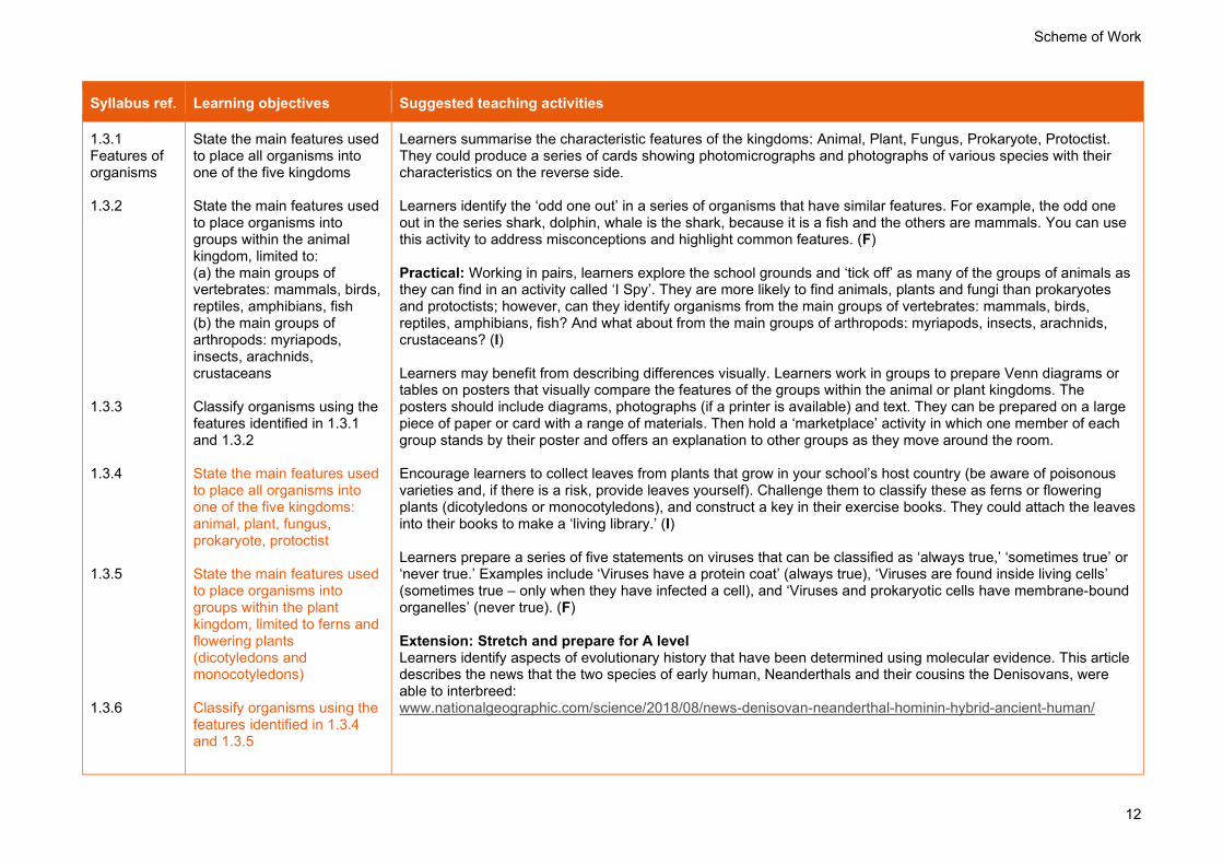
Syllabus ref. Learning objectives Suggested teaching activities
1.3.1 Features of organisms 1.3.2 1.3.3 1.3.4 1.3.5 1.3.6
State the main features used to place all organisms into one of the five kingdoms State the main features used to place organisms into groups within the animal kingdom, limited to: (a) the main groups of vertebrates: mammals, birds, reptiles, amphibians, fish (b) the main groups of arthropods: myriapods, insects, arachnids, crustaceans Classify organisms using the features identified in 1.3.1 and 1.3.2 State the main features used to place all organisms into one of the five kingdoms: animal, plant, fungus, prokaryote, protoctist State the main features used to place organisms into groups within the plant kingdom, limited to ferns and flowering plants (dicotyledons and monocotyledons) Classify organisms using the features identified in 1.3.4 and 1.3.5
Learners summarise the characteristic features of the kingdoms: Animal, Plant, Fungus, Prokaryote, Protoctist. They could produce a series of cards showing photomicrographs and photographs of various species with their characteristics on the reverse side. Learners identify the ‘odd one out’ in a series of organisms that have similar features. For example, the odd one out in the series shark, dolphin, whale is the shark, because it is a fish and the others are mammals. You can use this activity to address misconceptions and highlight common features. (F) Practical: Working in pairs, learners explore the school grounds and ‘tick off’ as many of the groups of animals as they can find in an activity called ‘I Spy’. They are more likely to find animals, plants and fungi than prokaryotes and protoctists; however, can they identify organisms from the main groups of vertebrates: mammals, birds, reptiles, amphibians, fish? And what about from the main groups of arthropods: myriapods, insects, arachnids, crustaceans? (I) Learners may benefit from describing differences visually. Learners work in groups to prepare Venn diagrams or tables on posters that visually compare the features of the groups within the animal or plant kingdoms. The posters should include diagrams, photographs (if a printer is available) and text. They can be prepared on a large piece of paper or card with a range of materials. Then hold a ‘marketplace’ activity in which one member of each group stands by their poster and offers an explanation to other groups as they move around the room. Encourage learners to collect leaves from plants that grow in your school’s host country (be aware of poisonous varieties and, if there is a risk, provide leaves yourself). Challenge them to classify these as ferns or flowering plants (dicotyledons or monocotyledons), and construct a key in their exercise books. They could attach the leaves into their books to make a ‘living library.’ (I) Learners prepare a series of five statements on viruses that can be classified as ‘always true,’ ‘sometimes true’ or ‘never true.’ Examples include ‘Viruses have a protein coat’ (always true), ‘Viruses are found inside living cells’ (sometimes true – only when they have infected a cell), and ‘Viruses and prokaryotic cells have membrane-bound organelles’ (never true). (F) Extension: Stretch and prepare for A level Learners identify aspects of evolutionary history that have been determined using molecular evidence. This article describes the news that the two species of early human, Neanderthals and their cousins the Denisovans, were able to interbreed: www.nationalgeographic.com/science/2018/08/news-denisovan-neanderthal-hominin-hybrid-ancient-human/

Scheme of Work
13
Syllabus ref. Learning objectives Suggested teaching activities
1.3.7
State the features of viruses, limited to a protein coat and genetic material
Past and specimen papers
Past/specimen papers and mark schemes are available to download at www.cambridgeinternational.org/support (F)

Scheme of Work
14
2. Organisation of the organism
Syllabus ref. Learning objectives Suggested teaching activities
2.1.1 Cell structure 2.1.2 2.1.3 2.1.4 2.1.5 2.1.6
Describe and compare the structure of a plant cell with an animal cell, limited to: cell wall, cell membrane, nucleus, cytoplasm, chloroplasts, ribosomes, mitochondria, vacuoles Describe the structure of a bacterial cell, limited to: cell wall, cell membrane, cytoplasm, ribosomes, circular DNA, plasmids Identify the cell structures listed in 2.1.1 and 2.1.2 in diagrams and images of plant, animal and bacterial cells Describe the functions of the structures listed in 2.1.1 and 2.1.2 in plant, animal and bacterial cells State that new cells are produced by division of existing cells State that specialised cells have specific functions, limited to: (a) ciliated cells – movement of mucus in the trachea and
Challenge learners to design a crossword (either using digital software or on paper). They should include various terms associated with cell structure, including cell wall, cell membrane, nucleus, cytoplasm, chloroplasts, ribosomes, mitochondria, and vacuoles. They must write clues for another learner to find them. (I) Learners make a display to compare the structures of animal cells and plant cells, either by using annotated drawings, printed copies of photomicrographs, or electron micrographs, or by constructing a large comparison table or a presentation. (I) Learners make cells and organelles out of modelling clay. They may use images of cells online to help them, including those found in websites such as: https://cellpics.cimr.cam.ac.uk/ and www.cellimagelibrary.org (I) Techniques such as matching words can be useful in this topic. Provide learners with a series of terms in boxes (such as structures found in cells or types of specialised cell), that they must match with their descriptions. (I) Learners play a game called ‘cell charades.’ Ask learners to work in pairs and take it in turns to use hand movements only to describe a number of organelles. They must not use any words in their description. For extra challenge, ask learners to attempt to illustrate the structure of a bacterial cell, including ribosomes, circular deoxyribonucleic acid (DNA) and plasmids, cytoplasm, cell membrane and cell wall. (I)
Learners produce a mini-poster summarising the different types of specialised cell in a multicellular organism such as a human. Challenge learners to work in groups of 3–4 to prepare a poster that illustrates the structure and describes the function of a wide variety of specialised cells, and how they relate to the terms tissue, organ, organ system and organism. Learners should keep the poster as small as possible: this encourages them to consider the content more carefully. (I) Encourage thinking among learners by challenging them to ask the question ‘Why?’ For example, ‘Why is a root hair cell adapted to its function?’ rather than ‘how.’ This encourages learners to consider the function of the cell, in addition to its visual appearance. Learners work in groups to prepare Venn diagrams to compare different specialised cells, related to their overall structure and the organelles found within them. Venn diagrams compare and contrast at least two different ideas (A and B). The overlapping area represents the characteristics that belong to both A and B, and the two areas without overlap are unique to those ideas. Differentiate this task by choosing types of cell that are more or less easily compared (e.g. comparing a red blood cell and a palisade mesophyll cell would be less demanding than

Scheme of Work
15
Syllabus ref. Learning objectives Suggested teaching activities
2.1.7
bronchi (b) root hair cells – absorption (c) palisade mesophyll cells – photosynthesis (d) neurones – conduction of electrical impulses (e) red blood cells – transport of oxygen (f) sperm and egg cells (gametes) – reproduction Describe the meaning of the terms: cell, tissue, organ, organ system and organism as illustrated by examples given in the syllabus
comparing a neurone and a root hair cell). The display must contain diagrams, photographs and text. Learners can prepare these on a large piece of paper or card with a range of materials. Ask one member of each group stands by their poster and offers an explanation to other groups as they circulate around the room. (I) Extension: Stretch and prepare for A level Learners carry out research into the endosymbiotic theory, which suggests that membrane-bound organelles such as the mitochondrion and the chloroplast were derived from smaller cells that came to live inside larger cells.
2.2.1 Size of specimens 2.2.2 2.2.3
State and use the formula: magnification = image size ÷ actual size Calculate magnification and size of biological specimens using millimetres as units Convert measurements between millimetres (mm) and micrometres (μm)
Learners explore how to use the magnification formula using a familiar object (a coin from your school’s country). Give each learner a low-value coin from your school’s country, a piece of paper on which is an image of the coin magnified by 5–10 times and a piece of paper on which is an image of the coin magnified by 0.1–0.01 times. Ask learners to calculate the magnification of the two images of the coin. They compare their answers with those of a peer. Relate this activity to cells and organelles using animations such as: www.cellsalive.com/howbig_js.htm. Learners design a ‘step-by-step’ guide, perhaps targeted at learners who have not yet studied the topic, on how to use the formula: magnification = image size/actual size. The guide could be a flow diagram with statements separated by arrows, a short story, or an animation produced on a computer. (F) Practical: Host practical activities for learners to use a light microscope and develop their ability to produce scientific drawings. If you have suitable equipment, project images from a microscope onto a screen to demonstrate. Specimens may include, for example, a temporary, stained mount of plant tissue stained with iodine solution, or cells taken from the skin of the wrist (wash the inside of the wrist and place a piece of sticky tape onto this part of the wrist, before applying the sticky tape to a glass slide with a drop of methylene blue).
Resource Plus Carry out the Cell structure and organisation experiment referring to the Teaching Pack for lesson plans and resources.

Scheme of Work
16
Syllabus ref. Learning objectives Suggested teaching activities
Extension: Stretch and prepare for A level Refer learners to the nanometre, which is a unit of measurement commonly used to measure viruses and structures found within cells. Provide a series of mathematical calculations using this unit.
Past and specimen papers
Past/specimen papers and mark schemes are available to download at www.cambridgeinternational.org/support (F)

Scheme of Work
17
3. Movement into and out of cells
Syllabus ref. Learning objectives Suggested teaching activities
3.1.1 Diffusion 3.1.2 3.1.3 3.1.4 3.1.5
Describe diffusion as the net movement of particles from a region of their higher concentration to a region of their lower concentration (i.e. down a concentration gradient), as a result of their random movement State that the energy for diffusion comes from the kinetic energy of random movement of molecules and ions State that some substances move into and out of cells by diffusion through the cell membrane Describe the importance of diffusion of gases and solutes in living organisms Investigate the factors that influence diffusion, limited to: surface area, temperature, concentration gradient and distance
Demonstration: Show learners how the coloured particles in a large potassium permanganate crystal will gradually dissolve and diffuse through a solvent to make a solution. As they watch, ask learners to describe what they see, and suggest explanations for this. More confident learners may be able to begin explaining why changes in factors such as temperature and concentration of solute will have an effect. Also use this opportunity to describe the role of water as a solvent in organisms with reference to digestion, excretion and transport. Host a class roleplay in which learners represent particles in a gas, and how they behave during the process of diffusion. Ask learners to stand in one corner of the room, approximately equally spaced from each other and not touching. Tell learners that they represent gas particles in a container (the room). Ask one learner to slowly move in one direction in a straight line. They only change course when they hit a wall of their ‘container’, or another particle, after which they bounce off and travel in a straight line in a different direction. Stop the activity after a period of time, asking learners to stand still and tell you what has happened – they should have spread around and be approximately evenly distributed. This shows how the kinetic energy of particles results in diffusion. (I) Demonstration: Investigate the relationship between the surface area to volume ratio and the rate of diffusion. Carefully use a knife to cut alkaline agar jelly stained with indicator into cubes of varying dimensions, and then place these into dilute hydrochloric acid while wearing safety glasses. Measure the time taken for the acid to diffuse through the cubes. Learners plot a graph to show the relationship between these two factors. Some learners may suggest how this method could be adapted to investigate the effect of temperature, concentration gradients and distance on diffusion rate, and how this is important for living organisms. (I) Extension: Stretch and prepare for A level
Resource Plus Carry out the Investigating the effect of changing surface area-to-volume ratio on diffusion experiment for AS&A Level Biology 9700, referring to the Teaching Pack for lesson plans and resources.
3.2.1 Osmosis
Describe the role of water as a solvent in organisms with reference to digestion, excretion and transport
Practical: Learners carry out a practical investigation in which they explore the effect of osmosis using dialysis tubing or plant tissue such as potato, yam or cassava. For example, learners place pieces of plant tissue into different solutions, and measure the effect on their length after a period of incubation. They can estimate the water potential of potato tuber cells by placing pieces of potato tuber into solutions with

Scheme of Work
18
Syllabus ref. Learning objectives Suggested teaching activities
3.2.2 3.2.3 3.2.4 3.2.5 3.2.6 3.2.7 3.2.8
State that water diffuses through partially permeable membranes by osmosis State that water moves into and out of cells by osmosis through the cell membrane Investigate osmosis using materials such as dialysis tubing Investigate and describe the effects on plant tissues of immersing them in solutions of different concentrations State that plants are supported by the pressure of water inside the cells pressing outwards on the cell wall Describe osmosis as the net movement of water molecules from a region of higher water potential (dilute solution) to a region of lower water potential (concentrated solution), through a partially permeable membrane Explain the effects on plant cells of immersing them in solutions of different concentrations by using the terms: turgid, turgor
different water potentials. Learners find the percentage change in mass for a range of solutions of known concentration and plot a graph. The concentration at which the potato cells neither gain nor lose water can be read from the graph. Alternative practical opportunities involving osmosis: Using hen’s eggs: https://pbiol.rsb.org.uk/exchange-of-materials/osmosis/investigating-osmosis-in-chickens-eggs Plasmolysis in onions: https://pbiol.rsb.org.uk/exchange-of-materials/osmosis/observing-osmosis-plasmolysis-and-turgor-in-plant-cells
Resource Plus Carry out the Investigating the effects of osmosis on plant tissues experiment referring to the Teaching Pack for lesson plans and resources.
Learners produce a mini-poster summarising the different effects of osmosis on animal and plant cells. Challenge them to work in groups of 3–4 to prepare a poster that is divided into two clear sections: 1. Descriptions, and 2. Explanations. They should show the difference that solutions of high and low water potential have on a red blood cell and a palisade mesophyll cell. The poster should include the terms turgid, turgor, plasmolysis and flaccid, and explain the importance of water potential gradient and osmosis in the uptake and loss of water. (I) Animations of diffusion and osmosis are useful as they illustrate particles as larger shapes to show how the process occurs. These can easily be found on video-sharing websites. Present a series of questions on the board. Give learners 5 minutes to write down all the key terms that are relevant to their answers. Then model how to incorporate relevant key words into clear, exam-style answers. (F) Extension: Stretch and prepare for A level Encourage learners to carry out research into a range of other cell types whose functions are dependent on osmosis, including flame cells in some species of flatworm, and the midrib cells in Mimosa pudica (the shame plant) and Dionaea muscipula (the Venus fly trap).

Scheme of Work
19
Syllabus ref. Learning objectives Suggested teaching activities
3.2.9
pressure, plasmolysis, flaccid Explain the importance of water potential and osmosis in the uptake and loss of water by organisms
3.3.1 Active transport 3.3.2 3.3.3
Describe active transport as the movement of particles through a cell membrane from a region of lower concentration to a region of higher concentration (i.e. against a concentration gradient), using energy from respiration Explain the importance of active transport as a process for movement of molecules or ions across membranes, including ion uptake by root hairs State that protein carriers move molecules or ions across a membrane during active transport
Write a list of key terms related to the concept of active transport onto the class whiteboard or digital platform. These include terms related to diffusion and osmosis and their key functions in organisms. As you call out a word, ask for a show of hands to see who has heard of it, then ask learners to keep their hand raised if they would like to link at least two of the words together. (F) Explain how active transport involves the movement of molecules or ions and how it is used by root hair cells. Explain that energy – provided by the mitochondria, which carry out aerobic respiration – is required. Learners work in pairs to produce an illustration that shows this phenomenon, without using any words. This activity helps learners to remember the key components of the process of active transport. (I) Learners review their knowledge by constructing a table or Venn diagram to compare and contrast diffusion, osmosis and active transport. (F) Extension: Stretch and prepare for A level Learners write a short guide for a younger learner to explain how active transport works, and why absence of energy means it cannot occur.
Past and specimen papers
Past/specimen papers and mark schemes are available to download at www.cambridgeinternational.org/support (F)

Scheme of Work
20
4. Biological molecules
Syllabus ref. Learning objectives Suggested teaching activities
4.1.1 Biological molecules 4.1.2 4.1.3 4.1.4
List the chemical elements that make up: carbohydrates, fats and proteins State that large molecules are made from smaller molecules, limited to: (a) starch, glycogen and cellulose from glucose (b) proteins from amino acids (c) fats and oils from fatty acids and glycerol Describe the use of: (a) iodine solution test for starch (b) Benedict’s solution test for reducing sugars (c) biuret test for proteins (d) ethanol emulsion test for fats and oils (e) DCPIP test for vitamin C Describe the structure of a DNA molecule: (a) two strands coiled together to form a double helix (b) each strand contains chemicals called bases (c) bonds between pairs of bases hold the strands together (d) the bases always pair up
Before learners arrive, write the following figures on the board for them to see as they enter, under the title ‘Ingredients’: water – 60%, protein – 16%, lipids (fats and oils) – 16%, carbohydrate – 1%, DNA – 1%. Engage learners in a ‘think, pair, share’ activity in which they have 10 seconds to consider by themselves what these ingredients make, and then another 30 seconds to share their ideas with a partner. Then, select a number of learners at random from the class to share their ideas and build a common understanding that these figures represent the substances found in an average adult human body. To help learners understand that large molecules are made from smaller molecules, provide beads that string together, or simple chemical modelling kits. Use these to illustrate how the carbohydrates starch, cellulose and glycogen are made from glucose; proteins from amino acids; lipids from fatty acids and glycerol; and DNA from nucleotides. (I) Learners work in groups to prepare Venn diagrams or tables on posters that compare the features of the four different types of biological molecule: carbohydrates, lipids, proteins and DNA. The posters should be highly visual, including diagrams, photographs (if a printer is available) and text. These can be prepared on a large piece of paper or card with a range of materials. Then hold a ‘marketplace’ activity in which one member of each group stands by their poster and offers an explanation to other groups as they move around the room. (I) Practical: Set up a practical circus for learners, in groups of 2–3, to conduct a series of laboratory tests for biological molecules. Emphasise the safety considerations during these practical activities because learners will use a hot water bath and toxic/ harmful reagents. Depending on the number of learners in the class, you could arrange the equipment at different desks, at which learners spend 10–15 minutes. Host a class discussion to compare learners’ observations and conclusions. (I)
Resource Plus Carry out the Food tests experiment referring to the Teaching Pack for lesson plans and resources.
Learners review their knowledge by constructing a table to list the biological molecules, test reagent, negative result and positive result in separate columns. (F) Ask learners to identify the ‘odd one out’ in a series of terms. For example, the odd one out in the series biuret, Benedict’s, iodine solution is the iodine solution, because it is not blue in colour. Alternatively, provide learners with a series of sentences to complete, to reinforce their knowledge, such as ‘The shape of a DNA molecules is…’. Ask learners to read out their ideas and ask for comments from other pairs. (F)

Scheme of Work
21
Syllabus ref. Learning objectives Suggested teaching activities
in the same way: A with T, and C with G (full names are not required)
Resource Plus Carry out the Extracting DNA from split peas experiment referring to the Teaching Pack for lesson plans and resources.
Learners construct a model of DNA using a piece of card to show how DNA nucleotides join together: (I) www.yourgenome.org/activities/origami-dna
Extension: Stretch and prepare for A level Discuss the use of a colorimeter to improve the accuracy of the calibration curves used to estimate the glucose concentration of a solution of unknown concentration. What other methods are there to quantitatively measure the concentration of a biological molecule?
Past and specimen papers
Past/specimen papers and mark schemes are available to download at www.cambridgeinternational.org/support (F)

Scheme of Work
22
5. Enzymes
Syllabus ref. Learning objectives Suggested teaching activities
5.1.1 Enzymes 5.1.2 5.1.3 5.1.4 5.1.5 5.1.6
Describe a catalyst as a substance that increases the rate of a chemical reaction and is not changed by the reaction Describe enzymes as proteins that are involved in all metabolic reactions, where they function as biological catalysts Describe why enzymes are important in all living organisms in terms of a reaction rate necessary to sustain life Describe enzyme action with reference to the shape of the active site of an enzyme being complementary to its substrate and the formation of products Investigate and describe the effect of changes in temperature and pH on enzyme activity with reference to optimum temperature and denaturation Explain enzyme action with
Revise learners’ knowledge of biological molecules using a brief multiple-choice quiz with questions taken from Cambridge IGCSETM past papers. Learners can ‘vote’ for their choice of answer by holding up their hand when you call out ‘A,’ ‘B,’ ‘C’ or ‘D.’ You could use this activity to formatively assess learners before they begin. (F) Demonstration: Carry out a demonstration to show learners that a small mass of pureed potato, when added to hydrogen peroxide, causes bubbles of oxygen, producing a foam. As the demonstration proceeds, explain that the potato tissue contains an enzyme, a protein that functions as a biological catalyst in all metabolic reactions, called catalase. Explain how this protein breaks down hydrogen peroxide, a dangerous by-product of respiration in cells, to water and oxygen. Introduce the terms ‘substrate’ and ‘products’ during this discussion. Learners make clay models of enzymes and substrates, ensuring that the shape of the substrate is specific and complementary to that of the active site of the enzyme. Help learners understand that when these two models attach, an enzyme–substrate complex forms and the substrate is converted to product, but the enzyme is unchanged. Ideally, provide learners with different colours of clay, so that they can show the enzyme and substrate as distinct structures. (I) Encourage learners to illustrate the modes of enzyme action as a series of diagrams in a ‘flipbook’ that they can convert into a ‘moving picture’ to illustrate the ‘lock and key’ hypothesis. You could provide them with ready-stapled booklets of paper and show them how to get started: you may wish to draw the first few images. (I) Learners may use animations of enzyme action. These can easily be found on video-sharing websites. Learners engage in a think-pair-share activity to consider why temperature and pH have an effect on enzyme activity, and why maintaining these factors at nearly constant levels is important in the human body. In the discussion that follows, ensure that learners have a good understanding of how the terms ‘kinetic energy’, ‘effective collisions’ and ‘denature’ relate to this concept. Practical: The effect of temperature and pH on the rate of enzyme-catalysed reactions. Practical activity options include those at: https://pbiol.rsb.org.uk/bio-molecules/factors-affecting-enzyme-activity Learners develop and practise skills in drawing tables, diagrams, graphs, in identifying sources of error and in evaluating procedures. If learners have mobile phones, they could use a boss clamp to video the events of the practical investigation for future reference and to aid with data collection. Emphasise the safety considerations during these practical activities because learners may use solutions of high or low pH and hot water baths. (I)

Scheme of Work
23
Syllabus ref. Learning objectives Suggested teaching activities
5.1.7 5.1.8 5.1.9
reference to: active site, enzyme-substrate complex, substrate and product Explain the specificity of enzymes in terms of the complementary shape and fit of the active site with the substrate Explain the effect of changes in temperature on enzyme activity in terms of kinetic energy, shape and fit, frequency of effective collisions and denaturation Explain the effect of changes in pH on enzyme activity in terms of shape and fit and denaturation
Prepare a summary of this topic with 5–10 spelling mistakes and conceptual errors. Learners spot and circle as many mistakes as possible, and offer corrections. For example, refer to the active site and substrate as having the ‘same shape’ instead of complementary shapes, and refer to the enzyme being ‘killed’ instead of denatured. (F) Extension: Stretch and prepare for A level Give more confident learners more independence during practical activities: provide them with a list of equipment, from which they choose the most relevant items, or encourage them to consider what range/intervals to use. This can develop higher-order thinking skills in decision-making.
Past and specimen papers
Past/specimen papers and mark schemes are available to download at www.cambridgeinternational.org/support (F)

Scheme of Work
24
6. Plant nutrition
Syllabus ref. Learning objectives Suggested teaching activities
6.1.1 Photosynthesis 6.1.2
6.1.3 6.1.4 6.1.5
Describe photosynthesis as the process by which plants synthesise carbohydrates from raw materials using energy from light State the word equation for photosynthesis as: carbon dioxide + water → glucose + oxygen in the presence of light and chlorophyll State that chlorophyll is a green pigment that is found in chloroplasts State that chlorophyll transfers energy from light into energy in chemicals, for the synthesis of carbohydrates Outline the subsequent use and storage of the carbohydrates made in photosynthesis, limited to: (a) starch as an energy store (b) cellulose to build cell walls (c) glucose used in respiration to provide energy (d) sucrose for transport in the phloem (e) nectar to attract insects for pollination
Throughout this topic, emphasise the importance of photosynthesis in terms of wider context to sustain learners’ interest and motivation to learn. For example, to introduce the topic, use a video clip such as: www.nasa.gov/content/goddard/seeing-photosynthesis-from-space-nasa-scientists-use-satellites-to-measure-plant-health/ or debate whether plants can accurately be referred to as chlorophyll-dependent ‘food factories’ given their role in making carbohydrates from raw materials using energy from light. Ask learners, ‘What could you do if you had chlorophyll in your skin?’ Through this engaging activity, which should lead to some interesting suggestions, ask learners to suggest what chlorophyll does: it is a green pigment found in chloroplasts in plant cells, which transfers energy from light into energy in carbohydrates. Practical: How leaves convert some of the glucose that they make in photosynthesis into starch instructions: https://pbiol.rsb.org.uk/standard-techniques/testing-leaves-for-starch-the-technique and https://pbiol.rsb.org.uk/energy/photosynthesis/identifying-the-conditions-needed-for-photosynthesis Learners boil a leaf in ethanol, to allow starch and iodine to make contact within the leaf and to remove chlorophyll, and then test the leaf for starch using iodine solution. The starch test can be used to compare the ability of two leaves on the same plant – one with carbon dioxide and one without – to make starch. Learners are challenged to deduce a conclusion for the experiment and the necessity of a control. This helps learners understand the need for chlorophyll, light and carbon dioxide for photosynthesis. Learners produce a concept map or interactive, digital infographic to demonstrate the subsequent use and storage of the carbohydrates made in photosynthesis, including starch as an energy store, cellulose to build cell walls, glucose used in respiration to provide energy, and sucrose for transport through the plant. (I) Encourage deeper and more holistic thinking among learners by challenging them to ask questions beginning with the prefix, ‘Why?’ For example, ‘Why are nitrates required for good plant health?’ or ‘Why are only the leaves of plants lacking magnesium yellow in colour, and not the roots?’ (F) Demonstration: Using hydrogen carbonate indicator solution, carry out a demonstration to show the effect of light and dark conditions on gas exchange of an aquatic plant. Instructions are at: https://pbiol.rsb.org.uk/energy/photosynthesis/investigating-photosynthesis-using-immobilised-algae Arrange learners into a line of four or five and ask them to pass small items (e.g. coins) from one end to the other, except for one learner who you ask to wait for at least a few seconds before passing the item on. Use this analogy to explain how limiting factors restrict the further increase in photosynthesis rate. Explain that knowledge of these factors is important in controlling the growing conditions of commercial crops.

Scheme of Work
25
Syllabus ref. Learning objectives Suggested teaching activities
6.1.6 6.1.7 6.1.8 6.1.9 6.1.10 6.1.11
Explain the importance of: (a) nitrate ions for making amino acids (b) magnesium ions for making chlorophyll Investigate the need for chlorophyll, light and carbon dioxide for photosynthesis, using appropriate controls Investigate and describe the effects of varying light intensity, carbon dioxide concentration and temperature on the rate of photosynthesis Investigate and describe the effect of light and dark conditions on gas exchange in an aquatic plant using hydrogencarbonate indicator solution State the balanced chemical equation for photosynthesis as: 6CO2 + 6H2O → C6H12O6 + 6O2 Identify and explain the limiting factors of photosynthesis in different environmental conditions
Resource Plus Carry out the Investigating photosynthesis experiment referring to the Teaching Pack for lesson plans and resources.
Practical: Building on the investigation described in the Resource Plus platform, in which learners vary light intensity and measure its effect on the rate of photosynthesis, learners plan an investigation into the effect on the rate of photosynthesis of changing either carbon dioxide concentration or temperature, to develop their scientific enquiry skills. To change carbon dioxide concentration, they should change the mass of sodium hydrogencarbonate. To change temperature, they could use electronic water baths, or ice and hot water. If possible, make sure that different groups in the class plan investigations that consider different variables. An outline structure of the report is provided, including an emphasis on what is meant by independent, dependent, and standardised variables, and how data can be made more reliable. Use a technique called ‘rainbow grouping’ to help learners share their practical experiences. Give learners a number or colour. Learners with the same number or colour then join up, making groups of representatives of each original group. In their new group, learners take turns to describe and explain the data they collected, and evaluate sources of error in the investigation. (I) Extension: Stretch and prepare for A level To summarise the topic of plant nutrition, learners design a glasshouse to grow a crop in their country. Inform learners that the glasshouse must provide ideal conditions to help the plants to photosynthesise quickly and grow fast, so that they will provide a high yield. Learners make a video, slideshow or models of their work.

Scheme of Work
26
Syllabus ref. Learning objectives Suggested teaching activities
6.2.1 Leaf structure 6.2.2 6.2.3
State that most leaves have a large surface area and are thin, and explain how these features are adaptations for photosynthesis Identify in diagrams and images the following structures in the leaf of a dicotyledonous plant: chloroplasts, cuticle, guard cells and stomata, upper and lower epidermis, palisade mesophyll, spongy mesophyll, air spaces, vascular bundles, xylem and phloem Explain how the structures listed in 6.2.2 adapt leaves for photosynthesis
Show learners a whole leaf, tear it in half and tell them that they are looking at the very thin edge. Provide learners with a sheet of paper with drawings of individual cells from each of the layers, which learners cut out and paste onto a sheet of paper to build up a ‘diagram’ of a transverse section through a leaf. To add an artistic element to this activity, find and include leaves that have a variety of shapes to represent the different cells. Learners label their diagrams to show how the structures listed in the syllabus are adaptations for photosynthesis and gas exchange. (I) Provide learners with a series of photomicrographs showing the cell layers in transverse sections of a dicotyledonous leaf. They produce a 2-minute sketch; most will draw too much detail, including individual cells. Use this as a source of discussion regarding best practice in drawing organs. ‘Tissue maps’ are best practice when drawing specimens like this: only the cuticle, cellular and tissue structures are necessary. (F) Extension: Stretch and prepare for A level Learners carry out research online to find the most unusual leaves in the plant kingdom. Examples include leaves of Fittonia, Aizoaceae, and Haworthia. Despite their unusual features, why are these structures still classified as leaves? Elicit the idea that they are structures with a large surface area and are thin.
Past and specimen papers
Past/specimen papers and mark schemes are available to download at www.cambridgeinternational.org/support (F)

Scheme of Work
27
7. Human nutrition
Syllabus ref. Learning objectives Suggested teaching activities
7.1.1 Diet 7.1.2 7.1.3
Describe what is meant by a balanced diet State the principal dietary sources and describe the importance of: (a) carbohydrates (b) fats and oils (c) proteins (d) vitamins, limited to C and D (e) mineral ions, limited to calcium and iron (f) fibre (roughage) (g) water State the causes of scurvy and rickets
Learners make an illustrated leaflet or digital infographic on the importance of having a balanced diet. The target audience of this work are patients waiting in a doctor’s clinic: learners must therefore aim to keep it simple and informative. It must describe the diseases and the symptoms resulting from deficiencies. (I) Show learners foods commonly eaten in your country (sealed or unprepared). Alternatively, cut out and photocopy food labels from a variety of foods, and use some of these to discuss with the class their nutrient content. Learners work in pairs or small groups to put together foods that are commonly eaten in their country, to make up a balanced diet. They decide which foods are good sources of each kind of nutrient (limited to carbohydrates, lipids, proteins, vitamins (C and D only), mineral salts (calcium and iron only), fibre (roughage) and water) and write these onto pieces of card. Learners construct a simple menu for the meals someone will eat in a day, ensuring that all the different nutrients are contained in the food. (I)
Resource Plus Carry out the Energy from food experiment referring to the Teaching Pack for lesson plans and resources.
Extension: Stretch and prepare for A level During the Resource Plus activity on energy from food, learners find out how to calculate the energy change using Q = mc∆T. They will need to look up the specific heat capacity of water, which is 4.2 J/ Kg °C.
7.2.1 Digestive system 7.2.2
Identify in diagrams and images the main organs of the digestive system, limited to: (a) alimentary canal: mouth, oesophagus, stomach, small intestine (duodenum and ileum) and large intestine (colon, rectum, anus) (b) associated organs: salivary glands, pancreas, liver and gall bladder Describe the functions of the organs of the digestive
Provide learners with marker pens and ask them to write down on the board as many words that they can think of that relate to ‘digestion’. They think this is an easy term to define, but the reality is usually different. Learners then work in pairs to construct a sentence that defines this term. They may choose to use only some of the words, if they feel some are not relevant. Pairs of learners then join to form groups of four, then eight, and then you elicit a definition that all learners agree on. This could be submitted in the form of a live Google Document or Word Cloud. Learners should be guided to understand that the purpose of digestion is to break down larger molecules before their constituents can be absorbed. In pairs, learners take it in turns to lay on the floor of the playground, and their partner draws their outline in chalk around them. Learners then decide where to draw the various organs of the digestive system and include labels. These should include the mouth, salivary glands, oesophagus, stomach, small intestine (duodenum and ileum), pancreas, liver, gall bladder and large intestine (colon, rectum and anus). When all pairs are finished, learners look at an image of the digestive system. They stand next to the one that they judge is the best representation of the image. Take a photograph of this image and, back in the classroom, quiz learners to find out what they know about the functions of named regions of the digestive system. (I)

Scheme of Work
28
Syllabus ref. Learning objectives Suggested teaching activities
system listed in 7.2.1, in relation to: (a) ingestion – the taking of substances, e.g. food and drink, into the body (b) digestion – the breakdown of food (c) absorption – the movement of nutrients from the intestines into the blood (d) assimilation – uptake and use of nutrients by cells (e) egestion – the removal of undigested food from the body as faeces
Learners use all of their knowledge of this topic to write an entertaining story of the passage of a meal, typical of your host country, from the mouth to the anus. Extension: Stretch and prepare for A level Learners carry out research to find out how various disorders of the digestive system are treated. This provides an opportunity for them to apply their knowledge.
7.3.1 Physical digestion 7.3.2 7.3.3 7.3.4
Describe physical digestion as the breakdown of food into smaller pieces without chemical change to the food molecules State that physical digestion increases the surface area of food for the action of enzymes in chemical digestion Identify in diagrams and images the types of human teeth: incisors, canines, premolars and molars Describe the structure of human teeth, limited to: enamel, dentine, pulp, nerves, blood vessels and cement, and understand that teeth are embedded in bone
Provide each learner with two pieces of blank card. On one card, learners sketch an image that represents an example of physical digestion: teeth chewing, a tongue, stomach contractions, etc.; or chemical digestion: the pancreas, a schematic diagram of an enzyme showing the active site, etc. On the other card, learners write a single sentence describing another process involved in physical or chemical digestion (e.g. ‘amylase is active in both the mouth and the small intestine’). Give learners 5–10 minutes to do this. Next, take in learners’ cards and arrange in two piles (one for diagrams, one for statements). Distribute two cards – one from each pile – to each learner at random, and then ask them to produce a sketch on the reverse of the card showing a statement, and a statement on the reverse of the card showing a sketch. Review learners’ responses to each other’s work. (F) Distribute small mirrors to give learners the opportunity to look at their own teeth. Mobile phones can also be used to capture images. Challenge learners to count their total number of teeth, identify different types of teeth (incisors, canines, premolars and molars) and if any teeth are damaged or repaired. Host a class discussion to extend understanding, including the internal structure of teeth and the role of different types of teeth in physical digestion. (I) Use a piece of rubber tubing and a marble to illustrate how muscles cause a bolus of food to move through the alimentary canal in the process of peristalsis. Help learners understand that this occurs as waves of contractions of longitudinal and circular muscles which move food through the digestive system. Compare teeth with kitchen utensils, woodwork tools or items of lab equipment. Can learners suggest examples of analogies to represent the teeth? An example would be that incisors are cutting knives/chisels, and that molars on either jaw represent pestles and mortars.

Scheme of Work
29
Syllabus ref. Learning objectives Suggested teaching activities
7.3.5 7.3.6 7.3.7
and the gums Describe the functions of the types of human teeth in physical digestion of food Describe the function of the stomach in physical digestion Outline the role of bile in emulsifying fats and oils to increase the surface area for chemical digestion
Extension: Stretch and prepare for A level Learners undertake research online to find out some of the procedures carried out by dentists.
7.4.1 Chemical digestion 7.4.2 7.4.3 7.4.4
Describe chemical digestion as the break down of large insoluble molecules into small soluble molecules State the role of chemical digestion in producing small soluble molecules that can be absorbed Describe the functions of enzymes as follows: (a) amylase breaks down starch to simple reducing sugars (b) proteases break down protein to amino acids (c) lipase breaks down fats and oils to fatty acids and glycerol State where, in the digestive system, amylase, protease and lipase are secreted and where they act
Provide a sheet of 20–25 key terms that learners will encounter in this topic. Learners cut them out and arrange them into as many groups of 2–3 as they can, with all words in each group similar in some way. Examples could be ‘mouth, saliva, chewing’ (easy) or ‘stomach, hydrochloric acid, protein’ (difficult). (F) Draw a very large diagram of the human digestive system on the whiteboard. Include between five and ten mistakes, both spelling mistakes, and conceptual errors, for example, show the pancreas linked to the large intestine. Use the ‘think, pair, share’ technique to help learners identify the errors. (F)
Resource Plus Carry out the Digestion: model gut experiment referring to the Teaching Pack for lesson plans and resources.
Explore the acidic contents of the stomach by investigating the remedies used to treat acid indigestion. Instructions: https://pbiol.rsb.org.uk/health-and-disease/how-medicines-work/no-stomach-for-it-investigating-antacid-medication. Learners write a short guide for a younger learner to explain how a particular enzyme (amylase, maltase, lipase, pepsin or trypsin) catalyses the digestion of a substrate. This could be accompanied by some brief sketches in the style of a ‘cartoon strip.’ Reducing the level of language used by learners can be challenging for them, but brings benefits as it helps them determine the extent to which their knowledge is secure. (I) Select a range of single-word terms and simple sentences, for which learners construct questions. Examples include: ‘Lipids are broken down into fatty acids and glycerol’ (the question would require learners to know the function of the enzyme lipase), ‘pH=9–10’ (the question would require learners to know that bile neutralises stomach acid in the duodenum in order to allow lipase and amylase to act), and ‘Bile’ (the question would require

Scheme of Work
30
Syllabus ref. Learning objectives Suggested teaching activities
7.4.5 7.4.6 7.4.7 7.4.8
Describe the functions of hydrochloric acid in gastric juice, limited to killing harmful microorganisms in food and providing an acidic pH for optimum enzyme activity Describe the digestion of starch in the digestive system: (a) amylase breaks down starch to maltose (b) maltase breaks down maltose to glucose on the membranes of the epithelium lining the small intestine Describe the digestion of protein by proteases in the digestive system: (a) pepsin breaks down protein in the acidic conditions of the stomach (b) trypsin breaks down protein in the alkaline conditions of the small intestine Explain that bile is an alkaline mixture that neutralises the acidic mixture of food and gastric juices entering the duodenum from the stomach, to provide a suitable pH for enzyme action
learners to know the role of bile in emulsifying fats to increase the surface area for the chemical digestion of fat to fatty acids and glycerol by lipase). Extension: Stretch and prepare for A level Learners carry out research online into unusual examples of digestion in the animal/plant/fungi kingdoms and contribute their research to a discussion – they may include examples such as the powerful digestive enzymes of sharks and snakes, carnivorous plants such as the Venus fly trap and extracellular digestion by saprotrophs.

Scheme of Work
31
Syllabus ref. Learning objectives Suggested teaching activities
7.5.1 Absorption 7.5.2 7.5.3 7.5.4 7.5.5
State that the small intestine is the region where nutrients are absorbed State that most water is absorbed from the small intestine but that some is also absorbed from the colon Explain the significance of villi and microvilli in increasing the internal surface area of the small intestine Describe the structure of a villus Describe the roles of capillaries and lacteals in villi
Provide learners with a series of unfinished sentences to complete, to reinforce their knowledge of this learning. Ask for learners to read out their ideas and ask for comments from other pairs. An example could be ‘Microvilli help absorption because…’ (I) Learners work in a group to make a model of the small intestine using any materials they choose (you may wish to provide cardboard tubes of various sizes, balloons, metre rules, and so on, or ask learners to bring in items of rubbish from home such as discarded food containers). Provide clear success criteria at the start of the activity, e.g. that they must include the structure of a villus and the roles of capillaries and lacteals, and be able to explain the significance of villi and microvilli in increasing the internal surface area of the ileum for water and nutrient absorption. When everyone has completed their models, they can be displayed around the room. One or two people from each group stay with their model, and explain it to others as they walk around the display. Then swap over, so that everyone has a chance to see all the other models, and to help to explain their own model. (I) Learners design a crossword to summarise the topic of absorption and assimilation. The clues must give enough information to allow the person answering to identify the key terms. (F)
Resource Plus Carry out the Digestion: model gut experiment referring to the Teaching Pack for lesson plans and resources.
Extension: Stretch and prepare for A level Challenge learners to draw the position of the hepatic portal vein on an outline diagram of the body. Provide them with an outline (‘It is the route taken to the liver by most of the molecules and ions absorbed from the ileum).
Past and specimen papers
Past/specimen papers and mark schemes are available to download at www.cambridgeinternational.org/support (F)

Scheme of Work
32
8. Transport in plants
Syllabus ref. Learning objectives Suggested teaching activities
8.1.1 Xylem and phloem 8.1.2 8.1.3
State the functions of xylem and phloem: (a) xylem – transport of water and mineral ions, and support (b) phloem – transport of sucrose and amino acids Identify in diagrams and images the position of xylem and phloem as seen in sections of roots, stems and leaves of non-woody dicotyledonous plants Relate the structure of xylem vessels to their function, limited to: (a) thick walls with lignin (details of lignification are not required) (b) no cell contents (c) cells joined end to end with no cross walls to form a long continuous tube
Provide magnifying glasses for learners to take home. Learners cut a small branch from a tree or small plant and take photographs of the section. Challenge them to identify the positions of tissues as seen in transverse sections of non-woody dicotyledonous roots and stems (limited to: xylem, phloem and cortex) and record their observations as scientific drawings. Encourage learners to link their observations of structure with the functions of these parts and how they are adapted to their function. (I) Show learners four or five exemplar answers to a Paper 5 or 6 question, that include diagrams of plant structures. Learners rank the diagrams in order of quality and then explain the order they select. This activity is to help learners understand mark schemes and success criteria. (F) Encourage learners to produce a ‘crib sheet’ describing some of the common mistakes when drawing diagrams. To support learners’ written descriptions, provide a series of half sentences that they should complete (e.g. ‘… are strengthened with lignin.’) Help learners further by using analogies to describe xylem vessels. An example is to refer to them as a long straws. This also helps learners understand transpiration later, because the cause of water uptake is due to events at the top of the tree. Investigate the pathway of water through the above-ground parts of a plant. Use a coloured dye to trace the path of water as it moves through a plant stem and leaf. Ask learners questions, such as ‘How did the dye move up the stem?’ and ‘How could this be used to measure the effect of temperature on water absorption?’ You need celery stalks or Busy Lizzie (Impatiens) stems in dye. Instructions can be found at: https://pbiol.rsb.org.uk/cells-to-systems/transport-in-plants/observing-water-moving-through-plants and/or https://pbiol.rsb.org.uk/cells-to-systems/transport-in-plants/investigating-transport-systems-in-a-flowering-plant Extend the activity by cutting up the celery into thin sections and providing them to each learner, ideally on a white tile. Learners use this to practise their drawing skills by sketching a large diagram of a cross-section and repeat the calculations required for magnification.
8.2.1 Water uptake 8.2.2
Identify in diagrams and images root hair cells and state their functions State that the large surface area of root hairs increases the uptake of water and mineral ions
Show Petri dishes containing bean or other seedlings that have roots covered with root hairs. Host a discussion: What is the purpose of root hairs and how do they achieve this role? Provide a series of cut-out statements that describe the pathway taken by water through the root, stem and leaf. Learners arrange the statements in order, starting at the top of the plant and working to the bottom. Extension: Stretch and prepare for A level Learners cut thin sections of root and make slides to view the hairs. They count them and compare the number at different points along the root. Learners draw what they see under a magnification of x40.

Scheme of Work
33
Syllabus ref. Learning objectives Suggested teaching activities
8.2.3 8.2.4
Outline the pathway taken by water through the root, stem and leaf as: root hair cells, root cortex cells, xylem, mesophyll cells Investigate, using a suitable stain, the pathway of water through the above-ground parts of a plant
8.3.1 Transpiration 8.3.2 8.3.3 8.3.4
Describe transpiration as the loss of water vapour from leaves State that water evaporates from the surfaces of the mesophyll cells into the air spaces and then diffuses out of the leaves through the stomata as water vapour Identify the positions of tissues as seen in transverse sections of non-woody dicotyledonous roots and stems, limited to: xylem, phloem and cortex Investigate and describe the effects of variation of temperature and wind speed on transpiration rate Explain how water vapour loss is related to: the large internal surface area provided by the interconnecting air spaces between mesophyll cells and
Show a short clip of David Attenborough, high up next to a tree, discussing how water can be brought upwards: www.youtube.com/watch?v=Qwb6mVeMpW8. Learners suggest ways in which water might move up a plant as a ‘think-pair-share’ activity, which is due to transpiration: the loss of water vapour from leaves. Consider the world’s tallest trees, the coastal redwoods Sequoia sempervirens, that can be found in some parts of California, USA. Practical: Investigation into the effect of a factor (temperature, humidity or light intensity) on the rate of transpiration of a young branch. https://pbiol.rsb.org.uk/exchange-of-materials/transpiration-in-plants Learners use a long piece of capillary tubing that has a short length of rubber tubing attached at one end. The whole apparatus can be supported vertically. Learners actually measure the rate water is taken up by a shoot and make the assumption that all the water that is taken up is lost by the leaves. They investigate the effect of a factor such as humidity, temperature or carbon dioxide concentration on the rate of transpiration of a plant. (I) Give learners 15–20 minutes to write a draft of a short story, which should be creative and entertaining, to describe the pathway of a water molecule from the soil to the leaves. They must use as few words as possible, but outline the pathway through the root, stem and leaf, limited to: root hair cells, root cortex cells, xylem and mesophyll cells. Learners then join into pairs to compare their stories, and decide on a final version that they transfer to a sheet of poster paper. If learners have time, they can add diagrams and photographs (if a printer is available) to illustrate their text, but the focus should be on the information. Then hold a ‘marketplace’ activity in which one member of each group stands by their poster and offers an explanation to other groups as they move around the room. (I) A useful animation that shows the movement of water in the xylem vessels of plants is at: www.saps.org.uk/animations/plant_biology/index.html?video=1 Emphasise how hydrogen bonding of water molecules is involved in this process.

Scheme of Work
34
Syllabus ref. Learning objectives Suggested teaching activities
8.3.5 8.3.6 8.3.7
the size and number of stomata Explain the mechanism by which water moves upwards in the xylem in terms of a transpiration pull that draws up a column of water molecules, held together by forces of attraction between water molecules Explain the effects on the rate of transpiration of varying the following factors: temperature, wind speed and humidity Explain how and why wilting occurs
Model how stomata open and close in response to stimuli. Use slightly-inflated small balloons, sticky tape and two pieces of string to build a stomata model. Stick the tape along the inside of each balloon. Tie the balloons together across the top with the strips each facing each other. Blow up the balloons slightly and tie the other two ends together. The tape represents thickened walls, the balloons are the guard cells and the air represents water. Challenge learners to take a series of photographs on their phone and merge these into a ‘time-lapse’ video to show the response of the leaf to humid, windy, sunny and other conditions. (I) Extension: Stretch and prepare for A level Learners study a graph showing how the rate of transpiration varies during a 24-hour day and interpret the plot using a word list (for example, stomata, photosynthesis, gas exchange, etc.).
8.4.1 Translocation 8.4.2 8.4.3
Describe translocation as the movement of sucrose and amino acids in phloem from sources to sinks Describe: (a) sources as the parts of plants that release sucrose or amino acids (b) sinks as the parts of plants that use or store sucrose or amino acids Explain why some parts of a plant may act as a source and a sink at different times
Learners cut out the different organs of a plant and place them into two piles, representing sources and sinks, when you call out ‘winter,’ ‘summer,’ ‘growth,’ and so on. Move round the room to ensure that learners make the correct choices. Provide an empty table for learners to fill in, to make a record of this activity; they list the sources and sinks depending on the scenario. (F) Learners may benefit from describing differences visually. Ask learners working in groups to prepare a poster that shows the differences between transpiration and translocation. (I) Extension: Stretch and prepare for A level Learners explore the evidence for translocation and how it was discovered. Experiments included ring barking of trees, studies with aphids and radioactive tracers. Furthermore, cells surrounding the phloem vessels have many mitochondria, and translocation can be stopped by metabolic poisons such as cyanide, which stops active transport and hence translocation.

Scheme of Work
35
Past and specimen papers
Past/specimen papers and mark schemes are available to download at www.cambridgeinternational.org/support (F)

Scheme of Work
36
9. Transport in animals
Syllabus ref. Learning objectives Suggested teaching activities
9.1.1 Circulatory systems 9.1.2 9.1.3 9.1.4
Describe the circulatory system as a system of blood vessels with a pump and valves to ensure one-way flow of blood Describe the single circulation of a fish Describe the double circulation of a mammal Explain the advantages of a double circulation
Carry out an initial assessment to find out what learners know. For example, provide an outline of a human body and ask learners to place an ‘X’ where they think the heart is and sketch a rough diagram of the internal structure of the heart. (F) To review their work and to help learners visualise the circulation of blood, show an animation such as: www.abpischools.org.uk/topic/heartandcirculation/1/1 Help learners form a model of a closed, double circulatory system. Provide a large piece of thick string or rope and ask two learners to each hold one end of the string and place a third learner in the middle holding a pair of scissors. Ask the other members of the class to give suggestions and instructions to enable the three learners to use the string to show how the double circulatory system in humans is arranged. Suggestions may include that the blood flow is represented by the string, that the first learner and second learner meet and make a loop, and that the third learner represents the heart, and uses scissors to make cuts in the string to allow the blood to pass into the atria (his or her two arms) and out of the legs (the two ventricles). Make sure that they show how a double circulation is a system in which blood passes through the heart twice for each complete circuit. Discuss the advantages of a double circulation. Extension: Stretch and prepare for A level The discovery of the circulation by William Harvey in the 17th century challenged the theories of Galen over 1500 years earlier. Learners stage a debate between the two scientists.
9.2.1 Heart 9.2.2
Identify in diagrams and images the structures of the mammalian heart, limited to: muscular wall, septum, left and right ventricles, left and right atria, one-way valves and coronary arteries State that blood is pumped away from the heart in arteries and returns to the heart in veins
Review learners’ knowledge of the structure and function of the heart by asking them to construct a dichotomous key to differentiate between the different chambers/valves. For example, the first branch could read ‘chamber’ or ‘vessel.’ The second branches leading from these could read ‘carry oxygenated blood’ or ‘carry deoxygenated blood,’ and so on. (F) Learners can find it very difficult to remember the names of the different structures of the heart, especially the position and names of the valves. Encourage them to prepare a table or concept map to categorise the structures as much as possible. Extend this activity by helping learners understand the reasons for the differences in the thickness of the walls of the atria and the ventricles, and between the right and left ventricles.
Resource Plus Carry out the Heart dissection experiment referring to the Teaching Pack for lesson plans and resources.
Learners investigate a virtual heart at: www.texasheart.org/ProjectHeart/Kids/Watch/watch.cfm. They use the resource to produce an information poster as they investigate the internal and external structures of the heart, including medical treatments such as heart transplants. (I)

Scheme of Work
37
Syllabus ref. Learning objectives Suggested teaching activities
9.2.3 9.2.4 9.2.5 9.2.6 9.2.7 9.2.8
State that the activity of the heart may be monitored by: ECG, pulse rate and listening to sounds of valves closing Investigate and describe the effect of physical activity on the heart rate Describe coronary heart disease in terms of the blockage of coronary arteries and state the possible risk factors including: diet, lack of exercise, stress, smoking, genetic predisposition, age and sex Discuss the roles of diet and exercise in reducing the risk of coronary heart disease Identify in diagrams and images the atrioventricular and semilunar valves in the mammalian heart Explain the relative thickness of: (a) the muscle walls of the left and right
Ask learners to feel their pulse (in the wrist or neck). Learners could listen to each other’s hearts, with a stethoscope if you have one, or with an ear placed to the chest of a friend. As a class, talk about what they can hear, and lead into a discussion of what happens during one heartbeat: blood is pumped away from the heart in arteries and returns to the heart in veins. Ask them what they can feel, and what they think is causing this. Bring out the idea of the pumping action of the heart and relate this to the contraction of muscle in the heart wall, increasing the pressure of the blood inside the heart and forcing the blood around the body. Extend the discussion to inform learners that artificial hearts can now pump blood in a ‘continuous’ fashion, without beats. This means that a recipient of such an artificial heart would not have a pulse. The cardiac cycle is a very dynamic process, which learners can find difficult to visualise. Challenge them to make a flip-book, which consists of a number of similar images that are drawn on 15–20 different pieces of paper. Stapling together these pieces of paper will allow another person to ‘bring the images to life’ by quickly flicking between them. To support, provide a series of statements that describe the cardiac cycle that learners need to organise into the correct order. Play the ‘cardiac cycle game’. Ask learners to call out structures in the sequence in which blood moves through the heart and round the body (right atrium, tricuspid valve, right ventricle, semilunar valve, pulmonary artery, lungs, pulmonary vein, left atrium, bicuspid valve, left ventricle, semilunar valve, aorta, arteries, arterioles, capillaries, venules, veins venae cavae). Keep going around the room, getting faster. (F) Learners work in teams to produce a digital infographic or poster on the role of diet and exercise in reducing the risk of coronary heart disease. As part of their work, they should describe coronary heart disease in terms of the blockage of coronary arteries and state the possible risk factors including diet, sedentary lifestyle, stress, smoking, genetic predisposition, age and gender. They can source histology images from appropriate websites such as: https://webpath.med.utah.edu/CVHTML/CVIDX.html (I) Experiment: Investigation of the effect of physical activity on heart rate Guidance is at: https://pbiol.rsb.org.uk/control-and-communication/control-of-heart-rate/observing-the-effects-of-exercise-on-the-human-body Ask learners to bring a mobile phone or smart watch. Ask them to place two fingers on their own neck – rest them gently close to one of the big tendons and try to feel the beating of their pulse. Help learners to design a results table to provide to learners who need it, and graph axes. Learners evaluate their investigation and make suggestions on how to improve the accuracy of the results. (I) Extension: Stretch and prepare for A level Investigating how the heart malfunctions builds a deeper understanding of its function. Learners could explore the issues and treatment options for babies born with a ‘hole in the heart,’ for example. They can combine this with researching the

Scheme of Work
38
Syllabus ref. Learning objectives Suggested teaching activities
9.2.9 9.2.10 9.2.11
ventricles (b) the muscle walls of the atria compared to those of the ventricles Explain the importance of the septum in separating oxygenated and deoxygenated blood Describe the functioning of the heart in terms of the contraction of muscles of the atria and ventricles and the action of the valves Explain the effect of physical activity on the heart rate
activity of the heart as monitored by electrocardiogram (ECG), pulse rate and listening to sounds of valves closing, which are often abnormal in people with heart defects.
9.3.1 Blood vessels 9.3.2 9.3.3
Describe the structure of arteries, veins and capillaries, limited to: relative thickness of wall, diameter of the lumen and the presence of valves in veins State the functions of capillaries Identify in diagrams and images the main blood vessels to and from the:
Use microscope slides of sections through an artery and a vein to help learners understand the differences in structure. Alternatively, use images: www.histology.leeds.ac.uk/circulatory/ Provide modelling clay of various colours, and challenge learners to build three-dimensional models of an artery, a vein and a capillary, paying careful attention to ensure that the relative widths of the layers of the vessels are correct (even though their relative diameters will not be to scale). To extend the activity, provide modelling knives to learners and ask them to cut their models in half in a transverse section, to display the structures in the wall and the relative lumen diameters. They can also prepare a longitudinal section to demonstrate how this would appear different. This is a good opportunity to challenge learners to practise their skills of calculation concerning magnification. (I) Learners draw a table or Venn diagram to compare arteries, veins and capillaries. They could show the three circles of a Venn diagram as the transverse sections of these three blood vessels (not to scale), and label these to make an interesting poster. Ask learners to include aspects such as how the structures are related to the pressure of the blood that they transport. (I)

Scheme of Work
39
Syllabus ref. Learning objectives Suggested teaching activities
9.3.4 9.3.5 9.3.6
(a) heart, limited to: vena cava, aorta, pulmonary artery and pulmonary vein (b) lungs, limited to: pulmonary artery and pulmonary vein (c) kidney, limited to: renal artery and renal vein Explain how the structure of arteries and veins is related to the pressure of the blood that they transport Explain how the structure of capillaries is related to their functions Identify, in diagrams and images, the main blood vessels to and from the liver as: hepatic artery, hepatic veins and hepatic portal vein
Prepare a crossword containing clues for words related to the content of the lesson. Include the names of the layers of the tissues in the walls of arteries and veins. Learners complete the crossword in pairs, with the pair that finishes first as the winning team. (F) Extension: Stretch and prepare for A level Learners research and contrast the mammalian circulatory system with organisms organised differently, e.g. insect, fish and amphibians.
9.4.1 Blood 9.4.2
List the components of blood as: red blood cells, white blood cells, platelets and plasma Identify red and white blood cells in photomicrographs and
Provide context at the beginning of this topic to help learners appreciate its importance. For example, show a video clip of mountaineers using oxygen cylinders. Use this information to revise the reasons why cells need oxygen, and why carbon dioxide must be removed from tissues. Develop understanding by asking further questions, such as ‘What is the purpose of a red blood cell?’ (F) Learners, working in groups of three or four, complete a table to compare the components of blood, limited to red blood cells, white blood cells, platelets and plasma. They will need a large sheet of paper, to make an illustrated table that can be displayed. In their groups, learners discuss what their table will look like, e.g. they may have three columns – one for

Scheme of Work
40
Syllabus ref. Learning objectives Suggested teaching activities
9.4.3 9.4.4 9.4.5 9.4.6
diagrams State the functions of the following components of blood: (a) red blood cells in transporting oxygen, including the role of haemoglobin (b) white blood cells in phagocytosis and antibody production (c) platelets in clotting (details are not required) (d) plasma in the transport of blood cells, ions, nutrients, urea, hormones and carbon dioxide State the roles of blood clotting as preventing blood loss and the entry of pathogens Identify lymphocytes and phagocytes in photomicrographs and diagrams State the functions of: (a) lymphocytes – antibody production (b) phagocytes – engulfing pathogens by phagocytosis
the component of blood, one for a description or picture of the structure of this component, and a third for a description of its functions. Use this opportunity to help learners understand the requirements of good scientific drawings. Challenge them to rank order a series of diagrams of cells found in the blood, in terms of the quality of their scientific drawing. You could take the diagrams from textbooks, or use learners’ diagrams from previous years. Provide learners with a series of words that relate to only one component of blood, e.g. haemoglobin, fibrinogen, infection, antibody, dissolve, and so on. Learners produce a table to show which words relate to which blood component. (F) Hold a quick round of ‘true or false’ questions to review learners’ knowledge of water and blood, for example: ‘Water is the main component of blood’ (true) and ‘Red blood cells have no contents’ (false). (F) Using diagrams will help learners’ understanding of the transfer of substances between blood in capillaries, tissue fluid and body cells. They could work in small groups to prepare a poster with a range of materials, perhaps based on a diagram of a capillary bed. Host a ‘marketplace’ to extend this activity into the next lesson. One member of each group stands by their poster and gives an explanation to other groups as they move around the room. (F) Prepare a written text that summarises the concepts that learners have studied in this subtopic and those previously. Include 5–10 spelling mistakes and conceptual errors such as ‘blood contains haemoglobin dissolved in the blood plasma’ and ‘red blood cells are responsible for the clotting of blood after an injury.’ (F) Extension: Stretch and prepare for A level Show micrographs of blood films from a variety of people with different disorders. Print these (ideally in colour) and place around the room. Ask learners to walk from one station to the next and make deductions based on what they see. What is different compared to a normal blood smear? What symptoms would be experienced by this person?

Scheme of Work
41
Syllabus ref. Learning objectives Suggested teaching activities
9.4.7
Describe the process of clotting as the conversion of fibrinogen to fibrin to form a mesh
Past and specimen papers
Past/specimen papers and mark schemes are available to download at www.cambridgeinternational.org/support (F)

Scheme of Work
42
10. Disease and immunity
Syllabus ref. Learning objectives Suggested teaching activities
10.1.1 Diseases and immunity 10.1.2 10.1.3 10.1.4 10.1.5 10.1.6
Describe a pathogen as a disease-causing organism Describe a transmissible disease as a disease in which the pathogen can be passed from one host to another State that a pathogen is transmitted: (a) by direct contact, including through blood and other body fluids (b) indirectly, including from contaminated surfaces, food, animals and air Describe the body defences, limited to: skin, hairs in the nose, mucus, stomach acid and white blood cells Explain the importance of the following in controlling the spread of disease: (a) a clean water supply (b) hygienic food preparation (c) good personal hygiene (d) waste disposal (e) sewage treatment (details of the stages of sewage treatment are not required) Describe active immunity as
Learners research what is meant by a ‘pathogen’ and a ‘transmissible disease’. Extend the discussion by asking learners to consider epidemics and pandemics in the past decade, such as Ebola (2013–16), Zika (2015–16) and COVID-19. Distinguish these from diseases such as diabetes and lung cancer, which are not transmissible. (F) Ask learners to think about and make a list of any transmissible diseases that they have had, and some information about how they got infected with it. They then compare their ideas with a partner and use internet research to check the results of their discussions. Use this activity to help distinguish between the terms ‘direct contact’ and ‘indirect contact’. (I) Experiment: Investigate the spread of disease Give each learner a paper cup containing distilled water, but ensure that one learner has a paper cup containing dilute sodium hydroxide. All learners should wear eye protection. Use this equipment to demonstrate how an infection can spread in the classroom by modelling the technique of contact tracing. Learners move around the room and exchange their ‘body fluid’ with three other people at random. They must remember with whom they made contact, and in what order. At the end of the activity the spread of the virus is revealed, by adding an indicator. The universal indicator will be green for most learners, but will appear blue or purple for those who have been infected. Challenge learners to deduce who was ‘patient zero’, the individual who started the infection. Use a data table on a Google Document / shared spreadsheet to aid the collection of information very quickly. Learners think of different ways in which pathogens might get inside the body. Use their ideas to construct a simple classification of methods of entry, involving direct and indirect transmission. Learners produce a series of flash cards that have a picture of a pathogen on one side, and its name on the other. Extend the discussion by considering whether or not the barriers of the human body would prevent infection. (I) Learners prepare short information sheets to list methods used to control the spread of disease, with an emphasis on the mode of infection. For example, learners might show how drinking water supplies are contaminated with sewage, then consumed, or how malarial parasites make their way from one host, via a mosquito, to another. You could photocopy their work and make it into a booklet for future reference. (I) Experiment: Transmission of bacteria Refer to cholera as a disease caused by a pathogen transmitted in contaminated water. To model the transmission of bacteria similar to the bacterium that causes cholera, set up a practical activity in which learners take swabs, using cotton buds, from everyday objects and then set up nutrient agar plates. They should incubate
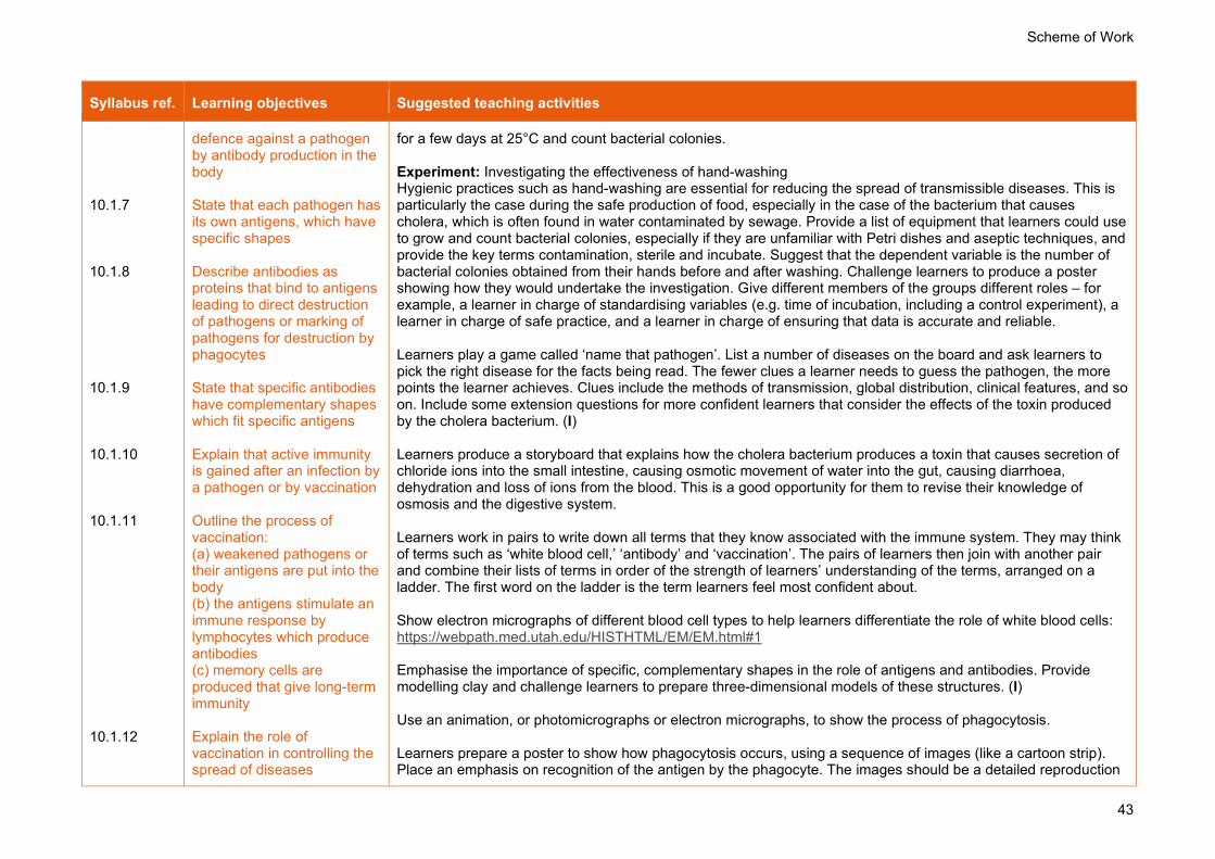
Scheme of Work
43
Syllabus ref. Learning objectives Suggested teaching activities
10.1.7 10.1.8 10.1.9 10.1.10 10.1.11 10.1.12
defence against a pathogen by antibody production in the body State that each pathogen has its own antigens, which have specific shapes Describe antibodies as proteins that bind to antigens leading to direct destruction of pathogens or marking of pathogens for destruction by phagocytes State that specific antibodies have complementary shapes which fit specific antigens Explain that active immunity is gained after an infection by a pathogen or by vaccination Outline the process of vaccination: (a) weakened pathogens or their antigens are put into the body (b) the antigens stimulate an immune response by lymphocytes which produce antibodies (c) memory cells are produced that give long-term immunity Explain the role of vaccination in controlling the spread of diseases
for a few days at 25°C and count bacterial colonies. Experiment: Investigating the effectiveness of hand-washing Hygienic practices such as hand-washing are essential for reducing the spread of transmissible diseases. This is particularly the case during the safe production of food, especially in the case of the bacterium that causes cholera, which is often found in water contaminated by sewage. Provide a list of equipment that learners could use to grow and count bacterial colonies, especially if they are unfamiliar with Petri dishes and aseptic techniques, and provide the key terms contamination, sterile and incubate. Suggest that the dependent variable is the number of bacterial colonies obtained from their hands before and after washing. Challenge learners to produce a poster showing how they would undertake the investigation. Give different members of the groups different roles – for example, a learner in charge of standardising variables (e.g. time of incubation, including a control experiment), a learner in charge of safe practice, and a learner in charge of ensuring that data is accurate and reliable. Learners play a game called ‘name that pathogen’. List a number of diseases on the board and ask learners to pick the right disease for the facts being read. The fewer clues a learner needs to guess the pathogen, the more points the learner achieves. Clues include the methods of transmission, global distribution, clinical features, and so on. Include some extension questions for more confident learners that consider the effects of the toxin produced by the cholera bacterium. (I) Learners produce a storyboard that explains how the cholera bacterium produces a toxin that causes secretion of chloride ions into the small intestine, causing osmotic movement of water into the gut, causing diarrhoea, dehydration and loss of ions from the blood. This is a good opportunity for them to revise their knowledge of osmosis and the digestive system. Learners work in pairs to write down all terms that they know associated with the immune system. They may think of terms such as ‘white blood cell,’ ‘antibody’ and ‘vaccination’. The pairs of learners then join with another pair and combine their lists of terms in order of the strength of learners’ understanding of the terms, arranged on a ladder. The first word on the ladder is the term learners feel most confident about. Show electron micrographs of different blood cell types to help learners differentiate the role of white blood cells: https://webpath.med.utah.edu/HISTHTML/EM/EM.html#1 Emphasise the importance of specific, complementary shapes in the role of antigens and antibodies. Provide modelling clay and challenge learners to prepare three-dimensional models of these structures. (I) Use an animation, or photomicrographs or electron micrographs, to show the process of phagocytosis. Learners prepare a poster to show how phagocytosis occurs, using a sequence of images (like a cartoon strip). Place an emphasis on recognition of the antigen by the phagocyte. The images should be a detailed reproduction

Scheme of Work
44
Syllabus ref. Learning objectives Suggested teaching activities
10.1.13 10.1.14 10.1.15 10.1.16 10.1.17
Explain that passive immunity is a short-term defence against a pathogen by antibodies acquired from another individual, including across the placenta and in breast milk Explain the importance of breast-feeding for the development of passive immunity in infants State that memory cells are not produced in passive immunity Describe cholera as a disease caused by a bacterium which is transmitted in contaminated water Explain that the cholera bacterium produces a toxin that causes secretion of chloride ions into the small intestine, causing osmotic movement of water into the gut, causing diarrhoea, dehydration and loss of ions from the blood
of the process, complete with explanatory labels. At the end of the activity, display an example of images showing phagocytosis. This will help learners to identify what they have missed and learn from their mistakes, but also reflect on what they feel their poster illustrates clearly. (F) Show an animation to consolidate understanding of the modes of phagocytosis. Examples can easily be found on video-sharing websites. Show learners the vaccination schedule used in your country, and ask them if they remember having any vaccinations. Contrast this with the vaccination programme for a country with very different risks to health. If there is resistance to vaccination in your country, find news reports about this and discuss the reasons behind it. Ask a number of questions to extend the class discussion, for example, ‘Do any of the vaccinations need to be given more than once, to provide good immunity?’, ‘At what ages are the vaccinations given?’ Provide learners with a diagram of the effect of changes in the number of antibodies and numbers of bacteria after a first and second infection. Ensure that only one member of each pair of learners can see this image. Ask them to decide who will be the describer (the one who can look at the image) and who will be the interpreter (the one who will convert the description into an image). Give the describers 2–3 minutes to describe the image – they cannot use hand signals or help the interpreter in any other way. The interpreter tries to reproduce the image from the verbal description only. As learners work, walk around the room and judge their progress, and then reveal the image to them on the board (or allow the describer to show the original image to the interpreter). Provide an opportunity for learners to consider what the missing labels are, before you reveal them. This activity helps learners to understand the relationship between an infection with a pathogen and the immune response. (I) Learners construct a table or Venn diagram to compare natural active immunity, artificial active immunity, natural passive immunity and artificial passive immunity. The points of comparison must focus on: exposure to antigen, presence or absence of an immune response, clonal selection, secretion of antibody molecules by plasma cells and memory cells. Ensure that learners include references to the placenta and breast milk when discussing the role of passive immunity. (F) Extension: Stretch and prepare for A level Learners read online sources related to monoclonal antibodies, such as: www.mayoclinic.org/diseases-conditions/cancer/in-depth/monoclonal-antibody/art-20047808
Past and specimen papers
Past/specimen papers and mark schemes are available to download at www.cambridgeinternational.org/support (F)

Scheme of Work
45
11. Gas exchange in humans
Syllabus ref. Learning objectives Suggested teaching activities
11.1.1 Human gas exchange 11.1.2 11.1.3 11.1.4 11.1.5 11.1.6
Describe the features of gas exchange surfaces in humans, limited to: large surface area, thin surface, good blood supply and good ventilation with air Identify in diagrams and images the following parts of the breathing system: lungs, diaphragm, ribs, intercostal muscles, larynx, trachea, bronchi, bronchioles, alveoli and associated capillaries Investigate the differences in composition between inspired and expired air using limewater as a test for carbon dioxide Describe the differences in composition between inspired and expired air, limited to: oxygen, carbon dioxide and water vapour Investigate and describe the effects of physical activity on the rate and depth of breathing Identify in diagrams and images the internal and external intercostal muscles
Challenge learners to prepare a glossary of key terms for this topic. As there are several key terms, you could set each learner the task of defining just 2–3 words each and linking them very clearly with diagrams. Examples include trachea, bronchi and bronchioles. (F) Show learners a figure that shows the human gas exchange system. Take learners outside and they take it in turns to draw around each other’s outline while lying on the floor of the playground. Each learner then draws a life-sized gas exchange system, with as many organs as they can remember from the discussion. As they work, walk around and identify any mistakes, providing learners with a stick of chalk of a different colour to make corrections. Back in class, highlight the most common misconceptions for all learners to discuss and resolve. (I) Remind learners of the gaseous composition of atmospheric air: https://www.bbc.co.uk/bitesize/guides/zysbgk7/revision/2
Resource Plus Carry out the Gas exchange in humans: model lung experiment referring to the Teaching Pack for lesson plans and resources.
This comprises two investigations: learners carry out the first, to investigate and explain the differences between inspired and expired air. (The second is later in this topic.) Demonstration: How a pair of actual lungs can be inflated Use the instructions at: https://pbiol.rsb.org.uk/cells-to-systems/ventilation-systems/dissecting-lungs This involves pumping air into lungs (car tyre pump) to observe rise and fall; it reveals some of the fine visible blood vessels that exist in the lung tissue. Help learners understand that the lungs do not contain muscle; they cannot inflate by their own actions. Demonstrate use of spirometer and recording, or use large diagram to show apparatus: https://pbiol.rsb.org.uk/cells-to-systems/ventilation-systems/using-a-spirometer-to-investigate-human-lung-function. A spirometer is the standard equipment used to measure the capacity of the human lungs. There are several versions of this laboratory apparatus available, but all consist of a chamber. Learners write the story of an oxygen molecule and its journey through the human gas exchange system, from the trachea and into the red blood cells where it binds to haemoglobin. Encourage learners to use plenty of descriptive language to show the route and the membranes that the oxygen molecule crosses. Animations can help to provide

Scheme of Work
46
Syllabus ref. Learning objectives Suggested teaching activities
11.1.7 11.1.8 11.1.9 11.1.10 11.1.11
State the function of cartilage in the trachea Explain the role of the ribs, the internal and external intercostal muscles and the diaphragm in producing volume and pressure changes in the thorax leading to the ventilation of the lungs Explain the differences in composition between inspired and expired air Explain the link between physical activity and the rate and depth of breathing in terms of: an increased carbon dioxide concentration in the blood, which is detected by the brain, leading to an increased rate and greater depth of breathing Explain the role of goblet cells, mucus and ciliated cells in protecting the breathing system from pathogens and particles
further guidance. Examples can easily be found on video-sharing websites. (I) Help learners describe how the features of gas exchange surfaces in humans increase the efficiency of gas exchange in humans, limited to: large surface area, thin surface, good blood and air supply. A table or a mind map are options.
Resource Plus Carry out the Gas exchange in humans: model lung experiment referring to the Teaching Pack for lesson plans and resources.
This comprises two investigations: learners carry out the second, to build a model gas exchange system to illustrate the role of the diaphragm in producing volume and pressure changes in the thorax, causing the movement of air into and out of the lungs. Learners investigate the effect of physical activity on rate and depth of breathing. Encourage learners to plan their own experiment in which they change one variable, measure another and try to keep other important variables the same. Challenge learners to identify the independent variable and how they will change it, and how to best draw a results table and plot this as a graph to support the ability to describe and explain their results. (I) Ask open-ended questions, for example, ‘What is the role of goblet cells, mucus and ciliated cells?’ ‘Why is it necessary for the passageways of the gas exchange system to get narrower and narrower?’ and ‘How could we estimate the number of alveoli in the lungs?’ Suggest the advantages of being able to adjust the diameter of bronchioles. You could link this to a research task in which learners research asthma or bronchitis or another chronic lung disease of the narrow air passages. (F) Extension: Stretch and prepare for A level Challenge learners to investigate diseases that impair the function of goblet cells, ciliated cells and mucus in protecting the gas exchange system from pathogens and particles. Examples are pneumonia and tuberculosis. How can they be treated?
Past and specimen papers
Past/specimen papers and mark schemes are available to download at www.cambridgeinternational.org/support (F)

Scheme of Work
47
12. Respiration
Syllabus ref. Learning objectives Suggested teaching activities
12.1.1 Respiration 12.1.2
State the uses of energy in living organisms, including: muscle contraction, protein synthesis, cell division, active transport, growth, the passage of nerve impulses and the maintenance of a constant body temperature Investigate and describe the effect of temperature on respiration in yeast
Challenge learners to write a short story to explain the function of respiration to a younger learner. They ought to convey the message that it comprises the chemical reactions in all living cells that release energy from glucose. Learners make a mind map or map of the human body, complete with labels, that illustrate the uses of energy in living organisms including muscle contraction, protein synthesis, cell division, active transport, growth, the passage of nerve impulses and the maintenance of a constant body temperature. (I)
Resource Plus Carry out the Investigating the effect of changing temperature on respiration in yeast experiment referring to the Teaching Pack for lesson plans and resources.
Extension: Stretch and prepare for A level Learners explore how experiments, using apparatus called respirometers, can be conducted to give accurate readings for values of respiration rate.
12.2.1 Aerobic respiration 12.2.2 12.2.3
Describe aerobic respiration as the chemical reactions in cells that use oxygen to break down nutrient molecules to release energy State the word equation for aerobic respiration as: glucose + oxygen → carbon dioxide + water State the balanced chemical equation for aerobic respiration as: C6H12O6 + 6O2 → 6CO2 + 6H2O
Give learners a strict time limit such as 10–15 seconds, to write out some key facts about aerobic respiration, working in small groups. This could include the range of uses of energy in cells, or the relationship between oxygen, glucose and energy in the word or chemical equation. Next, allow learners to walk around the class and speak with at least three people, to compare and refine their work. Learners then classify their own errors, to identify their strengths and weaknesses. This will allow learners to see that ‘everybody makes mistakes’ and develop a growth mind-set. This activity will help to build confidence and emphasise the benefits of patiently learning the material. (I) Provide learners with five exemplar responses to an extended-answer question on aerobic respiration that cover a range of levels. In groups, learners choose whether some are better examples than others. They rank the work in terms of quality and then report back. Compare with your own judgement. (F) Extension: Stretch and prepare for A level Learners explore the purpose of respiratory inhibitors in nature – e.g. in cyanogenic clover.
12.3.1 Anaerobic respiration
Describe anaerobic respiration as the chemical reactions in cells that break
Learners brainstorm and list what they know about anaerobic respiration. After a few minutes, pairs join together into groups of four and then groups of eight to discuss this further and come up with an agreed list of points. One or two learners from each group then write the group’s ideas on the class board to form a mind map. (F)

Scheme of Work
48
Syllabus ref. Learning objectives Suggested teaching activities
12.3.2 12.3.3 12.3.4 12.3.5 12.3.6 12.3.7
down nutrient molecules to release energy without using oxygen State that anaerobic respiration releases much less energy per glucose molecule than aerobic respiration State the word equation for anaerobic respiration in yeast as: glucose → alcohol + carbon dioxide State the word equation for anaerobic respiration in muscles during vigorous exercise as: glucose → lactic acid State the balanced chemical equation for anaerobic respiration in yeast as: C6H12O6 → 2C2H5OH + 2CO2 State that lactic acid builds up in muscles and blood during vigorous exercise causing an oxygen debt Outline how the oxygen debt is removed after exercise, limited to: (a) continuation of fast heart rate to transport lactic acid in the blood from the muscles to the liver
Learners can find it difficult to recall the distinctive features of aerobic and anaerobic respiration. Show learners a series of images that represent a 100 m race and challenge them to discuss which type of respiration occurs at each stage, including reasons why. Then discuss what happens after the race to break down the lactic acid that accumulates in muscles and blood during Excess Post-exercise Oxygen Consumption (EPOC) (‘oxygen debt’). Encourage learners to share their ideas in groups of 2–3 in a 10–15 minute ‘wander and wonder’ activity at the end of the session. Provide display stations for this purpose. (I) Learners draw a Venn diagram to compare aerobic respiration, anaerobic respiration in humans, and anaerobic respiration in yeast. Provide key terms to ensure complete coverage, e.g. energy, lactic acid and oxygen debt. (I) Prepare a crossword containing all the terms used in this lesson, with clear clues. Learners keep their completed copies as a sheet of definitions to refer to throughout the topics of aerobic and anaerobic respiration. (I) Encourage a class debate. The ‘motion’ must be a controversial statement, rather than a question, to prompt deeper thought among learners. For example, ask learners to evaluate a statement such as ‘Aerobic respiration is more important than anaerobic respiration’ or ‘It is possible to survive without anaerobic respiration’. Use the ‘think, pair, share’ technique as an introduction to help learners form an opinion. (I) In terms of anaerobic respiration, challenge learners to write the shortest sentence possible using the following key terms: energy, respiration, glucose and activity. This is a good way for learners to focus on developing their higher-order thinking skills to make sense of the meaning of these terms. To scaffold this activity for some learners, provide the first and final sentences, or reduce the number of terms that they are expected to use. (F) Show in a series of images that represent a 100 m race the stages at which the two types of respiration occur, including reasons why, and then discuss what happens after the race to repay the oxygen debt – and why. Extension: Stretch and prepare for A level Learners investigate what happens to the lactate produced during anaerobic respiration in animals. Ask more confident learners to give a brief description, as a five-minute ‘master class,’ to extend the knowledge of the rest of the class.

Scheme of Work
49
Syllabus ref. Learning objectives Suggested teaching activities
(b) continuation of deeper and faster breathing to supply oxygen for aerobic respiration of lactic acid (c) aerobic respiration of lactic acid in the liver
Past and specimen papers
Past/specimen papers and mark schemes are available to download at www.cambridgeinternational.org/support (F)

Scheme of Work
50
13. Excretion in humans
Syllabus ref. Learning objectives Suggested teaching activities
13.1.1 Excretion 13.1.2 13.1.3 13.1.4 13.1.5
State that carbon dioxide is excreted through the lungs State that the kidneys excrete urea and excess water and ions Identify in diagrams and images the kidneys, ureters, bladder and urethra Identify in diagrams and images the structure of the kidney, limited to the cortex and medulla Outline the structure and function of a nephron and its associated blood vessels, limited to: (a) the role of the glomerulus in the filtration from the blood of water, glucose, urea and ions (b) the role of the nephron in the reabsorption of all of the glucose, some of the ions and most of the water back into the blood (c) the formation of urine containing urea, excess water and excess ions (details of these processes are not required)
Hold a ‘think, pair, share’ activity and challenge learners to develop a definition of excretion. Ask for contributions and merge into a comprehensive statement that covers the removal from the body of metabolic waste and substances in excess, with examples. Learners will have already learnt about one excretory product – carbon dioxide produced in respiration – use this to explain to them what excretion is, and how it differs from egestion. Explain how, if it were to remain in the body, it would be toxic to cells. Learners prepare 2–3 examination questions, complete with mark schemes, to reinforce their knowledge of excretion. Inform learners that in the next lesson they will provide these questions to you, and you will select the best for a short formative test – this should motivate learners to write the best questions they can. (F) Experiment: Dissection of an animal kidney to investigate its gross structure Learners should note the difference between the blood content and size of the cortex and the medulla. (I) Learners draw diagrams of transverse and longitudinal sections of kidney tissue, including detail showing the tubules in different planes, labelling glomerulus, renal convoluted tubule (proximal and distal), Bowman’s capsule, loop of Henle and collecting duct. You could provide histology images, such as: https://webpath.med.utah.edu/RENAHTML/RENALIDX.html www.histology.leeds.ac.uk/urinary/kidney.php (I) Show a short animation of the movement of substances that occur in a nephron, such as: www.sumanasinc.com/webcontent/animations/content/kidney.html Pause the animation at regular intervals for learners to discuss, in small groups, and give a summary sentence that describes the events. Tell learners that they are to work in groups of 3–4 to make a model to show one of three processes involved in human excretion. The model should contain no writing – the challenge is for learners to be able to explain it verbally to you as you move around the room. Learners can choose to either make a model kidney or a model nephron. During the modelling task, move around the room and ask learners to explain what they are doing, and why. Encourage learners to do the same (have 2–3 minute ‘breaks’ in which learners can leave their work station), ensuring that by the end of the activity each learner has observed the work of groups who have undertaken the other two tasks to their own. (F) Use this topic as an opportunity to develop learners’ skills in making scientific drawings. Encourage them to recognise and draw structures from electron micrographs.

Scheme of Work
51
Syllabus ref. Learning objectives Suggested teaching activities
13.1.6 13.1.7 13.1.8 13.1.9
Describe the role of the liver in the assimilation of amino acids by converting them to proteins State that urea is formed in the liver from excess amino acids Describe deamination as the removal of the nitrogen-containing part of amino acids to form urea Explain the importance of excretion, limited to toxicity of urea
Good sources of kidney sections include: https://wellcomecollection.org/works/h2parxes https://wellcomecollection.org/works/ask2jkuq Discuss with learners how the colour of urine differs, depending on different times during the day (e.g. it is darker in colour first thing in the morning) and after certain activities (similar after extended periods of physical activity). Explain that this is an example of both excretion and homeostasis. You could use fake urine (water with yellow food colouring with and without glucose and/or proteins) as an effective prop. A significant number of key terms, both nouns and verbs, are introduced in this topic. To help familiarise with them, learners work in pairs to describe key words to each other, but without using other (listed) key words. For example, it is challenging for learners to describe the process of excretion without using the key terms: urethra, urine, ureter, urea. Put the key terms on the board as they are met to reinforce their importance and help learners become familiar with them. (F) Challenge learners to write a short story that shows what happens to an ‘unwanted amino acid,’ and how this molecule is converted into products including urea in the liver, before passing to the kidneys. (I) Extension: Stretch and prepare for A level Ask learners a series of questions to stretch their understanding of the role of the kidney. For example, why do we have two? Why do plants not need kidneys?
Past and specimen papers
Past/specimen papers and mark schemes are available to download at www.cambridgeinternational.org/support (F)

Scheme of Work
52
14. Coordination and response
Syllabus ref. Learning objectives Suggested teaching activities
14.1.1 Coordination and response 14.1.2 14.1.3 14.1.4 14.1.5 14.1.6
State that electrical impulses travel along neurones Describe the mammalian nervous system in terms of: (a) the central nervous system (CNS) consisting of the brain and the spinal cord (b) the peripheral nervous system (PNS) consisting of the nerves outside of the brain and spinal cord Describe the role of the nervous system as coordination and regulation of body functions Identify in diagrams and images sensory, relay and motor neurones Describe a simple reflex arc in terms of: receptor, sensory neurone, relay neurone, motor neurone and effector Describe a reflex action as a means of automatically and rapidly integrating and coordinating stimuli with the responses of effectors (muscles and glands)
Ask learners to stand up and then to sit down. Ask them how and why they did it. Use their answers to discuss the roles of receptors (their ears), coordination (the brain, where the response was decided) and the effectors (the muscles they used in standing up). Explain that all animal coordination relies on receptors, coordination and effectors. To consolidate key terms, provide each learner with a piece of paper divided in half. On one half, there is a key term, and on the other, there is a definition. However, the definition is not for that key term. Examples of terms to include are stimulus, receptor, effector, coordination centre, response, and so on. Allow learners to move around the room to find the learner who has the definition of their key word, and also another who has the key word for their definition. (F) Discuss the concept of reaction time in 100 m sprints. Ask questions about whether the difference between hearing the starting pistol and pushing off from the blocks is down to chance, or a very low reaction time. Learners could assess their own reaction time: https://humanbenchmark.com/ Learners draw a Venn diagram showing the similarities between the three types of neurone. (I) Learners take part in a roleplay activity to illustrate how a reflex action occurs. They arrange themselves into a circle and follow your instructions: they hold hands and pass the ‘message’ of ‘squeezes’ all round the circle. This can be timed on a stopwatch. Learners should keep repeating this, until the squeeze is going around as fast as possible. Record the time taken, and also the number of people in the circle – this allows for a relatively accurate estimation of the speed of the impulse, which takes into account its path from a left hand, to the spinal cord and to a right hand (multiply by the number of people, and then calculate speed by dividing total distance by total time taken). Point out to learners that they tend to get faster as they practice – refer to the effect of learning on reaction time. An alternative activity is the ‘ruler drop’ experiment in which learners work in pairs to catch a ruler dropped without notice, and read off the distance as a measure of the reaction time. Instructions are at: https://pbiol.rsb.org.uk/control-and-communication/reflex-nerves-and-reactions/measuring-reaction-time-of-a-human-nerve-controlled-reaction. Learners explore the process by which a reflex action occurs by collaborating in groups to produce a poster. The focus should be a sketch of a reflex arc, which could occur when our hand touches a hot object (for example). The posters should be highly visual, including diagrams, photographs (if a printer is available) and text. Then hold a

Scheme of Work
53
Syllabus ref. Learning objectives Suggested teaching activities
14.1.7 14.1.8 14.1.9 14.1.10
Describe a synapse as a junction between two neurones Describe the structure of a synapse, including the presence of vesicles containing neurotransmitter molecules, the synaptic gap and receptor proteins Describe the events at a synapse as: (a) an impulse stimulates the release of neurotransmitter molecules from vesicles into the synaptic gap (b) the neurotransmitter molecules diffuse across the gap (c) neurotransmitter molecules bind with receptor proteins on the next neurone (d) an impulse is then stimulated in the next neurone State that synapses ensure that impulses travel in one direction only
‘marketplace’ activity in which one member of each group stands by their poster and offers an explanation to other groups as they move around the room. (I) Challenge learners to further consider the roleplay activity. Ask them to consider what could be done to this roleplay model to indicate a synapse. One option is to ask the learner at the terminal end of the line to remove the lid from a perfume bottle when his or her hand is squeezed. How does this accurately model the role of vesicles, neurotransmitters and receptors in a synapse, and the fact that synapses ensure that impulses travel in one direction only?
14.2.1 Sense organs 14.2.2
Describe sense organs as groups of receptor cells responding to specific stimuli: light, sound, touch, temperature and chemicals Identify in diagrams and
Challenge learners to work in pairs to list as many stimuli as they can, that their bodies can detect. This could be a competition, with the pair who have made the longest list of correct stimuli / sense organs declared the winners. Learners carry out a dissection of an animal eye to investigate its gross structure. In particular, they should note the position and shape of the lens, the fact that the pupil is a hole in the iris, and the colour of the retina. (I) Set up an activity to show the existence of a blind spot in the retina (you could use online images). Use this as a

Scheme of Work
54
Syllabus ref. Learning objectives Suggested teaching activities
14.2.3 14.2.4 14.2.5 14.2.6 14.2.7
images the structures of the eye, limited to: cornea, iris, pupil, lens, retina, optic nerve and blind spot Describe the function of each part of the eye, limited to: (a) cornea – refracts light (b) iris – controls how much light enters the pupil (c) lens – focuses light on to the retina (d) retina – contains light receptors, some sensitive to light of different colours (e) optic nerve – carries impulses to the brain Explain the pupil reflex, limited to changes in light intensity and pupil diameter Explain the pupil reflex in terms of the antagonistic action of circular and radial muscles in the iris Explain accommodation to view near and distant objects in terms of the contraction and relaxation of the ciliary muscles, tension in the suspensory ligaments, shape of the lens and refraction of light Describe the distribution of rods and cones in the retina of a human
starting point to discuss the function of the retina: there are no receptor cells where the optic nerve leaves the retina, and if light lands here, no impulses will be sent to the brain. Ask learners to describe any other optical illusions that they may have encountered. Try to explain whether these are due to issues related to the eye, or the brain. Carry out a demonstration to show how accommodation occurs. The way in which the ciliary muscle, suspensory ligaments and lens can change the focus of the eye is difficult to understand, but it is possible to show the effect of different sizes and shapes of lenses on parallel beams of light. (You may be able to use apparatus from the physics department, and get advice from them on how to set it up and use it.) Alternatively, place a sealable plastic bag (e.g. zip-lock bag) full of water on top of the small print of a newspaper to demonstrate how focusing occurs. The ‘lens’ will magnify the text when pushed into a more spherical shape. Help learners link this with the changes that occur when they change from reading to looking at a distant object. Learners work in pairs to investigate the pupil reflex. One member of each pair should close his or her eyes, and cover them with something dark, to cut out as much light as possible. After about 30 seconds, the learner should remove the cover and look at their partner’s eyes as they adapt to the light. Ask a series of questions to elicit understanding of what happens and why this change occurs. (I) Learners produce two ‘flipbooks’ that show the events that occur during pupil reflex and accommodation. They should take care not to confuse the two mechanisms. (I) Provide each learner with a small piece of card or paper. Ask them to prepare a very short ‘to do’ list to indicate the ‘equipment’ needed by the eye for detecting colours, carrying out the pupil reflex or undertaking accommodation. (F) Resources to support learners’ study of the eye include: www.purposegames.com/game/label-the-eye-quiz Prepare three or four past paper questions, ideally of a multiple-choice or short-answer nature, which learners complete and pass to you as they leave the room. This ‘exit card’ technique can provide an opportunity for formative assessment to inform you whether you need to reinforce the content in the next lesson. (F) Extension: Stretch and prepare for A level Understanding what happens when the parts of the eye do not work properly can help learners develop a deeper understanding of their usual functions. Challenge learners to carry out research into three or four eye disorders. They should emphasise which parts of the eye are affected, and be prepared to describe their findings at the start of the next lesson. Examples may include the cornea (cataracts), macular degeneration (retina) and pseudomyopia (ciliary muscles). A useful website is:

Scheme of Work
55
Syllabus ref. Learning objectives Suggested teaching activities
14.2.8 14.2.9
Outline the function of rods and cones, limited to: (a) greater sensitivity of rods for night vision (b) three different kinds of cones, absorbing light of different colours, for colour vision Identify in diagrams and images the position of the fovea and state its function
www.nhs.uk/video/pages/Cataractanimation.aspx
14.3.1 Hormones 14.3.2 14.3.3
Describe a hormone as a chemical substance, produced by a gland and carried by the blood, which alters the activity of one or more specific target organs Identify in diagrams and images specific endocrine glands and state the hormones they secrete, limited to: (a) adrenal glands and adrenaline (b) pancreas and insulin (c) testes and testosterone (d) ovaries and oestrogen Describe adrenaline as the hormone secreted in ‘fight or flight’ situations and its effects, limited to: (a) increased breathing rate (b) increased heart rate (c) increased pupil diameter
Provide learners with a large outline of the human body. Ask them to draw the location, and approximate size, of all of the glands they have heard of. Choose an endocrine gland and ask learners to tell you what happens in their body when this secretes its hormone. For example, imagine that they are very excited or frightened: what are the effects that adrenaline has, and how do these help the body to prepare for action? Review learners’ diagrams and address misconceptions early. These will probably include: drawing the glands too big; not identifying the testes or ovaries as glands; failing to draw two adrenal glands, and including the salivary gland, which is not an example if an endocrine gland. (F) Learners engage in research to become ‘experts’ on one particular hormone listed in the syllabus, before delivering their findings to others in small groups. Use a system of ‘jigsaw’ grouping to focus on independent work and examination technique. Give each small group one past paper question, focusing on one hormone and its effects. Then break up into rearranged groups to ‘teach’ how to answer the question to their peers. This means that each learner is responsible for another’s learning, and provides them with alternative views and strategies to answer past paper questions. Circulate during the activity to highlight good ideas to encourage and motivate learners. (I) Help learners to compare the features of the nervous system and the endocrine system by constructing a table to show similarities and differences. Print and write on cards the sequence of events that occurs in control of blood glucose concentration. Shuffle the cards and ask learners to arrange them in the correct sequence. The cards include the secretion and effects of adrenaline as the ‘fight or flight’ hormone. (I) Extension: Stretch and prepare for A level Challenge learners to find out how the hormones listed in the syllabus differ in terms of the mechanism by which

Scheme of Work
56
Syllabus ref. Learning objectives Suggested teaching activities
14.3.4 14.3.5 14.3.6
Compare nervous and hormonal control, limited to speed of action and duration of effect State that glucagon is secreted by the pancreas Describe the role of adrenaline in the control of metabolic activity, limited to: (a) increasing the blood glucose concentration (b) increasing heart rate
they act. This should be limited to whether they are able to diffuse across the cell membrane or not.
14.4.1 Homeostasis 14.4.2 14.4.3 14.4.4 14.4.5 14.4.6
Describe homeostasis as the maintenance of a constant internal environment State that insulin decreases blood glucose concentration Explain the concept of homeostatic control by negative feedback with reference to a set point Describe the control of blood glucose concentration by the liver and the roles of insulin and glucagon Outline the treatment of Type 1 diabetes Identify in diagrams and images of the skin: hairs, hair erector muscles, sweat
Host a discussion with learners to identify the physiological factors that are maintained at a set point (e.g. temperature, blood glucose concentration, blood pH / carbon dioxide concentration, water balance / water potential, metabolic wastes) and explain the importance of maintaining the balance. Use this opportunity to revise the source of excretory substances, e.g. urea is produced in the liver from the deamination of excess amino acids. Give learners sentence stems to complete when describing steps in the mechanisms that control blood glucose concentration, temperature regulation and excretion. For example, ‘When _________ decreases, the body responds by _________.’ Provide more comprehensive writing frames to learners who need further support, to ensure their notes are complete and to build confidence. (F) Provide a simple definition of homeostatic control by negative feedback for learners to use and apply to other situations, e.g.:
• there is a set point – a normal level that the system tries to maintain • there is a 'measuring device' that keeps track of whether the level is within the range of the set point • if the level goes outside the set point, this triggers events that bring the level back into line again.
Introduce this topic using an analogy with an example that learners know. Prompt a discussion on how a thermostatically controlled water bath operates or by listing others that work in the same way – for example, ovens, central heating systems and air-conditioned rooms. Point out the role of the control panel in these machines, which is the equivalent of the hypothalamus in the body. By putting together cut-out shapes into diagrams and adding labels, learners explore the role of the skin in thermoregulation and the role of the pancreas in the regulation of blood glucose concentration. Provide different

Scheme of Work
57
Syllabus ref. Learning objectives Suggested teaching activities
14.4.7 14.4.8
glands, receptors, sensory neurones, blood vessels and fatty tissue Describe the maintenance of a constant internal body temperature in mammals in terms of: insulation, sweating, shivering and the role of the brain Describe the maintenance of a constant internal body temperature in mammals in terms of vasodilation and vasoconstriction of arterioles supplying skin surface capillaries
pairs of learners with large, photocopied images of the cross-section of the skin, how the hypothalamus controls thermoregulation, or the mechanism of control of blood glucose concentration. Learners display their work as posters, which can be peer assessed. (I) Show a short video clip about the extreme conditions that humans can (briefly) cope with. An example would be the heat that firefighters are exposed to, or the extreme cold that some people experience when they (choose to) swim in ice-cold water. Distinguish between the external and internal environments. Give learners several sentences to complete related to the video clip e.g. ‘The receptors sensitive to temperature change are found in the…’ and ‘Enzymes require a relatively stable body temperature because…’ (F) Write out a set of statements on separate cards that describe an example of a homeostatic mechanism that contributes to the maintenance of constant internal body temperature in mammals. Give these to learners to put into a logical sequence. These cards should include sweating, shivering, contraction of hair erector muscles, and vasodilation and vasoconstriction of arterioles supplying skin surface capillaries. (F) Experiment: Investigating how penguin groups maintain their body temperature Learners plan or undertake a practical in which they model the effect of penguins ‘huddling’ together in groups on their body temperature. Test tubes containing warm water can be used as model penguins, and the temperature of the water in a central test tube can be measured over time, compared to a test tube that is on the outside. (I) Learners identify analogies to describe the role of homeostasis in the body. Examples include a cooking oven with a thermostat, a thermostatically controlled water bath, central heating systems, and air-conditioned rooms. Then discuss homeostasis and link the analogies to key terms that you write on the board, such as stimulus (internal and external), receptor, coordination centre, effector and response. Learners record a summary of the discussion in the form of a flow diagram, including these key terms. Learners produce an infographic to summarise the signs of Type 1 diabetes (increased blood glucose concentration and glucose in urine) and its treatment (administration of insulin). (I) Extension: Stretch and prepare for A level Learners construct Venn diagrams to compare the origin, mode of action, targets and functions of insulin and glucagon. Draw a circle labelled ‘insulin’ overlapping with another circle labelled ‘glucagon’. Insulin and glucagon have many things in common (e.g. both are hormones and are released by the pancreas). However, there is much that is unique to each (e.g. the specific cells in the pancreas that release them, their effects on blood glucose concentration, and so on).
14.5.1 Tropic responses
Describe gravitropism as a response in which parts of a plant grow towards or away
Show video clips of unusual, rapid movement in plants (e.g. closure of the leaves of the Venus fly trap, ejection of spores from fern sporangia, dispersal of seeds from Himalayan balsam and folding leaflets of Mimosa): https://plantsinmotion.bio.indiana.edu/plantmotion/movements/nastic/nastic.html

Scheme of Work
58
Syllabus ref. Learning objectives Suggested teaching activities
14.5.2 14.5.3 14.5.4 14.5.5
from gravity Describe phototropism as a response in which parts of a plant grow towards or away from the direction of the light source Investigate and describe gravitropism and phototropism in shoots and roots Explain phototropism and gravitropism of a shoot as examples of the chemical control of plant growth Explain the role of auxin in controlling shoot growth, limited to: (a) auxin is made in the shoot tip (b) auxin diffuses through the plant from the shoot tip (c) auxin is unequally distributed in response to light and gravity (d) auxin stimulates cell elongation
Emphasise that there are numerous similarities between communication systems in plants and in animals. Both involve the secretion of chemicals that travel both short and long distances to their target organs. Both require communication systems to respond to changes in their external and internal environments. Expand learners’ thinking to explain that the responses they have just seen are quite unusual, but that all plants respond in more subtle ways to stimuli. These include gravitropism and phototropism. Experiment: Investigating tropisms in live plants Guidance can be found online at sites including: www.saps.org.uk Learners can use their mobile phones / digital cameras to produce a time-lapse video. Provide a range of plant models for learners to choose from, including:
• Grow bean or cereal seedlings in gas jars to keep shoot or coleoptiles and root systems straight. Then turn seedlings onto their side and pin onto board to show positive gravitropism of roots and negative gravitropism of coleoptiles. Pin some germinating beans to clinostat or keep rotating while growing the seedlings.
• Grow cress / cabbage seedlings in pots to show response to light from one side. If possible use different growth boxes with coloured filters to experiment with differing wavelengths.
Provide clear instructions to learners on how they should undertake and display their work and how it will be assessed. (I) Learners compile a list of similarities between communication in flowering plants and in mammals by comparing chemical communication in plants and animals. Present comparisons as a table or use a table to plan and then write out comparisons using examples. Extension: Stretch and prepare for A level Learners carry out research online to investigate how, and why, auxin is used in horticulture as a herbicide.
Past and specimen papers
Past/specimen papers and mark schemes are available to download at www.cambridgeinternational.org/support (F)
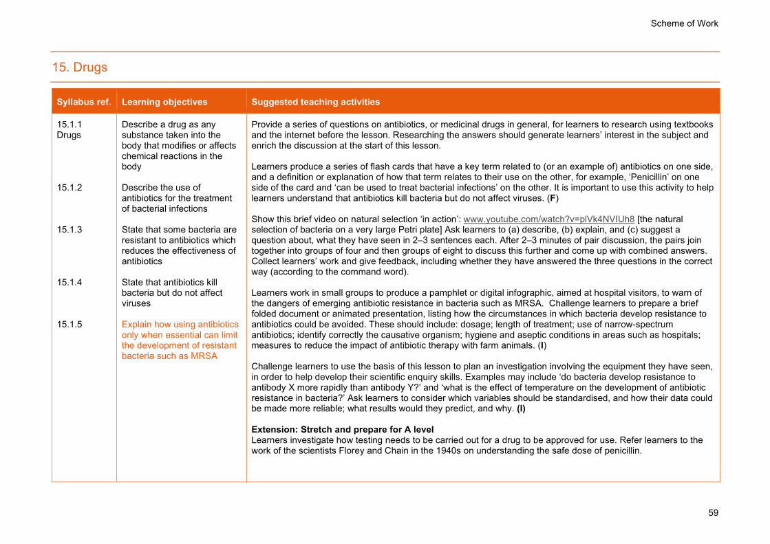
Scheme of Work
59
15. Drugs
Syllabus ref. Learning objectives Suggested teaching activities
15.1.1 Drugs 15.1.2 15.1.3 15.1.4 15.1.5
Describe a drug as any substance taken into the body that modifies or affects chemical reactions in the body Describe the use of antibiotics for the treatment of bacterial infections State that some bacteria are resistant to antibiotics which reduces the effectiveness of antibiotics State that antibiotics kill bacteria but do not affect viruses Explain how using antibiotics only when essential can limit the development of resistant bacteria such as MRSA
Provide a series of questions on antibiotics, or medicinal drugs in general, for learners to research using textbooks and the internet before the lesson. Researching the answers should generate learners’ interest in the subject and enrich the discussion at the start of this lesson. Learners produce a series of flash cards that have a key term related to (or an example of) antibiotics on one side, and a definition or explanation of how that term relates to their use on the other, for example, ‘Penicillin’ on one side of the card and ‘can be used to treat bacterial infections’ on the other. It is important to use this activity to help learners understand that antibiotics kill bacteria but do not affect viruses. (F) Show this brief video on natural selection ‘in action’: www.youtube.com/watch?v=plVk4NVIUh8 [the natural selection of bacteria on a very large Petri plate] Ask learners to (a) describe, (b) explain, and (c) suggest a question about, what they have seen in 2–3 sentences each. After 2–3 minutes of pair discussion, the pairs join together into groups of four and then groups of eight to discuss this further and come up with combined answers. Collect learners’ work and give feedback, including whether they have answered the three questions in the correct way (according to the command word). Learners work in small groups to produce a pamphlet or digital infographic, aimed at hospital visitors, to warn of the dangers of emerging antibiotic resistance in bacteria such as MRSA. Challenge learners to prepare a brief folded document or animated presentation, listing how the circumstances in which bacteria develop resistance to antibiotics could be avoided. These should include: dosage; length of treatment; use of narrow-spectrum antibiotics; identify correctly the causative organism; hygiene and aseptic conditions in areas such as hospitals; measures to reduce the impact of antibiotic therapy with farm animals. (I) Challenge learners to use the basis of this lesson to plan an investigation involving the equipment they have seen, in order to help develop their scientific enquiry skills. Examples may include ‘do bacteria develop resistance to antibody X more rapidly than antibody Y?’ and ‘what is the effect of temperature on the development of antibiotic resistance in bacteria?’ Ask learners to consider which variables should be standardised, and how their data could be made more reliable; what results would they predict, and why. (I) Extension: Stretch and prepare for A level Learners investigate how testing needs to be carried out for a drug to be approved for use. Refer learners to the work of the scientists Florey and Chain in the 1940s on understanding the safe dose of penicillin.

Scheme of Work
60
Past and specimen papers
Past/specimen papers and mark schemes are available to download at www.cambridgeinternational.org/support (F)

Scheme of Work
61
16. Reproduction
Syllabus ref. Learning objectives Suggested teaching activities
16.1.1 Asexual reproduction 16.1.2 16.1.3
Describe asexual reproduction as a process resulting in the production of genetically identical offspring from one parent Identify examples of asexual reproduction in diagrams, images and information provided Discuss the advantages and disadvantages of asexual reproduction: (a) to a population of a species in the wild (b) to crop production
Learners construct a dichotomous key that helps them differentiate between asexual and sexual reproduction. This should conform to the definitions listed in the syllabus. (I) Learners explore the differences between sexual and asexual reproduction through a debate. Arrange learners in pairs, and ask them to spend 5–10 minutes researching the roles and processes involved in asexual and sexual reproduction. After this time, identify learners to either represent ‘the case for sexual reproduction’ or ‘the case for asexual reproduction’ and clarify their arguments. Group pairs of learners together to arrange teams of four. Allow each team of four to give their arguments, and then the other team of four should provide their counter-argument. Host a discussion at the end of the debate to identify the characteristics of each type of reproduction, and the relative advantages and disadvantages of each method, in the two different contexts listed in the syllabus. (I) Experiment: Investigating asexual reproduction: https://pbiol.rsb.org.uk/genetics/introducing-gene-technologies/cloning-a-living-organism. This requires learners to clone a plant by taking cuttings, as an illustration of asexual reproduction. It can be done as an extended project. Extension: Stretch and prepare for A level Learners carry out research online to explore how horticulturists exploit asexual reproduction in bulbs and rhizomes, e.g. daffodils, orchids.
16.2.1 Sexual reproduction 16.2.2 16.2.3
Describe sexual reproduction as a process involving the fusion of the nuclei of two gametes to form a zygote and the production of offspring that are genetically different from each other Describe fertilisation as the fusion of the nuclei of gametes State that nuclei of gametes are haploid and that the
Challenge learners to write the shortest sentence possible using key terms (gamete, fusion, fertilisation, haploid, diploid, zygote) and numerical values relevant to this topic. This is a good way to focus learners on developing their higher-order thinking skills, rather than simply expecting them to recall key terms. Learners carry out research to find the number of chromosomes in diploid cells and gametes of a range of organisms, including those with very few (e.g. mosquito = 3) to very many (e.g. polar bear = 78). Learners undertake research and prepare, in groups of 2–3, a short ‘TED Talk’ on the subject, ‘Sexual reproduction: a more advanced method than asexual reproduction’. During the project, provide roles to learners to ensure that all members are engaged. Roles could include the decision maker, the scribe and the internet researcher. This can also be used to differentiate learning: provide a more challenging role for more confident learners. (I)

Scheme of Work
62
Syllabus ref. Learning objectives Suggested teaching activities
16.2.4
nucleus of a zygote is diploid Discuss the advantages and disadvantages of sexual reproduction: (a) to a population of a species in the wild (b) to crop production
Extension: Stretch and prepare for A level In their attempts to model meiosis, more confident learners could show, with guidance, how different alleles of the same genes on some of the homologous pairs can be represented by tying little pieces of different-coloured cotton to the chromosomes and showing how these can end up in different combinations in the daughter cells.
16.3.1 Sexual reproduction in plants 16.3.2 16.3.3 16.3.4 16.3.5 16.3.6
Identify in diagrams and images and draw the following parts of an insect-pollinated flower: sepals, petals, stamens, filaments, anthers, carpels, style, stigma, ovary and ovules State the functions of the structures listed in 16.3.1 Identify in diagrams and images and describe the anthers and stigmas of a wind-pollinated flower Distinguish between the pollen grains of insect-pollinated and wind-pollinated flowers Describe pollination as the transfer of pollen grains from an anther to a stigma State that fertilisation occurs when a pollen nucleus fuses with a nucleus in an ovule
Provide learners with mini-whiteboards. Inform learners that they will take part in a 30-second competition. Learners draw and label a flower in as much detail as they can. Use this activity as an opportunity to correct some misconceptions, e.g. if a learner draws a whole plant, point out that the flower is the reproductive organ only. (F) Learners collect or draw examples of flowers that are native to your country of residence. Help learners construct a definition of ‘flower’ and label the different parts, including the sepals, petals, stamens (anthers and filaments) and carpels (stigmas, styles, ovaries and ovules). Instructions to help learners ‘dissect’ their flower(s): https://pbiol.rsb.org.uk/cells-to-systems/reproductive-systems/comparing-the-flower-structure-of-different-angiosperms Learners make a model of a flower using a variety of resources including coloured paper, pipe cleaners and paper cups. Challenge learners to use the knowledge they have developed during this unit to construct a model of a wind- or insect-pollinated flower. Challenge learners to use their models to distinguish between self-pollination and cross-pollination. (I) Provide learners with several sets of seeds, such as beans, with different combinations of germination conditions. This experiment will enable learners to work out which conditions the seeds need before they will germinate and reveal the cotyledons – the plumule and radicle. Set up five large test tubes, as shown in the diagram. If there is no suitable dark place to leave tubes B and C, you can cover the tubes with black paper instead. Make sure that you put the same number of seeds into each tube. Check the tubes each day. Count how many seeds have germinated, and record this as a percentage. Learners use their results to decide which conditions these seeds require, in order to germinate. (I) Learners explore the differences between self-pollination and cross-pollination by engaging in a debate. Arrange learners in pairs, and ask them to spend 5–10 minutes researching the processes involved in self-pollination and cross-pollination. After this time, identify learners to either represent ‘the case for self-pollination or ‘the case for cross-pollination’ and clarify their arguments.

Scheme of Work
63
Syllabus ref. Learning objectives Suggested teaching activities
16.3.7 16.3.8 16.3.9 16.3.10 16.3.11 16.3.12
Describe the structural adaptations of insect-pollinated and wind-pollinated flowers Investigate and describe the environmental conditions that affect germination of seeds, limited to the requirement for: water, oxygen and a suitable temperature Describe self-pollination as the transfer of pollen grains from the anther of a flower to the stigma of the same flower or a different flower on the same plant Describe cross-pollination as the transfer of pollen grains from the anther of a flower to the stigma of a flower on a different plant of the same species Discuss the potential effects of self-pollination and cross-pollination on a population, in terms of variation, capacity to respond to changes in the environment and reliance on pollinators Describe the growth of the pollen tube and its entry into the ovule followed by fertilisation (details of production of endosperm and
Host a discussion at the end of this activity to identify the characteristics of self- and cross-pollination, and the relative advantages and disadvantages of each method, in terms of variation, capacity to respond to changes in the environment and reliance on pollinators. (I) Learners work in pairs for an activity on the structures involved in plant reproduction. Provide each learner with an image showing one of the structures important in the process. This could be, for example, the structure of a seed, limited to embryo (radicle, plumule and cotyledons) and testa, or the role of enzymes in the process of germination. Also provide each learner with a piece of blank paper. Each learner takes it in turn to describe the image to their partner using only spoken words (they cannot sketch or use hand signals). Their partner has to reproduce the diagram during the description and then both learners discuss what it shows. (I) Discuss with learners dispersal by wind and by animals. Ask them ‘Why?’, and what features of seeds and fruits have developed aid dispersal: https://www.bbc.co.uk/bitesize/guides/zs7thyc/revision/4 Learners prepare a factsheet on the topic of reproduction in plants. The audience for this work is next year’s learners, and its purpose is to give them an overview of the information they will learn. (F)
Resource Plus Carry out the Environmental factors affecting germination experiment referring to the Teaching Pack for lesson plans and resources.
Experiment: Investigate the function of pollen grains These can be collected from anthers of any flower with ripe stamens. A few pollen grains can be transferred to filter paper in a Petri dish and 1cm3 of 0.4M dm-3 sucrose solution added to the grains. The dish should be kept in the dark at room temperature and the pollen tube growth can be observed under a microscope after an hour or slightly more. Challenge learners to relate their observations to the growth of the pollen tube and its entry into the ovule followed by fertilisation. Instructions are at: https://pbiol.rsb.org.uk/cells-to-systems/reproductive-systems/observing-the-growth-of-pollen-tubes (I)

Scheme of Work
64
Syllabus ref. Learning objectives Suggested teaching activities
development are not required)
16.4.1 Sexual reproduction in humans 16.4.2 16.4.3 16.4.4 16.4.5 16.4.6
Identify on diagrams and state the functions of the following parts of the male reproductive system: testes, scrotum, sperm ducts, prostate gland, urethra and penis Identify on diagrams and state the functions of the following parts of the female reproductive system: ovaries, oviducts, uterus, cervix and vagina Describe fertilisation as the fusion of the nuclei from a male gamete (sperm) and a female gamete (egg cell) Explain the adaptive features of sperm, limited to: flagellum, mitochondria and enzymes in the acrosome Explain the adaptive features of egg cells, limited to: energy stores and the jelly coat that changes at fertilisation Compare male and female gametes in terms of: size, structure, motility and numbers
Ask learners to engage in a ‘think, pair, share’ activity to decide why humans, like all organisms, need to reproduce. Provide learners with paper, balloons and sticky tape. Learners make a model sperm and egg using balloons. Their models must show relative sizes of the gametes, and the number of chromosomes it carries, and what happens to this number when it fuses with another gamete. The relative size, structure, numbers and motility of the gametes should be reflected: the egg should be much larger than the sperm, for example. This could be achieved by inflating the balloon to a much greater size. The adaptations of the cell that enable it to engage in fertilisation should also be identified: for sperm, the tail could be formed by rolling paper into a tube, which is then attached to the balloon using sticky tape. Challenge learners to produce a model that shows the genetic makeup of the haploid cells (possibly 23 small pieces of paper could be inserted into the balloon before it is inflated). Close the activity by asking learners to attach their models to the wall or place them in an open space. Use this activity as the basis of a discussion on how fertilisation occurs. How could the gametes be shown to fertilise each other? Warning: this could result in some of the balloons being burst! (I) Provide learners with diagrams, ranging from those showing the male and female reproductive systems, to the implantation of the embryo into the uterus, that have unlabelled label lines. Ask learners to try to add labels to as many of the label lines as possible for 5 minutes, then move around the room to find labels that they don’t have. This is not a competition, so instruct learners to be open to sharing. At the end of the activity, ask whether any of the label lines remain blank. Share them with the learners. Discuss which ones they found easiest to identify, and why some could not be identified by any learner. (I) Learners work in small groups to produce a step-by-step guide to the structures and events involved in pregnancy. They could present their work in the form of a poster, an infographic or as a short talk. They should include the functions of the amniotic sac and amniotic fluid, the placenta and umbilical cord. Challenge learners to refer in their work to the fact that some viruses can pass across the placenta and affect the fetus. (I) Extension: Stretch and prepare for A level Challenge learners to produce a presentation or display about the similarities and differences between reproduction in flowering plants and in humans.

Scheme of Work
65
Syllabus ref. Learning objectives Suggested teaching activities
16.4.7 16.4.8 16.4.9 16.4.10
State that in early development, the zygote forms an embryo which is a ball of cells that implants into the lining of the uterus Identify on diagrams and state the functions of the following in the development of the fetus: umbilical cord, placenta, amniotic sac and amniotic fluid Describe the function of the placenta and umbilical cord in relation to the exchange of dissolved nutrients, gases and excretory products between the blood of the mother and the blood of the fetus State that some pathogens and toxins can pass across the placenta and affect the fetus
16.5.1 Sexual hormones in humans 16.5.2
Describe the roles of testosterone and oestrogen in the development and regulation of secondary sexual characteristics during puberty Describe the menstrual cycle in terms of changes in the ovaries and in the lining of the uterus
Learners write a story to describe the menstrual cycle, including the roles of follicle stimulating hormone (FSH), luteinising hormone (LH), oestrogen and progesterone. The recurring events of the menstrual cycle are controlled by the hormones oestrogen and progesterone, and menstruation occurs when the corpus luteum breaks down. Give learners 15–20 minutes to write a draft of a short story to describe this process. It could be from the perspective of the ovum. Learners then join into pairs to compare their stories, and decide on a final version that they transfer to a sheet of poster paper. If learners have time, they include diagrams, photographs (if a printer is available) with their text. Then hold a ‘marketplace’ activity in which one member of each pair stands by their poster and offers an explanation to other learners as they move around the room. (F) Learners design a reproductive system crossword. The clues they provide should be unambiguous definitions for the key terms that they have encountered during this topic. They should include the roles of testosterone and oestrogen in the development and regulation of secondary sexual characteristics during puberty. (F)

Scheme of Work
66
Syllabus ref. Learning objectives Suggested teaching activities
16.5.3 16.5.4
Describe the sites of production of oestrogen and progesterone in the menstrual cycle and in pregnancy Explain the role of hormones in controlling the menstrual cycle and pregnancy, limited to FSH, LH, progesterone and oestrogen
Prepare 2–3 past paper questions, ideally multiple-choice or short-answer questions, on the subject of sexual hormones in humans. Learners complete these and pass to you as they leave the room. This ‘exit card’ technique provides an opportunity for formative assessment, enabling you to judge if reinforcement of the content of this lesson is necessary in the next lesson. (F)
16.6.1 Sexually transmitted infections 16.6.2 16.6.3 16.6.4 16.6.5
Describe a sexually transmitted infection (STI) as an infection that is transmitted through sexual contact State that human immunodeficiency virus (HIV) is a pathogen that causes an STI State that HIV infection may lead to AIDS Describe the methods of transmission of HIV Explain how the spread of STIs is controlled
List a careful choice of HIV positive people (e.g. Arthur Ashe the tennis player, and Isaac Asimov the writer), and show a carefully-chosen video clip, such as the television adverts warning of HIV from the 1980s: www.youtube.com/watch?v=9SqRNUUOk7s Give learners 2–3 minutes, working in pairs, to research and write a list of everything they know about HIV/AIDS. This could include key words, or more thorough ideas. Then ask a number of questions, including ‘What type of pathogen is HIV?’ How is HIV transmitted?’ and ‘What is the relationship between HIV and AIDS?’ Learners share ideas in groups of four. Select learners to provide contributions to a whole-class discussion. Learners produce a short movie, the target audience for which is patients in a doctor’s waiting room, to inform the public about HIV/AIDS. Their work should cover what the HIV particle is, how it transmits between people, and what the virus does in the body. Discuss how health authorities have tried to combat the spread of HIV. Extension: Stretch and prepare for A level Challenge learners to research the mechanisms by which HIV affects the immune system, including the role of antigenic concealment to evade phagocytosis.
Past and specimen papers
Past/specimen papers and mark schemes are available to download at www.cambridgeinternational.org/support (F)

Scheme of Work
67
17. Inheritance
Syllabus ref. Learning objectives Suggested teaching activities
17.1.1 Chromosomes, genes and proteins 17.1.2 17.1.3 17.1.4 17.1.5 17.1.6 17.1.7
State that chromosomes are made of DNA, which contains genetic information in the form of genes Define a gene as a length of DNA that codes for a protein Define an allele as an alternative form of a gene Describe the inheritance of sex in humans with reference to X and Y chromosomes State that the sequence of bases in a gene determines the sequence of amino acids used to make a specific protein (knowledge of the details of nucleotide structure is not required) Explain that different sequences of amino acids give different shapes to protein molecules Explain that DNA controls cell function by controlling the production of proteins, including enzymes, membrane carriers and receptors for neurotransmitters
Tell learners that DNA is probably the most famous molecule in biology. But why? Challenge them to come up with an explanation. Suggestions may include: its link with inheritance, its link with disease, its association with a contemporary and controversial story of scientific detective work. Discuss learners’ explanations, and introduce the idea that DNA is a molecule that carries information from one generation to the next in the form of genes. Refer to the link between DNA and protein. (F) Provide learners with a series of unfinished sentences or parts of sentences that are written to summarise their learning. Initiate a ‘think, pair, share’ activity and then ask them to construct an ending, middle or beginning. Ask for learners to read out their ideas and ask for comments from other pairs. Examples would include: o … is a length of DNA that codes for a protein (low demand) o Different sequences of amino acids give different… to protein molecules (intermediate demand) o Examples of proteins include… (high demand) The process of protein synthesis consists of a series of steps that occur in a specific order: the sequence of bases in a gene determines the sequence of amino acids needed to make a specific protein. Provide poster paper or a roll of paper and learners draw a series of diagrams or write a series of statements in a flow chart, to show the process. Learners stick their work on the wall to display to others. Discuss which pieces of work best describe the process, and encourage learners to reflect on their work and suggest improvements. (I) Encourage learners to construct an analogy for protein synthesis, for example, the photocopying of some instructions (mRNA) from a page in an encyclopaedia (a gene on a chromosome) in a library (the nucleus) to build something in the school’s technology department (ribosome). It is important that learners include in their analogy reference to the fact that different sequences of amino acids give different shapes to protein molecules. (I) Discuss electron micrographs of karyotypes, which can be found online, to help learners understand the structure of chromosomes as comprising DNA, which carries genetic information in the form of genes. Ask learners to consider how they think the numbers written underneath each pair of chromosomes has been decided. Extend learning by considering abnormal human karyotypes that show trisomy (e.g. trisomy 21) that causes Down’s Syndrome. Extend by describing the inheritance of sex in humans with reference to X and Y chromosomes. Learners carry out research to find the number of chromosomes in diploid cells and gametes of a range of organisms, including those with very few (e.g. mosquito = 3) to very many (e.g. polar bear = 78). Reflect on their research to ensure that learners can distinguish between the terms ‘diploid’ and ‘haploid.’ (I) Prepare three or four past paper questions, ideally of multiple-choice or short-answer questions, which learners

Scheme of Work
68
Syllabus ref. Learning objectives Suggested teaching activities
17.1.8 17.1.9 17.1.10 17.1.11
Explain how a protein is made, limited to: • the gene coding for the protein remains in the nucleus • messenger RNA (mRNA) is a copy of a gene • mRNA molecules are made in the nucleus and move to the cytoplasm • the mRNA passes through ribosomes • the ribosome assembles amino acids into protein molecules • the specific sequence of amino acids is determined by the sequence of bases in the mRNA (knowledge of the details of transcription or translation is not required) Explain that most body cells in an organism contain the same genes, but many genes in a particular cell are not expressed because the cell only makes the specific proteins it needs Describe a haploid nucleus as a nucleus containing a single set of chromosomes Describe a diploid nucleus as a nucleus containing two sets of chromosomes
complete and pass to you as they leave the room. This ‘exit card’ technique enables you to judge whether you need to reinforce the content of this lesson in the next lesson. (F) Extension: Stretch and prepare for A level Challenge learners to think in greater depth about chromosomes. For instance, ask whether all organisms have the same number of chromosomes, and which chromosomes are likely to have the most genes. Encourage learners to consider what they know to make suggestions.

Scheme of Work
69
Syllabus ref. Learning objectives Suggested teaching activities
17.1.12
State that in a diploid cell, there is a pair of each type of chromosome and in a human diploid cell there are 23 pairs
17.2.2 Mitosis 17.2.2 17.2.3 17.2.4 17.2.5
Describe mitosis as nuclear division giving rise to genetically identical cells (details of the stages of mitosis are not required) State the role of mitosis in growth, repair of damaged tissues, replacement of cells and asexual reproduction State that the exact replication of chromosomes occurs before mitosis State that during mitosis, the copies of chromosomes separate, maintaining the chromosome number in each daughter cell Describe stem cells as unspecialised cells that divide by mitosis to produce daughter cells that can become specialised for specific functions
Learners work together in pairs to list what they know about the roles of cell division. Then ask the pairs to join together into groups of four and then eight to discuss this further and come up with an agreed list of points – which one or two learners from each group then write on the board as a ‘mind map.’ Learners make models of cells to illustrate how chromosomes are shared out in mitosis. They can use long pieces of coloured string, wire or other material to represent chromosomes. They should use a small number: 4–6 pieces, making up 2–3 pairs. Ask learners to place them on the table, and surround them by two concentric circles of string to represent the nuclear envelope and cell membrane. Produce an identical partner for each ‘chromosome’, and wrap them round each other once to form a ‘centromere’ linking the two ‘chromatids’. Remove the nuclear envelope. Move the chromosomes so they line up at the centre of the cell, then pull the chromatids apart and take them to each end of the cell. Place string around each one to represent a new nuclear envelope. Learners do not need to know any details of the stages of this process, so keep this very simple, concentrating on the production of two new daughter cells with exactly the same number and type of chromosomes as the original cell. Learners undertake research and prepare, in a group of 2-3, a short ‘TED Talk’ on the subject, ‘Stem cells in medicine’. During the project, provide roles to learners during the group work to ensure that all members are engaged. Roles could include the decision maker, the scribe and the internet researcher. This can also be used to differentiate learning: provide a more challenging role for more confident learner. (I) Extension: Stretch and prepare for A level Encourage learners to carry out research into cancer – a condition that results when cell division by mitosis is not controlled appropriately. Perhaps they could produce a factsheet aimed at younger learners about the disease.
17.3.2 Meiosis 17.3.2
State that meiosis is involved in the production of gametes Describe meiosis as a reduction division in which
Learners model the reduction division of meiosis in a similar way; focus on the production of haploid cells from diploid. Learners could use digital cameras to capture these ‘animations’ for future reference. (I) For a cell with a small number of chromosomes, ask learners to draw several stages of meiosis to show the position of the chromosomes, and/or identify the number of chromosomes in the gametes. (F)

Scheme of Work
70
Syllabus ref. Learning objectives Suggested teaching activities
the chromosome number is halved from diploid to haploid resulting in genetically different cells (details of the stages of meiosis are not required)
Learners prepare a piece of paper that has ‘mitosis’ on one side and ‘meiosis’ on the other. They hold the correct side up when you call out a question, for example:
• The new cells produced have the same number of chromosomes as the parent cell. • The new cells produced are genetically identical. • It is used to produce gametes. • It is involved in growth, repair of damaged tissues, replacement of worn out cells and asexual
reproduction. • In humans, it occurs only in the ovaries and testes. • It can happen in diploid or haploid cells. • It forms cells with 23 chromosome pairs in humans. (F)
17.4.1 Monohybrid inheritance 17.4.2 17.4.3 17.4.4 17.4.5 17.4.6
Describe inheritance as the transmission of genetic information from generation to generation Describe genotype as the genetic make-up of an organism and in terms of the alleles present Describe phenotype as the observable features of an organism Describe homozygous as having two identical alleles of a particular gene State that two identical homozygous individuals that breed together will be pure-breeding Describe heterozygous as having two different alleles of
Ask learners to sketch their family tree, or the family tree of a famous person (they may need to undertake internet research). Use this opportunity to emphasise that this is one of three types of image that can be used in the study of inheritance – display a Punnett square and a genetic diagram. Use this activity to help define the term ‘inheritance.’ (I) Learners use a card sort activity to match key genetics terms to their meaning and examples: www.nlm.nih.gov/exhibition/sciencemagicmedicine/pdf/teachersgeneticterms.pdf [includes gene, allele, dominant, recessive, phenotype, genotype, homozygous and heterozygous, but also terms to extend learners’ thinking to consider subsequent learning, such as pure-breeding, pedigree and phenotypic ratios] Help learners to work through a monohybrid cross involving dominant and recessive alleles. These could include the inheritance of Huntington’s disease or albinism, building up a genetic diagram and explaining the terms used. Challenge learners to attempt another similar example without guidance and then host a peer assessment exercise to identify areas of improvement. Based on their work, ask learners two or three key questions to identify whether learners have successfully recalled the key terms relevant to this topic. This will not take long but it is a good diagnostic tool in assessing how well they understand the relationships between the numbers. (F) Learners work in groups to model inheritance, demonstrate the process of monohybrid crosses and calculate phenotypic ratios, limited to 1:1 and 3:1 ratios. Provide learners with a container of beads or other small, coloured objects, which represents a parent. The colour of a bead represents the genotype of the gamete. Beads represent gametes containing different alleles, and randomly selecting pairs of beads to create diploid genotypes illustrates the results of different genetic crosses. This is useful because it helps learners appreciate that alleles are separate entities that do not combine. For example, a red bead might represent a gamete with genotype A, for ‘long tail’. A yellow bead might represent a gamete with the genotype a, for ‘short tail’. Use this activity to help learners explain why observed ratios often differ from expected ratios, especially when there are small numbers of offspring. (I)

Scheme of Work
71
Syllabus ref. Learning objectives Suggested teaching activities
17.4.7 17.4.8 17.4.9 17.4.10 17.4.11 17.4.12 17.4.13 17.4.14
a particular gene State that a heterozygous individual will not be pure-breeding Describe a dominant allele as an allele that is expressed if it is present in the genotype Describe a recessive allele as an allele that is only expressed when there is no dominant allele of the gene present in the genotype Interpret pedigree diagrams for the inheritance of a given characteristic Use genetic diagrams to predict the results of monohybrid crosses and calculate phenotypic ratios, limited to 1 : 1 and 3 : 1 ratios Use Punnett squares in crosses which result in more than one genotype to work out and show the possible different genotypes Explain how to use a test cross to identify an unknown genotype Describe codominance as a situation in which both alleles
Give one learner in each pair two completed worked examples of genetic diagrams showing dominance (e.g. cystic fibrosis), codominance and multiple alleles (e.g. the ABO blood group system) and sex linkage (e.g. haemophilia and red-green colour blindness). Give their partner two blank genetic diagrams. Each learner should take it in turns to describe the worked example to their partner using only spoken words (they cannot sketch or use hand signals). Their partner should reproduce the genetic diagram during the description. This activity helps learners understand why it is important to lay out a dihybrid cross in a step-by-step manner. (I) Encourage learners to spot patterns between different genetic crosses, which will include: o the usual ratios of phenotypes that are observed in these crosses o that 3 phenotypes occur when a characteristic is controlled by codominant alleles o sex-linked traits are more common in males than females (because there are fewer loci on the Y-chromosome than the X-chromosome). Extension: Stretch and prepare for A level This topic represents a good opportunity for learners to research and present an item that interests them. With careful planning, you can provide an opportunity to ‘flip’ the classroom; ask learners to pre-read the relevant section of the Coursebook, do some further research, and present mini-summaries of the concepts in a later lesson. They may wish to investigate the inheritance of a inherited disorder common to people in your community, for example.

Scheme of Work
72
Syllabus ref. Learning objectives Suggested teaching activities
17.4.15 17.4.16 17.4.17 17.4.18
in heterozygous organisms contribute to the phenotype Explain the inheritance of ABO blood groups: phenotypes are A, B, AB and O blood groups and ABO alleles are IA, IB and Io Describe a sex-linked characteristic as a feature in which the gene responsible is located on a sex chromosome and that this makes the characteristic more common in one sex than in the other Describe red-green colour blindness as an example of sex linkage Use genetic diagrams to predict the results of monohybrid crosses involving codominance or sex linkage and calculate phenotypic ratios
Past and specimen papers
Past/specimen papers and mark schemes are available to download at www.cambridgeinternational.org/support (F)

Scheme of Work
73
18. Variation and selection
Syllabus ref. Learning objectives Suggested teaching activities
18.1.1 Variation 18.1.2 18.1.3 18.1.4 18.1.5 18.1.6 18.1.7
Describe variation as differences between individuals of the same species State that continuous variation results in a range of phenotypes between two extremes; examples include body length and body mass State that discontinuous variation results in a limited number of phenotypes with no intermediates; examples include ABO blood groups, seed shape in peas and seed colour in peas State that discontinuous variation is usually caused by genes only and continuous variation is caused by both genes and the environment Investigate and describe examples of continuous and discontinuous variation Describe mutation as genetic change State that mutation is the way in which new alleles are
Challenge learners to work in pairs to define the term ‘variation.’ Ask pairs of learners for two or three suggestions and lead them to the definition as differences between individuals of the same species. Draw a line on the board with a label at one end stating ‘completely determined by genetics’ and at the other end, ‘completely determined by the environment’. Encourage learners to offer suggestions of human traits and where they must be placed on this ‘scale’. In humans, examples include gender and ABO blood group (both of which are determined entirely by genetics). Intelligence, taste preference and heart rate are determined by both genetics and environment. Language/dialect is determined entirely by environment. Help learners understand that discontinuous variation is usually caused by genes only and continuous variation is caused by genes and the environment. (F) Learners survey themselves and others to identify types of continuous and discontinuous variation. (I) Experiment: Investigating continuous variation Instructions: https://pbiol.rsb.org.uk/genetics/inheritance/introducing-ideas-about-inheritance Learners collect data about continuous variation in the people in their class. They record the data in tables and graphs. Provide guidance to help learners divide measurements such as height, length of middle finger, wrist circumference, into suitable categories for recording data and to draw a histogram to display their data Alternatively, use leaves, or the seed shape and seed colour in peas, to generate a range of results. (I) Prepare learners for the remainder of this topic by providing a series of questions for them to research in advance using internet sources. Questions include ‘What is the importance of variation between members of a species?’ and revise previous work by asking, ‘what are the benefits of producing sexually rather than asexually?’ (I) Challenge learners to assess the relative contributions of mutation, meiosis, random mating and random fertilisation to genetic variation in populations. Extension: Stretch and prepare for A level Learners plan and carry out a very simple investigation to test the hypothesis, ‘The ability to roll the tongue is not affected by environment.’ Tongue rolling is probably not controlled by a single gene, but it is one of the very few human traits that shows nearly discontinuous variation. That is, an individual either can or cannot do it. Challenge learners to produce a poster showing how they would undertake the investigation. To structure this activity, give different members of the groups different roles – for example, a learner in charge of standardising variables (e.g. age/gender of subjects), a learner in charge of safe practice, and a learner in charge of ensuring that data is

Scheme of Work
74
Syllabus ref. Learning objectives Suggested teaching activities
18.1.8 18.1.9 18.1.10
formed State that ionising radiation and some chemicals increase the rate of mutation Describe gene mutation as a random change in the base sequence of DNA State that mutation, meiosis, random mating and random fertilisation are sources of genetic variation in populations
accurate and reliable. Allow learners to circulate among the posters once complete, and add sticky notes to the work of other groups to provide constructive feedback. Extension: Stretch and prepare for A level Learners to plan and carry out a very simple investigation to test the hypothesis, ‘The ability to roll the tongue is not affected by environment.’
18.2.1 Adaptive features 18.2.2 18.2.3
Describe an adaptive feature as an inherited feature that helps an organism to survive and reproduce in its environment Interpret images or other information about a species to describe its adaptive features Explain the adaptive features of hydrophytes and xerophytes to their environments
Define ‘adaptive feature’ and, to assess prior knowledge, encourage learners to construct a concept map of features that they know of. This word should be placed in a box at the centre, but spread out across the whole page. (F) Display or draw a large picture of a hydrophyte or xerophyte which has been obscured by 12–15 small numbered ‘jigsaw’ pieces (this can be done virtually with computer software, or by affixing A3 sheets to the whiteboard). Learners are asked to choose which pieces to remove, thus gradually revealing the image, and to identify adaptations of the organism. Ask learners to work in pairs or small groups to prepare a presentation on the subject of challenges of a particular ecosystem for plants and animals, and how these organisms have adaptations to survive in them. Set clear ‘checkpoints’ for their independent work to ensure that learners remain focused. For example, by the 15-minute mark, learners will be able to show you an outline of their presentation. After half an hour, learners will have decided on the images that they want to use. Key to the success of this activity is requiring learners who are listening to remain engaged. Encourage learners to write five questions in response to each presentation they hear. Each question must start with a different prefix: What, When, Who, Why and Where. Alternatively, use five different command terms that reflect the different levels of Blooms’ taxonomy (for example State, List, Describe, Explain and Suggest). (I)
18.3.1 Selection
Describe natural selection with reference to: (a) genetic variation within populations
Using an example, introduce the idea that individuals best adapted to their environmental conditions succeed in the ‘struggle for existence’. Provide learners with a series of unfinished sentences to refresh their knowledge of this concept. Use a ‘think, pair, share’ activity and then ask learners for the ending or beginning of the sentence. To help learners appreciate the context of the theory of natural selection, a number of short films are available at:

Scheme of Work
75
Syllabus ref. Learning objectives Suggested teaching activities
18.3.2 18.3.3 18.3.4
(b) production of many offspring (c) struggle for survival, including competition for resources (d) a greater chance of reproduction by individuals that are better adapted to the environment than others (e) these individuals pass on their alleles to the next generation Describe selective breeding with reference to: (a) selection by humans of individuals with desirable features (b) crossing these individuals to produce the next generation (c) selection of offspring showing the desirable features Outline how selective breeding by artificial selection is carried out over many generations to improve crop plants and domesticated animals and apply this to given contexts Describe adaptation as the process, resulting from natural selection, by which populations become more suited to their environment over many generations
www.hhmi.org/biointeractive/origin-species www.hhmi.org/biointeractive/animated-life-ar-wallace Provide an activity in which learners have to find green pipe cleaners or orange pipe cleaners (large paper clips can also be used) on the school field or similar. Elicit the idea that because they (predators) are able to see the orange pipe cleaners more easily, they are more likely to be found (eaten) and hence less likely to ‘survive’ in their environment to pass on their alleles. Challenge learners to explain how this could result in the inherited features of a population evolving over time as a result of natural selection. Similar modelling activities are at: https://pbiol.rsb.org.uk/evolution/modelling-natural-selection. (I) Work through the interactive activity: https://phet.colorado.edu/en/simulation/legacy/natural-selection Learners record a step-by-step guide to explain how natural selection occurs, as a series of diagrams, a flow chart with statements separated by arrows or a short story. Examples of case studies include: warfarin resistance in rats; melanism in peppered moths; antibiotic resistance in bacteria; resistance in insects to insecticides. (I) Challenge learners to work in pairs to brainstorm species of animals and plants that have been changed by selective breeding. Depending on the size of the class, learners work in small groups to prepare a 5–10-minute presentation on a case study of selective breeding in the production of economically important plants and animals found in your local environment. Good examples include the introduction of disease resistance to varieties of maize, wheat and rice, and improving the milk yield of dairy cattle. (F) Learners prepare Venn diagrams or tables on posters that compare the processes of natural and artificial selection. They show their posters in a ‘marketplace’ activity where one member of each group stands by their poster and offers an explanation to other groups as they move around the room. To extend this activity, prepare a series of five statements that have the answers ‘always true,’ ‘sometimes true’ and ‘never true.’ You could use a comparison of natural selection and selective breeding such as: https://learn.genetics.utah.edu/content/evolution/artificialnatural/ Examples could include ‘Both natural and artificial selection result in changes to phenotype’ (always true). ‘Natural selection is faster than artificial selection’ (sometimes true – for example, in bacteria). ‘Artificial selection is a method that occurs in the wild’ (never true). (F) Extension: Stretch and prepare for A level Show the Tree of Life, a short animated video showing how the process of evolution is thought to have occurred: www.youtube.com/watch?v=H6IrUUDboZo Inspired by this, learners work in pairs to construct a one-sentence definition for the term ‘evolution’. They submit their work as sticky notes on the board, or on a shared electronic document or word cloud. Highlight key terms that are common to many learners’ submissions (expected: ‘change,’ ‘selection’ and ‘extinction’); and examples (some learners may write ‘Darwin’s finches’, ‘peppered moth’, and

Scheme of Work
76
Syllabus ref. Learning objectives Suggested teaching activities
18.3.5 18.3.6
Describe the development of strains of antibiotic resistant bacteria as an example of natural selection Outline the differences between natural and artificial selection
‘antibiotic resistance’).
Past and specimen papers
Past/specimen papers and mark schemes are available to download at www.cambridgeinternational.org/support (F)
Scheme of Work
77
19. Organisms and their environment
Syllabus ref. Learning objectives Suggested teaching activities
19.1.1 Energy flow 19.1.2
State that the Sun is the principal source of energy input to biological systems Describe the flow of energy through living organisms, including light energy from the Sun and chemical energy in organisms, and its eventual transfer to the environment
Write down on the class whiteboard or digital platform the words that will be used during the teaching of these syllabus points. Leave these words on the board for the duration of the lesson. Can learners define most, if not all, of these terms at the end of this sub-section? (F) Ask learners to work in groups of three to discuss a controversial statement, e.g. ‘the Sun is the source of all the energy through living organisms,’ and ‘producers are always plants’. Give learners the opportunity to reflect on their experiences during this discussion to identify ‘what went well (WWW)’ and ‘even better if (EBI).’
19.2.1 Food chains and food webs 19.2.2 19.2.3 19.2.4 19.2.5
Describe a food chain as showing the transfer of energy from one organism to the next, beginning with a producer Construct and interpret simple food chains Describe a food web as a network of interconnected food chains and interpret food webs Describe a producer as an organism that makes its own organic nutrients, usually using energy from sunlight, through photosynthesis Describe a consumer as an organism that gets its energy by feeding on other
Show learners some unusual food chains, for example, those involving dinosaurs or organisms that inhabit Antarctica or a deep ocean trench. Ask learners to infer the feeding relationships (energy flow) between different organisms in the picture and add annotations. You should write down the most common words on the board, including: producer, consumer, herbivore, carnivore and decomposer. Some learners may have used the term ‘niche.’ Leave these words on the board for the duration of the lesson. Can learners use all of these words in their annotations? Walk around the room and listen to learners as they talk. Reinforce the idea that, whatever the food chain, the Sun is the principal source of energy input to most biological systems. (F) Learners work in small groups to produce a visual display of the flow of energy through food chains and webs in a local ecosystem. They should decide which part of the poster each member of the group is responsible for producing, and should illustrate all key terms listed in the syllabus. After this preparation time, give learners just 2 minutes to draw their poster. When this time is up, learners mount their work on the wall and you score them out of 10, providing formative assessment to inform learners of how they could improve. (F) Learners work with a partner (on an electronic device, if available) to show a food web, ideally with animations. To help them with this task, provide success criteria very clearly at the start, including labelling each organism to show which trophic level it is at, or whether it is a producer or a primary, secondary or tertiary consumer. (I) Ask learners to work in groups of three to discuss a controversial statement, e.g. ‘All food chains have three organisms,’ ‘Producers are always plants,’ and ‘Pyramids of numbers are always pyramid-shaped.’ (F) Host a competitive learning game called ‘bingo’. Divide the class into two groups and identify a volunteer in each group who will call out definitions. Inform learners that there will be two games of ‘bingo’ on either side of the

Scheme of Work
78
Syllabus ref. Learning objectives Suggested teaching activities
19.2.6 19.2.7 19.2.8 19.2.9 19.2.10 19.2.11 19.2.12
organisms State that consumers may be classed as primary, secondary, tertiary and quaternary according to their position in a food chain Describe a herbivore as an animal that gets its energy by eating plants Describe a carnivore as an animal that gets its energy by eating other animals Describe a decomposer as an organism that gets its energy from dead or waste organic material Use food chains and food webs to describe the impact humans have through overharvesting of food species and through introducing foreign species to a habitat Draw, describe and interpret pyramids of numbers and pyramids of biomass Discuss the advantages of using a pyramid of biomass rather than a pyramid of numbers to represent a food chain
class. Within each, there is a competition to identify who can cross out their words the soonest. But also, there is a competition between the two groups – how many bingo ‘rounds’ can happen within the time permitted? Provide each learner with a grid of nine squares. Then provide 20 key terms on the board, taken from the topics listed in the syllabus (population, community, ecosystem, biodiversity, and so on). Learners select nine words at random to fill in the grid. The volunteers then call out definitions of each of the 20 key terms – in random order – and the first learner to tick off their nine words and call ‘bingo’ wins that round. (F) In a technique called ‘jigsaw grouping,’ learners engage in research to become ‘experts’ on one particular part of their learning about food chains and food webs. They then deliver their findings to others in small groups. Organise learners into small groups in which they carry out research to become experts on one part of their learning, such as the classification of consumers, energy efficiency in farming, and the relative advantages of using a pyramid of biomass rather than a pyramid of numbers to represent a food chain. Learners then break up into rearranged groups to ‘teach’ how this occurs to their peers. This means that each learner is responsible for another’s learning, and provides them with alternative views and strategies. (I) Challenge learners to draw a diagram to show all of the ways in which energy is lost in a food chain – but without using words. Indicate to learners that the best diagrams will be used in a subsequent formative test in which they will need to describe how energy losses occur in a food chain. (I) Extension: Stretch and prepare for A level Challenge learners to consider the disproportionate effect of keystone species loss on an ecosystem. Ask learners to consider which types of fish and plants can be considered keystone species – and why.

Scheme of Work
79
Syllabus ref. Learning objectives Suggested teaching activities
19.2.13 19.2.14 19.2.15 19.2.16 19.2.17 19.2.18 19.2.19
Describe a trophic level as the position of an organism in a food chain, food web or ecological pyramid Identify the following as the trophic levels in food webs, food chains and ecological pyramids: producers, primary consumers, secondary consumers, tertiary consumers and quaternary consumers Draw, describe and interpret pyramids of energy Discuss the advantages of using a pyramid of energy rather than pyramids of numbers or biomass to represent a food chain Explain why the transfer of energy from one trophic level to another is often not efficient Explain, in terms of energy loss, why food chains usually have fewer than five trophic levels Explain why it is more energy efficient for humans to eat crop plants than to eat livestock that have been fed on crop plants

Scheme of Work
80
Syllabus ref. Learning objectives Suggested teaching activities
19.3.1 Nutrient cycles 19.3.2 19.3.3
Describe the carbon cycle, limited to: photosynthesis, respiration, feeding, decomposition, formation of fossil fuels and combustion Describe the nitrogen cycle with reference to: • decomposition of plant and animal protein to ammonium ions • nitrification • nitrogen fixation by lightning and bacteria • absorption of nitrate ions by plants • production of amino acids and proteins • feeding and digestion of proteins • deamination • denitrification State the roles of microorganisms in the nitrogen cycle, limited to: decomposition, nitrification, nitrogen fixation and denitrification (generic names of individual bacteria, e.g. Rhizobium, are not required)
Challenge learners to define the term ‘cycle.’ Prompt discussion between learners by providing other examples from the Syllabus, including the cardiac cycle, menstrual cycle and the series of events that occur during ventilation. Help learners understand what they all have in common: the final stage leads into what was the first stage of a series of interdependent events. Host a roleplay that requires learners to act as carbon atoms in a demonstration of the carbon cycle. Choose and label 4-5 areas in the room to represent the difference places that a carbon atom can be at any one time – e.g. fossil fuel deposit, the air, a plant, a fungus, and an animal. Instruct learners to move between the different groups until the atoms are circulating between the different places. Ask the ‘carbon atom’ what it thinks it is doing or what is happening to it; highlight any instances of incorrect movements. Ask learners to critique this exercise, to identify aspects of the roleplay that did not represent the actual cycle. Can they suggest improvements? (I) Animations of the carbon and nitrogen cycle are available online: www.sumanasinc.com/webcontent/animations/content/globalcarboncycle.html is a good example. Extension: Stretch and prepare for A level Challenge learners to observe plant roots of the pea and bean family (legumes). Look for the pink coloration as these are actively fixing nitrogen. Use microscopes to observe sections through nodules. Instructions can be found at https://pbiol.rsb.org.uk/environment/nitrogen-cycle/nitrogen-fixing-bacteria-in-root-nodules-of-leguminous-plants.
19.4.1 Populations 19.4.2
Describe a population as a group of organisms of one species, living in the same area, at the same time Describe a community as all of the populations of different
Show learners a range of graphs showing a number of population growth scenarios. Examples include the growth of a population of yeast over several days (sigmoid-shaped curve) and changes in the human population in the last 6000-7000 years (exponential growth). Help learners understand that the growth of the human population is increasing the demand for global resources, and ask learners to suggest projections for future population growth. An estimate of the increase in the human population size in ‘real time’ is at: www.worldometers.info/world-population/

Scheme of Work
81
Syllabus ref. Learning objectives Suggested teaching activities
19.4.3 19.4.4 19.4.5 19.4.6 19.4.7
species in an ecosystem Describe an ecosystem as a unit containing the community of organisms and their environment, interacting together Identify and state the factors affecting the rate of population growth for a population of an organism, limited to food supply, competition, predation and disease Identify the lag, exponential (log), stationary and death phases in the sigmoid curve of population growth for a population growing in an environment with limited resources Interpret graphs and diagrams of population growth Explain the factors that lead to each phase in the sigmoid curve of population growth, making reference, where appropriate, to the role of limiting factors
Use plenty of actual examples during this topic to illustrate phenomena, for example, the interrelated populations of the snowshoe hare and the lynx in some Canadian communities. A range of downloadable images, activities and datasets that relate to interdependence of organisms is at: www.nationalgeographic.org/topics/resource-library-food-chains-and-webs/?q=&page=1&per_page=25 Learners explore the key terms related to the topic of populations and prepare a series of flash cards to summarise their work. These also act as useful revision aids. (I) Experiment: Investigating factors in the distribution of an organism Pleurococcus is a widespread alga whose distribution depends on exposure to light, wind and water. If this is not native to your country, other species of plant can be investigated using a similar method and using a mini-quadrat. Learners follow instructions at: https://pbiol.rsb.org.uk/environment/distribution-of-organisms/observing-patterns-in-the-distribution-of-a-simple-plant. (I) Extension: Stretch and prepare for A level Experiment: Investigating population growth of yeast Set up a practical, which progresses over several lessons during a week, in which a yeast culture is grown in a flask with low sugar content and assessed for population by considering turbidity or number assessed by microscope samples. Help learners to ensure that their investigation is valid, and provides accurate and reliable data. Highlight how this shows the lag, exponential (log), stationary and death phases in a sigmoid curve of population number.
Past and specimen papers
Past/specimen papers and mark schemes are available to download at www.cambridgeinternational.org/support (F)

Scheme of Work
82
20. Human influences on ecosystems
Syllabus ref. Learning objectives Suggested teaching activities
20.1.1 Food supply 20.1.2 20.1.3
Describe how humans have increased food production, limited to: (a) agricultural machinery to use larger areas of land and improve efficiency (b) chemical fertilisers to improve yields (c) insecticides to improve quality and yield (d) herbicides to reduce competition with weeds (e) selective breeding to improve production by crop plants and livestock Describe the advantages and disadvantages of large-scale monocultures of crop plants Describe the advantages and disadvantages of intensive livestock production
Learners make an illustrated factsheet about food supply. This could either be one page in detail, or an outline plan for the whole factsheet. The target audience of this factsheet is next year’s learners: they must therefore aim to keep it simple and informative, perhaps emphasising some of the misconceptions and mistakes that they have made while studying this topic. (I) Learners consider and calculate the number of ‘food miles’ – a measurement of how far food has travelled before it reaches the consumer – that their daily intake adds up to: www.foodmiles.com/ (I) Learners use their knowledge of the energy losses between trophic levels to consider why, for example, some farmers keep their animals in pens to restrict the loss of energy from the animals, and why it is more energy efficient for humans to eat crop plants than to eat livestock that have been fed on crop plants. Learners pose questions using ‘question shells’ on this topic. For example, write ‘How is _____ responsible for _____?’ on the board, and challenge learners to write questions for each other. This helps learners to commit to their choices. Examples could include ‘How is the use of chemical fertilisers responsible for improved yields?’ and ‘How is pollution due to intensive livestock production responsible for reducing biodiversity?’ (F) Extension: Stretch and prepare for A level Learners carry out research into the practical uses of the food conversion ratio (FCR) in farming.
20.2.1 Habitat destruction 20.2.2
Describe biodiversity as the number of different species that live in an area Describe the reasons for habitat destruction, including: (a) increased area for housing, crop plant production and livestock production (b) extraction of natural
Provide a sheet of 10–15 key terms that you predict learners will have heard of before beginning this topic: ‘biodiversity,’ ‘pollution,’ ‘extinction’ and so on. Learners cut them out and arrange them into as many groups of 2-3 as they can, with all words in each group similar in some way. Examples could be ‘habitat,’ ‘marine’ and ‘freshwater’, or ‘extinction,’ ‘deforestation’ and ‘biodiversity’. Use local examples to illustrate the causes and effects of habitat destruction. Try to take learners to visit places where habitat has clearly been lost, and encourage them to think about how this affects wildlife. You may be able to arrange a visit from an expert who can talk about the particular problems of habitat loss in the local area, and what is being done to try to mitigate these problems. Otherwise, there are many excellent videos on the internet. Learners produce a very short (1–2 minute) video to appeal to others about a topic that focuses on habitat

Scheme of Work
83
Syllabus ref. Learning objectives Suggested teaching activities
20.2.3 20.2.4
resources (c) freshwater and marine pollution State that through altering food webs and food chains, humans can have a negative impact on habitats Explain the undesirable effects of deforestation as an example of habitat destruction, to include: reducing biodiversity, extinction, loss of soil, flooding and increase of carbon dioxide in the atmosphere
destruction. The focus can be on anything they like from this topic in the syllabus – how to minimise climate change, how to reduce deforestation, or how to avoid altering food webs. Learners create a very short, highly-visual video that focuses on the harmful effects of deforestation, such as reducing biodiversity, extinction, loss of soil, flooding and increase of carbon dioxide in the atmosphere. It should be in the style of a video appeal to the public and/or international governments. (I) Extension: Stretch and prepare for A level Learners consider how their country or region will be affected by habitat destruction.
20.3.1 Pollution 20.3.2 20.3.3 20.3.4
Describe the effects of untreated sewage and excess fertiliser on aquatic ecosystems Describe the effects of non-biodegradable plastics, in both aquatic and terrestrial ecosystems Describe the sources and effects of pollution of the air by methane and carbon dioxide, limited to: the enhanced greenhouse effect and climate change Explain the process of eutrophication of water, limited to:
Experiment: Investigating the effects of pollution If possible, help learners to carry out an investigation into the effects of pollution by sampling local streams or rivers to find the diversity of invertebrates in an attempt to estimate biological oxygen demand and the level of pollution. Instructions are at: https://pbiol.rsb.org.uk/environment/environmental-indicators/monitoring-water-pollution-with-invertebrate-indicator-species (I) To set the scene for the next activity, show the following 3-minute video clip, which is the trailer for the American politician Al Gore’s documentary, An Inconvenient Truth: www.youtube.com/watch?v=Bu6SE5TYrCM Arrange learners into small groups. Discuss the video, and identify why it is effective as an appeal – it grabs the audience’s attention, it is very visual, and it presents very clear statements. Learners create a very short, highly-visual video that focuses on the harmful effects of one of the human impacts listed in the syllabus, such as the introduction of a fertilisers to aquatic ecosystems, or pollution due to non-biodegradable plastics in the environment. It should be in the style of a video appeal to the public and/or international governments.

Scheme of Work
84
Syllabus ref. Learning objectives Suggested teaching activities
• increased availability of nitrate and other ions • increased growth of producers • increased decomposition after death of producers • increased aerobic respiration by decomposers • reduction in dissolved oxygen • death of organisms requiring dissolved oxygen in water
Resource Plus Carry out the Environmental factors affecting germination experiment referring to the Teaching Pack for lesson plans and resources.
Extension: Stretch and prepare for A level Learners find out about what it is like to spend a day in the life of an environmental scientist. They carry out research online to find out what environmental scientists do and how this is likely to change in the next 50 years.
20.4.1 Conservation 20.4.2 20.4.3 20.4.4
Describe a sustainable resource as one which is produced as rapidly as it is removed from the environment so that it does not run out State that some resources can be conserved and managed sustainably, limited to forests and fish stocks Explain why organisms become endangered or extinct, including: climate change, habitat destruction, hunting, overharvesting, pollution and introduced species Describe how endangered species can be conserved, limited to: (a) monitoring and protecting
During this topic, it is best to consider at least two specific examples of threatened species; one local, and one from another part of the world. Examples include tigers in India, elephants in Africa, sun bears from Cambodia or orang-utans in Borneo. Other species on www.worldwildlife.org/species/directory?direction=desc&sort=extinction_status www.iucnredlist.org/ Engage learners with a documentary focusing on the threats to biodiversity. This could be set as homework in advance of this lesson, with a series of questions to answer as they watch the production. Good examples include David Attenborough’s State of the Planet (2004), The Truth About Climate Change (2008) and relevant episodes from the Blue Planet 2 series (2016). Project a world map onto the board. Encourage learners to put sticky notes onto the regions that they feel host key threats to biodiversity. These could be the same ones identified in previous lessons, but they could carry out further textbook or internet research to add further examples. Encourage learners to consider the patterns that are visible on the map, e.g. regions of the planet that are around the equator (coral reefs and rainforest) and have a high human population density. Ask questions to engage learners in discussions in small groups, e.g. ‘Why are resources not being used sustainably here?’ (F) Challenge them to read about and summarise how various approaches, including education, closed seasons for fishing, designating protected areas, controlling the types and mesh size of nets used to catch fish, applying quotas to fishing boats and monitoring the fishing methods and catches of fishing, can be used to conserve fish stocks. (I) Inform learners to identify a plant that is threatened by deforestation. Encourage them to read and summarise how

Scheme of Work
85
Syllabus ref. Learning objectives Suggested teaching activities
20.4.5 20.4.6 20.4.7 20.4.8 20.4.9
species and habitats (b) education (c) captive breeding programmes (d) seed banks Explain how forests can be conserved using: education, protected areas, quotas and replanting Explain how fish stocks can be conserved using: education, closed seasons, protected areas, controlled net types and mesh size, quotas and monitoring Describe the reasons for conservation programmes, limited to: (a) maintaining or increasing biodiversity (b) reducing extinction (c) protecting vulnerable ecosystems (d) maintaining ecosystem functions, limited to nutrient cycling and resource provision, including food, drugs, fuel and genes Describe the use of artificial insemination (AI) and in vitro fertilisation (IVF) in captive breeding programmes Explain the risks to a species if its population size
various approaches can be used to conserve this plant, including seed banks. (I) Encourage learners to consider the questions for discussion, including: do you think it is important for humans to try to prevent species from becoming extinct? What are your reasons for your point of view? Is it possible for us to grow enough food to support the growing human population, and also look after the natural environment? Ask pairs of learners to write a 1-minute speech to convince a government how forests can be conserved using education, protected areas, quotas and replanting, or how fish stocks can be conserved using education, closed seasons, protected areas, controlled net types and mesh size, quotas and monitoring. Inform learners that they should include in their speech reference to at least two species, reasons that relate to ethics, ecology, aesthetics, social and commercial. Arrange chairs in the classroom so that they are in two long lines facing each other. Pairs of learners should sit down facing each other. Tell learners to take it in turns to give their speech to each other. The other member of the pair should then explain what was the most convincing part of their speech, and why, and one piece of advice to help develop their speech further. Learners then move down a pair of seats to face another pair, and give their speech a second time, with some changes in response to the feedback they were given. (I) Provide an opportunity for each learner to research one species that is considered endangered. Direct learners to the websites for the International Union for the Conservation of Nature (IUCN) and the Convention on International Trade in Endangered Species of Wild Fauna and Flora (CITES) (www.iucnredlist.org and https://www.cites.org/). Each learner prepares a one-page summary that lists its key features. On the reverse of their sheet, learners should determine what has been done in an attempt to conserve it – ranging from National Parks, zoos, and assisted reproduction using artificial insemination (AI) and in vitro fertilisation (IVF). Extension: Stretch and prepare for A level Challenge learners to carry out research to investigate how biotechnology and genetic modification have or may in the future help species conservation and sustainable use of resources. This is a useful link with the next syllabus topic.

Scheme of Work
86
Syllabus ref. Learning objectives Suggested teaching activities
decreases, reducing genetic variation (knowledge of genetic drift is not required)
Past and specimen papers
Past/specimen papers and mark schemes are available to download at www.cambridgeinternational.org/support (F)

Scheme of Work
87
21. Biotechnology and genetic modification
Syllabus ref. Learning objectives Suggested teaching activities
21.1.1 Biotechnology and genetic modification 21.1.2
State that bacteria are useful in biotechnology and genetic modification due to their rapid reproduction rate and their ability to make complex molecules Discuss why bacteria are useful in biotechnology and genetic modification, limited to: (a) few ethical concerns over their manipulation and growth (b) the presence of plasmids
Challenge learners to collectively prepare a list of words, perhaps on the class whiteboard or on a digital platform, to find out what they know already about the role of bacteria in biotechnology and genetic modification. (F) Challenge learners to write a list of reasons to explain why bacteria are useful in biotechnology and genetic modification – and why plants and animals are less so in comparison. Learners could address a letter to bacteria to explain why they have been ‘chosen’ for the purpose. They should refer to the limited number of ethical concerns over their manipulation and growth and the presence of plasmids. (I)
21.2.1 Biotechnology 21.2.2 21.2.3 21.2.4 21.2.5
Describe the role of anaerobic respiration in yeast during the production of ethanol for biofuels Describe the role of anaerobic respiration in yeast during bread-making Describe the use of pectinase in fruit juice production Investigate and describe the use of biological washing powders that contain enzymes Explain the use of lactase to
Learners work together in pairs to list what they know about biotechnology. Then ask the pairs to join together into fours and then eights to discuss this further and come up with an agreed list of points. Ask one or two learners from each group to write their points on the board as a ‘mind map.’ Ensure that basic ideas, such as the role of yeast in the production of bread and ethanol, as well as the use of pectinase in fruit juice production, are included. Discuss the role of biological washing powders that contain enzymes. Explain that enzymes such as lipases and proteases are packed away inside the microscopic granules. Use this as an opportunity to remind learners of the specificity of enzymes to some of the molecules found in blood, food and plant-based stains. Tell learners that they are going to plan an investigation into the effectiveness of two different brands of washing powder. Give learners at least 5 minutes to discuss their thoughts, and then give a piece of A3 paper to produce a labelled diagram. During this activity, circulate to provide support and guidance. If learners find it difficult to make a start, provide some hints e.g. ‘mix the substrate with agar gel,’ and ‘use a cork borer to make a well in the agar gel’ (questions will vary depending on the choices learners make). Show learners some key items of equipment that they will use in the investigation – especially the Petri dishes. When everyone has completed their plan, each group in the class takes it in turns to present a short description of their work to the rest of the class. (I) Challenge learners to produce a model of a fermenter using a used toilet roll and various items of rubbish (e.g. empty food packets, cardboard, paper, etc.), which they then label. In their model, they should ensure that they illustrate how the conditions in the fermenter are controlled, limited to: temperature, pH, oxygen, nutrient supply
Scheme of Work
88
Syllabus ref. Learning objectives Suggested teaching activities
21.2.6 21.2.7
produce lactose-free milk Describe how fermenters can be used for the large-scale production of useful products by bacteria and fungi, including insulin, penicillin and mycoprotein Describe and explain the conditions that need to be controlled in a fermenter, including: temperature, pH, oxygen, nutrient supply and waste products
and waste products, in order to maximise the production of substances such as insulin, penicillin and mycoprotein. They then take it in turns to explain to you how this technology works with reference to sources such as textbooks or online resources. (I)
Resource Plus Carry out the Investigating the use of biological washing powders that contain enzymes experiment referring to the Teaching Pack for lesson plans and resources.
To summarise their learning, give learners a series of incomplete sentences for them to complete. Initiate a ‘think, pair, share’ activity to do this. Ask learners to read out their ideas and ask for comments from other groups. Examples include: ‘Washing powders contain...’ ‘... to remove the lactose,’ and ‘... is pectinase, which...’ (F)
21.3.1 Genetic modification 21.3.2 21.3.3
Describe genetic modification as changing the genetic material of an organism by removing, changing or inserting individual genes Outline examples of genetic modification: (a) the insertion of human genes into bacteria to produce human proteins (b) the insertion of genes into crop plants to confer resistance to herbicides (c) the insertion of genes into crop plants to confer resistance to insect pests (d) the insertion of genes into crop plants to improve nutritional qualities Outline the process of genetic modification using
Host a discussion about the structure of DNA, especially the presence of complementary base pairs and its role in encoding sequences of amino acids, to revisit learning from earlier in the course. Provide learners with a piece of paper that has ‘true’ on one side and ‘false’ on the other. Learners hold the correct side up when a question is asked about this molecule. For example, ‘DNA consists of two molecules in one’ (true) or ‘DNA is a polymer of amino acids’ (false). (F) Provide learners with a very simplified description of genetic engineering. This will involve the extraction of a gene from one organism in order to place it into another organism of the same or different species. The gene is introduced to the second organism in such a way that the receiving organism expresses the gene. Encourage learners to question why this is done. Challenge them to come up with an explanation. Throughout the activity, provide key prompts to learners who find this activity more challenging. These could include reference to key words such as plasmid, bacteria, yeast and insulin. To further challenge learners, ask them to suggest advantages of producing human proteins by recombinant DNA techniques. Encourage the use of analogies to help learners understand the role of genetic modification in changing the genetic material of an organism by removing, changing or inserting individual genes. For example, referring to:
• ‘toolkit’ for the structures and enzymes used in genetic engineering • the process having components (plasmid, genes, markers, and so on) and ‘tools’ that can be used to ‘fix
them’ together (the various enzymes) • ‘scissors’ to represent restriction enzymes • ‘glue’ to represent DNA ligase in the production of human insulin by bacteria.
Challenge learners to write a series of short sentences using pairs of the following key terms: crop, food,
Scheme of Work
89
Syllabus ref. Learning objectives Suggested teaching activities
21.3.4
bacterial production of a human protein as an example, limited to: (a) isolation of the DNA making up a human gene using restriction enzymes, forming sticky ends (b) cutting of bacterial plasmid DNA with the same restriction enzymes, forming complementary sticky ends (c) insertion of human DNA into bacterial plasmid DNA using DNA ligase to form a recombinant plasmid (d) insertion of recombinant plasmids into bacteria (specific details are not required) (e) multiplication of bacteria containing recombinant plasmids (f) expression in bacteria of the human gene to make the human protein Discuss the advantages and disadvantages of genetically modifying crops, including soya, maize and rice
genetically modified, nutrition, resistance. This is a good way to focus learners on developing their higher-order thinking skills to make sense of the meaning of these terms, rather than simply recalling them. Learners research and present a genetically-modified organism that interests them. Alternatively, provide key examples from the syllabus, including the resistance to herbicides, resistance to insect pests, and to provide additional vitamins in crop plants. Ask learners to read the relevant section of their textbook, do some further research, and present mini-summaries of the concepts in a later lesson. For example, Why do the rules regarding the growth of GMOs in different countries differ? What are the potential risks of genetic modification? (I) To conclude this topic, choose a series of key questions to elicit higher-order thinking skills among learners. One option is to ask them to compare key terms to reinforce their knowledge of key definitions, including: herbicide resistance / insecticide resistance genetically modified organism / genetically modified protein, and biotechnology / genetic modification. (F)
Extension: Stretch and prepare for A level Provide animations from the DNA Learning Centre, and challenge learners to explore the steps involved in producing recombinant DNA. Other examples can easily be found on video-sharing websites.
Past and specimen papers
Past/specimen papers and mark schemes are available to download at www.cambridgeinternational.org/support (F)

Cambridge Assessment International Education The Triangle Building, Shaftesbury Road, Cambridge, CB2 8EA, United Kingdom t: +44 1223 553554 e: [email protected] www.cambridgeinternational.org Copyright © UCLES January 2021