
SADJ - SADA
68
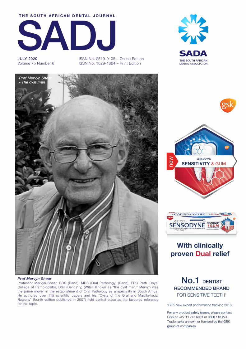
With clinically proven Dual relief No.1 DENTIST RECOMMENDED BRAND FOR SENSITIVE TEETH* *GFK New expert performance tracking 2018. For any product safety issues, please contact GSK on +27 11 745 6001 or 0800 118 274. Trademarks are own or licensed by the GSK group of companies. & new SENSITIVITY GUM Prof Mervyn Shear - The cyst man Prof Mervyn Shear Professor Mervyn Shear, BDS (Rand), MDS (Oral Pathology) (Rand), FRC Path (Royal College of Pathologists), DSc (Dentistry) (Wits). Known as “the cyst man,” Mervyn was the prime mover in the establishment of Oral Pathology as a speciality in South Africa. He authored over 115 scientific papers and his “Cysts of the Oral and Maxillo-facial Regions” (fourth edition published in 2007) held central place as the favoured reference for the topic. THE SOUTH AFRICAN DENTAL ASSOCIATION SADJ THE SOUTH AFRICAN DENTAL JOURNAL JULY 2020 Volume 75 Number 6 ISSN No. 2519-0105 – Online Edition ISSN No. 1029-4864 – Print Edition
-
Upload
khangminh22 -
Category
Documents
-
view
4 -
download
0
Transcript of SADJ - SADA
Cutterguide : No Printing Process : Off set GD : SB32430Size: 80 mm x 238 mm, Pages: 1 Colors : C M Y K (4 Colors)
Native File :Adobe Indwsign CS6 Windows Generated in : Acrobat Distiller 10.0
With clinically proven Dual relief
No.1 DENTIST RECOMMENDED BRAND
FOR SENSITIVE TEETH*
*GFK New expert performance tracking 2018.
For any product safety issues, please contact GSK on +27 11 745 6001 or 0800 118 274. Trademarks are own or licensed by the GSK group of companies.
& new
SENSITIVITY GUM
2019_SA_Sensodyne_Advert_GSKCH_209_D1.indd 1 4/29/2019 3:53:45 PM
Prof Mervyn Shear- The cyst man
Prof Mervyn Shear Professor Mervyn Shear, BDS (Rand), MDS (Oral Pathology) (Rand), FRC Path (Royal College of Pathologists), DSc (Dentistry) (Wits). Known as “the cyst man,” Mervyn was the prime mover in the establishment of Oral Pathology as a speciality in South Africa. He authored over 115 scientific papers and his “Cysts of the Oral and Maxillo-facial Regions” (fourth edition published in 2007) held central place as the favoured reference for the topic.
THE SOUTH AFRICANDENTAL ASSOCIATION
SADJT H E S O U T H A F R I C A N D E N TA L J O U R N A L
JULY 2020Volume 75 Number 6
ISSN No. 2519-0105 – Online Edition ISSN No. 1029-4864 – Print Edition

CONTENTS
Published by: On behalf of:
THE SOUTH AFRICANDENTAL ASSOCIATION
EDITORIAL OFFICE
Managing Editor
Prof NH Wood
Editorial Assistant
Mr Dumi Ngoepe
Email: [email protected]
Sub-editors
Prof N Mohamed
Prof P Owen
Prof L Sykes
Prof J Yengopal
Please direct all correspondence to:
South African Dental Association
Private Bag 1, Houghton 2041
Tel: +27 (0)11 484 5288
Fax: +27 (0)11 642 5718
Email: [email protected]
Editorial Board
Dr P Brijlal: University of the Western Cape
Prof T Dandajena: Private Practice USA
Prof W G Evans: University of the Witwatersrand
Dr H Gluckman: Private Practice
Dr M Hellig: University of the Witwatersrand
Prof B Kramer: University of the Witwatersrand
Prof AJ Louw: University of the Western Cape
Dr N Mohamed: University of the Western Cape
Prof J Morkel: University of the Western Cape
Prof MPS Sethusa: Sefako Makgatho Health Sciences University
Prof L Shangase: University of Pretoria
Prof L Sykes: University of Pretoria
Prof AW van Zyl: Private Practice
Prof NH Wood: Sefako Makgatho Health Sciences University
SADA OFFICE BEARERS
National Council
President: Dr RR Naidoo
Vice-President: Dr APL Julius
SADA Board of Directors
Chairperson: Dr R Putter
Vice-chairperson: Dr N Osman
Dr SJ Swanepoel
Dr SY Pieters
Dr FC Meyer
Dr EK Naidoo
Mrs C Wessels
Mr H Keshave
Mr KC Makhubele (CEO)
The theme for the Front Cover of the South African Dental Journal this year provides for some historical figures, some characters illuminating dental history and some important achievements in South African Dental history. The cover for the July looks at a dentists who made an important contribution to Oral Pathology in South Africa.
Prof Mervyn ShearProfessor Mervyn Shear, BDS (Rand), MDS (Oral Pathology) (Rand), FRC Path (Royal College of Pathologists), DSc (Dentistry) (Wits). Known as “the cyst man,” Mervyn was the prime mover in the establishment of Oral Pathology as a speciality in South Africa. He authored over 115 scientific papers and his “Cysts of the Oral and Maxillo-facial Regions” (fourth edition publish- ed in 2007) held central place as the favoured reference for the topic.
FRONT COVER PICTUREProf Mervyn Shear … a towering presence in Oral Pathology 285
EDITORIAL COVID-19: When recovery does not mean a return to health - NH Wood 286
LETTER TO THE EDITORFrom practitioner to patient- My COVID journey - NP Metsing 287
COMMUNIQUEIn the face of COVID-19, SADA continues to deliver to its members - KC Makhubele
289
RESEARCHThroughputs of two cohorts of dental students at Sefako Makgatho Health Sciences University: A comparison - SR Mthethwa, PM Nyalunga, TS Gugushe
291
Comparison of three different instruments for orthodontic study model analysis - JC Julyan, J Julyan, J de Lange
298
The effect of off-axis seating on the marginal adaptation of full coverage all ceramic crowns - GP Babiolakis, CP Owen
303
Our Front Cover for this Issue...
SADJT H E S O U T H A F R I C A N D E N TA L J O U R N A L
Editorial, Advertising and Copyright Policy
Opinions and statements, of whatever nature, are published under the authority of the submitting author, and the inclusion or exclusion of any medicine or procedure does not represent the official policy of the South African Dental Association or its associates, unless an express statement accompanies the item in question. All articles published as Original Research Papers are refereed, and articles published under Clinical Practice or Reviewed Notes are submitted for peer review.
The publication of advertisements of materials or services does not imply an endorsement by the Association or a submitting author, should such material feature as part of or appear in the vicinity of any contribution, unless an express statement accompanies the item in question. The Association or its associates do not guarantee any claims made for products by their manufacturers.
While every effort is made to ensure accurate reproduction, the authors, advisors, publishers and their employees or agents shall not be responsible, or in any way liable for errors, omissions or inaccuracies in the publication, whether arising from negligence or otherwise or for any consequences arising therefrom.
Accredited by the Department of Education
SADJ is published 10 times a year by E-Doc CC, a Division of Life-Long Learning Solutions CC, on behalf of The South African Dental Association. © 2020 Life-Long Learning Solutions CC.
CONTENTS
Published by: On behalf of:
THE SOUTH AFRICANDENTAL ASSOCIATION
SADA OFFICE BEARERS
Chairpersons of Board Sub-Committees
Audit & Risk Committee: Dr N Osman
Strategy, Ethics &
Remuneration Committee: Dr SY Pieters
Dental Practice Committee: Dr SJ Swanepoel
Chairperson of DDF Board of Trustees
Dr B Beilinsohn
PRODUCTION OFFICE
E-Doc CC,
a Division of Life-Long Learning Solutions CC
Tel: +27 (0)10 020 1013
Publisher and Project manager
Mr René Smulders
GENERAL AND ADVERTISING ENQUIRIES
Mr René Smulders
Email: [email protected]
Design and Layout
Ms Reine Combrinck
Email: [email protected]
Website smalls advertising / CPD Enquiries and
Member contact detail update
South African Dental Association
Tel: +27 (0)11 484 5288
Email: [email protected]
REVIEWPerforation of the palate - A report of two Syphilitic Gumma cases - F Titinchi, N Behardien, J Morkel, J Opperman
311
Calcific Metamorphosis - A review of literature and clinical management - PJ van der Vyver, M Vorster, CH Jonker, N Potgieter
316
CASE REPORTVisual assessment is no substitute for radiographic analysis - A forensic case report - L Robinson, C Nel, H Bernitz
323
CLINICAL W INDOWWhat’s new for the clinician? - Excerpts from and summaries of recently published papers - V Yengopal
328
RADIOLOGY CASEMaxillofacial Radiology 182 - CJ Nortjé 333
ETHICSThe renaissance of virtue ethics and its application in dentistry - HD Miniggio, PD Motloba, CS Wareham 334
CPDCPD questionnaire 339
AUTHOR GUIDELINESInstructions to authors and author’s checklist 342
CLASSIFIEDSInstructions to authors and author’s checklist 346
SADJT H E S O U T H A F R I C A N D E N TA L J O U R N A L
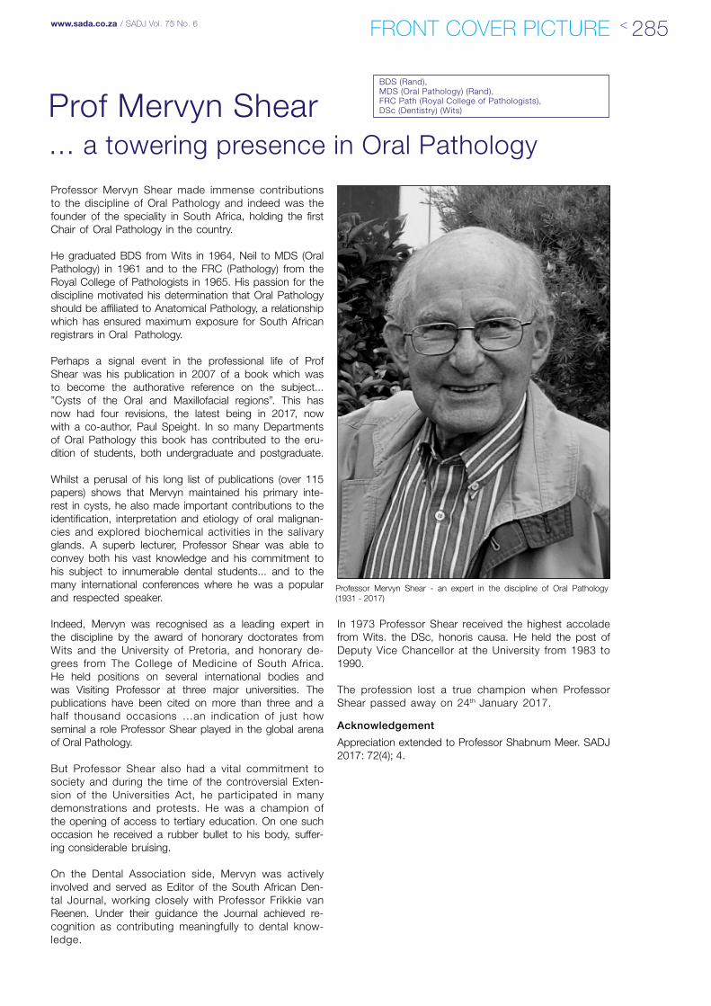
Professor Mervyn Shear made immense contributions to the discipline of Oral Pathology and indeed was the founder of the speciality in South Africa, holding the first Chair of Oral Pathology in the country.
He graduated BDS from Wits in 1964, Neil to MDS (Oral Pathology) in 1961 and to the FRC (Pathology) from the Royal College of Pathologists in 1965. His passion for the discipline motivated his determination that Oral Pathology should be affiliated to Anatomical Pathology, a relationship which has ensured maximum exposure for South African registrars in Oral Pathology. Perhaps a signal event in the professional life of Prof Shear was his publication in 2007 of a book which was to become the authorative reference on the subject... ”Cysts of the Oral and Maxillofacial regions”. This has now had four revisions, the latest being in 2017, now with a co-author, Paul Speight. In so many Departments of Oral Pathology this book has contributed to the eru- dition of students, both undergraduate and postgraduate.
Whilst a perusal of his long list of publications (over 115 papers) shows that Mervyn maintained his primary inte- rest in cysts, he also made important contributions to the identification, interpretation and etiology of oral malignan-cies and explored biochemical activities in the salivary glands. A superb lecturer, Professor Shear was able to convey both his vast knowledge and his commitment to his subject to innumerable dental students... and to the many international conferences where he was a popular and respected speaker.
Indeed, Mervyn was recognised as a leading expert in the discipline by the award of honorary doctorates from Wits and the University of Pretoria, and honorary de- grees from The College of Medicine of South Africa. He held positions on several international bodies and was Visiting Professor at three major universities. The publications have been cited on more than three and a half thousand occasions …an indication of just how seminal a role Professor Shear played in the global arena of Oral Pathology.
But Professor Shear also had a vital commitment to society and during the time of the controversial Exten- sion of the Universities Act, he participated in many demonstrations and protests. He was a champion of the opening of access to tertiary education. On one such occasion he received a rubber bullet to his body, suffer- ing considerable bruising.
On the Dental Association side, Mervyn was actively involved and served as Editor of the South African Den- tal Journal, working closely with Professor Frikkie van Reenen. Under their guidance the Journal achieved re- cognition as contributing meaningfully to dental know- ledge.
In 1973 Professor Shear received the highest accolade from Wits. the DSc, honoris causa. He held the post of Deputy Vice Chancellor at the University from 1983 to 1990. The profession lost a true champion when Professor Shear passed away on 24th January 2017.
Appreciation extended to Professor Shabnum Meer. SADJ 2017: 72(4); 4.
Acknowledgement
Professor Mervyn Shear - an expert in the discipline of Oral Pathology (1931 - 2017)
Prof Mervyn Shear
… a towering presence in Oral Pathology
BDS (Rand), MDS (Oral Pathology) (Rand), FRC Path (Royal College of Pathologists), DSc (Dentistry) (Wits)
FRONT COVER PICTUREwww.sada.co.za / SADJ Vol. 75 No. 6 < 285
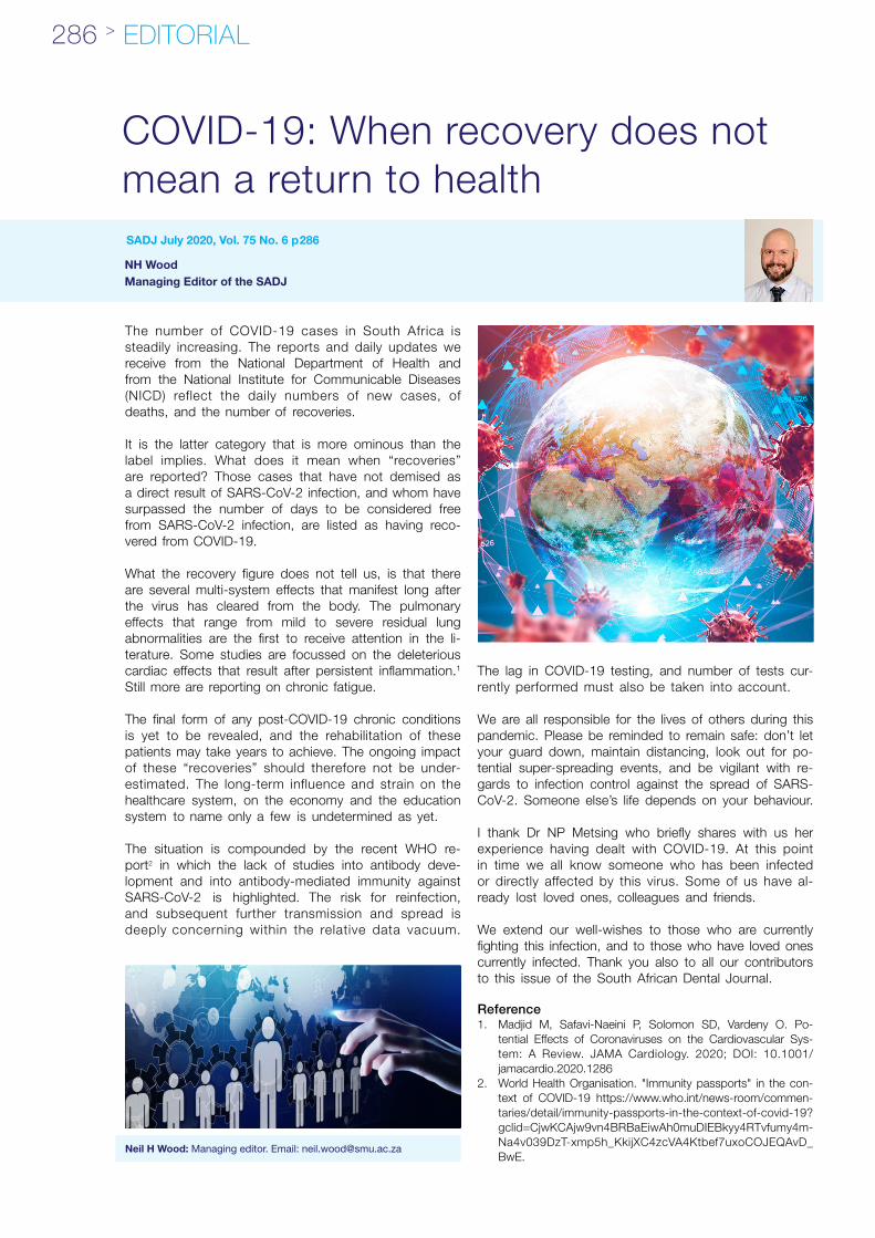
The number of COVID-19 cases in South Africa is steadily increasing. The reports and daily updates we receive from the National Department of Health and from the National Institute for Communicable Diseases (NICD) reflect the daily numbers of new cases, of deaths, and the number of recoveries.
It is the latter category that is more ominous than the label implies. What does it mean when “recoveries” are reported? Those cases that have not demised as a direct result of SARS-CoV-2 infection, and whom have surpassed the number of days to be considered free from SARS-CoV-2 infection, are listed as having reco- vered from COVID-19.
What the recovery figure does not tell us, is that there are several multi-system effects that manifest long after the virus has cleared from the body. The pulmonary effects that range from mild to severe residual lung abnormalities are the first to receive attention in the li- terature. Some studies are focussed on the deleterious cardiac effects that result after persistent inflammation.1 Still more are reporting on chronic fatigue.
The final form of any post-COVID-19 chronic conditions is yet to be revealed, and the rehabilitation of these patients may take years to achieve. The ongoing impact of these “recoveries” should therefore not be under- estimated. The long-term influence and strain on the healthcare system, on the economy and the education system to name only a few is undetermined as yet.
The situation is compounded by the recent WHO re- port2 in which the lack of studies into antibody deve- lopment and into antibody-mediated immunity against SARS-CoV-2 is highlighted. The risk for reinfection, and subsequent further transmission and spread is deeply concerning within the relative data vacuum.
The lag in COVID-19 testing, and number of tests cur- rently performed must also be taken into account.
We are all responsible for the lives of others during this pandemic. Please be reminded to remain safe: don’t let your guard down, maintain distancing, look out for po- tential super-spreading events, and be vigilant with re- gards to infection control against the spread of SARS- CoV-2. Someone else’s life depends on your behaviour.
I thank Dr NP Metsing who briefly shares with us her experience having dealt with COVID-19. At this point in time we all know someone who has been infected or directly affected by this virus. Some of us have al- ready lost loved ones, colleagues and friends. We extend our well-wishes to those who are currently fighting this infection, and to those who have loved ones currently infected. Thank you also to all our contributors to this issue of the South African Dental Journal.
Reference1. Madjid M, Safavi-Naeini P, Solomon SD, Vardeny O. Po-
tential Effects of Coronaviruses on the Cardiovascular Sys- tem: A Review. JAMA Cardiology. 2020; DOI: 10.1001/jamacardio.2020.1286
2. World Health Organisation. "Immunity passports" in the con- text of COVID-19 https://www.who.int/news-room/commen- taries/detail/immunity-passports-in-the-context-of-covid-19? gclid=CjwKCAjw9vn4BRBaEiwAh0muDIEBkyy4RTvfumy4m-Na4v039DzT-xmp5h_KkijXC4zcVA4Ktbef7uxoCOJEQAvD_BwE.
COVID-19: When recovery does not mean a return to health
Neil H Wood: Managing editor. Email: [email protected]
EDITORIAL286 >
NH WoodManaging Editor of the SADJ
SADJ July 2020, Vol. 75 No. 6 p286
It was on Wednesday the 24th of June when I started to feel fluish. Considering the pandemic that the world is currently facing, my first thought was that it may be COVID-19 yet there was another part of me that dis- missed it. Within a few hours of these first symptoms, my throat felt like I had swallowed a ball of fire, I was coughing, had a severe headache, and I was feeling very cold despite sitting in front of the heater.
I self-medicated and went to bed early and took a de- cision to self-isolate for 7 days. The next day I woke up feeling much better, so I told myself it may not be COVID-19 since it responded to the previous night’s remedies. I continued using some over-the-counter pre- parations, however on Friday I woke up and realized I lost my senses of taste and smell and I had no appetite. By Saturday I started to feel weak and lethargic and I couldn’t even get myself out of bed. I tried to eat but food nauseated me.
I spent the whole weekend either in bed, or sleeping on the couch, still not eating while taking medicine. At this time my symptoms were a loss of taste and smell, a severely sore throat and an excruciating headache.
By Monday the situation did not improve, and I decided I needed to get a COVID-19 test done. Tuesday, I started to feel worse, so my dad (All the way in Carletonville) called an ambulance for me. Unfortunately, when the paramedics arrived, they were not willing to take me to the hospital, and by that time I did not have the energy to communicate with them, so I let them leave.
Subsequently, my situation took a turn for the worse on top of the existing symptoms: I started to vomit. My father fetched me from my house and took me to hos- pital where I was taken into casualty. An IV line was placed, bloods were drawn, and the blood results re- vealed a high SARS-CoV-19 viral load.
I was feeling very dehydrated and asked the nurse for some water which made me vomit instantly after inges- tion. The drip I was given (Ringer’s lactate) to replace fluids and electrolytes, also contained analgesics and antiemetics. The casualty doctor recommended that I be admitted into the hospital immediately.
That is when I realized the crisis that COVID-19 had created in the hospitals: there was a huge shortage of beds. I stayed in casualty from around 12 midday until 23:30 which was the time they actually managed to find an available bed.
I was asked if I wanted to eat, but my appetite was still gone (+/- 5 days without a decent meal). To be honest, during this whole-time fear was also creeping and my anxiety level was also rising as I kept on asking my- self what if I lost my senses and never regained them. I even avoided googling any information regarding this.
I remember at one point I called a Professor who is an Oral Medicine specialist to find out if there was anything I could take, because in that moment of uncertainty one’s professional knowledge escapes you. The next morning in my ward I was asked what I wanted for my 3 meals for the day. I did not want to answer this because seeing food without being able to smell it made me panic, let alone not being able to taste it. I was told that I did not have a choice.
When breakfast came, I took a spoon of the porridge and asked the nurse to take it back because I felt like vomiting however something interesting happened at that time, I had a bit of taste sensation on my tongue, this was something that really lifted my mood a little bit. During lunch time I managed to eat my meal without nausea the same with supper.
That morning the hospital took a decision to re-swab me since my swab results were still not back. That eve- ning at 21:30 I received an email from Ampath labora- tory with my results which proved what I had already instinctively known, that I had COVID-19. I informed the nurse that was on duty and immediately I was taken to an isolation room as I was in a room with another pa- tient at that time. I won’t lie, even though I tried very hard to stay positive, receiving a COVID-19 positive test result did depress me a little bit, and I believe that is the time when your emotional intelligence really gets tested.
From practitioner to patient- My COVID journey
LETTER TO THE EDITOR < 287www.sada.co.za / SADJ Vol. 75 No. 6
NP MetsingHead of Professional Development at SADA
SADJ July 2020, Vol. 75 No. 6 p287 - p288
Although my senses were starting to improve and my appetite was recovering, night-time was my most drea- ded time. I had severe headaches, sore throat, and shortness of breath at night. When the attending doctor on ward rounds consulted me, I requested him to dis- charge me. Even though he was skeptical, I assured him that I was able to take care of myself from home. I felt trapped in the hospital room, and I wanted to avoid having to deal with mental issues in addition to all the symptoms I was already experiencing.
Day one out of the hospital went well, and I felt of a little stronger than previous 2 weeks. By this time my sense of taste was almost fully restored. My sense of smell was still recovering and it was a strange experience.
I was able to smell some things and others not: could not smell the soap in the shower while I was showering nor could I smell food, but I could smell scents like eucalyptus oil. I was happy though because I noticed the progress even though I still remained fatigued and had shortness of breath when standing, even for a short period.
On Monday the 6th of July I woke up feeling a great improvement, to the extent that I felt that I could resume work in a day or two. Around midday I was feeling fa- tigued and I started feeling very cold but it was nothing that a nap could not sort out, my sense of smell (the most stubborn of all senses lost) was now coming back although being lost intermittently.
On Tuesday the 7th of July, my mom came over to look after us and to also to assist with some housekeeping. I cannot overemphasize importance of family during this time, and support structures are extremely important to facilitate recovery. The fact that my mother is a nurse was an added bonus because my family was able to observe the full protocol while providing us the necessary emotional and physical support.
Words fail me when I have express my gratitude to my family for the love and support during this time even though I had moments of depression and at times I would lose my temper, my family never left me. It was my mother who insisted that I go to the hospital when she realized that my condition was deteriorating.
Throughout my journey with COVID-19 I never once had a high temperature. The highest reading while I was in hospital was 37° (the acceptable body tempera- ture), which made me wonder about the heavy empha- sis being placed on a fever as an indicator of possibly being COVID-19 positive. We must be aware that there are exceptions to these averages, and not to take any symptom or sign lightly.
Most people might ignore these symptoms all because they are not “running a high temperature”. This may be dangerous because we may have positive people in public spaces potentially spreading the virus without ever knowing it. It is important for people to appreciate that this virus affects our bodies differently, and that not all of us will necessarily experience the same symptoms that are emphasized.
On one end of the spectrum, some people die and on the other end of the same spectrum some people ex- perience no symptoms throughout the entire period of their infection. There appears to be extremely little to no focus on those in between, and who suffer the long-lasting sequelae of this infection. I really hope my story resonates with the readers, and that people who do get symptomatic may know that they are not alone in this scary journey.
I also realized the importance of safeguarding your mental health and remaining positive. Many of us have vastly different ways to approach this significant challenge. Personally, spiritual growth is eminent. If you follow any religion, it is during this time that divine intervention is of great importance to many. To me, as a Christian, my spiritual growth was very evident in my utterances and I started to pray more. Thank you for taking your time to read my story.
Regards,
Dr NP MetsingHead of Professional Development at SADA
LETTER TO THE EDITOR288 >
SADA is proud to be amongst the small elite group of professional associations representing healthcare practi-tioners, fully recognised worldwide. Recently we held both the SADA Annual General and National Council Meeting virtually through a meetings electronic platform – Zoom.
We have indeed entered a new era in the history of the Association as these were the first members meetings held electronically. We are pleased that members re- gistered and attending were able to actively participate and exercise their voting rights on various issues tabled at these meetings.
All Branch, National Council, Board and Board Com- mittee meetings will be conducted by electronic means in the foreseeable future. It is also reported with regret that the 2020 Dental Congress and Exhibition had to be cancelled at the Emperors Palace Conference Centre in Gauteng. Several alternatives and strategies are being investigated to meet the educational and CPD needs of members for the year.
We are proud to announce the appointment of Dr Rhonin Naidoo (Kwa-Zulu Branch) as the new President of the Association and Dr Anthony Julius (North-West Branch) as the Vice-President of the Association, they will both serve for a two-year term. On behalf of the Board, National Council and management, we welcome them and express our profound and heartfelt congra- tulations on their appointment. We wish them every success and we are confident they will proudly repre- sent our Association for the next two years.
As we enter advanced level 3 of the COVID-19 pan- demic, the extreme challenges facing our members are becoming more dire with shortages of required PPE, staff testing positive for COVID-19, patients fearful about consulting their dentists, some practitioners contemplat-ing earlier retirement than anticipated, as a measure of supporting our members, we have offered two months free membership. Members have been requested to provide their banking details to permit the Association refund them much needed funds.
The extended Dental Practice Committee together with the different workstreams specifically mandated to deal with different aspect of the pandemic have been hard at work from the start of the lockdown and after and we are proud to make available to the entire dental fraternity Protocols to guide members working under COVID-19 conditions.
The protocols have been enthusiastically received by all stakeholders we have been engaging with not only in South Africa, but even internationally it is seen as a leading protocol document. We are happy to report that dental professional associations abroad are equally impressed with the contents of a well-researched doc- ument.
The different workstreams have also been hard at work holding webinars every week keeping members properly informed about the different challenges facing the pro- fession during this time. Many more are being planned and we have consciously decided to make these avai- lable to members and non-members alike in the inte- rests of fulfilling our duties as citizens in contributing to the prevention of the spread of the disease.
www.sada.co.za / SADJ Vol. 75 No. 6
Image Source: Jürgen Randma / CC BY-SA (https://creativecommons.org/licenses/by-sa/4.0)
COMMUNIQUE < 289
KC MakhubeleCEO of SADA
SADJ July 2020, Vol. 75 No. 6 p289 - p290
In the face of COVID-19, SADA continues to deliver to its members
The electronic book titled “Infection Control Guidelines for Oral Health Care” by Dr Jeanné Oosthuysen and co-authors is available for members on the website which we hope guide our members in keeping their patients, staff and themselves safe.
Members were introduced to services of two labour consultants who will offer their services on labour related challenges members are likely to face now and in the coming months. Members are encouraged to utilise their services to prevent unnecessary and costly labour disputes. Several guidelines have been issued for the benefit of practitioners as employers.
Engagements with medical schemes, medical scheme administrators and third-party funders are ongoing as regards providing benefits for additional PPE for AGPs and non-AGPs performed by practitioners.
While some schemes are willing to consider benefits for the practitioner and assistants, others have resisted despite the fact these are also required for the protec- tion of their members.
Members have also started the debate about the continued reliance of the profession on third party fun- ders and its devastating impact on the future of the profession. To this end, the Board has grappled with this subject and will survey members about their practice circumstances and reliance in these tough economic times.
Perhaps the time has come for the profession to re- consider their position now and in the post pandemic era, to make their patients to whom they provide treat- ment responsible to them for payment of services ren- dered. It is now time that the profession takes charge of their own practices and like other businesses, rely on their customer to pay for services received.
Members would also have followed through webinars that Dental Protection is seeking to establish service centres and more personnel to assist members at a local level to provide more efficient and quicker turn- around for members seeking guidance, facing com- plaints and/or claims.
We are also pleased to announce the appointment of the erstwhile President, Professor P Moipolai to the South African Dental Technicians Council as a member attached to an academic institution. The Chairperson of the SADA Board of Directors, Dr R Putter was success- fully appointed to the PPS Trust Board. We wish them both success in their appointment. The Board also appoint two new independents, Mrs Carina Wessels (Admitted Advocate of the High Court of South Africa and Executive: Governance, Legal and Compliance at Alexander Forbes) and Mr Hiten Keshave (A charted accountant and CFO at PRP Solutions (Pty) Ltd.
Amidst all of the challenges the profession is facing, the business of the association continues.
COMMUNIQUE290 >
THE SOUTH AFRICANDENTAL ASSOCIATION
http://dx.doi.org/10.17159/2519-0105/2020/v75no6a1The SADJ is licensed under Creative Commons Licence CC-BY-NC-4.0.
The numbers of student dentists enrolled at dental schools across the country do not give an indication of the students’ progress to degree.
To describe and compare the throughputs of dentistry course for two cohorts of students at Sefako Makgatho Health Science University.
The progress to degree of the 2005 and 2010 cohorts of first year dental students was tracked and compared.
A comparative cross-sectional study.
Academic records of the 2005 and 2010 cohorts of first year dental students were followed up over a five- year period. Data related to the demographic charac- teristics, numbers enrolled, numbers who dropped out, and the numbers who graduated were acquired and then captured in Microsoft Excel software.
Female students constituted the majority of enrolees in both cohorts (53.8% vs. 51.3%). The proportions of students who started the course, completed the degree and graduated within the regulation time among the 2005 and 2010 cohorts were similar (42.1% vs. 41.2%).
A lone student among the 2010 cohort dropped out of the course. The majority of students (57.9% vs. 55.9%) in both cohorts took longer to qualify.
The throughputs of dentistry course for the two cohorts hovered around 40%.
There is a shortage of oral health personnel in South Africa. Oral health personnel, including dental assistants, oral hygienists, dental therapists, and dentists, have been estimated to constitute 0.2 per 1000 population.1 A meagre dental practitioner to population ratio of 1.09 per 10,000 has been estimated.2 There is currently a debate about how to alleviate this shortage. An argu- ment has been made that we should be training fewer dentists and more oral hygienists and dental therapists.3
This line of reasoning is supported by evidence emerg- ing from epidemiological studies of the burden of disease.
Evidence indicates that dental caries is the most prevalent oral disease4 - the latest national survey found that more than 80% of dental caries in children was untreated.5 Previously unobserved oral health priorities, which in- clude conditions such as periodontal disease, oral mani- festations of HIV/AIDS, dental trauma, oral cancer and craniofacial anomalies, are now under consideration, ad- ding to the burden.6,7
An analysis of the throughput of the various courses offered at dental schools in the country could enrich the debate on the number and mix of oral health personnel required to provide necessary services. The results of such an investigation will be useful to policy-makers in developing human resources plan for oral health and to the dental schools’ management in identifying impedi- ments to graduation in order to offer the necessary aca- demic and mentoring support to enable success.
The median throughput of the dental therapy courses for the period between 2004 and 2014 at Sefako Makgatho Health Sciences University has recently been estab- lished to be 45 percent with an interquartile range of 37 to 58.5 percent.8 This finding is rather disappointing. The current study seeks to describe the throughputs for two cohorts of students in the dentistry course at Sefako Makgatho Health Science University during the period between 2005 and 2014.
ABSTRACT
Introduction
Aims and objectives
Design
Methods
Results
Conclusions
INTRODUCTION AND BACKGROUND
Author affiliations:1. Sibusiso R Mthethwa: BDS, MPH, PhD, Sefako Makgatho Health
Sciences University, South Africa.ORCID Number: 0000-0003-0420-808X
2. Phabian M Nyalunga: BDT, BDS, Sefako Makgatho Health Sci- ences University, South Africa.
3. Tshepo S Gugushe: BSc, BDS, DHSM, MDent, MPhil, Sefako Makgatho Health Sciences University, South Africa.
Corresponding author: SR MthethwaMedunsa Campus, PO Box D24, Sefako Makgatho Health Sciences University 0204, South Africa.Email: [email protected] contributions:1. Sibusiso R Mthethwa: Conception; design; acquisition of data;
analysis and interpretation of data; drafting the article - 33.3%2. Phabian M Nyalunga: Conception; revising the article critically
for important intellectual content - 33.3% 3. Tshepo S Gugushe: revising the article critically for important
intellectual content - 33.3%
RESEARCH < 291
SR Mthethwa1, PM Nyalunga2, TS Gugushe3
SADJ July 2020, Vol. 75 No. 6 p291 - p297
Throughputs of two cohorts of dental students at Sefako Makgatho Health Sciences University: A comparison
The results of a recent review of Health Professions Council of South Africa (HPCSA) records indicate that the number of dentists increased at around 2% per annum during the period 2002 to 2015.9 The study revealed a welcome change in the demographic profile of dentists. It found a relatively sharp increase in the number of Coloured, Black and female registered dentists.9
The aggregate number of student dentists currently en- rolled in the four dental schools in the country is 1 158.10 This figure does not give an indication of the students’ progress to degree. A most recent systematic review of the literature clusters the determinants of university drop- out and delayed graduation into four main categories i.e. students’ characteristics, abilities and behaviour; paren- tal background and family networks; characteristics of tertiary education system and institutions, and labour market conditions.11
The strong association of race, gender, matriculation score and poverty with dropout or graduation at South African universities is well documented.12,13,14 The asso- ciation, if any, of student motivation with dropout or graduation is an important issue for future research. The results of a national survey among early-phase stu- dent dentists are extremely concerning. The survey found that for a third of respondents, dentistry was not a first choice – amongst the White students, it was a first choice for 82% compared with 59% amongst Black Africans.15
To describe the demographic changes undergone by the 2005 and 2010 cohorts of first year dental students in successive years of study.
To track and compare the progress to degree of the 2005 and 2010 cohorts of first year dental students. To determine the median numbers of students who com- pleted their degree and graduated within the regula- tion time.
This was a retrospective, comparative cross-sectional study in which existing academic records were reviewed.
The sampling frame consisted of academic records of a subpopulation of dental students who were enrolled at Sefako Makgatho Health Sciences University during the period 2005 to 2014 i.e. academic records of cohorts of first year dental students who were enrolled in 2005 and 2010 respectively were followed up over a five-year period.
Every available record was studied.
Data related to the demographic characteristics of the students, the numbers enrolled, numbers who dropped
out, and the numbers who graduated were acquired and then captured in Microsoft Excel software.
Age and gender refer to student age and sex as recor- ded in the academic records.
Population group breakdown of students into African, Indian, Coloured and White will be applied according to the Population Registration Act of 1950.16
Progress to degree refers to enrolment and academic progress.
Regulation time is the period of time normally expected for completion of the degree (five years).
Throughput, quite simply, is how many students who started studying complete the course of study. This may also be measured by the number of students who do not drop out.17 It is also referred to as the completion rate, or graduation rate.18
First-timer refers to a student who was enrolled in a year of study for the first time.
Ethical approval for the study was granted by the Ethics Committee of Sefako Makgatho Health Sciences Uni- versity (SMREC/D/1820/2017). Permission to conduct the study was granted by the Chief Executive Officer (CEO) of the Medunsa Oral Health Centre.
Collected data were subjected to univariate and bivariate analysis in Statistical Package for the Social Sciences (SPSS) software. Frequencies, means and proportions were calculated. Chi-square tests were performed to test the statistical significance of the differences in pro- portions. Chi-square tests for trends were performed to investigate trends in enrolments and examination pass rates. The chosen significance level of the tests was a p-value less than 0.05.
Academic records of the 2005 and 2010 cohorts of first year dental students were followed up over a five-year period and analysed.
The total number of enrollees followed up over the five years of study in the 2005 cohort was 10.4% more than that of the 2010 cohort. The first year class of the 2005 cohort was 28.9% larger than that of the 2010 cohort.
On the one hand, the fifth year class of the 2005 cohort was 26.9% smaller than the first year class. On the other hand, the fifth year class of the 2010 cohort was 27% larger than the first year class. Female students constituted the majority (54.5%) of the enrollees - they were the majority in all classes of the 2005 cohort.
OBJECTIVES OF THE STUDY
MATERIALS AND METHODS
Study design
Target population
Study sample
Data collection
Definition of variables and terms
Ethical considerations
STATISTICAL ANALYSIS/ HYPOTHESIS TESTING
RESULTS
RESEARCH292 >
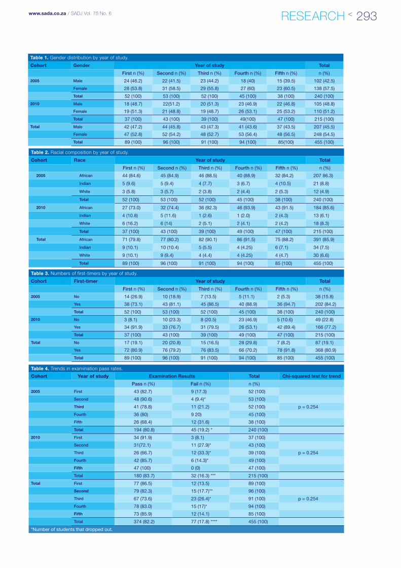
Table 1. Gender distribution by year of study.
Cohort Gender Year of study Total
First n (%) Second n (%) Third n (%) Fourth n (%) Fifth n (%) n (%)
2005 Male 24 (46.2) 22 (41.5) 23 (44.2) 18 (40) 15 (39.5) 102 (42.5)
Female 28 (53.8) 31 (58.5) 29 (55.8) 27 (60) 23 (60.5) 138 (57.5)
Total 52 (100) 53 (100) 52 (100) 45 (100) 38 (100) 240 (100)
2010 Male 18 (48.7) 22(51.2) 20 (51.3) 23 (46.9) 22 (46.8) 105 (48.8)
Female 19 (51.3) 21 (48.8) 19 (48.7) 26 (53.1) 25 (53.2) 110 (51.2)
Total 37 (100) 43 (100) 39 (100) 49(100) 47 (100) 215 (100)
Total Male 42 (47.2) 44 (45.8) 43 (47.3) 41 (43.6) 37 (43.5) 207 (45.5)
Female 47 (52.8) 52 (54.2) 48 (52.7) 53 (56.4) 48 (56.5) 248 (54.5)
Total 89 (100) 96 (100) 91 (100) 94 (100) 85(100) 455 (100)
Table 2. Racial composition by year of study.
Cohort Race Year of study Total
First n (%) Second n (%) Third n (%) Fourth n (%) Fifth n (%) n (%)
2005 African 44 (84.6) 45 (84.9) 46 (88.5) 40 (88.9) 32 (84.2) 207 86.3)
Indian 5 (9.6) 5 (9.4) 4 (7.7) 3 (6.7) 4 (10.5) 21 (8.8)
White 3 (5.8) 3 (5.7) 2 (3.8) 2 (4.4) 2 (5.3) 12 (4.9)
Total 52 (100) 53 (100) 52 (100) 45 (100) 38 (100) 240 (100)
2010 African 27 (73.0) 32 (74.4) 36 (92.3) 46 (93.9) 43 (91.5) 184 (85.6)
Indian 4 (10.8) 5 (11.6) 1 (2.6) 1 (2.0) 2 (4.3) 13 (6.1)
White 6 (16.2) 6 (14) 2 (5.1) 2 (4.1) 2 (4.2) 18 (8.3)
Total 37 (100) 43 (100) 39 (100) 49 (100) 47 (100) 215 (100)
Total African 71 (79.8) 77 (80.2) 82 (90.1) 86 (91.5) 75 (88.2) 391 (85.9)
Indian 9 (10.1) 10 (10.4) 5 (5.5) 4 (4.25) 6 (7.1) 34 (7.5)
White 9 (10.1) 9 (9.4) 4 (4.4) 4 (4.25) 4 (4.7) 30 (6.6)
Total 89 (100) 96 (100) 91 (100) 94 (100) 85 (100) 455 (100)
Table 3. Numbers of first-timers by year of study.
Cohort First-timer Year of study Total
First n (%) Second n (%) Third n (%) Fourth n (%) Fifth n (%) n (%)
2005 No 14 (26.9) 10 (18.9) 7 (13.5) 5 (11.1) 2 (5.3) 38 (15.8)
Yes 38 (73.1) 43 (81.1) 45 (86.5) 40 (88.9) 36 (94.7) 202 (84.2)
Total 52 (100) 53 (100) 52 (100) 45 (100) 38 (100) 240 (100)
2010 No 3 (8.1) 10 (23.3) 8 (20.5) 23 (46.9) 5 (10.6) 49 (22.8)
Yes 34 (91.9) 33 (76.7) 31 (79.5) 26 (53.1) 42 (89.4) 166 (77.2)
Total 37 (100) 43 (100) 39 (100) 49 (100) 47 (100) 215 (100)
Total No 17 (19.1) 20 (20.8) 15 (16.5) 28 (29.8) 7 (8.2) 87 (19.1)
Yes 72 (80.9) 76 (79.2) 76 (83.5) 66 (70.2) 78 (91.8) 368 (80.9)
Total 89 (100) 96 (100) 91 (100) 94 (100) 85 (100) 455 (100)
Table 4. Trends in examination pass rates.
Cohort Year of study Examination Results Total Chi-squared test for trend
Pass n (%) Fail n (%) n (%)
2005 First 43 (82.7) 9 (17.3) 52 (100)
Second 48 (90.6) 4 (9.4)* 53 (100)
Third 41 (78.8) 11 (21.2) 52 (100) p = 0.254
Fourth 36 (80) 9 20) 45 (100)
Fifth 26 (68.4) 12 (31.6) 38 (100)
Total 194 (80.8) 45 (19.2) * 240 (100)
2010 First 34 (91.9) 3 (8.1) 37 (100)
Second 31(72.1) 11 (27.9)* 43 (100)
Third 26 (66.7) 12 (33.3)* 39 (100) p = 0.254
Fourth 42 (85.7) 6 (14.3)* 49 (100)
Fifth 47 (100) 0 (0) 47 (100)
Total 180 (83.7) 32 (16.3) *** 215 (100)
Total First 77 (86.5) 12 (13.5) 89 (100)
Second 79 (82.3) 15 (17.7)** 96 (100)
Third 67 (73.6) 23 (26.4)* 91 (100) p = 0.254
Fourth 78 (83.0) 15 (17)* 94 (100)
Fifth 73 (85.9) 12 (14.1) 85 (100)
Total 374 (82.2) 77 (17.8) **** 455 (100)
*Number of students that dropped out.
RESEARCH < 293www.sada.co.za / SADJ Vol. 75 No. 6
In contrast, they were in the minority in the second and third year classes of the 2010 cohort.
African students constituted a vast majority (85.9%) of the enrolees – they were an overwhelming majority in all classes of both cohorts. Indian students comprised the second largest racial group of enrolees at a mere 7.5%.
A marginal (3.4%) increase in the proportion of White students was observed between the 2005 and 2010 cohorts.
One out of five (19.1%) enrolee was a repeater - they comprised 15.8% and 22.8% of the 2005 and 2010 cohorts respectively.
One out of four (26.9%) enrolee in the first year class of the 2005 cohort was a repeater compared with less than ten percent (8.1%) in the first year class of the of the 2010 cohort. Almost half (46.9%) of the fourth year class in the 2010 cohort were repeaters.
A steady decline in the proportion of repeaters was observed among the 2005 cohort from the first to fifth years of study.
The mean examination pass rate of the 2010 cohort was slightly higher (83.7% vs 80.8%) than that of the 2005 cohort. The difference was however not statisti- cally significant (p>0.05).
A two-thirds examination pass rate was the lowest rate achieved by both cohorts - 68.4% was recorded in the fifth year class of the 2005 cohort compared with 66.7% in the third year class of the 2010 cohort.
Three students, who failed the examinations, drop- ped out of the course among the 2010 cohort com- pared with one student among the 2005 cohort.
There was insufficient evidence to reject the null hypo- thesis of no trend in the proportion of students who passed the examination in the population (p> 0.05) for both cohorts.
The proportions of students who started the course in 2005 and 2010 and completed the degree in 2009 and 2014 and graduated in 2010 and 2015 within the regulation time was similar (42.1% vs 41.2%). A lone student among the 2010 cohort dropped out of the course. The majority of students (57.9% vs. 55.9%) of both cohorts took longer to qualify.
Eleven percent more first-timers with tertiary education among the 2010 cohort compared with the 2005 cohort completed the degree and graduated within the regulation time. The difference in proportions was how- ever not statistically significant (p >0.05). 59.1% (13/22) of students in the 2005 cohorts took longer to qualify. Twenty-five percent more first-timers without tertiary edu- cation among the 2005 cohort compared with the 2010
Table 5. Comparison of the throughput of all first-timers between cohorts.
CohortsYear of study Number of
graduatesThroughput (%) Chi-squared test
First Second Third Fourth Fifth
2005 38 29 27 20 16 16 42.1 p = 0.937
2010 34 30 (1) 20 16 14 14 41.2
( ) number of students that dropped out.Throughput = Number of graduates divided by number of students enrolled in first year.
Table 6. Comparison of the throughput of first-timers with tertiary education between cohorts.
CohortsYear of study Number of
graduatesThroughput (%) Chi-squared test
First Second Third Fourth Fifth
2005 22 19 18 13 9 9 40.9 p = 0.454
2010 23 21 14 12 12 12 52.2
Throughput = Number of graduates divided by number of students enrolled in first year.
Table 7. Comparison of the throughput of first-timers without tertiary education between cohorts.
CohortsYear of study Number of
graduatesThroughput (%) Chi-squared test
First Second Third Fourth Fifth
2005 16 10 9 7 7 7 43.8 p = 0.174
2010 11 9 6 4 2 2 18.2
Throughput = Number of graduates divided by number of students enrolled in first year.
Table 8. Comparison of the throughput of first-timers with and without tertiary education among the 2005 cohort.
Tertiary education
GraduatedTotal Chi-squared test
Yes n (%) No n (%)
Present 9 (40.91) 13 (59.09) 22 (100) p = 0.862
Absent 7 (43.75) 9 (56.25) 16 (100)
Total 16 (42.11) 22 (57.89) 38 (100)
Table 9. Comparison of the throughput of first-timers with and without tertiary education among the 2010 cohort.
Tertiary education
GraduatedTotal Chi-squared test
Yes n (%) No n (%)
Present 12 (52.17) 11 (47.83) 23 (100) p = 0.063
Absent 2 (18.8) 9 (81.82) 11 (100)
Total 14 (41.18) 20 (58.82) 34 (100)
RESEARCH294 >
cohort completed the degree and graduated within the regulation time. The difference in proportions was how- ever not statistically significant (p >0.05). 82% (9/11) of students in the 2010 cohorts took longer to qualify.
Slightly (2.84%) more students without tertiary educa- tion graduated within the regulation time than those with tertiary qualification. There was insufficient evidence (p>0.05) to reject the null hypothesis that the propor- tions of students that graduated were equal in the two groups in the population.
A little less than three times (2.78 times) as many stu- dents with tertiary education graduated within the regu- lation time as were those without tertiary education. There was however insufficient evidence (p >0.05) to reject the null hypothesis that the proportions of stu- dents that graduated were equal in the two groups in the population.
This study set out to track, over a five-year period, and compare the progress to degree of first year dental students who were enrolled in 2005 and 2010. The number of students who completed their degree and graduated within the stipulated time was also determined.
The current study found that the size of the 2005 cohort was 28.9% larger than that of the 2010 cohort in the first year of study (Table 1). This unanticipated discre- pancy coincided with the founding of the University of Limpopo (MEDUNSA Campus), a predecessor of Sefako Makgatho Health Sciences University. It may be due to institutional inexperience.
The results of this study indicate that female students constituted the majority (54.5%) of the enrolees of both cohorts (Table 1). The present findings seem to be consistent with other research which found that female students constitute the majority of dental students.15
The most interesting finding was that the variation in numbers of enrolees between the first and fifth year classes of both cohorts was in different directions yet similar i.e. a 26.9% decline among the 2005 cohort in contrast to the 27.0% rise among the 2010 cohort (Table 1). This rather contradictory result was due to the significant difference (31.6%) in the examination pass rates between the fifth year classes of the 2005 and 2010 cohorts.
The results of this study show that African students constituted an overwhelming majority (85.9%) of the enrolees (Table 2). This foreseeable finding is inextrica- bly linked with the founding mission of the predecessor institutions.
Medunsa, the Medical University of Southern Africa, was founded in 1976 to address both the under-represen- tation of blacks in the health professions and the lack of good health care in the homelands. The university trains most of the black physicians, dentists, veterina-
rians, and allied health professionals in South Africa.19
The results of this study show a significant reduction in the proportion of first year repeaters from a high of 26.9% in the 2005 cohort to a low of 8.1% in the 2010 cohort (Table 3). This encouraging finding suggests that the academic and mentoring support offered is effective.
In contrast, the current study found that repeaters con- stituted a strikingly large size (46.9%) of the fourth year class of the 2010 cohort. The observed significant in- crease in the number of repeaters in the fourth year class of 2013 could be attributed to fact that the large class size (59 students) of the previous year consisted of a relatively large number of weak students. Further research is required to establish the major factors that cause students to repeat a class.
The current study found that the size of the 2005 co- hort was 28.9% larger than that of the 2010 cohort in the first year of study (Table 1). This unanticipated discrepancy coincided with the founding of the Sefako Makgatho Health Sciences University. It may be due to institutional inexperience.
The results of this study indicate that female students constituted the majority (54.5%) of the enrolees of both cohorts (Table 1). The present findings seem to be consistent with other research which found that female students constitute the majority of dental students.15
The most interesting finding was that the variation in numbers of enrolees between the first and fifth year classes of both cohorts was in different directions yet similar i.e. a 26.9% decline among the 2005 cohort in contrast to the 27.0% rise among the 2010 cohort (Table 1). This rather contradictory result was due to the significant difference (31.6%) in the examination pass rates between the fifth year classes of the 2005 and 2010 cohorts.
The results of this study show that African students constituted an overwhelming majority (85.9%) of the enrolees (Table 2). This foreseeable finding is inextrica-bly linked with the founding mission of the predecessor institutions. Medunsa, the Medical University of Sou- thern Africa, was founded in 1976 to address both the under-representation of blacks in the health professions and the lack of good health care in the homelands. The university trains most of the black physicians, den- tists, veterinarians, and allied health professionals in South Africa.19
The results of this study show a significant reduction in the proportion of first year repeaters from a high of 26.9% in the 2005 cohort to a low of 8.1% in the 2010 cohort (Table 3). This encouraging finding suggests that the academic and mentoring support offered is effective.
In contrast, the current study found that repeaters con- stituted a strikingly large size (46.9%) of the fourth year class of the 2010 cohort. The observed significant in- crease in the number of repeaters in the fourth year class of 2013 could be attributed to fact that the large class size (59 students) of the previous year consisted of
DISCUSSION
Demographic characteristics
Demographic characteristics
RESEARCH < 295www.sada.co.za / SADJ Vol. 75 No. 6
a relatively large number of weak students. Further re- search is required to establish the major factors that cause students to repeat a class.
The results of this study did not show any trend in the examination pass rates (Table 4). It seems possible that these results are due to the highly variable pass rates - the pass rates ranged between 68.4% and 90.6% and between 66.7% and 100% for the 2005 and 2010 cohorts respectively.
It is however encouraging that the lowest examination pass rates were attained in different classes i.e. the fifth year class of the 2005 cohort and the third year class of the 2010 cohort respectively. These findings suggest that the lowest examination pass rates were random.
The current study found that the throughputs of the dentistry course at Sefako Makgatho Health Sciences University (SMU) hovered around 40% (Table 5).
Local studies of comparable cohorts were not found - the through-put of dentistry courses in the four dental schools in South Africa, namely the University of the Western Cape (UWC), University of the Witwatersrand (Wits), Sefako Makgatho Health Sciences University (SMU) and University of Pretoria (UP) has not previ- ously been described.
The findings of the current study differ greatly from the Ministry of Education’s target of 20% graduation rate for a 4-year or more undergraduate degree.20 However, they are broadly consistent with other research.
The Council for Higher Education (CHE) found that the regulation time throughput, for four year degrees, of co- horts of first year students enrolled in the years 2007, 2008, and 2009 ranged between 29% and 42%.21
Furthermore, a regulation time throughput of 36.9% among first year students enrolled in 2009 has been found by researchers of the Stellenbosch University working paper.22 The throughput of the dentistry course beyond regulation time is an important issue for future research.
The results of this study indicate that prior exposure of first-timers to tertiary education did not significant-ly (p>0.05) improve throughput (Tables 6, 8 and 9). This finding was unexpected. However, with a small sample size, caution must be applied in interpreting these findings.
The results of this study indicate that a lone student among the 2010 cohort dropped out of the course (Table 5). The findings of the current study do not sup- port the previous research – the dropout rate is much lower (2.9%) than previously reported. The Council for Higher Education (CHE) found that the dropout rate ranged between 30% and 33%.21 The Stellenbosch Uni- versity working paper reported a dropout rate of 28.4%.22
Data on age of the students was not available.
The determinants of university dropout and delayed graduation could not be identified as data was not available.
The throughputs of dentistry course for the two cohorts hovered around 40%.
References1. World Health Organization. Dentistry personnel density (per
1000 population) Retrieved from http://data.un.org/Data. aspx?q=dentistry&d=WHO&f=MEASURCODE%3AHRH_28 [Accessed 04/07/2018].
2. Department of Health. HRH Strategy for the Health Sector: 2012/13 – 2016/17 Retrieved from http://www.hst.org.za/publications/NonHST%20Publications/hrh_strategy-2.pdf [Accessed 04/07/2018].
3. Bhayat A, Chikte U. Human Resources for Oral Health Care in South Africa: A 2018 Update. Int J Environ Res. Public Health 2019; 16: 1668; doi:10.3390/ijerph16101668.
4. Department of Health. National Oral Health Survey: South Africa 1988/89. Pretoria: Government Printer; 1994.
5. van Wyk P, van Wyk C. Oral health in South Africa. Int Dent J. 2004 Dec; 54(6 Suppl 1): 373-7.
6. Petersen PE. Priorities for research for oral health in the 21st Century – the approach of the WHO Global Oral Health Programme. Community Dental Health 2005; 22: 71-4.
7. Department of Health. South African Oral Health Strategy. Retrieved from www.health.gov.za/.../strategic.../130-sd20- 05?...south-african-national-oral-health-stra.[Accessed 04/07/2018].
8. Masetla MM, Mthethwa SR. Dental Therapy Student co- horts: Trends in enrolment and progress at a South African University. SADJ. 2018; 73(6): 406-10.
9. Bhayat A, Chikte U. The changing demographic profile of dentists and dental specialists in South Africa: 2002-2015. Int Dent J. 2018; 68(2): 91-6.
10. Statistics - HPCSA. Retrieved from www.hpcsa.co.za/Publi- cations/Statistics [Accessed 28/06/2019].
11. Aina C, Baici E, Casalone G, Pastore F. The economics of university dropouts and delayed graduation: a survey. March 2018. IZA DP No. 11421. Retrieved from http://ftp.iza.org/dp11421.pdf Accessed [23/05/2019].
12. Council on Higher Education (CHE). A proposal for under-graduate curriculum reform in South Africa: The case for a flexible curriculum structure. Pretoria: CHE. 2013.
13. Letseka M, Maile S. High university dropout rates: A threat to South Africa’s future. Pretoria: Human Science Research Council 2008.
14. Fiske EB, Ladd HF. Elusive equity: Education reform in post-apartheid South Africa. Washington, D.C: Brookings Institution Press 2004.
15. Lalloo R, Ayo-Yusuf OA, Yengopal V. Early-phase dental students' motivations and expectations concerning the stu- dy and profession of dentistry. SADJ. 2008; 63(4): 216-20.
16. Union of South Africa (1950). Population Registration Act, No. 30 of 1950, in SA Government Gazette 1950.
17. van Broekhuizen H. In: FACTSHEET: How many South African students graduate? December 2016. Available https: //africacheck.org/factsheets/factsheet-many-south-african-students-graduate/ [Accessed 04/07/2018].
18. Jeynes K. In: FACTSHEET: How many South African stu- dents graduate? December 2016. Available https://africa- check.org/factsheets/factsheet-many-south-african-stu-dents-graduate/ [Accessed 04/07/2018].
Examination pass rate
Throughput
Limitations of the study
CONCLUSION
RESEARCH296 >
19. Haynes MA, Lee AB. Medunsa and the training of black doctors for South Africa. Acad Med. 1995 Feb; 70(2): 115-21.
20. Ministry of Education. National Plan for Higher Education in South Africa. Retrieved from http:/www.justice.gov.za/com- missions/FeesHET/docs/2001-NationalPlanForHigher Education.pdf [Accessed 04/07/2018].
21. Council on Higher Education. Vital Stats Public Higher Edu- cation 2014. Pretoria: CHE; 2016. Retrieved from https: //www.che.ac.za/sites/default/f i les/publications/Vital Stats2014%20-%20webversion.pdf [Accessed 04/07/2018].
22. Van Broekhuizen H, van der Berg S, Hofmeyr H. Higher Education Access and Outcomes for the 2008 National Matric Cohort. Stellenbosch Economic Working Papers: 16/16. 2016. Retrieved from https:/resepsun.ac.za/wp-con- tent/uploads/2016/10/Van-Broekhuizen-et-al.pdf [Accessed 04/07/2018].
RESEARCH < 297
Do the CPD questionnaire on page 339The Continuous Professional Development (CPD) section provides for twenty general questions and five ethics questions. The section provides members with a valuable source of CPD points whilst also achieving the objective of CPD, to assure continuing education. The importance of continuing professional development should not be underestimated, it is a career-long obligation for practicing professionals.
1 Go to the SADA website www.sada.co.za.
2 Log into the ‘member only’ section with your unique SADA username and password.
3 Select the CPD navigation tab.
4 Select the questionnaire that you wish to complete.
5 Enter your multiple choice answers. Please note that you have two attempts to obtain at least 70%.
6 View and print your CPD certificate.
Online CPD in 6 Easy Steps
www.sada.co.za / SADJ Vol. 75 No. 6
http://dx.doi.org/10.17159/2519-0105/2020/v75no6a2The SADJ is licensed under Creative Commons Licence CC-BY-NC-4.0.
A proper model analysis forms a vital part of the or- thodontic diagnosis process, but it remains a time- consuming procedure. In day-to-day practice, many orthodontists assess the models subjectively, without applying analytical tests, due to the time it takes to do proper model analysis.1,2
Plaster dental models have long been the gold standard for orthodontic study model analysis and to calculate the Bolton index for tooth size disproportions, as well as intra-arch space discrepancies.3,4 Vernier callipers or needle pointed dividers are traditionally used to perform measurements on dental models.5 More recently digital orthodontic study models that are computer-based have been developed and have the potential to replace the traditional plaster orthodontic models.6
The aim of this study was to do model analysis on one hundred orthodontic cases by making use of three different measuring tools. The objective was to see if a difference exists with regards to the measure-ments produced by the three different instruments and to compare the instruments with each other.
Three different instruments were used to measure five values on one hundred orthodontic study models. The three instruments included a Boley Gauge, Digital Vernier Calliper and Carestream 3600 scanner with ac- companying software.
The five values measured on the study models were: maxillary intercanine width, maxillary intermolar width, mesio-distal width of tooth 11, mesio-distal width of tooth 46 and mesio-distal width of tooth 41.
The statistical analysis performed showed that the dif- ference in measurements produced by the three instru- ments were not statistically significant for the inter-molar width (p =0.849), intercanine width (p= 0.657), mesio- distal width of tooth 11 (p= 0.178) and mesio-distal width of tooth 41 (p = 0.240 ).
The difference in measurements for the mesio-distal width of tooth 46 were statistically significant (p<0.01 ). However no clinically significant difference was found when the measurements produced by the three instru- ments were compared.
All three of the instruments produced accurate mea- surements and can be used confidently when doing a comprehensive study model analysis for ortho- dontic diagnosis and treatment planning. The values produced were similar for all three instruments with insignificant differences between the three.
Successful orthodontic treatment requires a comprehen- sive diagnosis and treatment plan. Some of the funda- mental factors of the diagnosis include: space analysis, arch form, tooth sizes and tooth-arch discrepancies.7
A comprehensive model analysis is a vital part in the orthodontic diagnostic process and should always be included. Orthodontic study models are used to plan treatment and to determine the extent of space defici- ency or tooth material discrepancy. A conventional model analysis consists of measuring the arch form, width and length as well as the intercanine and intermolar width.
Conventional plaster orthodontic study models have long been proven to be the gold standard for diagnosis and treatment planning in orthodontics. The plaster models also have the advantage of being inexpensive.
Their use has recently started to decline due to intra-oral scanners that can produce digital models. Begole8 was one of the first authors introducing a computer program to aid the direct analysis of study models.
ABSTRACT
Introduction
Aims and objectives
Material and Methods
Results
Conclusions
INTRODUCTION
Author affiliations:1. JC Julyan: BChD (UP), PDD (UWC), MSc (UWC), General Dentist
Private Practice.ORCID Number: 0000-0002-6186-5724
2. Jennifer Julyan: BChD (UP), PDD (UWC), Prosthodontic registrar, University of the Western Cape, South Africa.ORCID Number: 0000-0002-9347-5341
3. Johny de Lange: Bcomm Law (NWU), BChD (UP), MBChB (UP), DipOral Surg (CMSA), DipDent (UP), Medical Intern George Hospital, Western Cape, South Africa.ORCID Number: 0000-0002-4061-5267
Corresponding author: JC JulyanGeneral Dentist Private Practice, 40 Wellington Road, Durbanville, Cape Town, 7550. Email: [email protected] contributions:1. JC Julyan: Principal researcher and author of manuscript - 70%2. Jennifer Julyan: Co-author - 20% 3. Johny de Lange: Co-author - 10%
RESEARCH298 >
JC Julyan1, J Julyan2, J de Lange3
SADJ July 2020, Vol. 75 No. 6 p298 - p302
Comparison of three different instruments for orthodontic study model analysis
Rudge9 devised another computer system using an electronic X-Y reader in order to relate changes in dentition as a result of orthodontic treatment. At the same time, Yen10, proposed a simple computer program using a study model photocopy. This program predicts “required space” and compares it to “available space”.
OrthoCAD (Cadent, Carlstadt, NJ) developed virtual digi- tal dental casts in the late 1999. Soon after in 2001 E- models (GeoDigm, Chanhassen, Minn) developed their own version of digital dental casts. The technology de- veloped by these two companies enabled orthodontists to send alginate impressions to these companies for the fabrication of a 3-dimensional (3D) computerized image.
These 3D images could then be accessed by the ortho- dontist and used for viewing and planning treatment of patients.3
The replacement of conventional plaster orthodontic study models with digital models can benefit orthodontists in the following ways:
1. Models can be accessible instantly on a computer screen without having to retrieve them from storage.
2. Save money on storage costs and laboratory fees.
3. Accurate measurement of tooth and arch sizes, and severity of malocclusion.
4. The ability to send the file containing the digital models anywhere in the world for consultation with colleagues.3
Digital models constructed by an intra-oral scanner do not require impression material or plaster of paris and can therefore be used to evaluate the changes after orthodontic treatment without the added laboratory costs and time that it takes to construct orthodontic study models. Most of the normal parameters on the digital models can be measured reliably, and the digital models can be used to eventually eliminate the requirement for producing and storing multiple dental casts.
The digital orthodontic models have been found to be as reliable as traditional stone models and will probably become the standard for orthodontic clinical use in the near future.11 When doing model analysis on conven- tional plaster of paris orthodontic study models, instru- ments like the Boley gauge and digital vernier callipers are commonly used. Measurements made by callipers are regarded as the gold standard against which other techniques are compared for accuracy.6
Various studies have been done to compare measure- ments made on conventional plaster models and digital models. Studies done by Zilberman et al.12 and Garino and Garino13 compared linear measurements obtained from conventional plaster and digital models and found a statistically significant difference when comparing the two types of models. They did however conclude that although statistically there was a difference, it was clini- cally insignificant. Tomassetti et al.14 carried out Bolton analyses on digital and plaster models.
He made use of a Vernier calliper on the plaster models and software for the digital models. No statistical signi- ficant difference was found between the two different types of models. According Hirogaki et al.15, Santoro et al.16, Quimby et al.17, the use of computer based digital orthodontic study models possess the potential to replace the conventional plaster orthodontic study models.
The objective of this study was to compare measure- ments made on orthodontic study models by three avai- lable instruments for orthodontic study model analysis.
The measurements using the Boley guage and digital Vernier calliper were done using the same plaster ortho- dontic models and the digital version of the same models were measured using Carestream model soft- ware after the models were digitized using a Care Stream CS 3600 intra-oral scanner.
The study was conducted in a private practice in Cape Town. The sample comprised of one hundred plaster orthodontic study models of treated patients. All the study models were of good quality and included Class I, II and III malocclusions. All models had fully erupted permanent teeth including incisors, canines, premolars and first molars.
The morphology of the teeth were normal without any attrition, caries, fractures or restorations affecting the measurements. The gender distribution of the 100 cases used included 65 females and 35 males. The average age of all the cases were 15 years and 11 months.
Five parameters were measured on all the orthodontic study models using the three different techniques and then compared with each other.
These parameters included:
1. Maxillary intercanine width (tooth 13 – 23)
2. Maxillary intermolar width (tooth 16 – 26)
3. Mesio-distal width of tooth 11
4. Mesio-distal width of tooth 46
5. Mesio-distal width of tooth 41
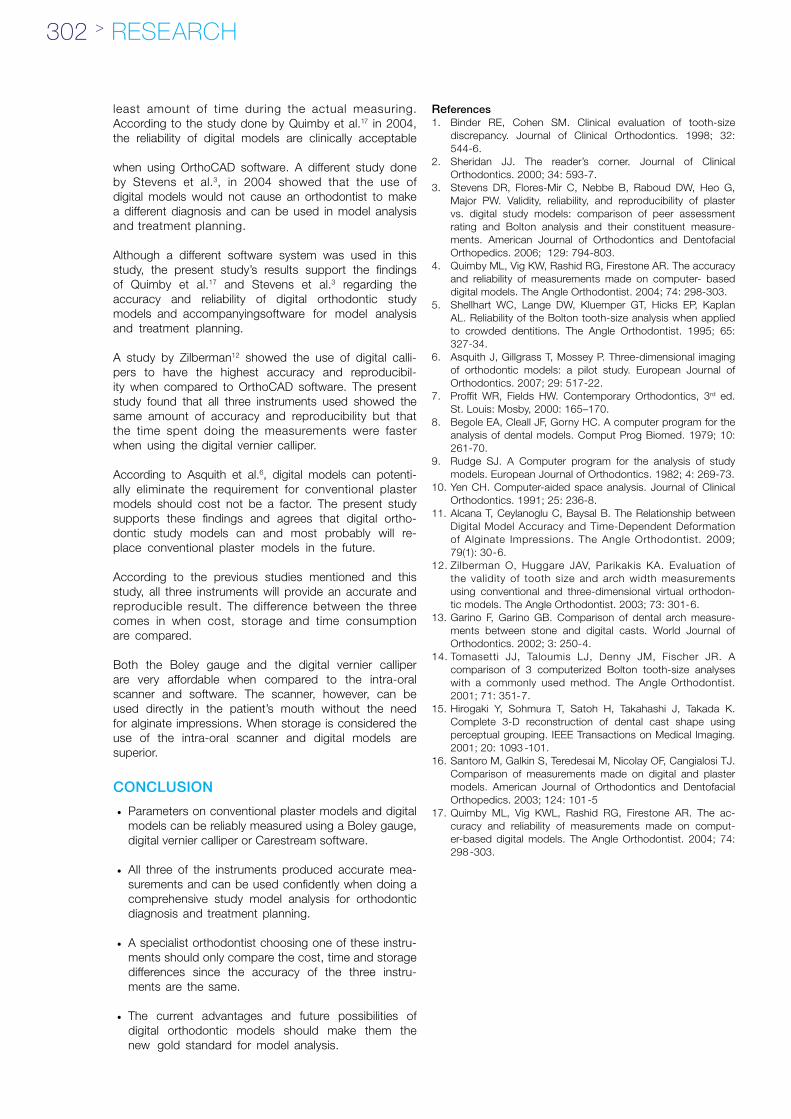
The Boley gauge (Figure 1) and digital vernier calliper (Figure 2) were used to measure the plaster models di- rectly by making use of points standardized for all cases.
The same one hundred study models were then scan- ned and measured using software provided by the Carestream CS 3600 scanner (Figure 3)
All one hundred cases were marked on the mesio- palatal cusps of teeth 16 and 26, as well as the cusp tips of teeth 13 and 23 (Figure 4). These markings pr- ovided standardized reference points to use during the measuring process.
MATERIALS AND METHODS
RESEARCH < 299www.sada.co.za / SADJ Vol. 75 No. 6
The markings were made using a small round diamond bur. The mesio-distal widths of teeth 11, 46 and 41 were measured at the greatest mesio-distal dimension between the two contact points.
The scans produced by the intra-oral scanner clearly showed the indentations made by the bur and were also used for standardizing the measurements, see Figure 5. The digital models were obtained by scanning the plaster orthodontic study models using a Carestream intra-oral scanner and imported as STL (Standard Tessellation Lan- guage) files into the Carestream model software where they could be rotated and magnified to help facilitate the measuring process.
All the measurements were done by the author of this article. All one hundred cases were measured and documented by the same operator. All the values were placed in an Excel file in order to conduct the statistical analysis of all the measurements and to compare the three different techniques with one another. The five measurements of all the cases were compared.
For the statistical analysis the three instruments were treated as independent variables. The difference between the instruments were tested per aspect in an attempt to avoid variation between the aspects and possible dif- ferences in measurement units. Models were treated as a co-variate in an attempt to control unexplained varia- tion between the models.
The results for the five parameters measured by each of the three instruments are summarized below:
The difference between the instruments with intermolar as dependent variable was not significant: F2,296 = 0.163; p = 0.849, see Table 1.
The difference between the instruments with intercanine as dependent variable was not significant: F2,296= 0.421; p= 0.657, see Table 2.
The difference between the instruments with the mesio- distal width of tooth 11 as dependent variable was not significant: F2,296 =1.735; p =0.178, see Table 3.
The difference between the instruments with the mesio-dis-tal width of tooth 46 as dependent variable was significant: F2,296 =7.097; p< 0.01, see Table 4.
The difference between the instruments with the mesio- distal width of tooth 41 as dependent variable was not significant: F2,296 =1.434; p= 0.240, see Table 5.
Orthodontic study models are used routinely for model analysis and treatment planning. The use of digital study models provides the opportunity to accurately determine the effects of orthodontic treatment and to do compre- hensive model analysis without the need for impres- sions and storage. The purpose of the study was to evaluate if a difference exists when the conventional
RESULTS
DISCUSSION
Figure 5. Example of how the standardised markings were used in order to measure on the digital orthodontic study models produced by the intra-oral scanner.
Figure 4. Canine and molar marked with diamond bur.
Figure 1. Boley gauge.
Figure 2. Digital Vernier calliper.
Figure 3. Carestream CS3600 intra-oral scanner
RESEARCH300 >

measuring method using a Boley gauge is compared to a digital calliper and the latest Carestream model analysis software. The study made use of three diffe- rent methods for orthodontic study model analysis and evaluated the differences between the three systems. The study was conducted by measuring five parameters on the study models of one hundred cases with all three of the instruments.
A sample t-test was conducted and the statistical ana- lysis showed the difference between the three instru- ments were not statistically significant for the intermolar width (p = 0.849 ), intercanine width (p = 0.657), mesio- distal width of tooth 11 ( p =0.178 ) and mesiodistal width of tooth 41 (p = 0.240). The difference in measure-ments between the three instruments for the mesiodistal width of tooth 46 was statistically significant (p<0.01).
Although a statistically significant difference was shown on the measurements obtained for tooth 46, the differ- ence was not found to have a clinical significance to the orthodontist when conducting a model analysis.The results of the five measurements for each of the three instruments were accurate and similar when com- pared to one another. An average value of 40.5 mm (intermolar width), 34.3 mm (intercanine width), 8.5 mm
(mesiodistal width of tooth 11), 10.8 mm (mesiodistal width of tooth 46) and 5.4 mm (mesiodistal width of tooth 41) was found when the values of all three measuring tools were considered, see Figure 6.
The values of the three instruments used in the study were very accurate which emphasizes that all three instruments are reliable and reproducible. Of the three instruments used, the digital vernier calliper took the
Table 1. Maxillary intermolar width.
Intermolar Width
Instruments Mean Std. Error95% Confidence Interval
Lower Bound Upper Bound
1. Boley` 40.597a .303 40.000 41.194
2. Vernier 40.383a .303 39.786 40.980
3. Carestream 40.386a .303 39.789 40.983
Table 2. Maxillary intercanine width.
Intercanine Width
Instruments Mean Std. Error95% Confidence Interval
Lower Bound Upper Bound
1. Boley` 34.450a .213 34.030 34.870
2. Vernier 34.196a .213 33.776 34.616
3. Carestream 34.228a .213 33.808 34.648
Table 3. Mesio-distal width of tooth 11.
Tooth 11
Instruments Mean Std. Error95% Confidence Interval
Lower Bound Upper Bound
1. Boley` 8.535a .059 8.418 8.652
2. Vernier 8.467a .059 8.350 8.584
3. Carestream 8.623a .059 8.506 8.740
Table 4. Mesio-distal width of tooth 46.
Tooth 46
Instruments Mean Std. Error95% Confidence Interval
Lower Bound Upper Bound
1. Boley` 10.779 .059 10.663 10.895
2. Vernier 10.708 .059 10.592 10.824
3. Carestream 11.008 .059 10.892 11.124
Table 5. Mesio-distal width of tooth 41.
Tooth 41
Instruments Mean Std. Error95% Confidence Interval
Lower Bound Upper Bound
1. Boley` 5.411a .037 5.338 5.484
2. Vernier 5.333a .037 5.260 5.406
3. Carestream 5.409a .037 5.336 5.482
Results
Boley Guage Digital Vernier Caliper Carestream Scanner
Figure 6. Results of all 5 measurements produced by the three instruments.
0Intermolar Intercanine Tooth 11 Tooth 46 Tooth 41
5
10
15
20
25
25
30
35
40
45
RESEARCH < 301www.sada.co.za / SADJ Vol. 75 No. 6
least amount of time during the actual measuring. According to the study done by Quimby et al.17 in 2004, the reliability of digital models are clinically acceptable
when using OrthoCAD software. A different study done by Stevens et al.3, in 2004 showed that the use of digital models would not cause an orthodontist to make a different diagnosis and can be used in model analysis and treatment planning.
Although a different software system was used in this study, the present study’s results support the findings of Quimby et al.17 and Stevens et al.3 regarding the accuracy and reliability of digital orthodontic study models and accompanyingsoftware for model analysis and treatment planning.
A study by Zilberman12 showed the use of digital calli- pers to have the highest accuracy and reproducibil-ity when compared to OrthoCAD software. The present study found that all three instruments used showed the same amount of accuracy and reproducibility but that the time spent doing the measurements were faster when using the digital vernier calliper.
According to Asquith et al.6, digital models can potenti- ally eliminate the requirement for conventional plaster models should cost not be a factor. The present study supports these findings and agrees that digital ortho- dontic study models can and most probably will re- place conventional plaster models in the future.
According to the previous studies mentioned and this study, all three instruments will provide an accurate and reproducible result. The difference between the three comes in when cost, storage and time consumption are compared.
Both the Boley gauge and the digital vernier calliper are very affordable when compared to the intra-oral scanner and software. The scanner, however, can be used directly in the patient’s mouth without the need for alginate impressions. When storage is considered the use of the intra-oral scanner and digital models are superior.
• Parameters on conventional plaster models and digital models can be reliably measured using a Boley gauge, digital vernier calliper or Carestream software.
• All three of the instruments produced accurate mea- surements and can be used confidently when doing a comprehensive study model analysis for orthodontic diagnosis and treatment planning.
• A specialist orthodontist choosing one of these instru- ments should only compare the cost, time and storage differences since the accuracy of the three instru- ments are the same.
• The current advantages and future possibilities of digital orthodontic models should make them the new gold standard for model analysis.
References1. Binder RE, Cohen SM. Clinical evaluation of tooth-size
discrepancy. Journal of Clinical Orthodontics. 1998; 32: 544-6.
2. Sheridan JJ. The reader’s corner. Journal of Clinical Orthodontics. 2000; 34: 593-7.
3. Stevens DR, Flores-Mir C, Nebbe B, Raboud DW, Heo G, Major PW. Validity, reliability, and reproducibility of plaster vs. digital study models: comparison of peer assessment rating and Bolton analysis and their constituent measure-ments. American Journal of Orthodontics and Dentofacial Orthopedics. 2006; 129: 794-803.
4. Quimby ML, Vig KW, Rashid RG, Firestone AR. The accuracy and reliability of measurements made on computer- based digital models. The Angle Orthodontist. 2004; 74: 298-303.
5. Shellhart WC, Lange DW, Kluemper GT, Hicks EP, Kaplan AL. Reliability of the Bolton tooth-size analysis when applied to crowded dentitions. The Angle Orthodontist. 1995; 65: 327-34.
6. Asquith J, Gillgrass T, Mossey P. Three-dimensional imaging of orthodontic models: a pilot study. European Journal of Orthodontics. 2007; 29: 517-22.
7. Proffit WR, Fields HW. Contemporary Orthodontics, 3rd ed. St. Louis: Mosby, 2000: 165–170.
8. Begole EA, Cleall JF, Gorny HC. A computer program for the analysis of dental models. Comput Prog Biomed. 1979; 10: 261-70.
9. Rudge SJ. A Computer program for the analysis of study models. European Journal of Orthodontics. 1982; 4: 269-73.
10. Yen CH. Computer-aided space analysis. Journal of Clinical Orthodontics. 1991; 25: 236-8.
11. Alcana T, Ceylanoglu C, Baysal B. The Relationship between Digital Model Accuracy and Time-Dependent Deformation of Alginate Impressions. The Angle Orthodontist. 2009; 79(1): 30-6.
12. Zilberman O, Huggare JAV, Parikakis KA. Evaluation of the validity of tooth size and arch width measurements using conventional and three-dimensional virtual orthodon- tic models. The Angle Orthodontist. 2003; 73: 301-6.
13. Garino F, Garino GB. Comparison of dental arch measure-ments between stone and digital casts. World Journal of Orthodontics. 2002; 3: 250-4.
14. Tomasetti JJ, Taloumis LJ, Denny JM, Fischer JR. A comparison of 3 computerized Bolton tooth-size analyses with a commonly used method. The Angle Orthodontist. 2001; 71: 351-7.
15. Hirogaki Y, Sohmura T, Satoh H, Takahashi J, Takada K. Complete 3-D reconstruction of dental cast shape using perceptual grouping. IEEE Transactions on Medical Imaging. 2001; 20: 1093 -101.
16. Santoro M, Galkin S, Teredesai M, Nicolay OF, Cangialosi TJ. Comparison of measurements made on digital and plaster models. American Journal of Orthodontics and Dentofacial Orthopedics. 2003; 124: 101-5
17. Quimby ML, Vig KWL, Rashid RG, Firestone AR. The ac- curacy and reliability of measurements made on comput-er-based digital models. The Angle Orthodontist. 2004; 74: 298-303.
CONCLUSION
RESEARCH302 >
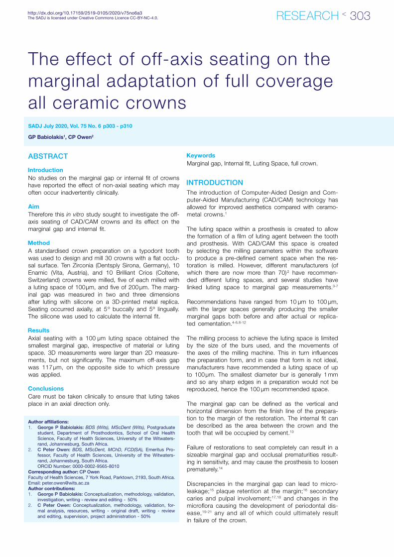
http://dx.doi.org/10.17159/2519-0105/2020/v75no6a3The SADJ is licensed under Creative Commons Licence CC-BY-NC-4.0.
No studies on the marginal gap or internal fit of crowns have reported the effect of non-axial seating which may often occur inadvertently clinically.
Therefore this in vitro study sought to investigate the off- axis seating of CAD/CAM crowns and its effect on the marginal gap and internal fit.
A standardised crown preparation on a typodont tooth was used to design and mill 30 crowns with a flat occlu- sal surface. Ten Zirconia (Dentsply Sirona, Germany), 10 Enamic (Vita, Austria), and 10 Brilliant Crios (Coltene, Switzerland) crowns were milled, five of each milled with a luting space of 100µm, and five of 200µm. The marg- inal gap was measured in two and three dimensions after luting with silicone on a 3D-printed metal replica. Seating occurred axially, at 5º buccally and 5º lingually. The silicone was used to calculate the internal fit.
Axial seating with a 100 µm luting space obtained the smallest marginal gap, irrespective of material or luting space. 3D measurements were larger than 2D measure- ments, but not significantly. The maximum off-axis gap was 117µm, on the opposite side to which pressure was applied.
Care must be taken clinically to ensure that luting takes place in an axial direction only.
Marginal gap, Internal fit, Luting Space, full crown.
The introduction of Computer-Aided Design and Com- puter-Aided Manufacturing (CAD/CAM) technology has allowed for improved aesthetics compared with ceramo- metal crowns.1
The luting space within a prosthesis is created to allow the formation of a film of luting agent between the tooth and prosthesis. With CAD/CAM this space is created by selecting the milling parameters within the software to produce a pre-defined cement space when the res- toration is milled. However, different manufacturers (of which there are now more than 70) 2 have recommen- ded different luting spaces, and several studies have linked luting space to marginal gap measurements.3-7
Recommendations have ranged from 10 μm to 100 μm, with the larger spaces generally producing the smaller marginal gaps both before and after actual or replica- ted cementation.4-6,8-12
The milling process to achieve the luting space is limited by the size of the burs used, and the movements of the axes of the milling machine. This in turn influences the preparation form, and in case that form is not ideal, manufacturers have recommended a luting space of up to 100μm. The smallest diameter bur is generally 1 mm and so any sharp edges in a preparation would not be reproduced, hence the 100 μm recommended space.
The marginal gap can be defined as the vertical and horizontal dimension from the finish line of the prepara- tion to the margin of the restoration. The internal fit can be described as the area between the crown and the tooth that will be occupied by cement.13
Failure of restorations to seat completely can result in a sizeable marginal gap and occlusal prematurities result- ing in sensitivity, and may cause the prosthesis to loosen prematurely.14
Discrepancies in the marginal gap can lead to micro- leakage;15 plaque retention at the margin;16 secondary caries and pulpal involvement;17,18 and changes in the microflora causing the development of periodontal dis- ease,19-21 any and all of which could ultimately result in failure of the crown.
ABSTRACT
Introduction
Aim
Method
Results
Conclusions
INTRODUCTION
Keywords
Author affiliations:1. George P Babiolakis: BDS (Wits), MScDent (Wits), Postgraduate
student, Department of Prosthodontics, School of Oral Health Science, Faculty of Health Sciences, University of the Witwaters- rand, Johannesburg, South Africa.
2. C Peter Owen: BDS, MScDent, MChD, FCD(SA), Emeritus Pro- fessor, Faculty of Health Sciences, University of the Witwaters- rand, Johannesburg, South Africa.ORCID Number: 0000-0002-9565-8010
Corresponding author: CP OwenFaculty of Health Sciences, 7 York Road, Parktown, 2193, South Africa.Email: [email protected] contributions:1. George P Babiolakis: Conceptualization, methodology, validation,
investigation, writing - review and editing - 50%2. C Peter Owen: Conceptualization, methodology, validation, for-
mal analysis, resources, writing - original draft, writing - review and editing, supervision, project administration - 50%
RESEARCH < 303
GP Babiolakis1, CP Owen2
SADJ July 2020, Vol. 75 No. 6 p303 - p310
The effect of off-axis seating on the marginal adaptation of full coverage all ceramic crowns
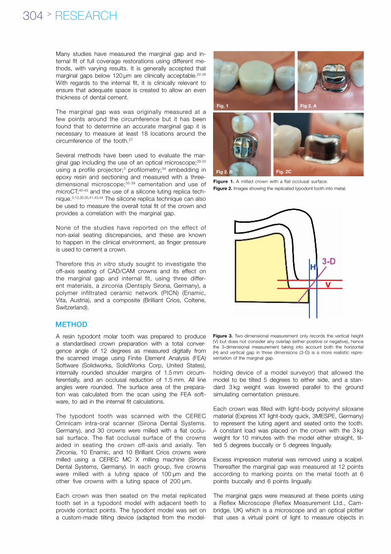
Many studies have measured the marginal gap and in- ternal fit of full coverage restorations using different me- thods, with varying results. It is generally accepted that marginal gaps below 120 µm are clinically acceptable.22-26 With regards to the internal fit, it is clinically relevant to ensure that adequate space is created to allow an even thickness of dental cement.
The marginal gap was was originally measured at a few points around the circumference but it has been found that to determine an accurate marginal gap it is necessary to measure at least 18 locations around the circumference of the tooth.27
Several methods have been used to evaluate the mar- ginal gap including the use of an optical microscope;28-33 using a profile projector;3 profilometry;34 embedding in epoxy resin and sectioning and measured with a three- dimensional microscope;35-39 cementation and use of microCT;40-43 and the use of a silicone luting replica tech- nique.3,13,30,35,41,43,44 The silicone replica technique can also be used to measure the overall total fit of the crown and provides a correlation with the marginal gap.
None of the studies have reported on the effect of non-axial seating discrepancies, and these are known to happen in the clinical environment, as finger pressure is used to cement a crown.
Therefore this in vitro study sought to investigate the off-axis seating of CAD/CAM crowns and its effect on the marginal gap and internal fit, using three differ- ent materials, a zirconia (Dentsply Sirona, Germany), a polymer infiltrated ceramic network (PICN) (Enamic, Vita, Austria), and a composite (Brilliant Crios, Coltene, Switzerland).
A resin typodont molar tooth was prepared to produce a standardised crown preparation with a total conver- gence angle of 12 degrees as measured digitally from the scanned image using Finite Element Analysis (FEA) Software (Solidworks, SolidWorks Corp, United States), internally rounded shoulder margins of 1.5 mm circum-ferentially, and an occlusal reduction of 1.5 mm. All line angles were rounded. The surface area of the prepara- tion was calculated from the scan using the FEA soft- ware, to aid in the internal fit calculations. The typodont tooth was scanned with the CEREC Omnicam intra-oral scanner (Sirona Dental Systems. Germany), and 30 crowns were milled with a flat occlu- sal surface. The flat occlusal surface of the crowns aided in seating the crown off-axis and axially. Ten Zirconia, 10 Enamic, and 10 Brilliant Crios crowns were milled using a CEREC MC X milling machine (Sirona Dental Systems, Germany). In each group, five crowns were milled with a luting space of 100 µm and the other five crowns with a luting space of 200 µm.
Each crown was then seated on the metal replicated tooth set in a typodont model with adjacent teeth to provide contact points. The typodont model was set on a custom-made tilting device (adapted from the model-
holding device of a model surveyor) that allowed the model to be tilted 5 degrees to either side, and a stan- dard 3 kg weight was lowered parallel to the ground simulating cementation pressure.
Each crown was filled with light-body polyvinyl siloxane material (Express XT light-body quick, 3MESPE, Germany) to represent the luting agent and seated onto the tooth. A constant load was placed on the crown with the 3 kg weight for 10 minutes with the model either straight, til- ted 5 degrees buccally or 5 degrees lingually.
Excess impression material was removed using a scalpel. Thereafter the marginal gap was measured at 12 points according to marking points on the metal tooth at 6 points buccally and 6 points lingually.
The marginal gaps were measured at these points using a Reflex Microscope (Reflex Measurement Ltd., Cam- bridge, UK) which is a microscope and an optical plotter that uses a virtual point of light to measure objects in
METHOD
Figure 2. Images showing the replicated typodont tooth into metal.
Figure 1. A milled crown with a flat occlusal surface.
Figure 3. Two-dimensional measurement only records the vertical height (V) but does not consider any overlap (either positive or negative), hence the 3-dimensional measurement taking into account both the horizontal (H) and vertical gap in three dimensions (3-D) is a more realistic repre- sentation of the marginal gap.
Fig 2. A
Fig 2. B
Fig. 1
Fig. 2C
RESEARCH304 >
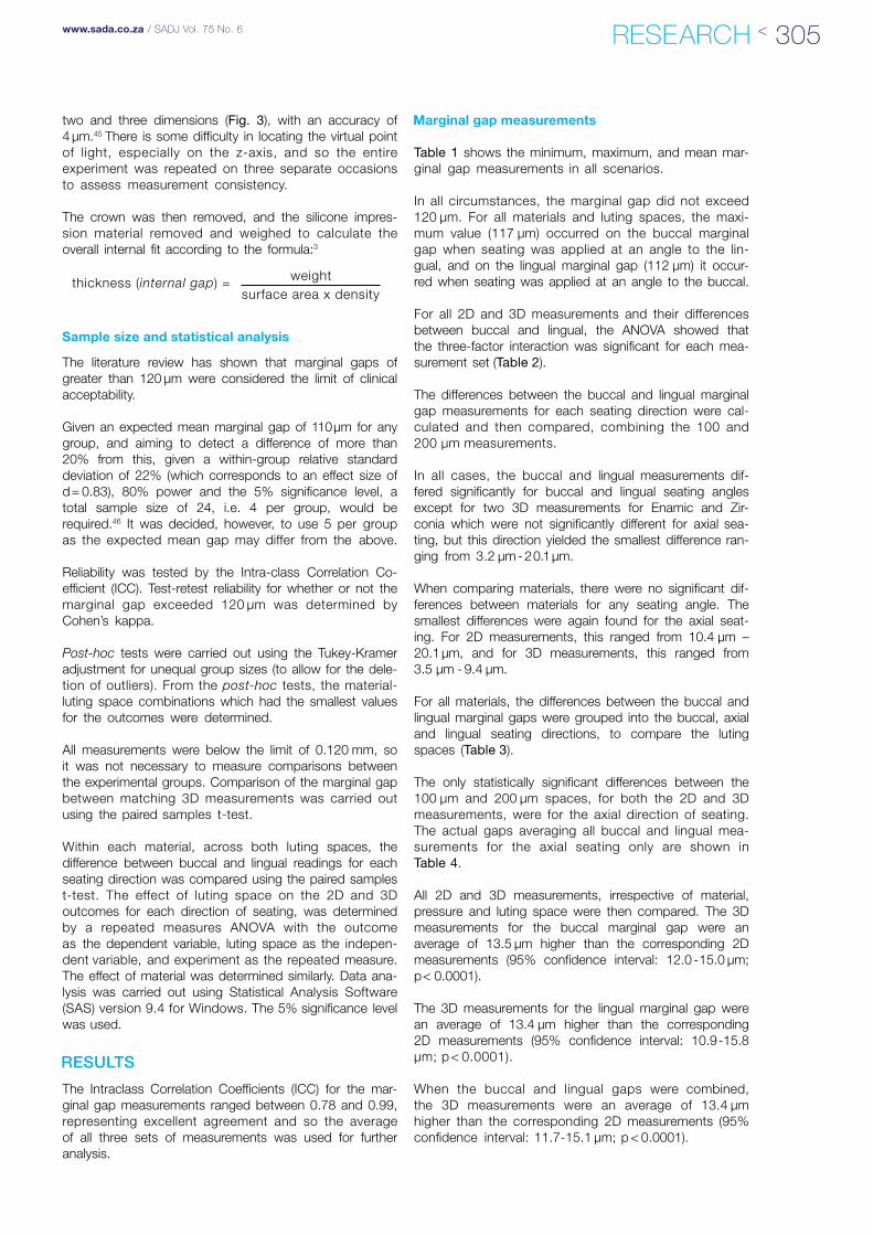
two and three dimensions (Fig. 3), with an accuracy of 4 μm.45 There is some difficulty in locating the virtual point of light, especially on the z-axis, and so the entire experiment was repeated on three separate occasions to assess measurement consistency.
The crown was then removed, and the silicone impres- sion material removed and weighed to calculate the overall internal fit according to the formula:3
The literature review has shown that marginal gaps of greater than 120 µm were considered the limit of clinical acceptability.
Given an expected mean marginal gap of 110µm for any group, and aiming to detect a difference of more than 20% from this, given a within-group relative standard deviation of 22% (which corresponds to an effect size of d= 0.83), 80% power and the 5% significance level, a total sample size of 24, i.e. 4 per group, would be required.46 It was decided, however, to use 5 per group as the expected mean gap may differ from the above.
Reliability was tested by the Intra-class Correlation Co- efficient (ICC). Test-retest reliability for whether or not the marginal gap exceeded 120 µm was determined by Cohen’s kappa.
Post-hoc tests were carried out using the Tukey-Kramer adjustment for unequal group sizes (to allow for the dele- tion of outliers). From the post-hoc tests, the material- luting space combinations which had the smallest values for the outcomes were determined.
All measurements were below the limit of 0.120 mm, so it was not necessary to measure comparisons between the experimental groups. Comparison of the marginal gap between matching 3D measurements was carried out using the paired samples t-test.
Within each material, across both luting spaces, the difference between buccal and lingual readings for each seating direction was compared using the paired samples t-test. The effect of luting space on the 2D and 3D outcomes for each direction of seating, was determined by a repeated measures ANOVA with the outcome as the dependent variable, luting space as the indepen- dent variable, and experiment as the repeated measure.The effect of material was determined similarly. Data ana- lysis was carried out using Statistical Analysis Software (SAS) version 9.4 for Windows. The 5% significance level was used.
The Intraclass Correlation Coefficients (ICC) for the mar- ginal gap measurements ranged between 0.78 and 0.99, representing excellent agreement and so the average of all three sets of measurements was used for further analysis.
Table 1 shows the minimum, maximum, and mean mar- ginal gap measurements in all scenarios.
In all circumstances, the marginal gap did not exceed 120 µm. For all materials and luting spaces, the maxi- mum value (117 µm) occurred on the buccal marginal gap when seating was applied at an angle to the lin- gual, and on the lingual marginal gap (112 µm) it occur- red when seating was applied at an angle to the buccal.
For all 2D and 3D measurements and their differences between buccal and lingual, the ANOVA showed that the three-factor interaction was significant for each mea- surement set (Table 2).
The differences between the buccal and lingual marginal gap measurements for each seating direction were cal- culated and then compared, combining the 100 and 200 µm measurements.
In all cases, the buccal and lingual measurements dif- fered significantly for buccal and lingual seating angles except for two 3D measurements for Enamic and Zir- conia which were not significantly different for axial sea- ting, but this direction yielded the smallest difference ran- ging from 3.2 µm - 20.1µm.
When comparing materials, there were no significant dif- ferences between materials for any seating angle. The smallest differences were again found for the axial seat- ing. For 2D measurements, this ranged from 10.4 µm – 20.1 µm, and for 3D measurements, this ranged from 3.5 µm - 9.4 µm.
For all materials, the differences between the buccal and lingual marginal gaps were grouped into the buccal, axial and lingual seating directions, to compare the luting spaces (Table 3). The only statistically significant differences between the 100 µm and 200 µm spaces, for both the 2D and 3D measurements, were for the axial direction of seating. The actual gaps averaging all buccal and lingual mea- surements for the axial seating only are shown in Table 4.
All 2D and 3D measurements, irrespective of material, pressure and luting space were then compared. The 3D measurements for the buccal marginal gap were an average of 13.5 µm higher than the corresponding 2D measurements (95% confidence interval: 12.0 -15.0 µm; p< 0.0001).
The 3D measurements for the lingual marginal gap were an average of 13.4 µm higher than the corresponding 2D measurements (95% confidence interval: 10.9-15.8 µm; p < 0.0001).
When the buccal and lingual gaps were combined, the 3D measurements were an average of 13.4 µm higher than the corresponding 2D measurements (95% confidence interval: 11.7-15.1 µm; p < 0.0001).
Sample size and statistical analysis
RESULTS
Marginal gap measurements
thickness (internal gap) = weightsurface area x density
RESEARCH < 305www.sada.co.za / SADJ Vol. 75 No. 6
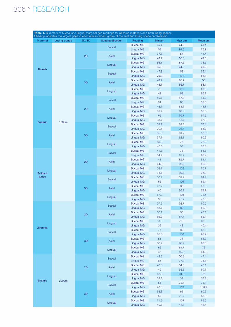
Table 1. Summary of buccal and lingual marginal gap readings for all three materials and both luting spaces. Shading indicates the largest gaps in each measurement group of material and luting space combination.
Material Luting space 2D/3D Seating direction Reading Min μm Max μm Mean μm
Zirconia
100µm
2D
BuccalBuccal MG 35.7 44.3 40.1
Lingual MG 53 91.3 70.9
AxialBuccal MG 37.3 57 44.4
Lingual MG 43.7 55.3 49.3
LingualBuccal MG 66.7 87.3 73.9
Lingual MG 35.3 44.3 40.9
3D
Buccal Buccal MG 47.3 59 55.4
Lingual MG 70.3 101 88.3
Axial Buccal MG 48.7 65.7 58
Lingual MG 45.7 59.7 53.1
LingualBuccal MG 78 101 86.8
Lingual MG 43 59 50.2
Enamic
2D
BuccalBuccal MG 40.7 47.3 44.6
Lingual MG 51 63 58.6
AxialBuccal MG 45.3 54.3 48.8
Lingual MG 51.7 60.3 56.3
LingualBuccal MG 63 65.7 64.3
Lingual MG 33.7 45.7 37.9
3D
Buccal Buccal MG 53.7 62.3 57.1
Lingual MG 70.7 91.7 81.3
Axial Buccal MG 55.3 61.7 57.5
Lingual MG 57.7 62.3 60.6
LingualBuccal MG 69.3 75 72.8
Lingual MG 40.3 58 50.1
Brilliant Crios
2D
BuccalBuccal MG 41.3 73 51.5
Lingual MG 54.7 92.7 65.2
AxialBuccal MG 41 82.7 51.4
Lingual MG 44.3 92.3 56.9
LingualBuccal MG 58.7 102 70.7
Lingual MG 34.7 39.3 36.2
3D
Buccal Buccal MG 50.7 81.7 61.9
Lingual MG 68 109 85.1
Axial Buccal MG 46.7 86 56.3
Lingual MG 45 95.3 59.7
LingualBuccal MG 67.3 108 78.4
Lingual MG 35 45.7 40.3
Zirconia
200µm
2D
BuccalBuccal MG 57.3 62.7 60.5
Lingual MG 58.7 89 69.9
AxialBuccal MG 30.7 56 46.8
Lingual MG 66.3 87.7 82.1
LingualBuccal MG 51.3 72.3 62.5
Lingual MG 32 48 40.1
3D
Buccal Buccal MG 75 89 82.3
Lingual MG 85.3 100 95.9
Axial Buccal MG 51 78 68.7
Lingual MG 66.7 98.7 82.6
LingualBuccal MG 69 81.7 76
Lingual MG 47 59.3 51.6
Enamic
2D
BuccalBuccal MG 43.3 50.3 47.4
Lingual MG 66 77.3 71.8
AxialBuccal MG 40.3 54.3 47.1
Lingual MG 49 68.3 60.7
LingualBuccal MG 48.3 94.3 75
Lingual MG 32.3 38 35.3
3D
Buccal Buccal MG 65 75.7 73.1
Lingual MG 97.3 113 106.9
Axial Buccal MG 56.3 65 60.5
Lingual MG 50 72.7 63.9
LingualBuccal MG 71.3 103 88.5
Lingual MG 40.7 48.7 44.1
RESEARCH306 >
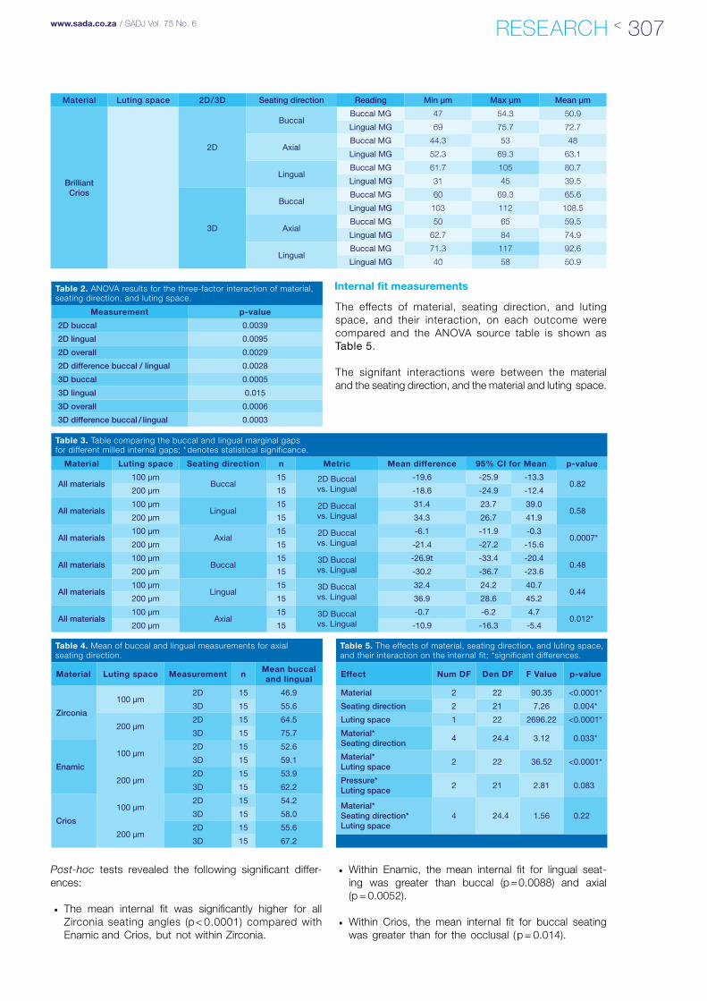
Material Luting space 2D/3D Seating direction Reading Min μm Max μm Mean μm
Brilliant Crios
2D
BuccalBuccal MG 47 54.3 50.9
Lingual MG 69 75.7 72.7
AxialBuccal MG 44.3 53 48
Lingual MG 52.3 69.3 63.1
LingualBuccal MG 61.7 105 80.7
Lingual MG 31 45 39.5
3D
Buccal Buccal MG 60 69.3 65.6
Lingual MG 103 112 108.5
Axial Buccal MG 50 65 59.5
Lingual MG 62.7 84 74.9
LingualBuccal MG 71.3 117 92.6
Lingual MG 40 58 50.9
The effects of material, seating direction, and luting space, and their interaction, on each outcome were compared and the ANOVA source table is shown as Table 5.
The signifant interactions were between the material and the seating direction, and the material and luting space.
Internal fit measurementsTable 2. ANOVA results for the three-factor interaction of material, seating direction, and luting space.
Measurement p-value
2D buccal 0.0039
2D lingual 0.0095
2D overall 0.0029
2D difference buccal / lingual 0.0028
3D buccal 0.0005
3D lingual 0.015
3D overall 0.0006
3D difference buccal / lingual 0.0003
Table 3. Table comparing the buccal and lingual marginal gaps for different milled internal gaps; *denotes statistical significance.
Material Luting space Seating direction n Metric Mean difference 95% CI for Mean p-value
All materials100 µm
Buccal15 2D Buccal
vs. Lingual-19.6 -25.9 -13.3
0.82200 µm 15 -18.6 -24.9 -12.4
All materials100 µm
Lingual15 2D Buccal
vs. Lingual31.4 23.7 39.0
0.58200 µm 15 34.3 26.7 41.9
All materials100 µm
Axial15 2D Buccal
vs. Lingual-6.1 -11.9 -0.3
0.0007*200 µm 15 -21.4 -27.2 -15.6
All materials100 µm
Buccal15 3D Buccal
vs. Lingual-26.9t -33.4 -20.4
0.48200 µm 15 -30.2 -36.7 -23.6
All materials100 µm
Lingual15 3D Buccal
vs. Lingual32.4 24.2 40.7
0.44200 µm 15 36.9 28.6 45.2
All materials100 µm
Axial15 3D Buccal
vs. Lingual-0.7 -6.2 4.7
0.012*200 µm 15 -10.9 -16.3 -5.4
Table 4. Mean of buccal and lingual measurements for axial seating direction.
Material Luting space Measurement nMean buccal and lingual
Zirconia
100 µm2D 15 46.9
3D 15 55.6
200 µm2D 15 64.5
3D 15 75.7
Enamic
100 µm2D 15 52.6
3D 15 59.1
200 µm2D 15 53.9
3D 15 62.2
Crios
100 µm2D 15 54.2
3D 15 58.0
200 µm2D 15 55.6
3D 15 67.2
Table 5. The effects of material, seating direction, and luting space, and their interaction on the internal fit; *significant differences.
Effect Num DF Den DF F Value p-value
Material 2 22 90.35 <0.0001*
Seating direction 2 21 7.26 0.004*
Luting space 1 22 2696.22 <0.0001*
Material* Seating direction
4 24.4 3.12 0.033*
Material* Luting space
2 22 36.52 <0.0001*
Pressure* Luting space
2 21 2.81 0.083
Material* Seating direction* Luting space
4 24.4 1.56 0.22
Post-hoc tests revealed the following significant differ- ences:
• The mean internal fit was significantly higher for all Zirconia seating angles (p< 0.0001) compared with Enamic and Crios, but not within Zirconia.
• Within Enamic, the mean internal fit for lingual seat- ing was greater than buccal (p =0.0088) and axial (p = 0.0052).
• Within Crios, the mean internal fit for buccal seating was greater than for the occlusal (p = 0.014).
RESEARCH < 307www.sada.co.za / SADJ Vol. 75 No. 6
When comparing the luting spaces, Post-hoc tests re- vealed the following significant differences:
• The mean internal fit was significantly higher for all 200 µm experiments compared with all 100 µm ex- periments (p< 0.0001).
• Within the 100 µm experiments, the mean internal fit decreased in the order Zirconia > Enamic (p<0.0001) > Crios (p < 0.0078).
• Within the 200 µm experiments, the mean internal fit decreased in the order Zirconia > Enamic (p < 0.0055) > Crios (p <0.0034).
This is the first study to be carried out to measure and compare the effect of off-axis seating on the adaptation of full coverage crowns using the marginal gap and inter- nal fit as excellent proxies for the clinical quality and success of a restoration.
Discrepancies in the marginal gap can lead to a variety of problems which could ultimately result in failure of the crown.15-21 It is generally accepted that marginal gaps below 120µm are clinically acceptable.22-26,47,48
With regards to the internal fit, it is clinically relevant to ensure that adequate space is created to allow an even thickness of dental cement. Theoretically, the space re- quired for the cement to lute is 20-40 µm, as cement thickness ranges from 25-50 µm, and an acceptable practical guide was set between 50 µm and 100µm.49
In CAD/CAM restorations, a luting space is used to allow for this, and several studies have linked luting space to marginal gap measurements.3-7 In the literature, luting space recommendations have ranged from 10 µm to 100µm. The larger spaces have produced the smaller marginal gaps both before and after actual or replicated cementation.4-6,8-12
In this study, it was decided to use luting spaces of 100 μm and 200 μm. In a pilot study it had been obser- ved that, as finger pressure is used to cement a crown, it is possible that it may not seat evenly if seated at an angle to the occlusal. No studies have reported on the effect of non-axial seating discrepancies. It was evident that seating the crown off-axis at just 5º did affect the marginal gap: there was a significant dif- ference between the buccal and lingual marginal gap measurements in all cases when the crowns were seated off axis, but it was interesting to note that none of the marginal gaps measured exceeded 120µm. However, the greatest discrepancies were observed in off-axis seating with a luting space of 200 µm for all materials, indicating that that luting space is proably too large and may produce more off-axis seating clinically.
There were statistically significant differences between the 100 µm and 200 µm spaces, for both the 2D and 3D measuremets, for the axial direction of seating, indi- cating that the luting space did affect the marginal gap.
The smallest gaps were from the axial seating using the 100 µm luting space. Overall, for all materials these differences for the 2D measurements ranged from 10.4 µm – 20.1µm, and for 3D measurements, from 3.5 µm – 9.4 µm.
Overall the 3D measurements were 13.4 µm greater, but not significantly different from the corresponding 2D measurements (p= 0.92). These measurements are to be expected, as the 3D gap is likely always to be higher
than the 2D measurement, but they are nevertheless all extremely low, which is a testament to the accuracy of the milling of these crowns. As with the marginal gaps, within each material, axial seating yielded the smallest internal fit when compared with off-axis seating.
The internal fit of a crown is just as important as the marginal gap, as it enables the seating of the crown and expression of cement, while also aiding in retention and resistance.31 The mean internal fit for all 200 µm crowns was significantly higher than the 100µm crowns, which was expected.
This also shows that the CAD/CAM process is highly accurate, generating an internal fit for each crown which closely resembles the luting space chosen. Clinically the results obtained in this study have implications.
Irrespective of material used when seating a crown, a minor tilt of even 5 degrees can result in a larger marginal gap specifically on the opposite side to the pressure being applied.
Although this study did not find these measurements to be above 120 µm, some marginal gaps were still large, with one reaching 117µm.
Previous studies which measured the marginal gap of crowns found that they ranged from <70μm50, 52μm to 74μm3, a median of 130.2 and 132.2 μm, 51 below 90 μm.52 The marginal gaps measured in this study which more closely resemble those of other studies are the values measured for axial seating.
Should other studies have taken into consideration the tilt that may be found when seating off axis, they may have measured larger results. In this study the marginal gaps ranged from 36 µm – 117 µm with off-axis seating and 31µm – 99 µm with axial seating.
The other factor not taken into consideration in other studies is measuring the marginal gap buccally and lin- gually separately. Gassino et al. (2004)27 found that to obtain an accurate overall marginal gap measurement requires at least18 points around the circumference of the tooth to be measured.
However, this again did not take into account the tilt and used an average of all measurements to arrive at a marginal gap. Considering that a larger marginal gap will be found on the opposite side to the pressure being applied it is necessary to measure the buccal and lin- gual sides separately, to yield an accurate result that resembles the correct fit of the crown.
DISCUSSION
RESEARCH308 >
Within the limitations of this study, it was found that, irrespective of the material, seating off-axis at 5 degrees buccally or lingually resulted in a marginal gap which was larger on the opposite side to which pressure was applied. The smallest marginal gaps and internal fit were obtained when seating axially, with a luting space of 100 µm.
All measurements made in three dimensions were lar- ger than those derived for two-dimensional measure- ment, but the difference, average of 13.4 µm, was not significant. None of the measurements, whether cemen- ted axially or off-axis were larger than 120 µm. However, when seating off axis, the largest gap was 117µm as opposed to seating axially which yielded a mean maxi- mum marginal gap measurement of 76 µm.
It is recommended that future studies should measure the marginal gap both buccally and lingually separately and not just use an average to obtain an accurate measurement, and that a method needs to be devised to cement crowns axially in the clinical environment to provide the best fit possible and minimise compli- cations.
We are grateful to Dr P Gaylard for statistical advice and analysis.
The authors declare no conflict of interest.
References1. Masek R. Achieving high-level esthetics with CEREC. Com
pend Contin Educ Dent. 2001; 22 (6 Suppl): 19-26. https://www.medicalexpo.com/medical-manufacturer/dental-labora- tory-milling-machine-28385.html. (Accessed April 2020).
2. Nakamura T, Dei N, Kojima T, Wakabayashi K. Marginal and internal fit of Cerec 3 CAD/CAM all-ceramic crowns. Int J Prosthodont. 2003; 16(3): 244-8.
3. Hmaidouch R1, Neumann P, Mueller WD. Influence of pre- paration form, luting space setting and cement type on the marginal and internal fit of CAD/CAM crown copings. Int J Comput Dent. 2011; 14(3): 219-26.
4. Shim JS, Lee JS, Lee JY, Choi YJ, Shin SW, Ryu JJ. Effect of software version and parameter settings on the marginal and internal adaptation of crowns fabricated with the CAD/CAM system. J Appl Oral Sci. 2015; 23(5): 515-22.
5. Kale E, Seker E, Yilmaz B, Özcelik TB. Effect of cement space on the marginal fit of CAD-CAM-fabricated monolithic zirconia crowns. J Prosthet Dent. 2016; 116(6): 890-5
6. Dauti R, Lilaj B, Heimel P, Moritz A, Schedle A, Cvikl B. Influence of two different cement space settings and three different cement types on the fit of polymer-infiltrated cera- mic network material crowns manufactured using a com- plete digital workflow. Clin Oral Investig. 2019; Sep 13. doi: 10.1007/s00784-019-03053-1 (Online ahead of print).
7. Wang CJ, Millstein PL, Nathanson D. Effects of cement, cement space, marginal design, seating aid materials, and seating force on crown cementation. J Prosthet Dent. 1992; 67(6): 786-90
8. Wilson, PR. Effect of increasing cement space on cementa- tion of artificial crowns. J Prosthet Dent. 1994; 71(6): 560-4.
9. Wu JC, Wilson PR. Optimal Cement Space for Resin Luting Cements Int J Prosthodont. 1993; 7(3): 209-215
10. Iwai T, Komine F, Kobayashi K, Saito A, Matsumura H. Influence of convergence angle and cement space on adap- tation of zirconium dioxide ceramic copings. Acta Odontol Scand. 2008; 66(4): 214-8.
11. Yildirim G, Uzun IH, Keles A. Evaluation of marginal and internal adaptation of hybrid and nanoceramic systems with microcomputed tomography: an in vitro study. J Prosthet Dent 2017; 118(2): 200 -7.
12. Kokubo Y, Tsumita M, Kano T, Sakurai S, Fukushima, S. Clinical marginal and internal gaps of zirconia all-ceramic crowns. J Prosthodont Res 2011; 55(1): 40-43.
13. Abelson J. Cementation of cast complete crown retainers. J Prosthet Dent. 1980; 43(2): 174-9.
14. Jacobs MS, Windeler AS. An investigation of dental luting cement solubility as a function of the marginal gap. J Prosthet Dent. 1991; 65: 436-42.
15. Valderhaug J, Heløe LA. Oral hygiene in a group of supervised patients with fixed prostheses. J Periodontol. 1977; 48(4): 221-4.
16. Goldman M, Laosonthorn P, White RR. Microleakage e full crowns and the dental pulp. J Endod 1982; 18(10): 473-5.
17. Rinke S, Fornefett D, Gersdorff N, Lange K, Roediger M. Multifactorial analysis of the impact of different manufacturing processes on the marginal fit of zirconia copings. Dent Mater J. 2012; 31: 601-9.
18. Lang N, Kiel R, Anderhalden K. Clinical and microbiological effects of subgingival restorations with overhanging or clinically perfect margins. J Clin Periodontol. 1983; 10(6): 563-78.
19. Hunter AJ, Hunter AR. Gingival margins for crowns: a review and discussion. Part II: Discrepancies and configurations. J Prosthet Dent. 1990; 64: 636-42.
20. Felton DA, Konoy BE, Bayne MS, Wirthman GP. Effect of in vivo crown margin discrepancies on periodontal health. J Prosthet Dent 1991; 65: 357-64.
21. McLean J, von Fraunhofer J. The estimation of cement film thickness by an in vivo technique. Brit Dent J 1971; 131(3): 107-111.
22. Belser U, MacEntee M, Richter W. Fit of three porcelain-fused-to-metal marginal designs in vivo: A scanning electron microscope study. J Prosthet Dent. 1985; 53(1): 24-9.
23. Weaver J, Johnson G, Bales D. Marginal adaptation of castable ceramic crowns. J Prosthet Dent 1991; 66(6): 747-53.
24. Fonseca J, Henriques G, Sobrinho L, de Góes M. Stress-relieving and porcelain firing cycle influence on marginal fit of commercially pure titanium and titanium–aluminium-vanadium copings. Dent Mater. 2003; 19(7): 686-91.
25. Karatasli O, Kursoglu P, Capa N, Kazazoglu E. Comparison of the marginal fit of different coping materials and designs produced by computer aided manufacturing systems. Dent Mater J. 2011; 30: 97-102.
26. Gassino G, Barone Monfrin S, Scanu M, Spina G, Preti G. Marginal adaptation of fixed prosthodontics: a new in vitro 360-degree external examination procedure. Int J Prostho- dont. 2004; 17(2): 218-23.
27. Yeo I, Yang J, Lee J. In vitro marginal fit of three all-ceramic crown systems. J Prosthet Dent. 2003; 90(5): 459-64.
28. Akbar J, Petrie C, Walker M, Williams K, Eick, J. Marginal Adaptation of Cerec 3 CAD/CAM Composite Crowns Using Two Different Finish Line Preparation Designs. J Prosthodont. 2006; 15(3): 155-63.
29. Lee K, Park C, Kim K, Kwon, T. Marginal and Internal Fit of All-ceramic Crowns Fabricated with Two Different CAD/CAM Systems. Dent Mater J. 2008; 27(3): 422-6.
30. Beuer F, Edelhoff D, Gernet W, Naumann M. Effect of pre- paration angles on the precision of zirconia crown copings fabricated by CAD/CAM system. Dent Mater J. 2008; 27(6): 814-20.
CONCLUSION
Acknowledgements
Declaration
RESEARCH < 309www.sada.co.za / SADJ Vol. 75 No. 6
31. Alghazzawi T, Liu P, Essig M. The Effect of Different Fabrica- tion Steps on the Marginal Adaptation of Two Types of Glass-Infiltrated Ceramic Crown Copings Fabricated by CAD/CAM Technology. J Prosthodont. 2012; 21(3): 167-72.
32. Jayaraman S, Rajan B, Kandhasamy B, Rajakumaran I. Evaluation of marginal fit and internal adaptation of zirconia copings fabricated by two CAD - CAM systems: An in vitro study. J Indian Prosthodont Soc. 2015; 15(2): 173-8.
33. Mitchell C, Pintado M, Douglas W. Nondestructive, in vitro quantification of crown margins. J Prosthet Dent. 2001; 85(6): 575-84.
34. Tsitrou E, Northeast S, van Noort R. Evaluation of the mar- ginal fit of three margin designs of resin composite crowns using CAD/CAM. J Dent. 2007; 35(1): 68-73.
35. Han H, Yang H, Lim H, Park Y. Marginal accuracy and inter- nal fit of machine-milled and cast titanium crowns. J Prosthet Dent. 2011; 106(3): 191-7.
36. Souza R, Özcan M, Pavanelli C, Buso L, Lombardo G, Michi- da S, et al. Marginal and Internal Discrepancies Related to Margin Design of Ceramic Crowns Fabricated by a CAD/ CAM System. J Prosthodont. 2011; 21(2): 94-100.
37. Biscaro L, Bonfiglioli R, Soattin M; Vigolo, P. An In Vivo Evaluation of Fit of Zirconium-Oxide Based Ceramic Single Crowns, Generated with Two CAD/CAM Systems, in Com- parison to Metal Ceramic Single Crowns. J Prosthodont. 2012; 22(1): 36-41.
38. Sachs C, Groesser J, Stadelmann M, Schweiger J, Erdelt K; Beuer F. Full-arch prostheses from translucent zirconia: Accuracy of fit. Dent Mater 2014; 30(8): 817-23.
39. Seo D, Yi Y; Roh, B. The effect of preparation designs on the marginal and internal gaps in Cerec3 partial ceramic crowns. J Dent. 2009; 37(5): 374-82.
40. Rungruanganunt P, Kelly J, Adams D. Two imaging techni- ques for 3D quantification of pre-cementation space for CAD/CAM crowns. J Dent. 2010; 38(12): 995-1000.
41. Alfaro D, Ruse N, Carvalho R; Wyatt, C. Assessment of the Internal Fit of Lithium Disilicate Crowns Using Micro-CT. J Prosthodont. 2015; 24(5): 381-6.
42. Moldovan O, Luthardt R, Corcodel N, Rudolph H. Three-dimensional fit of CAD/CAM-made zirconia copings. Dent Mater 2011; 27(12): 1273-8.
43. Anunmana C, Charoenchitt M, Asvanund C. Gap compari- son between single crown and three-unit bridge zirconia sub structures. J Adv Prosthodont. 2014; 6(4): 253-8.
44. Speculand B, Butcher G, Stephens C. Three-dimensional measurement: The accuracy and precision of the reflex microscope. Br J Oral Maxillofac Surg. 1988; 26(4): 276-83.
45. Faul F, Erdfelder E, Lang A, Buchner A. G*Power 3: A flexi- ble statistical power analysis program for the social, beha- vioral, and biomedical sciences. Behav Res Methods. 2007; 39(2): 175-91.
46. Hung S, Hung K, Eick J, Chappell, R. Marginal fit of porcelain-fused-to-metal and two types of ceramic crown. J Prosthet Dent. 1990; 63(1): 26-31.
47. Coli P, Karlsso S. Fit of a New Pressure-Sintered Zirconium Dioxide Coping. Int J Prosthodont. 2004; 17(1): 59-64.
48. Martins L, Lorenzoni F, Melo A, Silva L, Oliveira J, Oliveira P et al. Internal fit of two all-ceramic systems and metal-cera- mic crowns. J Appl Oral Sci. 2012; 20(2): 235-40.
49. May K, Russell M, Razzoog M, Lang B. Precision of fit: The Procera All Ceram crown. J Prosthet Dent. 1998; 80(4): 394-404.
50. Akın A, Toksavul S, Toman M. Clinical Marginal and Internal Adaptation of Maxillary Anterior Single All-Ceramic Crowns and 2-year Randomized Controlled Clinical Trial. J Prostho- dont 2014; 24(5): 345-50.
51. Anadioti E, Aquilino S, Gratton D, Holloway J, Denry I, Thomas G et al. 3D and 2D Marginal Fit of Pressed and CAD/CAM Lithium Disilicate Crowns Made from Digital and Conventional Impressions. J Prosthodont. 2014; 23(8): 610- 17.
RESEARCH310 >
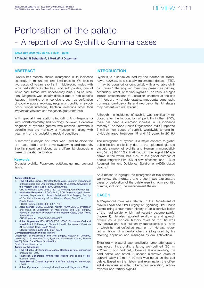
Syphilis has recently shown resurgence in its incidence especially in immune-compromised patients. We present two cases of tertiary syphilis in middle-aged males with large perforations in the hard and soft palates, one of which had Human Immunodeficiency Virus (HIV) co-infec-tion. Diagnosis was initially difficult due to non-specific features mimicking other conditions such as perforation of cocaine abuse aetiology, neoplastic conditions, sarcoi- dosis, fungal infections, bacterial infections other than Treponema pallidum and Wegeners granulomatosis.
With special investigations including Anti-Treponema Immunohistochemistry and histology, however, a definitive diagnosis of syphilitic gumma was reached. Intravenous penicillin was the mainstay of management along with treatment of the underlying medical conditions.
A removable acrylic obturator was used to close the oro-nasal fistula to improve swallowing and speech. Syphilis should be included as a differential diagnosis in cases of palatal perforation.
Orofacial syphilis, Treponema pallidum, gumma, oronasal fistula.
Syphilis, a disease caused by the bacterium Trepo- nema pallidum, is a sexually transmitted disease (STD). It may be acquired or congenital, with a variable clini- cal course.1 The acquired form may present as primary, secondary, latent, or tertiary syphilis.2 The various stages include presentations of ulceration (chancre) at the site of infection, lymphadenopathy, mucocutaneous rash, gummas, cardiosyphilis and neurosyphilis. All stages may present with oral lesions.3
Although the incidence of syphilis was significantly re- duced after the introduction of penicillin in the 1940’s, there has been a dramatic increase in its incidence recently.4 The World Health Organisation (WHO) reported 6 million new cases of syphilis worldwide among in- dividuals aged between 15 and 49 years in 2016.5
The resurgence of syphilis is a major concern to global public health, particularly due to the epidemiologic and biologic synergy of syphilis and Human Immunodefici- ency Virus (HIV).6-8 South Africa, with the largest HIV epi- demic in the world, has 19% of the global number of people living with HIV, 15% of new infections, and 11% of Acquired Immuno-Deficiency Syndrome (AIDS)-related deaths.9
As a means to highlight the resurgence of this condition, we review the literature and present two explanatory cases of perforation of the palate resulting from syphilitic gumma, including the management thereof.
A 35-year-old male was referred to the Department of Maxillo-Facial and Oral Surgery at Tygerberg Oral Health Centre citing a four-month history of an ulcerative lesion of the hard palate, which had recently become painful (Figure 1). He also reported swallowing and speech difficulties. A medical history revealed that he was HIV-positive and had pulmonary tuberculosis (TB), both of which he had defaulted treatment of. He also repor- ted a history of a genital chancre (diagnosed by his referring physician and managed by oral antibiotics).
Extra-orally, bilateral submandibular lymphadenopathy was noted. Intra-orally, a large, well-defined (20 mm x 20 mm), punched out, ulcerative lesion involving the hard palate was noted. A second ulcerative lesion of approximately (10 mm x 10 mm) was noted on the soft palate. Based on the history and examination the differ- ential diagnosis included tuberculous ulceration, actino-mycosis and tertiary syphilis.
ABSTRACT
Keywords
INTRODUCTION
CASE 1
Author affiliations:1. Fadi Titinchi: BChD, PDD (Oral Surg), MSc, Lecturer, Department
of Maxillofacial and Oral Surgery, Faculty of Dentistry, University of the Western Cape, Cape Town, South Africa.ORCID Number: 0000-0003-3182-7038 (Young Author Under 35)
2. Nashreen Behardien: BChD, MSc, PDD (Implantology), Senior Lecturer, Department of Maxillofacial and Oral Surgery, Faculty of Dentistry, University of the Western Cape, Cape Town, South Africa.ORCID Number: 0000-0003-2881-7891
3. Jean Morkel: BChD, MBChB, MChD, FCMFOS(SA), Professor and Head of Department of Maxillofacial and Oral Surgery, Faculty of Dentistry, University of the Western Cape, Cape Town, South Africa. ORCID Number: 0000-0003-3089-4597
4. Johan Opperman: BSc, BChD, PDD, MChD, Consultant Oral and Maxillofacial Pathologist. National Health Laboratory Services (NHLS), Cape Town, South Africa. ORCID Number: 0000-0002-9684-6670
Corresponding author: Fadi TitinchiDepartment of Maxillofacial and Oral Surgery, Faculty of Dentistry, University of the Western Cape, Tygerberg Oral Health Centre, Francie Van Zijl Drive, Cape Town, South Africa.Email: [email protected] Author contributions:1. Fadi Titinchi: Identification of cases, literature review, manuscript
preparation - 30% 2. Nashreen Behardien: Writing case reports and editing of dis-
cussion - 30% 3. Jean Morkel: Overall appraisal and final editing of manuscript
- 20%4. Johan Opperman: Histological sections and diagnosis - 20%
http://dx.doi.org/10.17159/2519-0105/2020/v75no6a4The SADJ is licensed under Creative Commons Licence CC-BY-NC-4.0. REVIEW < 311
F Titinchi1, N Behardien2, J Morkel3, J Opperman4
SADJ July 2020, Vol. 75 No. 6 p311 - p315
Perforation of the palate - A report of two Syphilitic Gumma cases
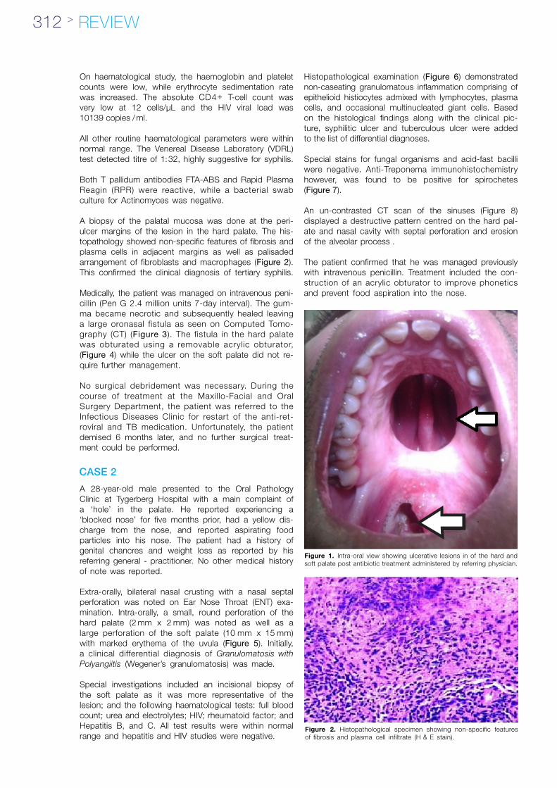
On haematological study, the haemoglobin and platelet counts were low, while erythrocyte sedimentation rate was increased. The absolute CD4+ T-cell count was very low at 12 cells/μL and the HIV viral load was 10139 copies /ml.
All other routine haematological parameters were within normal range. The Venereal Disease Laboratory (VDRL) test detected titre of 1:32, highly suggestive for syphilis.
Both T pallidum antibodies FTA-ABS and Rapid Plasma Reagin (RPR) were reactive, while a bacterial swab culture for Actinomyces was negative.
A biopsy of the palatal mucosa was done at the peri- ulcer margins of the lesion in the hard palate. The his- topathology showed non-specific features of fibrosis and plasma cells in adjacent margins as well as palisaded arrangement of fibroblasts and macrophages (Figure 2). This confirmed the clinical diagnosis of tertiary syphilis.
Medically, the patient was managed on intravenous peni- cillin (Pen G 2.4 million units 7-day interval). The gum- ma became necrotic and subsequently healed leaving a large oronasal fistula as seen on Computed Tomo- graphy (CT) (Figure 3). The fistula in the hard palate was obturated using a removable acrylic obturator, (Figure 4) while the ulcer on the soft palate did not re- quire further management.
No surgical debridement was necessary. During the course of treatment at the Maxillo-Facial and Oral Surgery Department, the patient was referred to the Infectious Diseases Clinic for restart of the anti-ret-roviral and TB medication. Unfortunately, the patient demised 6 months later, and no further surgical treat- ment could be performed.
A 28-year-old male presented to the Oral Pathology Clinic at Tygerberg Hospital with a main complaint of a ‘hole’ in the palate. He reported experiencing a ‘blocked nose’ for five months prior, had a yellow dis- charge from the nose, and reported aspirating food particles into his nose. The patient had a history of genital chancres and weight loss as reported by his referring general - practitioner. No other medical history of note was reported.
Extra-orally, bilateral nasal crusting with a nasal septal perforation was noted on Ear Nose Throat (ENT) exa- mination. Intra-orally, a small, round perforation of the hard palate (2 mm x 2 mm) was noted as well as a large perforation of the soft palate (10 mm x 15 mm) with marked erythema of the uvula (Figure 5). Initially, a clinical differential diagnosis of Granulomatosis with Polyangiitis (Wegener’s granulomatosis) was made.
Special investigations included an incisional biopsy of the soft palate as it was more representative of the lesion; and the following haematological tests: full blood count; urea and electrolytes; HIV; rheumatoid factor; and Hepatitis B, and C. All test results were within normal range and hepatitis and HIV studies were negative.
Histopathological examination (Figure 6) demonstrated non-caseating granulomatous inflammation comprising of epithelioid histiocytes admixed with lymphocytes, plasma cells, and occasional multinucleated giant cells. Based on the histological findings along with the clinical pic- ture, syphilitic ulcer and tuberculous ulcer were added to the list of differential diagnoses.
Special stains for fungal organisms and acid-fast bacilli were negative. Anti-Treponema immunohistochemistry however, was found to be positive for spirochetes (Figure 7).
An un-contrasted CT scan of the sinuses (Figure 8) displayed a destructive pattern centred on the hard pal- ate and nasal cavity with septal perforation and erosion of the alveolar process .
The patient confirmed that he was managed previously with intravenous penicillin. Treatment included the con- struction of an acrylic obturator to improve phonetics and prevent food aspiration into the nose.
CASE 2
Figure 2. Histopathological specimen showing non-specific features of fibrosis and plasma cell infiltrate (H & E stain).
Figure 1. Intra-oral view showing ulcerative lesions in of the hard and soft palate post antibiotic treatment administered by referring physician.
REVIEW312 >
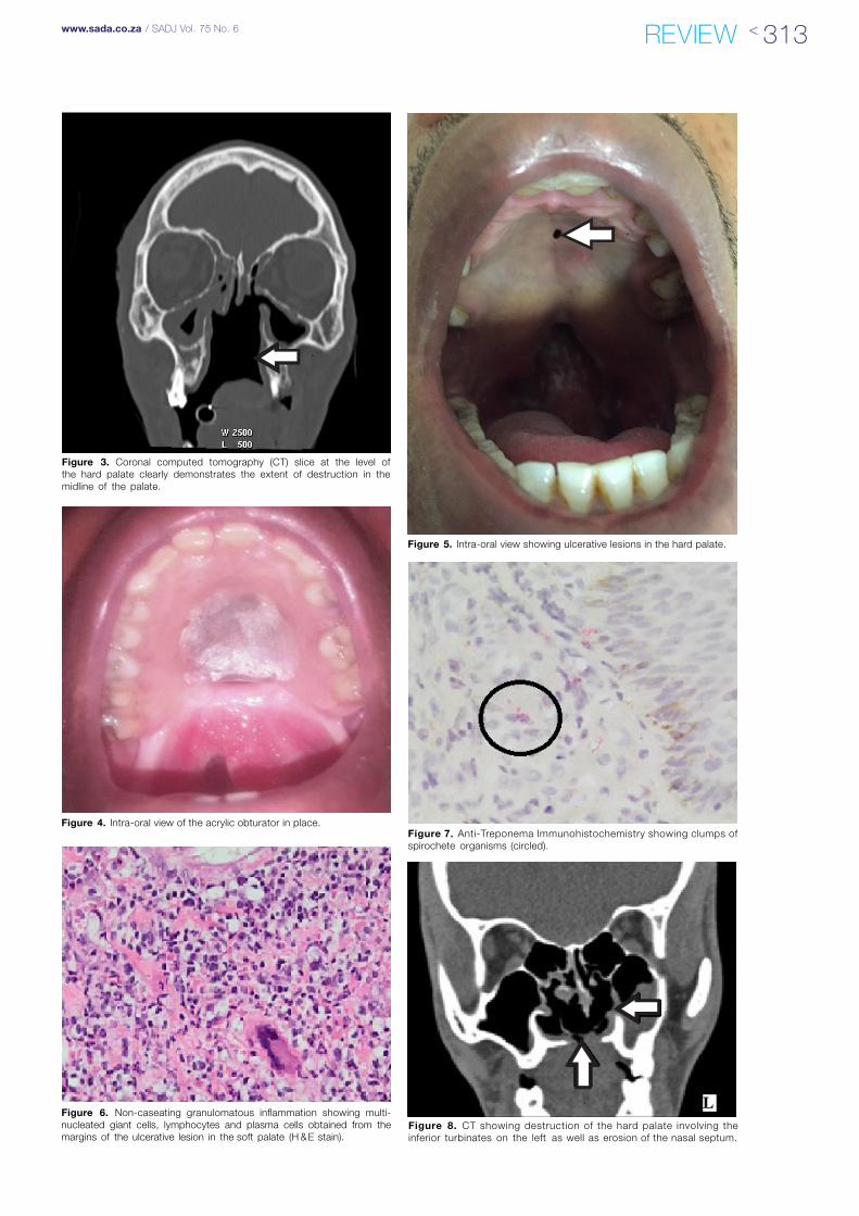
www.sada.co.za / SADJ Vol. 75 No. 6
Figure 4. Intra-oral view of the acrylic obturator in place.
Figure 3. Coronal computed tomography (CT) slice at the level of the hard palate clearly demonstrates the extent of destruction in the midline of the palate.
Figure 5. Intra-oral view showing ulcerative lesions in the hard palate.
Figure 6. Non-caseating granulomatous inflammation showing multi- nucleated giant cells, lymphocytes and plasma cells obtained from the margins of the ulcerative lesion in the soft palate (H &E stain).
Figure 7. Anti-Treponema Immunohistochemistry showing clumps of spirochete organisms (circled).
Figure 8. CT showing destruction of the hard palate involving the inferior turbinates on the left as well as erosion of the nasal septum.
REVIEW < 313
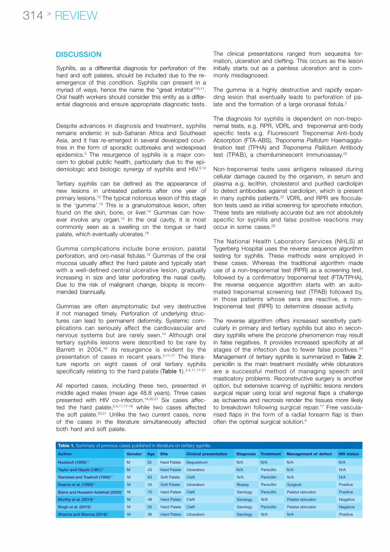
Syphilis, as a differential diagnosis for perforation of the hard and soft palates, should be included due to the re- emergence of this condition. Syphilis can present in a myriad of ways, hence the name the “great imitator”10,11. Oral health workers should consider this entity as a differ- ential diagnosis and ensure appropriate diagnostic tests.
Despite advances in diagnosis and treatment, syphilis remains endemic in sub-Saharan Africa and Southeast Asia, and it has re-emerged in several developed coun- tries in the form of sporadic outbreaks and widespread epidemics.3 The resurgence of syphilis is a major con- cern to global public health, particularly due to the epi- demiologic and biologic synergy of syphilis and HIV.3,12
Tertiary syphilis can be defined as the appearance of new lesions in untreated patients after one year of primary lesions.13 The typical notorious lesion of this stage is the ‘gumma’.13 This is a granulomatous lesion, often found on the skin, bone, or liver.14 Gummas can how- ever involve any organ.15 In the oral cavity, it is most commonly seen as a swelling on the tongue or hard palate, which eventually ulcerates.16
Gumma complications include bone erosion, palatal perforation, and oro-nasal fistulas.14 Gummas of the oral mucosa usually affect the hard palate and typically start with a well-defined central ulcerative lesion, gradually increasing in size and later perforating the nasal cavity. Due to the risk of malignant change, biopsy is recom- mended biannually.
Gummas are often asymptomatic but very destructive if not managed timely. Perforation of underlying struc- tures can lead to permanent deformity. Systemic com- plications can seriously affect the cardiovascular and nervous systems but are rarely seen.14 Although oral tertiary syphilis lesions were described to be rare by Barrett in 2004,16 its resurgence is evident by the presentation of cases in recent years.2,11,17 The litera- ture reports on eight cases of oral tertiary syphilis specifically relating to the hard palate (Table 1).2,4,11,17-21
All reported cases, including these two, presented in middle aged males (mean age 48.8 years). Three cases presented with HIV co-infection.14,20,21 Six cases affec- ted the hard palate,2,4,11,17-19 while two cases affected the soft palate.20,21 Unlike the two current cases, none of the cases in the literature simultaneously affected both hard and soft palate.
The clinical presentations ranged from sequestra for- mation, ulceration and clefting. This occurs as the lesion initially starts out as a painless ulceration and is com- monly misdiagnosed.
The gumma is a highly destructive and rapidly expan- ding lesion that eventually leads to perforation of pa- late and the formation of a large oronasal fistula.2
The diagnosis for syphilis is dependent on non-trepo- nemal tests, e.g. RPR, VDRL and treponemal anti-body specific tests e.g. Fluorescent Treponemal Anti-body Absorption (FTA-ABS), Treponema Pallidum Haemagglu- tination test (TPHA) and Treponema Pallidum Antibody test (TPAB), a chemiluminescent immunoassay.22
Non-treponemal tests uses antigens released during cellular damage caused by the organism, in serum and plasma e.g. lecithin, cholesterol and purified cardiolipin to detect antibodies against cardiolipin, which is present in many syphilis patients.22 VDRL and RPR are floccula-tion tests used as initial screening for spirochete infection. These tests are relatively accurate but are not absolutely specific for syphilis and false positive reactions may occur in some cases.23
The National Health Laboratory Services (NHLS) at Tygerberg Hospital uses the reverse sequence algorithm testing for syphilis. These methods were employed in these cases. Whereas the traditional algorithm made use of a non-treponemal test (RPR) as a screening test, followed by a confirmatory treponemal test (FTA/TPHA), the reverse sequence algorithm starts with an auto- mated treponemal screening test (TPAB) followed by, in those patients whose sera are reactive, a non- treponemal test (RPR) to determine disease activity.
The reverse algorithm offers increased sensitivity parti- cularly in primary and tertiary syphilis but also in secon- dary syphilis where the prozone phenomenon may result in false negatives. It provides increased specificity at all stages of the infection due to fewer false positives.23
Management of tertiary syphilis is summarized in Table 2. penicillin is the main treatment modality while obturators are a successful method of managing speech and masticatory problems. Reconstructive surgery is another option, but extensive scarring of syphilitic lesions renders surgical repair using local and regional flaps a challenge as ischaemia and necrosis render the tissues more likely to breakdown following surgical repair.17 Free vascula- rised flaps in the form of a radial forearm flap is then often the optimal surgical solution.4
DISCUSSION
Table 1. Summary of previous cases published in literature on tertiary syphilis.
Author Gender Age Site Clinical presentation Diagnosis Treatment Management of defect HIV status
Huebsch (1955)18 M 33 Hard Palate Sequestrum N/A N/A N/A N/A
Taylor and Hipple (1961)19 M 43 Hard Palate Ulceration N/A Penicillin N/A N/A
Ramstad and Traaholt (1980)20 M 63 Soft Palate Cleft N/A Penicillin N/A N/A
Kearns et al. (1993)21 M 43 Soft Palate Ulceration Biopsy Penicillin Surgical Positive
Bains and Hosseini-Ardehali (2005)4 M 70 Hard Palate Cleft Serology Penicillin Palatal obturator Positive
Murthy et al. (2014)11 M 48 Hard Palate Cleft Serology N/A Palatal obturator Negative
Singh et al. (2015)2 M 55 Hard Palate Cleft Serology Penicillin Palatal obturator Negative
Sharma and Sharma (2016)17 M 36 Hard Palate Ulceration Serology N/A N/A Positive
REVIEW314 >
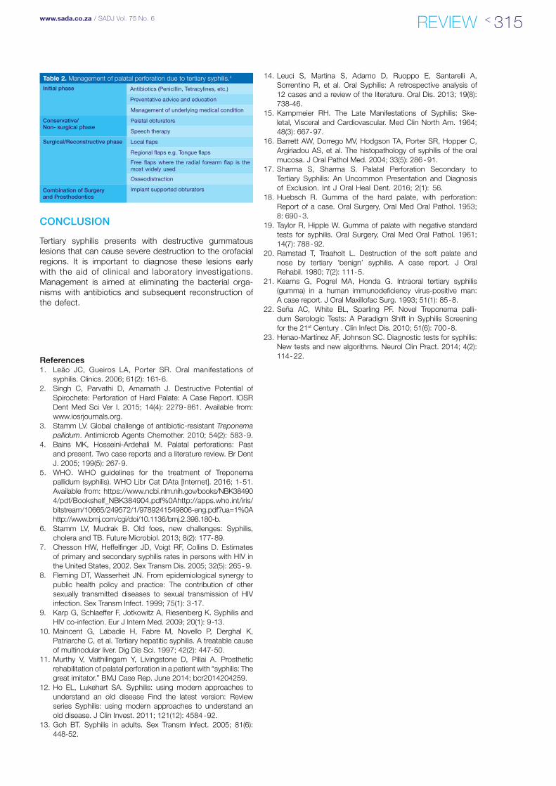
Tertiary syphilis presents with destructive gummatous lesions that can cause severe destruction to the orofacial regions. It is important to diagnose these lesions early with the aid of clinical and laboratory investigations. Management is aimed at eliminating the bacterial orga- nisms with antibiotics and subsequent reconstruction of the defect.
References1. Leão JC, Gueiros LA, Porter SR. Oral manifestations of
syphilis. Clinics. 2006; 61(2): 161-6.2. Singh C, Parvathi D, Amarnath J. Destructive Potential of
Spirochete: Perforation of Hard Palate: A Case Report. IOSR Dent Med Sci Ver I. 2015; 14(4): 2279-861. Available from: www.iosrjournals.org.
3. Stamm LV. Global challenge of antibiotic-resistant Treponema pallidum. Antimicrob Agents Chemother. 2010; 54(2): 583-9.
4. Bains MK, Hosseini-Ardehali M. Palatal perforations: Past and present. Two case reports and a literature review. Br Dent J. 2005; 199(5): 267-9.
5. WHO. WHO guidelines for the treatment of Treponema pallidum (syphilis). WHO Libr Cat DAta [Internet]. 2016; 1-51. Available from: https://www.ncbi.nlm.nih.gov/books/NBK38490 4/pdf/Bookshelf_NBK384904.pdf%0Ahttp://apps.who.int/iris/ bitstream/10665/249572/1/9789241549806-eng.pdf?ua=1%0A http://www.bmj.com/cgi/doi/10.1136/bmj.2.398.180-b.
6. Stamm LV, Mudrak B. Old foes, new challenges: Syphilis, cholera and TB. Future Microbiol. 2013; 8(2): 177-89.
7. Chesson HW, Heffelfinger JD, Voigt RF, Collins D. Estimates of primary and secondary syphilis rates in persons with HIV in the United States, 2002. Sex Transm Dis. 2005; 32(5): 265- 9.
8. Fleming DT, Wasserheit JN. From epidemiological synergy to public health policy and practice: The contribution of other sexually transmitted diseases to sexual transmission of HIV infection. Sex Transm Infect. 1999; 75(1): 3-17.
9. Karp G, Schlaeffer F, Jotkowitz A, Riesenberg K. Syphilis and HIV co-infection. Eur J Intern Med. 2009; 20(1): 9-13.
10. Maincent G, Labadie H, Fabre M, Novello P, Derghal K, Patriarche C, et al. Tertiary hepatitic syphilis. A treatable cause of multinodular liver. Dig Dis Sci. 1997; 42(2): 447-50.
11. Murthy V, Vaithilingam Y, Livingstone D, Pillai A. Prosthetic rehabilitation of palatal perforation in a patient with “syphilis: The great imitator.” BMJ Case Rep. June 2014; bcr2014204259.
12. Ho EL, Lukehart SA. Syphilis: using modern approaches to understand an old disease Find the latest version: Review series Syphilis: using modern approaches to understand an old disease. J Clin Invest. 2011; 121(12): 4584 -92.
13. Goh BT. Syphilis in adults. Sex Transm Infect. 2005; 81(6): 448-52.
14. Leuci S, Martina S, Adamo D, Ruoppo E, Santarelli A, Sorrentino R, et al. Oral Syphilis: A retrospective analysis of 12 cases and a review of the literature. Oral Dis. 2013; 19(8): 738-46.
15. Kampmeier RH. The Late Manifestations of Syphilis: Ske- letal, Visceral and Cardiovascular. Med Clin North Am. 1964; 48(3): 667-97.
16. Barrett AW, Dorrego MV, Hodgson TA, Porter SR, Hopper C, Argiriadou AS, et al. The histopathology of syphilis of the oral mucosa. J Oral Pathol Med. 2004; 33(5): 286 - 91.
17. Sharma S, Sharma S. Palatal Perforation Secondary to Tertiary Syphilis: An Uncommon Presentation and Diagnosis of Exclusion. Int J Oral Heal Dent. 2016; 2(1): 56.
18. Huebsch R. Gumma of the hard palate, with perforation: Report of a case. Oral Surgery, Oral Med Oral Pathol. 1953; 8: 690- 3.
19. Taylor R, Hipple W. Gumma of palate with negative standard tests for syphilis. Oral Surgery, Oral Med Oral Pathol. 1961; 14(7): 788- 92.
20. Ramstad T, Traaholt L. Destruction of the soft palate and nose by tertiary ‘benign’ syphilis. A case report. J Oral Rehabil. 1980; 7(2): 111-5.
21. Kearns G, Pogrel MA, Honda G. Intraoral tertiary syphilis (gumma) in a human immunodeficiency virus-positive man: A case report. J Oral Maxillofac Surg. 1993; 51(1): 85-8.
22. Seña AC, White BL, Sparling PF. Novel Treponema palli- dum Serologic Tests: A Paradigm Shift in Syphilis Screening for the 21st Century . Clin Infect Dis. 2010; 51(6): 700 -8.
23. Henao-Martínez AF, Johnson SC. Diagnostic tests for syphilis: New tests and new algorithms. Neurol Clin Pract. 2014; 4(2): 114- 22.
CONCLUSION
Table 2. Management of palatal perforation due to tertiary syphilis.4
Initial phase Antibiotics (Penicillin, Tetracylines, etc.)
Preventative advice and education
Management of underlying medical condition
Conservative/ Non- surgical phase
Palatal obturators
Speech therapy
Surgical/Reconstructive phase Local flaps
Regional flaps e.g. Tongue flaps
Free flaps where the radial forearm flap is the most widely used
Osseodistraction
Combination of Surgery and Prosthodontics
Implant supported obturators
www.sada.co.za / SADJ Vol. 75 No. 6 REVIEW < 315
Calcific metamorphosis (CM) or pulp canal obliteration (Fig. 1A-C) is a common occurrence following concus- sion and subluxation injuries.1,2 Although the exact me- chanism by which the canal obliterates is unknown, it is believed to be related to neurovascular damage and deposition of hard tissue within the canal.3,4
This calcification of the pulp canal space results in a loss of translucency leaving the crown with a yellow discolour- ation (Fig. 2 and 3).5 CM can be clinically detected as early as three months after injury but remains undetected in most cases for up to a year after trauma.6-8
Asymptomatic teeth presenting with CM do not initially require treatment other than annual review.1,9 However, the pulp status within partially obliterated canals may eventually lead to apical pathology requiring treatment.10-14
Treatment protocols and exact intervention times for the treatment of CM remains controversial and a chal- lenge for even the skilled clinician.15-17
CM is a common outcome of trauma frequently asso- ciated with anterior teeth of young adults and is charac-terised by partial or total obliteration of the root canal.6,8,15
In a study by Holcomb and Gregory,15 881 participants were investigated for the presence of CM. Initial peri- apical radiographs were taken of the participant and re- peated four years later.
The survey revealed an incidence of partial or complete canal obliteration of 3.86% with initial examination. Only three (7.3%) of the 41 teeth developed periradicular rare- factions after the four-year follow-up. Over 79% of the participants could recall a traumatic dental injury between the age of 10 and 16 years of age. The authors of this study concluded that a significant correlation exists be- tween a prior traumatic dental injury and the incidence of CM. They also commented that associated periapical rarefaction is uncommon and that endodontic interven- tion should only be considered in rare circumstances. In a follow-up study by Andreasen,6 where 189 teeth with history of dental luxation were examined, the authors described CM as an accelerated deposition of dentine and did not advocate early endodontic intervention. Pulp canal obliteration was observed in 22% of luxated teeth over a mean observation period of 3.4 years. Only 7% of teeth became necrotic over the follow up time. A relationship was found between the incidence of pulp obliteration and the stage of root development. The inci- dence of CM was higher in teeth with incomplete root development.
Another study by Andreason et al.,18 showed a 5% in- cidence of CM in 637 permanent incisors after luxation injuries. Only 1% of these teeth developed pulpal necrosis when evaluated over a five-year period.
Stalhane and Hedegard19 conducted a long-term study on 76 teeth that showed CM following traumatic injuries. Follow-up examinations were done 3-21 years after in- juries. Of the 76 teeth, 12 teeth (16%) developed periapi- cal rarefaction over this examination period.
The stage of root development, time of trauma as well as type of traumatic injury were also recorded. The au- thors commented on the fact that the success of mo- dern endodontic treatment should be weighed against the incidence of developing periapical pathology when deciding on clinical treatment options. A study by Jacobsen and Kerekes,20 followed up on radiographic hard tissue changes of 122 teeth, 10-23 years post injury. Of the 36% of cases with partial and 64% of cases with complete canal obliteration, none of the partially obliterated canals became necrotic with
INTRODUCTION
Aetiology and Incidence
Author affiliations:1. Peet J van der Vyver: BChD, PG Dip Dent (Endo), PG Dip Dent
(Aesthet Dent), MSc, PhD (Pret), Department of Odontology, School of Dentistry, University of Pretoria, Pretoria, South Africa.ORCID Number: 0000-0003-1951-6042
2. Martin Vorster: BChD, PG Dip Dent (Endo), MSc (Pret), De- partment of Odontology, School of Dentistry, University of Pretoria, Pretoria, South Africa.ORCID Number: 0000-0003-4470-1530
3. Casper H Jonker: BChD (Pret), Dip Odont (Endo), MSc (Pret), Department of Operative Dentistry, Sefako Magatho Health Sci- ences University, Ga-Rankuwa, South Africa. ORCID Number: 0000-0002-9110-5208
4. Nicoline Potgieter: BChD, PG Dip Dent (Endo), MSc (Pret), De- partment of Odontology, School of Dentistry, University of Pre- toria, Pretoria, South Africa. ORCID Number: 0000-0003-4061-3322
Corresponding author: Martin VorsterDepartment of Odontology, School of Dentistry, University of Pretoria, Gauteng, South Africa.Email: [email protected] Author contributions:1. Peet J van der Vyver: Treated patients, followed-up with patients,
scientific writing and proofreading of the manuscript - 25% 2. Martin Vorster: Scientific writing and proofreading of the manu-
script - 25% 3. Casper H Jonker: Scientific writing and proofreading of the
manuscript - 25%4. Nicoline Potgieter: Scientific writing and proofreading of the
manuscript - 25%
http://dx.doi.org/10.17159/2519-0105/2020/v75no6a5The SADJ is licensed under Creative Commons Licence CC-BY-NC-4.0.REVIEW316 >
PJ van der Vyver1, M Vorster2, CH Jonker3, N Potgieter4
SADJ July 2020, Vol. 75 No. 6 p316 - p322
Calcific Metamorphosis - A review of literature and clinical management
peri-apical rarefactions and only 13% of the complete obliterations eventually developed pulpal necrosis. The findings of this study strengthen the rationale behind performing endodontic treatment only when periradicular rarefactions develop.
In a study by Robertson et al.,21 82 permanent incisors with a history of trauma were followed up over a period of 7-22 years. Frequent yellow discolouration was seen and by evaluating periradicular bone loss, the authors reported that 8.5% of cases developed pulpal necrosis over this time.
The survival rate was found to be 84% after 20-years and it was reported that caries, new trauma, ortho- dontic treatment as well as crown coverage did not in- crease the frequency of pulpal necrosis. Rock and Grundy7 suggested a different approach to the previous studies and did a retrospective study in which 517 traumatised teeth were evaluated for the develop- ment of CM. They found that 16% of teeth developed CM. Although not statistically significant, CM was seen in the younger age groups (<9 years) while root resorp- tion was seen in the older age group (>9 years).
Most teeth presenting with canal obliteration are asymp- tomatic1,9,21 including the absence of sensitivity to per- cussion.10 CM is therefore often an incidental finding during clinical or radiographic investigations.1,9
CM is usually characterized by a dark yellow discolour- ation of the tooth.9,10 This distinct discolouration of the crown has been reported in 79% of 122 teeth with pulpal obliteration9,20 and may be attributed to the deposition of dark tertiary dentine.8,10,13,14
Oginni et al.1 reported that out of 276 teeth with partial canal obliteration, 186 (67%) had yellow discoloration and a further 34 (12%) teeth were grey in colour.
Interestingly, the incidence of pulp or periapical patho- logy were greater in the teeth with a grey discoloration compared to those with yellow discoloration.1 It is im- portant to note that teeth with radiographic signs of pulpal obliteration may also present without any colour change,9 and although colour change may indicate CM, it is not a necessarily an indication of pulpal or periapi- cal pathosis.1,9,20,21
In the presence of partial canal obliteration, it is generally accepted that vitality tests are unreliable.1,9,21,22 These teeth show a delayed or completely absent response to vitality tests.10,11,23 The response to vitality tests also tend to decrease as pulpal obliteration progresses.1,5,9,16
Clinical, radiographic and histological findings
Figure 2. Clinical view of the discoloured left maxillary central incisor.
Figure 3. Clinical view of the discoloured left maxillary central incisor taken with cross polarization photography resulting in no specular reflection. Note the severe change in hue and value of the affected tooth.
C. Sagittal slice of CBCT showing com- plete canal obliteration of the left max- illary central incisor.
B. Axial slice of CBCT at the level of CEJ showing com- plete canal obliteration of the left maxillary central incisor (arrow) compared to the other teeth in max- illary arch.
Figure 1.A. Periapical radiograph of a left maxillary
central incisor of a 27-year-old female patient that suffered a concussion in- jury at the age of 14 years. Note the canal obliteration in the coronal and apical third of the root canal (arrows), leaving a small amount of pulp tissue in the mid-root region.
Fig. 2
Fig. 3
Fig. 1
A BC
www.sada.co.za / SADJ Vol. 75 No. 6 REVIEW < 317
Generally, canal obliteration progresses in a corono-api-cal direction.10 A decrease in the pulp chamber volume is often the first radiographic sign, followed by a gra- dual narrowing of the complete root canal.10,23
Obliteration can be classified as either partial (coronal), or complete (extending radicular) obliteration of the pulp canal space.9,10 CM usually presents with a normal peri- odontal ligament space and intact lamina dura.11,14,23,24
Thickening of the periodontal ligament space or peri- radicula pathology may however be visible in the pre- sence of infection.9,10,14,24
Cone beam Computed Tomography (CBCT) may prove helpful in locating the root canals.10,24-26 The option to view canals in multiplanar reconstructions provides the clinician with valuable information in the diagnosis and management of CM.10,27
The absence of a root canal on conventional radio- graphs does not necessarily mean total absence of that canal.5,8,9,16 Histologic evaluation of pulp canals, radio- graphically diagnosed as being obliterated, almost always confirm the existence of a narrow pulp canal contain- ing pulpal tissue.10,12,14,23 Canal mineralization has there- fore been suggested as a more accurate term as opposed to canal obliteration.10,23,28
Pulp canal obliteration was initially described as an irre- gular tertiary dentine deposition, but more recently as multifocal, dystrophic calcifications composed of ill- defined secondary dentine.8,29-31 Calcifications have also been described by some authors as dentine-like, bone- like and fibrotic.10,32,33 Holan24 described calcificationsas tube-like osteo-dentine structures extending along the entire length of the pulp canal, surrounded by pulp tissue only connected to the root dentine at some areas.
In a study by Lundberg and Cvek,32 no micro-organisms were found in any of the specimens investigated. Inflammatory components, indicative of a pathologic process, are usually absent in teeth with CM.5,8,10,13,4,32,34 Based on these histopathological studies, asymptom-atic CM does not indicate the necessity for root canal treatment.10,14,18,32
The dark appearance of teeth affected by CM is not only a huge aesthetic concern but also presents the clinician with a great treatment challenge. Recently, West35 sug- gested four potential treatment options for the res- toration of aesthetics in discoloured teeth affected by sclerosis of the root canal complex. The four treat- ment options with an example of a case report on op- tion one and two are described below.
Due to its conservative nature, several authors advocate that external or vital bleaching should be considered as a first treatment option.35-38 Greenwall37 described a vital tooth whitening technique for affected single teeth. The author advocated the use of 20% carbamide pe-
roxide gel in a modified conventional vital bleaching tray where windows are created adjacent to the dis- coloured tooth, on either side.
This modification will prevent the overflow of bleaching liquid to adjacent teeth which might result in uneven whitening. The disadvantage of this particular technique is that progress can be slow due to the nature of discol-oration and the final result might not be acceptable. An advantage of this treatment option is that patients experience limited or no sensitivity during the whitening procedure.37
Joiner39 suggested that using a low con-centration for a longer treatment time will reduce the negative effects (as a result of rapid diffusion of free radicals) of bleaching. Additionally, the prolonged use of bleaching products with a low concentration (for example 10-20% carba- mide peroxide) provides greater stability.39
Haywood and DiAngelis40 discussed two approaches when considering external bleaching as a treatment op- tion to improve aesthetics:
Tray bleaching: the authors advocated a horseshoe-shaped cast on which the tray is manufactured with no vestibule to allow proper adaptation of bleaching solution.
Single tooth bleaching tray: a non-scalloped, non-reser-voir tray modified and trimmed to fit a single discoloured tooth. Patients are provided with a single syringe bleach- ing solution.
A 29-year-old female presented with a history of trau- ma to her maxillary anterior teeth at the age of 13 and agenesis of her maxillary left and right lateral incisors (Fig. 4). Historically, orthodontic treatment was attempted to move the canines into the position of the laterals. Following orthodontic treatment the patient relapsed with a resultant non-aesthetic appearance.
The main concern was a yellow discoloration of both the maxillary canines and left central incisor (Fig. 4). The patient sought an immediate aesthetic solution as she was getting married in four weeks’ time. Radio- graphic examination revealed complete canal obliteration of the asymptomatic maxillary left central incisor (Fig. 5A), with vital and visible root canal systems on the canines (Fig. 5B).
It was decided to manage the case using the vital bleaching technique with 16% carbamide peroxide (Flash Take Home Whitening System, Whitesmile GmbH, Ger- many) for three weeks. The patient was instructed to bleach the discoloured teeth every day for 45 minutes and the rest of the arch was bleached every third day.
Figure 6 shows the result after vital bleaching and Figure 7 the immediate postoperative result after mini- mally invasive direct composite restorations to mo- dify the anatomical shape of the teeth and to close the diastema between the 21 and 23.
Treatment options
1. Vital bleaching/External bleaching
CASE REPORT
REVIEW318 >
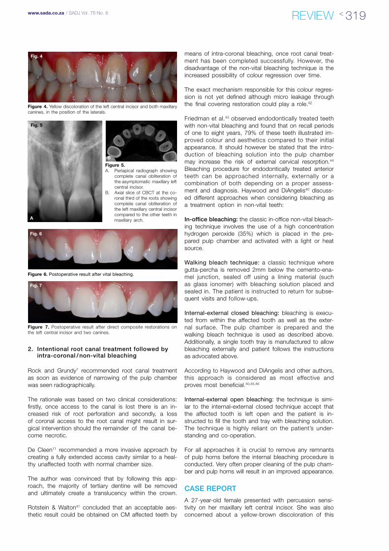
www.sada.co.za / SADJ Vol. 75 No. 6
Rock and Grundy7 recommended root canal treatment as soon as evidence of narrowing of the pulp chamber was seen radiographically.
The rationale was based on two clinical considerations: firstly, once access to the canal is lost there is an in- creased risk of root perforation and secondly, a loss of coronal access to the root canal might result in sur- gical intervention should the remainder of the canal be- come necrotic.
De Cleen11 recommended a more invasive approach by creating a fully extended access cavity similar to a heal- thy unaffected tooth with normal chamber size.
The author was convinced that by following this app- roach, the majority of tertiary dentine will be removed and ultimately create a translucency within the crown.
Rotstein & Walton41 concluded that an acceptable aes- thetic result could be obtained on CM affected teeth by
means of intra-coronal bleaching, once root canal treat- ment has been completed successfully. However, the disadvantage of the non-vital bleaching technique is the increased possibility of colour regression over time.
The exact mechanism responsible for this colour regres- sion is not yet defined although micro leakage through the final covering restoration could play a role.42
Friedman et al.43 observed endodontically treated teeth with non-vital bleaching and found that on recall periods of one to eight years, 79% of these teeth illustrated im- proved colour and aesthetics compared to their initial appearance. It should however be stated that the intro- duction of bleaching solution into the pulp chamber may increase the risk of external cervical resorption.44 Bleaching procedure for endodontically treated anterior teeth can be approached internally, externally or a combination of both depending on a proper assess- ment and diagnosis. Haywood and DiAngelis40 discuss- ed different approaches when considering bleaching as a treatment option in non-vital teeth:
In-office bleaching: the classic in-office non-vital bleach- ing technique involves the use of a high concentration hydrogen peroxide (35%) which is placed in the pre- pared pulp chamber and activated with a light or heat source.
Walking bleach technique: a classic technique where gutta-percha is removed 2mm below the cemento-ena- mel junction, sealed off using a lining material (such as glass ionomer) with bleaching solution placed and sealed in. The patient is instructed to return for subse- quent visits and follow-ups.
Internal-external closed bleaching: bleaching is execu- ted from within the affected tooth as well as the exter- nal surface. The pulp chamber is prepared and the walking bleach technique is used as described above. Additionally, a single tooth tray is manufactured to allow bleaching externally and patient follows the instructions as advocated above.
According to Haywood and DiAngelis and other authors, this approach is considered as most effective and proves most beneficial.40,45,46
Internal-external open bleaching: the technique is simi- lar to the internal-external closed technique accept that the affected tooth is left open and the patient is in- structed to fill the tooth and tray with bleaching solution. The technique is highly reliant on the patient’s under- standing and co-operation.
For all approaches it is crucial to remove any remnants of pulp horns before the internal bleaching procedure is conducted. Very often proper cleaning of the pulp cham- ber and pulp horns will result in an improved appearance.
A 27-year-old female presented with percussion sensi- tivity on her maxillary left central incisor. She was also concerned about a yellow-brown discoloration of this
2. Intentional root canal treatment followed byintra-coronal /non-vital bleaching
CASE REPORT
Figure 4. Yellow discoloration of the left central incisor and both maxillary canines, in the position of the laterals.
Figure 5. A. Periapical radiograph showing
complete canal obliteration of the asymptomatic maxillary left central incisor.
B. Axial slice of CBCT at the co- ronal third of the roots showing complete canal obliteration of the left maxillary central incisor compared to the other teeth in maxillary arch.A
B
Fig. 5
Figure 6. Postoperative result after vital bleaching.
Fig. 6
Figure 7. Postoperative result after direct composite restorations on the left central incisor and two canines.
Fig. 7
Fig. 4
REVIEW < 319
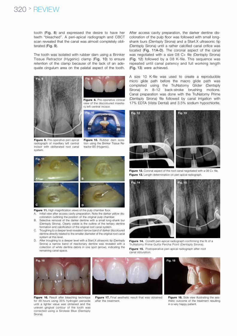
tooth (Fig. 8) and expressed the desire to have her teeth “bleached”. A peri-apical radiograph and CBCT scan revealed that the canal was almost completely obli- terated (Fig. 9).
The tooth was isolated with rubber dam using a Brinker Tissue Retractor (Hygenic) clamp (Fig. 10) to ensure retention of the clamp because of the lack of an ade- quate cingulum area on the palatal aspect of the tooth.
After access cavity preparation, the darker dentine dis- coloration of the pulp floor was followed with small long- shank burs (Dentsply Sirona) and a Start.X ultrasonic tip (Dentsply Sirona) until a rather calcified canal orifice was located (Fig. 11A-D). The coronal aspect of the canal was negotiated with a size 08 C+ file (Dentsply Sirona) (Fig. 12) followed by a 08 K- file. This sequence was repeated until canal patency and full working length (Fig. 13) were achieved.
A size 10 K-file was used to create a reproducible micro glide path before the macro glide path was completed using the TruNatomy Glider (Dentsply Sirona) in 8–12 back-stroke brushing motions. Canal preparation was done with the TruNatomy Prime (Dentsply Sirona) file followed by canal irrigation with 17% EDTA (Vista Dental) and 3.5% sodium hypochlorite.
Fig. 12 Fig. 13
Figure 12. Coronal aspect of the root canal negotiated with a 08 C+ file.Figure 13. Length determination on peri-apical radiograph.
Fig. 14 Fig. 15
Figure 14. Conefit peri-apical radiograph confirming the fit of aTruNatomy Prime Gutta Percha Point (Dentsply Sirona).
Figure 15. Postoperative peri-apical radiograph after root canal obturation.
Figure 16. Result after bleaching technique for 48 hours using 35% hydrogen peroxide until a lighter value was obtained and the uneven gingival contour of the tooth was corrected using a Sirolase Blue (Dentsply Sirona).
Figure 17. Final aesthetic result that was obtained after the treatment.
Figure 18. Side view illustrating the aes- thetic outcome of the treatment resulting in a very happy patient.
Fig. 16 Fig. 17 Fig. 18
Figure 8. Pre-operative clinical view of the discoloured maxilla- ry left central incisor.
Figure 9. Pre-operative peri-apical radiograph of maxillary left central incisor with obliterated root canal system.
Figure 10. Rubber dam isola- tion using the Brinker Tissue Re- tractor B5 (Hygenic).
Fig. 10
Fig. 9 Fig. 8
Figure 11. High magnification views of the pulp chamber floor. A. Initial view after access cavity preparation. Note the darker yellow dis-
coloration outlining the position of the original pulp chamber.B. Selective removal of the darker dentine with a small long-shank bur
(Dentsply Sirona). Clearly visible is the outline of the tertiary dentine formation and calcification of the original root canal system.
C. Troughing to a deeper level revealed narrow band of darker discoloured dentine directly related to the smaller diameter of the original root canal system at this level.
D. After troughing to a deeper level with a Start.X ultrasonic tip (Dentsply Sirona) a narrow band of reactionary dentine was revealed with a collection of white dentine debris in one spot (arrow), indicating the remaining canal space.
BA
D
Fig. 11
C
REVIEW320 >
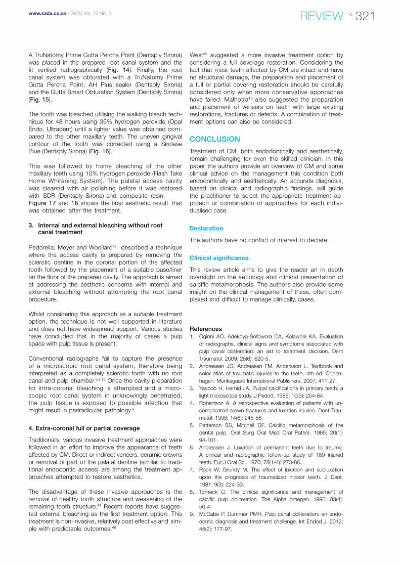
A TruNatomy Prime Gutta Percha Point (Dentsply Sirona) was placed in the prepared root canal system and the fit verified radiographically (Fig. 14). Finally, the root canal system was obturated with a TruNatomy Prime Gutta Percha Point, AH Plus sealer (Dentsply Sirona) and the Gutta Smart Obturation System (Dentsply Sirona) (Fig. 15).
The tooth was bleached utilising the walking bleach tech- nique for 48 hours using 35% hydrogen peroxide (Opal Endo, Ultradent) until a lighter value was obtained com- pared to the other maxillary teeth. The uneven gingival contour of the tooth was corrected using a Sirolase Blue (Dentsply Sirona) (Fig. 16).
This was followed by home bleaching of the other maxillary teeth using 10% hydrogen peroxide (Flash Take Home Whitening System). The palatal access cavity was cleaned with air polishing before it was restored with SDR (Dentsply Sirona) and composite resin.Figure 17 and 18 shows the final aesthetic result that was obtained after the treatment.
Pedorella, Meyer and Woollard47 described a technique where the access cavity is prepared by removing the sclerotic dentine in the coronal portion of the affected tooth followed by the placement of a suitable base/liner on the floor of the prepared cavity. The approach is aimed at addressing the aesthetic concerns with internal and external bleaching without attempting the root canal procedure.
Whilst considering this approach as a suitable treatment option, the technique is not well supported in literature and does not have widespread support. Various studies have concluded that in the majority of cases a pulp space with pulp tissue is present.
Conventional radiographs fail to capture the presence of a microscopic root canal system, therefore being interpreted as a completely sclerotic tooth with no root canal and pulp chamber.5,8,16 Once the cavity preparation for intra-coronal bleaching is attempted and a micro- scopic root canal system in unknowingly penetrated, the pulp tissue is exposed to possible infection that might result in periradicular pathology.9
Traditionally, various invasive treatment approaches were followed in an effort to improve the appearance of teeth affected by CM. Direct or indirect veneers, ceramic crowns or removal of part of the palatal dentine (similar to tradi- tional endodontic access) are among the treatment ap- proaches attempted to restore aesthetics.
The disadvantage of these invasive approaches is the removal of healthy tooth structure and weakening of the remaining tooth structure.44 Recent reports have sugges- ted external bleaching as the first treatment option. This treatment is non-invasive, relatively cost effective and sim- ple with predictable outcomes.48
West35 suggested a more invasive treatment option by considering a full coverage restoration. Considering the fact that most teeth affected by CM are intact and have no structural damage, the preparation and placement of a full or partial covering restoration should be carefully considered only when more conservative approaches have failed. Malhotra10 also suggested the preparation and placement of veneers on teeth with large existing restorations, fractures or defects. A combination of treat- ment options can also be considered.
Treatment of CM, both endodontically and aesthetically, remain challenging for even the skilled clinician. In this paper the authors provide an overview of CM and some clinical advice on the management this condition both endodontically and aesthetically. An accurate diagnosis, based on clinical and radiographic findings, will guide the practitioner to select the appropriate treatment ap- proach or combination of approaches for each indivi- dualised case.
The authors have no conflict of interest to declare.
This review article aims to give the reader an in depth oversight on the aetiology and clinical presentation of calcific metamorphosis. The authors also provide some insight on the clinical management of these, often com- plexed and difficult to manage clinically, cases.
References1. Oginni AO, Adekoya-Sofowora CA, Kolawole KA. Evaluation
of radiographs, clinical signs and symptoms associated with pulp canal obliteration: an aid to treatment decision. Dent Traumatol. 2009; 25(6): 620-5.
2. Andreasen JO, Andreasen FM, Andersson L. Textbook and color atlas of traumatic injuries to the teeth. 4th ed. Copen- hagen: Munksgaard International Publishers. 2007; 411-27.
3. Yaacob H, Hamid JA. Pulpal calcifications in primary teeth: a light microscope study. J Pedod. 1985; 10(3): 254-64.
4. Robertson A. A retrospective evaluation of patients with un- complicated crown fractures and luxation injuries. Dent Trau- matol. 1998; 14(6): 245-56.
5. Patterson SS, Mitchell DF. Calcific metamorphosis of the dental pulp. Oral Surg Oral Med Oral Pathol. 1965; 20(1): 94-101.
6. Andreasen J. Luxation of permanent teeth due to trauma. A clinical and radiographic follow-up study of 189 injured teeth. Eur J Oral Sci. 1970; 78(1-4): 273-86.
7. Rock W, Grundy M. The effect of luxation and subluxation upon the prognosis of traumatized incisor teeth. J Dent. 1981; 9(3): 224-30.
8. Torneck C. The clinical significance and management of calcific pulp obliteration. The Alpha omegan. 1990; 83(4): 50-4.
9. McCabe P, Dummer PMH. Pulp canal obliteration: an endo- dontic diagnosis and treatment challenge. Int Endod J. 2012; 45(2): 177-97.
3. Internal and external bleaching without root canal treatment
4. Extra-coronal full or partial coverage
CONCLUSION
Declaration
Clinical significance
www.sada.co.za / SADJ Vol. 75 No. 6 REVIEW < 321
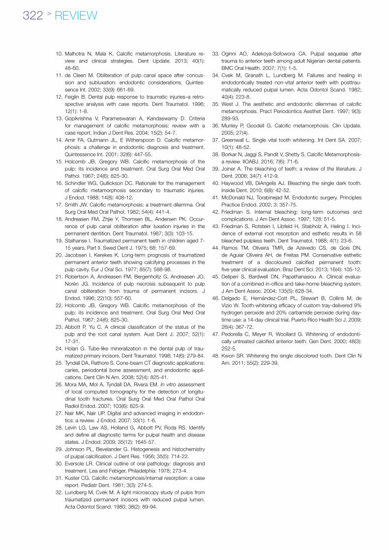
10. Malhotra N, Mala K. Calcific metamorphosis. Literature re- view and clinical strategies. Dent Update. 2013; 40(1): 48-60.
11. de Cleen M. Obliteration of pulp canal space after concus- sion and subluxation: endodontic considerations. Quintes- sence Int. 2002; 33(9): 661-69.
12. Feiglin B. Dental pulp response to traumatic injuries–a retro- spective analysis with case reports. Dent Traumatol. 1996; 12(1): 1-8.
13. Gopikrishna V, Parameswaran A, Kandaswamy D. Criteria for management of calcific metamorphosis: review with a case report. Indian J Dent Res. 2004; 15(2): 54-7.
14. Amir FA, Gutmann JL, E Witherspoon D. Calcific metamor-phosis: a challenge in endodontic diagnosis and treatment. Quintessence Int. 2001; 32(6): 447-55.
15. Holcomb JB, Gregory WB. Calcific metamorphosis of the pulp: its incidence and treatment. Oral Surg Oral Med Oral Pathol. 1967; 24(6): 825-30.
16. Schindler WG, Gullickson DC. Rationale for the management of calcific metamorphosis secondary to traumatic injuries. J Endod. 1988; 14(8): 408-12.
17. Smith JW. Calcific metamorphosis: a treatment dilemma. Oral Surg Oral Med Oral Pathol. 1982; 54(4): 441-4.
18. Andreasen FM, Zhjie Y, Thomsen BL, Andersen PK. Occur- rence of pulp canal obliteration after luxation injuries in the permanent dentition. Dent Traumatol. 1987; 3(3): 103-15.
19. Stalhanse I. Traumatized permanent teeth in children aged 7- 15 years, Part II. Swed Dent J. 1975; 68: 157-69.
20. Jacobsen I, Kerekes K. Long-term prognosis of traumatized permanent anterior teeth showing calcifying processes in the pulp cavity. Eur J Oral Sci. 1977; 85(7): 588-98.
21. Robertson A, Andreasen FM, Bergenholtz G, Andreasen JO, Norén JG. Incidence of pulp necrosis subsequent to pulp canal obliteration from trauma of permanent incisors. J Endod. 1996; 22(10): 557-60.
22. Holcomb JB, Gregory WB. Calcific metamorphosis of the pulp: its incidence and treatment. Oral Surg Oral Med Oral Pathol. 1967; 24(6): 825-30.
23. Abbott P, Yu C. A clinical classification of the status of the pulp and the root canal system. Aust Dent J. 2007; 52(1): 17-31.
24. Holan G. Tube-like mineralization in the dental pulp of trau- matized primary incisors. Dent Traumatol. 1998; 14(6): 279-84.
25. Tyndall DA, Rathore S. Cone-beam CT diagnostic applications: caries, periodontal bone assessment, and endodontic appli- cations. Dent Clin N Am. 2008; 52(4): 825-41.
26. Mora MA, Mol A, Tyndall DA, Rivera EM. In vitro assessment of local computed tomography for the detection of longitu- dinal tooth fractures. Oral Surg Oral Med Oral Pathol Oral Radiol Endod. 2007; 103(6): 825-9.
27. Nair MK, Nair UP. Digital and advanced imaging in endodon- tics: a review. J Endod. 2007; 33(1): 1-6.
28. Levin LG, Law AS, Holland G, Abbott PV, Roda RS. Identify and define all diagnostic terms for pulpal health and disease states. J Endod. 2009; 35(12): 1645-57.
29. Johnson PL, Bevelander G. Histogenesis and histochemistry of pulpal calcification. J Dent Res. 1956; 35(5): 714-22.
30. Eversole LR. Clinical outline of oral pathology: diagnosis and treatment. Lea and Febiger, Philadelphia: 1978; 273-4.
31. Kuster CG. Calcific metamorphosis/internal resorption: a case report. Pediatr Dent. 1981; 3(3): 274-5.
32. Lundberg M, Cvek M. A light microscopy study of pulps from traumatized permanent incisors with reduced pulpal lumen. Acta Odontol Scand. 1980; 38(2): 89-94.
33. Oginni AO, Adekoya-Sofowora CA. Pulpal sequelae after trauma to anterior teeth among adult Nigerian dental patients. BMC Oral Health. 2007; 7(1): 1-5.
34. Cvek M, Granath L, Lundberg M. Failures and healing in endodontically treated non-vital anterior teeth with posttrau-matically reduced pulpal lumen. Acta Odontol Scand. 1982; 40(4): 223-8.
35. West J. The aesthetic and endodontic dilemmas of calcific metamorphosis. Pract Periodontics Aesthet Dent. 1997; 9(3): 289-93.
36. Munley P, Goodell G. Calcific metamorphosis. Clin Update. 2005; 27(4).
37. Greenwall L. Single vital tooth whitening. Int Dent SA. 2007; 10(1): 48-52.
38. Borkar N, Jaggi S, Pandit V, Shetty S. Calcific Metamorphosis- a review. IIOABJ. 2016; 7(6): 71-6.
39. Joiner A. The bleaching of teeth: a review of the literature. J Dent. 2006; 34(7): 412-9.
40. Haywood VB, DiAngelis AJ. Bleaching the single dark tooth. Inside Dent. 2010; 6(8): 42-52.
41. McDonald NJ, Torabinejad M. Endodontic surgery. Principles Practice Endod. 2002; 3: 357-75.
42. Friedman S. Internal bleaching: long-term outcomes and complications. J Am Dent Assoc. 1997; 128: 51-5.
43. Friedman S, Rotstein I, Libfeld H, Stabholz A, Heling I. Inci- dence of external root resorption and esthetic results in 58 bleached pulpless teeth. Dent Traumatol. 1988; 4(1): 23-6.
44. Ramos TM, Oliveira TMR, de Azevedo CS, de Gois DN, de Aguiar Oliveira AH, de Freitas PM. Conservative esthetic treatment of a discoloured calcified permanent tooth: five-year clinical evaluation. Braz Dent Sci. 2013; 16(4): 105-12.
45. Deliperi S, Bardwell DN, Papathanasiou A. Clinical evalua- tion of a combined in-office and take-home bleaching system. J Am Dent Assoc. 2004; 135(5): 628-34.
46. Delgado E, Hernández-Cott PL, Stewart B, Collins M, de Vizio W. Tooth-whitening efficacy of custom tray-delivered 9% hydrogen peroxide and 20% carbamide peroxide during day- time use: a 14-day clinical trial. Puerto Rico Health Sci J. 2009; 26(4): 367-72.
47. Pedorella C, Meyer R, Woollard G. Whitening of endodonti- cally untreated calcified anterior teeth. Gen Dent. 2000; 48(3): 252-5.
48. Kwon SR. Whitening the single discolored tooth. Dent Clin N Am. 2011; 55(2): 229-39.
REVIEW322 >
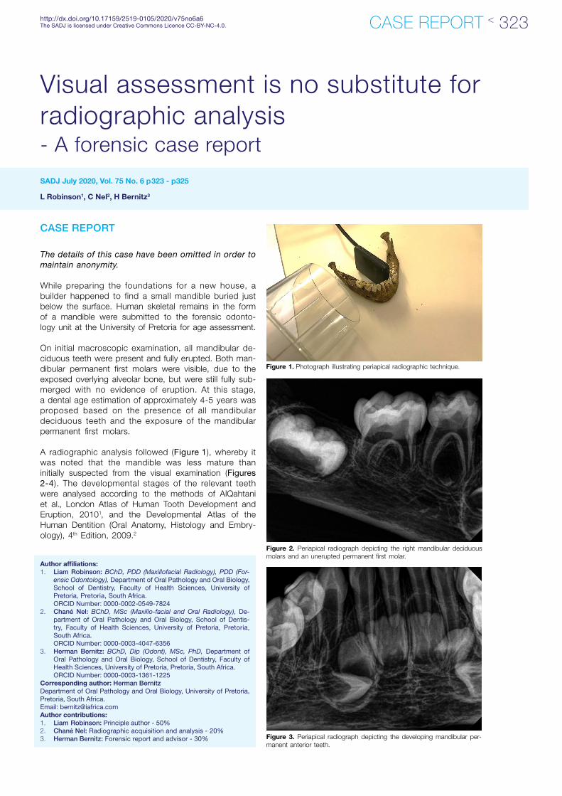
http://dx.doi.org/10.17159/2519-0105/2020/v75no6a6The SADJ is licensed under Creative Commons Licence CC-BY-NC-4.0.
The details of this case have been omitted in order to maintain anonymity.
While preparing the foundations for a new house, a builder happened to find a small mandible buried just below the surface. Human skeletal remains in the form of a mandible were submitted to the forensic odonto- logy unit at the University of Pretoria for age assessment.
On initial macroscopic examination, all mandibular de- ciduous teeth were present and fully erupted. Both man- dibular permanent first molars were visible, due to the exposed overlying alveolar bone, but were still fully sub- merged with no evidence of eruption. At this stage, a dental age estimation of approximately 4-5 years was proposed based on the presence of all mandibular deciduous teeth and the exposure of the mandibular permanent first molars.
A radiographic analysis followed (Figure 1), whereby it was noted that the mandible was less mature than initially suspected from the visual examination (Figures 2-4). The developmental stages of the relevant teeth were analysed according to the methods of AlQahtani et al., London Atlas of Human Tooth Development and Eruption, 20101, and the Developmental Atlas of the Human Dentition (Oral Anatomy, Histology and Embry- ology), 4th Edition, 2009.2
CASE REPORT
Figure 1. Photograph illustrating periapical radiographic technique.
Figure 2. Periapical radiograph depicting the right mandibular deciduous molars and an unerupted permanent first molar.
Figure 3. Periapical radiograph depicting the developing mandibular per- manent anterior teeth.
Author affiliations:1. Liam Robinson: BChD, PDD (Maxillofacial Radiology), PDD (For-
ensic Odontology), Department of Oral Pathology and Oral Biology, School of Dentistry, Faculty of Health Sciences, University of Pretoria, Pretoria, South Africa.ORCID Number: 0000-0002-0549-7824
2. Chané Nel: BChD, MSc (Maxillo-facial and Oral Radiology), De- partment of Oral Pathology and Oral Biology, School of Dentis- try, Faculty of Health Sciences, University of Pretoria, Pretoria, South Africa.ORCID Number: 0000-0003-4047-6356
3. Herman Bernitz: BChD, Dip (Odont), MSc, PhD, Department of Oral Pathology and Oral Biology, School of Dentistry, Faculty of Health Sciences, University of Pretoria, Pretoria, South Africa.ORCID Number: 0000-0003-1361-1225
Corresponding author: Herman BernitzDepartment of Oral Pathology and Oral Biology, University of Pretoria, Pretoria, South Africa.Email: [email protected] contributions:1. Liam Robinson: Principle author - 50% 2. Chané Nel: Radiographic acquisition and analysis - 20%3. Herman Bernitz: Forensic report and advisor - 30%
CASE REPORT < 323
L Robinson1, C Nel2, H Bernitz3
SADJ July 2020, Vol. 75 No. 6 p323 - p325
Visual assessment is no substitute for radiographic analysis- A forensic case report
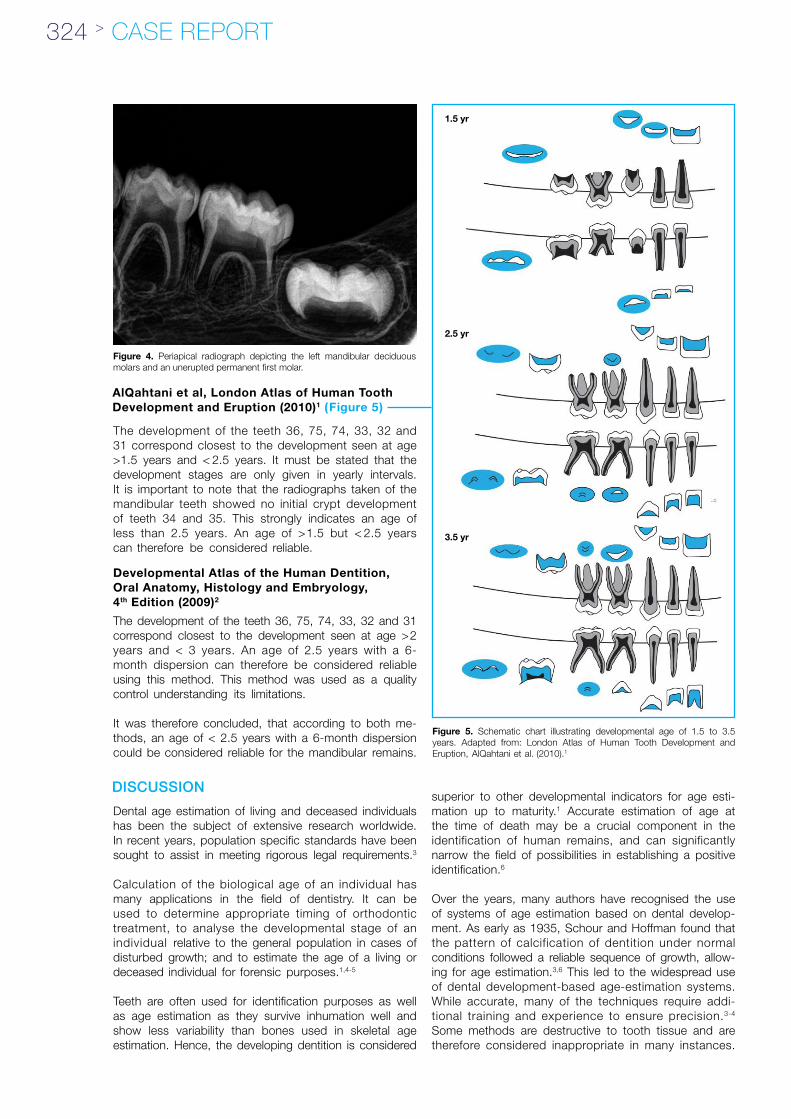
The development of the teeth 36, 75, 74, 33, 32 and 31 correspond closest to the development seen at age >1.5 years and < 2.5 years. It must be stated that the development stages are only given in yearly intervals. It is important to note that the radiographs taken of the mandibular teeth showed no initial crypt development of teeth 34 and 35. This strongly indicates an age of less than 2.5 years. An age of >1.5 but < 2.5 years can therefore be considered reliable.
The development of the teeth 36, 75, 74, 33, 32 and 31 correspond closest to the development seen at age >2 years and < 3 years. An age of 2.5 years with a 6- month dispersion can therefore be considered reliable using this method. This method was used as a quality control understanding its limitations.
It was therefore concluded, that according to both me- thods, an age of < 2.5 years with a 6-month dispersion could be considered reliable for the mandibular remains.
Dental age estimation of living and deceased individuals has been the subject of extensive research worldwide. In recent years, population specific standards have been sought to assist in meeting rigorous legal requirements.3
Calculation of the biological age of an individual has many applications in the field of dentistry. It can be used to determine appropriate timing of orthodontic treatment, to analyse the developmental stage of an individual relative to the general population in cases of disturbed growth; and to estimate the age of a living or deceased individual for forensic purposes.1,4-5
Teeth are often used for identification purposes as well as age estimation as they survive inhumation well and show less variability than bones used in skeletal age estimation. Hence, the developing dentition is considered
superior to other developmental indicators for age esti- mation up to maturity.1 Accurate estimation of age at the time of death may be a crucial component in the identification of human remains, and can significantly narrow the field of possibilities in establishing a positive identification.6
Over the years, many authors have recognised the use of systems of age estimation based on dental develop- ment. As early as 1935, Schour and Hoffman found that the pattern of calcification of dentition under normal conditions followed a reliable sequence of growth, allow- ing for age estimation.3,6 This led to the widespread use of dental development-based age-estimation systems. While accurate, many of the techniques require addi- tional training and experience to ensure precision.3-4 Some methods are destructive to tooth tissue and are therefore considered inappropriate in many instances.
AlQahtani et al, London Atlas of Human Tooth Development and Eruption (2010)1 (Figure 5)
Developmental Atlas of the Human Dentition, Oral Anatomy, Histology and Embryology, 4th Edition (2009)2
DISCUSSION
Figure 4. Periapical radiograph depicting the left mandibular deciduous molars and an unerupted permanent first molar.
Figure 5. Schematic chart illustrating developmental age of 1.5 to 3.5 years. Adapted from: London Atlas of Human Tooth Development and Eruption, AlQahtani et al. (2010).1
1.5 yr
1.5 yr2.5 yr
3.5 yr
CASE REPORT324 >
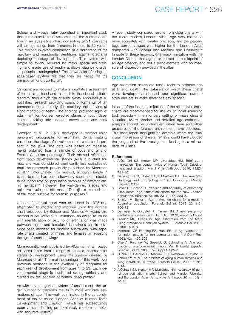
Schour and Massler later published an important study that summarised the development of the human denti- tion in an atlas-style chart consisting of 21 diagrams with an age range from 5 months in utero to 35 years.1
This method involved comparison of a radiograph of the maxillary and mandibular dentitions against diagrams depicting the stage of development. This system was simple to follow, required no major specialised train- ing, and made use of readily available diagnostic aids, i.e periapical radiographs.4 The drawbacks of using an atlas-based system are that they are based on the premise of ‘one size fits all’.
Clinicians are required to make a qualitative assessment of the case at hand and match it to the closest suitable diagram, thus a high risk of error exists. Moorrees et al. published research providing norms of formation of ten permanent teeth, namely, the maxillary incisors and all eight mandibular teeth. The findings provided ages of attainment for fourteen selected stages of tooth deve- lopment, taking into account crown, root and apex development.7
Demirjian et al., in 1973, developed a method using panoramic radiographs for estimating dental maturity based on the stage of development of each tooth pre- sent in the jaws. The data was based on measure- ments obtained from a sample of boys and girls of French Canadian parentage.5 Their method referenced eight tooth developmental stages (A-H) in a chart for- mat, and was considered significantly less complicated than the approach previously published by Moorrees et al.5,8 Unfortunately, this method, although simple in ts application, has been shown by subsequent studies to be inaccurate on population samples of different eth- nic heritage.6,9 However, the well-defined stages and objective evaluation still makes Demirjian’s method one of the most suitable for forensic purposes.9
Ubelaker’s dental chart was produced in 1978 and attempted to modify and improve upon the original chart produced by Schour and Massler.1,10 Again, this method is not without its limitations, as owing to issues with identification of sex, no differentiation was made between males and females.4 Ubelaker’s charts have since been modified for modern Australians, with sepa- rate charts created for males and females by adjusting the age of each drawing.4
More recently, work published by AlQahtani et al., based on cases taken from a range of sources, assessed for stages of development using the system devised by Moorrees et al.1 The main advantage of this work over previous methods is the availability of diagrams for each year of development from ages 1 to 23. Each de- velopmental stage is illustrated radiographically and clarified by the addition of written descriptions.1
As with any categorical system of assessment, the lar- ger number of diagrams results in more accurate esti- mations of age. This work culminated in the establish- ment of the so-called ‘London Atlas of Human Tooth Development and Eruption’, which has subsequently been validated using predominately modern samples with accurate results.3
A recent study compared results from older charts with the more modern London Atlas. Age was estimated more accurately with greater precision, and the percen- tage correctly aged was higher for the London Atlas compared with Schour and Massler and Ubelaker.10 In spite of these findings, one major limitation with the London Atlas is that age is expressed as a midpoint of an age category and not a point estimate with no mea- sure of dispersion provided in the atlas.
Age estimation charts are useful tools to estimate age at time of death. The datasets on which these charts were developed are based upon significant sample sizes and are in many instances sex specific.
In spite of the inherent limitations of the atlas style, these charts are recommended for use as an initial screening tool, especially in a mortuary setting or mass disaster situation. More precise and detailed age estimation analysis should be undertaken when time and other pressures of the forensic environment have subsided.4
This case report highlights an example where the initial visual impression of skeletal remains could have clouded the judgment of the investigators, leading to a miscar- riage of justice.
References1. AlQahtani SJ, Hector MP, Liversidge HM. Brief com-
munication: The London Atlas of Human Tooth Develop- ment and Eruption. Am J Phys Anthropol. 2010; 142(3): 481-90.
2. Berkovitz BKB, Holland GR, Moxham BJ. Oral Anatomy, Histology and Embryology. 4th ed. Edinburgh: Mosby/Elsevier; 2009.
3. Baylis S, Bassed R. Precision and accuracy of commonly used dental age estimation charts for the New Zealand population. Forensic Sci Int. 2017; 277: 223-8.
4. Blenkin M, Taylor J. Age estimation charts for a modern Australian population. Forensic Sci Int. 2012; 221(1-3): 106-12.
5. Demirjian A, Goldstein H, Tanner JM. A new system of dental age assessment. Hum Biol. 1973; 45 (2 ): 211- 27.
6. Blenkin MR, Evans W. Age estimation from the teeth using a modified Demirjian system. J Forensic Sci. 2010; 55(6): 1504-8.
7. Moorrees CF, Fanning EA, Hunt EE, Jr. Age variation of formation stages for ten permanent teeth. J Dent Res. 1963; 42: 1490-502.
8. Olze A, Reisinger W, Geserick G, Schmeling A. Age esti- mation of unaccompanied minors. Part II. Dental aspects. Forensic Sci Int. 2006; 159 Suppl 1: S65-7.
9. Cunha E, Baccino E, Martrille L, Ramsthaler F, Prieto J, Schuliar Y, et al. The problem of aging human remains and living individuals: A review. Forensic Sci Int. 2009; 193(1): 1-13.
10. AlQahtani SJ, Hector MP, Liversidge HM. Accuracy of den- tal age estimation charts: Schour and Massler, Ubelaker and the London Atlas. Am J Phys Anthropol. 2014; 154(1): 70-8.
CONCLUSION
www.sada.co.za / SADJ Vol. 75 No. 6 CASE REPORT < 325
Cutterguide : No Printing Process : Off set GD : SB32430 Size: 420 mm x 297 mm, Pages: 1 Colors : C M Y K (4 Colors) Native File :Adobe Indwsign CS6 Windows Generated in : Acrobat Distiller 10.0
With clinically proven Dual relief
No.1 DENTIST RECOMMENDED BRAND FOR SENSITIVE TEETH*
*GFK New expert performance tracking 2018.
For any product safety issues, please contact GSK on +27 11 745 6001 or 0800 118 274. Trademarks are own or licensed by the GSK group of companies.
& new
SENSITIVITY GUM
2019_SA_Sensodyne_Advert_A3Landscape_GSKCH_210_D1.indd 1 4/29/2019 4:05:24 PM
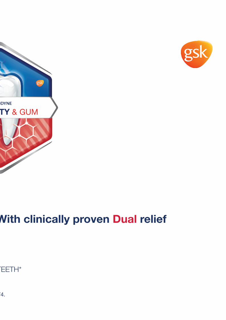
Self-adhesive restorative materials have been developed to simplify the restorative procedure by reducing the number of operative procedures and thus decreasing the number of possible errors from multiple steps such as inadequate acid etching in dentin, moisture conta- mination, etc.
The type of adhesive system used in the bonding of restorative material can contribute to marginal micro- leakage and sensitivity. To obtain adequate bonding, the smear layer, which is formed during cavity prepa- ration, must be treated or removed by adhesives.1 However, the effects of different adhesive systems vary widely both on the smear layer and in bonding quality. Biologically and technically, bonding mechanisms are different in etch-and-rinse and self-etch systems.1
In etch-and-rinse systems, the bonding mechanism is micromechanical and is based on the formation of a hybrid layer. In addition to micromechanical adhesion, diffusion and infiltration of resin within etched collagen fibrils are also effective in bonding to dentin.1 In self- etch systems that are easy to use, the bonding mecha- nism occurs by dissolving the smear layer and through penetration of acidic monomers into dentin to create a hybrid layer.1
The self-adhesive resin composite (SAC) acts simulta- neously as a self-etching adhesive and a flowable resin, thus eliminating the acid etching step and separate application of a bonding agent. de Oliveira and col-
leagues (2020)1 reported on a split mouth trial that sought to evaluate the post-operative sensitivity of res torations with self-adhesive resin composite (Vertise Flow) compared with conventional resin composite with self- etching adhesive (Filtek Z250 and Clearfil SE Bond).
The null hypothesis tested was that no difference would be found regarding the postoperative sensitivity between restorative techniques.
This study was a randomized, controlled, double-blind, split-mouth, two-arm clinical trial conducted in Brazil. Twenty-seven volunteers, aged between 18 and 40 years (mean 25.92 years) and of both sexes were recruited into this trial. A total of fifty-four third molars with an indication of extraction were included.
The study inclusion criteria were (1) two third molars in- dicated for extraction for orthodontic reasons; (2) heal- thy teeth without caries, score “0” according to the International Caries and Assessment System (ICDAS); (3) complete root development; and (4) fully erupted teeth.
Teeth without pulpal vitality or with altered pulpal vita- lity demonstrated using cold sensitivity tests, percussion, or palpation; the presence of pulpal calcification; and the impossibility of isolation with rubber dam were excluded.
A total of 54 restorations in 27 volunteers were performed by the same operator. Each participant received two restorations according – one was the test/experimental material and the other the control. The treatment allo- cation was done by the toss of a coin – “tails” meant that the tooth received a SAC whilst “heads” meant that the tooth restored using the conventional tech-
ABSTRACT
MATERIALS AND METHODS
1. Evaluation of postoperative sensitivity in restorations with self-adhesive resin - a randomized split-mouth design controlled study
NG de Oliveira, ASLC Lima, MT da Silveira, et al. Evaluation of postoperative sensitivity in res- torations with self-adhesive resin: a randomized split-mouth design controlled study. Clin Oral Invest. 2020; 24: 1829-35.
Veerasamy Yengopal: BChD, BScHons, MChD, PhD, Community Dentistry Department, School of Oral Health Sciences, University of Witwatersrand, Medical School, no. 7 York Road, Parktown 2193, South Africa. ORCID Number: 0000-0003-4284-3367Email: [email protected]
Compiled and edited by V Yengopal
SADJ July 2020, Vol. 75 No. 6 p328 - p332
What’s new for the clinician? - Excerpts from and summaries of recently published papers
http://dx.doi.org/10.17159/2519-0105/2019/v75no6a7 The SADJ is licensed under Creative Commons Licence CC-BY-NC-4.0.328 > CLINICAL WINDOW
nique with prior bonding procedure. Patients and eva- luators were not aware of the type of material used for each tooth.
One previously calibrated operator performed all restora- tions. The cavity preparation was standardized. The res- torative materials were applied according to the manu- facturers’ instructions. All photoactivation procedures were performed with halogen light (Ultralux) at a power density of 800 mW/cm2, previously measured with a radiometer.
After restoration placement, the occlusal contacts were evaluated with marking paper. The finishing and polish- ing were performed in the same session. Both resto- rations were done during the same clinical appointment.
Postoperative sensitivity was evaluated at 24 h and 15 and 30 days after the restorative procedure. Information on the presence of pain was collected (present or absent). If present, the characteristics of the pain were recorded: the localization of pain (localized or diffuse), the type of stimulus (triggered or spontaneous), the du- ration of pain (short or prolonged), its frequency (inter- mittent or continuous), and intensity of pain (mild, mo- derate or severe). Thermal stimulation (refrigerant spray Endo-Ice) was used to evaluate the type of stimulus. The pain intensity was recorded with the visual analog scale (VAS). The VAS consists of a 100 mm line divi- ded into equal intervals of 10mm, where 0 represents “absence of pain” and 100 “severe pain.” The results of VAS were classified as mild (0 -30 mm), moderate (40-70 mm), or severe pain (> 70 mm).
After each evaluation period, the patients had the tooth extracted as previously recommended.
Two patients with 4 third molars were excluded after enrolment because of lost to follow-up. The remaining 50 third molars of 25 patients (56% male vs. 44% female) were included. Regardless of the time inter- vals, postoperative sensitivity was observed in 52% and 48% of the conventional and self-adhesive resin composite (SAC) groups, respectively. No differences were observed between the postoperative sensitivity of the studied groups (p = 1.000) regardless of the time intervals.
None of the characteristics related to pain sensitivity, localization, type of stimulus, duration, frequency, and intensity demonstrated statistical differences between groups. Regarding the type of stimulus, triggered pain corresponded to 92.3% of the conventional group and 91.7% of the self-adhesive resin composite (SAC) group.
As for pain intensity, most was considered mild for both self-adhesive resin composite (SAC) group (75%) and conventional treatment group (76.9%). Moderate pain was observed in the conventional treatment group (23.1%), and severe pain (8.3%) was only observed in self-adhesive resin composite (SAC) group. No statis- tical differences were observed between groups.
All patients who had postoperative sensitivity reported that the pain was localized and of short duration with both materials.
When the evaluation period was considered for both groups, the 15-day time point presented the highest pain occurrence (87.5%) of mild intensity.
The authors found that Self-adhesive resin composite and conventional resin composite with a self-etch-ing bonding agent showed similar responses regarding postoperative sensitivity in deep class I cavities. When postoperative sensitivity was present, mild pain was observed in about half the participants; this decreased over time.
This study provides evidence that the self-adhesive resin composite performed similarly to the conventional resin composite with a self-etching bonding agent for the main outcome of postoperative sensitivity.
Reference1. de Oliveira NG, Lima ASLC, da Silveira MT, et al. Evaluation
of postoperative sensitivity in restorations with self-adhesive resin: a randomized split-mouth design controlled study. Clin Oral Invest. 2020; 24: 1829-35
RESULTS
CONCLUSION
Implications for practice
www.sada.co.za / SADJ Vol. 75 No. 6 < 329CLINICAL WINDOW
The success rates for implant therapy has been repor- ted to be well above 95% after a 10 year observation period in a number of published studies.1 The term “os- seointegration” was coined to describe the osseous bonding of the bone to implant and more recently this process has been referred to as “encapsulation” resulting from a foreign body reaction”. The quality and stability of the osseointegration (OI) or osseoencapsulation (OE) enables the implant to bear chewing forces.1
Implant stability is achieved at two levels—primary and secondary stability. Primary stability is mechanically ob- tained immediately after implant insertion as a result of friction between implant and bone, whereas secondary stability is achieved by deposition of mineral on the im- plant surface, beginning 2 weeks after surgery.1
Factors affecting primary stability are numerous and in- clude bone quality and quantity, different drilling proto- cols depending on bone density, surgical technique and surgeon’s skills, and implant and surface characteristics.1 The quality and timeline of secondary implant stability are influenced by surgical technique; surface character-istics of the implant such as roughness, hydrophilicity, and chemical modifications; and general health of the patient.1 Surfaces that attract water are termed hydro- philic, whereas surfaces that repel water are termed hy- drophobic. The degree to which a surface either attracts or repels water can be termed, respectively, the hydro- philicity or the hydrophobicity of that surface.
In vitro studies have demonstrated that cell differentiation and growth factor expression increase on hydrophilic implant surfaces. Moreover, animal studies indicated improved early soft and hard tissue integration of hydro- philic titanium implants. Some authors have demonstra- ted that titanium surfaces will be contaminated within 4 weeks and lose initial hydrophilicity.1
This effect is associated with the progressive deposition of hydrocarbons and a change of electrostatic charge from positive to negative. These effects have a negative impact on protein absorption capacity, osteoblast mi- gration, attachment, spread, and proliferation. Mineral- ization also decreases on 4-week-old titanium surfaces.
Application of ultraviolet (UV) light can regain hydrophili- city of titanium dioxide.1 In vitro and animal models in- vestigated the effect of photo-activation (PA) on fresh, aged, and photo-activation treated implant surfaces. PA was found to increase hydrophilicity, turns the electro- static charge to positive, and removed hydrocarbons from the surface.1
PA was also reported to have positive effects on alka- line phosphatase activity, calcium deposition spreading of human stem cells, protein absorption capacity, osteo- blast migration, attachment, spread, and proliferation.
Kumar and colleagues (2020)1 reported on a randomized controlled clinical trial that sought to investigate the in- fluence of photo-activation (PA) on implant stability using Removal torque (RT) at different time points as a sur- rogate outcome. RT is defined as the force which is necessary to detach an implant from the bone, thus indirectly providing information on the degree of bone- implant contact (BIC).
The null hypothesis stated that surface-treated implants (test group) will show the same deliberation force at specific time points as implants without surface treat- ment (control group).
In this single-center study, 180 partially edentulous pa- tients requiring two implants were selected and the po- sition of test and control implant was randomly assigned. Both implant locations had to be in the same jaw. Furthermore, the patients were randomly allocated to six groups. Randomization was performed using computer- generated random numbers, sealed in sequentially num- bered opaque envelopes.
One hundred eighty test implants and 180 control im- plants (N = 360) with a tapered design, a micro-tex- tured surface morphology in the neck area, and an internal connection (BioHorizons® Tapered Plus) were inserted epicrestally in single-stage surgery. Each pa- tient received one test and one control implant of the same diameter and length. The implants used had a diameter of 3.8, 4.6, or 5.8 mm and a length of 9.0, 10.5, 12.0, or 15.0 mm, respectively.
Patients, surgeon, and outcome assessors were blind- ed to the type of implant (test or control). Patients in good systemic health requiring (at least) two dental implants were included in the study.
Exclusion criteria were: • Patients unable to commit to the follow-up period. • Patients needing bone augmentation at implant place- ment.
• Post-extractive sites (implants were intended to be inserted after a healing period of at least 3 months).
• Patients with an acute infection or suppuration at the site intended for implant placement.
ABSTRACT
MATERIALS AND METHODS
2. Photo-activated implants: a triple-blinded, split-mouth, randomized controlled clinical trial on the resistance to removal torque at various healing intervals
A Puisys, M Schlee, T Linkevicius, P Petrakakis, A Tjaden. Photo-activated implants: a triple- blinded, split-mouth, randomized controlled clinical trial on the resistance to removal torque at various healing intervals. Clinical oral investigations. 2019 Sep; 11: 1-11.
330 > CLINICAL WINDOW
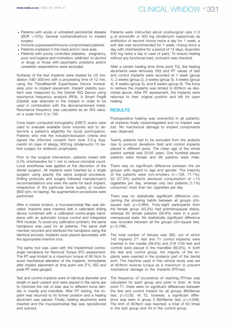
• Patients with acute or untreated periodontal disease (BOP > 15%); General contraindications to implant surgery.
• Immune-suppressed/immune-compromised patients. • Patients irradiated in the head and/or neck area. • Patients with poorly controlled diabetes, pregnancy, poor oral hygiene and motivation, addicted to alcohol or drugs or those with psychiatric problems and/or unrealistic expectations were excluded.
Surfaces of the test implants were treated by UV irra- diation (180 -300 nm) with a processing time of 12 min, using the TheraBeam® SuperOsseo Device immedi- ately prior to implant placement. Implant stability quo- tient was measured by the Osstell ISQ Device using resonance frequency analysis (RFA). A Smart Peg® (Osstell) was attached to the implant in order to be used in combination with the abovementioned meter. Resonance frequency was calculated as an ISQ value, on a scale from 0 to 100.
Cone beam computed tomography (CBCT) scans were used to evaluate available bone volumes and to de- termine a patient’s eligibility for study participation. Patients who met the inclusion/exclusion criteria and signed the informed consent form took 2.0-g Aug- mentin (in case of allergy, 600 mg clindamycin) 1h be- fore surgery for antibiotic prophylaxis.
Prior to the surgical intervention, patients rinsed with 0.2% chlorhexidine for 1 min to reduce microbial count. Local anesthesia was applied at the discretion of the dental surgeon. All implants were inserted by a single surgeon using exactly the same surgical procedure. Drilling protocols and surgery followed manufacturer’s recommendations and were the same for each implant, irrespective of the particular bone quality or location (800 rpm, no taping). No augmentation procedures were performed.
After a crestal incision, a mucoperiosteal flap was ele- vated. Implants were inserted with a calibrated drilling device combined with a calibrated contra-angle hand- piece with an automatic torque control and integrated RFA module. To avoid any calibration problem, the same handpiece was used for all patients. The same staff member recycled and sterilized the handpiece using the identical process. Implants were placed epicrestally with the appropriate insertion tool.
The same tool was used with the Implantmed contra- angle handpiece for Removal Torque (RT) assessment. The RT was limited to a maximum torque of 80 Ncm to avoid mechanical alteration of the implants. Immediately after implant placement at time point one (T1), ISQ and peak RT were gauged.
Test and control implants were of identical diameter and length in each patient and were placed in the same jaw to minimize the risk of bias due to different bone den- sity in maxilla and mandible. After RT testing, the im- plant was returned to its former position and a healing abutment was placed. Finally, healing abutments were inserted and the mucoperiosteal flap was repositioned and sutured.
Patients were instructed about postsurgical care (1.0 g of amoxicillin or 300 mg clindamycin respectively as antibiotics of second choice twice a day for 1 week, a soft diet was recommended for 1 week, rinsing twice a day with chlorhexidine for a period of 14 days, ibuprofen 400 mg twice a day in case of pain). To ensure healing without any functional load, occlusion was checked.
After a certain healing time (time point T2), the healing abutments were removed; ISQ and RT values of test and control implants were recorded at 1 week (group 1), 2 weeks (group 2), 3 weeks (group 3), 4 weeks (group 4), 6 weeks (group 5), and 8 weeks (group 6). The force to retrieve the implants was limited to 80Ncm as des- cribed above. After RT assessment, the implants were returned to their original position and left for open healing.
Postoperative healing was uneventful in all patients; all implants finally osseointegrated and no implant was lost. No mechanical damage to implant components was observed.
Twenty patients had to be excluded from the analysis due to protocol deviations (test and control implants placed in different jaws). The mean age of the whole patient sample was 50.65 years. One hundred eleven patients were female and 69 patients were male.
There was no significant difference between the six groups with regard to age and gender. The majority of the patients were non-smokers (n = 128, 71.1%); 50 (27.8%) patients declared consumption of ≤ 10 cigarettes per day, whereas only two patients (1.1%) consumed more than ten cigarettes per day.
There was no statistically significant difference con- cerning the smoking habits between all groups (chi- square test, p = 0.894). Forty-eight participants from the female group (43.2%) had premenopausal status, whereas 63 female patients (56.8%) were in a post- menopausal state. No statistically significant difference was recorded between all the groups (chi-square test, p = 0.786).
The total number of fixtures was 360, out of which 142 implants (71 test and 71 control implants) were inserted in the maxilla (39.4%) and 218 (109 test and control) were placed in the mandible (60.6%). In both the test and control group, the majority of the im- plants were inserted in the posterior part of the dental arch. The machine used in this clinical study was set at 80 Ncm reverse torque as a maximum to prevent mechanical damage to the implants (RTmax).
The frequency of occurrence of reaching RTmax was calculated for each group and point in time. At time point T1, there were no significant differences between the test and control implant for all groups (McNemar test, p ≥ 0.05). At T2, however, a significant differ- ence was seen in group 3 (McNemar test, p = 0.008). The limit of 80 Ncm was reached, a total of 63 times in the test group and 43 in the control group.
RESULTS
www.sada.co.za / SADJ Vol. 75 No. 6 < 331CLINICAL WINDOW
Comparing the amount of measurements above 80 Ncm between T1 and T2 for the test implant, there was a significant change for the test implant in groups 1, 2, and 4 (p < 0.05). In groups 3, 5, and 6, however, there was no significant change (p≥ 0.05).
Comparing the amount of RTmax values for the con- trol implants T1 versus T2, the statistical test was not applicable in groups 1, 2, and 5 as the tables were non-symmetrical. For groups 3 and 4, no significant changes were revealed. However, the amount of mea- surements above 80 Ncm was significant for group 6 (p < 0.001).
The statistical analysis showed that there is a strong correlation between the insertion torque and the re- moval torque at time point T1 for all groups and equally for test and control implants At the later time point (T2), there was only a small correlation between the original insertion torque (at T1) and the removal torque applied at T2 (R = 0.2, p < 0.05).
The authors concluded that Photo-activating (PA) the surface of titanium implants leads to higher resistance to RT forces compared with that of non-treated im- plants, showing higher implant stability especially in the early healing phase. Additionally, they found that Photo-activation results in an increased speed of osseointegration.
This trial has provided evidence that the use of PA leads to greater implant stability. The RT test seems to be a suitable method for the measurement of implant stability during the healing phase in humans.
Reference1. Puisys A, Schlee M, Linkevicius T, Petrakakis P, Tjaden A.
Photo-activated implants: a triple-blinded, split-mouth, ran- domized controlled clinical trial on the resistance to remo- val torque at various healing intervals. Clinical oral inves- tigations. 2019; Sep 11: 1-11.
CONCLUSION
Implications for practice
Do the CPD questionnaire on page 339The Continuous Professional Development (CPD) section provides for twenty general questions and five ethics questions. The section provides members with a valuable source of CPD points whilst also achieving the objective of CPD, to assure continuing education. The importance of continuing professional development should not be underestimated, it is a career-long obligation for practicing professionals.
1 Go to the SADA website www.sada.co.za.
2 Log into the ‘member only’ section with your unique SADA username and password.
3 Select the CPD navigation tab.
4 Select the questionnaire that you wish to complete.
5 Enter your multiple choice answers. Please note that you have two attempts to obtain at least 70%.
6 View and print your CPD certificate.
Online CPD in 6 Easy Steps
332 > CLINICAL WINDOW
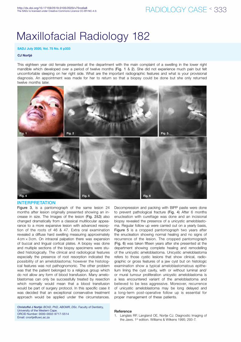
Figure 3, is a pantomograph of the same lesion 24 months after lesion originally presented showing an in- crease in size. The Images of the lesion (Fig. 2&3) also changed dramatically from a classical multilocular appea- rance to a more expansive lesion with advanced resorp- tion of the roots of 46 & 47. Extra oral examination revealed a diffuse hard swelling measuring approximately 4 cm × 3 cm. On intraoral palpation there was expansion of buccal and lingual cortical plates. A biopsy was done and multiple sections of the biopsy specimens were stu- died histologically. The clinical and radiological features especially the presence of root resorption indicated the possibility of an ameloblastoma; however the histolog-ical features was not pathognomonic. The other problem was that the patient belonged to a religious group which do not allow any form of blood transfusion. Many amelo- blastomas can only be successfully treated by resection which normally would mean that a blood transfusion would be part of surgery protocol. In this specific case it was decided that an exceptional conservative treatment approach would be applied under the circumstances.
Decompression and packing with BIPP paste were done to prevent pathological fracture (Fig. 4) After 6 months enucleation with curettage was done and an incisional biopsy revealed the presence of a unicystic ameloblasto-ma. Regular follow up were carried out on a yearly basis, Figure 5 is a cropped pantomograph two years after the enucleation showing normal healing and no signs of recurrence of the lesion. The cropped pantomograph (Fig. 6) was taken fifteen years after she presented at the department showing complete healing and remodelling of the unicystic ameloblastoma. Unicystic ameloblastoma refers to those cystic lesions that show clinical, radio- graphic or gross features of a jaw cyst but on histologic examination show a typical ameloblastomatous epithe- lium lining the cyst cavity, with or without luminal and/or mural tumour proliferation unicystic ameloblastoma is a less encountered variant of the ameloblastoma and believed to be less aggressive. Moreover, recurrence of unicystic ameloblastoma may be long delayed and a long-term post-operative follow up is essential for proper management of these patients.
Reference1. Langlais RP, Langland OE, Nortje CJ. Diagnostic Imaging of
the Jaws. 1st edition. Williams & Wilkens 1995; 293-7.
INTERPRETATION
This eighteen year old female presented at the department with the main complaint of a swelling in the lower right mandible which developed over a period of twelve months (Fig. 1 & 2). She did not experience much pain but felt uncomfortable sleeping on her right side. What are the important radiographic features and what is your provisional diagnosis. An appointment was made for her to return so that a biopsy could be done but she only returned twelve months later.
Fig. 1
Fig. 4 Fig. 5 Fig. 6
Fig. 2 Fig. 3
Christoffel J Nortjé: BChD, PhD, ABOMR, DSc. Faculty of Dentistry, University of the Western Cape.ORCID Number: 0000-0002-9717-5514Email: [email protected]
Maxillofacial Radiology 182
RADIOLOGY CASEhttp://dx.doi.org/10.17159/2519-0105/2020/v75no6a8 The SADJ is licensed under Creative Commons Licence CC-BY-NC-4.0.
< 333
CJ Nortjé
SADJ July 2020, Vol. 75 No. 6 p333
Virtue ethics, an ethical theory that focuses on concepts of how to live a good life and the characteristic traits or virtues of individuals, has enjoyed increasing popularity and considerable re-emergence in ethical assessment in various healthcare disciplines.
This popularity is, in part, attributable to its contrasting approach to the more formalized rule-based approaches to ethical assessment in healthcare practice.
The formalization of ethical healthcare practice (presen- ted as the four principles of autonomy, beneficence, non-maleficence and justice) coupled with various codes of ethics, has led to a considerable focus and reliance on rules and obligations.
A significant limitation of this type of approach arises from the difficulty in choosing which principle takes greater priority, in cases in which two or more princi- ples are in conflict. What is more, in this rule-oriented approach, motivations of individuals, characteristic traits and relationships, which are important aspects in ethical deliberations, have taken a back seat.
Against this background we describe and propose the incorporation of a virtue ethics approach, with a focus on the character and motivations of the individual or agent, as part of ethical deliberations in the practice of dentistry.
Traditionally, ethical analysis in the practice of medi- cine has been approached from a rule-based viewpoint. This has encouraged an action-oriented ethical analysis, often to the detriment of character, professional con- duct and judgement of healthcare practitioners.
It is now widely acknowledged that not everything that is important in professional life can be captured by codes of conduct and that professionalism goes beyond ethical principles.1
Similarly, the prevailing approach in discussions per- taining to ethical issues that arise in the context of dental practice is principle-based, otherwise known as rule-based.
The principle-based approach guides ethical reason- ing by focusing on what actions a moral agent should take. Aspects of internal motivations, character of the healthcare practitioner and professional judgement do not form part of this rule-based approach.
This is a shortcoming since these aspects assist the healthcare practitioner to “[interpret] the principles, [select] the ones to apply or ignore, [put] them in order of priority, and [shape] them in accord to his life history and current life situation”.2 Furthermore, virtue ethics also makes room for the incorporation of emo- tions as an “integral and important part of our moral perception”.3
Given the essential role of these aspects in the moral decision-making process, it is surprising that so little attention focuses on these in dental ethics literature. We propose the incorporation of virtue ethics, as part of ethical reflections in the practice of dentistry in con- junction with the four principles. Dental practitioners would, in this way, be better equipped to address ethi- cal issues pertinent to dental practice.
For the purpose of this article, we limit our discussion to aspects of virtue ethics advanced by Aristotle, Alasdair MacIntyre and Edmund Pellegrino. Similarly, from the several virtues which have been proposed as being important in the healthcare context, we restrict our focus to the virtues advanced by Beauchamp and Childress; we argue that these virtues have significant application to the dentist-patient relationship and assist in furthering the goals of the practice of dentistry.
ABSTRACT INTRODUCTION
Author affiliations:1. Hilde D Miniggio: BDS (UMF, Cluj-Napoca), MScMed (Bioethics
and Health Law) (Wits), PGDip (Health Sciences Education) (Wits)Senior Lecturer, Department of Oral Pathology and Oral Biology, Sefako Makgatho Health Sciences University, South Africa.
2. Pagollang D Motloba: BDS, MPH (Epid), MDent (Comm. Dent), MBL, Head, Department of Community Dentistry, School of Oral Health Sciences, Sefako Makgatho Health Sciences University, South Africa.ORCID Number: 0000-0003-1379-7576
3. Christopher S Wareham: BA Hons (Rhodes), MA (Rhodes), PhD (Milan), Senior Lecturer, Steve Biko Centre for Bioethics, School of Clinical Medicine, Faculty of Health Sciences, University of the Witwatersrand, South Africa.
Corresponding author: Hilde D MiniggioDepartment of Oral Pathology and Oral BiologySefako Makgatho Health Sciences University, South AfricaEmail:[email protected] contributions:1. Hilde D Miniggio: Conceptualization, first draft, revision, final
write-up and final approval - 60%2. Pagollang D Motloba: First draft, revision, final write-up and final
approval - 20%3. Christopher S Wareham: First draft, revision, final write-up and
final approval - 20%
http://dx.doi.org/10.17159/2519-0105/2020/v75no6a9 The SADJ is licensed under Creative Commons Licence CC-BY-NC-4.0.
The renaissance of virtue ethics and its application in dentistry
ETHICS334 >
HD Miniggio1, PD Motloba2, CS Wareham3
SADJ July 2020, Vol. 75 No. 6 p334 - p338
We begin with an exploration of Aristotle’s notions of virtue ethics and Alasdair MacIntyre’s concept of a practice with goods specific to it. We then briefly explore how virtues are linked to those goods. We also evaluate Edmund Pellegrino’s concepts of a morality that is inherent to healthcare practice with a focus on the goals specific to healthcare practice. An exploration of these concepts is significant in understanding virtue ethics theory as it was originally intended, and under- standing how these original concepts have subsequent- ly been applied in ethical deliberations in healthcare.
“When it comes to moral theories, virtue ethics is the old new kid on the block”.4 Our exploration of virtue ethics begins with the ‘old’ or original concepts of eudaimonia, doctrine of the mean and phronésis. For Aristotle, all human beings have a final purpose; not only, do all human beings have a final purpose but in fact, all human beings strive to achieve this final purpose.5 For Aristotle, this final purpose is to achieve a state of eudaimonia, generally translated as happiness or fulfillment.5
Aristotle proposes that eudaimonia or fulfillment is the highest good for human beings, and as such, it is chosen as an end in itself.5 He further considers that living a ful- filled life requires specific human excellences which he differentiates into intellectual excellences or wisdom (sophia) and practical excellences or wisdom (phronésis).5
A fulfilled life, in turn, is achieved by habitually exercising certain moral virtues. A moral virtue is an “acquired habit or disposition to do what is morally right or praiseworthy”.6
Aristotle suggests that virtues can be taught and that exercising a virtue occasionally does not make a virtuous agent, thus virtues must be exercised routinely through- out one’s life.5 For Aristotle, a moral virtue is a praise- worthy character trait that is found between two ex- tremes which can be explained by his concept of the doctrine of the mean.5 If we apply the doctrine of the mean to, for example the virtue of courage, we see that the virtue can be found between the two extremes; that of rashness (too much courage) and that of cowardice (too little courage).5 Practical wisdom (phronésis) then, is the virtue which assists the moral agent in choosing the appropriate action at the appropriate time.5 This virtue is often translated as prudence.
But how can a virtue theory developed 2400 years ago, have applicability in ethical assessment in modern day dentistry? We will attempt to answer this question by exploring notions of virtue ethics related to the practice of medicine as advanced by a couple of contemporary ethicists writing on the topic in the field.
A virtue in the context of a practice, such as the practice of medicine, can be described as an “acquired human quality, the possession and exercise of which tends to enable us to achieve those goods which are internal to practices and the lack of which effectively prevents us from achieving any such goods”.7
According to MacIntyre, a practice can be defined as “any coherent and complex form of socially established cooperative human activity, through which goods internal to that form of activity are realised in the course of trying to achieve those standards of excellence which are appropriate to, and partially definitive of, that form of activity, with the result that human powers to achieve excellence (virtue) and human conceptions of the ends and goods involved, are systematically extended”.7
Internal goods are in turn “defined by each particular practice and recognized by the experience of participat- ing in the practice in question”.7,8 The concept of inter- nal goods, refers to those goods which can only be achieved as part of being directly involved in that parti- cular practice; the concept of external goods, refers to those goods that can be achieved within the practice as well as outside of the practice or in many other types of practices or activities.7
Examples of external goods, are financial benefit and a certain standing in society.7 The internal goods in the practice of medicine are considered to be the doctor-pa-tient relationship and technical competence.7 MacIntyre considers that the internal goods in a practice can only be achieved through the exercise of certain virtues which lead to the attainment of those internal goods, specific to the practice.7
Pellegrino similarly applies the concept of practices to medicine in the particular sense that MacIntyre proposes, and considers that the practice of medicine is guided by specific goals that are particular to the practice. Pellegrino deems both principled-based ethics and virtue-based ethics as being inadequate, on their own, for ethical deli- berations in healthcare and advances the integration of both principles and character in such deliberations.2
Furthermore, he considers that the doctor-patient relation-ship is unique as it “centres on a vulnerable, anxious, dependent, often suffering individual person”.2 This relation-ship embodies a certain inherent morality to the practice of medicine and through this unique relationship the goals of medical practice are determined and achieved.2 For Pellegrino, the practice of medicine is a moral practice due to the “nature of illness, the non-proprietary nature of medical knowledge, and the nature and circumstances of a professional oath”.2 Pellegrino suggests that the goal of medical practice is “the good of the patient” and considers that both the doctor and the patient must jointly work towards this goal and that virtues lead to the realization of that goal.2
The practice of medicine, thus, flourishes most when virtue is pursued by a community of practitioners.2
Amongst the chief virtues, which date back to the time of Socrates, Aristotle and Plato and traditional Christian theology are temperance, courage, justice and prudence.
From these virtues, of particular relevance is the virtue of prudence which “emphasizes long term goals, good judgement in the face of uncertainty, and overcoming shortsighted choices” which “not only provides order for the principles, but also provides order for other virtues”.9
Virtue ethics
Virtue in practice
www.sada.co.za / SADJ Vol. 75 No. 6
The renaissance of virtue ethics and its application in dentistry
ETHICS < 335
While there is no specific nor exhaustive list of virtues applicable in healthcare practice, and several virtues have been proposed, the virtues that have particular relevance in healthcare practice have been designated by Beauchamp and Childress as, compassion, trustworthi-ness, integrity, discernment and conscientiousness.10
The continuous exercise of these virtues in the rela- tionship with patients leads to the development of a vir- tuous character; a virtuous character is characterized by Meara et al. as one who:
• is motivated to do what is good. • possesses vision and discernment. • realizes the role of affect or emotion in assessing or judging proper conduct.
• has a high degree of self-understanding and awareness. • is connected with and understands the mores of his or her community and the importance of community in moral decision making.11
Importantly, “virtuous agents are motivated to do what is right and have developed traits or dispositions or mo- tivations to act in accordance with high ethical standards or ideals”.11 Virtue ethics thus teaches us that we are all capable of becoming virtuous, by learning and rou- tinely practicing virtues, which will ultimately define our moral character.
We now apply MacIntyre’s concept of a practice and Pellegrino’s view of medical practice, which aims at a specific goal, in the context of dentistry. We also pro- pose that the virtues identified by Beauchamp and Childress as being essential in medical practice, are val- uable in ethical decision making in dentistry.
At this point we would like to point out that certain characteristic traits, or virtues, such as compassion and integrity, are mentioned in the ethics literature as being “core values of dentistry” and these are considered to “serve as the foundation of dental ethics”.12 However, these virtues are not presented, nor discussed within the broader context of a virtue ethics theory and as such, lose their intended meaning and application.
We now turn our attention to discuss dentistry as a practice as advanced by MacIntyre. Dentistry corre- sponds well with MacIntyre’s definition of a practice, namely “a coherent and complex form of socially es- tablished cooperative human activity” which leads to the attainment of certain goods internal to the practice.7
If we consider dentistry as a practice in the sense de- fined by MacIntyre and the goal of dental practice as being in line with the goal of medical practice, then the goal of dental practice can be considered as the good of the patient in relation to promoting, maintaining and restoring oral health. Furthermore, as a practice, den- tistry has internal goods, through which the goals of the practice are achieved. We suggest that the internal goods in the practice of dentistry in the sense that MacIntyre proposes, are technical competence and the dentist- patient relationship.
Patients are dependent on the technical expertise of dental practitioners for reasons ranging from pain relief to oral health rehabilitation and disease prevention.12
The dentist-patient relationship is a significant internal good of dental practice which hinges on trust. Trust is essential in this relationship, as patients accept certain physical intrusions by having portions of their bodies examined and treated by dental practitioners.12
They allow such intrusion “because they trust their den- tists to act in their best interests”.12 In other words, they trust the motivations of dental practitioners. Furthermore, as part of the consultation process, patients often dis- close personal and private information to the dental practitioner, and trust that this sensitive information will not be divulged to others.12
All these aspects render patients vulnerable and depen- dent on the character and motivations of the dental practitioners. Additionally, trust is important in the pa- tient-dentist relationship as dentists usually make use of their technical skills to earn a livelihood, thus there can be a “potential tension” between the commercial inte- rest of dentists and the best interest of the patient.12 The character of the dental practitioner safeguards against the possible exploitation of vulnerable patients.
We argue that a compassionate and trustworthy dental practitioner, who displays high levels of integrity, discern- ment and conscientiousness, will safeguard the trust that patients place in the dental practitioner as well as in the dental profession, and advance the good of the patient in relation to promoting, maintaining and restoring oral health.
In the sections to follow we explore these virtues in more detail and provide an overview of certain contexts in which these virtues can assist dental practitioners in the decision-making process.
Let us now consider how virtue ethics, could assist dental practitioners when faced with ethical challenges that arise in those cases where patients make inappro- priate treatment choices; choices which are clearly not in their best interest. Such inappropriate treatment choices may be less costly and less procedurally cumbersome in the short term, to the detriment of the oral health of the patient and increase in cost in the long term.
This can cause a difficult ethical dilemma and consi- derable frustration for the dental practitioner. In such cases, the application of the principle-based approach offers insufficient guidance. In trying to apply the princi- pled-approach to this scenario, we remark that the prin- ciple of autonomy (in this case, the right of the patient to make informed decisions regarding their oral health- care) is in conflict with the principle of beneficence (in this case, acting for the good of the patient). The question arises, should the dental practitioner choose to respect the patient’s autonomy or are considerations of beneficence more important? We note that, when faced with ethical dilemmas that arise from the conflict of two or more principles, the principle-based approach does not offer clear guidance as to how the conflict should
Virtue ethics in dentistry
ETHICS336 >
be solved. In such cases, the dental practitioner is then further required to make use of either intuition or reach out to other ethical theories in order to come to a resolution. In such an ethical dilemma, in which principles are at odds and the dental practitioner is uncertain which prin- ciple has greater importance, virtue ethics is helpful by encouraging a broader and more flexible evaluation of the situation.
From a virtue ethics standpoint, the virtue of compas- sion is one which refers to “an active regard for another’s welfare with an imaginative awareness and emotional response of deep sympathy, tenderness and discomfort at another’s misfortune or suffering”.10 In this scenario, the dental practitioner has an emotional response to the situation, seeing that the inappropriate treatment choice might negatively affect the oral health of the patient in the long term. Applying the virtue of compassion in this case assists the dental practitioner to respond with sympathy and active regard for the particular circum- stances of this case.
The virtue of compassion compels the dental practi-tioner to understand that, while it is ultimately the right of the patient to make informed decisions regarding their oral treatment, the patient may at times have diffi- culty with the treatment decision. Such treatment deci- sions might be, for example, based on irrational fears regarding certain procedures or previous bad experien- ces with similar procedures, or misconceptions regar- ding the consequences thereof. If the inappropriate treatment decisions are made due to such external fac- tors, the dental practitioner is in a position to help in alleviating such fears and misconceptions.
On the other hand, if the treatment choice is based solely on financial concerns, the virtue of compassion would compel the dental practitioner to have regard for the welfare of the patient and attempt to find creative solutions to balance the financial constraints with the good of the patient.
Compassion thus compels the dental practitioner to create a conducive environment in which a compre-hensive discussion and collaboration can take place. A compassionate response to this dilemma would also entail affording the patient time to reflect on his or her decision regarding treatment options. Such a compas- sionate response leads to a holistic interaction between the dental practitioner and the patient, in which the collaborative decision can be made and the goals of the practice achieved.
The virtue of trustworthiness is also valuable in this scenario. Trustworthiness is considered the “cornerstone of the doctor-patient relationship”.12 In this case building trust with the patient allows for meaningful collaboration in regards to common decision making that serves in the best interest of the patient.
Integrity refers to the “quality of being honest and fair”.13
Honesty and fairness represent essential components of the dental practitioner-patient relationship as patients
are dependent on the dental practitioner and in this vul- nerable position, patients become easily exploitable.
Honesty in this case would go a long way in managing patient expectations and being forthright about the outcomes and possible complications of the various treatment options.
The virtue of conscientiousness is “governed by or con- forming to the dictates of conscience”.13 In this case, conscientiousness leads to a concern regarding the consequences to the oral health of the patient as treat- ment options chosen by the patient might not in his or her best interest.
Discernment is the virtue that “brings sensitive insight, understanding and wise judgement to the situation”.13 Wise judgement and sensitive insight are important aspects that sensitizes the dental practitioner to the predicament that some patients might find themselves in when their oral healthcare choices are influenced by external factors.
The virtues of compassion, trustworthiness, integrity, dis- cernment and conscientiousness in this case can assist the dental practitioner in solving the conflict between the principles of autonomy and beneficence. Once the dilemma is viewed from a holistic standpoint which takes into account the individual needs of the patient coupled with the motivation to improve the oral health of the patient, a decision can be made in regards to which principle is more significant.
For instance, if the treatment decision is made out of fear of the treatment procedure, the compassionate dental practitioner can take steps to alleviate any un- founded fears, thereby acting in the interest of bene- ficence. If, however, the treatment option is chosen in the absence of external influences, and after ensuring that the patient understands the consequences there- of, the principle of autonomy takes preference.
The application of these aforementioned virtues can similarly be of value when, for example, dealing with ethi- cal issues that may arise from patient non-compliance and difficult patients, which can negatively impact on the oral health of these patients and thus on the goal of dental practice.
The development and continued exercise of these vir- tues advances the goals of dental practice and encour- ages professional judgement in the moral decision making process. Furthermore, these virtues can assist in balancing and choosing between competing principles in ethical issues that arise in dental practice. These virtues are significant in understanding the needs of individual patients and making use of sensible judge- ment when faced with conflicting ethical interests in dental practice.
A virtuous dental practitioner is able to identify and be sensitive to the various oral healthcare needs and cir- cumstances of individual patients, which goes a long way in improving patient satisfaction and treatment com- pliance thereby advancing the goals of dental practice.
www.sada.co.za / SADJ Vol. 75 No. 6 ETHICS < 337
In sum, virtue ethics is helpful in providing a more holistic and varied approach to ethical dilemmas encountered in dental practice. This is because the whole character of the dental practitioner takes center stage in this type of approach including the internal motivations.
Such an approach means that the dental practitioner “is not simply an inanimate observer in the process of ethical deliberation” but rather the “living body of ethi- cal practice”.4 Ultimately it is the character and motiva- tions of the dental practitioner that guides the ethical decision-making process.
Virtue ethics provides dental practitioners with an addi- tional tool that assists them when solving ethical dilem- mas for which the principle-based ethical approach canot provide an answer.
“Virtue ethics calls upon individuals to aspire towards ideals and to develop virtues or traits of character that enable them to achieve these ideals”.10 Virtue ethics recognizes the importance of the development of health- care practitioners’ character, which provides the foun- dation for professional judgement and ethical behavior.
Through an effective dentist-patient relationship based on trust and mutual collaboration, coupled with compe- tence in technical expertise, the goals of the practice of dentistry can be achieved. The incorporation of a virtue ethics approach provides dental practitioners an additio- nal tool for solving ethical issues in dentistry in con- junction with the four principles.
When faced with ethical dilemmas, dental practitioners can make use of the virtue ethics approach which high- lights the importance of character in the decision making process and incorporates the question, what type of dental practitioner should I be, as part of the ethical assessment. Such an approach makes room for the incorporation of virtues as part of the ethical assess- ment of issues pertaining to dental practice, which has, for many years been overlooked.
References1. Spielthenner G. Virtue-based Approaches to Professional
Ethics: a Plea for More Rigorous Use of Empirical Science. Ethic - Int J Moral Philos. 2017; 16(1): 15- 34.
2. Pellegrino ED, Thomasma DC. The Virtues in Medical Prac- tice. Oxford University Press 1993: 20-35.
3. Gardiner P. A virtue ethics approach to moral dilemmas in medicine. J Med Ethics. 2003; 29(5): 297.
4. Curzer HJ. Patients who make terrible therapeutic choices. J Am Coll Dent. 2014; 81(3): 41–5.
5. Aristotle. The ethics of Aristotle: the Nicomachean ethics. Harmondsworth: Penguin, 1976.
6. Beauchamp T, Childress J. Principles of biomedical ethics, 2nd
ed. New York: Oxford University Press, 1983.7. MacIntyre A. After virtue: a study of moral theory. Notre Dam:
University of Notre Dame Press, 1984.8. Stempsey WE. The virtuous pathologist. An ethical basis for
laboratory medicine. Am J Clin Pathol. 1989; 91(6): 730–8.9. Olivieri HM. Recta Ratio Agibilium in a medical context: the
role of virtue in the physician-patient relationship. Philos Ethics Humanit Med. 2018; 13(9).
10. Beauchamp T, Childress J. Principles of biomedical ethics, 6th ed. New York: Oxford University Press, 2008.
11. Meara NM, Schmidt LD, Day JD. Principles and virtues: A foundation for ethical decisions, policies, and character. Couns Psychol. 1996; 24(1): 4 -77.
12. Williams JR. Dental Ethics Manual. World Dental Federation, 2007. Available from: https://www.fdiworlddental.org/resour- ces/dental-ethics-manual.
13. Dictionary by Merriam-Webster: America’s most-trusted on- line dictionary (cited 2020 May 20). Available from: https://www.merriam-webster.com.
14. Koch K, Menezes CN. Virtue ethics: Beyond moral theory. South Afr J Bioeth Law. 2015; 26: 8(2): 48 -9.
CONCLUSION
ETHICS338 >
1. Identify the CORRECT answer. The following is/are determinants of university dropout and delayed graduation:A. Students’ characteristics, abilities and behaviourB. Parental background and family networksC. Characteristics of tertiary education system and
institutionsD. Labour market conditionsE. All of the above
2. Identify the CORRECT answer. In what year was Sefako Makgatho Health Sciences University (SMU) founded?A. 2004B. 2005C. 2006D. 2010E. None of the above
3. Identify the CORRECT answer. By what percentage did the number of dentists registered with HPCSA increase during the period 2002 to 2015?A. 1%B. 2%C. 5%D. 7%E. 10%
4. Identify the CORRECT answer. What were the in- struments used to conduct the study?A. Boley GaugeB. Vernier CalliperC. Carestream CS 3600D. None of the aboveE. All of the above
5. Identify the CORRECT answer. The standardised re- ference points used during the measuring process of the intermolar width were made on the?A. Mesiopalatal cuspB. Mesiobuccal cuspC. Distopalatal cuspD. Distobuccal cuspE. None of the above
6. Identify the CORRECT answer. A statistically signifi- cant difference was found when the measurements of the three instruments were evaluated for?A. Maxillary intercanine width (tooth 13 – 23)B. Maxillary intermolar width (tooth 16 – 26)C. Mesio-distal width of tooth 11D. Mesio-distal width of tooth 46E. Mesio-distal width of tooth 41
7. Identify the CORRECT statement. The luting space in a milled crown is:A. the space between the margin of the crown and
the margin of the tooth after cementationB. automatically created by the milling machineC. a space created on the inner surface of a full crown
for the luting cementD. added to the virtual preparation before designing
the crown
8. Identify the CORRECT statement The marginal gap in a full crown:A. should not occur after cementationB. can be up to 200μm in sizeC. is larger when measured in three dimensions rather
than in twoD. is the gap between the crown and the tooth be-
fore cementation
9. Identify the CORRECT statement. If a crown is seat- ed off-axis when cementing by seating it with a tilt to the buccal:A. There will be no effect on the marginal gapB. There are likely to be occlusal interferences after
cementing C. The marginal gap will be greater on the buccal
sideD. None of the above
10. Identify the CORRECT answer. Which of the follow- ing stages of Syphilis may present in the oral cavity?A. Primary SyphilisB. Secondary SyphilisC. Latent Syphilis D. Tertiary SyphilisE. All of the above
GENERAL
Throughputs of two cohorts of dental students at Sefako Makgatho Health Sciences University: A comparison
Comparison of three different instruments for orthodontic study model analysis
The effect of off-axis seating on the marginal adaptation of full coverage all ceramic crowns
Perforation of the palate - A report of two Syphilitic Gumma cases
CPD questionnaireThis edition is accredited for a total of 3 CEUs: 1 ethical plus 2 general CEUs
www.sada.co.za / SADJ Vol. 75 No. 6 CPD < 339
11. Identify the CORRECT answer. Clinical signs of Syphilis may include:A. presentations of ulceration (chancre) at the site of
infection, B. lymphadenopathy C. mucocutaneous rash D. Pathological fractureE. A, B and C only
12. Identify the CORRECT answer. Management of ter- tiary syphilis may include:A. Penicillin and tetracycline B. Palatal obturatorsC. Preventative advice and educationD. Management of underlying medical conditionE. All of the above
13. Identify the CORRECT answer. Teeth with Calcific Metamorphosis usually present with which of the following clinical or radiographic appearance?
A. Full or partial canal obliteration B. Percussion sensitivityC. Acute apical periodontitisD. No clinical or radiographic abnormalitiesE. All of the above
14. Identify the CORRECT answer. In which of the cases presenting with calcific metamorphosis is the inci- dence of periapical pathology higher?
A. Cases with a dark yellow discolorationB. Cases with a grey discoloration C. Cases with no colour changeD. Colour presentation has no influence on incidence
of periapical pathology
15. Identify the CORRECT answer. In which direction does calcification of root canal systems usually progress?A. From the largest part of canal to the canal extremitiesB. From apical to coronalC. Simultaneously from both sidesD. From coronal to apical
16. Identify the CORRECT statement. The Ulin et al. trial can be consider as “quasi-randomized” because of one of the following statements:A. The trail was single blindedB. There was a small sample sizeC. If the patient’s first visit was on an even-num
bered date, the concentration was 0.5%; if the visit was on an odd-numbered date, the concentration of the irrigant was 3%
D. The trial was double-blinded
17. Identify the CORRECT statement. In the Ulin et al. trial: what were the outcomes for bacterial levels and/or postoperative pain among the groups? A. There was a difference in bacterial levels or and
postoperative pain among the groupsB. There was only in difference in the bacterial levels
between the groupC. There was only a difference in the postoperative
pain levels between the groupsD. There was no difference in bacterial levels or post-
operative pain among the groups
18. Identify the CORRECT statement. The following factor(s) can have an influence on the dental developmental stage:
A. SexB. EthnicityC. Developmental disturbancesD. AgeE. All of the above
19. Identify the CORRECT answer. In the de Oliveira et al. trial, for the outcome postoperative sensitivity:A. The Self-adhesive resin composite group perfor-
med betterB. Conventional resin composite group with a self-
etching bonding agent has less sensitivityC. Both groups had similar sensitivity scoresD. None of the above
20. Identify the CORRECT statement. In the The Puisys et al. trial, Photo-activating (PA) the surface of tita- nium implants led to:A. higher resistance to Removal Torque forces com-
pared with that of non-treated implantsB. hower resistance to Removal Torque forces com-
pared with that of non-treated implantsC. higher resistance to Removal Torque forces com-
pared with that of PA treated implantsD. lower resistance to Removal Torque forces com-
pared with that of PA treated implants
21. Identify the CORRECT statement. Identification using dental records: A. cannot be used for badly burnt bodiesB. cannot be used in mass disaster situationsC. cannot be used for murder victimsD. may make use of hand written notes
22. Identify the CORRECT statement. Internal goods in healthcare refers to: A. financial benefitB. a high standing in societyC. the doctor-patient relationshipD. prestige
Calcific Metamorphosis: A review of literature and clinical management
Visual assessment is no substitute for radiographic analysis - A forensic case report
Clinical Window: What’s new for the clinician?
The renaissance of virtue ethics and its application in dentistry
ETHICS
CPD340 >

23. Identify the CORRECT statement. The concept of eudaimonia, proposed by Aristotle refers to: A. practical wisdomB. courageC. compassionD. fulfillment
24. Identify the CORRECT statement. One of the virtues identified by Beauchamp and Childress as being essential in medical practice is:A. compassionB. courageC. humilityD. open mindedness
25. Identify the CORRECT statement. The principle-based approach focuses on:
A. the character of the moral agentB. the actions of the moral agentC. the internal motivations of the moral agentD. the virtues of the moral agent
The Continuous Professional Development (CPD) section provides for twenty general questions and five ethics questions. The section provides members with a valuable source of CPD points whilst also achieving the objective of CPD, to assure continuing education. The importance of continuing professional development should not be underestimated, it is a career-long obligation for practicing professionals.
1 Go to the SADA website www.sada.co.za.
2 Log into the ‘member only’ section with your unique SADA username and password.
3 Select the CPD navigation tab.
4 Select the questionnaire that you wish to complete.
5 Enter your multiple choice answers. Please note that you have two attempts to obtain at least 70%.
6 View and print your CPD certificate.
Online CPD in 6 Easy Steps
www.sada.co.za / SADJ Vol. 75 No. 6 CPD < 341
SADJT H E S O U T H A F R I C A N D E N TA L J O U R N A L
Thank you for considering the submission of your work to the Journal for possible publication. We welcome papers which may be Original Research, Clinical Review, Case Reports, Clinical Communications, Letters or Notes.
The South African Dental Journal (SADJ) is a peer reviewed, Open Access Journal, published by the South African Dental Association (SADA). The Journal primarily carries research articles which reflect oral and general health issues in Africa but also publishes papers covering the widest consideration of all health sciences. In addition, items of specific relevance to members of the Association find dissemination through the Journal. The Journal is published ten times each year in electronic format. Hard copy is available by arrangement.
We shall be obliged if your submission is prepared respecting all the details listed in these Instructions. This facilitates our process and ensures more rapid responses to you. Please use and submit the Checklist for Authors suplied on page 345 for confirmation. Thank you. Address for submission of articlesThe Editorial Assistant, Mr Dumi Ngoepe, South African Dental Journal, South African Dental Association (SADA), Private Bag 1, Houghton 2041, South Africa. Email addresses: [email protected] [email protected] submit the paper in electronic format in Word and Figures sparately in JPEG., accompanied by a covering letter signed by the author(s).
LanguageAll articles must be submitted in English. Spelling should be in accord with the Shorter Oxford English Dictionary.All articles must be submitted in English. Spelling should be in accord with the Shorter Oxford English Dictionary.
Clinical ResearchArticles should adhere to the protocols of the Helsinki Declaration (https://www.wma.net/policies-post/wma-declaration-of-helsin-ki-ethical-principles-for-medical-research-involving-human-sub-jects/.
Clinical TrialsClinical trials should conform to the Consort Statement (Con- solidated Statements of Reporting Trials) and Reviews to the PRISMA checklist (Preferred Reporting Items for Systematic Reviews and Meta Analyses) (http://www.equator-network.org).
AuthorsAuthors should meet the criteria for authorship as in the documents of the International Committee of Medical Journal Editors (ICMJE): 1. Substantial contributions to the conception or design of the
work or the acquisition, analysis or interpretation of data for the work, AND
2. Drafting the work or revising it critically for important intellec-tual content, AND
3. Final approval of the version to be published, AND4. Agreement to be accountable for all aspects of the work in
ensuring that questions related to the accuracy or integrity of any part of the work are appropriately investigated and resolved (www.icmje.org).
• The front page of the manuscript should list the title of the article, the author’s(s’) name(s), and their qualification(s), affili-ations and positions held, telephone and fax numbers and address(es), including Email address(es), if available. It is especially important that details of the Corresponding Author should be clearly stated.
• Please submit on the front page a list of up to eight Keywords. • In the case of multiple authors, the role played and the
respective contribution made by each should be recorded. For example: “Principal Researcher- 40%, Writing Article- 30%, Tissue Analysis- 20%, Microscopic Examination- 10%”, etc.
• A recent requirement is that authors should be registered with ORCID. This is a number registering you as an Open Researcher and Contributor. Go to the ORCID website home page at https://orcid.org/ and follow the Three Easy Steps indicated in green. Please submit the ORCID number with your author details.
TitleTo be kept as brief, clear and unambiguous as possible.
AbstractThe abstract shall consist of not more than 200 words. For research articles, the summary should be structured under the following headings: Introduction, Aims and Objectives, Design, Methods, Results and Conclusions. Do not include references in the Abstract.
Text • Articles should be clear and concise.
• Text should be typed in Times New Roman font, size 11; double-spaced with a 3 cm. margin on the sides, top and bottom. Each page must be clearly numbered.
• Please include electronic numbering of lines throughout the document.
• Tables should be clearly identified, using Roman numerals ie. Table I, Table II etc.
• Authors are requested to note and adhere to the current style of the Journal particularly with respect to paragraph settings and headings.
Length of the articleIn general, papers should be between 4000 and 5000 words, although this is flexible. The Editor reserves the right to edit the length of an article in conjunction with the author(s) and SADJ reserves the right to charge for excess/additional pages. The first four pages of original research papers published in the SADJ will be free of charge after which a charge of R500 per page or part thereof wil l be levied.
Illustrations/graphics/photographs • Illustrations/graphics/photographs must be appropriate to the content of the manuscript.
• Digital images with a DPI of at least 300 should be supplied. Photocopies and pdf. files of photographs are not acceptable.
• Please note: Figures should be included in the text and sent separately in jpg. format.
• The Figure numbers must be in Arabic numerals and clearly identified for each illustration, graphic or photograph.Please remember to record Figure numbers in the text.
• Permission: Where any text, tables or illustrations are used from previously published work, permission must first be ob-
Instructions to authors
AUTHOR GUIDELINES342 >
THE SOUTH AFRICANDENTAL ASSOCIATION
www.sada.co.za / SADJ Vol. 75 No. 6
tained from the holder of copyright and a copy of the agree- ment must be submitted with the article. Suitable acknow- ledgement must be recorded in the article.
Continuing Professional DevelopmentPlease supply 4-5 Multiple-choice Questions (MCQ’s) with 4 or 5 options per question related to your article. Questions must have only one correct answer, and indicate this correct answer clearly.
References • References should be set out in the Vancouver style and
only approved abbreviations of journal titles should be used (consult the List of Journals Indexed in Index Medicus for these details at:http://www.nlm.nih.gov/tsd/serials/lji.html).
• References should be inserted seriatim in the text using superscript numbers and should be listed at the end of the article in numerical order.
• A reference in the text should appear as indicated:“...as the results of a previous study showed.23”
• Where there are several papers referenced, the superscript numbers would appear as:“...previous studies have shown. 3,5,7,9-12,14”
• Do not list the references alphabetically. • It is the author’s responsibility to verify each reference from
its original source. Please note that an article may be rejected if the referencing is inaccurate.
• Names and initials of all authors should be given unless there are more than six, in which case the first three names should be given, followed by ‘et al’. First and last page numbers should be given. Where it is applicable the page numbers should be abbreviated by omitting redundant numbers eg. pages 456 to 478 is recorded as 456-78, and 456 to 459 as 456-9, but 398 to 401 is recorded as 398-401.
• Notice that volume numbers are not given in bold, authors are not linked by ‘and’ or ‘&’, and the year of publication appears after the name of the journal. No item should appear in italics except for foreign terms, eg in vivo.
Journal references should appear thus:Smith NC, Haines A. The role of the dentist in public health promotion. Br Dent J. 1983; 298: 249-51.
Book references should be set out as follows:Terblanche N. Principles of Periodontology, 4th ed. London: Butterworth, 1985: 96-101.
Weinstein L, Swartz MN. Pathogenic properties of invading microorganisms. In: Sodeman WA, Smith RT, eds. Pathologic Physiology: Mechanisms of Disease. Philadelphia: WB Saunders, 1974: 457-72.
Manuscripts accepted but not yet published may be included as references followed by the words ‘in press’.
‘Unpublished observations’ and ‘personal communications’ may be cited in the text but not in the reference list.
DeclarationAll sources of funding, possible financial interest/s or incen- tives in products or services mentioned in the article must be disclosed. Authors are kindly requested to read and sign the attached declaration on page 344.
No articles that have been published previously, or that are currently being considered for publication elsewhere, will be accepted. Authors are kindly requested to verify that their article complies with this condition.
EthicsWhere relevant, authors should indicate whether their research has been approved by the Ethics Committee of their Institution or by other research ethics committees.
Conflict of interestAuthors must disclose their involvement with any company either owned by them or from which they have received a grant or remuneration or with which they have an association, and must declare any other personal interest they may have which would constitute a Conflict of Interest. These may include per- sonal relationships, academic competition, or intellectual beliefs. Should there be no applicable Conflicts of Interest this should also be so stated. The statement will be included at the end of the text.
CopyrightThe South African Dental Journal is a peer reviewed, Open Access Journal, adhering to the Budapest Open Access Initiative: “By ‘open access’ to this literature, we mean its free availability on the public internet, permitting any users to read, download, copy, distribute, print, search, or link to the full texts of these articles, crawl them for indexing, pass them as data to software, or use them for any other lawful purpose, without financial, legal, or technical barriers other than those inseparable from gaining access to the internet itself. The only constraint on reproduction and distribution, and the only role for copyright in this domain, should be to give authors control over the integrity of their work and the right to be properly acknowledged and cited.” https://access.okfn.org/definition/index.html
The Managing Editor reserves the right to decline articles, photographs or illustrations where products or services are mentioned that are not appropriate.
SubmissionThe paper should be submitted in one file including all Tables and Figures and their accompanying Legends Figures should also be submitted separately file in JPEG. format.Please submit the paper in electronic format in Word along with separate Figures in JPEG. format to: [email protected] and to [email protected], accompanied by a covering letter and the Declaration on page 344 signed by all the author(s).
Galley proofsChanges/corrections to the proofs supplied to authors must be returned to the publishers by email or by fax and not over the telephone. Galley proofs must please be returned to the publishers within four days after receipt thereof.
Editorial PolicyAuthors may also wish to refer to the Editorial Policy of the SADJ available on the SADA website.
EnquiriesEnquiries regarding Journal matters can be directed to Mr Dumi Ngoeped, Editorial Assistant, at SADA headquarters on: Tel: +27 (0)11 484 5288, Fax: +27 (0)11 642 5718, Shared Line +27 (0)86 011 0725 or Email: [email protected].
AUTHOR GUIDELINES < 343
SignatureInitial(s) and Surname Date
DateSignatureInitial(s) and Surname
DateSignatureInitial(s) and Surname
DateSignatureInitial(s) and Surname
DateSignatureInitial(s) and Surname
DateSignatureInitial(s) and Surname
Title:
Author/s:
I/We, the undersigned confirm and declare that:
1. This manuscript is my/our original work and I am/we are the owner/s of this manuscript and possess rights of copyright.
2. I/we confirm that this manuscript has not been published previously and it is not currently considered for publication elsewhere. Has this article been submitted to any other journal and if so, has it been rejected? YES NO
3. For no consideration or royalty, I/we hereby assign, transfer and make over to SADA all the rights of copyright, which have or will come into existence in relation to this manuscript.
4. I/we waive in favour of SADA or any successors in title any moral rights which may be vested in me/us.
5. The manuscript does not constitute an infringement of any copyright and I/we indemnify SADA against all loss or damage, from any cause arising which SADA may sustain as a result of having been granted copyrights to the manuscript.
6. The research has been approved by the Ethics Committee of my/our institution/s or the Ethics Committee/s of other accredited research facilities.
7. I/we have disclosed in my/our Acknowledgments all sources of funding, possible financial interest/s or incentives in products or services mentioned in the paper.
Declaration by Author/s
AUTHOR GUIDELINES344 >

www.sada.co.za / SADJ Vol. 75 No. 6
Author’s Checklist
Have you read the Instructions to Authors?
Are you submitting electronically?
Have you provided all author information including first names, affiliations, qualifications, positions held, Department and Institution, ORCID number, contact details?
Is the first author under the age of 35 on submission of the article?
Have you provided all details of the Communicating Author?
Have you submitted questions for the CPD section? (four or five multiple choice, one correct answer)?
Have you submitted details of the contribution of each author... can be percentage or descriptive... or both?
Have you confirmed the status of your paper in terms of any Conflict of Interest?
Have you submitted the Clearance Certificate number when Ethical permission has been required to undertake research or to publish data?
Does the paper adhere to the format requested in Instructions to Author?
Are the references quoted according to Journal policy, both in the text and in the list of references?
Have all authors s igned the Letter of Submission?
AUTHOR GUIDELINES < 345
Smalls advertising and placement procedure
www.sada.co.za
• All smalls advertisements are restricted to a maximum 100 words per advertisement.• All advertisement requests are required in writing, submit to [email protected], with full
contact details of the advertiser which should include: ◆ the wording of the advertisement as you require it to be published; ◆ the members professional number; (will not be published); ◆ the members contact details (will not be published).
• Advertisement lifespan is two weeks from the date of upload.• Advertisements to be repeated follow the same process as the original placement request.• All advertisements which exceed a word count of 100 words will be forwarded to
our publishers E-Doc for further processing as a potential advertisement to be placed in the SADJ electronically or as website advertising. E-Doc will contact you thereafter regarding your requirements.
• SADA Members may place advertisements at no cost providing their annual member- ship fees are either paid in full at the time of their request of a debit order request has been lodged.
• Non-SADA Member advertisers will be charged R25 per word for placement of their advertisements.• Advertisement must be paid in full prior to uploading on the web platform.• Invoice may be settled telephonically with the use of a credit card to prevent delay of placement.• Telephonically processed payments will result in uploading of advertisement within
24 hours of settlement.• Advertiser remains liable for placement costs should payment be dishonoured and invoice remains
unpaid.
Contact details:Ann BaymanSouth African Dental AssociationTel: +27 (0)11 484 5288Email: [email protected] THE SOUTH AFRICAN
DENTAL ASSOCIATION
CLASSIFIEDS346 >