Sacoglossa - Teses USP
298
UNIVERSIDADE DE SÃO PAULO MUSEU DE ZOOLOGIA Hilton de Castro Galvão Filho Taxonomy and Cladistic Analysis of Plakobranchidae (Gastropoda: Sacoglossa) São Paulo 2018
-
Upload
khangminh22 -
Category
Documents
-
view
1 -
download
0
Transcript of Sacoglossa - Teses USP

UNIVERSIDADE DE SÃO PAULO
MUSEU DE ZOOLOGIA
Hilton de Castro Galvão Filho
Taxonomy and Cladistic Analysis of Plakobranchidae
(Gastropoda: Sacoglossa)
São Paulo
2018

UNIVERSIDADE DE SÃO PAULO
MUSEU DE ZOOLOGIA
Hilton de Castro Galvão Filho
Taxonomy and Cladistic Analysis of Plakobranchidae
(Gastropoda: Sacoglossa)
Taxonomia e Análise Cladística de Plakobranchidae
(Gastropoda: Sacoglossa)
Original Version
São Paulo
2018
Thesis presented to the Post-Graduate Program of the Museu de Zoologia da Universidade de São Paulo, to obtain the degree of Doctor of Sciences in Systematics, Animal Taxonomy, and Biodiversity
Advisor: Luiz Ricardo Lopes de Simone, PhD.

“I authorize the reproduction and dissemination of this work in part or entirely by any digital or conventional means, for study and research, provide the source is cited.”

Nome: GALVÃO FILHO, Hilton de Castro Título: Taxonomia e análise cladística da família Plakobranchidae (Gastropoda: Sacoglossa) Tese apresentada ao Programa de Pós-Graduação do Museu de Zoologia da Universidade de São Paulo para a obtenção do título de Doutor em Ciências (Sistemática, Taxonomia Animal e Biodiversidade).
Aprovado: ___ / ___ / ______
Comissão Julgadora
Prof. Dr. _____________________________ Instituição: ________________________ Julgamento: ___________________________ Assinatura: ________________________ Prof. Dr. _____________________________ Instituição: ________________________ Julgamento: ___________________________ Assinatura: ________________________ Prof. Dr. _____________________________ Instituição: ________________________ Julgamento: ___________________________ Assinatura: ________________________ Prof. Dr. _____________________________ Instituição: ________________________ Julgamento: ___________________________ Assinatura: ________________________ Prof. Dr. _____________________________ Instituição: ________________________ Julgamento: ___________________________ Assinatura: ________________________

Dedico este trabalho a todos as pessoas LGBT+ que escolheram a ciência como profissão.

AGRADECIMENTOS
Agradeço imensamente à minha família, especialmente meu pai, Hilton, minha mãe,
Ivete, e minhas irmãs Marina, Marília e Mariana, pelo constante apoio e incentivo à minha
carreira acadêmica.
Ao meu namorado Pablo Antonio Lago, que me apoiou de todas as maneiras durante
todo o meu doutorado.
Ao meu orientador, professor Luiz Simone, pela confiança depositada em mim,
paciência e oportunidade para realização do trabalho no Brasil e no exterior na área e no
grupo de meu interesse. Agradeço também por todo o seu entusiasmo com moluscos, que me
serviu de inspiração para a execução deste trabalho.
Aos companheiros de laboratório, Ana Paula, Bárbara, Daniel Abbate, Daniel Cavallari,
Diogo, Fernanda, Jaime, Natan, Patrícia, Rodrigo, Sérgio e Vanessa pelos conhecimentos
compartilhados e o bom convívio em ambiente de trabalho.
Ao meu supervisor do estágio na Califórnia, professor Patrick Krug, pela grande
oportunidade de trabalhar em um dos únicos laboratórios especializados em Sacoglossa do
mundo. Também, agradeço pelos ensinamentos das técnicas de obtenção e análise de dados
moleculares para a realização da minha tese.
Agradeço também os meus colegas de laboratório no exterior, Jessika, Andre, Ariel,
Paige, Jermaine, Andrew e Lisa que me receberam de braços abertos e dispostos à troca de
experiências com Sacoglossa. Um agradecimento especial à Melanie Medina que me ensinou
toda a rotina de laboratório molecular e a como sobreviver em Los Angeles, além de ter se
tornado uma grande amiga pessoal.
Um agradecimento especial aos meus amigos Cristiane Xerez, Felipe Bezerra e Paulo
Pachelle, por incontáveis esforços em tornar essa jornada muito mais agradável, além das
sugestões durante a revisão da tese.
Às instituições que forneceram material de empréstimo para análise morfológica e
molecular: Prof. Dr. Helena Matthews-Cascon (Universidade Federal do Ceará) e José
Henrique Leal (Bailey-Matthews National Shell Museum, Flórida, EUA).

À Prof. Dra. Kathe Jensen, que me recebeu no Natural History Museum of Denmark e
doou importante material para esse trabalho. Agradeço especialmente à sua disposição em
ajudar e ensinar, com entusiasmo, sobre Sacoglossa.
À Dra. Jann Vendetti por me receber de braços abertos no Natural History Museum of
Los Angeles County e ter me ajudado com a microscopia eletrônica de boa parte do material
analisado.
À Fundação de Amparo à Pesquisa do Estado de São Paulo – FAPESP - pelo apoio
financeiro tanto no país (FAPESP 2014/06979-1) quanto no exterior (FAPESP 2016/22035-9),
sem o qual esse projeto jamais teria sido realizado.
À Universidade de São Paulo – USP - e ao Museu de Zoologia, que possibilitaram o
desenvolvimento do projeto.
E a todos aqueles que, direta ou indiretamente, ajudaram na execução desse trabalho.

‘If you wish to make an apple pie from scratch,
you must first invent the universe.’
Carl Sagan

ABSTRACT The comprehension of the evolutionary history of the order Sacoglossa is fundamental to understand the adaptive radiation of Heterobranchia, the most morphologically heterogeneous group in Gastropoda. Sacoglossans form a well-supported monophyletic group, but the relationship of its internal clades is still confused. Plakobranchidae is the most diverse family and the main systematic problem of Sacoglossa since the lack of detailed descriptions provided many synonyms and invalid taxa from genus to species level. The family Plakobranchidae resulted in a monophyletic group only in few phylogenetic hypotheses of Sacoglossa. However, either molecular or morphological data were usually analyzed to reconstruct Sacoglossa phylogeny, while an integrative approach has never been applied for such purpose. A total 42 species, including 30 species of Plakobranchidae, 10 related species in Sacoglossa and other two heterobranchs, was analyzed to test the monophyly of Plakobanchidae and the relationship of its internal clades. A parsimonious phylogenetic analysis was performed in TNT considering 109 characters of external morphology, internal anatomy and ecology data. Molecular partial sequences of two mitochondrial (COI, 16S) and three nuclear (H3, 18S and 28S) genes were used to build a phylogenetic hypothesis under Bayesian Inference and Maximum Likelihood criteria. One final BI analysis was performed with a combined matrix with molecular, morphological and ecological data of all analyzed taxa. Plakobranchidae resulted as monophyletic only in morphological and combined data, but the monophyletic status of the genera Plakobranchus van Hasselt, Thuridilla Bergh and Elysia Risso, 1818 was fully supported by all analyses. Species within the multi-diverse Elysia form similar clades in all different analysis matching with prior synonymized names, meaning that old names might be resurrected for a better explanation of the natural history of this genus. Keywords: Sea slugs; Heterobranchia; phylogeny; total evidence; morphology.

RESUMO Compreender a evolução da ordem Sacoglossa é de fundamental importância para o entendimento da irradiação adaptativa de Heterobranchia, o grupo de Gastropoda mais morfologicamente diverso. Os sacoglossos formam um grupo monofilético bem suportado, mas as relações dos seus clados internos ainda é confusa. Plakobranchidae é a família mais diversa e com os principais problemas sistemáticos de Sacoglossa, uma vez que a ausência de descrições detalhadas contribuiu para muitas sinonimizações e invalidações de táxons em níveis de gênero e de espécie. A família Plakobranchidae resultou em um grupo monofilético apenas em poucas hipóteses filogenéticas de Sacoglossa. Entretanto, apenas dados moleculares ou morfológicos foram analisados para reconstruir a filogenia de Sacoglossa, enquanto que uma abordagem integrativa nunca foi feita para esse propósito. Um total de 42 espécies, incluidon 30 espécies de Plakobranhida, 10 de espécies próximas em Sacoglossa e dois outros heterobrâqnuios, foram analisados para testar a monofilia de Plakobranchidae e as relações entre seus grupos internos. Uma análise filogenética de parcimônia foi realizada no TNT considerando 109 caracteres da morfologia externa, anatomia interna e dados ecológicos. Sequências moleculares dois genes mitocondriais (COI, 16S) e três nucleares (H3, 18S e 28S) foram usados para construir hipóteses filogenéticas sob os critérios de Inferência Bayesiana e Máxima Verossimilhança. Uma última análise bayesiana foi feita com os dados moleculares, morfológicos e cológicos combinados em uma matriz. Plakobanchidae resultou como um grupo monofilético apenas nas análises com dados morfológicos e dados combinados, mas a monofilia dos gêneros Plakobranchus, Thurudilla e Elysia foram bem suportados em todas as análises. Espécies do multi-diverso Elysia formam clados similares nas diferentes análises que coincidem com nomes sinonimizados do gênero, o que significa que nomes antigos do grupo podem ser reerguidos para explicar melhor a história evolutiva do grupo. Palavras-chave: Lesmas-do-mar; Heterobranchia; filogenia; evidência total; morfologia.

LIST OF TABLES
Table 1. List of species analyzed for acquisition of morphological characters.
Table 2. List of species used in molecular phylogenetic analysis with voucher name for tissue
sample from California State University – Los Angeles. X – partition included in the analysis.
Asterix means partial sequence and dash means the sequence was not included.

LIST OF FIGURES
Figure 1. Haminoea sp. live animal. A-B) Dorsal view; C) ventral view; D) shell aperture; E)
shell apex; F) egg mass.
Figure 2. Haminoea sp. anatomy: A) Whole dorsal view, shell and dorsal mantle removed,
focusing pallial cavity structures; B) hancock’s organ; C) heart, dorsal view; D) gill, dorsal view,
some adjavent structures also shown. Scales= 1 mm.
Figure 3. Digestive system of Haminoea sp. A) Dorsal view; B) ventral view of digestive gland;
C) internal surface of crop; D) detail of stomach; E) internal view of buccal mass.
Figure 4. Buccal hard parts of Haminoea sp. A-B) Radula; C-D) Jaws; E-F) Gizzard plate.
Figure 5. Reproductive system of Haminoea sp. A) posterior portion of reproductive system
with female and hermaphrodite parts; B) detail of female opening; C) anterior portion of
reproductive system; D-E) detail view of penis.
Figure 6. Nervous system of Haminoea sp. A) Overview; B) detail of cerebro-pleural ganglia;
C) visceral ganglia; D) pedal ganglia; E) buccal ganglia.
Figure 7. Cylindrobulla beauii live animal. A) lateral view; B) dorsal view. (Photo credit: C.A.O.
Meirelles).
Figure 8. Cylindrobulla beauii anatomy, shell removed, focusing pallial cavity structures: A)
Whole dorsal view; B) ventral view; C) lateral view. Scales= 1 mm.
Figure 9. Cylindrobulla beauii anatomy: A) Whole dorsal view, shell and dorsal mantle
removed, focusing pallial cavity structures; B) pallial cavity organs. Scales= 1 mm.
Figure 10. Cylindrobulla beauii anatomy of digestive system: A) Whole dorsal view; B) whole
left view (scales= 1 mm). C-E) Buccal mass anatomy: C) dorsal; D) ventral; E) right view (scales:
0.5 mm).
Figure 11. Cylindrobulla beauii radula. A and C) Whole view; B and D) detail of teeth.
Figure 12. Cylindrobulla beauii anatomy of reproductive and nervous systems: A) schematic
drawing of the reproductive system, dorsal view; B) whole dorsal view of central nervous

system, dorsal view; C) detail of cerebro-pleural ganglia, dorsal view; D) detail of pedal ganglia,
ventral view. Scales: 1 mm.
Figure 13. Ascobulla ulla live animal. A) dorsal view; B) lateral left view; C) ventral view; D)
shell apex.
Figure 14. Ascobulla ulla anatomy, shell removed, focusing pallial cavity structures: A) Whole
dorsal view; B) ventral view; C) lateral view. Scales= 1 mm.
Figure 15. Ascobulla ulla anatomy: A) Whole dorsal view, shell and dorsal mantle removed,
focusing pallial cavity structures; B) pallial cavity organs. Scales= 1 mm.
Figure 16. Ascobulla ulla anatomy of digestive system: A) Whole dorsal view; B) whole left
view (scales= 1 mm). C-E) Buccal mass anatomy: C) dorsal; D) ventral; E) right view (scales: 0.5
mm).
Figure 17. Ascobulla ulla radula. A) Whole view; B) detail of leading tooth.
Figure 18. Ascobulla ulla anatomy of reproductive and nervous systems: A) schematic drawing
of the reproductive system, dorsal view; B) whole dorsal view of central nervous system; C)
detail of cerebro-pleural ganglia and pedal ganglia, anterior view. Scales: 1 mm (A, B); 0.5 mm
(C).
Figure 19. Oxynoe antillarum live animal. A) dorsal view; B) ventral view. Length: 18 mm.
Figure 20. Oxynoe antillarum anatomy, shell removed, focusing pallial cavity structures: A)
Whole dorsal view; B) ventral view; C) lateral view, dorsal mantle removed. Scales= 1 mm.
Figure 21. Oxynoe antillarum anatomy: A) Whole dorsal view, shell and dorsal mantle
removed, focusing pallial cavity structures; B) pallial cavity organs. Scales= 1 mm.
Figure 22. Oxynoe antillarum anatomy of digestive system: A) Whole dorsal view; B) whole
left view. C-E) Buccal mass anatomy: C) dorsal; D) ventral; E) lateral right view. Scales: 1 mm.
Figure 23. Oxynoe antillarum radula. A and C) Whole view; B and D) detail of leading tooth.

Figure 24. Oxynoe antillarum anatomy of reproductive and nervous systems: A) schematic
drawing of the reproductive system, dorsal view; B-C) whole view of central nervous system;
B) anterior view; C) posterior view. Scales: 1 mm.
Figure 25. Live specimen of Caliphylla mediterranea. A) 18 mm length; B) 33 mm length.
Figure 26. External morphology of Caliphylla mediterranea. A) dorsal view with cerata; B)
ventral view; C) lateral right view with no cerata. Scale: 1 mm.
Figure 27. Digestive system of Caliphylla mediterranea. A) Dorsal view; B) Lateral left view;
Detail of buccal mass: C) dorsal view; D) ventral view side; E) lateral right view. Scales: 1 mm.
Figure 28. Scan electron microscope images of radula of Caliphylla mediterranea. A and C)
general view; B and D) detail of the leading tooth.
Figure 29. Reproductive system of Caliphylla mediterranea. A) Dorsal view of whole system;
B) schematic drawing, dorsal view. Scales = 1 mm.
Figure 30. Caliphylla mediterranea anatomy. A) Circulatory and excretory systems,
renopericardial cavity. B-C) Central nervous system; B) anterior view; D) posterior view.
Scales= 0.5 mm.
Figure 31. Live specimen of Olea n. sp.
Figure 32. External morphology of Olea n. sp. (A) Dorsal, (B) lateral, and (C) ventral view of a
1,5mm specimen with cerata. (D) Dorsal and (E) lateral view of a 2 mm specimen with no
cerata.
Figure 33. Digestive system of Olea n. sp. A) Lateral view. B) Dorsal view. C) Radula. D) Internal
view.
Figure 34. Internal morphology of Olea n. sp. A) Dorsal view of hemocoel organs disposition.
B) Ventral view of kidney and pericardium. C) Reproductive system. D) Anterior and posterior
(E) view of central nervous system.
Figure 35. Scanning electron microscope image of penis of Olea n. sp.
Figure 36. Live specimen of Stiliger vossi.

Figure 37. External morphology of Stiliger vossi. A) dorsal view with cerata; B) ventral; C) dorsal
view with no cerata; D) lateral view with cerata; E) lateral view with no cerata.
Figure 38. Stiliger vossi anatomy, circulatory and excretory systems. A) dorsal view of boy
withtou the dorsal mantle; B) ventral view of renopericardial organs. Scales: A = 1 mm; B = 0.2
mm.
Figure 38. Digestive system of Stiliger vossi. A) Dorsal view; B) Lateral view; Buccal mass: C)
left side; D) right side. E) Posterior portion of digestive system.
Figure 39. Scan electron microscope images of radula of Stiliger vossi (scale: 10 µm): A-B)
general view; C-D) detail of the leading tooth; E-F detail of ascus.
Figure 40. Visceral mass and reproductive system of Stiliger vossi. A) Dorsal view of visceral
mass; B) Ventral view of visceral mass; C) Overview of reproductive system; D) schematic
drawing of reproductive system.
Figure 41. Stiliger vossi anatomy, central nervous system: A) anterior view; B) posterior view.
Scale = 0.2 mm.
Figure 42. Live specimen of Bosellia mimetica. A, B = 7 mm length; C = 5 mm length.
Figure 43. External morphology of Bosellia mimetica. A) dorsal view; B) ventral view. Scales:
0.5 mm
Figure 44. Digestive system of Bosellia mimetica. A) Dorsal view; B) Lateral view; Buccal mass:
C) dorsal view side; D) ventral side. E) lateral right view.
Figure 45. Scan electron microscope images of radula of Bosellia mimetica : A) general view;
B) detail of the leading tooth; C) detail of ascus.
Figure 46. Scan electron microscope images of penis of Bosellia mimetica: A) general view; B)
detail of the stylet.
Figure 47. Anatomy of Bosellia mimetica. A) whole reproductive system, dorsal view; B)
schematic reproductive system; C) renopericardial cavity, ventral view; D) nervous system,
aneterior view; E) nervous system, posterior view. Scales A-C= 0.5 mm; D-E = 0.25 mm.

Figure 48. External morphology of Plakobranchus ocellatus. A) dorsal view; B) lateral view; C)
ventral view; D) dorsal view of parapodia opened.
Figure 49. External morphology of Plakobranchus ocellatus. A) dorsal view; B) ventral view; C)
lateral view.
Figure 50. Digestive system of Plakobranchus ocellatus. A) Dorsal view; B) lateral view. Buccal
mass: C) Dorsal view; D) Ventral view; E) Right side.
Figure 51. Scan electron microscope images of radula of Plakobranchus ocellatus. A)
descending limb; B & D) detail of some teeth; C) overview.
Figure 52. Scan electron microscope image of penis of Plakobranchus ocellatus.
Figure 53. Reproductive system of Plakobranchus ocellatus. A) Schematic drawing; B)
overview.
Figure 54. Circulatory and nervous systems of Plakobranchus ocellatus; A) Renopericardial
cavity. B) anterior view of nervous system. C) posterior view of nervous system.
Figure 55. External morphology of Plakobranchus ianthobapsus. A) dorsal view; B) lateral view;
C) dorsal view with parapodia oppened; D) ventral view.
Figure 56. Plakobranchus ianthobapsus internal morphology. Digestive system. A) dorsal view.
B) Lateral view. C-E) Buccal mass, dorsal (C), ventral (D) and lateral view (E). F) Internal surface
of digestive system.
Figure 57. Scan electron microscope images of radula of Plakobranchus ocellatus. A)
overview; B) detail of leading tooth; C) discarded teeth.
Figure 58. Scan electron microscope image of penis of Plakobranchus ianthobapsus.
Figure 59. Internal morphology of Plakobranchus ianthobapsus. A-B) Reproductive system,
overview (A) and schematic arrangement (B). C) Circulatory and excretory systems. D-E)
Nervous system, anterior (D) and posterior (E) view.
Figure 60. External morphology of Plakobranchus papua. A-B) Dorsal view; C) ventral view; C)
ventral view; D) dorsal view with parapodia oppend.

Figure 61. Internal morphology of Plakobranchus papua. Digestive system. A) dorsal view. B)
Lateral view. C-E) Buccal mass, dorsal (C), ventral (D) and lateral view (E). F) Internal surface
of digestive system.
Figure 62. Radula of Plakobranchus papua. A) overview; B) leading tooth; C) discarded tooth.
Figure 63. Penis of Plakobranchus papua.
Figure 64. Internal morphology of Plakobranchus papua. A-B) Reproductive system, overview
(A) and schematic arrangement (B). C) Circulatory and excretory systems. D-E) Nervous
system, anterior (D) and posterior (E) view.
Figure 65. External morphology of Plakobranchus sp. 1. A) Dorsal view; B) dorsal view with
parapodia opened; C) ventral view; D) lateral view.
Figure 66. Internal morphology of Plakobranchus sp. 1. Digestive system. A) dorsal view. B)
Lateral view. C-E) Buccal mass, dorsal (C), ventral (D) and lateral view (E). F) Internal surface
of digestive system.
Figure 67. Radula of Plakobranchus sp. 1. A) overview; B) leading tooth; C) old teeth; D)
discarded teeth.
Figure 68. Penis of Plakobranchus sp. 1. A) overview; B) detail of penial stylet.
Figure 69. Internal morphology of Plakobranchus sp. 1. A-B) Reproductive system, overview
(A) and schematic arrangement (B). C) Circulatory and excretory systems. D-E) Nervous
system, anterior (D) and posterior (E) view.
Figure 70. Photos of preserved specimens of Plakobranchus sp. 2 Africa. A) Dorsal view with
closed parapodia; B) ventral view; C) dorsal view with opened parapodia; D) lateral left view.
Figure 71. External morphology of Plakobranchus sp. 2 Africa. A) Dorsal view. B) Ventral view.
C) Lateral view.
Figure 72. Internal morphology of Plakobranchus sp. 2 africa. Digestive system. A) dorsal view.
B) Lateral view. C-E) Buccal mass, dorsal (C), ventral (D) and lateral view (E). F) Internal surface
of digestive system.

Figure 73. Radula of Plakobranchus sp. 2. A) overview; B) leading tooth; C) old teeth; D)
discarded teeth.
Figure 74. Penis of Plakobranchus sp. 2.
Figure 75. Internal morphology of Plakobranchus sp. 1 aff. purple. A-B) Reproductive system,
overview (A) and schematic arrangement (B). C) Circulatory and excretory systems.
Figure 76. External morphology of Thuridiila hopei. A) dorsal view; B) ventral view; C) lateral
right view. Scales: 0.5 mm
Figure 77. Digestive system of Thuridilla hopei. A) Dorsal view; B) Lateral view; Buccal mass:
C) dorsal view side; D) ventral side. E) lateral right view. Scales: 0.5mm
Figure 78. Scan electron microscope images of radula Thuridilla hopei : A) general view; B)
detail of the leading tooth; C) detail of older tooth; D) Ascus.
Figure 79. Anatomy of Thuridilla hopei. A) whole reproductive system, dorsal view; B)
schematic reproductive system; C) renopericardial cavity, ventral view; D) nervous system,
aneterior view; E) nervous system, posterior view. Scales A-C= 0.5 mm; D-E = 0.25 mm.
Figure 80. Live specimens of Thuridiila picta. A) dorsal view with closed parapodia; B) dorsal
view with oppend parapodia. Body length: 18 mm.
Figure 81. External morphology of Thuridiila picta. A) dorsal view; B) ventral view; C) lateral
right view. Scales: 0.5 mm.
Figure 82. Digestive system of Thuridilla picta. A) Dorsal view; B) Lateral view; Buccal mass: C)
dorsal view side; D) ventral side. E) lateral right view. Scales: 0.5mm
Figure 83. Scan electron microscope images of radula Thuridilla picta: A) general view; B) detail
of the leading tooth; C) detail of older tooth; D) Ascus.
Figure 84. Anatomy of Thuridilla picta. A) whole reproductive system, dorsal view; B)
schematic reproductive system; C) renopericardial cavity, ventral view; D) nervous system,
aneterior view; E) nervous system, posterior view. Scales A-C= 0.5 mm; D-E = 0.25 mm.

Figure 85. Live specimens of Thuridiila mazda. A) dorsal view with closed parapodia; B) lateral.
Body length: 5 mm.
Figure 86. External morphology of Thuridiila mazda. A) dorsal view; B) ventral view; C) lateral
right view. Scales: 0.5 mm
Figure 87. Digestive system of Thuridilla mazda. A) Dorsal view; B) Lateral view; Buccal mass:
C) dorsal view side; D) ventral side. E) lateral right view. Scales: 0.5mm
Figure 88. Scan electron microscope images of radula Thuridilla mazda: A) general view; B)
detail of the leading tooth; C) detail of older tooth.
Figure 89. Anatomy of Thuridilla mazda. A) whole reproductive system, dorsal view; B)
schematic reproductive system; C) renopericardial cavity, ventral view; D) nervous system,
aneterior view; E) nervous system, posterior view. Scales A-C= 0.5 mm; D-E = 0.25 mm.
Figure 90. External morphology of Elysia timida. A) dorsal view; B) ventral view.
Figure 91. Digestive system of Elysia timida. A) Dorsal view. Buccal mass: B) dorsal; C) ventral;
D) lateral.
Figure 92. Scan electron microscope images of radula Elysia timida: A) general view; B) detail
of the leading tooth; C) detail of older tooth.
Figure 93. Reproductive system of Elysia timida.
Figure 94. Circulatory and nervous systems of Elysia timida. A) Renopericardial cavity. B) anterior view of nervous system.
Figure 95. External morphology of Elysia viridis. A) dorsal; B) ventral; C) lateral.
Figure 96. Digestive system of Elysia viridis. A) Dorsal view; B) Lateral view; Buccal mass: C) dorsal; Buccal mass: D) ventral; E) left side; F) right side.
Figure 97. Scan electron microscope images of radula Elysia viridis: A-C) general view; B-D)
detail of the leading tooth.
Figure 98. Reproductive system of Elysia viridis. A) Schematic drawing; B) Overview.
Figure 99. Circulatory and nervous systems of Elysia viridis. A) Renopericardial cavity. B) anterior view of nervous system.

Figure 100. External morphology of Elysia australis. A) dorsal; B) ventral; C) lateral.
Figure 101. Digestive system of Elysia australis. A) Dorsal view; B) Lateral view; Buccal mass:
C) lateral; D) dorsal; E) ventral.
Figure 102. Scan electron microscope images of radula of Elysia australis (scale: 10 µm): A-B)
general view; C-D) detail of the leading tooth.
Figure 103. Reproductive system of Elysia australis - Schematic drawing.
Figure 104. Circulatory and nervous systems of Elysia australis. A) Renopericardial cavity. B) anterior view of nervous system.
Figure 105. External morphology of live Elysia ornata live animal: A) dorsal view (length: 22
mm) parapodia opened; B) ventral view of same animal; C) lateral left view (length: 31 mm).
Figure 106. External morphology of Elysia ornata (scale: 1 mm): A) dorsal view; B) ventral
view; C) lateral view.
Figure 107. Digestive system of Elysia ornata: A) dorsal view and B) lateral view of the whole
system. Buccal mass: C) dorsal; D) ventral and E) lateral right view.
Figure 108. Scan electron microscope images of radula of Elysia ornata: A) general view; B-C)
detail of the leading tooth; D) detail of old tooth.
Figure 109. Reproductive, circulatory, excretory and nervous systems of Elysia ornata: A)
general view of reproductive system; B) schematic view of reproductive system; C) ventral
view of renopericaldial cavity in 4mm specimen; D) ventral view of renopericaldial cavity in
6mm specimen; E) posterior view of the nervous system; F) anterior of the nervous system.
(scales: A-C = 1 mm; D-E = 0,5 mm).
Figure 110. External morphology of live Elysia crispata live animal: A) dorsal view (length: 28
mm) parapodia opened; B) ventral view of same animal; C) dorsal view (length: 37 mm); D)
ventral view of same animal.
Figure 111. External morphology of Elysia crispata (scale: 1 mm): A) dorsal view; B) ventral
view; C) lateral view.

Figure 112. Digestive system of Elysia crispata: A) dorsal view and B) lateral view of the
whole system. Buccal mass: C) dorsal; D) ventral and E) lateral right view.
Figure 113. Scan electron microscope images of radula of Elysia crispata: A and C) general
view; B and D) detail of the leading tooth.
Figure 114. Reproductive, circulatory, excretory and nervous systems of Elysia crispata: A)
general view of reproductive system; B) schematic view of reproductive system; C) ventral
view of renopericaldial cavity in 4mm specimen; D) ventral view of renopericaldial cavity in
6mm specimen; E) posterior view of the nervous system; F) anterior of the nervous system.
(scales: A-C = 1 mm; D-E = 0,5 mm).
Figure 115. External morphology of live Elysia diomedea live animal: A) dorsal view (length:
33 mm) parapodia opened; lateral right view of same animal.
Figure 116. External morphology of Elysia diomedea (scale: 1 mm): A) dorsal view; B) ventral
view; C) lateral view.
Figure 117. Digestive system of Elysia diomedea: A) dorsal view and B) lateral view of the
whole system. Buccal mass: C) dorsal; D) ventral and E) lateral right view.
Figure 118. Scan electron microscope images of radula of Elysia diomedea: A) general view;
B) detail of the leading tooth.
Figure 119. Reproductive, circulatory, excretory and nervous systems of Elysia diomedea: A)
general view of reproductive system; B) schematic view of reproductive system; C) ventral
view of renopericaldial cavity in 4mm specimen; D) ventral view of renopericaldial cavity in
6mm specimen; E) posterior view of the nervous system; F) anterior of the nervous system.
(scales: A-C = 1 mm; D-E = 0,5 mm).
Figure 120. External morphology of live Elysia papillosa: A) dorsal view (length: 12 mm); B)
ventral view (length 12 mm); C) dorsal view open parapodia (length: 11 mm); D) dorsal view
juvenile (length: 4 mm).
Figure 121. External morphology of Elysia papillosa (scale: 1 mm): A) dorsal view; B) ventral
view; C) lateral view.

Figure 122. Digestive system of Elysia papillosa (scale: 0,5 mm): A) dorsal view of digestive
system; B) lateral view of digestive system; C-F) buccal mass: C) dorsal; D); ventral; E) right
side; F) left side; G) detail morphology of posterior digestive system; H) Detail morphology of
buccal mass.
Figure 123. Scan electron microscope images of radula of Elysia papillosa (scale: 10 µm): A)
general view; B) detail of the leading tooth.
Figure 124. Reproductive, circulatory, excretory and nervous systems of Elysia papillosa (scale:
0,5 mm): A) general view of reproductive system; B) schematic view of reproductive system;
C) ventral view of renopericaldial cavity in 4mm specimen; D) ventral view of renopericaldial
cavity in 6mm specimen; E) posterior view of the nervous system; F) anterior of the nervous
system.
Figure 125. Live specimen of Elysia cornigera. A) dorsal; B) ventral.
Figure 126. External morphology of Elysia cornigera. A) dorsal; B) ventral.
Figure 127. Digestive system of Elysia cornigera. A) Dorsal view. Buccal mass: B) dorsal; C) lateral; D) ventral.
Figura 128. Reproductive, circulatory and nervous systems of Elysia cornigera. A) Renopericardial cavity. B) anterior view of nervous system.
Figure 129. External morphology of live Elysia zuleicae: A-B) dorsal view (length: 11 mm); B)
ventral view (length 11 mm); C) dorsal view juvenile (length: 4 mm); E) lateral view (length:
10 mm).
Figure 130. External morphology of Elysia zuleicae (scale: 1 mm): A) dorsal view; B) ventral
view; C) lateral view.
Figure 131. Digestive system of Elysia papillosa (scale: 0,5 mm): A) dorsal view of digestive
system; B) lateral view of digestive system; C-E) zuleicae mass: C) dorsal; D); ventral; E) right
side; F) detail morphology of posterior digestive system; G) Detail morphology of buccal
mass.
Figure 132. Scan electron microscope images of radula of Elysia zuleicae (scale: 10 µm): A)
general view; B) detail of the leading tooth.

Figure 133. Reproductive, circulatory, excretory and nervous systems of Elysia zuleicae
(scale: 0,5 mm): A) general view of reproductive system; B) schematic view of reproductive
system; C) ventral view of renopericaldial cavity in 4mm specimen; D) ventral view of
renopericaldial cavity in 6mm specimen; E) posterior view of the nervous system; F) anterior
of the nervous system.
Figure 134. External morphology of live Elysia orientalis: A) dorsal view (length: 6 mm); B)
dorsal view (length 4 mm); C) ventral view (length: 4 mm); D) lateral view (length: 6 mm).
Figure 135. Drawings of the external morphology of Elysia orientalis (scale: 1 mm): A) dorsal
view; B) ventral view; C) lateral view.
Figure 136. Digestive system of Elysia orientalis (scale: 0,5 mm): A) dorsal view of digestive
system; B) lateral view of digestive system; C) detailed morphology of posterior digestive
system; D-G) buccal mass: D) dorsal; E); ventral; F) right side; G) left side.
Figure 137. Scan electron microscope images of radula of Elysia orientalis BMSM 59035
(scale: 10 µm): A) general view; B) detail of the leading tooth; C) detail of teeth on ascus.
Figure 138. Reproductive, circulatory, excretory and nervous systems of Elysia orientalis
(scale: 0,5 mm): A) general view of reproductive system; B) schematic view of reproductive
system; C) ventral view of renopericaldial cavity in 4mm specimen; D) ventral view of
renopericaldial cavity in 6mm specimen; E) anterior view of the nervous system; F) posterior
of the nervous system.
Figure 139. External morphology of live Elysia pawliki: A-B) dorsal view of juveniles
specimens (length 11 mm (A); 8 mm (B)); C) lateral view; D) lateral view juvenile
(length: 9 mm); E-F) dorsal view (length: 22 mm); G) ventral view (length: 22 mm).
Figure 140. External morphology of Elysia pawliki (scale: 1 mm): A) dorsal view; B) ventral
view; C) lateral view.
Figure 141. Digestive system of Elysia papillosa (scale: 0,5 mm): A) dorsal view of digestive
system; B) lateral view of digestive system; C-E) zuleicae mass: C) dorsal; D); ventral; E) right
side; F) detail morphology of posterior digestive system; G) Detail morphology of buccal mass.

Figure 142. Scan electron microscope images of radula of Elysia orientalis BMSM 59035 (scale:
10 µm): A) general view; B) detail of the leading tooth; C) detail of teeth on ascus.
Figure 143. Reproductive, circulatory, excretory and nervous systems of Elysia orientalis
(scale: 0,5 mm): A) general view of reproductive system; B) schematic view of reproductive
system; C) ventral view of renopericaldial cavity in 4mm specimen; D) ventral view of
renopericaldial cavity in 6mm specimen; E) anterior view of the nervous system; F) posterior
of the nervous system.
Figure 144. Consensus tree of Plakobranchidae and outgroup species obtained through
parsimonious analysis in TNT with prior weight. Nodes are named with letters and numbers
bellow branches matching with the nodes with the implied weight final tree (Fig. 85). Relative
Bremer support is on top of branches.
Figure 145. Consensus tree of Plakobranchidae obtained through parsimonious analysis in TNT
with prior weight. Nodes are named with letters and numbers bellow branches matching with
the nodes in the implied weight final tree (Fig. 3). Relative Bremer support is on top of
branches.
Figure 146. Phylogenetic tree obtained through parsimonious analysis in TNT with implied
weight (K=8). A-G nodes with outgroup taxa and 1-19 with ingroup taxa.
Figure 147. Section of the phylogenetic tree obtained through parsimonious analysis in TNT
with implied weight (K=8) with Elysia matching nodes with previous synonyms.
Figure 148. Phylogenetic tree obtained through parsimonious analysis in TNT with implied
weight (K=8) and character optimization. A-G nodes with outgroup taxa and 1-18 with ingroup
taxa.
Figure 149. Phylogenetic tree (part 1) with the characters that suport each node. Nodes: A)
unamed; B) Sacoglossa; C) Oxynoacea; D) Plakobranchacea.
Figure 150. Phylogenetic tree (part 2) with the characters that suport each node. Nodes: D)
Plakobranchacea; E) Limapontioidea+ Plakobranchoidea; F) Limapontioidea; G)
Plakobranchoidea.

Figure 151. Phylogenetic tree (part 3) with the characters that suport each node. Nodes: G)
Plakobranchoidea; 1) Plakobranchidae; 2) Thuridilla + Plakobranchus; 3) Thuridilla; 4)
Plakobranchus; 5) Elysia.
Figure 152. Phylogenetic tree (part 4) with the characters that suport each node. Nodes are
unamed subdivision of Elysia (Node 5).
Figure 153. Phylogenetic tree obtained through Maximum Likelihood of concatenated DNA
sequences of two mitochondrial (COI, 16S) and three nuclear (H3, 18S and 28S) genes. Support
values as ML bootstrap percentages on each node.
Figure 154. Phylogenetic tree obtained through Bayesian analysis of concatenated DNA
sequences of two mitochondrial (COI, 16S) and three nuclear (H3, 18S and 28S) genes. Support
values as BI posterior probabilities on each node.
Figure 155. Section of phylogenetic tree obtained through Maximum Likelihood (Fig. 93) with
Elysia matching nodes with previous synonyms.
Figure 156. Section of phylogenetic tree obtained through Bayesian Inference (Fig. 7) with
Elysia matching nodes with previous synonyms.
Figure 157. Phylogenetic tree obtained through Bayesian analysis of combined data of 109
morphological characters and concatenated DNA sequences of two mitochondrial (COI, 16S)
and three nuclear (H3, 18S and 28S) genes. Support values as BI posterior probabilities on each
node.
Figure 158. Section of phylogenetic tree obtained through Bayesian Inference (Fig. 97) with
Elysia matching nodes with previous synonyms.
Figure 159. Phylogenetic tree obtained through Maximum Likelihood of concatenated DNA
sequences of two mitochondrial (COI, 16S) genes. Support values as ML bootstrap percentages
on branch.
Figure 160. Phylogenetic tree obtained through Maximum Likelihood of partial DNA
sequences of the nuclear gene H3. Support values as ML bootstrap percentages on branch.

ABBREVIATIONS
ab – abdominal ganglion;
ac – accessory ganglion;
ad – ampulla duct
ag - albumen gland;
am - ampulla;
an - anus;
ao – aorta;
as - ascus;
au – auricle;
ba- bulb armature
bc – bursa copulatrix;
bg – buccal gland
bm – buccal mass;
bp – bucal pouch
bu – buccal ganglia;
ca – capsule gland
cb – cerebro-bucal
connective;
cb – ciliary band;
cc – cerebral commissure
cg – cerebral ganglia;
cp – cerebropleral ganglia;
cr - crop;
cs – cephalic shield;
ct – cerata;
dg – digestive gland;
dl – dorsal lamellae;
dv – dorsal vessels;
ep – esophageal pouch;
es - esophagus;
ey – eye
fc – fertilization chamber
fd - female duct
fl – mesopodial groove
fo – foot
fp – female pore;
gg – genital ganglion;
gi – gill;
go – gonad;
gp – gizzard plates;
gr – genital receptacle;
gv – gametolitic vesicles
hd – hermaphrodite duct;
hf – hermaphrodite
folicule;
hg - hermaphrodite
groove;
hy – hypobranchial gland;
in - intestine;
ja – jaw;
ki – kidney;
ko – kidney opening;
lb – labial lobe
lg – lateral groove
ma – ascus musculature
md - male duct
me – membrane gland
mg – mucus gland;
mo – mouth;
mp – male pore;
ms – metapodial sole –
mudar para propodium
ns – nervous system;
od – odontophore;
on – optical nerve;
or – odontophore region
os - oral sphincter;
ot – oral tube;
ov - oviduct;
pa – parapodial sole;
pb – penile bulb
pc – pericardium
pd – pedal ganglia
pe - penis;
pe –posterior esophagus;
pg – pedal ganglia;
pl – pleural ganglia;
po - propodium

pp – parapodia
pr – prostate gland
ps –penis sheath;
ra – radulae;
rc – renopericardial cavity;
rh – rhinophore;
rp – renal papilla;
rr – renal ridge;
sb – subintestinal ganglion;
sg – salivary gland;
sm – dorsal septate
muscle;
sp – supraintestinal
ganglion;
st – stomach;
tl - tail;
to – oral tentacles;
vc - visceral connective;
vd – vas deferens;
vg – vagina;
vt – ventricle;

SUMÁRIO
1. INTRODUCTION ............................................................................................................................. 12
1.1. GENERAL CLASSIFICATION......................................................................................................... 12
1.2. THE ORDER SACOGLOSSA: GENERAL FEATURES ....................................................................... 13
1.3. THE ORDER SACOGLOSSA: THE EARLY CLASSIFICATION AND PHYLOGENETIC FRAMEWORK .. 15
2. OBJECTIVES ................................................................................................................................... 18
3. MATERIAL AND METHODS ........................................................................................................... 19
3.1. TAXON SAMPLING ..................................................................................................................... 19
3.2. MORPHOLOGICAL ANALYSIS ..................................................................................................... 19
3.3. MOLECULAR DATA .................................................................................................................... 22
3.4. PHYLOGENETIC ANALYSES ......................................................................................................... 24
4. RESULTS......................................................................................................................................... 25
4.1. SPECIES DESCRIPTION ............................................................................................................... 25
4.2. CHARACTER ANALYSIS ............................................................................................................. 239
4.2.1. External Morphology .......................................................................................................... 239
4.2.2. Circulatory and Excretory Systems .................................................................................... 240
4.2.3. Digestive System ................................................................................................................. 240
4.2.4. Reproductive System .......................................................................................................... 241
4.2.5. Nervous System .................................................................................................................. 242
4.2.6. Ecology and Behavior ......................................................................................................... 243
4.3. PHYLOGENETIC ANALYSIS........................................................................................................ 244
4.3.1. Morphology Based Analysis ............................................................................................... 244
4.3.1.1. Monophyly of Sacoglossa and internal clades ................................................................ 253
4.3.1.2. The family Plakobranchidae ............................................................................................ 255
4.3.1.3. The genus Plakobranchus ................................................................................................ 256
4.3.1.4. The genus Thuridilla ........................................................................................................ 257
4.3.1.5. The genus Elysia .............................................................................................................. 257
4.3.2. Molecular Based Analysis ................................................................................................... 259
4.3.3. Total Evidence Analysis ...................................................................................................... 264
4.4. MOLECULAR EVIDENCE OF MULTIPLE SPECIES UNDER THE NAME PLAKOBRANCHUS OCELLATUS (GASTROPODA: SACOGLOSSA) ........................................................................................ 268
5. CONCLUSIONS ............................................................................................................................. 271
6. REFERENCES ................................................................................................................................ 272


12
1. INTRODUCTION
1.1. GENERAL CLASSIFICATION
Phylum Mollusca is a highly diverse animal group with about 200.000 extant and
30.000 extinct species (PONDER & LINDBERG, 2008). Being the second most species animal
group, Mollusca comprises 8 living classes with distinct body organizations, comprising
interstitial worm-like specimens, chitons, bivalves, octopuses, snails, limpets and terrestrial
slugs. The diversity of body plans allowed the mollusks to explore different habitats in marine,
aquatic and terrestrial environments (PONDER & LINDBERG, 2008).
The class Gastropoda is the one with more successful adaption in distinct
environments and represents the richest class in Mollusca (BIELER, 1992). Gastropods are
usually the most common mollusks in all marine ecosystems. In addition, they are the only
group to have invaded the land and freshwater habitats on all continents (AKTIPIS et al., 2008).
The traditional division of Gastropoda in Prosobranchia, Opisthobranchia and
Pulmonata has changed drastically since phylogenetic analyses indicated the paraphyly of
these groups (PONDER & LINDBERG, 1997, ZAPATA et al., 2014). After the first work to use
cladistic methods, the groups Opisthobranchia, Pulmonata and some prosobranchs were
included in the clade Heterobranchia, and the remaining prosobranchs were grouped in the
clades Patellogastropoda, Cocculiniformia, Neritimorpha, Vetigastropoda and
Caenogastropoda (PONDER & LINDBERG, 1997; BOUCHET & ROCCOI, 2005; SIMONE, 2011).
Within the gastropods, Heterobranchia is the most morphologically diverse clade,
represented by up to 40.000 species inhabiting pelagic, benthic, terrestrial and freshwater
habitats (WÄGELE et al., 2008; MORDAN & WADE, 2008). The synapomorphies of
Heterobranchia includes the sinistral larval shell, absence of ctenidium, simple esophagus and
lack of odontophoral cartilages (PONDER & LINDBERG, 1997). Both morphological (PONDER &
LINDBERG, 1997; DAYRAT & TILLIER, 2002; SIMONE, 2011) and molecular phylogenies
(GRANDE et al., 2004; DINAPOLI & KLUSSMANN-KOLB, 2010) have recovered Heterobranchia
as monophyletic as proposed by Hazsprunar (1985).
Heterobranchia presents the internal clade Euthyneura, which is composed mostly by
previous opisthobranch and pulmonate groups and is characterized by different degrees of
detorsion on nervous system and nerve concentration (HASPRUNAR, 1985). The monophyly

13
of Euthyneura was largely corroborated by morphological based phylogenies (PONDER &
LIINDBERG, 1997; DAYRAT & TILLIER, 2002; WÄGELE & KLUSSMANN-KOLB, 2005), but was
changed after the inclusion of Glacidorboidea and Pyramidelloidea in molecular based
analyses (GRANDE et al., 2008; DINAPOLI & KLUSSMANN-KOLB, 2009; JÖRGER et al., 2010).
Relationships among opisthobranch and pulmonate groups has changed after the
inclusion of more “lower heterobranchs” groups in phylogenetic analysis of Heterobranchia.
A new classification of Euthyneura was proposed by Jörger et al. (2010) as following:
Nudipleura (Nudibranchia and Pleurobranchomorpha), Euopisthobranchia (Umbraculida,
Cephalaspidea s.s., Aplysiomorpha and Pteropoda), and Panpulmonata. The latter is
composed by the former opisthobranch clades Sacoglossa and Acochlidiacea, lower
heterobranch groups (Pyramidelloidea e Glacidorboidea) and all former pulmonated groups.
A monophyletic group was named as Eupulmonata including the pulmonate clades
Stylommatophora, Systellomatophora, Ellobioidea, Otiinoidea and Trimusculoidea. Other
authors found similar relationship among opisthobranch clades, but not for pulmonates
(GOBBELER & KLUSSMANN-KOLB, 2010; SCHRÖDL et al., 2011; DAYRAT et al., 2011). Medina
et al., (2011) was the only analysis that recovered Opisthobranchia as monophyletic.
One of the most intriguing findings with new molecular framework in Heterobranchia
was the relationship between the opisthobranch group Sacoglossa and Siphonarioidea. Both
groups were recovered as a clade (JÖRGER et al., 2010) or separated (GOBBELLER &
KLUSSMANN-KOLB, 2011) at the base of Panpulmonata, or forming a clade related to other
opisthobranch groups (MEDINA et al., 2011). Sacoglossans have small size, generally ranging
from 5 to 40 mm, and body characteristics resembling other marine (e. g., Cephalaspidea and
Nudibranchia) and terrestrial groups (Pulmonata) (JENSEN, 1996, 1997). Thus, the order
occupies a strategic position for understanding the evolutionary history of Heterobranchia.
1.2. THE ORDER SACOGLOSSA: GENERAL FEATURES
There are up to 300 described species of Sacoglossa, but many undescribed ones have
been reported in sea slugs field guides and website forums from different parts of the world
(COLEMAN, 2001; YONOW, 2008; VALDÉS, HAMANN & BEHRENS, 2006; GOSLINER, BEHRENS
& VALDÉS, 2008; GOSLINER, VALDÉS & BEHRENS, 2015). Some groups present a small and

14
fragile shell, varying in shape from slightly elongated to bulloid and bivalve forms, but in the
more diverse groups the shell is completely lacking in adults (JENSEN, 1996).
Sacoglossans are highly specific herbivores with body size reaching up to 4 cm, living
cryptically associated to marine plants (JENSEN, 1996). Many species feed on siphonalean
green algae (Caulerpales, Codiales and Dasycladales), few specialized on brown algae, diatoms
and sea grasses (JENSEN, 1997), and three non-herbivorous species feed on egg masses of
other opisthobranchs (JENSEN, 1986, 1997; COELHO et al., 2006).
Most genera of Sacoglossa are restricted to feed on one green algal group, and a wide
variety of diets among different species is apparently an evidence that the speciation process
in sacoglossans has been strongly driven by diet changes (JENSEN, 1997). This high specific
diet has also limited the species distributions to the oceanic photic zone (down to 100m
depth), with the highest number of sacoglossans occurring on tropical areas of the globe, such
as Central Pacific and Caribbean Sea (JENSEN, 2007).
As one of few specialized herbivores in marine environment, the sacoglossans have
developed a digestive system morphologically and physiologically adapted to feed on this
group of algae (MARÍN & ROS, 2004). The buccal apparatus is equipped with an uniseriate
radula that pierce the marine plants’ cells, while a muscular and modified buccal mass suck
the algal cytoplasm, that is later absorbed by the large internal surface of the digestive tract
(JENSEN, 1997).
In general, the algae that sacoglossans feed on are rich of secondary metabolites
capable of keep generalist herbivores away (MARÍN & ROS, 2004). However, sacoglossans can
sequester the algal secondary metabolites and use them for their own defense, either by
bioaccumulation or even modifying them in new compounds (biotransformation) (GAVAGNIN
et al., 2000). Those chemical defense strategies are commonly observed in shelled
sacoglossans, while shell-less sacoglossans produce their own deterrent chemicals (usually
polypropionates) by de novo bio-synthesis (MARÍN & ROS, 2004). This can be an evidence of a
parallel evolutionary pathway between morphological change from a shell to shell-less body
types, and a switch on chemical defense strategy from bioaccumulation and
biotransformation to bio-synthesis of new compounds (CIMINO, FONTANA & GHISELIN, 1999).
The evolution of Sacoglossa is also related to the ability of members of the clade to
keep functional plastids from the algae they consume within their digestive gland cells, a

15
biological phenomenon known as kleptoplasty (RUMPHO et al., 2007). This physiological
adaptation is common in ciliate and foraminifera protists. The order Sacoglossa, however, is
the only metazoan group known to retain chloroplasts intracellularly for different amount of
time (RUMPHO, SUMMER & MANHART, 2010).
The species of shelled sacoglossans in the genera Volvatella Pease, 1860 and Ascobulla
Ev. Marcus, 1972 are incapable to keep chloroplasts alive in their digestive tract. However,
other shelled genera such as Oxynoe Rafinesque, 1814 and Lobiger Krohn, 1847, retain
functional chloroplasts intact for up to four days, although with no carbon fixation (see
RUMPHO et al., 2007). On the other hand, functional kleptoplasty has evolved in two lineages
of shell-less sacoglossans, the cerata-bearing genus Costasiella Pruvot-Fol, 1951 and the highly
diverse clade Plakobranchoidea, which comprises all parapodia-bearing species (CHRISTA et
al., 2014).
Functional kleptoplasty is considered like short-term in the great majority of species,
in which the chloroplast is functional up for 14 days (HÄNDELER et al., 2009). When
kleptoplasty is considered as long-term, intact chloroplasts might last few weeks to months
without being digested by the sea slug. Only few species of different independent lineages are
capable of such rates of intracellular plastid survival, which is an evidence that long-term
retention might have evolved independently at least five times in Sacoglossa (CHRISTA et al.,
2013; 2014).
1.3. THE ORDER SACOGLOSSA: THE EARLY CLASSIFICATION AND PHYLOGENETIC
FRAMEWORK
The taxon Sacoglossa was erected by von Ihering (1876) to group shelled and non-
shelled sea slugs by sharing a modified gill, a central nervous system with seven closely spaced
ganglia and an uniseriate radula, which part of it is stored at a ventral pouch on buccal mass.
After that, the status of natural group of Sacoglossa was rarely debated, but the hypotheses
of relationship among internal groups were quite different in the first classifications (see
JENSEN, 1996).
Major taxonomic changes in Sacoglossa include the insertion of other groups of
gastropods after further morphological investigations. The former cephalaspidean group

16
Volvatellidae, the bivalve gastropods group Juliidae and the worm-like group Platyhedylidae
were transferred to Sacoglossa based on radula and buccal mass morphology (EVANS, 1950;
BABA, 1961; WAWRA, 1979; MARCUS, 1982).
Sacoglossa is subdivided in two major morphological groups: the shelled suborder
Oxynoacea and the shell-less suborder Plakobranchacea (JENSEN, 1996). Both groups have the
monophyly well-supported, although the relation of the genus Cylindrobulla P. Fischer, 1857
with the other sacoglossans is still widely debated (MIKKELSEN, 1998; LAETZ et al., 2014). This
former cephalaspidean genus was considered as sister to all Sacoglossa in the first
phylogenetic analysis of the order (JENSEN, 1996) and some molecular based phylogenies
(HÄNDELER & WÄGELE, 2007; HÄNDELER et al., 2009), but also placed in a more derived
position within Sacoglossa as sister to all shelled groups by both morphological and molecular
phylogenetic hypothesis (MIKKELSEN, 1998; LAETZ et al., 2014).
The suborder Oxynoacea is represented by headshielded species (Cylindrobullidae and
Volvatellidae), bivalve shelled species (Juliidae) and species with bulloid shell and autotomic
body parts (Oxynoidea). Plakobranchacea is subdivided in two shell-less groups, the cerata-
bearing superfamily Limapontioidea and the parapodia-bearing superfamily
Plakobranchoidea (JENSEN, 1996).
The superfamily Limapontioidea is a cerata-bearing group in Sacoglossa with a large
variety of morphological traits and food habits. The rhinophore can be simple, rolled, grooved
or absent in different genera, while the radular shape, anus position and food preference can
be variable in the same genus (JENSEN, 1997b). The morph diversity made the Limapontioidea
unsatisfactory classified for many authors in the past (PRUVOT-FOL, 1954, BABA & HAMATANI,
1970, GASCOIGNE, 1976). Furthermore, phylogenetic reconstructions based on morphological
(Jensen, 1996) and molecular evidences (HÄNDELER & WÄGELE, 2007, CHRISTA et al., 2014)
have not recovered Limapontioidea as monophyletic.
The family Platyhedylidae, later synonymized with Gascoignellidae by Jensen (1996),
has only two genera, both with characters that resemble less derived Sacoglossa. Platyhedyle
Salvini-Plawén, 1973 and Gascoignella Jensen, 1985 are interstitial worm-like animals with a
highly flattened body and no parapodia or rhinophores. The group were placed in
Limapontioidea in early classifications and transferred later to Plakobranchoidea by
morphological phylogenetic analysis (JENSEN, 1996; MIKKELSEN, 1998). However, molecular

17
based analysis placed Platyhedylidae back to Limapontioidea (KRUG et al., 2015) or sister to
all other shell-less Sacoglossa (CHRISTA et al., 2014).
The superfamily Plakobranchoidea is represented by the monogeneric family
Bosellidae and the hyper diverse family Plakobranchidae. The genus Bosellia Trinchese, 1891
was previously classified as Polybranchidae (a cerata-bearing group) and Plakobranchidae, but
later Marcus (1982) erected the family Bosellidae due to absence of parapodia and lower
number of chromosome (n=7) comparing to other sacoglossans (JESNEN, 1997a). The family
was later considered paraphyletic by a molecular phylogenetic hypothesis, because Bosellia
marcusi Marcus, 1972 was recovered in Plakobranchidae (BASS & KARL, 2006), and later
considered as a derived Elysia Risso, 1818 with reduced parapodia (see CARMONA et al.,
2011).
Plakobranchidae is the most diverse family by far, covering almost half of the
Sacoglossa richness (JENSEN, 2007). This group encompasses the genus Elysia Risso, 1818,
which includes 90 valid species and a list of synonymies with nearly 20 names (KRUG et al.,
2016). This vast synonymic list is mainly due to the large number of taxa created based on one
specific morphological character of one single species, such as in the genera Tridachia
Deshayes, 1857 and Tridachiella MacFarland, 1924 (KRUG et al., op. cit.), which were created
to place species with highly undulated parapodial margins.
The members of the Plakobranchidae family are especially notable by the presence of
many lineages that display short and long-term kleptoplasty, as well as a wide variety of diets
(RUMPHO et al., 2000; EVERTSEN et al., 2007; JENSEN, 1996, 1997). Moreover, species within
Plakobranchidae have been subjects of pharmaceutical studies (CIMINO & GAVAGNIN, 2007)
and proposed as potential bioinvasion control models (THIBAUT et al., 2001).
In one of the main systematic considerations of Plakobranchidae Jensen (1992)
proposed the validity of the genera Plakobranchus van Hasselt, 1824, Pattyclaya Ev. Marcus,
1982, Elysiella Bergh, 1871, Thuridilla Bergh, 1872 and Elysia, inserting Tridachia and
Tridachiella in Elysia. However, the first cladistic analyses of the group proposed the
monophyly of Elysia after insertion of Pattyclaya and Elysiella or subdivision of the genus into
smaller groups (GOSLINER, 1995; JENSEN, 1996).
Molecular-based analyses suggest that Elysia could be monophyletic after the inclusion
of Pattyclaya, Elysiella, Tridachia and Tridachiella (BASS & KARL, 2006; HÄNDELER & WÄGELE,

18
2007; CHRISTA et al., 2014, 2015; KRUG et al., 2016). Furthermore, the paraphyletic status of
the family Plakobranchidae has also been proposed (MAEDA et al., 2010; CHRISTA et al., 2014,
2015).
Species description of Plakobranchidae are almost exclusively restricted to external
morphology and few details of reproductive system. Hence, diagnoses of the genera, even
when based on phylogenetic systematics, are incomplete. Despite the efforts of the
aforementioned authors, there is a large gap in the knowledge of morphological diversity in
Plakobranchidae. Also, patterns of evolution and diversification in different lineages.
Therefore, clarifying some of these gaps can help elucidate more of the poorly known internal
relationship of Plakobranchidae.
2. OBJECTIVES
1. To provide a phylogenetic hypothesis of the family Plakobranchidae based on
morphological characters and molecular data of representatives;
2. To test the monophyly of Plakobranchidae and the genera Plakobranchus,
Thuridilla, and Elysia;
3. To propose a phylogenetic classification of the family Plakobranchidae based on
the putative phylogenies;
4. To describe the morphological diversity in Plakobranchidae.

19
3. MATERIAL AND METHODS
3.1. TAXON SAMPLING
A scenario with 42 species was chosen to obtain representative taxa sample of
Plakobranchidae and related taxa (Table 1). A total of 30 species representing the three valid
genera in Plakobranchidae (Plakobranchus, Thuridilla and Elysia) was included as ingroup.
Also, other families of Sacoglossa were included in the analysis as outgroup to test the
monophyly of Plakobranchidae: Bosellidae, Platyhedylidae, Stiligeridae, Caliphylidae,
Costasiellidae, Oxynoidae, Juliidae, Volvatellidae and Cylindrobulidae. In addition to outgroup,
other Heterobranchia taxa related to Sacoglossa were considered in this survey:
Siphonarioidea (Siphonaria pectinata (Linnaeus, 1758)) and Cephalaspidea (Haminoea sp.)
Specimens conserved in 70-95% EtOH were obtained from the following zoological
collections: California State University – Los Angeles (CSULA), Bailey-Matthews Shell Museum
(BMSM); Museu de Zoologia da Universidade de São Paulo (MZSP) and Malacological
Mollection Professor Henry Ramos Matthews (CMPHRM) – Federal University of Ceará.
3.2. MORPHOLOGICAL ANALYSIS
Different specimens from each taxon on Table 1 were dissected under a
stereomicroscope immerse in fixative. External morphology was illustrated by photographs of
live animals, and drawings were done with the aid of a camera lucida. Specimens were
dissected immersed in EtOH 70%, and the digestive, reproductive, circulatory, excretory, and
nervous systems were also drawn. Blue methylene was used to contrast the structures during
the dissections. The buccal mass was dissected and dissolved in 10% sodium hypochlorite
(NaClO) until the radula was totally cleaned, and subsequently rinsed in water and mounted
for examination in scanning electron microscope (SEM). The penises of some species were
removed and placed in 1 mL of hexamethyldisilazane until all the liquid evaporated and then
mounted for SEM examination of penial stylet.
Morphological characters and character states were obtained from the morphological
analysis to build a morphological matrix. Also, characters from previous phylogenetic analysis
based on morphology (GOSLINER, 1995; JENSEN, 1996; MIKKELSEN, 1998) were incremented

20
and included in this analysis. Three species were included based on data published in previous
work: 1) Siphonaria pectinata was recently and completely described by Simone & Seabra
(2017); 2) Platyhedyle denudata Salvini-Plawen, 1973 has its morphology reconstructed in 3D
by Rückert et al. (2008); and 3) Pattyclaya arena Marcus, 1982. Other two species were
analyzed to compliment previous works: 1) Berthelinia caribbaea Edmunds, 1963 (EDMUNDS,
1963); and 2) Costasiella ocellifera (Simroth, 1895) as the original description of Costasiella
lilianae (Ev. Marcus & Er. Marcus, 1969). Also, Elysia subornata Verrill, 1901 and Elysia sp. (as
Elysia sp. 2) were previously studied by Galvão Filho (2013).
Table 1. List of species analyzed for acquisition of morphological characters.
Taxon Voucher # Locality
Haminoeidae Haminoea sp. CMPHRM 3077B 6 Brazil
Siphonaroidea Siphonaria pectinata Simone & Seabra, 2017 - Portugal Cylindrobullidae Cilindrobulla beauii CMPHRM 2444B 1 Brazil CMPHRM 2413B 1 Brazil MZSP 132648 1 Brazil Volvatellidae Ascobulla ulla MZSP 97049 2 Brazil MZSP 132649 1 Brazil CMPHRM 3086B 1 Brazil Juliidae Berthelinia caribbaea Edmunds, 1963
MZSP 132019 - 1
Jamaica Brazil
MZSP 132048 2 Brazil Oxynoidae Oxynoe antillarum CMPHRM 4089B 2 Brazil MZSP 121856 6 Panama MZSP 121857 8 Panama Platyhedylidae Platyhedyle denudata Rückert et al., 2008 - Italy Costasiellidae Costasiella ocellifera Marcus & Marcus, 1969 - Brazil MZSP 122693 4 Brazil Caliphyllidae Caliphylla mediterranea CMPHRM 3089B 4 Brazil CMPHRM 3014B 3 Brazil Stiligeridae Olea sp. BMSM 1640 6 USA Stiligeridae Stiliger vossi MZSP 121850 8 Panama Bosellidae Bosellia mimetica MZSP 41942 1 Brazil CMPHRM 4237B 4 Brazil CMPHRM 4259B 2 Brazil Plakobranchidae Plakobranchus ocellatus MZSP 122699 5 Philippines Plakobranchidae Plakobranchus ianthobapsus CSULA 16Haw01-06 6 EUA, Hawaii CSULA 17Haw01-05 5 EUA, Hawaii Plakobranchidae Plakobranchus papua CSULA 1 Indonesia CSULA 1 Philippines CSULA 1 Indonesia

21
Plakobranchidae Plakobranchus sp. 1 CSLULA 08Mor01-02 2 French Polynesia 09Gua01-02 2 Guam Plakobranchidae Plakobranchus sp. 2 MZSP 99892 3 Djibouti Plakobranchidae Thurilla hopei MZSP 132634 2 Greece Plakobranchidae Thuridilla picta MZSP 97355 2 Portugal CMPHRM 2990B 2 Brazil CMPHRM 2991B 4 Brazil Plakobranchidae Thuridlla mazda CMPHRM 4250B 1 Brazil BMSM 59104 1 Bahamas BMSM 59107 1 Bahamas BMSM 59114 1 Bahams Plakobranchidae Elysia timida MZSP 132627B 1 Spain MZSP 132626B 1 Spain Plakobranchidae Elysia cornigera MZSP 131252 1 Panama BMSM 59034 1 Bahamas BMSM 59036 1 Bahamas Plakobranchidae Elysia orientalis BMSM 59035 1 Bahamas BMSM 59037 1 Bahamas Plakobranchidae Elysia marcusi CMPHRM 3144B 6 Brazil BMSM 59051 1 Bahamas BMSM 59052 1 Bahamas Plakobranchidae Elysia pusilla MZSP 122702 2 Singapore Plakobranchidae Elysia vellutinus CMPHRM 3141B 4 Brazil MZSP 103412 1 Brazil MZSP 121849 8 Panama Plakobranchidae Elysia papillosa MZSP 121852 10 Panama Plakobranchidae Elysia zuleicae MZSP 121853 6 Panama Plakobranchidae Elysia canguzua CMPHRM 3658B 2 Brazil MZSP 107977 4 Brazil MZSP 121846 1 Panama Plakobranchidae Elysia evelinae CMPHRM 3140B 1 Brazil CMPHRM 3775B 1 Brazil MZSP 75271 6 Brazil Plakobranchidae Elysia viridis MZSP 122698 8 Denmark Plakobranchidae Elysia serca MZSP 75269 10 Brazil MZSP 122692 2 Brazil Plakobranchidae Elysia australis MZSP 122696 9 Australia Plakobranchidae Elysia diomedea MZSP 132624 5 Peru MZSP 64257 1 Panama Plakobranchidae Elysia crispata MZSP 108711 2 Grenada MZSP 121855 5 Panama Plakobranchidae Elysia marginata MZSP 1 Japan Plakobranchidae Elysia ornata CMPHRM 4245B 3 Brazil MZSP 104106 1 Brazil MZSP 121854 1 Panama Plakobranchidae Elysia subornata Galvão Filho (2013) - Brazil MZSP 121848 1 Panama Plakobranchidae Elysia pawliki CMPHRM 2989B 1 Brazil

22
CMPHRM 3783B 1 Brazil MZSP 97061 4 Brazil Plakobranchidae Elysia tomentosa MZSP 122697 1 Australia CSULA 2 Australia Plakobranchidae Pattyclaya arena Marcus, 1982 - Guam Plakobranchidae Elysia sp. Galvão Filho (2013) Brazil CMPHRM 2988B 1 Brazil CMPHRM 3018B 1 Brazil
3.3. MOLECULAR DATA
Muscle tissue was dissected from foot of preserved specimens. The DNA then was
extracted using the Blood Tissue Kit (Qiagen) with minimal modifications. Partial sequences
from mitochondrial cytochrome c oxidase subunit I (COI), mitochondrial large ribosomal
subunit rRNA (16S), nuclear histone III (H3), nuclear large ribosomal subunit rRNA (28S), and
nuclear small ribosomal subunit rRNA (18S), were amplified and sequenced for at least two
specimens per species for phylogenetic analyses. The partial sequence for COI, 16S and H3
genes were sequenced from all available specimens of Plakobranchus for species delimitation
studies.
PCR products were purified and amplified in both directions by Retrogen, Inc. (San
Diego, CA). Chromatograms were edited, and primer sequences removed using Geneious
version 6.1.6 (http://www.geneious.com, Kearse et al., 2012). Resulting sequences were
blasted in GenBank and compared with the available data assemble. The new sequences will
be deposited at GenBank during manuscript preparation. In addition, some sequences
obtained from GenBank were included in the analysis. Sequences were initially aligned using
MUSCLE with default settings in Geneious v6.1.6, refined by hand using secondary structure
models for 16S and 28S, and sequence blocks masked by the least stringent criteria in Gblocks
v.0.91b were removed (CASTRESANA, 2000) as performed by Krug et al. (2016).

23
Table 2. List of species used in molecular phylogenetic analysis with voucher name for tissue sample from California State University – Los Angeles. X – partition included in the analysis. Asterix means partial sequence and dash means the sequence was not included.
Taxon Voucher GENE PARTITION
16S COI 28S H3 18S
Haminoea hydatis - KJ022796 KK615841 KF615802 KJ022925 AY427504 Siphonaria pectinata Siph_pectinata X X* X X - Cylindrobulla beaui Cylin_bea_09FL01 X X X X X
Ascobulla ulla As_ull_10Swe02 X X* X X X
Oxynoe antillarum Oxy_ant_04PanA1 X X* X X X
Berthelinia caribbea Ber_car_10Swe01 X X X X X Costasiella ocellifera Coce_06LKey02L X X X X X Olea sp. Ol_han_10FHL01 X X X X X Caliphylla mediteranea Ca_med_09Cur01 X X X X X Stiliger vossi H_vos_15Pan01 - X* - X X Bosellia mimetica Bmim_06Ber01 X X X X X
Plakobranchus occelatus Pk_wht_Jap01 X X X X X
Plakobranchus sp. Pk_aff_pur_08Mor01 X X X X X
Plakobranchus papua Pk_aff_sp1_PNG01 X X X X X
Plakobranchus ianthobapsus Pk_sp2_Phi02 X X X X X
Thuridilla hopeii Thop_07Ity01 X X X X X
Thurilla picta Tpic_10Nex01 X X X X X
Thuridilla mazda Tmaz_NCBI X X - X -
Elysia pusilla Epus_04Jap01 X X X X X
Elysia velutinus (=tuca) Evel_04Boc01 JN819133 KM086402 KM230538 KM040853 X
Elysia papillosa Epap_06Tar01 KP187840 KP187843 KP187837 KP187834 -
Elysia zuleicae Ezul_07Swe01 JN81946 JN819105 KM230541 JN819178 -
Elysia marcusi Emar_06Jam01 KM204234 KM086384 KM230512 KM040837 -
Elysia canguzua Ecan_10Dry01 KM204225 KM086376 KM230499 KM040827 -
Elysia serca Eser_06MUS03 X X* X X -
Elysia viridis Evir_05Ire01 KM204254 KM086403 KM230539 KM040854 -
Elysia evelinae Eeve_08Bra01 - X KM230505 KM040831 -
Elysia crispata Ecri_04Swe02 X X X* X -
Elysia diomedea Edio_07BLR03 KM204228 KM086379 KM230504 KM040830 X
Elysia timida Etim_07Fra01 EU140857 KM086400 KM230536 JN819155 -
Elysia cornigera Ecor_06Jam01 JN819125 JN819084 KM230501 JN819154 X
Elysia ornata Eorn_06Jam01 JN819132 JN819093 KM230516 JN819157 -
Elysia marginata E_cf_mar_sp3_08Jap01 KM204206 KC573752 KM230475 KM040810 -
Elysia tomentosa E_cf_tom_sp1_05Jap01 KM204204 KC573749 KM230473 KC597175 X
Elysia subornata Esub_06Jam01 JN819135 JN819111 KM230533 KM040849 X
Elysia pawliki Epaw_03Swe01 KM204205 KC573751 KM230474 KC597176 X
Elysia orientalis Eori_13FL01 KP187839 KP187842 KP187836 KP187833 X*
Elysia australis Eaus_07Aus03 JN819142 JN819109 KM230497 JN819176 -
Elysia pratensis Epra_07Pla04 KM204237 JN819112 KM230518 JN819169 X
TOTAL 37 39 37 39 25

24
3.4. PHYLOGENETIC ANALYSES
The morphological matrix was analyzed in TNT (GOLOBOFF et al., 2007) to infer
phylogenetic trees under maximum parsimony principles. The search for topologies was
performed by traditional search through TBR with replication of 1000 trees and retention of
90 trees with prior and implied weights. A strict consensus tree was generated from the
resulting trees with prior weight and relative Bremmer support was performed for this
analysis. For implied weight was adopted different values of K (3, 5, 6, 8, 10, 12, 15, 20, 40, 60,
80, 100) to assess topological changes under distinct weights. The character optimization was
performed using the tree from implied weight analysis on Winclada, version 0.9.9 (NIXON
1999).
The best nucleotide substitution model was selected using JMODELTEST 2.1.10 with
the Bayesian Information Criterion (BIC) for each gene partition, resulting on the evolutionary
models GTR + I + G for 16S, 28S, COI and H3 partitions and SYM + I + G for 18S. Phylogenetic
relationships were estimated using Maximum Likelihood (ML) implemented in RAxML v7.6.6
(STAMATAKIS, 2006) and Bayesian Inference (BI) implemented in BEAST 1.8.3 (DRUMMOND
et al., 2012). Both analyses were performed on CIPRES web platform (MILLER et al., 2015),
using the RAxML-HPC BlackBox tool with default parameters for ML analysis and BEAST on
XSEDE for BI analysis. For BI analyses was used Markov Chain Monte Carlo (MCMC) sampling
20 million generations, saving trees every 20.000 steps. In addition, a combined matrix with
morphological and molecular characters was analyzed by BI with the same evolutionary
models and parameters.

25
4. RESULTS
4.1. SPECIES DESCRIPTION
TAXONOMY
SYSTEMATICS
Order Cephalaspidea P. Fischer, 1883
Superfamily Haminoeoidea Pilsbry, 1895
Family Haminoeidae Pilsbry, 1895
Genus Haminoea Turton & Kingston [in Carrington], 1830
Type species: Bulla hydatis Linnaeus, 1758, type by monotypy
Haminoea sp. (Gould, 1952)
(Figures 1-6)
Haminoea elegans (Gray, 1825): Marcus, Er., 1957: 395; 1958: 35; Ev. Marcus & Er. Marcus,
1963: 6; 1967: 24, fig. 13; Redfern, 2001: 156, fig. 651, pl. 69, fig. 651D, pl. 114; Valdés et al.,
2006: 24; Rios, 2009: 403, fig. 1091; Redfern et al., 2013: 267, figs. 471A-C. Goodheart et al.,
2016: 17, fig. 9A.
Description
External morphology: (Figs. 1-2): Cephalic shield (cs) trapezoidal, developed posteriorly into
one tapered cephalic lobe, reaching most anterior part of shell. Hancock’s organ (ho) with
multiple lamellae. Parapodial lobes (pp) extended from foot sole to anterior dorsal side of
shell. Plicate gill (Fig. 2A: g) internal to mantle cavity. Posterior and anterior border of foot
rounded. Shape bulloid and globose, mainly formed by body whorl. Spire involuted. Lip thin,
sharp. Growth lines conspicuous forming grid pattern. Spiral striae thin, present all over shell.
Periostracum thin, orange-brown in color.
Circulatory and excretory systems (Fig. 2): Pericardial cavity dorsally positioned over anterior
region of digestive gland. Auricle (au) elongated thin-walled tissue anterior to gill and lateral
to ventricle, with two main connections. Kidney glandular-shape positioned between gill and
heart.

26
Digestive system (Fig. 3-4): Buccal mass (bm) elongated, 3x longer than wider with no dorsal
septate muscles. Oral sphincter bit elongated. Radular composed of rachidian tooth and
multiple lateral teeth (formula 28 x 22.1.22). Rachidian tooth curved, with three cusps and
smooth cutting edge. Lateral teeth elongated and curved towards central region. Jaws (Fig.
4C-D) with rhomboidal shape and irregular surface and positioned on posterior portion of
buccal mass. Anterior esophagus thinner and elongated, length bit longer than buccal mass
length, with inner surface with one pair of tiny folds. Salivary glands paired, thin and
elongated, connected to most anterior part with three gizzard plates attached and posterior
part very muscular and thicker. Gizzard plates composed of many V-shaped crests with
irregular surface. Stomach thin walled and positioned inside digestive gland. Digestive gland
like huge homogeneous mass and fused with gonad, occupying most part of posterior region
of body. Intestine elongated, thick, surrounding digestive gland and finishing on posterior
dorsal anus.
Genital system (Fig. 5): Gonad fused with digestive gland. One main hermaphrodite duct runs
anteriorly from central part of gonad to right side of body, internally expanded forming one
tubular ampulla (am). Ampulla occupies more than half of hermaphrodite duct.
Hermaphrodite duct connects to seminal receptacle (sr), right before mucus gland (mg). Bursa
copulatrix (bc) absent. Mucus gland granulose, positioned on posterior part of oviduct and not
fused with oviduct. Albumen gland (ag) forming one big compact mass positioned near to
mucus gland. Genital receptacle as larger genital structure, size ~4x mucus gland size,
connecter to oviduct in anterior position near to female opening (fp). Oviduct not glandular,
elongated and anteriorly curved and opening on female bulb on side of body. Male organs
positioned anteriorly on animal, inside right portion of cephalic shield and near to buccal mass.
Prostate gland formed by bilobed rounded gland. Vas deferens absent. Seminal groove
running from male aperture to female bulb on right side of body. Penis (pe) curved and
elongated, size bit smaller than prostate gland larger as longer, with a terminal slightly curved
stylet.
Nervous system (Fig. 6): Cerebral ganglia connected by short simple commissure, each
ganglion with more than 10 innervations, with larger one directly branching and connecting
to Hancock’s organ. Optic nerve ~4x longer than cerebral length. Pleural ganglia partially fused

27
with cerebral ganglia and not innervated. Pedal ganglia highly innervated and connected to
each other by one single and short pedal commissure. Buccal ganglia short, connected to
cerebral ganglia through elongated connective, united with single short buccal commissure.
Visceral loop very elongated. Subintestinal and visceral ganglia slightly fused, size large as half
of cerebral ganglion size. Supraintestinal ganglia size as larger as abdominal ganglia,
connected to abdominal ganglia by elongated connective.
Distribution: Florida to Brazil (Sao Paulo).
Habitat: Estuarine species, muddy bottoms (Rios, 2009).
Material examined: BRAZIL, Estuarine area in Ceara river, Caucaia, Ceará, CMPHRM 3077B,
100 specimens (H. Galvão Filho et al., 12/xii/2009).
Remarks: Haminoea elegans was described from Mediterranean Sea and was widely reported
in shell and sea slug guides (e. g. RIOS, 1994, 2009; VALDÉS et al., 2006; REDFERN, 2013) from
West Atlantic Ocean. However, Malaquias & Cervera (2006) stated that H. elegans is probably
a synonym of the Mediterranean species Haminoea navicula (da Costa, 1778) due to similar
shell morphology. Specimens previously identified as H. elegans from Brazil have differences
on penial and radular morphology from the description of H. navicula made by Malaquias &
Cervera (2006). In this regard, its considered that H. elegans from West Atlantic is probably a
previous synonymized name, and further taxonomic investigation must be done soon.

28
Figure 1. Haminoea sp. live animal. A-B) Dorsal view; C) ventral view; D) shell aperture; E) shell
apex; F) egg mass.

29
Figure 2. Haminoea sp. anatomy: A) Whole dorsal view, shell and dorsal mantle removed,
focusing pallial cavity structures; B) hancock’s organ; C) heart, dorsal view; D) gill, dorsal view,
some adjavent structures also shown. Scales= 1 mm.

30
Figure 3. Digestive system of Haminoea sp. A) Dorsal view; B) ventral view of digestive gland;
C) internal surface of crop; D) detail of stomach; E) internal view of buccal mass.

31
Figure 4. Buccal hard parts of Haminoea sp. A-B) Radula; C-D) Jaws; E-F) Gizzard plate.

32
Figure 5. Reproductive system of Haminoea sp. A) posterior portion of reproductive system
with female and hermaphrodite parts; B) detail of female opening; C) anterior portion of
reproductive system; D-E) detail view of penis.

33
Figure 6. Nervous system of Haminoea sp. A) Overview; B) detail of cerebro-pleural ganglia;
C) visceral ganglia; D) pedal ganglia; E) buccal ganglia.
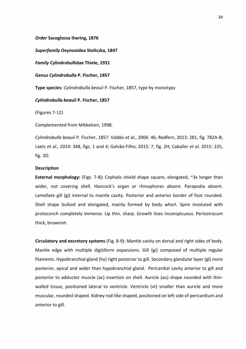
34
Order Sacoglossa Ihering, 1876
Superfamily Oxynooidea Stoliczka, 1847
Family Cylindrobullidae Thiele, 1931
Genus Cylindrobulla P. Fischer, 1857
Type species: Cylindrobulla beauii P. Fischer, 1857, type by monotypy
Cylindrobulla beauii P. Fischer, 1857
(Figures 7-12)
Complemented from Mikkelsen, 1998.
Cylindrobulla beauii P. Fischer, 1857. Valdés et al., 2006: 46; Redfern, 2013: 281, fig. 782A-B;
Laetz et al., 2014: 348, figs. 1 and 4; Galvão-Filho, 2015: 7, fig. 2H; Caballer et al. 2015: 225,
fig. 3O.
Description
External morphology: (Figs. 7-8): Cephalic shield shape square, elongated, ~3x longer than
wider, not covering shell. Hancock’s organ or rhinophores absent. Parapodia absent.
Lamellate gill (gi) internal to mantle cavity. Posterior and anterior border of foot rounded.
Shell shape bulloid and elongated, mainly formed by body whorl. Spire involuted with
protoconch completely immerse. Lip thin, sharp. Growth lines inconspicuous. Periostracum
thick, brownish.
Circulatory and excretory systems (Fig. 8-9): Mantle cavity on dorsal and right sides of body.
Mantle edge with multiple digitiform expansions. Gill (gi) composed of multiple regular
filaments. Hypobranchial gland (hy) right posterior to gill. Secondary glandular layer (gl) more
posterior, apical and wider than hypobranchial gland. Pericardial cavity anterior to gill and
posterior to adductor muscle (ac) insertion on shell. Auricle (au) shape rounded with thin-
walled tissue, positioned lateral to ventricle. Ventricle (vt) smaller than auricle and more
muscular, rounded shaped. Kidney rod-like shaped, positioned on left side of pericardium and
anterior to gill.

35
Digestive system (Fig. 10-11): Buccal mass (bm) elongated, 3x longer than wider with 16 dorsal
inconspicuous septate muscles (sm). Oral sphincter (os) elongated with 1/3 of buccal mass
length. Uniseriate radula composed of 80 rhomboid-shaped teeth with similar size, each tooth
with 11 smooth cusps decreasing on size laterally. Ascending and descending limbs not well-
delimited. Descending limb surrounded by thinner elongated ascus muscle (ma) on ventral
part of buccal mass and finishing inside of ventral part of buccal mass near to odontophore.
Odontophore region (or) rounded. Pharyngeal pouch (pp) on most posterior part of buccal
mass, width wider as buccal mass width. Jaws (Fig. 4C-D) absent. Esophagus (es) thinner on
anterior part before central nervous system and expanding and forming huge sack through
almost all its extension. Salivary glands (sg) paired, thick, elongated, not attached to
esophagus. Gizzard plates absent. Stomach inconspicuous, thin walled and positioned
anterior to digestive gland. Digestive gland (dg) like huge homogeneous mass and fused with
gonad, occupying most part of posterior region of body. Intestine (in) elongated, thick, running
inside digestive gland and finishing on posterior dorsal anus.
Reproductive system (Fig. 12): Gonad fused with digestive gland. One main hermaphrodite
duct runs anteriorly from central part of gonad to mucus gland (mg). Mucus gland granulose,
positioned on posterior part of oviduct and not fused with oviduct. Albumen gland (ag)
forming one big compact mass positioned near to mucus gland. Male duct (md) running from
hermaphrodite duct expands forming one tubular ampulla (am). Ampulla occupies half of
male duct length. Prostate gland anteriorly attached to male duct, just before the connection
of male duct to oviduct (ov). Bursa copulatrix (bc) connected to genital receptacle (gr) in one
duct that connects with oviduct near to female opening (fp). Penis elongated, straight,
positioned anteriorly on animal, inside right portion of cephalic shield, near to esophageal
expansion. Vas deferens absent. Seminal groove running from male aperture to female
oppening on right side of body.
Nervous system (Fig. 12): Cerebral and pleural ganglia fused. Cerebro-pleural (cp) connected
to each other by one slightly elongated commissure. Each cerebro-pleural ganglion with 6
main innervations highly branching toward cephalic shield borders. Optic nerve ~5x longer
than cerebral length and branching from or fused with cerebral innervation. Cerebro-pleural

36
ganglia connected to pedal ganglia (pd) by doubled short connective tissue. Pedal ganglion
with two paired innervations and connected by reduced connective tissue. Buccal ganglia (bu)
size small, connected to cerebral ganglia through elongated connective. Visceral loop very
elongated. Subintestinal (sb) ganglia inconspicuous and connected to abdominal ganglia (ab)
by very elongated connective tissue. Supraintestinal ganglia (sp) size as larger as abdominal
ganglia, connected to abdominal ganglia by elongated connective, but ~3x smaller than sb-ab
connective.
Distribution: Florida to Brazil (Sao Paulo) (MIKKELSEN, 1998).
Habitat and diet: Shallow waters associated with the calcareous algae Halimeda spp.
(MIKKELSEN, 1988).
Material examined: BRAZIL, Ceara, CMPHRM 2413B, 1 specimen (xii/2003), CMPHRM 2444B,
1 specimen (xii/2003), Pirangi do Norte, Rio Grande do Norte, MZSP 132648, 1 specimen
(01/viii/2007).
Remarks: The relationship between Cylindrobulla and the remain Sacoglossa was widely
debated (see MIKKELSEN, 1998; LAETZ, et al., 2014), specially because of its radular shape,
questionable presence of ascus and food habits. In a morphological based phylogeny Jensen
(1996) recovered Cylindrobulla as sister to Sacoglossa, while Mikkelsen (1998) included
Cylindrobulla in Sacoglossa as sister to all other shelled families. This same result was
supported later by molecular phylogenetic analysis (LAETZ et al., 2014). The present
phylogenic analysis also placed Cylindrobulla within Sacoglossa and sister to Ascobulla.

37
Figure 7. Cylindrobulla beauii live animal. A) lateral view; B) dorsal view. (Photo credit: C.A.O.
Meirelles).

38
Figure 8. Cylindrobulla beauii anatomy, shell removed, focusing pallial cavity structures: A)
Whole dorsal view; B) ventral view; C) lateral view. Scales= 1 mm.

39
Figure 9. Cylindrobulla beauii anatomy: A) Whole dorsal view, shell and dorsal mantle
removed, focusing pallial cavity structures; B) pallial cavity organs. Scales= 1 mm.

40
Figure 10. Cylindrobulla beauii anatomy of digestive system: A) Whole dorsal view; B) whole
left view (scales= 1 mm). C-E) Buccal mass anatomy: C) dorsal; D) ventral; E) right view (scales:
0.5 mm).

41
Figure 11. Cylindrobulla beauii radula. A and C) Whole view; B and D) detail of teeth.

42
Figure 12. Cylindrobulla beauii anatomy of reproductive and nervous systems: A) schematic
drawing of the reproductive system, dorsal view; B) whole dorsal view of central nervous
system, dorsal view; C) detail of cerebro-pleural ganglia, dorsal view; D) detail of pedal ganglia,
ventral view. Scales: 1 mm.

43
Family Volvatellidae Pilsbry, 1895
Genus Ascobulla Ev. Marcus, 1972
Type species: Cylindrobulla ulla Marcus & Marcus, 1970, type by original designation.
Ascobulla ulla (Er. Marcus & Ev. Marcus, 1970)
(Figures 13-17)
Complemented from Mikkelsen, 1998.
Ascobulla ulla (Er. Marcus & Ev. Marcus, 1970). Valdés et al., 2006: 50; Redfern, 2013: 281, fig.
781A-B; Galvão-Filho, 2015: 7, fig. 2I; Goodheart et al., 2116: 21, 11a.
Description
External morphology: (Figs. 13-14): Cephalic shield shape square, short, as longer as wider,
not covering shell. Hancock’s organ or rhinophores absent. Parapodia absent. Lamellate gill
(gi) internal to mantle cavity. Posterior and anterior border of foot rounded. Shell shape
bulloid and elongated, mainly formed by body whorl. Spire involuted with protoconch partially
immerse. Lip thin, sharp. Growth lines inconspicuous. Periostracum thin, translucent.
Circulatory and excretory systems (Fig. 14-15): Mantle cavity on dorsal and right sides of
body. Mantle edge with multiple digitiform expansions. Gill (gi) composed of multiple regular
filaments. Hypobranchial gland (hy) right posterior to gill. Secondary glandular band (cb) more
posterior, apical and wider than hypobranchial gland. Pericardial cavity anterior to gill and
posterior to adductor muscle (ac) insertion on shell. Auricle (au) shape rounded with thin-
walled tissue, positioned lateral to ventricle, size twice as larger as ventricle (vt). Ventricle (vt)
smaller than auricle and more muscular, rounded shaped. Kidney rod-like shaped, length half
of gill length, positioned on left side of pericardium and anterior to gill.
Digestive system (Fig. 16-17): Buccal mass (bm) elongated, twice longer as wider with 16
dorsal inconspicuous septate muscles. Oral sphincter (as) short with 1/6 of buccal mass length.
Uniseriate radula composed of 38 blade-shaped teeth decreasing in size towards ascus, each
tooth with two lateral cusps forming cutting edges with 40 elongated denticles, central cusp
smooth. Ascending limb with 5 well-formed teeth and 3 in formation. Descending limb with
30 teeth, which last 10 are organized in spiral inside ascus near to odontophore. Odontophore

44
region (or) rounded. Pharyngeal pouch (pp) on most posterior part of buccal mass, width
wider as buccal mass width, size ~3x smaller than buccal mass. Esophagus (es) anteriorly
expanded and curved to left side of animal, on anterior portion to central nervous system,
extending posteriorly and forming elongated esophageal pouch (ep). Esophageal pouch
positioned on ventrally to adductor muscle (ac), without internal folds. Salivary glands paired,
thick, elongated, not attached to esophagus. Stomach inconspicuous, thin walled and
positioned anterior to digestive gland. Digestive gland like huge homogeneous mass and fused
with gonad, occupying most part of posterior region of body. Intestine elongated, thick,
running inside digestive gland and finishing on posterior dorsal anus.
Reproductive system (Fig. 18): Gonad fused with digestive gland. One main hermaphrodite
duct (hd) runs anteriorly from central part of gonad to mucus gland (mg). Mucus gland
granulose, positioned on left side of animal, posterior and not fused to oviduct. Albumen
gland (ag) forming one big compact mass positioned near to mucus gland. Male duct (md)
running from hermaphrodite duct expands forming one tubular ampulla (am). Ampulla
occupies 1/5 of male duct length. Prostate gland anteriorly attached to male duct, just before
the connection of male duct to oviduct (ov). Bursa copulatrix (bc) connected to genital
receptacle (gr) in one duct that connects with oviduct near to female opening (fp). Penis (pe)
elongated, coiled, positioned anteriorly on animal, inside right portion of cephalic shield, near
to esophageal. Vas deferens absent. Seminal groove (se) running from male aperture to
female opening on right side of body.
Nervous system (Fig. 18): Cerebral and pleural ganglia fused. Cerebro-pleural (cp) connected
to each other by one elongated commissure. Each cerebro-pleural ganglion with 3 main
innervations highly branching toward cephalic shield borders. Optic nerve ~4x longer than
cerebral length and branching from or fused with cerebral innervation. Cerebro-pleural
ganglia connected to pedal ganglia (pd) by doubled short connective tissue. Pedal ganglion
with 4 paired innervations and connected by reduced connective tissue. Buccal ganglia (bu)
size small, bit smaller than subintestinal ganglion (sb), connected to each other by long
commissure, connected to cerebral ganglia through elongated connective. Visceral loop very
elongated. Subintestinal (sb) ganglia inconspicuous and connected to abdominal ganglia (ab)

45
by very elongated connective tissue. Supraintestinal ganglia (sp) size as half as abdominal
ganglia size, connected to abdominal ganglia by elongated connective, but ~3x smaller than
sb-ab connective.
Distribution: Florida to Brazil (Sao Paulo) (MIKKELSEN, 1998).
Habitat and diet: Shallow waters associated with the green algae Caulerpa spp..
Material examined: BRAZIL, Praia dos Dois Coqueiros, Caucaia, Ceara, MZSP 132649, 1
specimen (H. Galvão Filho, 02/ii/2014), Taíba beach, São Gonçalo do Amarante, Ceara,
CMPHRM 3086B, 1 specimen (H. Galvão Filho, 02/ii/2010); Rita beach, Entremoz, Rio Grande
do Norte, MZSP 97049, 2 specimens (M. Delgado, 24/vi/2009).
Remarks: Ascobulla ulla has many morphological similarities with Cylindrobulla on external
and internal morphology. Externally, Ascobulla can be distinguished by a short cephalic shield,
smaller and more fragile shell, and the protoconch is not immerse in the spire as in
Cylindrobulla. The reproductive system is very similar, but A. ulla has smaller ampulla and the
penis is much more elongated than C. beauii. In addition, the visceral loop in A. ulla is shorter
and the ganglia are less innervated. The most differences among them are in the digestive
system. Th buccal mass of Ascobulla has a very short oral sphincter and the pharyngeal pouch
is proportionally larger. The radula is formed by elongated aciculiform teeth in Ascobulla,
which is adapted to feed on siphonaleous green algae Caulerpa spp.. Instead, Cylindrobulla
has very modified short teeth with multiple cusps, which allow the species on this genera feed
on calcareous green algae Halimeda. Also, Ascobulla has a well-formed esophageal pouch,
which is absent in Cylindrobulla.

46
Figure 13. Ascobulla ulla live animal. A) dorsal view; B) lateral left view; C) ventral view; D)
shell apex.

47
Figure 14. Ascobulla ulla anatomy, shell removed, focusing pallial cavity structures: A) Whole
dorsal view; B) ventral view; C) lateral view. Scales= 1 mm.

48
Figure 15. Ascobulla ulla anatomy: A) Whole dorsal view, shell and dorsal mantle removed,
focusing pallial cavity structures; B) pallial cavity organs. Scales= 1 mm.

49
Figure 16. Ascobulla ulla anatomy of digestive system: A) Whole dorsal view; B) whole left
view (scales= 1 mm). C-E) Buccal mass anatomy: C) dorsal; D) ventral; E) right view (scales: 0.5
mm).

50
Figure 17. Ascobulla ulla radula. A) Whole view; B) detail of leading tooth.

51
Figure 18. Ascobulla ulla anatomy of reproductive and nervous systems: A) schematic drawing
of the reproductive system, dorsal view; B) whole dorsal view of central nervous system; C)
detail of cerebro-pleural ganglia and pedal ganglia, anterior view. Scales: 1 mm (A, B); 0.5 mm
(C).

52
Family Oxynoidae G. & H. Nevill, 1869
Genus Oxynoe Rafinesque, 1819
Type species: Oxynoe olivacea Rafinesque, 1819 type by monotypy.
Oxynoe antillarum Mörch, 1863
(Figures 19-24)
Oxynoe antillarum Mörch, 1836: 27; Ev. Marcus & Er. Marcus, 1963: 16; 1967: 25, fig. 20;
Marcus & Hughes, 1974: 502, figs. 5-6; Thompson, 1977: 120, figs. 22b, 23; Jensen & Clark,
1983: 3; Clark, 1984: 87, fig. 9; Valdés et al., 2006: 54; Padula, 2008: 22, figs. 3, 4, 6, 9, 10, 12,
13; Meirelles et al., 2010: 45, figs. 1-2; Redfern, 2013: 278, fig. 775A-C; Caballer et al., 2015:
220, fig. 3E; Goodheart et al., 2116: 21, 11b.
Oxynoe aguayoi Jaume, 1945: 22; Caballer et al., 2015: 220.
Description
External morphology: (Figs. 19-20): Head very distinguishable from rest of body, retractable
to under shell, elongated, length ~1/3 of animal body length. Rhinophores rolled, with no
papillae, elongated, length bit smaller than head length, pointed forwards. Parapodial lobes
extended from foot sole to dorsal side of animal and covering shell almost entirely. Lamellate
gill (gi) internal to mantle cavity. Anterior border of foot rounded. Posterior end of foot very
elongated forming long autotomized tail with almost half of total animal length. Small conic
papillae present on external face of parapodia and dorsal region of tail. Shell shape bulloid
and rounded, mainly formed by body whorl and wide aperture. Spire involuted with
protoconch immerse. Lip thin, sharp. Growth lines inconspicuous. Periostracum thin,
translucent.
Circulatory and excretory systems (Fig. 20-21): Mantle cavity on dorsal of body. Mantle edge
smooth. Gill (gi) composed of multiple regular filaments. Hypobranchial gland (hy) right
posterior to gill but concentrated only on left side of gill. Secondary glandular ciliated band
(cb) more posterior, apical and wider than hypobranchial gland, length as half as gill length.
Pericardial cavity very large, occupying 1/3 of mantle cavity, anterior to gill. Auricle (au) shape
triangular an elongated, size twice as larger as ventricle (vt), in contact with gill through all its

53
extension. Ventricle (vt) smaller than auricle and more muscular, rounded shaped. Kidney rod-
like shaped, reduced, positioned near to auricle and gill.
Digestive system (Fig. 22-23): Buccal mass (bm) with posterior part bit elongated, twice longer
as wider with 14 dorsal inconspicuous septate muscles. Oral sphincter (as) very muscular and
lar, size twice as larger as rest of buccal mass, composed of complex muscles in many
directions and forming huge cavity. Mouth (mo) opens almost ventrally. Uniseriate radula
composed of 34 blade-shaped teeth decreasing in size towards ascus, each tooth with two
lateral cusps forming cutting edges with multiple elongated and thin denticles, central cusp
smooth. Ascending limb with 9 well-formed teeth and other 2 in formation. Descending limb
with 23 teeth, finishing straight inside ascus near to odontophore. Odontophore region (or)
rounded. Pharyngeal pouch (pp) on most posterior part of buccal mass, rounded, width bit
wider than buccal mass width. Esophagus (es) with same width through all its extension.
Esophageal pouch (ep) on most posterior part of esophagus, shape very elongated, length as
longer as esophagus length, and curled on apex, internally folded. Salivary glands paired, thick
club-shaped, not attached to esophagus, with many hairy prolongations. Stomach
inconspicuous, thin walled and positioned anterior in digestive gland. Digestive gland like huge
homogeneous mass and fused with gonad, occupying most part of posterior region of body.
Intestine elongated, thick, running inside digestive gland and finishing on posterior dorsal
anus.
Reproductive system (Fig. 24): Gonad fused with digestive gland. One main hermaphrodite
duct (hd) runs anteriorly from central part of gonad to mucus gland (mg). Mucus gland
granulose, positioned on left side of animal, posterior and not fused to oviduct. Albumen
gland (ag) forming one big compact mass positioned near to mucus gland. Male duct (md)
running from hermaphrodite duct expands forming one tubular ampulla (am). Ampulla
occupies half of male duct length. Prostate gland (pr) anteriorly attached to male duct, just
before the connection of male duct to oviduct (ov). Bursa copulatrix (bc) connected to genital
receptacle (gr) in one duct that connects with oviduct near to female opening (fp). Penis (pe)
elongated, folded at base, with multiple papillae near top, positioned anteriorly on animal, on

54
right side to buccal mass. Vas deferens (vd) very elongated and curled near to penis, length
~3x penis length. Seminal groove absent.
Nervous system (Fig. 24): Cerebral and pleural ganglia fused. Cerebro-pleural (cp) connected
to each other by one short commissure. Each cerebro-pleural ganglion with 3 main
innervations highly branching toward cephalic shield borders. Optic nerve ~4x longer than
cerebral length and branching from or fused with cerebral innervation. Cerebro-pleural
ganglia connected to pedal ganglia (pd) by doubled short connective tissue. Pedal ganglion
with 5 innervations and connected by reduced connective tissue. Buccal ganglia (bu) size large,
size as twice as subintestinal ganglion (sb) size, connected to each other by long commissure,
connected to cerebral ganglia through elongated connective. Visceral loop reduced.
Subintestinal (sb) ganglia inconspicuous and connected to abdominal ganglia (ab) by reduced
connective tissue. Supraintestinal ganglia (sp) size as twice as abdominal ganglia size,
connected to abdominal ganglia by short connective.
Distribution: Florida to Brazil (Sao Paulo) (RIOS, 2009).
Habitat and diet: Shallow waters associated with the green algae Caulerpa spp..
Material examined: PANAMA, Isla Colón, Bocas del Toro, MZSP 121856, 6 specimens (P. Krug;
R. Ellingson; H. Galvão Filho, 07/viii/2015); MZSP 121857, 8 specimens (P. Krug; R. Ellingson;
H. Galvão Filho, 07/viii/2015); BRAZIL, Farol do Trapiá beach, Camocim, Ceara, CMPHRM
4089B, 2 specimens (H. Galvão Filho, 08/iv/2012).
Remarks: Oxynoe antillarum presents many morphological variations on its external
characters. Some populations may present a bluish pattern on rhinophores and parapodia
margins, more numerous and elongated papillae on parapodia and variation on shell shape.
For this reason, Oxynoe aguayoi Jaume, 1945 was synonymized with O. antillarum by Marcus
& Marcus (1967) and largely accepted by other authors (THOMPSON, 1977, MARCUS &
HUGHES, 1974, CLARK, 1984). Although other authors considerer the possibility of O.

55
antillarum be a complex of species or the validity of O. aguayoi (VALDÉS et al., 2006; CABALLER
et al., 2015), neither morphological or molecular investigation was done for such purpose.
Figure 19. Oxynoe antillarum live animal. A) dorsal view; B) ventral view. Length: 18 mm.

56
Figure 20. Oxynoe antillarum anatomy, shell removed, focusing pallial cavity structures: A)
Whole dorsal view; B) ventral view; C) lateral view, dorsal mantle removed. Scales= 1 mm.

57
Figure 21. Oxynoe antillarum anatomy: A) Whole dorsal view, shell and dorsal mantle
removed, focusing pallial cavity structures; B) pallial cavity organs. Scales= 1 mm.
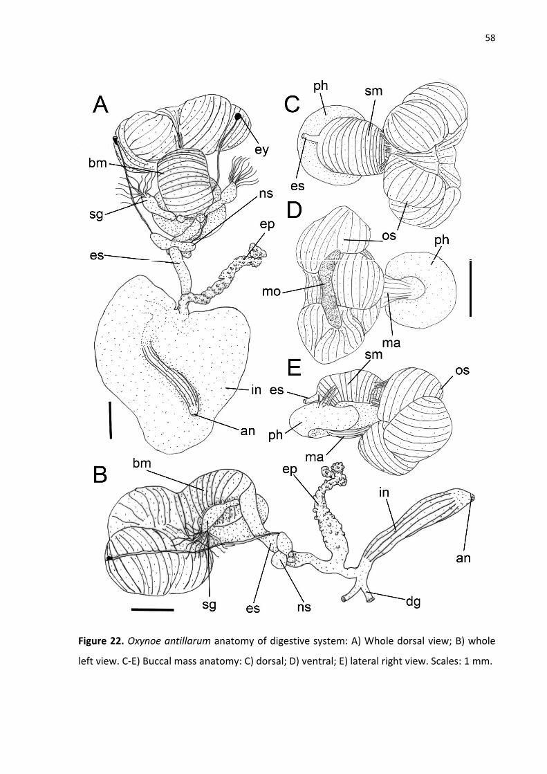
58
Figure 22. Oxynoe antillarum anatomy of digestive system: A) Whole dorsal view; B) whole
left view. C-E) Buccal mass anatomy: C) dorsal; D) ventral; E) lateral right view. Scales: 1 mm.

59
Figure 23. Oxynoe antillarum radula. A and C) Whole view; B and D) detail of leading tooth.

60
Figure 24. Oxynoe antillarum anatomy of reproductive and nervous systems: A) schematic
drawing of the reproductive system, dorsal view; B-C) whole view of central nervous system;
B) anterior view; C) posterior view. Scales: 1 mm.

61
Superfamily Limapontioidea Gray, 1847
Family Polybranchiidae O’Donogue, 1949
Genus Caliphylla A. Costa, 1867
Type species: Caliphylla mediterranea A. Costa, 1867 type by monotypy.
Caliphylla mediterranea A. Costa, 1867
(Figures 25-30)
Caliphylla mediterranea A. Costa, 1867: 17; Engel, 1927: 116, fig. 37; Pruvot-Fol, 1954: 177,
fig. 69; Marcus, 1958: 11, fig. 19-24; Gascoigne, 1979: 300, figs. 1-5; Valdés et al., 2006: 90;
Padula et al., 2012: 4: 4B; Jensen, 2014: 118, figs. 1ª, 2-5; Galvão-Filho et al., 2015: 8, fig. 3C;
Goodheart et al., 2016: 24, 12e; Padula, Wirtz & Schrödl, 2017: 750: 3D.
Description
External morphology (Fig. 25-26). Live animals reaching p 35 mm. Body smooth, elongated
and tapering posteriorly as pointed tail. Head rounded, hardly distinguishable from rest of
body, as longer as wider, length ~1/7 of animal length. Rhinophores (rh) bifid, with more
dorsal branch twice longer than ventral one ventral short, length bit smaller than head length,
smooth, tip rounded. Eyes (ey) big and positioned on posterior end of head on dorsolateral
position. Foot (fo) with mesopodial groove absent. Pericardial (pc) hump well-developed,
positioned on middle of body, right after head. Dorsal vessels absent. Cerata (ct) organized in
four longitudinal rows, each row with multiple flat leaf-like cerata. Male aperture positioned
on right side near to rhinophore. Female aperture on right side, posterior to male aperture
ate same level as anus. Vaginal opening absent. Anus positioned laterally on anterior part of
body on top of anal papilla.
Coloration (Fig. 25). Translucent body with dark green to brownish digestive ducts branching
internally through all animal body. Tiny and numerous opaque white spots spread through all
body. Preserved specimens completely yellowish.
Circulatory and excretory systems (Fig. 30A). Pericardium large, occupying almost half of
renopericardial cavity. Ventricle spherical and muscular, size bit larger than auricle. Auricle
thin-walled and folded. Kidney as thin elongated rounded gland, not branched, positioned
posteriorly to pericardium, near the base of auricle. Nephrostome not observed.

62
Digestive system (Fig. 27-28). Buccal mass (bm) barrel-shaped, elongated. Dorsal septate
muscle (sm) with about 20 thick muscular transversal bundles close to each other. Oral
sphincter (os) reduced, hardly distinguishable. Ascus musculature (ma) running ventrally from
oral sphincter to middle part of buccal mass, straight. Ascus external and very short. Radula
with ascending limb composed of 5 well-formed teeth and 2 other in formation. Descending
limb length as longer ascending limb, with long series of well-formed teeth plus uncountable
teeth in coiled ascus. Leading tooth blade-shaped, elongated, length ~3x longer than base
length, edge smooth and lateral cutting edges weakly marked. Esophagus (es) very elongated,
length twice as longer as buccal mass length. Salivary glands (sg) paired, attached on most
anterior part of esophagus, short, length less than half of esophageal length. Esophageal
pouch (ep) muscular, elongated, size twice as small as buccal mass size. Stomach (st) flat,
short, wide, positioned on middle of body under reproductive system; internally folded. Two
elongated digestive gland ducts (dg) run towards posterior and anterior end of body highly
branching through all animal including inside all cerata. Intestine (in) elongated, length as
longer as esophagus lentgh thick, with winner surface covered with irregular ridges.
Reproductive system (Fig. 29). Gonad composed of multiple hermaphrodite follicles (hf) with
rounded shape and similar size. Small ducts connect all follicles to one main hermaphrodite
duct (hd) that expands forming one tubular ampulla (am). Ampulla occupies ~1/3 of
hermaphrodite duct. Hermaphrodite duct connects to male duct (md) proximally to ampulla
expansion and runs to oviduct. Bursa copulatrix (bc) connected to handled formed by
hermaphrodite duct right after connection with male duct, sizebit larger the hermaphrodite
follicles. Genital receptacle (gr) connected to oviduct near to mucus gland (mg), close to
female aperture (fp). Mucus glande very elongated and not fused with oviduct. Albumen gland
(ag) highly branched lateral and ventral to hermaphrodite follicles and with and connected to
hermaphrodite duct before ampulla. Oviduct (ov) not glandular. Prostate gland as one curved
thick gland, positioned anteriorly in animal body under intestine and over esophagus. Vas
deferens (vd) very elongated, coiled near to penis (pe). Penis short and conical, length as wider
as longer
Nervous system (Fig. 30). Central nervous system composed by six ganglia and positioned
posterior to buccal mass around most anterior part of esophagus. Cerebro-pleural ganglia (cp)
size bit larger than pedal ganglia (pd), each ganglion with 5 main innervations. Optic nerve

63
length bit longer than cerebro-pleural ganglion length. Cerebral commissure reduced. Buccal
ganglia (bg) size large, size bit larger as subintestinal ganglion (sb), connected to each other
by short buccal commissure. Pedal ganglia with 4 main innervations running towards posterior
end of body attached to foot under all organs. Pedal commissure long, length as longer as
pedal ganglion length. Visceral loop reduced. Abdominal ganglion (ab) very large, size as larger
as cerebral ganglion size and with 4 main innervations. Supraintestinal ganglion twice smaller
than abdominal ganglia and with one innervation. Subintestinal ganglion small, ~5x smaller
than supraintestinal ganglion. Abdominal ganglion connected to supraintestinal and
subintestinal ganglia by reduced connective tissue.
Distribution. Amphi-Atlantic. Mediterranean Sea and tropical West Atlantic from Florida to
Brazil.
Material examined: BRAZIL, Pedra Rachada beach, Paracuru, Ceará, CMPHRM 3089B, 4
specimens (H. Galvão Filho, 17/x/2009); Barretinha, Atol das Rocas, Rio Grande do Norte,
CMPHRM 3014B, 3 specimens (03/ix/2009).

64
Figure 25. Live specimen of Caliphylla mediterranea. A) 18 mm length; B) 33 mm length.

65
Figure 26. External morphology of Caliphylla mediterranea. A) dorsal view with cerata; B)
ventral view; C) lateral right view with no cerata. Scale: 1 mm.

66
Figure 27. Digestive system of Caliphylla mediterranea. A) Dorsal view; B) Lateral left view;
Detail of buccal mass: C) dorsal view; D) ventral view side; E) lateral right view. Scales: 1 mm.

67
Figure 28. Scan electron microscope images of radula of Caliphylla mediterranea. A and C)
general view; B and D) detail of the leading tooth.

68
Figure 29. Reproductive system of Caliphylla mediterranea. A) Dorsal view of whole system;
B) schematic drawing, dorsal view. Scales = 1 mm.

69
Figure 30. Caliphylla mediterranea anatomy. A) Circulatory and excretory systems,
renopericardial cavity. B-C) Central nervous system; B) anterior view; D) posterior view.
Scales= 0.5 mm.

70
Family Limapontiidae Gray, 1847
Genus Olea Agersborg, 1923
Type species: Olea hansineensis Agersborg, 1923 type by monotypy.
Olea sp.
(Figs. 31-35)
Description
External morphology (Fig. 31-32). Live animals reach up 2.5 mm. Body smooth, elongated and
tapering posteriorly as pointed tail. Head square shaped, width as wider as body width, ~2x
wider than longer, length ~1/8 of animal length. Rhinophores extremely reduced to short
expansions on posterior end of head. Eyes big and positioned on posterior end of head on
dorsolateral position. Foot with a longitudinal groove and mesopodial groove absent.
Pericardial hump inconspicuous, positioned mid-centrally on body, right before cerata. Dorsal
vessels absent. Cerata organized in two longitudinal rows, each row with 3 fusiform cerata.
Male aperture positioned on right side of body laterally to right eye. Female aperture
posteriorly and close to male aperture. Vaginal opening centrally positioned on right side of
body, at same level as pericardium, and far from female aperture. Anus positioned mid
dorsally on body, just posterior to eyes.
Coloration (Fig. 31). Translucent body with dark brown and yellowish patches dorsally and
laterally on body. White rounded glands spread all over the body, head and cerata, bigger
bluish ones closer to posterior border of head and forming one centrally longitudinal row of
glands on tail. Kidney opaque white with renal aperture marked by one black dot. Digestive
gland yellowish. Preserved specimens still present all dorsolateral brown patches and white
glands, while digestive gland become whitish.
Circulatory and excretory systems (Fig. 34B). Pericardium extremely small. Ventricle spherical
and muscular, size ~5X bigger than auricle. Auricle thin-walled with smooth surface. Kidney as
thin elongated flat gland, not branched, positioned posteriorly to pericardium, near the base
of auricle. Nephrostome readily visible and marked as a dark spot near to pericardium.
Digestive system (Fig. 33). Buccal mass (bm) barrel-shaped, short, elongated, 3x longer than
wider. Dorsal septate muscle (ds) with tiny muscular transversal bundles close to each other

71
and hardly distinguishable. Oral sphincter reduced. Ascus musculature running ventrally from
oral sphincter to middle part of buccal mass, composed of tiny longitudinal muscles holding
the descending limb of buccal mass. Ascus apparently absent or extremely reduced. Radula
with ascending limb composed of 11 teeth in formation and barely recognizable. Descending
limb twice shorter than ascending limb, with leading tooth well-formed plus three rod-like
teeth. Leading tooth blade-shaped, short, length longer as base length, edge smooth and
lateral flanges absent. Esophagus thin and elongated, length ~3x longer than buccal mass
length and bit longer than intestine length. Esophageal inner surface with one pair of tiny
folds. Salivary glands paired, attached on most anterior part of esophagus, cover first 1/5 of
esophagus length. Esophageal pouch absent. Stomach flat, wide, positioned on middle of
body under albumen gland and some dorsal follicles; thin-walled with inner surface with no
folds. Two elongated digestive gland ducts run towards posterior end of body. Each duct
branches in three straight non-ramified ducts inside all cerata. Intestine elongated, thick, with
winner surface covered with irregular ridges.
Reproductive system (Fig 34A and C, 35). Gonad composed of multiple hermaphrodite
follicles (hf) with irregular shape and slightly variable in size. Multiple small ducts connect all
follicles to one main hermaphrodite duct (hd), that expands forming one tubular ampulla (am).
Ampulla occupies more than half of hermaphrodite duct. Hermaphrodite duct connects to
male duct (md) proximally to ampulla expansion, running to oviduct on fertilization chamber
(fc). Bursa copulatrix (bc) with its own aperture and one main duct connected to fertilization
chamber. Albumen gland (ag) highly branched over hermaphrodite follicles and with one
connection to fertilization chamber at the same point as hermaphrodite ducts and one distal
connection on bursa copulatrix duct. Genital receptacle size as larger as bursa copulatrix, also
connected to fertilization chamber, closer to glandular oviduct. Glandular oviduct with two
distinct glands. Mucus gland most distally one, larger, thicker and flimsy. Capsule gland
proximal to female opening on right side of animal body, less translucent and firmer than
mucus gland. Prostate gland formed by bilobed short gland, positioned anteriorly in animal
body under intestine and over esophagus. Vas deferens (vd) long, length ~3x longer than penis
length. Penis (pe) short and conical, size as larger as longer, with a terminal slightly curved
stylet. Stylet length as half as penis length.

72
Nervous system (Fig. 34D-E). Central nervous system composed of six ganglia and positioned
posterior to buccal mass around most anterior part of esophagus. Cerebro-pleural ganglia (cp)
size as large as pedal ganglia (pg), each ganglion with 5 main innervations. One innervation
highly ramified close to ganglion and runs towards posterior end of head to reduced
rhinophore region. Optical nerve length twice as longer as cerebro-pleural ganglion length.
Cerebral commissure external, short and thin. Buccal ganglia (bg) size large, as half as
supraintestinal ganglion size, with one main innervation running from each ganglion to
anterior part of buccal mass. Buccal commissure internal. Pedal ganglia with for main
innervation running towards posterior end of body attached to foot under all organs. Pedal
commissure internal. Visceral ganglia composed by one large abdominal ganglion, size half of
cerebral ganglion size and with two innervations, and one supraintestinal ganglion bit smaller
than abdominal ganglia and with one innervation. Abdominal ganglion connected to
supraintestinal ganglion by one short connective and connected to cerebro-pleural by one
longer innervated connective, length as longer as cerebro-pleural commissure length.
Distribution. Florida.
Remarks
Both molecular and morphological evidence have suggested the position of the new
egg-feeder sacoglossan belonging to the genus Olea. The probably new species Olea sp. was
recovered as sister to Olea hansineensis Agersborg, 1923 with a strong support for Bayesian
and maximum likelihood trees.
Although the phylogenetic relationship among the genera traditionally placed in
Limapontiidae is still debatable, the genera Calliopaea d'Orbigny, 1837 and Olea were resulted
as sister groups in both scenarios they were included in (JENSEN, 1996; KRUG et al., 2015).
Our results support this relationship, also placing Caliopaea belulla d'Orbigny, 1837 as sister
to O. hansineensis and Olea sp. This relationship suggests that the change of food habits from
herbivory to oophagy has happened only once in Sacoglossa evolutionary history.
Externally, the genera Olea and Calliopaea are quite similar, but Olea sp. looks more
like to O. hansineensis than Calliopaea spp. due to less number of cerata and reduced
rhinophore. However, similar external features have been reported to smaller Caliopaea
oophaga Lemche in Gascoigne & Sartory, 1974 specimen (GASCOIGNE & SIGURDSSON, 1977,

73
fig. 1c). The radular teeth in Olea n. sp. are quite unusual as in O. hansineensis, with the
ascending limb mostly with teeth in formation and descending limb with same number of
teeth and some rod-like shaped ones (GASCOIGNE, 1975). Although Gascoigne (1975) has
reported intraspecific variations on the number of fully formed teeth on O. hansieensis, it
represents more teeth of this type as in Olea sp., which it was reported only with one fully
formed in all specimens analyzed. Differently from Olea spp., Calliopaea spp. present a longer
radula with many fully formed teeth in both ascending and descending limbs (GASCOIGNE &
SIGURDSSON, 1977; GASCOIGNE & TODD, 1977). Also, the ascus in Olea is extremely reduced,
while is still remarkable.
Figure 31. Live specimen of Olea n. sp.

74
Figure 32. External morphology of Olea n. sp. (A) Dorsal, (B) lateral, and (C) ventral view of a
1,5mm specimen with cerata. (D) Dorsal and (E) lateral view of a 2 mm specimen with no
cerata.

75
Figure 33. Digestive system of Olea n. sp. A) Lateral view. B) Dorsal view. C) Radula. D) Internal
view.

76
Figure 34. Internal morphology of Olea n. sp. A) Dorsal view of hemocoel organs disposition.
B) Ventral view of kidney and pericardium. C) Reproductive system. D) Anterior and posterior
(E) view of central nervous system.

77
Figure 35. Scanning electron microscope image of penis of Olea n. sp.
Family Stiligeridae
Genus Stiliger Ehrenberg, 1828
Type species: Stiliger ornatus Ehrenberg, 1828 type by monotypy.
Stiliger vossi Ev. Marcus & Er. Marcus, 1960
(Figs. 36-41)
Stiliger vossi Ev. Marcus & Er. Marcus, 1960: 144, figs. 18-21.
Description
External morphology (Fig. 36-37). Live animals reaching 5 mm. Body smooth, elongated and
tapering posteriorly as pointed tail. Head rounded, bit longer than, length ~1/5 of animal
length. Rhinophores simple shape, short, length smaller than head length, smooth, tip
rounded. Eyes big and positioned on posterior end of head on dorsolateral position. Foot with
a longitudinal groove and mesopodial groove absent. Pericardial hump well-developed,

78
positioned anteriorly on body, right after head. Dorsal vessels absent. Cerata organized in two
longitudinal rows, each row with up to 9 fusiform cerata. Male aperture positioned on top of
penial bulb on right side of animal body. Female aperture at base of penial bulb, close to male
aperture. Vaginal opening absent. Anus positioned mid dorsally on body, just posterior to
eyes.
Coloration (Fig. 36). Translucent body with dark brown patches dorsally and laterally on body.
White rounded glands spread all over the body, head and cerata. Digestive gland yellowish.
Preserved specimens still present all dorsolateral brown patches and white glands, while
digestive gland become whitish.
Circulatory and excretory systems (Fig. 38). Pericardium extremely small. Ventricle spherical
and muscular, size ~3X bigger than auricle. Auricle thin-walled with smooth surface. Kidney as
thin elongated flat gland, not branched, positioned posteriorly to pericardium, near the base
of auricle. Nephrostome not observed.
Digestive system (Fig. 38-39). Buccal mass (bm) barrel-shaped, short, elongated, 2x longer
than wider. Dorsal septate muscle (ds) with tiny muscular transversal bundles close to each
other and hardly distinguishable. Oral sphincter reduced. Ascus musculature running ventrally
from oral sphincter to middle part of buccal mass in spiral shape. Ascus as same size as
odontophore region. Radula with ascending limb composed of 9 teeth. Descending limb ~9x
longer than ascending limb, with long series of well-formed teeth plus 6 rod-like teeth in ascus.
Leading tooth blade-shaped, elongated, length ~5x longer than base length, edge smooth and
lateral flanges absent. Esophagus thin and short, length ~5x shorter than buccal mass length
and bit smaller than intestine length. Salivary glands paired, attached on most anterior part of
esophagus, cover all esophagus length and anterior most part of stomach. Esophageal pouch
muscular, size bit smaller than buccal mass, internally folded. Stomach flat, elongated, wide,
positioned on middle of body under albumen gland and some dorsal follicles; thin-walled with
inner surface with no folds. Two elongated digestive gland ducts run towards posterior end of
body. Each duct branches in 9 straight non-ramified ducts inside cerata. Intestine short, thick,
with winner surface covered with irregular ridges.
Reproductive system (Fig. 40). Gonad composed of 2 hermaphrodite follicles (hf) with
rounded shape and similar size. Small ducts connect both follicles to one main hermaphrodite

79
duct (hd), that expands forming one tubular ampulla (am). Ampulla occupies less than half 1/5
of hermaphrodite duct. Hermaphrodite duct connects to male duct (md) proximally to
ampulla expansion and runs to oviduct. Bursa copulatrix (bc) absent. Genital receptacle (gr)
connected to oviduct after male duct. Albumen gland (ag) highly branched over
hermaphrodite follicles and with and connected to oviduct after genital receptacle. Glandular
oviduct as a big rounded gland. Prostate gland as one curved thick gland, positioned anteriorly
in animal body under intestine and over esophagus. Vas deferens (vd) short, length as longer
as penis length. Penis (pe) short and conical, size as larger as longer, positioned inside a penial
bulb.
Nervous system (Fig. 41). Central nervous system composed by six ganglia and positioned
posterior to buccal mass around most anterior part of esophagus. Cerebro-pleural ganglia (cp)
size as large as pedal ganglia (pg), each ganglion with 6 main innervations. Optical nerve length
bit longer than cerebro-pleural ganglion length. Cerebral commissure external, short and thin.
Buccal ganglia (bg) size large, as half as supraintestinal ganglion size, with one main
innervation running from each ganglion to anterior part of buccal mass. Buccal commissure
internal. Pedal ganglia with 2 main innervations running towards posterior end of body
attached to foot under all organs. Pedal commissure external, long, length as longer as pedal
ganglion length. Visceral ganglia composed by one large abdominal ganglion, size half of
cerebral ganglion size and with one main innervation, and one supraintestinal ganglion twice
smaller than abdominal ganglia and with one innervation. Abdominal ganglion connected to
supraintestinal ganglion by one short connective and connected to cerebro-pleural by one
longer innervated connective, length as longer as cerebro-pleural commissure length.
Distribution. Florida (type-locality); Panamá (present study).
Material examined: PANAMA, Isla Colón, Bocas del Toro, MZSP 121850, 8 specimens (P. Krug;
R. Ellingson; H. Galvão Filho, 07/viii/2015).
Remarks: This is the first finding of Stiliger vossi after its original description. The species was
placed in the genus Stiliger due to its general morphology, but some morphological characters
are similar to other species from other genera and family in Sacoglossa (MARCUS & MARCUS,
1960). The radula of S. vossi is quite different in shape and presence of serrate cutting edge
from species at the same genus, but similar to Hermaea (MARCUS & MARCUS, 1960).

80
Preliminary barcoding and phylogeny with other cerata-bearing genera have place S. vossi
with species previously named in Hermaea (KRUG, pers. com.). However, a more complete
revision must be done soon with the genera in Limapontiidae.
Figure 36. Live specimen of Stiliger vossi.

81
Figure 37. External morphology of Stiliger vossi. A) dorsal view with cerata; B) ventral; C) dorsal
view with no cerata; D) lateral view with cerata; E) lateral view with no cerata.

82
Figure 38. Stiliger vossi anatomy, circulatory and excretory systems. A) dorsal view of boy
withtou the dorsal mantle; B) ventral view of renopericardial organs. Scales: A = 1 mm; B = 0.2
mm.

83
Figure 38. Digestive system of Stiliger vossi. A) Dorsal view; B) Lateral view; Buccal mass: C)
left side; D) right side. E) Posterior portion of digestive system.

84
Figure 39. Scan electron microscope images of radula of Stiliger vossi (scale: 10 µm): A-B)
general view; C-D) detail of the leading tooth; E-F detail of ascus.

85
Figure 40. Visceral mass and reproductive system of Stiliger vossi. A) Dorsal view of visceral
mass; B) Ventral view of visceral mass; C) Overview of reproductive system; D) schematic
drawing of reproductive system.

86
Figure 41. Stiliger vossi anatomy, central nervous system: A) anterior view; B) posterior view.
Scale = 0.2 mm.
Superfamily Plakobranchoidea Gray, 1847
Family Bosellidae Marcus, 1982
Genus Bosellia Trinchese, 1891
Type species: Bosellia mimetica Trinchese, 1891 type by monotypy.
Bosellia mimetica Trinchese, 1891
(Figs. 42-47)
Bosellia mimetica Trinchese, 1891: 119, figs. 1-12; Pruvot-Fol, 1954: 179, fig. 70a-d;
Ballesteros, 1979: 13, fig. 1; Clark, 1984: 91, fig. 25; Thompson & Jaklin, 1988: 67, fig. 6; Valdés
et al., 2006: 56;
Bosellia mimetica curasoae Er. Marcus & Ev. Marcus, 1970: 50, figs. 91-98.; Marcus, 1973: 813,
fig. 2.

87
Description
External morphology (fig. 42-43): Body rounded when animal is relaxed, but it can be
posteriorly tapered after starts crawling. Body smooth Reaching up to 15 mm in live animals.
Head evident, short, length ~1/6 of total body length, 2x longer than wider. Rhinophores (rh)
short, length as half as head length, rolled, smooth, and with tip blunt. Eyes (ey) located
dorsolaterally behind rhinophores. Foot (fo) broad and muscular. Parapodial sole ~4x larger
than propodium (po) in live animals; anterior part of propodium rounded; mesopodial groove
(fl) evident. Renopericardial cavity prominent, but only pericardium (pc) form hump.
Pericardium small, size slightly smaller than head, muscular, globose. Dorsal vessels (dv)
organized in 3 pairs disposed radially on dorsum, arrangement symmetric. All vessels branch
few times, near to body margins. Parapodia absent. Female opening (fp) located on extreme
dorsal point of lateral groove (lg). Male opening (mp) below right rhinophore. Anus (a)
positioned dorsolaterally on posterior right side of head, close to lateral groove, above female
pore.
Coloration (Fig. 42): background color live green. Dorsal surface of body darker than foot.
Multiples white small dots spread through animal body, concentrated on body margin and
rhinhophores. Foot olive green. Some species might be extremely covered with white dots.
Circulatory and excretory system (Fig. 47C): Pericardium (pc) evident, globose, occupying 1/4
of renopericardial cavity; auricle (au) size ~6x smaller than ventricle (vt) size, auricle wall
smooth. Ventricle size large, occupying almost all pericardium, pear-shaped. Kidney slightly
branched in few thick renal ridges (rr). Renal opening (rp) on topo of elongated duct near to
most anterior part of pericardium.
Digestive system (Fig. 44-45): Buccal mass (bm) remarkable, very large, rounded. Dorsal
septate muscle (sm) composed of 14 large bundles. Oral sphincter (os) elongated, length ~4x
smaller than dorsal septate muscle length. Ascus musculature (ma) wide and short, occupying
almost all width and half-length of ventral side of buccal mass; ascus internal in buccal mass.
Odontophore region rounded. Radula with 16 teeth in ascending limb. Leading tooth
triangular shaped, very short, total length as longer as base length. Central cusp serrated only

88
on half near to tip. Ascus containing old teeth organized in spiral, last ones rod-like shape.
Salivary glands (sg) very elongated, positioned ventrally and laterally on all esophagus
extension. Esophagus length twice as longer as buccal mass length, width not variable.
Esophageal pouch (ep) elongated and coiled on top positioned on anterior part of esophagus.
Stomach (st) flatten, wide, width ~4x wider than intestine (in), not internally folded. Digestive
gland (dg) paired in each side of stomach; anterior ducts shorter than posterior ducts, runs
laterally and anteriorly inside head; posterior duct runs towards parapodial region. Intestine
(in), length bit shorter than esophagus length, width twice as wider as esophagus, internally
folded through all its extension, finishes in dorsolateral anus (a) positioned near to lateral
groove.
Reproductive system (Fig. 46-47A-B): Gonad composed of few hermaphrodite follicles (hf)
forming continuous area on central region of body. Follicles size few variable, size bit bigger
than genital receptacle (gr). Hermaphrodite ducts (hd) connect all follicles and and expands
forming one thick tubular ampulla (am). Hermaphrodite ampulla positioned on left side of
mucus gland (mg), occupies almost all hermaphrodite duct. Bursa copulatrix (bc) absent. Male
duct (md) connects prostate gland (pr) and vas deferens (vd) to hermaphrodite duct before
albumen gland (ag) connection. Prostate gland slightly branched in few lobes over most
central follicles, composed of one main glandular duct. Penis (p) size as half as mucus gland
size, shape conic and short, total length as longer as base length. Penial stylet straight, short,
length ~4x smaller than penis length. Vas deferens ~4x longer than penis length, slightly
undulated near to penis. Oviduct (ov) starts from hermaphrodite duct after connection with
male duct and forms 3 well delimited chambers completely evolved by mucus gland and
finishes on female lateral (fp) in lateral groove. First chamber of mucus gland smaller one,
rounded, albumen gland and male duct connect to oviduct in this area; middle chamber of
mucus gland bigger one, formed by a curved expansion of glandular oviduct; distal chamber
elongated. Genital receptacle connected to oviduct on posterior position, near to
hermaphrodite duct. Albumen gland (ag) composed of many dorsal thin branches ventrally to
follicles.

89
Nervous system (Fig. 47D-E): Cerebro-pleural ganglia (cp) size as larger as pedal ganglia (pd);
each one has 7 innervations; commissure cerebro-pleural not evident. Optic nerve length
short, bit longer than half of cerebro-pleural ganglion length. Buccal ganglia (bu) remarkable,
size larger than sub-intestinal ganglion (sb), with accessory ganglion ~3x smaller than buccal
ganglion; each buccal ganglion has 3 innervations. Buccal commissure simple, short, bit
smaller than buccal ganglion length. Pedal ganglia present 4 innervations by ganglion. Pedal
commissure reduced. Visceral loop very short. Abdominal ganglion (ab) size ~3x smaller than
pedal ganglion size, has 1 innervation. Sub-intestinal ganglion size ~5x as small as supra-
intestinal ganglion (sp), has 1 innervation. Supra-intestinal ganglion size as large as abdominal
ganglion and has 1 innervation. Connective tissue among visceral ganglia reduced.
Distribution: Amphi-Atlantic. Mediterranea Sea and West Atlantic Ocean from Florida to
Brazil.
Material examined: BRAZIL, Atol das Rocas, Rio Grande do Norte, CMPHRM 4237B, 4
specimens (H. Galvão Filho; C. Meirelles, 26/viii/2014). CMPHRM 4259B, 2 specimens (H.
Galvão Filho; C. Meirelles, 06/ix/2014); Ubatuba, São Paulo, 23º56’ S, 45º28’ W, MZSP 41942,
1 specimen (10/vi/2001).
Figure 42. Live specimen of Bosellia mimetica. A, B = 7 mm length; C = 5 mm length.

90
Figure 43. External morphology of Bosellia mimetica. A) dorsal view; B) ventral view. Scales:
0.5 mm

91
Figure 44. Digestive system of Bosellia mimetica. A) Dorsal view; B) Lateral view; Buccal mass:
C) dorsal view side; D) ventral side. E) lateral right view.

92
Figure 45. Scan electron microscope images of radula of Bosellia mimetica : A) general view;
B) detail of the leading tooth; C) detail of ascus.
Figure 46. Scan electron microscope images of penis of Bosellia mimetica: A) general view; B)
detail of the stylet.

93
Figure 47. Anatomy of Bosellia mimetica. A) whole reproductive system, dorsal view; B)
schematic reproductive system; C) renopericardial cavity, ventral view; D) nervous system,
aneterior view; E) nervous system, posterior view. Scales A-C= 0.5 mm; D-E = 0.25 mm.

94
Family Plakobranchidae Gray, 1840
Genus Plakobranchus van Hasselt, 1824
Type species: Plakobranchus ocellatus, van Hasselt, 1824, by monotypy.
Plakobranchus ocellatus van Hasselt, 1824
(Figs. 48-54)
See Meyers-Muñoz et al. (2016)
External morphology and Coloration (Fig. 48-49): Body elongated, reaching up to 45 mm in
live animals. Body surface smooth with no papillae. Head flatten, anteriorly cordiform, length
~1/5 of total body length, ~2x wider than longer. Rhinophores (rh) laterally inserted on head
corners, rolled, smooth, long, length as longer as head length, tip bit tapered. Eyes (ey)
located middle dorsally on a retractile cephalic protuberance. Foot (f) with parapodial sole
(pa) ~4x larger than metapodium (ms). Mouth slightly bilobed and with one long transversal
black line on upper lip. Anterior part of propodium rounded, with tapered corners forming
small pedal tentacles. Posterior end of parapodial sole truncated. Tail absent. Pericardium(pc)
fused with kidney prominence forming pericardial hump globose, slightly elongated, short,
length ~6x smaller than parapodia length. 12 dorsal vessels (dv) running radially from kidney
to posterior end of body or parapodial margins, located on top of digestive gland projections
forming lamellae (dl). Lamellae tall, numerous, multiple branching in bifurcations, but some
can anastomose, and covering almost all inner surface of parapodia except for extreme
posterior part of body. Dark green digestive gland branches inside lamellae but can get light
green or yellow in starving animals. Parapodia (pp) wide, covering all body when folded up,
with irregular edge and no papillae. Parapodia do not form a parapodial siphon, and stay
almost completely folded up on midline, except for anterior slightly opening on pericardial
area to water flows inward internal parapodial surface. Yellow glands on parapodial margins
where parapodium touch each other. Female aperture (fp) located on extreme dorsal level of
lateral groove (lg). Male aperture (mp) below right rhinophore. Anus (an)
positioned laterodorsally on posterior part of head near to pericardium.

95
Excretory and circulatory systems (Fig. 54): Pericardium (pc) large, occupying 1/3 of
renopericardial cavity. Auricle (au) thin walled, folded, voluminous, size bit smaller than
ventricle (vt). Ventricle size large, occupying almost half of pericardium, shape piriform.
Kidney posterior on renopericardial cavity where dorsal vessels open, composed of many
longitudinal tubules arranged on compact mass. Nephrostome (np) tubular running anteriorly
from kidney and opening on right side of renopericardium prominence.
Digestive system (Fig. 50-51): Buccal mass (bm) elongated, ~2x longer than wider, positioned
right after mouth opening, size ~3x larger as esophageal pouch (ep). Dorsal wall of buccal mass
composed of 10 dorsal septate muscle (sm), most anterior and posterior ones thinner than
middle ones; occupies half dorsal part of buccal mass. Oral sphincter (os) composed of small
circular muscles, size ~5x smaller than dorsal septate muscle length. Radula composed of
triangular shaped teeth with long base, length twice as small as tooth length, central cusp
reduced, lateral cutting edged with multiple short square projections. Ascending limb with 9
well-formed teeth and 3 others in formation, bit longer than descending limb. Ascus
musculature (ma) covering descending limb of radula, running ventrally from oral sphincter to
middle part of buccal mass, anteriorly wide and posteriorly tapered, short, finishing before
reaching pharyngeal pouch (ph). Ascus (as) internal too buccal mass, positioned under
odontophore muscles, occupying 1/3 of the buccal mass. Odontophore region (or) ventral,
rounded and occupies half of ventral part pf buccal mass. Pharyngeal pouch (ph) with lumen
connected with posterior part of oral tube, size as larger as odontophore region, long, as
longer as wider. Jaws absent. Esophagus long, length as longer as buccal mass length.
Esophageal pouch (ep) globose and muscular, positioned on middle part of esophagus.
Stomach (st) flat and wide, internally presenting multiples thin folds running toward intestine
and digestive gland ducts. Stomach can expand its volume and form pouches on posterior
region. Digestive gland (dg) with anterior ducts shorter than posterior one, running laterally
and anteriorly in head; posterior ducts running toward parapodial area and posterior end of
body, branching in many smaller ducts ventrally and among gonadal follicles and running in
multiple directions. Intestine (in) running from dorsal portion of stomach and opening in
laterodorsal anus on right side near to pericardium, length as longer as esophagus length.
Intestinal inner surface with multiple longitudinal folds through all its extension.

96
Reproductive system (Fig. 52-53): Gonad composed of innumerous spherical hermaphrodite
follicles (hf) forming one big group on central part of body, from posterior end of pericardium
to almost posterior end of body and expanding laterally at most basal part of parapodia.
Hermaphrodite follicles large, size as large as hermaphrodite ampulla (am). Multiple
hermaphrodite ducts (hd) connect all hermaphrodite follicles to one main middle-lateral
hermaphrodite duct. Hermaphrodite ampulla positioned on left side of mucus gland (mg),
large, ~3x smaller than genital receptacle (gr). Paired gametolytic vesicles positioned laterally
to posterior part pf mucus gland, right under renoperdicardial cavity; kidney-shaped bag with
granulose texture, yellowish color, easily breakable during manipulation and involved by fine
membrane; size ~2x bigger than penis size and ~2x smaller than second chamber of mucus
gland; connected to each other by one straight main tube, positioned on middle of dorsal part
of mucus gland, connected first to hermaphrodite ampulla duct and then to hermaphrodite
duct. Prostate gland (pr) and vas deferens (vd) connected to hermaphrodite duct after
connection with gametolytic vesicles and ampulla. Prostate gland as highly branched tubular
gland on both sides of mucus gland and over most anterior follicles. Vas deferens (vd) length
twice as longer as penis length, almost completely straight though all its extension. Penis (pe)
shape conic and short, as longer as wider. Penile stylet slightly curved, short, length ~4x
smaller than penis length, and centrally inserted on top of penis. Male opening posterior to
right rhinophore. Oviduct (ov) starting from hermaphrodite duct after connection with male
duct, forming 3 well delimited chambers completely evolved by mucus gland, finishing on
female apperture (fp) in lateral groove. Proximal chamber of mucus gland smallest one, bit
smaller than hermaphrodite ampulla, where hermaphrodite duct, genital receptacle and
albumen duct connect to oviduct. Middle chamber of mucus gland biggest one, where oviduct
is arranged as one big spiral involved by mucus gland and forming one big rounded gland,
positioned on midline of body. Distal chamber bit elongated, size ~2x smaller than remaining
mucus gland, darker glandular part of mucus gland, with one simple flatten lumen, and
forming one distal small duct opening on female bye. Albumen gland (ag) shaped and
branched as and mixed with prostate gland. Duct of albumen gland not glandular, running
posteriorly from oviduct and branching into smaller ducts dorsally to prostate gland duct.
Genital receptacle positioned anteriorly on right side of mucus gland, connected to oviduct in

97
proximal position after hermaphrodite and male ducts connections and on proximal chamber
of mucus gland, size bit smaller than gametolytic vesicles.
Nervous system (Fig. 54): Nervous ring located anteriorly on esophagus, covering most
anterior part of esophagus, just posterior to buccal mass. Cerebral and pleural ganglia fused.
Cerebro-pleural ganglia (cp) size as larger as pedal ganglia (pg), each ganglion with 6
nerves. Cerebro-pleural ganglia commissure reduced. Optic nerve very short, with eyes very
close to cerebro-pleural ganglia. Each pedal ganglion innervated with 7 highly branched
nerves running ventral-posteriorly on body. Pedal commissure elongated, positioned ventrally
between pedal ganglia, short, length ~5x smaller than pedal ganglion length. Visceral loop
composed of 3 ganglia, positioned very close to nervous ring and ventrally to esophagus;
connected among them and to base of cerebro-pleural ganglia by reduced connectives.
Abdominal ganglion (ab) size bit smaller than pedal ganglia, with one main innervation. Sub-
intestinal ganglion size ~3x smaller than supra-intestinal ganglion (sp), with 1 nerve. Supra-
intestinal ganglion bit smaller than abdominal ganglion, with 1 nerve. Buccal ganglia (bu) very
small, size ~10x smaller than sub-intestinal ganglion (sb); each ganglion with 1
main innervation; buccal commissure reduced.
Material examined: THAILAND, Puket, MZSP 122699, K. R. Jensen col., [5 specimens],
31/viii/1987.

98
Figure 48. External morphology of Plakobranchus ocellatus. A) dorsal view; B) lateral view; C)
ventral view; D) dorsal view of parapodia opened.

99
Figure 49. External morphology of Plakobranchus ocellatus. A) dorsal view; B) ventral view; C)
lateral view.

100
Figure 50. Digestive system of Plakobranchus ocellatus. A) Dorsal view; B) lateral view. Buccal
mass: C) Dorsal view; D) Ventral view; E) Right side.

101
Figure 51. Scan electron microscope images of radula of Plakobranchus ocellatus. A)
descending limb; B & D) detail of some teeth; C) overview.
Figure 52. Scan electron microscope image of penis of Plakobranchus ocellatus.

102
Figure 53. Reproductive system of Plakobranchus ocellatus. A) Schematic drawing; B)
overview.

103
Figure 54. Circulatory and nervous systems of Plakobranchus ocellatus; A) Renopericardial
cavity. B) anterior view of nervous system. C) posterior view of nervous system.

104
Plakobranchus ianthobapsus (Gould, 1852)
(Figs. 55-59)
Plakobranchus ianthobapsus (Gould, 1852)
Placobranchus ianthobapsus Gould, 1852: 307, pl.26, figs.407a-c (Honolulu, Hawaii); Bergh, 1872:
166; Ostergaard, 1955: 120-122, fig. 8a-f
Placobranchus gracilis Pease, 1871:303, pl.21 fig.1a-b (Tahiti, French Polynesia). Eliot, 1899: 521 (Apia,
Samoa)
Placobranchus argus Bergh, 1872: 151–165, pls. IX figs 6–9, XVII, XVIII (Honolulu, Hawaii);
Placobranchus sp.; Ostegaard, 1950: 107-108, fig. 34a-d;
Placobranchus ocellatus Eliot, 1904: 294-295, pl. XVII, fig 13-13a
Plakobranchus ocellatus Meyers-Muñoz et al., 2016:79-80, fig. 7b-e.
External morphology and Coloration (Fig. 55): Body elongated, reaching up to 60 mm in live
animals. Body surface smooth with no papillae. Head flattened, anteriorly cordiform, length
~1/5 of total body length, ~2x wider than longer. Rhinophores (rh) laterally inserted on head
corners, rolled, smooth, long, length as longer as head length, tip bit tapered. Eyes (ey)
located middle dorsally on a retractile cephalic protuberance. Foot (f) with metapodium (mt)
~4x larger than propodium (po). Mouth slightly bilobed and with one long transversal black
line on upper lip. Anterior part of propodium rounded, with tapered corners forming small
pedal tentacles. Posterior end of parapodial sole truncated. Tail absent. Renopericardial
prominence (rp) uniform with kidney area very similar to heart region, globose, slightly
longitudinally elongated, short, length ~5x smaller than parapodia length. 10 dorsal vessels
(dv) running longitudinally from kidney region to posterior end of body or parapodial margins
on inner side of parapodia, located on top of digestive gland projections forming lamellae, first
anterior pair highly branched before fuse to lamellae. Lamellae tall, numerous, multiple
branching in bifurcations, but can distally anastomose sometimes, and covering almost all
inner surface of parapodia except for extreme posterior part of body. Dark green digestive
gland branches inside lamellae but can get light green or yellow in starving animals.
Parapodia (pp) wide, covering all body when folded up, with irregular edge and no papillae.
Parapodia do not form a parapodial siphon, and stay almost completely folded up on midline,
except for anterior slightly opening on pericardial area to water flows inward internal

105
parapodial surface. Parapodial yellow glands on all area where parapodium touch each other,
varying on size, and rod-like or round shaped. Female pore (fp) located on extreme
dorsal level of lateral groove (lg). Male pore (mp) below right rhinophore. Anus (an)
positioned laterodorsally on posterior part of head near to pericardium.
Overall color pale yellow to dark brown varying among specimens. Some specimens
also can present dark brown or pale white regions dorsally on head and body. Ocellated spots
varying on size and spread all over the body except for the inner face of parapodia,
rhinophores and kidney region on renopericardial prominence, and only few on pericardial
region and propodium. Ocellated spots as orange circles surrounded by white ring; few of
them with orange circle darker on margin and light yellow, dark brown or bluish on central,
and fewer with darker orange margin turning to black forming either incomplete or complete
black rings; foot only with orange ocellated spots surrounded by white circle. Some specimens
can present few ocellated spots completely black on dorsal surface. Ocellated spots with black
ring usually bigger than others, more common close to foot on head and parapodia, less
numerous close to parapodial margins and absent on foot, inner surface of parapodia and
perdicadium. General foot color lighter than dorsal surface. Violet band present on anterior
end of head, rhinophore tips and posterior end of foot, fading to light violet or white on
rhinophore tips; violet color turns to black in preserved specimens. Retractile cephalic
protuberance colored as same as body background coloration.
Excretory and circulatory system (Fig. 59): Renopericardial area position mid dorsally
posteriorly to head. Pericardium (pc) large, occupying 1/3 of renopericardial cavity. Heart of
large size occupying 2/3 of pericardium. Auricle (au) thin walled, ventrally flatten, dorsally like
loose wrinkled bag, voluminous, size ~3x smaller than ventricle (vt). Ventricle size large,
occupying almost all pericardium, shape piriform. Kidney posterior on renopericardial cavity
where dorsal vessels open, composed of many longitudinal tubules arranged on compact
mass. Multiples crystals were observed in many different specimens from Hawaii.
Nephrostome (np) tubular running anteriorly from kidney and opening on right side of
renopericardium prominence.

106
Digestive system (Figs. 56-57): Mouth with elongated upper lip and shorter lower lip; mouth
opening centrally positioned. Buccal mass (bm) elongated, ~2x longer than wider, positioned
right after mouth opening, size twice as larger as esophageal pouch (ep). Dorsal wall of buccal
mass composed of 10 dorsal septate muscle (sm), most anterior and posterior ones thinner
than middle ones; occupies half of buccal mass. Oral sphincter (os) composed of small circular
muscles, size ~6x smaller than dorsal septate muscle length. Ascus musculature (ma) running
ventrally from oral sphincter to middle part of buccal mass, anteriorly wide and posteriorly
tapered, short, finishing before reaching pharyngeal pouch (ph). Ascus (as) internal too buccal
mass, positioned under odontophore muscles, occupying 1\4 of the buccal mass.
Odontophore region (or) ventral, rounded and occupies half of buccal mass. Pharyngeal pouch
(ph) present a lumen connected with posterior part of oral tube, size as larger as odontophore
region, long, as longer as wider. Jaws absent. One pair of salivary glands (sg) as elongated
thing layer positioned lateroventrally along all esophagus length, more concentrated on
posterior end of esophagus, open on posterior end of buccal mass dorsally to esophagus.
Esophagus long, length ~2x longer than buccal mass length, width similar to intestine width.
Esophageal inner surface with 2 longitudinal low folds running from buccal mass to stomach.
Esophageal pouch (ep) globose and muscular, positioned on posterior part of esophagus, size
~3x smaller than buccal mass; inner surface with many thin folds, which can be either
complete from base to top or incomplete. Stomach (st) flat and wide, internally presenting
multiples thin folds running toward intestine and digestive gland ducts. Stomach can expand
its volume and form pouches on posterior region. Digestive gland (dg) with anterior ducts
shorter than posterior one, running laterally and anteriorly in head; posterior ducts running
toward parapodial area and posterior end of body, branching in many smaller ducts ventrally
and among gonadal follicles and running in multiple directions. Intestine (in) running from
dorsal portion of stomach and opening in laterodorsal anus on right side near to pericardium,
length as longer as esophagus length. Intestinal inner surface with multiple longitudinal folds
through all its extension.
Reproductive system (Fig. 58-59): Gonad composed of innumerous rounded hermaphrodite
follicles (hf) forming one big group on central part of body, from posterior end of pericardium
to almost posterior end of body and expanding laterally to proximal part of parapodia.

107
Hermaphrodite follicles size really small, as large as hermaphrodite ampulla (am), weakly
variable in all specimens analyzed. Multiple hermaphrodite ducts (hd) connect all
hermaphrodite follicles to one main middle-lateral hermaphrodite duct. Hermaphrodite
ampulla positioned on left side of mucus gland (mg), really small, size ~12x smaller than genital
receptacle (gr). Paired gametolytic vesicles positioned lateroposterior on mucus gland, right
under renoperdicardial cavity; kidney-shaped bag with granulose texture, yellowish color,
easily breakable during manipulation and involved by fine membrane; size large as penis size,
and 5x smaller than second chamber of mucus gland; connected to each other by one dorsal-
lateral highly undulating main tube, positioned on left side of mucus gland, connects first to
hermaphrodite ampulla duct and then to hermaphrodite duct. One specimen presented one
of its gametolytic vesicles with doubled small duct connected to main one. Male duct (md)
connects prostate gland (pr) and vas deferens (vd) to hermaphrodite duct after its connection
with gametolytic vesicles and ampulla. Prostate gland composed of multiple acinous-shaped
glands, located on both sides of mucus gland, on region anterior to follicles and under
gametolytic vesicles and few anterior follicles; all braches connected to one dorsal transversal
glandular duct on each side of mucus gland and fused to each other on one main duct over
mucus gland. Vas deferens (vd) length bit longer than penis length, almost completely straight
though all its extension. Penis (pe) shape bit conic and elongated, twice longer than large, size
~2x smaller than distal chamber of mucus gland, positioned dorsally to distal` chamber of
mucus gland, involved by a penile sheath. Penile stylet curved, short, length ~5x smaller than
penis length, and centrally inserted on top of penis. Male opening lateroposterior to right
rhinophore. Oviduct (ov) starting from hermaphrodite duct after connection with male duct,
forming 3 well delimited chambers completely evolved by mucus gland, finishing on female
pore (fp) in lateral groove. Proximal chamber of mucus gland smallest one, size twice bigger
than hermaphrodite ampulla, where hermaphrodite duct, genital receptacle and albumen
duct connect to oviduct. Middle chamber of mucus gland biggest one, where oviduct is
arranged as one big spiral involved by mucus gland and forming one big rounded gland,
positioned on midline of body. Distal chamber bit elongated, size ~2x smaller than remaining
mucus gland, darker glandular part of mucus gland, with one simple flatten lumen, and
forming one distal small duct opening on female pore. Albumen gland (ag) shaped and
branched similar to prostate gland, but thinner, positioned mixed with prostate gland, but

108
only anteriorly to follicles. Duct of albumen gland not glandular, running posteriorly from
oviduct and branching into smaller ducts dorsally to prostate gland duct. Genital receptacle
positioned anteriorly on right side of mucus gland, connected to oviduct in proximal position
after hermaphrodite and male ducts connections and on proximal chamber of mucus gland,
size ~3x smaller than gametolytic vesicles.
Nervous system (Fig. 59): Nervous ring located anteriorly on esophagus, covering 1/3 of
esophagus, and just posteriorly to buccal mass. Cerebral and pleural ganglia fused. Cerebro-
pleural ganglia (cp) size as larger as pedal ganglia (pg). About 10 nerves innervating from each
cerebro-pleural ganglion to buccal mass, rhinophores and eye. Cerebro-pleural
ganglia commissure internal. Optic nerve long, length as twice as longer as cerebro-pleural
ganglion length. Each pedal ganglion innervated with 6 highly branched nerves running
ventral-posteriorly on body. Pedal commissure external, positioned centrally between pedal
ganglia, very short, length ~5x smaller than pedal ganglion length. Visceral ganglia composed
of 3 ganglia, positioned very close to nervous ring and ventrally to esophagus; connected
among them and to base of cerebro-pleural ganglia by reduced connectives. Abdominal
ganglion (ab) size larger than half of pedal ganglia size, with one main bifurcated nerve. Sub-
intestinal ganglion size twice as small as supra-intestinal ganglion (sp), with 1 nerve. Supra-
intestinal ganglion bit smaller than abdominal ganglion, with 1 nerve. Visceral ganglia
connective reduced. Buccal ganglia (bg) small, size ~8x smaller than sub-intestinal ganglion
(sb); each ganglion with 1 main innervation; buccal commissure reduced.

109
Figure 55. External morphology of Plakobranchus ianthobapsus. A) dorsal view; B) lateral view;
C) dorsal view with parapodia oppened; D) ventral view.

110
Figure 56. Plakobranchus ianthobapsus internal morphology. Digestive system. A) dorsal view.
B) Lateral view. C-E) Buccal mass, dorsal (C), ventral (D) and lateral view (E). F) Internal surface
of digestive system.

111
Figure 57. Scan electron microscope images of radula of Plakobranchus ocellatus. A)
overview; B) detail of leading tooth; C) discarded teeth.

112
Figure 58. Scan electron microscope image of penis of Plakobranchus ianthobapsus.

113
Figure 59. Internal morphology of Plakobranchus ianthobapsus. A-B) Reproductive system,
overview (A) and schematic arrangement (B). C) Circulatory and excretory systems. D-E)
Nervous system, anterior (D) and posterior (E) view.

114
Plakobranchus papua Meyers-Munoz & van der Velde, 2016
(Figs. 60-64)
Plakobranchus papua Meyers-Muñoz & van der Velde, 2016: 80-88, fig. 2-7a.
External morphology and coloration (Fig. 60): Body reaching up to 70 mm in live animals.
General body shape similar to P. ianthobapsus except for the following. Head proportionally
bit smaller, length ~1/6 of total body length Rhinophores (rh) length bit longer than head
length, tip blunt. Foot (fo) with metapodium (mt) ~5x larger than propodium (po). Anterior
part of propodium with pedal tentacles bit more tapered. Renopericardial very short, length
~11x smaller than parapodia length. 8 dorsal vessels (dv) running longitudinally from kidney.
Lamellae shorter, showing inner parapodial surface, multiple branching in bifurcations, not
distally anastomosing. Body overall color olive green to ochre yellow varying among
specimens. Some parts of external face of parapodia with yellowish patches. Foot color white,
but some specimens with fading blue on margins and usually more intense on posterior end.
Large black band present on middle of rhinophores, fading to bluish tone upwards top and
brownish downwards base. Black band present on posterior end of body. Retractile cephalic
protuberance completely white. Rounded white spots varying on size, covering almost all
dorsal surface of head and external side of parapodia, completely absent on inner side of
parapodia, renopericardial prominence, rhinophores and foot; bigger and less numerous close
to foot; smaller and in large quantity closer to parapodial margins. Ocellated spots completely
absent.
Digestive system (Fig. 61-62): Very similar to P. ianthobapsus, with following exceptions.
Buccal mass (bm) proportionally larger, posteriorly rounded and anteriorly tapered, size ~5x
larger than esophageal pouch (ep). Dorsal wall of buccal mass composed of 10 dorsal septate
muscle (sm) with similar width, occupies more than half of buccal mass. Ascus musculature
(ma) bit shorter. Ascus (as) occupying 1\3 of ventral side of buccal mass. Odontophore region
(or) occupies less than half of buccal mass. Pharyngeal pouch (ph), short, ~2x wider than
longer, length as half as dorsal septate muscles length. Salivary glands (sg) short, finishing
before posterior end of esophagus under esophageal pouch, equally distributed through its
extension. Esophagus short, as longer as buccal mass length, width ~3x smaller than intestine
width. Esophageal pouch (ep) size ~5x smaller than buccal mass, with inner surface with

115
multiple longitudinal thick folds, running from base to top. Intestine (in) longer, lenth bit
longer than esophagus length.
Excretory and circulatory system (Fig. 64C): Heart and kidney characters similar to P.
ianthobapsus.
Reproductive system (Figs. 63, 64A-B): The arrangement and characters are similar to P.
ianthobapsus with the following exceptions. Hermaphrodite ampulla proportionally larger,
size ~8x smaller than genital receptacle (gr) and ~3x bigger than hermaphrodite follicles.
Gametolytic vesicles (gv) elliptical shaped, size large as penis size and ~3x smaller than second
chamber of mucus gland, connected to each other by one middle-dorsal straight duct. Duct of
gametolytic vesicles bit expanded where bifurcates. Prostate glands (pr) more spread under
follicles and expanded through proximal part of parapodia. Vas deferens (vd) length twice as
longer as penis length. Penis (pe) shorter, bit longer than larger. Penile stylet less curved,
length ~3x smaller than penis length. Proximal chamber of mucus gland size as larger as
hermaphrodite ampulla size. Middle chamber of mucus gland proportionally smaller than in
P. ianthobapsus. Distal chamber size ~3x smaller than second one.
Nervous system (Fig. 64): Cerebro-pleural ganglia with about 8 innervations. Optic nerve long,
length as longer as cerebro-pleural ganglion length. Pedal commissure positioned ventrally,
longer and thicker, length ~3x smaller than pedal ganglion length. Abdominal ganglion (ab)
with two innervations. Sub-intestinal ganglion size ~4x smaller than supra-intestinal ganglion
(sp). Buccal ganglia (bg) proportionally larger, size twice as small as sub-intestinal ganglion
(sb).

116
Figure 60. External morphology of Plakobranchus papua. A-B) Dorsal view; C) ventral view; C)
ventral view; D) dorsal view with parapodia oppend.

117
Figure 61. Internal morphology of Plakobranchus papua. Digestive system. A) dorsal view. B)
Lateral view. C-E) Buccal mass, dorsal (C), ventral (D) and lateral view (E). F) Internal surface
of digestive system.

118
Figure 62. Radula of Plakobranchus papua. A) overview; B) leading tooth; C) discarded tooth.
Figure 63. Penis of Plakobranchus papua.

119
Figure 64. Internal morphology of Plakobranchus papua. A-B) Reproductive system, overview
(A) and schematic arrangement (B). C) Circulatory and excretory systems. D-E) Nervous
system, anterior (D) and posterior (E) view.

120
Plakobranchus sp. 1 (aff. Purple)
(Figs. 65-69)
Plakobranchus sp. 5 Gosliner et al., 2015: 99.
External morphology and coloration (Fig. 65): Body reaching up to 30 mm in live animals.
General body shape similar to P. ianthobapsus except for the following. Foot (f)
with metapodium (mt) ~5x larger than propodium (po). 8 dorsal vessels (dv) running
longitudinally from kidney. Lamellae shorter, showing inner parapodial surface, multiple
branching in bifurcations, not distally anastomosing. Body with overall color creamy or pale
white, varying in darkness among specimens. Dark grey reticulated pattern on head,
rhinophores and lateral of body close to foot. Green digestive gland visible on mouth region
and forming large patches on external surface of parapodia. Ocellated spots as iridescent dots
between pink and orange in color either darker orange border or surrounded by one complete
or incomplete black ring, spread over external face of parapodia, head and rhinophores, and
completely absent on inner face of parapodia, renopericardium prominence and foot. Smaller
and more numerous black dots spread among iridescent spots. Foot white, densely dotted
with black spots variable in size from tiny to large; each surrounded by diffuse ring of faint
greyish purple; no spots on anterior and posterior end of foot. Rhinophores with same color
pattern as head on dorsum side, and no color on ventral side. No colored band on rhinophores
and posterior end of body. Retractile cephalic protuberance colored as same as body
background coloration.
Excretory and circulatory system (Fig. 69): Heart and kidney characters similar to P.
ianthobapsus.
Digestive system (Fig. 66-67): Very similar to P. ianthobapsus, with following exceptions.
Buccal mass (bm) less elongated, as longer as wider, with posterior part rounded and anterior
part bit tapered, length ~3x longer than cerebro-pleural ganglia length. Dorsal wall of buccal
mass composed of 10 dorsal septate muscle (sm) with similar width; short, occupies less than
half of buccal mass. Ascus musculature (ma) shorter, finishing before middle part of
odontophore region. Odontophore region larger, occupying more than half of buccal mass.

121
Pharyngeal pouch (ph) size half as larger as odontophore region size, length as half as dorsal
buccal mass length, short, ~2x wider than longer. Ascus occupying 1/5 of ventral part of buccal
mass. Esophagus short, length as longer as buccal mass length, width as half as intestine
width. Inner surface of esophageal pouch (ep) with two thick longitudinal fold running from
base to almost top portion.
Reproductive system (Fig. 68-69): The arrangement and characters are similar to P.
ianthobapsus with the following exceptions. Gonad shorter, do not expand to posterior end
of body. Hermaphrodite follicles larger, size twice as large as hermaphrodite ampulla (am).
Hermaphrodite ampulla small as in P. ianthobapsus, size ~3x smaller than genital receptacle
(gr). Paired gametolytic elliptical shaped, longer, size twice as large as penis size, and large as
second chamber of mucus gland; connected to each other by one highly curled dorsolateral
main duct. Prostate gland (pr) as in P. papua. Vas deferens (vd) length ~3x longer than penis
length, bit curled close to penis. Penis (pe) shorter as in P. papua, twice longer than larger,
size ~3x smaller than distal chamber of mucus gland. Penile stylet longer, length big longer
than penis length, and laterally inserted on top of penis. Mucus gland similar to P. papua, but
distal chamber bit larger. Genital receptacle smaller, size ~10x smaller than gametolytic
vesicles.
Nervous system (Fig. 69): Nervous ring covering almost half of esophagus length. Cerebro-
pleural (cp) with about 8 innervations. Optic nerve shorter, length bit smaller than cerebro-
pleural ganglion length. Each pedal ganglion innervated with 4 highly branched nerves. Supra-
intestinal ganglion smaller than half of abdominal ganglion size, with 2 nerves. Buccal ganglia
(bg) proportionally larger as in P. papua, size twice as small as sub-intestinal ganglion (sb).

122
Figure 65. External morphology of Plakobranchus sp. 1. A) Dorsal view; B) dorsal view with
parapodia opened; C) ventral view; D) lateral view.

123
Figure 66. Internal morphology of Plakobranchus sp. 1. Digestive system. A) dorsal view. B)
Lateral view. C-E) Buccal mass, dorsal (C), ventral (D) and lateral view (E). F) Internal surface
of digestive system.

124
Figure 67. Radula of Plakobranchus sp. 1. A) overview; B) leading tooth; C) old teeth; D)
discarded teeth.
Figure 68. Penis of Plakobranchus sp. 1. A) overview; B) detail of penial stylet.

125
Figure 69. Internal morphology of Plakobranchus sp. 1. A-B) Reproductive system, overview
(A) and schematic arrangement (B). C) Circulatory and excretory systems. D-E) Nervous
system, anterior (D) and posterior (E) view.

126
Plakobranchus sp. 2
(Figs. 70-74)
External morphology (Figs. 70-71): Body reaching up to 18 mm in preserved specimens. Head
proportionally bit larger, length ~1/4 of total body length. Number of dorsal vessel not
observed. Dorsal lamellae less tall and less branched. Coloration on specimens preserved in
ethanol very similar to P. ianthobapsus except for the following. Head dark brown on middle
dorsal part. Ocellated spots with bluish inner potion seem to be absent. Foot with no orange
ocellated spots; densely dotted with black spots variable in size from medium to large; each
black spot surrounded by pale white ring; no spots on anterior and posterior end of foot.
Anterior end of head, posterior end of body and rhinophore tips with black band.
Excretory and circulatory system (Fig. 45): Heart and kidney characters similar to P.
ianthobapsus except for the following. Pericardium larger, occupying less than half of
renopericardial cavity. Heart occupying more than half of pericardium space. Ventricle larger,
size ~5x auricle size.
Digestive system (Fig. 42-43): Very similar to P. ianthobapsus, with following exceptions.
Buccal mass (bm) shorter, bit longer than wider, posteriorly rounded and anteriorly tapered.
Dorsal wall of buccal mass composed of 10 dorsal septate muscle (sm) with similar width. Oral
sphincter (os) as longer as half dorsal septate muscle (sm) length. Ascus musculature (ma)
posteriorly rounded, large, covering more than half of odontophore region. Ascus (as)
occupying almost half of ventral side of buccal mass. Buccal pouch (bp), short, ~2x wider than
longer, length as half as dorsal septate muscles length. Salivary glands (sg) short, finishing
before posterior end of esophagus under esophageal pouch, equally distributed through its
extension. Esophageal pouch (ep) size as larger as buccal mass size, with muscular top portion
occupying half of lumen, and inner surface with no folds. Intestine (in) longer, length ~2x
longer than esophagus length.
Reproductive system (Fig. 44-45): Very similar to P. iathobapsus with the following
exceptions. Hermaphrodite follicles size proportionally bit larger, size ~3x smaller than
hermaphrodite ampulla (am). Hermaphrodite ampulla larger as in P. papua, size ~6x smaller

127
than genital receptacle (gr). Gametolytic vesicles connected to each other by one dorsal-
lateral straight duct. Prostate gland arranged as is P. papua. Vas deferens (vd) length ~2x
longer as penis length, half proximally to penis highly curled and other half straight. Penis (pe)
shape bit longer than larger. Penile more curved, longer, length bit smaller than penis length.
Secondary genital receptacle positioned under the penis, and connected on oviduct right after
other genital receptacle.
Nervous system (Fig. 45): Nervous covering almost half esophagus length. Cerebro-pleural
ganglia (cp) with about 8 innervations. Optic nerve short, length bit smaller than cerebro-
pleural ganglion length. Pedal commissure as in P. papua. Abdominal ganglia (ab) elongated,
twice wider than longer. Sub-intestinal ganglion size ~3 smaller than supra-intestinal ganglion
(sp). Supra-intestinal ganglion ~3x smaller than abdominal ganglion. Buccal ganglia (bg) size as
small as sub-intestinal ganglion (sb). Buccal commissure exposed and short.

128
Figure 70. Photos of preserved specimens of Plakobranchus sp. 2 Africa. A) Dorsal view with
closed parapodia; B) ventral view; C) dorsal view with opened parapodia; D) lateral left view.

129
Figure 71. External morphology of Plakobranchus sp. 2 Africa. A) Dorsal view. B) Ventral view.
C) Lateral view.

130
Figure 72. Internal morphology of Plakobranchus sp. 2 africa. Digestive system. A) dorsal view.
B) Lateral view. C-E) Buccal mass, dorsal (C), ventral (D) and lateral view (E). F) Internal surface
of digestive system.

131
Figure 73. Radula of Plakobranchus sp. 2. A) overview; B) leading
tooth; C) old teeth; D) discarded teeth.
Figure 74. Penis of Plakobranchus sp. 2.

132
Figure 75. Internal morphology of Plakobranchus sp. 1 aff. purple. A-B) Reproductive system,
overview (A) and schematic arrangement (B). C) Circulatory and excretory systems. D-E)
Nervous system, anterior (D) and posterior (E) view.

133
Genus Thuridilla van Hasselt, 1824
Type species: Elysia splendida Grube, 1861 type by monotypy.
Thuridilla hopei (Vérany, 1853)
(Figs. 76-79)
Actaeon hopei Vérany, 1853: 392.
Elisia splendida Grube, 1861: 86, 133, pl. 1 figs. 1-3.
Thuridilla picta Malaquias et al., 2014:5, fig. 2F
Elysia hopei (Vérrany, 1853). Thompson, 1981: 77, fig. 5.
Thuridilla hopei (Vérrany, 1853). Pruvot-Fol, 1954: 203; Bouchet, 1984: 26, figs. 6, 11.
Description
External morphology (Fig. 76): Body elongated with 12 mm length in preserved animals. Head
evident, large, size ~1/3 of total body size, 2x longer than wider. Rhinophores (rh) short in
preserved specimens, length as half as head length, rolled, smooth, and with tip rounded. Eyes
(ey) located dorsolaterally behind rhinophores. Foot (fo) elongated and muscular. Parapodial
sole (pa) ~3x larger than propodium (po) in live animals; anterior part of propodium bit
expanded; mesopodial groove (fl) evident. Pericardium (pc) and renal prominence (rp) fused
forming short globose renopericardial hump. Dorsal vessels (dv) organized in one single pair,
branching multiple times only in central part of body. Parapodium (pp) large, with regular
margin and do not form closure pattern or parapodial tail. Female pore (fp) located on
extreme dorsal point of lateral groove (lg). Male pore (mp) below right rhinophore. Anus (a)
positioned dorsolaterally on posterior right side of head, near to pericardium.
Circulatory and excretory system (Fig. 79C): Pericardium (pc) evident, globose, occupying half
of renopericardial cavity. Auricle (au) size ~3x smaller than ventricle (vt) size, folded. Ventricle
size large, occupying almost all pericardium, pear-shaped. Kidney highly branched with renal
ridges forming a huge compact mass. Renal opening (rp) on top of elongated duct near to
most anterior part of pericardium.

134
Digestive system (Fig. 77-78): Buccal mass (bm) remarkable, very large, rounded. Dorsal
septate muscle (sm) composed of 8 large bundles. Oral sphincter (os) short, length ~6x smaller
than dorsal septate muscle length. Ascus musculature (ma) thin and elongated, occupying only
1/3 of ventral side of buccal mass; ascus internal in buccal mass. Odontophore region rounded.
Radula with 5 well-formed teeth in ascending limb and one in formation. Leading tooth
triangular shaped, short, total length twice as longer as base length. Central cusp reduced and
lateral cutting edges with small denticles. Ascus containing old teeth organized in spiral.
Salivary glands (sg) very thick and concentrated on anterior part of esophagus between
nervous ring (ns) and esophageal pouch (ep). Esophagus length twice as longer as buccal mass
length, width not variable. Esophageal pouch (ep) spherical, voluminous, size as half as buccal
mass size, positioned on middle of esophagus. Stomach (st) flatten, short, width similar to
intestine (in) width, internally folded. Digestive gland (dg) paired in each side of stomach;
anterior ducts shorter than posterior ducts, runs laterally and anteriorly inside head; posterior
duct runs towards parapodial region. Intestine (in), length bit longer than esophagus length,
width twice as wider as esophagus, internally folded through all its extension, finishes in
dorsolateral anus (a) positioned near to pericardium.
Reproductive system (Fig. 79A-B): Gonad composed of multiple hermaphrodite follicles (hf)
forming continuous area on central region of body. Follicles size few variable, size ~4x smaller
than genital receptacle (gr). Hermaphrodite ducts (hd) connect all follicles at one main central
duct, which runs toward ampulla (am). Hermaphrodite ampulla separated from
hermaphrodite duct, positioned centrally on body, posterior to mucus gland (mg), size bit
larger than genital receptacle. Bursa copulatrix (bc) with its own aperture near to female
opening, positioned on right side of mucus glang, size twice as larger as genital receptacle;
connected to genital receptacle and then to glandular oviduct at first chamber. Male duct (md)
connects prostate gland (pr) and vas deferens (vd) to hermaphrodite duct before albumen
gland (ag) connection at first chamber. Prostate gland highly branched over all follicles,
composed of one main glandular duct. Penis (p) size ~3x smaller than mucus gland size, shape
conic and short, total length as longer as base length. Penial stylet absent. Vas deferens ~3x
longer than penis length, slightly undulated near to penis. Oviduct (ov) starts from

135
hermaphrodite duct after connection with male duct and forms 3 well delimited chambers
completely evolved by mucus gland and finishes on female aperture (fp) in lateral groove. First
chamber of mucus gland very small, rounded, darker, where albumen gland and male duct
connect to oviduct. Middle chamber of mucus gland bigger one, formed by a curved expansion
of glandular oviduct; last chamber elongated. Albumen gland (ag) composed of many dorsal
thin branches mixed with prostate gland.
Nervous system (Fig. 79D-E): Cerebro-pleural ganglia (cp) size as larger as pedal ganglia (pd);
each one has 4 innervations; commissure cerebro-pleural reduced. Optic nerve length bit
shorter than cerebro-pleural ganglion length. Buccal ganglia (bu) large, size ~2x larger than
sub-intestinal ganglion (sb), with accessory ganglion ~4x smaller than buccal ganglion. Buccal
commissure simple, short, bit smaller than buccal ganglion length. Pedal ganglia present 4
innervations by ganglion. Pedal commissure very short. Visceral loop very short. Abdominal
ganglion (ab) size twice as small as pedal ganglion size, with 2 innervations. Sub-intestinal
ganglion size ~3x as small as supra-intestinal ganglion (sp), has 1 innervation. Supra-intestinal
ganglion size bit smaller than abdominal ganglion, with 1 innervation. Connective tissue
between supraintestinal ganglia ~3x longer than connective between subintestinal and
abdominal ganglia.
Distribution: Mediterranean Sea.
Material examined: PORTUGAL, Azores, São Jorge, Fajã de Santo Cristo, Lisboa, MASP 97355,
2 specimens (V. Padula, 30/viii/2007); GREECE, Agia Pelagia, Crete, MZSP 162634, 2
specimens.

136
Figure 76. External morphology of Thuridiila hopei. A) dorsal view; B) ventral view; C) lateral
right view. Scales: 0.5 mm

137
Figure 77. Digestive system of Thuridilla hopei. A) Dorsal view; B) Lateral view; Buccal mass:
C) dorsal view side; D) ventral side. E) lateral right view. Scales: 0.5mm

138
Figure 78. Scan electron microscope images of radula Thuridilla hopei : A) general view; B)
detail of the leading tooth; C) detail of older tooth; D) Ascus.

139
Figure 79. Anatomy of Thuridilla hopei. A) whole reproductive system, dorsal view; B)
schematic reproductive system; C) renopericardial cavity, ventral view; D) nervous system,
aneterior view; E) nervous system, posterior view. Scales A-C= 0.5 mm; D-E = 0.25 mm.

140
Thuridilla picta (Verrill, 1901)
(Figs. 80-84)
Elysia picta Verrill, 1901: 30-31, pl. 4, fig. 2; Thompson, 1977a: 217-218, fig. 1; Thompson,
1977b: 129, pl. 1f, fig. 26g, 27c-e; Marcus, 1980: 66-67, fig. 13, 33, 34, 54; Ortea, Luque &
Templado, 1988: 243, fig. 1-2.
Elysia duis Marcus & Marcus, 1967: 31-32, fig. 33-37; 1970:47, fig. 4 f.
Thuridilla picta Valdés et al., 2006: 58-59; Malaquias et al., 2009: 5, fig 2F; Carmona et al.,
2011: 9, fig. 3 D-F; Redfern, 2013: 287, fig. 296; Camacho-Gacía et al., 2014: 114.
Description
External morphology (Fig. 80-81): Body elongated with 18 mm length in live animals. Head
evident, large, size ~1/3 of total body size, 2x longer than wider. Rhinophores (rh) long, length
as long as head length, rolled, smooth, and with tip rounded. Eyes (ey) located dorsolaterally
behind rhinophores. Parapodial sole (pa) ~3x larger than propodium (po) in live animals;
anterior part of propodium bit expanded; mesopodial groove (fl) evident. Pericardium (pc)
and renal prominence (rp) fused forming short globose renopericardial hump. Dorsal vessels
(dv) organized in one single pair, highly branched and anastomosed multiples times, running
towards parapodia edge. Parapodium (pp) large, with regular margin and do not form closure
pattern or parapodial tail. Female pore (fp) located on extreme dorsal point of lateral groove
(lg). Male pore (mp) below right rhinophore. Anus (a) positioned dorsolaterally on posterior
right side of head, near to pericardium.
Circulatory and excretory system (Fig. 84C): similar to T. hopei, but with auricle (au)
proportionally smaller than ventricle (vt).
Digestive system (Fig. 82-83): Similar to T. hopei except for the following. Buccal mass (bm)
with pharyngeal pouch (ph) and oral sphincter (os) larger. Radula with 9 well-formed teeth in
ascending limb and 2 others in formation. Salivary glands (sg) less thick and more elongated,
trespassing esophageal pouch (ep).

141
Reproductive system (Fig. 84A-B): Similar to T. hopei with the following exceptions.
Hermaphrodite ampulla (am) and bursa copullatrix with similar size and ~2x smaller than
genital receptacle (gr). Prostate gland (pr) branched only over more central follicles. Penis (pe)
more elongated than T. hopei, total length twice as longer as base length. Vas deferens ~2x
longer than penis length. First chamber of mucus gland (mg) proportially larger.
Nervous system (Fig. 84D-E): Similar to T. hopei with the following exceptions. Cerebro-pleural
ganglia (cp) 6 innervations. Optic nerve length much shorter than cerebropleural ganglion.
Buccal ganglia (bu), size ~2x smaller than sub-intestinal ganglion (sb), with accessory ganglion
~3x smaller than buccal ganglion. Pedal ganglia (pg) with pedal commissure bit longer and
more evident than in T. hopei. Sub-intestinal (sb) ganglion size ~2x as small as supra-intestinal
ganglion (sp). Supra-intestinal with 2 innervations. Connective tissue between supraintestinal
ganglia and abdominal ganglia (ab) reduced.
Distribution: Caribbean to Brazil.
Remarks:
Thuridilla picta was originally described as belonging to the genus Elysia by Verrill
(1901). Later Marcus & Marcus (1967) described Elysia duis with a similar coloration but
synonymized with Elysia picta by Thompson (1977a) after a study of a topotype specimen and
confirmed by Thompson (1977b) and Marcus (1980). Jensen (1992) in a bigger review of the
genera in the family Elysiidae (=Plakobranchidae), placed Elysia picta and other twelve species
into the genus Thuridilla, arguing that external and internal features unite them, and proved
phylogenetically later by Gosliner (1995).
Thuridilla picta was previously considered amphiatlantic by some authors (ORTEA,
LUQUE & TEMPLADO, 1988; CERVERA et al., 2004; VALDÉS et al., 2006; MALAQUIAS et al.,
2009) and conspecific with Thuridilla hopei in Eastern Atlantic. Later Carmona et al. (2011)
point it out that T. picta exclusive occurs in Western Atlantic, while the Eastern specimens are
molecular distinct and then they really are T. hopei with various color patterns. A pattern color
of T. hopei similar to T. picta were found in Azores (MALAQUIAS et al., 2009; CARMONA et al.,
2012), the same place where Thuridilla mazda were found for the first time in Eastern Atlantic

142
(MALAQUIAS et al., 2011). As T. picta is poorly known in relation to biology and morphology,
a larger study is needed to confirm an exclusive Western Atlantic distribution. Possibly T. picta
occurs in Cape Verde and Canary Islands too, as recorded by Ortea, Luque & Templado (1988).
Material examined: BRAZIL, Atol das Rocas, Rio Grande do Norte, CMPHRM 2990B, 2
specimens (14/ii/2009); CMPHRM 2998B, 4 specimens (15/ii/2009).
Figure 80. Live specimens of Thuridiila picta. A) dorsal view with closed parapodia; B) dorsal
view with oppend parapodia. Body length: 18 mm.
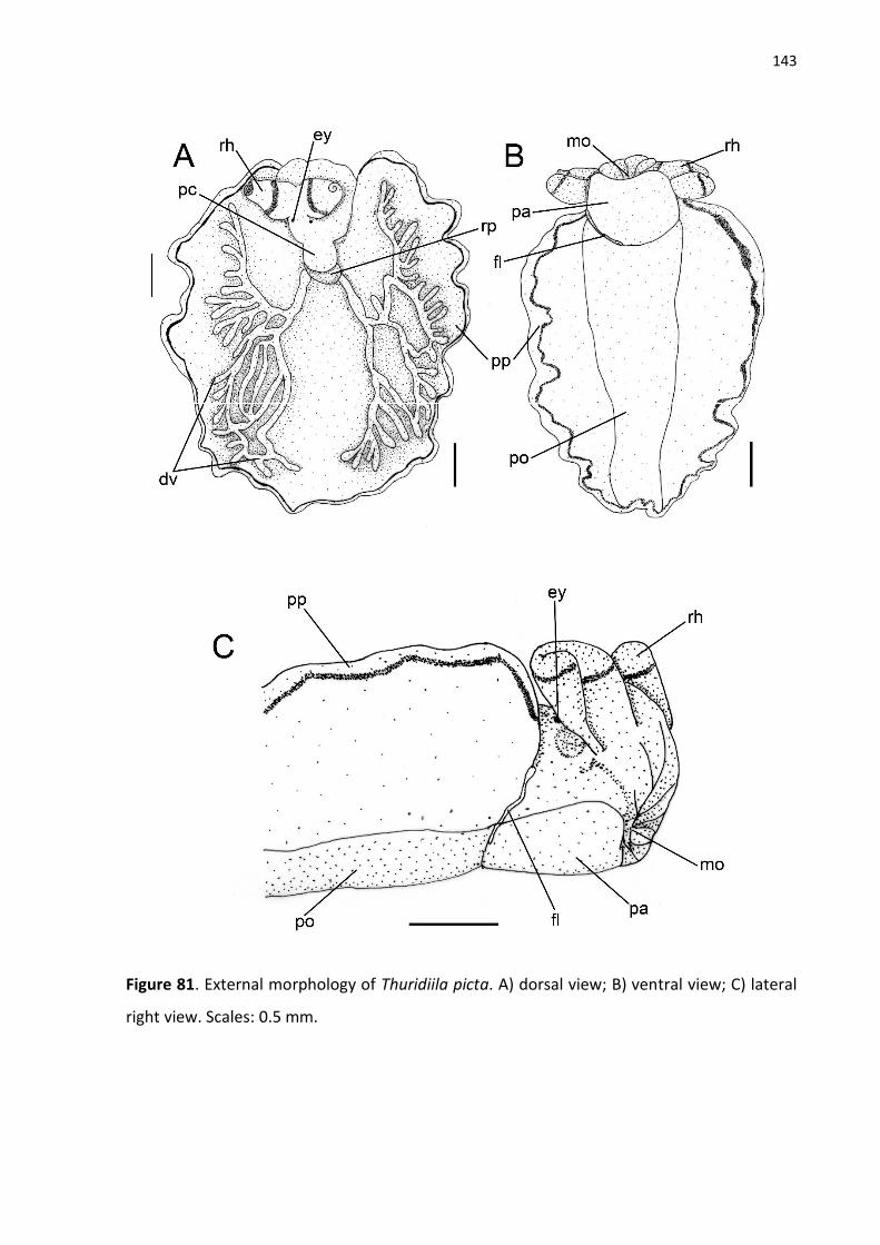
143
Figure 81. External morphology of Thuridiila picta. A) dorsal view; B) ventral view; C) lateral
right view. Scales: 0.5 mm.

144
Figure 82. Digestive system of Thuridilla picta. A) Dorsal view; B) Lateral view; Buccal mass: C)
dorsal view side; D) ventral side. E) lateral right view. Scales: 0.5mm

145
Figure 83. Scan electron microscope images of radula Thuridilla picta: A) general view; B) detail
of the leading tooth; C) detail of older tooth; D) Ascus.

146
Figure 84. Anatomy of Thuridilla picta. A) whole reproductive system, dorsal view; B)
schematic reproductive system; C) renopericardial cavity, ventral view; D) nervous system,
aneterior view; E) nervous system, posterior view. Scales A-C= 0.5 mm; D-E = 0.25 mm.

147
Thuridilla mazda Ortea & Espinosa, 2000
(Figs. 85-89)
Thuridilla mazda Ortea & Espinosa, 2000: 88-90 (Pl. 1); Malaquias et al., 2011: 1-3 p. (Fig. 1);
Redfern, 2013: 287 (Fig. 795 A-B); Camacho-Gacía et al., 2014: 114.
Description
External morphology (Fig. 85-86): Body elongated with 5 mm length in live animals. Head
evident, large, size ~1/3 of total body size, 2x longer than wider. Rhinophores (rh) long, length
as long as head length, rolled, smooth, and with tip bit tapered. Eyes (ey) located
dorsolaterally behind rhinophores. Parapodial sole (pa) ~3x larger than propodium (po) in live
animals; anterior part of propodium bit expanded; mesopodial groove (fl) evident.
Pericardium (pc) and renal prominence (rp) fused forming short globose renopericardial
hump. Dorsal vessels (dv) organized in one single pair, not branched. Parapodium (pp) large,
with regular margin and do not form closure pattern or parapodial tail. Female pore (fp)
located on extreme dorsal point of lateral groove (lg). Male pore (mp) below right rhinophore.
Anus (a) positioned dorsolaterally on posterior right side of head, near to pericardium.
Circulatory and excretory system (Fig. 89C): similar to T. hopei, but kidney is proportionally
smaller than pericardium.
Digestive system (Fig. 87-88): Similar to T. hopei except for the following. Buccal mass (bm)
with pharyngeal pouch (ph) smaller and oral sphincter (os) larger. Radula tooth more
elongated and poited, with lateral cutting edge well-marked, ascus not in spiral. Ascending
limb with 5 well-formed teeth and 2 others in formation. Salivary glands (sg) less thick as in T.
picta, but longer. Esophageal pouch (ep) proportionally smaller than buccal mass.
Reproductive system (Fig. 89A-B): Similar to T. hopei with the following exceptions.
Hermaphrodite ampulla (am) much bigger, bit smaller than mucus gland (mg) size and ~6x
bigger than genital receptacle (gr). Penis (pe) very elongated, total length ~4x longer than base
length. Vas deferens ~2x longer than penis length. First chamber of mucus gland (mg)

148
proportially larger. Bursa copulatrix absent and genital receptacle is directly connected to
oviduct at first chamber.
Nervous system (Fig. 89D-E): Similar to T. hopei with the following exceptions. Cerebro-pleural
ganglia (cp) with 6 innervations. Buccal ganglia (bu) with accessory ganglion ~4x smaller than
buccal ganglion. Pedal ganglia (pg) with pedal commissure bit longer and more evident than
in T. hopei. Abdominal ganglion (ab) size as half as pedal ganglion size. Sub-intestinal (sb)
ganglion size ~2x as small as supra-intestinal ganglion (sp). Connective tissue between
supraintestinal ganglia and abdominal ganglia reduced.
Distribution: Amphi-Atlantic: Azores; Caribbean to Brazil.
Remarks: The most remarkable features of T. mazda were present in original description. The
authors distinguish the only two Western Atlantic Thuridilla species by color pattern of the
body, shape and length of rhinophores (triangle and shortest in T. mazda) and radulae length.
Malaquias et al. (2011) report this species to the Eastern Atlantic Ocean, in Azores Island. The
specimens found were compared only by color pattern, which match almost exactly except by
the lighter colors. Another difference is the body length, the eastern specimen has 17 mm
while the western specimens were commonly reported with 5-8 mm (VALDÉS & ESPINOSA,
2000; REDFERN, 2013) (ORTIGOSSA et al., 2013 found one with 13 mm). Malaquias et al.
(2011) mentioned the importance of a molecular distinction test between Caribbean and
Azorean population, but we reinforce that the morphology of this species is almost unknown,
and this is also necessary. Thuridilla mazda was found for the first time in Brasil in Rocas Atoll,
where the occurrence of T. picta was reported too.
Material examined: BRAZIL, Atol das Rocas, Rio Grande do Norte, CMPHRM 4250B, 1
specimen (H. Galvão Filho; C. Meirelles, 31/viii/2014).

149
Figure 85. Live specimens of Thuridiila mazda. A) dorsal view with closed parapodia; B) lateral.
Body length: 5 mm.

150
Figure 86. External morphology of Thuridiila mazda. A) dorsal view; B) ventral view; C) lateral
right view. Scales: 0.5 mm

151
Figure 87. Digestive system of Thuridilla mazda. A) Dorsal view; B) Lateral view; Buccal mass:
C) dorsal view side; D) ventral side. E) lateral right view. Scales: 0.5mm

152
Figure 88. Scan electron microscope images of radula Thuridilla mazda: A) general view; B)
detail of the leading tooth; C) detail of older tooth.

153
Figure 89. Anatomy of Thuridilla mazda. A) whole reproductive system, dorsal view; B)
schematic reproductive system; C) renopericardial cavity, ventral view; D) nervous system,
aneterior view; E) nervous system, posterior view. Scales A-C= 0.5 mm; D-E = 0.25 mm.

154
Genus Elysia Risso, 1818
Elysia Risso, 1818. Tyoe species Elysia timida Risso, 1818 by monotypy
Laplysia Montagu, 1840. Type species Laplysia viridis Montagu, 1840 by monotypy
Actaeon Ferrusac, 1819. Type species Laplysia viridis Montagu, 1840
Aplysiopterus Delle Chiaje, 1830. Type species Aplysiopterus neapoliatanus Delle Chiaje,
1830 by monotypy
Rhyzobranchus Contraine, 1835. Type species Laplysia viridis Montagu, 1840 by designação
original.
Thallepus Swainson, 1840. Type species Thallepus ornatos Swainson, 1840 by monotypy
? Pterogasteron Pease, 1860. Type species Pterogasteron ornatum Pease, 1860 by monotypy
Elysiella Verrill, 1872. Type species Plakobranchus catulus Gould, 1870 by original
designation.
? Elysiobranchus Pruvot-Fol, 1930. Type species Elysiobranchus mercieri Pruvot-Fol, 1930 by
monotypy
? Elysiopterus Pruvot-Fol, 1946. Type species not designed.
Checholysia Ortea et al., 2005. Type species not designed.
Elysia timida Risso, 1818
(Figs. 90-94)
External morphology (Figs. 90): Body elongated, smooth, reaching up to 10 mm in live
animals. Head evident, with scattered small papillae, length as half as body length, 2x longer
than wider. Rhinophores (rh) rolled, smooth, long, length as longer as head length, and with
rounded tip; papillae small through all rhinophore length. Eyes (e) dorsolateral behind
rhinophores. Foot (f) easily distinguished from parapodia. Mesopodial groove (fl) conspicuous.
Parapodial sole (ps) length bit longer than propodium (po) length in live animals; anterior part
of propodium rounded. Renopericardial cavity (rc) remarkable, well subdivide into
pericardium (pc) and renal pad (rp), length ~5x smaller as parapodial length. Pericardium (pc)
muscular, globose, bit elongated longitudinally, with small papillae. Renal pad very short,
attached to pericardium. Dorsal vessels (dv) organized in one symmetric pair, branching
anteriorly and posteriorly several times Parapodium (pp) large, with irregular edge and

155
papillae on external surface. Tail absent. Lateral groove (lg) starts on anterior extreme of right
parapodia, where female bye (fp) is positioned, and finishes left to middle point of foot line.
Male bye (mp) positioned below to right rhinophore. Anus (a) positioned dorsolaterally on
posterior right side of head, close to anus.
Circulatory and excretory system (Fig. 94): Pericardium (pc) evident, globose, occupying half
of renopericardial cavity. Auricle (au) large, size as half as ventricle (vt) size and with wall
smooth. Ventricle occupying almost all pericardium, shape rounded. Kidney branched,
composed of many renal ridges (rr) connected in lacunar shape. Nephrostome not observed.
Digestive system (Fig. 91-92): Buccal mass (bm) remarkable, size ~20x larger than esophageal
pouch (ep), elongated, 3x longer than wider. Dorsal septate muscle (ds) composed of 10 thin
bundles. Oral sphincter (os) reduced, compose of many circular muscles, size ~10x smaller
than dorsal septate muscle length. Ascus musculature (ma) wide and long, occupying half of
wide and all length of ventral side of buccal mass. Ascus (as) as narrowest as ascus
musculature, short and rounded. Odontophore (od) rounded, parallel to oral cavity. Buccal
pouch absent. Ascus containing discarded teeth not organized. Salivary glands (sg) positioned
dorsal and lateral to esophagus between nervous system and stomach (st). Esophagus (es)
length ~5x shorter than buccal mass length. Esophageal pouch (ep) globose and muscular,
positioned on posterior part of esophagus. Stomach (st) short, flat, width as wider as intestine
(in) width. Digestive gland (dg) with anterior ducts shorter than posterior ducts, run laterally
and anteriorly inside head; posterior ducts run towards parapodial region. Intestine (in)
internally folded through all its extension, finishes in dorsolateral anus (a) positioned near to
lateral groove.
Reproductive system (Fig. 93):
Gonad composed of numerous hermaphrodite follicles (hf) forming two separated areas, each
area in one side of parapodia. Follicles size few variable, size up to 5x smaller than
hermaphrodite ampulla (am). Gametholitic vesicle (gv) absent. Hermaphrodite ducts (hd)
connect all follicles to one central duct which connects to hermaphrodite ampullas duct, width
univariable. Hermaphrodite ampulla paired, each one in each side of parapodia. Male duct

156
(md) connects prostate gland (pr) and vas deferens (vd) to hermaphrodite duct same as
preceding species. Prostate gland composed of many small clustered glands positioned among
follicles. Penis (p) shape conic, curved and elongated, ~8x longer than wider. Penial stylet
absent. Vas deferens (vd) curled through all its length. Oviduct (ov) starts from hermaphrodite
duct after connection with male duct and forms 3 well delimited chambers completely evolved
by mucus gland and finishes on female bye (fp) in lateral groove. Proximal chamber of mucus
gland smallest one, rounded, albumen duct and male duct connect to oviduct in it. Second
chamber of mucus gland biggest one, formed by a curved expansion of glandular oviduct,
where bursa copulatrix connects with oviduct. Distally chamber short and with folds. Albumen
gland (ag) composed of many dorsal thin branches over prostate gland; main duct of albumen
gland runs from hermaphrodite duct, more proximal than male duct connection with
hermaphrodite duct, then branches into many ducts in parapodial area dorsally to prostate
gland. Genital receptacle absent. Bursa copulatrix (bc) absent.
Nervous system (Fig. 94): Cerebro-pleural ganglia (cp) bit larger than pedal ganglia (pg) size;
each ganglion has 6 innervations; commissure cerebro-pleural short. Optic nerve long, bit
longer than cerebro-pleural ganglion length. Buccal ganglia (bg) remarkable, size bit larger
than sub-intestinal ganglion (sb); accessory ganglion absent; each ganglion has 2 innervations;
buccal commissure internal. Pedal ganglia with 5 innervations by ganglion; pedal commissure
simple and short, length bit smaller than pedal ganglion length. Visceral ganglia well
developed. Abdominal ganglion (ab) size as half as pedal ganglion size, with 2 innervations.
Sub-intestinal ganglion size ~4x as small as supra-intestinal ganglion (sp), with 1 innervation.
Supra-intestinal ganglion size 3x smaller than abdominal ganglion size, with 2 innervation;
connective between abdominal ganglion and sub-intestinal ganglion evident and long, length
as longer as sub-intestinal ganglion; connective between abdominal ganglion and supra-
intestinal long, as long as supra-intestinal ganglion length.
Distribution. Mediterranean Sea (THOMPSON & JAKLIN, 1988)
Material examined: SPAIN, Fornells, Menorca, MZSP 132627B, 1 specimen (27/v/2008), Cala
Galdana, Menorca, MZSP 132626B, 1 specimen (26/v/2008).

157
Figure 90. External morphology of Elysia timida. A) dorsal view; B) ventral view.
Figure 91. Digestive system of Elysia timida. A) Dorsal view. Buccal mass: B) dorsal; C) ventral;
D) lateral.

158
Figure 92. Scan electron microscope images of radula Elysia timida: A) general view; B) detail
of the leading tooth; C) detail of older tooth.

159
Figure 93. Reproductive system of Elysia timida.
Figure 94. Circulatory and nervous systems of Elysia timida. A) Renopericardial cavity. B) anterior view of nervous system.

160
Elysia viridis (Montagu, 1804)
(Figs. 95-99)
External morphology (Fig. 95): Body elongated, smooth, no papillae, reaching up to 12 mm in
live animals. Head evident, length up to 1/3 of total body length, 2x longer than wider.
Rhinophores (rh) rolled, smooth, short, length twice as smaller as head length, and with
rounded tip; papillae absent. Eyes (e) dorsolateral behind rhinophores. Foot (f) inconspicuous
and hard to distinguish from parapodia. Mesopodial groove (fl) inconspicuous. Parapodial sole
(ps) length 3x longer than propodium (po) length in live animals; anterior part of propodium
rounded. Renopericardial cavity (rc) remarkable, well subdivide into pericardium (pc) and
renal pad (rp), length 1/6 as longer as parapodial length. Pericardium (pc) muscular, globose,
bit elongated longitudinally, with no papillae. Renal pad very short, attached to pericardium.
Dorsal vessels (dv) organized in 4 symmetric pairs, three anterior pairs run parallel to
parapodia margin and most posterior pair runs posteriorly and branching several times
Parapodium (pp) large, with irregular edge and no papillae. Tail short. Lateral groove (lg) starts
on anterior extreme of right parapodia, where female bye (fp) is positioned, and finishes left
to middle point of foot line. Male bye (mp) positioned below to right rhinophore. Anus (a)
positioned dorsolateral on posterior right side of head, close to pericardium.
Circulatory and excretory system (Fig. 99): Pericardium (pc) evident, globose, occupying half
of renopericardial cavity. Auricle (au) size ~4x smaller than ventricle (vt) size and with wall
folded. Ventricle occupying half of pericardium, shape rounded. Kidney branched, composed
of many renal ridges (rr) connected in trabecular shape. Nephrostome not observed.
Digestive system (Fig. 96-97): Buccal mass (bm) remarkable, size ~20x larger than esophageal
pouch (ep), elongated, 2x longer than wider. Dorsal septate muscle (ds) composed of 26 thin
bundles. Oral sphincter (os) reduced, compose of many circular muscles, size ~10x smaller
than dorsal septate muscle length. Ascus musculature (ma) thin and long, occupying all length
of ventral side of buccal mass. Ascus (as) as narrowest as ascus musculature, short and
rounded. Odontophore (od) pointed, parallel to oral cavity. Buccal pouch absent. Ascus
containing discarded teeth not organized. Salivary glands (sg) positioned ventrally to

161
esophagus and stomach (st). Esophagus (es) length twice shorter than buccal mass length.
Esophageal pouch (ep) globose and muscular, positioned on posterior part of esophagus.
Stomach (st) short, flat, width as wider as intestine (in) width. Digestive gland (dg) with
anterior ducts shorter than posterior ducts, run laterally and anteriorly inside head; posterior
ducts run towards parapodial region. Intestine (in) internally folded through all its extension,
finishes in dorsolateral anus (a) positioned near to lateral groove.
Reproductive system (Fig. 98): Gonad composed of numerous hermaphrodite follicles (hf)
forming two separated areas, each area in one side of parapodia. Follicles size few variable,
size up to 10x smaller than hermaphrodite ampulla (am). Gametholitic vesicle (gv) absent.
Hermaphrodite ducts (hd) connect all follicles to one central duct which connects to
hermaphrodite ampullas duct, width univariable. Hermaphrodite ampulla positioned in
middle of body. Male duct (md) connects prostate gland (pr) and vas deferens (vd) to
hermaphrodite duct same as preceding species. Prostate gland composed of one highly
branched duct positioned on top and among follicles. Penis (p) shape conic and short, bit
longer than wider. Penial stylet absent. Vas deferens (vd) curled through all its length. Oviduct
(ov) starts from hermaphrodite duct after connection with male duct and forms 3 well
delimited chambers completely evolved by mucus gland and finishes on female bye (fp) in
lateral groove. Proximal chamber of mucus gland smallest one, rounded, albumen duct and
male duct connect to oviduct in it. Second chamber of mucus gland biggest one, formed by a
curved expansion of glandular oviduct, where bursa copulatrix connects with oviduct. Distally
chamber short and with folds. Albumen gland (ag) composed of many dorsal thin branches
over prostate gland; main duct of albumen gland runs from hermaphrodite duct, more
proximal than male duct connection with hermaphrodite duct, then branches into many ducts
in parapodial area dorsally to prostate gland. Genital receptacle absent. Bursa copulatrix (bc)
with its own aperture.
Nervous system (Fig. 99): Cerebro-pleural ganglia (cp) size as larger as pedal ganglia (pg) size;
each ganglion has 6 innervations; commissure cerebro-pleural short. Optic nerve long, length
twice as longer as cerebro-pleural ganglion length. Buccal ganglia (bg) remarkable, size bit
larger than sub-intestinal ganglion (sb); accessory ganglion absent; each ganglion has 2

162
innervations; buccal commissure internal. Pedal ganglia with 4 innervations by ganglion; pedal
commissure simple and long, length as longer as pedal ganglion length. Visceral ganglia well
developed. Abdominal ganglion (ab) size as half as pedal ganglion size, has 3 innervations. Sub-
intestinal ganglion size ~5x as small as supra-intestinal ganglion (sp), with 1 innervation. Supra-
intestinal ganglion size 4x smaller than abdominal ganglion size, with 1 innervation; connective
between abdominal ganglion and sub-intestinal ganglion evident and long, length as longer as
sub-intestinal ganglion; connective between abdominal ganglion and supra-intestinal short.
Distribution: Baltic Sea and Mediterranean Sea (THOMPSON, 1988).
Material examined: DENMARK, Roskilde Fjord, MZSP 122698, 8 specimens (K. Jensen,
ix/2013).

163
Figure 95. External morphology of Elysia viridis. A) dorsal; B) ventral; C) lateral.

164
Figure 96. Digestive system of Elysia viridis. A) Dorsal view; B) Lateral view; Buccal mass: C) dorsal; Buccal mass: D) ventral; E) left side; F) right side.

165
Figure 97. Scan electron microscope images of radula Elysia viridis: A-C) general view; B-D)
detail of the leading tooth.

166
Figure 98. Reproductive system of Elysia viridis. A) Schematic drawing; B) Overview.

167
Figure 99. Circulatory and nervous systems of Elysia viridis. A) Renopericardial cavity. B) anterior view of nervous system.
168
Elysia australis (Quoy & Gaimard, 1832)
(Figs. 100-104)
External morphology (Fig. 100): Body elongated, smooth, no papillae, reaching up to 8 mm in
live animals. Head evident, length up to 1/4 of total body length, 2x longer than wider.
Rhinophores (rh) rolled, smooth, short, length twice as smaller as head length, and with
rounded tip; papillae absent. Eyes (e) dorsolateral behind rhinophores. Foot (f) inconspicuous
and hard to distinguish from parapodia. Mesopodial groove (fl) inconspicuous. Parapodial sole
(ps) length 6x longer than propodium (po) length in live animals; anterior part of propodium
rounded. Renopericardial cavity (rc) remarkable, well subdivide into pericardium (pc) and
renal pad (rp), length 1/6 as longer as parapodial length. Pericardium (pc) muscular, globose,
bit elongated longitudinally, with no papillae. Renal pad very short, attached to pericardium.
Dorsal vessels (dv) organized in 3 symmetric pairs, two anterior pairs run parallel to parapodia
margin and most posterior pair runs posteriorly and branching several times Parapodium (pp)
large, with irregular edge and no papillae. Tail absent. Lateral groove (lg) starts on anterior
extreme of right parapodia, where female pore (fp) is positioned, and finishes left to middle
point of foot line. Male pore (mp) positioned below to right rhinophore. Anus (a) positioned
dorsolaterally on posterior right side of head, close to pericardium.
Circulatory and excretory system (Fig. 104): Pericardium (pc) evident, globose, occupying half
of renopericardial cavity. Auricle (au) size as large as ventricle (vt) size, auricle smooth.
Ventricle occupying half of pericardium, shape rounded. Kidney branched, composed of many
renal ridges (rr) connected in trabecular shape. Nephrostome not observed.
Digestive system (Fig. 101-102): Buccal mass (bm) remarkable, size ~5x larger than esophageal
pouch (ep), elongated, 2x longer than wider. Dorsal septate muscle (ds) composed of 24 large
bundles. Oral sphincter (os) evident, compose of many circular muscles, size ~3x smaller than
dorsal septate muscle length. Ascus musculature (ma) wide and long, occupying almost all
width and length of ventral side of buccal mass. Ascus (as) twice narrowest than ascus
musculature, short and straight. Odontophore (od) pointed, parallel to oral cavity. Buccal
pouch absent. Radula with 12 teeth in ascending limb; leading tooth elongated, total length

169
5x longer than base length; cusp bearing numerous small denticles through all extension;
lateral cutting absent. Ascus containing discarded teeth not organized. Salivary glands (sg)
positioned ventrally to esophagus and stomach (st). Esophagus (es) length twice shorter than
buccal mass length. Esophageal pouch (ep) globose and muscular, positioned on posterior part
of esophagus. Stomach (st) short, flat, width as wider as intestine (in) width. Digestive gland
(dg) with anterior ducts shorter than posterior ducts, run laterally and anteriorly inside head;
posterior ducts run towards parapodial region. Intestine (in) internally folded through all its
extension, finishes in dorsolateral anus (a) positioned near to lateral groove.
Reproductive system (Fig. 103): Gonad composed of numerous hermaphrodite follicles (hf)
forming a continuous area through all parapodia with central region of body, under the
renopericardial cavity, with no follicles. Follicles size few variable, size up to 10x smaller than
biggest hermaphrodite ampulla (am). Gametholitic vesicle (gv) absent. Hermaphrodite ducts
(hd) connect all follicles to one central duct which connects to hermaphrodite ampullas duct,
width univariable. Hermaphrodite ampulla absent. Male duct (md) connects prostate gland
(pr) and vas deferens (vd) to hermaphrodite duct same as preceding species. Prostate gland
composed of one highly branched duct positioned on top and among follicles. Penis (p) shape
conic and short, bit longer than wider. Penial stylet absent. Vas deferens (vd) curled near to
penis, length as longer as penis length. Oviduct (ov) starts from hermaphrodite duct after
connection with male duct and forms 3 well delimited chambers completely evolved by mucus
gland and finishes on female bye (fp) in lateral groove. Proximal chamber of mucus gland
smallest one, rounded, albumen duct and male duct connect to oviduct in it. Second chamber
of mucus gland biggest one, formed by a curved expansion of glandular oviduct, where bursa
copulatrix connects with oviduct. Distally chamber short and with folds. Albumen gland (ag)
composed of many dorsal thin branches over prostate gland; main duct of albumen gland runs
from hermaphrodite duct, more proximal than male duct connection with hermaphrodite
duct, then branches into many ducts in parapodial area dorsally to prostate gland. Genital
receptacle absent. Bursa copulatrix (bc) with its own aperture.
Nervous system (Fig. 104): Cerebro-pleural ganglia (cp) size bit smaller than pedal ganglia (pg)
size; each ganglion has 5 innervations; commissure cerebro-pleural short. Optic nerve short,

170
much smaller than cerebro-pleural ganglion length. Buccal ganglia (bg) remarkable, size bit
larger than sub-intestinal ganglion (sb); accessory ganglion absent; each ganglion has 2
innervations; buccal commissure simple and short, length 5x smaller than buccal ganglion
length. Pedal ganglia with 3 innervations by ganglion; pedal commissure simple and long,
length as longer as pedal ganglion length. Visceral ganglia well developed. Abdominal ganglion
(ab) size as half as pedal ganglion size, has 2 innervations. Sub-intestinal ganglion size ~5x as
small as supra-intestinal ganglion (sp), with 1 innervation. Supra-intestinal ganglion bit smaller
than abdominal ganglion size, with 1 innervation; connective between abdominal ganglion
and sub-intestinal ganglion evident and long, length as longer as sub-intestinal ganglion;
connective between abdominal ganglion and supra-intestinal short.
Distribution: Australia (JENSEN, 2007).
Material examined: Albany, Austrália: MZSP 122696, K. R. Jensen col., [9 espécimes],
10/i/1988.
.

171
Figure 100. External morphology of Elysia australis. A) dorsal; B) ventral; C) lateral.

172
Figure 101. Digestive system of Elysia australis. A) Dorsal view; B) Lateral view; Buccal mass:
C) lateral; D) dorsal; E) ventral.

173
Figure 102. Scan electron microscope images of radula of Elysia australis (scale: 10 µm): A-B)
general view; C-D) detail of the leading tooth.

174
Figure 103. Reproductive system of Elysia australis - Schematic drawing.
Figure 104. Circulatory and nervous systems of Elysia australis. A) Renopericardial cavity. B) anterior view of nervous system.

175
Elysia ornata (Swainson, 1840)
Figs. 105-109
See Krug, Vendetti & Valdés, 2016.
External morphology (Figs. 105-106): Body elongated, smooth, reaching up to 32 mm in live
animals. Head evident, length up to 1/4 of total body length, 3x longer than wider.
Rhinophores (rh) rolled, short, ~3x smaller than head length, and with bit tapered tip; papillae
absent. Foot sole (fo) similar to parapodial external surface. Foot line (fl) well evident.
Parapodial sole (ps) 5x larger than propodium (po) in live animals. Anterior part of propodium
rounded. Renopericardial cavity (rc) external, rounded, with renal pad (rp) partially fused with
pericardium (pc), length ~8x smaller than parapodial length; pericardium (pc) muscular,
globose, smooth. Dorsal vessels (dv) in symmetric arrangement, three pairs running through
all parapodia and anastomosing in some parts Parapodium (pp) large, with regular and with
no closure pattern. External face of parapodia smooth with similar ornamentation as rest of
body. Tail absent. Gametholitic vesicles absent.
Coloration (Fig. 105): in agreement with Krug et al., 2016.
Circulatory and excretory system (Fig. 109): Pericardium (pc) evident, globose, occupying 1/4
of renopericardial cavity. Auricle (au) size as half as ventricle (vt) size, auricle wall furrowed.
Ventricle occupying 1/3 or pericardium, shape rounded. Kidney branched, composed of many
renal ridges (rr) connected in trabecular shape. Nephrostome positioned on right side of renal
cavity near to pericardium.
Digestive system (Fig. 107-108): Buccal mass (bm) remarkable, size ~5x larger than esophageal
pouch (ep), elongated, 3x longer than wider; dorsal septate muscle (ds) composed of 24 large
bundles; oral sphincter (os) evident, compose of many circular muscles, size ~5x smaller than
dorsal septate muscle length; ascus musculature (ma) wide and long, occupying half width and
1/3 length of ventral side of buccal mass. Ascus (as) covered by ascus musculature and
straight. Odontophore (od) rounded, parallel to oral cavity. pharyngeal pouch absent. Radula
with 5 teeth in ascending limb; leading tooth elongated, total length ~5x longer than base

176
length; cusp bearing numerous small denticles through all extension; lateral cutting absent;
ascus containing discarded teeth not organized. Salivary glands (sg) positioned ventrally to
esophagus and stomach (st). Esophagus (es) bit bigger than buccal mass length, width ~5x
thinner than intestine width; internal surface with two longitudinal folds through all its
extension. Esophageal pouch (ep) globose and muscular, positioned on posterior part of
esophagus. Stomach (st) bulky, rounded, wide, width as twice as wider intestine (in); folds
internally present on dorsal portion. Digestive gland (dg) dilated forming stomach pouches
well developed, elongated, voluminous and with internal wall folded; anterior ducts shorter
than posterior ducts, run laterally and anteriorly inside head; posterior ducts run towards
parapodial region. Intestine (in) internally folded through all its extension, finishes in
dorsolateral anus (a) positioned near to lateral groove.
Reproductive system (Fig. 109): Gonad composed of numerous hermaphrodite follicles (hf)
forming a continuous area through all parapodia with central region of body, under the
renopericardial cavity, with no follicles. Follicles size few variable, size up to 5x smaller than
biggest hermaphrodite ampulla (am). Gametholitic vesicle (gv) absent. Hermaphrodite ducts
(hd) connect all follicles to one central duct which connects to hermaphrodite ampullas duct,
width univariable. Hermaphrodite ampulla variable in number from 10 to 22 on each side of
body, positioned on parapodia anteriorly to hermaphrodite follicles, size variable, biggest one
bit smaller than genital receptacle (gr) and 1/5 of mucus gland (mg) size. All ampulla on each
side of animal are united for one main duct which joins in a single duct that connects to
hermaphrodite duct. Male duct (md) connects prostate gland (pr) and vas deferens (vd) to
hermaphrodite duct same as preceding species. Prostate gland composed of ona main
glandular duct branching over ampulla and most anterior follicles. Penis (p) shape conic and
short, size bit smaller than mucus gland size, total length as longer as base diameter; stylet
absent. Vas deferens (vd) highly curled posteriorly and straight near to penial base, length ~4x
longer than penis length when outstretched. Oviduct (ov) starts from hermaphrodite duct
after connection with male duct and forms 3 well delimited chambers completely evolved by
mucus gland and finishes on female pore (fp) in lateral groove. Proximal chamber of mucus
gland smallest one, rounded, albumen duct and male duct connect to oviduct in it. Second
chamber of mucus gland biggest one, formed by a curved expansion of glandular oviduct,

177
where bursa copulatrix connects with oviduct. Distally chamber short and with folds. Albumen
gland (ag) composed of many dorsal thin branches over prostate gland; main duct of albumen
gland runs from hermaphrodite duct, more proximal than male duct connection with
hermaphrodite duct, then branches into many ducts in parapodial area dorsally to prostate
gland. Genital receptacle connected to oviduct in anterior position.
Nervous system (Fig. 109): Cerebro-pleural ganglia (cg) size as larger as pedal ganglia (pg);
each ganglion has 6 innervations; commissure cerebro-pleural absent. Optic nerve length
long, bit longer than cerebro-pleural ganglion length. Buccal ganglia (bg) remarkable, size bit
larger than sub-intestinal ganglion (sb); accessory ganglion size ~3x smaller as buccal ganglion;
each ganglion has 2 innervations; buccal commissure simple and long, length as longer as
buccal ganglion length. Pedal ganglia present 3 innervations by ganglion; pedal commissure
reduced. Visceral ganglia well developed; abdominal ganglion (ab) size bit smaller than pedal
ganglion size, has 2 innervations; sub-intestinal ganglion as half as supra-intestinal ganglion
(sp), has 2 innervation; supra-intestinal ganglion bit smaller than abdominal ganglion size, has
1 innervation. Connective among visceral ganglia reduced.
Material examined: PANAMA, Isla Colón, Bocas del Toro, MZSP 121848, 1 specimen; BRAZIL,
Arquipélago de São Pedro e São Paulo, Pernambuco, MZSP 104106, 1 specimen; Arquipélago
de São Pedro e São Paulo, Pernambuco, MZSP 104106, 1 specimen.

178
Figure 105. External morphology of live Elysia ornata live animal: A) dorsal view (length: 22
mm) parapodia opened; B) ventral view of same animal; C) lateral left view (length: 31 mm).

179
Figure 106. External morphology of Elysia ornata (scale: 1 mm): A) dorsal view; B) ventral
view; C) lateral view.

180
Figure 107. Digestive system of Elysia ornata: A) dorsal view and B) lateral view of the whole
system. Buccal mass: C) dorsal; D) ventral and E) lateral right view.

181
Figure 108. Scan electron microscope images of radula of Elysia ornata: A) general view; B-C)
detail of the leading tooth; D) detail of old tooth.

182
Figure 109. Reproductive, circulatory, excretory and nervous systems of Elysia ornata: A)
general view of reproductive system; B) schematic view of reproductive system; C) ventral
view of renopericaldial cavity in 4mm specimen; D) ventral view of renopericaldial cavity in
6mm specimen; E) posterior view of the nervous system; F) anterior of the nervous system.
(scales: A-C = 1 mm; D-E = 0,5 mm).

183
Elysia crispata Mörch, 1863
Figs. 110-114
See Krug, Vendetti & Valdés, 2016.
External morphology (Figs. 110-111): Body elongated, smooth, reaching up to 48 mm in live
animals. Head evident, length up to 1/5 of total body length, 3x longer than wider.
Rhinophores (rh) rolled, smooth, long, bit longer than head length, and slightly opened near
tip. Foot sole (fo) very muscular and quite different from parapodial musculature. Foot line (fl)
well evident. Parapodial sole (ps) 5x larger than propodium (po) in live animals. Anterior part
of propodium slightly expanded. Renopericardial cavity (rc) external, rounded, with renal pad
(rp) partially fused with pericardium (pc), length ~6x smaller than parapodial length;
pericardium (pc) muscular, globose, smooth. Dorsal vessels (dv) in symmetric arrangement,
two pairs running through all parapodia, with anterior most pair anastomosing with anterior
branching of posterior one. Parapodium (pp) large with the edge highly frilled and with no
closure pattern. External face of parapodia smooth with similar ornamentation as rest of body.
Tail absent. Gametholitic vesicles absent.
Coloration (Fig. 110): in agreement with Krug, Vendetti and Valdés, 2016.
Circulatory and excretory system (Fig. 114): Similar with the previous species with the
following exceptions. Pericardium (pc) as half as renal cavity (rc). Auricle (au) size ~4x smaller
than ventricle (vt) size and smooth. Ventricle occupying almost all pericardium.
Digestive system (Fig. 112-113): Similar to E. ornata except for the following. Buccal mass (bm)
quite small comparing animal body, size twice as larger as esophageal pouch (ep), bit rounded,
bit longer than wider; dorsal septate muscle (ds) composed of 12 large bundles; oral sphincter
(os) evident, compose of many circular muscles, size ~4x smaller than dorsal septate muscle
length; ascus musculature (ma) wide but short, finishing at half length of ventral side of buccal
mass. Odontophore region (or) bit elongated. Pharyngeal pouch absent. Radula with 8 well-
formed teeth and two others in formation in ascending limb; leading tooth elongated, total
length ~3x longer than base length; cusp bearing numerous small denticles through all

184
extension; lateral cutting reduced; ascus containing discarded teeth not organized. Stomach
(st) flatten, rounded, wide, width as twice as wider intestine (in); folds internally present on
dorsal portion.
Reproductive system (Fig. 113): Similar to E. ornata with the following exceptions. Gonad
composed of numerous hermaphrodite follicles (hf) on central part of body and absent on
frilled area of parapodia. forming a continuous area through all parapodia. Hermaphrodite
ampulla most numerous and smaller, size bit larger than follicles size. Prostate gland
composed of one main glandular duct branching over all ampulla and follicles area,
concentrating after follicles where parapodia get frilled. Penis (pe) more elongated, ~3x longer
than wider, but proportionally smaller, size bit smaller than genital receptacle (gr) size. Vas
deferens (vd) highly curled near to penial base, length twice as longer as penis length when
outstretched. Mucus gland (mg) as larger and well-developed.
Nervous system (Fig. 113): Similar to E. ornata except for the following. Cerebro-pleural
ganglia (cg) with 5 innervations. Optic nerve shorter than cerebro-pleural ganglion length.
Buccal ganglia (bg) bit smaller than sub-intestinal ganglion (sb); accessory ganglion size ~5x
smaller as buccal ganglion. Buccal commissure short, length ~3x shorter than buccal ganglion
length. Connective between sub-intestinal and abdominal ganglia external and short.
Material examined: PANAMA, Isla Colón, Bocas del Toro, MZSP 121855, 5 specimens.
GRENADA, Anse la Roche beach, L’Appelle, Carriacou, MZSP 108711 2 specimens.

185
Figure 110. External morphology of live Elysia crispata live animal: A) dorsal view (length: 28
mm) parapodia opened; B) ventral view of same animal; C) dorsal view (length: 37 mm); D)
ventral view of same animal.

186
Figure 111. External morphology of Elysia crispata (scale: 1 mm): A) dorsal view; B) ventral
view; C) lateral view.

187
7
Figure 112. Digestive system of Elysia crispata: A) dorsal view and B) lateral view of the
whole system. Buccal mass: C) dorsal; D) ventral and E) lateral right view.

188
Figure 113. Scan electron microscope images of radula of Elysia crispata: A and C) general
view; B and D) detail of the leading tooth.

189
Figure 114. Reproductive, circulatory, excretory and nervous systems of Elysia crispata: A)
general view of reproductive system; B) schematic view of reproductive system; C) ventral
view of renopericaldial cavity in 4mm specimen; D) ventral view of renopericaldial cavity in
6mm specimen; E) posterior view of the nervous system; F) anterior of the nervous system.
(scales: A-C = 1 mm; D-E = 0,5 mm).

190
Elysia diomedea Mörch, 1863
Figs. 115-119
External morphology (Figs. 115-116): Body elongated, smooth, reaching up to 40 mm in live
animals. Head evident, length up to 1/5 of total body length, 3x longer than wider.
Rhinophores (rh) rolled, smooth, long, bit longer than head length, and slightly opened near
tip. Foot sole (fo) very muscular and quite different from parapodial musculature. Foot line (fl)
well evident. Parapodial sole (ps) 5x larger than propodium (po) in live animals. Anterior part
of propodium slightly expanded. Renopericardial cavity (rc) external, rounded, with renal pad
(rp) partially fused with pericardium (pc), length ~6x smaller than parapodial length;
pericardium (pc) muscular, globose, smooth. Dorsal vessels (dv) in symmetric arrangement,
two pairs running through all parapodia, with anterior most pair anastomosing with anterior
branching of posterior one. Parapodium (pp) large with the edge highly frilled and with no
closure pattern. External face of parapodia smooth with similar ornamentation as rest of body.
Tail absent. Gametholitic vesicles absent.
Coloration (Fig. 115): in agreement with Krug, Vendetti and Valdés, 2016.
Circulatory and excretory system (Fig. 119): Similar with the previous species with the
following exceptions. Pericardium (pc) as half as renal cavity (rc). Auricle (au) size ~4x smaller
than ventricle (vt) size and smooth. Ventricle occupying almost all pericardium.
Digestive system (Fig. 117-118): Similar to E. ornata except for the following. Buccal mass (bm)
quite small comparing animal body, size twice as larger as esophageal pouch (ep), bit rounded,
bit longer than wider; dorsal septate muscle (ds) composed of 12 large bundles; oral sphincter
(os) evident, compose of many circular muscles, size ~4x smaller than dorsal septate muscle
length; ascus musculature (ma) wide but short, finishing at half length of ventral side of buccal
mass. Odontophore region (or) bit elongated. Pharyngeal pouch absent. Radula with 8 well-
formed teeth and two others in formation in ascending limb; leading tooth elongated, total
length ~3x longer than base length; cusp bearing numerous small denticles through all
extension; lateral cutting reduced; ascus containing discarded teeth not organized. Stomach

191
(st) flatten, rounded, wide, width as twice as wider intestine (in); folds internally present on
dorsal portion.
Reproductive system (Fig. 119): Similar to E. ornata with the following exceptions. Gonad
composed of numerous hermaphrodite follicles (hf) on central part of body and absent on
frilled area of parapodia. forming a continuous area through all parapodia. Hermaphrodite
ampulla most numerous and smaller, size bit larger than follicles size. Prostate gland
composed of one main glandular duct branching over all ampulla and follicles area,
concentrating after follicles where parapodia get frilled. Penis (pe) more elongated, ~3x longer
than wider, but proportionally smaller, size bit smaller than genital receptacle (gr) size. Vas
deferens (vd) highly curled near to penial base, length twice as longer as penis length when
outstretched. Mucus gland (mg) as larger and well-developed.
Nervous system (Fig. 119): Similar to E. ornata except for the following. Cerebro-pleural
ganglia (cg) with 5 innervations. Optic nerve shorter than cerebro-pleural ganglion length.
Buccal ganglia (bg) bit smaller than sub-intestinal ganglion (sb); accessory ganglion size ~5x
smaller as buccal ganglion. Buccal commissure short, length ~3x shorter than buccal ganglion
length. Connective between sub-intestinal and abdominal ganglia external and short.
Material examined: PANAMA, Isla Colón, Bocas del Toro, MZSP 121855, 5 specimens.
GRENADA, Anse la Roche beach, L’Appelle, Carriacou, MZSP 108711 2 specimens.

192
Figure 115. External morphology of live Elysia diomedea live animal: A) dorsal view (length:
33 mm) parapodia opened; lateral right view of same animal.

193
Figure 116. External morphology of Elysia diomedea (scale: 1 mm): A) dorsal view; B) ventral
view; C) lateral view.

194
Figure 117. Digestive system of Elysia diomedea: A) dorsal view and B) lateral view of the
whole system. Buccal mass: C) dorsal; D) ventral and E) lateral right view.

195
Figure 118. Scan electron microscope images of radula of Elysia diomedea: A) general view;
B) detail of the leading tooth.

196
Figure 119. Reproductive, circulatory, excretory and nervous systems of Elysia diomedea: A)
general view of reproductive system; B) schematic view of reproductive system; C) ventral
view of renopericaldial cavity in 4mm specimen; D) ventral view of renopericaldial cavity in
6mm specimen; E) posterior view of the nervous system; F) anterior of the nervous system.
(scales: A-C = 1 mm; D-E = 0,5 mm).

197
Elysia papillosa Verrill, 1901
Figs. 120-124
See Krug, Vendetti & Valdés, 2016.
External morphology (Fig. 120-121): Body elongated, reaching up to 12 mm in live animals;
ornamented with innumerous papillae scattered through external side of parapodia, head,
pericardium and rhinophores. Head evident, length ~1/4 of total body length, 2x longer than
wider. Rhinophores (rh) short, length as half as head length, rolled, papillose, and with tip
blunt; papillae size small, spread through all rhinophore extension. Eyes (ey) located
dorsolaterally behind rhinophores. Foot (f) with remarkable limit near to parapodia;
parapodial sole (ps) 3x larger than propodium (po) in live animals; anterior part of propodium
laterally expanded; foot line (fl) well evident. Renopericardial cavity (rc) prominent, well
subdivided into pericardium (pc) and renal pad (rp); total length slightly smaller than half of
parapodial length; pericardium (pc) muscular, globose, slight longitudinally elongated,
papillose, size as half as head size; renal pad (rp) smooth, thin-walled, length 3x longer than
pericardium length; dorsal vessels (dv) organized in 6 pairs, arrangement asymmetric; most
posterior pair of vessels runs posteriorly and branching several times, while other pairs branch
towards parapodia edge laterally. Parapodium (pp) large, with irregular edge in scalloped
shape; parapodial closure pattern forming many siphons through all parapodial extension;
each parapodium presents three papillae where siphons are formed. External face of
parapodia with many short papillae with similar size, while inner surface has few smaller and
scattered papillae. Tail (tl) very short, length ~5x smaller than head length, can be
proportionally larger in smaller specimens. Gametic vesicles (gv) remarkable in bigger
specimens, visible through parapodial surface, positioned on half-length of parapodia on same
length position than posterior end of renal pad. Female pore (fp) located on extreme dorsal
point of lateral groove (lg). Male pore (mp) below right rhinophore. Anus (a) positioned
dorsolaterally on posterior right side of head, close to lateral groove, above female pore.
Coloration (Fig. 120): in agreement with Krug et al., 2016.

198
Circulatory and excretory system (Fig. 124): Pericardium (pc) evident, globose, occupying 1/3
of renopericardial cavity; auricle (au) size as large as half of ventricle (vt) size, auricle wall
smooth; ventricle size large, occupying almost all pericardium, shape rounded. Kidney
branched, rounded, composed of many renal ridges (rr) connected in trabecular shape.
Nephrostome not observed.
Digestive system (Fig. 122-123): Buccal mass (bm) remarkable, size ~10x larger than
esophageal pouch (ep), rounded, as longer as wider; dorsal septate muscle (ds) composed of
14 large bundles; oral sphincter (os) evident, composed of small circular muscles, short, length
~4x smaller than dorsal septate muscle length; ascus musculature (ma) wide and short,
occupying almost all width and half-length of ventral side of buccal mass; ascus (as) ~4x
narrowest than ascus musculature, short, length twice as small as buccal mass length, curved
to right side of buccal mass; odontophore (od) elongated, voluminous, not parallel to oral
cavity, prominent, forming evident elongated protuberance ventrally on posterior side of
buccal mass; buccal pouch absent. Radula with 6 teeth in ascending limb; leading tooth
elongated, total length twice as longer as base length; cusp bearing numerous remarkable
denticles through all extension; lateral cutting edges inconspicuous and smooth; ascus
containing discarded teeth not organized. Salivary glands (sg) positioned ventrally and
laterally to esophagus between central nervous system (ns) and esophageal pouch. Esophagus
length as longer as buccal mass length, width as large as intestine width; internal surface with
two folds. Esophageal pouch (ep) globose and muscular, positioned on posterior part of
esophagus; internal surface with many thin complete folds. Stomach (st) flatten, wide, width
~4x wider than intestine (in); folds internally present on dorsal portion; stomach pouches
absent. Digestive gland (dg) paired in each side of stomach; anterior ducts shorter than
posterior ducts, runs laterally and anteriorly inside head; posterior duct runs towards
parapodial region. Intestine (in) width as wider as esophagus, internally folded through all its
extension, finishes in dorsolateral anus (a) positioned near to lateral groove.
Reproductive system (Fig. 124): Gonad composed of numerous hermaphrodite follicles (hf)
forming a continuous area through all parapodia and central region of body; follicles size few

199
variable, size ~8x smaller than hermaphrodite ampulla (am); one gametic vesicle (gv) occur on
each side of body, size up to ~10x larger than follicles and twice as larger as bursa copulatrix;
hermaphrodite ducts (hd) connect all follicles and gamethic vessicles to one central duct which
connects to hermaphrodite ampulla, width wider in branch where connects gametic vesicles
with remaining follicles. Hermaphrodite ampulla positioned mid-centrally in reproductive
system, size bit larger than bursa copulatrix (bc) and 1/3 of mucus gland (mg) size. Male duct
(md) connects prostate gland (pr) and vas deferens (vd) to hermaphrodite duct before
albumen gland (ag) connection; prostate gland ramified in net shape dorsally to follicles
through all parapodial area, composed of one main glandular duct; penis (p) size bit smaller
than ampulla and ~3x smaller than mucus gland size, shape conic and short, total length 3x
bigger as base diameter; stylet present; vas deferens twice longer than penis length,
proximally few undulated and not curled; distally curved and proximally bit straight inside
penis. Oviduct (ov) starts from hermaphrodite duct after connection with male duct and forms
3 well delimited chambers completely evolved by mucus gland, and finishes on female pore
(fp) in lateral groove. Proximal chamber of mucus gland smaller one, elongated, albumen duct
and male duct connect to oviduct connected before it; middle chamber of mucus gland bigger
one, formed by a curved expansion of glandular oviduct; distal chamber elongated and
unfolded. Bursa copulatrix connected to oviduct in distal chamber near to female pore.
Albumen gland (ag) composed of many dorsal thin branches over prostate gland; main duct
of albumen gland runs from hermaphrodite duct, more proximal than male duct connection
with hermaphrodite duct, then branches into many ducts in parapodial area dorsally to
prostate gland.
Nervous system (Fig. 124): Cerebro-pleural ganglia (cp) size as larger as pedal ganglia (pg);
each one has 7 innervations; commissure cerebro-pleural not evident. Optic nerve length
short, bit longer than half of cerebro-pleural ganglion length. Buccal ganglia (bg) remarkable,
size larger than sub-intestinal ganglion (sb); accessory ganglion (ac) present, size ~3x smaller
than buccal ganglion; each ganglion has 3 innervations, which one of them runs from
accessory ganglion; buccal commissure simple, length ~4x smaller than buccal ganglion length.
Pedal ganglia present 3 innervations by ganglion; pedal commissure present, simple and short,
smaller as half as pedal ganglia length. Visceral ganglia well developed; abdominal ganglion
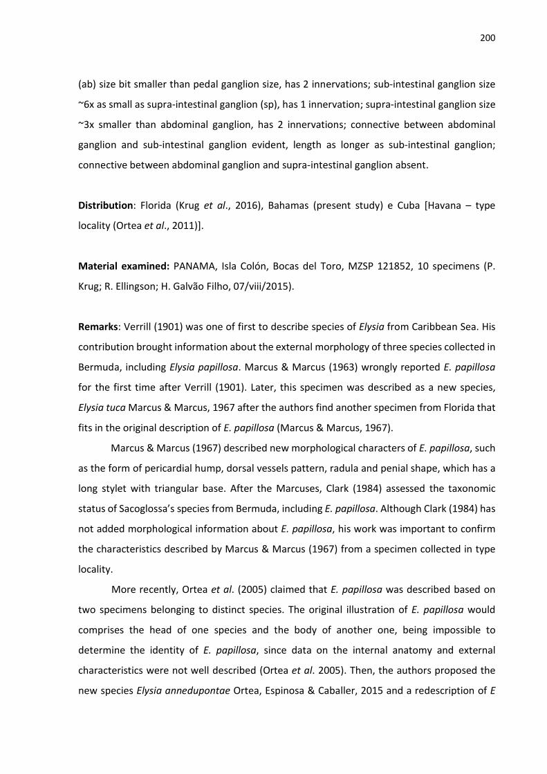
200
(ab) size bit smaller than pedal ganglion size, has 2 innervations; sub-intestinal ganglion size
~6x as small as supra-intestinal ganglion (sp), has 1 innervation; supra-intestinal ganglion size
~3x smaller than abdominal ganglion, has 2 innervations; connective between abdominal
ganglion and sub-intestinal ganglion evident, length as longer as sub-intestinal ganglion;
connective between abdominal ganglion and supra-intestinal ganglion absent.
Distribution: Florida (Krug et al., 2016), Bahamas (present study) e Cuba [Havana – type
locality (Ortea et al., 2011)].
Material examined: PANAMA, Isla Colón, Bocas del Toro, MZSP 121852, 10 specimens (P.
Krug; R. Ellingson; H. Galvão Filho, 07/viii/2015).
Remarks: Verrill (1901) was one of first to describe species of Elysia from Caribbean Sea. His
contribution brought information about the external morphology of three species collected in
Bermuda, including Elysia papillosa. Marcus & Marcus (1963) wrongly reported E. papillosa
for the first time after Verrill (1901). Later, this specimen was described as a new species,
Elysia tuca Marcus & Marcus, 1967 after the authors find another specimen from Florida that
fits in the original description of E. papillosa (Marcus & Marcus, 1967).
Marcus & Marcus (1967) described new morphological characters of E. papillosa, such
as the form of pericardial hump, dorsal vessels pattern, radula and penial shape, which has a
long stylet with triangular base. After the Marcuses, Clark (1984) assessed the taxonomic
status of Sacoglossa’s species from Bermuda, including E. papillosa. Although Clark (1984) has
not added morphological information about E. papillosa, his work was important to confirm
the characteristics described by Marcus & Marcus (1967) from a specimen collected in type
locality.
More recently, Ortea et al. (2005) claimed that E. papillosa was described based on
two specimens belonging to distinct species. The original illustration of E. papillosa would
comprises the head of one species and the body of another one, being impossible to
determine the identity of E. papillosa, since data on the internal anatomy and external
characteristics were not well described (Ortea et al. 2005). Then, the authors proposed the
new species Elysia annedupontae Ortea, Espinosa & Caballer, 2015 and a redescription of E

201
papillosa. A similar E. papillosa was described first by Thompson (1977), which illustrated a
drawing of the dorsal view with closed parapodia and a smooth radula.
Redfern (2013) was the first to report that E. papillosa of Thompson (1977) and Ortea
et al. (2005) was not the same species described by Verrill (1901). Redfern (2013) noticed that
the specimens collected and named as Elysia sp. A did not swim as mentioned by Verrill (1901)
in the original description of E. papillosa. After his observation, other authors considered that
Elysia papillosa sensu Ortea et al. (2005) was an undescribed species (Camacho-García et al.,
2014; Galvão-Filho et al., 2015).

202
Figure 120. External morphology of live Elysia papillosa: A) dorsal view (length: 12 mm); B)
ventral view (length 12 mm); C) dorsal view open parapodia (length: 11 mm); D) dorsal view
juvenile (length: 4 mm).
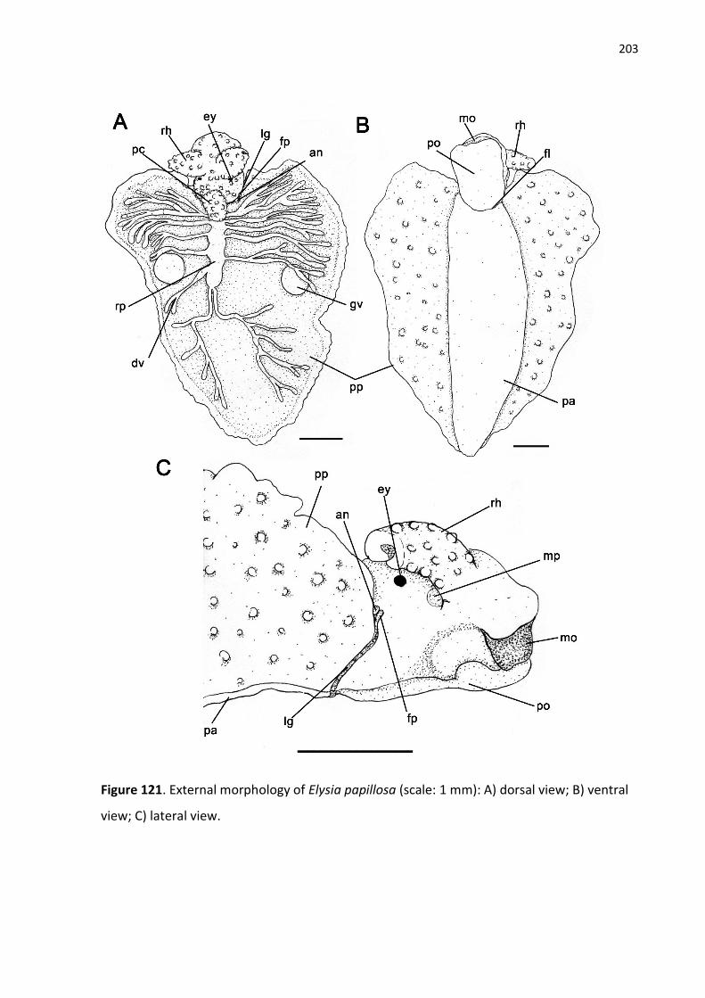
203
Figure 121. External morphology of Elysia papillosa (scale: 1 mm): A) dorsal view; B) ventral
view; C) lateral view.

204
Figure 122. Digestive system of Elysia papillosa (scale: 0,5 mm): A) dorsal view of digestive
system; B) lateral view of digestive system; C-F) buccal mass: C) dorsal; D); ventral; E) right
side; F) left side; G) detail morphology of posterior digestive system; H) Detail morphology of
buccal mass.

205
Figure 123. Scan electron microscope images of radula of Elysia papillosa (scale: 10 µm): A)
general view; B) detail of the leading tooth.

206
Figure 124. Reproductive, circulatory, excretory and nervous systems of Elysia papillosa (scale:
0,5 mm): A) general view of reproductive system; B) schematic view of reproductive system;
C) ventral view of renopericaldial cavity in 4mm specimen; D) ventral view of renopericaldial
cavity in 6mm specimen; E) posterior view of the nervous system; F) anterior of the nervous
system.

207
Elysia cornigera Nutall, 1989
See Krug, Vendetti & Valdés, 2016
(Figs. 125-128)
External morphology (Figs. 125-126): Body elongated, papillose, reaching up to 12 mm in live
animals. Head evident, with scattered small papillae, length as half as body length, 2x longer
than wider. Rhinophores (rh) rolled, papillose, long, length as longer as head length, and with
rounded tip; papillae small through all rhinophore length. Eyes (e) dorsolateral behind
rhinophores. Foot (f) easily distinguished from parapodia. Mesopodial groove (fl) conspicuous.
Parapodial sole (ps) length bit longer than propodium (po) length in live animals; anterior part
of propodium rounded. Renopericardial cavity (rc) remarkable, well subdivide into
pericardium (pc) and renal pad (rp), length ~5x smaller as parapodial length. Pericardium (pc)
muscular, globose, bit elongated longitudinally, with small papillae. Renal pad very short,
attached to pericardium. Dorsal vessels (dv) organized in one symmetric pair, branching
anteriorly and posteriorly several times Parapodium (pp) large, with irregular edge and
papillae on external surface. Tail absent. Lateral groove (lg) starts on anterior extreme of right
parapodia, where female bye (fp) is positioned, and finishes left to middle point of foot line.
Male bye (mp) positioned below to right rhinophore. Anus (a) positioned dorsolaterally on
posterior right side of head, close to anus.
Coloration (Fig. 125): Body with yellowish general color in live animals. Intense green
coloration on the foot and on the inside of the parapodia. Staining pattern formed by
numerous white dots close to the renopericardial and inner surface of parapodia. Orange
spots scattered over head and outer surface of parapodia. Renopericardial cavity yellowish on
anterior bytion and whitish posteriorly and orange dots through all its extension. Parapodial
margin colored as rest of body. Rhinophores yellowish and with some red dots scattered along
its length.
Circulatory and excretory system (Fig. 128): Pericardium (pc) evident, globose, occupying half
of renopericardial cavity. Auricle (au) very short, size ~12x smaller than ventricle (vt) size and
with wall smooth. Ventricle occupying almost all pericardium, shape rounded. Kidney
208
branched, composed of many renal ridges (rr) connected in lacunar shape. Nephrostome not
observed.
Digestive system (Fig. 127): Buccal mass (bm) remarkable, size ~20x larger than esophageal
pouch (ep), elongated, 3x longer than wider. Dorsal septate muscle (ds) composed of 10 thin
bundles. Oral sphincter (os) reduced, compose of many circular muscles, size ~10x smaller
than dorsal septate muscle length. Ascus musculature (ma) wide and long, occupying half of
wide and all length of ventral side of buccal mass. Ascus (as) as narrowest as ascus
musculature, short and rounded. Odontophore (od) rounded, parallel to oral cavity. Buccal
pouch absent. Ascus containing discarded teeth not organized. Salivary glands (sg) positioned
dorsal and lateral to esophagus between nervous system and stomach (st). Esophagus (es)
length ~5x shorter than buccal mass length. Esophageal pouch (ep) globose and muscular,
positioned on posterior part of esophagus. Stomach (st) short, flat, width as wider as intestine
(in) width. Digestive gland (dg) with anterior ducts shorter than posterior ducts, run laterally
and anteriorly inside head; posterior ducts run towards parapodial region. Intestine (in)
internally folded through all its extension, finishes in dorsolateral anus (a) positioned near to
lateral groove.
Reproductive system (Fig. 128): Gonad composed of numerous hermaphrodite follicles (hf)
forming two separated areas, each area in one side of parapodia. Follicles size few variable,
size up to 5x smaller than hermaphrodite ampulla (am). Gametholitic vesicle (gv) absent.
Hermaphrodite ducts (hd) connect all follicles to one central duct which connects to
hermaphrodite ampullas duct, width univariable. Hermaphrodite ampulla paired, each one in
each side of parapodia. Male duct (md) connects prostate gland (pr) and vas deferens (vd) to
hermaphrodite duct same as preceding species. Prostate gland composed of many small
clustered glands positioned among follicles. Penis (p) shape conic, curved and elongated, ~8x
longer than wider. Penial stylet absent. Vas deferens (vd) curled through all its length. Oviduct
(ov) starts from hermaphrodite duct after connection with male duct and forms 3 well
delimited chambers completely evolved by mucus gland and finishes on female bye (fp) in
lateral groove. Proximal chamber of mucus gland smallest one, rounded, albumen duct and

209
male duct connect to oviduct in it. Second chamber of mucus gland biggest one, formed by a
curved expansion of glandular oviduct, where bursa copulatrix connects with oviduct. Distally
chamber short and with folds. Albumen gland (ag) composed of many dorsal thin branches
over prostate gland; main duct of albumen gland runs from hermaphrodite duct, more
proximal than male duct connection with hermaphrodite duct, then branches into many ducts
in parapodial area dorsally to prostate gland. Genital receptacle absent. Bursa copulatrix (bc)
absent.
Nervous system (fig. 128): Cerebro-pleural ganglia (cp) bit larger than pedal ganglia (pg) size;
each ganglion has 6 innervations; commissure cerebro-pleural short. Optic nerve long, bit
longer than cerebro-pleural ganglion length. Buccal ganglia (bg) remarkable, size bit larger
than sub-intestinal ganglion (sb); accessory ganglion absent; each ganglion has 2 innervations;
buccal commissure internal. Pedal ganglia with 5 innervations by ganglion; pedal commissure
simple and short, length bit smaller than pedal ganglion length. Visceral ganglia well
developed. Abdominal ganglion (ab) size as half as pedal ganglion size, with 2 innervations.
Sub-intestinal ganglion size ~4x as small as supra-intestinal ganglion (sp), with 1 innervation.
Supra-intestinal ganglion size 3x smaller than abdominal ganglion size, with 2 innervation;
connective between abdominal ganglion and sub-intestinal ganglion evident and long, length
as longer as sub-intestinal ganglion; connective between abdominal ganglion and supra-
intestinal long, as long as supra-intestinal ganglion length.
Distribution: Caribbean Sea (VALDÉS et al., 2006).
Material examined: PANAMA, Isla Colón, Bocas del Toro, MZSP 131252, 1 specimens (P. Krug;
R. Ellingson; H. Galvão Filho, 07/viii/2015).

210
Figure 125. Live specimen of Elysia cornigera. A) dorsal; B) ventral.
Figure 126. External morphology of Elysia cornigera. A) dorsal; B) ventral.

211
Figure 127. Digestive system of Elysia cornigera. A) Dorsal view. Buccal mass: B) dorsal; C) lateral; D) ventral.

212
Figura 128. Reproductive system of Elysia cornigera.
Figure 129. Circulatory and nervous systems of Elysia cornigera. A) Renopericardial cavity. B) anterior view of nervous system.

213
Elysia zuleicae Ortea & Espinosa, 2002
(Figs. 129-133)
See. Krug, Vendetii & Valdés, 2016
External morphology (Figs. 129-130): Body elongated, reaching up to 12 mm in live animals;
ornamented with small papillae scattered through external side of parapodia, head,
pericardium and rhinophores. Head evident, length ~1/5 of total body length, 2x longer than
wider. Rhinophores (rh) folded, long, length as longer as head length, and with tip and base
unfolded; papillae size small, spread through all rhinophore extension, but more concentrated
on tip half. Eyes (e) dorsolateral behind rhinophores. Foot (f) remarkable distinct from
parapodia; foot line (fl) well evident; parapodial sole (ps) 3x larger than propodium (po) in live
animals; anterior part of propodium expanded laterally. Renopericardial cavity (rc)
remarkable, well subdivide into pericardium (pc) and renal pad (rp), length ~4x smaller than
parapodial length; pericardium (pc) muscular, globose, slight longitudinally elongated,
papillose, length ~3x smaller than head length; renal pad smooth, thin-walled, length twice as
longer as pericardium length; dorsal vessels (dv) organized in 2 symmetric pairs; most
posterior pair of vessels runs posteriorly and branching several times, while anterior pair
branch towards parapodia edge laterally. Parapodium (pp) large, with irregular edge and
papillae; parapodia closure pattern regular, with six apertures, do not form parapodial siphon.
External face of parapodia with dispersed small papillae, while inner surface has no
ornamentation. Tail (tl) short, length ~3x smaller than head length. Gametholitic vesicles (gv)
remarkable in bigger specimens, visible though parapodial surface, positioned on half-length
of parapodia or posteriorly after renal pad. Lateral groove (lg) starts where right parapodia
meets neck line, which female pore (fp) is located on, and finishes left to middle point of foot
line. Male pore (mp) positioned below to right rhinophore. Anus (a) positioned dorsolaterally
on posterior right side of head, really close to lateral groove, above female pore.
Coloration (Fig. 129): in agreement with Krug et al., 2016.

214
Circulatory and excretory system (Fig. 133): Pericardium (pc) evident, globose, occupying 1/4
of renopericardial cavity; auricle (au) size as large ventricle (vt) size, auricle wall furrowed;
ventricle size occupying 1/3 or pericardium, shape rounded. Kidney branched, composed of
many renal ridges (rr) connected in trabecular shape. Nephrostome not observed.
Digestive system (Fig. 131-132): Buccal mass (bm) remarkable, size ~10x larger than
esophageal pouch (ep), elongated, more longer than wider; dorsal septate muscle (ds)
composed of 11 large bundles; oral sphincter (os) evident, compose of a pair of large muscles,
size ~5x smaller than dorsal septate muscle length; ascus musculature (ma) narrow and long,
occupying almost all width and 1/3 length of ventral side of buccal mass; ascus (as) twice
narrowest than ascus musculature, elongated, length trespassing ventral length of buccal
mass, straight; odontophore (od) short, parallel to oral cavity, inconspicuous; buccal pouch
(bp) connected near to oral sphincter, size half of buccal mass size. Radula with 9 teeth in
ascending limb; leading tooth elongated, total length twice as longer as base length; cusp
bearing numerous small denticles through all extension; lateral cutting edges inconspicuous
and smooth; ascus containing discarded teeth not organized. Salivary glands (sg) positioned
ventrally and laterally to esophagus between central nervous system (ns) and esophageal
pouch. Esophagus (es) length as longer as buccal mass length, width as large as intestine
width; internal surface with two longitudinal folds through all its extension. Esophageal pouch
(ep) globose and muscular, positioned on posterior part of esophagus; internal surface with
two complete large folds. Stomach (st) flatten, wide, width ~4x wider than intestine (in); folds
internally present on dorsal portion; stomach pouches absent. Digestive gland (dg) paired in
each side of stomach; anterior ducts shorter than posterior ducts, run laterally and anteriorly
inside head; posterior ducts run towards parapodial region. Intestine (in) internally folded
through all its extension, finishes in dorsolateral anus (a) positioned near to lateral groove.
Reproductive system (Fig. 133): Similar to E. papillosa except for: hermaphrodite ampulla
larger than other reproductive structures, almost twice as larger as mucus gland (mg) size, and
~20x bigger than follicles; penis (p) length as longer as mucus length, shape conic and
elongated, total length 4x bigger as base diameter, stylet absent; vas deferens bit longer than
penis length, few undulated and not curled.

215
Nervous system (Fig. 133): Cerebro-pleural ganglia (cp) size bit larger than pedal ganglia (pg);
each ganglion has 7 innervations; commissure cerebro-pleural not evident. Optic nerve length
short, bit longer than half of cerebro-pleural ganglion length. Buccal ganglia (bg) remarkable,
size larger than sub-intestinal ganglion (sb); accessory ganglion present, size ~3x smaller than
buccal ganglion; each ganglion has 2 innervations, which one of them runs from accessory
ganglion; buccal commissure simple. Pedal ganglia present 2 innervations by ganglion; pedal
commissure short, much smaller than pedal ganglion length and ~3x longer than buccal
commissure. Visceral ganglia well developed; abdominal ganglion (ab) size bit smaller than
pedal ganglion size, has 3 innervations; sub-intestinal ganglion size ~6x as small as supra-
intestinal ganglion (sp), has 1 innervation; supra-intestinal ganglion bit larger than half of
abdominal ganglion size, has 1 innervations; connective between abdominal ganglion and sub-
intestinal ganglion evident and short; connective between abdominal ganglion and supra-
intestinal ganglion absent.
Distribution: Florida (Krug et al., 2016), Bahamas (present study) e Cuba [Havana – type
locality (Ortea et al., 2011)].
Material examined: PANAMA, Isla Colón, Bocas del Toro, MZSP 121853, 6 specimens (P. Krug;
R. Ellingson; H. Galvão Filho, 07/viii/2015).

216
Figure 129. External morphology of live Elysia zuleicae: A-B) dorsal view (length: 11 mm); B)
ventral view (length 11 mm); C) dorsal view juvenile (length: 4 mm); E) lateral view (length:
10 mm).

217
Figure 130. External morphology of Elysia zuleicae (scale: 1 mm): A) dorsal view; B) ventral
view; C) lateral view.

218
Figure 131. Digestive system of Elysia papillosa (scale: 0,5 mm): A) dorsal view of digestive
system; B) lateral view of digestive system; C-E) zuleicae mass: C) dorsal; D); ventral; E) right
side; F) detail morphology of posterior digestive system; G) Detail morphology of buccal
mass.

219
Figure 132. Scan electron microscope images of radula of Elysia zuleicae (scale: 10 µm): A)
general view; B) detail of the leading tooth.

220
Figure 133. Reproductive, circulatory, excretory and nervous systems of Elysia zuleicae
(scale: 0,5 mm): A) general view of reproductive system; B) schematic view of reproductive
system; C) ventral view of renopericaldial cavity in 4mm specimen; D) ventral view of
renopericaldial cavity in 6mm specimen; E) posterior view of the nervous system; F) anterior
of the nervous system.

221
Elysia orientalis Ortea, Moro, Caballer & Espinosa, 2011 (Fig. 134-1388)
Elysia orientalis Ortea, Moro, Caballer & Espinosa, 2011:205, fig. 5; Krug et al., 2016:94, fig. 56B. External morphology (Fig. 134-135): Body smooth, elongated, reaching up to 6 mm in live
animals. Head evident, length ~1/3 of total body length, 2x longer than wider. Rhinophores
(rh) very short, rolled, smooth, length ~1/5 of head length, and with tip rounded. Eyes (y)
located dorsolaterally behind rhinophores. Foot (f) with remarkable limit near to parapodia;
parapodial sole (ps) twice as large as propodium (po) in live animals; anterior part of
propodium rounded. Renopericardial cavity (rc) inconspicuous, slightly globose, twice as small
as parapodia length. Dorsal vessels absent. Parapodia(pp) short, with irregular edge and no
papillae, begin after lateral groove and end at third part of body length; parapodia pattern
totally opened and do not form parapodial siphon. Tail (tl) long as head length. Female pore
(fp) located on extreme dorsal point of lateral groove (lg). Male pore (mp) below right
rhinophore. Anus (an) positioned dorsolaterally on posterior part of head, above lateral
groove and near to pericardium.
Coloration (Fig. 134): Body of live animals translucent yellowish. Dorsal and lateral body
portion covered by innumerous white and few orange dots; greenish transversal pattern
between eyes and posteriorly to parapodia. Foot intense green with scattered colorful bright
orange, blue, green and white dots. Tip of tail translucent with fewer white dots than rest of
body. Rhinophores translucent, white dots through all extension, orange dots mainly on tips,
and green longitudinal line from digestive gland branch.
Digestive system (Fig. 136-137): Buccal mass (bm) size ~4x larger than esophageal pouch (ep),
rounded, as longer as wider; dorsal septate muscle (ds) composed of 16 thin bundles; oral
sphincter (os) inconspicuous, compose of small circular muscles, size ~6x smaller than dorsal
septate muscle length; ascus musculature (ma) wide and short, occupying almost all width
and half-length of ventral side of buccal mass; ascus (as) external, ~5x narrowest than ascus
musculature, long, curled twice to left of buccal mass; odontophore (od) rounded and parallel
to oral cavity. Radula with 6 teeth on ascending limb; leading tooth elongated, total length

222
twice as longer as base length; cusp bearing numerous very small denticles through all
extension; lateral cutting edges remarkable, partially serrated, small denticles present only in
first third near to tooth tip. Ascus containing discarded teeth organized in curled row following
ascus length. Salivary glands (sg) positioned dorsally and laterally to esophagus between
central nervous system (ns) and esophageal pouch. Esophagus length bit shorter than buccal
mass length, width ~3x thinner than intestine width; internal surface with no folds. Esophageal
pouch (ep) globose and muscular, positioned on posterior part of esophagus, size 6x smaller
than buccal mass; internal surface with no folds. Stomach (st) size similar to esophageal pouch,
globose shape, internally presents one big fold coming from intestine (in); stomach pouches
(sc) size bit smaller than stomach and not subdivided. Digestive gland (dg) paired in each
stomach pouch; anterior ducts shorter than posterior ducts, run laterally and anteriorly in
head; posterior duct run toward parapodial area and tail. Intestine (in) twice as wider as
esophagus, bit thinner than stomach, and folded through all extension, finishes in dorsolateral
anus (a) positioned near to pericardium.
Reproductive system (Fig. 138): Gonad composed of 9-12 hermaphrodite follicles (hf) on each
side of body and forming two isolated groups; follicles size few variable and large as
hermaphrodite ampulla (am), distributed through parapodial area; gametolytic vesicles
absent; hermaphrodite ducts (hd) connect all follicles to one central duct which connects to
hermaphrodite ampulla. Hermaphrodite ampulla positioned laterally in reproductive system,
on left side under mucus gland (mg), size bit small than genital receptacle (gr). Male duct (md)
connects prostate gland (pr) and vas deferens (vd) to hermaphrodite duct after
hermaphrodite ampulla connection; prostate gland dorsally ramified over follicles through all
parapodial area, composed of one main glandular duct; vas deferens twice as longer as penis
length, few undulated though all extension, not curled; penis (pn) size 3x bigger than ampulla
and 4x smaller than mucus gland (mg), shape conic and short, total length twice as longer as
base diameter, stylet absent. Oviduct (ov) starts from hermaphrodite duct after connection
with male duct and forms 3 well delimited chambers completely evolved by mucus gland, and
finishes on female pore (fp) in lateral groove; proximal chamber of mucus gland smallest one,
where male duct and albumen duct connect to oviduct; second chamber of mucus gland
biggest one, where oviduct forms a circular handle and connects to genital receptacle; distally

223
chamber rounded, size ~3x smaller than remaining mucus gland, form a dorsal darker
glandular area with simple lumen connected distally to oviduct after genital receptacle
connection. Albumen gland (ag) composed of many thin branches ventrally to follicles; main
duct of albumen gland runs from oviduct next to hermaphrodite duct, branching into smaller
ducts in parapodial area dorsally to prostate gland. Genital receptacle connected to oviduct in
distally position near to female pore, size bit larger than hermaphrodite ampulla.
Circulatory and excretory system (Fig. 138): Pericardium (pc) very small, occupying 1/4 of
renopericardial cavity; auricle (au) inconspicuous, size more than 10x smaller than ventricle
(vt); auricle wall smooth; ventricle size large, occupying almost all pericardium, shape piriform.
Kidney short, branched, composed of 3 (in 4mm specimen) or 5 (in 6 mm specimen) renal
ridges (rr) arranged radially, bisected posteriorly where they connect to each other in short
network. Nephrostome not observed.
Nervous system (Fig. 138): Cerebro-pleural ganglia (cp) size bit larger than pedal ganglia (pg);
each ganglion has 6 innervations; commissure cerebro-pleural absent. Optic nerve length
short, half of cerebro-pleural ganglion length. Buccal ganglia (bg) remarkable, size larger than
sub-intestinal ganglion (sb); each ganglion has three innervations; buccal commissure absent.
Pedal ganglia present 3 innervations by ganglion; pedal commissure inconspicuous. Visceral
ganglia well developed; abdominal ganglion (ab) size larger than half of pedal ganglia size, has
2 innervations; sub-intestinal ganglion size 3x as small as supra-intestinal ganglion (sp), has 1
innervation; supra-intestinal ganglion size twice as small as abdominal ganglion, has 1
innervation; connective between abdominal ganglion and sub-intestinal ganglion ~3x smaller
than connective between abdominal ganglion and supra-intestinal ganglion.
Distribution: Florida (KRUG, VENDETII & VALDÉS, 2016), Bahamas (present study) e Cuba
[Havana – type locality (ORTEA et al., 2011)].
Remarks
The two specimens analyzed herein have similar color pattern as the original
description, except for blue dots are exclusively on foot instead through dorsal surface. Also,
lateral body color was original described as pinkish, while Bahamian specimens have opaque

224
whitish color. The oblique eyes are diagnostic character in original description, nevertheless
this character is only evident in the smallest specimen. Rhinophores were not described
regarding their size, but the original illustration shows the rhinophores as longer as head
length, whereas the specimens analyzed present a really short rhinophore, almost 1/5 of head
length.
Elysia orientalis is easily identifiable due to its external characters, such as remarkable
oblique parapodial shape, long tail and absence of dorsal vessels. Elysia chitwa Marcus, 1955
is another western Atlantic Elysia species without dorsal vessels as compared by Ortea et al.
(2011). According to Krug et al. (2016) original description of E. chitwa is not much detailed,
then the status of nomen dubium was attributed to it. However, morphological traits of
external and internal morphology described by Marcus (1955, 1957) still allows comparison.
Other similarities between E. orientalis and E. chitwa are on the reproductive system,
including the anterior connection of genital receptacle, oviduct forming a handle surrounded
by mucus gland and the single hermaphrodite ampulla (see Marcus, 1955). Apart from
differences on body coloration, E. chitwa has a different parapodial shape and tail absent,
which externally distinguishes it from E. orientalis. Internally, E. chitwa has the kidney as a
simple sac (Marcus, 1957, p. 414), while E. orientalis has a branched kidney type. Although not
specified on the original description, the Marcus’ drawings of E. chitwa reveals that ascus is
not spiral (Marcus, 1957, p. 409, fig. 45), radula tooth does not have denticulate lateral edges
(Marcus, 1955, p.6 fig. 57) and proximal chamber of mucus gland is not evident as in E.
orientalis (Marcus, 1955, pl. 7, fig. 58).
Phylogenetically, Elysia orientalis is related to Elysia tomentosa complex, which
comprises large Caulerpa feeder species (Krug et al., 2016). The species on this clade present
papillose body, large parapodia, long renopericaridal prominence and no tail (Krug et al.,
2016), whereas E. orientalis has a smooth body, short parapodia and kidney and a long tail.
Except for Elysia pratensis Ortea & Espinosa, 1996, the radulae among the Caribbean species
on E. tomentosa complex are very similar. The leading tooth is elongate with curved cusp
bearing very fine, blunt denticles, pointed tip on club-shaped apex, and lateral edges
inconspicuous (see Elysia subornata Verrill, 1901, Elysia pawliki Krug, Vendetti & Valdés, 2016
and Elysia zemi Krug, Vendetti & Valdés, 2016 in Krug et al., 2016). On the other hand, E.

225
orientalis presents an elongated leading tooth with straight denticulate cusp, a very pointed
teeth apex, and remarkable lateral edges with denticles.
Regarding the morphological comparison of this study, we are aware about the
difficulties to infer similarities among species in the genus Elysia due to lacking detailed
information other than on radular, penis and external morphology. Besides the morphological
description of the rare E. orientalis, the goal of this study is to support the morphological study
of the genus Elysia as a resource for future taxonomy and phylogenetic study of the genus
Elysia.
Figure 134. External morphology of live Elysia orientalis: A) dorsal view (length: 6 mm); B)
dorsal view (length 4 mm); C) ventral view (length: 4 mm); D) lateral view (length: 6 mm).

226
Figure 135. Drawings of the external morphology of Elysia orientalis (scale: 1 mm): A) dorsal
view; B) ventral view; C) lateral view.

227
Figure 136. Digestive system of Elysia orientalis (scale: 0,5 mm): A) dorsal view of digestive
system; B) lateral view of digestive system; C) detailed morphology of posterior digestive
system; D-G) buccal mass: D) dorsal; E); ventral; F) right side; G) left side.

228
Figure 137. Scan electron microscope images of radula of Elysia orientalis BMSM 59035
(scale: 10 µm): A) general view; B) detail of the leading tooth; C) detail of teeth on ascus.

229
Figure 138. Reproductive, circulatory, excretory and nervous systems of Elysia orientalis
(scale: 0,5 mm): A) general view of reproductive system; B) schematic view of reproductive
system; C) ventral view of renopericaldial cavity in 4mm specimen; D) ventral view of
renopericaldial cavity in 6mm specimen; E) anterior view of the nervous system; F) posterior
of the nervous system.

230
Elysia pawliki Krug, Vendetti & Valdés, 2016
(Figs. 139-144)
External morphology (Figs. 139-140): Body elongated, reaching up to 30 mm in live animals;
ornamented with irregular papillae scattered through both sides of parapodia, head,
pericardium, renal pad and rhinophores. Head evident, length up to 1/5 of total body length,
2x longer than wider. Rhinophores (rh) rolled, short, bit smaller than head length, and with
rounded tip; papillae size variable, spread through all rhinophore extension. Eyes (e)
dorsolateral behind rhinophores. Foot (f) remarkable distinct from parapodia; foot line (fl) well
evident; parapodial sole (ps) 3x larger than propodium (po) in live animals; anterior part of
propodium rounded. Renopericardial cavity (rc) remarkable, well subdivide into pericardium
(pc) and renal pad (rp), length half as longer as parapodial length; pericardium (pc) muscular,
globose, papillose, length ~6x smaller than head length; renal pad few papillose, thin-walled,
length 4x longer than pericardium length; dorsal vessels (dv) in asymmetric arrangement, five
vessels in one side of parapodia and six in another one, which right/left variation among
specimens, most posterior pair of vessels runs posteriorly and branching several times, while
anterior pair branch towards parapodia edge. Parapodium (pp) large, with irregular edge and
papillae; parapodia closure pattern regular, with two apertures, most anterior aperture bit
bigger than posterior one, one big rounded glandular structure (gs) present on each extreme
point of both aperture. External face of parapodia with elongated papillae varying in size,
while inner surface has few small and scattered papillae. Tail absent. Gametholitic vesicles
absent. Lateral groove (lg) starts on anterior extreme of right parapodia, where female pore
(fp) is positioned, and finishes left to middle point of foot line. Male pore (mp) positioned
below to right rhinophore. Anus (a) positioned dorsolaterally on posterior right side of head,
close to lateral groove, above female pore.
Coloration (Fig. 139): in agreement with Krug et al., 2016.
Circulatory and excretory system (Fig. 144): Pericardium (pc) evident, globose, occupying 1/5
of renopericardial cavity. Auricle (au) size 3x smaller than ventricle (vt) size, auricle wall
furrowed. Ventricle occupying 1/3 or pericardium, shape rounded. Kidney branched,

231
composed of many renal ridges (rr) connected in trabecular shape, surrounded by a
compacted U-shaped gland; many digitiform papillae near close to dorsal vessels aperture on
renal cavity. Nephrostome positioned on right side of renal cavity near to pericardium.
Digestive system (Fig. 141-142): Buccal mass (bm) remarkable, size ~8x larger than esophageal
pouch (ep), elongated, 3x longer than wider; dorsal septate muscle (ds) composed of 22 large
bundles; oral sphincter (os) evident, compose of many circular muscles, size ~6x smaller than
dorsal septate muscle length; ascus musculature (ma) narrow and long, occupying almost all
width and 1/3 length of ventral side of buccal mass; ascus (as) twice narrowest than ascus
musculature, elongated, length trespassing ventral length of buccal mass, coiled only on
posterior region; odontophore (od) rounded, parallel to oral cavity; buccal pouch absent.
Radula with 6 teeth in ascending limb; leading tooth elongated, total length 3x longer than
base length; cusp bearing numerous small denticles through all extension; lateral cutting
absent; ascus containing discarded teeth not organized. Salivary glands (sg) positioned
ventrally to esophagus and stomach (st). Esophagus (es) bit bigger than buccal mass length,
width ~5x thinner than intestine width; internal surface with two longitudinal folds through
all its extension. Esophageal pouch (ep) globose and muscular, positioned on posterior part of
esophagus; internal surface with up to 8 incomplete large folds. Stomach (st) bulky, rounded,
wide, width as twice as wider intestine (in); folds internally present on dorsal portion.
Digestive gland (dg) dilated forming stomach pouches well developed, elongated, voluminous
and with internal wall folded; anterior ducts shorter than posterior ducts, run laterally and
anteriorly inside head; posterior ducts run towards parapodial region. Intestine (in) internally
folded through all its extension, finishes in dorsolateral anus (a) positioned near to lateral
groove.
Reproductive system (Fig. 143): Gonad composed of numerous hermaphrodite follicles (hf)
forming a continuous area through all parapodia with central region of body, under the
renopericardial cavity, with no follicles. Follicles size few variable, size up to 10x smaller than
biggest hermaphrodite ampulla (am). Gametholitic vesicle (gv) absent. Hermaphrodite ducts
(hd) connect all follicles to one central duct which connects to hermaphrodite ampullas duct,
width univariable. Hermaphrodite ampulla variable in number from 10 to 16 on each side of

232
body, positioned on parapodia anteriorly to hermaphrodite follicles, size variable, biggest one
bit smaller than genital receptacle (gr) and 1/5 of mucus gland (mg) size. All ampulla on each
side of animal are united for one main duct which joins in a single duct that connects to
hermaphrodite duct. Male duct (md) connects prostate gland (pr) and vas deferens (vd) to
hermaphrodite duct same as preceding species. Prostate gland composed of many glandular
sacs positioned among and under hermaphrodite ampullae and anterior follicles, one non-
glandular duct connects all glandular sacs on each side of animal which is joined in a single
duct that connects to vas deferens. Penis (p) shape conic and elongated, length ~3x longer
than biggest ampulla length and ~2x longer than mucus gland length, total length 3x bigger as
base diameter; stylet absent. Vas deferens (vd) completely curled, length ~2x longer than
penis length when outstretched. Oviduct (ov) starts from hermaphrodite duct after
connection with male duct and forms 3 well delimited chambers completely evolved by mucus
gland and finishes on female pore (fp) in lateral groove. Proximal chamber of mucus gland
smallest one, rounded, albumen duct and male duct connect to oviduct in it. Second chamber
of mucus gland biggest one, formed by a curved expansion of glandular oviduct, where bursa
copulatrix connects with oviduct. Distally chamber short and with folds. Albumen gland (ag)
composed of many dorsal thin branches over prostate gland; main duct of albumen gland runs
from hermaphrodite duct, more proximal than male duct connection with hermaphrodite
duct, then branches into many ducts in parapodial area dorsally to prostate gland. Genital
receptacle connected to oviduct in anterior position.
Nervous system (Fig. 144): Cerebro-pleural ganglia (cp) size bit larger than pedal ganglia (pg);
each ganglion has 6 innervations; commissure cerebro-pleural absent. Optic nerve length
long, bit longer than cerebro-pleural ganglion length. Buccal ganglia (bg) remarkable, size bit
larger than sub-intestinal ganglion (sb); accessory ganglion absent; each ganglion has 2
innervations; buccal commissure simple and short, length 5x smaller than buccal ganglion
length. Pedal ganglia present 4 innervations by ganglion; pedal commissure simple and long,
length as longer as pedal ganglion length. Visceral ganglia well developed; abdominal ganglion
(ab) size bit smaller than pedal ganglion size, has 2 innervations; sub-intestinal ganglion size
~5x as small as supra-intestinal ganglion (sp), has 2 innervation; supra-intestinal ganglion bit
smaller than abdominal ganglion size, has 1 innervation; connective between abdominal

233
ganglion and sub-intestinal ganglion evident and long, length as longer as sub-intestinal
ganglion; connective between abdominal ganglion and supra-intestinal ganglion absent.
Distribution: Florida (Krug et al., 2016), Bahamas (present study) e Cuba [Havana – type
locality (Ortea et al., 2011)].
Material examined: BRAZIL, Parrachos do Rio Pirangí, Parnamirim, Rio Grande do Norte,
CMPHRM 3783B, 1 specimen (H. Galvão Filho, 19/ix/2007); Praia de Búzios, Pirangi, Rio
Grande do Norte, MZSP 97061, 4 specimens (M. Delgado, 13/iii/2009); Atol das Rocas, Rio
Grande do Norte, CMPHRM 2989B, 1 specimen (14/ii/2009).

234
Figure 139. External morphology of live Elysia pawliki: A-B) dorsal view of juveniles
specimens (length 11 mm (A); 8 mm (B)); C) lateral view; D) lateral view juvenile
(length: 9 mm); E-F) dorsal view (length: 22 mm); G) ventral view (length: 22 mm).

235
Figure 140. External morphology of Elysia pawliki (scale: 1 mm): A) dorsal view; B) ventral
view; C) lateral view.

236
Figure 141. Digestive system of Elysia papillosa (scale: 0,5 mm): A) dorsal view of digestive
system; B) lateral view of digestive system; C-E) zuleicae mass: C) dorsal; D); ventral; E) right
side; F) detail morphology of posterior digestive system; G) Detail morphology of buccal mass.

237
Figure 142. Scan electron microscope images of radula of Elysia orientalis BMSM 59035 (scale:
10 µm): A) general view; B) detail of the leading tooth; C) detail of teeth on ascus.

238
Figure 143. Reproductive, circulatory, excretory and nervous systems of Elysia orientalis
(scale: 0,5 mm): A) general view of reproductive system; B) schematic view of reproductive
system; C) ventral view of renopericaldial cavity in 4mm specimen; D) ventral view of
renopericaldial cavity in 6mm specimen; E) anterior view of the nervous system; F) posterior
of the nervous system.

239
4.2. CHARACTER ANALYSIS
4.2.1. External Morphology
1. Shell: 0 = present; 1 = absent. (L= 1; CI= 100; RI= 100).
2. Shell type: 0 = bulloid; 1= limpet; 2= bivalve. (L= 3; CI= 66; RI= 0).
3. Shell size: 0 = reduced; 1 = covering the whole body. (L= 2; CI= 50; RI= 0).
4. Shell aperture: 0 = wide; 1 = narrow. (L= 1; CI= 100; RI= 100).
5. Shell columellar muscle: 0= short; 1= horseshoe shaped; 2= simple adductor muscle;
3= double adductor muscle . (L= 3; CI= 100; RI= 100). Optimization: AccTran.
6. Gill: 0 = present; 1 = absent. (L= 1; CI= 100; RI= 100).
7. Type of gill: 0 = plicate; 1 = lamellar. (L= 2; CI= 100; RI= 100). Optimization: AccTran.
8. Cephalic shield: 0 = present; 1 = absent. (L= 2; CI= 50; RI= 50).
9. Rhinophores: 0 = absent; 1 = present. (L= 2; CI= 50; RI= 75).
10. Shape of rhinophores: 0 = rolled; 1 = simple; 2= bifid. (L= 2; CI= 100; RI= 100).
11. Rhinophores length: 0 = shorter than head; 1 = longer than head. (L= 9; CI= 11; RI= 52).
Optimization: AccTran.
12. Rhinophore position: 0 = dorsal; 1 = anterior. (L= 1; CI= 100; RI= 100).
13. Papilla on rhinophores: 0 = absence; 1 = presence. (L= 4; CI= 25; RI= 66).
14. Oral tentacle: 0 = absent; 1 = present. (L= 2; CI= 50; RI= 0). Optimization: AccTran
15. Oral black dots: 0 = absent; 1 = present. (L= 1; CI= 100; RI= 100).
16. Eyes position: 0 = mid-line; 1 = lateral. (L= 4; CI= 25; RI= 57).
17. Ocular tubercle: 0 = absent; 1 = present. (L= 2; CI= 50; RI= 50).
18. Ciliated groove: 0 = present; 1 = absent. (L= 4; CI= 25; RI= 50). Optimization: DelTran.
19. Ciliated groove orientation: 0= longitudinal; 1=transversal. (L= 1; CI= 100; RI= 100).
Optimization: DelTran.
20. Mesopodial groove: 0 = absent; 1 = present. (L= 1; CI= 100; RI= 100).
21. Anterior border of foot: 0 = rounded; 1 = expanded. (L= 2; CI= 50; RI= 91).
22. Posterior border of foot: 0 = rounded; 1 = pointed; 2 = truncate. (L= 4; CI= 50; RI= 77).
23. Pigmentation on foot sole: 0 = absent; 1 = present. (L= 3; CI= 33; RI= 66).
24. Parapodia – foot extensions: 0 = absent; 1 = present. (L= 2; CI= 50; RI= 0).
25. Parapodia – body flap: 0 = absent; 1 = present. (L= 1; CI= 100; RI= 100).

240
26. Parapodial tail: 0 = absent; 1 = present. (L= 4; CI= 25; RI= 70).
27. Parapodial margins frilled: 0 = absence; 1 = presence. (L= 1; CI= 100; RI= 100).
28. Orange parapodial margin: 0 = absent; 1 = present. (L= 1; CI= 100; RI= 100).
29. Pigmentation of inner surface of parapodia: 0 = absence; 1 = as same as body
pigmentation. (L= 2; CI= 50; RI= 50). Optimization: AccTran.
30. Ocellated spots: 0 = absent; 1 = present. (L= 1; CI= 100; RI= 100).
31. Body papillae: 0 = absent; 1 = present. (L= 4; CI= 25; RI= 70).
32. Cerata: 0 = absent; 1 = present. (L= 1; CI= 100; RI= 100).
33. Pericardium: 0 = in mantle cavity; 1 = exposed. (L= 1; CI= 100; RI= 100).
34. Position of pericardium: 0 = middle of body; 1 = anterior position. (L= 2; CI= 50; RI= 0.
35. Dorsal vessels: 0 = absent; 1 = present. (L= 3; CI= 33; RI= 81).
36. Dorsal vessel type: 0 = simple; 1 = fused with digestive lamellae; 2 = mixed. (L= 3; CI=
66; RI= 80).
37. Dorsal vessels arrangements: 0 = single paired; 1 = radial; 2= multiple paired. (L= 6; CI=
33; RI= 69).
38. Kidney prominence: 0 = absent; 1 = present. (L= 1; CI= 100; RI= 100).
39. Kidney prominence length: 0 = short; 1 = up to middle of body; 2 = after middle of
body. (L= 3; CI= 66; RI= 88).
40. Anus position: 0 = posterior; 1 = anterior-lateral; 2 = anterior-dorsal. (L= 7; CI= 28; RI=
77). Optimization: AccTran.
4.2.2. Circulatory and Excretory Systems
41. Auricle wall: 0 = smooth; 1 = folded. (L= 2; CI= 50; RI= 50).
42. Kidney: 0 = solid; 1 = ramified. (L= 5; CI= 20; RI= 76).
43. Type of ramification: 0 = trabecular; 1 = compact; 2 = radial; 3 = semi-trabecular. (L= 3;
CI= 66; RI= 90).
44. Renal papilla: 0 = absent; 1 = present. (L= 1; CI= 100; RI= 100).
4.2.3. Digestive System
45. Pharyngeal pouch: 0 = absent; 1 = present. (L= 1; CI= 100; RI= 100).
46. Shape of pharyngeal pouch: 0 = short collar; 1 = single; 2 = paired. (L= 4; CI= 50; RI= 0).

241
47. epithelium-lined ascus-sac: 0 = absent; 1 = present. (L= 1; CI= 100; RI= 100).
48. length of ascus muscle: 0 = short; 1 = long. (L= 7; CI= 14; RI= 66).
49. attachment of ascus muscles: 0 = attached; 1 = posteriorly freely. (L= 3; CI= 33; RI= 33).
50. position of ascus: 0 = internal; 1 = external. (L= 3; CI= 33; RI= 33).
51. shape of ascus: 0 = straight; 1 = spiral; 2 = heap. (L= 7; CI= 28; RI= 58).
52. oral tube: 0 = non-eversible; 1 = eversible. (L= 1; CI= 100; RI= 100).
53. radula type. 0 = multiple teeth; 1 = uniseriate teeth. (L= 1; CI= 100; RI= 100).
54. rachidian teeth shape: 0 = rhomboid; 1 = blade; 2 = triangular. (L= 3; CI= 33; RI= 85).
55. Number of cusps on rachidian tooh: 0 = more than three; 1 = three; 2 = 1. (L= 9; CI= 22;
RI= 53).
56. serrated central cusp: 0 = absent; 1 = present. (L= 3; CI= 33; RI= 89).
57. serrated on lateral cusp: 0 = present; 1 = absent. (L= 5; CI= 20; RI= 55).
58. Type of lateral denticulation: 0 = short; 1 = ellongated. (L= 1; CI= 100; RI= 100).
59. storage area: 0 = elongated; 1 = short. (L= 1; CI= 100; RI= 100).
60. first preradular: 0 = very short; 1 = long rod. (L= 1; CI= 100; RI= 100).
61. esophageal pouch: 0 = absent; 1 = present. (L= 3; CI= 33; RI= 33).
62. esophageal pouch shape: 0 = short; 1 = elongated; 2 = rounded; 3 = conic. (L= 2; CI=
50; RI= 66).
63. digestive gland: 0 = solid; 1 = shortly branched; 2 = highly branched. (L= 2; CI= 100; RI=
100).
64. Salivary glands: 0 = free from esophagus; 1 = attached to esophagus. (L= 6; CI= 16; RI=
37).
65. salivary glands length: 0 = longer than esophagus; 1 = shorter than esophagus. (L= 6;
CI= 16; RI= 37).
66. esophagus length: 0 = longer than intestine; 1 = shorter than intestine. (L= 3; CI= 33;
RI= 66).
67. Odontophore size: 0 = smaller than radula; 1 = bigger than radula. (L= 3; CI= 33; RI=
81).
68. digestive system organization: 0 = spread; 1 = compact. (L= 1; CI= 100; RI= 100).
4.2.4. Reproductive System
69. penis: 0 = long; 1 = short. (L= 8; CI= 12; RI= 22).

242
70. penial stylet: 0 = absent; 1 = present. (L= 8; CI= 12; RI= 53).
71. shape of penial stylet: 0 = straight; 1 = curved. (L= 2; CI= 50; RI= 80).
72. length of penial stylet: 0 = smaller than penis; 1 = equal or longer than penis. (L= 1; CI=
100; RI= 100).
73. vas deferens: 0 = imbedded in body wall; 1 = free. (L= 2; CI= 50; RI= 50).
74. female genital papilla: 0 = present; 1 = absent. (L= 3; CI= 33; RI= 33).
75. prostate position: 0 = part of vas deferens; 1 = separate. (L= 1; CI= 100; RI= 100).
76. prostate type: 0 = short lobes; 1 = highly branched; 2 = clustered. (L= 1; CI= 100; RI=
100).
77. ampulla: 0 = part of duct; 1 = separate. (L= 3; CI= 66; RI= 91).
78. Ampulla pigmentation: 0 = absent; 1 = present. (L= 8; CI= 12; RI= 50).
79. Number of ampulla: 0 = one; 1 = two; 2 = more than two. (L= 5; CI= 40; RI= 80).
80. position of ampulla: 0 = central; 1 = lateral on mucus gland; 2 = on parapodia. (L= 5;
CI= 40; RI= 84).
81. genital receptacle: 0 = present; 1 = absent. (L= 3; CI= 33; RI= 60).
82. Position of Genital receptacle: 0 = proximal; 1 = distal. (L= 7; CI= 14; RI= 53).
83. Bursa copulatrix: 0 = absent; 1 = present. (L= 6; CI= 16; RI= 61).
84. bursa copulatrix aperture: 0 on female duct; 1 = own aperture. (L= 2; CI= 50; RI= 80).
85. Gametholitic vesicle on ganad: 0 = absent; 1 = present. (L= 1; CI= 100; RI= 100).
86. Gametholitic vesicles on hermaphrodite duct: 0 = absent; 1 = present. (L= 1; CI= 100;
RI= 100).
87. albumen gland: 0 = solid; 1 = branched. (L= 1; CI= 100; RI= 100).
88. gonad: 0 = solid; 1 = follicles. (L= 1; CI= 100; RI= 100).
89. follicles: 0 = clustered; 1 = partially divided; 2 = completely divided. (L= 11; CI= 18; RI=
62).
90. follicle shape: 0 = spherical; 1 = amorph. (L= 2; CI= 50; RI= 0).
91. gonad distribution: 0 = central; 1 = partially on parapodia; 2 = through all parapodia.
(L= 4; CI= 50; RI= 88).
4.2.5. Nervous System
92. visceral loop: 0 = long; 1 = short. (L= 2; CI= 50; RI= 75).

243
93. number of visceral ganglia: 0 = > three; 1 = three; 2 = two. (L= 4; CI= 150; RI= 50).
94. cerebral commissure: 0 = shorter than cp; 1 = reduced; 2 = longer than cp. (L= 3; CI=
33; RI= 77).
95. optic innervation: 0 = from cerebral ganglia; 1 = from cerebral innervation. (L= 1; CI=
100; RI= 100).
96. optic nerve – length: 0 = very long; 1 = long; 2 = short. (L= 6; CI= 33; RI= 77).
97. connective cerebro-pedal: 0 = present; 1 = absent. (L= 1; CI= 100; RI= 100).
98. pedal commissure: 0 = shorter than pd; 1 = longer than pd; 2 = reduced. (L= 8; CI= 12;
RI= 46).
99. size of ab-sp connective: 0 = bigger than supraintestinal ganglion; 1 =reduced; 2 = equal
or smaller than supraintestinal ganglion. (L= 2; CI= 50; RI= 75).
100. size of sb-ab connective: 0 = equal or bigger than sb ganglion; 1 = reduced; 2 =
smaller than sb ganglion. (L= 2; CI= 50; RI= 83).
101. proximity of sp-sb: 0 = far; 1 = close. (L= 3; CI= 33; RI= 66).
102. buccal commissure length: 0 = shorter than bu; 1 = longer than bu; 2= reduced.
(L= 3; CI= 33; RI= 0).
103. accessory buccal ganglia: 0 = absent; 1 = present. (L= 4; CI= 25; RI= 72).
4.2.6. Ecology and Behavior
104. spawn: 0 = clump; 1 = spiral; 2 = sausage. (L= 8; CI= 25; RI= 33).
105. extra-capsular yolk: 0 = absent; 1 = present. (L= 4; CI= 25; RI= 76).
106. swimming behavior: 0 = absent; 1 = present. (L= 1; CI= 100; RI= 100).
107. burrow behavior: 0 = present; 1 = absent. (L= 5; CI= 20; RI= 55).
108. algae food: 0 = halimeda; 1 = caulerpa; 2 = ulvophycea; 3 = udotacea 4 =
red/brown algae; 5 = dasycladales; 6 = seagrass. (L= 22; CI= 22; RI= 51).
109. Chloroplast retention: 0 = non-rentention; 1= retention.

244
4.3. PHYLOGENETIC ANALYSIS
4.3.1. Morphology Based Analysis
The morphological analysis was based on 109 characters, which 40 were related to
external morphology, 4 to excretory and circulatory systems, 28 to digestive system, 23 to
reproductive system and 17 to nervous system (see topic 4.2). Another six characters related
to behavior and reproduction were also included in this analysis. A total of 49 characters were
adapted from Gosliner (1995), Jensen (1996) and Mikkelsen (1998), while 60 were proposed
herein.
The traditional search through TBR (replication of 1000 trees and retention of 90 trees)
and prior weights resulted in 36 equally most parsimonious trees with 338 steps (best hit
scored 896 times out of 1000; IC= 38; IR=73), with the strict consensus resulting in one tree
with 424 steps (IC= 34; IR= 68) (Figs. 1 and 2).
The implied weigh comparative analysis with different values of K resulted in one tree
with 386 steps (IC=37; IR= 72) (Fig. 4). The following phylogenetic discussion and character
optimization are based on the implied weight tree, in which the characters (upper region) and
states (below) are represented for each node and terminal taxa. The homologous
synapomorphies are presented as a dark square and the homoplasies (reversions or
convergences) by an empty circle. The nodes are tagged with letters for outgroups and
numbers for the ingroup.

245
Figure 144. Consensus tree of Plakobranchidae and outgroup species obtained through
parsimonious analysis in TNT with prior weight. Nodes are named with letters and
numbers bellow branches matching with the nodes with the implied weight final tree (Fig.
85). Relative Bremer support is on top of branches.

246
Figure 145. Consensus tree of Plakobranchidae obtained through parsimonious analysis in
TNT with prior weight. Nodes are named with letters and numbers bellow branches
matching with the nodes in the implied weight final tree (Fig. 3). Relative Bremer support
is on top of branches.

247
Figure 146. Phylogenetic tree obtained through parsimonious analysis in TNT with implied
weight (K=8). A-G nodes with outgroup taxa and 1-19 with ingroup taxa.
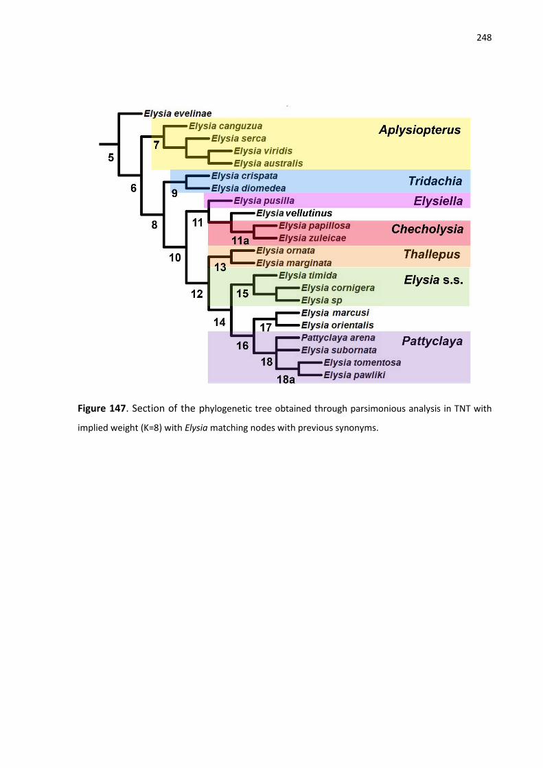
248
Figure 147. Section of the phylogenetic tree obtained through parsimonious analysis in TNT with
implied weight (K=8) with Elysia matching nodes with previous synonyms.

249
Figure 148. Phylogenetic tree obtained through parsimonious analysis in TNT with implied
weight (K=8) and character optimization. A-G nodes with outgroup taxa and 1-18 with
ingroup taxa.

250
Figure 149. Phylogenetic tree (part 1) with the characters that suport each node. Nodes:
A) unamed; B) Sacoglossa; C) Oxynoacea; D) Plakobranchacea.
Figure 150. Phylogenetic tree (part 2) with the characters that suport each node. Nodes:
D) Plakobranchacea; E) Limapontioidea+ Plakobranchoidea; F) Limapontioidea; G)
Plakobranchoidea.

251
Figure 151. Phylogenetic tree (part 3) with the characters that suport each node. Nodes:
G) Plakobranchoidea; 1) Plakobranchidae; 2) Thuridilla + Plakobranchus; 3) Thuridilla; 4)
Plakobranchus; 5) Elysia.

252
Figure 152. Phylogenetic tree (part 4) with the characters that suport each node. Nodes
are unamed subdivision of Elysia (Node 5).
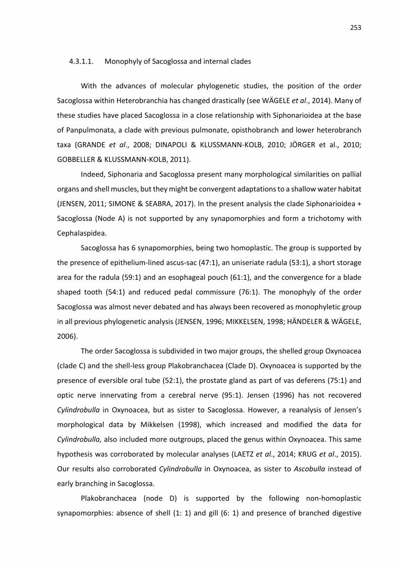
253
4.3.1.1. Monophyly of Sacoglossa and internal clades
With the advances of molecular phylogenetic studies, the position of the order
Sacoglossa within Heterobranchia has changed drastically (see WÄGELE et al., 2014). Many of
these studies have placed Sacoglossa in a close relationship with Siphonarioidea at the base
of Panpulmonata, a clade with previous pulmonate, opisthobranch and lower heterobranch
taxa (GRANDE et al., 2008; DINAPOLI & KLUSSMANN-KOLB, 2010; JÖRGER et al., 2010;
GOBBELLER & KLUSSMANN-KOLB, 2011).
Indeed, Siphonaria and Sacoglossa present many morphological similarities on pallial
organs and shell muscles, but they might be convergent adaptations to a shallow water habitat
(JENSEN, 2011; SIMONE & SEABRA, 2017). In the present analysis the clade Siphonarioidea +
Sacoglossa (Node A) is not supported by any synapomorphies and form a trichotomy with
Cephalaspidea.
Sacoglossa has 6 synapomorphies, being two homoplastic. The group is supported by
the presence of epithelium-lined ascus-sac (47:1), an uniseriate radula (53:1), a short storage
area for the radula (59:1) and an esophageal pouch (61:1), and the convergence for a blade
shaped tooth (54:1) and reduced pedal commissure (76:1). The monophyly of the order
Sacoglossa was almost never debated and has always been recovered as monophyletic group
in all previous phylogenetic analysis (JENSEN, 1996; MIKKELSEN, 1998; HÄNDELER & WÄGELE,
2006).
The order Sacoglossa is subdivided in two major groups, the shelled group Oxynoacea
(clade C) and the shell-less group Plakobranchacea (Clade D). Oxynoacea is supported by the
presence of eversible oral tube (52:1), the prostate gland as part of vas deferens (75:1) and
optic nerve innervating from a cerebral nerve (95:1). Jensen (1996) has not recovered
Cylindrobulla in Oxynoacea, but as sister to Sacoglossa. However, a reanalysis of Jensen’s
morphological data by Mikkelsen (1998), which increased and modified the data for
Cylindrobulla, also included more outgroups, placed the genus within Oxynoacea. This same
hypothesis was corroborated by molecular analyses (LAETZ et al., 2014; KRUG et al., 2015).
Our results also corroborated Cylindrobulla in Oxynoacea, as sister to Ascobulla instead of
early branching in Sacoglossa.
Plakobranchacea (node D) is supported by the following non-homoplastic
synapomorphies: absence of shell (1: 1) and gill (6: 1) and presence of branched digestive
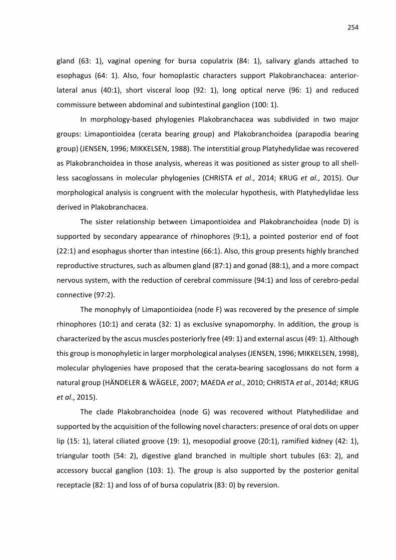
254
gland (63: 1), vaginal opening for bursa copulatrix (84: 1), salivary glands attached to
esophagus (64: 1). Also, four homoplastic characters support Plakobranchacea: anterior-
lateral anus (40:1), short visceral loop (92: 1), long optical nerve (96: 1) and reduced
commissure between abdominal and subintestinal ganglion (100: 1).
In morphology-based phylogenies Plakobranchacea was subdivided in two major
groups: Limapontioidea (cerata bearing group) and Plakobranchoidea (parapodia bearing
group) (JENSEN, 1996; MIKKELSEN, 1988). The interstitial group Platyhedylidae was recovered
as Plakobranchoidea in those analysis, whereas it was positioned as sister group to all shell-
less sacoglossans in molecular phylogenies (CHRISTA et al., 2014; KRUG et al., 2015). Our
morphological analysis is congruent with the molecular hypothesis, with Platyhedylidae less
derived in Plakobranchacea.
The sister relationship between Limapontioidea and Plakobranchoidea (node D) is
supported by secondary appearance of rhinophores (9:1), a pointed posterior end of foot
(22:1) and esophagus shorter than intestine (66:1). Also, this group presents highly branched
reproductive structures, such as albumen gland (87:1) and gonad (88:1), and a more compact
nervous system, with the reduction of cerebral commissure (94:1) and loss of cerebro-pedal
connective (97:2).
The monophyly of Limapontioidea (node F) was recovered by the presence of simple
rhinophores (10:1) and cerata (32: 1) as exclusive synapomorphy. In addition, the group is
characterized by the ascus muscles posteriorly free (49: 1) and external ascus (49: 1). Although
this group is monophyletic in larger morphological analyses (JENSEN, 1996; MIKKELSEN, 1998),
molecular phylogenies have proposed that the cerata-bearing sacoglossans do not form a
natural group (HÄNDELER & WÄGELE, 2007; MAEDA et al., 2010; CHRISTA et al., 2014d; KRUG
et al., 2015).
The clade Plakobranchoidea (node G) was recovered without Platyhedilidae and
supported by the acquisition of the following novel characters: presence of oral dots on upper
lip (15: 1), lateral ciliated groove (19: 1), mesopodial groove (20:1), ramified kidney (42: 1),
triangular tooth (54: 2), digestive gland branched in multiple short tubules (63: 2), and
accessory buccal ganglion (103: 1). The group is also supported by the posterior genital
receptacle (82: 1) and loss of of bursa copulatrix (83: 0) by reversion.

255
4.3.1.2. The family Plakobranchidae
Plakobranchoidea is represented by the families Bosellidae and Plakobranchidae (node
1). Bosellidae is a monogeneric family erected by Marcus (1982) and characterized by the
absence of parapodia, three ganglia on short visceral loop and small number of chromosomes
(n =7) comparing with other sacoglossans (n= 17). Two species of Bosellia were included in
this analysis to test the monophyly of Plakobranchidae. Bosellia mimetica was recovered as
sister group to Plakobranchidae, while Bosellia marcusi was recovered within
Plakobranchidae.
Previous works have already suggested the insertion of B. marcusi in Plakobranchidae
(HANDELER & WÄGELE, 2007; CHRISTA et al., 2014; KRUG et al., 2015) and officially
transferred to Elysia later (KRUG et al., 2016). Krug et al. (2016) have discussed that Marcus
(1972) misplaced E. marcusi in Bosellia because of the reduced parapodia and secondary loss
of dorsal vessels. The analysis of anatomical characters revealed that E. marcusi has no
pharyngeal pouch and presents a short spherical esophageal pouch, blade-shaped tooth and
external hermaphrodite ampulla, which are common characters found in Elysia spp.. On the
other hand, B. mimetica has a large pharyngeal pouch, elongated esophageal pouch,
triangular shaped teeth and ampulla internal to hermaphrodite duct. In the present
phylogenetic analysis, E. marcusi was also placed within Elysia.
The family Plakobranchidae has four exclusive synapomorphies: anterior border of
foot expanded (21: 1), parapodia formed by body flap (25:1), external kidney housed in a
prominence (38) and ampulla separated from hermaphrodite duct (71: 1). In addition,
Plakobranchidae is also supported by short optic nerve (96: 2) and elongated pedal
commissure (98: 0, 1).
The monophyletic status of Plakobranchidae resulted from this analysis is congruent
with some morphological and molecular analysis (JENSEN, 1996; BASS & KARL, 2006;
HÄNDELER & WÄGELE, 2007; HÄNDELER et al., 2009; KRUG et al., 2015). Other scenarios,
however, have been proposed. In Mikkelsen’s (1998) morphological analysis Plakobranchus
branches earlier in Plakobranchoidea and is sister to a group formed by Bosellidae and Elysia
+ Thuridilla. Christa et al. (2014) recovered Bosellidae sister to Plakobranchus + Thuridilla and
forming the sister clade to Elysia. In this case, the family Plakobranchidae would be

256
represented only by Plakobranchus and Thuridilla, while Elysia would be part of the
monogeneric family Elysiidae.
The phylogenetic tree with implied weight supports the clade Thuridilla +
Plakobranchus (node 2) only by one synapomorphy, which is the central distribution of
gonadal follicles (98: 0). This relationship is strongly support by many molecular analysis (BASS
& KARLS, 2006; CHRISTA et al., 2014d; KRUG et al., 2015; KRUG, VENDETTI & VALDÉS, 2016),
but with one exception in which Thuridilla branches earlier in Plakobranchidae (HANDELER &
WÄGELE, 2007).
Morphologically, the clade Thuridilla + Plakobranchus was only recovered by Gosliner
(2005) with five synapommorphies, but those characters are very homoplastic and do not
support the clade in analysis with more taxa (JENSEN, 1996; MIKKELSEN, 1998; present study).
The consensus tree with prior weight also recovered the monophyly of Plakobranchidae (Figs.
1 and 2) with Plakobranchus sister to Thuridilla + Elysia. This topology is similar to the one
proposed by Jensen (1996).
4.3.1.3. The genus Plakobranchus
The genus Plakobranchus was considered monospecific for a long time (see KRUG et
al., 2015), and for this reason only one species was included in most analysis (GOSLINER, 1995;
JENSEN, 1996; MIKKELSEN, 1998; BASS & KARL, 2006; MAEDA et al., 2010). However, Jensen
(1997a, 2007) considered that Plakobranchus could be represented by a complex with up to
14 species under the name P. ocellatus, which was subsequently proved by molecular analysis
(HÄNDELER & WÄGELE, 2007; CHIRSTA et al., 2014d; KRUG et al., 2013, 2015; KRUG, VENDETTI
& VALDÉS, 2016; MEYERS-MUÑOZ et al., 2016).
The present analysis considered five species of Plakobranchus with distinct
morphological characters, also recovered as independent lineages by molecular analysis (see
topic 4.5). In both topologies with prior and implied weight Plakobranchus resulted as
monophyletic and supported by nine synapomorphies, being 4 homoplastic. The genus is
characterized by the rhinophores on anterior position (12:1), secondary median eyes (16: 1-
>0) on top of ocular tubercle (17:1), posterior end of foot truncate (22:2), dorsal vessels on
top of lamellae (36:1), gametolytic vesicles connected on hermaphrodite ducts (86:1), loss of
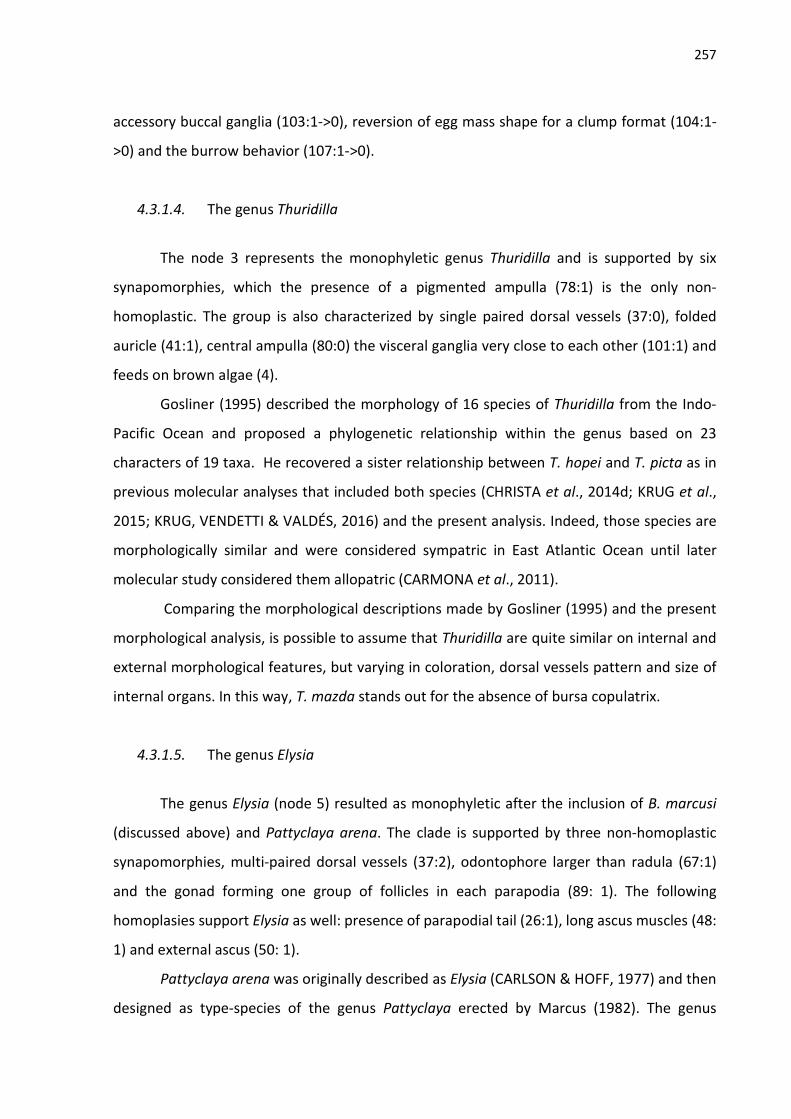
257
accessory buccal ganglia (103:1->0), reversion of egg mass shape for a clump format (104:1-
>0) and the burrow behavior (107:1->0).
4.3.1.4. The genus Thuridilla
The node 3 represents the monophyletic genus Thuridilla and is supported by six
synapomorphies, which the presence of a pigmented ampulla (78:1) is the only non-
homoplastic. The group is also characterized by single paired dorsal vessels (37:0), folded
auricle (41:1), central ampulla (80:0) the visceral ganglia very close to each other (101:1) and
feeds on brown algae (4).
Gosliner (1995) described the morphology of 16 species of Thuridilla from the Indo-
Pacific Ocean and proposed a phylogenetic relationship within the genus based on 23
characters of 19 taxa. He recovered a sister relationship between T. hopei and T. picta as in
previous molecular analyses that included both species (CHRISTA et al., 2014d; KRUG et al.,
2015; KRUG, VENDETTI & VALDÉS, 2016) and the present analysis. Indeed, those species are
morphologically similar and were considered sympatric in East Atlantic Ocean until later
molecular study considered them allopatric (CARMONA et al., 2011).
Comparing the morphological descriptions made by Gosliner (1995) and the present
morphological analysis, is possible to assume that Thuridilla are quite similar on internal and
external morphological features, but varying in coloration, dorsal vessels pattern and size of
internal organs. In this way, T. mazda stands out for the absence of bursa copulatrix.
4.3.1.5. The genus Elysia
The genus Elysia (node 5) resulted as monophyletic after the inclusion of B. marcusi
(discussed above) and Pattyclaya arena. The clade is supported by three non-homoplastic
synapomorphies, multi-paired dorsal vessels (37:2), odontophore larger than radula (67:1)
and the gonad forming one group of follicles in each parapodia (89: 1). The following
homoplasies support Elysia as well: presence of parapodial tail (26:1), long ascus muscles (48:
1) and external ascus (50: 1).
Pattyclaya arena was originally described as Elysia (CARLSON & HOFF, 1977) and then
designed as type-species of the genus Pattyclaya erected by Marcus (1982). The genus

258
Pattyclaya was characterized by presence of parallel transverse lamellae on upper surface of
parapodia connected with the long renopericardial prominence by dorsal vessels. After the
first phylogenetic analysis, the validity of the genus has been questioned, and Pattyclaya was
placed in the genus Elysia in all analysis (GOSLINER, 1995; JENSEN, 1996; MIKKELSEN, 1998).
The results of present analysis also positioned Pattyclaya in Elysia (node 18).
The monophyly of Elysia has been widely debated with the first phylogenetic
hypotheses for Plakobranchidae. Thus, the insertion of other previous genera (Pattyclaya,
Elysiella, Tridachia, Tridachiella and Checholysia) in Elysia was proposed by morphological and
molecular phylogenetic hypothesis (GOSLINER, 1995; JENSEN, 1996; MIKKELSEN, 1998; BASS
& KARL, 2006; KRUG et al., 2016) to recovery its monophyletic status. Gosliner (1995) and
Jensen (1996) also suggested that Elysia might be subdivided.
The present morphological analysis recovered some clades within Elysia that matches
with synonymized names (Fig. 6). Although, they have low Bremmer values in the consensus
topology with prior weight, almost all of them still form monophyletic groups. The node 7
includes the type species of Aplysiopterus, Elysia viridis. This clade is supported by non-
homoplastic synapomorphy for a compact digestive system (68:1) and the secondary loss of
accessory buccal ganglia (103: 0).
The synonymized genera Tridachia (type species: E. crispata) and Tridachiella (type
species: E. diomedea) were grouped together in node 9, which presents the non-homoplastic
synapomorphy for frilled parapodial margins (27: 1), and the homoplastic character states for
single paired dorsal vessels (37:0), short ascus muscle (48: 0), multiple ampulla (79:2), gonad
partially spread through parapodia (91: 1) and reduced pedal commissure (98:2).
The node 11 is represented by E. pusilla (type-species of Elysiella) plus the node 11a,
which includes E. papillosa previously placed in Checholysia. The clade Elysiella + Checholysia
is supported by four homoplastic synapomorphies: the presence of papillae on rhinophores
(13:1) and body (31:1), penial stylet (70:1) and ampulla on central position (80:0). Checholysia
(node 11a) presents two non-homoplastic synapomorphies for the presence of gametolytic
vesicles on gonad (85:1) and the capability to swim (106:1). Three other homoplastic character
supports the node: rhinophores smaller than head (11:1), longer commissure between
abdominal and subintestinal ganglia (100:0) and the change on diet for the calcareous algae
Udotacea (108:3).

259
The node 13 groups E. ornata (type-species for Thallepus) E. marginata (type species
for Pterogasteron) and is supported by the non-homoplastic synapomorphy for presence of
an orange band on parapodial margin (28: 1) and the following homoplastic characters:
pigmentation on foot sole (23: 1) and inner surface of parapodia (29: 1), short penis (69: 1),
multiple ampullae (79:2) and reduced pedal commissure (98:2).
The node 16 presents the type species of Elysia, Elysia timida, and is the only group to
have clustered prostate gland (76:2) and feed on Dasycladales algae (118: 5). The group also
presents the homoplastic synapomorphies for single paired dorsal vessels (37:0), short ascus
muscles (48: 0), seminal receptacle absent (81: 1), short pedal commissure (98:0), big
connective between subintestinal and abdominal ganglia (100: 0), short distance between
supraintestinal and subintestinal ganglia (101: 1) and reduced buccal commissure (102: 2).
Finally, the node 18 presents Pattyclaya arena and is supported by five
synapomorphies, which the kidney prominence very long (39: 2) and semi-trabecular kidney
ramification (43:3) are non-homoplastic. This clade is also characterized by the presence of
papillae on rhinophores (13:1) and body (31:1) and multiple ampullae (79:2).
4.3.2. Molecular Based Analysis
The molecular analysis was based on five partial genes of 49 species. Three species
included in the morphological analysis were excluded in both molecular analyses since no
gene sequence were obtained: Platyhedyle denudata, Elysia sp. and Pattyclaya arena. From
the total of 39 species used in this analysis, we obtained 37 16S sequences (~404 bp), 39 COI
sequences (~1271 bp), 28S sequences for 25 specimens (~1391 bp), 38 H3 sequences (328 bp)
and 36 18S sequences (~1781 bp). Some partial sequences were used according to Table 2.
The final alignment dataset was ~5175 bp long for each taxon.
Both topologies are quite similar on major groups relationship (Figs. 93 and 94), being
Oxynoacea and Plakobranchacea monophyletic groups. On the other hand, Limapontioidea
resulted as non-monophyletic, with Costasiella placed as sister to all shell-less sacoglossans
with high ML and BI values. This result is similar to previous molecular works and corroborates
the multiple independent origins of kleptoplasty proposed by Christa et al. (2014).
In both analysis Plakobranchus and Thuridilla are well-supported monophyletic groups
and form a clade, the family Plakobranchidae, however, resulted as paraphyletic. In this

260
scenario, Bosellidae is placed as sister to Thuridilla + Plakobranchus with higher value for BI
posterior probability. Similar result was found by Christa et al. (2014) for BI analysis, but Krug
et al. (2015) recovered Plakobranchidae as monophyletic and sister to Bosellidae although
with low BI value and no ML support.
The genus Elysia is highly supported by the present analyses after the inclusion of
Bosellia marcusi (as E. marcusi) and other species previously placed in synonymic genera of
Elysia. Although some internal nodes were not well resolved, many clades were recovered as
in previous molecular analysis with more species of Elysia (CHRISTA et al., 2014; KRUG et al.,
2015, 2016). The phylogenetic analysis performed by Krug et al. (2016) for 76 terminals and
four genes (16S, 28S, COI and H3) is the most similar, with the same relation among the
internal nodes.
The morpho groups comprising Elysiella and Checholysia is likely to form a trichotomy
with the other Elysia spp.. In this trichotomy, Elysia vellutinus was placed as sister to E. pusilla
or forming a clade with Checholysia. In both analysis the clade that groups the type species
for Tridachia and Aplysiopterus comprises the same species and similar topology, except for
E. canguzua branching earlier in BI analysis. The two topologies are completely similar on the
other branches and present high values supporting the nodes with the morpho groups
Thallepus, Elysia s.s. and Pattyclaya.

261
Figure 153. Phylogenetic tree obtained through Maximum Likelihood of concatenated DNA
sequences of two mitochondrial (COI, 16S) and three nuclear (H3, 18S and 28S) genes. Support
values as ML bootstrap percentages on each node.

262
Figure 154. Phylogenetic tree obtained through Bayesian analysis of concatenated DNA
sequences of two mitochondrial (COI, 16S) and three nuclear (H3, 18S and 28S) genes. Support
values as BI posterior probabilities on each node.

263
Figure 155. Section of phylogenetic tree obtained through Maximum Likelihood (Fig. 93) with
Elysia matching nodes with previous synonyms.

264
Figure 156. Section of phylogenetic tree obtained through Bayesian Inference (Fig. 7) with
Elysia matching nodes with previous synonyms.
4.3.3. Total Evidence Analysis
This is the first phylogenetic analysis with species of Sacoglossa that includes both
molecular and morphological data sets. The resulted topology (Fig. 97 and 98) presents
similarities and differences with the ones resulted from each type of data set, and comparison
will be made among bayesian trees and morphological tree resulted from MP analysis.
Sacoglossa resulted as monophyletic in the analyses with one type of data, resulted as
polyphyletic in the combined analysis. The shelled species were clustered together and
forming the monophyletic Oxynoacea, but sister to Siphonarioidea, whereas the shell-less
Plakobranchacea resulted as highly polyphyletic. This polyatomic topology with the outgroup
species might be a consequence of a low representation.
In the combined analysis the family Plakobranchidae resulted as monophyletic,
although with a low BI value. The valid genera of Plakobranchidae have also their monophyly
well-supported, and the sister relationship between Plakobranchus and Thuridilla is also
congruent with the results from analyses with one data set.

265
The internal clades in Elysia of both morphological and molecular topologies are quite
similar, with old names of Elysia matching similar nodes. However, the combined tree is
identical with the molecular one, but with lower BI values. The main differences between
morphological and molecular (= combined) are on the arrangement of the internal nodes in
Elysia.
On the morphological topology E. evelinae is sister to all Elysia, but it is grouped with
E. viridis in the combined tree. The clade matching the synonym Checholysia is sister to all
other Elysia in the molecular and combined trees, but more derived in the morphological
topology. Another well supported node in molecular tree groups the morpho group for
Aplysiopterus with Tridachia, while in the morpho tree Aplysiopterus is sister to all other
groups of Elsyia (except for E. evelinae). The node 12 on morphological tree (Fig. 86 and 87)
groups almost the same species in similar arrangement of molecular trees. The differences are
the inclusion of E. australis and E. marcusi branching earlier on the molecular tree. The
previous phylogenetic studies (CHRISTA et al., 2014; KRUG et al., 2015, 2016) also recovered
the same major groups.

266
Figure 157. Phylogenetic tree obtained through Bayesian analysis of combined data of 109
morphological characters and concatenated DNA sequences of two mitochondrial (COI, 16S)
and three nuclear (H3, 18S and 28S) genes. Support values as BI posterior probabilities on each
node.

267
Figure 158. Section of phylogenetic tree obtained through Bayesian Inference (Fig. 97) with
Elysia matching nodes with previous synonyms.

268
4.4. MOLECULAR EVIDENCE OF MULTIPLE SPECIES UNDER THE NAME PLAKOBRANCHUS OCELLATUS (GASTROPODA: SACOGLOSSA)
The genus Plakobranchus was described from Indonesia, and the type species
(Plakobranchus occelatus) was considered the only valid member for a long time. Other
species were later described from Hawaii (GOULD, 1852; BERGH, 1872), French Polynesia
(PEASE, 1871, BERGH, 1873), Japan (STIMPSON, 1855) and Philippines (BERGH, 1872), but
synonymized afterwards (BERGH, 1887; RISBEC, 1953; JENSEN, 1992). Including the type
species of Plakobranchus, many of these species were shortly described based only on
external features and color patterns. Bergh (1872, 1873) provided information on internal
morphology of some species, but later considered them as intraspecific variations of P.
ocellatus (BERGH, 1887).
However, distinct color morphs of Plakobranchus were reported in different localities
on Indo-Pacificic (see KRUG et al., 2013; GOSLINER et al., 2015). Later, molecular evidence has
found 10 candidate species under the name Plakobranchus ocellatus with distinct color
patterns (KRUG et al., 2013), and a new species were described based on molecular and
morphological evidence (MEYERS-MUÑOZ et al., 2016).
We analyzed COI and 16S partial sequences of 62 specimens of Plakobranchus spp..
Also, we included the sequences already published (total of 54 COI sequences and 58 16S
sequences) to check which species was used for different works (BASS & KARL, 2006;
KLUSSMANN-KOLB & DINAPOLI, 2006; MAEDA et al., 2010; HÄNDELER & WÄGELE, 2012;
TAKANO et al., 2013; LERAY et al., 2013; MEYER-MUÑOZ et al., 2016).
The phylogenetic data of concatenate analysis of COI and 16S resulted in 11 candidate
species. Krug et al. (2013) found 10 candidate species, and one of them was later described as
Plakobranchus papua. Our analysis found the same 10 species plus another new one from
Djibouti, Africa (Pk_saf_99829A-C) from the malacological collection of Zoological Museum of
Sao Paulo University.
We propose the resurrection of Plakobranchus ianthobapsus as a valid species, which
is the only species to occur in Hawaii. Also, we provide the redescription of Plkobranchus
papua and the description of two new species, Plakobranchus sp. 1 aff purple and
Plakobranchus sp. 2 Africa.

269
Figure 159. Phylogenetic tree obtained through Maximum Likelihood of concatenated DNA
sequences of two mitochondrial (COI, 16S) genes. Support values as ML bootstrap percentages
on branch.

270
Figure 160. Phylogenetic tree obtained through Maximum Likelihood of partial DNA
sequences of the nuclear gene H3. Support values as ML bootstrap percentages on branch.

271
5. CONCLUSIONS
1. The family Plakobranchidae resulted as a well-supported monophyletic group in all
morphological analysis;
2. Plakobranchidae has not resulted as a natural group in molecular phylogenies, but its
monophyletic status was recovered in total evidence analysis;
3. All the three valid genera within Plakobranchidae are monophyletic in analyses with
distinct criteria and data set;
4. Thuridilla and Plakobranchus are well-supported in all scenarios and, except for the
consensus topology with prior weights, resulted as sister groups;
5. The genus Elysia are low supported in all analysis, and its internal clades are grouped
slightly different among analyses;
6. Morpho groups in Elysia has been recovered as clades in most analyses, which clearly
reflects distinct evolutionary pathways, and meaning that a future subdivision of the
genus is necessary for a better understanding of their natural history;
7. Despite similar external morphology, plakobranchids present a large variation on their
internal morphology in all systems studied herein;

272
6. REFERENCES
Aktipis, S. W.; Giribet, G.; Lindberg, D. R., & Ponder, W. F. (2008). Gastropoda: an overview and analysis. Phylogeny and Evolution of the Mollusca, 201-237.
Baba, K. (1961). On the identification and the affinity of Tamanovalva limax, a bivalved
sacoglossan mollusc in Japan. Publications of the Seto Marine Biological Laboratory, 9(1): 37-62.
Baba, K., & Hamatani, I. (1970). Occurrences of specimens presumably identifiable with Stiliger
ornatus Ehrenberg, 1831, at Seto, Kii, middle Japan (Opisthobranchia: Sacoglossa). Ballesteros, M. (1979). Bosellia mimetica Trinchese, 1891 y Elysia timida Risso, 1818, dos
ascoglosos nuevos para la fauna ibérica. Publicaciones Dpto. Zoología Barcelona, 4, 13-17. Bass, A. L., & Karl, S. A. (2006). Molecular phylogenetic analysis of genera in the family
Plakobranchidae (Mollusca: Opisthobranchia: Sacoglossa). Records of the Western Australian Museum, 69: 61-68.
Bieler, R. (1992). Gastropod phylogeny and systematics. Annual Review of Ecology and
Systematics, 23(1), 311-338. Bouchet, P., Rocroi, J. P., Frýda, J., Hausdorf, B., Ponder, W., Valdés, Á., & Warén, A. (2005).
Classification and nomenclator of gastropod families. Malacologia, 47(1-2), 1-368 Brace, R. C. (1977). The functional anatomy of the mantle complex and columellar muscle of
tectibranch molluscs (Gastropoda: Opisthobranchia), and its bearing on the evolution of opisthobranch organization. Phil. Trans. R. Soc. Lond. B, 277(951), 1-56.
Caballer, M. G., Ortea, J., Rivero, N., Tucker, G. C., Malaquías, M. A. E., & Narciso, S. (2015).
The opisthobranch gastropods (Mollusca: Heterobranchia) from Venezuela: an annotated and illustrated inventory of species. Zootaxa, 4034(2), 201-256.
Carmona, L., Malaquias, M. A. E., Gosliner, T. M., Pola, M., & Cervera, J. L. (2011). Amphi-
Atlantic distributions and cryptic species in sacoglossan sea slugs. Journal of Molluscan Studies, 77(4), 401-412.
Christa, G., Händeler, K., Kück, P., Vleugels, M., Franken, J., Karmeinski, D., & Wägele, H.
(2014). Phylogenetic evidence for multiple independent origins of functional kleptoplasty in Sacoglossa (Heterobranchia, Gastropoda). Organisms Diversity & Evolution, 15(1), 23-36.
Christa, G., Wescott, L., Schäberle, T. F., König, G. M., & Wägele, H. (2013). What remains after
2 months of starvation? Analysis of sequestered algae in a photosynthetic slug, Plakobranchus ocellatus (Sacoglossa, Opisthobranchia), by barcoding. Planta, 237(2), 559-572.

273
Cimino, G. & Gavagnin, M. (2007). Molluscs: from chemo-ecological study to biotechnological application. Springer Science & Business Media, p. 397.
Cimino, G., Fontana, A., & Gavagnin, M. (1999). Marine opisthobranch molluscs: chemistry
and ecology in sacoglossans and dorids. Curr Org Chem, 3(4), 327-72. Clark, K. B. (1984). New records and synonymies of Bermuda opisthobranchs (Gastropoda).
The Nautilus, 98(2): 85-97. Coelho, R., Malaquias, M., & Calado, G. (2006). Calliopaea bellula feeding upon egg-masses of
Haminoea orbignyana: oophagy among opisthobranch molluscs. Journal of the Marine Biological Association of the United Kingdom, 86(2), 423-424.
Coleman, N. (2001). 1001 Nudibranchs: catalogue of Indo-Pacific sea slugs. Neville Coleman's
Underwater Geographic Pty Ltd. Costa, A. (1867). Saggio sui molluschi eolididei del Golfo di Napoli. Annuario del Museo
Zoologico della Reale Università di Napoli, 4: 26-37. Cruz, S., Calado, R., Serôdio, J., & Cartaxana, P. (2013). Crawling leaves: photosynthesis in
sacoglossan sea slugs. Journal of experimental botany, 64(13), 3999-4009. Dayrat, B., & Tillier, S. (2002). Evolutionary relationships of euthyneuran gastropods
(Mollusca): a cladistic re-evaluation of morphological characters. Zoological Journal of the Linnean Society, 135(4), 403-470.
Dayrat, B., Conrad, M., Balayan, S., White, T. R., Albrecht, C., Golding, R., ... & de Frias Martins,
A. M. (2011). Phylogenetic relationships and evolution of pulmonate gastropods (Mollusca): new insights from increased taxon sampling. Molecular Phylogenetics and Evolution, 59(2), 425-437.
Dinapoli, A., & Klussmann-Kolb, A. (2010). The long way to diversity–phylogeny and evolution
of the Heterobranchia (Mollusca: Gastropoda). Molecular Phylogenetics and Evolution, 55(1), 60-76.
Drummond, A.J., Suchard, M.A., Xie, D. and Rambaut, A. (2012). Bayesian phylogenetics with
BEAUTi and the Beast 1.7. Molecular Biology and Evolution 29(8), 1969–1973. Engel, H. (1927). Westindische Opisthobranchiate Mollusken. II. Aplysiidae, Pleurobranchidae,
Oxynoidae, Elysiidae, Phyllobranchidae. Bijdragen tot de Dierkunde, 25: 83-122. Evans, T. J. (1950). A review of Pease's genus Volvatella, together with a preliminary report on
a new sacoglossan genus. Journal of Molluscan Studies, 28(2-3), 102-106. Evertsen, J., Burghardt, I., Johnsen, G., & Wägele, H. 2007. Retention of functional chloroplasts
in some sacoglossans from the Indo-Pacific and Mediterranean. Marine Biology, 151(6): 2159-2166.

274
Fahrner, A., & Haszprunar, G. (2001). Anatomy and ultrastructure of the excretory system of a heart-bearing and a heart-less sacoglossan gastropod (Opisthobranchia, Sacoglossa). Zoomorphology, 121(2), 85-93.
GALVÃO FILHO, H. C. (2013). Taxonomia de espécies do gênero Elysia Risso 1818 (Mollusca:
Gastropoda: Sacoglossa) do Brasil. Universidade Federal do Ceará, Fortaleza, 127 p. Galvão Filho, H. C., Araújo, A. K., Silva, F. V., Azevedo, V. M., Meirelles, C. A., & Matthews-
Cascon, H. (2015). Sea slugs (Gastropoda: Heterobranchia) from a poorly known area in North-east Brazil: filling gaps in Atlantic distributions. Marine Biodiversity Records, 8.
Gascoigne, T. (1975). The radula and reproductive system of Olea hansineensis Agersborg,
1923. Veliger, 17, 313-317. Gascoigne, T. (1976). The reproductive systems and classification of the Stiligeridae
(Opisthobranchia: Sacoglossa). Journal of the Malacological Society of Australia, 3(3-4), 157-172.
Gascoigne, T. (1979). A redescription of Caliphylla mediterranea Costa, 1867
(Opisthobranchia: Ascoglossa). Journal of Molluscan Studies, 45(3), 300-311. Gascoigne, T., & Sigurdsson, J. B. (1977). Calliopaea oophaga Lemche, 1974, a species new to
the British fauna (Opisthobranchia: Sacoglossa). Journal of Molluscan Studies, 43(3), 286-289.
Gascoigne, T., & Todd, C. D. (1977). A description of a specimen of Calliopaea bellula
D'Orbigny, 1857 found at Robin Hood's Bay, North Yorkshire (Opisthobranchia: Sacoglossa). Journal of Molluscan Studies, 43(3), 290-295.
Gavagnin, M., Mollo, E., Montanaro, D., Ortea, J., & Cimino, G. (2000). Chemical studies of
Caribbean sacoglossans: dietary relationships with green algae and ecological implications. Journal of chemical ecology, 26(7), 1563-1578.
Göbbeler, K., & Klussmann-Kolb, A. (2011). Molecular phylogeny of the Euthyneura (Mollusca,
Gastropoda) with special focus on Opisthobranchia as a framework for reconstruction of evolution of diet. Thalassas, 27(2), 121-154.
Goodheart, J. A., Ellingson, R. A., Vital, X. G., Galvão Filho, H. C., McCarthy, J. B., Medrano, S.
M., ... & Hoover, C. A. (2016). Identification guide to the heterobranch sea slugs (Mollusca: Gastropoda) from Bocas del Toro, Panama. Marine Biodiversity Records, 9(1), 56.
Gosliner, T. M. (1994). Gastropoda. Opisthobranchia. In Microscopic Anatomy of
Invertebrates. Vol. 5. Mollusca I, Edited by F. W. Harrison and A. J. Kohn. New York: Wiley-Liss, pp. 253-355.

275
Gosliner, T. M. (1995). The genus Thuridilla (Opisthobranchia: Elysiidae) from the tropical Indo-Pacific, with a revision of the phylogeny and systematics of the Elysiidae. Proceedings of the California Academy of Sciences, 49(1): 1-54.
Gosliner, T. M., Behrens, D. W., & Valdés, Á. (2008). Indo-Pacific nudibranchs and sea slugs: a
field guide to the world's most diverse fauna. Sea Challengers Natural History Books; California Academy of Sciences.
Gosliner, T. M., Valdés, A. & Behrens, D. W. (2015). Nudibranch & sea slug identification: Indo-
Pacific. New World Publications. Gosliner, T. M., Valdés, A. & Behrens, D. W. (2015). Nudibranch & sea slug identification: Indo-
Pacific. New World Publications. Grande, C., Templado, J., Cervera, J. L., & Zardoya, R. (2004). Phylogenetic relationships among
Opisthobranchia (Mollusca: Gastropoda) based on mitochondrial cox 1, trnV, and rrnL genes. Molecular phylogenetics and evolution, 33(2), 378-388.
Händeler, K., & Wägele, H. (2007). Preliminary study on molecular phylogeny of Sacoglossa
and a compilation of their food organisms. Bonn Zool Beitr, 55, 231-254. Händeler, K., Grzymbowski, Y. P., Krug, P. J., & Wägele, H. (2009). Functional chloroplasts in
metazoan cells-a unique evolutionary strategy in animal life. Frontiers in Zoology, 6(1), 28. Ihering, H. v. (1876). Versuch eines natiirlichen Systems der Mollusken. J b , dt. Malakozool.
Ges. 3, 97-174. Jaume, M. L. (1945). El genero Oxynoe en Cuba occidental. Revista de la Sociedad Malacologia,
3:18-25. Jensen, K. R. (1986). Observations on feeding, copulation and spawning of the Ascoglossan
Opisthobranch Calliopaea oophaga lemche. Ophelia, 25(2), 97-106. Jensen, K. R. (1991). Comparison of Alimentary Systems in Shelled and Non-shelled Sacoglossa
(Mollusca, Opisthobranchia). Acta Zoologica, 72(3), 143-150. Jensen, K. R. (1993). Morphological adaptations and plasticity of radular teeth of the
Sacoglossa (= Ascoglossa) (Mollusca: Opisthobranchia) in relation to their food plants. Biological Journal of the Linnean Society, 48(2), 135-155.
Jensen, K. R. (1996). Phylogenetic systematics and classification of the Sacoglossa (Mollusca,
Gastropoda, Opisthobranchia). Phil. Trans. R. Soc. Lond. B, 351(1335), 91-122. Jensen, K. R. (1997a). Evolution of the Sacoglossa (Mollusca, Opisthobranchia) and the eco-
logical associations with their food plants. Evolutionary Ecology 11: 301–335.

276
Jensen, K. R. (1997b). Sacoglossernes systematik, fylogeni og evolution (Mollusca, Opisthobranchia). (Systematics, phylogeny and evolution of the Sacoglossa (Mollusca, Opisthobranchia). Vestjydsk Forlag, Copenhagen, 94 pp.
Jensen, K. R. (2007). Biogeography of the Sacoglossa (Mollusca, Opisthobranchia). Bonner
Zoologische Beiträge, 55(3/4), 255-281. Jensen, K. R. (2014). Anatomy of three sacoglossans (Mollusca," Opisthobranchia") newly
recorded from São Miguel, Azores. Açoreana, Suplemento, 10, 117-138. Jörger, K. M., Stöger, I., Kano, Y., Fukuda, H., Knebelsberger, T., & Schrödl, M. (2010). On the
origin of Acochlidia and other enigmatic euthyneuran gastropods, with implications for the systematics of Heterobranchia. BMC Evolutionary Biology, 10(1), 323.
Klussmann-Kolb, A. (2004). Phylogeny of the Aplysiidae (Gastropoda, Opisthobranchia) with
new aspects of the evolution of seahares. Zoologica Scripta, 33(5), 439-462. Krug, P. J., Vendetti, J. E., & Valdés, Á. (2016). Molecular and morphological systematics of
Elysia Risso, 1818 (Heterobranchia: Sacoglossa) from the Caribbean region. Zootaxa, 4148(1), 1-137.
Laetz, E., Christa, G., Haendeler, K., & Waegele, H. (2014). The Cylindrobulla/Ascobulla
complex—unraveling problems in identification and adding to Cylindrobulla diversity (Gastropoda, Heterobranchia, Sacoglossa) by describing a new species. Zootaxa, 3893(3), 339-362.
Laetz, E., Christa, G., Haendeler, K., & Waegele, H. (2014). The Cylindrobulla/Ascobulla
complex—unraveling problems in identification and adding to Cylindrobulla diversity (Gastropoda, Heterobranchia, Sacoglossa) by describing a new species. Zootaxa, 3893(3), 339-362.
Lewin, R. A. (1970). Toxin secretion and tail autotomy by irritated Oxynoe panamensis
(Opisthobranchiata; Sacoglossa). Pacific Science, 24(3), 356-358. Maeda, T., Kajita, T., Maruyama, T., & Hirano, Y. (2010). Molecular phylogeny of the
Sacoglossa, with a discussion of gain and loss of kleptoplasty in the evolution of the group. The Biological Bulletin, 219(1), 17-26.
Malaquias, M. A. E., & Cervera, J. L. (2005). The genus Haminoea (Gastropoda: Cephalaspidea) in Portugal, with a review of the European species. Journal of Molluscan Studies, 72(1), 89-103.
Marcus, Er. (1957). On Opisthobranchia from Brazil (2). Journal of the Linnean Society, London
34: 390-486. Marcus, Er. & Marcus, Ev. (1970). Opisthobranchs from Curaçao and faunistically related
regions. Studies on the Fauna of Curaçao and other Caribbean Islands 33(122): 1-129.

277
Marcus, Er. (1958). On Western Atlantic opisthobranchiate gastropods. American Museum Novitates 1906: 1-82.
Marcus, Ev. & Marcus, Er. (1963). Opisthobranchs from the Lesser Antilles. Studies on the
fauna of Curaçao and other Carribbean Islands 19: 1-76. Marcus, Ev. & Marcus, Er. (1967). Opisthobranchs from the southwestern Caribbean Sea.
Biological Investigations of the Deep Sea, 33. Bulletin of Marine Science 17: 597- 628. Marcus, Ev. (1973). On the genus Bosellia (Mollusca: Gastropoda; Ascoglossa). Bulletin of
Marine Science, 23(4), 811-823. Marcus, Ev. (1982). Systematics of the genera of the order Ascoglossa (Gastropoda). Journal
of Molluscan Studies, 48(supp10), 1-31. Marcus, Ev., & Hughes, H. P. (1974). Opisthobranch mollusks from Barbados. Bulletin of
Marine Science, 24(3), 498-532. Marcus, Ev., & Marcus, Er. (1960). Opisthobranchs from American Atlantic warm
waters. Bulletin of Marine Science, 10(2), 129-203. Marín, A., & Ros, J. (2004). Chemical defenses in Sacoglossan Opisthobranchs: Taxonomic
trends and evolutionary implications. Scientia Marina, 67(1), 227-241. Medina, M., Lal, S., Vallès, Y., Takaoka, T. L., Dayrat, B. A., Boore, J. L., & Gosliner, T. (2011).
Crawling through time: transition of snails to slugs dating back to the Paleozoic, based on mitochondrial phylogenomics. Marine Genomics, 4(1), 51-59.
Meirelles, C. A., Galvão Filho, H. C., Scramosin, K. A., & Matthews-Cascon, H. (2010). Oxynoe
antillarum (Mollusca, Oxynoidae) no estado do Ceará, Nordeste do Brasil. Revista Nordestina de Zoologia, 4, 42-47.
Meyers-Muñoz, M. A., van der Velde, G., van der Meij, S. E., Stoffels, B. E., van Alen, T., Tuti,
Y., & Hoeksema, B. W. (2016). The phylogenetic position of a new species of Plakobranchus from West Papua, Indonesia (Mollusca, Opisthobranchia, Sacoglossa). ZooKeys, (594), 73.
Mikkelsen, P. M. (1996). The evolutionary relationships of Cephalaspidea sl (Gastropoda:
Opisthobranchia): a phylogenetic analysis. Malacologia, 37(2), 375-442. Mikkelsen, P. M. (1998). Cylindrobulla and Ascobulla in the western Atlantic (Gastropoda,
Opisthobranchia, Sacoglossa): systematic review, description of a new species, and phylogenetic reanalysis. Zoologica Scripta, 27(1), 49-71.
Mikkelsen, P. M. (2002). Shelled opisthobranchs. Advances in Marine Biology, 42, 67-136.

278
Millen, S. V. (1980). Range extensions, new distribution sites, and notes on the biology of sacoglossan opisthobranchs (Mollusca: Gastropoda) in British Columbia. Canadian Journal of Zoology, 58(6), 1207-1209.
Miller, M.A., Schwartz, T., Pickett, B.E., He, S., Klem, E.B., Scheuermann, R.H., Passarotti, M. and Kaufman, S. (2015). A RESTful API for access to phylogenetic tools via the CIPRES Science Gateway. Evolutionary Bioinformatics 11, 43–48.
Mordan, P., & Wade, C. (2008). Heterobranchia II: The Pulmonata in W. F. Ponder. & D. R.
Lindberg (eds.). Phylogeny and Evolution of the Mollusca, 409-426. Nixon, K. C. (1999). Winclada (BETA) ver 0.9.9 Ortea, J., Espinosa, J., Buske, Y., & Caballer, M. (2013). Additions to the inventory of the sea
slugs (Opisthobranchia and Sacoglossa) from Guadeloupe (Lesser Antilles, Caribbean Sea). Revista de la Academia Canaria de Ciencias, 25, 163-194.
Padula, V. (2008). Notes on the morphology of Elysia subornata and Oxynoe antillarum
(Mollusca, Opisthobranchia, Sacoglossa) from the state of Rio de Janeiro, Brazil. Strombus, 15(2), 19.
Padula, V., Bahia, J., Correia, M. D., & Sovierzoski, H. H. (2012). New records of opisthobranchs
(Mollusca: Gastropoda) from Alagoas, northeastern Brazil. Marine Biodiversity Records, 5. Padula, V., Wirtz, P., & Schrödl, M. (2017). Heterobranch sea slugs (Mollusca: Gastropoda)
from Ascension Island, South Atlantic Ocean. Journal of the Marine Biological Association of the United Kingdom, 97(4), 743-752.
Ponder, W. F., & Lindberg, D. R. (1997). Towards a phylogeny of gastropod molluscs: an
analysis using morphological characters. Zoological Journal of the Linnean society, 119(2), 83-265.
Pruvot-Fol, A. (1954). Mollusques opisthobranches. Faune de France 58, 1-460. Lechevalier. Puillandre, N., Lambert, A., Brouillet, S. & Achaz, G. 2012. ABGD, Automatic Barcode Gap
Discovery for primary species delimitation. Molecular Ecology, 21: 1864–1877. Redfern, C. (2001). Bahamian Seashells – A Thousand Species from Abaco, Bahamas, 280 pp.
Bahamianseashells.com, Boca Ratón. Redfern, C. (2013). Bahamian seashells: 1161 species from Abaco, Bahamas.
Bahamianseashells.com, Incorporated. Rios, E. (2009). Compendium of Brazilian Sea Shells. Rio Grande: Ed. Evangraf. 668p. Rückert, I. M., Altnöder, A., & Schrödl, M. (2008). Computer-based 3D anatomical
reconstruction and systematic placement of the mesopsammic gastropod Platyhedyle

279
denudata Salvini-Plawen, 1973 (Opisthobranchia, Sacoglossa). Organisms Diversity & Evolution, 8(5), 358-367.
Rumpho, M. E., Dastoor, F. P., Manhart, J. R., & Lee, J. (2007). The kleptoplast. In The structure
and function of plastids (pp. 451-473). Springer Netherlands. Rumpho, M. E.; Summer, E. J.; Manhart, J. R. 2010. Solar-powered sea slugs. Mollusc/algal
chloroplast symbiosis. Plant Physiology, 123(1), 29-38. Schrödl, M., Jörger, K. M., Klussmann-Kolb, A., & Wilson, N. G. (2011). Bye bye
“Opisthobranchia”! A review on the contribution of mesopsammic sea slugs to euthyneuran systematics. Thalassas, 27(2), 101-112.
Simone, L. R. L. (2011). Phylogeny of the Caenogastropoda (Mollusca), based on comparative
morphology. Arquivos de Zoologia (São Paulo), 42(4), 161-323. Simone, L. R. L., & Seabra, M. I G. L. (2017). Shell and body structure of the plesiomorphic
pulmonate marine limpet Siphonaria pectinata (Linnaeus, 1758) from Portugal (Gastropoda: Heterobranchia: Siphonariidae). Folia Malacologica, 25(3), 147-164.
Stamatakis, A. 2006. RAxML-VI-HPC: maximum likelihood-based phylogenetic analyses with
thousands of taxa and mixed models. Bioinformatics, 22: 2688-90. Stirts, H. M., & Clark, K. B. (1980). Effects of temperature on products of symbiotic chloroplasts
in Elysia tuca Marcus (Opisthobranchia: Ascoglossa). Journal of Experimental Marine Biology and Ecology, 43(1), 39-47.
Thibaut, T.; Meinesz, A.; Amade, P.; Charrier, S.; De Angelis, K.; Ierardi, S.; Mangialajo, K.;
Melnick, J. & Vidal, V. 2001. Elysia subornata (Mollusca) a potential control agent of the alga Caulerpa taxifolia (Chlorophyta) in the Mediterranean Sea. Journal of the Marine Biological Association of the UK, 81(03): 497-504.
Thompson, T. E. (1977). Jamaican opisthobranch molluscs I. Journal of Molluscan
Studies, 43(2), 93-140. Thompson, T. E. (1988). Eastern Mediterranean Opisthobranchia: Oxynoidae,
Polybranchhdae, Stiligeridae (Sacoglossa). Journal of Molluscan Studies, 54(2), 157-172. Thompson, T. E., & Jaklin, A. (1988). Eastern Mediterranean Opisthobranchia: Elysiidae
(Sacoglossa= Ascoglossa). Journal of Molluscan Studies, 54(1), 59-69. Trinchese, S. (1891). Descrizione del nuovo genere Bosellia. Memorie della Regia Accademia
delle Scienze dell'Istituto di Bologna (5), 1, 773-8. Valdés, A., & Camacho-García, Y. E. (2000). A new species of Cyerce Bergh, 1871 (Mollusca,
Sacoglossa, Polybranchiidae) from the Pacific coast of Costa Rica. Una especie nueva de

280
Cyerce Bergh, 1871 (Mollusca, Sacoglossa, Polybranchiidae) de la costa Pacífica de Costa Rica. Bulletin of Marine Science., 66(2), 445-456.
Valdés, Á., Hamann, J., & Behrens, D. W. (2006). Caribbean Sea Slugs: A Field Guide to the Opisthobranch Mollusks from the Tropical Nortwestern Atlantic. Sea Challengers Natural History Books.
Wägele, H., & Klussmann-Kolb, A. (2005). Opisthobranchia (Mollusca, Gastropoda)–more than
just slimy slugs. Shell reduction and its implications on defence and foraging. Frontiers in Zoology, 2(1), 3.
Wägele, H., & Martin, W. F. (2014). Endosymbioses in sacoglossan seaslugs: plastid-bearing
animals that keep photosynthetic organelles without borrowing genes. In Endosymbiosis (pp. 291-324). Vienna: Springer.
Wägele, H., Klussmann-Kolb, A., Verbeek, E., & Schrödl, M. (2014). Flashback and
foreshadowing—a review of the taxon Opisthobranchia. Organisms Diversity & Evolution, 14(1), 133-149.
Wägele, H., Klussmann-Kolb, A., Vonnemann, V., & Medina, M. (2008). Heterobranchia I: the
opisthobranchia. In W. F. Ponder. & D. R. Lindberg (eds.). Phylogeny and Evolution of the Mollusca. Oakland: University of California Press, 385-408.
Wawra, E. (1979). Zur systematischen Stellung von Platyhedyle denudata Salvini-Plawen, 1973
(Opisthobranchia, Gastropoda). Journal of Zoological Systematics and Evolutionary Research, 17(3), 221-225.
Wilson, N. G., Howison, M., Andrade, S. C., Jörger, K. M., Schrödl, M., ... & Dunn, C. W. (2014).
Phylogenomic analyses of deep gastropod relationships reject Orthogastropoda. Proc. R. Soc. B, 281(1794), 20141739.
Yonow, N. (2008). Sea slugs of the Red Sea (Vol. 74). Pensoft Pub. Zapata, F., Wilson, N. G., Howison, M., Andrade, S. C., Jörger, K. M., Schrödl, M., ... & Dunn, C.
W. (2014). Phylogenomic analyses of deep gastropod relationships reject Orthogastropoda. Proc. R. Soc. B, 281(1794), 20141739.