Role of positive and negative regulation in modulation of the Fer promoter activity
13
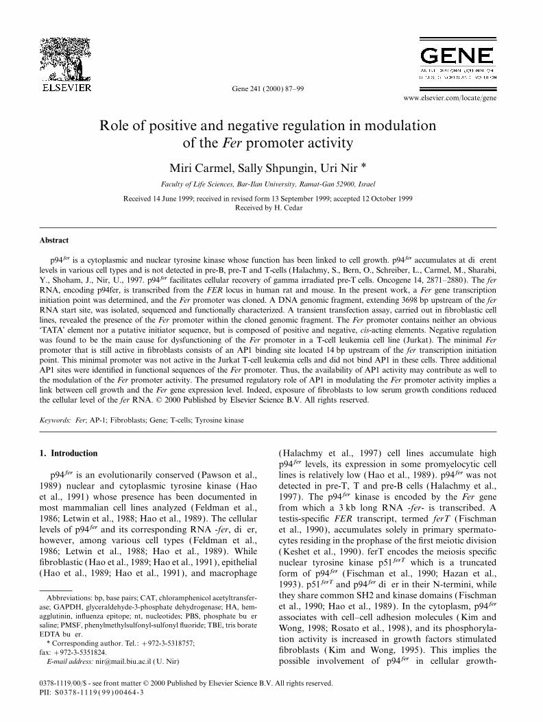
Gene 241 (2000) 87–99 www.elsevier.com/locate/gene Role of positive and negative regulation in modulation of the Fer promoter activity Miri Carmel, Sally Shpungin, Uri Nir * Faculty of Life Sciences, Bar-Ilan University, Ramat-Gan 52900, Israel Received 14 June 1999; received in revised form 13 September 1999; accepted 12 October 1999 Received by H. Cedar Abstract p94fer is a cytoplasmic and nuclear tyrosine kinase whose function has been linked to cell growth. p94fer accumulates at di erent levels in various cell types and is not detected in pre-B, pre-T and T-cells (Halachmy, S., Bern, O., Schreiber, L., Carmel, M., Sharabi, Y., Shoham, J., Nir, U., 1997. p94fer facilitates cellular recovery of gamma irradiated pre-T cells. Oncogene 14, 2871–2880). The fer RNA, encoding p94fer, is transcribed from the FER locus in human rat and mouse. In the present work, a Fer gene transcription initiation point was determined, and the Fer promoter was cloned. A DNA genomic fragment, extending 3698 bp upstream of the fer RNA start site, was isolated, sequenced and functionally characterized. A transient transfection assay, carried out in fibroblastic cell lines, revealed the presence of the Fer promoter within the cloned genomic fragment. The Fer promoter contains neither an obvious ‘TATA’ element nor a putative initiator sequence, but is composed of positive and negative, cis-acting elements. Negative regulation was found to be the main cause for dysfunctioning of the Fer promoter in a T-cell leukemia cell line (Jurkat). The minimal Fer promoter that is still active in fibroblasts consists of an AP1 binding site located 14 bp upstream of the fer transcription initiation point. This minimal promoter was not active in the Jurkat T-cell leukemia cells and did not bind AP1 in these cells. Three additional AP1 sites were identified in functional sequences of the Fer promoter. Thus, the availability of AP1 activity may contribute as well to the modulation of the Fer promoter activity. The presumed regulatory role of AP1 in modulating the Fer promoter activity implies a link between cell growth and the Fer gene expression level. Indeed, exposure of fibroblasts to low serum growth conditions reduced the cellular level of the fer RNA. © 2000 Published by Elsevier Science B.V. All rights reserved. Keywords: Fer; AP-1; Fibroblasts; Gene; T-cells; Tyrosine kinase 1. Introduction ( Halachmy et al., 1997) cell lines accumulate high p94fer levels, its expression in some promyelocytic cell lines is relatively low (Hao et al., 1989). p94fer was not p94fer is an evolutionarily conserved (Pawson et al., 1989) nuclear and cytoplasmic tyrosine kinase (Hao detected in pre-T, T and pre-B cells (Halachmy et al., et al., 1991) whose presence has been documented in 1997). The p94fer kinase is encoded by the Fer gene most mammalian cell lines analyzed (Feldman et al., from which a 3 kb long RNA -fer- is transcribed. A 1986; Letwin et al., 1988; Hao et al., 1989). The cellular testis-specific FER transcript, termed ferT ( Fischman levels of p94fer and its corresponding RNA -fer, di er, et al., 1990), accumulates solely in primary spermato- however, among various cell types (Feldman et al., cytes residing in the prophase of the first meiotic division 1986; Letwin et al., 1988; Hao et al., 1989). While ( Keshet et al., 1990). ferT encodes the meiosis specific fibroblastic ( Hao et al., 1989; Hao et al., 1991 ), epithelial nuclear tyrosine kinase p51ferT which is a truncated (Hao et al., 1989; Hao et al., 1991), and macrophage form of p94fer (Fischman et al., 1990; Hazan et al., 1993). p51ferT and p94fer di er in their N-termini, while they share common SH2 and kinase domains ( Fischman Abbreviations: bp, base pairs; CAT, chloramphenicol acetyltransfer- et al., 1990; Hao et al., 1989). In the cytoplasm, p94fer ase; GAPDH, glyceraldehyde-3-phosphate dehydrogenase; HA, hem- agglutinin, influenza epitope; nt, nucleotides; PBS, phosphate bu er associates with cell–cell adhesion molecules ( Kim and saline; PMSF, phenylmethylsulfonyl-sulfonyl fluoride; TBE, tris borate Wong, 1998; Rosato et al., 1998), and its phosphoryla- EDTA bu er. tion activity is increased in growth factors stimulated * Corresponding author. Tel.: +972-3-5318757; fibroblasts ( Kim and Wong, 1995). This implies the fax: +972-3-5351824. E-mail address: [email protected] ( U. Nir) possible involvement of p94fer in cellular growth- 0378-1119/00/$ - see front matter © 2000 Published by Elsevier Science B.V. All rights reserved. PII: S0378-1119(99)00464-3
-
Upload
independent -
Category
Documents
-
view
0 -
download
0
Transcript of Role of positive and negative regulation in modulation of the Fer promoter activity
Gene 241 (2000) 87–99www.elsevier.com/locate/gene
Role of positive and negative regulation in modulationof the Fer promoter activity
Miri Carmel, Sally Shpungin, Uri Nir *Faculty of Life Sciences, Bar-Ilan University, Ramat-Gan 52900, Israel
Received 14 June 1999; received in revised form 13 September 1999; accepted 12 October 1999Received by H. Cedar
Abstract
p94fer is a cytoplasmic and nuclear tyrosine kinase whose function has been linked to cell growth. p94fer accumulates at differentlevels in various cell types and is not detected in pre-B, pre-T and T-cells (Halachmy, S., Bern, O., Schreiber, L., Carmel, M., Sharabi,Y., Shoham, J., Nir, U., 1997. p94fer facilitates cellular recovery of gamma irradiated pre-T cells. Oncogene 14, 2871–2880). The ferRNA, encoding p94fer, is transcribed from the FER locus in human rat and mouse. In the present work, a Fer gene transcriptioninitiation point was determined, and the Fer promoter was cloned. A DNA genomic fragment, extending 3698 bp upstream of the ferRNA start site, was isolated, sequenced and functionally characterized. A transient transfection assay, carried out in fibroblastic celllines, revealed the presence of the Fer promoter within the cloned genomic fragment. The Fer promoter contains neither an obvious‘TATA’ element nor a putative initiator sequence, but is composed of positive and negative, cis-acting elements. Negative regulationwas found to be the main cause for dysfunctioning of the Fer promoter in a T-cell leukemia cell line (Jurkat). The minimal Ferpromoter that is still active in fibroblasts consists of an AP1 binding site located 14 bp upstream of the fer transcription initiationpoint. This minimal promoter was not active in the Jurkat T-cell leukemia cells and did not bind AP1 in these cells. Three additionalAP1 sites were identified in functional sequences of the Fer promoter. Thus, the availability of AP1 activity may contribute as well tothe modulation of the Fer promoter activity. The presumed regulatory role of AP1 in modulating the Fer promoter activity implies alink between cell growth and the Fer gene expression level. Indeed, exposure of fibroblasts to low serum growth conditions reducedthe cellular level of the fer RNA. © 2000 Published by Elsevier Science B.V. All rights reserved.
Keywords: Fer; AP-1; Fibroblasts; Gene; T-cells; Tyrosine kinase
1. Introduction (Halachmy et al., 1997) cell lines accumulate highp94fer levels, its expression in some promyelocytic celllines is relatively low (Hao et al., 1989). p94fer was notp94fer is an evolutionarily conserved (Pawson et al.,
1989) nuclear and cytoplasmic tyrosine kinase (Hao detected in pre-T, T and pre-B cells (Halachmy et al.,et al., 1991) whose presence has been documented in 1997). The p94fer kinase is encoded by the Fer genemost mammalian cell lines analyzed (Feldman et al., from which a 3 kb long RNA -fer- is transcribed. A1986; Letwin et al., 1988; Hao et al., 1989). The cellular testis-specific FER transcript, termed ferT (Fischmanlevels of p94fer and its corresponding RNA -fer, differ, et al., 1990), accumulates solely in primary spermato-however, among various cell types (Feldman et al., cytes residing in the prophase of the first meiotic division1986; Letwin et al., 1988; Hao et al., 1989). While ( Keshet et al., 1990). ferT encodes the meiosis specificfibroblastic (Hao et al., 1989; Hao et al., 1991), epithelial nuclear tyrosine kinase p51ferT which is a truncated(Hao et al., 1989; Hao et al., 1991), and macrophage form of p94fer (Fischman et al., 1990; Hazan et al.,
1993). p51ferT and p94fer differ in their N-termini, whilethey share common SH2 and kinase domains (FischmanAbbreviations: bp, base pairs; CAT, chloramphenicol acetyltransfer-et al., 1990; Hao et al., 1989). In the cytoplasm, p94ferase; GAPDH, glyceraldehyde-3-phosphate dehydrogenase; HA, hem-
agglutinin, influenza epitope; nt, nucleotides; PBS, phosphate buffer associates with cell–cell adhesion molecules ( Kim andsaline; PMSF, phenylmethylsulfonyl-sulfonyl fluoride; TBE, tris borate Wong, 1998; Rosato et al., 1998), and its phosphoryla-EDTA buffer.
tion activity is increased in growth factors stimulated* Corresponding author. Tel.: +972-3-5318757;fibroblasts ( Kim and Wong, 1995). This implies thefax: +972-3-5351824.
E-mail address: [email protected] (U. Nir) possible involvement of p94fer in cellular growth-
0378-1119/00/$ - see front matter © 2000 Published by Elsevier Science B.V. All rights reserved.PII: S0378-1119 ( 99 ) 00464-3

88 M. Carmel et al. / Gene 241 (2000) 87–99
Fig. 1. Primer extension analysis of the fer RNA. (A) 5∞ end sequence of the previously cloned murine fer cDNA. The 19 nt long complementarysynthetic primer used for the extension analysis is shown and is underlined with an arrow. (B) Primer extension analysis was carried out with: lane1, 50 mg total cytoplasmatic RNA, lane 2, 5 mg cytoplasmic poly(A)+ RNA (M ) molecular weight markers. Extension products were resolved ina 7% urea–PAGE. Migration distances of the two major extension products obtained are marked with arrows on the left. Migration distances ofknown molecular weight markers are given on the right-hand side of the figure.
controlling processes. The p94fer nuclear fraction was cloned fer 5∞ leader (Fig. 1A), was end-labeled with[c-32P] ATP and T4 polynucleotide kinase (Sambrookfound to associate with the cell chromatin (Hao et al.,
1991). et al., 1989). The labeled primer and NIH/3T3 RNAwere heated together to 80°C and allowed to anneal atTo study the mechanisms that direct the different Fer
expression levels in different cell types and the generation 42°C. The extension reaction was carried out asdescribed previously (Sambrook et al., 1989; Ausubelof both, somatic and meiotic transcripts from the FER
locus, the Fer gene 5∞ flanking sequences were cloned et al., 1996). Extension products were resolved in aurea–7% PAGE and visualized by autoradiography.and analyzed. A somatic fer transcription initiation
point has been identified, and the DNA sequences thatpromote somatic Fer expression are described.
2.2. Rapid amplification of cDNA end (RACE)
RACE was carried out as described previously2. Materials and methods( Volloch et al., 1994; Frohman et al., 1995), using aReverse Transcription (RT) primer (GTCTTGCAACT-2.1. Primer extensionTTAACACAGC) and then a nested primer (TCAGG-TCACTCCCAAATCC ) for PCR. PCR products wereThe 19 nt long primer, -TCAGGTCACTCCC-
AAATCC-, which is complementary to nt 57–75 in the cloned in the pGEM-T Easy vector (Promega) and

89M. Carmel et al. / Gene 241 (2000) 87–99
sequenced with the ABI PRISM 377 automatic DNA ing bases and religated. To obtain the −879D24FCATsequencer (Perkin Elmer, Weizmann Institute of plasmid, the −879FCAT plasmid was cut with GsuISciences). and treated with T4 DNA polymerase. It was then cut
with BglI, and the 1730 bp long fragment obtained was2.3. Cloning and sequencing of FER 5∞ flanking sequences ligated to a 2924 bp long fragment derived
from pMWCAT, which was cut with SmaI and BglI.A mouse genomic partial Sau3A library cloned in the −714FCAT was cut with XcmI and PstI, treated with
EMBL-3 SP6/T7 phage (Clontech), was screened with T4 DNA polymerase to remove overhanging bases anda 32P-labeled, 500 bp long XbaI–EcoRI fragment, religated to obtain −714D65FCAT. −714FCAT wasderived from the 5∞ end of the mouse fer cDNA. Two cut with GsuI and treated with T4 DNA polymerase. Itphages carrying extreme 5∞ fer sequences were isolated was then cut with BglI, and the 1481 bp long fragmentand further analyzed. XhoI–EcoRI fragments isolated obtained was ligated with a 2924 bp long fragmentfrom the inserts of the two phages were cloned into the derived from pMWCAT, which was cut with SmaI andsame sites in the pBluescript KS+ plasmid. The cloned BglI. This plasmid is called −714D24FCAT. To obtainfragments were sequenced using the ABI PRISM 377 the −64D28FCAT plasmid, the −64FCAT plasmid wasautomatic DNA sequencer (Perkin Elmer). BsaAI–XhoI cut with GsuI and treated with T4 DNA polymerase. Itgenomic subfragments were isolated and cloned into was then cut with BglI, and the 914 bp long fragmentXhoI–SmaI sites in the pMWCAT plasmid, upstream of obtained was ligated to a 2 bp fragment derived fromthe CAT gene. The new plasmids were termed a pMWCAT cut with SmaI and BglI. −879FCAT was−3698FCAT and −879FCAT. These plasmids carry cut with XhoI and SalI, and the 905 bp long fragment3698 and 879 bp upstream of the fer initiation point, obtained was inserted into the pMWCAT XhoI site.respectively. Only the reverse orientation was selected, and the
plasmid obtained was termed −879RFCAT.2.4. Construction of Fer promoter deletions
2.5. Cell transfections and analysis of Fer promoterThe plasmid −879FCAT was cut with XhoI and AatI. activity
The digested plasmid was incubated at 27°C with400 units of Exonuclease III. Samples were removed at NIH/3T3 and Jurkat cells were transiently1 min intervals and transferred to dry ice. Exonuclease co-transfected with 0.4 mg of CMVLuciferase and 3.6 mgIII was inactivated by heating the removed samples to of FCAT plasmids by using the lipofectamine reagent70°C. One hundred and forty units of S1 nuclease were according to the manufacturer’s instructionsadded to each sample, which was then incubated for (GibcoBRL). Cellular extracts were prepared from30 min at room temperature. The resulting DNA frag-
transfected cells, 48 h after transfection, by using a lysisments were first treated with the Klenow fragment ofbuffer (Promega), according to the manufacturer’sDNA polymerase I to blunt the ends and then with T4instructions. CAT activity was assayed as describedDNA ligase to close the resultant plasmids (Ausubelpreviously (Ausubel et al., 1996), and luciferase activityet al., 1996). The plasmids obtained with that strategywas determined using the Luciferase Promega assaywere termed: −714FCAT, −706FCAT, −630FCAT,system. Arbitrary CAT activity units were determined−548FCAT, −525FCAT, −435FCAT and −305FCAT.by normalizing the CAT activities to the obtained lucifer-The −305FCAT plasmid was cut with ApoI, and thease activity units, which served as an internal control.473 bp long fragment was inserted into EcoRIAll transfections were repeated five times, and the datacut pMWCAT. The new plasmid was termedrepresent the mean and one standard deviation.−138FCAT. The −879FCAT plasmid was cut with
XhoI and XcmI, treated with T4 DNA polymerase to2.6. Preparation of nuclear extractform blunt ends and then religated. This plasmid is
called −64FCAT. The −879FCAT plasmid was cutNuclear extracts were prepared essentially aswith GsuI, treated with T4 DNA polymerase and then
described previously (Schreiber et al., 1989). Cells werecut with KpnI. A 327 bp fragment was ligated with awashed with PBS, centrifuged and resuspended in buffer:3535 bp long fragment derived from a SmaI and KpnI10 mM Hepes, pH 7.9, 10 mM KCl, 1.5 mM MgCl2,cut pMWCAT plasmid. The obtained plasmid was0.5 mM DTT, 0.2 mM EDTA, 1 mM PMSF, 1 mg/ml oftermed −22FCAT. To form the +3FCAT plasmid, thePefabloc, 0.5 mg/ml of Aprotinin, 0.5 mg/ml of Leupeptin−64FCAT plasmid was cut with MslI and BglI, andand 1 mg/ml of Benzamidine. Cells were stored for 10 minthe 2971 bp long fragment obtained was ligated with anon ice for swelling and then lysed in the presence of891 bp long fragment derived from a SmaI and BglI0.1% NP-40. Nuclei were spun down by centrifugationcut pMWCAT. To obtain the −879D65FCAT plasmid,at 10 000×g for 15 s and suspended in a high-saltthe −879FCAT plasmid was cut with XcmI and PstI,
treated with T4 DNA polymerase to remove overhang- extraction buffer: 10 mM Hepes, pH 7.9, 420 mM NaCl,

90 M. Carmel et al. / Gene 241 (2000) 87–99
1.5 mM MgCl2, 0.2 mM DTT, 0.2 mM EDTA, 1 mM exposed to Northern blot analysis as described pre-viously (Halachmy et al., 1997). The blotted RNA wasPMSF, 1 mg/ml of Pefabloc, 0.5 mg/ml of Aprotinin,
0.5 mg/ml of Leupeptin and 1 mg/ml of Benzamidine. hybridized with a 32P-labeled, 500 bp long, EcoRI–XbaIfragment, derived from the 5∞ end of the murine ferNuclear proteins were extracted by rotating the nuclei
for 30 min in the extraction buffer at 4°C, followed by cDNA. In parallel, a 1600 bp long GAPDH cDNA was32P-labeled and used as a probe.centrifugation at 20 000×g for 15 min Supernatants
were collected, divided into aliquots and stored at−70°C.
3. Results2.7. Electromobility gel shift assay
3.1. Determination of the fer RNA transcriptioninitiation pointTwo synthetic oligonucleotides (5∞ ACTTCCTTC-
TAACTGACTCACGT 3∞ and 5∞ GACGTGAGTCA-GTTAGAAGG 3∞) were annealed at equimolar concen- The mouse fer cDNA has been cloned and its
sequence determined (Letwin et al., 1988) (Letwine, K.,trations. The double-stranded fragment was labeledwith [a-32P] dATP and dCTP by a fill-in reaction using Pawson, T., GenBank Accession No. U76762). The
sequence of the cloned fer 5∞ non-translated leader isa Klenow fragment and then purified on 12% acrylamidegel ( Katcoff et al., 1993). presented in Fig. 1A. To determine the 5∞ end of the fer
RNA, a primer extension (Ausubel et al., 1996;Five micrograms of nuclear protein extract wereincubated at 25°C for 15 min with 100 ng of poly dIdC Fischman et al., 1990) and RACE analysis (Frohman
et al., 1995) were carried out. A 19 nt long primer,and 0.1 ng of labeled probe in binding buffer: 4 mMTris–HCl, pH 8.0, 40 mM NaCl, 4 mM MgCl2, and 5% complementary to nt 57–75 of the cloned fer 5∞ leader
(Fig. 1A) was end-labeled and hybridized with RNAglycerol in a total volume of 25 ml. In supershift experi-ments, 3 mg of anti c-Fos antibodies (Santa Cruz) were extracted from NIH/3T3 cells. The reverse transcriptase
extension products were resolved in a denaturing 7%added to the reaction. The assay mixture was thenseparated on 4% acrylamide TBE gel at 100 V for 2– urea-PAGE and visualized by autoradiography. Two
major extension products were obtained in that assay.3 h. The gel was then dried and exposed over night toKodak film ( X-OMAT AR). One of these was about 100 nt long and was obtained
only from cytoplasmic poly(A)+ RNA (Fig. 1B, lane2). This size suggests that the fer RNA starts 79 nt2.8. Northern blot analysisupstream of the fer AUG translation initiation codon(Fig. 1A). Another 83 nt long band was obtained fromRNA was extracted from tissue culture cells with
Trireagent (Molecular Research Center), according to both poly(A)+ and total cytoplasmic RNA preparations(Fig. 1B). This implies the existence of a fer transcriptthe manufacturer’s instructions. RNA was fractionated
in 1.2% formamide/formaldehyde agarose gel and that initiates 62 nt upstream of the fer AUG (Fig. 1A).
Fig. 2. Race analysis of the fer RNA. (A) 5∞ sequence of the previously cloned murine fer cDNA. The nucleotide sequence of the reverse transcriptionprimer (RT primer) and the nested primer used for that analysis is shown. The primers are underlined with left-oriented arrows. (B) Codingsequence of the obtained RACE product. The eight nucleotides missing from the previously cloned murine fer cDNA are bold. The determinedfer initiation point is marked with an arrow.

91M. Carmel et al. / Gene 241 (2000) 87–99
These two main primer extension products extend 25 nt extending from −3698 to +26 (transcription initiationpoint is marked as +1 in Fig. 3B) was linked to the(100 nt long band) and 8 nt (83 nt band) beyond the 5∞
end of the previously cloned fer cDNA (Fig. 1A). The bacterial reporter CAT gene (Gorman et al., 1982). Thenewly constructed −3698FCAT plasmid (Fig. 4A) waspresence of additional primer extension products
(Fig. 1B lane 2) indicates the possible existence of other transiently introduced to NIH/3T3 cells, together with aCMVLuciferase plasmid that served as a normalizingfer transcription initiation sites.
To clone and determine the missing fer RNA internal control. These cells produce relatively high levelsof p94fer (Halachmy et al., 1997). The CAT activitysequences, NIH/3T3 whole-cell RNA was subjected to
RACE analysis (Frohman et al., 1995) (Fig. 2A). PCR driven by the −3698 fragment (−3698FCAT, Fig. 4B)was fourfold higher than the CAT activity obtained fromproducts were cloned and their sequence determined.
All PCR products analyzed were found to extend 8 nt a promoterless CAT plasmid +3FCAT (Fig. 4B).Removal of 2819 bp from the −3698 F fragment loweredbeyond the 5∞ end of the previously cloned mouse cDNA
(Fig. 2B), a fact that coincides with the existence of an the promoter activity by about twofold (−879FCAT,Fig. 4B). Inversion of the −879 Fer fragment in front of83 nt long fer RNA primer extension product (Fig. 1B).
These results revealed the usage of an adenosine that the CAT gene, brought the driven CAT activity down tothe basal values (−879RFCAT, Fig. 6). Deletion ofresides 62 nt upstream of the fer AUG, as a fer RNA
transcription initiation point (Fig. 2B). 165 bp from the 5∞ end of the −879 fragment, restoredthe original promoter activity (compare −714FCAT and−3698FCAT, Fig. 4B). Further sequential deletion of the3.2. Cloning and characterization of genomic Fer 5∞
flanking sequences 5∞ Fer sequences down to nt −305, led to gradual twofoldincrease over the −3698Fer promoter activity (compare−305FCAT and −3698FCAT, Fig. 4B). Out of the testedA 500 bp long Xba1–EcoRI fragment derived from
the 5∞ end of the mouse fer cDNA was labeled and used Fer fragments, −548 Fer, −435 and −305 Fer thus seemto exert the highest promoter activity (Fig. 4B). Toas a probe for screening a mouse partial Sau3A genomic
library, cloned in the EMBL-3 phage (Clontech). One determine the minimal and yet functional Fer promoter,further consecutive deletions were introduced. Removalof the isolated recombinant phages was found to carry
an insert that contained a 4480 bp long fragment at its of 167 bp from the −305 Fer fragment decreased the Ferpromoter activity by fourfold (Fig. 4B, −138FCAT ).5∞ portion. This fragment was completely sequenced
(GenBank Accession no. AF191493) and was found to Interestingly, shortening the Fer promoter down to the−64 point restored 60% of the maximal Fer promotercontain the first exon of the Fer gene and 3698 bp,
extending 5∞ upstream of the identified fer RNA tran- activity (−64FCAT, Fig. 4B). Deletion of a further 67 bpoff the −64 Fer fragment, abolished its promoter activityscription initiation point (Fig. 3A). The Fer first exon
was found to be 269 bp long (Fig. 3A). A donor splice and brought it down to the basal CAT vector activity(+3FCAT, Fig. 4B). Examination of the −64 Fersite links between the Fer first exon and the following
intron (Fig. 3A). The sequence of the 1178 bp, which sequence revealed the existence of a GATA-like-GGATAA- sequence (Orkin, 1992) ( located at positionflank the fer RNA start site and 84 bp extending beyond
it, is shown in Fig. 3B. Several known transcription −34 relative to the fer start site) and an AP1 bindingsite (Lee et al., 1987) ( located at position −14) (Fig. 3B).factors binding sites were found to reside in the cloned
genomic fragment (Fig. 3B). Two of these are located Removal of the GATA binding site did not affect theresidual −22 Fer promoter activity (Fig. 4B, −22FCAT ),in the vicinity of the fer initiation point. An AP1 binding
site (Lee et al., 1987) resides 14 bp upstream of the fer and the AP1 site containing FER fragment exerted arelatively high promoter activity (Fig. 4B, −22FCAT ).start site (Fig. 3B), and another adjacent sequence,
-GGATAA- (GATA-like) (Orkin, 1992), is located 34 bp This activity was completely abolished upon removal ofa further 25 bp, which included the AP1 site (Fig. 4B,upstream of the Fer transcription initiation point
(Fig. 3B). Three additional AP1 binding sites were iden- +3FCAT ). The relative CAT activity driven by theGATA factor binding site, was slightly weaker (by 25%)tified at positions −283, −336 and −562, in the cloned
FER genomic fragment (Fig. 3B). No obvious TATA than that exhibited by the AP1 sequence (see Fig. 6B,−64D28FCAT versus −64FCAT and −22FCAT ). Thebox (Breathnach and Chambon, 1981) or initiator
sequences (Javahery et al., 1994) were detected in the −64D28 and −22 Fer fragments that contain the GATAor AP1 binding sites, respectively, thus show a relativelyvicinity of the fer initiation point.high promoter activity, which reaches 50 and 60% of themaximal Fer promoter activity, respectively (Fig. 4B,3.3. Cell-type restricted promoter activity of the Fer 5∞
flanking sequences −305FCAT ).p94fer is not detected in pre-B, pre-T and T cells; nor
was the fer RNA detected in these cells (HalachmyTo identify transcription-promoting activity in the Fer5∞ flanking sequences, a 3724 bp long FER fragment et al., 1997). In an attempt to further the understanding

92 M. Carmel et al. / Gene 241 (2000) 87–99
A
B

93M. Carmel et al. / Gene 241 (2000) 87–99
Fig. 4. Functional analysis of the Fer promoter. (A) Descriptive presentation of the functionally analyzed genomic Fer deletions. The location ofthe AP1 site, which is most proximal to the fer transcription initiation point, is shown. Numbers denote the distance of 5∞ ends from the fertranscription initiation point. (B, C ) Relative CAT activity, promoted by the Fer genomic fragments described in (A). Transfections were carriedout in (B) NIH/3T3 and (C ) Jurkat cells. Arbitrary CAT activity units were calculated by normalizing the CAT activity values to the luciferaseinternal control activity. Bars denote standard deviations.
Fig. 3. Cloned, Fer 5∞ genomic sequences. (A) Schematic presentation of the Fer gene 5∞ flanking sequences, the Fer first exon and part of the Ferfirst intron. The cloned genomic Fer fragment is 4480 bp long and contains 3698 bp long Fer 5∞ flanking sequences (open box), the 269 bp long Ferfirst exon (squares) and the first 513 bp of the Fer first intron (hatched box). (B) Sequence of 1178 nt, which precedes the Fer transcription initiationpoint and 84 nt, which follows that site, carried by the cloned Fer genomic fragment. (For the sequence of further upstream 2520 nt, please seeGenBank Accession No. Bankit 296239, AF191493.) The numbers denote the distance from the fer RNA initiation site (marked with an arrow).Binding sites of some known transcription factors are underlined. GR,glucocorticoid receptor; HNF5, Hepatocyte nuclear factors 5; PEA1/3,polyomavirus enhancer activator proteins 1 and 3. The 47 bp long AC-rich sequence is double-underlined. The first eight amino acids of theencoded p94fer protein are also shown.

94 M. Carmel et al. / Gene 241 (2000) 87–99
Fig. 5. Electromobility gel-shift assay. (A) Sequence of the double-stranded oligonucleotide, which was used as a probe in this assay. The numbersindicate the position of that fragment in the Fer promoter (see also Fig. 3A). (B) Nuclear extracts from Jurkat ( lanes 2, 3), NIH/3T3 (lanes 4, 5)and F-9 cells ( lanes 6, 7) were incubated with a 32P-labeled probe in the absence ( lanes 2, 4, 6) or presence ( lanes 3, 5, 7) of anti-c-Fos antibodies.The reaction products were then run in a 4% polyacrylamide gel. Lane 1, free probe. The arrow on the left indicates the AP1 bound super-shiftedprobe. (C ) Nuclear extracts from NIH/3T3 cells were incubated with a 32P-labeled probe in the presence of influenza a-HA antibodies ( lane 1), anon-relevant competitor — 300 ng of double-stranded oligonucleotide derived from nt −33 to −56 in the Fer promoter ( lane 2), 3, 30, 150 and300 ng of the non-labeled fragment, which was used as a probe in this assay ( lanes 3–6, respectively).
of the mechanisms that direct the cell-type restricted minimal Fer promoter and is located 14 nt upstream ofthe fer initiation point did not exhibit transcriptionexpression of the Fer gene, the activity of the cloned Fer
promoter was tested in the Jurkat T-cell leukemia cell promoting activity in Jurkat cells (−22FCAT, Fig. 4Band C ).line. Introduction of the −3698FCAT plasmid to Jurkat
T cells drove the accumulation of CAT activities, which In order to further establish the importance of AP1in potentiating the Fer minimal promoter activity, anwere similar to those obtained from a promoterless
plasmid (+3FCAT, Fig. 4C). Removal of 2984 bp from electromobility gel-shift assay of the minimal Fer pro-moter containing the proximal AP1 binding site wasthe Fer 5∞ flanking sequences restored the same Fer
promoter activity that was obtained in fibroblastic cells performed. Nuclear protein extracts from NIH/3T3,Jurkat and F9 cells were incubated with 32P-labeled(Fig. 4B and C ). A negatively acting cis-regulatory
element(s) thus lies between nt −3698 and –714 (Fig. 4) oligonucleotide, which extended from nt −33 to nt −10in the Fer promoter (Fig. 5A), and were then exposedin the Fer promoter. Interestingly, unlike that which
was seen in fibroblasts, the AP1 site that constitutes the to an electromobility gel shift assay ( Katcoff et al.,
Fig. 6. Functional role of GATA and AP1 binding sites in Fer promoter activity. (A) Descriptive presentation of the internally deleted Fer promoterfragments. Numbers denote the limits of the introduced deletions. The locations of the AP1 (−14) and GATA (−34) binding sites are marked.(B, C ) Relative CAT activity, directed by the FER fragments presented in (A). Transfections were carried out in (B) NIH/3T3 and (C ) Jurkatcells (−879R represents the inversion of the −879 Fer fragment in front of the CAT gene). Arbitrary CAT activity units were calculated bynormalizing the CAT activity values to the luciferase internal control activity. Bars denote standard deviations.

95M. Carmel et al. / Gene 241 (2000) 87–99

96 M. Carmel et al. / Gene 241 (2000) 87–99
1993). The nuclear extract prepared from Jurkat cells levels were compared to those found in cells grownunder regular (10%) serum conditions. Lowering theshowed a binding profile that differed from that obtained
with the NIH/3T3 extract (Fig. 5B). Moreover, a serum growth conditions from 10% to 0.5 or 0.1%, ledto a 2.5-fold reduction in the fer RNA levels, after 72 hsupershift assay performed with anti-c-Fos antibodies
revealed the binding of AP1 to the Fer promoter (Fig. 7, lanes 3 and 6). The level of the GAPDH RNA,encoding a house-keeping enzyme, was only slightlysequences in NIH/3T3 but not in Jurkat cell extracts
(Fig. 5B, lanes 3 and 5). This supershift was not seen affected under these serum growth conditions (Fig. 7,lanes 3 and 6).with an irrelevant antiserum (Fig. 5C — a-HA anti-
body), thus substantiating the specificity of our assay.As expected (Caceres et al., 1990), AP1 binding activitywas also absent from extracts of F9 cells (Fig. 5B, lanes 4. Discussion6 and 7). These results substantiate the pivotal role ofAP1 in potentiating the transcription promoting activity The p94fer tyrosine kinase is expressed in most mouse
cell lines analyzed (Feldman et al., 1986; Letwin et al.,of the minimal Fer promoter.1988; Hao et al., 1989; Halachmy et al., 1997). Theexpression levels of that kinase and its corresponding3.4. Role of the proximal GATA and AP1 elements in the
functioning of the Fer promoter RNA differ, however, among different cell types. Whilebeing present at relatively high levels in epithelial andfibroblastic cells, the cellular levels of p94fer are muchThe absence of an obvious TATA element in the Fer
promoter and the close proximity of GATA and AP1 lower in hematopoietic cells, and it is absent from pre-B,pre-T and T-cells (Hao et al., 1989; Halachmy et al.,sites to the fer initiation point suggested a key role of
these elements in the functioning of the Fer promoter. 1997). The relatively high fer RNA levels in fibroblasticcells are serum-dependent (Fig. 7). In an attempt toTo analyze the functional role of the −34 GATA
and −14 AP1 sites in a longer Fer promoter fragment, unravel the mechanisms involved in the regulated expres-sion of the Fer gene, the Fer 5∞ flanking sequences werethe two sites were deleted from the −714 fragment.
Excision of both, GATA and AP1 binding sites from cloned and characterized. Primer extension and RACEanalysis of the fibroblastic fer RNA revealed the exis-the −714 Fer fragment caused a significant 2.5-fold
reduction in its promoter activity in both NIH/3T3 and tence of an adenosine lying 62 nt upstream of the ferAUG (Figs. 2 and 3B) that serves as an initiation siteJurkat cells (Fig. 6B and C, −714D65FCAT ). This
reduction was less prominent upon deleting the two sites in the Fer gene. However, based on the primer extensionassay (Fig. 1B), one cannot exclude the existence offrom the weakly active −879 Fer fragment (Fig. 6B and
C, −879D65FCAT ). Removal of the −14 AP1 site other Fer transcription initiation sites. Multiple tran-scription initiation sites are often found in TATA-lessalone, from the −879 (−879D24FCAT ), −714
(−714D24FCAT ) and –64 (−64D28FCAT ) fragments, promoters (Sideras et al., 1994).The identified fer transcription initiation point differsabolished only 20% of their transcription promoting
activity (Fig. 6B). The −34 GATA site and the −14 from the previously determined 5∞ end of the meiosisspecific FER transcript, ferT (Fischman et al., 1990).AP1 site, thus seem to play equally important roles in
the functioning of the −714 Fer fragment. All the above This difference suggests the usage of two separatedpromoters that direct the synthesis of the fer and ferTdescribed transfection experiments were repeated in
LTK− cells, and the relative CAT activities obtained transcripts, in the FER locus.Three thousand, six hundred and ninety-eight basewere similar to those obtained in NIH/3T3 cells (data
not shown). pairs preceding the identified fer initiation point exhib-ited transcription promoting activity in NIH/3T3 cells(−3698FCAT, Fig. 4B) but failed to do so in Jurkat3.5. Fer gene expression in fibroblasts is affected by
serum growth conditions cells (−3698FCAT, Fig. 4C). Removal of 2984 bp fromthe 5∞ end of Fer promoter restored its activity in Jurkatcells and equalized it with the values obtained inThe AP1 cellular level and activity can be induced
by growth factor stimulation (Banister et al., 1994). The NIH/3T3 cells (−714FCAT, Fig. 4B and C ). This sug-gests the presence of negatively acting cis element(s) inpresence of several AP1 binding sites in the Fer promoter
(Fig. 3B) prompted us to ask whether there were any the Fer promoter, which prevent its functioning inT-cells.differences in the steady-state fer RNA levels under
regular and low serum conditions. Murine fibroblastic The fer initiation point is not preceded by any obviousTATA element (Breathnach and Chambon, 1981)LTK− cells were grown in 0.5 and 0.1% fetal calf serum
for 24, 48 and 72 h. Whole-cell RNA was prepared from (Fig. 3B); nor does it reside within a consensus initiatorsequence (Javahery et al., 1994). The transcription ofthe ‘starved’ cells, and the relative fer RNA levels were
determined by using a ‘Northern blot’ analysis. These the fer RNA thus seems to depend on other elements

97M. Carmel et al. / Gene 241 (2000) 87–99
Fig. 7. fer and GAPDH RNA levels in normally grown and serum starved LTK− cells. (A) Northern blot analysis of the fer and GAPDH RNAin LTK− cells, grown in 0.1% ( lanes: 1–3), 0.5% ( lanes: 4–6) and 10% fetal calf serum ( lane 7). (B) The autoradiograms presented in (A) werescanned, and the band’s relative intensity was calculated by using the TINA application.
localized in the vicinity of its initiation point. Two such 1992). However, a 22 bp long FER genomic fragment,which lacks the GATA element but does contain anpotential elements, an AP1 binding site and the
GGATAAA sequence (GATA like), are located 14 AP1 binding site, suffices to function as a minimal Ferpromoter in fibroblastic cells. This fragment exerted(AP1) and 34 bp (GATA), upstream of the fer initiation
point. The GGATAAA element has been previously 60% of the highest Fer promoter activity, obtained inthe current work (−FCAT, Fig. 4B). The proximalimplied in binding GATA factors (Orkin, 1992) and
TFIID (Nakajima et al., 1988). The dispensability of localization of the AP1 binding site in the vicinity ofthe fer initiation point may enable it to function indepen-the GATA like sequence in the functioning of the
minimal Fer promoter (Fig. 4B, −22FCAT ) and the dently of the GATA element, in the minimal Fer pro-moter (−22FCAT, Fig. 6B). Interestingly, this AP1 sitelack of a functional TATA element in most tyrosine
kinase genes analyzed (Sideras et al., 1994) support the did not exert an independent promoter activity in theJurkat T-cell leukemia cells (−22FCAT, Figs. 4C andnotion that the GGATAAA element binds a GATA
protein (Orkin, 1992) rather than a basal transcription 6C). This could result from the interference of the YY1factor with AP1 binding to its cognate DNA site, infactor like TFIID (Nakajima et al., 1988). GATA and
AP1 elements were found to cooperate in potentiating T cells (Ye et al., 1996). However, no obvious YY1binding site (Ye et al., 1996) was identified in the vicinitythe transcription initiation of the endothelin-1 ( Kawana
et al., 1995) and globin genes ( Walters and Martin, of the −14 AP1 element (Fig. 3). The lack of AP1

98 M. Carmel et al. / Gene 241 (2000) 87–99
binding activity in Jurkat cells most likely results from factors ( Kim and Wong, 1995) and the putative involve-ment of p94fer in the modulation of cell growth.the relatively low levels of AP1 in non-stimulated T-cells
(McGuire and Iacobelli, 1997). It should be noted, Addition of 74 nucleotides upstream of the GATAbinding site (−138 Fer) significantly impaired the Ferhowever, that the Fer gene is not expressed in activated
T-cells as well (Halachmy et al., 1997), thus suggesting minimal promoter activity in NIH/3T3 cells (Fig. 4B−138FCAT ). Examination of the sequence of thethat repressive elements in the Fer promoter override
the positive activity of AP1. repressing fragment (−138 to −64), revealed the exis-tence of nine AACC repeats constituting most of thatIn the context of a longer Fer fragment, removal of
both the GATA and AP1 lowered the Fer promoter fragment (Fig. 3B). Further analysis should reveal thefunctional role of the AACC repeats in repressing theactivity (−714D65FCAT, Fig. 6B and C ). This impair-
ment became less prominent in a Fer promoter from Fer promoter activity. This repression was completelyrelieved upon extension of the Fer 5∞ flanking sequenceswhich only the −14 AP1 site was deleted
(−714D24FCAT, Fig. 6B). The functional role of the by a further 167 nucleotides, which carry another AP1binding site (Fig. 4B, −305FCAT ).–14 AP1 site seems, therefore, to depend on the context
of its functional interaction with other cis-acting ele- Extension of the Fer promoter from −548 to −879gradually repressed the Fer promoter activity (Fig. 4Bments in the Fer promoter. The cooperation between
the GATA and AP1 factors thus seems to be functionally and C ). This suggests the presence of additional nega-tively acting sequences, between position −879 andimportant in the context of the intact Fer promoter.
Adjacent localization of a GATA binding site, upstream −548, in the Fer promoter.The Fer promoter thus seems to be composed of bothto the initiation point in a ‘TATA less’ promoter was
shown in the erythropoietin receptor gene (Chin et al., positive and negative cis-acting elements. The interactionand interbalance between factors that bind to the two1995). In this promoter, GATA and SP1 binding sites
cooperate to constitute the minimal erythropoietin pro- types of elements may determine the relative expressionlevel of the Fer gene in different cell types. The mutualmoter. In the Fer promoter, a GATA element and an
AP1 binding site may constitute the core of the ‘TATA existence of positive and negative regulatory elementshas been well documented in eukaryotic promotersless’ promoter. Thus, as was previously shown for the
endothelin-1 promoter ( Kawana et al., 1995), GATA (Carty et al., 1997). Further characterization of tran-scription transacting factors that bind to the Fer regula-and AP1 factors are likely to cooperate in the function-
ing of the Fer promoter. The cooperative functioning of tory sequences should allow a further understanding ofthe mechanisms that direct tissue-specific expression ofthe GATA and AP1 factors may compensate for the
absence of a TATA or initiator element in the Fer the Fer tyrosine kinase.promoter.
Six GATA factors have been described in vertebrates(Arceci et al., 1993; Laverriere et al., 1994; Yamamoto
Acknowledgementset al., 1990). Out of these, GATA2, which is expressedin a wide variety of cells (Dorfman et al., 1992), is the
We thank Dr D. Katcoff for his critical commentsbest candidate for the Fer GATA element-binding factor.and Mrs A. Goldreich for typing this manuscript. ThisFour AP1 binding sites (−14, −283, −336 andwork was supported by a grant from The Israel Science−562) can be identified in the functional Fer promoter.Foundation.Removal of 167 bp, which contain one of these sites
(−283), from the −305 fer fragment significantly abro-gated its transcription promoting activity. AP1 may thusplay a key role in modulating the activity of the Fer Referencespromoter by binding to it at several sites. The involve-ment of AP1 in modulating the Fer promoter activity Arceci, R.J., King, A.A., Simon, M.C., Orkins, S.H., Wilson, D.B.,
1993. Mouse GATA-4: a retinoic acid-inducible GATA-bindingwould predict the sensitivity of the Fer gene expressiontranscription factor expressed in endodermally derived tissues andto stimulation of cells with growth factors (Banisterheart. Mol. Cell. Biol. 13, 2235–2246.et al., 1994). Indeed, the fer RNA level dropped 2.5-fold
Ausubel, F.M., Brent, R., Kingston, R.E., Moore, D.D., Seidman,upon exposing LTK− cells, to serum starvation (Fig. 7). J.G., Smith, J.A., Struhl, K., 1996. Current Protocols in MolecularHowever, the gradual decrease in the fer RNA cellular Biology. Wiley, New York.
Banister, A.J., Brown, H.J., Sutherland, J.A., Kouzarides, T., 1994.level does not necessarily directly reflect the drop in thePhosphorylation of the c-Fos and c-Jun HOB1 motif stimulates itsFer promoter activity in serum-starved cells. The half-activation capacity. Nucleic Acids Res. 22, 5173–5176.life of the fer RNA under these conditions also contrib-
Breathnach, R., Chambon, P., 1981. Organization and expression ofutes to the observed steady-state fer RNA levels. The Eucaryotic split genes coding for proteins. Annu. Rev. Biochem.serum-regulated expression of the Fer gene could be 50, 349–383.
Caceres, J., Glikin, G., Bravo, R., Weinmann, R., 1990. c-fos mediatedfunctionally related to the activation of p94fer by growth

99M. Carmel et al. / Gene 241 (2000) 87–99
stimulation of an AP-1 DNA binding activity in undifferentiated Kim, L., Wong, T.W., 1998. Growth factor-dependent phosphoryla-tion of the actin-binding protein cortactin is mediated by the cyto-teratocarcinoma cell lines. Oncogene 5, 59–67.plasmic tyrosine kinase FER. J. Biol. Chem. 273, 23542–23548.Carty, M.D., Lillquist, J.S., Pershavaria, M., Stein, R., Soeller, W.C.,
Laverriere, A.C., MacNeill, C., Mueller, C., Poelman, R.E., Burch,1997. Identification of cis- and trans-active factors regulatingJ.B.E., Evans, T., 1994. GATA-4/5/6, a subfamily of three tran-human islet amyloid polypeptide gene expression in pancreaticscription factors transcribed in developing heart and gut. J. Biol.beta-cells. J. Biol. Chem. 272, 11986–11993.Chem. 269, 23177–23184.Chin, K., Oda, N., Shen, K., Noguchi, C.T., 1995. Regulation of tran-
Lee, W., Mitchell, P., Tjian, R., 1987. Purified transcription factorscription of the human erythropoietin receptor gene by proteinsAP-1 interacts with TPA-inducible enhancer elements. Cell 49,binding to GATA-1 and Sp1 motifs. Nucleic Acids Res. 23,741–752.3041–3049.
Letwin, K., Yee, S.P., Pawson, T., 1988. Novel protein tyrosine kinaseDorfman, D.M., Wilson, D.B., Bruns, G.A., Orkins, S.H., 1992.cDNAs related to fps/fes and eph cloned using antiphosphotyrosineHuman transcription factor GATA-2. Evidence for regulation ofantibody. Oncogene 3, 621–627.preproendothelin-1 gene expression in endothelial cells. J. Biol.
McGuire, K.L., Iacobelli, M., 1997. Involvement of Rel, Fos, and JunChem. 267, 1279–1285.proteins in binding activity to the IL- 2 promoter CD28 responseFeldman, R.A., Tam, J.P., Hanafusa, H., 1986. Antipeptide antiserumelement/AP-1 sequence in human T cells. J. Immunol. 159,identifies a widely distributed cellular tyrosine kinase related to but1319–1327.distinct from the c-fps/fes- encoded protein. Mol. Cell. Biol. 6,
Nakajima, N., Horikoshi, M., Roeder, R.G., 1988. Factors involved1065–1073.in specific transcription by mammalian RNA polymerase II purifi-Fischman, K., Edman, J.C., Shakleford, G.M., Turner, J.A., Rutter,cation, genetic specificity, and TATA box-promoter interactions ofW.J., Nir, U., 1990. A murine fer testis-specific transcript (ferT)TFIID. Mol. Cell. Biol. 8, 4028–4040.encodes a truncated fer protein. Mol. Cell. Biol. 10, 146–153.
Orkin, S.H., 1992. GATA-binding transcription factors in hemato-Frohman, M.A., Diffenbach, C.W., Dveksler, G.S. (Eds.), Pcr Primer:poietic cells. Blood 80, 575–581.A Laboratory Manual 1995. Cold Spring Harbor Laboratory Press,
Pawson, T., Letwin, K., Lee, T., Hao, Q.L., Heisterkamp, N., Groffen,Cold Spring Harbor, NY, pp. 381–409.J., 1989. The fer gene is evolutionarily conserved and encodesGorman, C.M., Moffat, L.F., Howard, B.H., 1982. Recombinantwidely expressed members of the fps-fes protein/tyrosine kinasegenomes which express chloramphenical acetyltransferase in mam-family. Mol. Cell. Biol. 9, 5722–5725.malian cells. Mol. Cell. Biol. 2, 1044–1054.
Rosato, R., Veltmaat, J.M., Groffen, J., Heisterkamp, N., 1998.Halachmy, S., Bern, O., Schreiber, L., Carmel, M., Sharabi, Y.,Involvement of the tyrosine kinase Fer in cell adhesion. Mol. Cell.
Shoham, J., Nir, U., 1997. p94fer facilitates cellular recovery ofBiol. 18, 5762–5770.
gamma irradiated pre-T cells. Oncogene 14, 2871–2880.Sambrook, J., Fritsch, E.F., Maniatis, T., 1989. Molecular Cloning:
Hao, Q.L., Heisterkamp, N., Groffen, J., 1989. Isolation and sequenceA Laboratory Manual. Cold Spring Harbor Laboratory Press,
analysis of a novel human tyrosine kinase gene. Mol. Cell. Biol. 9, Cold Spring Harbor, NY.1587–1593. Schreiber, E., Matthias, P., Muller, M.M., Schaffner, W., 1989. Rapid
Hao, Q.L., Ferris, D.K., White, G., Heisterkamp, N., Groffen, J., detection of octamer binding proteins with ‘mini-extracts’, prepared1991. Nuclear and cytoplasmic location of the fer tyrosine kinase. from a small number of cells. Nucleic Acids Res. 17, 6419.Mol. Cell. Biol. 11, 1180–1183. Sideras, P., Muller, S., Shiels, H., Jin, H., Khan, W.N., Nilsson, L.,
Hazan, B., Bern, O., Carmel, M., Lejbkowicz, F., Goldstein, R.S., Nir, Parkinson, E., Thomas, J.D., Branden, L., Larsson, I., Rosen, F.S.,U., 1993. ferT encodes a meiosis-specific nuclear tyrosine kinase. Alt, F.W., Vetrie, D., Smith, C.I.E., Xanthopoulos, K.G., 1994.Cell Growth Differ. 4, 443–449. Genomic organization of mouse and human Bruton’s agammaglo-
Javahery, R., Khachi, A., Lo, K., Zenzie-Gregory, B., Smale, S.T., bulinemia tyrosine kinase (Btk) loci. J. Immunol. 153, 5607–5617.1994. DNA sequence requirements for transcriptional initiator Volloch, V., Schweitzer, B., Rits, S., 1994. Ligation-mediated amplifi-activity in mammalian cells. Mol. Cell. Biol. 14, 116–127. cation of RNA from murine erythroid cells reveals a novel class of
Katcoff, D.J., Yona, E., Hershkovits, G., Friedman, H., Cohen, Y., beta globin mRNA with an extended 5∞-untranslated region.Dgany, O., 1993. SIN1 interacts with a protein that binds the URS1 Nucleic Acids Res. 22, 2507–2511.region of the yeast HO gene. Nucleic Acids Res. 21, 5101–5109. Walters, M., Martin, D.I.K., 1992. Functional erythroid promoters
Kawana, M., Lee, M.E., Quertermous, E.E., Quertermous, T., 1995. created by interaction of the transcription factor GATA-1 withCooperative interaction of GATA-2 and AP1 regulates transcrip- CACCC and AP-1/NFE-2 elements. Proc. Natl. Acad. Sci. USAtion of the endothelin-1 gene. Mol. Cell. Biol. 15, 4225–4231. 89, 10444–10448.
Keshet, E., Itin, A., Fischman, K., Nir, U., 1990. The testis-specific Yamamoto, M., Ko, L.J., Leonard, M.W., Beug, H., Orkins, S., Engel,transcript (ferT) of the tyrosine kinase FER is expressed during J.D., 1990. Activity and tissue-specific expression of the transcrip-spermatogenesis in a stage-specific manner. Mol. Cell. Biol. 10, tion factor NF-E1 multigene family. Genes Dev. 4, 1650–1662.5021–5025. Ye, J., Cippitelli, M., Dorman, L., Ortaldo, J.R., Young, H.A., 1996.
Kim, L., Wong, T.W., 1995. The cytoplasmic tyrosine kinase FER is The nuclear factor YY1 suppresses the human gamma interferonassociated with the catenin-like substrate pp 120 and is activated promoter through two mechanisms: inhibition of AP1 binding and
activation of a silencer element. Mol. Cell. Biol. 16, 4744–4753.by growth factors. Mol. Cell. Biol. 15, 4553–4561.