Res035
Transcript of Res035
Journal of Chronotherapy and Drug Delivery
http://www.chronotherapyjournal.net e-ISSN: 2249-6785 55
ORIGINAL RESEARCH PAPER
Characterization of Mucilage from Grewia optiva as Pharmaceutical Excipient
Pravin Kumar*, Giriraj T Kulkarni
Laureate Institute of Pharmacy, Kathog, Dehra 177101 (India) * For correspondence
e-mail: [email protected]
J C
hro
noth
er D
rug
Del
iv
Vol 3 I
ssue
2 2
012
: 55-6
7
© A
uth
ors
Key words Grewia mucilage, Cytotoxicity, Physicochemical characterization, Micromeritic properties, Microbial load
Abstract The mucilage from the bark of Grewia optiva was isolated by maceration technique using distilled water and precipitated with acetone as non-solvent. The percentage yield was found to be 22.7% w/w with respect to dry weight of plant. Mucilage was studied for purity by performing chemical tests for different phytochemical constituents and only carbohydrates were present. The mucilage passed the limit test for lead and arsenic. It showed excellent swelling index, good water uptake capacity and wetting property. The pH of the mucilage was found to be 6.8. The isolated mucilage was non-toxic to normal cell lines. The Scanning electron microscopy (SEM) analysis revealed the rough and irregular nature of particles. The Fourier transform infrared (FT-IR) spectral analysis confirmed it as carbohydrate. The mucilage powder had poor flow property, high porosity and good cohesiveness. The isolated mucilage showed promising properties for application as multifunctional excipients in pharmaceutical formulations.
Pravin & Kulkarni: Characterization of Grewia optiva mucilage as pharmaceutical excipient
56 JChrDD 2012 Vol 3 Issue 2
INTRODUCTION Polysaccharides obtained from natural sources are commonly used excipients in the pharmaceutical preparations. Today, a number of plant based excipients such as starch, agar, acacia, alginate, cellulose, cocca butter, etc., are used in different pharmaceutical formulations in the form of diluents, binders, disintegrating agents, suspending agents, emulsifying agents, gelling agents, suppository base and drug release sustaining agents etc.1,2 The use of natural mucilages as excipients has developed tremendous research interest in recent years. The reason for this renewed interest is certain special property of natural mucilages such as low cost, biocompatibility, biodegradability, non- toxicity, local availability, environment - friendly processing, better patient tolerance and acceptance.2,3 Mucilages are one of the important class of polysaccharides found abundantly in nature in different parts of many higher plants.4 They are hydrophilic polymers which are either soluble or at least swell in water.4
Grewia optiva is an important agro forestry tree belonging to family Tiliaceae. It is popularly known as Bhimal (Hindi), Biul (Hindi) and Dhaman (Punjabi). It is a tree of 12 m height, found in deciduous forests of India, Srilanka, Pakistan, Thiland, China and Phillipines. In India, the tree grows in the sub-tropical climate of the Himalayan range at the height of 500-2000 m. Leaves are opposite, 5-13 cm × 3-6 cm, ovate, acuminate, closely serrate (teeth small and blunt, rough and hairy above, pubescent beneath, base rounded, slightly oblique and petiole is 0.3-1 cm long.5,6 Flowers can be collected in April- May and fruiting season is in the months from October to December.5,6 The tree is grown for fodder, fuel and fibre. The bark fibre is extensively used for ropes, nets, brooms and stick after peeling off the bark are used to lit fire. Leaves are a useful fodder.5,6 The present study deals with the isolation of mucilage from the bark of Grewia optiva and its characterization as pharmaceutical excipient.
MATERIALS AND METHODS Materials Acetone was procured from SD Fine Chem Pvt. Ltd., Mumbai, India. All the other chemicals used were of analytical reagent grade, if not otherwise mentioned.
Collection of Plant Material and Authentication The bark was collected during September-October, 2011, from the Kangra region of Himachal Pradesh, India. The plant was identified and authenticated by National Bureau of Plant Genetic Resources, Indian Council of Agricultural Research, New Delhi (NHCP/NBPGR/2011-34), where a voucher specimen is preserved.
Isolation of Mucilage For the isolation of mucilage the branches and stem were washed properly with distilled water to remove any dust and adhered particles. Bark was peeled off from the branches and stems and was sliced into small pieces and soaked in distilled water for 24 h. The soaked bark was further ground in a grinder and kept for 24 h for the release of mucilage. The material was squeezed through 8 fold muslin cloth to separate the marc from filtrate. Then acetone was added to the filtrate in a ratio (1:2) to precipitate the mucilage. The mucilage was separated and dried in hot air oven at 40ºC, powdered and passed through British standard Sieve (BSS) no. 80 (Mesh size 180 µm). The powder was kept in a desiccator until further use.
Identification of Mucilage Powdered mucilage was treated with ruthenium red solution and observed for the appearance of pink color.7
Determination of Purity To determine the purity of mucilage test for different phytochemical constituents such as carbohydrates, proteins, reducing and non-reducing sugars, alkaloids, starch, glycosides, steroids, tannins and phenols were carried out.7
Pravin & Kulkarni: Characterization of Grewia optiva mucilage as pharmaceutical excipient
JChrDD 2012 Vol 3 Issue 2 57
Limit Test for Lead Limit tests for lead was carried out as per the official method in United State Pharmacopoeia (USP).8
Standard Preparation Lead nitrate (159.8 mg) was dissolved in 100 mL of distilled water in a 1000 mL volumetric flask containing 1 mL of nitric acid. Final volume was adjusted to 1000 mL with distilled water. 10 mL of the above solution was diluted to 100 mL with distilled water (10 ppm). One millilitre of the above solution was diluted to 10 mL with diluted nitric acid (1 in 100) to obtain a solution containing 1 µg of lead per mL (1 ppm).
Test Preparation Mucilage powder (1 g) was taken in a 50 mL conical flask, to which 5 mL of sulphuric acid, and a few glass beads were added, and digested on a hot plate in a hood until charring begins. Then, 30 percent hydrogen peroxide was added dropwise. First few drops were added very slowly to prevent a rapid reaction. Flask was swirled in between digestion to prevent caking of any unreacted substance to the side of the conical flask. Digestion was continued until the substance was completely destroyed, copious fumes of sulphur trioxide were evolved and the solution became colourless. The flask was cooled and 10 mL of distilled water was added until sulphur trioxide again is evolved. Another 10 mL of distilled water was added to remove any traces of hydrogen peroxide. Further the above solution was diluted with 10 mL of distilled water and cooled. Procedure Test preparation was transferred in a separator by rinsing with 10 mL of distilled water. Ammonium citrate solution (6 mL) and hydroxylamine hydrochloride solution (2 mL) were added. Two drops of phenol red was added and the whole solution was made just alkaline (red in color) by the addition of ammonium hydroxide. Two drops of potassium cyanide solution was added and solution was immediately extracted with 5 mL portions of dithizone extraction solution, draining off each extract into another separator, until the dithizone solution retain its green color. Combined dithizone solution was shaken for 30 sec with 20 mL of dilute nitric acid (1 in 100) and chloroform layer was discarded. To the above acidic solution, 5 mL of standard dithizone solution and 4 mL of ammonium cyanide was added. The solution was shaken for 30 sec. The color of the chloroform layer should not be deeper shade of violet than that of control made with a volume of diluted standard lead solution equivalent to the amount of lead permitted in the sample under examination, and the same quantities of reagents and in the same manner as in the test with the sample.
Limit Test for Arsenic Limit tests for arsenic was carried out as per the official procedure described in Indian Pharmacopoeia (IP).9
Standard Preparation Arsenic trioxide (0.330 g) was dissolved in 5 mL of 2M sodium hydroxide in a 250 mL volumetric flask and volume was made up with distilled water. One volume of the above solution was diluted to 100 volumes with distilled water in a 100 mL volumetric flask (10 ppm).
Test Preparation Mucilage powder (1 g) was taken in a crucible and sufficient amount of sulphuric acid was added to wet the sample. It was carefully ignited at low temperature until charring. To the charred mass, 2 mL of nitric acid and 5 drops of sulphuric acid were added and heated cautiously until white fumes were no longer evolved. Then it was ignited at 500°C to 600°C in a muffle furnace until the carbon was completely burnt off. Mass was cooled and 4 mL of hydrochloric acid was added, crucible was covered and digested on a water bath for 15 min. Crucible was uncovered and mass was evaporated to dryness on a water bath. Again the residue was moistened with 1 drop hydrochloric acid, 10 mL of hot water was added and digested for 2 min. Ammonia solution was added dropwise until the solution was just alkaline to litmus
Pravin & Kulkarni: Characterization of Grewia optiva mucilage as pharmaceutical excipient
58 JChrDD 2012 Vol 3 Issue 2
paper, diluted to 25 mL with water and the pH was adjusted to 3.0 to 4.0 with dilute acetic acid. The above solution was diluted to 35 mL with distilled water in a 50 mL Nessler cylinder.
Apparatus The apparatus consists of a 100 mL bottle or conical flask closed with a rubber through which passes a glass tube (about 20 cm x 5 mm). The lower part of the tube is drawn to an internal diameter of 1.0 mm and 15 mm from its tip is a lateral orifice 2 to 3 mm in diameter. When the tube is in position in the stopper the lateral orifice should be at least 3 mm below the surface of the stopper. The upper end of the tube has perfectly flat surface at right angles to the axis of the tube. A second glass tube of the same internal diameter and 30 mm long with a similar flat surface is placed in contact with the first and is held in position by clips. Into the lower tube insert 50 to 60 mg of lead acetate cotton. Between the flat surfaces of the tubes a small square of mercuric chloride paper was placed.
Procedure The prepared test solution was introduced into the bottle. 5 mL of 1M potassium iodide and 10 g of zinc was added to the test solution. The apparatus was assembled immediately and bottle was immersed in water bath. After 40 min, mercuric chloride paper was observed for stain. The stain produced on the mercuric chloride paper should not be more intense than that obtained by treating in the same manner 1.0 mL of arsenic standard solution (10 ppm) diluted to 50 mL with water.
Physicochemical Characterization of Mucilage
Organoleptic Evaluation The organoleptic evaluation refers to the evaluation of color, shape, odor, taste, touch and texture. The information on the identity, quality and purity of the material can be drawn from these observations.
Solubility Solubility of mucilage was checked in different solvents such as water, hot water, acetone, ethanol, methanol, ether, chloroform and dimethyl sulfoxide (DMSO).
Swelling Index The swelling index of the Grewia optiva mucilage was determined in distilled water, as per a reported procedure.10 Accurately weighed (1 g) mucilage powder was taken in a 25 mL measuring cylinder having internal diameter of about 16 mm and length of the graduated portion was about 125 mm, marked in 0.2 mL divisions from 0 to 25 mL in an upward direction. 25 mL of distilled water was added and measuring cylinder was shaken thoroughly every 10 min for 1 h and allowed to stand for 3 h. The volume occupied by the mucilage powder was measured. Swelling index (SI) was calculated using formula given below. SI= [(Final volume – Initial volume)/Initial volume] (1)
Water Uptake Capacity Water uptake capacity was determined by placing excipient disc on agar gel. A known weight of powdered mucilage was compressed into a disc. Initial weight of the disc (W1) was recorded and disc was placed on 2% agar gel. At regular interval of 1 h excipient disc was removed, excess water was removed with filter paper and disc was weighed until a constant weight (W2) was obtained. The percentage water uptake was calculated using formula given below.11 Water uptake (%) = (W2 – W1/W1) × 100 (2)
Moisture Absorption
The mucilage powder was weighed accurately and placed in a desiccator containing 100 mL of saturated solution of aluminium chloride (79.5% RH). After 3 days, the mucilage powder was taken out and weighed. The percentage of moisture uptake was calculated as the difference between final weight and initial weight with respect to initial weight.12
Pravin & Kulkarni: Characterization of Grewia optiva mucilage as pharmaceutical excipient
JChrDD 2012 Vol 3 Issue 2 59
Loss on Drying (LoD) Moisture content of Grewia optiva mucilage was determined by loss on drying method. Accurately weighed 1 g sample was heated at 105°C to get a constant weight in a hot air oven and percent loss of moisture on drying was calculated using formula given below. LOD (%) = (Weight of moisture in sample / Weight of sample before drying) × 100 (3)
pH of Mucilage The pH of 1% w/v dispersion of the mucilage was determined using a digital pH meter (LI120, Elico, India).
Surface Tension The surface tension of the mucilage was determined by drop count method, using a stalagmometer.13 The stalagmometer was filled with purified water above the upper mark. With the help of screw pinch cork, the flow rate was adjusted to 10-15 drops/min. Then, number of drops of water was counted between the marks of the stalagmometer (n1). The water was removed and the stalagmometer was filled with the mucilage dispersion (1% w/v) and number of drops was counted (n2). The surface tension of the mucilage was determined using formula given below: Surface Tension (γ2) = n1ρ2γ1/n2ρ1 (4) Where, ‘ρ1’ is density of water (0.9956g/ml), ‘ρ2’ is density of sample and ‘γ1’ is the surface tension of water (71.18 dynes/cm).
Ash Values Ash values such as total ash, acid insoluble ash and water-soluble ash were determined according to the methods given in Indian Pharmacopoeia.9
Total Ash Three gram of sample was accurately weighed and taken in a silica crucible, which was previously ignited and weighed. The powder was spread as a fine, even layer on the bottom of the crucible. The crucible was incinerated gradually by increasing temperature until it become red hot. The crucible was cooled and weighed. The procedure was repeated to get constant weight. The percentage of total ash was calculated with reference to air dried sample.
Acid Insoluble Ash The total ash obtained above was boiled with 25 mL of 2N hydrochloric acid for five minutes. The insoluble ash was collected on an ash less filter paper and washed with hot water. The insoluble ash was transferred into a silica crucible, ignited and weighed. The procedure was repeated to get a constant weight. The percentage of acid insoluble ash was calculated with reference to the air-dried sample.
Water Soluble Ash The ash obtained as described in the determination of total ash was boiled for 5 min with 25 ml of water. The insoluble matter was collected on ashless filter paper and washed with hot water. The insoluble ash was transferred into silica crucible, ignited for 15 min, and weighed. The procedure was repeated to get a constant weight. The weight of insoluble matter was subtracted from the weight of the total ash. The difference of weight was considered as water-soluble ash. The percentage of water-soluble ash was calculated with reference to the air-dried drug.
Sulphated Ash Sulphated ash value was determined as per method reported in British Pharmacopoeia.14 One gram of sample was accurately weighed and taken in a silica crucible, which was previously ignited and weighed. The powder was spread as a fine, even layer on the bottom of the crucible and moistened with 1 mL of concentrated sulphuric acid. The crucible was first heated at low temperature until product charred. The charred product was once again moistened with 1 mL of concentrated sulphuric acid. The crucible was
Pravin & Kulkarni: Characterization of Grewia optiva mucilage as pharmaceutical excipient
60 JChrDD 2012 Vol 3 Issue 2
incinerated gradually by increasing temperature until it become red hot. The crucible was cooled and weighed. The procedure was repeated to get constant weight. The percentage of sulphated ash was calculated with reference to air dried sample.
Charring Point The powdered mucilage of Grewia optiva was taken into a capillary tube and using digital melting point apparatus (ADI-WTE-08, ATICO Lab, India), the charring point was determined.
Thermal Stability A sufficient quantity of the powdered mucilage was taken in a Petri dish and kept at successive higher temperatures (30°C, 40°C, 50°C, 60°C, 70°C, 80°C, 90°C, 100°C, 110°C, 120°C, 130°C and 140°C). The temperature at which the mucilage showed a change in color was noted. For thermal stability under liquid condition, 1% w/v dispersion of mucilage was kept to successive higher temperatures (30°C, 40°C, 50°C, 60°C, 70°C, 80°C, 90°C, 100°C, 110°C and 120°C) and temperature at which the dispersion showed an appreciable change in viscosity was noted.11
Determination of Toxicity of Mucilage The in vitro cytotoxicity of the isolated mucilage was tested on NIH3T3 cell line (normal, non-cancerous).15
Preparation of sample solutions The mucilage was dispersed in DMSO separately and the volume was made upto 10 mL with Dulbecco’s Modified Eagle Medium (DMEM) to obtain a stock solution of 1 mg/mL concentration and stored at –20°C until use. From the above stock solution, different concentrations (10, 20 and 30 µg/mL) were prepared by suitable dilutions with DMEM.
Cell lines and culture medium NIH3T3 normal cell lines cultures used in these experiments were obtained from National Centre for Cell Sciences, Pune. NIH3T3 cell lines were cultured in DMEM supplemented with 10% inactivated sheep serum, penicillin (100 IU/mL), streptomycin (100 μg/mL) and amphotericin B (5 μg/mL) in a humidified atmosphere of 5% CO2 at 37°C until confluent. The cells were dissociated with 0.2% trypsin, 0.02% EDTA, in PBS. The stock cultures were grown in 110 ml flat bottles and all experiments were carried out in 96 well microtiter plates, where the cell population was adjusted to 10,000 cells per well.
Cytotoxicity Assay (MTT Assay) The cytotoxicity assay was carried out as described below. Cell suspension (0.1 mL) containing 10,000 cells was seeded to each well of a 96 well microtiter plate (Nunc and Tarson) and fresh medium containing different concentrations of the mucilage was added after 24 h seeding. Control cells were incubated without the test compound and with DMSO (solvent). The microtiter plates were incubated at 37°C in a humidified incubator with 5% CO2 for a period of 3 days. Twelve wells were used for each concentration. Morphological changes were examined using inverted microscope. The cells were observed at different time intervals after incubation in the presence or absence of mucilage. Cellular viability was determined by using the standard MTT assay from the treated culture of four wells of each concentration. MTT assay is based on the reduction of the soluble MTT into a blue purple formazan product mainly by mitochondrial reductase activity inside living cells. The number of viable cells is proportional to the extent of formazan production. After incubation, the solutions in four wells of each concentration were discarded and 50 μL of a solution of 2 mg/mL of MTT (Sigma, St. Louis, MO, USA) in DMEM (without phenol red) was added and the cultures were incubated for an additional 3 h at 37°C. The supernatant was removed and the cells were dissolved in propanol (100 μL/well) and kept aside for 10 min at room temperature. The plate was read on a microtiter plate reader (Bio-Rad, Model 550) at a wavelength of 492 nm and the mean absorbance from four wells was recorded. Mean absorbance taken from cells grown in the absence of the mucilage was taken as 100% cell survival (control). The percentage inhibition was calculated using the formula:
Pravin & Kulkarni: Characterization of Grewia optiva mucilage as pharmaceutical excipient
JChrDD 2012 Vol 3 Issue 2 61
Growth inhibition (%) = 100–[{Absorbance of sample/Absorbance of control} × 100] (5) The percentage inhibition was plotted against concentration and the CTC50 (concentration required to reduce viability by 50 %) value for each cell line was calculated.
Determination of Microbial Load
The total microbial load of the mucilage in terms of colony forming units (cfu) per gram of substance was determined as per the procedure described in Indian Pharmacopoeia.16 Mucilage powder (1 g) was suspended in sufficient buffer solution (pH 7.2) to produce 10 mL in a sterile container. The container was incubated at 37°C for 60 min and the material was allowed to settle down to get the clear solution. Sterile nutrient agar and Sabouroud’s dextrose agar media plates were prepared as per standard procedures. Clear supernatant (0.1 mL) from the container was placed on the surface of the previously prepared sterile nutrient agar and Sabouroud’s dextrose agar media plates. Sample on the surface of the medium was spread using sterile autoclaved spreader uniformly maintaining aseptic conditions, in laminar air flow cabinet. Petri dishes were incubated for 24 h to 48 h at 37 ± 1°C for bacteria and at 28 ± 1°C for fungi, respectively. Plates were examined for microbial growth; the number of colonies were counted and expressed in terms of colony forming units per gram or per ml of the substance (cfu/g/mL).
Identification Tests for Pathogenic Microorganisms Identification test for Escherichia Coli One gram of test substance was placed in a sterile screw-capped container with 10 mL of nutrient broth, shaked and incubated at 37°C for 18 to 24 h.
Primary Test Enrichment culture (0.1 mL) was spreaded on Mac Conkey agar plate using sterile autoclaved spreader uniformly maintaining aseptic conditions, in laminar air flow cabinet. Plates were incubated at 37 ± 1°C.
Secondary Test Since the primary test for E.coli was negative, the confirmatory secondary tests were not performed.
Identification test for Salmonellae One gram of test substance was placed in a sterile screw-capped container with 10 mL of nutrient broth, shaked and incubated at 37°C for 1 h (4 h for gelatin).
Primary Test Enrichment culture (1.0 mL) was added to tubes containing 10 mL of salenite F broth and incubated at 37°C for 24 h. Tubes were observed for the microbial growth and the color of the broth. Microbial growth with no color in tubes shows presence of salmonellae.
Secondary Test Cultures from the preliminary test tubes were inoculated on to the brilliant green agar and bismuth sulphite agar plates. Plates were incubated at 37°C for 24 h. Small, transparent and colorless, or opaque, pinkish or white (frequently surrounded by a pink or red zone) colonies on brilliant green agar and black or green colonies on bismuth sulphite agar confirms the presence of salmonella typhi.
Identification test for Pseudomonas
Primary Test One gram of the sample was placed in a sterile screw capped jar containing 10 mL of Cetrimide broth and incubated at 37°C for 24 h. Sub-cultured on a plate containing a layer of Cetrimide agar and incubated at 37°Cfor 24-48 h, examined for the growth by Gram’s stain.
Pravin & Kulkarni: Characterization of Grewia optiva mucilage as pharmaceutical excipient
62 JChrDD 2012 Vol 3 Issue 2
Secondary Test Since the primary test for Pseudomonas was negative, the confirmatory secondary tests were not performed.
Identification test for Staphylococcus aureus One gram of sample placed in a sterile screw capped jar containing 10 ml of nutrient broth and incubated at 37°C for 24 h. Sub-cultured on a plate containing a layer of mannitol salt agar medium and Vogel-Johnson agar medium and incubated at 37°C for 24 h. Yellow colonies in mannitol salt agar medium and black colonies surrounded by yellow zones in Vogel-Johnson agar medium indicate the presence of Staphylococcus aureus.
Sterilization and Sterility Test The mucilage was subjected to sterilization by autoclaving at 121°C at 15 lbps for 20 min and sterility testing was done by direct inoculation method.16 Three different culture mediums were used. Nutrient agar for aerobic bacteria, fluid thioglycollate for anaerobic bacteria and casein soya bean digest broth for fungi were used.
Surface Characteristics Surface characteristics of the mucilage was studied by scanning electron micrograph (SEM) (EVO-50, ZEISS, UK) with 10 kv accelerating voltage.
FT-IR Spectrum FT-IR spectra of Grewia optiva mucilage was taken following potassium bromide (KBr) disc method using a FT-IR spectrophotometer (IR Affinity-1, Shimadzu, Japan).
Micromeritic Properties
Particle Size Distribution Particle size distribution of Grewia optiva mucilage was determined by optical microscopy method. The diameters of 600 particles were measured using a calibrated eye piece micrometer.13
Loose Bulk Density (LBD) and Tapped Bulk Density (TBD) LBD of mucilages was determined by taking mucilage powder sufficient to fill upto 35 mL in a 50 mL measuring cylinder. The initial volume occupied by the powder was recorded as loose bulk volume. The measuring cylinder was placed on tapped density tester (EDT- 1020, Electrolab, India) and subjected for 250 drops per minute from a height of 3 mm ± 10 %. The volume of powder bed was measured after each increment of 250 drops until change in volume becomes constant. The final volume was called as tapped bulk volume. Measuring cylinder was emptied and weight of mucilage powder was recorded. LBD and TBD were determined by dividing weight of mucilage powder by loose bulk volume and tapped bulk volume respectively.17
True Density True density of Grewia optiva mucilage was determined by liquid displacement method. True density was determined by liquid displacement method at 25°C. The weight (W1) of the clean, dry 15 mL density bottle was determined. The bottle was filled with distilled water and the top of the bottle was dried with filter paper and weighed as (W2). The procedure was repeated using carbon tetrachloride to obtain the weight of the bottle plus carbon tetrachloride (W3). Carbon tetrachloride was used as the displacement liquid. About 2g of the Grewia optiva mucilage powder was transferred to same clean and dried density bottle and weighed as (W4). The bottle was filled with carbon tetrachloride and the weight (W5) was measured.13 The density of the carbon tetrachloride was calculated using the formula given below. Density of Carbon tetrachloride (ρ) = [(W3 - W1) / (W2 - W1)] × 0.9971 (6) (Density of distilled water at 25°C = 0.9971 g/cc)
Pravin & Kulkarni: Characterization of Grewia optiva mucilage as pharmaceutical excipient
JChrDD 2012 Vol 3 Issue 2 63
The true density of Grewia optiva mucilage powder was calculated from following formula: Density of sample = (W4 - W1) / [{(W3 - W1)/ ρ} – {W5 – W4}/ ρ] (7) Porosity Percentage porosity was calculated using formula given below.17 Porosity (%) = [(Tapped bulk volume – True volume) / Tapped bulk volume] × 100 (8)
Carr’s Consolidation Index (Compressibility Index) Carr’s consolidation index was calculated using formula given below.17 Carr’s consolidation index (%) = [(TBD – LBD) / TBD] × 100 (9)
Hausner’s Ratio Hausner’s ratio was calculated using formula given below.13 Hausner’s ratio = TBD / LBD (10)
Angle of Repose Angle of repose of the mucilage was determined by the reported method, to evaluate the flow. A funnel with the end of the stem cut perpendicular to its axis of symmetry was fixed at a height above the graph paper placed on a flat horizontal surface. The powder was carefully poured through the funnel until the apex of the conical pile just touched the tip of the funnel. The radius (r) of the base of the pile and height (h) of the pile was determined. The angle of repose (θ) was calculated using formula given below.13 θ = tan -1 (h/r) (11)
RESULTS AND DISCUSSION The mucilage from the Grewia optiva bark was isolated using distilled water and precipitated using acetone as non-solvent. The percentage yield was found to be 22.7 ± 2.517 (% w/w) with respect to dry weight of plant. The isolated and purified product stained pink color with ruthenium red solution which confirmed it as mucilage. The purity of the isolated mucilage was determined by different phytochemical tests which indicated the absence of proteins, starch, reducing and non-reducing sugars, alkaloids, glycosides (cardiac glycosides, anthraquinone, saponins), steroids, tannins and phenolic compounds. Only carbohydrates were found to be present. This confirmed the purity of isolated mucilage (Table 1). The isolated mucilage passed the limit tests for lead and arsenic, which confirmed the absence of heavy metal contamination. The organoleptic properties of isolated mucilage are shown in Table 2. The mucilage was insoluble but swellable in cold water and hot water, insoluble in acetone, ethanol, methanol, ether, chloroform and DMSO. Swelling index of isolated mucilage in distilled water was found to be 41 ± 2.645. High swelling index indicated that the mucilage has excellent water uptake capacity. The percentage water uptake of the given sample was found to be 1100 ± 4.8941%. Swelling index and water uptake capacity indicated that mucilage can form a hydrated three dimensional network from which the drug release might follow diffusion.11 Mucilage can also interact by means of adhesion with mucus due to swelling by water absorption, and hence can be used as mucoadhesive agent for drug delivery systems.18 Moisture absorption and loss on drying of isolated mucilage was found to be 2.33 ± 0.5774 % and 1.3 ± 0.3606% respectively. Both these parameters give information about the hygroscopic nature of the excipient, which can alter the properties of dosage form. Hygroscopicity also influences the packaging and storage of excipient as well as finished product.11 Since the moisture absorption is less in the mucilage, special conditions may not be needed for its storage. It can be stored in a tightly closed container. pH of the isolated mucilage was found to be 6.8 ± 0.0577, which was near to neutral which indicating that mucilage can be considered non-irritant to the mucous membrane of buccal cavity (pH is 6.8), and
Pravin & Kulkarni: Characterization of Grewia optiva mucilage as pharmaceutical excipient
64 JChrDD 2012 Vol 3 Issue 2
hence can be used as excipient for this route of administration.11 Surface tension of the mucilage was found to be 83.16 ± 0.0123 dynes/cm. It has been reported that lower value of surface tension promotes better penetration and wetting of polymer solution over the powder mass and hence, results in formation of better quality of granules.11 The ash values such as total ash, acid insoluble ash, water soluble ash and sulphated ash were found to be 4.16 ± 1.755% w/w, 2.83 ± 0.763% w/w, 1.12 ± 0.8154% w/w and 0.397 ± 0.0503% w/w, respectively. The ash values acts as standards to control the batch to batch variation of excipients.11 The sulphated ash value tells about the degree of contamination of excipient and it should be less than 0.5% w/w in the case of natural excipients.11 Hence, the mucilage from Grewia optiva can be used as excipient. Thermal stability studies showed that in solid conditions isolated mucilage can withstand temperature up to 140°C. The mucilage got completely charred at 142.3 ± 0.5773°C. The 1% w/v dispersion of mucilage in distilled water showed drastic change in viscosity when heated up to 120°C. The mucilage can be autoclaved in solid conditions. Table 1 Determination of purity of selected mucilage
Tests for phytoconstituents Result
Carbohydrates +
Proteins -
Starch -
Reducing and non-reducing sugars -
Alkaloids -
Glycosides -
Steroids -
Tannins and phenolic compounds -
+ Present; - Absent Table 2 Organoleptic properties of selected polysaccharide hydrogels
Property Inference Color Yellowish brown
Shape Irregular
Odor odorless
Taste Tasteless
Touch Hard
Texture Rough
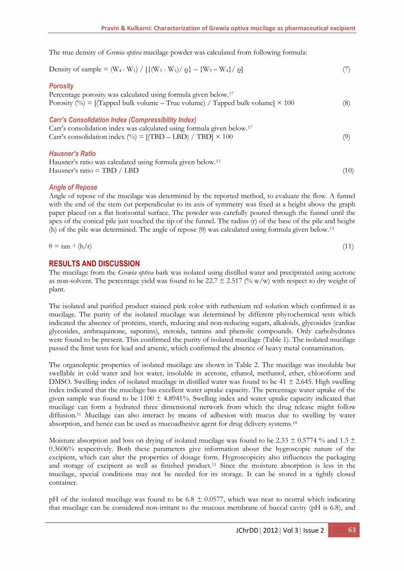
In the in vitro cytotoxicity study on NIH3T3 cell line, the mucilage did not show any cell lysis at all the concentrations used. The results of cytotoxicity study are shown in Fig 1. The mucilage can be considered as non-toxic on normal cell lines. The total bacterial count and total fungal count in the isolated Grewia optiva mucilage was found to be 43 x 104 and 16 x 104 respectively. E.Coli and Pseudomonas were absent in the isolated mucilage, but Salmonella
Pravin & Kulkarni: Characterization of Grewia optiva mucilage as pharmaceutical excipient
JChrDD 2012 Vol 3 Issue 2 65
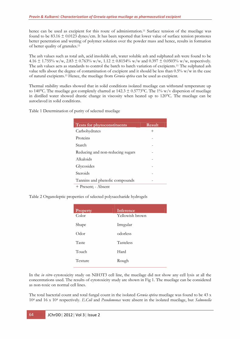
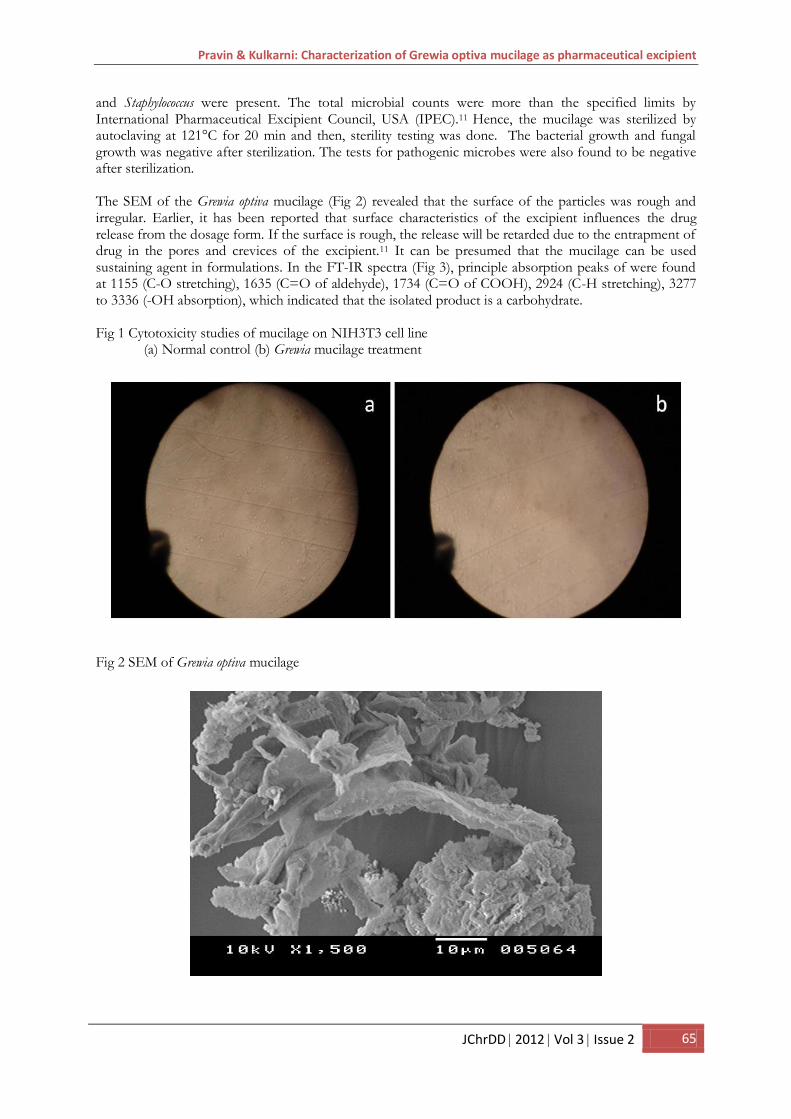
and Staphylococcus were present. The total microbial counts were more than the specified limits by International Pharmaceutical Excipient Council, USA (IPEC).11 Hence, the mucilage was sterilized by autoclaving at 121°C for 20 min and then, sterility testing was done. The bacterial growth and fungal growth was negative after sterilization. The tests for pathogenic microbes were also found to be negative after sterilization. The SEM of the Grewia optiva mucilage (Fig 2) revealed that the surface of the particles was rough and irregular. Earlier, it has been reported that surface characteristics of the excipient influences the drug release from the dosage form. If the surface is rough, the release will be retarded due to the entrapment of drug in the pores and crevices of the excipient.11 It can be presumed that the mucilage can be used sustaining agent in formulations. In the FT-IR spectra (Fig 3), principle absorption peaks of were found at 1155 (C-O stretching), 1635 (C=O of aldehyde), 1734 (C=O of COOH), 2924 (C-H stretching), 3277 to 3336 (-OH absorption), which indicated that the isolated product is a carbohydrate. Fig 1 Cytotoxicity studies of mucilage on NIH3T3 cell line
(a) Normal control (b) Grewia mucilage treatment Fig 2 SEM of Grewia optiva mucilage
Pravin & Kulkarni: Characterization of Grewia optiva mucilage as pharmaceutical excipient
66 JChrDD 2012 Vol 3 Issue 2
Fig 3 FT-IR spectrum of Grewia optiva mucilage Particle size distribution data for Grewia optiva mucilage is given in Table 3. The mean particle size was found to be 82.85 µm. The LBD, TBD, true density and porosity were found to be 0.6929 ± 0.0059 g/mL, 1.001 ± 0.0545 g/mL, 0.2059 ± 0.0027 g/mL and 59.51 ± 0.6864% respectively. All these properties gave a hint about the packing arrangement of the particles. The result of LBD, TBD and porosity indicate the loose packing of the particles.18 The high value of porosity supports the result of SEM analysis that the particles surface is rough and irregular. The release of drug could be sustained due the entrapment of drug particles in the pores of excipients.11 The Carr’s consolidation index, Hausner’s ratio and angle of repose were found to be 30.74 ± 1.713%, 1.44 ± 0.0306 and 32.43 ± 0.8452° respectively. The value of Carr’s consolidation index indicated that mucilage has cohesive property.18 The result of Hausner’s ratio and angle of repose indicated the poor flow property of mucilage, which can be improved by the addition of glidants.17 Table 3. Particle size distribution of Grewia optiva mucilage
Size range (µm) Number of particles 0-30 77 30-60 169 60-90 123 90-120 119 120-150 42 150-180 32 180-210 18 210-240 14 240-270 3 270-300 3
Pravin & Kulkarni: Characterization of Grewia optiva mucilage as pharmaceutical excipient
JChrDD 2012 Vol 3 Issue 2 67
CONCLUSION From the results of the present work it can be concluded that the isolated mucilage from the bark of Grewia optiva possess the potential to be used as excipient in pharmaceutical formulation. The preliminary studies indicate multifunctional property of mucilage. It can be used as binding agent due to its good wetting property. Mucilage can be tried to formulate matrix tablets and gels in regard to its excellent water absorption and swelling properties. Mucilage was found to have good water uptake capacity which may be suitable for formulation of mucoadhesive dosage forms.
DECLARATION OF INTEREST It is hereby declared that this paper does not have any conflict of interest.
REFERENCES 1. Kulkarni GT, Gowthamarajan K, Rao BG, Suresh B. Evaluation of binding properties of selected
natural mucilages. J Sci Ind Res (India). 2002; 61: 529-532. 2. Gowthamarajan K, Kulkarni GT, Muthukumar A, Mahadevan N, Samnta MK, Suresh B. Evaluation
of fenugreek mucilage as gelling agent. Int J Pharm Excip. 2002; 16-19. 3. Jani GK, Shah DP, Prajapati VD, Jain VC. Gums and mucilages: versatile excipients for
pharmaceutical formulations. Asian J Pharm Sci. 2009; 4 (5): 308-322. 4. Malviya R, Srivastava P, Kulkarni GT. Applications of mucilages in drug delivery – a review. Adv Biol
Res. 2011; 5(1): 1-7. 5. Anonymous. The wealth of India - Raw Materials. Vol. 4. New Delhi: Council for Scientific and
Industrial Research. 2005. p. 261-262. 6. Anonymous. The wealth of India - Raw Materials. Vol. 3. (Suppl). New Delhi: Council for Scientific
and Industrial Research. 2005. p. 223-224. 7. Khandelwal KR. Practical pharmacognosy - techniques and experiments: preliminary phytochemical
screening. 19th ed. Pune: Nirali Prakashan; 2008. p.149-156. 8. United State Pharmacopoeia. 24th Asain ed. New Delhi: Tata Donnelly Limited; 2000. 9. Indian Pharmacopoeia. Vol II. New Delhi: Controller of Publications. 1996. p. A42-A54. 10. Mukharjee PK. Quality control of herbal drugs. 4th ed. New Delhi: Business horizons; 2010. 11. Kulkarni GT. Process of development of excipients from natural sources. Indian J Pharmaceutics.
2010; 1(1): 1-7. 12. Madishetti SK, Palem CR, Gannu R, Thatipamula RP, Panakanti PK, Yamsani MR. Development of
domperidone bilayered matrix type transdermal patches: physicochemical, in vitro and ex vivo characterization. Daru. 2010; 18 (3): 221-229.
13. Phanikumar GK, Battu G, Lova Raju NS. Isolation and evaluation of tamarind seed polysaccharide being used as polymer in pharmaceutical dosage form. Res J Pharm Biol Chem Sci. 2011; 2 (2): 274-290.
14. British Pharmacopoeia. Vol IV. London: MHRA. 2005. p. A-241. 15. Kulkarni GT, Gowthamarajan K, Raghu C, Ashok G, Vijayan P. In vitro cytotoxicity and in vivo acute
toxicity of selected polysaccharide hydrogels as pharmaceutical excipients. Oriental Pharm Exptl Med. 2005; 5(1): 1-8.
16. Indian Pharmacopoeia. Vol I. New Delhi: Controller of Publications. 2007. p. 36-54. 17. Martin A, Swarbrick J, Cammarata A. Physical pharmacy. 3rd ed. Mumbai: Varghese Publishing
House; 1991. p. 492-520. 18. Junginger HE, Verhoef JC, Thanou M. Drug delivery - mucoadhesive hydrogels. In: Swarbrick J,
editor. Encyclopedia of pharmaceutical technology. 3rd ed. Vol 2. New York: Informa Healthcare; 2007. p. 1169.
Received: Jan 5, 2012; Revised: Mar 10, 2012; Accepted: Apr 10, 2012
Pravin & Kulkarni: Characterization of Grewia optiva mucilage as pharmaceutical excipient
68 JChrDD 2012 Vol 3 Issue 2

