Repozitorij PMF-a
148
Structural characterization of Heliobacter plyori proteinsrequired for survival of the bacterium Kekez, Ivana Doctoral thesis / Disertacija 2016 Degree Grantor / Ustanova koja je dodijelila akademski / stručni stupanj: University of Zagreb, Faculty of Science / Sveučilište u Zagrebu, Prirodoslovno-matematički fakultet Permanent link / Trajna poveznica: https://urn.nsk.hr/urn:nbn:hr:217:633407 Rights / Prava: In copyright Download date / Datum preuzimanja: 2022-07-26 Repository / Repozitorij: Repository of Faculty of Science - University of Zagreb
-
Upload
khangminh22 -
Category
Documents
-
view
3 -
download
0
Transcript of Repozitorij PMF-a

Structural characterization of Heliobacter plyoriproteinsrequired for survival of the bacterium
Kekez, Ivana
Doctoral thesis / Disertacija
2016
Degree Grantor / Ustanova koja je dodijelila akademski / stručni stupanj: University of Zagreb, Faculty of Science / Sveučilište u Zagrebu, Prirodoslovno-matematički fakultet
Permanent link / Trajna poveznica: https://urn.nsk.hr/urn:nbn:hr:217:633407
Rights / Prava: In copyright
Download date / Datum preuzimanja: 2022-07-26
Repository / Repozitorij:
Repository of Faculty of Science - University of Zagreb

FACULTY OF SCIENCE
Ivana Kekez
STRUCTURAL CHARACTERIZATION OF Helicobacter pylori PROTEINS REQUIRED FOR
SURVIVAL OF THE BACTERIUM
DOCTORAL THESIS
Zagreb, 2016


PRIRODOSLOVNO-MATEMATIČKI FAKULTET
Ivana Kekez
STRUKTURNA KARAKTERIZACIJA PROTEINA BAKTERIJE Helicobacter pylori VAŽNIH ZA
NJEZINO PREŽIVLJAVANJE
DOKTORSKI RAD
Zagreb, 2016


FACULTY OF SCIENCE
Ivana Kekez
STRUCTURAL CHARACTERIZATION OF
Helicobacter pylori PROTEINS REQUIRED FOR SURVIVAL OF THE BACTERIUM
DOCTORAL THESIS
Supervisors: Dr. sc. Dubravka Matković-Čalogović, Professor
Dr. sc. Giuseppe Zanotti, Professor
Zagreb, 2016

PRIRODOSLOVNO-MATEMATIČKI FAKULTET
Ivana Kekez
STRUKTURNA KARAKTERIZACIJA PROTEINA BAKTERIJE Helicobacter pylori VAŽNIH ZA
NJEZINO PREŽIVLJAVANJE
DOKTORSKI RAD
Mentori: prof. dr. sc. Dubravka Matković-Čalogović
prof. dr. sc. Giuseppe Zanotti
Zagreb, 2016

Acknowledgments
I would like to express special gratitude to my supervisor Prof. Dr. Dubravka Matković-
Čalogovoić for her continuous support, encouragement and guidance throughout my PhD study.
That being said, I would like to single out the enjoyable and memorable time we had spent
together at synchrotron as the time and place where her patient explanations regarding
crystallographic issues and her open-door policy allowed me to quickly discuss any results or
problems.
I am sincerely grateful to Prof. Dr. Giuseppe Zanotti for accepting to be my second supervisor
and for giving me the opportunity to carry out most of the experiments in his laboratory at the
Department of Biomedical Sciences, University of Padua. The project described in this thesis
is a continuation of his studies on the structural characterization of the Helicobacter pylori
proteins involved in the pathogenicity of this bacterium. Fast and easy communication, along
with his expertise in this field of research, made him an excellent tutor. Besides for Prof. Dr.
Zanotti, I would also like to thank all his past and present laboratory staff for all the kindness
and hospitality which I had felt during my stay in Padua.
Special thanks go to Dr. Laura Cendron for introducing me to the „world“ of protein
crystallography and for guiding me during my first stay in Padua. I appreciate her unselfish will
to share with me numerous helpful advice and practical experiences. Laura, thank you for
encouraging me never to give up on protein crystals and to always look for other possibilities
worth trying. I enjoyed our scientific discussions, all the nice times we spent together. All in
all, thank you for being Laura!
I would also like to thank all the girls from Prof. G. Zanotti’s group, especially Francesca,
Paola, Valentina and Elena, for a friendly and relaxed atmosphere and for always being kind
and willing to help – it was a pleasure to work with all of you!
My acknowledgments also go to members of the evaluation committee: Dr. Jasmina Rokov-
Plavec and Dr. Marija Luić whose comments and suggestions greatly improved the thesis.
I am also grateful to Prof. Dr. Patrizia Polverino de Laureto (University of Padua, Department
of Pharmaceutical Sciences) for giving me the opportunity to use the CD spectrometer and for
helping me with the mass measurements and analysis. Further gratitude is also extended to Prof.
Dr. Massimo Bellanda (University of Padua, Department of Chemistry) for providing access to
the NMR spectrometer.

My acknowledgments also go to many people working at the Department of Chemistry,
University of Zagreb. Here I would like to extend particular gratitude to the Division of
Biochemistry staff for allowing me to use their instruments and equipment and for giving me a
chance to continue my experimental work here in Zagreb. Also deserving of special
acknowledgments are my colleagues from the Division of General and Inorganic Chemistry
and Zoran Bojanić who provided me with IT support.
Irena, Željka and Biserka, thank you ever so much for giving me tremendous professional
support, for all the nice times we spent together, as well as for visiting Padua!
Warm thanks go to Dalibor who always believed in my work and motivated me.
Many thanks go to my friends Ines, Martina, Ivona and Vlatka for all the nice days and
pleasant evenings together.
A loving thank you goes to my husband Mario who always believed in me and gave me
advice and encouragement.
Last but not least, I would like to thank my parents, Marija and Joso, and my sister Josipa,
for invaluable support and encouragement during my life. I want to thank you for all the small
things and all the care - I am just happy to have you all! This thesis I dedicate to you.
Ivana Kekez

§ Contents xi
Ivana Kekez Doctoral Thesis
Contents
SAŽETAK .............................................................................................................................. XV
ABSTRACT ....................................................................................................................... XVII
PROŠIRENI SAŽETAK..................................................................................................... XIX
§ 1. INTRODUCTION .......................................................................................................... 1
§ 2. LITERATURE REVIEW .............................................................................................. 3
2.1. Helicobacter pylori – an interesting medical target ...................................................... 3
2.2. H. pylori virulence factors .............................................................................................. 4
2.3. H. pylori flagellum ......................................................................................................... 10
2.3.1. The hook .......................................................................................................................... 14
2.3.1. FlgD ................................................................................................................................ 15
2.4. Detoxification and metal ion homeostasis in H. pylori ............................................... 16
2.4.1. Copper ion homeostasis .................................................................................................. 17
2.4.2. CrdA ................................................................................................................................ 17
2.5. Heat shock proteins in H. pylori .................................................................................. 19
2.5.1. HP1026 ........................................................................................................................... 20
§ 3. MATERIALS AND METHODS ................................................................................. 25
3.1. Materials ........................................................................................................................ 25
3.1.1. Equipment ....................................................................................................................... 25
3.1.2. Chromatography supplies ............................................................................................... 26
3.1.3. Crystallographic supplies ............................................................................................... 26
3.1.4. Kits .................................................................................................................................. 27
3.1.5. Filters and membranes ................................................................................................... 27
3.1.6. Chemicals and buffers .................................................................................................... 27
3.1.7. Enzymes .......................................................................................................................... 28
3.1.8. Antibodies ....................................................................................................................... 29
3.1.9. Oligonucleotides ............................................................................................................. 29
3.1.10. Vectors .......................................................................................................................... 30
3.1.11. Bacterial strains: Escherichia coli ............................................................................... 32
3.1.11.1. Host cell for cloning .................................................................................................. 32
3.1.11.2. Strains for protein expression ................................................................................... 32

§ Contents xii
Ivana Kekez Doctoral Thesis
3.2. Microbiological methods .............................................................................................. 33
3.2.1. Media for cultivation of E. coli cells ............................................................................... 33
3.2.2. Transformation of chemically competent E. coli cells .................................................... 33
3.3. Molecular biological methods ...................................................................................... 33
3.3.1. Amplification and isolation of plasmid DNA from E. coli .............................................. 33
3.3.2. Determination of DNA concentration ............................................................................. 34
3.3.3. Polymerase chain reaction (PCR) .................................................................................. 34
3.3.4. Agarose gel electrophoresis ............................................................................................ 35
3.3.5. Cloning and sequencing .................................................................................................. 36
3.3.5.1. Standard cloning: restriction and ligation of DNA ..................................................... 36
3.3.5.2. Enzyme free-cloning .................................................................................................... 37
3.4. Protein biochemical methods ....................................................................................... 38
3.4.1. Protein quantification .................................................................................................... 38
3.4.2. Sodium dodecyl sulfate polyacrylamide gel electrophoresis (SDS-PAGE) ................... 38
3.4.3. Western blotting ............................................................................................................. 40
3.4.4. Proteolytic cleavage ....................................................................................................... 41
3.4.4.1. Removal of the hexa His-tag from HP1026 with thrombin ......................................... 41
3.4.4.2. Removal of the GST-tag from GST_HP1326 with PreScission protease .................... 42
3.4.4.3. Limited proteolysis of the FlgD (HP0907) protein ..................................................... 42
3.5. Expression of recombinant proteins in E. coli ........................................................... 43
3.5.1. Expression of CrdA (HP1236), HP1026 and FlgD (HP0907 ........................................ 43
3.5.2. Expression of selenomethionine- HP1026 and FlgD (HP0907) .................................... 44
3.6. Purification of CrdA, HP1026 and FlgD .................................................................... 45
3.6.1. Affinity chromatography ................................................................................................ 45
3.6.1.1. Affinity chromatography using a Strep-Tactin ............................................................ 45
3.6.1.2. Affinity chromatography with Ni-NTA agarose .......................................................... 46
3.6.1.3. Affinity chromatography with gluthatione sepharose ................................................. 47
3.6.2. Size-exclusion chromatography (SEC) ........................................................................... 48
3.6.3. Ultrafiltration ................................................................................................................. 49
3.7. Refolding of the CrdA protein solubilized from inclusion bodies ............................ 50
3.8. Protein characterization ............................................................................................... 51
3.8.1. Circular dichroism ......................................................................................................... 51
3.8.2. One-dimensional NMR spectroscopy (1D NMR) ........................................................... 53
3.8.3. Light scattering .............................................................................................................. 53

§ Contents xiii
Ivana Kekez Doctoral Thesis
3.8.3.1. Dynamic light scattering (DLS) ................................................................................... 53
3.8.3.2. Static light scattering (SLS).................................................................................................... 54
3.8.4. Thermofluor assay...................................................................................................................... 55
3.8.5. Mass spectrometry ...................................................................................................................... 58
3.8.6. ATPase activity assay by reversed-phase high-performance liquid chromatography (RP-
HPLC) ......................................................................................................................................... 59
3.8.7. Ultraviolet-visible (UV-VIS) titration...................................................................................... 60
3.8.8. Crystallographic studies ........................................................................................................... 60
3.8.8.1. Crystallization ........................................................................................................................... 60
3.8.8.2. Data processing and structure determination ........................................................................... 62
3.8.9. Bioinformatics methods and software tools ................................................................................. 62
§ 4. RESULTS AND DISCUSSION ................................................................................... 65
4.1. FlgD from H. pylori (HpFlgD) .............................................................................................. 65
4.1.1. Cloning, expression and purification ........................................................................................... 65
4.1.2. Crystallization and data collection .............................................................................................. 67
4.1.3. Size of HpFlgD in solution ........................................................................................................... 69
4.1.4. Size of HpFlgD in the crystal ....................................................................................................... 71
4.1.5. Structure of the FlgD monomer .................................................................................................... 73
4.1.6. Quaternary structure and interfaces ............................................................................................ 76
4.1.7. Comparison with other structures ................................................................................................ 81
4.1.8. Proposed physiological role of truncated HpFlgD ...................................................................... 84
4.2. CrdA from H. pylori .................................................................................................................. 86
4.2.1. Cloning, expression and purification of StrepCrdA, CrdAHis and GSTCrdA ............................. 86
4.2.2. Size of CrdA in solution ................................................................................................................ 89
4.2.3. Affinity of CrdA towards copper(II) ions ...................................................................................... 91
4.2.4. Crystallization of CrdA ................................................................................................................ 92
4.3. HP1026 from H. pylori .............................................................................................................. 93
4.3.1. Expression, purification and size of HP1026 in solution ............................................................. 93
4.3.2. Activity of HP1026 ....................................................................................................................... 94
4.3.3. Stability of HP1026 ...................................................................................................................... 96
4.3.4. Crystallization of HP1026 ............................................................................................................ 98
§ 5. CONCLUSIONS ......................................................................................................... 103
§ 6. LIST OF ABBREVIATIONS .................................................................................... 105
§ 7. REFERENCES ............................................................................................................ 107
§ 8. CURRICULUM VITAE...................................................................................... XXVIII


§ Sažetak xv
Ime Prezime Doktorska disertacija
Sveučilište u Zagrebu
Prirodoslovno-matematički fakultet
Kemijski odsjek
Doktorska disertacija
SAŽETAK
STRUKTURNA KARAKTERIZACIJA PROTEINA BAKTERIJE Helicobacter pylori
VAŽNIH ZA NJEZINO PREŽIVLJAVANJE
Ivana Kekez Horvatovac 102a, Zagreb, Hrvatska
Via Ugo Bassi 58/B, Padova, Italija
U sklopu ove disertacije strukturno su okarakterizirani proteini iz bakterije H. pylori koji su
važni za njezino preživljavanje (HpFlgD, CrdA, HP1026). Riješena kristalna struktura
skraćenog proteina HpFlgD ukazala je na drugačiju međusobnu orijentaciju dviju domena nego
što je nađeno u ostalim homolozima proteina FlgD. Također je pokazano da je skraćeni HpFlgD
i u otopini i kristalu prisutan kao tetramer što ukazuje na značajnu razliku u molekularnoj
organizaciji biča u različitih bakterijskih vrsta. Uočeno je da dodatak iona Cu2+ u otopinu CrdA,
proteina koji pretpostavljeno veže bakrove ione, potiče stvaranje monomernih vrsta u otopini
te da CrdA veže ione Cu2+ sa slabim afinitetom što je karakteristika proteina koji prenose
bakrove ione. Funkcionalna karakterizacija proteina HP1026 je po prvi put demonstrirala da
HP1026 obavlja ATPaznu aktivnost. Iako se proteni koji spadaju u skupinu AAA+ proteina
najčešće udružuju kao heksameri, za HP1026 je pokazano da u otopini stvara dimerne vrste.
(115 + xxxii stranica, 49 slika, 39 tablica, 147 literaturnih navoda, jezik izvornika: engleski)
Rad je pohranjen u Središnjoj kemijskoj knjižnici, Horvatovac 102a, Zagreb i Nacionalnoj i
sveučilišnoj knjižnici, Hrvatske bratske zajednice 4, Zagreb.
Ključne riječi: CrdA / FlgD / HP1026 / H. pylori / strukturna karakterizacija
Mentori: prof. dr. sc. Dubravka Matković-Čalogović
prof. dr. sc. Giuseppe Zanotti
Rad prihvaćen: 01.06.2016.
Ocjenitelji: doc. dr. sc. Jasmina Rokov-Plavec
prof. dr. sc. Dubravka Matković-Čalogović
dr. sc. Marija Luić, zn. savj.

§ Abstract xvii
University of Zagreb
Faculty of Science
Department of Chemistry
Doctoral Thesis
ABSTRACT
STRUCTURAL CHARACTERIZATION OF Helicobacter pylori PROTEINS REQUIRED
FOR SURVIVAL OF THE BACTERIUM
Ivana Kekez Horvatovac 102a, Zagreb, Croatia
Via Ugo Bassi 58/B, Padua, Italy
Within this thesis several proteins from H. pylori, important for survival of the bacterium, were
structurally characterized (HpFlgD, CrdA, HP1026). Crystal structure of the truncated form of
the HpFlgD protein revealed that spatial orientation of the two domains differs from that of the
homologous FlgD family members. This fact together with the observation that truncated
HpFlgD assembles into tetramers, both in the solution and in the crystal form, strongly suggests
that significant differences exist in the molecular organization of the flagella in different
bacterial species. It was shown that incubation of the putative copper binding CrdA protein with
Cu2+ ions favours formation of monomeric species in solution and that CrdA binds Cu2+ with
very low affinity which is a property of copper trafficking proteins. Functional assays of the
HP1026 protein demonstrated for the first time its ATPase activity. While proteins that belong
to the class of AAA+ proteins usually form hexamers, HP1026 was found to form dimers.
(115 + xxxii pages, 49 figures, 39 tables, 147 references, original in English)
Thesis deposited in Central Chemical Library, Horvatovac 102A, Zagreb, Croatia and National
and University Library, Hrvatske bratske zajednice 4, Zagreb, Croatia.
Keywords: CrdA / FlgD / HP1026 / H. pylori / structural characterization
Supervisors: Dr. Dubravka Matković-Čalogović, Professor
Dr. Giuseppe Zanotti, Professor
Thesis accepted: 01.06.2016.
Reviewers: Dr. Jasmina Rokov-Plavec, Assistant Professor
Dr. Dubravka Matković-Čalogović, Professor
Dr. Marija Luić, Senior Scientist


§ Prošireni sažetak xix
Sveučilište u Zagrebu
Prirodoslovno-matematički fakultet
Kemijski odsjek
Doktorska disertacija
PROŠIRENI SAŽETAK
Helicobacter pylori je patogena bakterija nađena na sluznici čovjekova želuca. Danas se zna da
je infekcija povezana s različitim ishodima bolesti. Iako su mnoge infekcije neprimjetne i bez
ikakvih simptoma bolesti, većina rezultira s različitim stupnjevima gastritisa. U 10 % inficirane
populacije zaraza će se razviti u teško želučano oboljenje poput peptičkog ulkusa (čira) te
gastritisa. U 1 % zaražene populacije doći će do razvoja adenokarcinoma želuca te limfoma
mukoznog tkiva (MALT). Stoga je 1994. Svjetska zdravstvena organizacija proglasila bakteriju
H. pylori karcinogenom I. reda [R. M. Jr Peek and M. J. Blaser, Nature Rev. Cancer 2 (1)
(2002) 28–37; S. Suerbaum and P.N. Michetti, Engl. J. Med. 347 (2002) 11751186]. Genom
sedamnaest sojeva bakterije H. pylori je u potpunosti sekvenciran, poput 26695, J99, G27 i
HPAG1 [J. F. Tomb, O. White, A. R. Kerlavage, R. A. Clayton, G. G. Sutton, R. D.
Fleischmann, K. A. Ketchum, H. P. Klenk, S. Gill, B. A.Dougherty, K. Nelson, J. Quackenbush,
L. Zhou, E. F. Kirkness, S. Peterson, B. Loftus, D. Richardson, R. Dodson, H. G. Khalak, A.
Glodek, K. McKenney, L. M. Fitzegerald, N. Lee, M. D. Adams, E. K. Hickey, D. E. Berg, J.
D. Gocayne, T. R. Utterback, J. D. Peterson, J. M. Kelley, M. D. Cotton, J. M. Weidman, C.
Fujii, C. Bowman, L. Watthey, E. Wallin, W. S. Hayes, M. Borodovsky, P. D. Karp, H. O.
Smith, C. M. Fraser, and J. C. Venter, Nature 388 (6642) (1997) 539547; R. A. Alm, L. S.
Ling, D. T. Moir, B. L. King, E. D. Brown, P. C. Doig, D. R. Smith, B. Noonan, B. C. Guild,
B. L. deJonge, G. Carmel, P. J. Tummino, A. Caruso, M. Uria-Nickelsen, D. M. Mills, C. Ives,
R. Gibson, D. Merberg, S. D. Mills, Q. Jiang, D. E. Taylor, G. F. Vovis, and T. J. Trust, Nature
397 (1999) 176180; J. D. Oh, H. Kling-Backhed, M. Giannakis, J. Xu, R. S. Fulton, L. A.
Fulton, H. S. Cordum, C. Wang, G. Elliott, J. Edwards, E. R. Mardis, L. G. Engstrand, and J. I.
Gordon, Proc. Natl. Acad. Sci. U.S.A. 103 (2006) 999910004; D. A. Baltrus, M. R. Amieva,
A. Covacci, T. M. Lowe, D. S. Merrell, K. M. Ottemann, M. Stein, N. R. Salama, and K.
Guillemin. J. Bacteriol. 191 (1) (2009) 447448]. Svi genomi imaju sličnu prosječnu duljinu i
gustoću kodirajućih sekvenci. Ekstremno brza rekombinacija zajedno sa značajnom razlikom

§ Prošireni sažetak xx
sekvenci upućuje na panmiktičku strukturu populacije [S. Suerbaum and C. Josenhans, Nat.
Rev. Microbiol. 5 (2007) 441452].
Prema patološkoj važnosti bakterija H. pylori je u posljednjih dvadeset godina postala važan
cilj istraživanja. Napravljene su i analizirane brojne interaktivne mape proteina iz bakterije H.
pylori [J. C. Rain, L. Selig, H. De Reuse, V. Battaglia, C. Reverdy, S. Simon, G. Lenzen, F.
Petel, J. Wojcik, V. Schachter, Y. Chemama, A. Labigne, and P. Legrain, Nature 409 (2001)
211215] koji joj omogućavaju preživljavanje u kiselom mediju [L. Cendron and G. Zanotti
Febs J. 278 (8) (2011) 12231231]. Suprotno prethodnom, broj podataka o riješenim
strukturama proteina ove bakterije je malen: do ožujka 2016. u Protein Data Bank pohranjene
su 439 datoteke s koordinatama atoma (http://www.pdb.org) dok su 33 datoteke u postupku
pohranjivanja.
U okviru ove disertacije strukturno je okarakterizirano nekoliko proteina važnih za
preživljavanje bakterije H. pylori: FlgD, CrdA, HP1026.
Pokretljivost stanice bakterije H. pylori uvjetovana je pravilnom strukturnom organizacijom
biča. Bič se sastoji od otprilike 50 različitih proteina važnih za pravilnu regulaciju i udruživanje
proteina, neophodnih za pokretanje stanice u bolje uvjete za preživljavanje [H. Zhou, M. Luo,
X. Cai, J. Tang, S. Niu, W. Zhang, Y. Hu, Y. Yin, A. Huang, and D. Wang, Proteins 79 (7)
(2011) 23462351]. Glavne komponente koje izgrađuju bič su: filament, kuka (engl. hook) i
bazalno tijelo (engl. basal body) [O. A. Soutourina and P. N. Bertin, FEMS Microbiol. Rev. 27
(2003) 505523]. FlgD pridonosi virulenciji bakterije H. pylori budući da je nužan za kontrolu
pravilne strukturne organizacije kape biča (engl. hook cap) [W. T. Kuo, K. H. Chin, W. T. Lo,
A. H. Wang, and S. H. Chou, J. Mol. Biol. 381 (1) (2008) 189199]. Do sada su riješene dvije
kristalne strukture proteina FlgD iz bakterija Xanthomonas campestris [W. T. Kuo, K. H. Chin,
W. T. Lo, A. H. Wang, and S.H. Chou, J. Mol. Biol. 381 (1) (2008) 189199] i Pseudomonas
aeruginosa [H. Zhou, M. Luo, X. Cai, J. Tang, S. Niu, W. Zhang, Y. Hu, Y. Yin, A. Huang,
and D. Wang, 79 (7) Proteins (2011) 23462351] međutim sličnost proteinskih sekvenci
između navedenih homologa je manja od 20 %.
CrdA (HP1326) je protein iz bakterije H. pylori važan za održavanje razine bakrovih iona u
citoplazmi ispod toksične razine [B. Waidner, K. Melchers, F. N. Stähler, M. Kist, and S.
Bereswill, J. Bacteriol. 187 (2005) 46834688]. Sve što se do danas zna o spomenutom
proteinu su studije o RNA profiliranju [B. Waidner, K. Melchers, I. Ivanov, H. Loferer, K. W.

§ Prošireni sažetak xxi
Bensch, M. Kist, and S. Bereswill, J. Bacteriol. 184 (2002) 67006708] koje upućuju na
uključenost proteina u homeostazu bakrovih iona, međutim nedostaje stukturna karakterizacija.
Geni za proteine koji se sintetiziraju uslijed izlaganja H. pylori termičkom šoku (engl. heat
shock proteins; HSPs) organizirani su kao tri višecistronska operona. Njihova transkripcija
kontrolirana je trima promotorima (Pgro, Phrc, Pcbp). Infekcija i prilagodba bakterije H. pylori
ekstremnim uvjetima želuca omogućena je i spomenutim HSP-ima koji su uključeni u
regulaciju aktivnosti ureaze i prijanjanje bakterijskih stanica za stijenku epitela želuca [D.
Roncarati, A. Danielli, and V. Scarlato, J. Bacteriol. 193 (20) (2011) 56295636]. HP1026
pripada ovoj skupini proteina i u potpunosti je neokarakteriziran stoga je razumijevanje funkcije
ovog proteina iznimno važno.
Cilj ovog istraživanja je strukturno karakterizirati proteine FlgD, CrdA i HP1026 iz
bakterije H. pylori i potencijalno objasniti njihovu ulogu s naglasakom na razumijevanju
funkcije spomenutih proteina pomoću podataka dobivenih strukturnom i biofizikalnom
karakterizacijom. Prema dosadašnjim istraživanjima pretpostavke su: (1) FlgD kontrolira
pravilnu polimerizaciju proteina FlgE koji izgrađuje kuku biča, (2) CrdA regulira razinu
bakrovih iona u stanici ispod toksične razine te (3) HP1026 posjeduje ATP-veznu domenu.
Rezultati istraživanja pridonijet će boljem razumijevanju uloge esencijalnih metala u strukturi
i funkciji homeostaze, kontrole sinteze strukturnih elemenata biča bakterije te uloge proteina
sintetiziranog uslijed izlaganja stanice termičkom šoku.
Istraživani proteini (FlgD, CrdA i HP1026 iz organizma Helicobacter pylori) proizvedeni
su u laboratoriju primjenom tehnologije rekombinantne DNA, ugradnjom u plazmidne vektore
i prekomjernom ekspresijom genskih produkata u bakteriji Escherichia coli. Ispravnost
ugradnje gena za željenu makromolekulu provjerena je sekvenciranjem, a identitet proteina s
privjeskom utvrđen je western analizom. U slučaju kad je prisutnost His-privjesaka ometala
ispravno formiranje kvaterne strukture željenog proteina, za kloniranje su korišteni drugi
privjesci (GST-privjesak, Strep-privjesak) ili je privjesak bio uklonjen. GST-privjesak također
pomaže i u topljivosti proteina koji bi u protivnom formirao inkluzijska tijela.
Izolacija proteina iz bakterije E. coli započeta je razaranjem bakterijskih stanica
sonikacijom ili razbijačem stanica (engl. cell cracker), te pročišćavanjem različitim
kromatografskim metodama (afinitetna kromatografija, gel-filtracija), ovisno o prisustvu,
odnosno odsustvu privjeska na proteinu. Čistoća završnog uzorka analizirana je korištenjem
diskontinuirane denaturirajuće elektroforeze (SDS-PAGE). Koncentracija dobivenih proteina

§ Prošireni sažetak xxii
određena je spektrofotometrijski, mjerenjem apsorbancije pri 280 nm, a prema teorijskim
vrijednostima ekstinkcijskih koeficijenata, te izračunata prema Beer-Lambertovom zakonu. U
slučaju velike razine ekspresije ali male topljivosti primjenjena je denaturacija i renaturacija
proteina.
Kružni dikroizam (engl. circular dichroism, CD) i jednodimenzijski NMR korišteni su radi
dobivanja informacije o prisutnosti elemenata sekundarne strukture u proteinu što je iznimno
važno nakon denaturacije i ponovne renaturacije proteina. Kvaliteta uzorka za kristalizaciju
ispitana je metodom dinamičkog raspršivanja svjetlosti (engl. dynamic light scattering, DLS).
Interakcija CrdA i bakrovih iona ispitana je UV-VIS titracijom dok je aktivnost proteina
HP1026 (sposobnost hidrolize ATP-a) istražena praćenjem promjene apsorbancije produkta
reakcije hidrolize ATP-a.
Testiranje optimalnih uvjeta za nastanak kristala i optimizacija kristalizacije provedeni su
metodom difuzije pare otapala iz sjedeće kapi upotrebom robota za automatsku kristalizaciju
(Oryx8 robot) u nanolitarskoj skali (minimalna veličina kapi je 200 nL). Za kristalizaciju su
korištene komercijalne kristalizacijske otopine, a nakon dobivanja prvih kristala kristalizacijski
uvjeti su optimizirani u svrhu dobivanja kristala dobrih difrakcijskih sposobnosti variranjem
različitih čimbenika (cijepljenje mikrokristalima, promjena temperature kristalizacije, dodatak
aditiva, promjena omjera precipitanta i proteina u kristalizacijskoj kapljici, dehidracija kristala).
Dobiveni kristali, ukoliko je bilo potrebno, namakani su u krio otopini, a potom skladišteni u
tekućem dušiku. Snimanje kristala proteina obavljeno je na sinkrotronu ESRF (Grenoble,
Francuska) ili SLS (Villigen, Švicarska) metodom rentgenske difrakcije na jediničnom kristalu.
Kristalna struktura proteina riješena je bilo metodom molekulske zamjene (u slučaju postojanja
dovoljno sličnog modela) ili metodom anomalnog raspršenja teškog elementa (u tu svrhu
pripremljen je derivat proteina sa selenometioninom).
Gen hp0907 ukloniran je iz dva različita bakterijska soja H. pylori (26695, G27). Proteini
su pročišćeni do zadovoljavajuće čistoće te je gel-filtracijom i MALS eksperimentom (engl.
multi angle light scattering) određeno da se cjeloviti proteini u otopini nalaze kao dimerne
molekule. Jedinični kristali su pripremljeni metodom difuzije pare otapala iz sjedeće kapi
korištenjem Oryx8 robota te su dobivene dvije različite kristalne forme skraćenog proteina
FlgD. FlgD_G27 kristalizira u monoklinskom sustavu, P2 prostornoj grupi, a difrakcijski
podatci su sakupljeni na sinkrotronskoj liniji ID14-4 (ESRF, Grenoble, Francuska) do
razlučivanja od 2,75 Å. FlgD_26695 i SeMetFlgD_26695 kristaliziraju u tetragonskom sustavu,

§ Prošireni sažetak xxiii
I422 prostornoj grupi, a podatci su sakupljeni na sinkrotronskoj liniji PXIII (SLS, Villigen,
Švicarska) do razlučivanja od 2,17 Å u slučaju FlgD_26695 te do razlučivanja od 2,8 Å u
slučaju SeMetFlgD_26695. Iako je u oba slučaja postavljena kristalizacija čitavog proteina
(FlgD_G27 – 316 aminokiselina; FlgD_26695 – 301 aminokiselina), riješene kristalne strukture
zajedno s rezultatima masene spektrometrije ukazuju da se u kristalnoj strukturi proteina
FlgD_G27 i FlgD_26695 nalazi skraćen protein FlgD (Asn127 Lys272). Kristalne strukture
proteina FlgD_G27 (HpFlgD_m) i FlgD_26695 (HpFlgD_t ) razlikuju se samo u dvije
aminokiseline (257 i 268) i njihovim preklapanjem se dobije standardna devijacija između 146
jednakih Cα atoma od 0,38 Å. U asimetričnoj jedinici HpFlgD_m nalaze se 4 molekule
monomera i zauzimaju Vm = 2,63 Å3 Da1. Struktura sadrži oko 53 % otapala. Struktura
HpFlgD_t ima u asimetričnoj jedinici samo 1 molekulu monomera i zauzima Vm = 3,26 Å3 Da1
te udio otapala iznosi oko 62 %. Nadalje, kvaterna struktura u obje kristalne forme je tetramer
koji je u strukturi HpFlgD_t generiran preko kristalografske osi četvrtog reda dok je u strukturi
HpFlgD_m generiran nekristalografskom simetrijom. Dimeri se stvaraju preko kontakata -
lanca 9 i -lanca 12 tudor domena susjednih monomera, a takva situacija se ponavlja četiri puta
stvarajući mrežu vodikovih veza između 4 molekule monomera. Rezultati dobiveni korištenjem
PISA programa (engl. Protein interfaces, surfaces and assemblies) ukazuju da je takav tetramer
vjerojatno postoji i u otopini. Razlika u oligomerizaciji cjelovitog proteina HpFlgD u otopini
(dimer) i skraćenog proteina HpFlgD u kristalu (tetramer) može biti prisustvo N-kraja proteina
blizu centra tetramera gdje se nalazi os četvrtog reda. Dulji N-kraj bi onemogućio stvaranje
kontakata nužnih za tetramerizaciju. Kristalna struktura proteina FlgD je pokazala da se
monomer sastoji od dvije domene, tudor i Fn-III domene, kao što je slučaj i u ostala dva
homologa proteina FlgD čije su strukture riješene. Fn-III domenu izgrađuju aminokiseline 141
– 234 dok tudor domenu čini 14 aminokiselina na N-kraju proteina (127 140) i ostatak
aminokiselinskog slijeda na C- kraju proteina (235 – 272). Lys272 je posljednja vidljiva
aminokiselina u strukturama monoklinske i tetragonske forme proteina HpFlgD. Međutim,
značajna razlika je uočena u međusobnom položaju tudor domene u odnosu na Fn-III domenu
u HpFlgD spram ostalim homolozima (XcFlgD i PaFlgD). Različita orijentacija domena
između HpFlgD i ostalih homologa može upućivati na prisutnost dva prepoznatljiva
orijentacijska modula koji predstavljaju dio mehanizma aktivacije proteina.
Pretraživanjem Uniprot banke podataka uočeno je da sve sekvence homologa proteina FlgD
u različitim vrstama roda Helicobacter sadrže prepoznatljive i neuređene regije na C-kraju

§ Prošireni sažetak xxiv
proteina redoslijeda XQK(X)2 (X = D / E / N; X2 = PI / PL / LS / PQ). Očuvane regije variraju
samo u broju ponavljanja, a uvijek završavaju slijedom TPPKETA. Ovakav ponavljajući motiv
nije pronađen u ostalim homolozima proteina FlgD, ali su slični motivi pronađeni u ostalim
bakterijskim proteinima što može ukazivati na važnost takvog motiva u prepoznavanju
proteinskih partnera. Štoviše, FliC protein (gradi filament biča) stvara interakcije preko svog
C-kraja sa šaperonom FliS te se na taj način spriječava preuranjena polimerizacija proteina
FliC. Također, nedostatak N-kraja u strukturi HpFlgD nije neobičan budući da ga i strukture
ostalih homologa ne sadrže, a poznato je da FlgD iz bakterije Escherichia coli sadrži 71
aminokiselinu kao signalnu sekvencu dok FlgD iz bakterije Salmonella enterica sadrži čak i
100 aminokiselina kao signal za izlazak iz stanice. U prilog tomu da N-kraj proteina predstavlja
signalnu sekvencu i da je zbog fleksiblnosti sklon degradaciji govori i analiza CD
spektroskopijom koja je potvrdila da cjelovit protein HpFlgD sadrži zavojnice (12,8 %) što
je obilježje signalnih sekvenca. U riješenoj kristalnoj strukturi HpFlgD -zavojnice nisu
prisutne.
Priređeni su različiti genski konstrukti za ekspresiju proteina CrdA (Strep_CrdA,
CrdA_His6 i GST_CrdA). Optimalna ekspresija proteina dobivena je pri nižoj temperaturi (16
C), međutim u slučaju CrdA_His6 i Strep_CrdA dobivene su velike količine proteina u obliku
inkluzijskih tijela dok je protein GST_CrdA prekomjerno eksprimiran s velikim udjelom u
topljivim frakcijama. Kako bi se povećala topljivost proteina, uspješno su primjenjene metode
za ponovno smatanje proteina, uključujući denaturaciju te renaturaciju polaganim
razrjeđivanjem. Različite varijante rekombinantnog proteina su nadalje pročišćene afinitetnom
kromatografijom i gel-filtracijom. Metodama kružnog dikroizma te jednodimenzijskog NMR-
a potvrđeno je postojanje elementa sekundarne strukture u renaturiranom proteinu. Na temelju
analitičke gel-filtracije izračunata je molekulska masa proteina CrdA s His privjeskom u
prisutnosti iona Cu2 (14,8 kDa). Dobivena vrijednost se podudara s predviđenom molekulskom
masom (12,4 kDa) i odgovara postojanju monomernih vrsta u otopini. Dodatkom reagensa za
keliranje metala (EDTA) dolazi do značajne promjene u ponašanju proteina CrdA, te tendencije
proteina ka različitom stupnju oligomerizacije. Protein CrdA je ugušćen do maksimalne
koncentracije, = 9 mg mL1. Kvaliteta uzorka za kristalizaciju ispitana je DLS-om koji je
ukazao na polidisperznost uzorka dok je dodatak deterdženta n-oktil--D-glukopiranozida
(nOG) deterdženta i iona Cu2+ povećao monodisperznost uzorka. Afinitet iona Cu2 prema

§ Prošireni sažetak xxv
proteinu CrdA ispitan je UV-VIS spektroskopijom te je izračunata Kd = 1,076 103 mol L1
što upućuje na nizak afinitet prema ionima Cu2 što je karakteristika proteina koji prenose
bakrove ione. Međutim, budući da aminokiselinski slijed CrdA obiluje metioninima, a ne sadrži
histidin i cistein, potrebno je ispitati afinitet iona Cu+ spram CrdA. Za kristalizacijske
eksperimente korišten je robot za automatsku kristalizaciju (Oryx8 robot). Pritom je korištena
metoda difuzije pare iz sjedeće kapi te je ispitano 686 različitih kristalizacijskih uvjeta.
Kristalizacijski eksperiment je u slučaju proteina CrdA u prisutnosti iona Cu2 i nOG-a
rezultirao formiranjem malih kristala u uvjetu s PEG'S II SUITE (Qiagen) (0,1 mol L1, Tris
pH 8,5, (v/v) 16 % PEG 4000) pri 293 K. Difrakcijska mjerenja dobivenih kristala provedena
su na liniji ID23-1 ESRF sinkrotrona (Grenoble, Francuska) te su potvrdila da su nastali kristali
proteina. Nažalost, kristali su slabo difraktirali (do razlučivanja od 20 Å), te je potrebna daljna
optimizacija kristalizacije.
Slijed aminokiselina proteina HP1026 sadrži očuvane sljedove aminokiselina 36 – 295 što
svrstava protein u skupinu AAA proteina. Proteini iz spomenute skupine se odlikuju
sposobnošću vezanja i hidrolize ATP-a, te sudjelovanjem u različitim staničnim procesima
(transport i degradacija proteina, transkripcija, replikacija i sl.). Protein HP1026 uspješno je
eksprimiran pri 28 C, te je zatim pročišćen afinitetnom kromatografijom i gel-filtracijom.
Molekulska masa monomera HP1026 iznosi 43,8 kDa te je analitičkom gel-filtracijom
ustanovljeno da su i Apo_HP1026 i kompleks HP1026:ATP--S prisutni kao dimerne vrste
(96,9 kDa) u otopini, te da dodatak ATP--S ne potiče oligomerizaciju. Nadalje, ispitana je
ATP-azna aktivnost HP1026, te su dobiveni sljedeći kinetički parametri: Km = 344 mol L1,
Vmax= 0,6019 mol L1 s1, kcat= 0,02 s1. Dobivene vrijednosti se podudaraju s kinetičkim
parametrima u slučaju aktivnosti proteina MgsA iz bakterije Escherichia coli. Analiza
sekundarne strukture proteina CD metodom je pokazala da se HP1026 glavninom sastoji od -
zavojnica (44,5 %). Protein se pokazao kao izuzetno topljiv te je ugušćen do koncentracije veće
od 40 mg mL1, a mjerenja DLS-om su pokazala da je uzorak proteina monodisperzan i pogodan
za kristalizacijske eksperimente. Također je ispitana termička stabilnost proteina uz dodatak
različitih aditiva, te je uočeno da ADP i ATP--S značajno povećavaju temperaturu mekšanja
(Tm) proteina HP1026 i to za +3,52 C u slučaju dodatka ATP--S te za +5,18 C u slučaju
dodatka ADP-a. Kristalizacijski eksperimenti postavljeni su korištenjem Oryx8 robota
metodom difuzije pare otapala iz sjedeće kapi. Kristali heksagonskog oblika su dobiveni u

§ Prošireni sažetak xxvi
različitim uvjetima PACT SUITE (Qiagen), te su snimljeni na sinkrotronskoj liniji PXIII (SLS,
Villigen, Švicarska) i pokazali slabu difrakciju (do razlučivanja od 15 Å). Uzevši u obzir
rezultate o termičkoj stabilnosti proteina pokušala se optimizacija kristalizacije uz dodatak
aditiva, te su dobiveni veći kristali u uvjetu 0,2 mol L1 NaBr, 0,1 mol L1 Bis-tris propan pH
8,5, 20 % PEG 3350, 0,015 mmol L1 CYMAL-7 pri 20 C. Iako su kristali bili veći i uređeniji,
difraktirali su samo do 7 Å što još uvijek nije dovoljno razlučivanje za rješavanje kristalne
strukture proteina.

§ 1. Introduction 1
Ivana Kekez Doctoral Thesis
§ 1. INTRODUCTION
The bacterium Helicobacter pylori colonizes the stomach of more than half of the world’s
population.1 According to the National Center for Biotechnology Information (NCBI) the
genomes of seventeen strains have been completely sequenced like 26695, J99, HPAG1 and
G27.25 All of the genomes have similar average lengths of coding sequences and coding
density. The extremely high recombination rate, in combination with a remarkable sequence
diversity, has suggested a panmictic population structure.6 Approximately 10 % of the infected
subjects will develop severe gastric pathologies, like peptic ulcer disease and atrophic gastritis.
Approximately 1 % of infected individuals develop gastric adenocarcinoma and lymphoma of
the mucosa-associated lymphoid tissue (MALT). The World Health Organization has declared
H. pylori a class 1 carcinogen in 1994.79
Due to its pathological relevance, H. pylori has become an important target for research in
the last twenty years, since its first discovery: a large amount of literature has been produced
on it, either from the medical or from the biological point of view; interaction maps of its
proteome have been created10 and analysed and its ability to survive in the acidic environment
has been investigated.11 On the contrary, structural data on the proteins of this bacterium are
relatively limited: 439 files of atomic coordinates were present in the Protein Data Bank (PDB)
in March 2016 (http://www.pdb.org), and 33 are waiting for release. Many proteins have not
been characterized at all.
Within this research several proteins from H. pylori, important for its survival, were
characterized (FlgD, CrdA, HP1026).
The flagellum is a protein complex composed of about 50 different proteins needed for its
proper regulation and assembly. It provides the cell a chance to move to a more favorable
environment and to avoid stressful conditions.12 Major sections that define the flagellum are:
the filament, the hook and the basal body.13 FlgD plays an important role in H. pylori virulence,
owing to its role in forming the hook cap component.14 Until now only two crystal structures
of FlgD have been solved (from Xanthomonas campestris and Pseudomonas aeruginosa) but
the protein sequence identity between FlgD from H. pylori and FlgD from the previously
mentioned bacteria is less than 20 %.

§ 1. Introduction 2
Ivana Kekez Doctoral Thesis
H. pylori copper resistance determinant CrdA (HP1326) is required for keeping the
concentration of free copper ions below toxic levels.15 At this moment there are no structural
data on the CrdA protein and the only available studies are on RNA profiling.16
Genes for heat shock proteins (HSPs) of H. pylori are organised in three multicistronic
operons, transcriptionally controlled by three upstream promoters (Pgro, Phrc, Pcbp). Successful
infection and adaption to the extreme gastric environment is also enabled by the heat shock
proteins (HSPs) which are involved in regulation of the urease activity and adhesion to the
epithelial cells.17 Since HP1026 is part of one of the heat shock operons controlled under the
HspR regulator with an unclear function, investigation of its role is very important.
Biophysical and structural characterization of the described proteins is crucial for
understanding the function of these H. pylori vital proteins for survival in a highly acidic
environment. The starting hypotheses of this work were: (1) FlgD controls the proper
polymerization of FlgE inside the hook of the flagellum, (2) the CrdA protein is involved in
maintaining the concentration of free copper ions below toxic levels and (3) the HP1026 protein
possesses an ATP-binding domain.
The goal of the doctoral dissertation was to structurally characterize FlgD, CrdA and
HP1026, and taking into account their biophysical and structural properties to potentially
demonstrate their function. The structural characterization was done by biophysical and
structural analysis utilizing methods like thermal stability assay, circular dichroism, X-ray
diffraction and 1D-NMR. To understand if CrdA exerts function of copper transport the
interaction between the copper ions and CrdA was investigated by UV-VIS titration. To obtain
experimental evidence on the role of HP1026, the ATP hydrolysis assay was performed using
the reversed phase HPLC by measuring the change of the absorbance of the reactions products.
The results obtained in this research widen our knowledge and contribute to better
understanding of the role of the essential metal ions in the structure and function of
metalloproteins, the control of proper flagellum construction, as well as the function of the heat
shock protein.

§ 2. Literature Review 3
Ivana Kekez Doctoral Thesis
§ 2. LITERATURE REVIEW
2.1. Helicobacter pylori – an interesting medical target
Helicobacter pylori (H. pylori) is a small (3.5 μm x 0.5 μm), slow growing, microaerophilic,
non-spore forming and spiral-shaped Gram-negative rod bacteria (Fig. 1). The bacterium
became an important and interesting target of research since it is known that it can colonize
human gastric cells and lead to development of severe gastrointestinal diseases such as chronic
gastritis, peptic ulcer disease, gastric mucosa associated lymphoid tissue (MALT) lymphoma,
and gastric cancer.18,19 The bacterium was isolated and characterized at the beginning of the
‘80s by scientists Marshall and Warren.20 It is known that more than half of the human
population is affected with this bacterium.21 Moreover, this bacterium has accompanied humans
for tens of thousands of years during migrations in the absence of a targeted antimicrobial
therapy.7 It has been hypothesized that H. pylori colonization could have provided benefits to
its human carriers and hence provided a selective advantage during long periods of human
history.22,6,9 The infection with this bacterium is mainly transmitted from human-to-human
possibly by the fecal–oral or oral–oral route. The infection can be spread vertically within
families, generally in early childhood. While many H. pylori infected individuals are clinically
asymptomatic, most will exhibit some degree of gastritis. Approximately 10 % of the infected
subjects will develop more severe gastric pathologies, like peptic ulcer disease and atrophic
gastritis. Approximately 1 % of infected individuals develop gastric adenocarcinoma and
lymphoma of the mucosa-associated lymphoid tissue (MALT), a correlation that prompted the
World Health Organization to declare H. pylori the first bacterial class 1 carcinogen in 1994.8,9

§ 2. Literature Review 4
Ivana Kekez Doctoral Thesis
Figure 1. Electron micrograph of H. pylori. Spiral bacterial bodies with a bundle of sheathed
flagella; bar = 1 µm, (taken from Yoshiyama and Nakazawa, 2002).23
H. pylori genome is relatively small containing less than 1600 genes,2 in comparison to
other Gram-negative prokaryotes, such as Escherichia coli and Salmonella enterica subsp.
enterica serovar, whose genomes include more than 4000 genes.24,25 The genomes of seventeen
strains of H. pylori have been completely sequenced, among them 26695, J99, HPAG1 and
G27.25 Genome of the strain 26695 contains 1,667,867 base pairs. It is 24 kb larger than the
J99 strain, which contains 1,643,831 base pairs, 71 kb larger than HPAG1, which contains
1,596,366 base pairs and 15 kb larger than the G27 strain, which contains 1,652,983 base pairs.
All the genomes have similar average lengths of coding sequences, coding density and the bias
of initiation codons. Plausible cause of a wide genetic diversity could be explained by the need
of H. pylori to adapt to the gastric conditions of its host as well as to the distinct patterns of the
host immune response.
2.2. H. pylori virulence factors
Several pathogenicity factors like cytotoxin associated gene pathogenicity island (cag PAI),
VacA vacuolating cytotoxin, urease, adhesins and outer membrane proteins are involved in
virulence of H. pylori.26 Large diversity of H. pylori strains indicated that some of the strains
are more virulent and can induce morphological changes and successive degeneration of in
vitro-cultured cells.27

§ 2. Literature Review 5
Ivana Kekez Doctoral Thesis
CagA was identified as a main marker for the evaluation of the strain pathogenicity and the
presence of a genomic PAI. Cag PAI is a DNA fragment about 40 kb long which encodes for
about 27 to 31 proteins, depending on the analyzed strain (Fig. 2).28,29 Eighteen proteins of cag
PAI are involved in the structural organization of the type IV secretion system (T4SS) which is
the main apparatus used in the infection process. T4SS enables penatration to the gastric
epithelial cells and translocation of CagA, peptidoglycan and other bacterial effectors to the
host cell.3032 After the injection of CagA into the host cells the protein becomes phosphorylated
and starts to interact with more than 20 different human proteins included in signal
transduction.33 This influences the proper function of epithelial cells like cell–cell adhesion,
signalling, adherence and proliferation.34 The bacterial peptidoglycan delivered by T4SS
triggers in the host cell the Nod1-response and induction of the nuclear factor-B pathway.35
Figure 2. (a) Genes of the H. pylori strain 26695 that encode for the cag PAI. Numbers
correspond to the HP0XXX number of the ORF represented by arrows;2 (b) schematic overview
of the prototypal T4SS VirB ⁄ D from A. tumefaciens (left) and comparison with components of
the Cag-T4SS from H. pylori (right). Cytoplasmic NTPases, proteins forming the core trans-

§ 2. Literature Review 6
Ivana Kekez Doctoral Thesis
membrane complex and pilus components are coloured in blue, various shades of green and
yellow⁄orange, respectively. Integral transmembrane segments or proteins are shown as squares.
The additional components that have been shown to participate in the Cag-T4SS complex are
coloured in pink while the effector CagA that has been located at the tip of the pilus is coloured
in black. The figure is adapted form Terradot and Waksman, 2011.32
VacA is another protein classified as a pathogenic factor of H. pylori.26 In its active form it
is a 95 kDa toxin responsible for massive vacuolization in gastric epithelial cells in vitro.36
Moreover, it is shown that peptic ulceration and gastric cancer can be caused by secretion of
VacA.37,38 Firstly, VacA is produced as a 140 kDa proprotein that is further cleaved into the 95
kDa mature protein during the secretion. There is a significant difference in the pathogenicity
of this toxin encoded by the vacA gene among the different H. pylori strains. VacA gene can
encode for several types of VacA protein that has different composition of signal and middle
regions (s1/m1, s1/m2, s2/m2). S1/m2 genotypes have the highest vacuolating activity and are
more involved in the peptic ulceration and gastric carcinoma, while the s2/m2 genotypes lack
the vacuolating activity.37 It is also interesting to highlight that even the specific vacA genotype
of the same patient-strain combination shows a different expression level of VacA over time,
showing the ability of rapid evolution of this bacterium to adapt its genetic material for
persistent infection.39,40 When secreted, VacA forms pores in the membrane of the epithelial
host cells allowing the release of urea, anions, cations and nutrients from the host cells. The
VacA protein is also included in several cellular processes like induction of apoptosis (through
the activation of the endogenous mitochondrial channels) and inhibition of T-cells activation
and proliferation (Fig. 3).

§ 2. Literature Review 7
Ivana Kekez Doctoral Thesis
Figure 3. The influence of VacA on several cellular processes and its involvement in the
infection process of H. pylori. This figure is taken form Kusters et al., 2006.26
Since H. pylori colonizes an acidic niche like gastric epithelial cells, regulation of acidity is
an important mechanism of survival of this bacterium in these conditions. H. pylori survives in
the range of pH 4 6.5 and thus this bacterium requires special mechanisms to adopt to the
extreme acidic enviroment. Urease is the main enzyme that can increase pH by catalyzing the
hydrolysis of urea that generates ammonia and carbon dioxide.26 Ammonia neutralizes the
acidity inside the gastric lumen and forms a neutral microenvironment surrounding the
bacterium (Scheme 1).41

§ 2. Literature Review 8
Ivana Kekez Doctoral Thesis
O O
NH3 H2N-C-OH H2N-C-NH2 + H2O
O
NH3 H2CO3 H2N-C-OH + H2O
H2CO3 H HCO3
2 NH3 2 H2O 2 NH4 2 OH
Scheme 1. Hydrolysis reaction of urea catalyzed by urease.
In general, urease can be found inside the cytoplasm of all other bacteria and plants,42 while
the urease from H. pylori can also be present on the surface of the cell but only when it
undergoes spontaneous autolysis.43 The role of cytoplasmic urease is protecting the bacterium
from acidity because it could increase the periplasmic pH and membrane potential in
combination with UreI, a proton-gated urea channel44,45 while the external urease is essential
for survival of the acid-exposed organism. The crystal structure of nickel-containing urease
from H. pylori (Fig. 4) shows that the protein is built from two different subunits - and (
- 61.7 kDa, - 26.5 kDa) forming a heterotrimeric (αβ)3 assembly.46
Urease
Urease

§ 2. Literature Review 9
Ivana Kekez Doctoral Thesis
Figure 4. Cartoon view of the crystal structure of urease from H. pylori (PDB ID: 1E97).46
and subunits are colored in yellow and pink, respectively, while the two nickel(II) ions are
labelled as grey spheres. This figure was produced using CCP4MG.47
Adhesins and outer membrane proteins are an additional class of H. pylori virulence
factors.26 They are responsible for the adhesion of bacterial cells to the gastric epithelium. A
list of part of the adhesion proteins involved in the infectious process is given in Table 1.

§ 2. Literature Review 10
Ivana Kekez Doctoral Thesis
Table 1. Adhesins and their role in the pathogenesis of H. pylori
Protein / gene
cluster
Predicted role Association with H. pylori
related disease
BabA Binds to fucosylated Leb blood group
antigen on cells48
babA2 allele has been
implicated in peptic ulcer
disease and gastric cancer49
SabA Mediates binding to sialic acid
containing glycoconjugates (sialyl-
Lex and sialyl-Lea antigens) and is
involved in the activation of
neutrophiles50
SabB Unknown binding specificity Absence of SabB expression by
the phase variation is involved
in the development of duodenal
ulcers51
In addition to the previously described virulence factors, several other factors are important
for the successful colonization and survival of H. pylori in the gastric epithelial cells such as
motility, detoxification, metal ion homeostasis and heat shock regulation. They will be in more
detail discussed in the next paragraphs.
2.3. H. pylori flagellum
H. pylori motility requires proper structural organization of 26 unipolar flagella.52 In order
to survive in the hard stomach environment and to permanently colonize it, H. pylori has to
move through the mucous layer and adhere to gastric epithelial cells, in particular during the
initial phases of the infection53,54 and in doing so it has to rely on flagella.55,56 In contrast to
many other Gram-negative bacteria, Helicobacter (and Campylobacter) species possess an
unusual velocity in viscous media, possibly due to their helical shapes and to the presence of
exclusively polar flagella.55 The structural organization and control of flagella in Gram-negative
bacteria has been thoroughly studied.55, 5759 The complex flagellar structure is composed of
approximately 30 different proteins, but many additional proteins (like chaperons and pumps)

§ 2. Literature Review 11
Ivana Kekez Doctoral Thesis
are necessary for flagella expression and assembly, resulting in a total of at least 45 proteins
(listed in Table 2).
Table 2. H. pylori proteins involved in flagellum assembly (adapted from Pulić et al., 2014).60
Location Protein
name
Name for
strain
26695
Typology Role Complex Number
of AA
PDB-ID structure
Export
system
FliO HP0583 Membrane protein Protein associated
with FliP
293
FliP HP0685 Protein associated
with FliO and MS-
ring
153
FliQ HP1419 Export component Associated
with FlhA
and FlhB
88
FliR HP0173 Associated with the
MS ring
225
FlhA HP1041 Target for soluble export proteins
733 3MYD
FlhB HP0770 Target for soluble
proteins
358
FliH HP0353 Soluble protein Negative regulator
of FliI
The
complex
FliHIJ forms an
ATPase
complex
258
FliI HP1420 ATPase; export
protein outside of C-
ring
Interaction
with FliH
434
FliJ HP0256 Putative protein 142
FlgJ HP0245 Putative protein 105
MS ring FliF HP0351 Transmembrane protein
Transmembrane component of rotor
Associated with FlhA
389
C ring FliG HP0352 Cytoplasmic
protein
Rotor switch protein Associated
with MotA FliM
343 3USW; 3USY
FliM HP1031 Involved in the rotor
switch; binds the CheY-P protein
N-terminus
interacts with FliF
354 4GC8, 4FQ0
FliN HP0584 Flagellar export
component; role in switching protein
106
FliY HP1030 Flagellar export
component
283
Stator
MotA HP0815 Transmembrane protein
Forms a proton channel through the
membrane
210
MotB HP0816 Associated with MotA
257 3SO3, 3SOH (periplasmic region),
3IMP, 3CYP, 3CYQ
(C- terminal domain)
P ring FlgI HP0246 Part of bushing 285
L ring FlgH HP0325 Part of bushing 237
Proximal
rod
FliE HP1557 Has a role in rod
stability during the growth; part of the
export gate; MS ring
rod junction protein
109
FlgB HP1559 140
FlgC HP15558 161
Distal rod
FlgF HP1092 269 FlgG HP1585 262
Hook-
filament
FlgK HP1119 Soluble HAP1 606
FlgL HP0295 HAP3 828

§ 2. Literature Review 12
Ivana Kekez Doctoral Thesis
junction protein
Filament
capping
FliD HP0752 HAP2 685
Hook cap FlgD HP0907 Soluble Rod-modification protein; required for
hook polymerization
301
Hook FliK HP0906 Hook length
regulator
77
FlgE HP0870 Hook protein Helped by
FlgD in
polymerization
718
Filament FlaA HP0601 Flagellin subunit Polymerize
s with FlaB
510
FlaB HP0115 Flagellin subunit 514
HP1076 Interacts
with FliS
3K1H
FliS HP0753 Cytosolic protein
specific for FlaA,
FlaB
120 3IQC; 3K1I
Regulato
ry protein
FlgM HP1122 Encodes the
inhibitor of
flagellum σ28-factor
77
FliA HP1032 Negative regulator
of FlgM
255
FlgR HP0730 Activates transcription with
σ54-factor
381
Chemota
ctic factor
CheY HP1067 Chemotaxis Interacts
with FliM
124 3GWQ; 3H1F;
3H1G; 3H1E
Chaperon FlgN HP1457 Chaperon FlgK-
FlgL
210
FliT HP0754 Chaperon FliD 79
FlgA HP1477 Periplasmic
protein
Chaperon involved
in P-ring assembly
218
Flagellar
biosynthe
sis protein
FlhF HP1035 459
Flagellar
assembly
factor
FliW1 HP1154 135
Paralysed
flagellar
protein
pFlA HP1274 801
Flagellar assembly
factor
FliW2 HP1377 129
The flagellum is a rotatory nano-machine that can be described as composed of two portions,
the extracellular filament and the hook-basal body (Fig. 5). The latter represents the flagellar
motor that converts the chemical into mechanical energy.57 The hook-basal body can be divided
into three substructures: (1) the base, localized in the inner membrane and spanning to the
cytoplasm; (2) the rod and ring structures, located in the periplasm; and (3) the hook, present
on the surface. The assembly of the flagella and the stator requires proper interactions with the
peptidoglycan layer through which the organelle has to pass for externalization.61 Roure et al.
(2012)61 demonstrated that even though the flagella were correctly assembled, lack of the

§ 2. Literature Review 13
Ivana Kekez Doctoral Thesis
appropriate lytic transglycosylase (MltD) resulted in incorrect localization of the flagellar motor
protein HpMotB to the bacterial pole and with a loss of motility.
Figure 5. Structural organization of the H. pylori flagellum (adapted from Pulić et al., 2014).60
Until know little is known about the structure and assembly of flagellar proteins from H.
pylori. Moreover, only seven 3D structures (including flagellar chaperons) have been
determined so far (Table 2).
The relevance of studying the molecular architecture of flagellar proteins is crucial for
designing new therapeutic targets since flagella are components essential for bacterial
colonization of the host. On the other side, the flagellum represents an ideal example of a
molecular machine and the mechanism of conversion of chemical into mechanical energy. The
bacteria have been able to devise it during billion years of evolution and it can eventually be
applied for nano-technological purposes.

§ 2. Literature Review 14
Ivana Kekez Doctoral Thesis
2.3.1. The hook
The hook, which is a tubular structure that connects the basal body to the extracellular
filament, is made of about 120 copies of a single protein, called FlgE. Until now, only the crystal
structure of FlgE from Salmonella enterica subsp. enterica serovar Typhimurium (PDB ID:
1WLG)62 has been determined at 1.8 Å resolution. The structure corresponds to residues 71-
369 out of a total of 402. The crystallized fragment of FlgE lacks both the N- and C-terminal
regions, since the full-length protein formed filaments and thus failed to crystallize. The same
behavior occurs during the crystallization of the full-length flagellin protein. The group of
authors that solved the crystal structure of FlgE built a model of the hook by using electron
cryomicroscopy and image analysis, together with the docked crystal structure of FlgE.
According to the density map, the hook is composed of three domains: the outermost domain
at the surface (7.5 nm), the middle domain (5-6 nm) and the inner core domain that forms a
tube (1 nm thick; 3 nm axial lumen).
FlgE is a protein that tends to form filaments and, for this reason, it needs the presence of
other proteins, FliK, FlhB and FlgD, in order to properly assemble the hook outside the external
membrane of the bacterium.63 FliK is supposed to be involved in measurement of the correct
hook length64,65 while FlhB (part of the type III secretion system, T3SS) is located in the inner
membrane and helps the hook formation by interacting with the C-domain of FliK. When this
interaction occurs, it induces a signal for termination of the export of proteins involved in the
hook assembly and a signal for the export of proteins necessary for the filament formation.66
There are also two proteins between the hook and the filament, called hook associated
proteins - FlgK and FlgL. These proteins form a very short hook-filament junction zone,
important for adapting these two mechanically different structures. The hook is relatively
flexible, while the filament works as a propeller and for this reason has a much more rigid
structure. According to this fact, those two proteins should share structural characteristics
similar to the hook and filament proteins.

§ 2. Literature Review 15
Ivana Kekez Doctoral Thesis
2.3.2. FlgD protein
FlgD in Salmonella typhimurium is predicted to control the correct number of FlgE
monomers that form the hook.67 In this bacterium FlgD is absolutely needed for the assembly
of the flagellar hook, but it has not been detected in the mature flagellum.68 FlgD, like the
majority of the flagellar extracellular components, is exported via a specific T3SS, located at
the base of the flagellum. After assembly of the basal body in the cytoplasmic membrane and
the C-ring in the cytoplasmic space, the flagellar export apparatus is built within the C-ring and
gets ready to export the axial components, among which is FlgD, to construct the rod, hook,
hook-filament and the long filament, according to this order.66 FlgD is associated to the
apparatus when the rod is completed, but it is discarded as soon as the hook is completed, i.e.
when FlgK is added to the hook. During this process the interaction between FlgE and FlgD
may take place.69
Two crystal structures of FlgD from other bacteria have been determined: Pseudomonas
aeruginosa (PaFlgD, PDB ID: 3OSV)12 and Xanthomonas campestris (XcFlgD, PDB ID:
3C12)14, Fig. 6. Both structures lack the N-terminal domain, which is predicted to be largely
flexible, at least for the isolated proteins. A study of FlgD from Escherichia coli70 indicated
that the first 71 N-terminal residues represent a signal for the export of the protein through a
T3SS into the flagellar channel. T3SS is an essential part of the flagellum apparatus that allows
the proper export of the proteins necessary for the assembly of the flagellum itself. In both FlgD
structures monomer is composed of two domains rich in strands: Fn-III nad tudor domains
(Fig. 6). Kuo et al. (2008)14 performed DALI search against the PDB using the coordinates of
the two separate domains of XcFlgD. They found that tudor domain superimposes well with the
spinal muscular atrophy tudor domain and a number of chromo domain (a member of tudor
family that can interact with specific methylated lysines in histones). Fn-III domain
superimposes well with the Fn-III domain of cell adhesion and heparin-binding proteins.

§ 2. Literature Review 16
Ivana Kekez Doctoral Thesis
Figure 6. Crystal structure of FlgD monomer from: (a) X. campestris (PDB ID: 3C12)14, (b) P.
aeruginosa (PDB ID: 3OSV)12. This figure is produced using CCP4MG.47
2.4. Detoxification and metal ion homeostasis in H. pylori
Regulation of metal ion homeostasis is of critical importance to all living organisms.
Particularly, adaptation of H. pylori to the conditions in the gastric mucosa involves acquisition
mechanisms that overcome a temporary lack of the metal ions. Metal ions have role in various
biochemical reactions and both deficency and excess of particular metal ion can lead to the cell
death or growth delay. Metal ions are cofactors of enzymes that catalyze reactions like electron
transport, energy metabolism, redox reactions and are involved in the regulation of the osmotic
pressure of the cell.26
Nickel ions are essential for the proper structural organization and activity of the main
pathogenicity factors, urease and hydrogenase. Moreover, several accessory proteins like
HypA, HypB and UreG are needed for the maturaion of the [Ni,Fe] hydrogenase and urease.71,72
In the acidic microaerobic environment H. pylori utilizes FeoB protein for transport of the
ferrous ion (Fe2).73 Ferric reductase is another type of enzyme that reduces ions Fe3 to Fe2

§ 2. Literature Review 17
Ivana Kekez Doctoral Thesis
which are then transported by the FeoB system. Beside the mentioned iron dependent enzymes,
iron storage proteins in H. pylori have been characterized – Pfr ferritin and HP-NAP
bacterioferritin. Pfr ferritin is a 19 kDa protein known to be responsible for the regulation of
the concentration of free iron ions below the toxic levels as well as an iron deposit that can
release and reuse the stored iron under the reduced iron conditions.74 Cobalt ions are also
important for the activity of arginase enzyme which is involved in the nitrogen metabolism75
and immune response of H. pylori.
2.4.1. Copper ion homeostasis
Besides nickel, iron and cobalt, copper ion is also an important cofactor in the regulation of
metal homeostasis in H. pylori and plays significant role in electron transport, oxidases,
hydroxylases and can mediate the formation of the oxygen reactive species. In addition, several
proteins are involved in the transport of copper ions and control of concentration of free copper
ions in the cytoplasm below toxic values. Among them are P-type ATPase CopA,76 HP1326
(CrdA), HP1327 (CrdB), HP1328 and HP1329.16 An interesting study reported by Bereswill et
al. (2000)77 suggested that the ferric uptake regulator (Fur) protein participates not only in the
regulation of ferritin-mediated iron storage but also in the copper repression of Pfr synthesis.
They showed that iron starvation, as well as medium supplementation with nickel, zinc, copper
and manganese at nontoxic concentrations, repressed synthesis of ferritin in the wild-type strain
but not in the H. pylori fur mutant.
2.4.2. CrdA protein
CrdA (HP1326) is a 13.8 kDa putative copper resistance determinant rich in methionines.
In contrast to known copper chaperones, the protein sequence of CrdA lacks cysteine and
histidine residues. Since CrdA is a secreted protein, it is predicted that it contains the first 20
amino acids as signal peptide. Protein sequences of CopC from Pseudomonas syringae and
CrdA from H. pylori share a common MXXMPGMP amino acid motif. Crystal structures of
the copper binding CopC protein from Pseudomonas syringae (PDB ID: 2C9Q),78 and
Methylosinus trichosporium (PDB ID: 5ICU),79 revealed that this class of proteins can contain
different binding arrangements: a canonical two binding site (Cu(II) and Cu(I)) or just a single
Cu(II) binding site, Fig. 7.

§ 2. Literature Review 18
Ivana Kekez Doctoral Thesis
Figure 7. Structure alignment of MstCopC (light pink) and PssCopC (purple). Copper ions are
presented as blue spheres. This figure is taken from Lawton et al. (2016).79
At this moment, there are no structural data on the CrdA protein and the only available
studies are RNA profiling.16 According to the former study ORF HP1326 that encodes the H.
pylori CrdA protein, showed a strong transcription activity upon increased copper
supplementation while the nonheme iron containing ferritin (Pfr) synthesis was repressed (Fig.
8). Similar behaviour was observed when the neighboring genes HP1327 (CrdB) and HP1328
(CzcB) were inactivated. Other homologs of known copper regulation activity like E. coli
CueR, Pseudomonas CopR/S and Ralstonia CzcRS are not present in the H. pylori genome.
Taking all together, Waidner et al. (2002)16 proposed a novel type of copper efflux pump called
Czc system that is consisted of copper resistance determinants CrdA (HP1326), CrdB
(HP1327), CzcB (HP1328) and CzcA (HP1329), Fig. 8, which H. pylori requires for keeping
the concentration of free copper ions below toxic levels.

§ 2. Literature Review 19
Ivana Kekez Doctoral Thesis
Figure 8. Schematic overview of H. pylori responses to increased copper concentrations.
Induction () and repression () of gene expression mediated by the increased copper pool are
indicated by arrows. The putative composition of the Czc system of H. pylori HP1326 to
HP1329 was constructed using the Ralstonia Czc system as a model. Putative location of CrdA
(HP1326) protein is indicated in green. This figure is adapted from Waidner et al. (2002).16
2.5. Heat shock proteins in H. pylori
Heat shock proteins (HSPs) are, together with the transcription regulatory factors HrcA and
HspR, another important factors of H. pylori successful infection and adaptation to the extreme
gastric environment.80,17 These factors, besides their main role in protection of the bacterium
from the environmental stresses, are also involved in the regulation of urease activity and
adhesion to epithelial cells.81,43 Generally, Gram-negative bacteria like E. coli show positive
regulation of HSPs through a specialized RNA polymerase 32 factor, which is absent in the
genome of H. pylori. Opposite to the E. coli regulation, transcription control of HSPs in H.

§ 2. Literature Review 20
Ivana Kekez Doctoral Thesis
pylori is negatively regulated by HrcA and HspR. The HSPs of H. pylori are organised in three
multicistronic operons, transcriptionally controlled by three upstream promoters (Pgro, Phrc,
Pcbp), Fig. 9. It has been demonstrated that under the normal growth conditions two regulators
– HrcA and HspR are both needed to repress transcription from Phrc and Pgro promoters in an
independent80 or cooperative manner82 while HspR by itself controls the Pcbp promoter. The
same dual heat shock regulation is presented in Staphylococcus aureus where CtsR and HrcA
synergistically repress transcription of dnaK and groESL operons.83 When H. pylori is exposed
to heat shock the repression is blocked and HSP transcription is induced. Moreover, Roncarati
et al. (2007)80 supposed that controlled synthesis of HSPs such as GroES, GroEL and DnaK
chaperones could affect the flagellar assembly or other cellular structures and contribute to the
proper motility.
Figure 9. Structural organization of H. pylori heat shock operons and their transcriptional
regulation controlled by HrcA and HspR regulators. Hp1026 gene (highlighted in red) is part
of one of the heat shock operons regulated by HspR. This figure is adapted from Roncarati et
al. (2011).17
2.5.1. HP1026 protein
HP1026 is an uncharacterized protein with an interesting gene location under the control of
one of the heat shock promoters (Pcbp), and the HspR repressor. Two other genes, cbpA and
hspR, that are located upstream of the hp1026 gene (Fig. 9) are regulated by the same promoter
and repressor. It is known that cbpA codes for a homologue of cochaperone curved DNA-

§ 2. Literature Review 21
Ivana Kekez Doctoral Thesis
binding protein CbpA of E. coli84 while hspR codes for the negative regulator of Pgro, Pcbp and
Phrc promoters.85
HP1026 is a 43.8 kDa protein that is according to UniProt database predicted to be a
conserved hypothetical helicase-like protein. The protein is classified to belong to the AAA+
superfamily of ATPases. AAA+ designation describes protein involvement in processes like
facilitation of protein folding and unfolding, assembly or disassembly of proteins complexes,
protein transport and degradation, function in replication, recombination, repair and
transcription.86 AAA+ superfamily comprises proteins that possess the conserved structural
central ATPase domain of about 250 amino acids called the AAA+ module. The AAA+ domain
is often divided into two structural subdomains, an N-terminal P-loop NTPase alpha-beta-alpha
() subdomain that is connected to a smaller C-terminal alpha helical () subdomain. The
subdomain adopts a Rossman fold and contains several motifs involved in ATP binding
and hydrolysis, including classical motifs Walker A and Walker B (Fig. 10).87 The subdomain
is much less conserved across AAA+ proteins. AAA+ proteins usually function as oligomeric
rings, with a common hexameric arrangement that provides a hole through which DNA or RNA
can be thread.

§ 2. Literature Review 22
Ivana Kekez Doctoral Thesis
Figure 10. Crystal structure of the DnaA monomer belonging to the AAA+ superfamily from
Aquifex aeolicus (PDB ID: 2HCB)88 (a) The α-helices and random coils (colored green) and
the β-strands (colored blue) of the core αβα nucleotide-binding domain, with the exception of
the two equal-sized helical inserts, which are colored pink. The small α-helical domain is
colored purple. (b) Major motifs in the AAA+ module are colored as indicated in the legend.
The bound adenosine 5'-[β,γ-methylene]triphosphate (β,γ-methylene-ATP, a nonhydrolyzable
ATP analog, orange sticks) and Mg2+ (black sphere) are also shown. This figure is adapted from
Snider et. al (2008).87

§ 2. Literature Review 23
Ivana Kekez Doctoral Thesis
H. pylori HP1026 shares an overall amino acid identity of 31 % and 26 % with E. coli MgsA
(maintenance of genome stability protein A) and Archaeoglobus fulgidus RFC (replication
factor C), respectively. MgsA and RFC are proteins involved in the process of DNA replication
while according to the UniProt database, HP1026 is supposed to be a helicase like protein.
EcMgsA is important in facilitating the recovery of stalled replication forks while AfRFC is
involved in opening and closing the DNA polymerase processivity factors onto DNA. Although
the amino-acid sequence homology is low between HP1026 and described proteins the
conserved fold of the nucleotide–binding domain is present in them like in HP1026. Crystal
structures of EcMgsA (PDB ID: 3PVS)89 and AfRFC (PDB ID: 2CHQ)90 revealed that both
proteins possess an ATP binding site, as it is also predicted for the HP1026 protein. The
EcMgsA monomer is composed of three domains: ATP binding and helical lid domains
(conserved among AAA+ proteins), and a tetramerization domain. The AfRFC protein is built
of conserved AAA domains 1 and 2, while domain 3 forms a five-helix bundle. As common
for the AAA+ proteins, AfRFC oligomerizes as a hexamer.
In general, H. pylori helicases are poorly structurally charaterized. So far the only known
crystal structures of H. pylori helicases are of proteins DnaB, a dodecameric replicative helicase
(PDB ID: 4ZC0),91 and of a helicase binding domain of the DnaG protein (PDB ID:4EHS).92
This finding places structural investigations on the H. pylori helicase-like proteins in a
challenging field of research.

§ 2. Literature Review 24
Ivana Kekez Doctoral Thesis
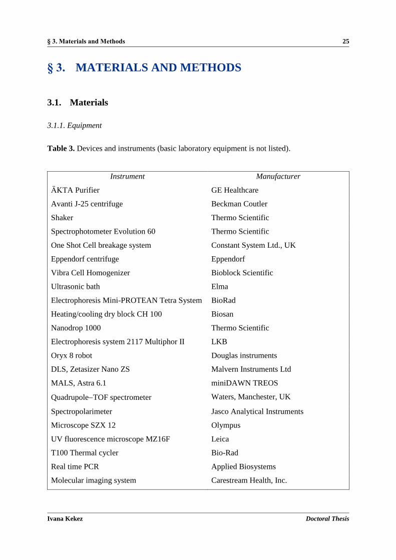
§ 3. Materials and Methods 25
Ivana Kekez Doctoral Thesis
§ 3. MATERIALS AND METHODS
3.1. Materials
3.1.1. Equipment
Table 3. Devices and instruments (basic laboratory equipment is not listed).
Instrument Manufacturer
ÄKTA Purifier GE Healthcare
Avanti J-25 centrifuge Beckman Coutler
Shaker Thermo Scientific
Spectrophotometer Evolution 60 Thermo Scientific
One Shot Cell breakage system Constant System Ltd., UK
Eppendorf centrifuge Eppendorf
Vibra Cell Homogenizer Bioblock Scientific
Ultrasonic bath Elma
Electrophoresis Mini-PROTEAN Tetra System BioRad
Heating/cooling dry block CH 100 Biosan
Nanodrop 1000 Thermo Scientific
Electrophoresis system 2117 Multiphor II LKB
Oryx 8 robot Douglas instruments
DLS, Zetasizer Nano ZS Malvern Instruments Ltd
MALS, Astra 6.1 miniDAWN TREOS
QuadrupoleTOF spectrometer Waters, Manchester, UK
Spectropolarimeter Jasco Analytical Instruments
Microscope SZX 12 Olympus
UV fluorescence microscope MZ16F Leica
T100 Thermal cycler Bio-Rad
Real time PCR Applied Biosystems
Molecular imaging system Carestream Health, Inc.

§ 3. Materials and Methods 26
Ivana Kekez Doctoral Thesis
3.1.2. Chromatography supplies
Table 4. Columns, media and molecular weight markers used for affinity and size-exclusion
chromatography.
Chromatography supplies Manufacturer
PD-10 desalting columns GE Healthcare
His Trap Ni-NTA agarose GE Healthcare
GSTrap GE Healthcare
Strep-Tactin IBA
Gel filtration molecular weight markers Sigma Aldrich
3.1.3. Crystallographic supplies
Table 5. Crystallization screens.
Screen Manufacturer
PEGs II, PACT SUITE, JCSG Core (Suites IIV) Qiagen
Structure Screen 1 & 2, PGA Screen, Solubility Tool Kit Molecular Dimensions
Additive Screen Hampton Research
Table 6. Crystallization plates.
Type Manufacturer
MRC 96-well sitting drop crystallization plate Molecular Dimensions
MRC MAXI 48-well sitting drop
crystallization plate
Molecular Dimensions
24-well XRL plate Molecular Dimensions

§ 3. Materials and Methods 27
Ivana Kekez Doctoral Thesis
Table 7. Cryocrystallography equipment.
Type Manufacturer
Mounted CryoLoops Molecular Dimensions
EMBL/ESRF sample changer kit Molecular Dimensions
Mounted Mesh LithoLoop Molecular Dimensions
3.1.4. Kits
Table 8. Kits used for preparation of DNA and for staining of the gels for visualizing proteins
according to the manufacturers’ protocols.
Type Manufacturer
QIAprep Spin Miniprep Kit Qiagen
QIAquick Gel Extraction Kit Qiagen
ProteoSilver Silver Stain Kit Sigma Aldrich
3.1.5. Filters and membranes
Table 9. Ultra centrifugal filters used to concentrate dilute protein solutions, membranes used
for filtering sterile aqueous solutions and transfer membrane used for western blotting.
Type Manufacturer
Amicon Ultra centrifugal filter units with a molecular
weight cut-off of 3 kDa,10 kDa and 30 kDa, respectively. Millipore
0.2 μm cellulose acetate membranes Millipore
PVDF membrane, 0.2 μm Millipore
3.1.6. Chemicals and buffers
All buffers were prepared with redistilled water (MilliQ gradient water system) and then filtered
using a Sartolab filter system with PES membrane (0.22 µm pore size). If not stated otherwise,
all chemicals were obtained in analytical grade from the following companies: Acros Organics;

§ 3. Materials and Methods 28
Ivana Kekez Doctoral Thesis
AppliChem; Carl Roth; Fisher Scientific; Fluka; Jena Bioscience, Merck, Roche GmbH
(Mannheim); Serva Electrophoresis; Sigma Aldrich.
3.1.7. Enzymes
Restriction enzymes and polymerases for molecular biological applications as well as proteases
for biochemical usage were obtained from New England Biolabs (NEB), Sigma Aldrich and
ThermoFisher Scientific. All enzymes were used in their appropriate buffer as specified by the
manufacturer.
Table 10. Features of enzymes used for molecular and biochemical applications. Recognition
sequences of restriction enzymes and proteases are in superscript.
DNA modifying enzymes Application Manufacturer
Phusion high-fidelity DNA
polymerase
Amplification of DNA
(proofreading)
ThermoFisher
Scientific
Taq DNA polymerase Amplification of DNA
(no proofreading)
ThermoFisher
Scientific
T4 DNA ligase Ligation of DNA
fragments
ThermoFisher
Scientific
BamHI5' G|GATCC 3' Restriction of DNA NEB
XhoI5' C|TCGAG 3' Restriction of DNA NEB
DNase I Digestion of DNA Sigma Aldrich
Rnase A Digestion of RNA ThermoFisher
Scientific
Trypsin K, R (on –COOH terminus) Proteolysis Sigma Aldrich
- Chymotrypsin Y, W, F, L, M (on –COOH terminus) Proteolysis Sigma Aldrich
- Thermolysin L, F (on –NH2 terminus) Proteolysis Sigma Aldrich
Thrombin LVPR|GS Removal of His6-tag
present at protein
Novagen

§ 3. Materials and Methods 29
Ivana Kekez Doctoral Thesis
expressed in vector
pET 15b / Proteolysis
Prescission ProteaseLEVLFQ|GP Removal of GST-tag
present at protein
expressed in vector
pGEX-6P-1
Sigma Aldrich
3.1.8. Antibodies
Table 11. Features of primary and secondary antibodies.
Primary antibody Secondary antibody Application Manufacturer
Monoclonal Anti-
His6 (mouse)
Detection of
HHHHHH-epitope in
proteins
Roche
Polyclonal Goat anti-
Mouse Antibody,
horseradish
peroxidase (HRP)
conjugate
General Electrics
3.1.9. Oligonucleotides
Oligonucleotides (Table 12) were synthesized by Sigma-Aldrich. The lyophilized and desalted
oligonucleotides were dissolved in sterile water at a concentration of 100 μmol L1 and stored
at 20 °C.
Table 12. Oligonucleotides used as primers to amplify gene sequences.
Name Sequence (5' to 3') Application
GST_HP1326-for
GST_HP1326-rev
GAGAGAGGGATCCATGCAAACCCTAAAAGCCAAC
GAGAGAGCTCGAGTTATAAATCCAGGCTTGTTTTTAG
Amplifying
GST_HP1326

§ 3. Materials and Methods 30
Ivana Kekez Doctoral Thesis
Strep_HP1326-for
Strep_HP1326-rev
GAAGGAGATATACATATGTGGAGCCACCCGCAGTTCGAAAA
ACAAACCCTAAAAGCCAAC
GTGATGGTGGTGATGATGTTATAAATCCAGGCTTGTTTTAGC
Amplifying
Strep_HP1326
HP1326_His-for
HP1326_His-rev
GAAGGAGATATACATATGCAAACCCTAAAAGCCAACGAC
GTGATGGTGGTGATGATGTAAATCCAGGCTTGTTTTAGC
Amplifying
HP1326_His
HP0907_His-for
HP0907_His-rev
GGAGATATACATATGGCTATTGATTTAGCAGAAG
GTGATGGTGGTGATGATGTGCTGTCTCTTTAGG GG
Amplifying
HP0907_His
3.1.10. Vectors
pET15b (Novagen), Fig. 11 and Table 13, is a 5708 bp long vector that carries a N-terminal
hexa His-tag sequence followed by a thrombin recognition site and MCS, including sites for
XhoI, BamHI and NdeI. Expression of genes cloned into this vector is under the control of the
T7-lac promoter. Additionally, the vector carries an ampicillin resistance gene (ampR) for
selection.93
pGEX-6P-1 (GE Healthcare), Fig. 11 and Table 13, is a 4984 bp long bacterial vector for
expression of N-terminal GST-tagged recombinant proteins under the tac promoter. The vector
contains a PreScission Protease recognition site for cleaving the protein from the GST-tag and
an ampicillin resistance gene (ampR) for selection.94
pETite C- His (Lucigen), Fig. 11 and Table 13, is a 2235 bp long pre-processed vector that
encodes the C-terminal hexa His-tag. The vector includes a kanamycin resistance gene (kanR)
for selection and inducible expression under control of the T7-lac promoter.95

§ 3. Materials and Methods 31
Ivana Kekez Doctoral Thesis
Figure 11. Schematic presentation of plasmid vector maps used for cloning.

§ 3. Materials and Methods 32
Ivana Kekez Doctoral Thesis
Table 13. List of all designed constructs with starting and ending positions of the amino acid
residues.
Protein Template Vector Start End
HP1326 (CrdA) H. pylori G27 pETite C-His 23 125
pGEX-6P-1 23 125
HP1026 H. pylori G27 pET15b 1 391
HP0907 (FlgD) H. pylori G27 pETite C-His 1 316
H. pylori 26695 pETite C-His 1 301
3.1.11. Bacterial strains: Escherichia coli
3.1.11.1. Host strain for cloning
One Shot TOP10 (F mcrA Δ(mrr-hsdRMS-mcrBC) Φ80lacZΔM15 Δ lacX74 recA1 araD139
Δ(araleu)7697 galU galK rpsL (StrR) endA1 nupG) (Invitrogen) are chemically competent
cells that allow stable replication of high-copy number plasmids.96
DH5 (F Φ80lacZΔM15 Δ(lacZYA-argF) U169 recA1 endA1 hsdR17(rk, mk
) phoA
supE44 thi-1 gyrA96 relA1 λ) (Invitrogen) are chemically competent cells that allow
blue/white screening on plates containing either X-Gal or Bluo-Gal due to the Φ80lacZΔM15
genotype. Moreover, the recA1 mutation helps to reduce the rate of recombination while
propagating plasmid DNA, and the endA1 mutation prevents unspecific digestion of DNA.96
3.1.11.2. Strains for protein expression
BL21 (DE3) (F ompT hsdSB (rB-mB-) gal dcm (DE3)) (Invitrogen) are chemically competent
cells deficient in both lon and ompT proteases. The designation (DE3) indicates that the host is
a lysogen of λDE3, and therefore carries a chromosomal copy of the T7 RNA polymerase gene
under control of the lacUV5 promoter. Such strains are suitable for production of protein from
target genes cloned in pET vectors using IPTG as an inducer.97
BL21pLysS (DE3) (F ompT hsdSB (rB-mB-) gal dcm (DE3) pLysS (CamR)) (Invitrogen)) are
chemically competent cells used to suppress basal expression of T7 RNA polymerase prior to
induction and thus stabilize pET recombinants encoding target proteins that affect cell growth

§ 3. Materials and Methods 33
Ivana Kekez Doctoral Thesis
and viability. The pLysS designation is given to hosts carrying a pET-compatible plasmid that
encodes T7 lysozyme, which is a natural inhibitor of T7 RNA polymerase.97
3.2. Microbiological methods
3.2.1. Media for cultivation of E. coli cells
Liquid cell cultures of E. coli were grown in LB (Luria Bertani) medium (0.5 % NaCl, 0.5 %
yeast extract, 1 % tryptone, all w/v). Solid media were additionally supplied with 1.5 % (w/v)
agar. All media were sterilized by autoclaving for 20 min at 120 °C. If necessary, appropriate
antibiotics were added prior to use: for cells harboring pET15b and pGEX-6P-1 based vectors,
ampicillin was added at a concentration of 100 μg mL1. Cells containing the pETite C-His
plasmid were selected using 30 μg mL1 kanamycin .
3.2.2. Transformation of chemically competent E. coli cells
Chemically competent cells (50 μL) were thawed on ice, mixed with 50300 ng DNA and
incubated on ice for 30 min. Afterwards the cells were heat shocked for 1 min at 42 °C and
then placed on ice for further 2 min. After addition of 300 μL of LB medium, the cells were
shaken at 37 °C for 4560 min. The cell suspension was plated onto LB-agar plates supplied
with appropriate antibiotics and incubated at 37 °C overnight (o/n).
3.3. Molecular biology methods
Experiments handling preparation and isolation of DNA were performed according to the
instructions from the laboratory handbook J. Sambrook and D. Russell: Molecular Cloning: A
Laboratory Manual, 3th edition, Cold Spring Harbor Laboratory Press (2001),98 or according to
the datasheet instructions of the specific chemical/kit that was used.
3.3.1. Amplification and isolation of plasmid DNA from E. coli
Isolation of plasmid DNA was performed with Qiagen's QIAprep Spin Miniprep Kit according
to the manufacturer’s manual. To amplify plasmid DNA, One shot TOP10 or DH5 cells were
transformed with the appropriate vector (see 3.2.2.) and 5 mL o/n cultures were set up with a
single colony.

§ 3. Materials and Methods 34
Ivana Kekez Doctoral Thesis
3.3.2 Determination of DNA concentration
Concentration of DNA in solution was determined spectrophotometrically using Nanodrop
1000 based on the absorbance at 260 nm.
3.3.3. Polymerase chain reaction (PCR)
PCR was used for DNA amplification. The proofreading high-fidelity DNA polymerase
Phusion from Pyrococcus was used for amplification of DNA fragments for cloning strategies
because of their low error rate. For colony-PCRs, the non-proofreading Taq DNA polymerase
from Thermus aquaticus was used. Standard PCR reactions contained the components listed in
Table 14. Annealing temperatures and elongation times were adjusted to primer and template
composition and the size of the amplified fragment (Table 15).
Table 14. Composition of PCR reactions.
Component Cloning PCR (Phusion) Cloning PCR (Taq)
Template DNA (25 ng) 1 µL 1 µL
5' Primer (10 μmol L1) 1 µL 0.25 µL
3' Primer (10 μmol L1) 1 µL
dNTPs (10 mmol L1) 0.4 µL 0.65 µL
Phusion DNA polymerase
(2U*/ µL) 0.2 µL
5x Phusion HF buffer with
MgCl2 4 µL
Taq polymerase (5U / µL) 0.25 µL
10x Taq reaction buffer
MgCl2 (25 mmol L1)
2.5 µL
1.5 µL
Nuclease free H2O 12.4 µL 18.85 µL
Final volume of: 20 µL 25 µL
* One unit is defined as the amount of enzyme that will incorporate 10 nmol of dNTP into acid insoluble
material in 30 minutes at 74 °C.

§ 3. Materials and Methods 35
Ivana Kekez Doctoral Thesis
Table 15. Thermal cycling profile for cloning and colony PCRs.
Component Cloning PCR (Phusion) Cloning PCR (Taq)
Initial dentauration 30 s / 98 °C 2 min / 94 °C
Denaturation 5-10 s / 98 °C 15 s / 94 °C
Annealing 35 cycles 10-30 s / primer specific
temperature
15 s / primer specific
temperature
Extension 15-30 s per kb / 72 °C 1 min per kb / 72 °C
Final extension 5-10 min / 72 °C 5-10 min / 72 °C
3.3.4. Agarose gel electrophoresis
DNA samples from PCR reactions were analyzed by agarose gel electrophoresis. Samples were
loaded into wells of an agarose gel and subjected to an electric field, causing the negatively
charged nucleic acids to move toward the positive electrode. Shorter DNA fragments traveled
more rapidly, while the longest fragments remain closest to the origin of the gel, resulting in
separation based on size. Supercoiled circular plasmid migrates more rapidly than the same
plasmid when linearized whereas the nicked circular plasmid is the slowest in migration in
comparison to the other two forms. Agarose gels (1 %, w/v) were prepared with TAE (40 mmol
L1 Tris-base, 20 mmol L1 acetic acid, 1 mmol L1 EDTA pH = 8) buffer and with an addition
of 50 μg L1 ethidium bromide. All samples were mixed with DNA loading dye (0.25 % (w/v)
bromophenol blue, 0.25 % (w/v) xylene cyanol FF, 0.01 mol L1 EDTA pH = 8, 3 % (w/v)
glycerol) prior to loading. Two different dyes are present for visual tracking of DNA migration
during electrophoresis. The presence of glycerol ensures that the DNA in the ladder and sample
forms a layer at the bottom of the well. The EDTA included in the solution binds divalent metal
ions and inhibits metal-dependent nucleases. 1 kb DNA ladder (NEB) was applied for size
estimation of DNA fragments. Electrophoresis was carried out at a constant voltage of 120 V.
Afterwards DNA bands were visualized by UV transillumination at 254 nm and documented
with the MiniBIS Imaging systems (DNR, Bio-Imaging Systems Ltd.). Ethidium bromide is a
fluorescent dye that intercalates between bases of nucleic acids and allows detection of DNA
fragments in gels. Ethidium bromide possesses UV absorbance maxima at 300 and 360 nm.
Additionally, it can absorb energy from nucleotides excited by absorbance of 260 nm radiation.
Ethidium re-emits this energy as yellow/orange light centered at 590 nm.99
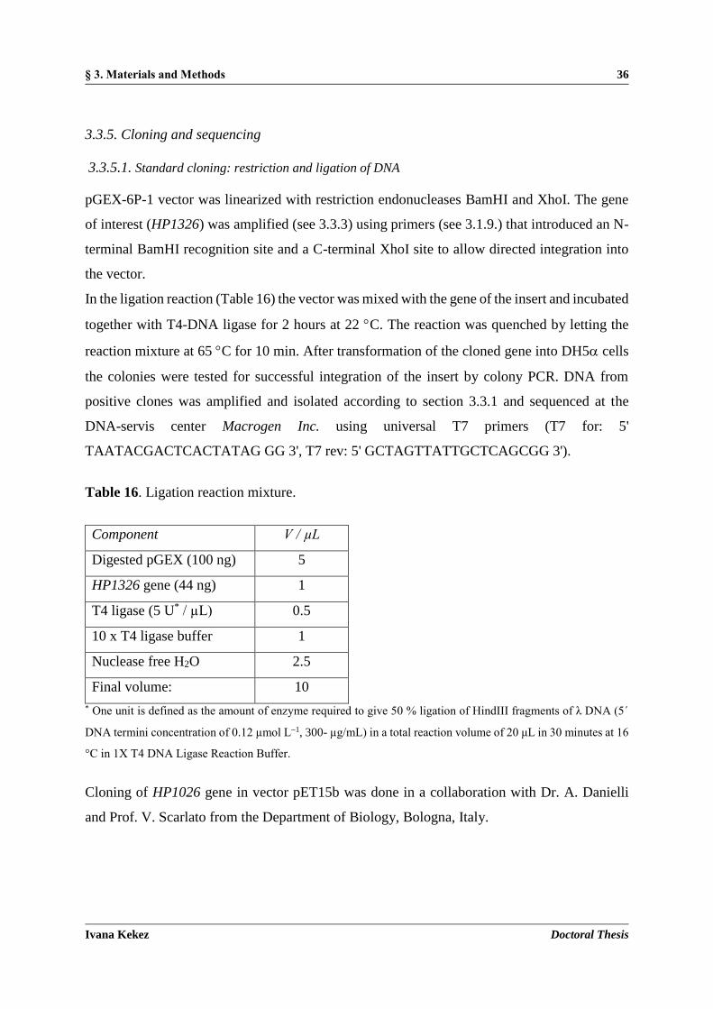
§ 3. Materials and Methods 36
Ivana Kekez Doctoral Thesis
3.3.5. Cloning and sequencing
3.3.5.1. Standard cloning: restriction and ligation of DNA
pGEX-6P-1 vector was linearized with restriction endonucleases BamHI and XhoI. The gene
of interest (HP1326) was amplified (see 3.3.3) using primers (see 3.1.9.) that introduced an N-
terminal BamHI recognition site and a C-terminal XhoI site to allow directed integration into
the vector.
In the ligation reaction (Table 16) the vector was mixed with the gene of the insert and incubated
together with T4-DNA ligase for 2 hours at 22 C. The reaction was quenched by letting the
reaction mixture at 65 C for 10 min. After transformation of the cloned gene into DH5 cells
the colonies were tested for successful integration of the insert by colony PCR. DNA from
positive clones was amplified and isolated according to section 3.3.1 and sequenced at the
DNA-servis center Macrogen Inc. using universal T7 primers (T7 for: 5'
TAATACGACTCACTATAG GG 3', T7 rev: 5' GCTAGTTATTGCTCAGCGG 3').
Table 16. Ligation reaction mixture.
Component V / µL
Digested pGEX (100 ng) 5
HP1326 gene (44 ng) 1
T4 ligase (5 U* / µL) 0.5
10 x T4 ligase buffer 1
Nuclease free H2O 2.5
Final volume: 10
* One unit is defined as the amount of enzyme required to give 50 % ligation of HindIII fragments of λ DNA (5´
DNA termini concentration of 0.12 µmol L1, 300- µg/mL) in a total reaction volume of 20 μL in 30 minutes at 16
°C in 1X T4 DNA Ligase Reaction Buffer.
Cloning of HP1026 gene in vector pET15b was done in a collaboration with Dr. A. Danielli
and Prof. V. Scarlato from the Department of Biology, Bologna, Italy.

§ 3. Materials and Methods 37
Ivana Kekez Doctoral Thesis
3.3.5.2. Enzyme free-cloning
1 µL of pre-processed linearized pETite C-His vector (25 ng) was mixed with 3 µL of the target
PCR product (50 ng of HP1326 / HP0907, see 3.1.10.) and transformed directly into 50 µL of
chemically competent One shot TOP10 cells (see 3.2.2.). The target gene had been previously
amplified with primers that contained short homology to the ends of the pETite C-His vector
allowing recombination within the host cell to fuse the blunt PCR product to the vector (Fig.
12) Sequencing of DNA from positive clones was performed like previously described in
section 3.3.5.1.
Figure 12. Preparation of construct by enzyme free-cloning.100

§ 3. Materials and Methods 38
Ivana Kekez Doctoral Thesis
3.4. Protein biochemical methods
Experiments handling preparation and isolation of proteins were performed according to the
instructions from the laboratory handbook J. Sambrook and D. Russell: Molecular Cloning: A
Laboratory Manual, 3th edition, Cold Spring Harbor Laboratory Press (2001),98 or according to
the datasheet instructions of the specific chemical/ kit that was used.
3.4.1. Protein quantification
The concentration of pure protein solutions was determined spectrophotometrically by reading
absorption at 280 nm due to the high absorbance of aromatic amino acid residues. Considering
absorbance and the protein specific absorption coefficient that was calculated on the basis of
the primary sequence using ExPASy's ProtParam software,101 the protein concentration was
determined by applying the Beer-Lambert law.
3.4.2. Sodium dodecyl sulfate polyacrylamide gel electrophoresis (SDS-PAGE)
The quality of the purified proteins samples, samples from expression studies, and protein
crystals was verified by discontinuous electrophoresis under denaturing conditions (SDS-
PAGE). SDS is an anionic detergent which denatures proteins and confers a negative charge to
all protein molecules, since a polypeptide chain binds an amount of SDS proportional to its
molecular mass. The final separation of proteins depends entirely on the differences in the
relative molecular mass of the polypeptides, so that in the electric field the negatively charged
molecules will migrate toward the positively charged anode. For the experiments in this study
the used percentage (w/v) of stacking and separating acrylamide gels was 4 % and 16 %,
respectively (Table 17). Prior to use, all samples were supplied with 4x loading buffer (Table
18) and incubated at 100 C (to improve denaturation) for 2 min in the case of pure proteins
and for 10 min for the samples from expression studies.

§ 3. Materials and Methods 39
Ivana Kekez Doctoral Thesis
Table 17. Composition of the stacking and separating acrylamide gels.
Component Stacking gel Separating gel
Acrylamide/bis-acrylamide (29:1 w/v) 40 %, (v/v) 4 % 16 %
Buffer Tris-HCl, pH 6.8 0.125 mol L1
Buffer Tris-HCl, pH 8.8 0.375 mol L1
SDS, (w/v) 0.1 % 0.1 %
APS 0.7 μg mL1 0.7 μg mL1
TEMED, (w/v) 0.05 % 0.05 %
Table 18. Composition of the 4x loading buffer.
Tris-HCl pH = 6.8 62.5 mmol L1
β-Mercaptoethanol 12.5 mmol L1
Glycerol, (v/v) 6.25 %
SDS, (w/v) 1.25 %
Bromophenol Blue, (w/v) 0,002 %
Different types of protein markers were used depending on the application. Unstained Bio-
Rad’s Precision Plus Protein Standard was used when gels were stained using the Coomassie
Brilliant Blue R-250 solution (Table 19) or by using the commercial ProteoSilver Silver Stain
Kit for silver staining. When western blotting was performed after electrophoresis, pre-stained
Bio-Rad’s Precision Plus Protein Standard was used. SDS-PAGE was carried out at constant
current (30-50 mA) and maximum voltage (600 V) within 12 h. Composition of the buffer
used during electrophoresis is presented in Table 20. Gels stained by the Coomassie solution
were de-stained with hot water.
Table 19. Composition of the Coomassie Brilliant Blue R-250 staining solution.
Coomassie Brilliant Blue R-250 2.5 g L1
Acetic acid, (v/v) 10 %
Ethanol, (v/v) 45 %

§ 3. Materials and Methods 40
Ivana Kekez Doctoral Thesis
Table 20. Composition of the electrophoresis buffer.
Buffer Tris-HCl, pH 8.3 3.03 g L1
Glycin 14.4 g L1
SDS, (w/v) 0.1 %
Silver staining is a very sensitive method to detect very low amounts of protein in a SDS-PAGE
gel with a detection limit of 0.1 ng.102 In this study the silver staining method was used to verify
samples of protein crystals. After electrophoresis, the gel was incubated in fixation solution (50
% (v/v) ethanol, 10 % (v/v) acetic acid) for 20 min, and then washed with ethanol (30 %, v/v)
for 10 min, and then with ultrapure water for additional 10 min. Afterwards the gel was treated
with a sensitizer solution (1 %, v/v ProteoSilver Sensitizer) for another 10 min, and then washed
twice for 10 min with ultrapure water. Silver solution (1 %, v/v ProteoSilver Silver Solution)
was applied for 10 min followed with washing for 1 min with ultrapure water. The staining
process (the reduction of silver ions) was initiated by addition of the developing solution (5 %,
v/v ProteoSilver Developer 1, 0.1 %, v/v ProteoSilver Developer 2) for 5 min, and was
terminated by addition of ProteoSilver Stop solution for 5 min, and then washed with ultrapure
water for 15 min. All used commercial solutions (ProteoSilver Silver Solution, ProteoSilver
Sensitizer, ProteoSilver Developer 1, ProteoSilver Developer 2, ProteoSilver Stop Solution) are
part of the ProteoSilver Plus Silver Stain Kit (Sigma Aldrich).
3.4.3. Western blotting
The western blotting method includes transfer of proteins from a gel to a membrane and their
detection on the surface of the membrane by antibodies. In this work it was used to detect
presence of His-tagged proteins. After SDS-PAGE, the separated protein samples were
transferred from the acrylamide gels to the polyvinylidene fluoride (PVDF) membrane by the
semi-dry western blotting method using the instrument 2117 Multiphor II (LKB). Before
transferring, the gels were incubated with shaking in the transfer buffer (Table 21) for 15 min.
All membranes and blotting papers were wetted in the transfer buffer, and the blot sandwich
was assembled as follows: three sheets of paper, the transfer membrane, the gel and finally three
more sheets of blotting paper were placed inside a semi-dry electro-blotting system with the
membrane facing the anode. Protein transfer was performed at a constant current of 1 mA/cm2
for 1.5 hours. After blotting, the protein transfer was confirmed by staining with the Ponceau S

§ 3. Materials and Methods 41
Ivana Kekez Doctoral Thesis
(0.1 % (w/v), 5 % (v/v) acetic acid) solution. Afterwards transfer membranes were further
analyzed by immunodetection. At first, membranes were washed with ultrapure water and
incubated with 1 % (w/v) casein-TBS solution (Table 21) to saturate free binding sites of the
membrane. After 60 min of incubation, membranes were washed three times with TBS buffer;
each step lasting 10 min. Primary antibody (see 3.1.9.) was diluted 1,000 times in a 1 % casein-
TBS solution and was applied to the membrane o/n at 4 °C. Membranes were washed again
three times for 10 min with the TBS buffer. The second antibody (see 3.1.9.) was diluted 10,000
times also in the 1 % casein-TBS solution and applied to the membrane for 60 min. Then,
washing with TBS buffer was repeated as before. The bound secondary antibodies were
detected via the linked enzyme horseradish peroxidase using Immun-Star WesternC
chemiluminescent kit (Bio-Rad Laboratories). The secondary antibody contains horseradish
peroxidase, which is an enzyme that catalyzes luminol oxidation in the presence of hydrogen
peroxide. The reaction forms an excited state of the substrate that emits light as it decays to the
ground state. Luminescent signals derived from the oxidation of luminol were detected and
quantified with the luminescent image analyzer Molecular Imaginig system (Carestream
Health, Inc.).
Table 21. List and composition of buffers used in western blotting.
Transfer buffer 14.4 g L1 glycin, 3 g L1 Tris-HCl pH = 7.5, 20 % (v/v) ethanol
TBS buffer 25 mmol L1 Tris-HCl pH = 7.5, 150 mmol L1 NaCl, 0.2 % (v/v) Tween
3.4.4. Proteolytic cleavage
3.4.4.1. Removal of the hexa His-tag from HP1026 with thrombin
Purified His-tagged HP1026 protein was incubated with thrombin (0.1 U / µL) in a protein-
protease ratio 1:100, and buffer containing 20 mmol L1 Tris-HCl pH 7.5, 150 mmol L1 NaCl.
The reaction mixture was incubated o/n at 4 C and gently shaked. The cleaved and uncleaved
HP1026 were separated by running the whole mixture through nickel nitrilotriacetic acid (Ni-
NTA) column (see 3.6.1.2.) since the cleaved protein does not bind to the resin. Collected
flowthrough was then passed through a benzamidine sepharose column (GE Healthcare) for

§ 3. Materials and Methods 42
Ivana Kekez Doctoral Thesis
binding of residual thrombin and thus preventing secondary activity from this source. The yield
of cleavage was monitored by SDS-PAGE and western blotting.
3.4.4.2. Removal of the GST-tag from GST_HP1326 with PreScission protease
Purified GST_HP1326 protein was treated with PreScission protease to cleave the N-terminal
GST tag from the fusion protein. The protein:protease ratio was adjusted to 1:200 in a buffer
containing 50 mmol L1 Tris-base pH 7.5, 50 mmol L1 NaCl, 1 mmol L1 EDTA, 1 mmol L1
dithiothreitol (DTT). The sample was incubated at 4 °C on a shaker o/n. PreScission protease
as well as uncleaved HP1326 contain the GST tag, so they were both removed from the cleaved
sample by passing the sample through a glutathione agarose (GE Healthcare) column according
to the manufacturer protocol (see 3.6.1.3.).
3.4.4.3. Limited proteolysis of the FlgD (HP0907) protein
Limited proteolysis of FlgD was also investigated. Protein regions exposed on the surface,
either present in loops or in unstructured regions, are usually exposed to proteolytic cleavage,
while the globular parts and stable secondary structure elements are not accessible to proteases.
Each of the four samples of FlgD ( = 3 mg mL1) in 20 mmol L1 Tris-base pH 7.5, 150 mmol
L1 NaCl was digested with selected proteases (trypsin, chymotrypsin, thermolysin or
thrombin) in 1:1000 and 1:500 ratios at 25 C and incubated for an hour. The yield of digestion
was monitored by SDS-PAGE.

§ 3. Materials and Methods 43
Ivana Kekez Doctoral Thesis
3.5. Expression of recombinant proteins in E. coli
3.5.1. Expression of CrdA (HP1236), HP1026 and FlgD (HP0907)
E. coli BL21(DE3) cells were chemically transformed with a desired plasmid (see Table 13 in
3.1.10.). The used vectors contain either T7 (pET15b, pETite) or tac (pGEX-6P-1) promoters
that are inducible by isopropyl-β-D-1-thiogalactopyranoside (IPTG) and are located upstream
from the gene of interest. BL21(DE3) is the T7 host strain used for expression of cloned genes
from the bacteriophage T7 promoter. The genome of bacteria harbors the T7 bacteriophage
RNA polymerase gene under the control of the IPTG inducible lacUV5 promoter. The lac
repressor protein is responsible for maintaining inducible control over the lacUV5 promoter as
well as the T7-lac promoter on the vector.100 In case of protein toxicity BL21pLysS (DE3) cells
were used for expression of toxic proteins since these cells provide tighter control of protein
expression. BL21pLysS (DE3) cells contain pLysS that constitutively expresses low levels of
T7 lysozyme, which reduces basal expression of recombinant genes by inhibiting basal levels
of T7 RNA polymerase.
LB medium was inoculated with a single colony from a freshly streaked plate and treated with
the appropriate antibiotic for the plasmid and host strain and was shaked (180 rpm) at 37 C
o/n. Fresh LB medium containing the appropriate antibiotic was inoculated with the overnight
culture and incubated with shaking at 28 C or 16 C. In this work expression performed at
different temperatures was tested to establish the best expression condition for different
constructs. When the OD600 of 0.6 was reached, the protein expression was induced by adding
0.5 mM IPTG. After induction of 4 h and o/n performed at 28 C and 16 C, respectively, cells
were harvested by centrifugation (5000 rpm, 15 min, 4 °C) and flash-frozen in liquid nitrogen
and stored at 80 °C. Afterwards, cells were thawed on ice, re-suspended in a protein specific
buffer (Table 22), and disrupted with the One Shot Cell breakage system (Constant System Ltd.,
UK; 1.36 kbar). By standard, the cell suspension was passed through the system twice. Buffers
used in cell disruption contain phenylmethylsulfonyl fluoride (PMSF) as a serine protease
inhibitor. The lysate was later clarified by centrifugation (18 000 rpm, 30 min, 4 C) and the
soluble fractions were used in further steps of purification.

§ 3. Materials and Methods 44
Ivana Kekez Doctoral Thesis
Table 22. Composition of buffers used in cell disruption for a specific protein.
CrdA GST_CrdA, HP1026, FlgD
20 mmol L1 Tris-base pH 7.5
200 mmol L1 NaCl
0.05 % Triton X-100
2 mmol L1 (PMSF)
20 mmol L1 Tris-base pH 7.5
150 mmol L1 NaCl
2 mmol L1 PMSF
3.5.2. Expression of selenomethionine- HP1026 and FlgD (HP0907)
Expression of recombinant selenomethionine derivatized proteins was performed by using the
non-methionine auxotrophic E. coli BL21 strain (DE3). The bacteria were grown in minimal
medium M9 (Table 23) supplemented with vitamin B1, amino acids (L-leucine, L-isoleucine, L-
valine, L-phenylalanine, L-lysine, L-threonine) and L-seleno-methionine103 at 28 C as described
in section 3.5.1. All amino acids were purchased from Sigma Aldrich.
Table 23. Composition of the M9 medium.
Na2HPO4 6.78 g L1
KH2PO4 3 g L1
NaCl 0.5 g L1
NH4Cl 5 g L1
MgCl2 1 mmol L1
CaCl2 0.1 mmol L1
Trace elements* (v/v) 0.01 %
D+ Glucose (w/v) 2 %
* Trace elements: 0.1 g L1 MnSO4, 0.5 g L1 FeCl3, 8 mg L1 CuCl2, 0.05 g L1 ZnO, 10 mg L1 Co(NO3)2, 10 mg
L1 (NH4)6Mo7O24, 5 g L1 EDTA

§ 3. Materials and Methods 45
Ivana Kekez Doctoral Thesis
3.6. Purification of CrdA, HP1026 and FlgD
Purity of samples during all steps of purification was monitored by the SDS-PAGE.
3.6.1. Affinity chromatography
3.6.1.1. Affinity chromatography using a Strep-Tactin
Strep-tagged CrdA was purified by affinity chromatography using a strep-tactin resin. The
Strep-tag (NH2-WSHPQFEK-COOH) purification system is based on the highly selective
interaction between the Strep-tag fused to the protein and a specially engineered streptavidin
called strep-tactin. The tagged protein binds to immobilized strep-tactin during affinity
purification. Protein is eluted with a buffer containing desthiobiotin, which is the natural ligand
of streptavidin (Fig. 13).104
The strep-tactin resin (1 mL) was equilibrated with buffer A (Table 24), and the crude lysate
was mixed with resin at 4 C o/n. Unbounded proteins were removed from the resin by washing
the resin with 30 mL of buffer A. The Strep_CrdA protein was eluted from the resin by addition
of 20 mL of buffer E (Table 24). Regeneration of the resin was performed by addition of yellow
HABA (2-[4’-hydroxy-benzeneazo] benzoic acid) solution. HABA displaced desthiobiotin and
turned the resin red due to the binding of HABA as a red colored hydrazine isomer to strep-
tactin and prior to the next purification it was removed from the resin by washing it with buffer
A.
Table 24. Composition of buffers used in affinity chromatography of StrepCrdA.
Buffer A Buffer E
20 mmol L1 Tris-base pH 7.5
200 mmol L1 NaCl
20 mmol L1 Tris-base pH 7.5
200 mmol L1 NaCl
5 mmol L1 desthiobiotin

§ 3. Materials and Methods 46
Ivana Kekez Doctoral Thesis
Figure 13. Schematic presentation of affinity chromatography with strep-tactin.104
3.6.1.2. Affinity chromatography with Ni-NTA agarose
Purification of polyhistidine affinity-tagged proteins can be facilitated by utilization of the
nickel-nitrilotriacetic acid resin. The nitrilotriacetic acid chelator linked to agarose binds His-
tagged proteins (5-10 mg His-tag protein / Ni-NTA resin mL) since the nitrogen atoms of the
imidazole ring inside the His-tag have affinity toward the nickel ion and occupy two out of six
nickel coordination sites (Fig. 14).

§ 3. Materials and Methods 47
Ivana Kekez Doctoral Thesis
Figure 14. Ni-NTA chelating (a) two water moleules, or (b) two histidines.105
In this study the Ni-NTA affinity purification of proteins (CrdA, HP1026, FlgD) containing the
His6-tag was performed using the 1 mL columns. The column was firstly equilibrated with
protein specific buffer A (Table 25) and then the soluble part of lysate was added to the column.
To obtain a high purity sample, the column was connected to an ÄKTA Purifier system, and a
washing step of 50 mL of buffer A supplemented with 20 mmol L1 imidazole was performed
to eliminate unspecifically bound proteins. Finally, elution of the protein was performed in
buffer A with a stepwise gradient of imidazole from 20 to 500 mmol L1.
Table 25. Composition of buffer A used in Ni-NTA affinity chromatography of CrdA_His,
FlgD_His and His_HP1026.
CrdA_His FlgD_His, His_HP1026
20 mmol L1 Tris-base pH 7.5
200 mmol L1 NaCl
20 mmol L1 Tris-base pH 7.5
150 mmol L1 NaCl
3.6.1.3. Affinity chromatography with gluthatione sepharose
GST-fused proteins were purified using a gluthatione sepharose resin. Glutathione S-transferase
(GST) is an enzyme found in most organisms and can make the tagged protein more soluble.
The purification method is based on the high affinity of GST for glutathione (binding capacity
is > 5 mg GST/mL chromatography medium). When applied to the affinity medium, GST-

§ 3. Materials and Methods 48
Ivana Kekez Doctoral Thesis
tagged proteins bind to the glutathione ligand, and impurities are removed by washing with
binding buffer. Tagged proteins are then eluted from the chromatography medium using a
buffer containing reduced gluthatione (Fig. 15).
In this work GST_CrdA was purified using a gluthatione sepharose 1 mL column that was
firstly washed with buffer A (Table 26), followed by the incubation of resin with a GST-tagged
protein for 1 h at 4 C. Afterwards, the column was washed with 10 mL of buffer A and protein
elution was performed using the buffer E (Table 26).
Figure 15. Principle of GST-tag protein purification.106
Table 26. Composition of buffers used in affinity chromatography of GST_CrdA.
Buffer A Buffer E
140 mmol L1 NaCl
2.7 mmol L1 KCl
10 mmol L1 Na2HPO4
1.8 mmol L1 KH2PO4 pH = 7.5
10 mmol L1 reduced gluthatione
50 mmol L1 Tris-HCl pH = 8.0
3.6.2. Size-exclusion chromatography (SEC)
Analytical size-exclusion chromatography was used to separate proteins according to the
different size and to determine homogeneity of a protein sample. In this study the used columns
were Superdex75 10/300 and Superdex200 10/300 for the separation of proteins in the range Mr

§ 3. Materials and Methods 49
Ivana Kekez Doctoral Thesis
= 3,000-70,000 and Mr =10,000-600,000, respectively (GE Healthcare). Prior to use, the
column was equilibrated at a flow rate of 0.5-1 mL/min with 1.52 column volume (CV) of the
appropriate buffer (Table 27). Fractions containing the pure protein were pooled and
concentrated for crystallization purpose or other structural characterization assays.
To estimate the oligomeric state of protein, calibration SEC was carried out with protein
molecular mass standards: vitamin B12 (1.35 kDa), myoglobin (17 kDa), ovalbumin (44 kDa)
and globulin (158 kDa), and the void volume was determined by using Blue Dextran (GE
Healthcare).
Table 27. Composition of buffers used for SEC.
CrdA FlgD HP1026
20 mmol L1 Tris-base pH 7.5
200 mmol L1 NaCl
20 mmol L1 Tris-base
pH 7.5
150 mmol L1 NaCl
20 mmol L1 Tris-base pH 7.5
150 mmol L1 NaCl
20 mmol L1 Tris-base pH 7.5
200 mmol L1 NaCl
0.5 mmol L1 Cu(OOCCH3)2
20 mmol L1 Tris-base pH 7.5
120 mmol L1 NaCl
30 mmol L1 KCl
1 mmol L1 MgCl2
3.6.3. Ultrafiltration
Ultrafiltration was used to concentrate dilute protein solutions or for buffer exchange. Amicon
Ultra centrifugal filters were used with a molecular weight cut-off of 3 kDa (for CrdA), 10 kDa
(for GST_CrdA) and 30 kDa (for HP1026, FlgD). This system uses anisotropic semi-permeable
membranes to separate macromolecular species and solvents primarily on the basis of size. The
filtrate (flow-through) is cleared of macromolecules which are significantly larger than the
membrane pores. Desired protein solution was transformed to the adequate filter and
centrifugation at 4000 rpm at 4 C was performed. Molecules smaller than the size of membrane
pores were passed through the membrane while the concentration of protein solution increased.
3.7. Refolding of the CrdA protein solubilized from inclusion bodies

§ 3. Materials and Methods 50
Ivana Kekez Doctoral Thesis
After the cell disruption (4 g of fresh cells), lysate-containing CrdA was centrifuged at 18,000
rpm for 30 min at 4 C to form a solid pellet. The pellet was solubilized by addition of 15 mL
of denaturation buffer (6 mol L1 guanidinium chloride, 25 mmol L1 Hepes pH 7.9, 0.1 mmol
L1 DTT, 0.2 mmol L1 PMSF) and shaked at 20 °C o/n. The sample was then centrifuged at
22,000 rpm and 4 °C for 30 min. The pellet was resuspended in 15 mL of ultrapure water while
the supernatant was collected and renaturation by dilution of CrdA was performed. The
denatured proteins were diluted hundred to thousand fold with a refolding buffer that did not
contain denaturants. In the dilution method, the protein concentrations are also decreased, thus
preventing intermolecular aggregation of protein during the procedure.107 A set of 7 buffers was
prepared to test for optimal refolding conditions (Table 28). Refolding buffers R1 to R7 (1.5
mL) were transferred to 2 mL reaction tubes and 100 μL of denatured supernatant were added
by slow dilution and incubated for 10 min at room temperature. Afterwards, samples were
centrifuged for 15 min at 14,000 rpm at 4 °C and the supernatant was purified by affinity
chromatography (see 3.6.1.1. and 3.6.1.2.). Samples from the elution, washing fractions and
pellet fraction after denaturation (15 μl) were supplied with 4x loading buffer and analyzed by
SDS-PAGE according to 3.4.2. The success of refolding treatment was evaluated by circular
dichroism and 1D-NMR spectroscopy.

§ 3. Materials and Methods 51
Ivana Kekez Doctoral Thesis
Table 28. Composition of re-naturation buffers.
Composition R1 R2 R3 R4 R5 R6 R7
MES / mmol L1
pH 6
50 50 50
TRIS / mmol L1
pH 8
50 50 50 50
NaCl / mmol L1 9.6 200 240 9.6 9.6 240 240
KCl / mmol L1 50 50 10 0.4 0.4 10 10
MgCl2 / mmol L1 2 2 2 2 2
CaCl2 /mmol L1 2 2 2 2 2
PEG 3350, w/v 0.05 % 0.05 % 0.05 % 0.05 %
Triton X-100, v/v 0.5 % 0.5 % 0.5 % 0.5 %
3.8. Protein characterization
3.8.1. Circular dichroism
Circular dichroism (CD) refers to the differential absorption of the left and right circularly
polarized components of circularly polarized light (Fig. 16a). The CD instrument displays the
dichroism at a given wavelength of radiation expressed as either the difference in absorbance
of the two components or as the ellipticity in degrees. In biological systems the observed
ellipticities are of the order of 10 millidegrees, so a careful attention should be paid for setting
the experimental conditions to detect a meaningful information.108 The chiral arrangement of
peptide bonds in different forms of regular secondary structure found in proteins exhibits
distinct far-UV CD spectra (Fig. 16b).

§ 3. Materials and Methods 52
Ivana Kekez Doctoral Thesis
Figure 16. (a) Principle of CD spectroscopy, (b) Far-UV CD spectra associated with various
types of secondary structure components.109
In this study, CD in the far-UV region (195260 nm) was used for secondary structure
analysis of proteins and for confirmation of the folding state after the refolding treatment. Far-
UV CD spectra were recorded with a Jasco spectro-polarimeter in a quartz cuvette with a path
length of 0.5 mm and a volume of 70 μL. All measurements were performed with water diluted
protein samples (c = 0.1 0.5 mg mL1) at room temperature. All spectra were corrected for
buffer absorption prior to analysis. Spectra were recorded with a step resolution of 1 nm and a
bandwidth of 2 nm. The scan speed was set to 50 nm/min and 4 spectra were accumulated.
Afterwards, using software CDNN110 for quantitative secondary structure estimation the
observed data expressed as (in milidegrees) were converted to by the equation:
= (MWR/ d) 100
- observed ellipticity in degrees
MWR- the mean residue molar weight of the protein (molecular mass in Daltons / number of
residues)
– protein concentration in mg mL1
d – the optical path length in cm.
CDNN is program which uses CD spectra of a set of proteins for which secondary and tertiary
structures are known to be comparable to the CD spectra of the protein being studied.

§ 3. Materials and Methods 53
Ivana Kekez Doctoral Thesis
3.8.2. One-dimensional NMR spectroscopy (1D NMR)
1D-NMR was used as an additional technique for rough evaluation of the presence of secondary
structure elements of the refolded sample of CrdA. This measurement was performed in
collaboration with Prof. Massimo Bellanda (University of Padua, Department of Chemistry,
Padua).
3.8.3. Light scattering
Dynamic and static light scattering techniques (DLS, SLS) are used in this work to investigate
the size distribution, stability and aggregation state of protein molecules in solution. The main
difference between DLS and SLS is that the DLS instrument measures at a fixed angle (90 to
the incident laser light) and can only determine the mean particle size in a limited size range,
while the multi-angle SLS instruments can determine the full particle size distribution.
3.8.3.1. Dynamic light scattering (DLS)
The basic principle in a dynamic light scattering experiment (also known as photon correlation
spectroscopy or quasi-elastic light scattering) is that the sample is illuminated by a laser beam
and the fluctuations of the scattered light are detected at a known scattering angle by a fast
photon detector (Fig. 17). Molecules in solution obey Brownian motion, which means that small
molecules move faster than large ones.

§ 3. Materials and Methods 54
Ivana Kekez Doctoral Thesis
Figure 17. Schematic representation of the optical setup of a DLS system.111
The obtained information during the DLS analysis is about the time scale of the movement of
the scatterers. The translational diffusion coefficient Dt is derived from these data using an auto-
correlation function. Since the hydrodynamic radius Rh is related to the DT of the particles Rh
can be calculated using the Stokes-Einstein relation:
Dt = kB T / 6 Rh,
where kB is Boltzmann’s constant (1.381 1023 J K1) and the dynamic viscosity of the
solvent.112
In this study, DLS measurements of proteins (Zetasizer Nano ZS, Malvern Instruments Ltd)
were performed to examine the dispersity of samples at concentration of 510 mg mL1 at 298
K. Prior to use samples were centrifuged for 10 min at 10,000 rpm, and 40 µL of supernatant
was transferred into the quartz ZEN 2112 cuvette.
3.8.3.2. Static light scattering (SLS)
SLS is used to determine the weight average molecular weight Mw of a protein. Since the
measurement of the scattering intensity is performed at different angles, it is possible to evaluate
the root mean square radius of the macromolecules, also called the radius of gyration Rg.
In order to examine the oligomeric state of the FlgD protein, the analytical size-exclusion
chromatography of the diluted protein (1 mg mL1) in combination with multi angle light

§ 3. Materials and Methods 55
Ivana Kekez Doctoral Thesis
scattering (MALS, Astra 6.1, miniDAWN TREOS) was carried out by using a Superdex 200
10/300 GL (GE Healthcare) column equilibrated with buffer containing 20 mmol L1 Tris-base
pH 7.5, 150 mmol L1 NaCl. This experiment was performed at the Division of Biochemistry,
Faculty of Science, University of Zagreb.
3.8.4. Thermofluor assay
The influence of different additives, buffers and affinity tag on the protein stability was tested
by fluorescence-based thermal stability assay. In this procedure a fluorescence dye interacts
with exposed hydrophobic regions of the protein because of their partial or full unfolding upon
heat denaturation. The obtained result is a sigmoidal curve that allows calculation of the melting
temperature (Tmi).113
The thermofluor assay was performed in case of the HP1026 protein using a real-time PCR
instrument (Applied Biosystems). Each well of a 96-wells plate was filled with 44 µL of protein
(1.7 µmol L1) solution, 2 µL of tested additive / buffer (Table 29 and 30) and 4 µL of Sypro
orange dye 62 (Invitrogen), and sealed with an optic view cover seal. The plate was then
centrifuged at 4 C at 1000 rpm for 30 seconds immediately before the start of the assay to
remove possible air bubbles. The measurement was set from 16 to 96 C with initial stepwise
increments of 1 C. The wavelengths for excitation and emission were 490 and 575 nm,
respectively.
Table 29. Composition of the Additive screen (Hampton research) used in the thermofluor
assay.
1. (A1) 0.1 mol L1 Barium chloride dihydrate
2. (A2) 0.1 mol L1 Cadmium chloride hydrate
3. (A3) 0.1 mol L1 Calcium chloride dihydrate
4. (A4) 0.1 mol L1 Cobalt(II) chloride hexahydrate
5. (A5) 0.1 mol L1 Copper(II) chloride dihydrate
6. (A6) 0.1 mol L1 Magnesium chloride hexahydrate
7. (A7) 0.1 mol L1 Manganese(II) chloride tetrahydrate
i Tm is a temeperature where the protein is 50 % unfolded.

§ 3. Materials and Methods 56
Ivana Kekez Doctoral Thesis
8. (A8) 0.1 mol L1 Strontium chloride hexahydrate
9. (A9) 0.1 mol L1 Yttrium(III) chloride hexahydrate
10. (A10) 0.1 mol L1 Zinc chloride
11. (A11) 0.1 mol L1 Iron(III) chloride hexahydrate
12. (A12) 0.1 mol L1 Nickel(II) chloride hexahydrate
13. (B1) 0.1 mol L1 Chromium(III) chloride hexahydrate
14. (B2) 0.1 mol L1 Praseodymium(III) acetate hydrate
15. (B3) 1.0 mol L1 Ammonium sulfate
16. (B4) 1.0 mol L1 Potassium chloride
17. (B5) 1.0 mol L1 Lithium chloride
18. (B6) 2.0 mol L1 Sodium chloride
19. (B7) 0.5 mol L1 Sodium fluoride
20. (B8) 1.0 mol L1 Sodium iodide
21. (B9) 2.0 mol L1 Sodium thiocyanate
22. (B10) 1.0 mol L1 Potassium sodium tartrate tetrahydrate
23. (B11) 1.0 mol L1 Sodium citrate tribasic dihydrate
24. (B12) 1.0 mol L1 Cesium chloride
25. (C1) 1.0 mol L1 Sodium malonate pH 7.0
26. (C2) 0.1 mol L1 L-Proline
27. (C3) 0.1 mol L1 Phenol
28. (C4) 30% v/v Dimethyl sulfoxide
29. (C5) 0.1 mol L1 Sodium bromide
30. (C6) 30% w/v 6-Aminohexanoic acid
31. (C7) 30% w/v 1,5-Diaminopentane dihydrochloride
32. (C8) 30% w/v 1,6-Diaminohexane
33. (C9) 30% w/v 1,8-Diaminooctane
34. (C10) 1.0 mol L1 Glycine
35. (C11) 0.3 mol L1 Glycyl-glycyl-glycine
36. (C12) 0.1 mol L1 Taurine
37. (D1) 0.1 mol L1 Betaine hydrochloride
38. (D2) 0.1 mol L1 Spermidine
39. (D3) 0.1 mol L1 Spermine tetrahydrochloride
40. (D4) 0.1 mol L1 Hexammine cobalt(III) chloride
41. (D5) 0.1 mol L1 Sarcosine
42. (D6) 0.1 mol L1 Trimethylamine hydrochloride
43. (D7) 1.0 mol L1 Guanidine hydrochloride
44. (D8) 0.1 mol L1 Urea
45. (D9) 0.1 mol L1 β-Nicotinamide adenine dinucleotide hydrate
46. (D10) 0.1 mol L1 Adenosine-5’-triphosphate disodium salt hydrate
47. (D11) 0.1 mol L1 TCEP hydrochloride
48. (D12) 0.01 mol L1 GSH (L-Glutathione reduced), 0.01 mol L1 GSSG (L-Glutathione oxidized)
49. (E1) 0.1 mol L1 Ethylenediaminetetraacetic acid disodium salt dihydrate
50. (E2) 5% w/v Polyvinylpyrrolidone K15
51. (E3) 30% w/v Dextran sulfate sodium salt
52. (E4) 40% v/v Pentaerythritol ethoxylate (3/4 EO/OH)
53. (E5) 10% w/v Polyethylene glycol 3,350
54. (E6) 30% w/v D-(+)-Glucose monohydrate
55. (E7) 30% w/v Sucrose
56. (E8) 30% w/v Xylitol
57. (E9) 30% w/v D -Sorbitol

§ 3. Materials and Methods 57
Ivana Kekez Doctoral Thesis
58. (E10) 12% w/v myo-Inositol
59. (E11) 30% w/v D -(+)-Trehalose dihydrate
60. (E12) 30% w/v D -(+)-Galactose
61. (F1) 30% v/v Ethylene glycol
62. (F2) 30% v/v Glycerol
63. (F3) 3.0 mol L1 NDSB-195
64. (F4) 2.0 mol L1 NDSB-201
65. (F5) 2.0 mol L1 NDSB-211
66. (F6) 2.0 mol L1 NDSB-221
67. (F7) 1.0 mol L1 NDSB-256
68. (F8) 0.15 mmol L1 CYMAL®-7
69. (F9) 20% w/v Benzamidine hydrochloride
70. (F10) 5% w/v n-Dodecyl-N,N-dimethylamine-N-oxide
71. (F11) 5% w/v n-Octyl-β- D -glucoside
72. (F12) 5% w/v n-Dodecyl-β- D -maltoside
73. (G1) 30% w/v Trimethylamine N-oxide dihydrate
74. (G2) 30% w/v 1,6-Hexanediol
75. (G3) 30% v/v (+/-)-2-Methyl-2,4-pentanediol
76. (G4) 50% v/v Polyethylene glycol 400
77. (G5) 50% v/v Jeffamine M-600 pH 7.0
78. (G6) 40% v/v 2,5-Hexanediol
79. (G7) 40% v/v (±)-1,3-Butanediol
80. (G8) 40% v/v Polypropylene glycol P 400
81. (G9) 30% v/v 1,4-Dioxane
82. (G10) 30% v/v Ethanol
83. (G11) 30% v/v 2-Propanol
84. (G12) 30% v/v Methanol
85. (H1) 10% v/v 1,2-Butanediol
86. (H2) 40% v/v tert-Butanol
87. (H3) 40% v/v 1,3-Propanediol
88. (H4) 40% v/v Acetonitrile
89. (H5) 40% v/v Formamide
90. (H6) 40% v/v 1-Propanol
91. (H7) 5% v/v Ethyl acetate
92. (H8) 40% v/v Acetone
93. (H9) 0.25% v/v Dichloromethane
94. (H10) 7% v/v 1-Butanol
95. (H11) 40% v/v 2,2,2-Trifluoroethanol
96. (H12) 40% v/v 1,1,1,3,3,3-Hexafluoro-2-propanol

§ 3. Materials and Methods 58
Ivana Kekez Doctoral Thesis
Table 30. List of additives (not present in Additive screen) and buffers used in the thermofluor
assay.
500 µmol L1 Adenosine-5’-(-thio)-triphosphate tetralithium salt
500 µmol L1 ADP Adenosine-5′-diphosphate sodium salt
500 µmol L1 GTP Guanosine-5’-triphosphate sodium salt hydrate
500 µmol L1 GDP Guanosine -5′-diphosphate sodium salt
1 mol L1 MES pH 6.5
1 mol L1 HEPES pH 7
1 mol L1 TRIS pH 7.5
1 mol L1 TRIS pH 8
1 mol L1 TRIS pH 8.5
1 mol L1 TRIS pH 8.8
1 mol L1 TRIS pH 9.5
3.8.5. Mass spectrometry
Mass spectrometry was used to monitor the level of degradation of FlgD_26695 in solution and
also the crystallized FlgD_26695. The sample from the crystal of the tetragonal form of
FlgD_26695 was prepared by dissolving the crystal in the SDS-PAGE loading buffer. This
sample together with full-length FlgD_26695 was checked by SDS-PAGE, and the bands
obtained from the crystallized sample and full-length FlgD_26695 were isolated and in gel
digested with trypsin. The fractions of the extracted peptides were dried out, dissolved in 50 %
acetonitrile, supplemented with 0.1 % formic acid and directly injected in the nanoESI source.
Mass measurements were performed with a quadrupoleTOF spectrometer (Waters,
Manchester, UK) (capillary voltage: 2800 – 3000 V; cone voltage: 45 V; scan time: 1 s;
interscan: 0.1 s). Analyses of the spectra were performed by using the MASSLYNX software
(Micromass, Wynthenshow, UK). The mass of the FlgD_G27 monomer was determined by
mass analysis of the peaks isolated by reverse phase chromatography (C4-column, RPHPLC).
All mass experiments were performed in collaboration with Prof. P. Polverino de Laureto from
the Department of Pharmaceutical Sciences, University of Padua.

§ 3. Materials and Methods 59
Ivana Kekez Doctoral Thesis
3.8.6. ATPase activity assay by reversed-phase high-performance liquid chromatography (RP-
HPLC)
ATPase activity of the HP1026 protein was tested by utilizing RP-HPLC with a detection limit
of 0.05 nmol of ADP hydrolyzed from ATP. After termination of the ATPase reaction the
product of enzymatic hydrolysis ADP and substrate ATP were separated by RP-HPLC. The
amount of ADP was quantified from the chromatographic peak by measuring the absorbance
of ADP at 259 nm.114
To examine the activity of purified enzyme a master mix reaction (260 µL) was prepared at a
concentration of 30 µmol L1 HP1026 in buffer containing 20 mmol L1 Tris-base pH 7.5, 150
mmol L1 NaCl and 10 mmol L1 MgCl2 and incubated for 1 min at 30 C. Afterwards ATP was
added in a concentration of 1 mmol L1. The reaction mixture was performed at 30 C and 40-
µL aliquots were taken in a time-dependent manner. The ATP hydrolysis reaction was
quenched by incubation of the reaction mixture at 100 C for 2 min and centrifuged at 14,000
rpm for 15 min. A 20-µL aliquot of the clear supernatant was applied to a RP-HPLC column
(Synergi MAX-RP 80A, 150 × 4.6 mm, 4 μm; Phenomenex) equilibrated with the buffer of 50
mmol L1 Na2HPO4 pH 6, 10 mmol L1 tetra-n-butylammonium bromide and 4 % (v/v)
acetonitrile at a flow rate of 0.8 mL/min. The tetra-n-butylammonium bromide was added to
the buffer for separation of ADP and ATP that are hydrophilic molecules. The absorbance of
ADP was monitored at 254.4 nm using a UV detector. Prior to running the reaction mixture,
runs of pure ADP and ATP solutions (in water and in a buffer used in the activity assay) at a
concentration of 1 mmol L1 was performed. The same experiment was performed only by
changing the concentration of substrate ATP in an incubation time of 15 min at 30 C. The
obtained results were evaluated using a nonlinear regression (applying Michaelis-Menten
model, performed using GraphPad Prism)115 for the calculation of kinetic parameters (Kmii,
vmaxiii).
ii Km is the substrate concentration needed to achieve a half-maximum enzyme velocity. iii Vmax is the velocity of the enzyme extrapolated to very high concentrations of substrate.

§ 3. Materials and Methods 60
Ivana Kekez Doctoral Thesis
3.8.7. Ultraviolet-visible (UV-VIS) titration
UV-VIS titration was performed to investigate the affinity of the CrdA protein toward copper(II)
ions. Before performing the titration experiment CrdA was incubated with 10 mmol L1 EDTA
to remove any possibly bound metal ions, and then the buffer was exchanged to the buffer
containing 30 mmol L1 Na2HPO4 pH 7 and 50 mmol L1 NaCl. Copper titrations were
performed on an Agilent 8453 UV-vis Spectroscopy System at room temperature in the
wavelength range 200800 nm. 1mL of CrdA (150 µmol L1) was titrated with CuSO4 using a
stock solution at a concentration of 10 mmol L1. All spectra were blanked against the used
buffer. Reaction was incubated for 5 min after every new addition of titrant before measuring
the absorbance.
3.8.8. Crystallographic studies
X-ray diffraction on a single crystal was used for the crystal structure determination.
3.8.8.1. Crystallization
The Solubility Tool Kit (Molecular Dimension) was used to get a more detailed information on
the solubility profile of proteins and to determine the suitable concentration of proteins for
crystallization trials. The strategy comprises testing solubility of proteins by changing the
concentration and type of salt at constant pH values of 4.5 and 9.0. This allows initial solubility
screening in salt solutions, since most proteins will carry a net positive and net negative charge
at pH 4.5 and pH 9.0, respectively. Another approach includes testing the ability for crystal
growth at a pH close to the pI in addition of organic precipitants, such as polyethylene glycols
(PEGs) and MPD (2-methyl-2,4-pentanediol) at different ratios since ionic strength is poor at
this pH value because the net charge of the protein is near zero.
Initial crystallization trials were carried out with different 96-well screens from Qiagen or
Molecular Dimensions. All initial screens were prepared by an Oryx 8 robot (Douglas
Instruments) using the sitting drop vapor diffusion technique (Fig. 18a) in the MRC 96-well
sitting drop plate (Molecular Dimensions). In this technique, water evaporates from the protein

§ 3. Materials and Methods 61
Ivana Kekez Doctoral Thesis
containing droplet since the precipitant concentration is lower than in the reservoir. The system
will equilibrate creating a supersaturated condition in the drop therefore increasing protein and
precipitant concentrations and the chance of crystal growth.116 Each drop was prepared by
mixing equal volumes of protein and precipitant solutions (500 nL), and equilibrated against 75
µL of the mother liquid solution. Plates were sealed with Crystal Clear Sealing Tape from
Hampton Research and stored at a temperature of choice (20 °C and / or 4 °C).
Figure 18. Diffusion experiment setup for: (a) sitting drop, (b) hanging drop.116
After the hit conditions containing crystals were obtained, they were optimized by varying the
drop size, volume ratio between the precipitant and protein, and ratio of components of the
precipitant solution. For the optimization purpose, the hanging drop vapor diffusion technique
was also used in a 24-well XRL plates (Fig. 18b). Additionally, the influence of 96 different
additives (Additive Screen) on the initial screening solution in a ratio 1:9 was explored. In case
of the HP1026, protein microseeding was applied as well. Crystals obtained from a sitting drop
experiment were transferred to a 1.5 mL reaction tube prepared with 50 μL of precipitant
solution and a PTFE bead. The crystals were sonicated twice for one minute. This stock solution
of seeds was used for further sitting drop trials in a set up with 1 μL drops and the final ratio of
seeding solution of 10 % (v/v).
Before the diffraction experiment, potential protein crystals were visualized by using the UV
fluorescence microscope for differentiation of protein from salt crystals. Prior to data collection,
crystals were directly cooled in liquid nitrogen or, if needed, frozen with a cryoprotectant.

§ 3. Materials and Methods 62
Ivana Kekez Doctoral Thesis
Diffraction quality of crystals was tested at the ID14-4 beamline of the European Synchrotron
Radiation Facility (ESRF, Grenoble, France) or at the PXIII beamline of the Swiss Synchrotron
Light Source (SLS, Villigen, Switzerland).
3.8.8.2. Data processing and structure determination
Diffraction data of the native monoclinic form of FlgD (FlgD_m) were measured at the ID14-
4 beamline of the European Synchrotron Radiation Facility (ESRF, Grenoble, France). Crystals
of both native and seleno-methionine tetragonal form of FlgD (FlgD_t) were measured at the
PXIII beamline of the Swiss Synchrotron Light Source (SLS, Villigen, Switzerland). In case of
SeMet_FlgD_t a fluorescence emission scan at the selenium edge was performed to optimize
the energy for anomalous measurements. Diffraction data of FlgD_m and FlgD_t were indexed
and integrated with software iMOSFLM117 and XDS118, respectively. All datasets were merged
and scaled with Scala119, contained in the CCP4 crystallographic package.120 The tetragonal
structure was solved by MAD data using the Shelx software.121 Initial phases were submitted
to the Buccaneer software122 for density modifications and model building. The obtained model
was used for refinement against the native data set at higher resolution with Phenix,123 and
checked and manually adjusted with the graphic software Coot.124 The monoclinic form was
solved by Molrep125 starting from the refined tetragonal structure as the template. During
refinement of FlgD_m non-crystallographic symmetry restraints were imposed under the
REFMAC5 program.126
3.8.9. Bioinformatics methods and software tools
Sequence similarity searches were performed with BLAST127 algorithm accessing databases
UniProtKB128 and NCBI’s non-redundant protein sequence database. For multiple alignments
of protein sequences ClustalW2129 or Clustal Omega130 were used. Translation of nucleotide
sequences was performed with Translate. For computing physicochemical properties of a
protein, the program ProtParam was used. Potential cleavage sites for proteases were identified
with PeptideCutter. All last three programs are part of ExPASy’s101 proteomics tools. For the
prediction of protein secondary structure elements, PSIPRED131 software was used. Prediction
of signal peptides was done by using the LipoP132 software. Molecular graphics images of
protein structures deposited at the protein database RCSB PDB (www.pdb.org)133 were

§ 3. Materials and Methods 63
Ivana Kekez Doctoral Thesis
produced with the CCP4MG47 or Pymol134 software. PISA135 software was used to calculate
crystal packing contacts.

§ 3. Materials and Methods 64
Ivana Kekez Doctoral Thesis

§ 4. Results and Discussion 65
Ivana Kekez Doctoral Thesis
§ 4. RESULTS AND DISCUSSION
4.1. FlgD protein from H. pylori (HpFlgD)
4.1.1. Cloning, expression and purification
The HP0907 gene was cloned from two different H. pylori strains, 26695 and G27. Cloning
from different strains was performed since the HpFlgD sequence analysis by the PSIPRED
bionformatic tool (http://bioinf.cs.ucl.ac.uk/psipred/) predicted 15 extra disordered amino acids
(Pro273 to Lys287) of the protein from strain G27 in respect to strain 26695 (Fig. 19). This
approach was used to gain a more successful crystallization outcome.
Recombinant HpFlgD was successfully expressed and purified to homogeneity by Ni-NTA
chromatography and size-exclusion chromatography (Fig. 20). Purified native HpFlgD_26695
and HpFlgD_G27 were concentrated to 17.5 mg mL1 and 30 mg mL1, respectively, and used
in the crystallization experiment.

§ 4. Results and Discussion 66
Ivana Kekez Doctoral Thesis
Figure 19. Sequence alignment of HpFlgD_26695 (HP, O25565_HELPY), HpFlgD_G27 (HP,
B5Z7R3_HELPG), PaFlgD (PA, Q9I4Q0_PSEAE) and XcFlgD (XC, Q8P9B5_XANCP)
using CLUSTAL W2.129 The alignment figure was generated using ESPript 3.136 The
numbering system is based on the HpFlgD_26695 sequence. Residues conserved in all four
sequences are labelled in white and highlighted in red, while similar residues are labelled in
red. Labels of the secondary structure elements ( sheets) found in the crystal structure of
HpFlgD_t are presented in light blue (Fn-III domain) and dark blue (tudor domain) while the
starting and ending residue found in the crystal structure of HpFlgD_t is marked with a green
arrow.

§ 4. Results and Discussion 67
Ivana Kekez Doctoral Thesis
Figure 20. (a) SDSPAGE; Lane 1 full-length HpFlgD_26695. (b) Analytical gel filtration
chromatogram of HpFlgD_26695 (purple curve; molecular mass of the monomer 34.5 kDa)
which elutes as a single peak at the volume of 13.82 mL corresponding to the molecular mass
of 77.19 kDa.
4.1.2. Crystallization and data collection
Good quality crystals were grown by the sitting drop vapor diffusion technique. 1 l drops were
prepared by mixing the protein and precipitant solutions in a ratio 1:1, and equilibrated against
75 l of the mother liquor in the reservoir. Two different crystal forms were obtained after
approximately 2 months (Fig. 21). Monoclinic crystals of HpFlgD_G27 (HpFlgD_m) were
grown from the screening solution 4 of the PACT suite (Qiagen) (0.1 mol L1 SPG pH 7.0, 25
% PEG 1500) at 20 C, while tetragonal crystals (HpFlgD_t) of the construct HpFlgD_26695
were obtained from the screening solution 79 of the same screening kit (0.2 mol L1 sodium
acetate, 0.1 mol L1 Bis-tris propane pH 7.5, 20 % PEG 3350) at 4 C. In order to solve the
phase problem, seleno-methionine derivatized HpFlgD_26695 was crystallized (concentration
of 15 mg mL1) at 277 K from solution 4 of the PACT suite. Monoclinic crystals were directly
cooled in liquid nitrogen, whilst the tetragonal crystals were cryoprotected by using the
precipitant solution supplemented with 20 % of ethylene glycol.

§ 4. Results and Discussion 68
Ivana Kekez Doctoral Thesis
Figure 21. (a) Monoclinic crystal of native HpFlgD_G27 and (b, c) tetragonal crystal of native
HpFlgD_26695. Picture (b) was captured under the microscope using a fluorescence excitation
filter (CWL / BW = 500 / 50 nm).
Diffraction data of native HpFlgD_m were measured at the ID14-4 beamline of the
European Synchrotron Radiation Facility (ESRF, Grenoble, France) and they belong to the P2
space group (Table 31). Crystals of both native and seleno-methionine HpFlgD_t belong to the
tetragonal space group I422 (Table 31). Diffraction data of this second crystal form were
measured at the PXIII beamline of the Swiss Synchrotron Light Source (SLS, Villigen,
Switzerland). A fluorescence emission scan at the selenium edge was performed to optimize
the energy for anomalous measurements. Diffraction data of HpFlgD_m and HpFlgD_t were
indexed and integrated with software iMOSFLM117 and XDS,118 respectively. All datasets were
merged and scaled with Scala119, contained in the CCP4 crystallographic package.120 Molecular
replacement was attempted by using different models that were built upon the template
structures of the orthologues from other species. This method failed due to significant
differences in the overall structure. The tetragonal structure was solved by MAD data using the
Shelx software.121 Initial phases were submitted to the Buccaneer software122 for density
modifications and model building. The obtained model was used for refinement against the
native data set at higher resolution (2.17 Å) with Phenix123, and checked as well as manually
adjusted with the graphic software Coot124. The monoclinic form was solved by Molrep125
starting from the refined tetragonal structure as the template. During refinement of HpFlgD_m
non-crystallographic symmetry restraints were imposed under the REFMAC5 program.126 The
final statistics on data collection and refinement are summarized in Tables 31 and 32.

§ 4. Results and Discussion 69
Ivana Kekez Doctoral Thesis
Table 31. Statistics of data collection and processing. Values for the outer shell are given in
parentheses. 1800 frames of 0.1° were collected. One crystal was used to collect diffraction
data for MAD anomalous phasing.
Native HpFlgD_m MAD data SeMet_HpFlgD_t Native HpFlgD_t
Peak Inflection Remote
Crystal system Monoclinic Tetragonal Tetragonal
Space group P2 I422 I422 Wavelength / Å 0.95372 0.97939 0.97957 0.97171 0.99988
Unit cell parameters / Å,
a = 77.42
b =34.22 c =131.64
= 99.6
a = 76.42 c = 144.98
a = 76.49 c = 145.05
a = 76.47 c = 145.03
a = 76.42 c = 145.43
Z unit cell 8 16 16 16 16
asymmetric unit 4 1 1 1 1
Resolution range / Å 54.09 2.75
(2.88 2.75)
43.33 – 2.78
(2.9 2.78)
43.36 – 2.74
(2.89 2.74)
43.35 – 2.73
(2.88 2.73)
43.37 2.17
(2.28 2.17)
Rmerge 0.077 (0.551) 0.099 (1.376) 0.112 (2.154) 0.100 (1.590) 0.059 (0.666) Rpim 0.047 (0.345) 0.047 (0.586) 0.047 (0.955) 0.045 (0.715) 0.025 (0.332)
<I /σ(I)> 14.4 (2.8) 15.1 (1.4) 14.5 (0.8) 15.5 (1.0) 20.5 (2.1)
CC half 0.991 (0.742) 0.998 (0.658) 0.998 (0.489) 0.998 (0.465) 0.999 (0.746) Completeness / % 99.9 (99.5) 96.4 (78.6) 95.6 (72.7) 94.9 (68.2) 98.5 (90.0)
Anomalous Completeness / % 95.3 (72.3) 94.0 (62.2) 93.3 (58.6)
Redundancy 3.6 (3.5) 6.9 (6.0) 6.9 (5.3) 6.9 (5.1) 6.9 (5.5) Unique reflections 18375 (1808) 5489 (632) 5675 (596) 5704 (573) 11714 (1019)
Table 32. Refinement statistics.
Native HpFlgD_m Native HpFlgD_t Rwork / Rfree 0.221/0.275 0.197/0.234
No. protein / solvent atoms 4628/44 1156/74
Mean B (Å2) 62.1 50.5
R.m.s.d. from ideal value
Bond length (Å) 0.011 0.008
Bond angles (°) 1.49 1.11
Geometry
Ramachandran favored (%) 97.0 95.14
Ramachandran outliers (%) 0 0
Rotamer outliers (%) 1.8 0
Overall score
1.32 1.70
4.1.3. Size of HpFlgD in solution
Recombinant His tagged HpFlgD_26695 was eluted as single peak, at a volume of 13.82 mL
(Fig. 20b). The molecular mass calculated from the analytical size-exclusion chromatography
analysis was about 77.2 kDa for the His tagged HpFlgD that was in agreement with the size of
protein estimated by MALS measurement (MW HpFlgD_26695_His6 = 78.7 kDa). Considering
that the HpFlgD_26695 monomer is a His tag fusion protein (2.7 kDa), its molecular mass is

§ 4. Results and Discussion 70
Ivana Kekez Doctoral Thesis
approx. 34.5 kDa and therefore, the HpFlgD oligomerization state is likely to be dimeric in the
solution.
CD analysis was used to confirm the folded state of the recombinant samples as well as to
evaluate the overall secondary structure content. According to the CD analysis, full-length
HpFlgD_G27 is mainly composed of -sheets and random coils. The presence of -helices was
also detected but with a minor contribution (Fig. 22 and Table 33).
Figure 22. CD spectrum of the full-length HpFlgD_G27 in the far UV region (190260 nm)
presented as a CD signal in milidegrees.

§ 4. Results and Discussion 71
Ivana Kekez Doctoral Thesis
Table 33. CD data of the full-length HpFlgD_G27 analyzed by the secondary structure analysis
software, CDNN.110 Deconvoluted results are shown as contributions of the various
components to the protein secondary structure.
SECONDARY STRUCTURE
ELEMENT
%
Helix 12.8
Antiparallel sheet 25.2
Parallel sheet 5.0
turn 22.8
Random coil 24.8
Results of the western blotting (Fig. 23) confirmed presence of a His tag at the C-terminus
in the full-length HpFlgD while the crystallized fragment is lacking the signal for the anti His
antibodies.
Figure 23. (a) SDSPAGE; (b) Western blot against His tag at the C-terminal end. Lane 1
full-length HpFlgD_26695 (top band) with initial degradation products (lower bands), Lane 2
– dissolved crystal of HpFlgD_t.
4.1.4. Size of HpFlgD in the crystal
Crystallization of the full-length proteins of both strains, HpFlgD_G27 and HpFlgD_26695,
was attempted as was demonstrated by SDS – PAGE electrophoresis that confirmed that full-

§ 4. Results and Discussion 72
Ivana Kekez Doctoral Thesis
length protein was present in the sample used to set up crystallization experiments (Fig. 20a).
Presence of the full-length protein was also confirmed by mass spectrometry. HpFlgD_G27
was prior mass analysis purified by RP-HPLC and major peak was isolated (Fig. 24). Mass
spectrometry analysis confirmed that distinctive peak corresponds to 36178 Da. However,
crystals of both forms (tetragonal, HpFlgD_t, and monoclinic, HpFlgD_m) contain a truncated
form of HpFlgD. The proteolytic cleavage in HpFlgD proceeds much slower than in XcFlgD
and PaFlgD where the crystals were obtained in 14 and 3 days, respectively, while in our case
2 months were needed.12,14
Figure 24. HPLC chromatogram of the full-length HpFlgD_G27. The major species present in
the solution corresponds to the size of 36,178 Da.
In both crystal structures the monomers comprise residues Asn127 Lys272, which are
well defined from the electron density. It can be seen from the sequence that amino acid residues
are missing in both N- and C-terminal regions (Fig. 19). The first 126 amino acid residues
cannot be seen in the electron density map, as well as the last 29 amino acid residues in
HpFlgD_t (strain 26695) and the last 44 residues in the case of HpFlgD_m (strain G27). It has
been argued that the N-terminal region is susceptible of proteolytic cleavage due to some degree
of flexibility.14 To determine whether these terminal parts were cleaved or are present in the
crystal as disordered regions, some tetragonal crystals were dissolved and checked by SDS –

§ 4. Results and Discussion 73
Ivana Kekez Doctoral Thesis
PAGE electrophoresis (Fig. 23a). The gel shows presence of two fragments corresponding to a
size of about 17 kDa. These bands were extracted for the nanoESI mass spectra analysis, which
confirmed presence of four peptides that started and ended at different residues within the range
of Glu122Lys246 (Fig. 25). Despite the fact that a protease inhibitor cocktail was used during
all of the steps of purification, after some time purifed HpFlgD contained degradation products
(Fig. 23b). It is hard to state whether the cleavage is a consequence of the presence of a small
amount of E. coli protease that could cleave flexible N- and C-ends of HpFlgD, or because of
the protein instability that causes spontaneous degradation over time.
Figure 25. Results of the mass spectrometry: the peptides found in full-length HpFlgD_26695
are bolded in red, while the peptides found in the tetragonal crystal of HpFlgD_26695 are
indicated by blue dotted lines.
4.1.5. Structure of the FlgD monomer
The two crystal forms are virtually identical, since the two strains, G27 (HpFlgD_m) and 26695
(HpFlgD_t), differ in only 2 amino acids in the crystallized fragments (residues 257 and 268,
Fig. 19). The root mean square deviation (r.m.s.d.) between 146 equivalent Cα atoms is 0.38 Å.
HpFlgD, similarly to the other two FlgD structures previously determined,14,12 is composed of
two separate domains (Fig. 26). Domain II is continuous along the polypeptide chain, spanning
from residue 141 to 234, whilst domain I includes 14 residues at the N-terminus (from 127 to
140) and all the remaining portion at the C-terminus, from 235 to 272. Lys272 is the last residue
visible in the structure of both crystal forms.

§ 4. Results and Discussion 74
Ivana Kekez Doctoral Thesis
Figure 26. Cartoon view of the HpFlgD_t monomer. Secondary structure elements of the tudor
and Fn-III domains are colored in dark blue and light blue, respectively. This figure was
produced using CCP4MG.47
Domain I can be classified as belonging to the family of tudor domains, consisting of a
strongly-bent five-strand β-barrel. Domain II can be defined as a fibronectin type III (Fn-III)-
like domain. In fibronectin the module consists of two layers of β-sheets, one with three
antiparallel strands and the other with four antiparallel strands. In HpFlgD, the Fn-III domain
forms a β-sandwich also made by seven antiparallel β-strands, however, one layer is composed
of only two antiparallel strands while the other has five antiparallel strands. This is the
consequence of the two main differences: β8-strand of HpFlgD does not overlap well with the
last β-strand of the C-terminal domain in fibronectin; there is no overlap between the first β-
strand of the Fn-III domain in HpFlgD (β2-strand) and fibronectin, instead there is an overlap
of the β2-strand of HpFlgD with the last β-strand of the C-terminal domain in fibronectin. The
superposition of Cα atoms of domain II of HpFlgD with the fibronectin domain of 1FNA (Fig.
27),137 the prototype of the family, gives a r.m.s.d. of 2.51 Å for 61 aligned residues. The Fn-
III domain is also found as one of the five domains in the streptococcal cell wall of C5a
peptidase (PDB ID 1XF1), a highly specific protease and adhesin/invasin.138 Fibronectin, a
multiple domains protein, can bind through a particular RGD motif in Fn-III domain to the
specific cell membrane receptors, collagen, fibrin, and heparin,139 and is a possible substrate of
adherence of microorganisms to the eukaryotic cells.140 Even though the flagellin component

§ 4. Results and Discussion 75
Ivana Kekez Doctoral Thesis
D does not possess the common RGD motif, a similar tripeptide sequence (WDG) is found in
the Fn-III domain of HpFlgD, as well as in the other two FlgD ortholuges (XcFlgD and PaFlgD,
Fig. 19). Similar architecture of the Fn-III domain in human proteins and proteins from
pathogenic bacteria could be relevant for the recognition of the human host cells (by the
extracellular matrix proteins or integrins) by pathogens for adhering to the human gastric
epithelial cells to set up a persistent infection. Moreover, sharing the same domain could imply
the ability of HpFlgD to be involved not only in the control of the proper structural organization
of the flagellar hook, but also directly in the H. pylori pathogenesis.
Figure 27. Superposition of the Fn-III domain in fibronectin (yellow) (PDB entry ID 1FNA) to
the same domain in HpFlgD_t (light blue). The r.m.s.d. for the superposition of 61 aligned C
atoms of fibronectin on HpFlgD_t is 2.51 Å. This figure was produced using CCP4MG.47
Differences of the inter-domain orientation in the crystal structures of HpFlgD and other
FlgD homologues are described in the second paragraph below.

§ 4. Results and Discussion 76
Ivana Kekez Doctoral Thesis
4.1.6. Quaternary structure and interfaces
The quaternary structure in both crystal forms is a tetramer. Four monomers are present in the
asymmetric unit of HpFlgD_m, corresponding to a molar volume (VM) of 2.61 Å3 Da1 and an
approximate solvent content of 53 %. Monomers A and B, together with their pair related by a
two-fold symmetry axis, form one tetramer (Fig. 28a). Another two monomers (C and D), from
the asymmetric unit, with their pair related by a two-fold symmetry axis form other tetramer.
In the structure of HpFlgD_t there is one monomer in the asymmetric unit, with a VM of 3.23
Å3 Da1 and an approximate solvent content of 62 %. The tetramer is generated by the four-fold
symmetry axis. The analysis of the interfaces by software PISA135 shows that the tetramer
observed in the crystal corresponds to the molecule of truncated protein in solution. The surface
area buried after tetramer formation corresponds to 3898 Å2, about 12.9 % of the total surface.
Dimerization takes place through pairing of β-strand 9 and β-strand 12 of the two tudor domains
from neighbouring monomers (Fig. 28b) giving rise to a continuous β-sheet between them. This
is the largest interface area in the crystal structures of both HpFlgD_t and HpFlgD_m. This
situation repeats four times, creating a network of hydrogen bonds and salt bridges between the
four monomers (Table 34).

§ 4. Results and Discussion 77
Ivana Kekez Doctoral Thesis
Figure 28. (a) HpFlgD_m tetramer (monomer A is shown in blue and B in gold color). (b)
Monomers A and B in the structure of HpFlgD_m interconnected by hydrogen bonds (blue
dashed bonds) with a close-up of the hydrogen bonds. This figure was produced using
CCP4MG.47
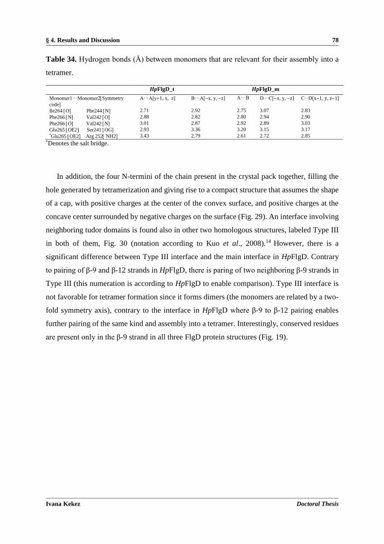
§ 4. Results and Discussion 78
Ivana Kekez Doctoral Thesis
Table 34. Hydrogen bonds (Å) between monomers that are relevant for their assembly into a
tetramer.
HpFlgD_t HpFlgD_m
Monomer1···Monomer2Symmetry
code
A···A[y1, x, z] B···A[x, y, z] A···B D···C[x, y, z] C∙∙∙D[x1, y, z1]
Ile264 O Phe244 N 2.71 2.92 2.75 3.07 2.83
Phe266 N Val242 O 2.88 2.82 2.80 2.94 2.90
Phe266 O Val242 N 3.01 2.87 2.92 2.89 3.03
Glu265 OE2 Ser241 OG 2.93 3.36 3.20 3.15 3.17 *Glu265 OE2 Arg 252 NH2 3.43 2.79 2.61 2.72 2.85
*Denotes the salt bridge.
In addition, the four N-termini of the chain present in the crystal pack together, filling the
hole generated by tetramerization and giving rise to a compact structure that assumes the shape
of a cap, with positive charges at the center of the convex surface, and positive charges at the
concave center surrounded by negative charges on the surface (Fig. 29). An interface involving
neighboring tudor domains is found also in other two homologous structures, labeled Type III
in both of them, Fig. 30 (notation according to Kuo et al., 2008).14 However, there is a
significant difference between Type III interface and the main interface in HpFlgD. Contrary
to pairing of β-9 and β-12 strands in HpFlgD, there is paring of two neighboring β-9 strands in
Type III (this numeration is according to HpFlgD to enable comparison). Type III interface is
not favorable for tetramer formation since it forms dimers (the monomers are related by a two-
fold symmetry axis), contrary to the interface in HpFlgD where β-9 to β-12 pairing enables
further pairing of the same kind and assembly into a tetramer. Interestingly, conserved residues
are present only in the β-9 strand in all three FlgD protein structures (Fig. 19).

§ 4. Results and Discussion 79
Ivana Kekez Doctoral Thesis
Figure 29. Qualitative electrostatic surface (represented as kb T e1) of the tetramer, seen along
the convex (on the left) and concave (on the right) surface. This figure was prepared with
software PyMol.134
Figure 30. Type of interface between the tudor domains present in the crystal structure of
HpFlgD (a) and XcFlgD (b). This figure was produced using CCP4MG.47

§ 4. Results and Discussion 80
Ivana Kekez Doctoral Thesis
Due to the shorter length of the β-12 strand in XcFlgD and PaFlgD than in HpFlgD a much
smaller interface of the type β-9 to β-12 could only be formed, possibly not favourable for
tetramer formation. Except for the main interface which is involved in the tetramer formation
in HpFlgD the other three interfaces have smaller interface areas (Fig. 31, Table 35). None of
these interfaces correspond to those in XcFlgD and PaFlgD, where interfaces of the Type I, II
and III (notation according to (Kuo et al., 2008)14 are present.
Figure 31. Different types of interfaces found between the molecules in the monoclinic crystal
structure of HpFlgD (a, b, c) and in the tetragonal crystal structure of HpFlgD (d, e, f). This
figure was produced using CCP4MG.47

§ 4. Results and Discussion 81
Ivana Kekez Doctoral Thesis
Table 35. Different types of interfaces between the molecules in the crystal structure of
HpFlgD. NHB and NSB refer to the number of hydrogen bonds and salt bridges, respectively. The
interfaces labelled af with a superscript* are shown in Fig. 31, while the interface labelled as
t* refers to the interface responsible for the tetramerization (as shown in Figs. 28b and 30).
Crystal
system
Interface
type Monomer1···Monomer2Symmetry
code
Interface area / Å2 NHB NSB
Monoclinic
t* B···A[x, y, z] 521.9 7 3
t D···C[x, y, z] 492.5 6 2
t B···A[x, y, z] 492.1 7 2
t C···D[x1, y, z1] 494.8 8 3
a* C···B[x, y, z] 302.3 b* D···B[x+1, y, z] 297.3 1 -
c* D···D[x, y1, z] 280.1 1 5
c A···A[x, y1, z] 268.2 1 5
c B···B[x, y1, z] 216.4 3 5
c C···C[x, y1, z] 194.3 3 5
Tetragonal
t A···A[y+1, x, z] 478.8 12 2
d* A···A[x, y+1, z] 299.9 4 4
e* A···A[x,y+1, z] 201.5 2 f* A···A[y1/2, x+1/2, z + 1/2] 119.2 4 4
4.1.7. Comparison with other structures
Identity of HpFlgD with PaFlgD and XcFlgD is 24 % and 20 %, respectively (Fig. 19). Despite
this low sequence identity the three hook scaffolding components share common folds of each
domain. However, the overall structure of each protein shows significant differences. In fact,
Fn-III domain superimposes quite well with that of the orthologue FlgD from the other species
(r.m.s.d.s are 2.17 Å for 93 residues in XcFlgD and 1.73 Å for 91 residues in PaFlgD). On the
contrary, by superimposing HpFlgD_t and PaFlgD the larger Fn-III domains superimpose well,
however it can be observed that the tudor domain in HpFlgD has a totally different orientation
with respect to the tudor domain in PaFlgD (Fig. 32), and also with respect to this domain in
XcFlgD which has the same orientation as in PaFlgD. The tudor domain by itself can be
superposed quite well in all three homologues (Figure 33). Different relative orientation of
domains in HpFlgD than in others could imply the presence of two distinct orientation modes
with a switch between them happening as a part of the mechanism during its activation.

§ 4. Results and Discussion 82
Ivana Kekez Doctoral Thesis
Figure 32. Stereoview of the superposed Cα chain trace of PaFlgD_A (red, PDB entry 3OSV;12
on HpFlgD_t (gold). The r.m.s.d. for the superposition of 91 aligned C atoms of PaFlgD_A
on HpFlgD_t is 1.73 Å. This figure was produced using CCP4MG.47
Figure 33. Superposition of the tudor domain in HpFlgD_t (dark blue), XcFlgD (green) and
PaFlgD_A (red). The r.m.s.d.s for the superposition of 47 aligned C atoms of XcFlgD on
HpFlgD_t and 43 aligned C atoms of PaFlgD_A on HpFlgD_t are 2.09 Å and 1.55 Å,
respectively. This figure was produced using CCP4MG.47

§ 4. Results and Discussion 83
Ivana Kekez Doctoral Thesis
FlgD is the hook cap protein, a necessary component in the formation of the filament of the
flagellum.141 To build the hook, FlgD must be assembled at the end of the rod structure,68 before
another flagellin, FlgE, is secreted and placed underneath it. The crystal structure of FlgE (or
at least of a proteolytic fragment) from Salmonella enterica subsp. enterica serovar
Typhimurium has been determined (PDB ID 1WLG),62 and a low resolution image of the
organization of the hook was obtained by electron microscopy: 120 copies of the flagellin are
arranged in a helical structure, with about 12 molecules per turn. Interestingly, HpFlgD shares
an architecture that roughly resembles that of HpFlgE, the protein that constitutes the hook of
the flagellum. Superposition of HpFlgD to a model of HpFlgE built by homology by the
software Phyre2 is shown in Fig. 34. The r.m.s.d for the superposition of Cα atoms is 3.44 Å for
68 aligned residues, despite a sequence identity of less than 14 %. The most relevant aspect is
that both HpFlgD and HpFlgE are composed of two domains, each of them with a similar
architecture, despite both domains in HpFlgE being larger than the corresponding domains in
HpFlgD. It can be speculated that this similarity could play some role in the mechanism of the
hook assembly
Figure 34. Stereoview of the superposed Cα chain trace of HpFlgD_t (gold) and the modeled
HpFlgE (purple). The r.m.s.d. for the superposition of 68 aligned C atoms of modeled HpFlgE
on HpFlgD_t is 3.44 Å. This figure was produced using CCP4MG.47

§ 4. Results and Discussion 84
Ivana Kekez Doctoral Thesis
4.1.8. Proposed physiological role of truncated HpFlgD
Proteome analysis identified HpFlgD in the H. pylori culture supernatant within the pool of the
most represented secreted components.142,143 However, its function as well as processing has
not been characterized so far. The N-terminal region of HpFlgD is susceptible to proteolytic
cleavage, as confirmed by crystallized fragments (Fig. 23a). Analogous proteolytic events gave
rise to crystals composed of only the C-terminal fragment in all FlgD structures characterized
till now.12,14
The N-terminal region (pfam03963), despite the fact that no typical features of a canonical
export signal are present, is well conserved among the flagellar hook capping proteins family
(cl15992). The HpFlgD N-terminal sequence is predicted to include an initial flexible segment
(corresponding to residues 1 to 27) followed by two predicted helical motifs and a further
disordered segment (93-116; Psi-pred server results). The presence of some helical content in
HpFlgD is confirmed by the CD data of the entire protein (Fig. 22 and Table 33). The N-
terminal region is not only flexible and more prone to degradation, but has been demonstrated
to be essential for the secretion in both Salmonella enterica and Escherichia coli variants.
Indeed, the export signal of EcFlgD has been demonstrated to be comprised of the first N-
terminal 71 amino acids,70 while the first 100 amino acids of SeFlgD were proven to guarantee
a secretion level equivalent to the full-length protein once expressed in fusion with a reporter
gene.144 Such secretion signals are surprisingly longer than the canonical ones and do not show
the typical Sec machinery consensus features.
Another intriguing aspect of HpFlgD is that the residues at the C-terminus, after Lys272,
are not visible in both of the crystal structures determined in this work. Indeed, all the sequences
of FlgD homologues in any Helicobacter species deposited in the UniProt data bank present a
peculiar and disordered fragment of variable length at the C-terminus. It is composed of repeats
of five amino acid motifs according to the consensus sequence XQK(X)2 where the first position
X is mainly present as D and E (rarely as N). The two last positions (X)2 can vary according to
PI and PL or less often as LS and PQ. The conserved and repeated motifs vary only in terms of
the number of repeats, while they are all followed by the sequence TPPKETA that represents,
in the majority of cases, the very C-terminal fragment. Such a peculiar sequence of repeats is
not present in the FlgD homologues of other bacteria and cannot be easily reconciled to a well-
known motif or signal. Nevertheless, the C-terminus of FliC protein, the flagellin subunit

§ 4. Results and Discussion 85
Ivana Kekez Doctoral Thesis
defining the filament, is bound in an extended form by the FliS chaperone,145 preventing its
premature polymerization. Such an interaction is a common strategy for flagellar export
chaperones, to bind their substrates and control flagella biosynthesis. While no data has been
acquired until now on the physiological role played by HpFlgD C-terminus, such a peculiar and
highly conserved sequence suggests it could represent an interaction or recognition motif. A
BLAST search throughout a non-redundant database of deposited sequences has shown that
similar patterns can be identified in other bacterial or plasmodia proteins with quite different
functions and in different positions in the primary structure, supporting the idea that it could
represent a common mode of recognition of interaction partners, rather than a signal sequence.
A relevant difference between HpFlgD and the other two FlgD homologues, whose
structures are known, is represented by the quaternary organization. The entire HpFlgD is a
dimer in solution, but it becomes a tetramer after cleavage of the N-terminus. This difference
in the oligomerization state is likely due to the long flexible N-terminal portion, which is present
in the purified protein, but is lost before crystallization. The latter apparently prevents
tetramerization, since the N-termini in the form found in the crystal are in the center of the
tetramer, in close proximity of the four-fold axis (Figs. 28a and 29). It is hard to say if the
aggregation state of HpFlgD has some physiological meaning or if it represents an artifact of
crystallization. However, previous considerations suggest that this cleavage has a functional
significance, since the protein has to be exported into the outside space in order to interact with
the hook, before the flagellum is fully formed. The flexible N-terminal part is also absent in the
crystal structures of both PaFlgD and XcFlgD, strongly supporting the hypothesis that this part
plays a role in the physiological mechanism of flagellar assembly. The N-terminal fragment
could act as a signal for the export and the crystallized protein could correspond to the mature
protein.

§ 4. Results and Discussion 86
Ivana Kekez Doctoral Thesis
4.2. CrdA protein from H. pylori
4.2.1. Cloning, expression and purification of StrepCrdA, CrdAHis and GSTCrdA
Cloning without the first 22 amino acids was performed since the CrdA sequence analysis by
the LipoP bionformatic tool (http://www.cbs.dtu.dk/services/LipoP/) predicted a cleavage site
for an export signal and a lipidation site (aa 2223), Fig. 35. To determine whether the CrdA
sequence possesses conserved regions for the binding of copper ions, the sequence was
compared with the sequences of the CopC and PcoC. These proteins belong to the CopC family
and are periplasmic proteins involved in process that results in a change in state or activity of a
cell or an organism as a result of a copper ion stimulus. The alignment showed (Fig 35) that
CrdA sequence contains conserved amino acids, mainly methionines, that could be involved in
the copper binding.
Figure 35. Sequence alignment of CrdA (HP, O25884), CopC (PS, P12376) and PcoC (EC,
Q47454) using CLUSTAL OMEGA.130 The alignment figure was generated using ESPript
3.0.136 The numbering system is based on the CrdA sequence. Residues conserved in all three
sequences are labelled in white and highlighted in red, while similar residues are labelled in

§ 4. Results and Discussion 87
Ivana Kekez Doctoral Thesis
black and highlighted in yellow. Possible ligands for the Cu(II) and Cu(I) sites in CopC and
PcoC are marked with magenta and turquoise arrows, respectively.78
In the case of the CrdAHis-tag and Strep-tagCrdA protein, high expression levels were
obtained but with a poor solubility (Fig. 36a), so refolding methods were implemented with
success. The insoluble portion of CrdA was denatured and then renatured by slow dilution. The
best renaturation outcome was obtained using the buffer R6 (Fig. 38b) since after the affinity
chromatography purification the flowthrough of refolded CrdA in buffer R6 contained the
lowest amount of protein (Fig. 36c).
Figure 36. SDSPAGE (a) StrepCrdA, Lane S soluble fraction, Lane P – insoluble fraction;
(b) Lanes R1 – R7 - refolded CrdAHis in different renaturation buffers (R1 – R7, see Table 28
in section 3.7); (c) Lanes S1 – S7 – flow-through of CrdAHis in different renaturation buffers
after the Ni-NTA affinity chromatography.
The best condition for expressing the GSTCrdA protein was obtained when E. coli cells were
grown at 16 C (Fig. 37a). As can be seen in Fig. 37b, SEC (Superdex 75 10/300 column) of
cleaved GSTCrdA was successfully applied for the separation of the CrdA protein from the

§ 4. Results and Discussion 88
Ivana Kekez Doctoral Thesis
GST-tag protein. This was possible since the GST protein is present as a dimer in the solution
while CrdA as a monomer (see section 4.2.2.).
Figure 37. SDS-PAGE (a) GSTCrdA (Molecular mass of the monomer 38 kDa); (b) cleaved
GSTCrdA purified by SEC with lanes 1 – 4 and 5 – 6 corresponding to the GST (Molecular
mass of the monomer 26 kDa) and CrdA (Molecular mass of the monomer 12.4 kDa)
fractions, respectively.
The proper folding of renaturated CrdA was confirmed by CD and 1D-NMR spectroscopy.
The far-UV circular dichroism spectrum (Fig. 38a) shows a minimum at 214 nm and a peak of
positive ellipticity for the values below 197 nm that are characteristic of a properly folded
protein rich in sheets (Table 36).108 The 1D NMR spectrum of refolded CrdA indicates
presence of secondary structure elements (Fig. 38b) since the peaks in the spectrum are sharp
and narrow and cover a large range of chemical shifts. and the spectrum also contains peaks in
the negative ppm range. In an unfolded protein, a given proton does not have one distinct
environment, so the peak is an average of all the different environments in which that proton is
found. Also, the environments of different protons aren’t as distinct in an unfolded protein, so
the peaks don’t cover a large range of chemical shifts. In a folded protein, most protons are
more constrained in a distinct conformation, and the environment of each proton is very
dependent on its position in the tertiary structure of the protein.

§ 4. Results and Discussion 89
Ivana Kekez Doctoral Thesis
Figure 38. Refolded CrdA (a) CD spectrum of the StrepCrdA in the far UV region (195260
nm) presented as ellipticity in milidegrees; (b) 1D NMR spectrum of the CrdAHis.
Table 36. CD data of the StrepCrdA analysed by the secondary structure analysis software,
CDNN.110 Deconvoluted results are shown as contributions of the various components to the
protein secondary structure.
SECONDARY STRUCTURE
ELEMENT
%
Helix 9.0
Antiparallel sheet 35.6
Parallel sheet 5.2
turn 19.5
Random coil 30.7
4.2.2. Size of CrdA in solution
Incubation of CrdA with copper(II) ions or EDTA, followed by SEC, was carried out.
Recombinant His tagged CrdA treated with copper was eluted as a single peak at a volume of
18.5 mL, while in case of CrdA treated with EDTA two different species were eluted: one at
14 mL and the other one at 18.5 mL (Fig. 39). The calculated molecular mass from the SEC
experiments of the CrdA species in the presence of copper ions was about 14.8 kDa that was in

§ 4. Results and Discussion 90
Ivana Kekez Doctoral Thesis
close agreement with the predicted molecular mass that is 12.4 kDa. The observed molecular
mass indicates that the CrdA protein treated with copper is present as a monomeric species in
the solution, which is consistent with the behaviour of the copper binding PsCopC protein in
the solution.78 In case of EDTA treatment, the CrdA protein eluted in two different oligomeric
states - monomeric (14.8 kDa) and dimeric (31 kDa). Treatment with a metal chelating agent
(EDTA) changed the behavior of CrdA, indicating that an equilibrium between the two different
oligomeric species is present.
Figure 39. Analytical gel filtration chromatogram of CrdA treated with Cu(II)+ ions (blue
curve; Molecular mass of the monomer 12.4 kDa) which elutes as a single peak at the volume
of 18.5 mL corresponding to the molecular mass of 14.8 kDa. CrdA chelated with EDTA (red
curve) is composed of two different species that elute at 14 mL and 18.5 mL and correspond to
the molecular mass of 31 kDa and 14.8 kDa, respectively.
The monodispersity of the CrdA protein with and without addition of detergent was
evaluated at a protein concentration of 2 mg mL1 and at 298 K by dynamic light scattering. As
it can be seen in Fig. 40 the dispersity of the pure CrdA sample and of the CrdA sample treated
with nOG detergent (c = 3.8 mmol L1) differs significantly. The solution of the CrdA protein
without detergent was heterogeneous and contained species in different oligomeric states and
with a high level of aggregation. The polydispersity index for the CrdA sample and CrdA plus
nOG was 0.941 and 0.335, respectively, implying that only the sample treated with detergent
could be used for the crystallization purpose. The influence of other detergents like LDAO (c

§ 4. Results and Discussion 91
Ivana Kekez Doctoral Thesis
= 0.4 mmol L1) and DDM (c = 0.034 mmol L1) on the dispersity of CrdA was also tested
(results not shown), however they only slightly affected the dispersity profile of CrdA.
Figure 40. The volume distributions as a function of particle diameter: (a) CrdA and (b) CrdA
treated with nOG detergent, c = 3.8 mmol L1.
4.2.3. Affinity of CrdA towards copper(II) ions
The copper binding properties of the CrdA protein were investigated by UV-VIS spectroscopy
in the wavelength range 200800 nm. Fig. 41a shows presence of a broad absorption band at
739 nm but unfortunately in these experiments saturation was not reached, Fig. 41b. The
dissociation constant (Kd) for metal binding to the protein was determined from the titration to
be 1.076 103 mol L1. Such a high value of the dissociation constant is characteristic of
copper trafficking proteins rather than metalloproteins with an enzymatic function.146 Low
affinity binding sites allow metal transfer to the recipient protein or enzyme while the high
affinity sites prevent loss of the metal during redox cycling. Another possible explanation of
such a high Kd is that the observed binding of copper(II) ions is unspecific, and maybe the
preferable ligand for the CrdA protein is the Cu(I) ion. Based on the known crystal structures
and the predictions for the Cu(II) and Cu(I) sites (Fig. 35) of similar copper binding proteins,
CrdA could more likely accommodate Cu than Cu2 since its sequence is rich in methionines
and lacks histidines and cysteines.78,79

§ 4. Results and Discussion 92
Ivana Kekez Doctoral Thesis
Figure 41. (a) Change in the UV-VIS spectrum upon titration of CrdA (c = 152 mmol L1) in
the phosphate buffer (30 mmol L1 Na2HPO4 pH 7, 50 mmol L1 NaCl) with an increasing
concentration of CuSO4. (b) Plot of A739 versus c(Cu2).
4.2.4. Crystallization of CrdA
Purified samples of CrdA concentrated to 9 mg mL1 in complex with Cu(II) ions and
supplemented with nOG were submitted to sparse matrix crystallization trials, applying the
sitting drop vapor diffusion method, using an automated crystallization platform (Oryx8 robot).
Each of the 686 independent crystallization conditions (Structure screen I & II, PEG’S II
SUITE, PACT SUITE, JCSG CORE I-IV) were screened using the sitting drop setup on the
MRC 2-well plates. Prior to crystallization, the freshly purified protein was incubated with a 1

§ 4. Results and Discussion 93
Ivana Kekez Doctoral Thesis
mmol L1 CuSO4 solution and 3.8 mmol L1 nOG solution. In case of CrdAHis-tag
microcrystals were observed (Fig. 42a, b) in the screening solution 32 of the PEG'S II SUITE
(Qiagen) (0.1 mol L1 TRIS pH 8.5, 16 % PEG 4000) at 293 K. Diffraction data were measured
at beamline ID23-1 of the ESRF synchrotron facility (Grenoble, France) but the small size of
crystals limited the diffraction experiment (diffraction until the maximum resolution of 20 Å
was observed). Other crystallization trials yielded drops containing aggregated forms or the
phase-separation could be observed, so further optimization should be carried out.
Figure 42. (a, b) Microcrystals of CrdA incubated with Cu(II) ions. Picture (b) was captured
under the microscope using a fluorescence excitation filter (CWL / BW = 450 / 50 nm).
4.3. HP1026 protein from H. pylori
4.3.1. Expression, purification and size of HP1026 in solution
His_HP1026 protein was overproduced in E. coli cells by growing the cell culture at 28 C and
successfully purified by the Ni-NTA affinity chromatography followed by SEC (Superdex 200
10/300), Fig. 43a, b. The resulting His_HP1026 from the SEC was 95% pure as clarified by
SDS-PAGE gel (Fig. 43b) and was concentrated to more than 40 mg mL1.
Fig. 43b shows that recombinant HP1026 was eluted in a single peak at the volume of 11.2
mL which corresponded to the size of 96.9 kDa. Since the size of the HP1026 monomer is 43.8

§ 4. Results and Discussion 94
Ivana Kekez Doctoral Thesis
kDa the calculated molecular mass from the analytical size-exclusion chromatography analysis
correlates with a dimeric species in the solution. Even though most of proteins that belong to
the AAA+ superfamily are present as hexamers in solution, some examples of lower
oligomerization arrangement like tetrameric (EcMgsA),89 and dimeric (PfRFC)147 are known
from the literature. Moreover, it is known for the EcMgsA protein and other similar clamp
loader proteins that the C-terminal domain is responsible for the protein oligomerization.
Figure 43. (a) SDS-PAGE of the HP1026 fractions eluted after the affinity chromatography;
(b) Overlayed analytical gel filtration chromatograms of Apo_HP1026 (blue curve; molecular
mass of the monomer 43.8 kDa) which elutes as a single peak at the volume of 11.2 mL
corresponding to the molecular mass of 96.9 kDa and of HP1026 incubated with ATP--S (red
curve) with a major peak eluted at 11.2 mL. SDS-PAGE of a pure and concentrated fraction of
HP1026 after the SEC.
4.3.2. Activity of HP1026
The HP1026 sequence contains a conserved AAA+ motif (aa 36295, Fig. 44) which shows
identity 94.2% with 250 H. pylori proteins classified as belonging to the AAA superfamily.
The highest sequence similarity is found with the EcMgsA protein that contains a nucleotide
binding site located at the N-terminus which is strongly conserved also in the sequence of

§ 4. Results and Discussion 95
Ivana Kekez Doctoral Thesis
HP1026 (aa 4451, Fig. 44). To test whether HP1026 possesses an ATPase activity, ATP
hydrolysis assay was carried. Performed experiments showed that HP1026 contains an ATPase
activity (Fig. 45) within kinetic parameters as follows: Km= 344 mol L1, Vmax= 0.6019 mol
L1 s1, kcat= 0.02 s1. HP1026 hydrolyzed ATP at a lower level (0.02 s1) then it is measured
for the EcMgsA protein in the absence of DNA (0.056 s1).89 However, in the case of EcMgsA,
activity tests performed with addition of DNA significantly stimulated the ATPase activity,89
so in our case future studies will be required to determine whether HP1026 activity is also DNA
dependent.
Figure 44. Sequence alignment of HP1026 (HP, O25670) and MgsA (EC, P0A731) using
CLUSTAL OMEGA.130 The alignment figure was generated using ESPript 3.0.136 The
numbering system is based on the HP1026 sequence. Residues conserved in both sequences are
labelled in white and highlighted in red, while similar residues are labelled in red.

§ 4. Results and Discussion 96
Ivana Kekez Doctoral Thesis
Figure 45. ATPase activity of HP1026. Calculated parameters by applying the Michaelis-
Menten model were Km= 344 mol L1, Vmax= 0.6019 mol L1 s1 and kcat = 0.02 s1.
4.3.3. Stability of HP1026
CD measurements showed that both Apo_HP1026 and HP1026 protein incubated with ATP--
S demonstrate the typical profile of a protein rich in -helices having negative bands at 222 nm
and 208 nm and a positive band around 190 nm (Fig. 46, Table 37).108 This result together with
SEC profiles (Fig. 43b) indicates that addition of ATP--S did not alter the secondary structure
profile nor the oligomerization profile of the HP1026 protein.

§ 4. Results and Discussion 97
Ivana Kekez Doctoral Thesis
Figure 46. CD spectrum of the HP1026 in the far UV region (190260 nm) presented as
ellipticity in milidegrees. Apo_HP1026, HP1026 incubated with ATP--S in a molar ratio 1:1
and HP1026 incubated with ATP--S in a molar ratio 1:2 are presented as blue, red and green
curves, respectively.
Table 37. CD data of the HP1026 analyzed by the secondary structure analysis software,
CDNN. Deconvoluted results are shown as contributions of the various components to the
protein secondary structure.
SECONDARY STRUCTURE
ELEMENT
%
Helix 44.5
Antiparallel sheet 1.1
Parallel sheet 8.8
turn 14.9
Random coil 30.7
Thermal stability of HP1026 after addition of different additives, as well as without a His-
tag, was also investigated while the buffer condition was kept constant. Tm value for the
HP1026:ADP, HP1026:ATP--S and HP1026:AMP complexes was at 49.75 (0.29) C, 48.09
-40
-30
-20
-10
0
10
20
30
40
50
60
70
190 200 210 220 230 240 250 260
/m
deg
/nm

§ 4. Results and Discussion 98
Ivana Kekez Doctoral Thesis
(0.03) C and 44.85 (0.09) C, respectively (Table 38). According to these values addition
of AMP didn't change the Tm value of the HP1026 protein while the removal of His-tag slightly
increased Tm by +1.38 C. From Fig. 47 and Table 38 it is evident that ATP--S and ADP
strongly affected the protein stability by shifting the protein-complex denaturation to higher Tm
values by +3.52 C and +5.18 C, respectively. The data after the ATP hydrolysis assay were
another confirmation that HP1026 belongs to the class of ATPase proteins. The results of this
thermofluor assay were also applied in the crystallization experiments.
Figure 47. Effect of different additives and His-tag on the HP1026 thermal stability.
Table 38. Melting temperatures of ApoHP1026 without His-tag, ApoHP1026 and HP1026 after
addition of different additives.
PROTEIN HP1026noHis HP1026 HP1026 HP1026 HP1026
ADDITIVE ATP--S ADP AMP
Tm / C 45.95 (0.03) 44.57 (0.57) 48.09 (0.03) 49.75 (0.29) 44.85 (0.09)
4.3.4. Crystallization of HP1026
Good quality of the sample for the crystallization studies was confirmed by the DLS experiment
(PDI < 0.12, Figure 50) for the HP1026 protein treated with 10 % (v/v) glycerol.

§ 4. Results and Discussion 99
Ivana Kekez Doctoral Thesis
Figure 48. The volume distributions as a function of particle diameter of HP1026 after addition
of 10 % (v/v) glycerol.
Since the HP1026 protein was very soluble ( 40 mg mL1) it was hard to estimate which
concentration was appropriate for the crystallization trials so the solubility profile of HP1026
was explored. Using the solubility tool kit an optimal concentration of 15 mg mL1 was
estimated to be suitable for the crystallization experiments. Screening of different
crystallization conditions was performed by using commercial screening solutions, Structure
screen I & II, PEG’S II SUITE, PACT SUITE and PGA Screen, by the sitting drop vapour
diffusion method using an automated crystallization platform (Oryx8 robot). Crystallization
drops were prepared by mixing the protein and precipitant solutions in a ratio 1:1, and
equilibrated against 75 L of the mother liquor in the reservoir. The initial hexagonal shaped
crystals were grown in a week at 20 C from different screening solutions of the PACT SUITE
(Table 39). Crystals were very sensitive and had tendency to dissolve in time (Fig. 49c).
Crystals were tested at the PXIII beamline of the Swiss Synchrotron Light Source (SLS,
Villigen, Switzerland) with and without cryoprotectant solutions. Cryoprotectant solutions were
prepared by mixing the precipitant solution and ethylene glycol / glycerol / oil in different
percentages (v/v, 20 %, 25 %, 30 %). Crystals without cryoprotectant contained ice rings and
didn’t diffract because they were destroyed during the freezing step in liquid nitrogen. Crystals
that were frozen in the cryoprotectant solution were without ice rings but, unfortunately,
diffracted only to 15 Å. During the diffraction experiment annealing was also tried but it didn’t
help in ordering the molecules inside the crystal. A number of different ways were explored to

§ 4. Results and Discussion 100
Ivana Kekez Doctoral Thesis
improve the diffraction quality. Microseeding resulted in too many small crystals while
dehydration of the crystal, carried out by transferring the crystal from the crystallization drop
into a drop of the dehydrating solution (containing 30 % of PEG 3350) which was then
equilibrated against a 75 L volume of dehydrating solution, didn’t improve crystal quality.
Crystallization at lower temperature was also tested but it resulted in drops containing only
precipitation.
Table 39. The PACT SUITE composition table of the conditions that gave crystals of HP1026.
Condition number Composition
43 0.2 mol L1 NaCl, 0.1 mol L1 TRIS pH 8.0, 20 % PEG 6000
78 0.2 mol L1 NaOCOOH, 0.1 mol L1 BIS TRIS PROPANE pH 7.5, 20 % PEG 3350
86 0.2 mol L1 NaBr, 0.1 mol L1 BIS TRIS PROPANE pH 8.5, 20 % PEG 3350
88 0.2 mol L1 KSCN, 0.1 mol L1 BIS TRIS PROPANE pH 8.5, 20 % PEG 3350
Taking into account all of the previous results together with the results of the protein
stability investigation, the crystallization trials were carried out for HP1026 without His-tag
and for HP1026 in complex with ATP--S or ADP. ATP--S was used instead of ATP since it
is a non-hydrolyzable ATP analogue. The HP1026 protein solution, at the final concentration
of 15 mg mL1, was incubated with ADP or ATP--S and MgCl2 at the final concentrations of
2 mmol L1 and 5 mmol L1, respectively, for 2 h prior to the crystallization experiments.
HP1026 lacking the His-tag failed to gave crystals while HP1026:ATP--S and HP1026:ADP
gave crystals. Optimization of HP1026:ATP--S and HP1026:ADP crystals was performed by
varying the protein and precipitant ratio and by screening the effect of different additives. The
best quality crystals were obtained for HP1026:ADP complex in a drop containing 2.5 L
protein and 2 or 1.5 L precipitant solution (0.2 mol L1 NaBr, 0.1 mol L1 Bis-tris propane pH
8.5, 20 % PEG 3350, 0.015 mmol L1 CYMAL-7) at 293(2) K (Fig. 49a,b).

§ 4. Results and Discussion 101
Ivana Kekez Doctoral Thesis
Figure 49. Hexagonal crystals of HP1026-ADP (a, b) after 5 days, (c) after 3 weeks.
Since the crystals were multiple crystals they were separated before freezing in liquid nitrogen.
Crystals were also tested at the PXIII beamline (SLS, Villigen, Switzerland) and diffracted until
7 Å which is still not enough for solving the phase problem, so further improvement should be
done.

§ 4. Results and Discussion 102
Ivana Kekez Doctoral Thesis

§ 5. Conclusions 103
Ivana Kekez Doctoral Thesis
§ 5. CONCLUSIONS
Crystal structures of truncated forms of Helicobacter pylori FlgD from two different strains in
two space groups, I422 and P2, have been solved, at 2.2 Å and 2.8 Å resolution, respectively.
Analogously to Pseudomonas aeruginosa and Xanthomonas campestris FlgD proteins,
crystallization experiments set up for the full-length protein resulted in crystals of a truncated
form, lacking both N- and C-terminus ends. The crystal structures of the truncated FlgD show
that the monomer is composed of a tudor and a fibronectin type III domain. The spatial
orientation of the two domains in HpFlgD differs from that of the homologous FlgD family
members, P. aeruginosa and X. campestris. Even though the full-length HpFlgD contains a
long N-terminal region which does not show the typical features of a canonical export signal,
results within this thesis, together with the bioinformatic studies, and the previously reported
studies on the essential role of the N-terminal region for secretion in both S. enterica and E.
coli, support the idea that the N-terminal part of the protein acts like an exporting signal.
Another interesting aspect is that the C-terminal part of HpFlgD could represent a recognition
motif for its partner. All these findings, together with the crystal structures of HpFlgD and the
crystal structures of FlgD from other bacterial homologues, imply that functioning of FlgD
requires proteolysis of the N-terminal pro-peptide. Finally, the structural differences of HpFlgD
with respect to the homologous FlgD family members present in other bacteria suggest that
significant differences exist in the molecular organization of the flagella in different bacterial
species.
Recombinant CrdA protein (His/Strep/GST tagged) was successfully overexpressed. In case
of CrdA_His and Strep_CrdA proteins high expression levels were obtained but the protein had
poor solubility, so denaturation and refolding methods were implemented with success. Circular
dichroism and 1D NMR confirmed the presence of secondary structure elements with a major
contribution of sheets (60.3 %). Dynamic light scattering showed that nOG detergent prevents
protein aggregation and reduces sample polydispersity. SEC of Cu:CrdA suggests that addition
of copper(II) ions stabilize formation of uniform species shifting the equilibrium toward
monomeric species. UV-VIS titration with copper(II) sulphate resulted with low Cu(II) affinity
(Kd = 1.076 103 mol L1), a property of copper trafficking proteins, but it also implies that

§ 5. Conclusions 104
Ivana Kekez Doctoral Thesis
since the CrdA protein is rich in methionines the affinity of Cu(I) should be tested.
Crystallization trials gave only microcrystals that were not suitable for diffraction experiment
so further optimization is needed.
HP1026 was overexpressed, purified to homogeneity and biophysically characterized. Gel
filtration profile indicated the presence of dimeric species in the solution for both Apo- and
ATP--S treated HP1026, so no oligomerization step was observed after the addition of ATP-
-S. CD analysis showed that -helices are present as major secondary structure elements (44.5
%). ATP hydrolysis assay confirmed the ATPase activity of a putative helicase like HP1026
protein within kinetics parameters as follows: Km= 344 mol L1, Vmax= 0.6019 mol L1 s1,
kcat= 0.02 s1. These data together with the thermofluor assay for the first time demonstrate the
role of the HP1026 protein as an ATPase. Good quality of the sample for the crystallization
studies was confirmed by the DLS experiment (PDI < 0.12). Crystallization trials were carried
out for Apo_HP1026, ATP--S:HP1026 and ADP:HP1026. The hexagonal shaped crystals
were grown in a week at 20 C. Though not of good quality for diffraction analysis, these results
outline a starting point for further crystallization trials.

§ 6. List of abbreviations 105
Ivana Kekez Doctoral Thesis
§ 6. LIST OF ABBREVIATIONS
ADP – Adenosine 5′-diphosphate
AMP - Adenosine 5′-monophosphate
ATP--S - Adenosine 5′-[γ-thio]triphosphate
APS - Ammonium persulfate
CV- Column volume
DDM – n-Dodecyl--D-maltopyranoside
dNTP- Deoxynucleotides
DTT - 1,4-Dithio-D-threitol
EDTA - Ethylenediaminetetraacetic acid
HABA - 2-[4’-Hydroxy-benzeneazo] benzoic acid
HEPES - 4-(2-Hydroxyethyl)-1-piperazineethanesulfonic acid
IPTG - Isopropyl-β-D-1-thiogalactopyranoside
Km - Michaelis-Menten constant
LDAO – N, N-Dimethyldodecylamine N-oxide
MES - 2-(N-Morpholino)ethanesulfonic acid
MPD - 2-Methyl-2,4-pentandiol
nOG – n-Octyl--D-glucopyranoside
PEG - Poly(ethylene glycol)
PES – Polyethersulfone
PMSF - Phenylmethylsulfonyl fluoride
PTFE - Poly(tetrafluoroethylene)
SDS – Sodium dodecyl sulfate
SDS-PAGE - Sodium dodecyl sulfate polyacrylamide gel electrophoresis
TBS – Tris-buffered saline TEMED – Tetramethylethylenediamine
TRIS-base - Tris(hydroxymethyl)aminomethane
TRIS-HCl - Tris(hydroxymethyl)aminomethane hydrochloride
Vmax - maximum enzyme velocity

§ 6. List of abbreviations 106
Ivana Kekez Doctoral Thesis

§ 7. References 107
Ivana Kekez Doctoral Thesis
§ 7. REFERENCES
1. D. Rothenbacher and H. Brenner, Microbes. Infect. 5 (8) (2003) 693703.
2. J. F. Tomb, O. White, A. R. Kerlavage, R. A. Clayton, G. G. Sutton, R. D. Fleischmann,
K. A. Ketchum, H. P. Klenk, S. Gill, B. A.Dougherty, K. Nelson, J. Quackenbush, L.
Zhou, E. F. Kirkness, S. Peterson, B. Loftus, D. Richardson, R. Dodson, H. G. Khalak,
A. Glodek, K. McKenney, L. M. Fitzegerald, N. Lee, M. D. Adams, E. K. Hickey, D.
E. Berg, J. D. Gocayne, T. R. Utterback, J. D. Peterson, J. M. Kelley, M. D. Cotton, J.
M. Weidman, C. Fujii, C. Bowman, L. Watthey, E. Wallin, W.S. Hayes, M.
Borodovsky, P. D. Karp, H. O. Smith, C. M. Fraser, and J. C. Venter, Nature 388 (6642)
(1997) 539547.
3. R. A. Alm, L. S. Ling, D. T. Moir, B. L. King, E. D. Brown, P. C. Doig, D. R. Smith,
B. Noonan, B. C. Guild, B. L. deJonge, G. Carmel, P. J. Tummino, A. Caruso, M. Uria-
Nickelsen, D. M. Mills, C. Ives, R. Gibson, D. Merberg, S. D. Mills, Q. Jiang, D. E.
Taylor, G. F. Vovis, and T. J. Trust, Nature 397 (1999) 176180.
4. J. D. Oh, H. Kling-Backhed, M. Giannakis, J. Xu, R. S. Fulton, L. A. Fulton, H. S.
Cordum, C. Wang, G. Elliott, J. Edwards, E. R. Mardis, L. G. Engstrand, and J.I.
Gordon, Proc. Natl. Acad. Sci. U.S.A. 103 (2006) 999910004.
5. D. A. Baltrus, M. R. Amieva, A. Covacci, T. M. Lowe, D. S. Merrell, K. M. Ottemann,
M. Stein, N. R. Salama, and K. Guillemin, J. Bacteriol. 191 (1) (2009) 447448.
6. S. Suerbaum and C. Josenhans, Nat. Rev. Microbiol. 5 (2007) 441452.
7. M. J. Blaser, Br. Med. J. 316 (1998) 15071510.
8. R. M. Jr Peek and M. J. Blaser, Nature Rev. Cancer 2 (1) (2002) 28–37.
9. S. Suerbaum and P.N. Michetti, Engl. J. Med. 347 (2002) 11751186.
10. J. C. Rain, L. Selig, H. De Reuse, V. Battaglia, C. Reverdy, S. Simon, G. Lenzen, F.
Petel, J. Wojcik, V. Schachter, Y. Chemama, A. Labigne, and P.Legrain, Nature 409
(2001) 211215.
11. L. Cendron and G. Zanotti, Febs J. 278 (8) (2011) 12231231.
12. H. Zhou, M. Luo, X. Cai, J. Tang, S. Niu, W. Zhang, Y. Hu, Y. Yin, A. Huang, and D.
Wang, Proteins 79 (7) (2011) 23462351.

§ 7. References 108
Ivana Kekez Doctoral Thesis
13. O. A. Soutourina and P. N. Bertin, FEMS Microbiol. Rev. 27 (2003) 505523.
14. W. T. Kuo, K. H. Chin, W. T. Lo, A. H. Wang, and S.H. Chou, J. Mol. Biol. 381 (1)
(2008) 189199.
15. B. Waidner, K.Melchers, F. N. Stähler, M. Kist and S. Bereswill, J. Bacteriol. 187
(2005) 46834688.
16. B. Waidner, K.Melchers, I. Ivanov, H. Loferer, K. W. Bensch, M. Kist, and S.
Bereswill, J. Bacteriol. 184 (2002) 67006708.
17. D. Roncarati, A. Danielli, and V. Scarlato, J. Bacteriol. 193 (20) (2011) 56295636.
18. C. S. Goodwin, M. M. Mendall, and T. C. Northfield, Lancet. 349 (1997) 265269.
19. J. G. Kusters, A. H. van Vliet, and E.J. Kuipers, Clin. Microbiol. Rev. 19 (2006)
449490.
20. B. J. Marshall and J.R. Warren, Lancet. 1 (1984) 13111315.
21. A. Covacci, J. L. Telford, G. Del Giudice, J. Parsonnet, and R. Rappuoli. Science 284
(1999) 13281333.
22. D. Kersulyte, A. K. Mukhopadhyay , B. Velapatiño, W. Su, Z. Pan, C. Garcia, V.
Hernandez, Y. Valdez, R. S. Mistry, R. H. Gilman, Y. Yuan, H. Gao, T. Alarcón, M.
López-Brea, G. Balakrish Nair, A. Chowdhury, S. Datta, M. Shirai, T. Nakazawa, R
Ally, I. Segal, B. C. Wong, S. K. Lam, F. O. Olfat, T. Borén, L. Engstrand, O. Torres,
R. Schneider, J. E. Thomas, S. Czinn, and D. E. Berg, J. Bacteriol. 182 (11) (2000)
32103218.
23. H. Yoshiyama and T. Nakazawa, Microbes Infect. 2 (2000) 5560.
24. F. R. Blattner, G. Plunkett, C. A. Bloch, N. T. Perna, V. Burland, M. Riley, J. Collado-
Vides, J. D. Glasner, C. K. Rode, G. F. Mayhew, J. Gregor, N. W. Davis, H. A.
Kirkpatrick, M. A. Goeden, D. J. Rose, B. Mau, and Y. Shao, Science 277 (1997)
14531462.
25. C. T. Parker, S. Huynh, L. Gorski, K. K. Cooper, and W. G. Miller, Genome Announc.
3 (6) (2015) e0136515.
26. J. G. Kusters, A. H. M. van Vliet, and Ernst J. Kuipers, Clin. Microbiol. Rev. 19 (2006)
449490.
27. R. D. Leunk, P. T. Johnson, B. C. David, W. G. Kraft, and D. R. Morgan, J. Med.
Microbiol. 26 (1988) 93–99.
http://www.ncbi.nlm.nih.gov/pubmed/?term=Hernandez%20V%5BAuthor%5D&cauthor=true&cauthor_uid=10809702
http://www.ncbi.nlm.nih.gov/pubmed/?term=Chowdhury%20A%5BAuthor%5D&cauthor=true&cauthor_uid=10809702
http://www.ncbi.nlm.nih.gov/pubmed/?term=Engstrand%20L%5BAuthor%5D&cauthor=true&cauthor_uid=10809702

§ 7. References 109
Ivana Kekez Doctoral Thesis
28. N. S. Akopyants, S. W. Clifton, D. Kersulyte, J. E. Crabtree, B. E. Youree, C. A. Reece,
N. O. Bukanov, E. S. Drazek, B. A. Roe, and D. E. Berg, Mol. Microbiol. 28 (1998) 37–
53.
29. S. C. Lange Censini, Z. Xiang, J. E. Crabtree, P. Ghiara, M. Borodovsky, R. Rappuoli,
and A. Covacci, Proc. Natl. Acad. Sci. USA 93 (1996) 14648–14653.
30. M. Asahi, T. Azuma, S. Ito, Y. Ito, H. Suto, Y. Nagai, M. Tsubokawa, Y. Tohyama, S.
Maeda, M. Omata, T. Suzuki, and C. Sasakawa, J. Exp. Med. 191 (2000) 593–602.
31. P. J. Christie and J. P. Vogel, Trends Microbiol. 8 (2000) 354–360.
32. L. Terradot and G. Waksman, FEBS J. 278 (2011) 1213–1222.
33. S. Backert, N. Tegtmeyer, and M. Selbach, Helicobacter 15 (2010) 163–176.
34. M. Hatakeyama, Curr. Opin. Microbiol. 11 (2008) 30–37.
35. J. Viala, C. Chaput, I. G. Boneca, A. Cardona, S. E. Girardin, A. P. Moran, R. Athman,
S. Memet, M. R. Huerre, A. J. Coyle, P. S. DiStefano, P. J. Sansonetti , A. Labigne, J.
Bertin, D. J. Philpott, and R. L. Ferrero, Nat. Immunol. 5 (2004) 1166–1174.
36. T. L. Cover and M. J. Blaser, J. Biol. Chem. 267 (1992) 10570–10575.
37. J. C. Atherton, P. Cao, R. M. Peek, Jr., M. K. Tummuru, M. J. Blaser, and T. L. Cover,
J. Biol. Chem. 270 (1995) 17771–17777.
38. K. Ogura, S. Maeda, M. Nakao, T. Watanabe, M. Tada, T. Kyutoku, H. Yoshida, Y.
Shiratori, and M. Omata, J. Exp. Med. 192 (2000)1601–1610.
39. D. Falush, C. Kraft, N. S. Taylor, P. Correa, J. G. Fox, M. Achtman, and S. Suerbaum,
Proc. Natl. Acad. Sci. USA 98 (2001) 15056–15061.
40. E. J. Kuipers, D. A. Israel, J. G. Kusters, M. M. Gerrits, J. Weel, A. Van Der Ende, R.
W. van Der Hulst, H. P. Wirth, J. Hook-Nikanne, S. A. Thompson, and M. J. Blaser, J.
Infect. Dis. 181 (2000) 273–282.
41. R. A. Burne and Y. M. Chen, Microbes Infect. 2 (2000) 533–542.
42. H. L. Mobley, M. D. Island and R. P Hausinger, Microbiol. Rev. 59 (1995) 451–480.
43. S. H. Phadnis, M. H. Parlow, M. Levy, D. Ilver, C. M. Caulkins, J. B. Connors, and B.
E. Dunn, Infect. Immun. 64 (1996) 905–912.
44. D. R. Scott, D. Weeks, C. Hong, S. Postius, K. Melchers, and G. Sachs,
Gastroenterology 114 (1998) 58–70.
45. D. L. Weeks, S. Eskandari, D. R. Scott, and G. Sachs, Science 287 (2000) 482–485.

§ 7. References 110
Ivana Kekez Doctoral Thesis
46. N. C. Ha, S. T. Oh, J. Y. Sung, K. A. Cha, Hyung Lee, M., and B. H. , Nat. Struct. Biol.
8 (2001) 505509.
47. S. McNicholas, E. Potterton, K. S. Wilson, and M. E. M. Noble, Acta Crystallogr. D
Biol. Crystallogr. 67 (2011) 386–394.
48. T. Boren, P. Falk, K. A. Roth, G. Larson, and S. Normark, Science 262 (1993) 1892–
1895.
49. M. Gerhard, N. Lehn, N. Neumayer, T. Boren, R. Rad, W. Schepp, S. Miehlke, M.
Classen, and C. Prinz, Proc. Natl. Acad. Sci. USA 96 (1999) 2778–12783.
50. H. Ota, J. Nakayama, M. Momose, M. Hayama, T. Akamatsu, T. Katsuyama, D. Y.
Graham, and R. M. Genta, Virchows Arch. 433 (1998) 419–426.
51. R. de Jonge, R. G. Pot, R. J. Loffeld, A. H. van Vliet, E. J. Kuipers, and J. G. Kusters,
Helicobacter 9 (2004) 158–164.
52. P. W. O’Toole, M. C. Lane, and S. Porwollik, Microbes Infect. 2 (2000) 1207–1214.
53. M. R. Amieva and E. M. El-Omar, Gastroenterology 134 (2008) 306323.
54. C. Josenhans and S. Suerbaum, Int. J. Med. Microbiol. 291 (2002) 605614.
55. P. Lertsethtakarn, K.M. Ottemann, and D. R. Hendrixson, Annu. Rev. Microbiol. 65
(2011) 389410.
56. K. M. Ottemann and A. C. Lowenthal, Infect. Immun. 70 (2002) 19841990.
57. D. R. Thomas, N. R. Francis, C. Xu, and D. J. DeRosier, J. Bacteriol. 188 (2006)
70397048.
58. F. F. V. Chevance and K. T. Hughes, Nat. Rev. Microbiol. 6 (2008) 455465.
59. K. Paul, G. Gonzalez-Bonet, A. M. Bilwes, B. R. Crane, and D. Blair, EMBO J. 30
(2011) 29622971.
60. I. Pulić, V. Loconte, G. Zanotti, Am. J. Biochem. Biotechnol. 10 (3) (2014) 143161.
61. S. Roure, M. Bonis, C. Chaput, C. Ecobichon, A. Mattox, C. Barrière, N. Geldmacher,
S. Guadagnini, C. Schmitt, M. C. Prévost, A. Labigne, S. Backert, R. L. Ferrero, and I.
G. Boneca, Mol. Microbiol. 86 (2012) 845856.
62. F. A. Samatey, H. Matsunami, K. Imada, S. Nagashima, T. R. Shaikh, D. R. Thomas, J.
Z. Chen, D. J. DeRosier, A. Kitao, and K. Namba, Nature 431 (2004) 10621068.
63. R. M. Macnab, Annu. Rev. Microbiol. 57 (2003) 77–100.
64. N. Kamal, N. Dorrell, A. Jagannathan, S. M. Turner, C. Constantinidou, D. J.

§ 7. References 111
Ivana Kekez Doctoral Thesis
Studholme, G. Marsden, J. Hinds, K. G. Laing, B. W. Wren, and C. W. Penn,
Microbiology 153 (2007) 30993111.
65. K. A. Ryan, N. Karim, M. Worku, C. W. Penn, and P.W. O'Toole, J. Bacteriol. 187
(2005) 57425750.
66. M. Erhardt, T. Hirano, Y. Su, K. Paul, D. H. Wee, S. Mizuno, S. I. Aizawa, and K. T.
Hughes, Mol. Microbiol. 75 (5) (2010) 1272–1284.
67. T. Kubori, N. Shimamoto, S.Yamaguchi, K. Namba, and S. Aizawa, J. Mol. Biol. 226
(1992) 433–446.
68. K. Ohnishi, Y. Ohto, S. Aizawa, R. M. Macnab, and T. Iino, J. Bacteriol. 176 (1994)
2272–2281.
69. N. Moriya, T. Minamino, K. Imada, and K. Namba, Microbiology 157 (2011) 1354–
1362.
70. C. Weber-Sparenberg, P. Pöplau, H. Brookman, M. Rochón, C. Möckel, M. Nietschke,
and H. Jung, Arch. Microbiol. 186 (2006) 307–316.
71. S. L. Benoit, A. L. Zbell, and R. J. Maier, Microbiology 153 (2007) 37483756.
72. N. Mehta, J. W. Olson, and R. J. Maier, J Bacteriol. 185 (2003) 726734.
73. J. Velayudhan, N. J. Hughes, A. A. McColm, J. Bagshaw, C. L. Clayton, S. C. Andrews,
and D. J. Kelly, Mol. Microbiol. 37 (2000) 274–286.
74. B. Waidner, S. Greiner, S. Odenbreit, H. Kavermann, J. Velayudhan, F. Stahler, J. Guhl,
E. Bisse, A. H. M. van Vliet, S. C. Andrews, J. G. Kusters, D. J. Kelly, R. Haas, M.
Kist, and S. Bereswill, Infect. Immun. 70 (2002) 3923–3929.
75. G. L. Mendz, E. M. Holmes, and R. L. Ferrero, Biochim. Biophys. Acta 1388 (1998)
465–477.
76. D. Bayle, S. Wangler, T. Weitzenegger, W. Steinhilber, J. Volz, M. Przybylski, K. P.
Schafer, G. Sachs, and K. Melchers, J. Bacteriol. 180 (1998) 317–329.
77. S. Bereswill, S. Greiner, A. H. M. van Vliet, B. Waidner, F. Fassbinder, E. Schiltz, J.
G. Kusters, and M. Kist, J. Bacteriol. 182 (2000) 5948–5953.
78. L. Zhang, M. Koay, M. J. Maher, Z. Xiao, and A.G. Wedd, J. Am. Chem. Soc. 128
(2006) 58345850.
79. T. J. Lawton, G. E. Kenney, J. D. Hurley, and A. C. Rosenzweig, Biochemistry (2016),
doi: 10.1021/acs.biochem.6b00175
80. D. Ronacarti, A. Danielli, G. Spohn, I. Delany, and V. Scarlato, J. Bacteriol. 189 (2007)

§ 7. References 112
Ivana Kekez Doctoral Thesis
72347243.
81. D. J. Evans, Jr. D. G. Evans, L. Engstrand, and D. Y. Graham, Infect. Immun. 60 (1992)
21252127.
82. G. Spohn, A. Danielli, D. Roncarati, I. Delany, R. Rappuoli, and V. Scarlato, J.
Bacteriol. 186 (2004) 29562965.
83. A. Chastanet, J. Fert, and T. Msadek, Mol. Microbiol. 47 (2003) 10611073.
84. C. Ueguchi, M. Kakeda, H. Yamada, and T. Mizuno, Proc. Natl. Acad. Sci. USA 91
(1994) 10541058.
85. G. Spohn and V. Scarlato, Mol. Microbiol. 34 (1999) 663674.
86. T. Ogura and A. J. Wilkinson, Genes Cells 6 (2001) 575–597.
87. J. Snider, G. Thibault, and W. A. Houry, Genome Biol. 9 (2008) 216.1216.8.
88. J. P. Erzberger, M. L Mott, and J. M. Berger, Nat. Struct. Mol. Biol. 13 (2006) 676683.
89. A. N. Page, N. P. George, A. H. Marceau, M. M. Cox, and J. L. Keck, J. Biol. Chem.
286 (2011) 1207512085.
90. A. Seybert, M. R. Singleton, N. Cook, D. R. Hall, and D. B. Wigley, EMBO J. 25 (2006)
22092218.
91. J. P. Erzberger, M. L. Mott, and J. M. Berger, Nat. Struct. Mol. Biol. 13 (2006) 676683.
92. A. Bazin, M. el V. Cherrier, I. Gutsche, J. Timmins, and L. Terradot, Nucleic Acids Res.
43 (2015) 8564–8576.
93. pET System Manual, 11. edition, Novagen (Merck), 2007.
94. https://www.gelifesciences.com (downloaded on March 4th, 2016.)
95. https://www.lucigen.com (downloaded on March 4th, 2016.)
96. https://www.thermofisher.com (downloaded on March 4th, 2016.)
97. One shot BL21(DE3), BL21(DE3)pLysS, BL21(DE3)pLysE competent cells Manual,
part 28-0812, Invitrogen (Life technologies), 2010.
98. J. Sambrook and D. Russell: Molecular Cloning: A Laboratory Manual, 3th edition,
Cold Spring Harbor Laboratory Press (2001).
99. https://www.thermofisher.com/hr/en/home/life-science/dna-rna-purification-
analysis/nucleic-acid-gel-electrophoresis/dna-stains/etbr.html (downloaded on March
5th, 2016.)
100. Expresso T7 Cloning and Expression System Manual, Lucigen, 2014.

§ 7. References 113
Ivana Kekez Doctoral Thesis
101. E. Gasteiger, C. Hoogland, A. Gattiker, S. Duvaud, M.R. Wilkins, R.D. Appel, and A.
Bairoch; Protein Identification and Analysis Tools on the ExPASy Server; (In) John M.
Walker (ed): The Proteomics Protocols Handbook, Humana Press (2005).
pp. 571607.
102. http://www.sigmaaldrich.com/content/dam/sigma-
aldrich/docs/Sigma/Bulletin/protsil2bul.pdf (downloaded on March 5th, 2016.)
103. Z. Li, M. Nimtz, and U. Rinas, Appl Microbiol Biotechnol 92 (2011) 823–833.
104. Tools for protein expression and purification, IBA.
105. http://scd-theses.u-strasbg.fr/776/01/html/these_body.html (downloaded on March 9th,
2016.)
106. https://www.bioke.com (downloaded on March 9th, 2016.)
107. L. F. Vallejo and U. Rinas, Microb Cell Fact 3:11 (2004) 112.
108. S. M. Kelly and N. C. Price, Biochim. Biophys. Acta 1338 (1997) 161185.
109. http://web.nmsu.edu/~kburke/Instrumentation/CD1.html (downloaded on March 10th,
2016.)
110. G. Bohm, R. Muhr, and R. Jaenicke, Protein Eng. 5 (1992) 191–195.
111. http://www.lsinstruments.ch (downloaded on March 11th, 2016.)
112. B. Lorber, F. Fischer, M. Bailly, H. Roy, and D. Kern, Biochem. Mol. Biol. Educ. 40
(6) (2012) 372382.
113. S. Boivin, S. Kozak and R. Meijers, Protein Expres. Purif. 91 (2013) 192206.
114. K. Samizo, R. Ishikawa, A. Nakamura, and K. Kohama, Anal. Biochem. 293 (2001)
212215.
115. GraphPad Prism version 6.00 for Windows, GraphPad Software, La Jolla California
USA, www.graphpad.com
116. B. Rupp, Protein crystallization, Biomolecular Crystallography: Principles, Practice,
and Application to Structural Biology, Garland Science, Taylor and Francis Group, New
York, USA, 2010, pp. 77137.
117. T. G. G. Battye, L. K Ontogiannis, O. Johnson, H. R. Powell, and A. G. W. Leslie, Acta
Crystallogr D Biol Crystallogr 67 (2011) 271–281.
118. W. Kabsch, Acta Crystallogr D Biol Crystallogr 66 (2010) 133–144.
119. P. Evans, Acta Crystallogr D Biol Crystallogr 62 (2006) 72–82.
120. M. D. Winn, C. C. Ballard, K. D. Cowtan, E. J. Dodson, P. Emsley, P. R. Evans, R. M.

§ 7. References 114
Ivana Kekez Doctoral Thesis
Keegan, E. B. Rissinel, A. G. W. Leslie, A. McCoy, S. J. McNicholas, G. N.
Murshudov, N. S. Pannu, E. A. Potterton, H. R. Powell, R. J. Read, A. Vagin, and K. S.
Wilson, Acta Crystallogr D Biol Crystallogr 67 (2011) 235–242.
121. G. M. Sheldrick, Acta Crystallogr A Found Crystallogr 64 (2008) 112–122.
122. K. Cowtan, Acta Crystallogr D Biol Crystallogr 62 (2006) 1002–1011.
123. P. D. Adams, P. V. Afonine, G. Bunkóczi, V. B. Chen, I. W. Davis, N. Echols, J. J.
Headd, L. W. Hung, G. J. Kapral, R. W. Grosse-Kunstleve, A. J. Mccoy, N. W.
Moriarty, R. Oeffner, R. J. Read, D. C. Richardson, J. S. Richardson, T. C. Terwilliger,
and P.H. Zwart, Acta Crystallogr D Biol Crystallogr 66 (2010) 213–221.
124. P. Emsley, B. Lohkamp, W. G. Scott, and K. Cowtan, Acta Crystallogr D Biol
Crystallogr 66 (2010) 486–501.
125. A. A. Lebedev, A. A. Vagin, and G. N. Murshudov, Acta Crystallogr D Biol Crystallogr
64 (2007) 733–39.
126. G. N. Murshudov, P. Skubák, A. A. Lebedev, N. S. Pannu, R. A. Steiner, R. A. Nicholls,
M. D. Winn, F. Long, and A.A. Vagin, Acta Crystallogr D Biol Crystallogr 67 (2011)
355–367.
127. S. F. Altschul, T. L. Madden, A. A. Schäffer, J. Zhang, Z. Zhang, W. Miller, and D. J.
Lipman, Nucleic Acids Res. 25 (1997) 33893402.
128. The UniProt Consortium, The Universal Protein Resource (UniProt): a hub for protein
information. Nucleic Acids Res. 43 (2015) D204D212.
129. M. A. Larkin, G. Blackshields, N. P. Brown, R. Chenna, P. A. McGettigan, H.
McWilliam, F. Valentin, I. M. Wallace, A. Wilm, R. Lopez, J. D. Thompson, T. J.
Gibson, and D. G. Higgins, Bioinformatics 23 (2007) 2947–2948.
130. H. McWilliam, W. Li, M. Uludag, S. Squizzato, Y. M. Park, N. Buso, A. P. Cowley,
and R. Lopez. Nucleic acids research 41 (2013) (Web Server issue) W597600.
131. D.T. Jones, J. Mol. Biol. 292 (1999) 195202.
132. A. S. Juncker, H. Willenbrock, G. von Heijne, H. Nielsen, S. Brunak, and A. Krogh,
Protein Sci. 12 (2003) 16521662.
133. H. M. Berman, J. Westbrook, Z. Feng, G. Gilliland, T. N. Bhat, H. Weissig, I. N.
Shindyalov, and P.E. Bourne, Nucleic Acids Research, 28 (2000) 235242.
134. The PyMOL Molecular Graphics System, Version 1.5.0.4 Schrödinger, LLC
135. E. Krissinel and K. Henrick, J. Mol. Biol. 372 (2007) 774797.

§ 7. References 115
Ivana Kekez Doctoral Thesis
136. X. Robert, P. Gouet, Nucleic Acids Res. 42 (2014) W320–324.
137. C. D. Dickinson, B. Veerapandian, X. P. Dai, R. C. Hamlin, N. H. Xuong, E. Ruoslahti,
and K. R. Ely, J. Mol. Biol. 236 (1994) 1079–1092.
138. C. K. Brown, Z. Y. Gu, Y. V. Matsuka, S. S. Purushothaman, L. A. Winter, P. P. Cleary,
S. B. Olmsted, D. H. Ohlendorf, and C. A. Earhart, Proc. Natl. Acad. Sci. USA 102
(2005) 18391–18396.
139. N. Tegtmeyer, R. Hartig, R. M. Delahay, M. Rohde, S. Brandt, J. Conradi, S. Takahashi,
A. J. Smolka, N. Sewald, and S. Backert, J. Biol. Chem. 285 (30) (2010) 2351523526.
140. J. D. Dubreuil, G. Del Giudice, and R. Rappuoli, Microbiol. Mol. Biol. R. 66 (4) (2002)
617629.
141. C. R. Courtney, L. M. Cozy, and D. B. Kearns, J. Bacteriol. 194 (2012) 4619–4629.
142. D. Bumann, S. Aksu, M. Wendland, K. Janek, U. Zimny-Arndt, N. Sabarth, T. F.
Meyer, and P. R. Jungblut, Infect. Immun. 70 (2002) 3396–3403.
143. G. Zanotti, World J. Gastroenterol. 20 (2014) 14021423.
144. H. M. Singer, C. Kühne, J. A. Deditius, K. T. Hughes, and M. Erhardt, J. Bacteriol. 196
(2014) 1448–1457.
145. A. G. Evdokimov, J. Phan, J. E. Tropea, K. M. Routzahn, H. K. Peters, M. Pokross, and
D. S. Waugh, Nat. Struct. Biol. 10 (2003) 789–793.
146. J. T. Rubino and K. J. Franz. J. Inorg. Biochem. 107 (2012) 129143.
147. T. Oyama, Y. Ishino, I. K. O. Cann, S. Ishino, and K. Morikawa, Mol. Cell. 8 (2001)
455463.


§ 8. Curriculum Vitae xxviii
§ 8. CURRICULUM VITAE
Name: Ivana Kekez (Pulić)
Date of birth: May 14, 1986
Place of birth: Zagreb, Croatia
Academic Degree:
2009 Diploma Degree in Chemistry and Biology, University of Zagreb, Faculty
of Science, Croatia
Diploma Thesis: „Studies on the reaction between hesperetin and
titanium(IV) with spectrophotometric method“ (original in Croatian;
supervisor: Dr. Vlasta Vojković, Associate Professor)
Education:
2011 now Dr. Sc. student in Chemistry (field: Inorganic and Structural Chemistry),
University of Zagreb, Faculty of Science, Croatia (supervisors: Dr. D.
Matković-Čalogović, Professor and Dr. G. Zanotti, Professor)
2004 2009 Undergraduate student in Chemistry and Biology, University of Zagreb,
Faculty of Science, Croatia
2000 2004 High school (VII. gimnazija, Zagreb, Croatia)
1992 2000 Elementary school (Zagreb, Croatia)
Working experience:
2011 now Research / Teaching Assistant, University of Zagreb, Faculty of Science,
Division of General and Inorganic Chemistry, Croatia
2009 2011 Teacher of Chemistry and Biology in high schools: “Lucijan Vranjanin” and
“Gornjogradska gimnazija” in Zagreb, Croatia
Teaching:
2011 now
Teaching Assistant for undergraduate courses (University of Zagreb, Faculty
of Science, Division of General and Inorganic Chemistry)

§ 8. Curriculum Vitae xxix
Laboratory Exercises in General and Inorganic Chemistry (for
students in Biology),
Laboratory Exercises in Chemistry 1 (for students in Geology),
Laboratory Exercises in General Chemistry 1 (for students in
Chemistry, Biology and Chemistry),
Laboratory Exercises in General Chemistry 2 (for students in
Chemistry, Biology and Chemistry),
Introduction to Inorganic Chemistry (laboratory exercises for
students in Environmental Science),
General and Inorganic Chemistry (seminar for students in Physics,
Geophysics, Physics and Polytechnics, Physics and Informatics),
Chemistry 1 (seminar for students in Geology),
General and Inorganic Chemistry (seminar for students in Biology)
Methods in Chemistry Teaching (seminar for students in Biology and
Chemistry, Chemistry, Physics and Chemistry)
Trained and directed 1 undergraduate student during his research for diploma
thesis.
Projects – associate:
June 2015
now
September
2013 2016
May 2012
December
2012
2011 2013
Research Assistant in project financed by the Croatian Science Foundation
“Essential metal ions in H. pylori proteins and model complexes- structure
and function/property” (supervisor: Dr. D. Matković-Čalogović, Professor) at
the University of Zagreb, Faculty of Science, Department of Chemistry
Research Assistant in PRIN project financed by the Republic of Italy
“Structural Characterization of the Pathogenic Factors from H. pylori” at the
University of Padua, Department of Biomedical Sciences (supervisor: Dr. G.
Zanotti, Professor)
Research Assistant in project financed by the Croatian Science Foundation
“Structural studies on the [NiFe]hydrogenase maturation system from
Helicobacter pylori” at the University of Padua, Department of Biomedical
Sciences (supervisors: Dr. G. Zanotti, Professor and Dr. D. Matković-
Čalogović, Professor)
Research Assistant in project “Structural studies on biological
macromolecules by X-ray diffraction” (supervisor: Dr. D. Matković-
Čalogović, Professor) at the University of Zagreb, Faculty of Science,
Department of Chemistry

§ 8. Curriculum Vitae xxx
Awards:
2012
2009
2007 – 2008
2005 – 2007
Grant awarded by the Croatian Science Foundation for working on the
project “Structural studies on the [NiFe]hydrogenase maturation system
from Helicobacter pylori” at the University of Padua, Department of
Biomedical Sciences
Medal awarded by the Department of Chemistry for outstanding
achievements throughout the studies
Fellowship awarded for the gifted students by the City of Zagreb
Fellowship awarded for the gifted students in the A category by the Ministry
of Science, Education and Sports
Additional Training:
2015
2012 2014
2014
2012
2011
Symposium & Workshop on Microscale Thermophoresis, University of
Zagreb, Faculty of Science, Zagreb, Croatia, June 30 July 1, 2015
1 year and 3 months spent at the University of Padua, Department of
Biomedical Sciences (Prof. Zanotti’s group)
1st European Crystallography School, Pavia, Italy, August 28 September 6,
2014
International School of Crystallography, 47th Course, Structural Basis of
Pharmacology: Deeper Understanding of Drug Discovery through
Crystallography, Erice, Italy, May 30 June 8, 2014
3rd Granada International School of Crystallization, Granada, Spain, May 21
25, 2012
XI School on Synchrotron Radiation: Fundamentals, Methods and
Applications, Duino, Italy, September 516, 2011
Membership in Professional Affiliations:
2009 now
2011 now
Croatian Chemical Society
Croatian Crystallographic Association
Main Scientific Interests:
Molecular cloning, gene expression and protein purification (affinity chromatography, size-
exclusion chromatography, ionic-exchange), biophysical protein characterization (UV-Vis
spectroscopy and fluorimetry, CD, DLS, ITC, NMR), protein crystallization, protein X-ray
crystallography, small molecule X-ray crystallography.

§ 8. Curriculum Vitae xxxi
Scientific Publications
5. I. Pulić, L. Cendron, M. Salamina, P. Polverino de Laureto, D. Matković-Čalogović, G.
Zanotti. Crystal structure of truncated FlgD from the human pathogen Helicobacter pylori.
J. Struct. Biol. 194 (2) (2016) 147155.
4. V. Vrdoljak, B. Prugovečki, I. Pulić, M. Cigler, D. Sviben, J. Parlov Vuković, P. Novak, D.
Matković- Čalogović, M. Cindrić. Dioxidomolybdenum(VI) complexes with isoniazid-
related hydrazones: solution-based, mechanochemical and UV-light assisted deprotonation.
New J. Chem. 39 (2015) 73227332.
3. I. Pulić, V. Loconte, G. Zanotti. Structural Characterization at the Atomic Level of a
Molecular Nano-Machine: The state of the art of Helicobacter pylori flagellum organization.
Am. J. Biochem. Biotechnol. 10 (3) (2014) 143161.
2. B. Foretić, I. Picek, V. Damjanović, D. Cvijanović, I. Pulić, B.-M. Kukovec, D. Matković-
Čalogović. Spectroscopic and structural insights into N-substituted pyridinium-4-aldoximes
and their pentacyanoferrate(II) complexes. Polyhedron. 52 (2013) 733742.
1. B. Prugovečki, I. Pulić, M. Toth, D. Matković-Čalogović. High resolution structure of the
manganese derivative of insulin. Croat. Chem. Acta. 85 (4) (2012) 435439.
Oral Presentation at Scientific Meetings:
1. I. Pulić, L. Cendron, D. Matković-Čalogović, G. Zanotti, Structural characterization of the
metal homeostasis factors from Helicobacter pylori, XXIII Croatian Meeting of Chemists
and Chemical Engineers, Osijek, Croatia, April 21 24, 2013, Abstr. 41.
Poster Presentations at Scientific Meetings:
10. I. Pulić, G. Zanotti, D. Matković-Čalogović, Cloning and expression of different constructs
of HP1326 from the bacterium Helicobacter pylori, XI Meeting of Young Chemical
Engineers, Zagreb, Croatia, February 18 19, 2016, Abstr. 113.
9. I. Pulić, L. Cendron, M. Salamina, D. Matković-Čalogović, G. Zanotti, Crystal structure of
truncated FlgD from the human pathogen Helicobacter pylori, 29th European
Crystallographic Meeting, Rovinj, Croatia, August 23 28, 2015, Acta Crystallogr. Sect. A
71 (2015) s253.
8. I. Pulić, L. Cendron, M. Kekez, G. Zanotti, D. Matković-Čalogović, Characterization of
CrdA – the copper homeostasis factor from bacterium Helicobacter pylori, Symposium &
Workshop on Microscale Thermophoresis, University of Zagreb, Faculty of Science,
Zagreb, Croatia, June 30 July 1, 2015, Abstr. 27.
7. I. Pulić, L. Cendron, M. Salamina, G. Zanotti, D. Matković-Čalogović, Structural studies of
FlgD - flagella hook capping protein from Helicobacter pylori, 1st European Crystallography
School, Pavia, Italy, August 28 September 6, 2014.

§ 8. Curriculum Vitae xxxii
6. I. Pulić, L. Cendron, G. Zanotti, D. Matković-Čalogović, Structural and functional
characterization of ATP binding HP1026 - Helicobacter pylori heat shock protein,
International School of Crystallography, 47th Course, Structural Basis of Pharmacology:
Deeper Understanding of Drug Discovery through Crystallography, Erice, Italy, May 30
June 8, 2014, Abstr. 271.
5. I. Pulić, M. Salamina, L. Cendron, G. Zanotti, M. Kekez, D. Matković- Čalogović, Cloning,
purification and crystallization of FlgD and CrdA – Helicobacter pylori survival required
proteins, Synthetic Life: Molecules, Cells and Tissues, Rehovot, Israel, October 13 16,
2013.
4. I. Pulić, L. Cendron, D. Matković-Čalogović, G. Zanotti, Structural studies about HypB and
CrdA, two Helicobacter pylori metal homeostasis key factors, XI Annual Retreat - Venetian
Institute of Molecular Medicine - VIMM (Advanced Biomedical Research Foundation),
Marostica, Italy, October 19 20, 2012, Abstr. 42.
3. I. Pulić, B. Prugovečki, A. Jurković, D. Matković-Čalogović, Crystallization of insulin
derivatives, 3rd Granada International School of Crystallization, Granada, Spain, May 21
25, 2012, Abstr. 123.
2. I. Pulić, B. Prugovečki, D. Matković-Čalogović, The crystal structure of vanadium insulin
derivative, IX Meeting of Young Chemical Engineers, Zagreb, Croatia, February 16 17,
2012, Abstr. 163.
1. I. Pulić, B. Prugovečki, D. Matković-Čalogović, High resolution structure of the Mn
derivative of insulin, XI School on Synchrotron Radiation: Fundamentals, Methods and
Applications, Duino, Italy, September 516, 2011.
Co-authored Contributions, but not the Presenting Author:
4. E. Šimunić, S. Vuk, A. Skuhala, N. Smrečki, M. Babić, O. Jović, I. Pulić, D. Matković-
Čalogović, Z. Popović, Biologically active copper(II) complexes with N-
alkyliminodiacetamide derivatives, XI Meeting of Young Chemical Engineers, Zagreb,
Croatia, February 18 19, 2016, Abstr. 100.
3. I. Tkalčec, K. Bukvić, N. Smrečki, B.-M. Kukovec, I. Pulić, D. Matković-Čalogović, Z.
Popović, Reactions of nickel(II) and cobalt(II) salts with iminodiacetamide derivatives, XI
Meeting of Young Chemical Engineers, Zagreb, Croatia, February 18 19, 2016, Abstr. 103
2. B. Prugovečki, V. Vrdoljak, M. Cindrić, I. Pulić, D. Matković- Čalogović, Synthesis and
structural studies of dioxomolybdenum(VI) complexes with isoniazid related hydrazones,
29th European Crystallographic Meeting, Rovinj, Croatia, August 23 28, 2015, Acta
Crystallogr. Sect. A 71 (2015) s420421.
1. I. Pavličić, R. Biba, N. Smrečki, I. Pulić, D. Matković-Čalogović, Z. Popović, Co(II), Ni(II)
and Cu(II) complexes with N-cyclohexyliminodiacetamide, XXIV Croatian Meeting of
Chemists and Chemical Engineers, Zagreb, Croatia, April 21 24, 2015, Abstr. 98.