Reg no:- 11013366 - LPU
68
DEPARTMENT OF PARAMEDICAL SCIENCES 1 Submitted By:- Bakhtawar Hussain Sheikh Reg no:- 11013366 Dr. Pranay Punj Pankaj LOVELY SCHOOL OF PHYSIOTHERAPY AND PARAMEDICAL SCIENCES LOVELY PROFESSIONAL UNIVERSITY, PUNJAB, INDIA 2015 Report of training at Medinova Diagonostic and Clinical Laboratory J&K Under the Supervision of
-
Upload
khangminh22 -
Category
Documents
-
view
0 -
download
0
Transcript of Reg no:- 11013366 - LPU

DEPARTMENT OF PARAMEDICAL SCIENCES
1
Submitted By:- Bakhtawar Hussain Sheikh
Reg no:- 11013366
Dr. Pranay Punj Pankaj
LOVELY SCHOOL OF PHYSIOTHERAPY AND PARAMEDICAL
SCIENCES
LOVELY PROFESSIONAL UNIVERSITY, PUNJAB, INDIA
2015
Report of training at Medinova Diagonostic and Clinical Laboratory J&K
Under the Supervision of

CERTIFICATE
2
This is to certify that Bakhtawar Hussain Sheikh a student of BSc. MLT
from Lovely Professional University bearing Registration no.11013366 has done
his semester training at Medinova diagonostic and clinical laboratory Jammu And
Kashmir. This is to certify that he has worked under our direct supervision during the
training period. His code of conduct during the said period was excellent we wish her
very best for the future
(Dr.Pranay Punj Pankaj)
Supervisor
FACULTY OF APPLIED MEDICAL SCIENCES

Acknowledgement
I wish to express my heartful gratitude to my HOD of Pathology, Biochemistry, and
Microbiology for their invaluable suggestions, help and guidance. Their enthusiasm,
meticulous approach and active interest in work made my task simple and interesting.
Their constant guidance and lucid understanding of the subject was largely
responsible for the successful completion of this study. I consider myself extremely
privileged to have worked under their supervision.
I would be ungrateful on my behalf if I do not thank my family for their
unstinted support and constant encouragement.
3
Place: LPU, Punjab Bakhtawar Hussain Sheikh
I would also thank my Internal Guide Dr. Pranay Punj Pankaj,
Professor, Lovely Professional University, Punjab. I also express my thankfulness to
, Mr Javaid Ahmed senior lab technician of Medinova for his constant moral support
throughout my training.
My sincere thanks to all technical staff of laboratory of Medinova
diagonostic and clinical laboratory J&K
Date: 29/04/2015

TABLE OF CONTENTS
S.No
TOPIC
PAGE NO
1.
Section 1 (Pathology)
SECTION A Histopathology
Introduction Histopathology
• Processing of tissue in histopathology
Reception
Grossing
Tissue processing
Embedding
Sectioning
Staining
CYTOLOGY
• Introduction Cytology
Exfoliate cytology
FNAC
Staining
SECTION B Haematology
Introduction of Haematology.
Introduction CBC Counter (Sysmex XS800i)
Principle (Sysmex XS800i)
Case 1
Case 2
E.S.R
2-10
11-26
4

2.
Urine
Coaglogram Profile
Blood Coagulation Analyzer CA-50
Introduction
Analysis Parameters
Calculation Parameters
Principle of CA-50
ABO Grouping
Section 2(Biochemistry)
Introduction of Clinical Biochemistry.
Analyzers used in Biochemistry lab
Principle, working and Parameters BS400
Interpretation:
Blood sugar
Bilirubin
Alkaline phosphatase
SGOT & SGPT
Cholesterol
Uric acid
Urea
Creatine
HbA1C
27-45
5

3.
SECTION 3 (Microbiology)
MICROBIOLOGYINTRODUCTION
Different Sections of Microbiology lab
Media Room
Collection
Serology
Immunology
SUMMARY OF MY REPORT
References
46-58
59-60
61-62
6
7
ABSTRACT
The project report was based on my 4 months Internship that I had completed from
Medinova Diagonostic and Clinical Laboratory and my training period was started
from 01-01-2015.From these 4 months I learn a practical knowledge in different
departments from this hospital and my duty was in different sections i.e.:-Pathology,
Biochemistry, Pathology. The purpose of my project report is to investigate that what
number of abnormal results was daily seen in Hospital from different departments and
in which condition they come and this helps for patient treatment because treatment
was depend on the result. Different tests we perform in different departments.
PATHOLOGY Section-Histopathology-Grossing, processing, staining, microscopy
and another section was Hematology in we perform CBC, Urine and Coagulation
Profile. In Biochemistry section routinely test which we done are blood sugar, liver
function test, kidney function test, and lipid profile. In Microbiology department we
did different tests in different sections such as Bacteriology, Collection, Media Room,
Serology and Immunology.

SECTION – 1
HISTOPATHOLOGY
Histopathology, the study of tissues affected by disease, can be very useful in making
a diagnosis and in determining the severity and progression of a disease.
Understanding the normal structure and function of different tissues is essential for
interpreting the changes that occur during disease. This unit introduces the basic
principles that apply to the preparation of microscope sections. It also shows how to
identify a number of human tissues and interpret the changes that occur in disease.
TheHistopathological specimen are mostly collected by a surgeon in an operation
theatre.The specimen in the form of small pieces of tissues are submitted to the
histopathology section of the pathology laboratory. Each specimen is immediately
placed in a proper fixative and then it is entered in a log book and given unique
identification number.
PROCESSING OF TISSUE SPECIMEN IN HISTOPATHOLOGY SECTION
RECEPTION
On arrival in the departmental reception, the specimen is checked at the earliest
opportunity for the following:
1. That the specimen is for histological examination.
2. That the container is clearly labeled and accompanied by a completed request form.
3. That sufficient fixative is in the container, or if the specimen is not in fixative or is
in a wrong fluid. The request form is dated and stamped; the specimen is given as
identification serial number which remains with the specimen until all the
investigations have been carried out.
8

GROSSING: (Only observing)
It is done by Pathologist Take only few amount of infected tissue like lobes
bone, infected cyst etc. in the cascade for examination after processing and
staining.
Tissues removed from the body for diagnosis arrive in the Pathology Department and
are examined by a pathologist, pathology assistant, or pathology resident. Gross
examination consists of describing the specimen and placing all or parts of it into a
small plastic cassette which holds the tissue while it is being processed to a paraffin
block. Initially, the cassettes are placed into a fixative.
Tissue Processing:
Processing of tissue was done by automated tissue processor (histokinette) for 12
hours.
1O% Formalin – 1 hour.
1O% Formalin – 1 hour.
70% Ethyl Alcohol – 1 hour.
80% Ethyl Alcohol – 1 hour.
95% Ethyl Alcohol – 1 hour.
95% Ethyl Alcohol – 1 hour.
Absolute Alcohol – 1 hour.
Absolute Alcohol – 1 hour.
Xylene – 1 hour.
Xylene – 1 hour.
Wax – 1 hour.
Wax – 1 hour.
9
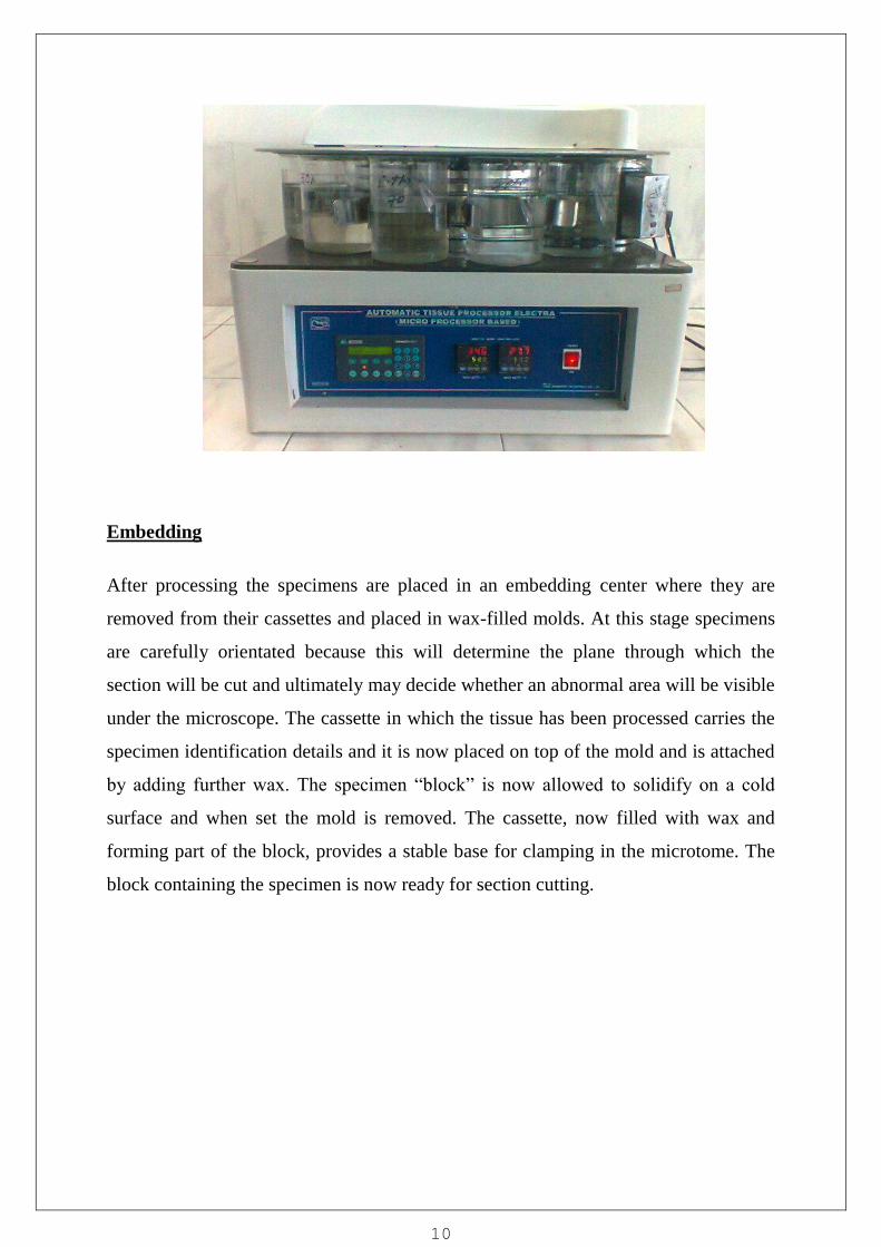
Embedding
After processing the specimens are placed in an embedding center where they are
removed from their cassettes and placed in wax-filled molds. At this stage specimens
are carefully orientated because this will determine the plane through which the
section will be cut and ultimately may decide whether an abnormal area will be visible
under the microscope. The cassette in which the tissue has been processed carries the
specimen identification details and it is now placed on top of the mold and is attached
by adding further wax. The specimen “block” is now allowed to solidify on a cold
surface and when set the mold is removed. The cassette, now filled with wax and
forming part of the block, provides a stable base for clamping in the microtome. The
block containing the specimen is now ready for section cutting.
10

Sectioning:
Sections are cut on a precision instrument called a “microtome” using extremely fine
steel blades. Paraffin sections are usually cut at a thickness of 3 - 5µm ensuring that
only a single layer of cells makes up the section. One of the advantages of paraffin
wax as an embedding agent is that as sections are cut they will stick together edge-to-
edge, forming a “ribbon” of sections. This makes handling easier. Sections are now
“floated out” on the surface of warm water in a flotation bath to flatten them and then
picked up onto microscope slides. After thorough drying they are ready for staining.
11

Staining
The embedding process must be reversed in order to get the paraffin wax out of the
tissue and allow water soluble dyes to penetrate the sections. Therefore, before any
staining can be done, the slides are "deparaffinized" by running them through xylenes
(or substitutes) to alcohols to water. There are no stains that can be done on tissues
containing paraffin. The staining process makes use of a variety of dyes that have
been chosen for their ability to stain various cellular components of tissue. The routine
stain is that of hematoxylin and eosin (H and E).
CASE: 1
Patient Name: Kaushalya
Mrd. No: 1300069107
Age/Gender: 40/Female
Specimen: Cervix Biopsy
H&E Staining was performed.
Hematoxylin& Eosin Principle:-
Hematoxylin and eosin are the principle stain used for the demonstration
of nucleus and cytoplasmic inclusion.
Alum acts as a mordent &hematoxylene containing an alum stain the nucleus light blue
when turn red in the present of acid tissue with acid solution when imparts pink color of
cytoplasm.
Hematoxylin Stain process:-
1. After cutting section dewax with xylene for 5 mins.
2. Dip in xylene II for 5 mins.
3. Dip slide in90% ethanol for 2 min.
4. Then in 80%ethanol for 2 min.
5. Then in 70%ethanol for 2 min.
12

6. Wash in running tape water for 2 min.
7. Dip in Mayer‟s hematoxylin for 3 min.
8. Wash with running tape water for 2 min.
9. Stain in 1 % eosin for 1minutes.
10. Wash with running tap water.
11. Dehydrate with 50%, 70%, 90% ethanol.
12. Air dry and mount with mounting media DPX...
13. Examination under microscope.
Result: Nucleus – Blue.
Cytoplasm – Pink.
CYTOLOGY
The cytology laboratory is geared to the study of single cells mainly in detecting
malignant and premalignant conditions as well as identifying infectious agents. The
laboratory maintains ongoing proficiency testing and performance improvement
programs.
The following specimens are examined:
Exfoliative cytology.
a. Gynecologic cervico/vaginal smears
b. Non-Gynecologic specimens
i. Sputum
ii. Bronchial washings brushings and lavages
iii. Body cavity effusions
iv. Nipple discharge
13
v. Cerebrospinal fluid
vi. Urine
vii. Endoscopic brushings and washings
viii. Other
B. FINE NEEDLE ASPIRATIONS (FNA):
a. FNAC is very valuable for preliminary diagnosis of carcinomas as well as
inflammatory conditions. Palpable masses performed by the pathologist:
These include any palpable mass such as breast, lymph node, thyroid,
soft tissue, etc.
Case 2
Patient Name: Indu Sheema
Mrd. No. 11177761
Age/Gender: 32/Female
Ref. Doctor: Dr. Disha
Investigation: Cytological exam. OfPAP smear
PAP Staining was performed
PRINCIPLE OF PAPANICOLAOU STAIN
The most widely used staining procedure for cytological specimens is Papanicolaou‟s
technique. In the first staining step the nuclei are stained by a haematoxylin solution.
Nuclei are stained blue, dark violet to black. The second staining step is cytoplasmic
14
staining by orange staining solution, especially for demonstration of mature and
keratinized cells. The target structures are stained orange in different intensities. In the
third staining step the so-called polychromatic solution is used, a mixture of eosin,
light green SF and Bismarck brown. The polychromatic solution is used for
demonstration of differentiation of squamous cells e.g. cervical cancer and cycle
diagnosis for examination under microscope.
PAP Staining Procedure:-
1. Prepare the smear and air dry it.
2. Fix the smear in alcohol for 15 min.
3. Rinse in water for 5 mins
4.Hematoxylin for 2 min.
5. Wash with tap water for 5 mins.
6. Rehydrate in 95% alcohol for 2 mins.
7. Again, Rehydrate in 95% alcohol for 2 mins.
8. Stain with OG-6 2mins.
9. Dip in 95% alcohol 2mins.
10. Again dip in 95% alcohol 2mins.
11. Counter stain with EA36 2 min.
12. Rinse in 95% alcohol for 2 mins.
13. Rinse in 95% alcohol for 2 mins.
14. Take the xylene dip.
15. Mounting with DPX.
16. Examine under microscope.
RESULT:
Cytoplasm Cyanophilic (basophilic)- Blue-green.
Cytoplasm Eosinophilic (acidophilic)– Pink.
15
10
Cytoplasm Keratinized - Pink-orange.
Erythrocytes -Red.
Nuclei Blue - dark violet, black.
Microorganisms - Grey-blue.
Trichomonas - Grey-green.
PRINCIPLE OF MAY-GRUNWALDS GIEMSA STAIN:
May-Grunwaldsstains acidophilic cells and the neutrophilicgranulations of leukocytes,
whereasGiemsa stains the cytoplasm of monocytes and lymphocytes, as well as
nuclear chromatin.
Procedure: We first pre-label the slide and spread the collected sample evenly. Fix the
slide in the fixative (Methanol) for 15 minutes. Stain the slides with MGG staining. It is the
type of Romanowsky Stains. It is also called as Azure that means it is made of more than
two stains. It contains acidic and basic dye. Acidic part takes basic dye and basic part
takes acidic dye. After fixing of slides we pour off slides with 1:1 ratio of May
Grunewald's stain for 8 minutes.After that drain the stain from the slides by first
washing.Pour second stain i.e., Giemsa 1:9 ratio on the slides for 12 minutes. Blot the slides
and mount with DPX.
Result: Nucleus – Purple.
Cytoplasm– Blue.
Red Blood Cells – Pink
16

11
SECTION
HAEMATOLOGY
Hematology is the study of blood and is concerned primarily with the study of the
formed elements of the blood.These include erythrocytes (RBC), leucocytes (WBC),
andThrombocytes (PLT).The hematology laboratory routinely reports the enumeration
of cells in circulation, hemoglobin concentration and differential count of leukocytes
based on the study of the stained blood smear. Study of the blood smear also helps in
detecting the morphological abnormalities of various cells seen in the peripheral blood
circulation.Another aspect of the hematology laboratory is to investigate causes of
bleeding disorders.
Sections of Hematology:
• Reception.
• CBC Analyzer.
• Coagulogram Analyzer.
• Urine Processing.
• Reporting.
SYSMEX XS 800i FULLY AUTOMATED HAEMATOLOGY ANALYZER
Introduction
The Sysmex XS-800i is a compact new, fully automated haematologyanalyser,
designed to generate complete blood counts with five-part leucocyte differential. A
Sysmex XS-800i instrument was evaluated according to Clinical Laboratory
Standards Institute (CLSI) and International Council for Standardization in
Haematology (ICSH) guidelines. Precision, carry-over and linearity were determined.
Using quality control material, total
17

and within-run imprecision was less than 3% except for platelets. The system
demonstrated good linearity over the entire reporting range and no carry-over
(<0.5%). Overall flagging sensitivity and specificity were 91% and 48%, respectively.
In conclusion, the Sysmex XS-800i demonstrated good analytical performance, is able
to generate a complete blood count with five-part differential on low blood volumes
and has considerable back-up capacity.
Principle of CBC counter:-
Sysmax developed Electric Capacitance method rathor then the resistance
Method when the partical is located in the detection area a change in electrical
capacitance occurs and this proportional to the volume of the partical, it is possible the
count the total no. of cells as they pass through the detection area.
In another word, when the diluted blood coming in the contact with the
Capacitance, the electrical current is absorbed by blood cells, the amount of current is
Absorbed by cells detector is detect the current and count the cells.
18
Samples on Automatic rotator
Process of this test:-
When the sample coming from collection center.
Rotate the sample on Automatic rotator
For Sysmex 800i counter
• Machine should be on before running the sample
• press Sample no. (Enter the file no. or request no.)
• Then press enter key.
• Aspirate the sample in aspiration areas
• Wait for few minute and print will ready.
19
Case No: 1

Investigation Results Ref.Range/Unit
• Haemoglobin 5.7 11.0-16.0 g/dl
• Haematocrit 22.0 33-48 %
• RBC Count 2.36 2.5-5.5 million/cumm
• MCV 93.2 80-96 fl
• MCH 24.1 27-38 pg
•MCHC 25.931-37 g/dl
• TLC 2,500 4000-11,000 Cells/cumm
• DLC
• Neutrophils 41 37-72 %
20
Age: 63 Yrs /male
Mrd No: 12008395
Sample Date: 07/01/2015
Report Date: 10/001/2015
Doctor Name:Dr. Mukhtar
Complete blood count

• Lymphocyte 38 25-48 %
• Monocyte 03 0-11 %
• Eosinophils 08 00-06 %
• Basophils 00 00-01%
• Platelets count 1, 20,000 1, 50,000 - 4, 00,000
Clinical significance: -A decrease in Hb concentration in blood below normal values
is a sign of anemia. The Hb concentration is lower in adult women as compared to
adult males.Hb values further drops during pregnancy due to haemodilution.
21

Case No- 2
Complete blood count
Investigation Results Ref.Range/Unit
• Haemoglobin 12.9 11.0-16.0 g/dl
• Haematocrit 42.1 33-48 %
• RBC Count 3.39 2.5-5.5 million/cumm
• MCV 111.1 80-96 fl
• MCH 38.05 27-38 pg
•MCHC 30.6 31-37 g/dl
• TLC 12,520 4000-11,000 Cells/chum
22
Age: 28 Years / female
Mrd No: 130003779
Sample Date: 12/01/2015
Report Date: 14/01/2015
Doctor Name: Dr. Fatima

• DLC
• Neutrophils 70 37-72 %
• Lymphocyte 17 25-48 %
• Monocyte 01 0-11 %
• Eosinophils 06 00-06 %
• Besophils 00 00-01%
• Platelets count 2, 26,000 1, 50,000 - 4, 00,000
Clinical significance:-Increased MCV is a sign of macrocytosis, which may be
related to anemia due to deficiency of vitamin B12 and Folic acid with the occurrence
of megloblastic anemia.
23

E.S.R
Requirements: ESR tubes, Rack, Timer, and Anticoagulated blood (Sodium Citrate).
Procedure:
Remove the cap on the prefilled anticoagulated vial of blood.
Insert the pipette through the mouth of the tube it will aspirate blood upto
specific level.
Let the pipette stand for one hour, then read the numerical results of the ESR.
Dispose of all bio hazardous waste in the appropriate containers.
Principle:-
The speed at which the red blood cells in normal blood settle is relatively slow.
However in many diseases, change occurs in physiological properties of plasma.
Change in the surface electrical charge of the red blood cells caused the erythrocyte to
aggregate clump, or to form rouleaux .The large clump of cells thus forms fall at a fast
rate. The changes in the proportion of the soluble constituents of plasma such as
increased fibrinogen of globulin also result in increased rateof erythrocyte fall.
24
Case: 1
Investigation Results Ref.Range/Unit
• Erythrocyte sedimentation rate 130 mm/hr 0 - 20 mm/hr
Case: 2
Mrd. No.:130005383
Investigation Results Ref.Range/Unit
• Erythrocyte sedimentation rate 10 mm/hr 0 - 20 mm/hr
Interpretations:
ESR is increased in all conditions where there is tissue breakdown or where there is
entry of foreign proteins in the blood, except for localized mild infections. The
determination is useful to check the progress of the disease. If the patient is improving
the ESR tends to fall. If the patient‟s condition is getting worse the ESR tends to rise.
The changes of the ESR are, however, not diagnostic of any specific disease.
25
Sample Date : 16/01/2015
Age: 30 Years / Male
Mrd. No.:130002835
Sample Date : 20/01/2015
Age:23 Years / Male

Urine routine & Microscopic
Routine urine analysis is mainly performed for two purposes:-
To find out metabolic or endocrine distribution of the body.
To detect intrinsic conditions that may adversely affect the urinary tract or
kidney.
Procedure:-
1. Take about 5 ml of urine in a RIA vial.
2. Liable properly and centrifuge them.
3. Discard the supernatant .
4. Place the one drop of sediment on the glass slide and place the
coverslip
5. Examine under microscope at 40x
Principle: -The microscopic elements present in urine (in suspension) are
collected in the form of deposit by centrifugation. Small drop of the
sediment is examined by making a coverslip preparation under microscope.
Case: 1
26
Date: 26/01/2015
Ref. Doctor: Dr.Muzafar
Mrd. No: 130002249

Physical Examination
Total volume 40ml
Color Pale Yellow
Turbidity Clear
Reaction Acidic
Clinical Examination:-
• Albumin Nil
• Sugar Nil
Microscopic Examination:-
• Pus Cells 40 - 50 /hpf
• RBCs Nil / hpf
• Epithelial cells 2 - 4 / HPF
• Cast Nil / HPF
• Crystals Nil / hpf
• Significance cells Nil.
Case 2
Mrd. No: 130001093
27
Date: 28-01-15
Ref. Doctor: Dr. Muzafar

Physical Examination
Total volume
Color Pale Yellow
Turbidity Clear
Reaction Acidic
Clinical Examination:-
• Albumin Nil
• Sugar Nil
Microscopic Examination:-
• Pus Cells 40 - 50 /HPF
• RBCs Nil / HPF
• Epithelial cells 2 - 4 / HPF
• Cast Nil / hpf
• Crystals Nil / hpf
Interpretations:
Protein-Nephrotic syndrome, Pyelonephritis, Glomerulonephritis, Malignant
hypertension.
Glucose: Lowered renal threshold, Renal tubular disease, Diabetes Mellitus,
Pancreatitis.
Pus: An increase number of WBC‟s in the urine indicates renal infection, which can
be in the bladder (cystitis) or in the kidney (pyelonephritis).
28

Epithelial Cell: Presence of few squamous cells (from urethra) is considered to be
normal. Dirty appearing tubular cells are seen in acute tubular necrosis. Presence of
transitional cells (from bladder or ureters) might suggest transitional cell carcinoma. [9]
Red Blood Cell: Haemoglobinuria might result from haemolytic anemia, incompatible
blood transfusion or paroxysmal haemoglobinuria(PNH), whilehaematuria might
indicate bleeding in the urinary tract.
COAGLOGRAM PROFILE
In Coagulation Lab the following investigations were performed,
Prothrombin Time (PT), Activated Partial Thromboplastin Time (APTT),
Bleeding time (BT) and Clotting time (CT).
Procedure:
Pipette 50 µL plasma into a reaction tube and set it into detector by pushing it
into bottom of the detector firmly
Press the start button. Incubation of plasma will start and the countdown of 180
seconds will begin, the detector LED blinks red.
When the time of reagent addition comes, an alarm will begin to sound. The
detector LED will blink green.
When alarm begins to sound take the reagent 100 µL and add with a pipette
without forming bubbles inside the wall of reaction tube and close the lid.
When the reagent is added, mixing will start automatically and detector LED
will light up in red.
In case of APTT 2nd
reagent is required the countdown will begin again and 2nd
reagent is added and the result is read out on the display.
Observations:
PTI Name Age/Sex MRD NO. Result Normal
Range
29
Case 1 Showkat 38/M 130002654 39.0 11.5 – 15.5

INR
Name
Age/Sex
MRD NO.
Result
Normal
Range
Interpretations: PT may be prolonged due to lack of vitamin k absorption in
obstruction or lack of synthesis in hepatocellular disease. The PT is abnormal in
coagulation defects due to liver disease because it is affected by deficiencies of more
than one factor.PT is normal or slightly prolonged in majority of patients with acute
infectious or toxic hepatitis and also in chronic liver disease.
CA-50 BLOOD COAGULATION ANALYZER
30
Case 2 Shahnawaz 36/M 130002091 17.5 11.5 – 15.5
Case 1 Showkat 38/M 130002654 2.6 0.8 – 1.2
Case 2 Shahnawaz 36/M 130002091 1.1 0.8 – 1.2

The CA-50 is an automated blood coagulation analyzer that can quickly analyze
sample with a high degree of accuracy. The CA-50 can analyze sample using a
coagulation method and the analyzed data can be displayed on its LCD screen and
printed by the built in printer. The CA-50 also has some supplemental functions
including quality control.
Analysis Parameters and calculation Parameters:The following list shows the
analysis and calculation parameters that can be analyzed by the CA-50
Method Analysis Parameters
Coagulated method PT (Prothrombin Time),
APTT (ActivatedPartialThromboplastin Time),
Fibrinogen, ThrombinTime (TT), Normotest,
Analysis Principle of CA-50:
Coagulation reaction detection method (scattered light detection method) irradiates
red light (660nm) onto a mixture of blood plasma and reagent and detects the change
in turbidity (when the fibrin clots are formed) as the change in scattered light and
measures the coagulation time.
Blood grouping
There are nearly 300 blood group systems so far discovered. The ABO and Rh are the
major,clinically significant and the most important of all the blood group systems.All
people (with few exceptions) of ABO system can be divided into 4 major groups in this
system and they are agroup, Bgroup, AB group,O group.This depends on the reactions
obtained by mixing their red blood cells to different reagents,known as Anti-A,Anti-B.
Slide method: - Take three drop of blood sample on a slide and place the monoclonal
antibody each drop of blood sample.
31

Observe the agglutination of blood.
If agglutination occur in A sera Blood group is- “A”
If agglutination occur in B sera Blood group is- “B”
If agglutination occur in AB sera Blood group is- “AB”
If no agglutination occur in AB sera Blood group is- “O”
ABO Grouping:-
Reaction Monoclonal Monoclonal Monoclonal Result
Antibodies A antibodies B antibodies D
Agglutination + - + A positive
Agglutination + - - A Negative
Agglutination - + + B Positive
Agglutination - + - B Negative
Agglutination + + + AB Positive
Agglutination + + - AB Negative
Agglutination - - + O Positive
Agglutination - - - O Negative
Keys
• + =Agglutination.
• -=No agglutination
32

SECTION – 2
BIOCHEMISTRY
Clinical Biochemistry deals with the biochemistry laboratory applications to find out
cause of a disease. The chemical constitutes of various body fluids such as
blood(serum or plasma),urine,CSF and other body fluids are analyzed in clinical
biochemistry laboratory. These determinations are useful in diagnosing various
clinical conditions such as diabetes mellitus,jaundice,gout,hyperlipidemia,pancreatitis,
rickets etc.The biochemistry tests are very useful in determining the severity of
diseases of many organs such as liver,stomach,heart,kidneys,brain as well as
endocrine disorders and related status of acid-base balance of the body. The clinical
biochemistry tests, in relation to the various clinical conditions can:-
Reveal the cause of the disease.
Screen easy diagnosis.
Suggest effective treatment.
Assist in monitoring progress of pathological conditions and
Help in assessing response to therapy.
Collection and separation of samples: In biochemistry we received the
samples from OPD/IPD for analyses in vacutainerhaving red top/cap that
indicated the samples are without anticoagulant. But for some specific test
such as HbA1c samples were collected in purple top/cap tubes. The tubes
werecentrifuged at 3000 RPM.
33

The most important automated analyzer in the laboratory was Mindray BS 400, the
laboratory also possessed EM 360, and it also hadErbaChem 7 a semi-automated
analyzer for emergency parameters.
34
These are the test which is performed in BS400 Analyzer in the
laboratory and there procedure is same.
1. Glucose
• Blood sugar Random (BSR)
• Blood sugar Fasting (BSF)
• Blood Sugar Post Prandial (BSPP)
• HbA1c
2. Kidney function test (K.F.T.)
• Urea
• Serum creatinine.
• Blood Urea Nitrogen(BUN)
3. Liver Function Test (L.F.T)
• Bilirubin Direct and Total
• SGPT,
• SGOT,
• ALP,
4. Lipid Profile.
• Cholesterol
• Triglyceride
• HDL
5. Uric Acid.
35

Principle and working of BS400:-
The Principle of BS400 is based on the Spectro photometer, is based on the Beer's Law.
The amount of light absorbed by the colouredsolution, when illuminated with light of
suitable wavelength is directly proportional to the concentration of the colour solution
and length of the light path thought the solution therefore the amount of light decrease
exponentially with increase the concentration of the solution and which increase the
thickness of the layer of solution thought which light passes or Which state that optical
density is directly proportional to the concentration of colouredsolution.
Procedure to run the sample on Mindray BS 400
Check all the cans of distilled water, cleaning solution and water.
Switch on the UPS and then the computer.
Switch on the analyzer, water filter and instrument refrigerator.
Wait for system initialization.
Click on Calibration
Select the Parameters
Click on QC
Select the test and request
Keep saline at S1, Control (Normal and Pathological) C1, C2 and calibrator at
S2.
Click on run test.
Click on sample and put patient ID
Select the test and request
Click on run test
The analyses will begin and the results will be displayed on the screen.
36

Case 1
Blood Glucose
Principle(Glucose oxidase Peroxidase): Glucose oxidase catalyses the oxidation of
glucose to produce hydrogen peroxide and gluconic acid. The hydrogen peroxide, in
the presence of enzyme peroxidase is broken down and the oxygen given off reacts
with 4- amino antipyrine and phenol to give a pink color
Mrd.No:130005844
Age/Sex: 32/F
Investigation: Blood Sugar Random
Result: 195
Normal Range: 90-140 mg/dl.
Clinical Significance: Increased value of Glucose in the case of Diabetic mellitus,
Endocrine disorder, acromegaly, stress, chronic renal failure, Pancreatitis, Drugs,
steroids, thiazides, oral contraceptives etc.
Decreased values in the case of Insulinoma, Hypopituitarism, Adrenal, Adrenal cortical
insufficiency, Sever liver disease, Extra pancreatic neoplasm, Ethanol ingestion, Drugs.
Etc.
37
Patient Name: Shafi
Case 2
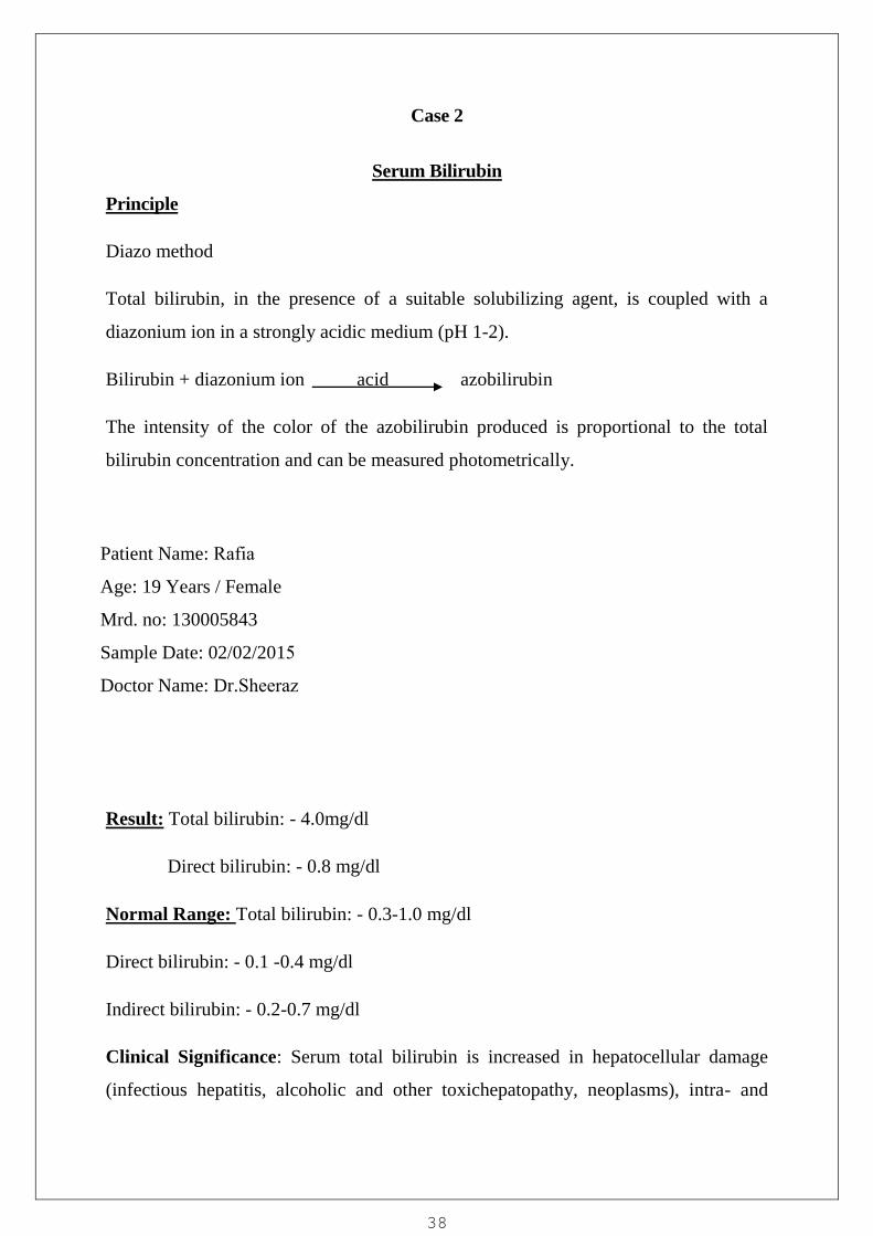
Serum Bilirubin
Principle
Diazo method
Total bilirubin, in the presence of a suitable solubilizing agent, is coupled with a
diazonium ion in a strongly acidic medium (pH 1-2).
Bilirubin + diazonium ion acid azobilirubin
The intensity of the color of the azobilirubin produced is proportional to the total
bilirubin concentration and can be measured photometrically.
Result: Total bilirubin: - 4.0mg/dl
Direct bilirubin: - 0.8 mg/dl
Normal Range: Total bilirubin: - 0.3-1.0 mg/dl
Direct bilirubin: - 0.1 -0.4 mg/dl
Indirect bilirubin: - 0.2-0.7 mg/dl
Clinical Significance: Serum total bilirubin is increased in hepatocellular damage
(infectious hepatitis, alcoholic and other toxichepatopathy, neoplasms), intra- and
38
Patient Name: Rafia
Age: 19 Years / Female
Mrd. no: 130005843
Sample Date: 02/02/2015
Doctor Name: Dr.Sheeraz

extrahepatic biliary haemolysis, physiologic neonatal jaundice, Crigler-Najjar
syndrome, Gilbert‟s disease, Dubin-Johnsonsyndrome, and fructose intolerance.
Elevation of direct (conjugated) bilirubin is seen in cholestasis and late in the course
of chronic liver disease. Indirect (unconjugated) bilirubin tends to predominate in
haemolysis and Gilbert‟s disease. Decreased serum total bilirubin is probably not of
clinical significance but has been observed in iron deficiency anemia. [22]
Case No- 3
ALKALINE PHOSPHATASE
Normal Range: Adult: 25-100 U/L
Result: 284 U/L
Principle
Colorimetric assay in accordance with a standardized method.
In the presence of magnesium and zinc ions, p-nitrophenyl phosphate is cleaved by
phosphatases into phosphate and p-nitrophenol.
39
Patient Name: Monika
Age: 23 Years /female
Mrd no: 130005248
Sample Date:05/02/2015
Doctor Name: Dr.Mukhtar
Children: Less than 350 U/L
P-nitrophenylphosphate +H2O ALP phosphate + p-nitrophenol
The p-nitrophenol released is directly proportional to the catalytic ALP activity. It is
determined by measuring the increase in absorbance.
Clinical Significance
Very high levels of ALP can be caused by liver problems, such as hepatitis,
blockage of the bile ducts (obstructive jaundice), gallstones, cirrhosis, liver
cancer, or cancer that has spread (metastasized) to the liver from another part of
the body.
High ALP levels can be caused by bone diseases, such as Paget's disease,
osteomalacia, rickets, bone tumors, or tumors that have spread from another
part of the body to the bone, or by overactive parathyroid glands
(hyperparathyroidism). Normal healing of a bone fracture can also raise ALP
levels.
Heart failure, heart attack, mononucleosis, or kidney cancer can raise ALP
levels. A serious infection that has spread through the body (sepsis) can also
raise ALP levels.
Case No- 4
AST (SGOT)
40
Patient Name: Abdul Rehman
Age: 45 Years / Male
Mrd. No: 130005108
Sample Date: 10/02/2015
Doctor Name: Dr. Mukhtar

Result: 117 IU/L
Normal Range: 10-40 IU/L
Principle
AST in the sample catalyzes the transfer of an amino group between L-aspartate and
2-oxoglutarate to form oxaloacetate and L-glutamate. The oxaloacetate then reacts
with NADH, in the presence of malate dehydrogenase (MDH), to form NAD+.
Pyridoxal phosphate serves as a coenzyme in the amino transfer reaction. It ensures
full enzyme activation.
L-Aspartate + 2-oxoglutarate AST oxaloacetate + L-glutamate
Oxaloacetate + NADH + H+ MDH L-malate + NAD+
The rate of the NADH oxidation is directly proportional to the catalytic AST activity.
It is determined by measuring the decrease in absorbance.
Clinical Significance
An increase in AST levels may indicate:
Acute hemolytic anemia
Acute pancreatitis
Acute renal failure
Liver cirrhosis
Heart attack
Hepatitis
Infectious mononucleosis
Liver cancer
Liver necrosis
41

Case No- 5
ALT (SGPT)
Result: 145 IU/L
Normal Range: 7-56 IU/L
Principle
AST catalyzes the reaction between L-alanine and 2-oxoglutarate.The pyruvate
formed is reduced by NADH in a reaction catalyzed by lactate dehydrogenase (LDH)
to form L-lactate andNAD+.
Pyridoxal phosphate serves as a coenzyme in the amino transfer reaction. It ensures
full enzyme activation.
Pyruvate + NADH + H+ LDH L-lactate + NAD
+
The rate of the NADH oxidation is directly proportional to the catalytic ALT activity.
It is determined by measuring the decrease in absorbance.
42
Patient Name: Bhura Ram
Age: 45 Years / Male
Mrd. No: 130005108
Sample Date: 18/02/2015
Doctor Name: Dr. Muzafar
L-Alanine + 2-oxoglutarate ALT pyruvate + L-glutamate

Clinical Significance
Greater-than-normal ALT levels may indicate:
Celiac disease
Cirrhosis
Hepatitis (viral, autoimmune)
Hereditary hemochromatosis
Liver ischemia (blood flow deficiency to the liver)
Liver tumor
Use of drugs that are poisonous to the liver
Case No- 6
CHOLESTEROL
Result: 295 mg/dl
Normal Range: less than 200 mg/dl
43
Patient name – Shingara Ram
Age - 46 /Male
Mrd.no:130005208
Sample date- 04/03/2015
Doctor name- Dr.Fatima

Principle
Enzymatic, colorimetric method.
Cholesterol esters are cleaved by the action of cholesterol esterase to yield free
cholesterol and fatty acids. Cholesterol oxidase then catalyzes the oxidation of
cholesterol to cholest-4-en-3-one and hydrogen peroxide. In the presence of
peroxidase, the hydrogen peroxide formed effects the oxidative coupling of phenol
and 4-aminoantipyrine to form a red quinine-imine dye.
Cholesterol esters + H2O CE cholesterol + RCOOH
Cholesterol + O2 CHOD cholest-4-en-3-one + H2O2
2 H2O2 + 4-AAP + phenol POD quinone-imine dye + 4H2O
The color intensity of the dye formed is directly proportional to the cholesterol
concentration. It is determined by measuring the increase in absorbance.
Clinical Significance
Increased in: 1) Diabetes mellitus Decreased in: 1) Severe infection
2) Nephrosis 2) Severe anemia
3) Biliary cirrhosis 3) Hyperthyroidism
4) Lipoprotenemias 4) Malnutrition
5) Hypothyroidism
44

Case No- 7
URIC ACID
Result: 7.1 mg/dl
Normal Range: Men- 2 to 7.5 mg/dL,
Principle
Enzymatic colorimetric test.
Uric acid + 2 H2O + O2 Uricase allantoin + CO2 + H2O2
2 H2O2+ H+ + TOOSa + 4-aminophenazone peroxidase quinone-diimine
dye + 4 H2O
a) N-ethyl-N-(2-hydroxy-3-sulfopropyl)-3-methylaniline
The color intensity of the quinone-diimine formed is directly proportional to the uric
acid concentration and is determined by measuring the increase in absorbance.
45
Patient name – Gh. Qadir
Age - 70 /Male
Mrd. No - 130005532
Sample date- 10/03/2015
Doctor name- Dr. Mukhtar
Women- 2 to 6.5 mg/dL.
Uricase cleaves uric acid to form allantoin and hydrogen peroxide.

Clinical Significance
Increase in serum uric acid is seen idiopathically and in renal failure, disseminated
neoplasms, toxemia of pregnancy, psoriasis, liver disease, ethanol consumption, etc.
Many drugs elevate uric acid, including most diuretics, catecholamines, ethambutol,
pyrazinamide, salicylates, and large doses of nicotinic acid.
Decreased serum uric acid level may not be of clinical significance. It has been
reported in Wilson's disease, Fanconi's syndrome, and Hodgkin's disease, myeloma,
and bronchogenic carcinoma.
Case no-8
UREA
Result: 186 mg/dl
Principle
Kinetic test with urease and glutamate dehydrogenase. Urea is hydrolyzed by urease
to form ammonium and carbonate.
Urea+2H2O Urease 2 NH4+ + CO3
2-
46
Patient name – Gani kak
Age - 55 /Male
Mrd. No - 130005374
Sample date- 14/03/2015
Doctor name- Dr.Humaira
Normal Range: 17-56 mg/dl,

In the second reaction 2-oxoglutarate reacts with ammonium in the presence of
glutamate dehydrogenase (GLDH) and the coenzyme NADH to produce L-glutamate.
In this reaction two moles of NADH are oxidized to NAD for each mole of urea
hydrolyzed.
NH4+ + 2-oxoglutarate + NADH GLDH L-glutamate + NAD
+ + H2O
The rate of decrease in the NADH concentration is directly proportional to the urea
concentration in the specimen and is measured photometrically.
Clinical Significance
An abnormally high level of urea nitrogen in the blood is an indication of kidney
function impairment or failure. Some other causes of increased values for urea
nitrogen include prerenal azotemia (e.g. shock), postrenal azotemia, GI bleeding and a
high protein diet. Some causes of decreased values for urea nitrogen include
pregnancy, severe liver insufficiency, overhydration and malnutrition.
Normal range: 17-56 mg/dl.
Result: 120 mg/dl
Case No- 9
CREATININE
47
Patient name – Mohd. Jabbar
Age - 55 /Male
Mrd. No - 130005374
Sample date- 21/03/2015
Doctor name- Dr. Sheeraz

Result: 5.1 mg/dl
Normal Range: 0.6-1.4mg/dl,
Principle
This kinetic colorimetric assay is based on the Jaffe method. In alkaline solution,
creatinine forms a yellow-orange complex with picrate. The rate of dye formation is
proportional to the creatinine concentration in the specimen.
Creatinine + picric acid Alkaline pH yellow-red complex.
Clinical Significance
Creatinine is a breakdown product of Creatine. Measurement of creatinine levels is
used as one indicator of kidney function. Serum creatinine is a waste product of the
dehydration of Creatine. Most of the body Creatine is present in muscle tissue. It
represents an important alternative energy source for the body. The primary source of
energy for the body is the conversion of ATP to ADP by breaking a high-energy
phosphate bond and releasing energy that can be used. Creatine is important as it is an
efficient energy source used to covert ADP back to ATP.
Case 10: HbA1c
48
Principle: Glycosylated Hemoglobin (GHb) has been defined operationally as the fast
fraction hemoglobinsHbA1 (HbA1a, A1b, A1C) which elute first during column
chromatography. The non-glycosylated hemoglobin, which consists of the bulk of
haemoglobin, has been designated HbAo.
A hemolysed preparation of whole blood is mixed continuously for 5 minutes with a
weakly binding cation-exchange resin. The labile fraction is eliminated during the
hemolysate preparation and during the binding. During the mixing, HbAo binds to the
ion exchange resin leaving GHb free in supernatant. After the mixing period, a filter
separator is used to remove the resin from the supernatant. The percent glycosylated
hemoglobin is determined by measuring absorbance of the glycosylated (GHb)
fraction and the total (THb) fraction. The ratio of the Glycosylated hemoglobin and
the Total hemoglobin fraction of the control and the test are used to calculate the
percent Glycosylated hemoglobin of the sample. Requirements:
Reagent-Ion Exchange Resin (Predispensed Tubes), Lysing reagent, Control (10%
GHb), Resin separators. Blood sample (EDTA) Reagent Preparation: The Ion
Exchange resin tubes and the lysing reagent are ready to use. Reconstitute the control
with 1ml of distilled water. Allow to stand for 10 minutes with occasional mixing. The
reconstitute control is stable for at least 7 days when stored at 2-8oc.
Wavelength: 415 nm
Procedure:A)Hemolysate preparation.
1. Dispense 0.5 ml lysing reagent into tubes labelled as Control(C) & Test.
2. Add 0.1 ml of the reconstitute control & well mixed blood sample into the
appropriately labelled tubes. Mix until complete lysis is evident.
3. Allow to stand for 5 minutes.
B) Glycosylated hemoglobin (GHb) separation
1. Remove cap from the Ion exchange resin tubes and label as Control and Test.
49

2. Add 0.1 ml of the hemolysate from step A into the appropriately labelled Ion
exchange resin tubes.
3. Insert a resin separator into each tube so that the rubber sleeve is appropriately
1cm above the liquid level of the resin suspension.
4. Mix the tubes on a rocker continuously for 5 minutes.
5. Allow the resin to settle, and then push the resin separator into the tubes until
the resin is firmly packed.
6. Pour or aspirate each supernatant directly into a cuvette and measure each
absorbance against distilled water.
C) Total Hemoglobin (THb) fraction
1. Dispense 5.0 ml of distilled water into tubes labeled as Control and Test
2. Add to it 0.02 ml of hemolysate from step A into appropriately labeled tube.
Mix well
3. Read each absorbance against distilled water
Calculations: Ratio of Control (Rc) = Abs. Control GHb/Abs. Control THb
Ratio of Test (Rt) = Abs Test GHb/Abs Test THb
GHb in % = Ratio of test (Rt)/Ratio of Control (Rc) ×10(Value of Control)
Case 1
Patients
Name:Ravinder
Singh.
Age/Sex: 22/Male
MRD No.
120078890
HbA1c 8.40
50

Case 2
Patients Name:
Ravinder Singh.
Age/Sex: 22/Male
MRD No.120078890
HbA1c 9.40
Mean Blood Glucose (MBG) in mg/dl = 33.3×HbA1c Value – 86.These values are
linear in the range of 6.5 – 13% of HbA1c values.
Normal reference values:
Normal:< 8.0%
Good Control: 8.0 – 9.0%
Fair Control: 9.0 – 10.0%
Poor Control:> 10.0%
Clinical significance: GHb reflects the metabolic control of glucose level over a
period of time unaffected by diet, insulin, other drugs or exercise on the day of testing.
GHb is now widely recognized as an important test for the diagnosis of Diabetes
Mellitus and is a reliable indicator of the efficacy of therapy.
51

SECTION 3
MICROBIOLOGY
Introduction: Microbiology involves the study of microscopic organisms. Although
microorganisms are generally beneficial and essential for life, some are, however,
pathogenic and cause infectious diseases. The diagnostic microbiology laboratory is
engaged in the identification of infectious agents. These infectious agents are broadly
classified as viruses, bacteria, mycotic agents and parasites (protozoa and
helminths).Identification of the infectious agent is the principle function of diagnostic
microbiology laboratory. In addition, the laboratory also provides guidance in
therapeutic management. This is particularly true in the case of bacterial infection
where the laboratory provides information regarding the most effective antimicrobial
agent and its dosage to be used for the specific patient.
Sections of Microbiology Laboratory:
Media Preparation
Collection
Bacteriology
Serology
Immunology
Bacterial culture media:
One of the most important reasons for culturing bacteria in vitro is its utility in
diagnosing infectious diseases. Isolating a bacterium from sites in body normally
known to be sterile is anindication of its role in the disease process. Culturing bacteria
is also the initial step in studying its morphology and its identification. Bacteria have
to be cultured in order to obtain antigens from developing serological assays or
vaccines. Certain genetic studies and manipulations of the cells also need that bacteria
be cultured in vitro. Culturing bacteria also provide a reliable way estimating their
numbers (viable count). Culturing on solid media is another convenient way
ofseparating bacteria in mixtures.
52

History:
Louis Pasteur used simple broths made up of urine or meat extracts. Robert Koch reali
zed the importance of solid media and used potato pieces to grow bacteria. It was
on the suggestion of Fannie Eilshemius, wife of Walther Hesse (who was an assistant
to Robert Koch) that agar wasused to solidify culture media. Before the use of
agar, attempts were made to use gelatin assolidifying agent. Gelatin had some
inherent problems; it existed as liquid at normal incubating temperatures (35-37oC)
and was digested by certain bacteria.
Composition of culture media:
Bacteria infecting humans (commensals or pathogens) are chemoorganoheterotro
phs. When culturing bacteria, it is very important to provide similar environmental
and nutritional conditions that exist in its natural habitat. Hence, an artificial culture
medium must provide all the nutritional components that a bacterium gets in its
natural habitat. Most often, a culture medium contains water, a source of carbon &
energy, source of nitrogen, trace elements and some growth factors. Besides these, the
pH of the medium must be set accordingly. Some of the ingredients of culture media
include water, agar, peptone, casein hydrolysate, meat extract, yeast extract and malt
extract.
Classification:
Bacterial culture media can be classified in at least three ways; Based on consistency,
based on nutritional component and based on its functional use.
1) Classification based on consistency:
Culture media are liquid, semi-solid or solid and biphasic.
A) Liquid media: These are available for use in test-
tubes, bottles or flasks. Liquid media
are sometimes referred as “broths”(e.g nutrient broth). In liquid medium, bacteria
grow uniformly producing general turbidity. Certain aerobic bacteria and those
containing fimbriae (Vibrio & Bacillus) are known to grow as a thin film called
53

„surface pellicle‟ on the surface of undisturbed broth. Bacillus anthracis is known to
produce stalactite growth on ghee containing broth. Sometimes the initial turbidity
may be followed by clearing due to autolysis, which is seen in penumococci. Long
chains of Streptococci when grown in liquid media tend to entangle andsettle to the
bottom forming granular deposits. Liquid media tend to be used when a large
number of bacteria have to be grown. These are suitable to grow bacteria when the
numbers in the inoculum is suspected to be low. Inoculating in the liquid medium also
helps to dilute any inhibitors of bacterial growth. This is the practical approach in
blood cultures. Culturing in liquid medium can be used to obtain viable count
(dilution methods). Properties of bacteria are not visible in liquid media and presence
of more than one type of bacteria can not be detected.
B) Solid media: Any liquid medium can be rendered by the addition of certain solidify
ing
agents. Agar agar (simply called agar) is the most commonly used solidifying ag
ent. It is anunbranched polysaccharide obtained from the cell membranes of some
species of red algae such as the genera Gelidium. Agar is composed of two long-
chain polysaccharides (70% agarose and 30%agarapectin). It melts at 95oC (sol) and
solidifies at 42oC (gel), doesn‟t contribute any nutritive property, it is not
hydrolyzed by most bacteria and is usually free from growth promoting or
growth retarding substances.However, it may be a source of calcium & organic
ions
Most commonly, it is used at concentration of 1-3% to make a solid agar medium.
New Zealand agar has more gelling capacity than the Japanese agar. Agar is
available as fibres (shreds) or as powders.
C) Semi-solid agar: Reducing the amount of agar to 0.2-0.5% renders a medium semi-
solid. Such media are fairly soft and are useful in demonstrating bacterial
motility and separating motile from non-motile strains (U-tube and Cragie‟s
tube). Certain transport media such as Stuart‟s and Amies media are semi-solid in
consistency. Hugh &Leifson‟s oxidation fermentation test medium as well as mannitol
motility medium are also semi-solid.
54

D) Biphasic media: Sometimes,a culture system comprises of both liquid and solid
medium in the same bottle. This is known as biphasic medium (Castaneda system for
blood culture). The inoculum is added to the liquid medium and when
subcultures are to be made, the bottle is simply tilted to allow the liquid to
flow over the solid medium. This obviates the need for frequent opening of the
culture bottle to subculture. Besides agar, egg yolk and serum too can be used to
solidify culture media. While serum and egg yolk are normally liquid, they can be
rendered solid by coagulation using heat. Serum containing medium such as
Loeffler‟s serum slope and egg containing media such as Lowenstein Jensen
medium and Dorset egg medium are solidified as well as disinfected by a process of
inspissation.
2) Classification based on nutritional component:
Media can be classified as simple, complex and synthetic (or defined). While
most of the nutritional components are constant across various media, some bacteria
need extra nutrients. Those bacteria that are able to grow with minimal requirements
are said to non-fastidious and those that require extra nutrients are said to be
fastidious. Simple media such as peptone water, nutrient agar can support most non-
fastidious bacteria. Complex media such as blood agar have ingredients whose exact
components are difficult to estimate. Synthetic or defined media such as composition
of every component is well known.
3) Classification based on functional use or application:
These include basal media, enriched media, selective/enrichment media, indicator/diff
erential media, transport media and holding media.
A) Basal media are basically simple media that supports most non-
fastidious bacteria. Peptone water, nutrient broth and nutrient agar considered basal
medium.
B) Enriched media: Addition of extra nutrients in the form of blood, serum, egg yolk e
tc,
to basal medium makes them enriched media. Enriched media are used to gro
w nutritionally exacting (fastidious) bacteria. Blood agar, chocolate agar, Loeffler‟s
serum slope etc are few of the enriched media.
55

C) Selective and enrichment media are designed to inhibit unwanted commensal
or
contaminating bacteria and help to recover pathogen from a mixture of bacteria. While
selective media are agar based, enrichment media are liquid in consistency. Both
these media serve the same purpose. Any agar media can be made selective by
addition of certain inhibitory agents that don‟t affect the pathogen. Various
approaches to make a medium selective include addition of antibiotics, dyes,
chemicals, alteration of pH or a combination of these.
D) Enrichment media are liquid media that also serves to inhibit commensals i
n the clinical specimen. Selenite F broth, tetrathionate broth and alkaline peptone
water are used to recover pathogens from fecal specimens.
E) Differential media or indicator media: Certain media are designed in such a way th
at
different bacteria can be recognized on the basis of their colony colour. Variou
s approaches include incorporation of dyes, metabolic substrates etc, so that those
bacteria that utilize them appear as differently coloured colonies. Such media are
called differential media or indicator media. Exmples: MacConkey‟s agar, CLED
agar, TCBS agar, XLD agar etc.
F) Transport media:Clinical specimens must be transported to the laboratory immediat
ely after collection to prevent overgrowth of contaminating organisms or commensals.
This can be achieved by using transport media. Such media prevent drying
(desiccation) of specimen, maintain the pathogen to commensal ratio and inhibit
overgrowth of unwanted bacteria. Some of these media (Stuart‟s & Amie‟s) are
semi- solid in consistency. Addition of charcoal serves to neutralize inhibitory
factors. Cary Blair medium and VenkatramanRamakrishnan medium are used to
transport feces from suspected cholera patients. Sach‟s buffered glycerol saline
is used to transport feces from patients suspected to be suffering from bacillary
dysentery.
G) Anaerobic media: Anaerobic bacteria need special media for growth because they
Need
56

low oxygen content, reduced oxidation – reduction potential and extra nutrients.
Media for anaerobes may have to be supplemented with nutrients like hemin
and vitamin K. Boiling the medium serves to expel any dissolved oxygen. Addition
of 1% glucose, 0.1% thioglycollate, 0.1% ascorbic acid, 0.05% cysteine or red hot
iron filings can render a medium reduced. Robertson cooked meat that is
commonly used to grow Clostridium spps medium contain a 2.5 cm column of bullock
heart meat and 15 ml of nutrient broth. Before use the medium must be boiled in water
bath to expel any dissolved oxygen and then sealed with sterile liquid paraffin.
Methylene blue or resazurin is an oxidation- reduction potential indicator that is
incorporated in the thioglycollate medium. Under reduced condition, methylene blue
is colourless.
Preparation and preservation
Care must be taken to adjust the pH of the medium before autoclaving. Various pH
indicators that are in use include phenol red, neutral red, bromothymol blue,
bromocresol purple etc. Dehydrated media are commercially available and must be
reconstituted as per manufacturers‟recommendation. Most culture media are
sterililized by autoclaving. Certain media that contain heat labile components like
glucose, antibiotics, urea, serum, blood are not autoclaved. Thesecomponents are
filtered and may be added separately after the medium is autoclaved.Certain highly
selective media such as Wilson and Blair‟s medium and TCBS agar need not be
sterilized. It is imperative that a representation from each lot be tested for performance
and contamination before use. Once prepared, media may be held at 4-5oC in the
refrigerator for 1-2 weeks. Certain liquid media in screw capped bottles or tubes or
cotton plugged can be held at room temperature for weeks.
Preparation of McConkey Agar:
For 1000 ml preparation
20 gm. Peptone
2o gm. Agar
57

20 gm. Lactose
5 gm. Sodium Taurocholate
Add 1ml of Neutral Red as an indicator
Final pH should be 7.3.Autoclave at 121oc for 15 minutes.
Lactose Fermenter: Pink e.g. E.coli.
Non Lactose Fermenter: Transparent e.g. Pseudomonas
Late Lactose Fermenter: Light Pink e.g. Acinetobacter
58

Name
Age/Sex
MRD NO. Requisition
Type
Result
Case
1
Case
2
Collection
Depending on the type of disease, various specimens are submitted to the laboratory.
These include blood, wound, exudate, throat swabs, urine, spinal fluid, faecal material
and others. However, the laboratory technician is still expected to be knowledgeable
about which specimen are appropriate for a given disease, how those specimens
should be collected and how they should be transported to the laboratory. One should
always bear in mind that „a bad specimen cannot yield good results.‟ If any inadequate
or inappropriate specimen has been received in the laboratory, it is preferable to reject
the specimen rather than to provide inaccurate results later. However, the technician
must confer with the microbiologist before rejecting any valuable specimen.
Bacteriology
Gram Staining:
Principle:
The air dried fixed smear of bacteria, picks up stain and looks purple, when treated
with crystal violet stain and iodine. Iodine enhances the staining reaction between the
dye and the internal cellular constituents of bacteria. The alcohol decolourizes Gram
negative bacteria while Gram positive bacteria retain the color. Counterstaining with
safranin stains the Gram negative bacteria red. Under oil immersion microscopic
examination, Gram negative organisms appear red e.g. E.coli, Salmonella etc. and
Gram positive organism appear purple in color e.g. Staph. Aureus, Strep. Pyogenic
etc.
59
Manzoor 38/M 130002245 IPD Non Lactose
Fermenter
Lateef 36/M 130002776 OPD Lactose Fermenter

Procedure:
Prepare a smear using culture/sample
Air dried the smear and heated it so that it is fixed
Poured crystal violet solution for 1 minute and then rinsed in water
Poured Gram‟s Iodine for 1 minute
Wash with water
Decolorize with Acetone for 30 seconds
Wash with water and counterstain with Safranin for 30 seconds
Wash in water and air dry
Observe under microscope
Results:
Name Age/Sex MRD NO. Requisition
Type
Result
Case
1
Case
2
AFB Staining (ZN Staining)
Principle: A few species of bacteria in the genera Mycobacterium and Nocardia, and
the parasite Cryptosporidium do not readily stain with simple stains. However, these
microorganisms can be stained by heating them with carbol-fuchsin. The heat drives
the stain into the cells. Once the microorganisms have taken up the carbolfuchsin, they
are not easily decolorized by acid-alcohol, and hence are termed acid-fast. This acid-
fastness is due to the high lipid content (mycolic acid) in the cell wall of these
microorganisms. The Ziehl-Neelsen acid-fast staining procedure (developed by Franz
Ziehl, a German bacteriologist, and Friedrich Neelsen, a German pathologist, in the
late 1800s) is a very useful differential staining technique that makes use of this
difference in retention of carbolfuchsin. Acid-fast microorganisms will retain this dye
60
Manzoor 38/M 130002245 IPD Gram Positive
Lateef 36/M 130002776 OPD Gram Negative
and appear red. Microorganisms that are not acid-fast, termed non-acid-fast, will
appear blue or brown due to the counterstaining with methylene blue after they have
been decolorized by the acid-alcohol. A modification of this procedure that employs a
wet-ting agent rather than heat to ensure stain penetration is known as the Kinyoun
staining procedure (developed by Joseph Kinyoun, German bacteriologist, in the early
1900s).
Procedure:
Prepare smear on the slide and heat fix.
Add boiling CorbolFuchsin on slide.
Wait for 5-10 minutes and then wash.
Add 3% Acid Alcohol on slide for 5 minutes.
Add 20% Sulphuric acid for decolourization for 4 minutes.
Wash with water and add Methyl blue for 30 seconds.
Rinse with water.
Air dry and observe under microscope.
Results:
Name Age/Sex MRD NO. Requisition
Type
Result
Case
1
Case
2
Biological Culture Method:
Methods of culturing bacteria vary and depend on the types of media
being inoculated. Whatever medium is used, an aseptic technique must
be used for two reasons.
To prevent contamination of cultures and specimens in order to
61
Manzoor 38/M 130002245 IPD AFB Positive
Lateef 36/M 130002776 OPD Negative

avoid interference with the interpretation of culture results.
To prevent contamination of the work area.
In order to achieve these two objectives, the lab worker should;
Flame wire loops, straight wires and forceps before and after use.
Flame the necks of the specimen bottles, culture tubes, and test
tube after removing and replacing caps or plugs.
Not let bottle tops or caps touch an unsterile surface.
Always inoculate culture media before making smears
Use the safety cabinet when treating hazardous pathogens.
When sterilizing a wire loop, hold it in the blue part of flame
with the loop part facing down. This is to ensure quick and
proper sterilization of the wire loop and prevent sputtering
material from falling on hand.
Different methods
1. Plate culture
2. Tube culture
3. Slope culture
4. Deep culture
5. Stab culture
PROCEDURE(Plate culture):
1. Collect the equipment‟s required for processing the urine specimen for culture.
2. After receiving the urine sample in the laboratory. We check the label
on the urine container and make sure that it is similar with the requisite
form. If there is any discrepancy we reject the sample.
3. Mix the urine specimen by inverting, Sterile the loop, and allow it to cool.
4. Open the urine container in front of flame and pick up loop full of inoculum on to
62

the Blood agar plate and then on McConkey agar plate and make primary inoculum
and go for streaking i.e. primary streaking , secondary and finally tertiary streaking.
5. Incubate the inoculated plate in an incubator at 370c for overnight (optimum
temperature for bacterial growth).
6. Next morning see the plate for bacterial growth, if no growth is present culture is
sterile; if present go for identification and colony count.
7. For identification we make smear, and perform Gram stain.
8. Perform Biochemical tests and simultaneously perform antimicrobial
sensitivity testing using Kirby Bauer‟s disk diffusion method on
MullerHinton agar plate.
9. Incubate plate at 370c in an incubator for overnight and next day see the
sensitivity pattern of the organism against their respective antibiotic panel.
Result:
Name Age/Sex MRD NO. Requisition
Type
Microscopy/Result
Case
1
Case
2
63
Manzoor 38/M 130002245 IPD Pus 15-20,Bacteria
++/Sterile
Lateef 36/M 130002776 OPD Pus 20-25,Bacteria
++/Growth of
E.Coli>100000
cfu/ml

Antibiotic Sensitivity Pattern
64
SUMMARY ON MY PROJECT REPORT
in histopathology of the pathology department it was having two
sections(Histopathology & Hematology) for 1st fifteen days in this session of
training I worked in different sections of histopathology and cytology starting
from reception, tissue processing, block preparation and also perform staining like
(H &E,PAP,MGG).
In the histopathology department these work is done by me Block
preparation, HematoxylinStaining, PAPStaining. From 21stJan I was shifted to
the hematology section, in this session I worked in the different sections of
hematology lab like sample receiving and entering in the log book ,processing
and examine of the urine specimen, coagulationprofile, blood grouping and CBC
and reporting.
65
This project Report is based on mostly positive patient only which has performed
by me and I have mention short description laboratory work which is done by me
in laboratory during training session in Medinova Diagonostic & Clinical
Laboratory J&K. There are following department in the hospital in which I have
worked like Pathology it has two sections (Histopathology & Hematology),
Biochemistry, &Microbiology. The training period was of 4 month
approximately, in this period of 4 months I worked in all the three departments
(Pathology, Biochemistry,&
Microbiology) on rotation basis, my training session was starting from Ist jan 2015
Then from 11th feb-13 to 17
th march-15, I was send to the Biochemistry
department in this session of training I worked in different sections of the
biochemistry lab like sample receiving and entering in the log book and then in the
serum separation and then processing through different analyzers and reporting.
And from 18th march to 20
th april-15, I was send in the microbiology
department in this session of training I worked in the different sections of the
microbiology lab the sections which include in microbiology department are
media room, in this section all the media which are used in the
microbiology department is prepared in this room. Thencomes the collection
room in this room all the samples were collected and received in this room.
Then there is

bacteriology section in these different types of staining like ZN staining& Gram
staining, urine culture and antibiotic sensitivity test was performed. The next
section was of serology in this section different investigation was performed like
anti-HIV trilling cassette test for AIDS, stained salmonella antigens for
WIDAL(slide test),RF latex agglutination slide test, one step rapid test for
Hepatitis B surface antigen. Then was the immunology section in this section
HCV was performed on ELISA technique.
66

REFERENCES
1. Text Book of Medical Laboratory Technology, 2nd edition, By
Dr.PrafulB.Godhkar, Ph.D& Dr. DashingP.Godkar, M.D.
2. Histopathology Laboratories Armed Forces Institute of Pathology, Washington,
D.C. 20305
3. 25. Harley, J. P, Prescott, L. M, Laboratory Exercises in Microbiology, 5th
Edition & Tata McGraw Hill Company 2002
4. Medical laboratory science theory and practical (10th
Reprint 2008),By J
Ochei& A Kolhatkar
5. Mukherjee, Kanai L., Medical Laboratory Technology 2nd
Edition, New Delhi:
Tata McGraw Hill,2010
6. Merck Source (2002). Dorland's Medical Dictionary. Retrieved 2005-01-26.
7. Stedman's Medical Dictionaries (2005). Stedman's Online Medical Dictionary.
Retrieved 2005-01-26.
8. Lee, G. Richard et al. Wintrobe‟s Clinical Hematology
9th
Edition,Philadelphia:Lea and Feblger,1993
9. http://www.medicinenet.com/liver_blood_tests/ht
10. http://www.globalrph.com/labinter.htm.
11. http://sysmexmarketing.com/images/product_pdfs/Brochure_XS-800i_MKT-
10-1139_01-2012.pdf.
12. Sysmex CA-50 Operators Manual Revised December 2000 1-12.
13. http://ncbi.nlm.nih.gov/…mc/articles/PMC499065/
14. http://bio-optica.it/pdf3/080802L.pdf
15. http://wisegeek.com/what-is-the-value-of-exfoliative-cytology.htm
16. https://in.vwr.com/app/asset?type=hi_res&id=8095780&name=350405X_3516
95T%16.Papanicolaou%20stains&filename=350405X_351695T_Papanicolaou
_stains.pdf
17.http://ruf.rice.edu/~bioslabs/methods/protein/spectrophotometer.html
18.http://klsindia.com/details/SGOT-DB.pdf
67

19.http://klsindia.com/details/SGPT-DB.pdf
20.http://tulipgroup.com/Common_New/Tech_Pubs_PDF/GlycatedTech.pdf
21.http://monobind.com/product-inserts?range=101%2C120%2C120
22.P.N,ShridharRao.,“BacterialCultureMedia”
http://microrao.com/micronotes/culture_media.pdf
68