rede nordeste de biotecnologia - RI/UFS
162
REDE NORDESTE DE BIOTECNOLOGIA UNIVERSIDADE FEDERAL DE SERGIPE FABIO NEVES SANTOS DISTRIBUIÇÃO DA INERVAÇÃO DA RELAXINA -3 NO TECTUM E TEGMENTUM NO RATO SUGERE ENVOLVIMENTO DO NUCLEO INCERTUS EM REDES DEFENSIVAS CENTRAIS São Cristovão - SE 2012
-
Upload
khangminh22 -
Category
Documents
-
view
2 -
download
0
Transcript of rede nordeste de biotecnologia - RI/UFS

REDE NORDESTE DE BIOTECNOLOGIA UNIVERSIDADE FEDERAL DE SERGIPE
FABIO NEVES SANTOS
DISTRIBUIÇÃO DA INERVAÇÃO DA RELAXINA -3 NO TECTUM E
TEGMENTUM NO RATO SUGERE ENVOLVIMENTO DO NUCLEO
INCERTUS EM REDES DEFENSIVAS CENTRAIS
São Cristovão - SE 2012

REDE NORDESTE DE BIOTECNOLOGIA UNIVERSIDADE FEDERAL DE SERGIPE
FABIO NEVES SANTOS
DISTRIBUIÇÃO DA INERVAÇÃO DA RELAXINA -3 NO TECTUM E
TEGMENTUM NO RATO SUGERE ENVOLVIMENTO DO NUCLEO
INCERTUS EM REDES DEFENSIVAS CENTRAIS
Tese apresentada ao Programa de Pós-Graduação Biotecnologia da Rede Nordeste de Biotecnologia, como requisito parcial à obtenção do título de Doutor em Biotecnologia.Orientador: Prof. Dr. Murilo Marchioro. Co-orientador: Prof. Dr. Francisco E. Olucha Bordonau
São Cristovão - SE 2012

FICHA CATALOGRÁFICA ELABORADA PELA BIBLIOTECA CENTRAL UNIVERSIDADE FEDERAL DE SERGIPE
S237d
Santos, Fabio Neves Distribuição da inervação da relaxina-3 no tectum e tegmentum no rato sugere envolvimento do núcleo incertus em redes defensivas centrais / Fabio Neves Santos ; orientador Murilo Marchioro. – São Cristóvão, 2012.
160 f. : il. Tese (Doutorado em Biotecnologia) – Rede Nordeste de
Biotecnologia – RENORBIO, Universidade Federal de Sergipe, 2012.
1. Biotecnologia – Aplicações médicas. 2. Neurologia. 3. Imunocitoquímica. 4. Relaxina-3. 5. Ácido gama-aminobutírico. I. Marchioro, Murilo, orient. II. Título.
CDU 606:616.8


Dedico este trabalho a Deus, a quem pertence
tudo que sou e tenho.

AGRADECIMENTOS
Nos longos anos do doutorado a jornada não foi fácil, houve muitas pedras no
caminho, e alguns verdadeiros pedregulhos que tiveram que ser enfrentados, mas graças à
ajuda, esses pedregulhos transformaram-se em apenas grãos de areia.
Agradeço a Deus, responsável desta “façanha” que enviou como ferramentas:
- Meus pais, que sempre me apoiaram e educaram para a vida;
- Minha amada esposa, que participou ativamente em todas as etapas desta tese;
- Aos meus grandes orientadores, Murilo e “Paco” Olucha, que me ensinaram muito;
- Aos que colaboraram nos trabalhos: Ana Sánchez-Pérez, Marcos Otero-García, Juan e Rocío pelo convívio e colaboração; - A professora Paula Lenz, que agilizou junto a Capes a bolsa de doutorado no
exterior;
- A Fapitec e Capes pelas bolsas;
- Agradeço a todos aqueles, que apesar de não terem sido citados, colaboraram direta
ou indiretamente com a realização desse trabalho.

“Penso que só há um caminho para a ciência ou para a filosofia:
encontrar um problema, ver a sua beleza e apaixonar-se por ele;
casar e viver feliz com ele até que a morte vos separe — a não ser
que encontrem outro problema ainda mais fascinante, ou,
evidentemente, a não ser que obtenham uma solução. Mas, mesmo
que obtenham uma solução, poderão então descobrir, para vosso
deleite, a existência de toda uma família de problemas-filhos,
encantadores ainda que talvez difíceis, para cujo bem-estar poderão
trabalhar, com um sentido, até ao fim dos vossos dias.”
Karl Popper
RESUMO
Nos mamíferos, as divisões tectal e tegmental do tronco cerebral estão envolvidas em
mecanismos de atenção e de respostas a estímulos ameaçadores, como os predadores. Esses
centros são regulados por conexões ascendentes, mas os detalhes anatômicos e neuroquímicos
desta unidade não são totalmente conhecidos. O núcleo incertus (NI) no tegmento pontino é a
fonte de projeções ascendentes de GABA para prosencéfalo cognitivo/centros emocionais, e
os neurônios do NI contêm alguns neuropeptídeos, incluindo relaxina-3 (RLN3). Estudos com
traçadores descreveram projeções do NI para o tectum, e neste estudo, descrevemos a
distribuição de fibras relaxina-3 nas áreas tectal e tegmental. Foram feitas imunocitoquímica
para RLN3 conjugadas com outros marcadores neuroquímicos, tais como, sinaptofisina, óxido
nítrico sintase neuronal, tirosina hidroxilase, calbindina, calretinina e 5-HT, para ajudar na
demarcação da área. Fibras contendo RLN3 estavam concentradas na nucleos pretectais
ventrolaterais, olivar e medial; na camada intermediária medial cinzenta do colículo superior;
e na área pericentral de colículo inferior. Algumas fibras marcadas também foram detectadas
nos núcleos cuneiforme, parabigeminal e sagulum. Fibras RLN3 foram concentradas em torno
da feixes comissurais ao longo da linha mediana do tectum, nas colunas dorsais da substância
cinzenta periaquedutal e na rafe dorsal. Em todas as áreas, a marcação para RLN3 e
sinaptofisina co-existiu, indicando uma associação do péptideo com as sinapses. Estruturas-
alvo para as projeções de RLN3 do tecto e tegmento compõem o "sistema defensivo"
envolvidos na detecção e resposta a estímulos ameaçadores. Neurônios do NI, são uma
importante fonte de fibras RLN3 e expressam fatores de liberação de receptores para
corticotropina, que podem contribuir para a respostas ao estímulos de estresse.
Palavras-chave: imunocitoquímica para relaxina-3, GABA, mecanismos de atenção,
mecanismos de defesa; monoaminas, óxido nítrico sintase, sinaptofisina

ABSTRACT
In mammals, tectal and tegmental divisions of the brainstem are involved in attentional
mechanisms and responses to threatening stimuli such as predators. These centers are
regulated by ascending connections, but the anatomical and neurochemical details of this
drive are not fully known. The nucleus incertus (NI) in the pontine tegmentum is the source
of ascending GABA projections to forebrain cognitive/emotional centers and NI neurons
contain a number of neuropeptides, including relaxin-3 (RLN3). Tract-tracing studies have
described NI projections within the tectum; and in this study we describe the distribution of
relaxin-3 fibers within tectal and tegmental areas/nuclei of rat brain. RLN3-immunostained
sections were also reacted with antisera against other neurochemical markers, as
synaptophysin, nitric oxyde synthetase, tyrosine hydroxylase, calbindin, calretinin and 5-HT,
to assist in demarcation of the area. RLN3-containing fibers were concentrated in the medial,
olivary and ‘ventrolateral’ pretectal nuclei; the medial intermediate grey layer of superior
colliculus; and the pericentral area of inferior colliculus. Some labeled fibers were also
detected in the cuneiform, parabigeminal and sagulum nuclei. RLN3 fibers were concentrated
around the commissural bundles along the midline of the tectum, in the dorsal columns of the
periaqueductal gray and in the dorsal raphe. In all areas, RLN3 and synaptophysin staining
co-existed, indicating an association of the peptide with synapses. RLN3 projections target
structures within the tectum and tegmentum that comprise the ‘defensive system’ involved in
detection of and response to unexpected threatening stimuli. NI neurons, which are a major
source of RLN3 fibers and express corticotrophin-releasing factor receptors, may contribute
to these responses following activation by stress-related stimuli.
Key-words: Relaxin3-like immunorreactivity, GABA, attentional mechanisms, defensive
mechanisms stress, monoamines, nitric oxyde synthetase, synaptophysin

SUMÁRIO
INTRODUÇÃO 10
2 REVISÃO DA LITERATURA 13
2.1 Núcleo Incertus e Relaxina 3 13
2.2 Tectum e Tegmentum: Modulação de Diversas Atividades Biológicas 15
3 OBJETIVOS 22
3.1 Objetivo Geral 22
3.2 Objetivos Específicos 22
4 APLICAÇÕES FUTURAS E SUGESTÕES 23
5 REFERÊNCIAS 24
APÊNDICE A: Electrolytic Lesion of The Nucleus Incertus and its Ascending Projections
Retards Extinction of Conditioned Fear 34
APÊNDICE B Distribution of Relaxin-3 Innervation of Tectum and Tegmentum in the Rat
Suggests a Role for Nucleus Incertus in Central Defensive Networks 90
APÊNDICE C Medial Septal Projections to the Pontine Tegmentum Targets the Nucleus
Incertus 121
APÊNDICE D: Comunicações Produzidas Durante o Período de Realização do Doutorado
(2008-2012)______________________________________________________________158

10
INTRODUÇÃO
O termo núcleo incertus (NI) foi utilizado pela primeira vez em 1903 por Streeter para
descrever uma região situada no assoalho do quarto ventrículo que se localizava ao nível do
joelho do nervo facial à substancia cinza periaquedutal (PAPEZ, 1929 apud GOTO; SWANSON;
CANTERAS, 2001). Essa mesma região também recebeu diferentes nomenclaturas por outros
autores: ventromedialis (subdivisão do núcleo dorsal tegmentar) (GUILLERY, 1957); núcleo O
(MEESSEN; OLSZEWSKY, 1949); núcleo recessus pontis medialis por Jennes e colaboradores
(TANAKA et al., 2005); locus insertus (CHATFIELD; LYMAN, 1954) e foi considerado por
outros autores, apenas como uma prolongação da rafe dorsal (TABER, 1961; HAYAKAWA;
ZYO, 1983). Trabalhos recentes descrevendo a citoarquitetura e a neuroquimica ajudaram a
elucidar melhor essa região, sendo a nomenclatura introduzida por Streeter a atualmente adotada
(GOTO; SWANSON; CANTERAS, 2001; OLUCHA-BORDONAU et al., 2003; TERUEL-
MARTI, 2004; TERUEL-MARTI et al., 2008; MIYAMOTO; WATANABE; TANAKA, 2008).
Dois trabalhos podem ser apontados como os divisores de água em termo de
conhecimento sobre a hodologia do NI. O primeiro deles é o da Marina Goto e colaboradores, da
Universidade de São Paulo (2001), intitulado Connections of the Nucleus Incertus e o segundo
trabalho é o do Francisco Olucha-Bordonau e colaboradores, da Universidade de Valência
(Espanha), com o trabalho Cytoarchitecture and Efferent Projections of the Nucleus Incertus of
the Rat, publicado em 2003. Goto (2001), baseando-se na citoarquitetura, divide o NI em duas
partes: pars compacta (NIc), essa corresponderia com o núcleo O de Meessen e Olszewski (1949)
e pars dissipata (NId), que corresponderia com as subdivisões pars alfa (CGA), pars beta (CGB)
e pars gama (CGG) da substancia cinzenta central. O NIc situa-se caudo-ventralmente à
substancia cinza do pontino, próximo a linha medial e dorso-medial ao fasciculo longitudinal; em
sua dimensão rostro-caudal estende-se desde o pólo caudal do núcleo dorsal da rafe até a
extremidade caudal da substancia cinzenta periventricular do pontino; contendo
predominantemente células de tamanho médias, multipolares e neurônios densamente corados.
Enquanto o NId pode ser distinguido próximo do limite ventro-medial da extremidade caudal do
núcleo dorsal do tegumento. Apresenta também neurônios de tamanho médios, multipolares,
porém mais dispersos, tendendo a se orientar ao longo do eixo médio-lateral do núcleo (GOTO;
SWANSON; CANTERAS, 2001).

11
Os neurônios do NI são uma importante fonte de fibras RLN3 e expressam hormônios
liberadores da corticotropina, podendo contribuir para respostas de ativação relacionados ao
estímulos de estresse (BATHGATE et al., 2002).
As projeções aferentes mais destacadas do NI são desde as áreas hipotalâmicas e córtex
pré-frontal, e também foram evidenciadas marcações retrogradas desde o núcleo medial da rafe,
núcleo da rafe paramediano e núcleo da habénula lateral. Tal conjunto de aferências aponta a
capacidade que o NI possui de integrar informações diversas, como por exemplo, controlar a
função hipocampal e oculomotora (GOTO; SWANSON; CANTERAS, 2001; NUÑEZ et al.,
2006). As projeções eferentes do NI são bastante diversificadas e significativas, abrangendo
estruturas do sistema límbico, córtex entorrinal, regiões da amígdala, area lateral hipotalâmica,
núcleo reuniens, núcleo laterodorsal tegmental, substância cinzenta periaquedutal e os colículos
superior e inferior, dentre outras regiões mesencefálicas (OLUCHA-BORDONAU et al., 2003).
De fato, o amplo conjunto de conexões do NI situam-no em uma posição crítica dentro do
funcionamento da rede troncoencefálica ascendente.
O mesencéfalo ou cérebro médio constitui a porção mais cefálica do tronco cerebral e
rodea o aqueduto cerebral. Internamente é dividido em três regiões, que são o tectum, o tegmento
e o pé (pedúnculos cerebrais). O teto ou tectum constitue a porção posterior ao aqueduto cerebral,
e tem como principais estruturas os colículos superiores, que formam parte do sistema visual e os
colículos inferiores que são parte do sistema auditivo (AGUILAR-MORALES, 2012).
Dados obtidos de estudos comportamentais usando modelos animais de ansiedade
observaram que antagonistas de receptores gabaérgicos injetados no tectum causam reações
defensivas similares às que se observa com a estimulação elétrica, o que sugere que o complexo
GABA-benzodiazepínico exibe um papel inibitório tônico sobre o substrato neural da ansiedade
no teto mesencefálico (BRANDÃO et al., 1994; 1999).
O tegmento é a porção que se encontra entre o teto e o pedúnculo. O tegmento inclui o
extremo rostral da formação reticular, vários núcleos, a susbstância periaquedutal cinzenta, a
substância nigra e a área tegmental ventral. A porção dos pedúnculos ocupa a região mais
anterior do mesencéfalo. O componente ventricular do mesencéfalo é o aqueduto cerebral (de
Silvio), que é um estreito conduto que comunica o III e IV ventrículos e cuja importante função é
permitir o fluxo de líquor desde os ventrículos prosencefalicos até o IV ventrículo. Na região
subependimaria está a substância cinzenta periaquedutal que contém em sua parta mais ventral

12
importantes núcleos cranianos (núcleos do III e IV par, núcleo mesencefálico do V par)
(AGUILAR-MORALES, 2012).
Através desse trabalho buscamos identificar as projeções alvo da RLN3 em estruturas
dentro do tecto e tegmento que compõem o "sistema defensivo" envolvidos na detecção e
resposta a inesperados estímulos ameaçadores. Construímos mapas detalhados de cada região
estudada a fim de identificar tais projeções do NI. A partir dos resultados, nós propomos que o NI
pode desempenhar um papel na modulação das respostas comportamentais e de atenção através
de projeções generalizadas contendo vários neuropeptídeos, incluindo a relaxina-3, e GABA.
Esses resultados, abrem uma importante via de ação com a ampliação do entendimento de
mecanismos envolvidos nos transtornos de déficit de atenção, tais como o autismo que, no Brasil
segundo o primeiro estudo sobre a epidemiologia de transtornos de espectros autista (TEA),
publicado em 2011, estimou que numa população de 20000 pessoas, 0,3% eram portadores de
TEA o que significaria cerca de 570 mil pessoas com o transtorno no país (PAULA, et al., 2011).
De modo que nossos resultados, além de auxiliarem na compreensão dos mecanismos
neurobiológicos envolvidos em transtornos de déficit de atenção, podem auxiliar no
desenvolvimento futuro de técnicas terapêuticas visando minimizar os efeitos de tais distúrbios
em humanos.

13
2 REVISÃO DA LITERATURA
2.1 Núcleo Incertus e Relaxina-3
Os trabalhos de Olucha-Bordonau (2003) e de Goto (2001) foram importantes para definir
a localização do NI, pois trabalhos anteriores não conseguiram distinguir os limites com a rafe
dorsal (DR) (TABER, 1961; HAYAKAWA; ZYO, 1983). Agora, sabe-se que o NI está
localizado ventro-medialmente ao núcleo tegmental postero-dorsal (PDTg) na substancia
cinzenta periventricular do quarto ventriculo e que envia projeções a vários centros superiores do
cérebro (GOTO; SWANSON; CANTERAS, 2001; OLUCHA-BORDONAU et al., 2003). A
diferenciação do NI com a DR só foi possível graça a diferença da natureza neuroquímica da DR,
que apresenta natureza serotoninégica e do NI, por esse ser CCK e GAD positivo (TERUEL-
MARTI, 2004; OLUCHA-BORDONAU et al., 2003; KUBOTA et al., 1983).
Diferente da DR que ocupa a linha média, as células CCK e GAD-positivas localizam-se
lateralmente, e em ambos os lados, dessa linha. Limita-se dorsalmente por um tecido
ependimário; laterodorsalmente pelo núcleo dorsal tegmentar (DTg) ou dependendo do nível,
pelo núcleo poster-dorsal tegmentar (PDTg); e ventralmente pelo fascículo medial longitudinal
(mlf). Essa região CCK/GAD-positiva, conhecida como NI, também apresentava positividade
para a acetilcolisnesterase (AChE) enquanto a DR apresenta negatividade tanto para CCK como
para AChE. O DTg também apresenta reação negativa para AChE, permitindo delimitar os
contornos do NI (OLUCHA-BORDONAU et al., 2003).
No rato, o NI envia projeções ascendentes a áreas de processamento cognitivo, emocional
e visceral do cérebro, incluindo núcleos dorsal da rafe mediana, interpeduncular e
supramamilar, o hipotálamo lateral, septo medial e lateral, amígdala, o hipocampo, o córtex
entorrinal e córtex pré-frontal (GOTO; SWANSON; CANTERAS, 2001; OLUCHA-
BORDONAU et al., 2003).
O NI também é caracterizado como uma fonte primária de relaxina-3 (RLN3), o qual é
um neuropeptídeo descoberto em 2002. A relaxina-3 é um membro ancestral da família
peptídeo/hormônio de relaxina em humanos (relaxina-2) e sua distribuição aparece bastante
conservada em diferentes animais (BATHGATE et al., 2002; MA et al., 2009).

14
A relaxina-2 é um hormônio periférico que desempenha papéis fisiológicos vitais na
reprodução de mamíferos, tais como a promoção do crescimento e amolecimento do colo do
útero e do desenvolvimento do aparelho mamário (SHERWOOD, 2004). Por outro lado, a
relaxina-3 é um neuropeptídeo com um perfil específico de funções fisiológicas, com ação no
sistema nervoso central (SNC), e que oferece considerável potencial terapêutico, uma vez que
peptídeos estão atraindo interesse crescente como alvos farmacológicos para o tratamento de uma
variedade de doenças neuropsiquiátricas. Até o momento foi demonstrado que a relaxina-3 possui
uma capacidade de modular processos neuronais/comportamentaiss como respostas ao humor,
stress e cognição, que são aberrantes em doenças mentais, de modo que, existe um considerável
potencial para o desenvolvimento de fármacos relacionados à relaxina-3 para o tratamento da
depressão e outras doenças (SMITH, et al., 2011).
A distribuição neuroanatômica das células relaxina-3 positivas tem sido exaustivamente
estudadas no cérebro de ratos adultos e camundongos e o NI tem sido caracterizado como sua
principal fonte de produção, estimando-se que contenha cerca de 2000 neurônios relaxina-3
positivos. Também há regiões que possuem populações menores de células produtoras relaxina-3,
a exemplo do núcleo da rafe pontina e a substância nigra com 350 neurônios relaxina-3 positivos
(SMITH et al., 2011).
Como o gene da relaxina-3 é altamente conservado em peixes, sapos, roedores e primatas,
sugere-se que esse peptídeo desempenha importantes funções fisiológicas. Surgiram especulações
sobre o possível papel funcional da relaxina-3 dentro do cérebro desde os primeiros estudos
anatômicos básicos sobre NI, e o sistema relaxina-3, os quais sugeriam que o NI fazia parte de
uma " rede de ativação comportamental" (ou excitação), com base em fortes associações entre NI
e outros regiões com papéis estabelecidos em tais processos, tais como o núcleo mediano da rafe
(GOTO; SWANSON; CANTERAS, 2001; OLUCHA-BORDONAU et al., 2003;
(CALLANDER; BATHGATE, 2010).
Mais recentemente, alguns estudos funcionais demonstraram a capacidade de relaxina-3
para modular os comportamentos que lhe são conferidas por circuitos que são ricos em relaxin-
3/RXFP3. Tomados em conjunto, este conhecimento é consistente
com a teoria de que o sistema relaxin-3/nucleus incertus representa um sistema de excitação
ascendente. Esta teoria baseia-se nas semelhanças anatômicas entre o sistema núcleo
incertus/relaxin-3 e outros bem caracterizados sistemas de excitação ascendentes, tais como a
15
monoamina 5-HT/rape, histamina/ tuberomamilar e noradrenalina/vias locus coeruleus que são
sistemas que modulam a excitação, englobando controle dos estados de sono e vígilia e funções
como atenção, motivação, entre outros (SMITH et al., 2011).
2.2 Tectum e Tegmentum: Modulação de Diversas Atividades Biológicas
O comportamento defensivo consiste em uma série de respostas defensivas que os animais
apresentam frente a estímulos ameaçadores ou situações de perigo como, por exemplo, a
exposição a predadores. Fatores ambientais, como a iluminação, altura, exposição a lugares ou
objetos novos também representam condições aversivas para os animais e uma vez expostos a
essas situações os animais expressam respostas de defesa, tais como fuga, congelamento, esquiva,
ataque defensivo, dentre outras (BLANCHARD; BLANCHARD, 1988; GRAEFF;
ZANGROSSI, 2002).
Adaptar-se a essas ameaças de ambiente e/ou metabólicas modificando o comportamento
de acordo com a necessidade exige coordenação de múltiplos sistemas neurais. Existem dois
sistemas neurais, de modo geral, que comandam a reação de defesa dos animais: o sistema
encefálico aversivo e o sistema de inibição comportamental. A ativação desses dois sistemas é
acompanhada por componentes subjetivos e neurovegetativos da reação de defesa e são
importantes na luta pela sobrevivência. O sistema encefálico aversivo estaria envolvido com a
geração de comportamentos defensivos e a elaboração de estados motivacionais e emocionais
aversivos. Esse sistema é representado por estruturas envolvidas nos processos que compreendem
agressão/defesa, tais como amígdala, substância cinzenta periaquedutal dorsal, colículo inferior
e camadas profundas do colículo superior. O sistema encefálico aversivo processa principalmente
estímulos incondicionados, produzindo um aumento expressivo da atividade dirigida para o
ataque e a fuga (BRANDÃO et al., 1999).
Por outro lado, o sistema de inibição comportamental controlaria o comportamento
quando há necessidade de movimento em direção à fonte de perigo, ou seja, quando existem dois
planos conflitantes: obter a segurança e satisfizer a tendência de aproximação. É o sistema
responsável pela avaliação de risco, acionado apenas na existência de um conflito e não
simplesmente em resposta à presença de estímulos aversivos. Para sua ativação é necessário
16
ativar o sistema de aproximação comportamental colocando o animal em uma situação de
conflito entre a aproximação e a esquiva. Daí decorre o comportamento de avaliação de risco,
associados à análise de informações relacionadas que estão armazenados na memória. O papel do
sistema de inibição comportamental no controle da memória seria o de resolver conflitos. Enfim,
como consequência da ativação desse sistema, temos a alteração do equilíbrio entre as tendências
de aproximação e esquiva em direção a fonte de perigo (GRAY; MCNAUGHTON, 2000).
Nos mamíferos, os núcleos tectal e tegmental estão envolvidos em mecanismos da
atenção, incluindo orientações de cabeça e de movimentos de olho, e respostas a estímulos
ameaçadores como aos predadores. Estes centros, que incluem a área pretectal, colículos
superior e inferior, a substância periaquedutal cinzenta são regulados por outras conexões
ascendentes, mas os detalhes anatômicos e neuroquímicos desta unidade não são totalmente
conhecidos. A área pretectal é uma coleção de núcleos, como o núcleo pretectal olivar e o
núcleo do trato optico, tradicionalmente associados com o sistema oculomotor. Rastreamento de
conexões neurais têm mostrado que esse núcleos desempenham papéis críticos no nistagmo
optocinético, movimentos oculares de rastreio lento, e adaptação do reflexo vestíbulo-ocular
horizontal, bem como o reflexo pupilar à luz (GAMLIN, 2006). Por sua vez, o núcleo pré-tectal
anterior está incluído entre as estruturas que participam do processamento do estímulo nocivo e a
atividade anormal desse núcleo pode estar relacionada com a síndrome da dor central
(MURRAY; MASRI; KELLER, 2010).
Já o colículo superior (CS) é um componente fundamental dos circuitos que medeiam
respostas de orientação para informação sensorial relevante (MUNOZ et al., 1992, 1996;
KROUT, 2001). Projeções topográficas diretas a partir da retina formam uma representação do
espaço visual sobre as camadas superficiais do colículo superior e estudos recentes sustentam que
o CS é responsável somente pela codificação dos componentes horizontal e vertical do olhar e
que os mecanismos de coordenação tridimensional da cabeça, são implementados após o controle
primário do CS (STEIN; MEREDITH; WALLACE, 1993; KLIER et al., 2003). O CS também
contribui para comportamentos defensivos evocados por estímulos sensoriais e em suas camadas
profundas as diferentes modalidades sensoriais (auditiva, somática e visual) apresentam mapas
espaciais sobrepostos. Isso faz com que um único neurônio responda a mais de uma modalidade
sensorial (multisensorial), e por isso, tenha resposta aumentada quando um evento é

17
multisensorial. Esse alinhamento é de grande utilidade na região profunda, pois permite que um
único comando oriente olhos, cabeça e corpo ao mesmo tempo (KING, 2004).
Importante destacar que, um novo estudo mostra que o colículo superior está envolvido no
processo da atenção dissimulada, ou seja, o CS é o responsável pela seleção de coisas para as
quais vamos dar nossa atenção, mesmo que não estejamos olhando diretamente para elas. Assim,
o CS estabelece a distinção entre o controle do olhar e controle da atenção. Os resultados
mostram que decidir o que assistir e o que ignorar não é tarefa realizada apenas com o neocórtex
e o tálamo, mas também depende de estruturas filogeneticamente mais antigas no tronco cerebral.
Isso tem particular importância em distúrbios como autismo em que há um déficit de atenção e no
comportamento social (LOVEJOY; KRAUZLIS, 2010).
O primeiro estudo da conectividade de NI documentou projeções de conectividade
esparsas para o CS e a substância cinzenta periaquedutal (GOTO; SWANSON; CANTERAS,
2001). Em um segundo estudo porém, observou-se uma projeção significativa do NI para CS que
foi confirmada por injeções de traçadores retrógrados de CS para NI (OLUCHA-BORDONAU et
al., 2003). Neurônios localizados em aspectos mediais são maximamente sensíveis a pequenos
objetos em movimento a partir dos campos visuais superiores (DEAN; REDGRAVE; WESTBY,
1989; KING, 2004) que pode advertir do perigo dos predadores aéreos (BLANCHARD;
FLANNELLY; BLANCHARD, 1986). Em contraste, os aspectos laterais do CS são
maximamente sensível a objetos no campo visual inferior que está associado com alimentos em
potencial e responde pela orientação, abordagem de caça e consumo (FURIGO et al., 2010).
Em relação ao processamento de informações auditivas a estrutura mesencefálica
envolvida primariamente é o colículo inferior (CI). Essa região é dividida didaticamente em
núcleos externo, central e dorsal, com destaque para a região dorsal que contém a maior
concentração de células, enquanto o núcleo externo apresenta a menor densidade de células e o
núcleo central tem uma distribuição intermediária (SOLAN; NG; MORRIS, 2008).
Existem evidências do envolvimento do núcleo central do CI na elaboração de
comportamentos aversivos (OSAKI et al., 2003). O chamado sistema encefálico aversivo (SEA) é
composto pelo hipotálamo medial, a amígdala, a substância cinzenta periaquedutal (PAG) e
também pelo CI. De fato, a estimulação elétrica ou química dessa estrutura mesencefálica em
ratos desencadeia reações de defesa. Assim, aumentos graduais na intensidade de estimulação
elétrica do CI induzem, de modo progressivo, respostas defensivas características, tais como,

18
estado de alerta, de congelamento e de fuga (BRANDÃO et al., 1999). A análise da expressão da
proteína c-fos com o intuito de mapear áreas encefálicas ativadas pela estimulação elétrica
aversiva do CI mostraram acentuada imunorreatividade c-fos no núcleo central e no complexo
basolateral da amígdala e no córtex pré-frontal (LAMPREA et al., 2002).
Existem conexões funcionais do CI com a amígdala, que é uma estrutura relacionada a
estímulos aversivos, e lesões no núcleo central da amígdala reduzem a aversividade, conseqüente
à estimulação elétrica do CI, enquanto efeitos opostos ocorrem com lesões no complexo
basolateral da amígdala (MAISONNETE et al., 1996; BRANDÃO et al.,2003). A amígdala está
associada aos comportamentos de luta e fuga e também é frequentemente relacionada com
déficits do comportamento social, já tendo sido demonstrado que os neurônios dessa região estão
mais ativados que outros e que a amígdala possui um tamanho maior que o normal em crianças
com autismo (KLEINHANS et al., 2009; MOSCONI, et al., 2009; GROEN, et al., 2010). O CI,
por sua vez, envia informações auditivas para centros motores e estes participam de
comportamentos como captura de presas, fuga de predadores, análise de novos sons ou
comunicação sonora (CASSEDAY; COVEY, 1996).
Os neurônios do CI projetam-se para corpo geniculado medial. As conexões anatômicas
ascendentes entre amígdala e CI parecem estar ligadas indiretamente ao núcleo geniculado
medial do tálamo (LEDOUX et al., 1991). Duas das projeções auditivas são executados em
paralelo - o percurso tegmental envolve o núcleo central e projeta para o núcleo ventral principal
do corpo geniculado medial (WENSTRUP; LARUE; WINER, 1994; MERCHÁN, et al., 2005),
enquanto a via extra-tegmental surge a partir do córtex dorsal e núcleo externo pericentral
(LEDOUX et al., 1987). Estas áreas de projeção de núcleos em torno da divisão ventral do corpo
geniculado medial, principalmente, estão envolvidos no processamento auditivo emocional
(LEDOUX; SAKAGUCHI; REIS, 1984). A projeção do córtex externo do CI para as camadas
profundas do CS podem contribuir para o alinhamento dos mapas visual e auditivo e análise
recentes demonstram que os neurônios do colículo inferior estão bem preparados para a análise
de sons complexos (PORTFORS; WENSTRUP, 2002).
Por outro lado, a substância cinzenta periaquedutal mesencefálica (PAG) desempenha um
papel central no enfrentamento e geração de diferentes tipos de respostas ao estresse emocional
e ameaças. Padrões de expressão diferenciais de óxido nítrico sintase neuronal (nNOS),
acetilcolinesterase e receptores de GABA conduzem a uma visão que PAG é organizado

19
longitudinalmente arranjando-se em colunas dorsomedial (MS), dorsolateral (DL), lateral (L) e
ventro-laterais (VL) (ONSTOTT; MAYER; BEITZ, 1993; RUIZ-TORNER et al., 2001). Cada
coluna exibe um padrão diferente de conexões neurais e efeitos diferentes da estimulação
fisiológica direta das diferentes regiões (BANDLER et al., 1994; 2000; SUBRAMANIAN;
BALNAVE; HOLSTEGE, 2008) e padrões de expressão de c-fos de acordo com diferentes
estímulos comportamentais (KEAY et al., 1993; VALVERDE-NAVARRO, 1996). Carrive et al.
(1997) confirmaram que as colunas anatomicamente definidas correlacionam-se com aspectos da
função PAG. Por exemplo, a coluna DL recebe aferências do córtex pré-frontal e projeta para o
núcleo cuneiforme, enquanto que colunas PAG recebem aferências da amígdala e projetam para o
tronco cerebral e coluna espinal. Evidências sugerem que a coluna ventrolateral da PAG integra
respostas condicionadas a estímulos aversivos (VIANNA et al., 2001). Além disso, o núcleo
precomissural é considerado uma divisão separada, partilhando algumas conexões e
características fisiológicas da coluna PAG dorsolateral, e é ativada quando ratos são expostos a
um predador (COMOLI; RIBEIRO-BARBOSA; CANTERAS, 2003; SUKIKARA et al., 2006;
MOTTA et al., 2009; MOTA-ORTIZ et al., 2009).
Os núcleos pontinos dorsal da rafe contem neurônios serotoninérgicos, os quais projetam
amplamente para centros diencefálico e telencefálicos que modulam vários aspectos cognitivo,
de humor e comportamentos emocionais (VERTES et al., 1994; 2010). O Locus coeruleus
garante uma rede de conexões noradrenérgicas generalizada com o córtex cérebral e é um dos
principais sistemas neurais que promovem a vigília (NELSON, 2002; 2003) com uma estreita
correlação entre a atividade do locus coeruleus e do nível de excitação (SAMUELS; SZABADI,
et al., 2008). A capacidade de um dado estímulo para aumentar a atividade de descarga do locus
coeruleus aparece independente da valência afetiva (afetivo vs. aversivo). A desregulação do
locus coeruleus pode contribuir para uma variedade de distúrbios psiquiátricos, incluindo déficit
de atenção, distúrbios do sono, bem como estresse pós-traumático, entre outros (BERRIDGE;
WATERHOUSE, 2003).
Neurônios no tegmento laterodorsal e neurônios colinérgicos no tegmento pontino são
supostamente envolvidos na regulação do sono REM desde que lesões neurotóxicos destes
neurônios tem resultado em perda do sono REM. Atualmente, sabe-se que os núcleos
colinérgicos pontinos látero-dorsais, tegmento pedúnculo-pontino e núcleo colinérgico do
prosencéfalo basal tem atividade máxima durante o sono REM e vigília, sendo a atividade

20
mínima ou ausente durante o sono não REM (NREM). Portanto, os núcleos colinérgicos ativam-
se durante a vigília e durante o sono REM com dessincronização do EEG (PACE-SCHOTT;
HOBSON, 2002).
O NI está localizado no linha pontina mediana do tegmento e envia proeminentes
projeções para vários centros superiores do cérebro (GOTO; SWANSON; CANTERAS, 2001;
OLUCHA-BORDONAU et al., 2003). O primeiro estudo da conectividade de NI documentou
projeções de conectividade esparsas ao PAG e CS (GOTO; SWANSON; CANTERAS, 2001).
Em um segundo estudo porém, observou-se uma projeção significativa do NI para CS que foi
confirmada por injeções de traçadores retrógrados do CS para NI (OLUCHA-BORDONAU et
al., 2003).
Nós propomos que NI pode desempenhar um papel na modulação das respostas
comportamentais e de atenção através de projeções generalizadas contendo vários neuropeptídeos
e GABA.
Alguns estudos tem sido realizados para testar essas possibilidades. Administração central
de RLN3 e peptídeos agonistas RLN3 receptor tem demonstrado aumentar a alimentação em
ratos saciados (MCGOWAN et al., 2005; 2006; HIDA et al., 2006). Há também consideráveis
evidências de que a NI e RLN3 pode modular a sinalização do ritmo teta hipocampal (NUÑEZ et
al., 2006; MA et al., 2009). Finalmente, estressores neurogênicos, incluindo nado forçado
(TANAKA et al., 2005; BANERJEE et al., 2009) e estresse induzido por insônia (CANO;
MOCHIZUKI; SAPER, 2008) ativou os neurônios NI, e injeção intracerebroventricular de CRF
aumentou expressão de c-fos em neurônios NI e RLN3 (BITTENCOURT; SAWCHENKO,
2000; TANAKA et al., 2005), sugerindo um papel deste núcleo em adaptações metabólicas e
comportamentais ao estresse. Outra importante via de ação que abre-se com esse estudo é a
ampliação do entendimento de mecanismos envolvidos aos transtornos de déficit de atenção, tais
como o autismo. As características do autismo indicam lesões fundamentais das áreas de audição,
fala e linguagem do cérebro. Lesões hipóxico-isquêmica do colículo inferior, ocorridas durante o
parto poderia explicar o posterior desenvolvimento do autismo (SIMON; MORLEY, 2005).
Nesse estudo voltado à melhor compreensão do papel de NI e RLN3 no tegmento, usamos
dupla marcação imunohistoquímica para correlacionar a distribuição das fibras com RLN3
positiva subgrupos neuronais diferenciados no tegmento e ponte usando marcadores
neuroquímicos estabelecidos, incluindo nNOS, as proteínas de ligação, cálcio calbindina-28kD

21
(CB-28kD) e calretinina (CR), o transmissor-associado a enzima, tirosina hidroxilase (TH), e, o
transmissor, 5-hidroxitriptamina (5-HT). Como o tegmento é uma área atravessada por uma alta
densidade de fibras ascendentes, nós usamos dupla marcação de RLN3 e sinaptofisina (JAHN et
al., 1985; WIEDENMANN; FRANKE, 1985) para avaliar o localização de relaxina-3 a terminais
sinápticos putativos.
Assim, foi construído um mapa detalhado da distribuição de fibras RLN3 por todo o
mesencéfalo dorsal e a ponte, que compreende o pretectum, colículos, substância cinzenta
periaquedutal, núcleos ventral tegmental e pontino cinzento central. Com emprego de uma dupla
marcação imunohistoquímica para corar cortes coronais e de marcadores específicos para RLN3
de cada um destes núcleos, este estudo detalha com precisão a distribuição de elementos RLN3
no tectum e tegmento.
Dado que todas essas regiões estão envolvidas em mecanismos atencionais e de defesa é
importante estabelecer qual o real papel do NI como modulador da região a partir da
determinação das projeções do NI para essas áreas. Através da construção detalhada dos mapas
de distribuição, concluí-se que o NI pode exercer um papel de interface modulatória em
distúrbios de atenção e de comportamento abrindo-se, assim, uma nova linha de investigação.
22
3 OBJETIVOS
3.1 OBJETIVO GERAL
• Elaborar mapas/atlas detalhados para relaxina-3 no mesencéfalo de ratos, utilizando
técnicas imuno-histoquímicas de dupla marcação para relaxina-3 e outros marcadores
neuroquímicos.
3.2 OBJETIVOS ESPECÍFICOS
• Elaborar mapa da distribuição de relaxina-3 na área pré-tectal do mesencéfalo;
• Fazer o mapa da distribuição de relaxina-3 na área do tectum;
• Elaborar mapa da distribuição de relaxina-3 na área do tegmentum;
• Verificar a localização da relaxina-3 em terminais sinápticos.

23
4 APLICAÇÕES FUTURAS E SUGESTÕES
Devido a distribuição da relaxina-3 nas áreas do tectum e tegmentum sugerimos que o
NI/RLN3 possa o sistema central de defesa, porém o exato mecanismo pelo qual as projeções do
NI/RLN3 deve ser objeto de estudos futuros.
Os neurônios NI são gabaérgicos e apresentam muitas projeções ascendentes inibitórias que
podem ser moduladas pela transmissão de RLN3. Diversos achados, incluindo os efeitos
decobertos recentemente da sinalização RLN3/RXFP3 sobre o ritmo teta em ratos, permitem-nos
postular que RLN3 pode potencializar a ação inibitória do GABA de forma cooperativa e estas
idéias, bem como os mecanismos subjacentes precisos são testáveis experimentalmente em
estudos futuros.
Outra importante via de ação que abre-se com esse estudo é a ampliação do entendimento de
mecanismos envolvidos nos transtornos de déficit de atenção, tais como o autismo, uma vez que
as projeções do NI para regiões do mesencéfalo, atinge áreas de audição, fala e linguagem que
estão afetadas nesses distúrbios.
24
5 REFERÊNCIAS
AGUILAR-MORALES, J. E. La estructura del sistema nervioso. México: Asociación
Oaxaqueña de Psicología A.C. Acesso em 05 de janeiro de 2012. Disponível em:
http://www.conductitlan.net/
BANDLER, R.; KEAY, K. A.; FLOYD, N.; PRICE, J. Central circuits mediating patterned
autonomic activity during active vs. passive emotional coping. Brain Res. Bull. v. 53, p. 95-104,
2000.
BANDLER, R.; SHIPLEY, M. T. Columnar organization in the midbrain periaqueductal gray:
modules for emotional expression? Trends Neurosci. v. 17, p. 379-389, 1994.
BANERJEE, A.; SHEN, P. J.; MA, S.; BATHGATE, R. A.; GUNDLACH, A. L. Swim stress
excitation of nucleus incertus and rapid induction of relaxin-3 expression via CRF(1) activation.
Neuropharmacology. v. 58, n. 1, p.145-155, 2009.
BATHGATE, R. A. D.; SAMUEL, C. S.; BURAZIN, T. C. D.; LAYFIELD, S.; CLAASZ, A.
A.; REYTOMAS, I. G.; DAWSON, N. F.; ZHAO, C., BOND, C., SUMMERS, R. J., PARRY, L.
J., WADE, J. D., TREGEAR, G. W. Human relaxin gene 3 (H3) and the equivalent mouse
relaxin (M3) gene. Novel members of the relaxin peptide family. J. Biol. Chem. v. 277, p. 1148–
1157, 2002.
BERRIDGE, C. W.; WATERHOUSE, B. D. The locus coeruleus–noradrenergic system:
modulation of behavioral state and state-dependent cognitive processes. Brain Research
Reviews. v. 42, p. 33–84, 2003.
BITTENCOURT, J. C.; SAWCHENKO, P. E. Do centrally administered neuropeptides access
cognate receptors?: an analysis in the central corticotropin-releasing factor system. J. Neurosci.
v. 20, p. 1142- 1156, 2000.
BLANCHARD, R. J.; BLANCHARD, D. C. Ethoexperimental approaches to the biology of
emotion. Annu. Rev. Psychol., v. 39, p. 43-68. 1988.

25
BLANCHARD, R. J.; FLANNELLY, K. J.; BLANCHARD, D. C. Defensive behavior of
laboratory and wild Rattus norvegicus. J. Comp. Psychol. v. 100, p. 101-107, 1986.
BRANDÃO, M. L.; ANSELONI, V. Z.; PANDOSSIO, J. E.; ARAUJO, J. E.; CASTILHO, V.
M. Neurochemical mechanisms of the defensive behavior in the dorsal midbrain. Neuroscience
Biobehavioural Reviews. v. 23, n. 6, p. 863-875, 1999.
BRANDÃO, M. L.; CARDOSO, S. H.; MELO, L. L.; MOTTA, V.; COIMBRA, N. C. Neural
substrate of defensive behavior in the midbrain tectum. Neuroscience Biobehavioral Reviews.
v. 18, n. 3, p. 339-346, 1994.
BRANDÃO, M. L.; TRONCOSO, A. C.; SOUZA SILVA, M. A.; HUSTON, J. P. The relevance
of neuronal substrates of defense in the midbrain tectum to anxiety and stress: empirical and
conceptual considerations. Eur J Pharmacol. v. 463, p. 225-233, 2003.
CALLANDER, G. E.; BATHGATE, R. A. D. Relaxin family peptide systems and the central
nervous system. Cell. Mol. Life Sci. v. 67, p. 2327–2341, 2010.
CANO, G.; MOCHIZUKI, T.; SAPER, C. B. Neural circuitry of stress-induced insomnia in rats.
J. Neurosci. v. 28, p. 10167-10184, 2008.
CARRIVE, P.; LEUNG, P.; HARRIS, J.; PAXINOS, G. Conditioned fear to context is associated
with increased Fos expression in the caudal ventrolateral region of the midbrain periaqueductal
gray. Neuroscience. v. 78, p. 165-177, 1997.
CASSEDAY, J. H., COVEY, E. A neuroethological Theory of the Operation of the Inferior
Colliculus. Brain Behav Evol. v. 47, p. 311-336, 1996.
CHATFIELD, P. O.; LYMAN, C. P. An unusual structure in the floor of the fourth ventricle of
the golden hamster, Mesocricetus auratus. J Comp Neurol. v. 101, n. 1, p. 225–235, 1954.
COMOLI, E.; RIBEIRO-BARBOSA, E. R.; CANTERAS, N. S. Predatory hunting and exposure
to a live predator induce opposite patterns of Fos immunoreactivity in the PAG. Behav. Brain
Res. v. 138, p. 17-28, 2003.
26
DEAN, P.; REDGRAVE, P.; WESTBY, G. W. Event or emergency? Two response systems in
the mammalian superior colliculus. Trends Neurosci. v. 12, p. 137-147, 1989.
DOUBELL, T. P.; SKALIORA, I.; BARON, J.; KING, A. J. Functional connectivity between the
superficial and deeper layers of the superior colliculus: an anatomical substrate for sensorimotor
integration. J. Neurosci. v. 23, p. 6596-6607, 2003.
FURIGO, I. C.; OLIVEIRA, W. F.; OLIVEIRA, A. R.; COMOLI E, BALDO, M. V.; MOTA-
ORTIZ, S. R.; CANTERAS, N. S. The role of the superior colliculus in predatory hunting.
Neuroscience v. 165, p. 1-15, 2010.
GAMLIN, P. D. The pretectum: connections and oculomotor-related roles. Prog. Brain Res. v.
151, p. 379-405, 2006.
GOTO, M.; SWANSON, L. W.; CANTERAS, N. S. Connections of the nucleus incertus. J.
Comp. Neurol. v. 438, p. 86-122, 2001.
GRANTYN, R. Gaze control through superior colliculus: structure and function. Rev. Oculomot.
Res. v. 2, p. 273-333, 1988.
GRAEFF, F. G.; ZANGROSSI, H. Animal models of anxiety disorders. Em: D’HAENEN, H.;
DEN BOER, J. A.; WESTENBERG, H.; WILLNER, P. (eds.). Textbook of biological
psychiatry. John Wiley & Sons, London, pp. 879-893, 2002.
GRAY, J. A.; MCNAUGHTON, N. The neuropsychology of anxiety. Oxford University Press.
2ªed, New York, 2000.
GROEN, W,; TELUIJ, M.; BUITELAAR, J.; TENDOLKAR, I. Amygdala and hippocampus
enlargement during adolescence in autism. J Am Acad Child Adolesc Psychiatry, v. 49, n. 6, p.
552-560, 2010.
GUILLERY, R. W. Degeneration in the hypothalamic connexions of the albino rat. J Anat. v.
91, n. 1, p. 91–115, 1957.
HAYAKAWA, T.; ZYO, K. Comparative cytoarchitectonic study of Gudden’s tegmental nuclei
27
in some mammals. J Comp Neurol. v. 216, n. 3, p. 233–244, 1983.
HIDA, T.; TAKAHASHI, E.; SHIKATA, K.; HIROHASHI, T.; SAWAI, T.; SEIKI, T.;
TANAKA, H.; KAWAI, T.; ITO, O.; ARAI, T.; YOKOI, A.; HIRAKAWA, T.; OGURA, H.;
NAGASU, T.; MIYAMOTO, N.; KUROMITSU, J. Chronic intracerebroventricular
administration of relaxin-3 increases body weight in rats. J. Recept. Signal Transduct. Res. v.
26, p. 147-158, 2006.
JACOBS, B. L.; AZMITIA, E. C. Structure and function of the brain serotonin system. Physiol.
Rev. v. 72, p. 165-229, 1992.
JAHN, R.; SCHIEBLER, W.; OUIMET, C.; GREENGARD, P. A 38,000-dalton membrane
protein (p38) present in synaptic vesicles. Proc. Natl. Acad. Sci. U. S. A. v. 82, p. 4137-4141,
1985.
KEAY, K. A.; BANDLER, R. Deep and superficial noxious stimulation increases Fos-like
immunoreactivity in different regions of the midbrain periaqueductal grey of the rat. Neurosci.
Lett. v. 154, p. 23-26, 1993.
KING, A. J. Sensory experience and the formation of a computational map of auditory space in
the brain. Bioessays. v. 21, p. 900-911, 1999.
KING, A. J. The superior colliculus. Current Biology, v. 14, n. 9, p. 335 - 338, 2004.
KLEINHANS, N.M.; JOHNSON, L.C.; RICHARDS, T.; MAHURIN, R.; GREENSON, J.;
DAWSON, G.; AYLWARD, E. Reduced Neural Habituation in the Amygdala and Social
Impairments in Autism Spectrum Disorders. Am J Psychiatry, v. 166, p.467-475, 2009.
KLIER, E.; WANG, H.; CRAWFORD, D. Three-dimensional eye-head coordination is
implemented downstream from the superior colliculus. J Neurophysiol, v. 89, n. 5, p. 2839–2853,
2003.

28
KROUT, K. E.; LOEWY, A. D.; WESTBY, G. W.; REDGRAVE, P. Superior colliculus
projections to midline and intralaminar thalamic nuclei of the rat. J. Comp. Neurol. v. 431, p.
198-216, 2001.
KUBOTA, Y.; INAGAKI, S.; SHIOSAKA, S.; CHO, H. J.; TATEISHI, K.; HASHIMURA, E.;
HAMAOKA, T.; KUBOTA, M. TOHYAMA Y.; INAGAKI, S.; SHIOSAKA, S.; CHO, H. J.;
TATEISHI, K.; HASHIMURA, E.; HAMAOKA, T.; TOHYAMA, M. The distribution of
cholecystokinin octapeptide-like structures in the lower brain stem of the rat: an
immunohistochemical analysis. Neuroscience. v. 9, n. 3, p. 587–604, 1983.
LAMPREA, M.R.; CARDENAS, F.P.; VIANNA, D.M.; CASTILHO, V.M.; CRUZ-MORALES,
S.E.; BRANDÃO, M.L. The distribution of fos immunoreactivity in rat brain following freezing
and escape responses elicited by electrical stimulation of the inferior colliculus. Brain Research,
v. 950, n. 1-2. p.186-194, 2002.
LEDOUX, J.E.; FARB, C.R. ; MILNER, T.A. Ultrastructure and synaptic associations of
auditory thalamo-amygdala projections in the rat. Exp. Brain Res., 85:577-586, 1991.
LEDOUX, J. E.; RUGGIERO, D. A.; FOREST, R.; STORNETTA, R.; REIS, D. J. Topographic
organization of convergent projections to the thalamus from the inferior colliculus and spinal
cord in the rat. J. Comp. Neurol. v. 264, p. 123-146, 1987.
LEDOUX, J. E.; SAKAGUCHI, A.; REIS, D. J. Subcortical efferent projections of the medial
geniculate nucleus mediate emotional responses conditioned to acoustic stimuli. J. Neurosci. v.
4, p. 683-698, 1984.
LOVEJOY, L.P.; KRAUZLIS, R.J. Inactivation of the primate superior colliculus impairs covert
selection of signals for perceptual judgments. Nature Neuroscience, v. 13 p. 261–266, 2010.
MA, S.; OLUCHA-BORDONAU, F. E.; HOSSAIN, M. A.; LIN, F.; KUEI, C.; LIU, C.; WADE,
J. D.; SUTTON, S. W.; NUÑEZ, A.; GUNDLACH, A. L. Modulation of hippocampal theta
oscillations and spatial memory by relaxin-3 neurons of the nucleus incertus. Learn. Mem. v. 16,
p. 730-742, 2009.

29
MA, S.; SANG, Q.; LANCIEGO, J. L.; GUNDLACH, A. L. Localization of relaxin-3 in brain of
Macaca fascicularis: identification of a nucleus incertus in primate. J. Comp. Neurol. v. 517, p.
856–872, 2009.
MAISONNETE, S. S.; KAWASAKI, N. C.; COIMBRA, N. C.; BRANDÃO, M. L. Effects of
lesions of amgdaloid nuclei and nigra on aversive responses induced by electrical stimulation of
the inferior colliculus. Brain Res Bulletin. v. 40, n. 2, p. 93-98, 1996.
MCGOWAN, B. M.; STANLEY, S. A.; SMITH, K. L.; MINNION, J. S.; DONOVAN, J.;
THOMPSON, E. L.; PATTERSON, M.; CONNOLLY, M. M.; ABBOTT, C. R.; SMALL, C. J.;
GARDINER, J. V.; GHATEI, M. A.; BLOOM, S. R. Effects of acute and chronic relaxin-3 on
food intake and energy expenditure in rats. Regul. Pept. v. 136, p. 72-77, 2006.
MCGOWAN, B. M.; STANLEY, S. A.; SMITH, K. L.; WHITE, N. E.; CONNOLLY, M. M.;
THOMPSON, E. L.; GARDINER, J. V.; MURPHY, K. G.; GHATEI, M. A.; BLOOM, S. R.
Central relaxin-3 administration causes hyperphagia in male Wistar rats. Endocrinology. v. 146,
p. 3295-3300, 2005.
MEESSEN, H. Y.; OLSZEWSKI, J. A Cytoarchitectonic Atlas of the Rhombencephalon of the
Rabbit. Basel: S. Karger, 1949.
MERCHÁN, M.; AGUILAR, A.; LOPEZ-POVEDA, E.A.; MALMIERCA, M.S. The inferior
colliculus of the rat: quantitative immunocytochemical study of GABA and glycine.
Neuroscience, v. 136, p. 907–925, 2005.
MIYAMOTO, Y.; WATANABE, Y.; TANAKA, M. Developmental expression and serotonergic
regulation of relaxin 3/INSL7 in the nucleus incertus of rat brain. Regul Pept. v.145, p. 54–59,
2008.
MOSCONI, M.; HAZLETT, H.; POE, M.; GERIG, G.; SMITH, R.; PIVEN, J. A longitudinal
Study of Amygdala Volume and Joint Attention in 2-4 Year Old Children with Autism. Archives
of General Psychiatry, v. 66, p. 509-516, 2009.

30
MOTA-ORTIZ, S. R.; SUKIKARA, M. H.; FELICIO, L. F.; CANTERAS, N. S. Afferent
connections to the rostrolateral part of the periaqueductal gray: a critical region influencing the
motivation drive to hunt and forage. Neural Plast. v. 2009, p. 612-698, 2009.
MOTTA, S. C.; GOTO, M.; GOUVEIA, F. V.; BALDO, M. V.; CANTERAS, N. S.;
SWANSON, L. W. Dissecting the brain's fear system reveals the hypothalamus is critical for
responding in subordinate conspecific intruders. Proc. Natl. Acad. Sci. U. S. A. v. 106, p. 4870-
4875, 2009.
MUNOZ, D. P.; WAITZMAN, D. M.; WURTZ, R. H. Activity of neurons in monkey superior
colliculus during interrupted saccades. J. Neurophysiol. v. 75, p. 2562-2580, 1996.
MUNOZ, D. P.; WURTZ, R. H. Role of the rostral superior colliculus in active visual fixation
and execution of express saccades. J. Neurophysiol. v. 67, p. 1000-1002, 1992.
MURRAY, P. D.; MASRI, R.; KELLER, A. Abnormal Anterior Pretectal Nucleus Activity
Contributes to Central Pain Syndrome. J Neurophysiol, v. 103, p. 3044-3053, 2010.
NELSON, L. E.; GUO, T. Z.; LU, J.; SAPER, C. B.; FRANKS, N. P.; MAZE, M. The sedative
component of anesthesia is mediated by GABA(A) receptors in an endogenous sleep pathway.
Nat. Neurosci. v. 5, p. 979-984, 2002.
NELSON, L. E.; LU, J.; GUO, T.; SAPER, C. B.; FRANKS, N. P.; MAZE, M. The alpha2-
adrenoceptor agonist dexmedetomidine converges on an endogenous sleep-promoting pathway to
exert its sedative effects. Anesthesiology. v. 98, p. 428-436, 2003.
NUÑEZ, A.; CERVERA-FERRI, A.; OLUCHA-BORDONAU, F.; RUIZ-TORNER, A.;
TERUEL, V. Nucleus incertus contribution to hippocampal theta rhythm generation. Eur J
Neurosci . v. 23, n. 10, p. 2731–2738, 2006.
OLUCHA-BORDONAU, F. E.; TERUEL, V.; BARCIA-GONZALEZ, J.; RUIZ-TORNER, A.;
VALVERDE-NAVARRO, A. A.; MARTINEZ-SORIANO, F. Cytoarchitecture and efferent
projections of the nucleus incertus of the rat. J Comp Neurol. v. 464, n. 1, 2003.

31
ONSTOTT, D.; MAYER, B.; BEITZ, A. J. Nitric oxide synthase immunoreactive neurons
anatomically define a longitudinal dorsolateral column within the midbrain periaqueductal gray
of the rat: analysis using laser confocal microscopy. Brain Res. v. 610, p. 317-324, 1993.
OSAKI, M.Y.; CASTELLAN-BALDAN, L.; CALVO, F.; CARVALHO, A.D.; FELIPPOTTI,
T.T.; DE OLIVEIRA, R.; UBIALI, W.; PASCHOALIN-MAURIN, T.; ELIAS-FILHO, D.H.;
MOTTA, V.; DA SILVA, L.A. & COIMBRA, N.C. Neuroanatomical and neuropharmacological
study of opioid pathways in the mesencephalic tectum: effect of mu1-and-kappaopioid receptor
blockade on escape behavior induced by electrical stimulation of the inferior colliculus. Brain
Res., v. 2, p. 179-192, 2003.
PACE-SCHOTT, E. F.; HOBSON, J. A. The neurobiology of sleep: genetics, cellular physiology
and subcortical networks. Nat Rev Neurosci. v. 3, n. 9, p. 591-605, 2002.
PAULA, C.S; RIBEIRO, S.H.; FOMBONNE, E.; MERCADANTE, M.T. Brief report:
prevalence of pervasive developmental disorder in Brazil: a pilot study. J Autism Dev Disord, v.
41, n. 12, p. 1738-1742, 2011.
PORTFORS, C.V.; WENSTRUP, J.J. Excitatory and facilitatory frequency response areas in the
inferior colliculus of the mustached bat. Hearing Research, v. 168, 1–2, p. 131–138, 2002.
RUIZ-TORNER, A.; OLUCHA-BORDONAU, F.; VALVERDE-NAVARRO, A. A.;
MARTINEZ-SORIANO, F. The chemical architecture of the rat's periaqueductal gray based on
acetylcholinesterase histochemistry: a quantitative and qualitative study. J. Chem. Neuroanat. v.
21, p. 295-312, 2001.
SAMUELS, E.R.; SZABADI, E. Functional Neuroanatomy of the Noradrenergic Locus
Coeruleus: Its Roles in the Regulation of Arousal and Autonomic Function Part I: Principles of
Functional Organization. Curr. Neuropharmacol. v. 6, n. 3, p. 235–253, 2008.
SHERWOOD, O. D. Relaxin’s Physiological Roles and Other Diverse Actions. Endocrine
Reviews. v. 25 n. 2, p. 205-234, 2004.

32
SIMON, E.N.; MORLEY, G.M.; Birth brain injury: etiology and prevention - Part III: Concealed
and clandestine trauma. Medical Veritas, v. 2, p. 513–520, 2005.
SMITH, C. M.; RYAN, P. J.; HOSKEN, I. T.; MA, S.; GUNDLACH, A. L. Relaxin-3 systems in
the brain--the first 10 years. J. Chem. Neuroanat. v. 42, p. 262-275, 2011.
SOLAN, B. M.; NG, L.L.; MORRIS, J.A. Inferior colliculus (IC). Annotation report. Nature
Precedings: Allen Brain Atlas, Mouse Brain, 2008. Acesso em 05 de outubro de 2011,
disponível em <http://precedings.nature.com/documents/2033/version/1>.
STEIN, B. E.; MEREDITH, M. A. Multisensory integration. Neural and behavioral solutions for
dealing with stimuli from different sensory modalities. Ann. N. Y. Acad. Sci. v. 608, p. 51-70,
1990.
STEIN, B. E.; MEREDITH, M. A.; WALLACE, M. T. The visually responsive neuron and
beyond: multisensory integration in cat and monkey. Prog. Brain Res. v. 95, p. 79-90, 1993.
SUBRAMANIAN, H. H.; BALNAVE, R. J.; HOLSTEGE, G. The midbrain periaqueductal gray
control of respiration. J. Neurosci. v. 28, p. 12274-12283, 2008.
SUKIKARA, M. H.; MOTA-ORTIZ, S. R.; BALDO, M. V.; FELICIO, L. F.; CANTERAS, N.
S. A role for the periaqueductal gray in switching adaptive behavioral responses. J. Neurosci. v.
26, p. 2583-2589, 2006.
TABER, E. The cytoarchitecture of the brain stem of the cat. I. Brain stem nuclei of cat. J. Comp
Neurol . v. 116, p. 27–69, 1961.
TANAKA, M.; IIJIMA, N.; MIYAMOTO, Y.; FUKUSUMI, S. Neurons expressing relaxin
3/INSL in the nucleus incertus respond to stress. Eur J Neurosci, v. 21, n. 6, 2005.
TERUEL-MARTI, V. Conexiones telencefálicas del núcleo incertus y su contribución al ritmo
theta. Tese de doutorado, Universitat de València (Espanha), 2004.
TERUEL-MARTÍ, V.; CERVERA-FERRI, A.; NUÑEZ, A.; VALVERDE-NAVARRO, A. A.;
OLUCHA-BORDONAU, F. E.; RUIZ-TORNER, A. Anatomical evidence for a ponto-septal

33
pathway via the nucleus incertus in the rat. Brain Res. v. 1218, p. 87–96, 2008.
VALVERDE-NAVARRO, A.A.; OLUCHA-BORDONAU, F.E.; GARCÍA-VERDUGO, J.M.;
HERNÁNDEZ-GIL, T.;, RUIZ-TORNER, A.; MARTÍNEZ-SORIANO, F. Distribution of basal-
expressed c-fos-like immunoreactive cells of the periaqueductal grey matter of the rat.
Neuroreport, v. 4, n. 7 (15-17), p.2749-2752, 1996.
VERTES, R. P.; KOCSIS, B. Projections of the dorsal raphe nucleus to the brainstem: PHA-L
analysis in the rat. J. Comp. Neurol. v. 340, p. 11-26, 1994.
VERTES, R. P.; LINLEY, S. B.; HOOVER, W. B. Pattern of distribution of serotonergic fibers
to the thalamus of the rat. Brain Struct. Funct. v. 215, n. 1, p. 1-28, 2010.
VIANNA, D. M. L.; GRAEFF, F. G.; LANDEIRA-FERNANDEZ, J.; BRANDÃO, M. L. Lesion
of the Ventral Periaqueductal Gray Reduces Conditioned Fear but Does Not Change Freezing
Induced by Stimulation of the Dorsal Periaqueductal Gray. Learning & Memory. v. 8, p. 164–
169, 2001.
WENSTRUP, J. J.; LARUE, D. T.; WINER, J. A. Projections of physiologically defined
subdivisions of the inferior colliculus in the mustached bat: targets in the medial geniculate body
and extrathalamic nuclei. J. Comp. Neurol. v. 346, p. 207-236, 1994.
WIEDENMANN, B.; FRANKE, W. W. Identification and localization of synaptophysin, an
integral membrane glycoprotein of Mr 38,000 characteristic of presynaptic vesicles. Cell. v. 41,
p. 1017-1028, 1985.

34
APÊNDICE A
DISTRIBUTION OF RELAXIN-3 INNERVATION OF TECTUM AND
TEGMENTUM IN THE RAT SUGGESTS A ROLE FOR NUCLEUS
INCERTUS IN CENTRAL DEFENSIVE NETWORKS
OTERO-GARCÍA, M.; PEREIRA, C.W.; SANTOS, F.N.; MARCHIORO, M.; MA, S.;
GUNDLACH, A.L.; OLUCHA-BORDONAU, F.E.

Powered by Editorial Manager® and Preprint Manager® from Aries Systems Corporation

Powered by Editorial Manager® and Preprint Manager® from Aries Systems Corporation
Brain Structure and Function - February 7, 2012
Distribution of relaxin-3 innervation of tectum and tegmentum in the rat suggests a role for nucleus incertus in central defensive networks M Otero-García,1 CW Pereira,1,2 FN Santos,1,2 M. Marchioro2, S Ma,3,4 AL Gundlach,3,5 FE Olucha-Bordonau1* 1 Departamento de Anatomía y Embriología Humana, Universidad de Valencia, 46010 Valencia, Spain 2 Universidade Federal de Sergipe, Laboratorio de Neurofisiologia, Av. Marechal Rondon,s/n, Jardim Rosa Elze, São Cristóvão, SE. 49100-000, Sergipe, Brazil 3Florey Neuroscience Institutes, 4Department of Medicine (Austin Health) and 5Department of Anatomy and Cell Biology, The University of Melbourne, Victoria 3010, Australia *Correspondence to: FE Olucha-Bordonau Departamento de Anatomía y Embriología Humana Universidad de Valencia Av Blasco Ibáñez 15 46010 Valencia, Spain Telephone +34 9 6398 3504 FAX +34 9 6386 4159 E-mail: [email protected]

2
Abstract In mammals, tectal and tegmental divisions of the brainstem are involved in attentional mechanisms and responses to threatening stimuli such as predators. These centers are regulated by ascending connections, but the anatomical and neurochemical details of this drive are not fully known. The nucleus incertus (NI) in the pontine tegmentum is the source of ascending GABA projections to forebrain cognitive/emotional centers and NI neurons contain a number of neuropeptides, including relaxin-3 (RLN3). Tract-tracing studies have described NI projections within the tectum; and in this study we describe the distribution of relaxin-3 fibers within tectal and tegmental areas/nuclei of rat brain. RLN3-immunostained sections were also reacted with antisera against other neurochemical markers to assist in demarcation of the area. RLN3-containing fibers were concentrated in the
colliculus; and the pericentral area of inferior colliculus. Some labeled fibers were also detected in the cuneiform, parabigeminal and sagulum nuclei. RLN3 fibers were concentrated around the commissural bundles along the midline of the tectum, in the dorsal columns of the periaqueductal gray and in the dorsal raphe. In all areas, RLN3 and synaptophysin staining co-existed, indicating an association of the peptide with synapses. RLN3 projections target structures within the tectum and
on of and response to unexpected threatening stimuli. NI neurons, which are a major source of RLN3 fibers and express corticotrophin-releasing factor receptors, may contribute to these responses following activation by stress-related stimuli. Key words: Relaxin3-like immunorreactivity, GABA, attentional mechanisms, defensive mechanisms Stress, monoamines, nitric oxyde synthetase, synaptophysin

3
Introduction Adapting to threats from the environment and/or modifying behavior according to metabolic necessity requires coordination of multiple neural systems. In mammals, tectal and tegmental nuclei are involved in attentional mechanisms that direct sensory systems towards relevant target (Masino T, 1992); and to increase the effectiveness of this process, mechanisms of attention and performance must be optimized and coordinated during episodes of danger. In mammals, tectal and tegmental divisions of the brainstem are involved in coordinating attentional responses, including head- and eye-orienting movements, and responses to threatening stimuli such as predators. These centers, which include the pretectal area, superior and inferior colliculi, the mesencephalic periaqueductal grey, and the pontine central grey, are regulated by other ascending connections, but the anatomical and neurochemical details of this drive are not fully known.
The pretectal area is a collection of nuclei traditionally associated with the oculomotor system. Neuronal recordings and tracing of neural connections have shown that the olivary pretectal nucleus and the nucleus of the optic tract play critical roles in optokinetic nystagmus, short latency ocular following, smooth pursuit eye movements, and gain adaptation of the horizontal vestibulo-ocular reflex, as well as the pupillary light reflex (Klooster et al., 1995; Gamlin, 2006). The medial and posterior pretectal nuclei project to the intergeniculate leaflet (Mikkelsen and Vrang, 1994), which is activated by non-photic circadian stimulation (Mikkelsen et al., 1998).
The superior colliculus (SC) is a key component of circuits that mediate orienting responses to relevant sensory information (Grantyn, 1988; Munoz and Wurtz, 1992; Munoz et al., 1996; Krout et al., 2001). Direct topographic projections from the retina form a representation of the visual space over the superficial layers of SC (Stein et al., 1993). In addition to thalamic projections, the superficial layers project to deep layers of SC, where visual space overlaps with other sensory maps (Doubell et al., 2003). For audition, a topographic representation of the auditory space must be aligned with the coordinates of the visual space (King, 1999) in order to facilitate responses to stimuli derived from the same source (Stein and Meredith, 1990). The SC also contributes to defensive behaviors elicited by sensory inputs. Neurons located in medial aspects are maximally responsive to small objects slowly moving from the upper visual field (Drager and Hubel, 1975; Dean et al., 1989), which may warn of danger from airborne predators (Blanchard et al., 1986). In contrast, lateral aspects of SC are maximally responsive to objects in the lower visual field that is associated with potential food and elicits orienting, approach, hunting and consumption (Furigo et al., 2009; Furigo et al., 2010).
The inferior colliculus (IC) is a key node in the ascending auditory projection to the medial geniculate body. Two systems of auditory projections run in parallel - the tegmental pathway involves the central nucleus and projects to the main ventral nucleus of the medial geniculate body (Wenstrup et al., 1994; Winer et al., 1996); while the extra-tegmental pathway arises from the pericentral external cortex and dorsal nuclei (Ledoux et al., 1987). These areas project to nuclei around the ventral division of the medial geniculate body that are mainly involved in auditory emotional processing (LeDoux et al., 1984). An important efferent from the extra-tegmental pathway arising in the external cortex of IC reaches the inner layers of SC and the dorsolateral column of the PAG (Garcia Del Cano et al., 2006). The projection from the external cortex of IC to the deep layers of SC may contribute to the alignment of visual and auditory azimuthal maps (Thornton and Withington, 1996).
The mesencephalic periaqueductal grey (PAG) plays a central role in generating coping responses to different kinds of emotional stress and threats. Differential expression patterns of neuronal nitric oxide synthase (nNOS), acetylcholinesterase and GABAA receptors lead to a view that

4
PAG is organized in longitudinally arranged dorsomedial (DM), dorsolateral (DL), lateral (L) and ventrolateral (VL) columns (Onstott et al., 1993; Ruiz-Torner et al., 2001). Each column displays a different pattern of neural connections, and differential physiological effects of direct stimulation of the different regions (Bandler and Carrive, 1988; Bandler and Shipley, 1994; Bandler et al., 2000);(Subramanian et al., 2008) and c-fos expression patterns following different behavioral stimuli (Keay and Bandler, 1993; Keay et al., 1994); Carrive et al., (1997) have confirmed that the anatomically defined columns correlate with aspects of PAG function. For example, the DL column receives afferents from the prefrontal cortex and projects to the cuneiform nucleus, while the other PAG columns receive afferents from the amygdala and project to brainstem and spinal cord. Furthermore, the precommissural nucleus is considered a separate division, sharing some connections and physiological features with the PAG dorsolateral column (Canteras and Goto, 1999a); and it is activated when rats are exposed to a predator (Canteras and Goto, 1999a; Comoli et al., 2003; Sukikara et al., 2006; Mota-Ortiz et al., 2009; Motta et al., 2009).
The pontine central grey, lying caudal to PAG, is composed of several nuclei that are central to arousal mechanisms. The pontine and dorsal raphe nuclei contain serotonin neurons, which project widely over diencephalic and telencephalic centers to modulate several aspects of cognitive, emotional and mood behaviors (Jacobs and Azmitia, 1992; Vertes et al., 1994; Vertes et al., 2010). The locus coeruleus provides a widespread noradrenergic network of connections to the cerebral cortex (Jones and Yang, 1985) and is one of the main neural systems promoting wakefulness (Nelson et al., 2002; Nelson et al., 2003), with a close correlation between locus coeruleus activity and the level of arousal (Foote et al., 1980; Aston-Jones and Bloom, 1981). Neurons in the laterodorsal tegmentum and cholinergic neurons in the pontine tegmentum are thought to be involved in regulating rapid eye movement (REM) sleep since neurotoxic lesions of these neurons result in loss of REM sleep (Jones and Webster, 1988; Webster and Jones, 1988).
The nucleus incertus (NI) (or nucleus O) is located in the midline pontine tegmentum and sends prominent projections to various higher brain centers (Goto et al., 2001; Olucha-Bordonau et al., 2003). The first study of NI connectivity documented sparse projections to PAG and SC ) . In a second study however, we observed a significant NI projection to SC that was confirmed by retrograde tracer injections from SC to NI (Olucha-Bordonau et al., 2003). We propose that NI may play a role in modulating attentional and behavioral responses through its widespread projections containing GABA and various neuropeptides. In this regard, the NI is characterized as a primary source of relaxin-3 (RLN3), the ancestral member of the relaxin peptide/hormone family (Burazin et al., 2002; Bathgate et al., 2003) , and its distribution appears conserved in mouse (Smith et al., 2010) rat (Tanaka et al., 2005; Ma et al., 2007) and macaque brain (Maet al., 2009). In the rat, NI sends ascending projections to cognitive, emotional and visceral processing brain areas, including dorsal and median raphé, interpeduncular and supramammillary nuclei, lateral hypothalamus, the perifornical area, medial and lateral septum, amygdala, hippocampus, and entorhinal and prefrontal cortex (Goto et al., 2001; Olucha-Bordonau et al., 2003). Importantly, brain areas containing anterograde fiber labeling following tracer injections into NI are largely identical to those containing RLN3 immunopositive fibers (see (Goto et al., 2001; Olucha-Bordonau et al., 2003; Tanaka et al., 2005; Ma et al., 2007)).
Anatomical studies conducted so far have lead to speculation about the likely functions of NI (Goto et al., 2001; Olucha-Bordonau et al., 2003) and the related relaxin-3 system (Liu et al., 2003; Sutton et al., 2004; Tanaka et al., 2005; Ma et al., 2007), and have provoked some functional testing of these possibilities. Central administration of RLN3 and selective RLN3 receptor agonist peptides

5
has been shown to increase feeding in satiated rats (McGowan et al., 2005; Hida et al., 2006; McGowan et al., 2006), but blockade of RLN3 receptor had no significant effect on feeding or body weight, (even in fasted rats; PJ Ryan, AL Gundlach, unpublished observation)), despite effective blockade of agonist-induced feeding (Kuei et al., 2007; Haugaard-Kedstrom et al., 2011). There is also considerable evidence that the NI and RLN3 signaling can modulate hippocampal theta rhythm (Ma et al., 2009b). Finally, neurogenic stressors, including forced swim stress (Tanaka et al., 2005; Banerjee et al., 2009) and stress-induced insomnia (Cano et al., 2008) activate NI neurons; and icv injection of CRF increases c-fos expression in NI and RLN3 neurons (Bittencourt and Sawchenko, 2000; Tanaka et al., 2005), suggesting a role of this nucleus in metabolic and behavioral adaptations to stress.
In studies aimed at better understanding the role of NI and RLN3 in the tegmentum, we used double-label immunohistochemistry to correlate the distribution of RLN3 positive fibers with differentiated neuronal subgroups in the tegmentum and pons using established neurochemical markers, including nNOS; the calcium binding proteins, calbindin-28kD (CB-28kD), calretinin(CR); the transmitter-associated enzyme, tyrosine hydroxylase (TH); and the transmitter, 5-hydroxytryptamine (5-HT). As the tegmentum is an area traversed by a high density of ascending fibers, we used double-labeling of RLN3 and synaptophysin (Jahn et al., 1985; Wiedenmann and Franke, 1985), to assess the localization of relaxin-3 at putative synaptic terminals.
(1450 words)

6
Materials and Methods
Animals Male, Sprague-Dawley rats (300-400 g, n = 10) were used. All protocols were approved by the Animal Ethics Committee of the Universitat de València. All procedures were in line with directive 86/609/EEC of the European Community on the protection of animals used for experimental and other scientific purposes. Details of experimental procedures are provided in Table 1.
Brain fixation and sectioning
Rats were deeply anesthetized with an overdose of Nembutal (150 mg/kg, Euthalender, Spain) and transcardially perfused with 250 ml saline followed by 450 ml fixative (4% paraformaldehyde in 0.1 M phosphate buffer (PB), pH 7.4). After infusion of fixative over 30 min, the brain was removed from the skull and immersed in the same fixative for 4 h at 4°C. Brains were then cryoprotected by incubation in 30% sucrose in 0.01 M PBS pH7.4 for 48-using a freezing microtome (Leica SM2010R, Leica Microsystems, Heidelberg, Germany). For each brain, 6 series of sections were collected in 0.01 M PBS.
Double-label immunohistochemistry for relaxin-3 and nNOS, CB-28kD, CR, TH and 5HT
A double-label immunohistochemistry protocol was used to detect RLN3 immunoreactivity in nerve fibers and their distribution relative to various neuronal markers, including possible contacts with labeled target neurons. As the primary antibodies used were raised in different hosts (rabbit and mouse), protocols employed a combined single primary antibody incubation, followed by dual secondary antibody detection and visualization steps. For primary antibody incubations, sections were rinsed twice in Tris-buffered 0.05 M saline, pH 8.0 (TBS) and transferred to blocking solution (4% normal donkey serum (NDS), 2% bovine serum albumin (BSA) and 0.2 % Triton X100 in TBS) for 1 h at room temperature. Sections were then transferred to incubation media containing 1:2,500 rabbit anti-relaxin-3 (Ma et al., 2007; Ma et al., 2009b; Smith et al., 2009) and either 1:500 mouse anti-nNOS, AB144P (Chemicon, Temecula, CA, USA) 1:5,000 mouse anti-CB-28kD, Swant 300 (Swant, Bellinzona, Switzerland), 1:2,500 mouse anti-CR, Swant 6B3 (Swant), 1:10,000 mouse anti-TH, T1299 (Sigma, St Louis, MO, USA), or 1:50 mouse anti-5HT, H-209 (Abcam, Cambridge, MA, USA) diluted in 2% NDS, 2% BSA and 0.2% Triton X100 in TBS for 48 h at 4°C. RLN3 and different markers were then revealed in consecutive secondary antibody incubations.
For RLN3, sections were rinsed in 2 × TBS and incubated in 1:200 biotinylated donkey anti-rabbit IgG (711-065-152; Jackson Immunoresearch, West Grove PA, USA) for 0 h. Sections were then rinsed in 2 × TBS and transferred to 1:100 ABC (Vectastain PK-6100; Vector Laboratories, Burlingame, CA, USA). After rinsing (2 × TBS) the immunolabeling was revealed by immersing the sections in 0.025% DAB, 0.5% ammonium nickel sulfate, 0.0024% H2O2 in Tris HCl, pH 8.0 until a black colour reaction was observed. Sections were then rinsed for at least 2 h. Cell markers were then by incubation in 1:200 biotinylated donkey anti-mouse IgG (Jackson 715-065-150) for 0 h. Sections were then rinsed 2 × TBS and immersed in 1:100 ABC (Vector Laboratories) for 1 h. After rinsing (2 × TBS), the immunolabeling was revealed by incubating sections in 0.025% DAB, 0.0024% H2O2 in Tris HCl, pH 7.6 until a light-brown reaction was observed. Following several rinses in 0.01M PBS, sections were mounted onto gelatin-chrom alum-coated slides, air dried, dehydrated with graded ethanol, cleared with xylene, and coverslipped with DPX (Sigma).

7
Double-label immunofluorescence for relaxin-3 and nNOS, TH, 5HT, Syn
For detection of RLN3 and marker proteins/peptides/molecules, sections were rinsed 2 × 10 min and immersed in blocking media containing 4% NDS, 2% BSA and 0.1% Triton X-100 in TBS for 1 h at room temperature. Sections were then transferred to primary antibody solution containing 1:1,250 rabbit anti-RLN3 and either 1:1,000 mouse anti-Syn, S5768 (Sigma); 1:250 mouse anti-nNOS, N2280 (Sigma); 1:10,000 mouse anti-tyrosine hydroxylase, T1299 (Sigma); or 1:50 mouse anti-5HT, H-209 (Abcam) for 48h at 4°C. Sections were then rinsed 3 × in TBS and incubated in 1:200 FITC-labeled donkey anti-rabbit (711-095-152, Jackson Immunoresearch) and 1:200 Dylight-549 donkey anti-mouse (715-505-150, Jackson Immunoresearch) in TBS for 90 min. Sections were then briefly rinsed in 0.01 M PBS and rinsed 3 × 0.1 M PB with 2% porcine gelatine before mounting on clean slides. Slides were then air-dried, coverslipped with gelvatol and stored at -20°C.
Microscopic analysis
Permanent DAB immunohistochemistry was studied using a Nikon Eclipse E600 microscope with a DMX2000 digital camera (Nikon, Tokyo, Japan). Mappings were constructed using a camera lucida tube attached to a Zeiss Axioskop microscope (Carl Zeiss GmbH, Jena, Germany). Drawings at different coronal levels (see details below) were made with a 20× objective, then scanned and reduced to the final size. Confocal studies were conducted with a Leica TCS SPE (Leica Microsystems) using a 63× oil objective. Z-series of optical sections were obtained by sequential scanning and Z-stacks were processed with Image J software (NIH; http://rsb.info.nih.gov/ij). Wavelengths used to visualize FITC were excitation 499 nm and emission 520 nm; and for Dylight-549, excitation was 553 nm and emission 534 nm.
8
Results
In a series of comparative immunostaining experiments, we have documented the distribution of the relaxin-3 (RLN3) innervation within the tectum and tegmentum, relative to a number of neuronal populations labeled by characterized protein/enzyme markers, including the calcium-binding proteins calbindin-28kD (CB-28kD) and calretinin (CR); the transmitter synthetic enzymes neuronal nitric oxide synthase (nNOS) and tyrosine hydroxylase (TH); and the transmitter, serotonin (5-HT). A consistent pattern of RLN3 fibers and cells was observed in double immunostained sections, and in addition to their differential staining distribution, RLN3 fibers were morphologically different from those containing other markers - swelling in fibers with granular precipitates was commonly observed and RLN3 staining was rarely observed in smooth fibers. Lastly, because the brainstem area analyzed is traversed by a high density of ascending fibers that do not form synaptic contacts, RLN3 immunostaining was assessed for colocalization with synaptophysin (SYP) using confocal microscopy.
Comparative distribution of relaxin-3 and other markers in tectum and tegmentum
Relaxin-3 in pretectum The distribution of RLN3 and marker protein immunoreactivity was assessed at four coronal levels within the pretectal area (Figs 1 and 2; Table 2). The pretectal area is composed of two groups of nuclei and is delineated medially by the posterior commissure, laterally by the latero-posterior thalamic nucleus and ventrally by the magnocellular nucleus of the posterior commissure. The medial group of nuclei is composed of the nucleus of the optic tract, olivary pretectal nucleus, posterior pretectal nucleus, medial pretectal nucleus and ventrolateral pretectal nucleus, while the lateral group of nuclei contains the dorsal and ventral anterior pretectal nuclei. The nucleus of the optic tract was void of nNOS- or CB-immunostaining. The olivary pretectal nucleus is composed of a central core that was void of nNOS and CB immunolabeling, although this was encapsulated by a shell that containing neurons immunopositive for both CB and nNOS. The medial pretectal nucleus also contained some CB-positive neurons that were located dorsally to the bundles of the posterior commissure, though they were void of nNOS-immunostaining. We have defined the ventrolateral pretectal nucleus as a wedge-shaped nucleus located medial to the anterior pretectal nucleus and containing disperse nNOS- and CB-labeled neurons. The posterior pretectal nucleus, lateral to the medial pretectal nucleus, was void of nNOS- and CB-immunostaining. The anterior pretectal nuclei were an irregular-shaped region that was void of CB- or nNOS-immunostaining.
A consistent pattern of RLN3-labeled fibers was observed in pretectal nuclei of all brains examined (n = 6). RLN3-positive fibers were present at all levels of the pretectal nucleus extending from level A the most rostral (bregma -4.5) to level D the most caudal (bregma -5.3). RLN3-labeled fibers were observed in the medial pretectal nucleus, olivary pretectal nucleus and ventrolateral pretectal nucleus. Immunostaining in the medial pretectal nucleus extended across the entire rostrocaudal extent of the posterior commissure. Immunolabeling in the olivary pretectal nucleus was evenly distributed in the core and shell. The ventrolateral pretectal nucleus also contained RLN3-positive neurons that extended from level B to D. Extensive colocalization of synaptophysin and RLN3-immunoreactivity was observed in nerve fibers in the medial pretectal area (MPT) (Fig. 3E-G) and other pretectal nuclei (Fig. 3), indicating the presence of putative synaptic contacts in these regions.
Relaxin-3 in the superior colliculus
9
RLN3 and marker protein immunoreactivity was assessed at five rostrocaudal levels in the superior colliculus (SC). The relative density of RLN3 labeling is summarized in Table 3. The presence of alternating grey and white matter layers and differential distributions of nNOS and CB immunoreactivity were used to define the layers of SC. The superficial grey layer below the zonal layer displayed a high density of small nNOS- and CB-positive neurons, whereas ventrally, the optic tract layer contained a lower density. The next intermediate grey layer displayed a high CB-positive neuron density and sparse nNOS-positive neurons. The intermediate white layer was void of CB-immunostaining, although it contained sparse nNOS-positive neurons. At caudal levels, the deep grey layer contained a moderate density of CB and nNOS neurons, while the deep white layer contained scarce CB and nNOS positive neurons. nNOS-labeled soma in the deep grey and deep white layers were continuous with the dorsolateral column of the periaqueductal gray (PAG).
A consistent pattern of RLN3-labeled fibers was observed throughout SC of all brains examined. This pattern differed along the rostrocaudal and mediolateral axis of each layer and was dependent upon layer stratification (Figs. 4 and 5). Level 1 corresponds to the posterior commissure at which some dispersed RLN3-labeled fibers were observed in the intermediate white layer.
A higher density of RLN3-labeled fibers was observed at the level of the SC commissure (level 2). RLN3-labeling was denser in the intermediate grey layer, which also displayed a higher density of CB-labeled neurons (Fig. 5B). Some scattered fiber staining was also observed in the optic and deep layers at this level. The most lateral area of the deep layers was void of both RLN3- and CB-labeling (Figs. 4 and 5B).
At level 3, nNOS positive cells of the dorsolateral PAG column were evident and extended to the deep layers of SC (Figs. 4 and 5C). In addition to their presence in the superficial gray layer (SuG), nNOS-labeled neurons were also concentrated in the intermediate (InG) and deep grey (DpG) layers (Fig. 4C). A high density of RLN3-labeled fibers was present in the InG, particularly in medial aspects. Some RLN3-labeled fibers were also observed in the medial aspects of the other deep layers and the optic layer (Op) (Figs. 4 and 5C).
At the level of the bed nucleus of the brachium of inferior colliculus (IC) (level 4), RLN3-labeled fibers were very dense in the medial area of InG compared to more rostral levels. Other deep layers also contained RLN3-labeling with a high to low gradient along the mediolateral axis (Figs. 4 and 5D). Presence of RLN3-labeled fibers coincided with areas containing nNOS-labeled cells (Fig 4,. Some RLN3-labeled fibers were also observed in lateral aspects of the Op (Figs. 4 and 5D).
At level 5, where the rostral external cortex of IC was present (Figs. 4 and 5E), dense RLN3-labeled plexuses were observed in the medial InG, coinciding with the distribution of CB-labeled soma (Fig. 5E). Some RLN3 fibers were detected in the medial aspects of the DpG and deep white (DpW) layers. The DpG layer displayed greater nNOS- and CB-labeling than adjacent white layers (Figs. 4 and 5E).
In addition to labeling in SC, dense RLN3 plexuses were observed in the area dorsal to the posterior and superior colliculus commissures that contained the tectal longitudinal column (Saldana et al., 2007). RLN3-labeled fibers were present in these columns at a similar density to that observed in medial aspects of SC (Figs. 3C,D, 4 and 5). Colocalization of synaptophysin and RLN3 in nerve fibers in the InG layer (Fig. 6E-G) and other deep layers indicated the presence of putative RLN3 synaptic contacts in these regions.
Relaxin-3 in inferior colliculus

10
Immunostaining was assessed at four rostrocaudal levels of IC. The relative density of RLN3-labeling is summarized in Table 4. The most rostral level 1 is predominantly occupied by the external cortex (EC). Neuronal NOS-labeled neurons were concentrated in layer 3 and were not observed in layers 1 and 2 (Fig. 7). The central nucleus of IC is composed of a dorsomedial area containing nNOS-positive neurons and an unlabeled lateral strip. Dorsal to the PAG, a medium density of nNOS-labeled neurons was observed in the dorsal nucleus of IC. Finally, no nNOS-labeling was detected in the intercollicular nucleus, which lies between the IC and PAG.
At levels 2 and 3, the external nucleus of IC (ECIC) occupies a superficial band of 3 layers and we observed dense nNOS-labeled neurons in layer 3 (Fig. 7). The central nucleus of IC was composed of a dorsomedial nNOS-positive area and a ventrolateral nNOS-negative area, with a gradual transition between them. The dorsal nucleus of IC contained some dispersed nNOS-positive cells intermingled with fiber bundles of the IC commissure. The intercollicular nucleus consisted of a band separating the central nucleus and PAG.
At level 4, the ECIC appeared displaced dorsally. The nNOS-negative area of the central nucleus largely occupied the ventrolateral aspects, with the nNOS-positive area restricted to the dorsomedial corner. The dorsal nucleus of IC and the intercollicular area were reduced to a thin strip. Dense RLN3-labeled plexuses were observed in the dorsal nucleus of IC (DCIC) and layer 1 of ECIC. The boundaries between layers of the external cortex were defined by NOS-positive cells in layer 3. Regions containing RLN3 fibers formed a band around the central and dorsal nuclei and were in continuity with the commissural and intercollicular areas medially, the sagulum and parabigeminal nuclei ventrolaterally, and the cuneiform nucleus ventrally (Figs. 7 and 8A-D). Colocalization of synaptophysin and RLN3 in nerve fibers in the DCIC (Fig. 8E-G) and ECIC, indicated the presence of putative RLN3-containing synapses.
Relaxin-3 in the periaqueductal grey RLN3 and marker protein immunoreactivity was assessed at six levels of the periaqueductal grey (PAG), with reference to several anatomical landmarks. The relative density of RLN3 labeling is summarized in Table 5.
At level 1, the posterior commissure capped the aqueduct dorsally. Neurons around the aqueduct were classed as the precommissural nucleus (PrC) that extended rostrally (Canteras and Goto, 1999b). RLN3-labeled fibers were concentrated along the inner ring of the aqueduct and nNOS- and TH-positive cells were detected as two rows parallel to the aqueduct in the ventral half. The dorsal part contained a lower density of RLN3-labeled fibers where TH- or NOS-labeled neurons were absent. Nevertheless, some RLN3-labeled fibers were observed in the medio-dorsal tip, which we identified as the precommissural nucleus. Some RLN3-labeled fibers were observed ventrally between the bilateral Darkschewitsch nuclei together with a row of nNOS- and TH-positive neurons lying parallel to the ependyma. In the ventral tip of the aqueduct, RLN3-labeled fibers continued ventrally to the supramammillary nucleus and the posterior hypothalamic nucleus. In contrast, no RLN3 was observed in the parafascicular nucleus adjacent to the fasciculus retroflexus or in the subcommissural organ (Figs. 9 and 10A). nNOS-positive neurons and fibers extended ventrally between the medial forebrain bundle fascicles, in apparent continuity with the supramammillary nucleus (Fig. 9A).
At level 2, a region dorsal to the aqueduct, between the posterior and the superior colliculus commissures, was occupied by the intercommissural area (InC) and has not been formally described as part of PAG. Ventral to the InC, the lateral column flanked both sides of the aqueduct. At this level,

11
the InC nucleus contained dense plexuses of RLN3-labeled fibers that were largely confined to the posterior and the superior colliculus commissures. More ventrally, the lateral part of the PAG column lacked RLN3-immunostaining. In contrast, RLN3-labeling was denser in the juxta-aqueductal ring where some nNOS- and TH-positive cells of fusiform morphology were detected. Some RLN3-labeled fibers were observed in the Edinger-Westfal nucleus, among TH- and nNOS-positive neurons (Figs. 9 and 10B).
At level 3, the dorsomedial column (DM) replaced the intercommissural nucleus. Scattered RLN3-labeled fibers were detected in the outer division of the dorsomedial column, and a high density was observed in the inner division of the dorsomedial column. Sparse labeled fibers were observed in the lateral column, though denser labeling was observed in the inner half, where nNOS- and TH-positive cells and processes were observed. RLN3-labeled fibers coursed vertically through the Edinger-Westfal nucleus (Figs. 9 and 10C).
At level 4, the dorsolateral column (DL) appeared as a wedge-shaped nucleus containing nNOS-positive neurons that divided the PAG into the DM column located dorsally and the lateral column (L) ventrally. Ventral to the lateral column and dorsal to the accessory nucleus of the oculomotor nucleus (Su3), a row of nNOS-positive neurons were detected in the supraoculomotor cap (Su3C). RLN3-labeled fibers were observed among nNOS-labeled soma of the DL column, and densely near the aqueduct (Fig. 9D). Some labeling was also observed in the most medial division of the DM column. RLN3-labeling was absent in the L PAG column, although some labeling was detected in the inner half among TH-positive neurons. Some labeled fibers were also observed in the nNOS-positive Su3C and Edinger-Westfal nucleus (Figs. 9 and 10D).
At level 5, the DL column persisted and the mesencephalic trigeminal nucleus defines the lateral border of PAG. At this level, the PAG protruded laterally and is composed of the lateral (L) and the ventrolateral (VL) columns. The dorsal raphe nucleus lies ventral to the aqueduct, bordered by the medial longitudinal fascicle bundles. The trochlear oculomotor nucleus appears just dorsal to this fascicle. Dense plexuses of RLN3-labeled fibers were observed among nNOS-positive neurons of the dorsolateral column (Fig. 9E). Sparse fiber labeling was detected in the DM column, whereas RLN3-labeling in the lateral and ventrolateral columns was dense in the inner half. Densely RLN3-labeled fibers were detected in both the dorsal and ventral divisions of the dorsal raphe, which contained nNOS- and TH-positive neurons. In contrast, the trochlear oculomotor nucleus was void of any immunostaining (Figs. 9 and 10E).
Level 6 comprised of the expansion of the aqueduct into the fourth ventricle. Laterally and dorsally, the PAG was reduced to a strip of cells around the aqueduct, where nNOS-positive neurons were observed in the dorsal PAG, just above the ependyma. The L column extended to the dorsal border of the mesencephalic trigeminal nucleus. The VL column occupied the entire area ventrolaterally, bordered by the bundles of the medial longitudinal fascicle and medially, bordered by the dorsal raphe. RLN3-labeled fibers were densely observed in the DL and DM columns, and less labeling was observed in L and VL columns. RLN3-labeling was less evident in the dorsal, ventral and lateral divisions of the dorsal raphe, which contained nNOS- and TH-positive neurons (Figs. 9, 10F and 12A,B).
Dense RLN3 labeling in the DL coincided with nNOS-positive neurons. High magnification analyses of RLN3-labeled terminals revealed close synaptic contacts with nNOS-labeled soma and processes. In contrast, analyses of RLN3 and synaptophysin colocalization (Fig. 11I-K) indicate that the primary targets of RLN3-containing terminals are not nNOS neurons, but more likely another unknown population of neurons of the DL column.

12
Relaxin-3 in ventral tegmental nuclei The cuneiform nucleus, microcellular tegmental nucleus and parabigeminal nucleus were
observed ventral to the IC. At caudal levels, only the cuneiform nucleus and nucleus sangulum were detectable. RLN3-labeled fibers were observed in the nucleus sagulum and parabigeminal nucleus, and were absent in the microcellular tegmental nucleus and pedunculopontine tegmental nucleus (Fig. 7C-D). A band of RLN3-labeled fibers was observed between IC and PAG and continued into the cuneiform nucleus (Fig. 12C, D). Colocalization of synaptophysin and RLN3 was observed in nerve terminals in the cuneiform nucleus (Fig. 12E-G).
Relaxin-3 in pontine central grey Immunostaining was assessed at five rostrocaudal levels of the pontine central grey (PCG) that extended from the dorsal raphe (DR) area to the prepositus hypoglossal nucleus. At level 1, the midline region is occupied by the DR, which contained nNOS- and 5HT-positive neurons. Laterally, we identified the dorsal tegmental nucleus (DTg) as a circular area that was void of any marker and flanked laterally by the PAG VL column. The median raphe (MnR) was located ventral to the DR. At this level of the pons, the central grey occupied the floor of the fourth ventricle (Figs. 13A, 14A and 15A). The rostral-most level is continuous with the caudal PAG. In the midline, the DR displayed a dense plexus of RLN3-labeled fibers, which was continuous with that in the median and paramedian raphe and in the caudal interpeduncular nucleus, which also contained 5-HT-positive neurons (Figs. 13A, 14A and 15A).
Levels 2 and 3 were characterized by the presence of the laterodorsal tegmental nucleus (LDTg), which contained a high density of nNOS-positive neurons and some TH-positive cells and processes. The pontine raphe nucleus (PnR) appeared as a midline group of 5HT- and nNOS-positive cells that lie between the medial longitudinal fasciculus (mlf) bundles. At these levels, sparse RLN3-labeled fibers were observed among nNOS-positive neurons of the LDTg (Fig. 13B,C), and the DTg was void of RLN3-immunostaining. In the midline, the DR contained a dense plexus of RLN3-labeled fibers intermingled among nNOS-positive neurons (Fig. 13B-D). This caudal area of the DR contained some nNOS-positive, but no TH-positive cells (Figs. 13BC, 14BC and 15B,C).
At level 4, the locus coeruleus (LC) was evident with dense TH-nucleus (Bar) was identified between the LC and LDTg. At this level, RLN3-labeled neurons were observed intermingled with 5-HT-positive neurons of the midline PnR, this was considered the rostral-most extent of the NI. 5-HT-positive neurons were also encapsulated by dense RLN3-labeled fibers, which extended dorsolaterally and ventrolaterally around the unlabeled DTg. RLN3-immunostaining was dense in the LDTg and Bar, but was absent in LC (Figs. 13D, 14D and 15D).
At level 5, the NI pars compacta (NIc) was observed in the midline, which extended ventrolaterally as the pars dissipata (NId). The LDTg was not present at this level as reflected by the lack of nNOS-immunostaining. The NI contained the highest density of RLN3-labeled neurons, which were observed in both the midline NIc and lateral NId (Figs. 13, 14 and 15E). Higher magnification analyses of NI neurons revealed intense RLN3-immunoreactivity within the soma, and within neurites that co-localized with synaptophysin (Fig. 16 A, I-K). Interestingly, RLN3-positive fibers were observed to make close synaptic contact with 5-HT-labeled soma of DR (Fig. 16C-E) and colocalization of RLN3 and synaptophysin (Fig. 16F-H) was greatest in DR compared to other nuclei.
13
Discussion
In this study, we have constructed a detailed map of the distribution of RLN3 fibers throughout the dorsal mesencephalon and pons, which comprises the pretectum, colliculi, periaqueductal grey, ventral tegmental nuclei and pontine central grey. By employing double-label immunohistochemistry to stain coronal sections for RLN3 and specific markers of each of these major nuclei, this study accurately details the distribution of RLN3 elements in the tectum and tegmentum. Our observations reveal that RLN3 fibers preferentially target medial and olivary pretectal nuclei and an area identified as the ventrolateral pretectal nucleus. The innervated areas extend along the midline between cell groups located around the posterior SC and IC commissures. In the SC, RLN3 fibers target medial aspects of the InG and caudomedial aspects of deep layers. In the IC, labeled fibers encapsulate the central nucleus of layer 1 of the ECIC and DCIC, which extends rostrally to the brachium of the IC and ventrally to the cuneiform nucleus. In the PAG, RLN3 fibers principally target the dorsolateral column containing nNOS-positive neurons. At more caudal PAG levels, some fibers were observed in the medial half of the lateral and ventrolateral columns, which contained TH-positive neurons. RLN3 fibers were detected among nNOS- and TH-positive neurons of the LDTg and in DR and PnR, identified by the presence of 5HT-positive neurons.
One important aspect to be considered is the origin of the RLN3 immunoreactive fibers. Tracer methods reveal that the NI provides innervation to all labeled areas (Goto et al., 2001; Olucha-Bordonau et al., 2003). However, three other centers also display a few amount of RLN3 positive somata that are the lateral part of the substantia nigra, the rostral ventromedial part of the PAG and the raphe pontis nucleus (Tanaka et al., 2005; Ma et al., 2007). The possibility that part of the innervation to the areas described in this article arise from other RLN3 other than NI it is not excluded. Tracer injections in the peripeduncular area that surrounds, but may also include the substantia nigra pars lateralis, result in anterograde labeling in the superior and inferior colliculus, central grey and cuneiform nucleus (Arnault and Roger, 1987). It has been found connections from the ventral medial periaquerductal grey to the intergeniculate leaflet (Blasiak et al., 2009) , however, to our knowledge there is not a description of the efferent connections of this particularly small group of the PAG. Finally, the neurons in the pontine raphe nucleus could be considered as a rostral extension of the NI.
In general, unimodal sensory areas of SC and IC (i.e. superficial layers of SC and the central and dorsal nuclei of IC), are devoid of RLN3 fibers, while polymodal sensory areas such as the deep layers of SC and paracentral areas of IC are strongly innervated. Similarly, the anterior pretectal nuclei that are mainly involved in nociception are generally devoid of RLN3 fibers, in contrast to other pretectal nuclei involved in visuomotor control that receive a strong RLN3 innervation. Importantly from the point of likely functional activity, in all RLN3-positive areas, we observed colocalization of RLN3 with synaptophysin, indicating that RLN3 immunoreactivity is present in presynaptic structures, consistent with active sites of release. These observations are consistent with the presence of high concentrations of RLN3 receptors (RXFP3 mRNA and RLN3 agonist binding sites) in these areas (Sutton et al., 2004; Ma et al., 2007) . Some relevant technical issues and the specific and broader functional implications of this complex innervation pattern are discussed below.
Technical considerations The specificity of the RLN3 antisera used has been validated in rat (Ma et al., 2007), mouse (Smith et al., 2010)and primate(Ma et al., 2009a), and the distribution of RLN3 observed in different species appears highly conserved. The distribution patterns observed for the different neurochemical

14
markers used (nNOS, CB, CR, TH and 5-HT) agree with previous findings (e.g. (Onstott et al., 1993), Paxinos and Watson, 1998). In addition to the NI, three other nuclei contain RLN3 expressing neurons - the PnR, the anterior-ventral part of PAG, and a region dorsal to the lateral substantia nigra in both rat (Tanaka et al., 2005; Ma et al., 2007) and mouse(Smith et al., 2010). While it is more likely, based on the dense labeling observed in the tectum and tegmentum following anterograde-tracing from the NI (Olucha-Bordonau et al., 2003), that the RLN3 innervation of these regions predominantly arises from the NI, these other nuclei may provide some tectal and tegmental RLN3-positive elements, and it will be of interest to assess this possibility in future studies.
Synaptophysin, a constituent of the synaptic vesicle membrane, is widely used as a marker of presynaptic terminals. Although there is some controversy about its exact role, synaptophysin is believed to be involved in transmitter secretion (Thomas and Betz, 1990), and synaptic vesicle recycling (Daly et al., 2000; Daly and Ziff, 2002; Pennuto et al., 2003). We observed widespread colocalization of RLN3 with granular synaptophysin immunoreactivity, suggesting the capability and occurrence of synaptic RLN3 transmission in the tectum and tegmentum under normal and/or pathological conditions (see(Tanaka et al., 2005; Ma et al., 2007)).
Functional considerations
Pretectum In earlier studies, some anterograde labeling of efferents of the rat NI was observed in the posterior pretectum and medial pretectum (Olucha-Bordonau et al., 2003) and specific RLN3 immunostaining was observed in the rat medial pretectal nucleus(Ma et al., 2007). This study has provided further strong support for the existence of an NI/RLN3 projection to the pretectal area in the rat. We observed dense RLN3 fibers in the medial pretectal nucleus, and moderate labeling in the olivary pretectal nucleus, and in a triangular area just medial to the anterior pretectal nucleus and ventral to the olivary pretectal nucleus, which we named the ventrolateral pretectal nucleus. This area is clearly different from the surrounding nuclei based on its neurochemical content (Paxinos et al. 2008), and here we observed dense nNOS- and CR-positive cells with RLN3 fibers in the ventrolateral pretectal nucleus.
The olivary pretectal nucleus receives direct afferents from the retina that are thought to be responsible for the pupillary reflex (Klooster et al., 1995). Interestingly, efferent projections from the olivary pretectal nucleus innervate medial aspects of the intermediate grey layer of SC and the adjacent PAG (Klooster et al., 1995), which contain dense RLN3 fibers plexuses. The medial and posterior pretectal nuclei project to the intergeniculate leaflet and the suprachiasmatic nucleus and are involved in the regulation of behavior across the light/dark cycle. The intergeniculate leaflet receives a substantial RLN3 innervation (Tanaka et al., 2005; Ma et al., 2007) and projections exist from the NI to the ventral suprachiasmatic nucleus (Olucha-Bordonau et al., 2003), although the relative magnitude of the RLN3 innervation of the suprachiasmatic nucleus was not described (Tanaka et al., 2005; Ma et al., 2007). The medial pretectal nucleus contains a dense plexus of RLN3 fibers and the medial (and posterior) pretectal nuclei send projections to the thalamic nucleus reuniens (Krout et al., 2001) that in turn supplies a strong excitatory input to the CA1 field of hippocampus. A role for the medial pretectal nucleus in control of circadian activity has been suggested by results from studies in hamsters using non-photic circadian stimulation (Marchant and Morin, 1999). Furthermore, recent anatomical and physiological studies have revealed that the

15
intergeniculate leaflet in the rat receives its RLN3 innervation primarily from RLN3 neurons in the anterior PAG and IGL neuron activity is modulated by RXFP3 activation in vitro (Blasiak et al., 2009)
Superior colliculus In previous studies, NI efferents were observed in superficial layers of rat SC following BDA injections into the NI (Olucha-Bordonau et al., 2003) and discrete RLN3 staining was reported in SC (Tanaka et al., 2005) (Ma et al., 2007). In the present study, a high density of RLN3 fibers was detected in the medial half of SC and a denser innervation of the intermediate grey layer than of adjacent layers. This pattern correlates well with that for the RLN3 receptor, RXFP3 in SC (Ma et al., 2007). In situ hybridization histochemistry for RXFP3 mRNA and radioligand binding autoradiography for RXFP3 protein revealed a very high density of receptors in the medial region of what appears to be the optic or intermediate grey layer(s). In fact, the pattern is consistent with the expression of receptor by
- presence of receptor protein in axonal or dendritic elements of these cells in the most dorsal neuropil, immediately adjacent to the dorsal surface of the SC. Further studies of this anatomy and associated function are warranted.
Anatomical and electrophysiological studies of SC early orienting responses to relevant stimuli (Grantyn, 1988; Munoz and Wurtz, 1992; Munoz et al., 1996; Krout et al., 2001) independent of its modality (Stein and Meredith, 1990; Stein et al., 1993). The SC is suited to this function because it processes topographic, visual representations of the environment, whereby information from the retina is topographically represented in the superficial layers (Berman and Cynader, 1972). Visual information is then transferred to inner layers in such a way that the upper visual field is represented in the medial aspects of SC, while the lower visual field is represented in the lateral aspects (Meredith and Stein, 1990). In deep layers of SC, visual information converges with other auditory and/or somatosensory modalities (Wallace et al., 1998). This overlap and neural integration then enables an animal to localize the source of an auditory stimulus and direct its head and eyes towards it. In the case of auditory information, a tonotopic map must be adapted to the visual map and the convergence of these maps is organized by projections from the external cortex and the brachium of IC (Skaliora et al., 2004). In this study, we observed that these IC components receive an RLN3 innervation, suggesting that RLN3 transmission may help to shape these sensory maps.
In addition, we hypothesize that RLN3 projections to SC participate in defensive mechanisms. Different groups of SC cells are activated by particular types of stimuli. Medially located neurons are maximally responsive to small, dark and slowly moving objects from the upper visual field (Drager and Hubel, 1975; Dean et al., 1989). These signals may warn danger (e.g. for a rat, an airborne predator) and elicit freezing (Blanchard et al., 1986). In contrast, neurons in the lateral aspects of SC are maximally responsive to the lower visual field (e.g. associated with potential food in the rat) and may enable orienting, approach, hunting and consumption (Furigo et al., 2009; Furigo et al., 2010). As various stressors are able to activate NI neurons (Tanaka et al., 2005; Smith et al., 2009; Banerjee et al., 2010; Ryan et al., 2011), the NI/RLN3 projection to the medial deep layers of SC may provide a feedback regulatory modulation of the centers involved in performing an orienting response to threatening stimuli. An influence of RLN3 transmission on neurons in lateral areas involved more in approaching behavior, appears to be less important.
Inferior colliculus

16
Anterograde labeling in IC after BDA injections into the rat NI and RLN3 labeling in the region have not been reported (Olucha-Bordonau et al., 2003). However, here we observed a consistent tract of RLN3 fibers present in a thin band surrounding the external cortex and the dorsal nucleus of IC, while the central nucleus and the inner layers of both the external cortex and dorsal nucleus lacked labeling. These RLN3 fibers continued in a ventromedial band bordering the caudal levels of PAG and into the intercollicular nucleus. These areas are reported to belong to the extra-tegmental auditory pathway that projects to the inner layers of SC (Skaliora et al., 2004; Garcia Del Cano et al., 2006) and the paralaminar thalamic nuclei (Ledoux et al., 1987). The paralaminar thalamic nuclei, as a whole, project to the amygdala and the afferents from the IC are thought to transmit multisensory information. The projection mapped from the external cortex and dorsal nucleus of IC to SC (Garcia Del Cano et al., 2006) overlaps very closely the pattern of RLN3 fibers we observed in SC. In addition, the efferent fibers from the external cortex of IC to SC branched to the dorsolateral column of PAG (Garcia Del Cano et al., 2006), where a dense plexus of RLN3 fibers was observed. The projection from the external cortex and dorsal nucleus of IC to SC may be involved in the generation of an auditory map over SC (Skaliora et al., 2004).
Medial tectal groups Throughout the rostrocaudal extent of the tectum, a plexus of RLN3-positive fibers was observed in the midline area of the posterior, SC and IC commissures, intermingled with fiber tracts and cell groups. Similar labeling has been observed around the commissure of IC (Tanaka et al., 2005) and the commissure of SC (Ma et al., 2007). In fact, the RLN3 projection to this area extends from the pretectal area to IC. RLN3 labeling was not restricted to the area of the defined tectal longitudinal column (Saldana et al., 2007; Aparicio et al., 2010), which receives projections from SC, IC and the brachium of IC, and is mainly involved in auditory processing (Aparicio and Saldana, 2009; Aparicio et al., 2010). The longitudinal organization of RLN3 projections over the midline tectum appears to parallel the longitudinal organization of PAG columns and appears superimposed on the nuclear organization of the pretectal area, the stratigraphical organization of SC and the concentric organization of IC. Notably, a similar pattern was observed in non-human primate brain (Ma et al., 2009a).
Periaqueductal gray Anterograde-labeled NI efferents and RLN3 fibers have been described in PAG (Goto et al., 2001; Olucha-Bordonau et al., 2003) (Tanaka et al., 2005; Ma et al., 2007). In addition, RLN3-labeled soma are also observed in the ventral juxta-aqueductal ring (Tanaka et al., 2005; Ma et al., 2007; Brailoiu et al., 2009). Occasional soma were observed in the current study, but without colchicine pre-treatment to produce cellular accumulation of RLN3, somatic labeling was relatively poor. Nonetheless, on the basis of our double-label maps, the nNOS-positive DL column of PAG was the area most densely targeted by RLN3 fibers. Earlier in situ hybridization studies of RXFP3 expression revealed two strong hybridization signals, one in the dorsal aspects, which likely includes the dorsolateral and dorsomedial columns and another, corresponding to the ventrolateral column, with the lateral column unlabeled (Ma et al., 2007).
The best studied of PAG subdivisions, the dorsolateral column, was similarly targeted by RLN3 fibers, and has been documented to display strong c-fos activation following exposure to a predator (Canteras and Goto, 1999a). However, activation of the dorsolateral column induces inhibition of the ventrolateral column, resulting in inhibition of the freezing response (De Oca et al., 1998). This is

17
consistent with the view that PAG is organized so that dorsal and lateral columns drive active responses to threatening stimuli, while the ventrolateral column drives quiescent responses such as freezing (Bandler and Keay, 1996). According to this model, stress-induced activation of the NI could activate the GABA/RLN3 projection to the dorsolateral column reducing its inhibition of the ventrolateral column, and resulting in an increased freezing response. The dorsolateral column also has a putative role in the control of anxiety as different anxiogenic drugs induce c-fos in its rostral part (Singewald and Sharp, 2000). Infusion of a substance P receptor antagonist into the dorsolateral column, but not the deep layers of SC, reduced fear-potentiated startle (Zhao et al., 2009). The dorsolateral column, unlike other PAG columns, receives projections from areas 10m, 25 and 32 of prefrontal cortex (Sesack et al., 1989; An et al., 1998; Floyd et al., 2001; Gabbott et al., 2005); and projects to the cuneiform nucleus (Cameron et al., 1995); and both the prefrontal cortex and the cuneiform nucleus receive RLN3 fibers.
Notably, RLN3 fibers were observed in an area between the posterior and SC commissures that which displayed different cytoarchitectonic features from the caudally
located dorsomedial column (Ruiz-Torner et al., 2001). Cells of this rostral PAG are activated by predator encounter and c-fos activation diminishes at more caudal levels where the dorsomedial column is present (Canteras and Goto, 1999a). Furthermore, the density of neurons expressing estrogen receptor alpha is higher in the rostral tip of the PAG compared to the more caudally located dorsomedial column (VanderHorst et al., 1998), suggesting a different neurochemical profile for neurons in this area.
The lateral and ventrolateral PAG columns were essentially devoid of RLN3 innervation, although we did observe fibers in the inner area containing TH-positive cells and processes. This data supports the view that there are two regions within the lateral and ventrolateral columns (Ruiz-Torner et al., 2001) with different functional roles (e.g. predator-related behavior evokes c-fos activity specifically in the lateral half of the mid-rostrocaudal levels of the lateral PAG column(Comoli et al., 2003).
Lateral tegmental cell groups RLN3 fibers were observed in several regions within the midbrain tegmentum, which comprises the cuneiform nucleus, the nucleus sagulum, the parabigeminal nucleus and the brachium of IC. The presence of RLN3 fibers in the latter area appears to be the continuation of fibres in the external cortex and dorsal nucleus of IC. Electrophysiological studies have shown that visual and auditory inputs from the superficial SC and the brachium of IC, respectively, are summed linearly in cells of deep layers of SC, thus providing a framework in which visual and auditory maps are aligned (Skaliora et al., 2004). The RLN3 projection to the brachium and deep layers of IC may contribute to the modulation of this process.
The parabigeminal nucleus is reciprocally connected with SC (Roldan et al., 1983; Usunoff et al., 2007) and projects to the amygdala (Usunoff et al., 2006) -
-geniculate visual information to the amygdala to mediate quick responses to threatening visual stimuli (Usunoff et al., 2007). The nucleus sagulum receives afferents from auditory cortical areas and projects to the dorsal nucleus of IC and the dorsal division of the medial geniculate body (Beneyto et al., 1998), with a role in processing auditory information. Thus together, the parabigeminal nucleus and the nucleus sagulum form a continuous visual and auditory lateral column involved in aspects of sensory processing that may be modulated by RLN3.

18
Anterogradely-labeled NI efferents were observed in the cuneiform nucleus (Olucha-Bordonau et al., 2003); and here we observed RLN3 fibers. The cuneiform nucleus, like the medial region of the intermediate grey layer of SC, receives projections from the so-
tromedial hypothalamic nucleus and dorsal premammillary nucleus (Canteras and Swanson, 1992). Notably, the cuneiform nucleus, like SC, responds to visual threatening stimuli such as suddenly expanding shadows in the upper visual field, and together with the dorsolateral PAG column is considered as part of the neural network involved in defense behaviors in response to predatory attacks (Canteras, 2002).
Pontine central grey In the pontine central grey, RLN3 fibers avoid the DTg and LC (Tanaka et al., 2005; Ma et al., 2007),but strong labeling was observed in the DR and PnR, LDTg, and eus. Like the NI, the DR and PnR display a widespread pattern of connections with
nuclei (Vertes et al., 2010), but in contrast to the NI, few, if any, projections from the raphe nuclei reach the tectal area (Vertes and Kocsis, 1994). The raphe nuclei have been shown to modulate general arousal (Mendelson et al., 1987; Jacobs and Azmitia, 1992), and more recent evidence in the mouse suggests a role for RLN3 signaling in arousal regulation (Smith et al., 2010; Smith et al., 2011; Smith et al., 2012)
The LDTg is a cholinergic group considered to be involved in regulation of the sleep/wake cycle. Neurons of this area provide a major input to the thalamus (Wainer and Mesulam 1990) and activity of these neurons is linked to REM sleep (Steriade and McCarley 1990). In addition, hypocretin/orexin peptides linked to sleep-wakefulness, evoked prolonged firing of LDTg neurons and produced an increase in frequency and amplitude of EPSCs in these cells (Burlet et al., 2002). Thus, the RLN3 projection to the LDTg may provide modulation of systems involved in arousal during stress. For example, stress-induced insomnia produced a strong activation of NI neurons (Cano et al., 2008).
Conclusions Our findings indicate that RLN3 projections originating predominantly from the NI avoid unimodal
sensory areas and preferentially target tectal areas, including SC and IC, where different sensory modalities overlap. For example, the IC central nucleus and the SC superficial layer, corresponding to the primary auditory and visual areas, are not targeted by RLN3 fibers, whereas the multimodal peri-central area of IC and deep layers of SC contain a high density of RLN3-positive fibers. Another general principle evident from this data is that in most cases all components of an interconnected network receive equally weighted projections from the RLN3 (NI) system. For example, the external cortex of IC, the intermediate grey layer of SC, the DL column of PAG and the cuneiform nucleus are thought to be interconnected (Cano et al., 2008) and all are strongly innervated by RLN3 (NI) projections; consistent with a modulatory influence of RLN3 signaling over this system. Indeed, similar to the role of other widely projecting pontine nuclei, such as the LC or the raphe nuclei, the NI
more appropriately predicted to play a modulatory role contributing to the fine-tuning of major functional systems.
In the current literature, there is strong evidence that an important trigger for increased NI (RLN3) neuron activity is stress and elevated brain levels of stress-related peptides (CRF), and the systems targeted and modulated by the NI during stressful conditions participate in the generation of
19
responses towards environmental threats. Several forms of neurogenic stressor have been shown to activate the NI (Tanaka et al., 2005; Cano et al., 2008; Banerjee et al., 2010) .This activation is likely via CRF1 receptors that are highly expressed in the rat NI, relative to levels in other brainstem centers involved in arousal mechanisms such as the DR or LC (Bittencourt and Sawchenko, 2000; Van Pett et al., 2000). Additionally, the NI receives afferents from the prefrontal cortex that may drive goal-oriented behavior (Goto et al., 2001; Goto et al., 2001; Goto et al., 2001) in the context of both normal behavior and stress responses. In this regard, the network of interconnected tectal and tegmental regions subserve quick responses towards possible threatening stimuli. As mentioned, the external cortex of IC, the SC intermediate grey layer, the DL column of PAG and the cuneiform nucleus form an interconnected network activated by predator threats and targeted by the RLN3 and NI and may play a pivotal role in modulating behavior associated with such stressful episodes.
An important question beyond the scope of this study is the exact role of the RLN3 (NI) projections to the targeted tectal and tegmental areas. NI neurons are GABAergic and represent long ascending inhibitory projections that can likely be modulated by RLN3 transmission. Various findings, including recent effects of RLN3/RXFP3 signaling on theta rhythm in rats, allow us to postulate that RLN3 may potentiate the inhibitory action of GABA transmission in a cooperative fashion and these ideas as well as the precise underlying mechanisms are testable experimentally in future studies. Acknowledgements
Grant sponsors: FNS received a grant from CAPES-Brasil Bex: 4494/09-1 and Fapitec edital # 01/08. CWP received a grant from CAPES-Brasil Bex: 4496/09-4. The Ministry of Health, Spain; Grant number: ISCIII-FIS PI061816 (to FEO-B); National Health and Medical Research Council of Australia; Grant number: 520299 (to SM); Grant number: 277609 and 509246 (to ALG); Johnson & Johnson Pharmaceutical Research & Development, LLC, San Diego, USA; Collaborative Research Grant (to ALG).
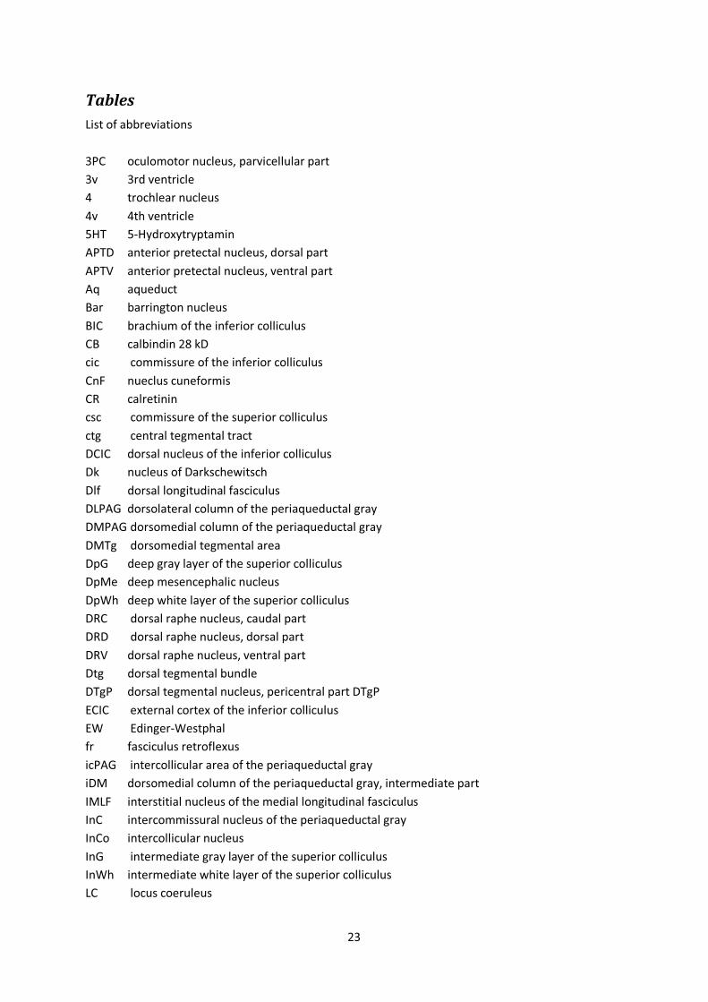
23
Tables List of abbreviations 3PC oculomotor nucleus, parvicellular part 3v 3rd ventricle 4 trochlear nucleus 4v 4th ventricle 5HT 5-Hydroxytryptamin APTD anterior pretectal nucleus, dorsal part APTV anterior pretectal nucleus, ventral part Aq aqueduct Bar barrington nucleus BIC brachium of the inferior colliculus CB calbindin 28 kD cic commissure of the inferior colliculus CnF nueclus cuneformis CR calretinin csc commissure of the superior colliculus ctg central tegmental tract DCIC dorsal nucleus of the inferior colliculus Dk nucleus of Darkschewitsch Dlf dorsal longitudinal fasciculus DLPAG dorsolateral column of the periaqueductal gray DMPAG dorsomedial column of the periaqueductal gray DMTg dorsomedial tegmental area DpG deep gray layer of the superior colliculus DpMe deep mesencephalic nucleus DpWh deep white layer of the superior colliculus DRC dorsal raphe nucleus, caudal part DRD dorsal raphe nucleus, dorsal part DRV dorsal raphe nucleus, ventral part Dtg dorsal tegmental bundle DTgP dorsal tegmental nucleus, pericentral part DTgP ECIC external cortex of the inferior colliculus EW Edinger-Westphal fr fasciculus retroflexus icPAG intercollicular area of the periaqueductal gray iDM dorsomedial column of the periaqueductal gray, intermediate part IMLF interstitial nucleus of the medial longitudinal fasciculus InC intercommissural nucleus of the periaqueductal gray InCo intercollicular nucleus InG intermediate gray layer of the superior colliculus InWh intermediate white layer of the superior colliculus LC locus coeruleus

24
LDTg laterodorsal tegmental nucleus LDTgV laterodorsal tegmental nucleus, ventral part LPAG lateral column of the periaqueductal gray LPLC lateral posterior thalamic nucleus, laterocaudal part LPMC lateral posterior thalamic nucleus, mediocaudal part LPMR lateral posterior thalamic nucleus, mediorostral part MCPC magnocellular nucleus of the posterior commissure Me5 mesencephalic trigeminal nucleus MG medial geniculate nucleus MGD medial geniculate nucleus, dorsal part MGM medial geniculate nucleus, medial part MiTg microcellular tegmental nucleus mlf medial longitudinal fasciculus MnR median raphe nucleus MPT medial pretectal nucleus NIc nucleus incertus, pars compacta NId nucleus incertus, pars disipata NOS nitric oxide synthase Op optic nerve layer of the superior colliculus OPT olivary pretectal nucleus OT optic tract PAG priaqueductal gray PBG parabigeminal nucleus pc posterior commissure PC paracentral thalamic nucleus PCom nucleus of the posterior commissure PDTg posterodorsal tegmental nucleus PF parafascicular thalamic nucleus PL paralemniscal nucleus PLi posterior limitans thalamic nucleus PmR `paramedian raphe PnO pontine reticular nucleus, oral part PnR pontine raphe nucleus PPT posterior pretectal nucleus PrC precommissural nucleus RtTg reticulotegmental nucleus of the pons Sag sagulum nucleus SC superior colliculus SCO subcommissural organ SG suprageniculate thalamic nucleus Sph sphenoid nucleus Su3 supraoculomotor periaqueductal gray Su3C supraoculomotor cap SuG superficial gray layer of the superior colliculius SuMM supramammillary nucleus, medial part

25
sumx supramammillary decussation syn synaptophysin RLN3 relaxin-3 TH tyrosine hydroxylase VLPAG ventrolateral column of the periaqueductal gray VTg ventral tegmental nucleus

26
Table 1. Details of the analysis and type of procedure conducted in these studies
Rat Immunohistochemical Methods Analysis
FH78, NIF2, PCM12
NIC6, MIC7, MIC8
Double immunohistochemistry - RLN3 and nNOS or CB-28kD or CR or TH or 5HT
Camera lucida
CNT2, CNT3, MIC9, MIC10
Double immunofluorescence - RLN3 and SP or nNOS or TH or 5HT
Confocal

27
Table 2. Semi-quantitative estimates of the distribution of RLN3 immunopositive fibers relative to neural markers in the pretectal area
Region/Marker RLN3 nNOS CB
MPT +++ - +
PPT + + +++
VLP ++ ++ +
OPT ++ + +
OT - - -
APTD - - -
APTV - - -
LPMR ++ - +++
LPLC ++ - +++
PLi ++ - -
Semi-quantitative estimates were made by visual observation of microscopic fields using a 20× objective: (-) no labelling, (+) 1-4 labeled fibers or cells, (++) 5-8 labeled fibers or cells, (+++) 9-12 labeled fibers or cells, (++++) more than 12 labeled fibers or cells

28
Table 3. Semi-quantitative estimates of the distribution of RLN3 immunopositive fibers relative to neural markers in the superior colliculus
Semi-quantitative estimates were made by visual observation of microscopic fields using a 20× objective: (-) no labelling, (+) 1-4 labeled fibers or cells, (++) 5-8 labeled fibers or cells, (+++) 9-12 labeled fibers or cells, (++++) more than 12 labeled fibers or cells
Region/Marker RLN3 CB nNOS
SuG Anterior Medial +++ +++ -
Lateral +++ +++ -
Posterior Medial ++ +++ -
Lateral ++ +++ -
Op Anterior Medial - - -
Lateral - + +
Posterior Medial + - -
Lateral - - ++
InG Anterior Medial +++ - +
Lateral + - -
Posterior Medial +++ + ++++
Lateral + + +
InWh Anterior Medial - - -
Lateral - + +
Posterior Medial - + ++
Lateral - - -
DpG Anterior Medial - - -
Lateral - + -
Posterior Medial + ++ ++
Lateral + - -
DpWh Anterior Medial + + -
Lateral - + -
Posterior Medial + ++ ++
Lateral + - +

29
Table 4. Semi-quantitative estimates of the distribution of RLN3 immunopositive fibers relative to neural markers in the inferior colliculus and lateral tegmental nuclei
Region/Marker nNOS RLN3 Region/Marker nNOS RLN3
DCIC + +++ Sagulum n. - +
InCo - - Parabigeminal n. - ++
CIC medial +++ - Brachium of IC ++ +
CIC lateral - - Cuneiform n. - ++
ECIC1 + +++ MiTg - -
ECIC2 + - Nuclei of
lateral lemniscus - -
ECIC3 +++ -
Semi-quantitative estimates were made by visual observation of microscopic fields using a 20× objective: (-) no labelling, (+) 1-4 labeled fibers or cells, (++) 5-8 labeled fibers or cells, (+++) 9-12 labeled fibers or cells, (++++) more than 12 labeled fibers or cells

30
Table 5. Semi-quantitative estimates of the distribution of RLN3 immunopositive fibers relative to neural markers in the periaqueductal gray
Region/Marker RLN3 TH nNOS
Dk - +++ ++
DL-PAG +++ - ++++
DM-PAG ( inner iDM) +++ - +
DM-PAG (outer oDM) + - -
EW ++ ++ ++
InC ++++ - -
LPAG (medial) + ++ +
LPAG (lateral) - - -
MCPC - - ++++
PCom - - -
PrC ++ - -
SCO - - -
Su3 - - -
Su3C + + +++
SuMM ++++ + ++
TLC ++ - -
VLPAG (medial) + + +
VLPAG (lateral) - - -
Semi-quantitative estimates were made by visual observation of microscopic fields using a 20× objective: (-) no labelling, (+) 1-4 labeled fibers or cells, (++) 5-8 labeled fibers or cells, (+++) 9-12 labeled fibers or cells, (++++) more than 12 labeled fibers or cells

31
Table 6. Semi-quantitative estimates of the distribution of RLN3 immunopositive fibers relative to neural markers in the pontine central gray
Region/Marker nNOS TH 5HT RLN3
LDTg ++++ ++ - ++
LDTgV + ++ - -
DRd ++ ++ + +++
DRv ++ - ++ +++
DTgP + - - -
DTgC - - - +
MnR ++ - + ++
PMR - - - +
PnR - - ++ ++
VTg - - - -
Bar - +++ - +
LC - ++++ - +
DRC - - ++ +++
Sph - - - -
NIc - - - ++++
NId - - - +++
PDTg + - - -
Semi-quantitative estimates were made by visual observation of microscopic fields using a 20× objective: (-) no labelling, (+) 1-4 labeled fibers or cells, (++) 5-8 labeled fibers or cells, (+++) 9-12 labeled fibers or cells, (++++) more than 12 labeled fibers or cells

32
References An X, Bandler R, Ongur D, Price JL. (1998) Prefrontal cortical projections to longitudinal columns in the midbrain periaqueductal gray in macaque monkeys. J. Comp. Neurol. 401: 455-479..
Aparicio MA, Saldana E. (2009) Tectotectal neurons and projections: a proposal to establish a consistent nomenclature. Anat. Rec. (Hoboken) 292: 175-177.
Aparicio MA, Vinuela A, Saldana E. (2010) Projections from the inferior colliculus to the tectal longitudinal column in the rat. Neuroscience 166: 653-664.
Arnault P, Roger M. (1987) The connections of the peripeduncular area studied by retrograde and anterograde transport in the rat. J. Comp. Neurol. 258: 463-476.
Aston-Jones G, Bloom FE. (1981) Activity of norepinephrine-containing locus coeruleus neurons in behaving rats anticipates fluctuations in the sleep-waking cycle. J. Neurosci. 1: 876-886. .
Bandler R, Keay KA. (1996) Columnar organization in the midbrain periaqueductal gray and the integration of emotional expression. Prog. Brain Res. 107: 285-300.
Bandler R, Shipley MT. (1994) Columnar organization in the midbrain periaqueductal gray: modules for emotional expression? Trends Neurosci. 17: 379-389.
Bandler R, Carrive P. (1988) Integrated defence reaction elicited by excitatory amino acid microinjection in the midbrain periaqueductal grey region of the unrestrained cat. Brain Res. 439: 95-106.
Bandler R, Keay KA, Floyd N, Price J. (2000) Central circuits mediating patterned autonomic activity during active vs. passive emotional coping. Brain Res. Bull. 53: 95-104.
Banerjee A, Shen PJ, Ma S, Bathgate RA, Gundlach AL. (2010) Swim stress excitation of nucleus incertus and rapid induction of relaxin-3 expression via CRF1 activation. Neuropharmacology 58: 145-155.
Banerjee A, Shen PJ, Ma S, Bathgate RA, Gundlach AL. (2009) Swim stress excitation of nucleus incertus and rapid induction of relaxin-3 expression via CRF(1) activation. Neuropharmacology .
Bathgate RA, Samuel CS, Burazin TC, Gundlach AL, Tregear GW. (2003) Relaxin: new peptides, receptors and novel actions. Trends Endocrinol. Metab. 14: 207-213.
Beneyto M, Winer JA, Larue DT, Prieto JJ. (1998) Auditory connections and neurochemistry of the sagulum. J. Comp. Neurol. 401: 329-351.
Berman N, Cynader M. (1972). Comparison of receptive-field organization of the superior colliculus in Siamese and normal cats. J. Physiol. 224: 363-389 .
Bittencourt JC, Sawchenko PE. (2000) Do centrally administered neuropeptides access cognate receptors?: an analysis in the central corticotropin-releasing factor system. J. Neurosci. 20: 1142-1156.

33
Blanchard RJ, Flannelly KJ, Blanchard DC. (1986) Defensive behavior of laboratory and wild Rattus norvegicus. J. Comp. Psychol. 100: 101-107.
Blasiak A, Blasiak T, Czubak W, Gundlach A, Lewandowski M. (2009) Relaxin-3 innervation of the intergeniculate leaflet in the rat. .
Brailoiu E, Dun SL, Gao X, Brailoiu GC, Li JG, Luo JJ, Yang J, Chang JK, Liu-Chen LY, Dun NJ. (2009) C-peptide of preproinsulin-like peptide 7: localization in the rat brain and activity in vitro. Neuroscience 159: 492-500.
Burazin TC, Bathgate RA, Macris M, Layfield S, Gundlach AL, Tregear GW. (2002) Restricted, but abundant, expression of the novel rat gene-3 (R3) relaxin in the dorsal tegmental region of brain. J. Neurochem. 82: 1553-1557.
Burlet S, Tyler CJ, Leonard CS. (2002) Direct and indirect excitation of laterodorsal tegmental neurons by Hypocretin/Orexin peptides: implications for wakefulness and narcolepsy. J. Neurosci. 22: 2862-2872.
Cameron AA, Khan IA, Westlund KN, Willis WD. (1995) The efferent projections of the periaqueductal gray in the rat: a Phaseolus vulgaris-leucoagglutinin study. II. Descending projections. J. Comp. Neurol. 351: 585-601.
Cano G, Mochizuki T, Saper CB. (2008) Neural circuitry of stress-induced insomnia in rats. J. Neurosci. 28: 10167-10184.
Canteras NS. 2002. The medial hypothalamic defensive system: hodological organization and functional implications. Pharmacol. Biochem. Behav. 71: 481-491. *
Canteras NS, Goto M. (1999a). Fos-like immunoreactivity in the periaqueductal gray of rats exposed to a natural predator. Neuroreport 10: 413-418.
Canteras NS, Goto M. (1999b) Connections of the precommissural nucleus. J. Comp. Neurol. 408: 23-45.
Canteras NS, Swanson LW. (1992) The dorsal premammillary nucleus: an unusual component of the mammillary body. Proc. Natl. Acad. Sci. U. S. A. 89: 10089-10093.
Carrive P, Leung P, Harris J, Paxinos G. (1997) Conditioned fear to context is associated with increased Fos expression in the caudal ventrolateral region of the midbrain periaqueductal gray. Neuroscience 78: 165-177.
Comoli E, Ribeiro-Barbosa ER, Canteras NS. (2003) Predatory hunting and exposure to a live predator induce opposite patterns of Fos immunoreactivity in the PAG. Behav. Brain Res. 138: 17-28.
Daly C, Ziff EB. (2002) Ca2+-dependent formation of a dynamin-synaptophysin complex: potential role in synaptic vesicle endocytosis. J. Biol. Chem. 277: 9010-9015.
Daly C, Sugimori M, Moreira JE, Ziff EB, Llinas R. (2000) Synaptophysin regulates clathrin-independent endocytosis of synaptic vesicles. Proc. Natl. Acad. Sci. U. S. A. 97: 6120-6125.

34
De Oca BM, DeCola JP, Maren S, Fanselow MS. (1998) Distinct regions of the periaqueductal gray are involved in the acquisition and expression of defensive responses. J. Neurosci. 18: 3426-3432.
Dean P, Redgrave P, Westby GW. (1989) Event or emergency? Two response systems in the mammalian superior colliculus. Trends Neurosci. 12: 137-147.
Doubell TP, Skaliora I, Baron J, King AJ. (2003) Functional connectivity between the superficial and deeper layers of the superior colliculus: an anatomical substrate for sensorimotor integration. J. Neurosci. 23: 6596-6607.
Drager UC, Hubel DH. (1975) Responses to visual stimulation and relationship between visual, auditory, and somatosensory inputs in mouse superior colliculus. J. Neurophysiol. 38: 690-713.
Floyd NS, Price JL, Ferry AT, Keay KA, Bandler R. (2001) Orbitomedial prefrontal cortical projections to hypothalamus in the rat. J. Comp. Neurol. 432: 307-328.
Foote SL, Aston-Jones G, Bloom FE. (1980) Impulse activity of locus coeruleus neurons in awake rats and monkeys is a function of sensory stimulation and arousal. Proc. Natl. Acad. Sci. U. S. A. 77: 3033-3037.
Furigo IC, de Oliveira WF, de Oliveira AR, Comoli E, Baldo MV, Mota-Ortiz SR, Canteras NS. (2010) The role of the superior colliculus in predatory hunting. Neuroscience 165: 1-15.
Gabbott PL, Warner TA, Jays PR, Salway P, Busby SJ. (2005) Prefrontal cortex in the rat: projections to subcortical autonomic, motor, and limbic centers. J. Comp. Neurol. 492: 145-177.
Gamlin PD. (2006) The pretectum: connections and oculomotor-related roles. Prog. Brain Res. 151: 379-405.
Garcia Del Cano G, Gerrikagoitia I, Alonso-Cabria A, Martinez-Millan L. (2006) Organization and origin of the connection from the inferior to the superior colliculi in the rat. J. Comp. Neurol. 499: 716-731.
Goto M, Swanson LW, Canteras NS. (2001) Connections of the nucleus incertus. J. Comp. Neurol. 438: 86-122.
Grantyn R. (1988) Gaze control through superior colliculus: structure and function. Rev. Oculomot. Res. 2: 273-333.
Halliday G, Harding A, and Paxinos G. Serotonin and tachykinin systems. In: The Rat Nervous System, Ed Paxinos G. San Diego: Academic Press, 1995, p. 929-974.
Haugaard-Kedstrom LM, Shabanpoor F, Hossain MA, Clark RJ, Ryan PJ, Craik DJ, Gundlach AL, Wade JD, Bathgate RA, Rosengren KJ. (2011) Design, synthesis, and characterization of a single-chain peptide antagonist for the relaxin-3 receptor RXFP3. J. Am. Chem. Soc. 133: 4965-4974.
Hida T, Takahashi E, Shikata K, Hirohashi T, Sawai T, Seiki T, Tanaka H, Kawai T, Ito O, Arai T, Yokoi A, Hirakawa T, Ogura H, Nagasu T, Miyamoto N, Kuromitsu J. (2006) Chronic intracerebroventricular administration of relaxin-3 increases body weight in rats. J. Recept. Signal Transduct. Res. 26: 147-158.

35
Jacobs BL, Azmitia EC. (1992) Structure and function of the brain serotonin system. Physiol. Rev. 72: 165-229.
Jahn R, Schiebler W, Ouimet C, Greengard P. (1985) A 38,000-dalton membrane protein (p38) present in synaptic vesicles. Proc. Natl. Acad. Sci. U. S. A. 82: 4137-4141.
Jones BE, Webster HH. (1988) Neurotoxic lesions of the dorsolateral pontomesencephalic tegmentum-cholinergic cell area in the cat. I. Effects upon the cholinergic innervation of the brain. Brain Res. 451: 13-32.
Jones BE, Yang TZ. (1985) The efferent projections from the reticular formation and the locus coeruleus studied by anterograde and retrograde axonal transport in the rat. J. Comp. Neurol. 242: 56-92.
Keay KA, Bandler R. (1993) Deep and superficial noxious stimulation increases Fos-like immunoreactivity in different regions of the midbrain periaqueductal grey of the rat. Neurosci. Lett. 154: 23-26.
Keay KA, Clement CI, Owler B, Depaulis A, Bandler R. (1994) Convergence of deep somatic and visceral nociceptive information onto a discrete ventrolateral midbrain periaqueductal gray region. Neuroscience 61: 727-732.
King AJ. (1999) Sensory experience and the formation of a computational map of auditory space in the brain. Bioessays 21: 900-911.
Klooster J, Vrensen GF, Muller LJ, van der Want JJ. (1995) Efferent projections of the olivary pretectal nucleus in the albino rat subserving the pupillary light reflex and related reflexes. A light microscopic tracing study. Brain Res. 688: 34-46.
Krout KE, Loewy AD, Westby GW, Redgrave P. (2001) Superior colliculus projections to midline and intralaminar thalamic nuclei of the rat. J. Comp. Neurol. 431: 198-216.
Kuei C, Sutton S, Bonaventure P, Pudiak C, Shelton J, Zhu J, Nepomuceno D, Wu J, Chen J, Kamme F, Seierstad M, Hack MD, Bathgate RA, Hossain MA, Wade JD, Atack J, Lovenberg TW, Liu C. 2007. R3(BDelta23 27)R/I5 chimeric peptide, a selective antagonist for GPCR135 and GPCR142 over relaxin receptor LGR7: in vitro and in vivo characterization. J. Biol. Chem. 282: 25425-25435. .
LeDoux JE, Sakaguchi A, Reis DJ. (1984) Subcortical efferent projections of the medial geniculate nucleus mediate emotional responses conditioned to acoustic stimuli. J. Neurosci. 4: 683-698.
Ledoux JE, Ruggiero DA, Forest R, Stornetta R, Reis DJ. (1987) Topographic organization of convergent projections to the thalamus from the inferior colliculus and spinal cord in the rat. J. Comp. Neurol. 264: 123-146.
Liu C, Eriste E, Sutton S, Chen J, Roland B, Kuei C, Farmer N, Jornvall H, Sillard R, Lovenberg TW. (2003) Identification of relaxin-3/INSL7 as an endogenous ligand for the orphan G-protein-coupled receptor GPCR135. J. Biol. Chem. 278: 50754-50764.
Ma S, Sang Q, Lanciego JL, Gundlach AL. (2009a) Localization of relaxin-3 in brain of Macaca fascicularis: identification of a nucleus incertus in primate. J. Comp. Neurol. 517: 856-872.

36
Ma S, Olucha-Bordonau FE, Hossain MA, Lin F, Kuei C, Liu C, Wade JD, Sutton SW, Nunez A, Gundlach AL. (2009b) Modulation of hippocampal theta oscillations and spatial memory by relaxin-3 neurons of the nucleus incertus. Learn. Mem. 16: 730-742.
Ma S, Bonaventure P, Ferraro T, Shen PJ, Burazin TC, Bathgate RA, Liu C, Tregear GW, Sutton SW, Gundlach AL. (2007) Relaxin-3 in GABA projection neurons of nucleus incertus suggests widespread influence on forebrain circuits via G-protein-coupled receptor-135 in the rat. Neuroscience 144: 165-190.
Marchant EG, Morin LP. (1999) The hamster circadian rhythm system includes nuclei of the subcortical visual shell. J. Neurosci. 19: 10482-10493.
McGowan BM, Stanley SA, Smith KL, White NE, Connolly MM, Thompson EL, Gardiner JV, Murphy KG, Ghatei MA, Bloom SR. (2005) Central relaxin-3 administration causes hyperphagia in male Wistar rats. Endocrinology 146: 3295-3300.
McGowan BM, Stanley SA, Smith KL, Minnion JS, Donovan J, Thompson EL, Patterson M, Connolly MM, Abbott CR, Small CJ, Gardiner JV, Ghatei MA, Bloom SR. (2006) Effects of acute and chronic relaxin-3 on food intake and energy expenditure in rats. Regul. Pept. 136: 72-77.
Mendelson WB, Martin JV, Perlis M, Wagner R. (1987) Arousal induced by injection of triazolam into the dorsal raphe nucleus of rats. Neuropsychopharmacology 1: 85-88.
Meredith MA, Stein BE. (1990) The visuotopic component of the multisensory map in the deep laminae of the cat superior colliculus. J. Neurosci. 10: 3727-3742.
Mikkelsen JD, Vrang N. (1994). A direct pretectosuprachiasmatic projection in the rat. Neuroscience 62: 497-505.
Mikkelsen JD, Vrang N, Mrosovsky N. (1998) Expression of Fos in the circadian system following nonphotic stimulation. Brain Res. Bull. 47: 367-376.
Mota-Ortiz SR, Sukikara MH, Felicio LF, Canteras NS. (2009) Afferent connections to the rostrolateral part of the periaqueductal gray: a critical region influencing the motivation drive to hunt and forage. Neural Plast. 2009: 612698.
Motta SC, Goto M, Gouveia FV, Baldo MV, Canteras NS, Swanson LW. (2009) Dissecting the brain's fear system reveals the hypothalamus is critical for responding in subordinate conspecific intruders. Proc. Natl. Acad. Sci. U. S. A. 106: 4870-4875.
Munoz DP, Wurtz RH. (1992) Role of the rostral superior colliculus in active visual fixation and execution of express saccades. J. Neurophysiol. 67: 1000-1002. .
Munoz DP, Waitzman DM, Wurtz RH. (1996) Activity of neurons in monkey superior colliculus during interrupted saccades. J. Neurophysiol. 75: 2562-2580.
Nelson LE, Lu J, Guo T, Saper CB, Franks NP, Maze M. (2003) The alpha2-adrenoceptor agonist dexmedetomidine converges on an endogenous sleep-promoting pathway to exert its sedative effects. Anesthesiology 98: 428-436.
37
Nelson LE, Guo TZ, Lu J, Saper CB, Franks NP, Maze M. (2002). The sedative component of anesthesia is mediated by GABA(A) receptors in an endogenous sleep pathway. Nat. Neurosci. 5: 979-984. .
Olucha-Bordonau FE, Teruel V, Barcia-Gonzalez J, Ruiz-Torner A, Valverde-Navarro AA, Martinez-Soriano F. (2003) Cytoarchitecture and efferent projections of the nucleus incertus of the rat. J. Comp. Neurol. 464: 62-97.
Onstott D, Mayer B, Beitz AJ. (1993) Nitric oxide synthase immunoreactive neurons anatomically define a longitudinal dorsolateral column within the midbrain periaqueductal gray of the rat: analysis using laser confocal microscopy. Brain Res. 610: 317-324.
Paxinos G., Ashwell KWS, Kirkcaldie M (2008) Chemoarchitectonic Atlas of the Rat Brain. Academic Press 6th Ed, San Diego
Pennuto M, Bonanomi D, Benfenati F, Valtorta F. (2003). Synaptophysin I controls the targeting of VAMP2/synaptobrevin II to synaptic vesicles. Mol. Biol. Cell 14: 4909-4919.
Roldan M, Reinoso-Suarez F, Tortelly A. (1983) Parabigeminal projections to the superior colliculus in the cat. Brain Res. 280: 1-13.
Ruiz-Torner A, Olucha-Bordonau F, Valverde-Navarro AA, Martinez-Soriano F. (2001) The chemical architecture of the rat's periaqueductal gray based on acetylcholinesterase histochemistry: a quantitative and qualitative study. J. Chem. Neuroanat. 21: 295-312.
Ryan PJ, Ma S, Olucha-Bordonau FE, Gundlach AL. (2011) Nucleus incertus--an emerging modulatory role in arousal, stress and memory. Neurosci. Biobehav. Rev. 35: 1326-1341.
Saldana E, Vinuela A, Marshall AF, Fitzpatrick DC, Aparicio MA. (2007) The TLC: a novel auditory nucleus of the mammalian brain. J. Neurosci. 27: 13108-13116.
Sesack SR, Deutch AY, Roth RH, Bunney BS. (1989) Topographical organization of the efferent projections of the medial prefrontal cortex in the rat: an anterograde tract-tracing study with Phaseolus vulgaris leucoagglutinin. J. Comp. Neurol. 290: 213-242.
Singewald N, Sharp T. (2000) Neuroanatomical targets of anxiogenic drugs in the hindbrain as revealed by Fos immunocytochemistry. Neuroscience 98: 759-770.
Skaliora I, Doubell TP, Holmes NP, Nodal FR, King AJ. (2004) Functional topography of converging visual and auditory inputs to neurons in the rat superior colliculus. J. Neurophysiol. 92: 2933-2946.
Smith CM, Lawrence AJ, Sutton SW, Gundlach AL. (2009) Behavioral phenotyping of mixed background (129S5:B6) relaxin-3 knockout mice. Ann. N. Y. Acad. Sci. 1160: 236-241.
Smith CM, Hosken IT, Sutton SW, Lawrence AJ, Gundlach AL. (2012) Relaxin-3 null mutation mice display a circadian hypoactivity phenotype. Genes Brain Behav. 11: 94-104.
Smith CM, Ryan PJ, Hosken IT, Ma S, Gundlach AL. (2011) Relaxin-3 systems in the brain--the first 10 years. J. Chem. Neuroanat. 42: 262-275.

38
Smith CM, Shen PJ, Ma S, Sutton SW, Gundlach AL. (2009) Verification of a relaxin-3 knockout/LacZ reporter mouse as a model of relaxin-3 deficiency. Ann. N. Y. Acad. Sci. 1160: 259-260.
Smith CM, Shen PJ, Banerjee A, Bonaventure P, Ma S, Bathgate RA, Sutton SW, Gundlach AL. (2010) Distribution of relaxin-3 and RXFP3 within arousal, stress, affective, and cognitive circuits of mouse brain. J. Comp. Neurol. 518: 4016-4045.
Stein BE, Meredith MA. (1990) Multisensory integration. Neural and behavioral solutions for dealing with stimuli from different sensory modalities. Ann. N. Y. Acad. Sci. 608: 51-65; discussion 65-70.
Stein BE, Meredith MA, Wallace MT. (1993) The visually responsive neuron and beyond: multisensory integration in cat and monkey. Prog. Brain Res. 95: 79-90.
Steriade M, McCarley RW (1990) In: Brainstem Control of Wakefulness and Sleep. New York: Plenum.
Subramanian HH, Balnave RJ, Holstege G. (2008) The midbrain periaqueductal gray control of respiration. J. Neurosci. 28: 12274-12283.
Sukikara MH, Mota-Ortiz SR, Baldo MV, Felicio LF, Canteras NS. (2006) A role for the periaqueductal gray in switching adaptive behavioral responses. J. Neurosci. 26: 2583-2589.
Sutton SW, Bonaventure P, Kuei C, Roland B, Chen J, Nepomuceno D, Lovenberg TW, Liu C. (2004) Distribution of G-protein-coupled receptor (GPCR)135 binding sites and receptor mRNA in the rat brain suggests a role for relaxin-3 in neuroendocrine and sensory processing. Neuroendocrinology 80: 298-307.
Tanaka M, Iijima N, Miyamoto Y, Fukusumi S, Itoh Y, Ozawa H, Ibata Y. (2005) Neurons expressing relaxin 3/INSL 7 in the nucleus incertus respond to stress. Eur. J. Neurosci. 21: 1659-1670.
Thomas L, Betz H. (1990) Synaptophysin binds to physophilin, a putative synaptic plasma membrane protein. J. Cell Biol. 111: 2041-2052.
Thornton SK, Withington DJ. (1996) The role of the external nucleus of the inferior colliculus in the construction of the superior collicular auditory space map in the guinea-pig. Neurosci. Res. 25: 239-246.
Usunoff KG, Schmitt O, Itzev DE, Rolfs A, Wree A. (2007) Efferent connections of the parabigeminal nucleus to the amygdala and the superior colliculus in the rat: a double-labeling fluorescent retrograde tracing study. Brain Res. 1133: 87-91.
Usunoff KG, Itzev DE, Rolfs A, Schmitt O, Wree A. (2006) Brain stem afferent connections of the amygdala in the rat with special references to a projection from the parabigeminal nucleus: a fluorescent retrograde tracing study. Anat. Embryol. (Berl) 211: 475-496.
Van Pett K, Viau V, Bittencourt JC, Chan RK, Li HY, Arias C, Prins GS, Perrin M, Vale W, Sawchenko PE. (2000) Distribution of mRNAs encoding CRF receptors in brain and pituitary of rat and mouse. J. Comp. Neurol. 428: 191-212. .

39
VanderHorst VG, Schasfoort FC, Meijer E, van Leeuwen FW, Holstege G. (1998) Estrogen receptor-alpha-immunoreactive neurons in the periaqueductal gray of the adult ovariectomized female cat. Neurosci. Lett. 240: 13-16.
Vertes RP, Kocsis B. (1994). Projections of the dorsal raphe nucleus to the brainstem: PHA-L analysis in the rat. J. Comp. Neurol. 340: 11-26.
Vertes RP, Linley SB, Hoover WB. (2010) Pattern of distribution of serotonergic fibers to the thalamus of the rat. Brain Struct. Funct.
Vertes RP, Kinney GG, Kocsis B, Fortin WJ. (1994) Pharmacological suppression of the median raphe nucleus with serotonin1A agonists, 8-OH-DPAT and buspirone, produces hippocampal theta rhythm in the rat. Neuroscience 60: 441-451.
Wainer BH, Mesulam M-M (1990) Ascending cholinergic pathways in the rat brain. In: Brain Cholinergic Systems (Steriade M, Biesold D, Eds), pp 65-119. New York: Oxford University Press.
Wallace MT, Meredith MA, Stein BE. (1998) Multisensory integration in the superior colliculus of the alert cat. J. Neurophysiol. 80: 1006-1010.
Webster HH, Jones BE. (1988) Neurotoxic lesions of the dorsolateral pontomesencephalic tegmentum-cholinergic cell area in the cat. II. Effects upon sleep-waking states. Brain Res. 458: 285-302.
Wenstrup JJ, Larue DT, Winer JA. (1994) Projections of physiologically defined subdivisions of the inferior colliculus in the mustached bat: targets in the medial geniculate body and extrathalamic nuclei. J. Comp. Neurol. 346: 207-236.
Wiedenmann B, Franke WW. (1985) Identification and localization of synaptophysin, an integral membrane glycoprotein of Mr 38,000 characteristic of presynaptic vesicles. Cell 41: 1017-1028. .
Winer JA, Saint Marie RL, Larue DT, Oliver DL. (1996) GABAergic feedforward projections from the inferior colliculus to the medial geniculate body. Proc. Natl. Acad. Sci. U. S. A. 93: 8005-8010.
Zhao Z, Yang Y, Walker DL, Davis M. (2009) Effects of substance P in the amygdala, ventromedial hypothalamus, and periaqueductal gray on fear-potentiated startle. Neuropsychopharmacology 34: 331-340.

PF
PrC
OPT
APTD
APTV
LPMRLPMC
LPLC
OT
MPT
DpMe
OT
VLP
OPTAPTD
APTV
LPMCpc
AqMCPC
PCom
MPT
PAG PrC
LPMR
OT
PPTVLP
APTD PLi
APTV
LPMC
AqMCPC
PCom
MPT
PAG
SC
MG
pc
csc
SC
MPT
AqAPTV
APTDPLi
SG
MGD
LPMC
OT
VLPPPT
PCom
MCPC
PL
A
B
C
D
NOS/RLN3
PPT
TLC
Figure 1. Distribution of RLN3-positive fibers within the pretectum mapped in sections immunostained for RLN3 and nNOS. Immunoreactivity was mapped using camera lucida and a 20× objective. RLN3 fibers and soma are indicated in red and nNOS positive soma and primary processes in blue. Boundaries of pretectal divisions were drawn in black and revectorized (for further details see Materials and Methods). The MCPC and PPT, containing nNOS-positive soma, are present at rostral levels, while an nNOS- and RLN3-immunopositive area identified as ‘the ventrolateral pretectal nucleus’ is present at caudal levels ventrolateral to SC. At level 4, SC stratification is clearly evident and nNOS positive soma in the MCPC are less abundant. Intense RLN3 labeling was observed in MPT and some labeled fibers were detected in the OPT and VLP. A RLN3 innervation reaches the LPMR and PLi, whereas the APT and OT are unlabeled. Scale bar: 500 μm. For abbreviations, see list.
APTD anterior pretectal nucleus, dorsal partAPTV anterior pretectal nucleus, ventral partAq aqueductcsc commissure of the superior colliculusDpMe deep mesencephalic nucleusLPLC lateral posterior thalamic nucleus, laterocaudal partLPMC lateral posterior thalamic nucleus, mediocaudal partLPMR lateral posterior thalamic nucleus, mediorostral partMCPC magnocellular nucleus of the posterior commissureMG medial geniculate nucleusMGD medial geniculate nucleus, dorsal partMPT medial pretectal nucleusOPT olivary pretectal nucleusOT optic tractPAG priaqueductal graypc posterior commissurePCom nucleus of the posterior commissurePF parafascicular thalamic nucleusPL paralemniscal nucleusPLi posterior limitans thalamic nucleusPPT posterior pretectal nucleusPrC precommissural nucleusSC superior colliculusSG suprageniculate thalamic nucleusTLC tectal longitudinal columnVLP ventroposterolateral nucleus
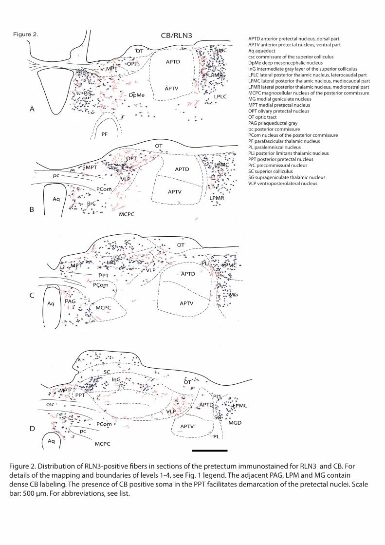
PF
PrC
OPT APTD
APTV
LPMR
LPLC
MPT
DpMe
LPMC
OT
VLP
OPT
APTD
APTV
LPMC
pc
Aq
MCPC
PCom
MPT
PrCLPMR
OT
PPTVLP
APTD
PLi
APTV
LPMC
AqMCPC
PCom
MPT
PAG
SC
MG
pc
csc
SC
MPT
Aq
APTV
APTD
PLi
SGMGD
LPMC
OT
VLP
PCom
MCPCPL
OT
A
B
C
D
CB/RLN3
PPT
InG
InG
Figure 2.
Figure 2. Distribution of RLN3-positive fibers in sections of the pretectum immunostained for RLN3 and CB. For details of the mapping and boundaries of levels 1-4, see Fig. 1 legend. The adjacent PAG, LPM and MG contain dense CB labeling. The presence of CB positive soma in the PPT facilitates demarcation of the pretectal nuclei. Scale bar: 500 μm. For abbreviations, see list.
APTD anterior pretectal nucleus, dorsal partAPTV anterior pretectal nucleus, ventral partAq aqueductcsc commissure of the superior colliculusDpMe deep mesencephalic nucleusInG intermediate gray layer of the superior colliculusLPLC lateral posterior thalamic nucleus, laterocaudal partLPMC lateral posterior thalamic nucleus, mediocaudal partLPMR lateral posterior thalamic nucleus, mediorostral partMCPC magnocellular nucleus of the posterior commissureMG medial geniculate nucleusMPT medial pretectal nucleusOPT olivary pretectal nucleusOT optic tractPAG priaqueductal graypc posterior commissurePCom nucleus of the posterior commissurePF parafascicular thalamic nucleusPL paralemniscal nucleusPLi posterior limitans thalamic nucleusPPT posterior pretectal nucleusPrC precommissural nucleusSC superior colliculusSG suprageniculate thalamic nucleusVLP ventroposterolateral nucleus
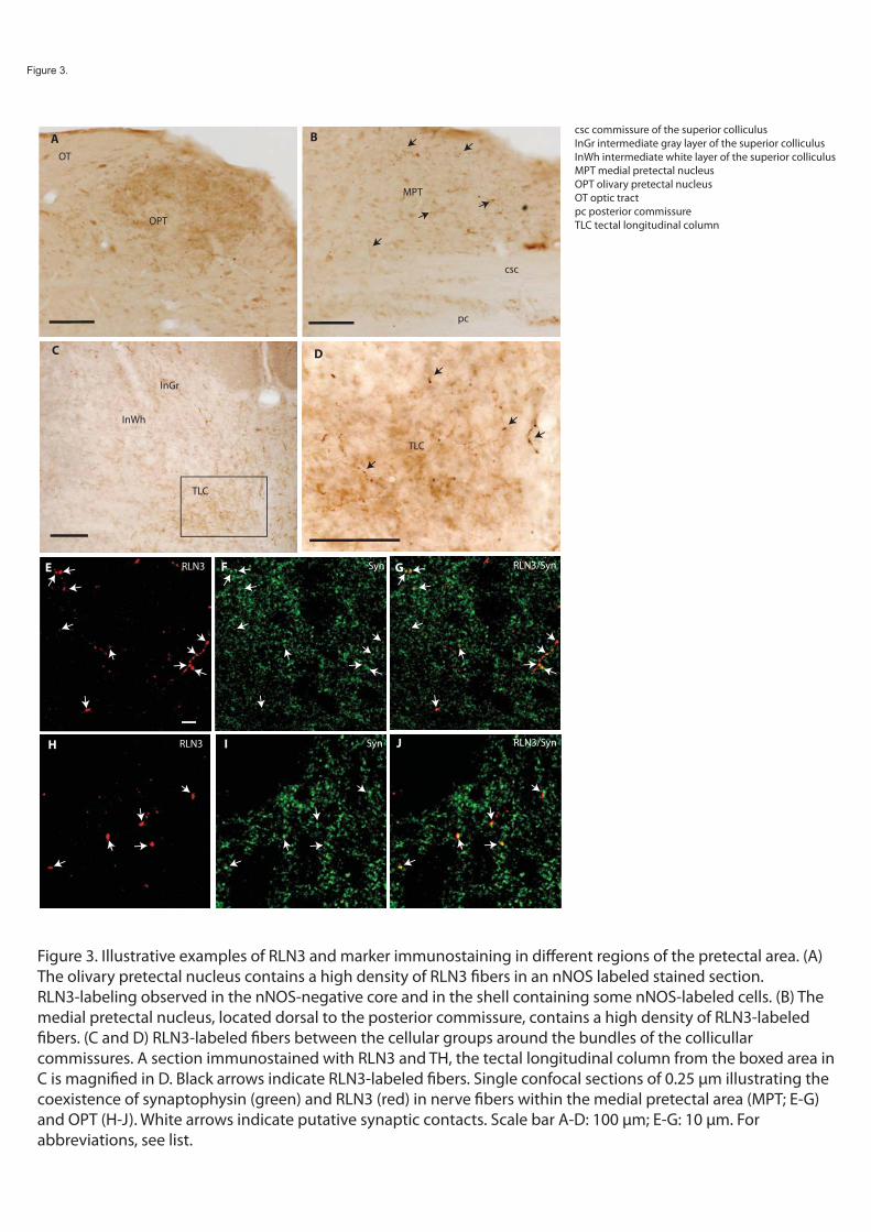
csc
pc
OPT
OT
A B
C D
InGr
InWh
TLC
TLC
MPT
RLN3/SynSyn RLN3
RLN3/Syn RLN3 Syn
E F G
H I J
Figure 3.
Figure 3. Illustrative examples of RLN3 and marker immunostaining in different regions of the pretectal area. (A) The olivary pretectal nucleus contains a high density of RLN3 fibers in an nNOS labeled stained section. RLN3-labeling observed in the nNOS-negative core and in the shell containing some nNOS-labeled cells. (B) The medial pretectal nucleus, located dorsal to the posterior commissure, contains a high density of RLN3-labeled fibers. (C and D) RLN3-labeled fibers between the cellular groups around the bundles of the collicullar commissures. A section immunostained with RLN3 and TH, the tectal longitudinal column from the boxed area in C is magnified in D. Black arrows indicate RLN3-labeled fibers. Single confocal sections of 0.25 μm illustrating the coexistence of synaptophysin (green) and RLN3 (red) in nerve fibers within the medial pretectal area (MPT; E-G) and OPT (H-J). White arrows indicate putative synaptic contacts. Scale bar A-D: 100 μm; E-G: 10 μm. Forabbreviations, see list.
csc commissure of the superior colliculusInGr intermediate gray layer of the superior colliculusInWh intermediate white layer of the superior colliculusMPT medial pretectal nucleusOPT olivary pretectal nucleusOT optic tractpc posterior commissureTLC tectal longitudinal column
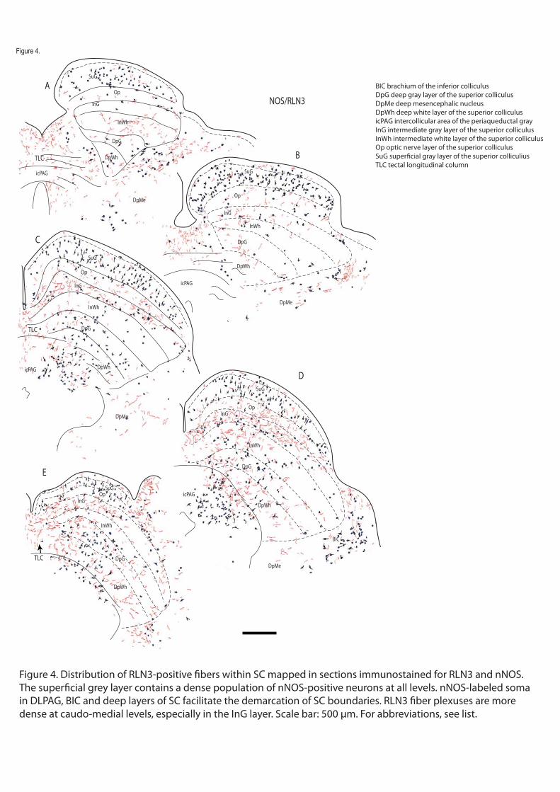
DpWh
DpG
InWh
InG
Op
SuG
DpMe
icPAG
DpWh
DpG
InWh
InG
Op
SuG
DpMe
icPAG
DpWh
DpG
InWh
InG
Op
SuG
DpMe
icPAG
DpWh
DpG
InWh
InGOp
SuG
DpMe
icPAG
DpWh
DpG
InWh
InGOp
SuG
BIC
A
B
C
D
NOS/RLN3
E
TLC
TLC
TLC
Figure 4.
Figure 4. Distribution of RLN3-positive fibers within SC mapped in sections immunostained for RLN3 and nNOS. The superficial grey layer contains a dense population of nNOS-positive neurons at all levels. nNOS-labeled soma in DLPAG, BIC and deep layers of SC facilitate the demarcation of SC boundaries. RLN3 fiber plexuses are more dense at caudo-medial levels, especially in the InG layer. Scale bar: 500 μm. For abbreviations, see list.
BIC brachium of the inferior colliculusDpG deep gray layer of the superior colliculusDpMe deep mesencephalic nucleusDpWh deep white layer of the superior colliculusicPAG intercollicular area of the periaqueductal grayInG intermediate gray layer of the superior colliculusInWh intermediate white layer of the superior colliculusOp optic nerve layer of the superior colliculusSuG superficial gray layer of the superior colliculiusTLC tectal longitudinal column

DpWh
DpG
InWh
InG
Op
SuG
DpMe
PAG
DpWh
DpG
InWh
InG
Op
SuG
DpMe
icPAG
DpWh
DpG
InWh
InG
Op SuG
DpMe
icPAG
DpWh
DpG
InWh
InGOp
SuGBIC
DpWh
DpG
InWh
InG
Op
SuG
DpMePrC
pc
MPT
A
B
C
D
CB/RLN3
E
TLC
TLC
Figure 5.
Figure 5. Distribution of RLN3-positive fibers within SC mapped in sections immunostained for RLN3 and CB. Small and large CB-positive soma were observed in the SuG and InG layers, respectively. CBlabeling was virtually absent in the OP and InW layers. RLN3 fibers are concentrated in the medial aspects of the InG, where larger CB neurons were observed. Scale bar: 500 μm. For abbreviations, see list
BIC brachium of the inferior colliculusDpG deep gray layer of the superior colliculusDpMe deep mesencephalic nucleusDpWh deep white layer of the superior colliculusicPAG intercollicular area of the periaqueductal grayInG intermediate gray layer of the superior colliculusInWh intermediate white layer of the superior colliculusMPT medial pretectal nucleusOp optic nerve layer of the superior colliculuspc posterior commissurePrC precommissural nucleusSuG superficial gray layer of the superior colliculiusTLC tectal longitudinal column

SuG
Op
InG
NOS-R3
InWh
InG
Op
SuG
NOS-R3
InWh
InG
Op
SuGCB-R3 CB-R3
A B
C D
RLN3/SynRLN3 Syn
Figure 6.
Figure 6. Illustrative examples of RLN3 and marker immunostaining in different areas of SC. (A and B) SuG and particularly the InG contain the highest density of RLN3-labeled fibers (indicated by black arrows). (C and D) In deep layers of SC, a high concentration of RLN3-labeled fibers was present in the InG; and in some cases fibers were closely apposed to CR-labeled soma. (E-G) Single confocal sections of 0.25 μm illustrating the colocalization of synaptophysin (green) and RLN3 (red) in nerve fibers in the caudomedial InG layer. White arrows indicate putative synaptic contacts; stars indicate unlabeled neuronal soma. Scale bar A-D: 100 μm; E-G: 10 μm. For abbreviations, see list.
InG intermediate gray layer of the superior colliculusInWh intermediate white layer of the superior colliculusOp optic nerve layer of the superior colliculusSuG superficial gray layer of the superior colliculius

InCo
MiTg PBG
BIC
2
CnF
DLPAG
MiTg
PBGCnF
DLPAG
1
CnF
cic
cicDCIC
ECIC 23
Sag
1
ECIC
12
3
cic
ECIC
12
3
cic
DCIC
DCIC
cic
CnF
ECIC
23
DMPAGDLPAG
LPAG
VLPAG
DCIC
A
B
C D
NOS/RLN3
Figure 7.
Figure 7. Distribution of RLN3-positive fibers within IC mapped in sections immunostained for RLN3 and nNOS. nNOS-positive populations are dense in the medial CIC and ECIC3 , but completely absent in the lateral half of the CIC. The RLN3 innervation avoids the central area of the IC and is particularly dense in layer 1 of DCIC and ECIC. Scale bar: 500 μm.
cic commissure of the inferior colliculusBIC brachium of the inferior colliculusCnF nueclus cuneformisDCIC dorsal nucleus of the inferior colliculusDLPAG dorsolateral column of the periaqueductal grayDMPAG dorsomedial column of the periaqueductal grayECIC external cortex of the inferior colliculusInCo intercollicular nucleusLPAG lateral column of the periaqueductal grayMiTg microcellular tegmental nucleusPBG parabigeminal nucleusSag sagulum nucleusVLPAG ventrolateral column of the periaqueductal gray
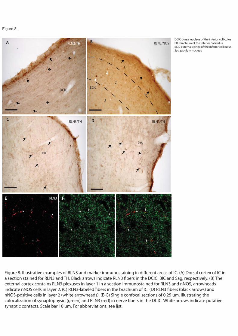
DCIC
A B
C D
BIC
RLN3 FE
ECIC
Sag
1
2
3
RLN3/TH
RLN3/TH RLN3/TH
RLN3/NOS
Figure 8.
Figure 8. Illustrative examples of RLN3 and marker immunostaining in different areas of IC. (A) Dorsal cortex of IC in a section stained for RLN3 and TH. Black arrows indicate RLN3 fibers in the DCIC, BIC and Sag, respectively. (B) The external cortex contains RLN3 plexuses in layer 1 in a section immunostained for RLN3 and nNOS, arrowheads indicate nNOS cells in layer 2. (C) RLN3-labeled fibers in the brachium of IC. (D) RLN3 fibers (black arrows) and nNOS-positive cells in layer 2 (white arrowheads). (E-G) Single confocal sections of 0.25 μm, illustrating the colocalization of synaptophysin (green) and RLN3 (red) in nerve fibers in the DCIC. White arrows indicate putativesynaptic contacts. Scale bar 10 μm. For abbreviations, see list.
DCIC dorsal nucleus of the inferior colliculusBIC brachium of the inferior colliculusECIC external cortex of the inferior colliculusSag sagulum nucleus

MPT
pc
MCPC
PAGSCO
Dk
IMLF
SuMM
sumx
fr
3V
dlfctg
PrC
dtg
ctg
pc
MPT
dlfAq
EW
Dk
DpMe
PCom
MCPC
InC
PCom
csc
TLC
DpWh
dtg
EW
Dk
csc
dlf
Aq
DpG
LPAG
oDMiDM
DpMe
IMLF
Aq
EW
dtg
csc
Su3C
LPAG
DLPAG
DMPAG
3
Su3
4
mlf
DRD
3PC
Me5
DLPAG
DMPAG
LPAG
DRV
V LPAG
mlf
DRD
Me5
DLPAG
DMPAG
LPA G
DRVDRL
V LPAG
A
B
C
D
E
F
NOS/RLN3Figure 9.
Figure 9. Distribution of RLN3-positive fibers within PAG mapped in sections immunostained for RLN3 and NOS. At rostral levels, nNOS-positive soma were observed in the MCPC, the periaqueductal ring and the EW nucleus. At levels 4 to 6, the nNOS-positive cells distribution allows the demarcation of the DL column and DR. At rostral levels the RLN3 innervation is concentrated in the InC and iDM. At caudal levels RLN3-positive fibers are observed in the DM column and the DR at level 4. RLN3-immunostaining is more intense in the medial half of the lateral column. Scale bar: 500 μm.
3PC oculomotor nucleus, parvicellular part3v 3rd ventricle4 trochlear nucleusAq aqueductcsc commissure of the superior colliculusctg central tegmental tractDk nucleus of Darkschewitschdlf dorsal longitudinal fasciculusDLPAG dorsolateral column of the periaqueductal grayDMPAG dorsomedial column of the periaqueductal grayDpG deep gray layer of the superior colliculusDpMe deep mesencephalic nucleusDRD dorsal raphe nucleus, dorsal partDRL dorsal raphe nucleus, lateral partDRV dorsal raphe nucleus, ventral partdtg dorsal tegmental bundleEW Edinger-Westphalfr fasciculus retroflexusiDM inner division of the dorsomedial columnIMLF interstitial nucleus of the medial longitudinalIMLF interstitial nucleus of the medial longitudinalInC intercommissural nucleus of the periaqueductal grayLPAG lateral column of the periaqueductal grayMCPC magnocellular nucleus of the posterior commissuremlf medial longitudinal fasciculusMPT medial pretectal nucleusoDM outer division of the dorsomedial columnPAG priaqueductal graypc posterior commissurePCom nucleus of the posterior commissurePrC precommissural nucleusSCO subcommissural organSu3 supraoculomotor periaqueductal graySu3C supraoculomotor capSuMM supramammillary nucleus, medial partsumx supramammillary decussationTLC tectal longitudinal columnVLPAG ventrolateral column of the periaqueductal gray

MPT
pc
MCPC
PAG
Dk
IMLF
SuMM
sumx
fr
3V
dlf
ctg
PrC
dtg
ctg
pc
MPT
dlfAq
EWDk
DpMe
PCom
MCPC
InC
PCom
cscTLC
DpWh
dtg
EW
Dk
csc
dlfAq LPAG
oDMiDM
DpMe
IMLF
Aq
EW
dtg
csc
Su3C
LPAG
DLPAG
DMPAG
3
Su3
4
mlf
DRD
3PC
Me5
DLPAG
DMP AG
LPAG
DRV
VLP AG
mlf
DRD
Me5
DL PAG
DM PAG
LPAG
DRV
DRL
VL PA G
A
B
C
D
E
F
TH/RLN3Figure 10.
Figure 10. Distribution of RLN3-positive fibers within PAG mapped in sections immunostained for RLN3 and TH. At levels 5 and 6, TH-positive cells and processes in the DR facilitated the definition of its subdivisions. The ventral half of the juxta-aqueductal ring and EW also contain TH-positive labeling. At level 1, the PrC contains RLN3-positive fibers. Scale bar: 500 μm. For abbreviations, see list.
3PC oculomotor nucleus, parvicellular part3v 3rd ventricleAq aqueductcsc commissure of the superior colliculusctg central tegmental tractctg central tegmental tractDk nucleus of Darkschewitschdlf dorsal longitudinal fasciculusdlf dorsolateral funiculusDLPAG dorsolateral column of the periaqueductal grayDMPAG dorsomedial column of the periaqueductal grayDpMe deep mesencephalic nucleusDpWh deep white layer of the superior colliculusDRD dorsal raphe nucleus, dorsal partDRL dorsal raphe nucleus, lateral partDRV dorsal raphe nucleus, ventral partdtg dorsal tegmental bundleEW Edinger-Westphalfr fasciculus retroflexusiDM dorsomedial column of the periaqueductal gray, intermediate partIMLF interstitial nucleus of the medial longitudinalIMLF interstitial nucleus of the medial longitudinal fasciculusLPAG lateral column of the periaqueductal grayMCPC magnocellular nucleus of the posterior commissureMe5 mesencephalic trigeminal nucleusmlf medial longitudinal fasciculusMPT medial pretectal nucleusoDM outer division of the dorsomedial columnPAG priaqueductal graypc posterior commissurepc posterior commissurePCom nucleus of the posterior commissurePCom nucleus of the posterior commissurePrC precommissural nucleusSu3 supraoculomotor periaqueductal graySu3C supraoculomotor capSuMM supramammillary nucleus, medial partsumx supramammillary decussationTLC tectal longitudinal columnVLPAG ventrolateral column of the periaqueductal gray

CSC
pc
InC
PrC
DMPAGDLPAG
LPAGAqAq
SON3NLR RLN3/NOSRLN3 SP RLN3/SP
A B
C D EI J KEDC RLN3/SynSyn RLN3
RLN3/NOSRLN3/NOS
Figure 11.
Aq aqueductcsc commissure of the superior colliculusDLPAG dorsolateral column of the periaqueductal grayDMPAG dorsomedial column of the periaqueductal grayInC intercommissural nucleus of the periaqueductal grayLPAG lateral column of the periaqueductal graypc posterior commissurePrC precommissural nucleus
Figure 11. Illustrative examples of RLN3 and marker immunostaining in PAG. (A) Abundant RLN3-labeled fibers in the precommissural and intercollicular nuclei. (B) Intense nNOS-immunoreactivity in the DL column of PAG. (C-E) Single confocal sections of 0.25 μm illustrating the colocalization of synaptophysin (green) and RLN3 (red) in nerve fibers in the DL column. White arrows indicate putative synaptic contacts, stars point at putative neuronal somata. LN3-positive fibers in the THpositive area of the ventrolateral PAG. Scale bar A,B: 200 μm, C-K: 10 μm. For abbreviations, see list.

DR
VLPAG
CnF
VLPAG
LDTg
cic
TH-RLN3
A B
C D
RLN3 nyS/3NLRnyS GFE
TH-RLN3
CnF
VLPAG
LDTg
LDTg
TH-RLN3 TH-RLN3B
Figure 12.
Figure 12. Illustrative examples of RLN3 and marker immunostaining in LDTg (A-B) and cuneiform nucleus (C,D). Confocal images illustrating the colocalization of synaptophysin (green) and RLN3 (red) in nerve fibers in the cuneiform nucleus (E-G). White arrows indicate putative synaptic contacts. Scale bars A 200 μm B-C: 100 μm, D: 50 μm, E-G: 10 μm.
cic commissure of the inferior colliculusCnF nueclus cuneformisDR dorsal raphe nucleusLDTg laterodorsal tegmental nucleusVLPAG ventrolateral column of the periaqueductal gray

Bar
LC
DRC
mlf
DTgC
PnR
LDTg BarDTgP
LC
4V
Sph
PDTg
mlfNId
NIc
PnR
MnR
PnO
DTgP
LDTgV
LDTg
PMR
DRd
DRv
PnO
VTg
DTgPLDTgV
LDTg
DRd
PnR
RtTg
DMTg
VTg
DTgP
LDTgV
LDTgDRd
PnR DMTg
A
B
C
D
E
NOS/RLN3
Figure 13.
Figure 13. Distribution of RLN3-labeled fibers within the pontine central grey and DR mapped in sections immunostained for RLN3 and nNOS. nNOS staining distinguishes the LDTg nucleus at level 2 to 4 and divisions of DR from level 1 to 3. A high density of RLN3-labeled fibers is present in the Bar, LDTg and raphe divisions, whereas the DTgP is unlabeled. The rostral area of the NI containing RLN3 soma is observed at level 5. Scale bar: 500 μm. For abbreviations, see list.
4v 4th ventricleBar barrington nucleusDMTg dorsomedial tegmental areaDRC dorsal raphe nucleus, caudal partDRD dorsal raphe nucleus, dorsal partDRV dorsal raphe nucleus, ventral partDTgC dorsal tegmental nucleus, central part DTgPDTgP dorsal tegmental nucleus, pericentral part DTgPLC locus coeruleusLDTg laterodorsal tegmental nucleusLDTgV laterodorsal tegmental nucleus, ventral partmlf medial longitudinal fasciculusMnR median raphe nucleusNIc nucleus incertus, pars compactaNId nucleus incertus, pars disipataPDTg posterodorsal tegmental nucleusPmR paramedian raphePnO pontine reticular nucleus, oral partPnR pontine raphe nucleusRtTg reticulotegmental nucleus of the ponsSph sphenoid nucleusVTg ventral tegmental nucleus

DRC
mlf
DTgC
PnR
LDTg
Bar
DTgP
LC
MnR
PnO
DTgP
LDTgV
LDTg
PmR
DRd
DRv
PnO
VTg
DTgP
LDTgV
LDTg
DRd
PnR DMTg
VTg
DTgP
LDTgV
LDTg
DRv
PnR DMTg
LC
4V
Sph
PDTg
mlf
NIdNIc
Bar
LC
PnR
MnR
VTg
MnRPMR
MnR
A
B
C
D
E
TH/RLN3
Figure 14.
Figure 14. Distribution of RLN3-labeled fibers within PCG and DR mapped in sections immunostained for RLN3 and TH. At level 1, DR contains TH-positive cells and processes. At rostral levels, THinmunoreactive tracts help define the LDTg nucleus. At levels 3, 4 and 5, intense TH-labeling appears in LC. RLN3-labeled fibers avoid the DTg and are dense in the midline and raphe nuclei. Scale bar: 500 μm. For abbreviations, see list.
4v 4th ventricleBar barrington nucleusDMTg dorsomedial tegmental areaDRC dorsal raphe nucleus, caudal partDRD dorsal raphe nucleus, dorsal partDRV dorsal raphe nucleus, ventral partDTgC dorsal tegmental nucleus, central part DTgPDTgP dorsal tegmental nucleus, pericentral part DTgPLC locus coeruleusLDTg laterodorsal tegmental nucleusLDTgV laterodorsal tegmental nucleus, ventral partmlf medial longitudinal fasciculusMnR median raphe nucleusNIc nucleus incertus, pars compactaNId nucleus incertus, pars disipataPmR paramedian raphePnO pontine reticular nucleus, oral partPnR pontine raphe nucleusRtTg reticulotegmental nucleus of the ponsSph sphenoid nucleusVTg ventral tegmental nucleus

DRC
mlf
DTgC
PnR
LDTg
Bar
DTgP
LC
MnR
PnO
DTgP
LDTgV
LDTg
PMR
DRd
DRv
PnO
VTg
DTgP
LDTgV
LDTg
DRd
PnR
RtTg
DMTg
VTg
DTgP
LDTgV
LDTg
DRv
PnR DMTg
LC
4VSph
PDTg
mlf
NIdNIc
Bar
LC
PnR
MnR
VTgN
A
B
C
D
E
5HT/RLN3
Figure 15.
Figure 15. Distribution of RLN3-positive fibers in PCG and DR mapped in sections immunostained for RLN3 and 5HT. At levels 1 to 3, 5HT-labeled soma are present in DR and rostral PnR. MnR also contains some 5HT-labeled somata. At levels 4 and 5, RLN3-labeled soma appear in caudal PR and NI. The DTg contains no RLN3 fibers, although its surrounding area, midline region and raphe nuclei contain dense plexuses. Scale bar: 500 μm. For abbreviations, see list.
4v 4th ventricleBar barrington nucleusDMTg dorsomedial tegmental areaDRD dorsal raphe nucleus, dorsal partDRV dorsal raphe nucleus, ventral partDTgC dorsal tegmental nucleus, central part DTgPDTgP dorsal tegmental nucleus, pericentral part DTgPLC locus coeruleusLDTg laterodorsal tegmental nucleusLDTgV laterodorsal tegmental nucleus, ventral partmlf medial longitudinal fasciculusMnR median raphe nucleusNIc nucleus incertus, pars compactaNId nucleus incertus, pars disipataPmR paramedian raphePnO pontine reticular nucleus, oral partPnR pontine raphe nucleusRtTg reticulotegmental nucleus of the ponsSph sphenoid nucleusVTg ventral tegmental nucleus

NIc PDTg
PnR
mlf
TH53NLR RLN3/5HT
nyS3NLR RLN3/Syn
nyS3NLR RLN3/Syn
A B
C D E
F G H
I J K
BN
LC
RLN3/TH RLN3/TH
NId
PDTg BN
LC
NIc NIc NIc
mlf mlfmlf
DR RDRD
Figure 16.
Figure 16. Illustrations of RLN3-labeling and marker immunostaining in pontine central grey. (A) Dense TH-immunoreactive cells and fibers (brown) in LC, and RLN3-labeled neurons in midline NIc (black), and lateral NId. (B) Abundant RLN3-immunoreactive fibers in the PnR between the PDTg and extending below the floor of the 3V. (C-E) Confocal images illustrating the presence of RLN3-labeled fibers (red) adjacent to 5HT-positive neurons (green) in caudal DR. White arrows indicate putative contacts. (F-H) Confocal images illustrating the colocalization of synaptophysin (green) and RLN3 (red) in the caudal DR. White arrows indicate putative synaptic contacts. (I-K) Confocal images illustrating the RLN3-containing neurons of the NI (asterisks) and possible synaptic contacts of RLN3-labeled fibers (white arrows). Scale bar A: 200 μm; B: 100 μm; C-K: 10 μm. For abbreviations, see list.
BN Barrington's nucleusDR dorsal raphe nucleusLC locus coeruleusmlf medial longitudinal fasciculusNIc nucleus incertus, pars compactaNId nucleus incertus, pars disipataPDTg posterodorsal tegmental nucleusPnR pontine raphe nucleus

Fig
ure
17
.
Figu
re 1
7. A
sch
emat
ic il
lust
ratio
n su
mm
ariz
ing
the
RLN
3-co
ntai
ning
pro
ject
ions
with
in th
e te
ctal
and
tegm
enta
l sys
tem
s of
rat b
rain
. RLN
3 pr
ojec
tions
are
indi
cate
d by
red
arro
ws
and
man
y, if
not
all,
of t
hese
may
orig
inat
e fr
om th
e N
I. M
idbr
ain
area
s ar
e gr
oupe
d in
blu
e sq
uare
s. M
ain
phys
iolo
gica
l and
beh
avio
ral o
utpu
ts o
f the
targ
eted
sys
tem
s ar
e in
dica
ted
by y
ello
w a
rrow
s.
BIC
brac
hium
of t
he in
ferio
r col
licul
usCI
C co
mm
issu
re o
f the
infe
rior c
ollic
ulus
DCI
C do
rsal
nuc
leus
of t
he in
ferio
r col
licul
usD
LPAG
dor
sola
tera
l col
umn
of th
e pe
riaqu
educ
tal g
ray
DpG
ray
deep
gra
y la
yer o
f the
sup
erio
r col
licul
usD
pWhi
te d
eep
whi
te la
yer o
f the
sup
erio
r col
licul
usD
R do
rsal
raph
e nu
cleu
sEC
IC e
xter
nal c
orte
x of
the
infe
rior c
ollic
ulus
IC in
ferio
r col
licul
usIn
Gra
y in
term
edia
te g
ray
laye
r of t
he s
uper
ior c
ollic
ulus
InW
hite
inte
rmed
iate
whi
te la
yer o
f the
sup
erio
r col
licul
usM
PT m
edia
l pre
tect
al n
ucle
usN
I NIc
nuc
leus
ince
rtus
OPT
oliv
ary
pret
ecta
l nuc
leus
PAG
pria
qued
ucta
l gra
yPB
G p
arab
igem
inal
nuc
leus
PFCo
rtex
pre
fron
tal c
orte
xPP
T po
ster
ior p
rete
ctal
nuc
leus
Prec
omPA
G n
ucle
us o
f pr
ecom
mis
sure
, per
iaqu
educ
tal g
ray
Sag
sagu
lum
nuc
leus
SC s
uper
ior c
ollic
ulus
SupG
ray
supe
rfici
al g
ray
laye
r of t
he s
uper
ior c
ollic
uliu
sVL
PAG
ven
trol
ater
al c
olum
n of
the
peria
qued
ucta
l gra
yVL
PT v
entr
olat
eral
pre
tect
al n
ucle
us

90
APÊNDICE B
ELECTROLYTIC LESION OF THE NUCLEUS INCERTUS AND ITS
ASCENDING PROJECTIONS RETARDS EXTINCTION OF CONDITIONED
FEAR
PEREIRA, C.W.; SANTOS, F.N.; SÁNCHEZ-PÉREZ, A.M.; OTERO-GARCÍA, M.;
MARCHIORO, M.; MA, S.K.; GUNDLACH, A.L.; OLUCHA-BORDONAU, F.E.

Elsevier Editorial System(tm) for Behavioural Brain Research
Manuscript Draft
Manuscript Number:
Title: Electrolytic lesion of the nucleus incertus and its ascending projections retards extinction of
conditioned fear
Article Type: Research Reports
Keywords: Amygdala
Electrolytic lesion
Extinction
Fear memory
Neural tract-tracing
Nucleus incertus
Emotion
Corresponding Author: Mr Francisco E Olucha-Bordonau, PhD
Corresponding Author's Institution: Universitat de València
First Author: Celia W Pereira, PhD student
Order of Authors: Celia W Pereira, PhD student; Fabio N Santos, PhD student; Ana M Sánchez-Pérez,
PhD; Marcos Otero-García, PhD student; Murilo Marchioro, PhD; Sherie K Ma, PhD; Andrew L Gundlach,
PhD; Francisco E Olucha-Bordonau, PhD
Abstract: Fear memory helps animals and humans recognize putative sources of danger and adopt the
appropriate behavioral response. The primary neural circuits for fear acquisition and extinction
involve connections between prefrontal cortex, ventral hippocampus and amygdala, and these areas
are modulated by brainstem networks. The nucleus (n.) incertus in the dorsal pontine tegmentum
provides a strong GABAergic projection to these forebrain centers and is strongly activated by
neurogenic stressors. In this study in male, adult rats, we injected miniruby anterograde tracer into n.
incertus and delineated its projections to the amygdala; and examined the effect of electrolytic lesions
of n. incertus on different stages of the fear conditioning-extinction process. N. incertus-derived nerve
fibers were observed in anterior medial amygdala, endopiriform nucleus, intra-amygdala bed nucleus
of stria terminalis, amygdalohippocampal transition area, and the ventromedial nucleus of the lateral
amygdala, with a broad fiber band present between the basolateral amygdala and the olfactory nuclei
of amygdala. In a conventional contextual fear conditioning paradigm, we compared freezing behavior
in control (naïve) rats (n = 13), with that in rats after sham- or electrolytic lesions of n. incertus (n =
9/group). There were no differences between the three groups in the habituation, acquisition, or
context conditioning phases; but n. incertus-lesioned rats displayed a markedly slower (delayed)
extinction of conditioned freezing responses than sham/control rats; suggesting n. incertus-related
circuits normally promote extinction through inhibitory projections to amygdala and prefrontal cortex.
Suggested Reviewers: Gregory J Quirk PhD
Professor, Department of Psychiatry , University of Puerto Rico School of Medicine
He is an specialist in all processes controlling fear extinction

René García PhD
Professor, Laboratoire de Neurobiologie et Psychotraumatologie, Université de Nice-Sophia Antipolis,
France
He is an specialist in modulation of fear extinction
Llorenç Diaz-Mataix PhD
PostDoc, Center for Neural Science, New York University
He is specialist in fear acquisition and emotional processes

FRANCISCO E. OLUCHA BORDONAU Dep of Human Anatomy and Embryology Fac. de Medicina y Odontología University of Valencia
February 8, 2012
Professor Stephen Maren Editor and Editor-in-Chief Behavioral Brain Research Dear Professor Maren, Please find attached our manuscript entitled Electrolytic lesion of the nucleus incertus and its ascending projections retards extinction of conditioned fear Pereira CW and others, which we submit for consideration by the Behavioral Brain Research as a Research Article. We can confirm that this manuscript has been submitted solely to the Behavioral Brain Research and is not published, in press, or submitted elsewhere. We can also confirm that all the research meets the ethical guidelines of the relevant agencies and Animal Welfare Committees. We have endeavoured to adhere to the correct style and format requested for submissions. We look forward to receiving editorial comments on our article in the near future. Yours sincerely, Francisco E. Olucha-Bordonau Dep of Human Anatomy Univ. Valencia, Spain
Cover letter

Research Highlights
We studied the pontine n. incertus role in fear conditioning and extinction
We have shown an important projection from the n. incertus to amygdala nuclei
Lesions of the n. incertus do not affect to the acquisition or retrieval of fear to cue or context
Lesions of the n. incertus delay the within trial extinction.
Thus n. incertus facilitates extinction
*Research Highlights
1 2 3 4 5 6 7 8 9 1011121314151617181920212223242526272829303132333435363738394041424344454647484950515253545556575859606162636465
Behavioural Brain Research, 8 February 2012
Electrolytic lesion of the nucleus incertus and its ascending projections
retards extinction of conditioned fear
Pereira CWa,b, , Santos FNa,b, , Sánchez-Pérez AMc, Otero-García Md, Marchioro Mb, Ma
Se,f, Gundlach ALe,g, Olucha-Bordonau FEa**
aDepartamento de Anatomía y Embriología Humana, Facultad de Medicina, Universitat de
Valencia, 46010 Valencia, Spain bUniversidade Federal de Sergipe, Laboratorio de Neurofisiologia, São Cristóvão, SE 49100-
000, Sergipe, Brazil cDepartamento de Fisiología, Facultad de Medicina, Universitat de Valencia, 46010 Valencia,
Spain dDepartamento de Biología Funcional y Antropología Física, Facultad de CC Biológicas,
Universitat de Valencia, 46100 Valencia, Spain e Florey Neuroscience Institutes, fDepartment of Medicine (Austin Health) and gDepartment of Anatomy and Cell Biology, The University of Melbourne, Victoria 3010, Australia
*These authors contributed equally to this research
Current Address: Universidade Federal de Sergipe, Laboratorio de Neurofisiologia, Av
Marechal Rondon, s/n, Jardim Rosa Elze, São Cristóvão, SE 49100-000, Sergipe, Brazil
**Corresponding author:
FE Olucha-Bordonau
Departamento de Anatomía y Embriología Humana
Universitat de Valencia
Av Blasco Ibáñez 15
46010 Valencia, Spain
Tel: +34 9 6398 3504 Fax: +34 9 6386 4159
E-mail: [email protected]
*ManuscriptClick here to view linked References

1 2 3 4 5 6 7 8 9 1011121314151617181920212223242526272829303132333435363738394041424344454647484950515253545556575859606162636465
2
Abstract Fear memory helps animals and humans recognize putative sources of danger and adopt the
appropriate behavioral response. The primary neural circuits for fear acquisition and
extinction involve connections between prefrontal cortex, ventral hippocampus and
amygdala, and these areas are modulated by brainstem networks. The nucleus (n.) incertus
in the dorsal pontine tegmentum provides a strong GABAergic projection to these forebrain
centers and is strongly activated by neurogenic stressors. In this study in male, adult rats, we
injected miniruby anterograde tracer into n. incertus and delineated its projections to the
amygdala; and examined the effect of electrolytic lesions of n. incertus on different stages of
the fear conditioning-extinction process. N. incertus-derived nerve fibers were observed in
anterior medial amygdala, endopiriform nucleus, intra-amygdala bed nucleus of stria
terminalis, amygdalohippocampal transition area, and the ventromedial nucleus of the lateral
amygdala, with a broad fiber band present between the basolateral amygdala and the
olfactory nuclei of amygdala. In a conventional contextual fear conditioning paradigm, we
compared freezing behavior in control (naïve) rats (n = 13), with that in rats after sham- or
electrolytic lesions of n. incertus (n = 9/group). There were no differences between the three
groups in the habituation, acquisition, or context conditioning phases; but n. incertus-lesioned
rats displayed a markedly slower (delayed) extinction of conditioned freezing responses than
sham/control rats; suggesting n. incertus-related circuits normally promote extinction through
inhibitory projections to amygdala and prefrontal cortex.
Keywords: Amygdala, Electrolytic lesion, Extinction, Fear memory, Neural tract-tracing,
Nucleus incertus

1 2 3 4 5 6 7 8 9 1011121314151617181920212223242526272829303132333435363738394041424344454647484950515253545556575859606162636465
3
1. Introduction Acquisition and extinction of fear memories are neural processes that allow animals and
humans to face physical and emotional challenges in a potentially dangerous environment. In
mammals and possibly other vertebrates, the central node for managing fear-related
information in the brain is the amygdala [1]. Research over many years has established that
meaningless information (conditioned stimulus, CS) acquires emotional value when
presented together with a meaningful stimulus (unconditioned stimulus, US) [2]. After
acquisition, the presentation of the CS alone elicits the same response as those displayed
after presentation of the US (conditioned response, CR).
Commonly, under experimental conditions the CS used is a tone, the US is a footshock
and the CR, which can be interpreted as the level of acquisition, is the amount of time that
the animal
repeatedly presented without the US, the animal learns that the CS no longer predicts the
occurrence of the US, a phenomenon known as extinction. While the amygdala plays a key
role in acquisition of conditioning and extinction, extinction memories are thought to be
stored in the ventromedial prefrontal cortex [3].
The anatomical framework of the conditioning-extinction process is a series of intrinsic
connections running from the lateral amygdala, where the emotional acquisition commences,
to the central amygdala, which signals the appropriate emotional response to be performed
[4]. In addition, there are intrinsic connections formed by the intercalated GABAergic cell
groups that lie adjacent to/between the lateral and central nuclei. Neural processing along
this pathway is modulated by several intrinsic and extrinsic afferents, some of them derived
from within the amygdala.
The processing of conditioning and extinction signals can be altered by modulatory
systems ascending from the brainstem. For example, stimulation of the locus ceruleus
inhibits amygdala neurons [5], intra-amygdala infusion of norepinephrine enhances
contextual fear conditioning [6] -adrenergic stimulation impairs extinction [7], basolateral
noradrenergic activity mediates corticosterone enhancement of fear conditioning, [8],
serotonin transporter knockout mice exhibit a selective deficit in extinction recall of fear
memory [9, 10] and neuropeptide S containing projections from neurons located between
[11].
We propose that the nucleus (n.) incertus in the pontine tegmentum is another area
that could potentially affect the conditioning-extinction process. Although the projections from
[12], in subsequent studies we
observed projections to several amygdala nuclei [13]. The main target areas of n. incertus
afferents include the amygdala, medial and lateral septum, ventral hippocampus and

1 2 3 4 5 6 7 8 9 1011121314151617181920212223242526272829303132333435363738394041424344454647484950515253545556575859606162636465
4
prefrontal and entorhinal cortex, revealing a putative involvement in the control of cognitive
and emotion-related functions [12, 13]. Moreover, n. incertus neurons express high levels of
the corticotrophin-releasing hormone receptor-1 (CRH-R1) [14, 15], consistent with the
integration by n. incertus of stress-
cognitive and emotional centers. In fact, several neurogenic stressors activate the n.
incertus, as reflected by increased c-Fos expression [16-18] (see [19] for review).
Furthermore, ascending n. incertus projections target consecutively the median raphe,
supramammillary nucleus, posterior hypothalamus, medial septum and hippocampus [12,
13], the components of [20]. A proposed role for the n.
incertus in modulating hippocampal theta rhythm was supported by observations that
electrical stimulation of n. incertus increased hippocampal theta activity in urethane-
anesthetized rats; and that lesion of the n. incertus produced disruption of hippocampal theta
induced by stimulation in the reticularis pontis oralis (RPO) [21].
Importantly, there is a close relationship between synchronization of neural firing at
theta frequency in the amygdala and hippocampus and fear conditioning. During fear-related
emotional arousal, lateral amygdala neurons increase oscillatory activity at theta frequency
[22, 23]. Moreover, it has been shown that during consolidation and reconsolidation of
conditioned fear, lateral amygdala and CA1 cells increased synchrony between the theta
band [24]. Furthermore, there is evidence of theta-related coordination between the
hippocampus, amygdala and prefrontal cortex [25], and as all these areas receive projections
from n. incertus, we hypothesized that the n. incertus plays a role in the control of fear
conditioning.
Therefore, the goal of this study was to determine whether lesion of the n. incertus
could markedly modulate fear expression and/or extinction behavior. We performed discrete
electrolytic lesions of n. incertus in adult, male rats and tested the effect on performance in a
paradigm of fear acquisition and extinction. In addition, we used neural tract-tracing to
identify putative target regions in the amygdala that may mediate actions of increased or
decreased n. incertus activity on fear processes.
(766 words)

1 2 3 4 5 6 7 8 9 1011121314151617181920212223242526272829303132333435363738394041424344454647484950515253545556575859606162636465
5
2. Materials and Methods 2.1. Animals
A total of 34 adult, male Sprague-Dawley rats weighting 250-350 g were used in this study -
3 rats were used for the neuroanatomical tract-tracing study; and 31 rats were used for the
lesion/behavioral study and were housed in individual cages with a reverse cycle 12:12 h
light/dark (lights off at 8 am) for 21 days before commencing the behavioral procedures. Rats
were randomly distributed in two groups; control and surgery, and lesion surgery was done 7
days after the reversal of the light cycle (Fig. 1). All behavioral procedures conducted during
the dark phase. All animal manipulations were approved by the Committee of Ethics and
Animal Welfare of the Universitat de Valencia, Spain and were in agreement with directive
86/609/EEC of the European Community on the protection of animals used for experimental
and other scientific purposes.
2.2. Surgical procedures
2.2.1. Tracer injection
Rats were deeply anesthetized with ketamine 55 mg/kg (Imalgene, Merial Laboratorios SA,
Barcelona Spain) and xylacide 20 mg/kg (Xilagesic, Laboratorios Calier, Barcelona, Spain)
and anesthesia was maintained with isoflurane as required. Under stereotaxic control, a
small hole was drilled in the skull and anterograde tracer injections were made into the n.
incertus using 40 µm glass micropipettes (coordinates AP -9.6 mm, ML 0 mm and DV 7.4
mm with respect to bregma [26]. For anterograde tracing, we used 15% miniruby (mR, 10 kD
biotinylated dextran amine rhodamine-labeled, Molecular Probes, Cat No. D-3312, Paisley,
UK) dissolved in 0.1M PB, pH 7.4. Tracer was iontophoretically delivered into the n. incertus
by passing a positive current of 1 µA 2 sec on 2 sec off over 15 min with a current generator
(GC2N model, Direlec, Madrid, Spain). After injections, the surgical wound was sutured and
rats were injected with buprex (0.05 mg/kg, i.p., Lab Esteve, Barcelona, Spain) for analgesia.
Rats were then allowed to recovery, prior to further processing. After 7 days of survival time,
rats were euthanized with an overdose of Nembutal (150 mg/kg, i.p. Euthalender, Normon,
Barcelona, Spain). Brains were perfused by transcardial infusion of saline (250 ml) followed
by fixative (4% paraformaldehyde in 0.1M phosphate buffer (PB), pH 7.4). Brains were then
removed from the skull, postfixed in the same fixative overnight at 4ºC, and cryoprotected in
30% sucrose in 0.01 M PB, pH 7.4
2.2.2. Electrolytic lesions
Rats were deeply anesthetized with ketamine (Imalgene) and xylacide (Xilagesic) (55 mg/kg
and 20 mg/kg, i.p., respectively) and anesthesia was maintained with isoflurane as required.
Under stereotaxic control a monopolar electrode was lowered into the brain at coordinates

1 2 3 4 5 6 7 8 9 1011121314151617181920212223242526272829303132333435363738394041424344454647484950515253545556575859606162636465
6
AP -9.6 mm, ML 0 mm and DV -7.4 mm with respect to bregma. Positive continuous current
of 1.5 mA was injected for 10 sec by means of a current generator connected to a SIU
(Cibertec, Madrid, Spain). Five min after the lesion, the electrode was removed and the
wound sutured. Rats were injected with buprex (0.05 mg/kg, i.p., Lab Esteve) for post
surgery analgesia. Rats were left to recover for an additional 15 days before use in the
behavioral procedures.
2.3. Apparatus
The behavioral procedures were conducted in a contextual fear conditioning apparatus
(Panlab, Barcelona Spain), consisting of three cages of 250 × 250 × 250 mm and a floor
containing a 22-rod grid connected to a shocker and a speaker in the ceiling. On a lateral
wall there was a soft light. The tray containing the grid was suspended over a load cell that
detected movements of the rat in the cage. The rat enclosure was inside a sound proof cage
of 600 (W) × 500 (D) × 500 (H) mm, which contained four 9V light bulbs. All procedures were
controlled by the Freezing software (Panlab).
Two kinds of contexts were used. In context A (for acquisition), lateral and posterior
walls were black and the front wall was Plexiglas, and the interior light was on. In context B
(for extinction), the lateral and posterior walls were horizontal, vertical and diagonal black
and white strips, the grid was covered with a white Plexiglas lamina, the interior light was off,
the cage lights were on, and four open tubes containing lemon-flavored soap were added.
2.4. Behavioral procedures
The entire behavioral procedure was completed over 5 days (Fig. 1). On days 1 and 2 each
rat visited contexts A and B for 15 min per session. On day 3 (acquisition), each rat received
3 tone-shock pairings in context A. The tone (2.8 kHz and 75 db SPL) was continuous for 30
sec. The shock was 0.3 mA and 0.5 sec duration and co-terminated with the tone. The tone
and shock pairings were presented randomly at sequences of 180 ± 60 sec. On day 4, each
rat was again exposed to the two contexts, first context A (acquisition) and two hours later,
context B (extinction). On day 5, in context B, the rats received 5 extinction tones (2.8 kHz
and 75 db SPL) of 30 sec, presented with a random delay from each other at times of 180 ±
60 sec. Following the procedure, rats were euthanized with an overdose of Nembutal (150
mg/kg, i.p.). Brains were perfused by transcardial infusion of saline (250 ml) followed by
fixative (4% paraformaldehyde in 0.1M PB, pH 7.4). Brains were removed from the skull,
postfixed in the fixative overnight at 4ºC, and cryoprotected in 30% sucrose in 0.01M PB
buffer pH 7.4.

1 2 3 4 5 6 7 8 9 1011121314151617181920212223242526272829303132333435363738394041424344454647484950515253545556575859606162636465
7
2.5. Behavioral measurements
The load cell at the bottom of the grid tray transformed horizontal movements into electrical
signals that was amplified and converted into arbitrary signals. The interface magnified the
signal 500× and the software amplified the signal a further 8 times. Under these conditions
movements were displayed in a 0-100 scale. After visual checking of responses, we
considered freezing as movement signals <10 lasting at least 2 sec. The Freezing software
displayed freezing as a percentage of the total time of the step for each step of the protocol.
2.6. Histological procedures
Brains were left in cryoprotectant for 1-3 days until they had sunk to the bottom of the vessel.
Leica Microsystems, Heidelberg, Germany). For each brain, 6 series of sections were
obtained and collected in 0.01 M PBS. In brains from rats that had undergone surgery,
calretinin immunohistochemistry was used to determine the degree of neuronal injury in the
n. incertus and surrounding areas. The distribution of mR, following injections of the tracer
into the n. incertus, was revealed using the standard ABC-DAB method.
2.6.1. Tracer histochemistry
Sections were rinsed in 2 × TBS and transferred to 1:100 ABC (Vectastain, Vector PK-6100,
Vector Laboratories, Burlingame, CA, USA) and incubated overnight at 4°C. After rinsing (2 ×
TBS), the tracer was revealed by immersing the sections in 0.025% DAB, 0.5% ammonium
nickel sulfate, 0.0024% H2O2 in Tris HCl, pH 8.0. The reaction was stopped after 15 min by
adding TBS. Sections were rinsed (3 × 0.01 M PBS pH 7.4) and mounted on gelatin-subbed
slides, air dried, dehydrated with graded ethanol, cleared and coverslipped with DPX.
2.6.2. Calretinin immunohistochemistry
Sections were rinsed twice in 0.05 M Tris-buffered saline pH 8.0 (TBS) and transferred to
blocking solution (4% normal donkey serum (NDS), 2% bovine serum albumin (BSA) and 0.2
% Triton-X100 in TBS) for 1 h at room temperature. Sections were then transferred to
incubation media containing 1:2,500 mouse anti-calretinin (Swant, Bellinzona, Switzerland)
6B3, 2% NDS, 2% BSA and 0.2% Triton X100 in TBS for 48 h at 4ºC. Sections were rinsed
(2 × TBS) and transferred to the biotinylated secondary antibody (biotinylated donkey anti-
mouse (Jackson Laboratories, Cat No. 715-065-150) for 1 h. Sections were then rinsed (2 ×
TBS) and immersed in 1:100 ABC (Vector) for 1 h. After rinsing (2 × TBS), immunolabeling
was revealed by incubating the sections in 0.025% DAB, 0.0024% H2O2 in Tris HCl, pH 7.6.
After several rinses in 0.01 M PBS, sections were mounted on chrome alum, gelatine-coated
slides, air dried, dehydrated, cleared and coverslipped (Sigma-Aldrich).

1 2 3 4 5 6 7 8 9 1011121314151617181920212223242526272829303132333435363738394041424344454647484950515253545556575859606162636465
8
2.7. Histological examination
Lesions and anterograde labeling were photographed using a Nikon Eclipse E600
microscope with a DMX2000 digital camera (Nikon, Tokyo, Japan). Mappings were made
with a camera lucida tube attached to a Zeiss Axioskop microscope (Zeiss, Munich,
Germany) with the 20× objective, scanned and reduced to the final size.
2.8. Statistical analysis
Two-way ANOVA statistical analysis was performed on the three groups - control, lesion and
sham- -hoc test was performed
to specify the differences between points after the lesion effect was found to be significant.

1 2 3 4 5 6 7 8 9 1011121314151617181920212223242526272829303132333435363738394041424344454647484950515253545556575859606162636465
9
3. Results 3.1. Projections from the nucleus incertus to the amygdala
In all three cases assessed, the mR tracer injection was restricted to the n. incertus, with no
spread to the dorsal tegmental nucleus or the pontine raphe nucleus (Fig. 2A). Anterogradely
labeled fibers entered the amygdala complex from the medial forebrain bundle and the
subventricular area (Fig. 2B,C). Within the amygdala, anterogradely labeled fibers were
observed in the anterior nuclei of the medial amygdala, while, in contrast, the posterior nuclei
of the medial amygdala were free of labeling. Fibers ran caudally and were densely
concentrated in the intra-amygdala bed nucleus of the stria terminalis (IABST). Some fibers
were also dispersed in the basal nuclei and the cortical and olfactory amygdala, and some of
these fibers ran laterally and concentrated in the endopiriform nucleus, whereas the central
nuclei were essentially free of labeled fibers. Finally, in the basolateral complex, labeled
fibers concentrated in the ventromedial division of the lateral amygdala (Fig. 2C). Although
not highly concentrated, some fibers were observed within the intercalated nuclei.
3.2. Behavioral studies
The extent of the lesion in each rat studied was analyzed by calretinin immunohistochemistry
(Fig. 3A,B). In none of the lesioned rats analyzed, did the damaged area spread to the
tegmental nucleus. Similarly, no histological alterations were observed in the pontine raphe,
ventrally adjacent medial longitudinal fasciculus that connects nuclei involved in oculomotor
coordination (Fig. 3B). According to the severity of the lesion/damage to the n. incertus, rats
-
(with the electrode placed dorsally in the cerebellum or ventrally in the reticular formation)
aïve rats that did not undergo surgery).
Evolution of the levels of freezing occurred during the behavioral procedure (Table 1;
Fig. 4). There were no significant changes in freezing in the habituation trials prior to the
acquisition session; and no significant changes in freezing levels between the lesioned,
sham and control groups during the acquisition trials. Rats initially displayed 10-20%
freezing, during the tone of the first pairing, with a shift to 70-80% freezing during the tone of
the second pairing, and reaching close to 90% during the tone of the third pairing. This
pattern followed the Rescorla and Wagner model of acquisition [27].
In the next session we assessed acquisition of conditioning to the context. Indeed, the
level of freezing in context A shifted significantly from 20% for the last habituation trial to 50-
70% during exposure to the same context after acquisition (F(1,32) = 35.32, P< 0.0001).
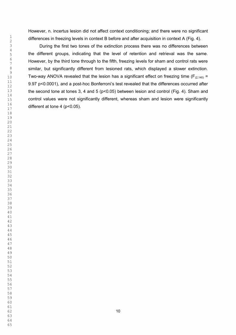
1 2 3 4 5 6 7 8 9 1011121314151617181920212223242526272829303132333435363738394041424344454647484950515253545556575859606162636465
10
However, n. incertus lesion did not affect context conditioning; and there were no significant
differences in freezing levels in context B before and after acquisition in context A (Fig. 4).
During the first two tones of the extinction process there was no differences between
the different groups, indicating that the level of retention and retrieval was the same.
However, by the third tone through to the fifth, freezing levels for sham and control rats were
similar, but significantly different from lesioned rats, which displayed a slower extinction.
Two-way ANOVA revealed that the lesion has a significant effect on freezing time (F(2,140) =
9.97 p<0.0001), and a post-
the second tone at tones 3, 4 and 5 (p<0.05) between lesion and control (Fig. 4). Sham and
control values were not significantly different, whereas sham and lesion were significantly
different at tone 4 (p<0.05).

1 2 3 4 5 6 7 8 9 1011121314151617181920212223242526272829303132333435363738394041424344454647484950515253545556575859606162636465
11
4. Discussion In this study, we used conventional neural tract-tracing techniques to identify the presence of
n. incertus projections within the amygdala and adjacent structures. The presence of this
innervation and other aspects of the connectivity of the n. incertus support the possibility that
this largely inhibitory projection might modulate neural processes associated with contextual
fear conditioning. Indeed, we discovered that an electrolytic lesion of the n. incertus 14 days
prior to subjecting rats to a classical contextual fear conditioning protocol produced a specific
delay of fear extinction, but had no effect on the acquisition of fear or on contextual
conditioning.
The anatomical findings fit with and extend our earlier observations on anterograde
labeling of projections from n. incertus to the amygdala region [13]. Efferent fibers arising
from the n. incertus make plexuses in the intra-amygdala bed nucleus of the stria terminalis,
in the area between the olfactory amygdala and the basal and lateral amygdala, in the
ventromedial subnucleus of the lateral amygdala, and in the anterior and posteroventral
medial amygdala nucleus. Dense terminations were also observed in the dorsal and ventral
endopiriform nuclei.
Neurons in the n. incertus express glutamic acid decarboxylase (GAD) [13, 28],
consistent with the expression of the major inhibitory transmitter, GABA, by these large
projections neurons. In the rat, these neurons also express a number of peptides, including
neurotensin, neuromedin B, cholecystokinin, atrial natriuretic peptide and relaxin-3 (see e.g.
[29], [30-32]; see [19] for review), suggesting GABA and peptides may be co-released under
various physiological conditions and in different patterns in different target areas, including
the extended amygdala and the hippocampus. Depending on the intensity of n. incertus
neuron activity and the distribution of different GABA and peptide receptors within the target
structures, GABA transmission may have a rapid and direct synaptic action on n. incertus
target cells, while co-released peptides may have a slower, more dispersed action on
neurons expressing high concentrations of receptors in different regions of the amygdala.
In related studies of the potential influence of the brainstem on fear memory, it has
been shown that several pontine nuclei can have a modulatory effect on fear acquisition
and/or extinction. For example, neuropeptide S projections arising from the pontine
tegmentum enhance the actions of glutamatergic projections on the intercalated GABAergic
neurons of the amygdala, thus potentiating extinction (e.g. [11]). With respect to the effect of
the n. incertus lesion on fear extinction, we attempted to eliminate any effect of a lesion in
surrounding areas by only analyzing data from rats with lesions restricted to the n. incertus
and with no apparent effect on surrounding structures. We observed no differences between
controls (non-lesioned and sham-lesioned) and n. incertus-lesioned groups during acquisition
of fear conditioning, or in fear expression during the initial steps of extinction, indicating that

1 2 3 4 5 6 7 8 9 1011121314151617181920212223242526272829303132333435363738394041424344454647484950515253545556575859606162636465
12
the n. incertus network does not have a major role in these processes or the retrieval of fear
memory. The lesioned rats also displayed normal conditioning to context, as all groups
displayed higher freezing in the context where conditioning took place than in the context
experienced during habituation trials prior to acquisition. All rats discriminated well between
the contexts, indicating n. incertus lesions did not affect the perception and configuration of a
context. However, in response to the last tones presented during extinction, rats with lesions
of the n. incertus displayed a higher level of freezing than sham and naïve rats, which
extinguished normally. Thus, we conclude that lesions of n. incertus neurons and their
projections impair the extinction process, delaying it.
Current reports suggest three main brain structures participate in extinction processes,
i.e. hippocampus, amygdala and prefrontal cortex; and the integrity of this interconnected
system is important for effective extinction processing [33, 34]. Nonetheless, some value can
be attributed to a particular structure in specific aspects of extinction. Since lesions in the
ventromedial [35], it follows that
acquisition of extinction must be processed downstream of the prefrontal connections to the
central nucleus of the amygdala, where fear responses are elaborated. However, this is in
apparent contradiction of recent experiments in which muscimol inactivation of the infralimbic
cortex resulted in a delay in the extinction process, but had no effect on expression of fear
[34]. However, in this latter experiment the prelimbic cortex was preserved. If the infralimbic
cortex induces rapid extinction through the intercalated GABAergic inhibitory neurons of the
amygdala [36], it is predicted that the inhibitory GABAergic projection from n. incertus to the
amygdala functions in a similar way. With no overt evidence for a strong, direct projection
from n. incertus to central amygdala, it may be the projection to the ventromedial lateral
amygdala that mediates this effect. Otherwise, additional effects may be mediated by
peptides that have high densities of receptors in the central amygdala, via slower volume
transmission effects. We observed an important n. incertus input to the intra-amygdala bed
nucleus of the stria terminalis, another area that expresses high levels of relaxin-3 and its
receptor [29], but to our knowledge, a specific contribution of this area to fear conditioning
has not been described. More generally, the amygdalohippocampal transition area and the
medial amygdala receive a projection from the ventral subiculum of the hippocampus [37]
and all of these areas are innervated by the n. incertus, increasing the potential modulatory
role of the n. incertus over the amygdala.
An important consideration regarding the apparent ability of the n. incertus to modulate
fear extinction is its potential ability to regulate the synchronized firing between the neuronal
populations of hippocampus, prefrontal cortex and amygdala. In this regard, we have shown
that electrical stimulation of n. incertus induces hippocampal theta rhythm [21] and
stimulation of the RPO results in phase-locked synchronization between the n. incertus and
1 2 3 4 5 6 7 8 9 1011121314151617181920212223242526272829303132333435363738394041424344454647484950515253545556575859606162636465
13
hippocampus [38]. Notably, neural synchronization at theta frequency is a feature associated
with fear conditioning, and lateral amygdala and CA1 hippocampal activity becomes
synchronized at theta frequency during consolidation and reconsolidation of conditioned fear
[24]. In addition, retrieval of fear memories increases theta coupling between the lateral
amygdala, ventromedial prefrontal cortex and hippocampus and is reduced during extinction
[25].
In conclusion, we have discovered that the n. incertus appears to be modulator of the
broader neural system associated with fear extinction and as n. incertus neurons express
high levels of CRH-R1 [14, 15], we propose that n. incertus may be an interface between
stress signalling and telencephalic circuits involved in fear processing. If as observed here,
the main effect of a discrete electrolytic lesion of the n. incertus is to delay the extinction
process, an effect also observed after infralimbic cortex inactivation, it is possible that the
normal actions of both these pathways occur in parallel. Thus, the effect of infralimbic
activation is transferred to the intercalated GABA neurons and the GABAergic NI projections
may signal to the glutamate neurons in the lateral amygdala. In turn, both intercalated GABA
and lateral amygdala glutamate neurons project to the central nucleus, where behavioral
responses are evoked (Fig. 5).
N. incertus neurons express GABA and a majority also synthesize relaxin-3 and/or
CCK. In an earlier study we demonstrated that injection of the relaxin-3 receptor agonist,
R3/I5, into the medial septum mimics the effect of n. incertus stimulation (i.e. increasing
hippocampal theta rhythm) [39]. In this study we have shown that n. incertus appears to
participate in the brainstem networks that modulate fear extinction, but the specific effect of
disruption/activation of the different transmitters within populations of n. incertus neurons
(GABA and peptides, such as relaxin-3) on the fear conditioning/extinction process remains
to be determined.
(1256 words)
1 2 3 4 5 6 7 8 9 1011121314151617181920212223242526272829303132333435363738394041424344454647484950515253545556575859606162636465
14
Acknowledgements This research was supported by grants from CAPES-Brasil Bex - 4494/09-1 (FNS) and
4496/09-4 (CWP) and Fapitec edital # 01/08 (FNS); the Ministry of Health, Spain - ISCIII-FIS
PI061816 (FEO-B); the National Health and Medical Research Council of Australia - 520299
(SM); 277609 and 509246 (ALG); and the Victorian Government Operational Infrastructure
Support Program (SM and ALG).

1 2 3 4 5 6 7 8 9 1011121314151617181920212223242526272829303132333435363738394041424344454647484950515253545556575859606162636465
15
References
[1] LeDoux JE. Emotion circuits in the brain. Annu Rev Neurosci, 2000;23:155-184 doi:
10.1146/annurev.neuro.23.1.155.
[2] Nader K, Majidishad P, Amorapanth P, LeDoux JE. Damage to the lateral and central, but not
other, amygdaloid nuclei prevents the acquisition of auditory fear conditioning. Learn Mem,
2001;8:156-163 doi: 10.1101/lm.38101.
[3] Quirk GJ, Mueller D. Neural mechanisms of extinction learning and retrieval.
Neuropsychopharmacology, 2008;33:56-72 doi: 10.1038/sj.npp.1301555.
[4] Pitkanen A, Savander V, LeDoux JE. Organization of intra-amygdaloid circuitries in the rat: an
emerging framework for understanding functions of the amygdala. Trends Neurosci, 1997;20:517-523.
[5] Chen FJ, Sara SJ. Locus coeruleus activation by foot shock or electrical stimulation inhibits
amygdala neurons. Neuroscience, 2007;144:472-481 doi: 10.1016/j.neuroscience.2006.09.037.
[6] LaLumiere RT, Buen TV, McGaugh JL. Post-training intra-basolateral amygdala infusions of
norepinephrine enhance consolidation of memory for contextual fear conditioning. J Neurosci,
2003;23:6754-6758.
[7] Debiec J, Bush DE, LeDoux JE. Noradrenergic enhancement of reconsolidation in the amygdala
impairs extinction of conditioned fear in rats--a possible mechanism for the persistence of traumatic
memories in PTSD. Depress Anxiety, 2011;28:186-193 doi: 10.1002/da.20803; 10.1002/da.20803.
[8] Roozendaal B, Hui GK, Hui IR, Berlau DJ, McGaugh JL, Weinberger NM. Basolateral amygdala
noradrenergic activity mediates corticosterone-induced enhancement of auditory fear conditioning.
Neurobiol Learn Mem, 2006;86:249-255 doi: 10.1016/j.nlm.2006.03.003.
[9] Wellman CL, Izquierdo A, Garrett JE, Martin KP, Carroll J, Millstein R, Lesch KP, Murphy DL,
Holmes A. Impaired stress-coping and fear extinction and abnormal corticolimbic morphology in
serotonin transporter knock-out mice. J Neurosci, 2007;27:684-691 doi: 10.1523/JNEUROSCI.4595-
06.2007.
[10] Narayanan V, Heiming RS, Jansen F, Lesting J, Sachser N, Pape HC, Seidenbecher T. Social
Defeat: Impact on Fear Extinction and Amygdala-Prefrontal Cortical Theta Synchrony in 5-HTT
Deficient Mice. PLoS One, 2011;6:e22600 doi: 10.1371/journal.pone.0022600.

1 2 3 4 5 6 7 8 9 1011121314151617181920212223242526272829303132333435363738394041424344454647484950515253545556575859606162636465
16
[11] Jungling K, Seidenbecher T, Sosulina L, Lesting J, Sangha S, Clark SD, Okamura N, Duangdao
DM, Xu YL, Reinscheid RK, Pape HC. Neuropeptide S-mediated control of fear expression and
extinction: role of intercalated GABAergic neurons in the amygdala. Neuron, 2008;59:298-310 doi:
10.1016/j.neuron.2008.07.002.
[12] Goto M, Swanson LW, Canteras NS. Connections of the nucleus incertus. J Comp Neurol,
2001;438:86-122.
[13] Olucha-Bordonau FE, Teruel V, Barcia-Gonzalez J, Ruiz-Torner A, Valverde-Navarro AA,
Martinez-Soriano F. Cytoarchitecture and efferent projections of the nucleus incertus of the rat. J
Comp Neurol, 2003;464:62-97 doi: 10.1002/cne.10774.
[14] Bittencourt JC, Sawchenko PE. Do centrally administered neuropeptides access cognate
receptors?: an analysis in the central corticotropin-releasing factor system. J Neurosci, 2000;20:1142-
1156.
[15] Van Pett K, Viau V, Bittencourt JC, Chan RK, Li HY, Arias C, Prins GS, Perrin M, Vale W,
Sawchenko PE. Distribution of mRNAs encoding CRF receptors in brain and pituitary of rat and
mouse. J Comp Neurol, 2000;428:191-212.
[16] Tanaka M, Iijima N, Miyamoto Y, Fukusumi S, Itoh Y, Ozawa H, Ibata Y. Neurons expressing
relaxin 3/INSL 7 in the nucleus incertus respond to stress. Eur J Neurosci, 2005;21:1659-1670 doi:
10.1111/j.1460-9568.2005.03980.x.
[17] Cano G, Mochizuki T, Saper CB. Neural circuitry of stress-induced insomnia in rats. J Neurosci,
2008;28:10167-10184 doi: 10.1523/JNEUROSCI.1809-08.2008.
[18] Banerjee A, Shen PJ, Ma S, Bathgate RA, Gundlach AL. Swim stress excitation of nucleus
incertus and rapid induction of relaxin-3 expression via CRF1 activation. Neuropharmacology,
2010;58:145-155 doi: 10.1016/j.neuropharm.2009.06.019.
[19] Ryan PJ, Ma S, Olucha-Bordonau FE, Gundlach AL. Nucleus incertus-An emerging modulatory
role in arousal, stress and memory. Neurosci Biobehav Rev, 2011;35:1326-1341 doi:
10.1016/j.neubiorev.2011.02.004.
[20] Vertes RP, Kocsis B. Brainstem-diencephalo-septohippocampal systems controlling the theta
rhythm of the hippocampus. Neuroscience, 1997;81:893-926.
1 2 3 4 5 6 7 8 9 1011121314151617181920212223242526272829303132333435363738394041424344454647484950515253545556575859606162636465
17
[21] Nunez A, Cervera-Ferri A, Olucha-Bordonau F, Ruiz-Torner A, Teruel V. Nucleus incertus
contribution to hippocampal theta rhythm generation. Eur J Neurosci, 2006;23:2731-2738 doi:
10.1111/j.1460-9568.2006.04797.x.
[22] Pare D, Collins DR. Neuronal correlates of fear in the lateral amygdala: multiple extracellular
recordings in conscious cats. J Neurosci, 2000;20:2701-2710.
[23] Pape HC, Narayanan RT, Smid J, Stork O, Seidenbecher T. Theta activity in neurons and
networks of the amygdala related to long-term fear memory. Hippocampus, 2005;15:874-880 doi:
10.1002/hipo.20120.
[24] Seidenbecher T, Laxmi TR, Stork O, Pape HC. Amygdalar and hippocampal theta rhythm
synchronization during fear memory retrieval. Science, 2003;301:846-850 doi:
10.1126/science.1085818.
[25] Lesting J, Narayanan RT, Kluge C, Sangha S, Seidenbecher T, Pape HC. Patterns of Coupled
Theta Activity in Amygdala-Hippocampal-Prefrontal Cortical Circuits during Fear Extinction. PLoS
One, 2011;6:e21714 doi: 10.1371/journal.pone.0021714.
[26] Paxinos G, Watson C. The Rat Brain in Stereotaxic Coordinates. 3rd ed. New York: Academic
Press, 1996.
[27] Rescorla RA, Wagner AR. A theory of Pavlovian conditioning: Variations in the effectiveness of
reinforcement and nonreinforcement, In: "Black A.H." , "Prokasy W.F.", editor. Classical
Conditioning II. : Appleton-Century-Crofts, 1972, 64-99.
[28] Ford B, Holmes CJ, Mainville L, Jones BE. GABAergic neurons in the rat pontomesencephalic
tegmentum: codistribution with cholinergic and other tegmental neurons projecting to the posterior
lateral hypothalamus. J Comp Neurol, 1995;363:177-196 doi: 10.1002/cne.903630203.
[29] Ma S, Bonaventure P, Ferraro T, Shen PJ, Burazin TC, Bathgate RA, Liu C, Tregear GW, Sutton
SW, Gundlach AL. Relaxin-3 in GABA projection neurons of nucleus incertus suggests widespread
influence on forebrain circuits via G-protein-coupled receptor-135 in the rat. Neuroscience,
2007;144:165-190 doi: 10.1016/j.neuroscience.2006.08.072.
[30] Sutin EL, Jacobowitz DM. Immunocytochemical localization of peptides and other
neurochemicals in the rat laterodorsal tegmental nucleus and adjacent area. J Comp Neurol,
1988;270:243-270 doi: 10.1002/cne.902700206.

1 2 3 4 5 6 7 8 9 1011121314151617181920212223242526272829303132333435363738394041424344454647484950515253545556575859606162636465
18
[31] Chronwall BM, Skirboll LR, O'Donohue TL. Demonstration of a pontine-hippocampal projection
containing a ranatensin-like peptide. Neurosci Lett, 1985;53:109-114.
[32] Ryan MC, Gundlach AL. Anatomical localisation of preproatrial natriuretic peptide mRNA in the
rat brain by in situ hybridisation histochemistry: novel identification in olfactory regions. J Comp
Neurol, 1995;356:168-182 doi: 10.1002/cne.903560204.
[33] Pare D, Quirk GJ, Ledoux JE. New vistas on amygdala networks in conditioned fear. J
Neurophysiol, 2004;92:1-9 doi: 10.1152/jn.00153.2004.
[34] Sierra-Mercado D, Padilla-Coreano N, Quirk GJ. Dissociable roles of prelimbic and infralimbic
cortices, ventral hippocampus, and basolateral amygdala in the expression and extinction of
conditioned fear. Neuropsychopharmacology, 2011;36:529-538 doi: 10.1038/npp.2010.184.
[35] Quirk GJ, Russo GK, Barron JL, Lebron K. The role of ventromedial prefrontal cortex in the
recovery of extinguished fear. J Neurosci, 2000;20:6225-6231.
[36] Quirk GJ, Likhtik E, Pelletier JG, Pare D. Stimulation of medial prefrontal cortex decreases the
responsiveness of central amygdala output neurons. J Neurosci, 2003;23:8800-8807.
[37] Kishi T, Tsumori T, Yokota S, Yasui Y. Topographical projection from the hippocampal
formation to the amygdala: a combined anterograde and retrograde tracing study in the rat. J Comp
Neurol, 2006;496:349-368 doi: 10.1002/cne.20919.
[38] Cervera-Ferri A, Guerrero-Martinez J, Bataller-Mompean M, Taberner-Cortes A, Martinez-Ricos
J, Ruiz-Torner A, Teruel-Marti V. Theta synchronization between the hippocampus and the nucleus
incertus in urethane-anesthetized rats. Exp Brain Res, 2011;211:177-192 doi: 10.1007/s00221-011-
2666-3.
[39] Ma S, Olucha-Bordonau FE, Hossain MA, Lin F, Kuei C, Liu C, Wade JD, Sutton SW, Nunez A,
Gundlach AL. Modulation of hippocampal theta oscillations and spatial memory by relaxin-3 neurons
of the nucleus incertus. Learn Mem, 2009;16:730-742 doi: 10.1101/lm.1438109.

1 2 3 4 5 6 7 8 9 1011121314151617181920212223242526272829303132333435363738394041424344454647484950515253545556575859606162636465
19
Tables Table 1. Levels of freezing as % total time in each step of the conditioning-extinction procedure
Phase/Day Context/Tone # Lesion (n = 9) Sham (n = 9) Control (n = 13)
Mean ± SEM Mean ± SEM Mean ± SEM
Habituation Day 1 Context B 21 ± 8
33 ± 10
19 ± 6
27 ± 7
19 ± 8
68 ± 7
94 ± 2
65 ± 10
30 ± 8
80 ± 4
76 ± 9
87 ± 5*
78 ± 7*
64 ± 10*
8 ± 6
15 ± 7
21 ± 6
19 ± 5
21 ± 11
75 ± 8
91 ± 3
66 ± 5
20 ± 5
80 ± 3
87 ± 4
55 ± 11
45 ± 10
39 ± 8
8 ± 2
26 ± 7
17 ± 5
15 ± 3
7 ± 3
75 ± 5
89 ± 4
46 ± 8
22 ± 8
74 ± 6
73 ± 6
52 ± 9
45 ± 10
34 ± 72
Context B
Habituation Day 2 Context A
Context B
Acquisition Day 3
Context A
Tone 1
Tone 2
Tone 3
Day 4 Context A
Context B
Extinction Day 5
Context B
Tone 1
Tone 2
Tone 3
Tone 4
Tone 5
*P<0.05.
1 2 3 4 5 6 7 8 9 1011121314151617181920212223242526272829303132333435363738394041424344454647484950515253545556575859606162636465
20
List of abbreviations
IV 4th ventricle
AStr amygdalostriatal transition area
BLP posterior basolateral amygdala
BLV ventral basolateral amygdala
BMP posterior basomedial amygdala
BSTIA intraamygdalar part of the bed nucleus of the stria terminalis
DEn dorsal endopiriformis
Ect ectorhinal cortex
IC intercalated nuclei of the amygdala
LaDL dorsolateral division of the lateral nucleus of the amygdala
LaVL ventrolateral division of the lateral nucleus of the amygdala
LaVM ventromedial division of the lateral nucleus of the amygdala
Lent lateral entorhinal cortex
MePD posterodorsal division of the medial amygdala
mlf medial longitudinal fascicle
mPFC medial prefrontal cortex
NIc nucleus incertus pars compacta
NId nucleus incertus pars disipata
PDTg posterodorsal tegmental nucleus
Pir piriform cortex
PLCo posterolateral division of the cortical amygdala
PMCo posteromedial division of the cortical amygdala
PRh perirhinal cortex
VEn ventral endopiriform nucleus
vHip ventral hippocampus

1 2 3 4 5 6 7 8 9 1011121314151617181920212223242526272829303132333435363738394041424344454647484950515253545556575859606162636465
21
Figure captions
Fig. 1. Behavioral procedure for fear conditioning and extinction. Trials were performed during the
active/dark phase. Surgery/lesions were done 15 days before the start of the behavioral procedures.
Fig. 2. Presence of n. incertus projections in the amygdala. The distribution of anterogradely-labeled
fibers in the amygdala following miniruby injection into the n. incertus (NI) revealed the areas of
amygdala receiving inputs from NI. A. Injection site in NI. Calibration bar, 100 µm. B. High power
image of NI projecting fibers in the LaVM, observed by darkfield illumination. C. Collage of darkfield
images of anterogradely labeled fibers in amygdala at a mid-caudal level. Dense NI projections are
found in the BSTIA and ventromedial subnucleus of the lateral amygdala (LaVM). Scale bars, 100 µm
for A-C.
Fig. 3. Electrolytic lesion of n. incertus (NI). (A) Calretinin immunoreactivity in the n. incertus of a sham
surgery case. (B) Calretinin immunoreactivity in a rat with an electrolytic lesion. The lesion affected the
n. incertus and medial longitudinal fasciculus, but not the adjacent posterodorsal tegmental nucleus.
Scale bar, 100 µm.
Fig. 4. Lesion of the n. incertus impairs fear extinction. Graphs illustrate the level of freezing measured
in each step of the behavioral protocol. All groups of rats displayed a similar level of fear expression in
response to the acquisition protocol and handling. During the extinction protocol, controls and sham-
operated rats behaved similarly, but rats with a lesion of the n. incertus displayed a significant delay in
extinction. Rats were handled on the two days prior to the acquisition protocol in both contexts. * P<
0.05.
Fig. 5. Schematic of the putative connectivity and influence of the n. incertus (NI) on the amygdaloid
nuclei involved in fear expression and extinction. The conditioned response is conveyed by the central
nucleus which can be inhibited by intercalated neurons which receive projections from the infralimbic
cortex, as it has been demonstrated previously, or by a direct inhibition of amygdala centers, which in
turn project to the central amygdala (present study). Abbreviations: AHi, amygdalohippocampal area;
BSTIA, bed nucleus of stria terminalis, intra-amygdala; Ce, central nucleus of amygdala; IL, infralimbic
cortex; ITC, intercalated cells; LaVM, lateral amygdala, ventromedial subnucleus; +, excitatory
input/effect; -, inhibitory input/effect.

Acqu
isiti
on
E
xtin
ctio
n
Hab
CxA
/B
C
xA
CxA
/B
CxB
1
2 3
4 5
Cha
nge
light
/dar
k cy
cle
Surg
ery
Day
s of
beh
avio
ral p
roce
dure
1
7
2
1
26
Day
s of
pro
cedu
re
Fig
ure
1

Figure 2Click here to download high resolution image

Figure 3Click here to download high resolution image

day 1
a day 1
b day 2
a day 2
b
tone 1
tone 2
tone 3
day 4
a day 4
b
tone 1
tone 2
tone 3
tone 4
tone 5
020406080100
lesi
onsh
amco
ntro
l
Acqu
isiti
onco
ntex
t aEx
tinct
ion
cont
ext b
**
Habi
tuat
ion
1Ha
bitu
atio
n 2
% freezing
Fig
ure
4

Fig
ure
5

121
APÊNDICE C
MEDIAL SEPTAL PROJECTIONS TO THE PONTINE TEGMENTUM
TARGETS THE NUCLEUS INCERTUS
SÁNCHEZ-PÉREZ, A.M.; SANTOS, F.N.; PEREIRA, C.W.; MARCHIORO, M.;
ELMLILI, N.; SANJUAN, J.; MA, S.; GUNDLACH, A.L.; OLUCHA-BORDONAU,
F.E.

1
Journal of Comparative Neurology
Medial septal projections to the pontine tegmentum targets the nucleus incertus
Ana M Sánchez-Pérez1
, Fabio N. Santos2
, Celia W. Pereira2
, Murilo Marchioro2
, Nisrin ElMlili3
,
Julio Sanjuan4
, Sherie Ma5,6
, Andrew L. Gundlach5,6,7,
Francisco E. OluchaBordonau8
*
1
Department of Physiology. University of Valencia. 2
Department of Physiology, Universidade Federal
de Sergipe, Aracaju, Brasil 3
Laboratoire de Neurobiologie et santé, Faculté des Sciences, Université
Abdelmalek Essaadi, Tetouan- Morocco 4
Departamento de Medicina, Facultad de Medicina,
Universidad de Valencia, 46010 Valencia, Spain. 5
Florey Neuroscience Institutes, 6
Department of
Medicine (Austin Health) and 7
Department of Anatomy and Cell Biology, The University of
Melbourne, Victoria 3010, Australia 8
Departamento de Anatomía y Embriología Humana, Facultad de
Medicina, Universidad de Valencia, 46010 Valencia, Spain
*Correspondence to: Francisco E. Olucha-Bordonau Telephone +34-963983504 Facsimile
+34-963864159 Email [email protected]
Running title: Medial septal projections to the nucleus incertus
Key words: NI, nucleus incertus: septohippocampal system, hippocampal theta rhythm,
Grant sponsors: Fundation Alicia Koplowitz to AMSP; Capes Bex 4494/09-1 and Fapitec edital # 01/08 to FNS; Capes Bex 4496/09-4 to CWP (FNS); the Ministry of Health, Spain - ISCIII-FIS PI061816 (FEO-B); the National Health and Medical Research Council of Australia – 520299 (SM); 277609 and 509246 (ALG); and the Victorian Government Operational Infrastructure Support Program (SM and ALG)

2
ABSTRACT
Projections from the nucleus incertus of the pontine tegmentum to the medial septum are involved in
modulating hippocampal theta rhythm. Hippocampal rhythmicity depends on the integrity of the
system and adjustments may also be influenced by converging projections at the nucleus incertus. In
this work we have studied these afferents that may provide a feed back modulation on the ascending
projections from the nucleus incertus. Fluorogold injections into the nucleus incertus result in strong
retrograde labeling in the medial septum area. Labeling was concentrated in the horizontal limb while
scattered in the vertical limb of the medial septum. Double immunofluorescence of fluorogold and
neuronal markers indicate that NI projecting and choline acetyl transferase positive cells occupy
different compartments within the medial septum. In addition, FG labeling was seen in parvalbumin
positive neurons. There was a high degree of co-localization between GAD65 positive neurons and
FG labeling, indicating that the descending system is to some extent GABAergic. Anterograde tracer
miniRuby injections in the medial septum reveal descending fibers running through the medial
forebrain bundle which arrive to the median and paramedian raphe and continue caudally to reach the
nucleus incertus. Interestingly, we found that anterogradely labeled terminal-like varicosities display
co-localization with synaptophysin indicating that medial septal fibers make synapses on nucleus
incertus neurons. Some anterogradely labeled fibers co-localize with GAD65 positive puncta and in
some cases, these pucta contact with retrogradely labeled neurons from injections in either
hippocampus or amygdala. These data provide evidence of a descending inhibitory pathway from the
medial septum to the nucleus incertus that may work as a feedback loop, which may modulate
ascending nucleus incertus projections to the hippocampus and amygdala.
INDEXING TERMS: nucleus incertus; relaxin-3; septohippocampal system; stress; theta rhythm,
brain circuitries,
3
INTRODUCTION
Awakening processes adapt behavior to environmental conditions. One way to drive arousal
mechanisms is a general and widespread activation of telencephalic centers that process information
which flows in a particular moment. Research on general mechanisms of arousal activation started
following the classical Morruzi and Magoon’s experiments where an ascending system of connections
arising from the reticular formation was found to activate telencephalic centers regulating awaking
states.
The septal area has been traditionally viewed as a connecting interface between the so-called
limbic telencephalon on one side and the hypothalamus and brainstem on the other. This concept
arises from the complementary pattern of connections for the medial and the lateral septum. In a
simplified view, while the medial septum receives ascending connections from the diencephalon and
brainstem and relays them mainly over the hippocampus, cortex and amygdala (Vertes and Kocsis,
1997) the lateral septum receives projections from the hippocampus which, in turn, are relayed to the
hypothalamus and brainstem (Leranth and Vertes, 1999). Consequent with this view, most of the
physiological studies on medial septum have been centered in its role in driving hippocampal theta
rhythm. Lesions in the medial septum result in impaired hippocampal theta rhythm (Andersen et al.,
1979) (Vinogradova, 1995) (Petsche and Stumpf, 1962).
Moreover, recent EEG recordings in freely-moving rats demonstrated that the integrity of the
entire medial and lateral septum-hippocampal network is critical for the generation of theta rhythm
(Nerad and McNaughton, 2006). Different types of medial septal neurons display different roles in
driving hippocampal activity. Slow-firing cholinergic neurons facilitate hippocampal activity (Sotty et
al., 2003). On the other hand, fast-firing parvalbumin GABAergic neurons innervate GABAergic
hippocampal interneurons driving disinhibition of pyramidal or granule cell. These two events, i.e.
facilitation and desinhibition allow hippocampal synchrony at theta frequency (Freund and Antal,
1988)(Freund and Gulyas, 1997) (Toth, Freund, Miles, 1997) (Wu et al., 2000) (Sotty et al., 2003).
We have shown that stimulation of nucleus incertus in urethane-anesthetized rats increased
theta and decreased delta activity of the hippocampus, whereas, electrolytic lesion of the nucleus
incertus abolished hippocampal theta induced by stimulation of the nucleus reticularis pontis oralis
(RPO) (Nunez et al., 2006) a key brainstem generator of hippocampal theta rhythm (Vertes, 1981;
Vertes, 1982; Vertes et al., 1993)(Vertes and Kocsis, 1997)(Nunez, de Andres, Garcia-Austt, 1991).
In fact, the nucleus incertus is presumed to be the major relay station of RPO inputs to the
medial septum (and hippocampus), as there are no direct projections from the RPO to hippocampus
(Teruel-Marti, 2008). The hippocampal area where field potentials were recorded receives only sparse

4
inputs from the nucleus incertus. Thus, it was concluded that the influence of the nucleus incertus on
hippocampal theta rhythm was most likely mediated by the medial septum and/or other lower brain
structures. The nucleus incertus contains glutamic acid decarboxylase (GAD) positive neurons
(Olucha-Bordonau et al., 2003) which are also characterized as the main source of relaxin3 (RLN3)
(Ma et al., 2007). Fibres containing this peptide occur in the nucleus incertus’ projection areas that are
particularly conspicuous in the medial septum (Ma et al., 2009a; Olucha-Bordonau et al., 2011).
Infusion of a RLN3 agonist into the medial septum results in increasing hippocampal theta rhythm
and this effect was reverted by a previous infusion of an RLN3 antagonist (Ma et al., 2009b).
Furthermore, injections of the antagonist in the medial septum revert the theta obtained by stimulation
of RPO.
Although much has been said about the ascending connections over the septohippocampal
system, there are few data on the afferent connections to the nucleus incertus, other than the
pioneering work by (Goto, Swanson, Canteras, 2001) where the medial septal area was included
within the list of telencephalic afferents to the nucleus incertus. As the septal area provides both
ascending and descending connections, it is possible that a circuitry connecting septum and NI exists,
forming a feed-back loop. The goal of the present work is to confirm and characterize septal
projections to NI. In order to do that, we have made a comprehensive map of the nucleus incertus
retrograde labeling and carry out characterization studies combining immunofluorescence maker with
retrograde labeling. In addition, we have used anterograde labeling following injections in the septal
area to prove the occurrence of septal arising fibers in the nucleus incertus. Furthermore, retrograde
and anterograde tracing studies has been combined with GAD fluorescence to determine if neurons of
the nucleus incertus projecting to hippocampus and amygdala receive GAD projections from the
medial septum.

5
MATERIALS AND METHODS
Animals
Male Sprague-Dawley rats (300-400 g, n = 22) were used in this study. All protocols were
approved by the Animal Ethics Committees of the Universitat de València (Spain), and Howard
Florey Institute (Australia). All procedures were in line with directive 86/609/EEC of the European
Community on the protection of animals used for experimental and other scientific purposes and the
guidelines on animal welfare issued by the National Health and Medical Research Council of
Australia. Details of the experimental protocols employed are provided below (see Table 1).
Tracer injections
Rats were anesthetized with ketamine (Imalgene 55 mg/kg i.p.) and xylacide (xilagesic 20 mg/kg
i.p.) and trephine holes were drilled in the skull. Anterograde tracer injections into Medial Septum
were made using 40 µm I.D. glass micropipettes (coordinates from bregma: AP 0.2 to -0.4 mm, ML
0.2-0.8 mm and DV -7 to -8 mm). For anterograde tracing, 15% miniruby (mR, 10 kD biotinylated
dextran amine rhodamine-labeled, Cat No. D-3312, Molecular Probes, Paisley, UK) dissolved in
0.1M PB, pH 7.6 was iontophoretically delivered into the Medial Septum by passing a positive
current of 1 µA, 2 sec on, 2 sec off over 20 min. The micropipette was left in place for an additional
10 min before withdrawal. Injections of 4% Fluorogold retrograde tracer dissolved in dH2O (FG,
5-hydroxystabilamide (Cat No 80014, Biotium, Hayward, CA, USA), were made into the
hippocampus (coordinates from bregma: AP -5.4 mm, ML 5 mm, DV 5 mm) the nucleus incertus
(coordinates from bregma: AP -9.6 mm, ML 0 ± 0.2 mm and DV 7.4 mm) and amygdala (coordinates
from bregma: AP
3.4 mm, ML 4 mm, DV -7.2 mm) Volumes of 0.04-0.08 μl of 4% FG in dH2O were injected using a
40 µm I.D. glass micropipette attached to an IM-300 microinjector (Narishige, Tokyo, Japan) over 10
min. After injections, the surgical wound was sutured and rats were injected with Buprex (0.05 mg/kg,
i.p., Lab Esteve, Barcelona, Spain) for analgesia. Rats were then allowed to recover for at least 7 days,
prior to further processing.
Brain fixation and sectioning
For analysis of tracing studies, rats were deeply anesthetized with Nembutal (150 mg/kg i.p.,
Euthalender, Barcelona, Spain) and transcardially-perfused with saline (250 ml) followed by fixative
(4% paraformaldehyde in 0.1M PB, pH 7.4) for 30 min (~500 ml). Brains were dissected and
immersed in the same fixative for 4 h at 4ºC. They were then incubated in 30% sucrose in 0.01 M
6
PBS pH7.4 for 48 h at 4ºC. The brains were cut coronally at the level of the flocculi by using a rat’s
brain methacrylate matrix in order to obtain reliable sections displaying equal orientation. Coronal
sections (40 μm) were collected using a freezing slide microtome (Leica SM2010R, Leica
Microsystems, Heidelberg, Germany). For each brain, 6 series of sections were obtained and collected
free-floating in 0.01M PBS plus 30% sucrose
Immunofluorescent detection of neuronal markers in the medial septum
For detection of medial septal marker proteins, sections were rinsed 2 × 10 min and immersed in
a blocking media of TBS containing 4% NDS, 2% BSA and 0.1% Triton X-100 for 1 h at RT.
Sections were then incubated in primary antibody solution containing 1:2,500 mouse anti-PV (Swant),
1:2,500 mouse anti-CB-28kD (Swant), 1:500 goat anti-ChAT (Chemicon), 1:1,000 mouse
anti-GAD67 (Chemicon) or 1:5,00 mouse anti-synaptophysin (Sigma) in TBS containing 2% NDS,
2% BSA and 0.2% Triton X100 for 48 h at 4ºC. Sections were then rinsed (3 × TBS) and incubated in
1:200 488- donkey anti-ms) 1:200 Cy5labeled donkey anti-goat (Cat No. 705-175-003, Jackson) in
TBS. Sections were then briefly rinsed in 0.01M PBS and mounted on chrome-alum gelatin-coated
slides, air-dried, dehydrated in graded ethanol and coverslipped with DPX.
Antibody characterization
A number of characterized antisera were used in these studies (see Table 2). The goat polyclonal
choline acetyltransferase (ChAT) antiserum (Cat No. AB144P, Chemicon, Temacula, CA, USA)
stains a single band of 68-70 kD molecular weight on Western blot analysis of mouse brain lysate
(manufacturer's technical data). Its antigen specificity has been determined by preadsorption with the
appropriate purified protein (Rico and Cavada, 1998).
The CB-28kD antibody (Cat No. 300, Swant) is a mouse IgG1 produced by hybridization of
mouse myeloma cells with spleen cells from mice immunized with CB-28kD purified from chicken
gut (Celio et al., 1990). This antibody reacts specifically with CB-28kD on immunoblots of extracts of
tissue originating from human, monkey, guinea pig, rabbit, rat, mouse and chicken, and does not
cross-react with CR or other known calcium binding-proteins (manufacturer's technical data). The
monoclonal parvalbumin (PV) antibody (Cat No. 235, Swant) was produced by hybridization of
mouse myeloma cells with spleen cells from mice immunized with PV purified from carp muscles
(Celio et al., 1988). It recognized a single 12 kD protein (pI 4.9) on a 2-dimensional immunoblot of
rat cerebellar tissue; values identical to those expected for purified PV (Celio et al., 1988); and labeled
a subpopulation of neurons in normal brain, but did not stain the brain of PV knockout mice

7
(manufacturer's technical data).
The antibody against glutamate decarboxylase-67 (GAD-67) (Cat No. MAB5406, Chemicon)
reacts with the 67kD isoform of GAD from rat, mouse and human. It displayed no detectable cross
reactivity with GAD65 on Western blots of rat brain lysate, compared to antibody AB1511
(Chemicon) that reacts with GAD65 and GAD67 (manufacturer's technical data). The tyrosine
hydroxylase (TH) antibody (clone TH-2; Sigma) recognizes an epitope present in the N-terminal
region (~aa 9-16) of both rodent and human TH and reacts with intact TH subunits. The
synaptophysin antibody (clone SVP-38, Cat No. S5768, Sigma) is an IgG1 monoclonal antibody
raised using rat retina synaptosomes as the immunogen and it labels synaptophysin in neurons
(Booettger et al., 2003). The specificity of the FG antibody (Biotium) was verified by the presence
and absence of labeling in rats injected or not injected with FG, respectively.
ImmunoFuorescence analyses
FG immunohistochemistry was studied using a Nikon Eclipse E600 microscope with a
DMX2000 digital camera (Nikon, Tokyo, Japan) and maps were constructed using a camera lucida
tube attached to a Zeiss Axioskop microscope (Zeiss, Munich, Germany) (Fig. 1). Drawings were
made with 20× and 40 × magnifications, scanned and reduced to the final size.. Confocal
immunofluoresence was analyzed with a laser confocal scan unit TCS-SP2 equipped with argon and
helio-neon laser beams attached to a Leica DMIRB inverted microscope (Leica Microsystems).
Wavelengths for FG excitation were 351 nm and 364 nm and for emission 382-487 nm; mR/Texas
red wavelength for excitation was 433 nm and for emission 560-618 nm; 488 labeled antibodies
wavelength of excitation was 488 nm and for emission was 510-570 nm; Cy5 wavelength of
excitation was 633 nm and for emission 644719 nm. Serial 1-micron sections of captured images
were obtained in the Z-plane and a ‘maximal projection’ was generated with Leica Confocal Software,
Version 2.61.

8
RESULTS
We have observed retrograde labeling in the septal area after FG injection in the nucleus
incertus (Figure 1A-D) and, conversely, anterograde labeling in the nucleus incertus after mR
injection in the septal area (Figure 1 E-F) . Moreover, we have found that these afferents fibers
arriving from septal area colocalized with neurons that in turn project to hippocampus and or
amygdala, providing anatomical basis for an additional loop regulating hippocampal and amygdala
function.
Septal terminology
We have adopted the same terminology used in our previous paper on relaxin3 projections to
the septum (Olucha-Bordonau et al., 2011), based on the description by Jacab and Leranth of the
medial and posterior septal areas (Jacab and Leranth, 1995), and the parcellation of the lateral septum
described by Risold and Swanson (Risold and Swanson, 1997). Briefly, the medial septum was
divided into the vertical (vMS) and the horizontal (hMS) medial septum. Within the lateral septum
three main divisions were considered, rostral (LSr), caudal (LSc) and ventral (LSv). In addition, at
caudal levels, corresponding to the area also known as posterior septum, we described the
septofimbrial (SF) nucleus, an unpaired triangular shaped nucleus located centrally between the
fornix bundles and the triangularis septalis (TS).
We have analyzed five rostrocaudal levels in coronal sections. Level 1 is the most anterior
level and corresponds to the point in which the medial septum from both sides of the brain is
separated by the ependymal (Figure 2A). Level 2 is the section where the medial septum consists of
two clearly defined segments, the vertical and horizontal limbs (Figure 2B). At Level 3 the fiber tracts
of the fornix became evident (Figure 2C). At level 4 the septofimbrial nucleus between the bundles of
the fornix and the triangularis septalis is clearly distinguished (Figure 2D). And, finally, at level 5 the
only representative of the septal complex is the hMS (Figure 2E).
Retrograde tracer injections in the nucleus incertus
Up to 5 cases showed optimal tracer injection located in the nucleus incertus (Figure 1A-B). In
all of them some FG labeling spread to the surrounding dorsal tegmental nuclei and the reticular
formation ventral to the medial longitudinal fascicle and no diffusion to the dorsal raphe nucleus were
observed. The pattern of retrograde labeling in the septal area was similar in all 5 cases. As a
representative injection, we show case CCH5 retrograde labeling in the septal area. Most FG positive
neurons concentrated in the medial septum, although some disperse neurons could also be seen in the

9
lateral septal area (Figure 2)
At level 1, there was a row of retrogradely labeled neurons in the medial septum although
some retrograde labeling also occurred in the lateral septum (Figure 2A). In the vMS, neurons
appeared dispersed but in the hMS concentrated in the area just over the ependyma. Another group of
cells appeared in a band just over the nucleus accumbens of the rostral lateral septum (LSr). Some
other cells appeared scattered in the LSr but not in the LSc.
At level 2, only few dispersed retrogradely labeled neurons were seen in the lateral septum
corresponding to the LSr, most retrograde labeling concentrated in the medial septum (Figure 2B). In
the vMS disperse retrogradely labeled neurons were found mostly towards the centre. A rather
concentrated cluster of labeled neurons appeared in the hMS in a deep area leaving a narrow
superficial band unlabeled.
At level 3, virtually no FG labeling was found in the lateral septum contrasting with the
intense labeling in the medial septum (Figure 2C). In medial septum retrogradely labeled neurons
appeared dispersed in the vMS and concentrated in the hMS. Within the vMS labeled cells also
occurred between the fiber bundles of the fornix. Labeled cells in the hMS became visible as a band
at a certain distance of the septal surface.
At level 4, there was a group of labeled cells between the bundles of the fornix which
belonged to the septofimbrial nucleus. No retrograde labeling occurred in the triangularis septalis or
in the caudal lateral septum (LSc). We observed retrograde labeled neurons in the hMS and also
some neurons appeared in the SIB (Figure 2D). At level 5, there was no retrograde labeling in the posterior septal nuclei. In contrast, some
labeling was observed in the hMS within the transition area between the preoptic hypothalamus and the basal forebrain. (Figure 2E).
Double labeling
In order to characterize the type of septal neuron projecting to the nucleus incertus, we performed
double fluorescence to see putative co-occurrence between FG retrograde labeled neurons from
injections in the nucleus incertus and specific neuronal markers within the medial septum.
No overlapping was seen between FG and ChAT immunofluorescence. In fact, ChAT occupied
different compartments within the medial septum. In the vMS, ChAT positive neurons appeared
dispersed in its lateral aspects, while retrograde FG labelled neurons located mainly in the medial
aspects of the vMS. In contrast, ChAT positive neurons of the hMS form a rather concentrated cluster
configuring a superficial band, while FG retrogradely labelled neurons grouped in another packed
band in the deeper regions of the hMS. No double labelled neurons were observed in any subject
studied.

10
Parvalbumin positive neurons appeared in a more dispersed way and in some cases it was
possible to find FG and PV double labeling in both the vMS and the hMS.
In contrast to ChAT, we found a high degree of co-localization between GAD65 positive neurons
and retrogradely labelled neurons. GAD positive neurons appeared dispersed, hMS and vMS. We
found double labeled neurons in both vMS and hMS (Figure 3A-D).
Anterograde labeling in the nucleus incertus following mR injection in the medial septum
We performed mR injections in 12 cases (Figure 1). In 2 cases, large injections included vMS and
hMS, In another 2 cases injections were located just outside the vMS and hMS; 4 subjects had very
restricted injections of 150-250 um within the hMH and the 2 left had restricted injections in the vMS.
In 6 of these subjects an additional FG injection were made in ventral hippocampus and in 4 cases an
additional large FG injection was performed in the amygdala, In these cases, retrograde labelling in
the hMS co-incided with the location of the miniruby injection (Figure 4).
Although with variable proportions, septal anterograde labeled fibers were always present in the
NI. Labeling was more conspicuous when tracer was injected in the hMS (Fig 6A). Anterograde
terminal-like labeling in the NI was present even in the smallest injection restricted to the hMS. In
these cases, it was possible to follow labeled axons from the septal area, along the hypothalamus
running through the medial forebrain bundle (mfb).and brainstem until the nucleus incertus (Figure 5).
A dorsal tract was also seen, directed to the lateral habenula. Several labeled fibres were found
primarily in the lateral preoptic area from where they dispersed over the perifornical area (Figure 5).
More caudally, labeled fibers located in the lateral hypothalamic and perifornical areas. These fibers
then entered into the dorsal and ventral premammillary nuclei (Figure 5). Then fibers locate in the
lateral supramammillary nucleus (Figure 5), also some fibers were seen in the posterior hypothalamic
nucleus (not shown). In the midbrain, a few labeled fibers were followed in the ventral tegmental area
(Figure 5) and in the median and paramedian rafe (Figure 5). No labeled fibres were seen in the dorsal
raphe in the restricted injections. Caudally, labelled fibres ran towards caudal regions, targeting the
pontine raphe and continuing dorsally to get into the nucleus incertus. Although with ipsilateral
dominance some fibres also entered into the contralateral nucleus incertus (Fig. 6).
Interestingly, in cases were the FG tracer was injected in the hippocampus and mR was injected in
the medial septum we were able to find localization of anterograde fibers in the same areas containing
retrograde labeling. This overlapping was fond in theta related areas like the supramammillary nucleus,
median rafe as well as the nucleus incertus (Figure 7).

11
Characterization of anterograde labeling
As we found GAD positive retrogradely labelled neurons in the medial septum we studied if
anterograde fibers also contained GAD punctae. We performed GAD65 immunofluorescence and
studied the colocalization of miniruby fibers with GAD65 staining. Most, but not all mR labelled
fibers also displayed GAD65 immunofluorescence. Also, many of the nucleus incertus cells displayed
GAD65 immunofluorescence and part of them were retrogradely labeled from injections in the
hippocampus or in the amygdala.
Finally, in a few cases, it was possible to find anterograde labeled fibers which also displayed
GAD65 immunofluorescence on nucleus incertus cells that were retrogradely labeled from fluorogold
injections in the amygdala or hippocampus. In order to know if anterogradely labeled fibers in the
nucleus incertus from injections in the medial septum corresponded to synaptic terminals we have
studied the co-localization of miniRuby fibers with synaptophysin immunofluorescence. Indeed
co-localization occurred in the few terminal-like labeled fibers
12
DISCUSSION
In the current study, we have described descending projections arising from the medial septum
and arriving to the nucleus incertus of the prepontine tegmentum. This projection arise from both the
vMS and the hMS and has been confirmed with both anterograde and retrograde labelling. These
connections provide anatomical basis for a feedback loop between the septal area and the nucleus
incertus which may function controlling, as a whole, the hippocampus and amygdala activity. We
have found that medial septal neurons projecting to the nucleus incertus also contain GAD. Moreover,
we have shown that fibres that arise from the medial septum targets hippocampal and amygdala
projecting neurons within the nucleus incertus. Double direction pathways from the hippocampus
and/or amygdala to the nucleus incertus through the medial septum may provide an autoregulatory
system that stabilizes hippocampal activity during active processes like exploration or theta
synchronization.
Descending projections from the medial septum
Descending connections from the medial septum to the midbrain were described in the
pioneering work by (Swanson and Cowan, 1979) where autoradiographic signal in the raphe nuclei
and ventral tegmental area was observed after tritiated aminoacids injections in medial septum.
However, they were not able to find H3-aspartate accumulation in the medial septum ruling out the
possibility to be an aminoacid excitatory connection. In addition, retrograde tracer injections in the
median and paramedian raphe revealed descending connections arising from the medial septum
(Behzadi et al., 1990). Early works in cats also showed that while the vertical limb of the diagonal
band projects primarily to the hippocampus, the main projection of the horizontal limb runs caudally
to the ventral tegmental area (VTA) and interpeduncular nuclei (Krayniak, Weiner, Siegel, 1980).
Descending projections to the histaminergic neuronal groups of the posterior hypothalamus
have been observed by injecting PHA-L in the medial septum and double immunolabeling with
histamine decarboxilase (Wouterlood et al., 1988). Cornwall et al., (1990) observed retrograde
labeling in the diagonal band after tracer injection in the laterodorsal tegmentum (LDTg) (Cornwall,
Cooper, Phillipson, 1990). However these descending projections have not been confirmed until now
by anterograde tracer injection into the medial septum. Our observations revealed that when
anterograde tracer is placed into the medial septum (MS) labeled fibers do not appear in the LDTg but
in the NI.
Descending projections from the medial septum has been displayed in the Allen’s map with
AAV injection in the septal area and two photon microscopic assembling of consecutive levels

13
(reference Allen’s brain)
More recently, we have observed that activity of single nucleus incertus neurons can be
recorded from both orthodromic (normal conduction from soma to axon terminals) and antidromic
(opposite direction, stimulation of the target tissue can be registered in the origin) stimulation in the
medial septum (Ma et al., submitted), confirming that the functional connexion between setpal area
and NI is bidirectional.
The parcellation of the medial septum
We have previously demonstrated that the medial septum contains different groups of neurons
that are not intermingled but occupy distinct areas within the vMS and hMS. We have already shown
that the area located in the limit between the MS and the LS contains neurons projecting to the
hypothalamus (Olucha-Bordonau et al., 2011). In the present paper we show that the neurons within
the vMS projecting downwards to the NI occupy the outer layer of the vMS, closer to the LS. This
situation reverses in the hMS, where descending neurons projecting to the NI reside in the inner band
and ChAT positive neurons (presumably projecting to the hippocampus, amygdala and cortex) are
located more superficially.
Wouterlood et al., 1988 showed that the medial septal projections to the histaminergic
hypothalamic groups are also arranged in a band’s pattern. Thus, anterograde tracer progressively
injected towards the centre of the medial septum, resulted in a decreased number of labeled fibres and
varicosities over the histaminergic hypothalamic neurons (Wouterlood et al., 1988). According to this
analysis, a strip-fashion organization of the MS has also been documented recently by (Tsurusaki and
Gallagher, 2006). These authors observed that the midline medial septal neurons are not cholinergic
and display a different pattern of electrophysiological activity compared to more laterally located
areas of the medial septum. Also afferent projections from a lateral strip of the medial septum
innervates the hypothalamic attack area (Roeling et al., 1994). Taken altogether these findings suggest
that the medial septum does not behave as a homogeneous nucleus but contains different neuronal
groups with distinct function.
Theta rhythm as a consequence of a double direction pathway
During the 90s an important body of evidence was reported demonstrating the central role of
ascending subcortico-hippocampal projections in driving the hippocampal theta rhythm (for review
see (Vertes and Kocsis, 1997). According to this model, the nucleus reticularis pontis oralis (RPO)
could activate theta through a series of ascending projections, involving the median raphe, the nucleus
supramammillaris, and the medial septum. Each step of this pathway seems to regulate a particular
14
feature of the hippocampal theta rhythm. According to this model, the RPO is responsible for the
induction of the rhythm (Nunez, de Andres, Garcia-Austt, 1991; Vertes et al., 1993). The median
raphe can desynchronize theta (Kinney, Kocsis, Vertes, 1994; Vertes et al., 1994; Kinney, Kocsis,
Vertes, 1995; Kocsis and Vertes, 1996; Viana Di Prisco et al., 2002). The supramammillary nucleus
modulates the theta frequency (McNaughton et al., 1995; Kocsis and Vertes, 1997; Pan and
McNaughton, 1997). Finally, the medial septum is considered the pacemaker (Nerad and McNaughton,
2006; Varga et al., 2008; Hangya et al., 2009). This model considers only the ascending path which
collects projections from caudally placed nuclei to rostral regions.
Recently, new data have partially challenged this view. Experiments from Nerad and
McNaughton showed that freely moving rats recorded at the hippocampus and septum simultaneously,
displays a number of discrepancies in the synchrony between these nuclei. The authors, therefore,
postulate that the pacemaker of hippocampal theta is “probably a set of functionally differentiated
components rather than a single homogenous unit” (Nerad and McNaughton, 2006). In agreement
with this hypothesis, our findings reveal a novel pathway involving septal descending projections to
the NI that, in turn, project to hippocampus and amygdala. This circuitry implicates a higher degree of
complexity since most (although not all) subcortical nuclei involved in theta generation and
modulation, are also targeted by descending projections from the septal area (Goto et al., 2001). We
postulate that this circuitry loop provides anatomical basis for the fine tuning of hippocampal rhythm
pacemaking.
We have observed that an important cluster of medial septal neurons projecting to the nucleus
incertus occurred in the hMS, an area that projects to amygdala. This relationship could be important
in coupling theta rhythm between nucleus incertus, septum, hippocampus and amygdala. One of the
best known roles of the amygdala is its contribution to emotional learning (LeDoux, 2000). Recently,
it has been published that theta also occurs in the amygdala in different phases of the learning process
(Pape et al., 2005). Theta coupling occurs in lateral amygdala, hippocampus and medial prefrontal
cortex during retrieval of conditioned fear and declines during extinction trials. Moreover, during
retrieval of the extinction memory there is a re-bound of theta in amygdala (Lesting et al., 2011).
Interestingly, there is a high degree of theta coupling between the hippocampus and amygdala during
memory consolidation (Seidenbecher et al., 2003) and reconsolidation (Narayanan et al., 2007).
According to the model we are presenting, descending connections from the medial septum to the
nucleus incertus could modulate the projections back to the hippocampus and amygdala in such a way
that both structures may work simultaneously.

Implications for NI-Septum connections in behaviour.
There is not an only function assigned to the medial septum but in fact it participates in
modulation of functions carried out buy other centers. Lesions in the medial septum induce an increase
in open arm exploration and shock-probe burying test, indicating an anxiogenic role of medial septum
(Lamprea, Garcia, Morato, 2010), (Menard and Treit, 1996) (Degroot, Kashluba, Treit, 2001)(Pesold
and Treit, 1992; Menard and Treit, 1996; Menard and Treit, 1999).
This anxiogenic effect has been generally attributed to be mediated by the septohippocampal
projection. According to the present results, the medial septal projection to the nucleus incertus could
also contribute to the general effect of the medial septum over telencephalic centres managing anxiety.
Interestingly, an important role of the nucleus incertus in anxiety has been described. Knock out mice
for relaxin3, a peptide that is predominantly produced in this nucleus, display an increase of anxiety
on the plus maze compared to control littermates (Watanabe et al., 2011). Thus, if lesions in medial
septum lead to a less anxious behaviour whereas RLN3 knockout are more anxious, our results would
provide an further mechanistic explanation, since medial septum would be inhibiting nucleus incertus
activity and consequently RLN3 release, perhaps to amygdala or hippocampus.
McNaughton and Gray proposed that the septo-hippocampal pathway together with some
related structures configure the behavioral inhibition system (McNaughton and Gray, 2000). Under
that theory, the behavioral inhibition would compare the events that are actually happening with those
expected. When there is a mismatch between them, the behavioral inhibition system would produce a
significant output in order to halt the current motor program and most likely to reduce its activity in
the future.
Under these circumstances, the septo-hypocampal pathway, as a behavioural inhibition system,
would act in response to outside signals that can be aversive (punishment, physical pain, reward
omission) or unexpected, novelty. Perceiving them as a potential threat, the actions of the
septo-hypocampal pathway could lead to anxiety.
The medial septal projection to the nucleus incertus may provide a new source of inhibitory
pathway over telencephalic centers. We report here that the medial septal projection is GABAergic
and also targets GABAergic nucleus incertus neurons which project to the amygdala.
On the other hand, a great body of evidence have been reported in the last decades regarding
the role of the medial septum and medial septum-hippocampus interactions in exploration, spatial
navigation and object recognition (Poucet, 1989; Poucet and Buhot, 1994)(Okada and Okaichi, 2010).
The occurrence of descending projections from the medial septum to the nucleus incertus and the
subsequent ascending connections from the nucleus incertus to the hippocampus is configuring a loop
which acts as a whole to generate a cognitive internal and external map. Specific disruption of relaxin

16
3 projections from nucleus incertus to the medial septum by infusing antagonist of relaxin3 receptor,
results in impairment of spatial working memory in the spontaneous alternation test (Ma et al., 2009).
Under the light of the present results, these data could be re-interpreted not only as a disruption of the
septo-hippocampal but also the disruption of the septo-incertus pathway or more likely both.

17
CONCLUSIONS
Complex inhibitory systems converge into the nucleus incertus to perform behavioral responses
towards particular situations. The majority of the stressors analysed to date induce c-fos activation in
NI cells and over 50% of NI cells express CRH-R1. (Ma et al., 2012 submitted). Activation of NI
results in increasing hippocampal theta activity, and this seems to be associated with an increase in
exploratory behaviour. The fact that descending medial septum projection to the nucleus incertus is
mainly GABAergic adds a component in the general inhibitory system. The septo-incertus
hippocampal pathway also adds a level of complexity as NI efferent connections also target areas
related to the regulation of the hippocampal theta rhythm (Figure 8). The fact that all components of
the ascending projections to the hippocampus are connected in both directions and mostly through
inhibitory systems may rise the possibility for an auto regulatory system that adjust firing as a result of
overlapped projections. This may result in phase locking between the hippocampal field potentiual and
the nucleus incertus neural firing.
It is important to point out the different role that descending projections from vMS and hMS may
have on nucleus incertus activity. vMS mainly projects to the hippocampus while hMS projects
mainly to the amygdala. Thus, a pathway could be outlined running from the hMS, which also
controls the amygdala, to the nucleus incertus that, in turn, projects to the ventral hippocampus. This
subregion is more closely related to emotional aspects of cognition as i.e. extinction of fear memories.
This last process is context dependent and this dependency stands on ventral hippocampus
connections to the prefrontal cortex and amygdala.

18
ACKNOWLEDGEMENTS
Grant sponsors: Fundation Alicia Koplowitz to AMSP; Capes Bex 4494/09-1 and Fapitec edital # 01/08 to FNS; Capes Bex 4496/09-4 to CWP; the Ministry of Health, Spain - ISCIII-FIS PI061816 (FEO-B); the National Health and Medical Research Council of Australia – 520299 (SM); 277609 and 509246 (ALG); and the Victorian Government Operational Infrastructure Support Program (SM and ALG)

19
LIST OF ABBREVIATIONS USED IN FIGURES
3V third ventricle ac anterior commissure
Acb accumbens nucleus shell
ADP anterodorsal preoptic nucleus
ax axon
APir amygdalopiriform transition ares
BSTm bed nucleus of the stria terminalis, medial
CA1 field CA1 of hippocampus
CB-28kD calbindin 28 kD
ChAT choline acetyltransferase
DG dentate gyrus
f fornix
FG fluorogold
GAD glutamic acid decarboxylase
hMS medial septum, horizontal limb
I intercalated nuclei
ICjM insula of Calleja magna
is injection site
Lent lateral entorhinal area
LSc-d lateral septum, caudal dorsal
LSc-v lateral septum, caudal ventral
LSr-dl lateral septum, rostral, dorsolateral division
LSr-m lateral septum, rostral, medial division
LSr-vl lateral septum, rostral, ventrolateral division
LSv lateral septum, ventral
MnPO median preoptic nucleus
MPA medial preoptic area
mR miniruby
NIc Nucleus incertus pars compacta
NId Nucleus incertus pars dissipata
oc optic chiasma
PDTg posterodorsal tegmental nucleus Pe periventricular hypothalamic nucleus

20
PV parvalbumin RLN3 relaxin-3 S subiculum SFi septofimbrial nucleus SHi septohippocampal
nucleus SIB substantia innominata, basal part Syn synaptophysin TS triangular septal nucleus vMS
medial septum, vertical limb

21
TABLES Table 1. Neural tracer treatments of rats and immunohistochemistry studies analysed
* These rats received a foot shock of 0.6 mA for 2 sec, 90 min prior to processing.

22
23
REFERENCES
Andersen P, Bland HB, Myhrer T, Schwartzkroin PA. 1979. Septo-hippocampal pathway necessary for dentate theta production. Brain Res. 165: 13-22.
Behzadi G, Kalen P, Parvopassu F, Wiklund L. 1990. Afferents to the median raphe nucleus of the rat: retrograde cholera toxin and wheat germ conjugated horseradish peroxidase tracing, and selective D-[3H]aspartate labelling of possible excitatory amino acid inputs. Neuroscience 37: 77-100.
Celio MR, Baier W, Scharer L, de Viragh PA, Gerday C. 1988. Monoclonal antibodies directed against the calcium binding protein parvalbumin. Cell Calcium 9: 81-86.
Celio MR, Baier W, Scharer L, Gregersen HJ, de Viragh PA, Norman AW. 1990. Monoclonal antibodies directed against the calcium binding protein Calbindin D-28k. Cell Calcium 11: 599-602.
Cornwall J, Cooper JD, Phillipson OT. 1990. Afferent and efferent connections of the laterodorsal tegmental nucleus in the rat. Brain Res. Bull. 25: 271-284.
Degroot A, Kashluba S, Treit D. 2001. Septal GABAergic and hippocampal cholinergic systems modulate anxiety in the plus-maze and shock-probe tests. Pharmacol. Biochem. Behav. 69: 391-399.
Freund TF, Gulyas AI. 1997. Inhibitory control of GABAergic interneurons in the hippocampus. Can. J. Physiol. Pharmacol. 75: 479-487.
Freund TF, Antal M. 1988. GABA-containing neurons in the septum control inhibitory interneurons in the hippocampus. Nature 336: 170-173.
Goto M, Swanson LW, Canteras NS. 2001. Connections of the nucleus incertus. J. Comp. Neurol. 438: 86-122.
Hangya B, Borhegyi Z, Szilagyi N, Freund TF, Varga V. 2009. GABAergic neurons of the medial septum lead the hippocampal network during theta activity. J. Neurosci. 29: 80948102.
Jacab RL, Leranth C. 1995. Septum. In The Rat Nervous System, Paxinos G (ed). Academic Press: 405-442.
Kinney GG, Kocsis B, Vertes RP. 1995. Injections of muscimol into the median raphe nucleus produce hippocampal theta rhythm in the urethane anesthetized rat. Psychopharmacology (Berl) 120: 244-248. Kinney GG, Kocsis B, Vertes RP. 1994. Injections of excitatory amino acid antagonists into the median raphe nucleus produce hippocampal theta rhythm in the urethane-anesthetized rat. Brain Res. 654: 96-104.
Kocsis B, Vertes RP. 1997. Phase relations of rhythmic neuronal firing in the supramammillary nucleus and mammillary body to the hippocampal theta activity in urethane anesthetized rats. Hippocampus 7: 204-214.
Kocsis B, Vertes RP. 1996. Midbrain raphe cell firing and hippocampal theta rhythm in urethane-anaesthetized rats. Neuroreport 7: 2867-2872.

24
Krayniak PF, Weiner S, Siegel A. 1980. An analysis of the efferent connections of the septal area in the cat. Brain Res. 189: 15-29.
Lamprea MR, Garcia AM, Morato S. 2010. Effects of reversible inactivation of the medial septum on rat exploratory behavior in the elevated plus-maze using a test-retest paradigm. Behav. Brain Res. 210: 67-73.
LeDoux JE. 2000. Emotion circuits in the brain. Annu. Rev. Neurosci. 23: 155-184.
Leranth C, Vertes RP. 1999. Median raphe serotonergic innervation of medial septum/diagonal band of broca (MSDB) parvalbumin-containing neurons: possible involvement of the MSDB in the desynchronization of the hippocampal EEG. J. Comp. Neurol. 410: 586-598.
Lesting J, Narayanan RT, Kluge C, Sangha S, Seidenbecher T, Pape HC. 2011. Patterns of Coupled Theta Activity in Amygdala-Hippocampal-Prefrontal Cortical Circuits during Fear Extinction. PLoS One 6: e21714.
Ma S, Shen PJ, Sang Q, Lanciego JL, Gundlach AL. 2009a. Distribution of relaxin-3 mRNA and immunoreactivity and RXFP3-binding sites in the brain of the macaque, Macaca fascicularis. Ann. N. Y. Acad. Sci. 1160: 256-258.
Ma S, Olucha-Bordonau FE, Hossain MA, Lin F, Kuei C, Liu C, Wade JD, Sutton SW, Nunez A, Gundlach AL. 2009b. Modulation of hippocampal theta oscillations and spatial memory by relaxin-3 neurons of the nucleus incertus. Learn. Mem. 16: 730-742.
Ma S, Bonaventure P, Ferraro T, Shen PJ, Burazin TC, Bathgate RA, Liu C, Tregear GW, Sutton SW, Gundlach AL. 2007. Relaxin-3 in GABA projection neurons of nucleus incertus suggests widespread influence on forebrain circuits via G-protein-coupled receptor-135 in the rat. Neuroscience 144: 165-190.
McNaughton N, Gray JA. 2000. Anxiolytic action on the behavioural inhibition system implies multiple types of arousal contribute to anxiety. J. Affect. Disord. 61: 161-176.
McNaughton N, Logan B, Panickar KS, Kirk IJ, Pan WX, Brown NT, Heenan A. 1995. Contribution of synapses in the medial supramammillary nucleus to the frequency of hippocampal theta rhythm in freely moving rats. Hippocampus 5: 534-545. Menard J, Treit D. 1999. Effects of centrally administered anxiolytic compounds in animal models of anxiety. Neurosci. Biobehav. Rev. 23: 591-613.
Menard J, Treit D. 1996. Lateral and medial septal lesions reduce anxiety in the plus-maze and probe-burying tests. Physiol. Behav. 60: 845-853.
Narayanan RT, Seidenbecher T, Sangha S, Stork O, Pape HC. 2007. Theta resynchronization during reconsolidation of remote contextual fear memory. Neuroreport 18: 1107-1111.
Nerad L, McNaughton N. 2006. The septal EEG suggests a distributed organization of the pacemaker of hippocampal theta in the rat. Eur. J. Neurosci. 24: 155-166.
Nuñez A, de Andres I, Garcia-Austt E. 1991. Relationships of nucleus reticularis pontis oralis neuronal discharge with sensory and carbachol evoked hippocampal theta rhythm. Exp. Brain Res. 87: 303-308.
25
Nuñez A, Cervera-Ferri A, Olucha-Bordonau F, Ruiz-Torner A, Teruel V. 2006. Nucleus incertus contribution to hippocampal theta rhythm generation. Eur. J. Neurosci. 23: 27312738.
Okada K, Okaichi H. 2010. Functional cooperation between the hippocampal subregions and the medial septum in unreinforced and reinforced spatial memory tasks. Behav. Brain Res.209: 295-304. Olucha-Bordonau FE, Otero-Garcia M, Sanchez-Perez AM, Nunez A, Ma S, Gundlach AL. 2011. Distribution and targets of the relaxin-3 innervation of the septal area in the rat. J. Comp. Neurol.
Olucha-Bordonau FE, Teruel V, Barcia-Gonzalez J, Ruiz-Torner A, Valverde-Navarro AA, Martinez-Soriano F. 2003. Cytoarchitecture and efferent projections of the nucleus incertus of the rat. J. Comp. Neurol. 464: 62-97.
Pan WX, McNaughton N. 1997. The medial supramammillary nucleus, spatial learning and the frequency of hippocampal theta activity. Brain Res. 764: 101-108.
Pape HC, Narayanan RT, Smid J, Stork O, Seidenbecher T. 2005. Theta activity in neurons and networks of the amygdala related to long-term fear memory. Hippocampus 15: 874-880.
Pesold C, Treit D. 1992. Excitotoxic lesions of the septum produce anxiolytic effects in the elevated plus-maze and the shock-probe burying tests. Physiol. Behav. 52: 37-47.
Petsche H, Stumpf C. 1962. The origin of theta-rhytm in the rabbit hippocampus. Wien. Klin. Wochenschr. 74: 696-700.
Poucet B. 1989. Object exploration, habituation, and response to a spatial change in rats following septal or medial frontal cortical damage. Behav. Neurosci. 103: 1009-1016.
Poucet B, Buhot MC. 1994. Effects of medial septal or unilateral hippocampal inactivations on reference and working spatial memory in rats. Hippocampus 4: 315-321. Rico B, Cavada C. 1998. Adrenergic innervation of the monkey thalamus: an immunohistochemical study. Neuroscience 84: 839-847.
Risold PY, Swanson LW. 1997. Chemoarchitecture of the rat lateral septal nucleus. Brain Res. Brain Res. Rev. 24: 91-113.
Roeling TA, Veening JG, Kruk MR, Peters JP, Vermelis ME, Nieuwenhuys R. 1994. Efferent connections of the hypothalamic "aggression area" in the rat. Neuroscience 59: 1001-1024.
Seidenbecher T, Laxmi TR, Stork O, Pape HC. 2003. Amygdalar and hippocampal theta rhythm synchronization during fear memory retrieval. Science 301: 846-850.
Sotty F, Danik M, Manseau F, Laplante F, Quirion R, Williams S. 2003. Distinct electrophysiological properties of glutamatergic, cholinergic and GABAergic rat septohippocampal neurons: novel implications for hippocampal rhythmicity. J. Physiol. 551: 927-943.
Swanson LW, Cowan WM. 1979. The connections of the septal region in the rat. J. Comp. Neurol. 186: 621-655.
Toth K, Freund TF, Miles R. 1997. Disinhibition of rat hippocampal pyramidal cells by GABAergic afferents from the septum. J. Physiol. 500 ( Pt 2): 463-474.
26
Tsurusaki M, Gallagher JP. 2006. A distinct group of non-cholinergic neurons along the midline of the septum and within the rat medial septal nucleus. Neurosci. Lett. 410: 20-24.
Varga V, Hangya B, Kranitz K, Ludanyi A, Zemankovics R, Katona I, Shigemoto R, Freund TF, Borhegyi Z. 2008. The presence of pacemaker HCN channels identifies theta rhythmic GABAergic neurons in the medial septum. J. Physiol. 586: 3893-3915.
Vertes RP. 1982. Brain stem generation of the hippocampal EEG. Prog. Neurobiol. 19: 159 186.
Vertes RP. 1981. An analysis of ascending brain stem systems involved in hippocampal synchronization and desynchronization. J. Neurophysiol. 46: 1140-1159.
Vertes RP, Kocsis B. 1997. Brainstem-diencephalo-septohippocampal systems controlling the theta rhythm of the hippocampus. Neuroscience 81: 893-926.
Vertes RP, Kinney GG, Kocsis B, Fortin WJ. 1994. Pharmacological suppression of the median raphe nucleus with serotonin1A agonists, 8-OH-DPAT and buspirone, produces hippocampal theta rhythm in the rat. Neuroscience 60: 441-451.
Vertes RP, Colom LV, Fortin WJ, Bland BH. 1993. Brainstem sites for the carbachol elicitation of the hippocampal theta rhythm in the rat. Exp. Brain Res. 96: 419-429.
Viana Di Prisco G, Albo Z, Vertes RP, Kocsis B. 2002. Discharge properties of neurons of the median raphe nucleus during hippocampal theta rhythm in the rat. Exp. Brain Res. 145: 383-394.
Vinogradova OS. 1995. Expression, control, and probable functional significance of the neuronal theta-rhythm. Prog. Neurobiol. 45: 523-583.
Watanabe Y, Tsujimura A, Takao K, Nishi K, Ito Y, Yasuhara Y, Nakatomi Y, Yokoyama C, Fukui K, Miyakawa T, Tanaka M. 2011. Relaxin-3-deficient mice showed slight alteration in anxiety-related behavior. Front. Behav. Neurosci. 5: 50.
Wouterlood FG, Gaykema RP, Steinbusch HW, Watanabe T, Wada H. 1988. The connections between the septum-diagonal band complex and histaminergic neurons in the posterior hypothalamus of the rat. Anterograde tracing with Phaseolus vulgaris-leucoagglutinin combined with immunocytochemistry of histidine decarboxylase. Neuroscience 26: 827-845.
Wu M, Shanabrough M, Leranth C, Alreja M. 2000. Cholinergic excitation of septohippocampal GABA but not cholinergic neurons: implications for learning and memory. J. Neurosci. 20: 3900-3908.

27
LIST OF FIGURES
Figure 1. Injections points of Fluorogold in Nucleus Incertus and miniRuby in Septum.
Camera lucida drawing corresponding to the injection points of retrograde marker Fluorogold in
nucleus incertus, cases CCH5 (A), CCH11, CCH 12 (B). Injection points outside the nucleus incertus
used as controls, cases CCH10 (C) and CCH8 (D). Camera lucida drawings corresponding to the
injection points of anterogade marker miniRuby in Septal area, cases RS 2, RS6, RS5, RS16 (E), RS7,
RS15, RS18 (F). Scale bar 1mm
Figure 2. Patterns of retrograde labeling in the septal area after Nucleus incertus
Fluorogold injections.
Level 1: a cavity is present in the midline (A). Level 2: continuity exists between hMS and vMS (B).
Level 3: a clear division is observed between hMS and vMS and the fornix becomes evident in the
dorsal tip of the vMS (C). Level 4: the anterior commissure is interposed between the hMS and vMS
(D). Level 5: the posterior septum is composed of the triangular septal and the septofimbrial nucleus.
At this level, the medial septum is only represented by the hMS (E). The distribution of FG positive
neurons is sparse and found almost exclusively in the medial septum. Scale bar, 500 µm. For
abbreviations, see list.
Figure 3. Characterization of septal neurons projecting to nucleus incertus.
Flurogold G injection in Nucleus Incertus in a case (CCH11). FG positive cells are located in medial
septum (A,D). Labeling of GAD 65 (B,E) shows exact co-localization with FG positive neurons (C,F),
indicating that septal projection to NI is inhibitory of long projection. Black arrows points to double
labeled FG and GAD65. Wide white arrows points to FG positive but GAD65 negative neurons, and
narrow white arrows heads points at FG negative and GAD65 positive neurons. Scale bar 50μm.
Figure 4. Injections points of Fluorogold in amygdala and hippocampus.
Fluorescent pictures show two cases of injections of FG in amygdala, case RS18 (C) and in ventral
hippocampus case RS10 (F). Fluorogold positive neurons in both cases are shown. Case RS18 has
sparse FG labeling close to the injection point of mRuby (D, E).
Figure 5. Fibers track from Medial Septum. Camera lucida drawings representing
anterogradely labeled fibers running from the injection site in the hMS.

28
Figure 6. Anterograde labeling in the nucleus incertus after a very restricted injection in the
hMS. C-terminal-like labeling occurred mainly in the ipsilateral medial septum although some
labeling also occurred in the contralateral site all along its rostrocaudal extent.
Figure 7. Overlaping of areas containing anterograde labeling from injections in the vMS
and retrograde labeling from injections in the ventral hippocampus.
A-C) supramammillary nucleus. D-F) median raphe. G-I) nucleus incertus.
Figure 8. A diagram model of the hippocampus-septum-incertus loop.
Over the traditional view of ascending projections from the NI to the hippocampus the descending
projection from the septum to hypothalamus and nucleus incertus transform the ascending pathways
into a double direction loop. Septum presents descending projections to Hypothalamic nucleus,
Brain Stem and Nucleus Incertus. All these areas, in turn, innervate hippocampus in ascending
projections. Because septum receives projections from all these nucleus, our data suggests a model
for a feedback mechanism of septal function regulation.

29
Figure 1.
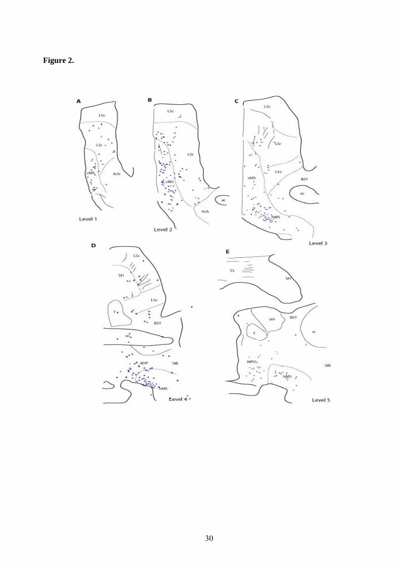
30
Figure 2.

31
Figure 3.

32
Figure 4.

33
Figure 5.

34
Figure 6.

35
Figure 7.

36
Figure 8.
158
APÊNDICE D
COMUNICAÇÕES PRODUZIDAS DURANTE O PERÍODO DE
REALIZAÇÃO DO DOUTORADO
(2008 – 2012)
159
___________________________________________Apêndice D
1. PEREIRA, Celia Waylan, SANTOS, Fabio Neves, MARCHIORO, Murilo,
OLUCHA-BORDONAU, Francisco E. Atuação do Sistema Núcleo Incertus/Relaxina-3
Sobre o Condicionamiento Pavloviano In: VII Reunião Regional da Fesbe, 2012.
2. SANTOS, Fabio Neves, PEREIRA, Celia Waylan, MARCHIORO, Murilo,
OLUCHA-BORDONAU, Francisco E. Papel do Núcleo Incertus na Modulação da
Pressão Sanguínea In: VII Reunião Regional da Fesbe, 2012, Maceió. Anais., 2012.
3. OLUCHA-BORDONAU, Francisco E., SÁNCHEZ-PÉREZ, Ana M., SANTOS,
Fabio Neves, PEREIRA, Celia Waylan, MA, Sherie, GUNDLACH, Andrew L.
Behavioral effects of lesions of the nucleus incertus on fear conditioning and extinction
and social exploration in rats In: 8th Fens – Forum of Neuroscience, 2012, Barcelona.
Fens Forum., 2012. v.Único.
4. SANTOS, Fabio Neves, OLUCHA-BORDONAU, Francisco E., OTERO-GARCIA,
Marcos, PEREIRA, Celia Waylan. As projeções relaxina 3 sobre o tectum e tegmentum
revelam um provável papel na modulação de respostas de atenção In: XXXIV
Congresso Anual da Sociedade Brasileira de Neurociências e Comportamento,
2010, Caxambu. Anais., 2010. v.Único.
5. PEREIRA, Celia Waylan, OTERO-GARCIA, Marcos, OLUCHA-BORDONAU,
Francisco E., SANTOS, Fabio Neves Distribuição de fibras relaxina 3 no diencéfalo de
ratos In: XXXIV Congresso Anual da Sociedade Brasileira de Neurociências e
Comportamento, 2010, Caxambu. Anais. , 2010. v.Único.
6. PEREIRA, Celia Waylan, SANTOS, Fabio Neves, OTERO-GARCIA, Marcos,
NUÑEZ, Angel, MA, Sherie, GUNDLACH, Andrew L., OLUCHA-BORDONAU,
Francisco E. Efectos de la Transmisión Núcleo Incertus/Relaxin3 Sobre la Modulación
de la Presión Sanguínea In: VII Congreso de la Sociedad Española de Psicofisiología
y Neurociencia Cognitiva y Afectiva (SEPNECA), 2010, Valencia. VII Sociedad
Española de Psicofisiología y Neurociencia Cognitiva y Afectiva. Barcelona: Viguera
Editores, 2010. v.52. p.306 – 315. Os anais do evento foram publicados na Revista de
Neurologia

160
7. OLUCHA-BORDONAU, Francisco E., OTERO-GARCIA, Marcos, PEREIRA,
Celia Waylan, SANTOS, Fabio Neves, NUÑEZ, Angel, MA, Sherie, GUNDLACH,
Andrew L. El núcleo incertus del tegmentum pontino: un sistema de proyecciones
amplias para la modulación de procesos de atención y memória In: VII Congreso de la
Sociedad Española de Psicofisiología y Neurociencia Cognitiva y Afectiva
(SEPNECA), 2010, Valencia. VII Sociedad Española de Psicofisiología y Neurociencia
Cognitiva y Afectiva. Barcelona: Viguera Editores, 2010. v.52. p.306 – 315. Os anais
do evento foram publicados na Revista de Neurología
8. SANTOS, Fabio Neves, PEREIRA, Celia Waylan, OTERO-GARCIA, Marcos,
NUÑEZ, Angel, MA, Sherie, GUNDLACH, Andrew L., OLUCHA-BORDONAU,
Francisco E. Papel del Sistema Núcleo Incertus/Relaxin3 sobre el Condicionamiento
Pavloviano In: VII Congreso de la Sociedad Española de Psicofisiología y
Neurociencia Cognitiva y Afectiva (SEPNECA), 2010, Valencia. VII Congreso de la
Sociedad Española de Psicofisiología y Neurociencia Cognitiva y Afectiva. Barcelona:
Viguera Editores, 2010. v.52. p.306 – 315 Os anais do evento foram publicados na
Revista de Neurología
9. OTERO-GARCIA, Marcos, PEREIRA, Celia Waylan, SANTOS, Fabio Neves,
NUÑEZ, Angel, MA, Sherie, GUNDLACH, Andrew L., OLUCHA-BORDONAU,
Francisco E. Relación de la Distribución de Fibras Relaxin3 en el Tectum y Tegmentum
con Mecanismos Defensivos y Ofensivos In: VII Congreso de la Sociedad Española
de Psicofisiología y Neurociencia Cognitiva y Afectiva (SEPNECA), 2010, Valencia.
Sociedad Española de Psicofisiología y Neurociencia Cognitiva y Afectiva. Barcelona:
Viguera Editores, 2010. v.52. p.306 – 315. Os anais do evento foram publicados na
Revista de Neurología
10. OLUCHA-BORDONAU, Francisco E., OTERO-GARCIA, Marcos, PEREIRA,
Celia Waylan, SANTOS, Fabio Neves, BLASIAK, Anna, MA, Sherie, GUNDLACH,
Andrew L. Tectal and tegmental targets of relaxin 3 fibers arising from the nucleus
incertus in rat brain In: 40ª Annual Meeting Neuroscience 2010, 2010, San Diego.
Neuroscience Meeting Planner. San Diego: Society for Neuroscience, 2010. p.809.23.