Reactivity of anticancer metallodrugs with serum proteins: new insights from size exclusion...
22
Reactivity of anticancer metallodrugs with serum proteins: new insights from size exclusion chromatography-ICP-MS and ESI- MS† Michael Groessl ‡,a , Mattia Terenghi ‡,b , Angela Casini a , Lisa Elviri b , Ryszard Lobinski c,d , and Paul J. Dyson a a Institut des Sciences et Ingénierie Chimiques, Ecole Polytechnique Fédérale de Lausanne (EPFL), CH-1015 Lausanne, Switzerland. [email protected]; Fax: +41 (0)21 6939885; Tel: +41 (0)21 6939860 b Dipartimento di Chimica Generale ed Inorganica, Chimica Analitica, Chimica Fisica, Università degli Studi di Parma, Viale G.P. Usberti 17/A, I-43100 Parma, Italy. [email protected]; Fax: +39 0521 905557; Tel: +39 0521 905476 c CNRS/UPPA, Laboratoire de Chimie Analytique Bio-inorganique et Environnement, UMR 5254, Hélioparc 2, Av. Pr. Angot, F-64053 Pau, France d Department of Analytical Chemistry, Warsaw University of Technology, 00-664 Warsaw, Poland Abstract A method based on the coupling of high resolution size-exclusion liquid chromatography using a polymer stationary phase with inductively coupled plasma mass spectrometry was developed to study the interactions of two metallodrugs – cisplatin and RAPTA-T – with the serum proteins albumin and transferrin. In contrast to previous approaches, the technique allowed the total recovery of the metals from the column and was able to discriminate between the different species of the metallodrugs and their complexes with the proteins at femtomolar detection levels. Metal binding was found to be dependent on the protein concentration and on the incubation time of the sample. Cisplatin was found to bind the serum proteins to the same extent, whereas RAPTA-T showed marked preference for transferrin. The affinity of the ruthenium complex for holo- transferrin was higher than for the apo-form suggesting a cooperative iron-mediated metal binding mechanism. RAPTA-T binding to holo-transferrin was further investigated by electrospray mass spectrometry using both the intact protein and a model peptide mimicking the iron-binding pocket. Introduction Dating back to the Renaissance and to Paracelsus’s alchemical studies, metals and minerals have profoundly fascinated physicians and have played a major role in the history of medicine. 1 A large number of different metal salts and compounds are still used to treat a wide range of diseases. Notably, platinum coordination complexes are extensively used to treat cancer. Indeed, following the discovery of the anticancer properties of cisplatin in 1965 by Rosenberg, 2 considerable efforts have been made to unravel the mechanism by which it exerts its anticancer effect and to develop alternative metal-based drugs. 3 – 5 In recent years a † This article is part of a themed issue devoted to highlighting the work of outstanding young analytical scientists (YAS) working in the area of analytical atomic spectrometry. This 3rd YAS issue has been guest edited by Professor Spiros Pergantis. © The Royal Society of Chemistry 2010 ‡ Author contributions: M.G and M.T. contributed equally to this work. M.G. performed the ESI-MS analysis; M.T. carried out the SEC-ICP-MS determination and interpretation. Europe PMC Funders Group Author Manuscript J Anal At Spectrom. Author manuscript; available in PMC 2010 December 09. Published in final edited form as: J Anal At Spectrom. 2010 March ; 25(3): 305–313. doi:10.1039/B922701F. Europe PMC Funders Author Manuscripts Europe PMC Funders Author Manuscripts
-
Upload
independent -
Category
Documents
-
view
1 -
download
0
Transcript of Reactivity of anticancer metallodrugs with serum proteins: new insights from size exclusion...

Reactivity of anticancer metallodrugs with serum proteins: newinsights from size exclusion chromatography-ICP-MS and ESI-MS†
Michael Groessl‡,a, Mattia Terenghi‡,b, Angela Casinia, Lisa Elvirib, Ryszard Lobinskic,d,and Paul J. Dysona
aInstitut des Sciences et Ingénierie Chimiques, Ecole Polytechnique Fédérale de Lausanne(EPFL), CH-1015 Lausanne, Switzerland. [email protected]; Fax: +41 (0)21 6939885; Tel:+41 (0)21 6939860 bDipartimento di Chimica Generale ed Inorganica, Chimica Analitica, ChimicaFisica, Università degli Studi di Parma, Viale G.P. Usberti 17/A, I-43100 Parma, [email protected]; Fax: +39 0521 905557; Tel: +39 0521 905476 cCNRS/UPPA,Laboratoire de Chimie Analytique Bio-inorganique et Environnement, UMR 5254, Hélioparc 2, Av.Pr. Angot, F-64053 Pau, France dDepartment of Analytical Chemistry, Warsaw University ofTechnology, 00-664 Warsaw, Poland
AbstractA method based on the coupling of high resolution size-exclusion liquid chromatography using apolymer stationary phase with inductively coupled plasma mass spectrometry was developed tostudy the interactions of two metallodrugs – cisplatin and RAPTA-T – with the serum proteinsalbumin and transferrin. In contrast to previous approaches, the technique allowed the totalrecovery of the metals from the column and was able to discriminate between the different speciesof the metallodrugs and their complexes with the proteins at femtomolar detection levels. Metalbinding was found to be dependent on the protein concentration and on the incubation time of thesample. Cisplatin was found to bind the serum proteins to the same extent, whereas RAPTA-Tshowed marked preference for transferrin. The affinity of the ruthenium complex for holo-transferrin was higher than for the apo-form suggesting a cooperative iron-mediated metal bindingmechanism. RAPTA-T binding to holo-transferrin was further investigated by electrospray massspectrometry using both the intact protein and a model peptide mimicking the iron-binding pocket.
IntroductionDating back to the Renaissance and to Paracelsus’s alchemical studies, metals and mineralshave profoundly fascinated physicians and have played a major role in the history ofmedicine.1 A large number of different metal salts and compounds are still used to treat awide range of diseases. Notably, platinum coordination complexes are extensively used totreat cancer. Indeed, following the discovery of the anticancer properties of cisplatin in 1965by Rosenberg,2 considerable efforts have been made to unravel the mechanism by which itexerts its anticancer effect and to develop alternative metal-based drugs.3–5 In recent years a
†This article is part of a themed issue devoted to highlighting the work of outstanding young analytical scientists (YAS) working inthe area of analytical atomic spectrometry. This 3rd YAS issue has been guest edited by Professor Spiros Pergantis.
© The Royal Society of Chemistry 2010‡Author contributions: M.G and M.T. contributed equally to this work. M.G. performed the ESI-MS analysis; M.T. carried out theSEC-ICP-MS determination and interpretation.
Europe PMC Funders GroupAuthor ManuscriptJ Anal At Spectrom. Author manuscript; available in PMC 2010 December 09.
Published in final edited form as:J Anal At Spectrom. 2010 March ; 25(3): 305–313. doi:10.1039/B922701F.
Europe PM
C Funders A
uthor Manuscripts
Europe PM
C Funders A
uthor Manuscripts

vast number of non-platinum compounds have been prepared and evaluated as experimentalanticancer drugs.6,7 Among them, ruthenium(II)-arene complexes bearing 1,3,5-triaza-7-phosphaadamantane (PTA) ligands – named RAPTA – have been found to exhibitpromising antimetastatic properties in vivo.8 Their mechanism of action is still largelyunknown, however, there is evidence to suggest that RAPTA compounds work on moleculartargets other than DNA implying a biochemical mode of action profoundly different toclassical platinum anticancer drugs.9,10 Indeed, ruthenium compounds have been shown todirectly interfere with specific proteins involved in signal transduction pathways and/or toalter cell adhesion and migration processes.11
An understanding of metallodrug-protein interactions is crucial since the pharmacologicalactivity of a drug, even for drugs where DNA is the ultimate target, is usually modified uponprotein binding. Metal drug-protein interactions are not only important with regard toapoptosis, but also with respect to unwanted side-effects (due to indiscriminate proteinbinding), drug resistance (due to binding/deactivation by metal-lothioneins,12 glutathionetransferases,13,14 etc.) and even possibly drug delivery and storage.15 Upon intravenousapplication, serum proteins including albumin, transferrin and globulins are thought to playa crucial role in the transport, delivery, and storage of anticancer metallodrugs. Humanserum albumin (HSA) is the most abundant protein (about 52% of total serum protein) witha concentration of 40–45 g L−1 in healthy humans (ca. 600 μM; Mw 66–67 kDa). Itcomprises a single chain of 585 amino acids organized in three similar domains, eachcomposed of two subdomains.16 HSA is known to bind a remarkably wide range of drugs,thereby restricting their free, active concentrations.17 Reducing this binding affinityrepresent a major challenge in drug development. However, HSA can also be exploited fortargeted delivery strategies, as in the case of organometallic ruthenium(II)-arene anticancercompounds, to develop compounds that can selectively accumulate in tumors cells.18
Human serum transferrin (Tf) is a single-chain glycoprotein containing 679 amino acidswith a molecular mass of about 80 kDa and is found in blood at a concentration of about 2.5g L−1 (35 μM). Tf acts as an iron transporter and is capable of binding two iron(III) ions(Fe3+ is bound selectively over Fe2+).19 Tf is normally only 30% saturated with iron in thebody,19 and at least 30 other metal ions can also bind to Tf.20 Therefore, it is possible to useTf as a metal transporter within the body and the cellular uptake mechanism via the Tf-transferrin receptor transport system has the potential to be exploited for site-specificdelivery of various therapeutic metal ions, drugs, proteins and genes.21 In the case ofmetallodrugs the resulting conjugates significantly improve the cytotoxicity and selectivityof the drugs itself. For example, anticancer Ru(III) complexes were reported not only to bindto Tf,22,23 but it has been shown that injection of Ru(III)-Tf conjugates resulted in hightumor uptake of the metal,24 which suggests that Tf uptake might be the more importantmode of transport of Ru(III) anticancer complexes to the tumor. In some cases, the Ru-Tfadducts exhibit a significantly higher antitumor activity against human cancer cells than theRu(III) complex itself, attributable to the Tf-mediated uptake mechanism, which may alsolower drug toxicity.25
The development of analytical methods capable of providing a better understanding of howmetallodrugs interact with proteins remains challenging.26,27 The direct monitoring of real-time protein-mediated metabolism of anticancer agents using hyphenated (coupled, tandem)techniques, that combine chromatographic separation with sensitive and element-specificdetection techniques such as inductively coupled plasma-mass spectrometry (ICP-MS) havebeen effectively used.28–30 In terms of separation techniques, high performance liquidchromatography (HPLC)31,32 and capillary zone electrophoresis (CZE)29,33–35 wereproposed for the study of platinum and ruthenium based drugs, to assess kinetic constants ofhydrolysis and subsequent reactions with a variety of physiological targets. This concept has
Groessl et al. Page 2
J Anal At Spectrom. Author manuscript; available in PMC 2010 December 09.
Europe PM
C Funders A
uthor Manuscripts
Europe PM
C Funders A
uthor Manuscripts

also been transferred to study the distribution of gallium-based pharmaceuticals in humanserum.36
Within this frame, we propose size-exclusion liquid chromatography (SEC) coupled to ICP-MS to study metal complexes-proteins interactions. SEC has certain advantages compared toother separation techniques to characterize metallodrug binding to biomolecules in that itcan be used together with other hyphenated systems to obtain complementary information.In comparison to ion exchange chromatography (IEC), the main advantage of the SECapproach lies in the more “gentle” conditions, as IEC usually requires buffers with highionic strength and extreme pH values.37 The importance of working under morephysiological-type conditions is crucial when studying metallodrugs to avoid interferenceswhich might alter the metal-ligand bonds and the metal-protein adducts. With respect tocapillary electrophoresis (CE) the major advantage of the SEC separation lies in the ease androbustness of the coupling to ICP-MS, which is considered one of the main problems of CE-ICP-MS, and usually leads to higher detection limits compared to LC-ICP-MS.35,38,39
Up to now, SEC coupled to ICP-MS has been used to probe protein interactions withruthenium drugs, but suffers from poor recoveries because of the reactivity of thecompounds with the column stationary phase.40 This problem was alleviated by using ashort size-exclusion column at the expense of the chromatographic resolution, or it wassimply used as a first separation technique to roughly discriminate between bound andunbound species prior to ion-exchange separation.40 Therefore, the primary objective of ourresearch was to evaluate a polymeric stationary phase with regard to the unspecificretention, reactivity and carryover of an organometallic drug RAPTA-T (Ru(η6-C6H5Me)(PTA)Cl2) (Chart 1), and its adducts, with the serum proteins albumin and transferrin, incomparison to cisplatin. The binding of RAPTA-T towards intact Tf and the possiblebinding sites of the metallodrugs on a model peptide mimicking one of the iron-bindingpockets of Tf were also investigated using electrospray ionization-mass spectrometry (ESI-MS).
ExperimentalReagents
Cisplatin, human serum albumin (>99%, HSA), human holo transferrin (>98%, hTf) andhuman apo transferrin (>97%, apo-Tf) were obtained from Sigma-Aldrich. Sodium chloride,formic acid, Tris(hydroxymethyl)aminomethane hydrochloride, ammonium acetate andammonium hydrogen carbonate were also purchased from Aldrich and were of analyticalreagent grade. Buffers used for SEC-ICP-MS analysis were prepared as follows: ammoniumacetate buffer (100 mM, pH 6.8) and Tris buffer saline (20 mM Tris-HCl, 100 mM NaCl,pH 7.2). Deionized water (18 MΩ/cm) (Millipore, Bedford, MA) was used throughout.RAPTA-T was synthesized as described elsewhere.8 The HPLC-purified model peptide(KDCHLAQVPSHTV) was purchased from PSL (Heidelberg, Germany).
ICP-MS instrumentationAn Agilent model 1200 binary pump, equipped with an auto-sampler, automatic injectionsystem with a loop of 900 μL and a UV-VIS detector was used as the solvent deliverysystem and was controlled by Agilent ChemStation software. An Agilent 7500ce ICP-MSfitted with a Meinhardt nebulizer was used as the detector in SEC-ICP-MS experiments,employing the following conditions: plasma gas (Ar) 15 L min−1, forward power 1500 W,carrier gas 1.05 L min−1. The working conditions were optimised using a 1 μg L−1 solutionof 7Li+, 89Y+ and 205Tl+ in 2% HNO3, with a dwell time of 100 ms for each isotope. Foriron isotope monitoring, a collision cell was used to remove polyatomic interferences with
Groessl et al. Page 3
J Anal At Spectrom. Author manuscript; available in PMC 2010 December 09.
Europe PM
C Funders A
uthor Manuscripts
Europe PM
C Funders A
uthor Manuscripts

H2 (3–3.5 ml min−1) and He (1.0 ml min−1) as auxiliary gases. Double charged ions andoxide levels were minimized by using 140Ce+ and were typically < 2%. The isotopesmonitored (in time resolved analysis mode with a dwell time of 100 ms each)were 54Fe, 56Fe, 195Pt, 196Pt, 197Pt, 198Pt, 100Ru, 101Ru and 102Ru.The 54Fe, 56Fe, 102Ru, 195Pt and isotopes were used for quantification of binding anduptake.
Size-exclusion chromatographySamples were analyzed on a Superdex™ 200 HR 10/30 (10 mm × 300 mm × 13 mm) (GEHealthcare) exclusion column with approximate bed volume of 24 mL and a linearseparation range of 10–600 kDa for globular proteins. The column was calibrated asspecified by the manufacturer with a mixture of thyroglobulin (670 kDa), hTf (81 kDa),bovine albumin (66 kDa), chicken egg albumin (44 kDa), Mn superoxide dismutase (39.5kDa), Cu/Zn superoxide dismutase (32.5 kDa), carbonate dehydratase (29 kDa) andmyoglobin (16.7 kDa) using UV/VIS detection at 280 nm with baseline evaluation at 800nm. Retention times (in min) plotted versus the logarithm of molecular mass (in kDa)showed linear correlation (y=− 0.134x + 4.385; r2 0.992). Elution was carried outisocratically at 0.7 mL min−1 using 100 mM ammonium acetate (pH 6.8) as the mobilephase. Solvent flow from the chromatographic system was introduced into the ICP-MSnebulizer by means of PEEK tubing.
Sample preparation for SEC-ICP-MSCisplatin and RAPTA-T stock solutions were prepared by dissolving the compounds inammonium acetate buffer or in Tris buffer. Proteins were dissolved in ammonium acetate ata concentration 0.5 mM. To investigate the interactions between serum proteins and themetal complexes, samples were initially prepared with various protein concentrations (1, 50and 250 μM) and adding the appropriate amounts of metal complex to achieve differentprotein : metal stoichiometric ratios (1 : 1, 1 : 2 or 1 : 5) at each protein concentration. In thecase of 250 μM protein samples only a maximum two-fold molar excess of the metalcomplex was applied due to protein precipitation phenomena. Samples were then incubatedfor 1 h at 37 °C and then diluted in ammonium acetate buffer to yield a final proteinconcentration of 1 μM prior to injection into the SEC column. Alternatively, for theevaluation of maximum metal binding, the protein concentration was fixed at 50 μM, whileeach metal complex was added in stoichiometric amount or in 2, 5, 10 and 18-fold excesses.Metal complex solutions were added freshly prepared (sample Set A) or 24 h afterpreparation (sample Set B; solution stored at 4 °C) in ammonium acetate buffer. Sampleswere then incubated for 1 h at 37 °C and diluted to 1 μM protein before injection.Quantification of the metal content in each chromatographic fraction was calculated directlyfrom the integrated signal from the SEC-ICP-MS chromatograms using the formula cb = Mb× cinj × D, where cb represents the initial concentration of metal in the original sample, Mb isthe fraction of metal bound to protein in the chromatogram (calculated as a percentage ofbound metal signal with respect to the total sum of signals), cinj is the concentration of metalinjected for analysis and D is the dilution factor applied to the samples prior to injection.
ESI-MS analysisFor ESI-MS experiments with the model peptide, samples were prepared with ametallodrug : peptide ratio of 1 : 1 (25 μM) in 20 mM carbonate buffer (pH 7.4) andincubated for 24 h at 37 °C prior to analysis. Samples were then mixed with acetonitrile andformic acid (final concentration 30% and 0.1%, respectively) and placed into a 96-well platein an Advion TriVersa robot (Advion Biosciences, Ithaca, NY) equipped with a 5.5 mm-nozzle chip. The ESI robot was controlled with ChipSoft v7.2.0 software employing thefollowing parameters: gas pressure 0.45 psi; voltage 1.7 kV. The samples were analyzed in
Groessl et al. Page 4
J Anal At Spectrom. Author manuscript; available in PMC 2010 December 09.
Europe PM
C Funders A
uthor Manuscripts
Europe PM
C Funders A
uthor Manuscripts

positive ion mode using a LTQ XL mass spectrometer (ThermoFisher Scientific, Bremen,Germany). The Xcalibur software bundle was utilized for data acquisition and data analysis.
ESI-MS measurements of apo transferrin-metallodrug interactions were carried out on anUltima II q-TOF mass spectrometer (Waters, Manchester, UK) operated in positive mode.Data acquisition and analysis were carried out using the MassLynx software bundle(Waters). The instrument was calibrated daily using a 0.01% phosphoric acid solution in50% acetonitrile. Samples were prepared with a metallodrug : protein ratio of 5 : 1 in 20mM carbonate buffer (pH 7.4) and incubated for up to 24 h at 37 °C. Prior to analysis,samples were ultracentrifuged using 30 kDa cut-off filters (VWR, Switzerland) to removeunbound metallodrugs and injected into the ESI-MS system after dilution with acetonitrileand formic acid (final concentrations 25% and 0.05%, respectively).
Results and discussionSize-exclusion chromatography of cisplatin and RAPTA-T
A SEC-ICP-MS system was initially used to evaluate solutions of the two metal drugs,cisplatin and RAPTA-T (Chart 1), in the absence of any protein in order to establish theevolution of metal species. Based on the molecular weight of the complexes (300–400 Da)an expected single peak with a total bed column volume (≈24 mL), corresponding to 35 minat flow rate of 0.7 mL min−1, might be expected to elute from the column. Two differentbuffers, namely Tris buffer saline (TBS) and ammonium acetate (pH ~ 7), were evaluated asincubation media and eluent for cisplatin and RAPTA-T hydrolysis. For methoddevelopment, 50 μL of 1 μM cisplatin (195 ng mL−1, 50 pmol of Pt) or RAPTA-T (102 ngmL−1, 50 pmol of Ru) in the respective buffers were injected into the SEC column eitherfreshly prepared or pre-incubated for 12 h at 37 °C.
Elution profiles for the two drugs in TBS are shown in Fig. 1. In the case of cisplatin (Fig.1A), freshly prepared solution gave signals at 32 and 42 min, while for the sample obtainedafter 12 h incubation an additional peak appeared at 28 min. In both cases the peak at 42 min(intact cisplatin) was the most intense. Peaks at 32 and 28 min corresponded to cisplatinhydrolysis products (mono- and bis-aqua species, respectively) as confirmed by ESI-MSanalysis. In ammonium acetate buffer, these compounds were detected at the same elutiontimes, but more extensive hydrolysis of cisplatin was observed due to the absence ofchlorido ions in the buffer. It is worth mentioning that the separation of different forms ofmetallodrugs arise from aspecific interactions with the stationary phase, since nodifferentiation is possible based on their low molecular weights. Elution times were found tobe constants after multiple injection of metallodrug. Only for peaks eluting at 40 min, wellafter the total column volume, minor shifts of ±2 min in the elution time were observed.
When RAPTA-T was injected, either freshly prepared or after 12 h of incubation, only asingle symmetric peak at 28 min of the chromatographic run was observed regardless of thebuffer employed (Fig. 1B). This peak probably corresponds to the intact complex or to themono-aqua [Ru(η6-C6H5Me)(PTA)ClH2O] species as suggested by ESI-MS analysis andUV-visible spectrophotometry (data not shown).
Recovery experiments, based on the comparison of the metal content in the injected sampleand the eluate, showed that the metals were not retained on the stationary phase. Indeed,recovery values for cisplatin and RAPTA-T were 100 ± 2% and 93 ± 3% respectively, inboth buffers. The baseline remained stable during the analyses and a post-analysis injectionof HSA gave a blank chromatogram.
Groessl et al. Page 5
J Anal At Spectrom. Author manuscript; available in PMC 2010 December 09.
Europe PM
C Funders A
uthor Manuscripts
Europe PM
C Funders A
uthor Manuscripts
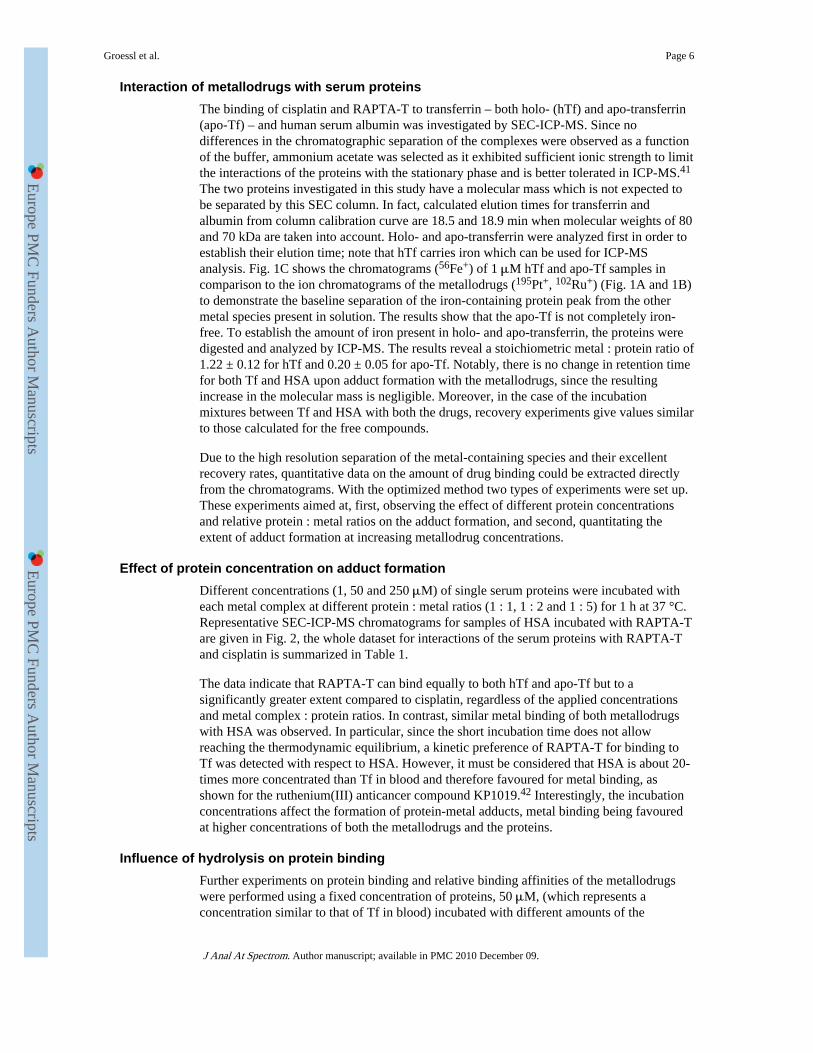
Interaction of metallodrugs with serum proteinsThe binding of cisplatin and RAPTA-T to transferrin – both holo- (hTf) and apo-transferrin(apo-Tf) – and human serum albumin was investigated by SEC-ICP-MS. Since nodifferences in the chromatographic separation of the complexes were observed as a functionof the buffer, ammonium acetate was selected as it exhibited sufficient ionic strength to limitthe interactions of the proteins with the stationary phase and is better tolerated in ICP-MS.41
The two proteins investigated in this study have a molecular mass which is not expected tobe separated by this SEC column. In fact, calculated elution times for transferrin andalbumin from column calibration curve are 18.5 and 18.9 min when molecular weights of 80and 70 kDa are taken into account. Holo- and apo-transferrin were analyzed first in order toestablish their elution time; note that hTf carries iron which can be used for ICP-MSanalysis. Fig. 1C shows the chromatograms (56Fe+) of 1 μM hTf and apo-Tf samples incomparison to the ion chromatograms of the metallodrugs (195Pt+, 102Ru+) (Fig. 1A and 1B)to demonstrate the baseline separation of the iron-containing protein peak from the othermetal species present in solution. The results show that the apo-Tf is not completely iron-free. To establish the amount of iron present in holo- and apo-transferrin, the proteins weredigested and analyzed by ICP-MS. The results reveal a stoichiometric metal : protein ratio of1.22 ± 0.12 for hTf and 0.20 ± 0.05 for apo-Tf. Notably, there is no change in retention timefor both Tf and HSA upon adduct formation with the metallodrugs, since the resultingincrease in the molecular mass is negligible. Moreover, in the case of the incubationmixtures between Tf and HSA with both the drugs, recovery experiments give values similarto those calculated for the free compounds.
Due to the high resolution separation of the metal-containing species and their excellentrecovery rates, quantitative data on the amount of drug binding could be extracted directlyfrom the chromatograms. With the optimized method two types of experiments were set up.These experiments aimed at, first, observing the effect of different protein concentrationsand relative protein : metal ratios on the adduct formation, and second, quantitating theextent of adduct formation at increasing metallodrug concentrations.
Effect of protein concentration on adduct formationDifferent concentrations (1, 50 and 250 μM) of single serum proteins were incubated witheach metal complex at different protein : metal ratios (1 : 1, 1 : 2 and 1 : 5) for 1 h at 37 °C.Representative SEC-ICP-MS chromatograms for samples of HSA incubated with RAPTA-Tare given in Fig. 2, the whole dataset for interactions of the serum proteins with RAPTA-Tand cisplatin is summarized in Table 1.
The data indicate that RAPTA-T can bind equally to both hTf and apo-Tf but to asignificantly greater extent compared to cisplatin, regardless of the applied concentrationsand metal complex : protein ratios. In contrast, similar metal binding of both metallodrugswith HSA was observed. In particular, since the short incubation time does not allowreaching the thermodynamic equilibrium, a kinetic preference of RAPTA-T for binding toTf was detected with respect to HSA. However, it must be considered that HSA is about 20-times more concentrated than Tf in blood and therefore favoured for metal binding, asshown for the ruthenium(III) anticancer compound KP1019.42 Interestingly, the incubationconcentrations affect the formation of protein-metal adducts, metal binding being favouredat higher concentrations of both the metallodrugs and the proteins.
Influence of hydrolysis on protein bindingFurther experiments on protein binding and relative binding affinities of the metallodrugswere performed using a fixed concentration of proteins, 50 μM, (which represents aconcentration similar to that of Tf in blood) incubated with different amounts of the
Groessl et al. Page 6
J Anal At Spectrom. Author manuscript; available in PMC 2010 December 09.
Europe PM
C Funders A
uthor Manuscripts
Europe PM
C Funders A
uthor Manuscripts

metallodrugs. Quantitative metal binding data were calculated based on the fraction eluted at19 min, related to the complexed forms of protein with metallodrugs, and the evaluation ofall the other forms of free metallodrugs in the SEC-ICP-MS profiles by the equation statedin the Experimental Part.
It should be noted that the same results are obtained if the equation developed by Timerbaevet al.33 is applied to the integrated values for the eluted species in our SEC-ICP-MSchromatogram. In Table 2, results from each protein sample treated with RAPTA-T orcisplatin and incubated for 1 h at 37 °C are shown. Dataset A represents data for sampleswith freshly prepared solutions of the metal complexes and Dataset B for samples withmetal complex solutions pre-incubated for 24 h prior to addition to the protein to study theeffect of metallodrug hydrolysis on protein binding.
The most notable difference between the two preparations corresponds to the availability ofthe different forms of the metallodrugs. For instance, in Dataset B higher contents ofhydrolyzed products are present for cisplatin with respect to Set A. Since cisplatinhydrolysis products carry a positive charge, their interaction with negatively charged aminoacid residues on the protein chains (possible under these conditions considering calculatedpI values are 5.92 and 6.81 for HSA and Tf, respectively)43 potentially increases interactionscompared to the neutral starting compound. Moreover, the hydrolyzed product is morereactive as the aqua ligands are more labile than chlorido ligands. In Set B, platinum adductsare formed to a greater extent with respect to Set A at any concentration. In general, for bothdatasets, cisplatin shows a slightly increased affinity towards HSA at lower molar excess ofdrug and no apparent preference for the two forms of transferrin. Upon increasing the drugexcess, the reaction proceeds more rapidly and, as expressed by an increased number ofmetal ions bound per protein molecule, the proteins are metallated to a higher degree.
For RAPTA-T, a higher affinity is found for hTf with respect to apo-Tf and HSA, and theamount of ruthenium bound to hTf is similar in Set A and Set B. For apo-Tf and HSA,increased binding is observed for the Set B samples, especially at high metal : protein ratios.Based on the ratio of the metal concentration of the bound fraction, for cisplatin maximumbinding was in the range from 0.2 to 0.35 for all three proteins. For RAPTA-T the highestmetal binding was observed for hTf, with an average of ca. 1.4 ruthenium ions per protein.For apo-Tf and HSA an average 0.5 to 1.0 ruthenium ions bind per protein, which is higherthan observed for cisplatin. In comparison to previously reported data using CZE-ICP-MS,which shows extensive binding of cisplatin toward the protein (up to 4 Pt ions per moleculeof HSA for a sample containing a 10-fold excess of metal complex after 24 h incubation inPBS buffer), the values obtained for cisplatin-HSA adduct formation in our experimentswere unexpected.33 Therefore, protein samples incubated for 24 h with a 10-fold excess ofcisplatin or RAPTA-T were studied. For all the three proteins increased metal binding forboth metallodrugs was detected (Table 3), although not to the same levels observed by CZE-ICP-MS.
Overall, the low detection limits of the applied method allowed analysis of samplescontaining 400 fmol of metals bound to the proteins, with as little as 2 pmol of proteininjected into SEC column. In the case of RAPTA-T, the higher amount of ruthenium boundto hTf with respect to apo-Tf suggests that the presence of iron bound to protein mightinfluence the formation of metal complex adducts, most likely maintaining a protein foldingwhich favours metallodrug binding.
Metallodrug binding versus iron loss in holo-transferrinDespite the higher affinity of RAPTA-T than cisplatin for hTf, no differences in terms of thereleased iron were observed. Fig. 3 shows the binding behaviour of both metal complexes
Groessl et al. Page 7
J Anal At Spectrom. Author manuscript; available in PMC 2010 December 09.
Europe PM
C Funders A
uthor Manuscripts
Europe PM
C Funders A
uthor Manuscripts

for hTf, together with the decrease of the iron signal recorded by ICP-MS, for eachconcentration of metal complex. The graph shows that the iron is not released in a linearfashion. In fact, the iron signal decreases only slightly until a 10-fold molar excess (500μM) of the metallodrug and then immediately drops to the half of its original value. As theemployed hTf was only ca. 60% loaded with iron, these data suggest that initiallymetallodrug binding occurs at the iron-free protein active sites and therefore does not affectthe iron release. Afterwards, in the presence of excess of metallodrug, significant ironrelease may result from the partial denaturation or conformational changes of Tf uponaddition of excess of metal, in agreement with previous observation on cisplatin binding tohTf by Hoshino et al.33
ESI-MS studies with transferrin and an iron binding site-model peptideAs the interactions of cisplatin with intact transferrin studied by ESI-MS have already beenreported earlier,44 only the binding of RAPTA-T to transferrin was further investigated byESI-MS in this study. Fig. 4 shows the spectrum of apo-Tf treated with a 5-fold excess ofRAPTA-T and incubated for 1 h at 37 °C, in comparison to the pure protein. The spectra ofthe RAPTA-T treated samples show additional signals corresponding to adduct formationwith the ruthenium compound. Based on the mass difference, these signals are attributable toRAPTA-T after loss of both chlorido and PTA ligands and subsequent addition of acarbonate anion. The fact that only the arene ligand stays attached to the metal centre is ingood agreement with data obtained for interactions of RAPTA compounds with cytochromec,45 and the coordination of carbonate is also not surprising under the conditions employed.It is worth noting that a synergistic carbonate anion interaction is also crucial for stabilizingthe binding site to ensure efficient binding of iron to transferrin.
Furthermore, the exact binding mode of cisplatin and RAPTA-T towards Tf and the natureof the species that bind to the protein as well as the identification of the amino acidsinvolved were probed by ESI-MS utilizing a model peptide consisting of the active siteresidues 239KDCHLAQVPSHTV251 of Tf. This peptide contains the His249 residue, whichis involved in the iron binding in Tf, but also threonine and cysteine, residues which arepossible binding partners.44,46,47 The model peptide was incubated with the metallodrugsunder simulated physiological conditions (pH 7.4, 37 °C) for 24 h and then analysed. Thebound species were identified as RAPTA-T and cisplatin after loss of the chlorido ligands,indicating a bidentate binding to the peptide. Although loss of a chlorido ligand could alsobe induced during ionization, a multidentate binding mode seems more probably due tohydrolysis of the compounds under the used incubation conditions. Different amino acidsdepending on the metal complex were identified as binding partners upon collision induceddissociation (CID) of the modified peptide, as can be seen from Fig. 5 and 6. The insetsshow signals of metallated ions, exhibiting characteristic isotopic patterns induced byruthenium and platinum, which facilitate the identification of modified residues. In general,the remaining ligands of the metallodrugs (amino for cisplatin, toluene and PTA forRAPTA-T) were dissociated more easily than the peptide bonds under fragmentationconditions, indicating the formation of strong bonds between the amino acids and the metals.
For RAPTA-T (Fig. 5), the first histidine residue appears to be the major binding partner, asevidenced by the presence of the modified b4 ion in contrast to the unmodified y9 ionfollowing CID. This is not surprising since nitrogen donor ligands are known to form strongcomplexes with ruthenium. Interestingly, no binding towards the histidine residue inposition 11 of the peptide (iron binding His249 in transferrin) could be detected for theruthenium compound, indicating that the cysteine residue might also be involved in thebinding of RAPTA-T. It is also worth mentioning that cleavage of the peptide occurspredominately next to the metal binding site, resulting in low relative abundances of other band y fragment ions.
Groessl et al. Page 8
J Anal At Spectrom. Author manuscript; available in PMC 2010 December 09.
Europe PM
C Funders A
uthor Manuscripts
Europe PM
C Funders A
uthor Manuscripts

In the case of cisplatin (Fig. 6), the sulfur containing cysteine residue seems to beresponsible for the binding. CID resulted in a more extensive fragmentation of the peptide ascompared to RAPTA-T, and unmodified b2 and y10 in contrast to modified b3 and y11 ionsallow unambiguous identification of the binding site. Although these data are in agreementwith previous studies which show that cisplatin readily reacts with sulfur containingbiomolecules such as glutathione or metallothionein within the cell,48 caution should beapplied to the validity of the model peptide in terms of comparability to intact proteins. As ithas been shown in earlier proteomic studies of transferrin-cisplatin interaction44 as well ason samples extracted from E. coli upon incubation with a ruthenium-arene compound andcisplatin,49,50 oxygen atoms in the carboxy- and hydroxyl-functions of certain residues (e.g.aspartate, serin, threonine) may also serve as binding partners in biological systems.Consequently, protein conformation has a strong influence on the site and type of adductformed as exposed residues might be more easily accessible and therefore more reactivethan those buried in the folded protein. Although such structural features cannot bemimicked by a short, linear model peptide, the general reactivity of a compound in thepresence of several possible binding amino acids can be assessed.
ConclusionsHigh resolution SEC-ICP-MS based on a polymer stationary phase was demonstrated, forthe first time, to be a powerful tool for the analysis of metal drugs with serum proteins. Theapproach provides information on the affinity of each metallodrug for the selected proteins,quantification of metal uptake and on the stoichiometry of the metal-protein complexes. Theprimary advantage of this approach over the previous SEC-ICP-MS methods is that all themetal species are quantitatively recovered under the separation conditions. Although theSEC system was not suitable for the baseline separation of samples containing both HSAand Tf, it proved to be an efficient tool to analyze the binding of metallodrugs to singleproteins and in principle could be developed to investigate protein mixtures. Indeed, recentlythe use of SEC hyphenated to ICP atomic-emission spectrometry has been successfullyapplied for metalloproteome studies of human plasma.51 The reported results highlight thedifferent reactivity of cisplatin and the antimetastatic ruthenium-based drug RAPTA-T. Thelatter compound showed a preferential binding for Tf with respect to HSA, while cisplatinwas less selective, being able to bind both proteins to a similar extent. Differences in theselectivity of cisplatin and RAPTA compounds were already identified using an ESI-MSbased approach to study protein mixtures.52,53 In particular, cisplatin was found to bemoderately reactive towards the protein without any discrimination/selectivity, whereas theRAPTA compound was considerably more reactive and could also bind selectively. Suchselectivity has important implications for the mode of action of the metallodrugs in the cell,leading to the efficacy of the compound towards specific tumor types, and presumably alsofor their toxic side effects.
AcknowledgmentsA.C gratefully acknowledges the Swiss National Science Foundation (AMBIZIONE project n° PZ00P2_121933),the Swiss Confederation (Action COST D39 - Accord de recherche - SER project n° C09.0027) and COST D39 forfinancial support. M.G. thanks the Austrian Science Foundation for financial support (Schrödinger FellowshipJ2882-N19).
References1. Koehler CSW. Today’s Chemists at Work. 2001; 10:61–65.
2. Rosenberg B, Vancamp L, Krigas T. Nature. 1965; 205:698. [PubMed: 14287410]
3. Clarke MJ, Zhu FC, Frasca DR. Chem. Rev. 1999; 99:2511–2533. [PubMed: 11749489]
4. Reedijk J. Proc. Natl. Acad. Sci. U. S. A. 2003; 100:3611–3616. [PubMed: 12655051]
Groessl et al. Page 9
J Anal At Spectrom. Author manuscript; available in PMC 2010 December 09.
Europe PM
C Funders A
uthor Manuscripts
Europe PM
C Funders A
uthor Manuscripts
5. Casini A, Hartinger C, Gabbiani C, Mini E, Dyson PJ, Keppler BK, Messori L. J. Inorg. Biochem.2008; 102:564–575. [PubMed: 18177942]
6. Nobili S, Mini E, Landini I, Gabbiani C, Casini A, Messori L. Med. Res. Rev. 2009 DOI: 10.1002/med.20168.
7. Gianferrara T, Bratsos I, Alessio E. Dalton Trans. 2009; (37):7588–7598. [PubMed: 19759927]
8. Scolaro C, Bergamo A, Brescacin L, Delfino R, Cocchietto M, Laurenczy G, Geldbach TJ, Sava G,Dyson PJ. J. Med. Chem. 2005; 48:4161–4171. [PubMed: 15943488]
9. Dyson PJ, Sava G. Dalton Trans. 2006:1929–1933. [PubMed: 16609762]
10. Bergamo A, Sava G. Dalton Trans. 2007:1267–1272. [PubMed: 17372640]
11. Bergamo A, Masi A, Dyson PJ, Sava G. Int J Oncol. 2008; 33:1281–1289. [PubMed: 19020762]
12. Knipp M. Curr. Med. Chem. 2009; 16:522–537. [PubMed: 19199919]
13. Ang WH, Parker LJ, De Luca A, Juillerat-Jeanneret L, Morton CJ, Lo Bello M, Parker MW,Dyson PJ. Angew. Chem., Int. Ed. 2009; 48:3854–3857.
14. Ang WH, De Luca A, Chapuis-Bernasconi C, Juillerat-Jeanneret L, Lo Bello M, Dyson PJ.ChemMedChem. 2007; 2:1799–1806. [PubMed: 17918761]
15. Reedijk J. Chem. Commun. 1996:801–806.
16. He XM, Carter DC. Nature. 1992; 358:209–215. [PubMed: 1630489]
17. Ghuman J, Zunszain PA, Petitpas I, Bhattacharya AA, Otagiri M, Curry S. J. Mol. Biol. 2005;353:38–52. [PubMed: 16169013]
18. Ang WH, Daldini E, Juillerat-Jeanneret L, Dyson PJ. Inorg. Chem. 2007; 46:9048–9050.[PubMed: 17918830]
19. Gomme PT, McCann KB. Drug Discovery Today. 2005; 10:267–273. [PubMed: 15708745]
20. Sun HZ, Li HY, Sadler PJ. Chem. Rev. 1999; 99:2817–2842. [PubMed: 11749502]
21. Li HY, Qian ZM. Med. Res. Rev. 2002; 22:225–250. [PubMed: 11933019]
22. Smith CA, SutherlandSmith AJ, Keppler BK, Kratz F, Baker EN. JBIC, J. Biol. Inorg. Chem.1996; 1:424–431.
23. Kratz F, Hartmann M, Keppler B, Messori L. J. Biol. Chem. 1994; 269:2581–2588. [PubMed:8300587]
24. Som P, Oster ZH, Matsui K, Guglielmi G, Persson BRR, Pellettieri ML, Srivastava SC, RichardsP, Atkins HL, Brill AB. Eur. J. Nucl. Med. Mol. Imaging. 1983; 8:491–494.
25. Kratz F, Keppler BK, Hartmann M, Messori L, Berger MR. Met.-Based Drugs. 1996; 3(1):15–23.[PubMed: 18472789]
26. Timerbaev AR, Hartinger CG, Aleksenko SS, Keppler BK. Chem. Rev. 2006; 106:2224–2248.[PubMed: 16771448]
27. Sun XS, Tsang CN, Sun HZ. Metallomics. 2009; 1:25–31.
28. Becker JS, Jakubowski N. Chem. Soc. Rev. 2009; 38:1969–1983. [PubMed: 19551177]
29. Brouwers EEM, Tibben M, Rosing H, Schellens JHM, Beijnen JH. Mass Spectrom. Rev. 2008;27:67–100. [PubMed: 18231971]
30. Lobinski R, Schaumloffel D, Szpunar J. Mass Spectrom. Rev. 2006; 25:255–289. [PubMed:16273552]
31. Hann S, Stefanka Z, Lenz K, Stingeder G. Anal. Bioanal. Chem. 2005; 381:405–412. [PubMed:15455190]
32. Esteban-Fernandez D, Montes-Bayon M, Gonzalez EB, Gomez MMG, Palacios MA, Sanz-MedelA. J. Anal. At. Spectrom. 2008; 23:378–384.
33. Timerbaev AR, Aleksenko KS, Polec-Pawlak K, Ruzik R, Semenova O, Hartinger CG,Oszwaldowski S, Galanski M, Jarosz M, Keppler BK. Electrophoresis. 2004; 25:1988–1995.[PubMed: 15237398]
34. Polec-Pawlak K, Abramski JK, Semenova O, Hartinger CG, Timerbaev AR, Keppler BK, JaroszM. Electrophoresis. 2006; 27:1128–1135. [PubMed: 16440400]
35. Groessl M, Hartinger CG, Polec-Pawlak K, Jarosz M, Keppler BK. Electrophoresis. 2008;29:2224–2232. [PubMed: 18512673]
36. Groessl M, Bytzek A, Hartinger CG. Electrophoresis. 2009; 30:2720–2727. [PubMed: 19621374]
Groessl et al. Page 10
J Anal At Spectrom. Author manuscript; available in PMC 2010 December 09.
Europe PM
C Funders A
uthor Manuscripts
Europe PM
C Funders A
uthor Manuscripts

37. Michalke B. TrAC, Trends Anal. Chem. 2002; 21:142–153.
38. Moini M. Anal. Bioanal. Chem. 2002; 373:466–480. [PubMed: 12172682]
39. Yin XB, Li Y, Yan XP. TrAC, Trends Anal. Chem. 2008; 27:554–565.
40. Szpunar J, Makarov A, Pieper T, Keppler BK, Lobinski R. Anal. Chim. Acta. 1999; 387:135–144.
41. Makarov A, Szpunar J. Analusis. 1998; 26:M44–M48.
42. Sulyok M, Hann S, Hartinger CG, Keppler BK, Stingeder G, Koellensperger G. J. Anal. At.Spectrom. 2005; 20:856–863.
43. www.expasy.org
44. Khalaila I, Allardyce CS, Verma CS, Dyson PJ. ChemBioChem. 2005; 6:1788–1795. [PubMed:16196027]
45. Casini A, Mastrobuoni G, Ang WH, Gabbiani C, Pieraccini G, Moneti G, Dyson PJ, Messori L.ChemMedChem. 2007; 2:631. [PubMed: 17366652]
46. Allardyce CS, Dyson PJ, Coffey J, Johnson N. Rapid Commun. Mass Spectrom. 2002; 16:933–935. [PubMed: 11968124]
47. Will J, Wolters DA, Sheldrick WS. ChemMedChem. 2008; 3:1696–1707. [PubMed: 18855968]
48. Wang D, Lippard SJ. Nat. Rev. Drug Discovery. 2005; 4:307–320.
49. Will J, Sheldrick WS, Wolters D. JBIC, J. Biol. Inorg. Chem. 2008; 13:421–434.
50. Will J, Kyas A, Sheldrick WS, Wolters D. JBIC, J. Biol. Inorg. Chem. 2007; 12:883–894.
51. Manley SA, Gailer J. Expert Rev. Proteomics. 2009; 6:251–265. [PubMed: 19489698]
52. Casini A, Gabbiani C, Michelucci E, Pieraccini G, Moneti G, Dyson PJ, Messori L. JBIC, J. Biol.Inorg. Chem. 2009; 14:761–770.
53. Casini A, Karotki A, Gabbiani C, Rugi F, Vasak M, Messori L, Dyson PJ. Metallomics. 2009;1:434–441. [PubMed: 21305148]
Groessl et al. Page 11
J Anal At Spectrom. Author manuscript; available in PMC 2010 December 09.
Europe PM
C Funders A
uthor Manuscripts
Europe PM
C Funders A
uthor Manuscripts

Fig. 1.SEC-ICP-MS elution profiles of the investigated metallodrugs and Tf. A: The ionchromatogram (195Pt+) compares a freshly prepared cisplatin solution (red bold line) to acisplatin solution after 12 h incubation at 37 °C (red dotted line). Peak 3 at 28 min: di-aquacomplex; peak 2 at 31 min: mono-aqua complex; peak 1 at 40 min: intact cisplatin. B:Chromatogram shows the single elution peak (peak 4, 102Ru+) of RAPTA-T at 28 min. C:Overlay of ion chromatograms for both hTf (grey trace) and apo-Tf (black trace) eluting at19 min (peak 5: Tf; 56Fe+).
Groessl et al. Page 12
J Anal At Spectrom. Author manuscript; available in PMC 2010 December 09.
Europe PM
C Funders A
uthor Manuscripts
Europe PM
C Funders A
uthor Manuscripts

Fig. 2.SEC-ICP-MS chromatogram of HSA incubated with RAPTA-T at different protein : metalratios for 1 h at 37 °C. 1 : 1 (top) and 1 : 5 (bottom). Protein concentrations are 1 μM (dottedline), 50 μM (thin line) and 250 μM (bold line). All samples were diluted to 1 μM (protein)prior to injection into the SEC-ICP-MS system. The peak at 19 min corresponds to the HSA-RAPTA-T adduct, the peak at 28 min to free RAPTA-T.
Groessl et al. Page 13
J Anal At Spectrom. Author manuscript; available in PMC 2010 December 09.
Europe PM
C Funders A
uthor Manuscripts
Europe PM
C Funders A
uthor Manuscripts

Fig. 3.Concentration dependent binding of cisplatin and RAPTA-T towards holo-transferrin. Pt =■; Ru=○. Dotted traces represent the corresponding iron signal.
Groessl et al. Page 14
J Anal At Spectrom. Author manuscript; available in PMC 2010 December 09.
Europe PM
C Funders A
uthor Manuscripts
Europe PM
C Funders A
uthor Manuscripts

Fig. 4.ESI-MS spectrum of pure apo-transferrin (left) and incubated with RAPTA-T for 1 h at 37°C at a metallodrug : protein ratio of 5 : 1 (right). Stars indicate the additional peaks causedby the adduct formation.
Groessl et al. Page 15
J Anal At Spectrom. Author manuscript; available in PMC 2010 December 09.
Europe PM
C Funders A
uthor Manuscripts
Europe PM
C Funders A
uthor Manuscripts

Fig. 5.MS/MS spectra of the transferrin iron-binding site model peptide after incubation withRAPTA-T revealing histidine as the binding partner. The insets show the [M + Ru(T)]2+ (T= toluene) ion at 813.6 m/z as well as the [b4 + Ru(T)-H]+ ion at 675.9 m/z with an isotopicdistribution characteristic for ruthenium. The fragmented parent ion corresponds to [M +Ru(PTA)(T)+H]3+.
Groessl et al. Page 16
J Anal At Spectrom. Author manuscript; available in PMC 2010 December 09.
Europe PM
C Funders A
uthor Manuscripts
Europe PM
C Funders A
uthor Manuscripts

Fig. 6.MS/MS spectra of the transferrin iron-binding site model peptide after incubation withcisplatin revealing cysteine as the binding partner. The insets shows the [y11 + Pt(NH3)-H]+
ion at 1401.2 m/z with an isotopic distribution characteristic of platinum. The fragmentedparent ion corresponds to [M + Pt(NH3)2 + H]3+.
Groessl et al. Page 17
J Anal At Spectrom. Author manuscript; available in PMC 2010 December 09.
Europe PM
C Funders A
uthor Manuscripts
Europe PM
C Funders A
uthor Manuscripts

Chart 1.Structures of cisplatin (left) and RAPTA-T (right).
Groessl et al. Page 18
J Anal At Spectrom. Author manuscript; available in PMC 2010 December 09.
Europe PM
C Funders A
uthor Manuscripts
Europe PM
C Funders A
uthor Manuscripts

Europe PM
C Funders A
uthor Manuscripts
Europe PM
C Funders A
uthor Manuscripts
Groessl et al. Page 19
Tabl
e 1
Eva
luat
ion
of m
etal
bin
ding
by
SEC
-IC
P-M
S fo
r sa
mpl
es w
ith d
iffe
rent
met
allo
drug
: pr
otei
n ra
tios,
incu
bate
d fo
r 1
h at
37
°C. R
esul
ts a
re th
e av
erag
e of
thre
e in
depe
nden
t exp
erim
ents
with
S.D
. < 2
%
Pro
tein
(μ
M)
Dru
g (μ
M)
Mol
ar r
atio
met
al :
pro
tein
Met
al b
ound
(%
)
cisp
lati
nR
AP
TA
-T
hTf
250
250
117
.338
.4
250
500
216
.030
.4
5050
15.
919
.0
5025
05
5.7
16.9
11
10.
21.
1
15
50.
30.
8
apo-
Tf
250
250
114
.537
.2
250
500
214
.836
.6
5050
12.
910
.1
5025
05
3.2
9.7
11
10.
20.
7
15
50.
20.
7
HSA
250
250
121
.118
.7
250
500
220
.818
.1
5050
16.
67.
5
5025
05
5.3
6.8
11
10.
50.
3
15
50.
60.
4
J Anal At Spectrom. Author manuscript; available in PMC 2010 December 09.

Europe PM
C Funders A
uthor Manuscripts
Europe PM
C Funders A
uthor Manuscripts
Groessl et al. Page 20
Tabl
e 2
Qua
ntif
icat
ion
of a
dduc
t for
mat
ion
betw
een
cisp
latin
and
RA
PTA
-T w
ith th
e se
rum
pro
tein
s hT
f, a
po-T
f an
d H
SA. P
rote
in c
once
ntra
tion
was
fix
ed a
t 50
μM
, the
met
al :
prot
ein
ratio
var
ied
betw
een
1 : 1
and
18
: 1. S
ampl
es w
ere
incu
bate
d fo
r 1
h at
37
°C. (
Set A
: fre
shly
pre
pare
d m
etal
com
plex
sol
utio
nad
ded.
Set
B: 2
4 h
pre-
incu
bate
d m
etal
com
plex
sol
utio
n ad
ded.
) R
esul
ts a
re th
e av
erag
e of
thre
e in
depe
nden
t exp
erim
ents
with
S.D
. < 2
%
Com
poun
d
Mol
ar r
atio
met
al :
pro
tein
(the
oret
.)Se
t A
(μM
)
Mol
ar r
atio
met
al :
pro
tein
(exp
erim
enta
l)Se
t B
(μM
)
Mol
ar r
atio
met
al :
pro
tein
(exp
erim
enta
l)
hTf
Cis
plat
in1
1.4
0.03
1.7
0.03
22.
70.
053.
30.
07
56.
40.
137.
50.
15
108.
10.
1614
.40.
29
1811
.40.
2316
.90.
34
RA
PTA
-T1
8.5
0.17
8.2
0.16
214
.70.
2915
.30.
31
533
.10.
6632
.30.
65
1056
.81.
1457
.01.
14
1870
.91.
4269
.41.
39
apo-
Tf
Cis
plat
in1
1.0
0.02
1.7
0.03
22.
00.
043.
10.
06
54.
40.
097.
00.
14
107.
60.
1514
.00.
28
1812
.20.
2419
.20.
38
RA
PTA
-T1
3.2
0.06
4.6
0.09
27.
80.
168.
80.
18
516
.00.
3218
.50.
37
1026
.10.
5234
.70.
69
1833
.10.
6652
.91.
06
HSA
Cis
plat
in1
2.1
0.04
3.9
0.08
24.
30.
096.
80.
14
57.
30.
1511
.80.
24
1010
.00.
2016
.70.
33
J Anal At Spectrom. Author manuscript; available in PMC 2010 December 09.

Europe PM
C Funders A
uthor Manuscripts
Europe PM
C Funders A
uthor Manuscripts
Groessl et al. Page 21
Com
poun
d
Mol
ar r
atio
met
al :
pro
tein
(the
oret
.)Se
t A
(μM
)
Mol
ar r
atio
met
al :
pro
tein
(exp
erim
enta
l)Se
t B
(μM
)
Mol
ar r
atio
met
al :
pro
tein
(exp
erim
enta
l)
1811
.20.
1617
.40.
35
RA
PTA
-T1
3.3
0.07
3.8
0.08
26.
70.
137.
10.
14
513
.30.
2715
.90.
32
1020
.30.
4130
.40.
61
1824
.20.
4832
.00.
64
J Anal At Spectrom. Author manuscript; available in PMC 2010 December 09.

Europe PM
C Funders A
uthor Manuscripts
Europe PM
C Funders A
uthor Manuscripts
Groessl et al. Page 22
Tabl
e 3
Infl
uenc
e of
dif
fere
nt in
cuba
tion
times
(1
and
24 h
) on
the
exte
nt o
f ad
duct
for
mat
ion
betw
een
met
allo
drug
s an
d th
e se
rum
pro
tein
s. P
rote
in c
once
ntra
tion
was
fix
ed a
t 50 μ
M w
ith a
met
al c
ompl
ex :
prot
ein
ratio
= 1
0 : 1
. Res
ults
are
the
aver
age
of th
ree
inde
pend
ent e
xper
imen
ts w
ith S
.D. <
2%
Incu
bati
onti
me
Cis
plat
inR
AP
TA
-T
met
albo
und
(μM
)
met
al :
pro
tein
rati
o
met
albo
und
(μM
)
met
al :
pro
tein
rati
o
1 h
hTf
8.1
0.16
56.8
1.14
apo-
Tf
7.6
0.15
26.1
0.52
HSA
10.0
0.20
20.3
0.41
24 h
hTf
31.6
0.6
93.5
1.9
apo-
Tf
25.6
0.5
50.8
1.0
HSA
36.6
0.7
46.3
0.9
J Anal At Spectrom. Author manuscript; available in PMC 2010 December 09.