R - T - GHENIFFER FORNARI.pdf - Acervo Digital – UFPR
135
FEDERAL UNIVERSITY OF PARANÁ GHENIFFER FORNARI VIRULENCE POTENTIAL OF THE Fonseceae SPECIES RELATED TO THE CHROMOBLASTOMYCOSIS DISEASES CURITIBA 2017
-
Upload
khangminh22 -
Category
Documents
-
view
3 -
download
0
Transcript of R - T - GHENIFFER FORNARI.pdf - Acervo Digital – UFPR

FEDERAL UNIVERSITY OF PARANÁ
GHENIFFER FORNARI
VIRULENCE POTENTIAL OF THE Fonseceae SPECIES RELATED TO THE CHROMOBLASTOMYCOSIS DISEASES
CURITIBA
2017

GHENIFFER FORNARI
VIRULENCE POTENTIAL OF THE Fonseceae SPECIES RELATED TO THE CHROMOBLASTOMYCOSIS DISEASES
Tese apresentada ao curso de Pós-Graduação em Microbiologia, Parasitologia e Patologia, área de concentração em Microbiologia, Departamento de Patologia Básica, Setor de Ciências Biológicas, Universidade Federal do Paraná, como parte das exigências para a obtenção do título de Doutor em Microbiologia, Parasitologia e Patologia.
Orientador: Vania Aparecida Vicente, Ph.D Coorientador: Gerrit Sybren de Hoog, Ph.D Coorientador: Renata Rodrigues Gomes PhD
CURITIBA
2017


Dedico
A todos os pacientes portadores da doença Cromoblastomicose;
Aos meus pais Ari e Nelcy;
Ao meu irmão Geovani;
Ao meu esposo Ricardo.
I dedicate
To the Chromoblastomycosis patients;
To my parents Ari and Nelcy;
To my brother Geovani;
To my husband Ricardo.

ACKNOWLEDGMENTS
I’m thankful to God for enlightening me and giving me strength in this journey.
I’m grateful for the support from Federal University of Paraná (UFPR) and also
to the professors from the Basic Pathology Department.
I would to thank the Brazilian Development Agencies Coordenação de
Aperfeiçoamento de Pessoal de Nível Superior (CAPES), Conselho Nacional
de Desenvolvimento Científico e Tecnológico (CNPq) e Fundação Araucária the
different forms of support received during the development of this thesis.
I’m thankful to my superadvisor, Professor Dr. Vania Aparecida Vicente for her
excellent scientific supervision, patient guidance, ideas, advice, and critical
reading of the manuscripts.
I would like to express my gratitude to my co-advisor, Professor Dr. Sybren
Hoog, I’m honoured to have his guidance during my Doctorate course. I’m also
thankful for scientific supervision of Renata Rodrigues which represented a hard
contribuition in this work.
A part of my PhD research was done in the Brazilian Agricultural Research
Corporation-Forestry under the supervision of Dr. Juliana Degenhardt-
Goldbachb. Thank you for receiving me in your lab and assisting me in the
experimental design, data analysis, for contributing to the manuscript,
encouragement, guidance and confidence.
I would like to thank my fellow co-works at LabMicro, specially Germana,
Renata, Morgana, Bruna, Flávia, Amanda, Tatiane, Patrícia, Mariana and
Juciliane for the discussions, help, and for all the fun we have had in the last
four years.
To all those who directly or indirectly made it possible to carry out this job.My
sincere thanks

“A great discovery does not spring from the
brain of a ready and finished scientist, like
Minerva jumping fully armed from the head of
Jupiter; It is the result of an accumulation of
preliminary work”- Marie Curie.

RESUMO GERAL
As leveduras negras associadas à infecção em hospedeiros animais pertencentes à família Herpotrichiellaceae são microorganismos extremamente relevantes quanto a sua natureza ecológica e consequentemente, em relação às interações desenvolvidas com diferentes substratos e hospedeiros. As infecções recorrentes e consistentes causadas por estes agentes indicam uma possibilidade de adaptação ao hospedeiro. Neste contexto, a doença cromoblastomicose caracterizada por uma infecção subcutânea em humanos, causada por diferentes espécies desta família, é decorrente da implantação destes agentes de forma traumática, com posterior adaptação no tecido subcutâneo do hospedeiro através da formação de estruturas diferenciadas, denominadas de corpos muriformes. A hipótese atual baseada em dados epidemiológicos é que os pacientes adquirem a doença principalmente a partir de um trauma causado por material de origem vegetal, como por exemplo, espinhos de plantas. Portanto, o objetivo principal deste estudo foi estabelecer protocolos de isolamento e avaliação do potencial de virulência das espécies do gênero Fonsecaea relacionadas à doença cromoblastomicose. Para os testes de virulência foram utilizadas linhagens de procedência clínica, agentes de cromoblastomicose, Fonsecaea pedrosoi (CBS 271.37) e Fonsecaea monophora (CBS 102248); e a de origem ambiental Fonsecaea erecta (CBS 125763) isolada de planta viva, em região endêmica da doença. Para o estudo foram estabelecidos protocolos de infecção utilizando como modelo vegetal, plantas, produtoras de espinhos: a planta rastejante Mimosa pudica e a palmeira Bactris gasipaes. E, em animais foi adotado como modelo a larva de Tenebrio molitor para estudos de virulência e de isolamento ambiental. Testes complementares de imunogenicidade foram realizados utilizando camundongos BALB/c. As plantas eram produzidas in vitro com posterior transferência para vasos. Plantas in vitro e em vasos eram inoculadas por injeção direta da solução fúngica na concentração 102cels/mL no caule e através da contaminação do meio de cultura e ou do solo. Todas as linhagens foram capazes de colonizar os tecidos das duas espécies de plantas produzidas in vitro e em vaso. As linhagens clínicas F. monophora e F. pedrosoi permaneciam na região da epiderme das plantas inoculadas, enquanto que, a espécie associada à planta viva F. erecta apresentava um perfil colonizador na planta, semelhante aos isolados endofíticos utilizados como controles neste trabalho. As análises histológicas mostraram que as linhagens clínicas eram capazes de invadir os tecidos das plantas, permanecendo como células leveduriformes e poucas hifas na epiderme, enquanto que a F. erecta isolada de planta viva, assim como os endofíticos utilizados como controle, produziam hifas dentro dos tecidos das plantas atingindo a região de vascularização. Nos testes de virulência utilizando T. molitor observou-se que as larvas infectadas com F. erecta apresentaram menor taxa de sobrevida, seguidas das infectadas por F. monophora e F. pedrosoi, demonstrando que a espécie procedente de planta viva pode sobreviver dentro do hospedeiro animal. Entretanto, somente a espécie de origem clínica F. pedrosoi foi capaz de formar células muriformes, considerada característica clinica da cromoblastomicose. Tais resultados, demonstraram

diferenças na ecologia e virulência destas linhagens, além do potencial dessas larvas como modelo de reprodução da doença. O re-isolamento dos fungos a partir das larvas infectadas após 240 horas demonstrou que F. erecta apresentava diminuição significativa, baseado nas UFC/mL, em relação às linhagens clínicas, o que pode ter sido induzida pela alta imunogenicidade desta espécie verificada nos ensaios imunológicos em camundongos BALB/c. O T. Molitor foi também avaliado como meio de enriquecimento e isolamento onde foram obtidos a partir de larvas inoculadas com amostras de coco da palmeira babassu quatro isolados da família Herpotrichiellaceae, identificados como F. erecta (n=2) e F. monophora (n=2) e seis isolados da ordem Capnodiales identificados como Cladosporium cladospoioides (Cladosporiacea) e Pseudocercospora norchiensis (Mycosphaerellaceae). Estes resultados indicaram a eficácia deste modelo como um método alternativo de isolamento de leveduras negras do meio ambiente. Em conclusão, estes estudos demonstraram que as linhagens de origem clínica podem sobreviver em tecidos vegetais, confirmando a hipótese de infecção traumática destes agentes a partir de substratos vegetais, revelando, no entanto, um comportamento diferenciado das espécies de procedência clinica e daquelas isoladas de plantas vivas. Além disso, os testes realizados em T. Molitor mostraram um modelo potencialmente útil para elucidar e comparar a virulência e o dimorfismo desses agentes, e assim, proposto como o primeiro modelo animal para a reprodução de células muriformes.
Palavras-chave: Leveduras negras, Testes de virulência, Isolamento, F. pedrosoi, F. monophora, F. erecta, cromoblastomicose.

GENERAL ABSTRACT The black yeasts belonging to Herpotrichiellaceae family cause infections in animal hosts and are extremely relevant microorganisms with regard to their ecology and interactions with different substrates and hosts. Recurrent and consistent infections caused by these agents indicate a possible adaptation potential in the host. In this context, chromoblastomycosis is characterized by a subcutaneous infection due to traumatic implantation of these agents within the host tissue, followed by their adaptation through the formation of differentiated structures called muriform cells. According to the current hypothesis that is based on existing epidemiological data, patients acquire this disease mainly from the trauma caused by plant materials (e.g., thorns). Therefore, the aims of this study were to establish protocols for their isolation and to evaluate virulence potential of the Fonsecaea species that associate with animals and plants and leads to chromoblastomycosis. For the virulence tests, the clinical strains of chromoblastomycosis agents namely, Fonsecaea pedrosoi (CBS 271.37) and Fonsecaea monophora (CBS 102248) and an environmental species isolated from living plants in the endemic area known as Fonsecaea erecta (CBS 125763) were used. The infection protocols, in plants, were established using the thorny plants, Mimosa pudica (creeper) and Bactris gasipaes (palm tree), as models. In animals, the Tenebrio molitor larvae were chosen for the evaluation of virulence and environmental isolation and BALB/c mice were used for performing complementary immunogenicity tests. The plants were produced in vitro with subsequent transfer to culture vessels. These in vitro and in vessel plants were then inoculated by direct injection of the fungal solution (102 cells/mL) into the stem and by contamination of the culture medium and/or soil. All strains were able to colonize tissues of both the in vitro and in vessel produced plant species. The histological analysis showed that the clinical strains of F. monophora and F. pedrosoi remained in the epidermal region of the infected plants as yeast cells and hyphae, while the clinical strain of F. erecta (associated with the living plant) produced hyphae within the tissues of the plants, reaching the region of vascularization. The colonization profile of F. erecta was similar to that of the endophytic isolates, used as controls in this work. In the virulence tests, it was observed that the T. molitor larvae that were infected by F. erecta had the lowest survival rate followed by the larvae infected with F. monophora and F. pedrosoi, indicating that the fungal species from a living plant have better survivability inside a host animal. However, it was also observed that only the clinical strain of F. pedrosoi was able to form muriform cells, constituting a hallmark of chromoblastomycosis. These results demonstrated that there were differences in ecology and virulence among these strains and also hinted at the potential of these larvae to be used as disease models. The re-isolation of fungi from the infected larvae after 240 h showed a significant decrease in the quantity (in CFU/mL) of F. erecta, in relation to other clinical strains. This seemed to be induced due to the high immunogenicity of this species that was seen in immunological assays of BALB/c mice. The T. molitor was also employed in methods for enrichment and isolation of chromoblastomycosis causing agents from the environment. A total of four isolates of the family Herpotrichiellaceae, namely, F. erecta (n=2) and F. monophora (n=2) and six isolates of the order Capnodiales, namely,

Cladosporium cladospoioides (Cladosporiacea) and Pseudocercospora norchiensis (Mycosphaerellaceae), were obtained from the larvae inoculated with coconut samples from the babassu palm tree. These results revealed the efficacy of this model as an alternative method for the isolation of black yeast from the environment. In conclusion, this study showed the ability of clinical strains to survive in plant tissues, confirming the hypothesis that these traumatic infection-causing agents are derived from plant substrates. The different behavior of these species originating from the clinical and environmental (living plants) source was also recorded. In addition, the T. molitor larvae functioned as a potentially useful model for elucidation of virulence and dimorphism of these agents and thus proposed as the first animal model for the reproduction of muriform cells in research. Keys-words: Black yeasts, virulence tests, isolation, F. pedrosoi, F. monophora, F. erecta, chromoblastomycosis.

FIGURE LIST
CHAPTER I Figure 1. Global distribution of black yeast based on report of clinical cases and
environmental isolation ..................................................................................... 23 Figure 2. Phylogeny of Chaetothyriales and related Ascomycetes .................. 24 Figure 3. Clinical types of chromobalstomycosis lesions and the Invasive form
inside the tissues. ............................................................................................. 34
CHAPTER II Figure 1. Diagram of inoculation experiments. ................................................ 60 Figure 2. Sections Mimosa pudica micropropagated of plant histological prudish
with coloring astra blue and 1% safranin .......................................................... 66 Figure 3. Sections Bactris gasipaes of plant histological prudish with coloring
0,5% toluidine blue safranin ............................................................................. 68 Figure 4. Mimosa pudica in vessel sections histological prudish with coloring
0,5% toluidine blue safranin ............................................................................. 70 Figure 5. Bactris gasipaes in vessel sections histological prudish with coloring
0,5% toluidine blue safranin ............................................................................. 72 Figure 6. Virulence test using Tenebrio molitor infected with Fonsecaea
pedrosoi (Fp), Fonsecaea erecta (Fe) and Fonsecaea monophora (Fm) ....... 74 Figure 7. Detection of antigenic components and histologic analysis of infected
tissue of Balb/c mice with Fonsecaea species ................................................ 76
CHAPTER III Figure 1. Diagram of isolation experiments ..................................................... 92

LISTA DE TABELAS
CHAPTER III Table 1 – The isolated strains .......................................................................... 95
Table 2 - Overview of methods applied for environmental isolation of fonsecaea
sibling species. ................................................................................................. 96

LIST OF ACRONYMS
CAPES - Coordination for the Improvement of Post-Graduate Education
CBM- Chromoblastomycosis
CBS - Fungal Biodiversity Centre, Centraalbureau voor Schimmelcultures
CDC- Cell Division Cycle
CFU- Colony-forming unit CMRP - Microbiological Collections of Paraná Network
CNPq - Brazilian Agency for Scientific and Technological Development CTAB - Cetyltrimethylammonium Bromide DNA - Deoxyribonucleic Acid ITS - Internal Transcribed spacer region LSU - Large ribosomal subunit
PCR- Polymerase chain reaction PCZ- Posaconazole
PDT- Photodynamic Therapy
pH- Potential of hydrogen
rDNA- ribosomal Deoxyribonucleic Acid
SSU- Small subunit ribosomal TEF1 - Translation Elongation Factor 1-alpha UFPR - Federal University of Parana
UV- Ultraviolet light

SUMMARY
CHAPTER I: OUTLINE OF THE THESIS ........................................................ 16
1 GENERAL INTRODUCTION ........................................................................ 17
2 OBJECTIVE .................................................................................................. 20
2.1 GENERAL ............................................................................................... 20 2.2 SPECIFIC ............................................................................................... 20
3 LITERATURE REVIEW ............................................................................. 21
3.1 BLACK YEASTS, TAXONOMY AND BIODIVERSITY ............................ 21 3.2 VIRULENCE FACTORS ......................................................................... 26 3.3 CHROMOBLASTOMYCOSIS, DIAGNOSIS AND TREATMENT ............ 31 3.4 EXPERIMENTAL MODELS .................................................................... 35
4 REFERENCES .............................................................................................. 39
CHAPTER II: A MODEL FOR TRANS-KINGDOM PATHOGENICITY IN Fonsecaea AGENTS OF HUMAN CHROMOBLASTOMYCOSIS . ........................................................................................................................ 53
1. ABSTRACT ................................................................................................. 55
2. INTRODUCTION ......................................................................................... 55
3. MATERIAL AND METHODS ...................................................................... 57
3.1 VIRULENCE TEST USING PLANTS AS MODEL ................................... 57 3.1.1 Strains .............................................................................................. 57 3.1.2 Plant inoculum preparation ........................................................... 58 3.1.3 In vitro and in vessel assays using Mimosa pudica and Bactris gasipaes . ................................................................................................. 58 3.1.4 Plants in vitro inoculation .............................................................. 59 3.1.5 Vessel plant inoculation ................................................................. 61 3.1.6 Plant samples microscopy ............................................................. 61 3.1.7 Identification of fungal colony recovered from plants tissue ..... 61
3.4 VIRULENCE TEST USING Tenebrio molitor AS MODEL ....................... 62 3.4.1 Virulence testing with Tenebrio molitor ........................................ 62 3.4.2 Tenebrio molitor tissue burden and histopathology ................... 62
3.5 IMMUNOGENIC TEST USING THE IMMUNOCOMPETENT BALB/C
MICE AS A MODEL ...................................................................................... 63

3.5.1 Immunogenic testing in murine model ......................................... 63 3.5.2 Murine tissue histopathology ........................................................ 64
4 RESULTS ...................................................................................................... 64
VIRULENCE IN THE PLANT MODEL ........................................................... 64 VIRULENCE IN THE T. molitor LARVAE AS A MODEL. .............................. 73 VIRULENCE IN MURINE MODEL ............................................................... 75
5 DISCUSSION ................................................................................................ 77
6 REFERENCES .............................................................................................. 81
CHAPTER III: ENVIROMENTAL ISOLATION OF Fonsecaea SIBLING SPECIES USING Tenebrio molitor (COLEOPTERA: Tenebrionidae) AS A BAIT. ................................................................................................................ 86
1. ABSTRACT .................................................................................................. 88
2. INTRODUCTION .......................................................................................... 88
3. MATERIAL AND METHODS ....................................................................... 90
3.1 SAMPLES ............................................................................................... 90 3.2 INOCULUM PREPARATION ................................................................... 90 3.3 Tenebrio molitor LARVAE AND BAIT MODEL ......................................... 90 3.4 PUPA HEMOCELL PREPARATION AND ISOLATION.................................. 91
3.5 PRELIMINARY IDENTIFICATION .............................................................. 92
3.6 DNA EXTRACTION ................................................................................ 92 3.7 AMPLIFICATION AND SEQUENCING ................................................... 93 3.8 ALIGNMENT AND PHYLOGENETIC ANALYSES ......................................... 94
4. RESULTS ..................................................................................................... 94
5. DISCUSSION ............................................................................................... 97
6. REFERENCES ........................................................................................... 100
CHAPTER IV: GENERAL DISCUSSION AND FUTURE PERPECTIVES .... 105
GENERAL DISCUSSION AND FUTURE PERPECTIVES ............................ 106
GENERAL REFERENCES..............................................................................109 APPENDIX ..................................................................................................... 128
MEDIUM CULTURE AND SOLUTIONS USED ............................................. 129
METHODOLOGICAL DIAGRAM ................................................................... 132
LIST OF PUBLICATIONS (2013-2017) ......................................................... 133

16
CHAPTER I: OUTLINE OF THE THESIS
17
1 GENERAL INTRODUCTION
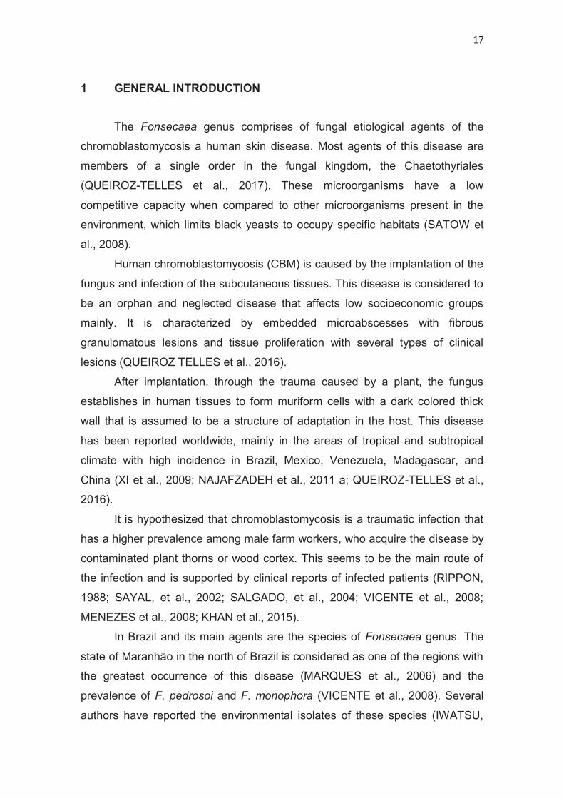
The Fonsecaea genus comprises of fungal etiological agents of the
chromoblastomycosis a human skin disease. Most agents of this disease are
members of a single order in the fungal kingdom, the Chaetothyriales
(QUEIROZ-TELLES et al., 2017). These microorganisms have a low
competitive capacity when compared to other microorganisms present in the
environment, which limits black yeasts to occupy specific habitats (SATOW et
al., 2008).
Human chromoblastomycosis (CBM) is caused by the implantation of the
fungus and infection of the subcutaneous tissues. This disease is considered to
be an orphan and neglected disease that affects low socioeconomic groups
mainly. It is characterized by embedded microabscesses with fibrous
granulomatous lesions and tissue proliferation with several types of clinical
lesions (QUEIROZ TELLES et al., 2016).
After implantation, through the trauma caused by a plant, the fungus
establishes in human tissues to form muriform cells with a dark colored thick
wall that is assumed to be a structure of adaptation in the host. This disease
has been reported worldwide, mainly in the areas of tropical and subtropical
climate with high incidence in Brazil, Mexico, Venezuela, Madagascar, and
China (XI et al., 2009; NAJAFZADEH et al., 2011 a; QUEIROZ-TELLES et al.,
2016).
It is hypothesized that chromoblastomycosis is a traumatic infection that
has a higher prevalence among male farm workers, who acquire the disease by
contaminated plant thorns or wood cortex. This seems to be the main route of
the infection and is supported by clinical reports of infected patients (RIPPON,
1988; SAYAL, et al., 2002; SALGADO, et al., 2004; VICENTE et al., 2008;
MENEZES et al., 2008; KHAN et al., 2015).
In Brazil and its main agents are the species of Fonsecaea genus. The
state of Maranhão in the north of Brazil is considered as one of the regions with
the greatest occurrence of this disease (MARQUES et al., 2006) and the
prevalence of F. pedrosoi and F. monophora (VICENTE et al., 2008). Several
authors have reported the environmental isolates of these species (IWATSU,

18
1981; VICENTE et al., 1999, 2008, 2013). In the environmental study, Marques
et al., (2006) isolated F. pedrosoi that was assumed as the main agent of this
disease in the Amazonia areas and reported the source of infection as the
coconut shell of Babassu palm tree (Orbignya phalerata). According to Silva et
al., (1995) during the Babassu harvesting, the workers have the habit of sitting
on the fruits of the palm tree, justifying the incidence of lesions on their thighs
and regions of the lower limb. Further, Salgado et al. (2004), isolated F.
pedrosoi from the thorns of Mimosa pudica plant after it reportedly caused injury
and subsequent skin lesion in a patient. However, this isolate was never
identified by molecular methods. These methods are the main tools that are
used to clarify the identity and relatedness among Fonsecaea species, that are
even though very similar in morphology but are different according to molecular
studies and hence different in terms of their virulence potentials (HOOG et al.,
2000, 2004; SUN et al., 2012; VICENTE et al., 2013; NAJAFZADEH et al., 2011
a; QUEIROZ-TELLES et al., 2016).
Additionally, according to literature, the fungus Cladophialophora carrionii
is considered the most frequent etiological agent in arid and semi-arid regions
of Central and South America (LAVELLE, 1980) and Australia (TREJOS, 1954).
According to the authors, the infection in these regions usually associates with
trauma by cactus spines (RUBIN et al., 1991; FERNÁNDEZ-ZEPPENFELDT et
al., 1994). However, Hoog et al., (2007) demonstrated by molecular studies that
the clinical isolates belonged to C. carrionii species, whereas the environmental
isolates obtained from plant spines cactus (Stenocereus griseus) belonged to a
new species described as Cladophialophora yegresii. Likewise, Vicente et al., in
2013, investigated the occurrence of black fungi in living area of patients with
chromoblastomycosis and described two novel species, F. erecta and F.
minima, which were isolated from living plants and were related to the clinical
ones. Therefore, the question that remains to be elucidated is whether the
environmental and clinical strains associated with chromoblastomycosis
represent exactly the same species and genotypes.
Since the discovery of this disease, several attempts have been made in
order to develop a model of study in animal and vegetable hosts, aimed at
understanding the infection route and clinical evolution of chromoblastomycosis.

19
Although some models have been established in animal hosts (MARTINEZ et
al., 2005; XIE et al., 2010), the chronic infection of human mycosis has not been
successful (MARTINEZ et al., 2005). Similar lesions have been found in rats
and mice that were infected by the peritoneum and subcutaneous pathway.
However, such experimental models tend to cure spontaneously which leads to
unreliability (BORELLII, 1972). Thereby, the objective of this work was to
understand the environmental origin and the route of infection of the agents
causing chromoblastomycosis disease in humans, by evaluating the pathogenic
potential of environmental and clinical strains of Fonsecaea with the help of
virulence tests, using different animal and plant hosts.
This study was, therefore, divided into three chapters. Chapter I
consisted of a review addressing the themes black yeasts, taxonomy, and
biodiversity, with emphasis on virulence factors and experimental models for
reproduction of pathogenesis. In Chapter II, infection models were tested in
plants and animals for evaluation of closely related Fonsecaea species
associated with chromoblastomycosis disease. For the virulence tests the
clinical strains of F. pedrosoi (CBS 271.37), F. monophora (CBS 102248) and
Fonsecaea erecta (CBS 125763), isolated from a living plant, were used.
Chapter III was developed based on the study model proposed in Chapter II,
where the Tenebrio molitor larvae were evaluated as an alternative source for
environmental isolation of Fonsecaea species.
20
2 OBJECTIVES 2.1 GENERAL
The main objective of this study was to establish protocols for isolation
and virulence evaluation of the animal and plant associated Fonsecaea species
related to chromoblastomycosis.
2.2 SPECIFIC
To establish stablish Mimosa pudica plants in vitro culture as a model of
study;
Evaluate the development of Fonsecaea species inoculated in Mimosa
pudica and Bactris gasipaes (palm tree) plants;
Analyze the virulence potential of Fonsecaea sibling species in animal
hosts through inoculation in Tenebrio molitor and mice Balb/c.

21
3 LITERATURE REVIEW
3.1 BLACK YEASTS, TAXONOMY AND BIODIVERSITY
Black yeasts belong to the orders Dothideales and Chaetothyriales
(HOOG and MCGINNIS, 1987) and have the characteristic presence of a dark
pigment called melanin, in their cell wall. They are also known as dematiaceous
fungi, as they are capable of producing melanized yeast cells during a part of
their life cycle (HOOG et al., 2000).
The order Dothideales presents adaptive ability in a hostile environment,
has rapid growth rate, contains large and multinucleated hyphae and is
melanized depending on the stage of development and environmental
conditions (VICENTE, 2000). Most species of this family grow in a low
concentration of nutrients and are halotolerant or halophilic (HOOG et al.,
2006). Some groups such as Alternaria, Stemphylium, Epicoccum, and
Ulocladium have multicellular conidia and transverse and longitudinal septate,
whereas groups like Curvularia and Drechslera have transverse multicellular
conidia. Other groups like Cladosporium, Nigrospora, Aureobasidium, and
Hortaea belonging to the saprobe genus present only unicellular conidia
(HOOG et al., 2000; STERFLINGER et al., 2001).
The members of the order Chaetothyriales cause diseases, such as
chromoblastomycosis, mycetoma, and phaeohyphomycosis (MCGINNIS, 1992),
in animals. Within this order, the Herpotrichiellaceae family includes fungal
species that have highly diversified life style and cause recurrent infection in a
variety of vertebrate (HOOG et al., 2014) as well invertebrate hosts with intact
immunity, that are commonly affected by these agents, e.g., cold blooded
animals (SEYEDMOUSAVI et al., 2014). These agents are slow growing with
yeast like appearance and with time they become velvety colonies with
mononuclear hyphae showing uniform pigmentation (DIXON, POLAK-WISS,
1991). Some of the main species belonging to this group are from the genus
Exophiala, Cladophialophora, Capronia, Fonsecaea, Phialophora,
Rhinocladiella, Veronae, and Cyphellophora (HOOG et al., 2000).

22
The genus Cladophialophora has colonies that are pulverulent, wooly
green to grayish-green, are conidiophores or imperceptible and depigmented
conidial scars (HOOG, GENE, and FIGUERAS, 2000; HOOG et al., 2004).
They are frequently isolated from decomposing organic matter and may be
associated with several infections in human and animal hosts (BADALI et al.,
2008; VICENTE et al., 2012).
The Fonsecaea species, associated with human diseases, are also
described in animals, e.g., Fonsecaea multimorphosa causes disseminated
infection in cats (NAJAFZADEH et al., 2011 a) and Fonsecaea brasiliensis
causes disseminated infection in crabs (VICENTE et al., 2012). Some species
such as Fonsecaea erecta and Fonsecaea minima have been isolated from the
environment (VICENTE et al., 2013). The main micromorphological
characteristic of this group is the presence of branched conidiophores, having
primary and secondary conidia with predominant variation in morphology
(HOOG et al., 2000; VICENTE, 2000). Macromorphologically they present flat
or raised velvety colonies of slow growth, with coloration ranging from black,
olive to dark gray reverse (AL-DOORY, 1983; DE HOOG, et al., 1998,
VICENTE, 2000).
The genus Exophiala exhibits typical black-olivaceous colonies, which
are reduced, have a viscous center, with yeast growth, and smooth margins.
With the passage of time, the colonies become velvety or wooly. They have
intercalated cell structures, having a cylindrical shape with relative narrowing;
the conidium is subhyaline with a smooth wall. Some chains with spherical cells
are formed by the profusion of shoots (HOOG et al., 2000; HOOG; GENE;
FIGUERAS, 2004). The members of this family that were isolated from aquatic
environments, presenting no thermotolerance, caused diseases in cold-blooded
animals such as fishes, amphibians and some invertebrates (HOOG et al.,
2011), while other species such as E. jeanselmei, E. spinifera, and E.
dermatitidis were found to infect humans (SEYEDMOUSAVI et al. 2013).
The habitats of black yeast are diverse. They are commonly found in
tropical and subtropical regions (FIGURE 1), as well as glacial regions
(GUNDE- CIMERMAN et al., 2003; STERFLINGER, 2006; BRIDGE;
NEWSHAM, 2009), in plant substrates, soil, wood and decomposing organic

23
matter (MCGINNIS et al., 1999; VICENTE, 2000). In addition, these agents, due
to their extremophile nature, are also present in special conditions such as,
during the low availability of nutrients, at high temperatures, UV radiation and in
the presence of aromatic compounds, osmotic stress or combinations of these
factors (STERFLINGER, 2006).
Figure 1. Global distribution of Black yeast based on the report of clinical cases and environmental isolation. The prevalent species ecologies are shown by the balls in the world map. Black: rock-inhabiting; Blue: waterborne; Green: plant-associated; Red: invasive in warm-blooded animals; Brown: soil; Pink: food; White: ice.
The black yeasts and their relatives harbor numerous agents of human
infection. Virulence factors involved are not known yet, but given the highly
polyphyletic pathogenic potential of the concerned ascomycete order, the
Chaetothyriales present a significant predisposition toward growth in human
tissue; a capacity that must have arisen during the evolution of this organism
(FIGURE 2). The CBM is caused by the genera Fonsecaea and
Cladophialophora, to some extent by species of Phialophora and Exophiala and
less frequently by Rhinocladiella. Like other Endemic mycoses, CBM is not a
notifiable disease, thus making it difficult to accurately assess its prevalence.
Based on the number of cases, it can be noted that the incidence of CBM varies
from 1: 6,800 in Madagascar to 1: 8,625,000 in the United States. Most cases
are reported in tropical and subtropical regions, mainly between latitudes of
30°N and 30°S (AL-DOORY, 1983; QUEIROZ-TELLES et al., 2011) with more

24
than 500 cases in China, Brazil, Mexico and Madagascar (QUEIROZ-TELLES
et al., 2016) (FIGURE 1).
Figure 2. Phylogeny of Chaetothyriales and related Ascomycetes. Tree based on LSU sequences constructed with Maximum likelihood implemented in Mega7 with Kimura’s two-parameter model. The bootstrap support was calculated using 100 replicates and values ≥ 80%

25
are shown in the branches. The main agents and host associated are represented in each family. The symbols before the species represent the animal affected.
According to Hoog et al. (1998), black yeasts cannot be identified by
observing the macro and micro morphological structures due to their
morphological plasticity. Furthermore, the little difference in the conidial
structures, the size variation, and the shape makes it difficult to define the
species.
Molecular tools have helped in identifying the species and consequently
in clarifying an infection route which helps to build the epidemiological data
(HOOG et al., 2006). Such concise results allow the construction of
phylogenetic trees that are essential for elucidating the evolution and ecology of
these microorganisms (VICENTE, 2000; STERFLINGER, 2006).
Taxonomic studies and the identification of molecular markers, in the
species of black yeasts of clinical relevance, have been performed by
sequencing the SSU, LSU, ITS, 1-alpha elongation factor (EF1α), β-tubulin and
actin (ZENG et al., 2007; BADALI et al., 2008) and CDC42 (SUN et al., 2012).
The partial ribosomal DNA (rDNA) large-subunit (LSU) sequences are
conserved to show relationships at the level of order or family (VICENTE et
al.2013, AZEVEDO et al. 2015). Within Chaetothyriales, the pathogenic species
are polyphyletic with dispersion all over the tree (FIGURE 2), wherein the main
species involved in CBM are in three clusters. In the Fonsecaea cluster, that is
included in the “bantiana clade", the prevalent agents of CBM namely F.
pedrosoi, F. nubica and F. monophora (HOOG et al., 2000; SUN, et al., 2012;
NAJAFZADEH, et al., 2010) are grouped. It also includes the recently described
F. pugnacious, which is also related to this clade (DE AZEVEDO et al., 2015).
Additionally, it was observed that the bantiana clade contained species of
Cladophialophora and some unknown saprobes species involved in human
disease along with the agents of primary brain infection and disseminated
infections (SURASH, et al., 2005).
The agents of chromoblastomycosis are also grouped in a separate
clade named “carrionii clade” which includes Cladophialophora carrionii
(HOOG, et al., 2004; BADALI, et al., 2008), the main agent of CBM in semiarid
areas, C. samoensis, the causal agent of CBM in Samoa (located south of the

26
equator, between Hawaii and New Zealand in the Polynesian region of the
Pacific Ocean) (HOOG, et al., 2007) and Phialophora verrucosa, isolated from
CBM lesions of chromomycosis reported in northern Africa.(GUGNANI, et al.,
1978; SURASH, et al., 2005; HOFMANN, et al., 2005).
Furthermore, the species Rhinocladiella aquaspersa, one of the CBM
agents (BORELLI, et al., 1972; GONZÁLES, et al., 2013), is located in a
separate clade from the ones described above. This separate clade includes
the species of Exophiala that cause human infection, such as Exophiala
jeanselmei, E. dermatitidis and E. spinifera (NAKA, et al., 1986; QUEIROZ-
TELLES, et al., 2003; TOMSOM, et al., 2006). The groups of other species that
are associated with the environmental occurrence and animal disease (ZENG,
et al., 2007; VICENTE, et al., 2012) belong to this clade.
3.2 VIRULENCE FACTORS
Black yeasts can adapt to various environmental conditions; the
presence of melanin in the cell wall and the ability to alternate between yeast
and filamentous forms (pleomorphism), the production of biofilms and
extracellular polysaccharides, allow these yeasts to tolerate conditions like
variations in pH and salt concentration, UV radiation and high temperatures
(HOOG, 1993; GOSTINCAR, 2011).
The factors that are probably of importance for the pathogenicity of black
yeasts include, the presence of melanin and carotene, the formation of thick cell
walls and the growth of meristem, the presence of yeast-like, thermotolerant
and osmotolerant phases, adhesion, hydrophobicity, assimilation of aromatic
hydrocarbons and the production of siderophores (SEYEDMOUSAVI et al.,
2014).
The adaptation of these fungi to human hosts, also established as a
hostile habitat, is favored by the presence of melanin in their cell wall and their
thermotolerance (RIBEIRO et al., 2006). Melanin is a complex polymer, which is
widely distributed in nature and is known to be an important virulence factor in
the opportunistic and pathogenic dematiaceous fungi (SUN et al., 2012).

27
The melanin that is synthesized by the DHN and DOPA pathways is
known as the eumelanin (LANGFELDER et al., 2003). According to Teixeira et
al., (2017), the members of Herpotrichiellaceae possess several melanin-
associated genes, suggesting that melanin production can use several
pathways (LI et al., 2016). The genomic studies of Herpotrichiellaceae agents
have confirmed the presence of multiple laccases, which have several
functions. According to these studies, it is possible that the diversification of this
enzyme occurred late during the evolution of black yeasts (TEIXEIRA et al.,
2017; MORENO, et al., 2017). Apart from the pigmentation function, melanin
also plays a role in the degradation of organic pollutants such as lignin and in
stress tolerance. The various functions of this enzyme may justify the presence
of multiple laccase genes in the genome (BALDRIAN 2006, RODRIGUEZ
COUTO & TOCA HERRERA 2006).
Fungal species that are not thermotolerant tend to be saprobic and such
species are rarely reported as a cause of infection in humans. Therefore it can
be said that the thermotolerance influences the choice of host. During the last
decade, some species of Exophiala that were isolated from environments
without thermotolerance were also seen to be involved in animal diseases
(HOOG et al., 2011). A generalized suite of adaptations to extreme
environments, including melanin production and meristematic growth, as well as
A generalized suite of adaptations to extreme environments, including melanin
production and meristematic growth, as well as thermotolerance, is suggested
to contribute to pathogenic potential (BADALI et al., 2008).
The ability to grow at 37 °C gives black yeasts the potential to cause
diseases. Some clades that are composed of yeasts and grow above 37 °C, like
bantiana, dermatitidis, and jeanselmei, can cause systemic infections in
humans. Whereas other species that can grow at a maximum temperature of 36
°C, like the carrionii and the europaea can cause only subcutaneous and
surface skin infections (HOOG, et al., 2011).
The salmonis clade group members originate in water and are also
known as “water-borne” species. It was observed that the maximum
temperature for their growth depended on the temperature of the host habitat,
for example, the agents from this clade cause diseases in amphibians,
28
suggesting that the thermotolerance determines the choice of the host. This
indicates that fungi can cause disease in cold-blooded animals if they grow at
27 ºC and infections in mammals if they grow above 37 ºC (HOOG et al., 2011;
VICENTE et al., 2012; SEYEDMOUSAVI; GUILLOT; HOOG, 2013; VICENTE,
et al., 2014).
Fonsecaea brasiliensis is considered as a secondary agent of lethargic
crab disease. The etiological agent of this disease is Exophiala cancerae, which
has been isolated from the sick crabs but never from the mangrove environment
(VINCENT et al., 2012). In a study conducted by Hoog et al., (2011), it was
verified that the optimal growth temperature for E. cancerae is between 24–27
ºC, whereas for F. brasiliensis it is 37 ºC (VICENTE et al., 2012). This factor
suggests that thermotolerance can determine the host predilection. According to
Gostincar et al., (2011) the term poly-extremotolerant could be adopted for
these yeasts in order to describe their high ability to colonize a wide variety of
environments and to withstand different ecological conditions.
Among the factors for pathogenicity of CBM, muriform cells are the most
important. These cells are present either in single or clustered form, consisting
of darkly pigmented, thick, transverse, and longitudinal walls. These cells are
present only in the order Chaetothyriales and are considered to be invasive with
respect to the host tissue (ESTERRE and QUEIROZ-TELLES, 2006), as they
are highly resistant to immune system. However, the knowledge regarding how
the differentiation occurs in cells is fundamental for the investigation of new and
more efficient therapeutic approaches in the treatment of CBM (HAMZA et al.,
2003).
Proteolytic enzymes play an important role in the pathogen-host
interaction, leading to immunological escape or antimicrobial resistance
(KNEIPP et al., 2004; PALMEIRA et al., 2006). According to Kneipp et al.,
(2004) muriform cells display increased activity of phosphatase enzyme during
pathogenicity (KNEIPP et al., 2004; KNEIPP et al., 2008). The CBM agents
secrete several hydrolytic enzymes; the muriform cells of F. pedrosoi and R.
aquaspersa show higher phosphatase activity during CBM and thus cause an
increase in the cell adhesion (KNEIPP et al., 2004; KNEIPP et al., 2008;
KNEIPP et al., 2012). The earlier studies suggest that Ecto-ATPases may favor

29
the survival of the fungus in environments such as the human body (FILIPPINI
et al., 1990; COLLOPY et al., 2006).
In relation to innate immunity, macrophages carry out an important
function in the regulation of fungal growth. CBM fungal cells can be detected in
intra-cytoplasmic vacuoles of skin macrophages (WALTER et al., 1982;
QUEIROZ TELLES et al., 2017). The chronic and granulomatous inflammation
is generally characterized by the dominating presence of macrophages in the
injured tissue (SEYEDMOUSAVI et al., 2014). Sotto et al., (2004) investigated
the cellular immune response in lesional biopsy specimens of patients with
CBM and reported that phagocytes are involved in innate immunity against the
fungal agents.
Bocca et al., (2006) showed that during infection with F. pedrosoi, the
macrophages were unable to produce nitric oxide even after stimulation with
lipopolysaccharide (LPS) and gamma interferon (IFN-). This was attributed to
the host’s inability to destroy F. pedrosoi, leading to a chronic disease.
According to SILVA et al. (2007), phagocytosis of fungal cells by Langerhans
cells inhibited the expression of CD40 and CD86. Gimenes et al., (2005),
analyzed the production of proinflammatory cytokines by macrophages and
found that the peripheral blood mononuclear cells (PBMC) from patients with
chromoblastomycosis secreted high levels of tumor necrosis factor alpha (TNF-
y), which was not dependent on the clinical form of the disease. Souza et al.,
(2011) showed that the cytokine response to F. pedrosoi was defective due to
the lack of fungus recognition by Toll like receptors (TLRs).
Polymorphonuclear neutrophils and dendritic cells are important
components in the first line of defense during an infection that arises due to
cutaneous or subcutaneous inoculation in tissues. The dendritic cells play an
important role in phagocytosis, by serving as major antigen-presenting cells and
inducers of adaptive T-cell responses (SOUZA et al., 2009). Mature dendritic
cells prime the naive lymphocytes and polarize them toward the T-helper type 1
(Th1) response, whereas immature dendritic cells have been shown to induce
tolerance. The severity of diseases caused by chaetothyrialean black yeasts is
dependent on Th1/Th2 activation. Patients with severe clinical forms produce
interleukin-10 (IL10) with inhibition of IFN-γ and immune response between Th1

30
and Th2 activation, whereas in mild cases IFN-γ is present along with
decreased levels of interleukin-10 (IL10) and increased number of T cells
favoring a Th1 profile (MAZO FÁVERO GIMENES et al., 2005; BOCCA et al.,
2006).
The immunity in patients with Chromoblastomycosis is impaired, because
of which these patients are unable to develop an effective immune reaction to
fungal antigens, thereby resulting in disease chronicity (FUCHS and PECHER,
1992). Experimental infection models using athymic mice showed that during
the course of infection, granulomas become diffuse and confluent accompanied
by a random fungal distribution, suggesting a role for a T-cell-mediated immune
response (AHRNES et al., 1989).
In addition to melanin, black yeasts are also known to contain mannose-
bearing structures as surface components that activate mannose receptors and
help the host to control infections (ALVIANO et al., 2003). Melanin, an
immunologically active fungal molecule, activates both humoral and cellular
responses that might help to control the infection (ALVIANO et al., 2004).
Melanin is potentially involved in the activation of TLR4, with consequent
production of the proinflammatory chemokine IL–8 (EL-OBEID et al., 2006).
According to Alviano et al.,(2003), the interaction of Fonsecaea pedrosoi with
phagocytes in the presence of melanin resulted in higher levels of fungal
internalization and destruction by host cells, accompanied by greater degrees of
oxidative burst (ALVIANO et al., 2004).
The presence of immunoglobulin M (IgM), IgA, and IgG antibodies has
been identified in some studies (VIDAL et al., 2004; ESTERRE et al., 2000), but
the humoral immune response does not seem to protect from infections caused
by black yeasts and their filamentous relatives. Moreover, in vitro studies
indicate that the interactions between fungi and anti-melanin antibodies inhibit
fungal growth (VIDAL et al., 2004).
Further studies are warranted to evaluate the roles of host genetic
susceptibility and immunologic defects in the pathophysiology of black yeast
infections. A preliminary study showed that CARD9 mutation was linked to
subcutaneous phaeohyphomycosis and Th17-cell deficiencies (WHEELER, et
al. 2008). In all patients, the CARD9 mutation was linked to decreased

31
proportions of cytokines in Th17 with consequently cells impaired immune
responses against the infection. Additionally, one more aspect that should be
considered in the patients with defects in humoral immunity, such as
agammaglobulinemia or hypogammaglobulinemia, is that they might have
relatively normal resistance to black yeasts and their filamentous relatives.
Nevertheless, vaccination and induction of protective antibodies may provide
additional protection in specific groups of patients (NETEA, 2013).
3.3 CHROMOBLASTOMYCOSIS, DISEASE AND DIAGNOSIS
In the middle of the nineteenth century, black yeasts became known by
the scientific society as a pathogen of the disease, which was later termed as
chromoblastomycosis (CBM). Although the disease was first observed in 1911
by Alexandrino Pedroso, a Brazilian, it was described only three years later by
Max Rudolph. Since then, several researchers have dedicated themselves to
conduct research on the disease; however, the restricted understanding and
knowledge of it has made its identification difficult and complex (HOOG, et al.,
2000; MARTINEZ and TOVAR, 2007). Over the years, several terms have been
used by the scientific community to denote the disease, but the term
chromoblastomycosis, initially used by Terra et al. (1922), has turned out be the
most acceptable and is still valid.
The island of Madagascar represents an area endemic to CBM, with
1,323 cases reported between the years 1955 and 1995 (ESTERRE et al., 1996
a). With the presence of two distinct ecosystems in the island, the main agents
of CBM are Fonsecaea pedrosoi and Cladophialophora carrionii. Although CBM
is a disease primarily restricted to tropical and subtropical regions,
approximately 31 cases have been reported in Europe (PINDYCKA-
PIASZCZYNSKA et al., 2014).
In the Asian continent, Japan has the highest incidence of CBM (1 in
416,000 individuals). Since the first description of the disease in 1930,
approximately 700 cases have been reported (KANO, 1937; KONDO et al.,
2005). In China, research groups have reported the occurrence of several

32
hundred cases of CBM in 21 provinces (YEW, 1951; DAI et al., 1998). India has
long been known for autochthonous CBM infections, with more than 100 cases
reported (SHARMA et al., 1999; SAYAL et al., 2002). Within the region of
Oceania, approximately 200 cases have been reported in Australia, where the
main causative agent is C. carrionii due to the climatic conditions. Some cases
have also been reported in New Zealand and Solomon Islands (WOODGYER et
al., 1992; WEEDON et al., 2013).
The Caribbean region in the Central America has reported around 600
cases of implantation mycosis (TORRES-GUERRERO et al., 2012), where
CBM was found to be the main cause of the infection. Of these cases, 450
patients belonged to Dominican Republic (ISA-ISA, 2006). Approximately 100
cases were reported from Cuba (DÍAZ-ALMEIDA et al., 1978; BADALI et al.,
2013), and few cases have also been reported from Puerto Rico (CARRIÓN,
1975), Jamaica, and Haiti (BANSAL et al., 1989; ISA-ISA, 2006). The main
etiological agent in these countries is F. pedrosoi followed by F. monophora and
C. carrionii (CARRIÓN, 1975; BADALI et al., 2013).
Brazil has the highest number of cases in South America; the last decade
witnessed the reporting of 872 cases in the northern regions (MELLO et al.,
1992; SILVA et al., 1998; QUEIROZ-TELLES et al., 2011; PIRES et al., 2012).
However, 332 cases of CBM were reported from other regions of this country
(LONDERO et al., 1976; MINOTTO et al., 2001) as well. These included 71
cases in the state of Paraná (southern region) in 11 years; 325 cases in 55
years for Pará (northern region); 13 cases in 3 years for Maranhão
(northeastern region); and 73 cases in 28 years for Rio Grande do Sul
(southern region, MINOTTO et al., 2001; QUEIROZ-TELLES et al., 2003;
QUEIROZ-TELLES et al., 2009; QUEIROZ-TELLES et al., 2011). The principal
etiological agent of CBM in Brazil is F. pedrosoi (QUEIROZ-TELLES et al.,
2016).
Black yeasts serve as the potential causative agents of CBM, which is a
chronic skin disease infecting humans and commonly described as causing
polymorphic skin lesions. Although the disease majorly affects the cutaneous
and subcutaneous tissues, patients with this disease respond inefficiently to the
treatment approaches utilized (HOOG et al., 2000; MARTINEZ and TOLVAR,

33
2007). The diagnosis is based on fungal culture; CBM agents demonstrate an
initial slow growth, with deep green colonies that acquire a velvet dark aspect
with time (HOOG et al., 2004; QUEIROZ-TELLES, 2015).
Clinically, the CBM initially appears as an erythematous macular skin
lesion that progresses to a pink and smooth papular lesion (FIGURE 3). The
appearance of peripheral lesions is common, usually originating from
autoinoculation or disseminated through the lymphatic vessels (QUEIROZ-
TELLES et al., 2009; NAJAFZADEH et al., 2011 a). Over time, the lesion may
manifest itself as a papulosquamous lesion and starts evolving with polymorphic
aspects, which may be confused with several other infectious and noninfectious
diseases. The initial lesion may spread locally and produce satellite lesions
(MCGINNIS, 1992; QUEIROZ-TELLES and SANTOS, 2012; QUEIROZ-
TELLES, 2015). The CBM lesions are classified as nodular, tumoral
(cauliflower-like), verrucous, scarring, and plaque (CARRIÓN, 1950;
MCGINNIS, 1983; QUEIROZ-TELLES et al., 2011).
Microscopic analysis has revealed the presence of muriform cells in
verrucous lesions, which can often be macroscopically confused with neoplastic
formations. The microscopic diagnostic method is applicable within two months
of CMB infection. In the third month, the generalized granulomatous
inflammation can be observed; the microscope-based identification is not
possible at this stage. The differential diagnosis for CBM, which provides a
strong evidence for verrucous nodules, can be performed by biopsy. One of the
techniques used to collect material for analysis is the fine needle aspiration
(FNAB). The biopsy and FNAB may not be effective after the third month, as the
muriform cells can no longer be observed; in this case, the gold standard
technique is histopathology (XIE, et al., 2010).

34
Figure 3. Clinical types of Chromoblastomycosis lesions and the invasive form inside the tissues. (A) Initial lesion within three months after infection in a lower leg. (B) Confluent nodular lesions on a knee. (C) Tumoral (cauliflower-like) lesion on the posterior part of a foot. (D) Cicatricial lesion with verruca showing serpiginous and verrucous contours. (E) Hyperkeratotic verrucous lesion on the sole of a foot. (F) Soft violaceous plaque lesion in the root of a thigh. SOURCE: QUEIROZ-TELLES et al., (2016).
In the case of CBM, spontaneous healing is rare. The treatment
approach includes the use of drugs, surgery, and physical therapy; these
constitute the major allies in the treatment modality. An amputation is rarely
required in the case of CBM since the fungus does not directly reach the
muscles and bones and therefore, dissemination usually does not reach the
deeper tissues (MARTINEZ and TOVAR, 2007; QUEIROZ-TELLES et al.,
2009). In most clinical cases, the various modalities of physical methods
available are used as adjuvant therapy in combination with antifungal agents,
surgery, thermotherapy, laser therapy, and photodynamic therapy (PDT,
QUEIROZ-TELLES et al., 2017).
A study conducted by Bonifaz (2001) suggested certain forms of
treatments for CBM. Cryosurgery using liquid nitrogen has been demonstrated
to be effective for small lesions. However, contraindications exist for
cryosurgery; for example, in the case of larger lesions, treatment with
itraconazole or 5-FC is chosen until maximal reduction, upon reaching which,
termination of cryosurgery treatment is recommended to cure the previously

35
reduced lesions. Heat therapy in combination with other treatment modalities
has been applied in Japan achieving great results (TAGAMI et al., 1984;
TAGAMI et al., 1979). A one-month application of a disposable chemical pocket
warmer occluded with a bandage over the lesions 24 h per day resulted in an
improvement of lesions as evident by negative microscopic examination and
culture results (HIRUMA et al., 1993; YANASE and YAMADA, 1978). The use
of local heat therapy for 2 h per day combined with the administration of
posaconazole (PCZ) at 400 mg twice a day for an eight-month period led to a
significant reduction in lesions, while a combination of heat therapy for 12 h per
day and terbinafine (125 to 250 mg daily) led to negative direct examination and
culture results within two weeks (TANUMA et al., 2000; WU et al., 2010).
However, in some cases, additional surgical therapy was necessary.
Currently, laser therapy with carbon dioxide (CO2) has been used as
monotherapy; in combination with other treatment modalities, lasers promote
photocoagulation. Recently, another therapy that has been tried is the
photodynamic therapy (PDT) that exerts an effect on CBM similar to that on
actinic keratosis or other types of skin cancers. This technique combines visible
light photons of an appropriate wavelength to stimulate the intracellular
molecules of a photosensitizer, activation of which produces several reactive
molecules, including oxygen species, culminating in damage of target cells
(LYON et al., 2011, 2013). Having property of minimal invasiveness with few
side effects, PDT is considered a promising treatment option; also, it may
shorten the time of treatment. Moreover, the effectiveness of PDT against the
most common species causing CBM can be verified using an in vitro study
(LYON, et al., 2013).
3.4 EXPERIMENTAL MODELS
To develop an efficient and chronic animal model, several models have
been developed and worked on; however, chronicity is only manifested in
humans. The disease in its chronic form is characterized by the presence of
muriform cells (XIE, 2010).

36
Since the description of the disease in 1911, the researchers have been
trying to establish a reliable animal model of the disease. However, most
attempts were unsuccessful and could not achieve a human-like model. Medlar
in 1915 inoculated F. pedrosoi into guinea pigs, rats, and mice, thereby
producing a non-progressive granulomatous reaction. In 1945, Azulay
performed experiments in which F. pedrosoi was delivered via intratesticular
and intraperitoneal routes but could not produce granulomatous reaction
containing sclerotic cells.
In 1966, Simson described the first case of CBM in a horse;
subsequently, CBM was found in different animals ranging from frogs to dogs,
indicating that it could infect both humans and animals. Similar lesions were
detected in rats and mice infected subcutaneously and intraperitoneally,
respectively; however, such models developed the acute form of the disease,
without success in generation of a chronic model, with a tendency of
spontaneous cure, leading to unreliable models (BORELLII, 1972; POLAK,
1984; CARDONA-CASTRO et al., 1999; TEIXEIRA et al., 2006; MACHADO et
al., 2010).
A similar CBM infection can be observed in amphibians, such as the toad
and frog, in a systemic form (ANVER 1980; BUBE et al., 1992). According to
Cicmanec (1973) and Schmidt (1984), these animals appeared to be more
susceptible to the disease when they were debilitated or under stress.
Velasquez and Restrepo, in 1975, reported two toads (Bufo marinus) infected
by black yeast. These animals had granulomatous lesions containing brown
fungi identical to those found in human chromomycosis infection. After isolation,
the isolates shared common antigens with the recognized human pathogens, F.
pedrosoi, P. verrucosa, and C. carrioni. Bube et al. (1992) performed a
necropsy in the tissues of two marine toads, held in captivity; the animals
presented lesions similar to CBM. In the first case, granuloma with necrosis was
observed with multinucleated giant cells present in the meninges of the brain,
cervical, and thoracic spinal cord, resulting in the compression of spinal nerves
in skin, kidneys, and at a single focus in the liver. While in the second case,
ulcerating nodules were found in the skin, and the nodules were present in the
lungs with granulomas detected in the heart, spleen, bone marrow cavities of

37
the skull, and in close proximity to spinal nerves. Sclerotic bodies were present
in the nasal cavity and the skin. Black yeast was recovered in small numbers
from the nodular lesions. Hosoya et al. (2015) reported, in an anuran breeder’s
water tank in Tokyo, Japan that 10 frogs of at least three species had died in
the same facility in a limited period. The animals presented polypoid lesions
with ulceration found on the body surfaces. The symptoms did not improve with
oral treatment of antifungal agents. Histopathological analysis revealed
granulomatous lesions, containing brown hyphae. The result of phylogenetic
analysis demonstrated that the query sequence was situated among Veronaea
botryosa.
However, CBM is assumed to be a human disease due to the scarce
reports of this infection in other animals. The most clinical cases of this skin
lesion reported in animals have been considered as pheohyphomycosis (PHM),
because typical muriform cells were lacking (QUEIROZ–TELLES, 2017).
Regarding the animal model for disease reproduction, Juopperi et al.
(2002) reported, in the North Carolina State University (NCSU), a spadefoot
toad (Scaphiopus holbrookii) for the evaluation of proliferative to ulcerative
dermatitis. Histological analyses displayed sclerotic bodies in small clusters and
occasionally in the multinucleated giant cells, some brown-pigmented septate
hyphae have also been observed. However, isolation of the fungus was not
possible because of bacterial overgrowth. The diagnosis of chromomycosis was
based on cytologic and histologic findings.
The advances in molecular biology techniques and new immunology
tools in the past two decades have expanded the available data regarding
interactions of CBM fungus with immune cells. Mazo et al. (2005) demonstrated
that the absence of CD4+ T cells induced a more severe form of the disease
during experimental infection in mice (TEIXEIRA et al., 2006). According to a
study conducted by Siqueira et al. (2017), muriform cells were demonstrated to
induce inflammatory response and chronicity during the course of murine CBM.
A study by Xie et al. (2010) tried to induce chronic lesions for more than
three months in Wistar rats by F. monophora, by infecting the animals
intradermally in the ventral region. In another study by Machado et al. (2011), it
was possible to observe that mice, infected through plantar cushion region with

38
conidiogenic cells and conidia, developed lesions for a longer period than when
using hyphae and conidia.
Non-mammalian models have been used to investigate fungal virulence,
such as Cryptococcus neoformans (MYLONAKIS, et al., 2005; SOUZA et al.,
2015), Candida albicans (FUCHS, et al., 2010; MOWLDS, et al., 2010; SOUZA
et al., 2015), Microsporum gypseum, Trichophyton rubrum (ACHTERMAN, et
al., 2011), Aspergillus flavus (ST LEGER, et al., 2000), A. fumigatus (REEVES,
et al., 2004), and Madurella mycetomatis (KLOEZEN et al., 2015). The larvae of
Galleria mellonella and Tenebrio molitor are inexpensive to keep, easy to
manipulate, and their use may reduce the need for pathogenicity testing in
mammals, with a concomitant reduction in potential mammalian suffering
(FUCHS, et al., 2010; KLOEZEN et al., 2015). These models could be used to
analyze the fungal virulence and pathogenicity of etiologic agents of CBM.
Artificial inoculation in plants was performed in a study by Hoog et al.
(2007). The cactus (Stenocereus griseus) plants were cultivated from seeds in
the greenhouse, followed by inoculation of two species: environmental C.
yegresii and clinical C. carrionii. Both the species persisted in the cactus tissues
and upon reaching the spines, produced muriform cells similar to the one
described as the invasive form of CBM. However, C. carrionii isolate was more
virulent than the environmental C. yegresii, which was described from the
isolation performed in the same species of cactus in the vicinity of a CBM
patient’s residence.
In summary, these data indicate that studies to clarify CBM
immunopathology, understand the evolutionary cycle of these agents, their
environmental origin and infection route are warranted to improve the patients'
quality of life and to cure them.

39
4 REFERENCES
ACHTERMAN, R. R.; SMITH, A. R.; OLIVER, B. G.; WHITE, T. C. Sequenced dermatophyte strains: growth rate, conidi- ation, drug susceptibilities, and virulence in an inver- tebrate model. Fungal Genet Biol., v. 48, p. 335- 41, 2011. AHRENS, J.; GRAYBILL, J. R.; ABISHAWL, A.; TIO, F. O.; RINALDI, M. G. Experimental murine chromomycosis mimicking chronic progressive human disease. Am. J. Trop. Med. Hyg. v.40, p. 651–658, 1989. AL DOORY, Y. Chromomycosis. In: DI SALVO, A.F. Occupational mycoses. Philadelphia: Lea & Febiger, p.95-121, 1983. ALVIANO, D. S.; KNEIPP, L. F.; LOPES, A. H.; TRAVASSOS, L. R.; MEYER-FERNANDES, J. R.; RODRIGUES, M. L.; ALVIANO, C. S. Differentiation of Fonsecaea pedrosoi mycelial forms into sclerotic cells is induced by plateletactivating factor. Res. Microbiol. v. 154, p. 689–695, 2003. ALVIANO, D. S.; FRANZEN, A. J.; TRAVASSOS, L. R.; HOLANDINO, C.; ROZENTAL, S.; EJZEMBERG, R.; ALVIANO, C. S.; RODRIGUES, M. L. Melanin from Fonsecaea pedrosoi induces production of human antifungal antibodies and enhances the antimicrobial efficacy of phagocytes. Infect. Immun. v.72, p. 229–237, 2004. ANVER, M. R. Diagnostic exercise: systemic chromomycosis in frogs. Lab Anim Sci., v. 30, p. 165-166, 1980. AZULAY, R. D. Experimental studyes on chromoblastomycosis. J Invest Dermatol, v.6, p. 281-92, 1945. BADALI, H.; GUEIDAN, C.; NAJAFZADEH, M.J.; BONIFAZ, A.; GERRITS VAN DEN ENDE, A. H. G.; HOOG, G. S. de. Biodiversity of the genus Cladophialophora. Studies in Mycology, v. 175–191, 2008. BADALI, H.; FERNANDEZ-GONZALES, M.; MOUSAVI, B.; ILLINAIT-ZARAGOZI, M. T.; GONZALES-RODRIGUEZ, J. C.; DE HOOG, G. S.; MEIS, J. F. Chromoblastomy- cosis due to Fonsecaea pedrosoi and F. monophora in Cuba. Mycopathologia, v.175, p. 439–444, 2013. BALDRIAN, P. Fungal laccases ‒ occurrence and properties. FEMS Microbiology Reviews, v. 30, p. 215–242, 2006. BANSAL, A.S. & PRABHAKAR, P. Tropical Geographi Medicine v.41, p.222- 226, 1989.

40
BOCCA, A. L.; BRITO, P. P.; FIGUEIREDO, F.; TOSTA, C. E. Inhibition of nitric oxide production by macrophages in chromoblastomycosis: a role for Fonsecaea pedrosoi melanin. Mycopathologia, v. 161, p. 195–203, 2006. BONIFAZ A. Chromoblastomycosis. In: Arenas R, Estrada R, editors. Tropical dermatology, n. 7, p. 1-7, 2001. BORELLI, D. Acrotecha aquaspersa nova new species agents of chromomycosis. Acta Cient Venez, n. 23, p. 193-196, 1972. BRIDGE, P. D.;NEWSHAM, K. K. Soil fungal community composition at Mars Oasis, a southern maritime Antarctic site, assessed by PCR amplification and cloning. Fungal Ecology, v. 2, p. 66-74, 2009. BUBE, A.; BURKHARDT, E.; WEISS, R. Spontaneous chromomycosis in the marine toad (Bufo marinus). J Comp Pathol., v. 106, p. 73-77, 1992. CALIGIORNE, R. B.; LICINIO, P.; DUPONT, J.; DE HOOG, G. S. Internal transcribed spacer rRNA gene-based phylogenetic reconstruction using algorithms with local and global sequence alignment for black yeasts and their relatives. J. Clin. Microbiol., v. 43, p. 2816–2823, 2005. CARDONA-CASTRO, N.; AGUDELO-FLOREZ, P. Development of a chronic chromoblastomycosis model in immunocom- petent mice. Med Mycol, v. 37, p. 81-3, 1999. CARRIÓN, A. L. Chromoblastomycosis and related infections: new concepts, differential diagnosis, and nomenclatorial implications. Int J Dermatol, v. 14, p. 27–32, 1975. CARRION, A. L. Chromoblastomycosis. Ann NY Acad Sci., v. 50, p.1255–1282, 1950. CICMANEC, J. L.; RINGLER, D. H.; BENEKE, E. S. Spontaneous occurrence and experimental transmission of the fungus Fonsecaea pedrosoi, in the marine toad, Bufo marinus. Lab Anim Sci., v. 23, p. 43-47, 1973. COLLOPY, I.; J. R.; KNEIPP, L. F.; DA SILVA, F. C.; RODRIGUES, M. L.; ALVIANO, C. S.; MEYER- FERNANDES, J. R. Characterization of an ecto-ATPase activity in Fonsecaea pedrosoi. Arch Microbiol, v. 185, p. 355–362, 2006.
DA SILVA, J. P.; DA SILVA, M. B.; SALGADO, U. I.; DINIZ, J. A.; ROZENTAL,
S.; SALGADO, C. G. Phagocytosis of Fonsecaea pedrosoi conidia, but not
sclerotic cells caused by Langerhans cells, inhibits CD40 and B7-2 expression.
FEMS Immunol. Med. Microbiol, v. 50, p. 104 –111, 2007.

41
DAI, W.; CHEN, R.; REN, Z. Laboratorial observation and analysis of 287 strains pathogenic Fonsecaea. Chin J Dermatol., v. 5, p. 8 –9, 1998. DE AZEVEDO, C. M. P. S.; GOMES, R. R.; VICENTE, V. A.; SANTOS, D. W. C. L.; MARQUES, S. G.; NASCIMENTO, M. M. F.; ANDRADE, C. E. W.; SILVA, R. R.; QUEIROZ-TELLES, F.; DE HOOG, G. S. Fonsecaea pugnacius, a novel agent of disseminated chromoblastomycosis. J Clin Microbiol, v. 53, p. 2674–2685, 2015. DÍAZ-ALMEIDA, J. G.; TABOAS-GONZÁLEZ, M.; DUBE-DUBE, A. A. Cromoblastomicosis en Cuba. Estudio retrospectivo clínico epidemiológico de 72 pacientes. Rev Cubana Med Trop., v. 30, p. 95–108, 1978. DIXON, D.M.; POLAK-WISS, A. The medically important dematiaceous fungi and their identification, Mycoses, v.34, p.1-18, 1991. EL-OBEID, A.; HASSIB, A.; PONTEN, F.; WESTERMARK, B. Effect of herbal melanin on IL-8: a possible role of Toll-like receptor 4 (TLR4). Biochem. Biophys. Res. Commun. v.344, p. 1200–1206, 2006. ESTERRE, P.; INZAN, C. K.; RAMARCEL, A.; ANDRIANTSIMAHAVANDY, M.; RATSIOHARANA, M.; PECARRERE, J. L.; ROIG, P. Treatment of chromomycosis with terbinafine: preliminary results of an open pilot study. Br J Dermatol., v. 134, p. S33–S36, 1996. ESTERRE, P.; QUEIROZ-TELLES, F. Management of chromoblastomycosis: novel perspectives. Curr Opin Infect Dis, v.19, p. 148–152, 2006. ESTERRE, P.; JAHEVITRA, M.; ANDRIANTSIMAHAVANDY, A. Humoral immune response in chromoblastomycosis during and after therapy. Clin. Diagn. Lab. Immunol. v. 7, p. 497–500, 2000. FERNÁNDEZ-ZEPPENFELDT G.; RICHARD-YEGRES, N.; YEGRES, F.; HERNÁNDEZ, R. Cladosporium carrionii: hongo dimórfico en cactáceas de la zona endêmica para la cromomicosis en Venezuela. Revista Iberoamericana de Micologia, v.11, p. 61–63, 1994. FILIPPINI, A.; TAFFS, R. E.; AGUI, T.; SITKOVSKY, M. V. Ecto-ATPase activity in cytolytic T-lymphocytes. Protection from the cytolytic effects of extra- cellular ATP. J Biol Chem, v. 265, p. 334–340, 1990. FUCHS, B. B.; MYLONAKIS, E. Using non-mammalian hosts to study fungal virulence and host defense. Curr Opin Microbiol, v. 9, p. 346-51, 2006.
FUCHS, J.; PECHER, S. Partial suppression of cell mediated immunity in chromoblastomycosis. Mycopathologia, v. 119, p. 73–76, 1992. GIMENES, V. M. F.; SOUSA, M. G.; FERREIRA, K. S.; MARQUES, S. G.; GONÇALVES, A. G.; SANTOS, D. V. C. L.; DE SILVA AZEVEDO, C. M. P.;

42
ALMEIDA, S. R. Cytokine and lymphocyte proliferation in patients with different clinical forms of chromoblastomycosis. Microbes Infect. v. 7, p. 708–713, 2005. GONZÁLEZ, G. M.; ROJAS, O. C.; GONZÁLEZ, J. G.; KANG, Y.; DE HOOG, G. S. Chromoblastomycosis caused by Rhinocladiella aquaspersa. Med Mycol Case Rep, v. 2, p.148–151, 2013. GOSTINČAR, C.; GRUBE, M.; GUNDE-CIMERMAN, N. Evolution of fungal pathogens in domestic environments? Fungal biology, v. 115, n. 10, p. 1008–18, 2011. GUGNANI, H. C.; EGERE, J. U.; SUSEELAN, A. V.; OKORO, A. N.; ONUIGBO, W. I. Chromomycosis caused by Philaphora pedrosoi in eastern Nigeria. J Trop Med Hyg, v. 81, p. 208–210, 1978. GUNDE-CIMERMAN, N.; SONJAK, S.; ZALAR, P.; FRISVAD, J.C.; DIDERICHSEN; B. AND PLEMENITAŠ, A. Extremophilic fungi in Arctic ice: a relationship between adaptation to low temperature and water activity. Physics and Chemistry of the Earth, v. 28, p. 1273-1278, 2003. HAMZA, S. H.; MERCADO, P. J.; SKELTON, H. G.; SMITH, K. J. An unusual dema- tiaceous fungal infection of the skin caused by Fonsecaea pedrosoi:a case report and review of the literature. J Cutan Pathol, v. 30, p. 340–343, 2003. HAYAKAWA, M.; GHOSN, E. E.; DA GLORIA TEIXERIA DE SOUSA, M.; FERREIRA, K. S.; ALMEIDA, S. R. Phagocytosis, production of nitric oxide and pro- inflammatory cytokines by macrophages in the presence of of dema- tiaceus [sic] fungi that cause chromoblastomycosis. Scand J Immunol, v. 64, p. 382–387, 2006. HIRUMA, M.; KAWADA, A.; YOSHIDA, M.; KOUYA, M. Hyperthermic treat- ment of chromomycosis with disposable chemical pocket warmers. Report of a successfully treated case with a review of the literature. Mycopathologia, v. 122, p. 107–114, 1993. HOFMANN, H.; CHOI, S. M.; WILSMANN-THEIS, D.; HORRE, R.; HOOG, G. S.; BIEBER, T. Invasive chromoblastomycosis and sinusitis due to Phialophora verrucosa in a child from northern Africa. Mycoses, v. 48, p. 456–461, 2005. HOOG, G. S.; MCGINNIS, M. R. Ascomycetous black yeasts, p. 187–199. In: The expanding realm of yeast-like fungi. Elsevier, Amsterdam, 1987. HOOG GS DE, NISHIKAKU AS, FERNÁNDEZ ZEPPENFELDT G, PADÍN-GONZÁLEZ C, BURGER E, BADALI H, GERRITS VAN DEN ENDE AHG. Molecular analysis and pathogenicity of the Cladophialophora carrionii complex, with the descriptionof a novel species. Studies in Mycology, v. 58, p. 219–234, 2007.

43
HOOG, G S; VICENTE, V. A; NAJAFZADEH, M. J. et al., Waterborne Exophiala species causing disease in cold-blooded animals. Persoonia, v. 27, p. 46-72, 2011. HOOG, G.; GUARRO, J.; GENÉ, J.; FIGUERAS, M. J. Atlas of Clinical Fungi, 3rd edn. (e-version). Centraalbureau voor Schimmelcultures, Utrecht, The Netherlands / Universitat Rovira i Virgili, Reus, 2013a. HOOG, G. S. de; BOWMAN, B.; GRASER, Y.; HAASE, G.; ELFARI, M.; GARRITS VAN DEN ENDE, A. H. G.; MELZER-KRICK, B.; UNTEREINER, W. A. Molecular phygeny and taxonomy of medically important fungi. Medical Mycology, v.36, p.52-56, 1998. Supplement 1. HOOG, G. S.; GUARRO, J.; GENÉ, J.; FIGUERAS, M. J. Atlas of clinical fungi, 2nd ed. Centraalbureau voor Schimmelcultures, Universitat Rovira i Virgilli, Utrecht, 2000. HOOG, G. S.; VICENTE, V. A.; CALIGIORNE, R.; KANTARCIOGLU, S.; TINTELNOT, K.; GERRITS, A. H. G. V. D. E.; HAASE, G. Species Diversity and Polymorphism in the Exophiala spinifera clade containing opportunistic black yeast-like fungi. Journal of Clinical Microbiology, v. 41, n. 10, p. 4767-4778, 2003. HOOG, G.S.; GUARRO, J. GENÉ, J.; FIGUERAS, M.J. Atlas of clinical fungi Realization Computer Science II University of Wurzburg, Germany Atlas version, 2004. HOOG, G. S.; ZENG, J. S.; HARRAK, M. J. Exophiala xenobiotica sp. nov., an opportunistic black yeast inhabiting environments rich in hydrocarbons. Antonie van Leeuwenhoek, v. 90, p. 257-268, 2006. HOOG GS DE, NISHIKAKU AS, FERNÁNDEZ ZEPPENFELDT G, PADÍN-GONZÁLEZ C, BURGER E, BADALI H, GERRITS VAN DEN ENDE AHG. Molecular analysis and pathogenicity of the Cladophialophora carrionii complex, with the descriptionof a novel species. Studies in Mycology, v. 58, p. 219–234, 2007. HOOG, G. S. de. Evolution of black yeast: possible adaptation to the human host. Antonie van Leeuwenhoek, v. 63, p. 105-109, 1993. HOOG, G.S.; GUARRO, J. GENÉ, J.; FIGUERAS, M.J. Atlas of clinical fungi Realization Computer Science II University of Wurzburg, Germany Atlas version, 2004. HOOG, G. S. Ecology and phylogeny of black yeast-like fungi: diversity in unexplored habitats. Fungal Diversity, v. 65, p. 1–2, 2014.

44
HOSOYA, T.; HANAFUSA, Y.; KUDO, T.; TAMUKAI, K.; UNE, Y. First report of Veronaea botryosa as a causal agent of chromomycosis in frogs. Medical Mycology. v. 53, p. 369–377, 2015. HUBE, B. Extracellular peptidases of human pathogenic fungi. Contrib Microbiol, v. 5, p.126–137, 2000. ISA-ISA, R. Cromoblastomicosis, p 167–169. In Mendez-Tovar LJ, López-Martínez R, Hernández-Hern´ andez F (ed), Actualidades en micologia médica. UNAM, Mexico City, Mexico, 2006. JUOPPERI, T.; DE VOE, K. K. R.; GRINDEM, C. B. Granulomatous Dermatitis in a Spadefoot Toad (Scaphiopus holbrooki). Veterinary Clinical Pathology, v. 31, p. 137-139, 2002. KANO, K. Uber die Chromoblastomykose durch einen noch nicht als pathogen beschriebenen Pilz Hormiscium dermatitidis n. sp. Arch Dermatol Syph., v. 176, v. 282–294, 1937. KHAN, S.; KUMAR, A.; VINOD, V.; PRABHAKAR, V.; EAPEN, M.; THOMAS, J.; DINESH, K.; KARIM, S. Chromoblastomycosis due to Fonsecaea pedrosoi: an old wine in a rare bottle. J Infect Dev Ctries, n. 9, p. 325-329, 2015. KONDO, M.; HIRUMA, M.; NISHIOKA, Y.; MAYUZUMI, N.; MOCHIDA, K.; IKEDA, S.; OGAWA, H. A case of chromomycosis caused by Fonsecaea pedrosoi and a review of reported cases of dematiaceous fungal infection in Japan. Mycoses, v. 48, p. 221–225, 2005. KLOEZEN, W.; POPPEL, M. V. H.V.; FAHAL, A. H.; SANDE, W. W. J. V. A Madurella mycetomatis Grain Model in Galleria mellonella Larvae. PLOS Neglected Tropical Diseases, v. 9, p. 1-14, 2015. KNEIPP, L. F.; MAGALHÃES, A. S.; ABI-CHACRA, E. A.; SOUZA, L. O.; ALVIANO, C. S.; SANTOS, A. L.; MEYER-FERNANDES, J. R. Surface phosphatase in Rhinocladiella aquaspersa: biochemical properties and its involvement with adhesion. Med Mycol, v. 50, p. 570 –578, 2012. KNEIPP, L. F.; PALMEIRA, V. F.; PINHEIRO, A. A.; ALVIANO, C. S.; ROZENTAL, S.; TRAVASSOS, L. R.; MEYER-FERNANDES, J. R. Phosphatase activity on the cell wall of Fonsecaea pedrosoi. Med Mycol, v. 41, p. 469–477, 2003. KNEIPP, L. F.; RODRIGUES, M. L.; HOLANDINO, C.; ESTEVES, F. F.; SOUTO-PADRON, T.; ALVIANO, C. S.; TRAVASSOS, L. R.; MEYER-FERNANDES, J. R. Ectophospha- tase activity in conidial forms of Fonsecaea pedrosoi is modulated by exogenous phosphate and influences fungal adhesion to mam- malian cells. Microbiology, v. 150, p. 3355–3362, 2004.

45
LANGFELDER, K.; STREIBEL, M.; JAHN, B. et al. Biosynthesis of fungal melanins and their importance for human pathogenic fungi. Fungal Genetics and Biology, v. 38, p.143–158, 2003. LAVELLE P. Chromoblastomycosis in Mexico. Pan American Health Organization Scientific Publications, n. 396, p. 235–247, 1980. LI, X. Q.; GUO, B. L.; CAI, W. Y, et al. The role of melanin pathways in extremotolerance and virulence of Fonsecaea revealed by de novo assembly transcriptomics using Illumina paired-end sequencing. Studies in Mycology, v. 83, p. 1–18, 2016. LONDERO, A. T.; RAMOS, C. D. Chromomycosis: a clinical and mycologic study of thirty-five cases observed in the hinterland of Rio Grande do Sul, Brazil. Am J Trop Med Hyg., v. 25, p. 132–135, 1976. LYON, J. P.; PEDROSO E SILVA AZEVEDO, C. D. M.; MOREIRA, L. M.; DE LIMA, C. J.; DE RESENDE, M. A. Photodynamic antifungal therapy against chromoblastomycosis. Mycopathologia, v. 172, p. 293–297, 2011. LYON, J. P.; MOREIRA, L. M.; DE CARVALHO, V. S.; DOS SANTOS, F. V.; DE LIMA, C. J.; DE RESENDE, M. A. In vitro photodynamic therapy against Fonsecaea pedrosoi and Cladophialophora carrionii. Mycoses, v. 56, p. 157–161, 2013. MACHADO, A. P.; SILVA, M. R. R.; FISCHMAN, O. Prolonged infection by Fonsecaea pedrosoi after antigenic co-stimulation at different sites in experimental murine chromoblastomycosis. Virulence. v. 1, p. 29-36, 2010. MACHADO, A. P.; SILVA, M. R.; FISCHMAN, O. Local phagocytic pesponses after murine infection whit different forms of F. pedrosoi and sclerotic bodies originating from on inoculum of conidiogenus cells. Mycoses, v. 54, p. 202-11, 2011. MARQUES, S. G.; PEDROSO SILVA, C. M.; SALDANHA, P. C., REZENDE, M. A.; VICENTE, V. A.; QUEIROS-TELLES, F, LOPES COSTA, J. M. Isolation of Fonsecaea pedrosoi from the shell of the babassu coconut (Orbignya phalerata Martius) in the Amazon region, Maranhão, Brazil. Japanese Journal of Medical Mycology, v. 47, p. 305–311, 2006. MARTINEZ, R. B.; TOVAR, L. J. M. Chromoblastomycosis. Clinics in Dermatology, n. 25, p.188–194, 2005. MAZO, F. G. V.; DA GLORIA, D. S.; FERREIRA, K. S.; MARQUES, S. G.; GONCALVES, A. G.; SANTOS, V. C. L. et al., Cytokines and lymphocyte proliferation in patients with different clinical forms of chromoblastomycosis. Microbes Infect, v.7, p. 708-13, 2005.

46
MCGINNIS, M. R.; LEMON, S. M.; WALKER, D. H.; DE HOOG, G. S.; HAASE, G. Fatal cerebritis caused by a new species of Cladophialophora. Stud. Mycol, n. 43, p.166–171. 1999. MCGINNIS, M.R. Black fungi. A model for understanding tropical mycosis. In: WALKER DH, ed. Global infectious diseases. New York: Springer Verlog, p.129-149, 1992. MCGINNIS M. R. Chromoblastomycosis and phaeohyphomycosis: new concepts, diagnosis, and mycology. J Am Acad Dermatol, v. 8, p. 1–16, 1983, MCGINNIS, M.R. Black fungi. A model for understanding tropical mycosis. In: WALKER DH, ed. Global infectious diseases. New York: Springer Verlog, p.129- 149, 1992. MEDLAR, E. M. A cutaneous infection caused by a new fungus, Phialophora verrucosa, with a study of the fungus. J Med Res, v. 32, p. 507-522, 1915. MELLO e SILVA, A. C. C.; SERRA NETO, A.; GALVÃO, C. E. S.; MARQUES, S. G.; SALDANHA, A. C. R.; PEDROSO e SILVA, C. D. M.; FISCHMAN, O.; DA SILVA, R. R.; COSTA, M. D. R. D. S. R.; COSTA, J. M. Fonsecaea pedrosoi-caused chromoblastomycosis in the state of Maranhão. The clinical, epidemiological and evolutionary aspects. Rev Soc Bras Med Trop., v. 25, p. 37– 44, 1992. MENEZES, N.; VARELA, P.; FURTADO, A.; COUCEIRO, A.; CALHEIROS, I.; ROSADO, L.; MOTA, G.; BAPTISTA, A. Chromoblastomycosis associated with Fonsecaea pedrosoi in a carpenter handling exotic woods. Dermatol Online J, v. 14, p. 1- 9, 2008. MINOTTO, R.; BERNARDI, C. D. V.; MALLMANN, L. F. et al., Chromoblastomycosis: a review of 100 cases in the state of Rio Grande do Sul, Brazil. J Am Acad Dermatol. v. 44, p. 585-92, 2001.
MORENO, L., F.; FENG, P.; WEISS, V. A.; VICENTE, V. A.; STIELOW, B.; DE HOOG, S. Phylogenomic analyses reveal the diversity of laccase-coding genes in Fonsecaea genomes. PLoS ONE, v. 12, e0171291, 2017. MOWLDS, P.; COATES, C.; RENWICK, J.; KAVANAGH, K. Dose- dependent cellular and humoral responses in Galleria mellonella larvae following beta-glucan inoculation. Microbes Infect., v. 12, p. 146-53, 2010. MYLONAKIS, E.; MORENO, R.; EL KHOURY, J. B.; IDNURM, A.; HEITMAN, J.; CALDERWOOD, S. B.; AUSUBEL, F. M.; DIENER, A. Galleria mellonella as a model system to study Cryptococcus neoformans pathogenesis. Infect Immun., v 73, p. 3842–50, 2005. NAJAFZADEH, M. J, J. SUN, V. A. VICENTE, CORNE H.W. KLAASSEN, ALEXANDRO BONIFAZ, A.H.G. GERRITS VAN, DEN ENDE, STEPH B.J.

47
MENKEN, AND G. SYBREN DE HOOG. Molecular Epidemiology of Fonsecaea species. EID Journal, v.17, p. 464-9, 2011a. NAJAFZADEH, M. J, V. A. VICENTE , J. SUN, SYBREN DE HOOG. Fonsecaea multimorphosa sp. nov, a new species of Chaetothyriales isolated from feline cerebral abscess. Fungal Biology, v. 115, p. 1066-76, 2011b. NAJAFZADEH, M. J.; J. SUN4, V. VICENTE, L. XI, A. H. G. GERRITS VAN DEN ENDE & G. S. HOOG Fonsecaea nubica, a new species of agent of human chromoblastomycosis revealed using molecular data. Medical Mycology, v. 48, p. 800-6, 2010a. NAJAFZADEH, M. J.; REZUSTA, A.; D’CAMEO, M. I.; ZUBIRI, M. L.; YUS, M. C.; BADALI, H.; REVILLO, M. J.; DE HOOG, G. S. Successful treatment of chromoblasto- mycosis of 36 years duration caused by Fonsecaea monophora. Med Mycol, v. 48, p. 390–393, 2010. NETEA, M. G. Training innate immunity: the changing concept of immunological memory in innate host defence. Eur. J. Clin. Invest. v. 43, p. 881–884, 2013. PALMEIRA, V. F.; KNEIPP, L. F.; ALVIANO, C. S.; DOS SANTOS, A. L. Secretory aspartyl peptidase activity from mycelia of the human fungal pathogen Fonsecaea pedrosoi: effect of HIV aspartyl proteolytic inhibitors. Res Microbiol, v. 157, p. 819–826, 2006. PINDYCKA-PIASZCZYŃSKA, M.; KRZYŚCIAK, P.; PIASZCZYŃSKI, M.; CIEŚLIK, S.; JANUSZEWSKI, K.; IZDEBSKA-STRASZAK G.; JARZĄB J.; DE HOOG, S.; JAGIELSKI, T. Chromoblastomycosis as an endemic disease in temperate Europe: first confirmed case and review of the literature. Eur J Clin. Microbiol. Infect. Dis. v. 33, p. 391–398, 2014.
PIRES, C. A. A.; SIMÕES, J. A.; XAVIER, M. B.; QUARESMA, B. R.; MACEDO, G. M. M.; SOUZA, B. R. M.; BRITO, A. C. Clinical, epidemiological and mycological report on 65 patients from the Eastern Amazon region with chromoblastomycosis. An Bras Dermatol., v. 87, p. 555–560, 2012. POLAK A. Experimental infection of mice by Fonsecaea pedrosoi and Wangiella dermatitidis. Sabouraudia, v. 22, p.167-9, 1984. QUEIROZ-TELLES, F.; MCGINNIS, M. R.; SALKIN, I.; GRAYBILL, J. R. Subcutaneous mycoses. Infect Dis Clin North Am., v. 17, p. 59–85, 2003. QUEIROZ-TELLES, F.; ESTERRE, P.; PEREZ-BLANCO, M. et al., Chromo- blastomycosis: an overview of clinical manifestations, diag- nosis and treatment. Med Mycol, v. 47, p. 3-15, 2009. QUEIROZ-TELLES, F.; SANTOS, D. W. C. L. Chromoblastomycosis in the clinical practice. Curr Fungal Infect, v. 6, p. 312–319, 2012.

48
QUEIROZ-TELLES, F.; Escuissato, D. Pulmonary paracoccidioidomyco- sis. Semin Respir Crit Care Med, v.32, p. 764–774, 2011. QUEIROZ-TELLES, F.; DE HOOG, S.; SANTOS, D. W. C. L.; SALGADO, C. G.; VICENTE, V. A.; BONIFAZ, A.; et al., Chromoblastomycosis. Clinical Microbiology Reviews, v. 3, p. 233-276, 2017. QUEIROZ-TELLES,F. CHROMOBLASTOMYCOSIS: A NEGLECTED TROPICAL DISEASE. Rev. Inst. Med. Trop, v.57, p. 46-50, 2015. REEVES, E. P.; MESSINA, C. G.; DOYLE, S.; KAVANAGH, K. Correlation between gliotoxin production and virulence of Aspergillus fumigatus in Galleria mel- lonella. Mycopathologia, v. 158, p. 73-9, 2004. RIBEIRO, E. L.; SOARES, A. J.; FERREIRA, W. M.; CARDOSO, C. G.; NAVES, P. L. F.; DIAS, S.M. S. Cromoblastomycosis: present disease in brazilian population reality. RBAC. v.38, p. 189-192, 2006. RIPPON, J. W. Chromoblastomycosis. Medical Mycology, v. 2, p. 276–296, 1988. RODRIGUEZ COUTO, S.; TOCA HERRERA, J. L Industrial and biotechnological applications of laccases: a review. Biotechnology Advances, v. 24, p. 500–513, 2006. RUBIN, H. A.; BRUCE, S.; ROSEN, T.; MCBRIDE, M. E. Evidence for percutaneous inoculation as the mode of transmission for chromoblastomycosis. J Amer Acad Dermatol, v. 25, p. 951–954, 1991. SALGADO, L.; PEREIRA DA SILVA, J.; PICANÇO DINIZ, J.A.; BATISTA DA SILVA, M.; FAGUNDES DA COSTA, P.; TEIXEIRA, C.; SALGADO, U.I. Isolation of Fonsecaea pedrosoi from thorns of Mimosa pudica, a probable natural source of chromoblastomycosis. Revista Instituto de Medicina Tropical, Sao Paulo, v. 46, p. 33–36, 2004. SATOW, M.M; ATILLI- ANGELLIS ; HOOG G.S.; VICENTE,V.A. Selective factors in oil flotation isolation of balck yeasts from the environment. Studies in Mycology, v.61, p. 157–163, 2008. SAYAL, S. K.; PRASAD, G. K.; JAWED, K. Z.; SANGHI, S.; SATYANARAYANA, S. Chromoblastomycosis. Indian J Dermatol Venereol Leprol, v. 68, p. 233-234, 2002. SCHMIDT, R. E. Amphibian chromomycosis. In: Hoff GL, Frye FL, Jacobson ER, eds. Diseases of Amphibians and Reptiles. New York, NY: Plenum; p. 169-181, 1984. SEYEDMOUSAVI, S.; MIHAI, G. N.; MOUTON, J. N.; WILLEM J. G. MELCHERS, W. J. G.; PAUL E. VERWEIJ, P. E.; DE HOOG, S. G. Black

49
Yeasts and Their Filamentous Relatives: Principles of Pathogenesis and Host Defense. Clinical Microbiology Reviews, v. 27, p. 527–542, 2014. SEYEDMOUSAVI, S.; GUILLOT, J.; DE HOOG, G. S. Phaeohyphomycoses, emerging opportunistic diseases in animals. Clinical microbiology reviews, v. 26, p. 19-35, 2013. SHARMA, N. L.; SHARMA, R. C.; GROVER, P. S.; GUPTA, M. L.; SHARMA, A. K.; MAHAJAN, V. K. Chromoblastomycosis in India. Int J Dermatol, v. 38, p. 846 – 851, 1999. SILVA, J. P.; ROCHA, R. M. da; MORENO, J. S. The coconut babaçu (Orbignya phalerata) as a probable risk of human infection by the agent of chromoblastomycosis in the State of Maranhão, Brazil. Revista do Sociedade Brasiliera de MedicinaTropical, v. 28, p. 49–52, 1995. SILVA, J. P.; DE SOUZA, W.; ROZENTAL, S. Chromoblastomycosis: a retrospective study of 325 cases on amazonic region (Brazil). Mycopathologia, v. 143, p. 171-5, 1998.
SILVA, C. D.; MARQUES, S. G.; SILVA, R. R.; SOUZA JUNIOR, S. D.; MENEZES, J. D. R.; COSTA, J. M. Cromoblastomicose tratada com itraconazol sistêmico associado a anfitericina B itralesional. An bras. Dermatol, v. 74, p. 41-44, 1999. SIMSON, W. F.; HARRINGTON, C.; BARNETSON, J. Chromoblastomycosis: a report of six CASES. J Path Bact, v.45, p. 191-8, 1943. SIQUEIRA, I. M.; CASTRO, R. J. A.; LEONHARDT, L. C. M.; JERONIMO, M. S.; SOARES, A. C.; RAIOL, T.; NISHIBE, C.; ALMEIDA, N.; TAVARES, A. H.; HOFFMANN, C.; BOCCA, A. L. Modulation of the immune response by Fonsecaea pedrosoi morphotypes in the course of experimental chromoblastomycosis and their role on inflammatory response chronicity. PLOS Neglected Tropical Diseases, v. 11, p. 1-23, 2017. SOTTO, M. N.; DE BRITO, T.; SILVA, A. M.; VIDAL, M.; CASTRO, L. G. Antigen distribution and antigen-presenting cells in skin biopsies of human chromoblastomycosis. J Cutan Pathol, v. 31, p. 14 –18, 2004. SOUZA, P. C.; MOREY, A. T.; CASTANHEIRA, M. G.; et al. Tenebrio molitor (Coleoptera: Tenebrionidae) as an alternative host to study fungal infections. Journal of Microbiological Methods, v. 118, p. 182-186, 2015. SOUSA, M. G.; GHOSN, E. E.; NASCIMENTO, R. C.; BOMFIM, G. F.; NOAL, V.; SANTIAGO, K.; DE MARIA PEDROZO, E. S. A. C.; MARQUES, S. G.; GONCALVES, A. G.; DE CASTRO LIMA SANTOS, D. W.; CRIADO, P. R.; COSTA MARTINS, J. E.; ALMEIDA, S. R. Monocyte-derived dendritic cells from patients with severe forms of chromoblastomycosis induce CD4 T cell activation in vitro. Clin. Exp. Immunol, v. 156, p. 117–125, 2009.

50
STERFLINGER. K; PRILLINGER, H. Molecular taxonomy and biodiversity of rock fungal communities in an urban environment (Vienna, Austria). Antonie van Leeuwenhoek., v. 80, p.275–286, 2001. STERFLINGER, K. Black Yeasts and Meristematic Fungi: Ecology, Diversity and Identification. In: ROSA, C. e PÉTER, G. (Org.). Biodiversity and Ecophysiology of Yeasts. Heidelberg: Spring-Verlag Berlin Heidelberg, p. 501-514, 2006. SUN, J.; NAJAFZADEH, M. J.; ENDE, A. H. G. G. VAN DEN; et al., Molecular Characterization of Pathogenic Members of the Genus Fonsecaea Using Multilocus Analysis.Mycosis , v. 7, n. 8, p. 1–10, 2012. SURASH, S.; TYAGI, A.; DE HOOG, G. S.; ZENG, J. S.; BARTON, R. C.; HOBSON, R. P. Cerebral phaeohyphomycosis caused by Fonsecaea monophora. Med Mycol, v. 43, p. 465–472, 2005. TAGAMI, H.; GINOZA, M.; IMAIZUMI, S.; URANO-SHEISA. Successful treatment of chromoblastomycosis with topical heat therapy. J Am Acad Dermatol, v. 10, p. 615–619, 1984. TAGAMI, H.; OHI, M.; AOSHIMA, T.; MORIGUSHI, M.; SUZUKI, N.; YAMADA, M. Topical heat therapy for cutaneous chromomycosis. Arch Dermatol, v. 115, p. 740 –741, 1979. TANUMA, H.; HIRAMATSU, M.; MUKAI, H.; ABE, M.; KUME, H.; NISHIYAMA, S.; KATSUOKA, K. Case report A case of chromoblastomycosis effec- tively treated with terbinafine. Characteristics of chromoblastomycosis in the Kitasato region, Japan. Mycoses, v. 43, p. 79–83, 2000. TERRA, F.; TORRES, M.; DA FONSECA,O.; LEAO, A. E. Novo typo de dermatite verrucosa mycose por Acrotheca com associacao de leishmaniosa. Bras Med, n. 2, p.368- 78, 1922. TEIXEIRA DE SOUSA, M.; GHOSN, E.; ALMEIDA, S. Absence of CD4+ T cells impairs host defence of mice infected with Fonsecaea pedrosoi. Scand J Immunol, v.64, p. 595- 600, 2006. TEIXEIRA, M. M.; MORENO, L. F.; STIELOW, B. J.; MUSZEWSKA, A.; HAINAUT, M.; GONZAGA, L.; ABOUELLEIL, A.; PATANÉ, J. S. L.; PRIEST, M.; SOUZA, R.; YOUNG, S.; FERREIRA K. S.; ZENG, Q. Exploring the genomic diversity of black yeasts and relatives (Chaetothyriales , Ascomycota). Studies in Mycology, v. 86, p. 1-28, 2017. TORRES-GUERRERO, E.; ISA-ISA, R.; ISA, M.; ARENAS, R. Chromoblastomycosis. Clin Dermatol, v. 30, p. 403-408, 2012.

51
TREJOS, A. Cladosporium carrionii n. sp. and the problem of cladosporia isolated from chromoblastomycosis. Revista de Biologia Tropical, v. 2, p. 75–112, 1954. VELASQUEZ, L. F.; RESTREPO, A. Chromomycosis in the toad (Bufo marinus) and a comparison of the etiologic agent with fungi causing human chromomycosis. Sabouraudia, v. 1, p. 1-9, 1975. VIDAL, M. S.; CASTRO, L. G.; CAVALCANTE, S. C.; LACAZ, C. S. Highly specific and sensitive, immunoblot-detected 54 kDa antigen from Fonsecaea pedrosoi. Med. Mycol. v. 42, p. 511–515, 2004.
VICENTE, V. A. Isolamento e caracterização de fungos da cromoblastomicose. ESAL-USP, Tese de Doutorado, 2000. VICENTE, V. A.; ANGELIIS D. A.; QUEIRÓZ-TELLES F. F.; PIZZIRANI-KLEINER, A. A. Isolation of Herpotrichiellacious fungi from the environment. Brazilian Journal of Microbiology, v. 32, p.47-51, 2001. VICENTE, V. A.; ATTILI-ANGELIS, D.; PIE, M. R.; QUEIROZ-TELLES, F.; CRUZ, L. M.; NAJAFZADEH, M. J.; DE HOOG, G.S.; ZHAO, J.; . PIZZIRANI-KLEINER. Environmental isolation of black yeast-like fungi involved in human infection. Studies in Mycology, v.61, p. 137–144, 2008. VICENTE, V. A.; NAJAFZADEH, M. J.; SUN, J.; GOMES, R. R.; ROBL, D.; MARQUES, S. G.; AZEVEDO, C. M. P. S.; DE HOOG, G. S.; Environmental siblings of black agents of human chromoblastomycosis. Fungal Diversity, n. 65, p. 47–63, 2013. VICENTE, V. A.; ORÉLIS-RIBEIRO, R.; NAJAFZADEH, M. J.; SUN, J.; GUERRA, R. S.; MIESCH, S.; OSTRENKY, A.; MEIS, J. F; KLAASSEN, C. H.; DE HOOG, G. S.; BOEGER, W. A. Black yeast-like fungi associated with Lethargic Crab Disease (LCD) in the mangrove-land crab, Ucides cordatus (Ocypodidae). Veterinary Microbiology, v.158, p.109–122, 2012. WALTER, P.; GARIN, Y.; RICHARD-LENOBLE, D. Chromoblastomycosis. A morphological investigation of the host-parasite interaction. Virchows Arch A Pathol Anat Histol, v. 397, p. 203–214, 1982. WEEDON, D.; VAN DEURSE, M.; ALLISON, S.; ROSENDAHL, C. Chromoblastomycosis in Australia: an historical perspective. Pathology, v. 45, p. 489–491, 2013. WHEELER, M. H.; ABRAMCZYK, D.; PUCKHABER, L. S.; NARUSE, M.; EBIZUKA, Y.; FUJII, I.; SZANISZLO, P. J. New biosynthetic step in the melanin pathway of Wangiella (Exophiala) dermatitidis: evidence for 2-acetyl-1,3,6,8- tetrahydroxynaphthalene as a novel precursor. Eukaryot. Cell, v. 7, p. 1699– 1711, 2008.

52
WOODGYER, A. J.; BENNETTS, G. P.; RUSH-MUNRO, F. M. Four non-endemic New Zealand cases of chromoblastomycosis. Australas J Dermatol, v. 33, p. 169–176, 1992. WU, P. A.; TURNER, M. L.; COWEN, E. W.; WILSON, E.; SHEA, Y. R.; JANCEL, T.; FREEMAN, A. F. Sixty-year old male with slowly expanding nodular plaque on the thigh. J Am Acad Dermatol, v. 63, p. 1083–1087, 2010. XI, L.; LU, C.; LIU, H.; XIE, Z.; FUKUSHIMA, K.; TAKIZAWA, K. et al., Molecular diversity of Fonsecaea (Chaetothyriales) causing chromoblastomycosis in southern China. Med Mycol, ed. 47, p. 27-33, 2009. XIE, Z.; ZHANG, J.; XI, L.; LI, X.; WANG, L.; LU, C.; et al., A chronic chromoblastomycosis model by Fonsecaea monophora in Wistar rat. Med Mycol, p. 1-6, 2010. YANASE, K.; YAMADA, M. ‘Pocket-warmer’ therapy of chromomycosis. Arch Dermatol, v. 114, p. 1095, 1978. YEW, C. C. Chromoblastomycosis: preliminary report case observed in China. Chin Med J., v. 69, p. 476, 1951 (In Chinese). ZENG, J. S. et al., Phylogeny of the Exophiala spinifera clado using multilocus sequence data and exploring phylogenetic species concept. In: ZENG, J.S. Developing Species Recognition and Diagnostics of Rare Opportunistic Fungi. 2007. Tese de doutorado, Universidade de Amsterdam, Amsterdam and Centraalbureau voor Schimmelculture, Utrecht, p. 85-106, 2007.
53
CHAPTER II: Model for evaluation of Fonsecaea species in plant and animal association.

54
A Model for Trans-Kingdom Pathogenicity in Fonsecaea Agents of Human Chromoblastomycosis
Gheniffer Fornari1, Renata Rodrigues Gomes1, Juliana Degenhardt-
Goldbach2, Israella Rotenski Colombo1, Suelen Santos3, Sandro Almeida3, Marisol Dominguez Muro4, Cleusa Bona4, Luis Fernando Fávaro5, Edvaldo Trindade5, Daiane Kestring2, Douglas André Steinmacher7, Germana D’avila dos Santos1, Mariana Nascimento Machado Fideliss1, Bruna Soley6, Daniela Cabrini de Almeida6, Vania Aparecida Vicente1*., Sybren De Hoog8**,
1- Microbiology, Parasitology and Pathology Post-Graduation Program, Department of Basic Pathology, Federal University of Parana, Curitiba
2- Brazilian Agricultural Research Corporation-Forestry, Estrada da Ribeira, Colombo, Parana, Brazil.
3- Department of Clinical and Pharmacological Analysis, College of Pharmaceutical Sciences, State University of São Paulo, SP, Brazil.
4- Support and Diagnosis Unit, Mycology Laboratory, Hospital of Clinics, Federal University of Paraná, Brazil.
5- Department of Botanic, Federal University of Parana, Curitiba. 6- Department of Cellular Biology, Federal University of Parana,
Curitiba. 7- Vivetech Agrociências, Marechal Cândido Rondon, Parana, Brazil. 8- Department of pharmacology, Federal University of Parana,
Curitiba. 9- Medical Mycology, Fugal Biodiversity Center, Utrecht,
Netherlands. Correspondence authors: Vania Aparecida Vicente and Sybren De Hoog

55
1. ABSTRACT The fungal genus Fonsecaea comprises etiological agents of human chromoblastomycosis, a chronic implantation skin disease. The current hypothesis is that patients acquire the infection through an injury from plant material. The present study aimed to evaluate a model of infection in plant and animal hosts to understand the parameters of trans-Kingdom pathogenicity. Clinical strains of causative agents of chromoblastomycosis F. monophora and F. pedrosoi were compared with a strain from a living plant belonging to an environmental species (F. erecta). Artificial inoculation of Mimosa pudica plants and Bactris gasipaes palm tree demonstrated the ability of all fungal strains to persist in plant tissues. The clinical strains of F. monophora and F. pedrosoi were concentrated near the epidermis, whereas F. erecta colonized deeper plant tissues, resembling an endophytic behavior. In an invertebrate infection model with larvae of a beetle, Tenebrio molitor, F. erecta exhibited the lowest survival rates. However, F. pedrosoi produced dark, spherical to ovoidal cells that resembled muriform cells, the invasive form of human chromoblastomycosis. Re-isolation of fungus from the larvae after 240 h of infection yielded low rates of colony forming units (CFUs) for F. erecta. An immunologic assay in BALB/c mice demonstrated F. erecta to be more immunogenic, as evidenced by antigen recognition of serum from infected animals. In histological sections of the spleen, no changes in the cells were observed in the animals infected with F. pedrosoi, F. monophora, and F. erecta. Keywords: Fonsecaea, F. pedrosoi, F. monophora, F. erecta, virulence, Mimosa pudica, Bactris gasipaes, T. molitor, animal model.
2. INTRODUCTION The fungal genus Fonsecaea comprises several etiologic agents of
human chromoblastomycosis, a severely mutilating skin disease. The genus
belongs to the family Herpotrichiellaceae, which consists of numerous species
potentially causing a wide range of recalcitrant infections. Among these are
cerebral and disseminated diseases which if untreated mostly lead to the death
of the patient (DE HOOG et al., 2000; NAJAFZADEH et al., 2011; GUERRA et
al., 2013; DOYMAZ et al., 2015). In general, immunocompromised individuals
are more susceptible to fungal infections; however, in black fungi infection is
frequently observed in apparently healthy individuals. Vertebrate hosts other
than humans include fish and amphibians, other host animals being very rare
(DE HOOG et al., 2011).

56
Chromoblastomycosis is characterized by a chronic involvement of
cutaneous and subcutaneous tissues containing the fungal invasive form, the
muriform cell (MENDOZA et al., 1993), embedded in microabscesses and
fibrosis. The infection often shows skin tissue proliferation, leading to clinically
recognizable nodular, tumoral (cauliflower-like), verrucous, scarring, or plaque-
like lesions (QUEIROZ-TELLES et al., 2016). The prevalent agents of the
disease include F. pedrosoi, F. monophora, Cladophialophora carrionii
(NAJAFZADEH et al., 2009; DOYMAZ et al., 2015), and Rhinocladiella
aquaspersa (GONZÁLEZ et al., 2013). Cladophialophora carrionii is found in
arid and semi-arid climates (LAVELLE, 1980), while Fonsecaea species are
endemic to the areas with a warm and humid climate (BADALI et al., 2010b ;
NAJAFZADEH et al., 2011).
The causative fungal species seem to be implanted into the host skin
through sharp specimens of plant debris, such as thorns, carrying the
respective opportunistic agent. In this regard, a report indicated their isolation
from plant debris, while non-pathogenic relatives were occasionally derived
from living plants (VICENTE et al., 2008). Epidemiological data provided
evidence of traumatic infection by puncture of plant material (Queiroz-Telles
and SANTOS, 2013; QUEIROZ-TELLES et al., 2016). Marques et al. (2006)
isolated Fonsecaea-like fungi from the shells of babassu coconuts of the palm
tree, Orbignya phalerata and Salgado et al. (2004) found a species
morphologically resembling Fonsecaea on the thorns of a Mimosa pudica plant,
which the patient identified as the possible source of his disease.
However, molecular studies have demonstrated that environmental
strains morphologically similar to clinical strains do not necessarily belong to the
same species (CROUS et al., 2006; MOSTERT et al., 2006; VICENTE et al.,
2014). Closely related taxa may have a slightly different ecology that seem to
interfere with infectious potential and causes them to remain strictly
environmental. Rubin et al. (1991) and Fernández-Zeppenfeldt et al. (1994)
suggested that Cladophialophora might be transmitted by the thorns of the
cactus plant (Stenocereus griseus) or by wood splinters. However, sequencing
of the clinical strains of C. carrionii and of a Cladophialophora species isolated
from cactus thorns surrounding the cottage of a patient by de Hoog et al. (2007)

57
demonstrated them to belong to a different species, C. yegresii, which had
never been observed to infect a human host. Upon environmental sampling to
recover the clinical species (MARQUES et al., 2006; VICENTE et al., 2008;
VICENTE et al., 2014), F. pedrosoi and F. monophora were only rarely
encountered, while the majority of environmental species were not known from
human infections. Vicente et al. (2013) described the environmental Fonsecaea
sibling species as novel taxa, named F. minima and F. erecta.
The apparent selection by human tissue of F. monophora and F.
pedrosoi remains unexplained. The disease is characterized by the presence of
muriform cells inside host tissues, which can also be reproduced in vitro in non-
pathogenic species (BADALI et al., 2008). De Hoog et al. (2007) demonstrated
that both the clinical species, C. carrionii and the environmental, C. yegresii
could produce muriform cells upon their artificial inoculation into cactus plants,
which seemed to contradict the observed predilections for animal hosts
(VICENTE et al., 2014). In addition, more detailed understanding of the natural
ecology and environmental niches of chromoblastomycosis agents is required
to monitor the possible routes of infection (GÜMRAL et al., 2014). Adequate
infection models in animal and plant hosts are required to build up plausible
evolutionary hypotheses. Therefore, the present study compared plant and
animal models to evaluate the associations of hosts with clinical and
environmental Fonsecaea strains to understand the mechanisms involved in the
adaptation of fungal animal and plant disease to elucidate routes of infection of
this implantation disease.
3. MATERIAL AND METHODS 3.1 VIRULENCE TEST USING PLANTS AS MODEL
3.1.1 Strains
The fungal strains were acquired from the reference collection of
Centraalbureau voor Schimmelcultures (CBS; housed at the Westerdijk Fungal
Biodiversity Institute, Utrecht, The Netherlands) and Microbiological Collections

58
of Paraná Network (CMRP, Curitiba, Brazil). i.e. F. pedrosoi (CBS 271.37) and
F. monophora (CBS 102248), both isolated from human chromoblastomycosis,
and the environmental species, F. erecta (CBS 125763) isolated from a living
plant (Vicente et al. 2013). In addition, an endophyte, Colletogloeopsis
dimorpha (CMRP 1417) isolated from Mimosa pudica and a strain of
Cladosporium tenuissimum (CMRP 1441) isolated as endophyte from Bactris
gasipaes were utilized as controls. The cultures were maintained on 2% malt
extract agar (MEA) and Sabouraud’s glucose agar (SGA) at 28 °C.
3.1.2 Plant inoculum preparation
The fungal strains were grown on potato dextrose agar (PDA) at 28 °C
for 7 days. Conidial production was enhanced by passing the cells through a
liquid PD medium in a shaker at 200 rpm at 30 °C. After 5 days, the cells were
allowed to settle; hyphae and conidia were decanted, and the supernatant was
filtered through a cell strainer membrane with 40 μm pore size. After repeated
washings with phosphate buffered saline (PBS), the inocula were adjusted to
the concentration tested and the cell viability was determined by plating of the
suspensions on Mycosel agar followed by incubation for 7 days at 28 °C and 37
°C for Fonsecaea species, while the control strains were incubated on PDA at
28 °C for 7 days.
3.1.3 In vitro and in vessel assays using M. pudica and B. gasipaes
Seeds of M. pudica were provided by the Brazilian Agricultural Research
Corporation-EMBRAPA (Manaus, Brazil). Seeds were washed by keeping them
for 30 min in flasks under running tap water, then surface sterilized inside a
laminar flow chamber with 70 % ethanol for 1 min and 5 % sodium hypochlorite
(NaOCl) for 10 min followed by rinsing thrice with autoclaved distilled water. The
seeds were incubated in concentrated sulfuric acid for 15 to 20 min followed by
rinsing thrice in autoclaved distilled water (SOUZA FILHO et al., 2001; PAIVA
and ALOUFA, 2009). Subsequently, the seeds were cultured in agar-solidified
Murashige and Skoog (MS) medium (MURASHIGE and SKOOG, 1962)

59
containing 3 % sucrose for germination. The seeds began to germinate after 48
h under aseptic conditions. An in vitro cultured palm plant (B. gasipaes) was
provided by Vivetech Agrociências using the protocols based on Steinmacher et
al. (2011).
In the in vitro and in vessel assays, both plants tested were inoculated
with three Fonsecaea species (Group I: F. pedrosoi, Group II: F. monophora,
and Group III: F. erecta), the endophyte, C. dimorpha (CMRP 1417) was utilized
as the positive control and sterile saline (NaCl 0.85 %) was utilized as negative
control.
3.1.4 In vitro plant inoculation
Plants of M. pudica were inoculated with 10 μL of culture solutions of test
strains at concentrations of 102, 103, 104, 105, and 106 cells/mL, whereas the
plants of B. gasipaes were inoculated at concentrations of 102 and 105 cells/mL.
Positive and negative controls had equal volumes. The inocula were applied at
five different points on the plant stem using 10 μL containing 102 cells/mL of
each tested strain according to three protocols: (I) direct injection into the stem,
(II) stem injury followed by micropipette inoculation, and (III) suspension of
culture medium around the root (Fig. 1A). The plants were maintained in vitro
under controlled temperature at 28 °C.

60
Figure 1: Diagram of inoculation experiments. A. Inoculation of plants in vitro (Injection, injury/injection and medium culture) and M. pudica transferred to the vessel previously in vitro infected; B. Inoculation of plants in vessel (Injection, injury/injection and soil); C. Tests in Tenebrio molitor; D. Tests in Balb/c mice. *Inoculum of studies strains; **All experiments was monitored by slides and re-isolation in culture in the plate.

61
3.1.5 Vessel plant inoculation
The M. pudica plants previously inoculated in vitro were transplanted to
vessels with soil substrate (Fig. 1A) and maintained at a temperature of 30 °C
with 85 % humidity in a Versatile Environmental Test Chamber (VETC;
Panasonic MLR–352). Additionally, 61 plants of M. pudica obtained by a
micropropagation protocol and maintained under aseptic conditions during 60
days were transplanted to the substrate soil containing loam/vermiculite (2:1). In
addition, 24 plants of B. gasipaes were maintained in soil vessels at a
temperature of 30 °C with 85 % humidity in a VETC at the EMPRAPA forest
facility. After 15 days of incubation, inoculations were performed following three
protocols: (I) direct injection into the stem, (II) injury associated with injection in
the stem, and (III) suspension of 250 mL of liquid culture of each strain tested
containing 1 × 10⁶ cells/mL (Fig. 1B). Histological sections were obtained at
days 15, 30, 45, and 60 after infection.
3.1.6 Plant samples microscopy
Plant samples were fixed in FAA (50 % formalin, 50 % ethanol, acetic
acid, 18:1:1; Johansen, 1940). For inclusion in polyethylene glycol (PEG) 1500,
samples were pre-infiltrated with PEG solution and ethanol in the ratio 1:1 at 56
°C for 12 h, followed by infiltration with pure PEG solution for 4 h. The sections
of 7 mm thickness were obtained with a rotation microtome equipped with a
steel razor and stained with 1 % astra blue and safranin (Bukatsch, 1972) with
0.5 % toluidine blue (O’Brien and Mccully, 1981). The slides were mounted
semi-permanently with glycerin gelatin according to Kaiser (Bernal et al., 2015).
The slides were observed and photomicrographs were captured using an
Olympus microscope equipped with an SC30 camera.
3.1.7 Identification of fungal colony recovered from plant tissue
The re-isolation of fungi from plant tissues was performed by culturing in
Sabouraud’s medium for two weeks at 28 °C and by flotation technique in

62
mineral oil (Vicente et al., 2000). DNA extraction was performed with
maceration using silica:celite (2:1) in cetyl trimethylammonium bromide (CTAB)
and chloroform isoamyl alcohol (CIA, Vicente et al., 2008). The fungal ID was
confirmed by rDNA internal transcribed spacer (ITS) sequencing (Vicente et al.,
2013).
3.4 VIRULENCE TEST USING Tenebrio molitor AS MODEL
3.4.1 Virulence testing with Tenebrio molitor The larvae of Tenebrio molitor, approximately 100 to 200 mg were
selected with 10 larvae per group (I, II, III) for the assessment of survival after
fungal infection (Fig. 1C). The inocula of 1 × 10⁶ cells/mL in sterile PBS in
aliquots of 5 μL were injected using a Hamilton syringe with a 0.75 mm diameter
needle into the hemocoel, the second or third sternite visible above the legs,
and the ventral portion. Negative controls included sterile PBS and control sham
without physical damage (no treatment). The larvae were placed in sterile Petri
dishes and kept in darkness at 37 °C. Mortality was monitored once a day for 10
days. The pupae were omitted from the calculation.
To detect melanization, the hemolymph of each larva was collected at 4
h, 24 h, 3 days, 7 days, and 10 days post infection (Scorzoni et al., 2013). The
hemolymph was collected in clean 1.5 mL tubes and diluted in a ratio of 1:10
with PBS. The optical density of diluted hemolymph was measured at 405 nm to
determine melanization (Perdoni et al., 2014). Each hemolymph sample was
measured thrice independently.
3.4.2 Tenebrio molitor tissue burden and histopathology Three caterpillars per group (I, II, and III) were weighed and
homogenized in 1 mL sterile PBS with a TissueLyser (Qiagen), plated on
Mycosel agar, and incubated at 30 °C for 14 days. Fungal growth from
caterpillars was measured by the number of colony forming units (CFUs) per
mL of solution and the re-isolated fungi were sequenced. The samples were
63
embedded in Adracanth gum (7 g Adracanth in 100 mL of distilled water, 1 drop
of formaldehyde), immersed in liquid nitrogen, and sectioned (8 μm) using steel
blades in a cryostat (Leica CM 1850). The samples were observed using an
Axio Imager Z2 (Carl Zeiss; Jena, Germany) equipped with Metafer 4/VSlide
automated capture software (Metasystems; Altlussheim, Germany) and a
CoolCube 1 (MetaSystems) camera.
3.5 IMMUNOGENIC TEST USING THE IMMUNOCOMPETENT BALB/C MICE
AS A MODEL
3.5.1 Immunogenic testing in murine model
Immunocompetent BALB/c male mice (SPF, 6–8-week-old) were
purchased from the animal laboratory of the Federal University of Paraná and
maintained under pathogen-free conditions (Fig. 1D). The animal experiments
were performed in accordance with the recommendations on animal welfare by
the Institutional Ethics Committee of the Federal University of Paraná (approval
certificate no. 1002). The experiments were conducted in accordance with
international standards. Inocula of F. pedrosoi (a), F. monophora (b), and F.
erecta (c) were adjusted to 5 × 10⁷ cells/mL using sterile PBS as the negative
control. A volume of 100 μL was injected subcutaneously into the abdomen of
the mice. The mice were monitored weekly for up to 30 days post inoculation,
on which day histopathological examination of spleen tissue was performed.
Western blot analysis was performed using crude protein extracts of the
test species obtained by maceration of mycelia using liquid nitrogen. The
protein content was quantified using Bradford’s reagent (Sigma; Saint Louis, MI,
U.S.A.). Bovine serum albumin (BSA, Sigma) was used as the standard. The
proteins were transferred to nitrocellulose membranes (GE Healthcare; Little
Chalfont, U.K.) using the semi-dry Blotter Enhanced Chemiluminescence (ECL)
unit (Amersham Biosciences; Piscataway, NJ, U.S.A.). The membranes were
blocked using a solution containing 0.05 % PBS-Tween 20, 5 % nonfat milk,
and 1 % BSA for 1 h at room temperature followed by incubation with primary
antibody (sera from mice infected with strains tested, and control with sera from

64
uninfected mice), diluted 1:200 in 0.05 % PBST overnight under gentle agitation
at 4 °C. After three washes with 0.05 % PBST, the membranes were incubated
with anti-mouse IgG linked to horseradish peroxidase (BioRad), diluted at
1:5,000 in 0.05 % PBST for 2 h at room temperature under gentle agitation. The
ECL substrate solution Novex (Invitrogen; Carlsbad, CA, U.S.A.) was employed
to reveal the membrane using the ImageQuant LAS software.
3.5.2 Murine tissue histopathology
Histopathological samples obtained from mice were fixed in ALFAC
solution containing 80 % ethanol, 40 % formalin, and glacial acetic acid for 16 h.
Subsequently, the samples were subjected to serial dehydration, embedded in
paraffin, sectioned into 5 μm slices, and stained with hematoxylin and eosin
(H&E) stain (Cardona-Castro et al., 1996) followed by mounting of the samples
with Entellan™. The samples were observed and photomicrographs were
captured using an Olympus microscope BX51 equipped with capture software
cellSenses (2008, Olympus Soft Imaging Solutions, Münster, Germany) and
coupled with a camera model DP72.
4 RESULTS
VIRULENCE IN THE PLANT MODEL
The environmental species, F. erecta and the clinical strains of F.
pedrosoi and F. monophora were inoculated into plant hosts using M. pudica
and B. gasipaes. A total of 152 plants were used, of which 112 were inoculated
in vitro and 40 were transplanted into the vessels containing autoclaved soil as
the substrate. The average height of in vitro plants varied from 5 to 12 cm and
that of vessel plants varied from 30 to 45 cm. Since plants of the M. pudica in
vitro model were small in height, with structures too delicate to support injury or
injection, the infection was conducted by the inoculation of culture medium (Fig.
1A–III). In both the infection models, inocula with densities above 102 cells/mL
proved to be invasive, leading to weakened and deteriorated plants. In other

65
words, at this concentration of the inoculum, all studied fungal strains could
grow inside in vitro plant tissues (Figs. 2, 3); the host plants did not exhibit any
visible external lesions during the assay period.
In the in vitro plants of M. pudica and B. gasipaes infected with the
clinical species, F. monophora and F. pedrosoi, presented pseudomycelial cells
within the epidermal and cortical tissues, mainly in the intercellular spaces.
Hyphae and pseudomycelial cells were concentrated in the parenchyma close
to the sclerenchymatic sheath; however, they were absent in the vascular tissue
(Fig. 2F, H; Fig. 3H, J). The invasive property of the fungi was demonstrated by
their ability to penetrate the epidermis, reaching the cortical region and
consequently leading to the separation of parenchymal cells, with the formation
of intercellular spaces that were subsequently colonized by the fungus (Fig. 2E,
F, H). No colonization of the vascular tissue was observed in the plants infected
by the strains of the clinical species; reproductive structures of F. pedrosoi were
observed to grow on the surface of the plant (Fig. 2G).
In the in vitro plants infected with F. erecta, smooth, pale brown hyphae
with some septa and pseudomycelial cells forming a dense mass in the stalk
primary cortex were observed at the location of application of the inoculum
(Figs. 2C, 3F). The fungus was present inside the vascular tissue, while the
endoderm served as an internal barrier (Figs. 2C, 3F). The developments were
similar in both M. pudica and B. gasipaes plant models. The endophytic fungi,
C. dimorpha and Cladosporium tenuissimum were utilized as the positive
controls in M. pudica, and B. gasipaes, respectively (Fig. 3C, D) and
demonstrated similar growth patterns in plant tissues as F. erecta (Fig. 3E, F).
Identities of the artificially inoculated fungi were confirmed by internal
transcribed spacer (ITS) sequencing.

66
Figure 2: Sections Mimosa pudica micropropagated of plant histological prudish with coloring astra blue and 1% safranin. (A-B) sections stem in plant control without the presence

67
of fungus; (C-D) root inoculated F. erecta upright; (E-F) root inoculated F. monophora. (G-H) root inoculated F. pedrosoi. I- Epidermis; II- Cortex; III- Fiber sheath; IV- Phoelm; V- Xylem; VI- Medulla.

68

69
Figure 3: Sections Bactris gasipaes of plant histological prudish with coloring 0,5% Toluidine blue safranin. (A-B) sections stalk and root in plant control without the presence of fungus; (C-D) stalk and root inoculated positive control (Cladosporium tenuissimum) upright; (E- F) stalk and root inoculated F. erecta; (G-H) stalk and root inoculated F. monophora (Structures and fungal displaying demostradas by arrow) and (I-J) stalk and root inoculated F. pedrosoi. I- Epidermis; II- Cortex; III- Fiber sheath; IV- Phoelm; V- Xylem; VI- Medulla.
Plants of the in vitro infected M. pudica demonstrated root invasion by all
fungi analyzed after 30 days of culture followed by their transfer to vessels (Fig.
1A–IV). After 60 days, both clinical and environmental strains were observed in
the root and stalk tissues; epidermal regions and cortical cells contained large
fungal concentrations (Fig. 4C, E–G, I) with the cells of F. erecta in the vascular
tissue (Fig. 4E).
The plants of M. pudica previously produced in vitro and transferred to
vessels for later inoculation by the injection and/or injury respectively (Fig. 1B–I
and II) did not display fungal presence in the histological sections of stalk tissue.
In contrast, fungi were observed in the stalk tissues of B. gasipaes in the
vessels and infected by both methods (Fig. 5C, E, G, I).
The plants of M. pudica and B. gasipaes that were grown in vitro and
transferred to the vessel were infected via soil (Fig. 1BIII). Pseudomycelial cells
and hyphae were observed only in the roots of plants infected by F. erecta,
which grew as an endophyte similar to the controls (Fig. 4D, F; Fig. 5D, F). The
clinical species were unable to invade the plants by this route (Fig. 4H, J; Fig.
5H, J) during the test period of 60 days, implying their ability to invade the
plants by this route only during early growth phases via small roots. In contrast,
the environmental species could invade the plants through the soil route during
all growth phases, indicating a higher ability of invasion and adaptation of these
species to living plant tissue.

70

71
Figure 4: Mimosa pudica in vessel sections histological prudish with coloring 0,5% Toluidine blue safranin. (A-B) cortes stalk and root in plant control without the presence of fungus; (C-D) stalk and root inoculated Colletogloeopsis dimorpha (positive control); (E-F) stalk and root inoculated F. erecta (Structures and fungal displaying demostradas by arrow); (G and I) stalk inoculated F. monophora (Structures and fungal displaying demostradas by arrow) and F. pedrosoi; (H and J) root in plant inoculated F. monophora and F. pedrosoi without the presence of fungus. I- Epidermis; II- Cortex; III- Fiber sheath; IV- Phoelm; V- Xylem; VI- Medulla.

72

73
Figure 5: Bactris gasipaes in vessel sections histological prudish with coloring 0,5% Toluidine blue safranin. (A, B) sections stem and root in plant control without the presence of fungus; (C, D) stalk and root inoculated Cladosporium tenuissimum (positive control); (E, G and I) stalk inoculated F. erecta, F. monophora and F. pedrosoi. (F, H and J) root in plant inoculated F. erecta, F. monophora and F. pedrosoi without the presence of fungus. I- Epidermis; II- Cortex; III- Fiber sheath; IV- Phoelm; V- Xylem; VI- Medulla. Structures and fungal displaying demostradas by arrow.
VIRULENCE IN THE TENEBRIO MOLITOR LARVAE MODEL
This study is the first of Fonsecaea virulence testing using T. molitor as
an infection model. In all species analyzed, the larvae were successfully
infected with inoculum concentrations of 5 × 10⁶ cells/mL. During the 10 days of
monitoring, the infected larvae displayed lower survival rates than compared to
those in the control groups (Fig. 6B). The larvae infected with F. erecta
exhibited the lowest survival rates, followed by F. monophora and F. pedrosoi,
demonstrating that all species evaluated were able to survive inside the animal
host. The infection rates were significant (F: 33.50; df: 4; p < 0.01), with larval
mortality rates ranging from 41 to 65 % (control: 5 %; Fig. 6C). In the
histological sections of larvae, 48 h post inoculation, F. pedrosoi was observed
to produce spherical or ovoidal cells, 5 to 20 μm in diameter, with a dark, thick,
multilayered cell wall resembling muriform cells (Fig. 6F).
Larvae melanization is an intracellular and key defense response to
microbial infection. The larvae inoculated with all Fonsecaea species presented
a gradual increase in the concentration of melanin (pigmentation) within 24 h
after inoculation, as assessed both by visual observation and detection of
melanization in the hemolymph using spectrophotometry (Fig. 6A). The
pigmentation could be observed during the entire 10-day period of analysis (Fig.
6D).
The fungal burden inside the larvae was assessed 24 h post inoculation.
Significantly higher CFUs of F. erecta in the culture medium than that of F.
monophora and F. pedrosoi were observed (Fig. 6E). The environmental
species of F. erecta yielded 220 CFUs/mL, while the clinical species, F.
monophora and F. pedrosoi yielded 50 and 38 CFUs/mL, respectively. These
results were confirmed by an increase in the number of cells after 72 h, again
with F. erecta presenting the highest number of 495 CFUs/mL, followed by F.

74
monophora with 97 CFUs/mL and F. pedrosoi with 101 CFUs/mL. However,
after 240 h, the infected larvae that had survived exhibited a significant
reduction in the growth of F. erecta with 23 CFUs/mL, while those infected with
F. monophora and F. pedrosoi yielded 77 CFUs/mL and 71 CFUs/mL,
respectively, suggesting that the environmental species could survive inside the
animal but were controlled over time due to the activation of immune system of
the larvae. With the clinical species, a more stable pattern was observed (Fig.
6B).

75
Figure 6: Virulence test using Tenebrio molitor infected with Fonsecaea pedrosoi, Fonsecaea erecta and Fonsecaea monophora . In panel A, five larvae PBS and SHAM and infected with F. erecta; F. monophora; F. pedrosoi are shown. In graphic B, the survival curves of the larvae are shown. For each experiment two different controls were used: untouched larva (SHAM) and larvae injected with PBS. In graphic C percentual mortality of larvae infected with F. pedrosoi, F. erecta and F. monophora (Fm) is shown. In graphic D, the melanization of the haemolymph is demonstrated by measuring the OD405nm of the haemolymph at time points; 4h, 24h, 72h, 168h and 240h after infection. In graphic E, larvae were homogenized in PBS at time points 4h, 24h, 72h, 168h and 240h after infection with Fonsecaea for CFU determination. In panel F muriform cells in larvae infected with Fonsecaea pedrosoi.
VIRULENCE IN MURINE MODEL
The plant-associated species, F. erecta, appeared to be more
immunogenic in mice than the clinical species, as evident by the recognition of
the fungal antigens in the serum of infected mice. The sera obtained from mice
30 days post infection with F. pedrosoi, F. monophora, and F. erecta were
incubated with the total proteins obtained from the same fungi to detect the
antigenic components. Figure 7A depicts that a band of approximately 50 kDa
was recognized by the sera. This reaction was more evident when mice were
infected with F. erecta against all protein extracts. More intense reaction was
obtained with F. erecta protein extract (Fig. 7A2). At 30 days after systemic
inoculation of F. pedrosoi, F. monophora and F. erecta, no change in the
sectioned spleen tissues was observed (Fig. 7B).

76

77
Figure 7: Detection of antigenic components and histologic analysis of infected tissue of Balb/c mice with Fonsecaea species. In panel A: Western blot analysis with total proteins from F. erecta, F. monophora and F. pedrosoi against serum from mice infected with the same species. Serum was collected from Balb/c mice 30 days post-infection. In panel B: (1-2) PBS; (3-4) F. erecta; (5-6) F. monophora and (7-8) F. pedrosoi.
5 DISCUSSION
Chromoblastomycosis is supposedly a traumatic fungal implantation
disease characterized by the infection of patients from an environmental source
(QUEIROZ-TELLES et al., 2017), suggesting the etiologic agents of the disease
would be saprobes associated e.g. with thorny plants. Several authors have
reported a direct link between the disease and plant thorn inoculation;
moreover, plant remains have occasionally been found in the infected tissue of
patients (SALGADO et al., 2004).
Not all Fonsecaea species are considered as agents of disease, as
expressed by large differences in predilection between closely related members
of the genus. Upon the isolation of the etiologic agent from clinical samples, F.
monophora or F. pedrosoi are frequently detected, while the isolation from
environmental sources has yielded another species spectrum (VICENTE et al.,
2013). Environmental searches of the pathogens may not have been conducted
yet at their preferred habitats, or perhaps isolation methods were inappropriate.
The rationale of the present study was to establish the degree to which
the etiologic agents of chromoblastomycosis could reside in plant material.
Fonsecaea pedrosoi and F. monophora are endemic in humid climates
(BADALI et al., 2010b; NAJAFZADEH et al., 2011a), particularly tropical areas,
such as the Amazon rainforest (VICENTE et al., 2013). Two plant hosts with
similar distribution were selected. Mimosa pudica is a creeping annual or
perennial herb belonging to the pea family, Fabaceae; it is native to South and
Central America and currently considered a pantropical weed that is also
prevalent in the Asian countries (SOUZA FILHO et al., 2001). It has been
associated with thorn trauma, leading to chromoblastomycosis (SALGADO et
al., 2004). Bactris gasipaes is a palm native to the tropical forests of South and
Central America. It is a long-living perennial plant, which on average is

78
productive for 50 to 75 years. Species of Palmaceae have been reported as a
habitat of the melanized fungi (GEZUELE et al., 1972; CALIGIORNE et al.,
2005). Silva et al. (1995) reported patients presenting with
chromoblastomycosis lesions on the buttocks due to an exposure to palm
debris while processing babassu coconuts, a common activity in the Maranhao
state of Brazil.
Our data demonstrated that both clinical and plant-associated Fonsecaea
species could cause infection in the two plant models, M. pudica and B.
gasipaes (Figs 2–5) using both in vitro and vessel plants as hosts. Mimosa
pudica represents an attractive model for in vitro analysis because of its rapid
growth. It was invaded when the fungus was inoculated via culture medium in
vitro; however, the vessel invasion only occurred when it was previously
infected in vitro (Fig. 4C, E, G, I). The fine stems of the plant are difficult to
manipulate, which interferes with the in vitro inoculation. The palm tree, B.
gasipaes, our second model, with a long life cycle serves as an excellent model
once the plant has a more robust stalk, which makes handling easier; the plant
could be infected by different methods of inoculation both in vessels and in vitro.
We believe that the method of inoculation may have influenced our results. The
observation that the inoculum in the in vitro culture medium applied around the
root allowed invasion of several tissues in both plants used (Figs 2 and 3).
The current hypothesis is that patients suffering from
chromoblastomycosis acquire the infection via injury from plant material
(QUEIROZ-TELLES and SANTOS 2013; QUEIROZ-TELLES et al., 2017). This
may be considered a possibility; however, it fails to provide the explanation
behind the different isolation rates between the species from humans and plant
material. The species must differ significantly in predilection for either host, as
exemplified the Cladophialophora agents of chromoblastomycosis, the clinical
species, C. carrionii being restricted to humans while the cactus thorns next to
the house of the patient exclusively carried the environmental species, C.
yegresii (DE HOOG et al., 2007).
In the present study, we expected low invasion rates of plants by the
clinical species and low animal virulence in the plant species. Contrary to this,
we found F. erecta to have the largest CFUs in the larvae model; also, it was

79
found to be the most immunogenic in mice. It was also observed to be the most
invasive in the used plant models, where it developed as an endophyte with
deep invasion.
The above-mentioned finding led us to formulate an alternative
hypothesis to explain the observed etiology of chromoblastomycosis by
Fonsecaea species. The clinical species, F. monophora and F. pedrosoi were
seen to remain in the epidermis of the stem in the inoculated plants. The
epidermis is responsible for the formation of the thorn, and thus might provide
an explanation of the thorn acting as the vehicle of transmission of the ”clinical”
species, rather than of F. erecta that invaded the deeper tissues with a
colonizing profile similar to that of the endophyte controls. The larvae initially
displayed no defense against this uncommon invader, but later developed an
immune response that could control the infection.
After transcutaneous implantation into a human host, propagules of the
agents of chromoblastomycosis become meristematic and form muriform cells
(QUEIROZ-TELLES et al., 2017); this is considered to be the pathogenic form
of the fungus (ESTERRE and QUEIROZ-TELLES, 2006). Muriform cells can
also be produced in vitro by the environmental relatives of the agents of
chromoblastomycosis, which has never been reported from this disease
(BADALI et al., 2009). In the present study, we observed swollen, dark and
thick-walled cells that could be considered as a structure similar to the true
muriform cells observed in human tissues, where they occasionally have
transverse and longitudinal walls.
The mealworm beetle, T. molitor is a non-vertebrate animal model that
has been used earlier, e.g., in Candida albicans, Cryptococcus neoformans,
and Madurella mycetomatis (MYLONAKIS et al., 2005; FUCHS et al., 2010;
MOWLDS et al., 2010; KLOEZEN et al., 2015; SOUZA et al., 2015). The larvae
infected by F. erecta reported the lowest survival rates, but in 240 h, the
survived larvae could destroy more F. erecta cells than that of F. pedrosoi. This
suggested that due the fact that F. erecta promoted lower survival rates after
prolonged periods of infection, higher fungal reduction was achieved, which
may be attributed to an increased immune response. In other words, F. erecta
appeared to be more immunogenic. It may be interpreted that F. erecta is less

80
adapted to animal host tissue and therefore elicits a more pronounced immune
response, eventually leading to cure.
In conclusion, the plant infection models employed by our research group
suggested that all Fonsecaea species were saprobic; however, some were
more prone to survive in human tissues, consequently being less immunogenic.
Considering the fact that all agents have to be traumatically inoculated to cause
disease, it may be speculated that no primary animal pathogenicity is present in
any of the species. Unexpectedly, F. erecta demonstrated to be a virulent
species in the animal model, confirming earlier data by Vicente et al. (2017),
who concluded the same using Galleria mellonella as the animal model.
However, T. molitor represented a good model to reproduce the disease. The
larvae of T. molitor have a longer cycle life than those of G. mellonella, and we
observed for the first time structures similar to muriform cells produced during
infection of human tissue in a larvae model. Assuming that all species
concerned are opportunists rather than primary pathogens, we might
hypothesize that lower virulence is an indication of higher adaptation to the
animal host. This paradigm change complicates the parameter of initial issue
destruction as a marker of adaptation to pathogenicity (as opposed to
opportunism), since our saprobic species also demonstrated high virulence in
animal models, but it was subsequently controlled via higher immune response.
The results confirm the role of muriform cells as a pathogenic adaptation in
animal tissues.

81
6 REFERENCES
BADALI, H.; CARVALHO, V. O.; VICENTE, V.; ATTILI-ANGELIS, D.; KWIATKOWSKI, I. B.; GERRITS VAN DEN ENDE, A. H.; DE HOOG, G. S. Cladophialophora saturnica sp. nov., a new opportunistic species of Chaetothyriales revealed using molecular data. Medical Mycology. v. 47, p. 51–66, 2009. BADALI, H.; BONIFAZ, A.; BARRÓN-TAPIA, T.; VÁZQUEZ-GONZÁLEZ, D.; ESTRADA-AGUILAR, L.; OLIVEIRA, N. M.; SOBRAL FILHO, J. F.; GUARRO, J.; MEIS, J. F.; DE HOOG, G. S. Rhinocladiella aquaspersa, proven agent of verrucous skin infection and a novel type of chromoblastomycosis. Medical Mycology. v. 48, p. 696–703, 2010. BADALI, H.; NAJAFZADEH, M. J.; VAN ESBROECK, M.; VAN DEN ENDEN, E.; TARAZOOIE, B.; MEIS, J. F. G. M. et al. The clinical spectrum of Exophiala jeanselmei, with a case report and in vitro antifungal susceptibility of the species. Med. Mycol. v. 48, p. 318-27, 2010b. BERNAL, A. A.; SMIDT, E. C.; BONA, C. Spiral root hairs in Spiranthinae (Cranichideae: Orchidaceae). Brazilian Journal of Botany. v. 38, p. 411–415, 2015. BUKATSCH, F. Bemerkungen zur doppelfärbung astrablau - safranin. Mikrokosmos. v. 61, p. 255, 1972. CALIGIORNE, R. B.; LICINIO, P.; DUPONT, J.; DE HOOG, G. S. Internal tran- scribed spacer rRNA gene-based phylogenetic reconstruction using algorithms with local and global sequence alignment for black yeasts and their relatives. J Clin Microbiol. v. 43, p. 2816–2823, 2005. CARDONA-CASTRO, N.; AGUDELO-FLOREZ, P.; RESTREPO-MOLINA, R.; Chromoblastomycosis murine model and in vitro test to evaluate the sensitivity of Fonsecaea pedrosoi to ketoconazole, itraconazole and saperconazole. Mem Inst Oswaldo Cruz. v. 91, p. 779-84, 1996. CROUS, P. W.; SLIPPERS, B.; WINGFIELD, M. J.; RHEEDER, J.; MARASAS, W. F. O.; PHILIPS, A. J. L.; et al. Phylogenetic lineages in the Botryosphaeriaceae. Studies in Mycology. v. 55, p. 235-253, 2006. DE HOOG, G. S.; GUARRO, J.; GENE, J.; FIGUERAS, M. J. Atlas of Clinical Fungi, 2nd edn. Centraalbureau voor Schimmelcultures, Utrecht/Universitat Rovira i Virgili, Reus. 2000. DE HOOG, G. S.; NISHIKAKU, A. S.; FERNANDEZ-ZEPPENFELDT, G.; PADÍN-GONZÁLEZ, C.; BURGER, E.; BADALI, H.; et al. Molecular analysis and pathogenicity of the Cladophialophora carrionii complex, with the description of a novel species. Stud Mycol. v. 58, p. 219-27, 2007.

82
DE HOOG, G. S.; VICENTE, V. A.; NAJAFZADEH, M. J.; HARRAK, M. J.; BADALI, H.; SEYEDMOUSAVI, S. Waterborne Exophiala species causing disease in cold-blooded animals. Persoonia. v. 27, p. 46-72, 2011. DOYMAZ, M. Z.; SEYITHANOGLU, M. F.; HAKYEMEZ, I.; GULTEPE, B. S.; CEVIK, S.; ASLAN, T. A case of cerebral phaeohyphomycosis caused by Fonsecaea monophora, a neurotropic dematiaceous fungus, and a review of the literature. Mycoses. v. 58, p. 187-92, 2015. ESTERRE, P.; QUEIROZ-TELLES, F. Management of chromoblastomycosis: novel perspectives. Current Opinion in Infectious Diseases. v. 19, p. 148-52 2006. FERNÁNDEZ-ZEPPENFELDT, G.; RICHARD-YEGRES, N.; YEGRES, F.; HERNÁNDEZ, R. Cladosporium carrionii: hongo dimórfico en cactáceas de la zona endémica para la cromomicosis en Venezuela. Revta Iberoam Micol. v. 11, 1994. FUCHS, B. B.; O'BRIEN, E.; KHOURY, J. B. E.; MYLONAKIS, E. Methods for using Galleria mellonella as a model host to study fungal pathogenesis. Virulence. v. 1, p. 475-82, 2010. GEZUELE, E.; MACKINNON, J. E.; CONTI-DIAZ, I. A. The frequent isolation of Phialophora verrucosa and Phialophora pedrosoi from natural sources. Sabouraudia. v. 10, p. 263-83, 1972. GONZÁLEZ, G. M.; ROJAS, O. C.; GONZÁLEZ, J. G.; KANG, Y.; DE HOOG, G. S. Chromoblastomycosis caused by Rhinocladiella aquaspersa. Med Mycol Case Rep. v. 2, p. 148-51, 2013. GUERRA, R. S.; DO NASCIMENTO, M. M. F.; MIESCH, S.; NAJAFZADEH, M. J.; RIBEIRO, R. O.; OSTRENSKY, A. et al. Black Yeast Biota in the Mangrove, in Search of the Origin of the Lethargic Crab Disease (LCD). Mycopathologia. v. 175, p. 421-30, 2013. GÜMRAL, R.; TÜMGÖR, A.; SARAÇLI, M. A.; YILDIRAN, S. T.; ILKIT, M.; DE HOOG, G. S. Black Yeast Diversity on Creosoted Railway Sleepers Changes with Ambient Climatic Conditions. Microb Ecol. v. 68, p. 699-707, 2014. JOHANSEN, D. A. Plant microtechnique. Mc Graw Hill Book, New York. 1940. 523p. KLOEZEN, W.; POPPEL, M. V. H. N.; FAHAL, A. H.; SANDE, W. W. J. V. A Madurella mycetomatis Grain Model in Galleria mellonella Larvae. PLOS Neglected Tropical Diseases. p. 1-14, 2015. LAVELLE, P. Chromoblastomycosis in Mexico. Pan Amer Health Organ Sci Publ. 1980. 396p.

83
MACHADO, A. P.; SILVA, M. R. R.; FISCHMAN, O. Prolonged infection by Fonsecaea pedrosoi after antigenic co-stimulation at different sites in experimental murine chromoblastomycosis. Virulence. v. 1, p. 29-36, 2010. MARQUES, S. G.; PEDROSO SILVA, C. M.; SALDANHA, P. C.; REZENDE, M. A.; VICENTE, V. A.; QUEIROS-TELLES, F. et al. Isolation of Fonsecaea pedrosoi from the shell of the babassu coconut (Orbignya phalerata Martius) in the Amazon region of Maranhão Brazil. Japan J Med Mycol. v. 47, 2006. MENDOZA, L.; KARUPPAYIL, S. M.; SZANISZLO, P. J. Calcium regulates in vitro dimorphism in chromoblastomycotic fungi . Mycoses. v. 36, p. 157-64, 1993. MOSTERT, L.; GROENEWALD, J. Z.; SUMMERBELL, R. C.; GAMS, W.; CROUS, P. W. Taxonomy and pathology of Togninia (Diaporthales) and its Phaeoacremonium anamorphs. Studies in Mycology. v.54, 2006. MURASHIGE, T.; SKOOG, F. “A revised medium for rapid growth and bio assays with tobacco tissue cultures”. Physiologia Plantarum. v. 15, 1962. MYLONAKIS, E.; MORENO, R.; EL KHOURY, J. B.; IDNURMA, HEITMAN, J.; CALDERWOOD, S. B. Galleria mellonella as a model system to study Cryptococcus neoformans pathogenesis. Infect Immun. v. 73, p. 3842- 50, 2005. NAJAFZADEH, M. J.; GUEIDAN, C.; BADALI, H.; GERRITS VAN DEN ENDE, A. H. G.; XI, L.; DE HOOG, G. S. Genetic diversity and species delimitation in the opportunistic genus Fonsecaea. Med Mycol. v. 47, 2009. NAJAFZADEH, M. J.; SUN, J.; VICENTE, V. A.; KLAASSEN, C. H.; BONIFAZ, A.; GERRITS VAN DEN ENDE, A. H. G.; et al. Molecular epidemiology of Fonsecaea species. Emerg Infect Dis. v. 17, 2011a. OBRIEN, T.P. AND MCCULLY, M.E. The study of plant structure principles and select methods. Melbourne: Termarcarphi Pty. 1981. PAIVA, A. M. S.; ALOUFA, M. A. I. Estabelecimento in vitro de aroeira da praia (Schinus terebinthifolius Raddi) em diferentes concentrações de 6-benzilaminopurina (BAP). Revista Brasileira de Plantas Medicinais. v. 11, 2009. PERDONI, F.; FALLENI, M.; TOSI, D.; CIRASOLA, D.; ROMAGNOLI, S.; BRAIDOTTI, P. et al. A histological procedure to study fungal infection in the wax moth Galleria mellonella. Eur J Histochem. v. 58, 2014. QUEIROZ-TELLES, F.; SANTOS, D. W. C. L.Challenges in the therapyof chromoblastomycosis. Mycopathologia. v. 175, 2013.

84
QUEIROZ-TELLES, F.; DE HOOG, S.; SANTOS, D. W. C. L.; SALGADO, C. G.; VICENTE, V. A.; BONIFAZ, A. et al. Chromoblastomycosis. Clinical Microbiology Reviews. v. 3, 2017. RUBIN, H. A.; BRUCE, S.; ROSEN, T.; MCBRID, E. M. E. Evidence for percutaneous inoculation as the mode of transmission for chromoblastomycosis. J Amer Acad Dermatol. v. 25, 1991. SALGADO, C. G.; DA SILVA, J. P.; DINIZ, J. A.; DA SILVA, M. B.; DA COSTA, P. F.; TEIXEIRA, C. et al. Isolation of Fonsecaea pedrosoi from thorns of Mimosa pudica, a probable natural source of chromoblastomycosis. Rev InstMed Trop São Paulo. v. 46, 2004. SCORZONI, L.; DE LUCAS, M. P.; MESA-ARANGO, A. C.; FUSCO-ALMEIDA, A. M.; LOZANO, E.; CUENCA-ESTRELLA, M. et al. Antifungal efficacy during Candida krusei infection in non-conventional models correlates with the yeast in vitro susceptibility profile. PLoS One. v. 8, 2013. SILVA, C. M.; DA ROCHA, R. M.; MORENO, J. S.; BRANCO, M. R.; SILVA, R. R.; MARQUES, S. G.; COSTA, J. M. The coconut babaçu (Orbignya phalerata martins) as a probable risk of human infection by the agent of chromoblastomycosis in the State of Maranhão, Brazil. Rev Soc Bras Med Trop. v. 28, p. 49-52, 1995. SOUZA FILHO, A. P. S.; ALVES, S. M.; FIGUEIREDO, F. J. C.; DUTRA, S. Germinação de Sementes de Plantas Daninhas de Pastagens Cultivadas: Mimosa pudica e Ipomoea asarifolia. Planta Daninha. v. 19, 2001. SOUZA, P. C.; MOREY, A. T.; CASTANHEIRA, M. G.; BOCATE, K. P.; PANAGIO, L. A.; ITO, F. A. et al. Tenebrio molitor (Coleoptera: Tenebrionidae) as an alternative host to study fungal infections. Journal of Microbiological Methods. v. 118, 2015. STEINMACHER, D. A.; GUERRA, M. P.; SAARE-SURMINSKI, K.; LIEBEREI, R. A temporary immersion system improves in vitro regeneration of peach palm through secondary somatic embryogenesis. Ann Bot. v. 108, p. 1463–1475, 2011. VICENTE, V. A.; ATTILI-ANGELIS, D.; PIE, M. R.; QUEIROZ-TELLES, F.; CRUZ, L. M.; NAJAFZADEH, M. J. et al. Environ- mental isolation of black yeast-like fungi involved in human infection. Stud Mycol. v. 61, 2008. VICENTE, V. A.; NAJAFZADEH, M. J.; SUN, J.; GOMES, R. R.; ROBL, D.; MARQUES, S. G.; DE HOOG, G. S. Environmental siblings of black agents of human chromoblastomycosis. Fungal Divers. v. 65, p. 47–63, 2013. VICENTE, V. A.; NAJAFZADEH, M. J.; SUN, J.; GOMES, R. R.; ROBL, D.; PEDROSO, C. et al. Environmental siblings of black agents of human chromoblastomycosis. Fungal Diversity. v. 65, 2014.

85
VICENTE, V. A.; WEISS, V. A.; BOMBASSARO, A.; MORENO, L. F.; COSTA, F. F.; RAITTZ, R. T.; LEAO, A. C. R.; GOMES, R. R.; BOCCA, A. L.; FORNARI, G.; DE CASTRO, R. J. A.; SUN, J.; FAORO, H.; TADRA-STFEIR, M. Z.; BAURA, V.; BALSANELLI, E.; ALMEIDA, R. S.; SANTOS, S. S.; TEIXEIRA, M.; FELIPE, M. S. S.; NASCIMENTO, M. M. F.; PEDROSA, F. O.; STEFFENS, M. B.; ATTILI-ANGELIS, D.; NAJAFZADEH, M. J.; QUEIROZ-TELLES, F.; SOUZA, E. M.; DE HOOG, G. S. Comparative genomics of sibling species of Fonsecaea associated with human chromoblastomycosis. Frontiers in Microbiology. 2017.

86
CHAPTER III: Enviromental isolation of Fonsecaea sibling species using Tenebrio molitor (Coleoptera: Tenebrionidae) as a bait.

87
Enviromental isolation of Fonsecaea sibling species using Tenebrio molitor (Coleoptera: Tenebrionidae) as a bait
Mariana Machado Fidelis do Nascimento1,2**, Gheniffer Fornari1**, Juliana Vitória Messias Bittencourt3, Renata Rodrigues Gomes1, Alex Sandro Poltronieri1; Conceição de Maria Pedrozo Silva de Azevedo5, Gerrit Sybren de Hoog1,2,4, Vania A. Vicente1*
1 Microbiology, Parasitology and Pathology Graduate Programme, Federal University of Paraná, Curitiba Department of Basic Pathology, Federal University of Paraná, Curitiba 81540-970, Brazil. 2 Westerdijk Fungal Biodiversity Institute, P.O. Box 85167, 3508 AD, Utrecht, The Netherlands. 3 Bioengineering laboratory, Technological Federal University of Paraná, Ponta Grossa 84016210, Brazil. 4 Department of Medicine, Federal University of Maranhão, São Luís 65020070, Brasil. 5 Institute for Biodiversity and Ecosystem Dynamics, University of Amsterdam, PO Box 94248, 1090 GE Amsterdam, The Netherlands . *Corresponding author. ** Shared authorship.

88
1. ABSTRACT The environmental isolation of etiological agents responsible for chromoblastomycosis (CBM) is considered difficult due to the saprobic lifestyle of the fungi. While most of the conventional methods applied are able to retrieve species considering the environment, the frequency of recovery of species related to CBM are specifically low. The aim of this study was to develop an alternative isolation method using Tenebrio molitor as bait to recover Fonsecaea sibling species from the environment. Six babassu samples previously analyzed in a diversity study and stored at –20ºC were used to investigate the presence of Fonsecaea sibling species in babassu. Ten black yeast-like fungi were isolated using T. molitor as bait; the analysis of the sequence data assigned the species isolated to the orders Chaetothyriales and Capnodiales. Within Chaetothyriales, we recovered four isolates belonging to the Herpotrichiellaceae family, which were identified as Fonsecaea erecta and Fonsecaea monophora. In Capnodiales, six isolates were identified as Cladosporium cladosporioides (Cladosporiaceae) and Pseudocercospora norchiensis (Mycosphaerellaceae). The alternative method using Tenebrio molitor has proved to be very effective in isolating Fonsecaea sibling species from the environment. The research with this model should be mainly in endemic regions with environmental samples to identify the routes of infection of these black yeasts. . Keywords: T. molitor, fungi, isolation, Fonsecaea sibling.
2. INTRODUCTION
Chromoblastomycosis (CBM) is a chronic fungal infection of the skin and
subcutaneous regions caused by traumatic fungal elements into the skin
(BADALI et. al., 2008) mostly affecting people living in tropical and subtropical
areas (QUEIROZ-TELLES et. al., 2017).The verrucous skin lesions with
muriform cells, the invasive form of the fungi in the tissue (QUEIROZ-TELLES
et al., 2017), are the main characteristics of the infection. It is considered a
globally distributed fungal disease, prevalent in tropical and subtropical regions
(QUEIROZ-TELLES et al., 2017).
This fungal group has extremotolerant or extremophilic characteristics,
being able to tolerate stress factors such as high and low temperatures, osmotic
stress, high solar and UV radiation, and can grow at arid, acidic, and
hydrocarbon-contaminated sites (STERFLINGER, 2006; SELBMANN et al.,

89
2008, 2011, 2013, 2014a; ZAKHAROVA et al., 2013; DE HOOGet al., 2011;
ISOLA et al., 2013a).
CBM is considered a neglected tropical disease as defined by the World
Health Organization (WHO), due to its global distribution, impact on the
impoverished, and refractoriness (GARNICA et al., 2009; AL-DOORY, 1983;
VAN et al., 2014). The mechanism of CBM infection is poorly understood, the
current hypothesis is that patients acquire the disease after injury with thorns or
wood splinters (RUBIN et al. 1991; FERNÁNDEZ-ZEPPENFELDT et al. 1994;
SALGADO et al., 2004; SILVA et al., 1995). Evidence in a study conducted by
Salgado et al., (2004) showed an association between the source of trauma
(Mimosa pudica thorns) and the onset of the disease in the North-eastern region
of Brazil.
Among the etiologic agents of chromoblastomycosis, Fonsecaea and
Cladophialophora species are considered the most common agents (QUEIROZ-
TELLES et al., 2015). Sibling species of Fonsecaea (family Herpotrichiellaceae)
are agents of chromoblastomycosis belonging to the bantiana-clade (VICENTE
et al. 2012), such as Fonsecaea pedrosoi and Fonsecaea nubica, cause
localized infection while Fonsecaea monophora and Fonsecaea pugnacius
cause disseminated infection to the brain (QUEIROZ-TELLES et al., 2015; DE
AZEVEDO et al., 2015). The bantiana-clade also contains Fonsecaea species
associated with animal hosts other than humans, as Fonsecaea multimorphosa
causing infection in cats (NAJAFZADEH et al., 2011), F. brasiliensis associated
with disseminated infection in crabs (VICENTE et al., 2012), and also plant
related species such as Fonsecaea erecta and Fonsecaea. minima were
isolated from living plant (VICENTE et al., 2014).
The environmental isolation of etiological agents causing CBM is difficult
due to the saprobic lifestyle of the fungi. Although most of the conventional
applied methods can retrieve the species, the recovery of species related to
CBM is low (QUEIROZ-TELLES et al., 2017).
In Brazil, Fonsecaea pedrosoi is supposed to be the main etiologic agent
of the disease and considered endemic to the state of Maranhão in
Northeastern Brazil (VICENTE et al., 2014; DE AZEVEDO et al., 2015).
Recently, Nascimento et al., (2017) evaluated black yeast diversity in babassu

90
samples from an area in Maranhão; although several black yeasts species were
recovered; however, none of them was of the genus Fonsecaea, which is the
causative agent of CBM. The present study focused on the hypothesis that
Fonsecaea species causing chromoblastomycosis are present in the babassu,
which showed a low isolation frequency of these agents by traditional isolation
methods. The present work aimed to develop an alternative method using
Tenebrio molitor as bait to recover Fonsecaea sibling species from the
environment.
3. MATERIAL AND METHODS
3.1 BABASSU SAMPLES
Six babassu coconut shell samples previously analyzed in a diversity
study by Nascimento et al., (2017) and stored at –20ºC were used in this study
to investigate the presence of Fonsecaea sibling species in babassu coconut.
3.2 INOCULUM PREPARATION
The babassu coconut shell samples were taken in a 1.5-mL
microcentrifuge tube and were processed with a TissueLyser (Qiagen), through
high-speed shaking with tungsten carbide beads for 5 min at 30 Hz frequency.
An initial solution (0.1 g/L) of each sample (n=6) was prepared using
phosphate-buffered saline (PBS) and incubated at room temperature overnight.
Afterwards, a serial dilution (1:100) was prepared.
3.3 Tenebrio molitor LARVAE AND BAIT MODEL
Larvae weighing approximately 100–200 mg showing no dark spots or
grayish marks were selected for the experiments. T. molitor larvae were
inoculated with 10 μL of each prepared inoculum in the last left pro-leg with a
29G U–100 insulin syringe (BD Diagnostics, Sparks, USA). Two control groups
were used: uninoculated larvae and larvae injected with 10 μL of PBS. Each

91
group evaluated in this study consisted of 15 larvae. The larvae were incubated
at 28 °C in Petri dishes containing rearing diet until the metamorphosis to pupal
phase. After the change, the pupae were kept at 8 °C.
3.4 PUPA HEMOCELL PREPARATION AND ISOLATION
Initially, a longitudinal incision was made in the pupa and the hemocell
was separated. The hemocell from three pupae was transferred to a 1.5 mL
microcentrifuge tube containing 300 μL phosphate buffer (PBS) and about 80
mg of a silica mixture (Silica gel H, Merck 7736, Darmstadt, Germany /
Kieselguhr Celite 545, Machery, Düren, Germany, 2:1, w/w). The pupa
hemocell was disrupted manually using a sterile pestle for approximately 5 min
(Figure 1).
Subsequently, the oil flotation method adapted by Vicente et al. 2008,
was carried out on a smaller scale. The macerated hemocoel was incubated at
room temperature for 30 min in 10 mL of a sterile saline solution containing 200
U penicillin, 200 mg/L streptomycin, 200 mg/L chloramphenicol, and 500 mg/L
cycloheximide. Next, 2 mL of sterile mineral oil was added, followed by vigorous
shaking for 5 min. The flasks were left to settle for 20 min, after which, 100 μL of
oil-water interphase was carefully collected, inoculated onto Mycosel agar
(Difco, BD), and incubated for 3 weeks at 28 °C. Stock cultures were
maintained on slants of 2% malt extract agar.

92
Figure 1: Isolation experiments. The inoculation of Tenebrio molitor subsequently by the oil flotation method was carried out on a small scale for the isolation. *Inoculum of babassu coconut; **oil flotation method.
The isolates obtained (Table 1) were deposited to the Microbiological
Collections of Paraná Network – CMRP.
3.5 PRELIMINARY IDENTIFICATION
Initial identification was carried out using macro- and microscopic
characteristics of the colonies after culture on malt extract agar (MEA, malt
extract 20 g/L, glucose 10 g/L, peptone 1 g/L, agar 15 g/L). Subsequently, the
molecular identification of the isolates based on the Internal Transcribed Spacer
(ITS) rDNA region was done.
3.6 DNA EXTRACTION

93
The isolates were cultivated on Sabouraud dextrose agar (SDA, dextrose
40 g/L, peptone 10 g/L, agar 15 g/L) for 14 days, and genomic DNA extraction
was performed following a previously described method (VICENTE et al., 2008).
Approximately 1 cm2 of culture was transferred to a 1.5 mL microcentrifuge tube
filled with 300 μL 2% cetyltrimethylammonium bromide (CTAB) buffer and about
80 mg of a silica-celite mixture (2:1, w/w). The cells were disrupted manually
with a sterile pestle for approximately 5 min. To this mixture, 200 μL CTAB
buffer was added and incubated at 65 °C for 10 min. Following incubation, 500
μL chloroform:isoamyl alcohol (24:1) was added. The tubes were then
centrifuged for 7 min at 20,500 ×g, the supernatants were collected in fresh 1.5
mL microcentrifuge tubes, and 800 μL of ice-cold 96% ethanol was added. DNA
was allowed to precipitate overnight at –20 °C and then centrifuged again for 7
min at 20,500 ×g. The pellets were washed with 1 mL ice-cold 70% ethanol,
dried at room temperature, and resuspended in 100 μL water. DNA
concentrations were measured using the NanoDrop 2000 spectrophotometer
(Thermo Fisher, Wilmington, U.S.A.).
3.7 AMPLIFICATION AND SEQUENCING
Sequencing of rDNA spanning the internal transcribed spacer region
(ITS) was performed using the universal ITS markers (WHITE et al., 1990).
PCR reaction mixtures were prepared containing 1 μL template DNA (30 ng),
2.5 μL 10X PCR buffer, 1 μL MgCl2 (2 mM), 2 μL dNTP mix (2.5 mM), 1 μL of
each primer (10 pmol), 0.2 μL Taq polymerase (Invitrogen, Brazil) (5 U/mL), and
water to bring up the final volume to 25 μL. The PCR reaction was performed in
an Axygen MaxyGene thermocycler (Foster City, USA). The amplifications were
performed with cycling conditions as follows: 95 ºC for 5 min, followed by 30
cycles of 94 ºC for 45 s, 52 ºC for 45 s, and 72 ºC for 2 min, followed by a final
elongation step at 72 ºC for 7 min. The PCR products were visualized by
electrophoresis on a 1% (w/v) agarose gel and sent for sequencing (Myleus
Biotechnology, www.myleus.com). The sequencing was done on an ABI 3730
DNA Analyzer (Applied Biosystems).

94
3.8 ALIGNMENT AND PHYLOGENETIC ANALYSES
The molecular identification was performed based on sequencing of the
ITS rDNA region. DNA extraction, amplification, and sequencing were
performed according to Vicente et al., (2008). Sequences were aligned using
the SEQMAN package (DNAStar, Madison, U.S.A.).
Comparisons of ITS region were performed using the CBS databank
(http://www.westerdijkinstitute.nl/Collections) which searches all major
sequence databases including GenBank, and a black yeast research database
maintained at CBS. A phylogenetic reconstruction using the maximum likelihood
method was carried out. The sequences were aligned using the server version
of the MAFFT program (http://www.ebiac.uk/Tools/mafft) and manually
corrected using MEGA6. Phylogenetic relationships were estimated using the
maximum likelihood method using MEGA V.7 software (KUMAR et al., 2016) to
generate the best evolutionary model for this dataset and 1000 bootstrap
replicates.
4. RESULTS Ten black yeast-like fungi were isolated using T. molitor as bait (Table 1).
From the analyzed samples, black fungi were isolated in four out of six samples
of babassu coconut shell (samples 1, 3, 4, and 6); however, no black yeast-like
fungi were isolated from samples 2 and 5. Likewise, no black yeast-like fungi
were recovered from the pupae in the control groups.

95
Table 1 – The isolated strains
Species CMRP Collection number GenBank Acession number (ITS)
Fonsecaea erecta CMRP2542 In progress
Fonsecaea monophora CMRP2543 In progress
Fonsecaea monophora CMRP2544 In progress
Pseudocercospora norchiensis CMRP2545 In progress
Pseudocercospora norchiensis CMRP2546 In progress
Pseudocercospora norchiensis CMRP2547 In progress
Cladosporium cladospoioides CMRP2548 In progress
Fonsecaea erecta CMRP2637 In progress
Cladosporium cladospoioides CMRP2636 In progress
Cladosporium cladospoioides CMRP2638 In progress
The occurrence of black yeasts in human-dominated environments has
previously been underestimated due to the application of routine microbial
isolation methods. Under common laboratory culture conditions, black yeasts
are seldom encountered due to their slow growth and limited competitive
abilities. Numerous protocols for the selective isolation of black yeasts have
been developed during the last decade (an overview is presented in Table 2),
which have yielded new data regarding the presence of these fungi in a wide
range of artificial environments, sometimes with no obvious counterpart in
nature.

96
Tabl
e 2
- Ove
rvie
w o
f met
hods
app
lied
for e
nviro
nmen
tal i
sola
tion
of F
onse
caea
sib
ling
spec
ies.
Sp
ecie
s M
etho
d N
umbe
r of
isol
ates
So
urce
G
eogr
aphi
c Ec
olog
y R
efer
ence
F. b
rasi
liens
is
Oil
Flot
atio
n 16
R
ottin
g w
ood,
soi
l in
rotte
n,
anim
al to
oth
deco
mpo
sitio
n B
razi
l O
ppor
tuni
st
Vic
ente
et a
l. 20
14,
O
il Fl
otat
ion
4 M
angr
ove
envi
ronm
ent
Bra
zil
Opp
ortu
nist
G
uerr
a et
al.
2013
O
il Fl
otat
ion
4 C
rab
Bra
zil
Opp
ortu
nist
V
icen
te e
t al.
2012
F. e
rect
a O
il Fl
otat
ion
4
Livi
ng p
lant
B
razi
l O
ppor
tuni
st
Vic
ente
et a
l. 20
00,
2008
, 201
2,20
14
F. m
ínim
a O
il Fl
otat
ion
6 Li
ving
pla
nt
Bra
zil
Opp
ortu
nist
V
icen
te e
t al.
2014
F.
mon
opho
ra
Oil
Flot
atio
n 1
Rot
ting
Bab
assu
coc
onut
B
razi
l P
atho
geni
c V
icen
te e
t al.
2014
O
il Fl
otat
ion
3 S
oil,
plan
t litt
er
Bra
zil
Pat
hoge
nic
Vic
ente
et a
l. 20
08
F. m
ultim
orph
orsa
Oil
Flot
atio
n 1
Bab
assu
coc
onut
pam
tree
ba
rk
P
atho
geni
c V
icen
te e
t al.
2014
O
il Fl
otat
ion
1 M
angr
ove
envi
ronm
ent
Bra
zil
Pat
hoge
nic
Gue
rra
et a
l. 20
13
O
il Fl
otat
ion
6 S
oil c
over
, Woo
d, D
ecay
ing
trunk
, Bab
assu
coc
onut
B
razi
l P
atho
geni
c N
ajaf
zade
h et
al.
2010
F.
ped
roso
i O
il Fl
otat
ion
2 R
ottin
g w
ood
Bra
zil
Pat
hoge
nic
Vic
ente
et a
l. 20
14
O
il Fl
otat
ion
1 M
angr
ove
envi
ronm
ent
Bra
zil
Pat
hoge
nic
Gue
rra
et a
l. 20
13
O
il Fl
otat
ion
1 C
amba
ra w
ood
in
deco
mpo
sitio
n B
razi
l P
atho
geni
c V
icen
te e
t al.
2000
O
il Fl
otat
ion
2 B
abas
su c
ocon
ut s
hell,
de
com
posi
ng p
lant
mat
ter
each
Bra
zil
Pat
hoge
nic
Mar
ques
et a
l. 20
06
O
il Fl
otat
ion
1 S
apro
phyt
ic
Japa
n P
atho
geni
c Iw
atsu
et a
l. 19
81
A
nim
al b
ait (
Mou
se
pass
age)
4
Soi
l V
enez
uela
P
atho
geni
c N
egro
ni, 1
936

97
The obtained isolates were identified by macro- and micro-morphology
and subsequent analysis of the sequence data, which assigned the species
isolated to the orders Chaetothyriales and Capnodiales. Within the
Chaetothyriales family, we recovered four isolates belonging to the
Herpotrichiellaceae family, which were identified as F. erecta and F.
monophora. In Capnodiales, six isolates were identified as Cladosporium
cladosporioides (Cladosporiaceae) and Pseudocercospora norchiensis
(Mycosphaerellaceae). Among the isolates, F. monophora is the only species
related to CBM and is a known agent of disseminated human CBM. F. erecta is
associated with living plants and until now has not been reported as a causative
agent of CBM. Pseudocercospora norchiensis and Cladosporium
cladosporioides show a disease-causing association with living plants.
However, this is the first isolation performed using the novel method and
the data represent preliminary results at best. Currently, a new experiment
using fresh, unfrozen samples of babassu coconut shells collected from
Maranhão is underway. In addition, samples of the Butia capitata palm tree and
Mimosa pudica are being included in order to evaluate other plants already
associated as a source of these causative agents of CBM.
5. DISCUSSION
Chromoblastomycosis has been reported in the literature as an
overlooked disease as defined by World Health Organization (WHO, 2010;
QUEIROZ-TELLES et al., 2015) and include a number of endemic diseases that
prevail in tropical or subtropical areas worldwide. In the present study, the
dematiaceous fungi, F. monophora and F. erecta, (Table 1) were isolated from
natural sources in several areas of the Maranhão state, a region regarded as an
important focus of CBM in Brazil (SILVA et al., 1992; MARQUES et al., 2006).
The prevalent species of the humid climate are particularly members of
the genus Fonsecaea, such as F. pedrosoi, F. monophora, F. nubica and F.
pugnacius (NAJAFZADEH et al., 2009; BADALI et al., 2010; DE AZEVEDO et
al., 2015). Fonsecaea pedrosoi has been isolated from CBM, while F.

98
monophora repeatedly causes brain infection and F. pugnacius combines the
two disorders by starting as CBM with cerebral dissemination in the same host
(DE AZEVEDO et al., 2015). In Brazil, environmental isolation studies
demonstrated that clinical species are rarely recovered with few isolates
obtained from plant debris (VICENTE et al., 1999, 2008, 2013; SALGADO et al.,
2004; MARQUES et al., 2006; SILVA et al., 2009). With respect to living plant
sources, Vicente et al., (2013) described the new species F. erecta and F.
minima, which are closely related to the pathogenic species associated with
human skin infection found in the bantiana clade, but these living plant
associated species were not reported as an agent of CBM. Moreover, the
species P. norchiensis and C. cladosporioides both show an association with
living plants and are mainly found causing the disease in plants (CROUS et al.,
2013; WANG et al., 2013).
Some earlier studies have shown that different taxa of black yeasts from
different environmental samples can efficiently be isolated using the oil flotation
isolation technique (IWATSU et al., 1981; MARQUES et al., 2006; SATOW et
al., 2008; VICENTE et al., 2008, 2012, 2014; GUERRA et al., 2013). The high
success rate and selectivity has been ascribed to the hydrophobicity of the
strongly melanized fungal cells (PRENAFETA-BOLDÚ et al., 2006; SATOW et
al., 2008). Phylogenetic analysis shows that the isolates usually belong to the
Chaetothyriales, but only rarely an agent of CBM is found (VICENTE et al.,
2008, 2013). However, according to the frequency of isolation of the black yeast
in this preliminary study, it was demonstrated that the methods proposed here
for recovering the black yeast-like fungi associated with CBM is highly efficient.
Moreover, these data confirm the babassu coconut shell as a source of these
causative agents, which have been classified as an important risk factor for
CBM disease (SILVA et al., 1998; MARQUES et al., 2006).
In a study carried out by Vicente et al., (2008) Fonsecaea-like strains
were isolated from sharp materials in the immediate vicinity of patients with
Fonsecaea CBM using the oil flotation method. If CBM is a random process, it
remains unclear why clinical cases are dominated by the three species, F.
pedrosoi, F. monophora, and F. nubica, since the disease is never propagated
by any of the environmental factors. However, it is possible that their higher
99
frequency in nature could have much higher chance of inoculation
(NAJAFZADEH et al., 2010). Kawasaki et al., (1999) showed that the clinical
strains studied belong to a unique group, while environmental isolates are
usually clustered in another group. Judging from their results, the three species
F. pedrosoi, F. monophora, and F. nubica must be much more virulent than
other taxa that are strictly environmental.
The present study proposes an alternative method using T. molitor and
demonstrates a very effective method for the isolation of Fonsecaea sibling
species from the environment. This new isolation method represents a low-cost,
easy-to-handle method for use in tests of virulence (SOUZA et al., 2015).
However, through this study, we could verify that it is an efficient model of
isolation with high growth rate including selectivity for causative agents of CBM.
Research with this model should be recapitulated in endemic regions of CBM
with environmental samples to decipher further the route of infection of these
black yeasts.

100
6. REFERENCES AL-DOORY, Y. 1983. Chromomycosis, p 95–121. In Di Salvo AF (ed), Occupational mycoses. Lea & Febiger, Philadelphia, PA. AZEVEDO, C. M.; GOMES, R. R.; VICENTE, V. A.; SANTOS, D. W.; MARQUES, S. G.; DO NASCIMENTO, M. M.; ANDRADE, C. E.; SILVA, R. R.; QUEIROZ-TELLES, F.; DE HOOG, G. S. Fonsecaea pugnacius, a novel agent of disseminated chromoblastomycosis. Journal of Clinical Microbiology. v. 53, p. 2674-85, 2015. BADALI, H.; GUEIDAN, C.; NAJAFZADEH, M J.; BONIFAZ, A.; GERRITS VAN DEN ENDE, A. H. G.; DE HOOG, G. S. Biodiversity of the genus Cladophialophora. Studies in Mycology, v. 61, p. 175–191, 2008. BADALI, H.; BONIFAZ, A.; BARRON-TAPIA, T.; VA ´ZQUEZ-GONZA ´LEZ, D.; ESTRADA-AGUILAR, L.; OLIVEIRA, N. M.; SOBRAL FILHO, J. F.; et al. Rhinocladiella aquaspersa, proven agent of verrucous skin infection and a novel type of chromoblastomycosis. Med Mycol, v. 48. p. 696–703, 2010. CONTI-DÍAZ, I. A.; MACKINNON, J. E.; CIVILA, E. Isolation and identification of black yeasts from the external environment in Uruguay. Pan Amer Health Org Sci Publ, v. 356, p. 109–114, 1977. CROUS, P. W.; SLIPPERS, B.; WINGFIELD, M. J.; RHEEDER, J.; MARASAS, W. F. O.; PHILIPS, A. J. L.; ALVES, A.; BURGESS, T.; BARBER, P.; GROENEWALD, J. Z. Phylogenetic lineages in the Botryosphaeriaceae. Studies in Mycology, v. 55, p. 235-253, 2006. CROUS, P. W.; QUAEDVLIEG, W.; SARPKAYA, K.; CAN, C.; ERKILIÇ, A. Septoria-like pathogens causing leaf and fruit spot of pistachio. IMA Fungus, v. 4, p. 187–199, 2013. DIXON, D. M.; WALSH, T. J.; MERZ, W. G.; MCGINNIS, M. R. Infections due to Xylohypha bantiana (Cladosporium trichoides). Rev Infect Dis, v. 11, p. 515–525, 1989. FERNÁNDEZ-ZEPPENFELDT, G.; RICHARD-YEGRES, N.; YEGRES, F.; HERNÁNDEZ, R. Cladosporium carrionii: hongo dimórfico en cactáceas de la zona endémica para la cromomicosis en Venezuela. Rev Iberoam Micol, v. 11, p. 61– 63, 1994. GARNICA, M.; NUCCI, M.; QUEIROZ-TELLES, F. Difficult mycoses of the skin: advances in the epidemiology and management of eumycetoma, phaeohyphomycosis and chromoblastomycosis. Curr Opin Infect Dis, v.22, p. 559 –563, 2009. GUERRA, R. S.; NASCIMENTO, M. M. F.; RIBEIRO, R. O.; OSTRENSKY, A.; MIESCH, S.; NAJAFZADEH, M. J.; HOOG, S.; VICENTE, V. A.; BOEGER, W.

101
A. P. Black Yeast Biota in the Mangrove, in Search of the Origin of the Lethargic Crab Disease (LCD). Mycopathologia, v. 175, p. 421-430, 2013. HOOG, G. S.; NISHIKAKU, A. S.; FERNANDEZ-ZEPPENFELDT, G.; PADÍN-GONZÁLEZ, C.; BURGER, E.; BADALI, H.; et al., Molecular analysis and pathogenicity of the Cladophialophora carrionii complex, with the description of a novel species. Stud Mycol, v. 58, p. 219-34, 2007. ISOLA, D.; SELBMANN, L.; DE HOOG, G. S.; FENICE, M.; ONOFRI, S.; PRENAFETA-BOLDÚ, F. X.; ZUCCONI, L. Isolation and screening of black fungi as degraders of volatile aromatic hydrocarbons. Mycopathologia, v. 175, p. 369–379, 2013a. IWATSU, T.; MIYAJI, M.; OKAMOTO, S. Isolation of Phialophora verrucosa and Fonsecaea pedrosoi from nature in Japan. Mycopathologia, v. 75, p. 149–58, 1981.
KAWASAKI, M.; AOKI, M.; ISHIZAKI, H.; MIYAJI, M.; NISHIMURA, K.; NISHI- MOTO, K. et al. Molecular epidemiology of Fonsecaea pedrosoi using mitochondrial DNA analysis. Med Mycol, v. 37, p. 435–40, 1999. KUMAR, S.; STECHER, G.; TAMURA, K. MEGA7: Molecular Evolutionary Genetics Analysis version 7.0 for bigger datasets. Mol Biol Evol 22, 2016. MARQUES, S. G.; PEDROSO SILVA, C. M.; SALDANHA, P. C.; REZENDE, M. A.; VICENTE, V. A.; QUEIROS- TELLES, F. LOPES COSTA, J. M. Isolation of Fonsecaea pedrosoi from the shell of the babassu coconut (Orbignya phalerata Martius) in the Amazon region of Maranhão Brazil. Japanese Journal of Medical Mycology, v. 47, p. 305–311, 2006.
MOSTERT, L.; GROENEWALD, J. Z.; SUMMERBELL, R. C.; GAMS, W.; CROUS, P. W. Taxonomy and pathology of Togninia (Diaporthales) and its Phaeoacremonium anamorphs. Studies in Mycology, v. 54, p. 1–115, 2006. NAJAFZADEH, M. J.; REZUSTA, A.; D’CAMEO, M. I.; ZUBIRI, M. L.; YUS, M. C.; BADALI, H.; REVILLO, M. J.; DE HOOG, G. S. Successful treatment of chromoblasto- mycosis of 36 years duration caused by Fonsecaea monophora. Med Mycol, v. 48, p. 390–393, 2010.
NAJAFZADEH, M. J.; GUEIDAN, C.; BADALI, H.; GERRITS VAN DEN ENDE, A. H. G.; XI, L.; DE HOOG, G. S. Genetic diversity and species delimitation in the opportunistic genus Fonsecaea . Med Mycol, v. 47, p. 17–25, 2009. NAJAFZADEH, M. J.; SUN, J.; VICENTE, V. A.; KLAASSEN, C. H.; BONIFAZ, A.; GERRITS VAN DEN ENDE, A. H. G.; MENKEN, S. B. J.; DE HOOG, G. S.

102
Molecular epidemiology of Fonsecaea species. Emerging Infectious Diseases Journal, v. 17, p. 464–469, 2011. NASCIMENTO, M. M.; VICENTE, V. A.; BITTENCOURT, J. V.; GELINSKI, J. M.; PRENAFETA-BOLDÚ, F. X.; ROMERO-GÜIZA, M.; FORNARI, G.; GOMES, R. R.; SANTOS, G.; GERRITS VAN DEN ENDE, A. H.; DE AZEVEDO, C. D.; DE HOOG, G. S. Diversity of opportunistic black fungi on babassu coconut shells, a rich source of esters and hydrocarbons. Fungal Biology, v. 121, p. 488-500, 2017. NASCIMENTO, M. M. F.; SELBMANN, L.; SHARIFYNIA, SOMAYEH; AL-HATMI, ABDULLAH M.S.; VOGLMAYR, H.; VICENTE, V. A.; DENG, SHUWEN, S.; KARGL, A.; MOUSSA, T. A. A.; AL-ZAHRANI, HASSAN S.; ALMAGHRABI, OMAR A.; DE HOOG, G. S. Arthrocladium, an unexpected human opportunist in Trichomeriaceae (Chaetothyriales). Fungal Biology, v.120, p. 207-218, 2016. NEGRONI, P. Estudio del primer caso Argentino de cromomicosis, Fonsecaea (Negroni) pedrosoi (Brumpt). Rev Inst Bacteriol, v. 7, p. 419–426, 1936.
PRENAFETA-BOLDÚ, F. X.; SUMMERBELL, R.; DE HOOG, G. S. Fungi growing on aromatic hydrocarbons: biotechnology‘s unexpected encounter with biohazard? FEMS Microbiol Rev, v. 30, p. 109–30, 2006. QUEIROZ-TELLES, F. - Chromoblastomycosis: a neglected tropical disease. Revista do Instituto de Mededicina Tropical de Sao Paulo, v. 57(19), p.46-50, 2015. QUEIROZ-TELLES, F.; DE HOOG, SYBREN; SANTOS, D. W. C. L.; SALGADO, C. G; VICENTE, V. A.; BONIFAZ, A.; ROILIDES, E.; XI, L.; AZEVEDO, C. D. M. P. S.; SILVA, M. B.; PANA, Z. D.; COLOMBO, A. L.; WALSH, T. J. Chromoblastomycosis. Clinical Microbiology Reviews, v.30 (1), p. 233-276, 2016. RUBIN, H. A.; BRUCE, S.; ROSEN, T.; MCBRID, E. M. E. Evidence for percutaneous inoculation as the mode of transmission for chromoblastomycosis. J Amer Acad Dermatol, v. 25, p. 951-4, 1991. SALGADO, C. G.; DA SILVA, J. P.; DINIZ, J. A.; DA SILVA, M. B.; DA COSTA, P. F.; TEIXEIRA, C.; et al. Isolation of Fonsecaea pedrosoi from thorns of Mimosa pudica, a probable natural source of chromoblastomycosis. Rev Inst. Med. Trop. São Paulo, v.46, p. 33-36, 2004. SATOW, M. M.; ATTILI-ANGELIS, D.; DE HOOG, G. S.; ANGELIS, D. F.; VICENTE, V. A. Selective factors involved in oil flotation isolation of black yeasts from the environment. Stud Mycol, v. 61, p. 157–163, 2008.

103
SELBMANN, L.; DE HOOG, G. S.; ZUCCONI, L.; ISOLA, D.; RUISI, S.; GERRITS VAN DEN ENDE, A. H. G.; RUIBAL, C.; DE LEO, F.; URZÌ, C.; ONOFRI, S. Drought meets acid: three new genera in a dothidealean clade of extremotolerant fungi. Stud Mycol, v. 61, p. 1–20, 2008. SELBMANN, L.; ISOLA, D.; ZUCCONI, L.; ONOFRI, S. Resistance to UV-B induced DNA damage in extreme-tolerant cryptoendolithic Antarctic fungi: detection by PCR assays. Fungal Biol, v. 115, p. 937–944, 2011. SELBMANN, L.; EGIDI, E.; ISOLA, D.; ONOFRI, S.; ZUCCONI , Z.; DE HOOG, G. S.; CHINAGLIA, S.; TESTA, L.; TOSI, S.; BALESTRAZZI, A.; LANTIERI, A.; COMPAGNO, R.; TIGINI, V.; VARESE, G. Biodiversity, evolution and adaptation of fungi in extreme environments. Plant Biosyst, v. 147, p. 237–246, 2013. SELBMANN, L.; DE HOOG, G. S.; ZUCCONI, L.; ISOLA, D.; ONOFRI, S (2014a) Black Yeasts From Cold Habitats. In: Buzzini P, Margesin R (eds) Yeasts from cold Habitats. Springer, Berlin, pp 173–189. SEYEDMOUSAVI, S. Waterborne Exophiala species causing disease in cold-blooded animals. Persoonia, v. 27, p. 46–72, 2011. SILVA, J. P.; ROCHA, R. M. da; MORENO, J. S. The coconut babaçu (Orbignya phalerata) as a probable risk of human infection by the agent of chromoblastomycosis in the State of Maranhão, Brazil. Revista do Sociedade Brasiliera de MedicinaTropical, v. 28, p. 49–52, 1995.
SILVA, A. C. C. M.; NETO, A. S.; GALVÃO, C. E. S.; MARQUES, S. G.; SALDANHA, A. C. R.; PEDROSO E SILVA, C.; FISCHMAN, O.; SILVA, R. R.; COSTA, M. R. S. R.; COSTA, J. M. L. Cromoblastomicose produzida por Fonsecaea pedrosoi no estado do maranhão. i - aspectos clínicos, epidemiológicos e evolutivos. Revista da Sociedade Brasileira de Medicina Tropical, v. 25, p. 7-44, 1992.
SILVA, J. P.; DE SOUZA, W.; ROZENTAL, S. Chromoblastomycosis: a retro- spective study of 325 cases on Amazonic Region (Brazil). Mycopathologia, v.143, p. 171–175, 1998. SOUZA, P. C.; MOREY, A. T.; CASTANHEIRA, M. G.; BOCATE, K. P.; PANAGIO, L. A.; ITO, F. A.; et al. Tenebrio molitor (Coleoptera: Tenebrionidae) as an alternative host to study fungal infections. Journal of Microbiological Methods, v. 118, p. 182-6, 2015. STERFLINGER, K. (2006) Black yeasts and meristematic fungi: ecology, diversity and identification. In: Rosa C, Gabor P (eds) Yeast handbook: biodiversity and ecophysiology of yeasts. Springer, New York, pp 505–518.

104
VAN DE SANDE, W. W. J.; MAGHOUB, E. S.; FAHAL, A. H.; GOODFELLOW, M.; WELSH, O.; ZIJLSTRA, E. D. The mycetoma knowledge gap: identification of research priorities. PLoS Negl Trop Dis, v. 8, 2014. VICENTE, V. A.; NAJAFZADEH, M. J.; SUN, J.; GOMES, R. R.; ROBL, D.; MARQUES, S. G.; AZEVEDO, C. M. P. S.; DE HOOG, G. S. Environmental siblings of black agents of human chromoblastomycosis. Fungal Diversity, v. 65, p. 47–63, 2014. VICENTE, V. A.; ORÉLIS-RIBEIRO, R.; NAJAFZADEH, M. J.; SUN, J.; GUERRA, R. S.; MIESCH, S.; OSTRENSKY, A.; MEIS, J. F.; KLAASSEN, C. H.; DE HOOG, G. S.; BOEGER, W. A. Black yeast-like fungi associated with Lethargic Crab Disease (LCD) in the mangrove-land crab, Ucides cordatus (Ocypodidae). Veterinary Microbiology, v. 158, p. 109-122, 2012. VICENTE, V. A; ATTILI-ANGELIS, D.; PIE, M. R.; QUEIROZ-TELESCRUZ, L. M.; NAJAFZADEH, M. J.; DE HOOG, G. S.; ZHAO, J.; PIZZIRANI-KLEINER, A. Environmental isolation of black yeast-like fungi involved in human infection. Studies in Mycology, v. 61, p. 137-144, 2008. VICENTE, V. A. (2000) Isolamento e caracterização de fungos da cromoblastomicose. [doctoral thesis]. [ESAL-USP]. Sao Paulo University. WANG, X.; RADWAN, M . M .; TARÁWNEH, A. H.; GAO, J.; WEDGE, D. E.; ROSA, L. H.;. HORACE G. CUTLER, H. G.; CUTLER, S. J. Antifungal Activity Against Plant Pathogens of Metabolites from the Endophytic Fungus Cladosporium cladosporioides. J Agric Food Chem., v. 61, p. 4551-4555, 2013. WHITE, T. J.; BRUNS, T.; LEE, S.; TAYLOR, J. (1990). Amplification and direct sequencing of fungal ribosomal RNA genes for phylogenetics. In: PCR Protocols: a Guide to Methods and Applicationspp. Academic Press, San Diego. WORLD HEALTH ORGANIZATION (2010) Working to overcome the global impact of neglected tropical dis- eases. First report on neglected tropical diseases. Geneva, Switzerland: World Heath Organization. Available from: http://apps.who.int/iris/bitstream/10665/44440/1/9789241564090_eng.pdf. ZAKHAROVA, K.; TESEI, D.; MARZBAN, G.; DIJKSTERHUIS, J.; WYATT, T.; STERFLINGER, K. Microcolonial fungi on rocks: a life in constant drought? Mycopathologia, v. 175, p. 537–547, 2013.

105
CHAPTER IV: GENERAL DISCUSSION AND FUTURE PERPECTIVES

106
GENERAL CONCLUSION AND FUTURE PERPECTIVES
The present thesis aimed to evaluate a model of CBM infection in plants
and animal hosts (Chapter II) along with designing a new and alternative
method of isolation of the causative agents using T. molitor as the bait to
recover species of Fonsecaea from the environment (Chapter III).
Chapter II provided a description of the evaluation of the potential of
environmental fungi (F. erecta) and clinical fungi (F. pedrosoi and F.
monophora) to infect plant model using Mimosa pudica and the palm tree,
Bactris gasipaes as the models. The histological analyses demonstrated that
clinical strains that could invade the plant tissues remained as yeast cells within
the host tissues and few hyphae were present in the epidermis. On the other
hand, plant associate-strains demonstrated an endophytic nature with a
predominance of the hyphae inside the plant tissues that could also reach the
vascular system of the plants, which was also observed in positive (endophytic)
controls used.
This work was based on the hypothesis that patients suffering from CBM
acquired the lesion by infection through a plant material (HOONG et al., 2000;
VINCENTE, et al., 2013), and the results demonstrated the ability of the human
clinical species of the fungi to survive inside the plant tissues, with maximum
concentration in the region responsible for the formation of thorns. These
observations implied that the plants could be considered as a part of the cycle
of infection.
Further, these results demonstrated that the models evaluated were
promising for infection in plants and animals to clarify the hypothesis that CBM
was caused by injury through the plant material, as already described by
Salgado et al. (2004), Marques et al. (2006), Queiroz-Telles, et al. (2009), and
Vicente, et al. (2013).
The tests performed using T. molitor as the animal model indicated it to
be a potentially useful model to elucidate and compare the virulence and
dimorphism of these agents. Thus, it qualified as an excellent model for the
reproduction of muriform cells. In the experiments conducted in Balb/c mice, F.
erecta appeared to be more immunogenic that is considered to be an important

107
mechanism of defense against fungal infection. These models represent an
effective tool to elucidate the immunological aspects of the disease revealing
differences in the host responses to the infection caused by the clinical human
and/or environmental strains.
In Herpotrichiellaceae family, warm- and cold-blooded hosts with intact
immunity are commonly affected by the disease, suggesting the presence of
intrinsic virulence factors and temperature independence (SEYEDMOUSAVI et
al. 2014). For clarification of the above-mentioned fact, Teixeira et al. (2017)
performed a comparative genome analysis to comprehend the fungal ecology,
adaptation to harsh and toxic environments, and association with the hosts. The
results demonstrated that opportunistic species often possessed different
pathways to sequester carbon present in the environment. Therefore, when an
opportunistic pathogen colonizes its host, genes associated with acquiring
energy might be an advantage.
The main characteristic of CBM is to cause chronic infection in humans,
immunological tests in patients with CBM demonstrated the presence of
macrophages with fungistatic and non-fungicidal roles, thereby allowing the
survival of the fungus inside the macrophages (SOTTO et al., 2004). The study
by Sousa et al. (2011) demonstrated that inoculation of conidia of F. pedrosoi
increased the levels of IL–10 and decreased TNF-α production by spleen cells
in mice. In vitro study demonstrated a failure of conidia to stimulate
macrophages, suggesting that failure of the innate immune system to recognize
the pathogen could result in chronic infection. Based on these data, it may be
concluded that F. erecta, with no such mechanisms, appeared to more
immunogenic in mice and depicted lower survival in larvae tests, compared to
human pathogenic species, F. pedrosoi and F. monophora.
According to Vicente et al. (2013), the black yeasts and their relatives
harbor numerous agents of human and animal infection, thereby presenting
significant predisposition to growth in the animal host tissues. Similarly, these
fungal agents occupy specific environmental micro-habitats probably due to
their oligotrophism, which permits them to grow at low density on adverse
substrates where common saprobes are absent, justifying their low competitive
ability toward co-occurring microorganisms. In addition, many species have

108
composite life cycles, indicating dynamic niches or vectored transmission
(SUDHADHAM et al., 2008).
The above-mentioned characteristics of these fungi significantly enhance
their isolation by the use of selective methods. Therefore, we proposed a model
using T. molitor larvae as a means of enriching the Iwatsu technique to select
causal agents of CBM as described in Chapter III. The analyses were
performed with samples of Babassu used in a previous study performed by
Nascimento et al. (2017). The results obtained verified that the proposed model
could serve as a promising tool for the isolation of Fonsecaea species. Future
studies need to be conducted using environmental samples, such as M. pudica
plants from the endemic region, to improve the understanding of the ecological
niche of these fungi.
In conclusion, judgment on the hypotheses tested in the present study
indicates the two species, namely, F. pedrosoi and F. monophora, to be
considerably more virulent than F. erecta. The studies indicate that the
mechanisms of pathogenicity of the causative agents of CBM include
thermotolerance, the formation of muriform cells with thick cell walls, cell
adhesion, hydrophobicity, and melanin production. Together, these factors are
significant contributors of disease chronicity (MACHADO et al., 2010; SUN et
al., 2011; SUN et al., 2012; VICENTE et al., 2012; QUEIROZ-TELLES et al.,
2016). The virulence assays performed suggested that the species, particularly
those causing cutaneous human infection, could grow within the plant tissues.
The plant-associated species in the animal models of infection induced higher
larval mortality with respective immunogenic response in mice. However, during
long periods of infection in larvae, the host immune response was more
effective against F. erecta cells manifested by a reduction in the number of
fungal cells than the pathogenic species of Fonsecaea, which formed muriform
cells inside the larval tissues, a factor that probably attributed to their survival
inside the animal host.

109
GENERAL REFERENCES
ACHTERMAN, R. R.; SMITH, A. R.; OLIVER, B. G.; WHITE, T. C. Sequenced dermatophyte strains: growth rate, conidi- ation, drug susceptibilities, and virulence in an inver- tebrate model. Fungal Genet Biol., v. 48, p. 335- 41, 2011. AHRENS, J.; GRAYBILL, J. R.; ABISHAWL, A.; TIO, F. O.; RINALDI, M. G. Experimental murine chromomycosis mimicking chronic progressive human disease. Am. J. Trop. Med. Hyg. v.40, p. 651–658, 1989. AL DOORY, Y. Chromomycosis. In: DI SALVO, A.F. Occupational mycoses. Philadelphia: Lea & Febiger, p.95-121, 1983. ALVIANO, D. S.; KNEIPP, L. F.; LOPES, A. H.; TRAVASSOS, L. R.; MEYER-FERNANDES, J. R.; RODRIGUES, M. L.; ALVIANO, C. S. Differentiation of Fonsecaea pedrosoi mycelial forms into sclerotic cells is induced by plateletactivating factor. Res. Microbiol. v. 154, p. 689–695, 2003. ALVIANO, D. S.; FRANZEN, A. J.; TRAVASSOS, L. R.; HOLANDINO, C.; ROZENTAL, S.; EJZEMBERG, R.; ALVIANO, C. S.; RODRIGUES, M. L. Melanin from Fonsecaea pedrosoi induces production of human antifungal antibodies and enhances the antimicrobial efficacy of phagocytes. Infect. Immun. v.72, p. 229–237, 2004. ANVER, M. R. Diagnostic exercise: systemic chromomycosis in frogs. Lab Anim Sci., v. 30, p. 165-166, 1980. AZEVEDO, C. M.; GOMES, R. R.; VICENTE, V. A.; SANTOS, D. W.; MARQUES, S. G.; DO NASCIMENTO, M. M.; ANDRADE, C. E.; SILVA, R. R.; QUEIROZ-TELLES, F.; DE HOOG, G. S. Fonsecaea pugnacius, a novel agent of disseminated chromoblastomycosis. Journal of Clinical Microbiology. v. 53, p. 2674-85, 2015. AZULAY, R. D. Experimental studyes on chromoblastomycosis. J Invest Dermatol, v.6, p. 281-92, 1945. BADALI, H.; GUEIDAN, C.; NAJAFZADEH, M.J.; BONIFAZ, A.; GERRITS VAN DEN ENDE, A. H. G.; HOOG, G. S. de. Biodiversity of the genus Cladophialophora. Studies in Mycology, v. 175–191, 2008. BADALI, H.; CARVALHO, V. O.; VICENTE, V.; ATTILI-ANGELIS, D.; KWIATKOWSKI, I. B.; GERRITS VAN DEN ENDE, A. H.; DE HOOG, G. S. Cladophialophora saturnica sp. nov., a new opportunistic species of Chaetothyriales revealed using molecular data. Medical Mycology. v. 47, p. 51–66, 2009.

110
BADALI, H.; BONIFAZ, A.; BARRÓN-TAPIA, T.; VÁZQUEZ-GONZÁLEZ, D.; ESTRADA-AGUILAR, L.; OLIVEIRA, N. M.; SOBRAL FILHO, J. F.; GUARRO, J.; MEIS, J. F.; DE HOOG, G. S. Rhinocladiella aquaspersa, proven agent of verrucous skin infection and a novel type of chromoblastomycosis. Medical Mycology. v. 48, p. 696–703, 2010. BADALI, H.; NAJAFZADEH, M. J.; VAN ESBROECK, M.; VAN DEN ENDEN, E.; TARAZOOIE, B.; MEIS, J. F. G. M. et al. The clinical spectrum of Exophiala jeanselmei, with a case report and in vitro antifungal susceptibility of the species. Med. Mycol. v. 48, p. 318-27, 2010b. BADALI, H.; FERNANDEZ-GONZALES, M.; MOUSAVI, B.; ILLINAIT-ZARAGOZI, M. T.; GONZALES-RODRIGUEZ, J. C.; DE HOOG, G. S.; MEIS, J. F. Chromoblastomy- cosis due to Fonsecaea pedrosoi and F. monophora in Cuba. Mycopathologia, v.175, p. 439–444, 2013. BALDRIAN, P. Fungal laccases ‒ occurrence and properties. FEMS Microbiology Reviews, v. 30, p. 215–242, 2006. BANSAL, A.S. & PRABHAKAR, P. Tropical Geographi Medicine v.41, p.222- 226, 1989. BERNAL, A. A.; SMIDT, E. C.; BONA, C. Spiral root hairs in Spiranthinae (Cranichideae: Orchidaceae). Brazilian Journal of Botany. v. 38, p. 411–415, 2015. BOCCA, A. L.; BRITO, P. P.; FIGUEIREDO, F.; TOSTA, C. E. Inhibition of nitric oxide production by macrophages in chromoblastomycosis: a role for Fonsecaea pedrosoi melanin. Mycopathologia, v. 161, p. 195–203, 2006. BONIFAZ A. Chromoblastomycosis. In: Arenas R, Estrada R, editors. Tropical dermatology, n. 7, p. 1-7, 2001. BORELLI, D. Acrotecha aquaspersa nova new species agents of chromomycosis. Acta Cient Venez, n. 23, p. 193-196, 1972. BRIDGE, P. D.;NEWSHAM, K. K. Soil fungal community composition at Mars Oasis, a southern maritime Antarctic site, assessed by PCR amplification and cloning. Fungal Ecology, v. 2, p. 66-74, 2009. BUBE, A.; BURKHARDT, E.; WEISS, R. Spontaneous chromomycosis in the marine toad (Bufo marinus). J Comp Pathol., v. 106, p. 73-77, 1992. BUKATSCH, F. Bemerkungen zur doppelfärbung astrablau - safranin. Mikrokosmos. v. 61, p. 255, 1972. CALIGIORNE, R. B.; LICINIO, P.; DUPONT, J.; DE HOOG, G. S. Internal transcribed spacer rRNA gene-based phylogenetic reconstruction using

111
algorithms with local and global sequence alignment for black yeasts and their relatives. J. Clin. Microbiol., v. 43, p. 2816–2823, 2005. CARDONA-CASTRO, N.; AGUDELO-FLOREZ, P. Development of a chronic chromoblastomycosis model in immunocom- petent mice. Med Mycol, v. 37, p. 81-3, 1999. CARDONA-CASTRO, N.; AGUDELO-FLOREZ, P.; RESTREPO-MOLINA, R.; Chromoblastomycosis murine model and in vitro test to evaluate the sensitivity of Fonsecaea pedrosoi to ketoconazole, itraconazole and saperconazole. Mem Inst Oswaldo Cruz. v. 91, p. 779-84, 1996. CARRIÓN, A. L. Chromoblastomycosis and related infections: new concepts, differential diagnosis, and nomenclatorial implications. Int J Dermatol, v. 14, p. 27–32, 1975. CARRION, A. L. Chromoblastomycosis. Ann NY Acad Sci., v. 50, p.1255–1282, 1950. CICMANEC, J. L.; RINGLER, D. H.; BENEKE, E. S. Spontaneous occurrence and experimental transmission of the fungus Fonsecaea pedrosoi, in the marine toad, Bufo marinus. Lab Anim Sci., v. 23, p. 43-47, 1973. COLLOPY, I.; J. R.; KNEIPP, L. F.; DA SILVA, F. C.; RODRIGUES, M. L.; ALVIANO, C. S.; MEYER- FERNANDES, J. R. Characterization of an ecto-ATPase activity in Fonsecaea pedrosoi. Arch Microbiol, v. 185, p. 355–362, 2006. CONTI-DÍAZ, I. A.; MACKINNON, J. E.; CIVILA, E. Isolation and identification of black yeasts from the external environment in Uruguay. Pan Amer Health Org Sci Publ, v. 356, p. 109–114, 1977.
CROUS, P. W.; SLIPPERS, B.; WINGFIELD, M. J.; RHEEDER, J.; MARASAS, W. F. O.; PHILIPS, A. J. L.; et al. Phylogenetic lineages in the Botryosphaeriaceae. Studies in Mycology. v. 55, p. 235-253, 2006. CROUS, P. W.; QUAEDVLIEG, W.; SARPKAYA, K.; CAN, C.; ERKILIÇ, A. Septoria-like pathogens causing leaf and fruit spot of pistachio. IMA Fungus, v. 4, p. 187–199, 2013.
DA SILVA, J. P.; DA SILVA, M. B.; SALGADO, U. I.; DINIZ, J. A.; ROZENTAL, S.; SALGADO, C. G. Phagocytosis of Fonsecaea pedrosoi conidia, but not sclerotic cells caused by Langerhans cells, inhibits CD40 and B7-2 expression. FEMS Immunol. Med. Microbiol, v. 50, p. 104 –111, 2007. DAI, W.; CHEN, R.; REN, Z. Laboratorial observation and analysis of 287 strains pathogenic Fonsecaea. Chin J Dermatol., v. 5, p. 8 –9, 1998.

112
DE AZEVEDO, C. M. P. S.; GOMES, R. R.; VICENTE, V. A.; SANTOS, D. W. C. L.; MARQUES, S. G.; NASCIMENTO, M. M. F.; ANDRADE, C. E. W.; SILVA, R. R.; QUEIROZ-TELLES, F.; DE HOOG, G. S. Fonsecaea pugnacius, a novel agent of disseminated chromoblastomycosis. J Clin Microbiol, v. 53, p. 2674–2685, 2015. DE HOOG, G. S.; GUARRO, J.; GENE, J.; FIGUERAS, M. J. Atlas of Clinical Fungi, 2nd edn. Centraalbureau voor Schimmelcultures, Utrecht/Universitat Rovira i Virgili, Reus. 2000. DE HOOG, G. S.; NISHIKAKU, A. S.; FERNANDEZ-ZEPPENFELDT, G.; PADÍN-GONZÁLEZ, C.; BURGER, E.; BADALI, H.; et al. Molecular analysis and pathogenicity of the Cladophialophora carrionii complex, with the description of a novel species. Stud Mycol. v. 58, p. 219-27, 2007. DE HOOG, G. S.; VICENTE, V. A.; NAJAFZADEH, M. J.; HARRAK, M. J.; BADALI, H.; SEYEDMOUSAVI, S. Waterborne Exophiala species causing disease in cold-blooded animals. Persoonia. v. 27, p. 46-72, 2011. DÍAZ-ALMEIDA, J. G.; TABOAS-GONZÁLEZ, M.; DUBE-DUBE, A. A. Cromoblastomicosis en Cuba. Estudio retrospectivo clínico epidemiológico de 72 pacientes. Rev Cubana Med Trop., v. 30, p. 95–108, 1978. DIXON, D.M.; POLAK-WISS, A. The medically important dematiaceous fungi and their identification, Mycoses, v.34, p.1-18, 1991. DOYMAZ, M. Z.; SEYITHANOGLU, M. F.; HAKYEMEZ, I.; GULTEPE, B. S.; CEVIK, S.; ASLAN, T. A case of cerebral phaeohyphomycosis caused by Fonsecaea monophora, a neurotropic dematiaceous fungus, and a review of the literature. Mycoses. v. 58, p. 187-92, 2015. EL-OBEID, A.; HASSIB, A.; PONTEN, F.; WESTERMARK, B. Effect of herbal melanin on IL-8: a possible role of Toll-like receptor 4 (TLR4). Biochem. Biophys. Res. Commun. v.344, p. 1200–1206, 2006. ESTERRE, P.; INZAN, C. K.; RAMARCEL, A.; ANDRIANTSIMAHAVANDY, M.; RATSIOHARANA, M.; PECARRERE, J. L.; ROIG, P. Treatment of chromomycosis with terbinafine: preliminary results of an open pilot study. Br J Dermatol., v. 134, p. S33–S36, 1996. ESTERRE, P.; QUEIROZ-TELLES, F. Management of chromoblastomycosis: novel perspectives. Curr Opin Infect Dis, v.19, p. 148–152, 2006. ESTERRE, P.; JAHEVITRA, M.; ANDRIANTSIMAHAVANDY, A. Humoral immune response in chromoblastomycosis during and after therapy. Clin. Diagn. Lab. Immunol. v. 7, p. 497–500, 2000. FERNÁNDEZ-ZEPPENFELDT G.; RICHARD-YEGRES, N.; YEGRES, F.; HERNÁNDEZ, R. Cladosporium carrionii: hongo dimórfico en cactáceas de la

113
zona endêmica para la cromomicosis en Venezuela. Revista Iberoamericana de Micologia, v.11, p. 61–63, 1994. FILIPPINI, A.; TAFFS, R. E.; AGUI, T.; SITKOVSKY, M. V. Ecto-ATPase activity in cytolytic T-lymphocytes. Protection from the cytolytic effects of extra- cellular ATP. J Biol Chem, v. 265, p. 334–340, 1990. FUCHS, B. B.; MYLONAKIS, E. Using non-mammalian hosts to study fungal virulence and host defense. Curr Opin Microbiol, v. 9, p. 346-51, 2006.
FUCHS, J.; PECHER, S. Partial suppression of cell mediated immunity in chromoblastomycosis. Mycopathologia, v. 119, p. 73–76, 1992. GARNICA, M.; NUCCI, M.; QUEIROZ-TELLES, F. Difficult mycoses of the skin: advances in the epidemiology and management of eumycetoma, phaeohyphomycosis and chromoblastomycosis. Curr Opin Infect Dis, v.22, p. 559 –563, 2009. GEZUELE, E.; MACKINNON, J. E.; CONTI-DIAZ, I. A. The frequent isolation of Phialophora verrucosa and Phialophora pedrosoi from natural sources. Sabouraudia. v. 10, p. 263-83, 1972. GIMENES, V. M. F.; SOUSA, M. G.; FERREIRA, K. S.; MARQUES, S. G.; GONÇALVES, A. G.; SANTOS, D. V. C. L.; DE SILVA AZEVEDO, C. M. P.; ALMEIDA, S. R. Cytokine and lymphocyte proliferation in patients with different clinical forms of chromoblastomycosis. Microbes Infect. v. 7, p. 708–713, 2005. GONZÁLEZ, G. M.; ROJAS, O. C.; GONZÁLEZ, J. G.; KANG, Y.; DE HOOG, G. S. Chromoblastomycosis caused by Rhinocladiella aquaspersa. Med Mycol Case Rep, v. 2, p.148–151, 2013. GOSTINČAR, C.; GRUBE, M.; GUNDE-CIMERMAN, N. Evolution of fungal pathogens in domestic environments? Fungal biology, v. 115, n. 10, p. 1008–18, 2011. GUERRA, R. S.; DO NASCIMENTO, M. M. F.; MIESCH, S.; NAJAFZADEH, M. J.; RIBEIRO, R. O.; OSTRENSKY, A. et al. Black Yeast Biota in the Mangrove, in Search of the Origin of the Lethargic Crab Disease (LCD). Mycopathologia. v. 175, p. 421-30, 2013. GUGNANI, H. C.; EGERE, J. U.; SUSEELAN, A. V.; OKORO, A. N.; ONUIGBO, W. I. Chromomycosis caused by Philaphora pedrosoi in eastern Nigeria. J Trop Med Hyg, v. 81, p. 208–210, 1978. GUNDE-CIMERMAN, N.; SONJAK, S.; ZALAR, P.; FRISVAD, J.C.; DIDERICHSEN; B. AND PLEMENITAŠ, A. Extremophilic fungi in Arctic ice: a relationship between adaptation to low temperature and water activity. Physics and Chemistry of the Earth, v. 28, p. 1273-1278, 2003.

114
GÜMRAL, R.; TÜMGÖR, A.; SARAÇLI, M. A.; YILDIRAN, S. T.; ILKIT, M.; DE HOOG, G. S. Black Yeast Diversity on Creosoted Railway Sleepers Changes with Ambient Climatic Conditions. Microb Ecol. v. 68, p. 699-707, 2014. HAMZA, S. H.; MERCADO, P. J.; SKELTON, H. G.; SMITH, K. J. An unusual dema- tiaceous fungal infection of the skin caused by Fonsecaea pedrosoi:a case report and review of the literature. J Cutan Pathol, v. 30, p. 340–343, 2003. HAYAKAWA, M.; GHOSN, E. E.; DA GLORIA TEIXERIA DE SOUSA, M.; FERREIRA, K. S.; ALMEIDA, S. R. Phagocytosis, production of nitric oxide and pro- inflammatory cytokines by macrophages in the presence of of dema- tiaceus [sic] fungi that cause chromoblastomycosis. Scand J Immunol, v. 64, p. 382–387, 2006. HIRUMA, M.; KAWADA, A.; YOSHIDA, M.; KOUYA, M. Hyperthermic treat- ment of chromomycosis with disposable chemical pocket warmers. Report of a successfully treated case with a review of the literature. Mycopathologia, v. 122, p. 107–114, 1993. HOFMANN, H.; CHOI, S. M.; WILSMANN-THEIS, D.; HORRE, R.; HOOG, G. S.; BIEBER, T. Invasive chromoblastomycosis and sinusitis due to Phialophora verrucosa in a child from northern Africa. Mycoses, v. 48, p. 456–461, 2005. HOOG, G. S.; MCGINNIS, M. R. Ascomycetous black yeasts, p. 187–199. In: The expanding realm of yeast-like fungi. Elsevier, Amsterdam, 1987. HOOG GS DE, NISHIKAKU AS, FERNÁNDEZ ZEPPENFELDT G, PADÍN-GONZÁLEZ C, BURGER E, BADALI H, GERRITS VAN DEN ENDE AHG. Molecular analysis and pathogenicity of the Cladophialophora carrionii complex, with the descriptionof a novel species. Studies in Mycology, v. 58, p. 219–234, 2007. HOOG, G S; VICENTE, V. A; NAJAFZADEH, M. J. et al., Waterborne Exophiala species causing disease in cold-blooded animals. Persoonia, v. 27, p. 46-72, 2011. HOOG, G.; GUARRO, J.; GENÉ, J.; FIGUERAS, M. J. Atlas of Clinical Fungi, 3rd edn. (e-version). Centraalbureau voor Schimmelcultures, Utrecht, The Netherlands / Universitat Rovira i Virgili, Reus, 2013a. HOOG, G. S. de; BOWMAN, B.; GRASER, Y.; HAASE, G.; ELFARI, M.; GARRITS VAN DEN ENDE, A. H. G.; MELZER-KRICK, B.; UNTEREINER, W. A. Molecular phygeny and taxonomy of medically important fungi. Medical Mycology, v.36, p.52-56, 1998. Supplement 1.
115
HOOG, G. S.; GUARRO, J.; GENÉ, J.; FIGUERAS, M. J. Atlas of clinical fungi, 2nd ed. Centraalbureau voor Schimmelcultures, Universitat Rovira i Virgilli, Utrecht, 2000. HOOG, G. S.; VICENTE, V. A.; CALIGIORNE, R.; KANTARCIOGLU, S.; TINTELNOT, K.; GERRITS, A. H. G. V. D. E.; HAASE, G. Species Diversity and Polymorphism in the Exophiala spinifera clade containing opportunistic black yeast-like fungi. Journal of Clinical Microbiology, v. 41, n. 10, p. 4767-4778, 2003. HOOG, G.S.; GUARRO, J. GENÉ, J.; FIGUERAS, M.J. Atlas of clinical fungi Realization Computer Science II University of Wurzburg, Germany Atlas version, 2004. HOOG, G. S.; ZENG, J. S.; HARRAK, M. J. Exophiala xenobiotica sp. nov., an opportunistic black yeast inhabiting environments rich in hydrocarbons. Antonie van Leeuwenhoek, v. 90, p. 257-268, 2006. HOOG GS DE, NISHIKAKU AS, FERNÁNDEZ ZEPPENFELDT G, PADÍN-GONZÁLEZ C, BURGER E, BADALI H, GERRITS VAN DEN ENDE AHG. Molecular analysis and pathogenicity of the Cladophialophora carrionii complex, with the descriptionof a novel species. Studies in Mycology, v. 58, p. 219–234, 2007. HOOG, G. S. de. Evolution of black yeast: possible adaptation to the human host. Antonie van Leeuwenhoek, v. 63, p. 105-109, 1993. HOOG, G.S.; GUARRO, J. GENÉ, J.; FIGUERAS, M.J. Atlas of clinical fungi Realization Computer Science II University of Wurzburg, Germany Atlas version, 2004. HOOG, G. S. Ecology and phylogeny of black yeast-like fungi: diversity in unexplored habitats. Fungal Diversity, v. 65, p. 1–2, 2014. HOSOYA, T.; HANAFUSA, Y.; KUDO, T.; TAMUKAI, K.; UNE, Y. First report of Veronaea botryosa as a causal agent of chromomycosis in frogs. Medical Mycology. v. 53, p. 369–377, 2015. HUBE, B. Extracellular peptidases of human pathogenic fungi. Contrib Microbiol, v. 5, p.126–137, 2000. ISA-ISA, R. Cromoblastomicosis, p 167–169. In Mendez-Tovar LJ, López-Martínez R, Hernández-Hern´ andez F (ed), Actualidades en micologia médica. UNAM, Mexico City, Mexico, 2006. ISOLA, D.; SELBMANN, L.; DE HOOG, G. S.; FENICE, M.; ONOFRI, S.; PRENAFETA-BOLDÚ, F. X.; ZUCCONI, L. Isolation and screening of black fungi as degraders of volatile aromatic hydrocarbons. Mycopathologia, v. 175, p. 369–379, 2013a.

116
IWATSU, T.; MIYAJI, M.; OKAMOTO, S. Isolation of Phialophora verrucosa and Fonsecaea pedrosoi from nature in Japan. Mycopathologia, v. 75, p. 149–58, 1981. JOHANSEN, D. A. Plant microtechnique. Mc Graw Hill Book, New York. 1940. 523p. JUOPPERI, T.; DE VOE, K. K. R.; GRINDEM, C. B. Granulomatous Dermatitis in a Spadefoot Toad (Scaphiopus holbrooki). Veterinary Clinical Pathology, v. 31, p. 137-139, 2002. KANO, K. Uber die Chromoblastomykose durch einen noch nicht als pathogen beschriebenen Pilz Hormiscium dermatitidis n. sp. Arch Dermatol Syph., v. 176, v. 282–294, 1937. KHAN, S.; KUMAR, A.; VINOD, V.; PRABHAKAR, V.; EAPEN, M.; THOMAS, J.; DINESH, K.; KARIM, S. Chromoblastomycosis due to Fonsecaea pedrosoi: an old wine in a rare bottle. J Infect Dev Ctries, n. 9, p. 325-329, 2015. KAWASAKI, M.; AOKI, M.; ISHIZAKI, H.; MIYAJI, M.; NISHIMURA, K.; NISHI- MOTO, K. et al. Molecular epidemiology of Fonsecaea pedrosoi using mitochondrial DNA analysis. Med Mycol, v. 37, p. 435–40, 1999. KONDO, M.; HIRUMA, M.; NISHIOKA, Y.; MAYUZUMI, N.; MOCHIDA, K.; IKEDA, S.; OGAWA, H. A case of chromomycosis caused by Fonsecaea pedrosoi and a review of reported cases of dematiaceous fungal infection in Japan. Mycoses, v. 48, p. 221–225, 2005. KLOEZEN, W.; POPPEL, M. V. H.V.; FAHAL, A. H.; SANDE, W. W. J. V. A Madurella mycetomatis Grain Model in Galleria mellonella Larvae. PLOS Neglected Tropical Diseases, v. 9, p. 1-14, 2015. KNEIPP, L. F.; MAGALHÃES, A. S.; ABI-CHACRA, E. A.; SOUZA, L. O.; ALVIANO, C. S.; SANTOS, A. L.; MEYER-FERNANDES, J. R. Surface phosphatase in Rhinocladiella aquaspersa: biochemical properties and its involvement with adhesion. Med Mycol, v. 50, p. 570 –578, 2012. KNEIPP, L. F.; PALMEIRA, V. F.; PINHEIRO, A. A.; ALVIANO, C. S.; ROZENTAL, S.; TRAVASSOS, L. R.; MEYER-FERNANDES, J. R. Phosphatase activity on the cell wall of Fonsecaea pedrosoi. Med Mycol, v. 41, p. 469–477, 2003. KNEIPP, L. F.; RODRIGUES, M. L.; HOLANDINO, C.; ESTEVES, F. F.; SOUTO-PADRON, T.; ALVIANO, C. S.; TRAVASSOS, L. R.; MEYER-FERNANDES, J. R. Ectophospha- tase activity in conidial forms of Fonsecaea pedrosoi is modulated by exogenous phosphate and influences fungal adhesion to mam- malian cells. Microbiology, v. 150, p. 3355–3362, 2004.

117
KUMAR, S.; STECHER, G.; TAMURA, K. MEGA7: Molecular Evolutionary Genetics Analysis version 7.0 for bigger datasets. Mol Biol Evol 22, 2016. LANGFELDER, K.; STREIBEL, M.; JAHN, B. et al. Biosynthesis of fungal melanins and their importance for human pathogenic fungi. Fungal Genetics and Biology, v. 38, p.143–158, 2003. LAVELLE P. Chromoblastomycosis in Mexico. Pan American Health Organization Scientific Publications, n. 396, p. 235–247, 1980. LI, X. Q.; GUO, B. L.; CAI, W. Y, et al. The role of melanin pathways in extremotolerance and virulence of Fonsecaea revealed by de novo assembly transcriptomics using Illumina paired-end sequencing. Studies in Mycology, v. 83, p. 1–18, 2016. LONDERO, A. T.; RAMOS, C. D. Chromomycosis: a clinical and mycologic study of thirty-five cases observed in the hinterland of Rio Grande do Sul, Brazil. Am J Trop Med Hyg., v. 25, p. 132–135, 1976. LYON, J. P.; PEDROSO E SILVA AZEVEDO, C. D. M.; MOREIRA, L. M.; DE LIMA, C. J.; DE RESENDE, M. A. Photodynamic antifungal therapy against chromoblastomycosis. Mycopathologia, v. 172, p. 293–297, 2011. LYON, J. P.; MOREIRA, L. M.; DE CARVALHO, V. S.; DOS SANTOS, F. V.; DE LIMA, C. J.; DE RESENDE, M. A. In vitro photodynamic therapy against Fonsecaea pedrosoi and Cladophialophora carrionii. Mycoses, v. 56, p. 157–161, 2013. MACHADO, A. P.; SILVA, M. R. R.; FISCHMAN, O. Prolonged infection by Fonsecaea pedrosoi after antigenic co-stimulation at different sites in experimental murine chromoblastomycosis. Virulence. v. 1, p. 29-36, 2010. MACHADO, A. P.; SILVA, M. R.; FISCHMAN, O. Local phagocytic pesponses after murine infection whit different forms of F. pedrosoi and sclerotic bodies originating from on inoculum of conidiogenus cells. Mycoses, v. 54, p. 202-11, 2011. MARQUES, S. G.; PEDROSO SILVA, C. M.; SALDANHA, P. C., REZENDE, M. A.; VICENTE, V. A.; QUEIROS-TELLES, F, LOPES COSTA, J. M. Isolation of Fonsecaea pedrosoi from the shell of the babassu coconut (Orbignya phalerata Martius) in the Amazon region, Maranhão, Brazil. Japanese Journal of Medical Mycology, v. 47, p. 305–311, 2006. MARTINEZ, R. B.; TOVAR, L. J. M. Chromoblastomycosis. Clinics in Dermatology, n. 25, p.188–194, 2005. MAZO, F. G. V.; DA GLORIA, D. S.; FERREIRA, K. S.; MARQUES, S. G.; GONCALVES, A. G.; SANTOS, V. C. L. et al., Cytokines and lymphocyte proliferation in patients with different clinical forms of chromoblastomycosis. Microbes Infect, v.7, p. 708-13, 2005.

118
MENDOZA, L.; KARUPPAYIL, S. M.; SZANISZLO, P. J. Calcium regulates in vitro dimorphism in chromoblastomycotic fungi . Mycoses. v. 36, p. 157-64, 1993. MCGINNIS, M. R.; LEMON, S. M.; WALKER, D. H.; DE HOOG, G. S.; HAASE, G. Fatal cerebritis caused by a new species of Cladophialophora. Stud. Mycol, n. 43, p.166–171. 1999. MCGINNIS, M.R. Black fungi. A model for understanding tropical mycosis. In: WALKER DH, ed. Global infectious diseases. New York: Springer Verlog, p.129-149, 1992. MCGINNIS M. R. Chromoblastomycosis and phaeohyphomycosis: new concepts, diagnosis, and mycology. J Am Acad Dermatol, v. 8, p. 1–16, 1983, MCGINNIS, M.R. Black fungi. A model for understanding tropical mycosis. In: WALKER DH, ed. Global infectious diseases. New York: Springer Verlog, p.129- 149, 1992. MEDLAR, E. M. A cutaneous infection caused by a new fungus, Phialophora verrucosa, with a study of the fungus. J Med Res, v. 32, p. 507-522, 1915. MELLO e SILVA, A. C. C.; SERRA NETO, A.; GALVÃO, C. E. S.; MARQUES, S. G.; SALDANHA, A. C. R.; PEDROSO e SILVA, C. D. M.; FISCHMAN, O.; DA SILVA, R. R.; COSTA, M. D. R. D. S. R.; COSTA, J. M. Fonsecaea pedrosoi-caused chromoblastomycosis in the state of Maranhão. The clinical, epidemiological and evolutionary aspects. Rev Soc Bras Med Trop., v. 25, p. 37– 44, 1992. MENEZES, N.; VARELA, P.; FURTADO, A.; COUCEIRO, A.; CALHEIROS, I.; ROSADO, L.; MOTA, G.; BAPTISTA, A. Chromoblastomycosis associated with Fonsecaea pedrosoi in a carpenter handling exotic woods. Dermatol Online J, v. 14, p. 1- 9, 2008. MINOTTO, R.; BERNARDI, C. D. V.; MALLMANN, L. F. et al., Chromoblastomycosis: a review of 100 cases in the state of Rio Grande do Sul, Brazil. J Am Acad Dermatol. v. 44, p. 585-92, 2001.
MORENO, L., F.; FENG, P.; WEISS, V. A.; VICENTE, V. A.; STIELOW, B.; DE HOOG, S. Phylogenomic analyses reveal the diversity of laccase-coding genes in Fonsecaea genomes. PLoS ONE, v. 12, e0171291, 2017. MOSTERT, L.; GROENEWALD, J. Z.; SUMMERBELL, R. C.; GAMS, W.; CROUS, P. W. Taxonomy and pathology of Togninia (Diaporthales) and its Phaeoacremonium anamorphs. Studies in Mycology. v.54, 2006.

119
MOWLDS, P.; COATES, C.; RENWICK, J.; KAVANAGH, K. Dose- dependent cellular and humoral responses in Galleria mellonella larvae following beta-glucan inoculation. Microbes Infect., v. 12, p. 146-53, 2010. MURASHIGE, T.; SKOOG, F. “A revised medium for rapid growth and bio assays with tobacco tissue cultures”. Physiologia Plantarum. v. 15, 1962. MYLONAKIS, E.; MORENO, R.; EL KHOURY, J. B.; IDNURM, A.; HEITMAN, J.; CALDERWOOD, S. B.; AUSUBEL, F. M.; DIENER, A. Galleria mellonella as a model system to study Cryptococcus neoformans pathogenesis. Infect Immun., v 73, p. 3842–50, 2005. NAJAFZADEH, M. J.; GUEIDAN, C.; BADALI, H.; GERRITS VAN DEN ENDE, A. H. G.; XI, L.; DE HOOG, G. S. Genetic diversity and species delimitation in the opportunistic genus Fonsecaea. Med Mycol. v. 47, 2009. NAJAFZADEH, M. J, J. SUN, V. A. VICENTE, CORNE H.W. KLAASSEN, ALEXANDRO BONIFAZ, A.H.G. GERRITS VAN, DEN ENDE, STEPH B.J. MENKEN, AND G. SYBREN DE HOOG. Molecular Epidemiology of Fonsecaea species. EID Journal, v.17, p. 464-9, 2011a. NAJAFZADEH, M. J, V. A. VICENTE , J. SUN, SYBREN DE HOOG. Fonsecaea multimorphosa sp. nov, a new species of Chaetothyriales isolated from feline cerebral abscess. Fungal Biology, v. 115, p. 1066-76, 2011b. NAJAFZADEH, M. J.; J. SUN4, V. VICENTE, L. XI, A. H. G. GERRITS VAN DEN ENDE & G. S. HOOG Fonsecaea nubica, a new species of agent of human chromoblastomycosis revealed using molecular data. Medical Mycology, v. 48, p. 800-6, 2010a. NAJAFZADEH, M. J.; REZUSTA, A.; D’CAMEO, M. I.; ZUBIRI, M. L.; YUS, M. C.; BADALI, H.; REVILLO, M. J.; DE HOOG, G. S. Successful treatment of chromoblasto- mycosis of 36 years duration caused by Fonsecaea monophora. Med Mycol, v. 48, p. 390–393, 2010. NASCIMENTO, M. M.; VICENTE, V. A.; BITTENCOURT, J. V.; GELINSKI, J. M.; PRENAFETA-BOLDÚ, F. X.; ROMERO-GÜIZA, M.; FORNARI, G.; GOMES, R. R.; SANTOS, G.; GERRITS VAN DEN ENDE, A. H.; DE AZEVEDO, C. D.; DE HOOG, G. S. Diversity of opportunistic black fungi on babassu coconut shells, a rich source of esters and hydrocarbons. Fungal Biology, v. 121, p. 488-500, 2017. NASCIMENTO, M. M. F.; SELBMANN, L.; SHARIFYNIA, SOMAYEH; AL-HATMI, ABDULLAH M.S.; VOGLMAYR, H.; VICENTE, V. A.; DENG, SHUWEN, S.; KARGL, A.; MOUSSA, T. A. A.; AL-ZAHRANI, HASSAN S.; ALMAGHRABI, OMAR A.; DE HOOG, G. S. Arthrocladium, an unexpected human opportunist in Trichomeriaceae (Chaetothyriales). Fungal Biology, v.120, p. 207-218, 2016.

120
NEGRONI, P. Estudio del primer caso Argentino de cromomicosis, Fonsecaea (Negroni) pedrosoi (Brumpt). Rev Inst Bacteriol, v. 7, p. 419–426, 1936.
NETEA, M. G. Training innate immunity: the changing concept of immunological memory in innate host defence. Eur. J. Clin. Invest. v. 43, p. 881–884, 2013. OBRIEN, T.P. AND MCCULLY, M.E. The study of plant structure principles and select methods. Melbourne: Termarcarphi Pty. 1981. PAIVA, A. M. S.; ALOUFA, M. A. I. Estabelecimento in vitro de aroeira da praia (Schinus terebinthifolius Raddi) em diferentes concentrações de 6-benzilaminopurina (BAP). Revista Brasileira de Plantas Medicinais. v. 11, 2009 PALMEIRA, V. F.; KNEIPP, L. F.; ALVIANO, C. S.; DOS SANTOS, A. L. Secretory aspartyl peptidase activity from mycelia of the human fungal pathogen Fonsecaea pedrosoi: effect of HIV aspartyl proteolytic inhibitors. Res Microbiol, v. 157, p. 819–826, 2006. PERDONI, F.; FALLENI, M.; TOSI, D.; CIRASOLA, D.; ROMAGNOLI, S.; BRAIDOTTI, P. et al. A histological procedure to study fungal infection in the wax moth Galleria mellonella. Eur J Histochem. v. 58, 2014. PINDYCKA-PIASZCZYŃSKA, M.; KRZYŚCIAK, P.; PIASZCZYŃSKI, M.; CIEŚLIK, S.; JANUSZEWSKI, K.; IZDEBSKA-STRASZAK G.; JARZĄB J.; DE HOOG, S.; JAGIELSKI, T. Chromoblastomycosis as an endemic disease in temperate Europe: first confirmed case and review of the literature. Eur J Clin. Microbiol. Infect. Dis. v. 33, p. 391–398, 2014.
PIRES, C. A. A.; SIMÕES, J. A.; XAVIER, M. B.; QUARESMA, B. R.; MACEDO, G. M. M.; SOUZA, B. R. M.; BRITO, A. C. Clinical, epidemiological and mycological report on 65 patients from the Eastern Amazon region with chromoblastomycosis. An Bras Dermatol., v. 87, p. 555–560, 2012. POLAK A. Experimental infection of mice by Fonsecaea pedrosoi and Wangiella dermatitidis. Sabouraudia, v. 22, p.167-9, 1984. PRENAFETA-BOLDÚ, F. X.; SUMMERBELL, R.; DE HOOG, G. S. Fungi growing on aromatic hydrocarbons: biotechnology‘s unexpected encounter with biohazard? FEMS Microbiol Rev, v. 30, p. 109–30, 2006. QUEIROZ-TELLES, F.; MCGINNIS, M. R.; SALKIN, I.; GRAYBILL, J. R. Subcutaneous mycoses. Infect Dis Clin North Am., v. 17, p. 59–85, 2003. QUEIROZ-TELLES, F.; ESTERRE, P.; PEREZ-BLANCO, M. et al., Chromo- blastomycosis: an overview of clinical manifestations, diag- nosis and treatment. Med Mycol, v. 47, p. 3-15, 2009.

121
QUEIROZ-TELLES, F.; SANTOS, D. W. C. L. Chromoblastomycosis in the clinical practice. Curr Fungal Infect, v. 6, p. 312–319, 2012. QUEIROZ-TELLES, F.; Escuissato, D. Pulmonary paracoccidioidomyco- sis. Semin Respir Crit Care Med, v.32, p. 764–774, 2011. QUEIROZ-TELLES, F.; SANTOS, D. W. C. L.Challenges in the therapyof chromoblastomycosis. Mycopathologia. v. 175, 2013. QUEIROZ-TELLES, F.; DE HOOG, S.; SANTOS, D. W. C. L.; SALGADO, C. G.; VICENTE, V. A.; BONIFAZ, A.; et al., Chromoblastomycosis. Clinical Microbiology Reviews, v. 3, p. 233-276, 2017. QUEIROZ-TELLES,F. CHROMOBLASTOMYCOSIS: A NEGLECTED TROPICAL DISEASE. Rev. Inst. Med. Trop, v.57, p. 46-50, 2015. REEVES, E. P.; MESSINA, C. G.; DOYLE, S.; KAVANAGH, K. Correlation between gliotoxin production and virulence of Aspergillus fumigatus in Galleria mel- lonella. Mycopathologia, v. 158, p. 73-9, 2004. RIBEIRO, E. L.; SOARES, A. J.; FERREIRA, W. M.; CARDOSO, C. G.; NAVES, P. L. F.; DIAS, S.M. S. Cromoblastomycosis: present disease in brazilian population reality. RBAC. v.38, p. 189-192, 2006. RIPPON, J. W. Chromoblastomycosis. Medical Mycology, v. 2, p. 276–296, 1988. RODRIGUEZ COUTO, S.; TOCA HERRERA, J. L Industrial and biotechnological applications of laccases: a review. Biotechnology Advances, v. 24, p. 500–513, 2006. RUBIN, H. A.; BRUCE, S.; ROSEN, T.; MCBRIDE, M. E. Evidence for percutaneous inoculation as the mode of transmission for chromoblastomycosis. J Amer Acad Dermatol, v. 25, p. 951–954, 1991. SALGADO, L.; PEREIRA DA SILVA, J.; PICANÇO DINIZ, J.A.; BATISTA DA SILVA, M.; FAGUNDES DA COSTA, P.; TEIXEIRA, C.; SALGADO, U.I. Isolation of Fonsecaea pedrosoi from thorns of Mimosa pudica, a probable natural source of chromoblastomycosis. Revista Instituto de Medicina Tropical, Sao Paulo, v. 46, p. 33–36, 2004. SATOW, M.M; ATILLI- ANGELLIS ; HOOG G.S.; VICENTE,V.A. Selective factors in oil flotation isolation of balck yeasts from the environment. Studies in Mycology, v.61, p. 157–163, 2008. SAYAL, S. K.; PRASAD, G. K.; JAWED, K. Z.; SANGHI, S.; SATYANARAYANA, S. Chromoblastomycosis. Indian J Dermatol Venereol Leprol, v. 68, p. 233-234, 2002.

122
SCHMIDT, R. E. Amphibian chromomycosis. In: Hoff GL, Frye FL, Jacobson ER, eds. Diseases of Amphibians and Reptiles. New York, NY: Plenum; p. 169-181, 1984. SCORZONI, L.; DE LUCAS, M. P.; MESA-ARANGO, A. C.; FUSCO-ALMEIDA, A. M.; LOZANO, E.; CUENCA-ESTRELLA, M. et al. Antifungal efficacy during Candida krusei infection in non-conventional models correlates with the yeast in vitro susceptibility profile. PLoS One. v. 8, 2013. SELBMANN, L.; DE HOOG, G. S.; ZUCCONI, L.; ISOLA, D.; RUISI, S.; GERRITS VAN DEN ENDE, A. H. G.; RUIBAL, C.; DE LEO, F.; URZÌ, C.; ONOFRI, S. Drought meets acid: three new genera in a dothidealean clade of extremotolerant fungi. Stud Mycol, v. 61, p. 1–20, 2008. SELBMANN, L.; ISOLA, D.; ZUCCONI, L.; ONOFRI, S. Resistance to UV-B induced DNA damage in extreme-tolerant cryptoendolithic Antarctic fungi: detection by PCR assays. Fungal Biol, v. 115, p. 937–944, 2011. SELBMANN, L.; EGIDI, E.; ISOLA, D.; ONOFRI, S.; ZUCCONI , Z.; DE HOOG, G. S.; CHINAGLIA, S.; TESTA, L.; TOSI, S.; BALESTRAZZI, A.; LANTIERI, A.; COMPAGNO, R.; TIGINI, V.; VARESE, G. Biodiversity, evolution and adaptation of fungi in extreme environments. Plant Biosyst, v. 147, p. 237–246, 2013. SELBMANN, L.; DE HOOG, G. S.; ZUCCONI, L.; ISOLA, D.; ONOFRI, S (2014a) Black Yeasts From Cold Habitats. In: Buzzini P, Margesin R (eds) Yeasts from cold Habitats. Springer, Berlin, pp 173–189. SEYEDMOUSAVI, S. Waterborne Exophiala species causing disease in cold-blooded animals. Persoonia, v. 27, p. 46–72, 2011. SEYEDMOUSAVI, S.; MIHAI, G. N.; MOUTON, J. N.; WILLEM J. G. MELCHERS, W. J. G.; PAUL E. VERWEIJ, P. E.; DE HOOG, S. G. Black Yeasts and Their Filamentous Relatives: Principles of Pathogenesis and Host Defense. Clinical Microbiology Reviews, v. 27, p. 527–542, 2014. SEYEDMOUSAVI, S.; GUILLOT, J.; DE HOOG, G. S. Phaeohyphomycoses, emerging opportunistic diseases in animals. Clinical microbiology reviews, v. 26, p. 19-35, 2013. SHARMA, N. L.; SHARMA, R. C.; GROVER, P. S.; GUPTA, M. L.; SHARMA, A. K.; MAHAJAN, V. K. Chromoblastomycosis in India. Int J Dermatol, v. 38, p. 846 – 851, 1999. SILVA, A. C. C. M.; NETO, A. S.; GALVÃO, C. E. S.; MARQUES, S. G.; SALDANHA, A. C. R.; PEDROSO E SILVA, C.; FISCHMAN, O.; SILVA, R. R.; COSTA, M. R. S. R.; COSTA, J. M. L. Cromoblastomicose produzida por Fonsecaea pedrosoi no estado do maranhão. i - aspectos clínicos,

123
epidemiológicos e evolutivos. Revista da Sociedade Brasileira de Medicina Tropical, v. 25, p. 7-44, 1992. SILVA, J. P.; ROCHA, R. M. da; MORENO, J. S. The coconut babaçu (Orbignya phalerata) as a probable risk of human infection by the agent of chromoblastomycosis in the State of Maranhão, Brazil. Revista do Sociedade Brasiliera de MedicinaTropical, v. 28, p. 49–52, 1995. SILVA, J. P.; DE SOUZA, W.; ROZENTAL, S. Chromoblastomycosis: a retrospective study of 325 cases on amazonic region (Brazil). Mycopathologia, v. 143, p. 171-5, 1998.
SILVA, C. D.; MARQUES, S. G.; SILVA, R. R.; SOUZA JUNIOR, S. D.; MENEZES, J. D. R.; COSTA, J. M. Cromoblastomicose tratada com itraconazol sistêmico associado a anfitericina B itralesional. An bras. Dermatol, v. 74, p. 41-44, 1999. SIMSON, W. F.; HARRINGTON, C.; BARNETSON, J. Chromoblastomycosis: a report of six CASES. J Path Bact, v.45, p. 191-8, 1943. SIQUEIRA, I. M.; CASTRO, R. J. A.; LEONHARDT, L. C. M.; JERONIMO, M. S.; SOARES, A. C.; RAIOL, T.; NISHIBE, C.; ALMEIDA, N.; TAVARES, A. H.; HOFFMANN, C.; BOCCA, A. L. Modulation of the immune response by Fonsecaea pedrosoi morphotypes in the course of experimental chromoblastomycosis and their role on inflammatory response chronicity. PLOS Neglected Tropical Diseases, v. 11, p. 1-23, 2017. SOTTO, M. N.; DE BRITO, T.; SILVA, A. M.; VIDAL, M.; CASTRO, L. G. Antigen distribution and antigen-presenting cells in skin biopsies of human chromoblastomycosis. J Cutan Pathol, v. 31, p. 14 –18, 2004. SOUZA, P. C.; MOREY, A. T.; CASTANHEIRA, M. G.; et al. Tenebrio molitor (Coleoptera: Tenebrionidae) as an alternative host to study fungal infections. Journal of Microbiological Methods, v. 118, p. 182-186, 2015. SOUZA FILHO, A. P. S.; ALVES, S. M.; FIGUEIREDO, F. J. C.; DUTRA, S. Germinação de Sementes de Plantas Daninhas de Pastagens Cultivadas: Mimosa pudica e Ipomoea asarifolia. Planta Daninha. v. 19, 2001. SOUSA, M. G.; GHOSN, E. E.; NASCIMENTO, R. C.; BOMFIM, G. F.; NOAL, V.; SANTIAGO, K.; DE MARIA PEDROZO, E. S. A. C.; MARQUES, S. G.; GONCALVES, A. G.; DE CASTRO LIMA SANTOS, D. W.; CRIADO, P. R.; COSTA MARTINS, J. E.; ALMEIDA, S. R. Monocyte-derived dendritic cells from patients with severe forms of chromoblastomycosis induce CD4 T cell activation in vitro. Clin. Exp. Immunol, v. 156, p. 117–125, 2009. STERFLINGER. K; PRILLINGER, H. Molecular taxonomy and biodiversity of rock fungal communities in an urban environment (Vienna, Austria). Antonie van Leeuwenhoek., v. 80, p.275–286, 2001.

124
STERFLINGER, K. Black Yeasts and Meristematic Fungi: Ecology, Diversity and Identification. In: ROSA, C. e PÉTER, G. (Org.). Biodiversity and Ecophysiology of Yeasts. Heidelberg: Spring-Verlag Berlin Heidelberg, p. 501-514, 2006. SUN, J.; NAJAFZADEH, M. J.; ENDE, A. H. G. G. VAN DEN; et al., Molecular Characterization of Pathogenic Members of the Genus Fonsecaea Using Multilocus Analysis.Mycosis , v. 7, n. 8, p. 1–10, 2012. SURASH, S.; TYAGI, A.; DE HOOG, G. S.; ZENG, J. S.; BARTON, R. C.; HOBSON, R. P. Cerebral phaeohyphomycosis caused by Fonsecaea monophora. Med Mycol, v. 43, p. 465–472, 2005. TAGAMI, H.; GINOZA, M.; IMAIZUMI, S.; URANO-SHEISA. Successful treatment of chromoblastomycosis with topical heat therapy. J Am Acad Dermatol, v. 10, p. 615–619, 1984. TAGAMI, H.; OHI, M.; AOSHIMA, T.; MORIGUSHI, M.; SUZUKI, N.; YAMADA, M. Topical heat therapy for cutaneous chromomycosis. Arch Dermatol, v. 115, p. 740 –741, 1979. TANUMA, H.; HIRAMATSU, M.; MUKAI, H.; ABE, M.; KUME, H.; NISHIYAMA, S.; KATSUOKA, K. Case report A case of chromoblastomycosis effec- tively treated with terbinafine. Characteristics of chromoblastomycosis in the Kitasato region, Japan. Mycoses, v. 43, p. 79–83, 2000. TERRA, F.; TORRES, M.; DA FONSECA,O.; LEAO, A. E. Novo typo de dermatite verrucosa mycose por Acrotheca com associacao de leishmaniosa. Bras Med, n. 2, p.368- 78, 1922. TEIXEIRA DE SOUSA, M.; GHOSN, E.; ALMEIDA, S. Absence of CD4+ T cells impairs host defence of mice infected with Fonsecaea pedrosoi. Scand J Immunol, v.64, p. 595- 600, 2006. TEIXEIRA, M. M.; MORENO, L. F.; STIELOW, B. J.; MUSZEWSKA, A.; HAINAUT, M.; GONZAGA, L.; ABOUELLEIL, A.; PATANÉ, J. S. L.; PRIEST, M.; SOUZA, R.; YOUNG, S.; FERREIRA K. S.; ZENG, Q. Exploring the genomic diversity of black yeasts and relatives (Chaetothyriales , Ascomycota). Studies in Mycology, v. 86, p. 1-28, 2017. TORRES-GUERRERO, E.; ISA-ISA, R.; ISA, M.; ARENAS, R. Chromoblastomycosis. Clin Dermatol, v. 30, p. 403-408, 2012.
TREJOS, A. Cladosporium carrionii n. sp. and the problem of cladosporia isolated from chromoblastomycosis. Revista de Biologia Tropical, v. 2, p. 75–112, 1954.

125
VAN DE SANDE, W. W. J.; MAGHOUB, E. S.; FAHAL, A. H.; GOODFELLOW, M.; WELSH, O.; ZIJLSTRA, E. D. The mycetoma knowledge gap: identification of research priorities. PLoS Negl Trop Dis, v. 8, 2014. VELASQUEZ, L. F.; RESTREPO, A. Chromomycosis in the toad (Bufo marinus) and a comparison of the etiologic agent with fungi causing human chromomycosis. Sabouraudia, v. 1, p. 1-9, 1975. VIDAL, M. S.; CASTRO, L. G.; CAVALCANTE, S. C.; LACAZ, C. S. Highly specific and sensitive, immunoblot-detected 54 kDa antigen from Fonsecaea pedrosoi. Med. Mycol. v. 42, p. 511–515, 2004.
VICENTE, V. A. Isolamento e caracterização de fungos da cromoblastomicose. ESAL-USP, Tese de Doutorado, 2000. VICENTE, V. A.; ANGELIIS D. A.; QUEIRÓZ-TELLES F. F.; PIZZIRANI-KLEINER, A. A. Isolation of Herpotrichiellacious fungi from the environment. Brazilian Journal of Microbiology, v. 32, p.47-51, 2001. VICENTE, V. A.; ATTILI-ANGELIS, D.; PIE, M. R.; QUEIROZ-TELLES, F.; CRUZ, L. M.; NAJAFZADEH, M. J.; DE HOOG, G.S.; ZHAO, J.; . PIZZIRANI-KLEINER. Environmental isolation of black yeast-like fungi involved in human infection. Studies in Mycology, v.61, p. 137–144, 2008. VICENTE, V. A.; NAJAFZADEH, M. J.; SUN, J.; GOMES, R. R.; ROBL, D.; MARQUES, S. G.; AZEVEDO, C. M. P. S.; DE HOOG, G. S.; Environmental siblings of black agents of human chromoblastomycosis. Fungal Diversity, n. 65, p. 47–63, 2013. VICENTE, V. A.; ORÉLIS-RIBEIRO, R.; NAJAFZADEH, M. J.; SUN, J.; GUERRA, R. S.; MIESCH, S.; OSTRENKY, A.; MEIS, J. F; KLAASSEN, C. H.; DE HOOG, G. S.; BOEGER, W. A. Black yeast-like fungi associated with Lethargic Crab Disease (LCD) in the mangrove-land crab, Ucides cordatus (Ocypodidae). Veterinary Microbiology, v.158, p.109–122, 2012. VICENTE, V. A.; NAJAFZADEH, M. J.; SUN, J.; GOMES, R. R.; ROBL, D.; PEDROSO, C. et al. Environmental siblings of black agents of human chromoblastomycosis. Fungal Diversity. v. 65, 2014. VICENTE, V. A.; WEISS, V. A.; BOMBASSARO, A.; MORENO, L. F.; COSTA, F. F.; RAITTZ, R. T.; LEAO, A. C. R.; GOMES, R. R.; BOCCA, A. L.; FORNARI, G.; DE CASTRO, R. J. A.; SUN, J.; FAORO, H.; TADRA-STFEIR, M. Z.; BAURA, V.; BALSANELLI, E.; ALMEIDA, R. S.; SANTOS, S. S.; TEIXEIRA, M.; FELIPE, M. S. S.; NASCIMENTO, M. M. F.; PEDROSA, F. O.; STEFFENS, M. B.; ATTILI-ANGELIS, D.; NAJAFZADEH, M. J.; QUEIROZ-TELLES, F.; SOUZA, E. M.; DE HOOG, G. S. Comparative genomics of sibling species of Fonsecaea associated with human chromoblastomycosis. Frontiers in Microbiology. 2017.

126
WANG, X.; RADWAN, M . M .; TARÁWNEH, A. H.; GAO, J.; WEDGE, D. E.; ROSA, L. H.;. HORACE G. CUTLER, H. G.; CUTLER, S. J. Antifungal Activity Against Plant Pathogens of Metabolites from the Endophytic Fungus Cladosporium cladosporioides. J Agric Food Chem., v. 61, p. 4551-4555, 2013. WALTER, P.; GARIN, Y.; RICHARD-LENOBLE, D. Chromoblastomycosis. A morphological investigation of the host-parasite interaction. Virchows Arch A Pathol Anat Histol, v. 397, p. 203–214, 1982. WEEDON, D.; VAN DEURSE, M.; ALLISON, S.; ROSENDAHL, C. Chromoblastomycosis in Australia: an historical perspective. Pathology, v. 45, p. 489–491, 2013. WHEELER, M. H.; ABRAMCZYK, D.; PUCKHABER, L. S.; NARUSE, M.; EBIZUKA, Y.; FUJII, I.; SZANISZLO, P. J. New biosynthetic step in the melanin pathway of Wangiella (Exophiala) dermatitidis: evidence for 2-acetyl-1,3,6,8- tetrahydroxynaphthalene as a novel precursor. Eukaryot. Cell, v. 7, p. 1699– 1711, 2008. WHITE, T. J.; BRUNS, T.; LEE, S.; TAYLOR, J. (1990). Amplification and direct sequencing of fungal ribosomal RNA genes for phylogenetics. In: PCR Protocols: a Guide to Methods and Applicationspp. Academic Press, San Diego. WOODGYER, A. J.; BENNETTS, G. P.; RUSH-MUNRO, F. M. Four non-endemic New Zealand cases of chromoblastomycosis. Australas J Dermatol, v. 33, p. 169–176, 1992. WORLD HEALTH ORGANIZATION (2010) Working to overcome the global impact of neglected tropical dis- eases. First report on neglected tropical diseases. Geneva, Switzerland: World Heath Organization. Available from: http://apps.who.int/iris/bitstream/10665/44440/1/9789241564090_eng.pdf. WU, P. A.; TURNER, M. L.; COWEN, E. W.; WILSON, E.; SHEA, Y. R.; JANCEL, T.; FREEMAN, A. F. Sixty-year old male with slowly expanding nodular plaque on the thigh. J Am Acad Dermatol, v. 63, p. 1083–1087, 2010. XI, L.; LU, C.; LIU, H.; XIE, Z.; FUKUSHIMA, K.; TAKIZAWA, K. et al., Molecular diversity of Fonsecaea (Chaetothyriales) causing chromoblastomycosis in southern China. Med Mycol, ed. 47, p. 27-33, 2009. XIE, Z.; ZHANG, J.; XI, L.; LI, X.; WANG, L.; LU, C.; et al., A chronic chromoblastomycosis model by Fonsecaea monophora in Wistar rat. Med Mycol, p. 1-6, 2010. YANASE, K.; YAMADA, M. ‘Pocket-warmer’ therapy of chromomycosis. Arch Dermatol, v. 114, p. 1095, 1978.
127
YEW, C. C. Chromoblastomycosis: preliminary report case observed in China. Chin Med J., v. 69, p. 476, 1951 (In Chinese). ZAKHAROVA, K.; TESEI, D.; MARZBAN, G.; DIJKSTERHUIS, J.; WYATT, T.; STERFLINGER, K. Microcolonial fungi on rocks: a life in constant drought? Mycopathologia, v. 175, p. 537–547, 2013. ZENG, J. S. et al., Phylogeny of the Exophiala spinifera clado using multilocus sequence data and exploring phylogenetic species concept. In: ZENG, J.S. Developing Species Recognition and Diagnostics of Rare Opportunistic Fungi. 2007. Tese de doutorado, Universidade de Amsterdam, Amsterdam and Centraalbureau voor Schimmelculture, Utrecht, p. 85-106, 2007.

128
APPENDIX

129
MEDIUM CULTURE AND SOLUTIONS USED Potato Dextrose medium (PDA) Potato sliced 200 g
Dextrose 20 g
Ágar 15 g
Destilled water 1000 mL
Autoclave 20 min at 121°C.
Final pH, 5.6 ± 0.2.
Potato Dextrose medium (PDA)
Potato slices 200 g
Dextrose 20 g
Destilled water 1000 mL
Autoclave 20 min at 121°C.
Final pH, 5.6 ± 0.2.
Saboraud medium Glucose 40 g
Peptone 10 g
Ágar 15 g
Destilled water 1000 mL
Autoclave 20 min at 121°C.
Final pH, 5.6 ± 0.2.
Mycosel medium Dextrose 40g
Peptone 10g

130
Agar 15g
Cycloheximide 0,5g
Chloramphenicol 0,05g
Autoclave 20 min at 121°C.
Final pH, 5.6 ± 0.2.
Astra Blue and 1% Astra blue 150mg
Demineralized water with 2 mL acetic acid 1000mL
0,5% Toluidine Blue Toluidine blue 5mL
1% sodium chloride 45mL
Safranin Safranin 0,1g
Destilled water 100mL
CTAB
CTAB 2 g
NaCl 8,12 g
EDTA 4 mL
Tris 10 mL
PVP 10g
Destilled water 100 mL
CIA - Chloroform Isoamylic alcohol
Chloroform 96 mL
Isoamylic alcohol 4 mL

131
Extraction Buffer
Tris-HCl pH 8,0 200 mM
NaCl 250 mM
EDTA 25 mM
SDS (p/v) 1%
TBE 5X
Tris 54g
Boric acid,27,5g
EDTA 0,5M 20 mL
TBE 1X TBE 5X 100 mL
Ultrapure water 400 mL
PBS NaCl- 137 mM;
KCl- 2,7 mM;
Na2HPO4-10 mM;
KH2PO4- 2 mM Destilled water- 1000mL

132
METHODOLOGICAL DIAGRAM

133
LIST OF PUBLICATIONS (2013-2017) 1. VICENTE, VANIA A; WEISS, VINICIUS A; BOMBASSARO, AMANDA; MORENO, LEANDRO F; COSTA, FLAVIA F; RAITTZ, ROBERTO T; BOCCA, ANAMELIA L; LEAO, ANIELE; FAORO, HELISSON; TADRA-SFEIR, MICHELI ZVB; BALSANELLI, EDUARDO; SUN, JIUFENG; GOMES, RENATA R; NASCIMENTO, MARIANA MF; FORNARI, GHENIFFER; ALMEIDA, SANDRO R; SANTOS, SUELEN S; TEIXEIRA, MARCUS; FELIPE, MARIA SS; PEDROSA, FABIO O; STEFFENS, MB; ATTILI-ANGELIS, DERLENE; NAJAFZADEH, MOHAMMAD J; QUEIROZ-TELLES, FLAVIO; SOUZA, EMANUEL M; DE HOOG, S. Comparative genomics of sibling species of Fonsecaea associated with human chromoblastomycosis. Plos genetics. Subject.
2. D’AVILA, GERMANA; GOMES, RENATA R.; GONÇALVES, ROSANA; FORNARI, GHENIFFER; MAIA, BEATRIZ; GLIENKE, CHIRLEI; DEGENHARDT, JULIANA; RIBAS, MARINA; VICENTE, VANIA A. Molecular identification and antimicrobial activity foliar endophytic fungi reveal new specie of Diaporthe from Schinus terebinthifolius. Fungal Biology. Subject. 3. NASCIMENTO, MARIANA M.F.; VICENTE, VANIA A.; BITTENCOURT, JULIANA V.M.; GELINSKI, JANE MARY L.; PRENAFETA-BOLDÚ, FRANCESC X.; ROMERO-GÜIZA, MAYCOLL; FORNARI, GHENIFFER; GOMES, RENATA R.; SANTOS, GERMANA D.; GERRITS VAN DEN ENDE, A.H.G.; DE AZEVEDO, CONCEIÇÃO D.M.P.S.; DE HOOG, G. SYBREN Diversity of opportunistic black fungi on babassu coconut shells, a rich source of esters and hydrocarbons. Fungal Biology. , v.121, p.488 - 500, 2017. 4. GOMES, RENATA R.; VICENTE, VANIA A.; AZEVEDO, CONCEIÇÃO M. P. S. DE; SALGADO, CLAUDIO G.; DA SILVA, MOISES B.; QUEIROZ-TELLES, FLÁVIO; MARQUES, SIRLEI G.; SANTOS, DANIEL W. C. L.; DE ANDRADE, TANIA S.; TAKAGI, ELIZABETH H.; CRUZ, KATIA S.; FORNARI, GHENIFFER; HAHN, ROSANE C.; SCROFERNEKER, MARIA L.; CALIGINE, RACHEL B.; RAMIREZ-CASTRILLON, MAURICIO; DE ARAÚJO, DANIELLA P.; HEIDRICH, DAIANE; COLOMBO, ARNALDO L.; DE HOOG, G. S. Molecular Epidemiology of Agents of Human Chromoblastomycosis in Brazil with the Description of Two Novel Species. PLoS Neglected Tropical Diseases (Online). , v.10, p.e0005102, 2016. 5. FORNARI, GHENIFFER; VICENTE, VANIA APARECIDA; GOMES, RENATA RODRIGUES; MURO, MARISOL DOMINGUEZ; PINHEIRO, ROSANGELA LAMEIRA; FERRARI, CAROLINA; HERKERT, PATRICIA FERNANDA; TAKIMURA, MARCOS; CARVALHO, NEWTON SÉRGIO DE; QUEIROZ-TELLES, FLAVIO. Susceptibility and molecular characterization of Candida species from patients with vulvovaginitis. Brazilian Journal of Microbiology (Impresso). , v.47, p.1 - , 2016.

134
6. HERKERT, PATRICIA FERNANDA; GOMES, RENATA RODRIGUES; MURO, MARISOL DOMINGUEZ; PINHEIRO, ROSANGELA LAMEIRA; FORNARI, GHENIFFER; VICENTE, VÂNIA APARECIDA; QUEIROZ-TELLES, FLÁVIO In vitro susceptibility and molecular characterization of Candida spp. from candidemic patients. Revista Iberoamericana de Micología. , v.32, p.1 - 8, 2014. 7. CARVALHO, VANIA O.; VICENTE, VANIA A.; WERNER, BETINA; GOMES, RENATA R.; FORNARI, GHENIFFER; HERKERT, PATRICIA F.; RODRIGUES, CRISTINA O.; ABAGGE, KERSTIN T.; ROBL, RENATA; CAMIÑA, RICARDO H Onychomycosis by Fusarium oxysporum probably acquired in utero. Medical Mycology Case Reports. , v.6, p.58 - 61, 2014.