post-anesthetic recovery room registered nurses' experiences
187
POST-ANESTHETIC RECOVERY ROOM REGISTERED NURSES’ EXPERIENCES WITH TECHNOLOGY ASSISTED RESPIRATORY ASSESSMENT DURING PHASE 1 RECOVERY: AN INTERPRETIVE DESCRIPTIVE STUDY by Helen Elizabeth Shannon B.Sc., Canterbury University, 1993 A THESIS SUBMITTED IN PARTIAL FULFILLMENT OF THE REQUIREMENTS FOR THE DEGREE OF MASTER OF SCIENCE IN NURSING THE COLLEGE OF GRADUATE STUDIES THE UNIVERSITY OF BRITISH COLUMBIA (Okanagan) August 2019 © Helen Elizabeth Shannon, 2019
-
Upload
khangminh22 -
Category
Documents
-
view
1 -
download
0
Transcript of post-anesthetic recovery room registered nurses' experiences
POST-ANESTHETIC RECOVERY ROOM REGISTERED NURSES’ EXPERIENCES
WITH TECHNOLOGY ASSISTED RESPIRATORY ASSESSMENT DURING PHASE 1
RECOVERY: AN INTERPRETIVE DESCRIPTIVE STUDY
by
Helen Elizabeth Shannon
B.Sc., Canterbury University, 1993
A THESIS SUBMITTED IN PARTIAL FULFILLMENT OF
THE REQUIREMENTS FOR THE DEGREE OF
MASTER OF SCIENCE IN NURSING
THE COLLEGE OF GRADUATE STUDIES
THE UNIVERSITY OF BRITISH COLUMBIA
(Okanagan)
August 2019
© Helen Elizabeth Shannon, 2019

ii
The following individuals certify that they have read, and recommend to the College of Graduate
Studies for acceptance, a thesis/dissertation entitled:
Post Anesthetic Recovery Room Registered Nurses’ Experiences with Technology Assisted
Respiratory Assessment During Phase 1 Recovery: An Interpretive Descriptive Study
submitted by Helen Elizabeth Shannon in partial fulfillment of the requirements of
the degree of Master of Science in Nursing .
Kathy L. Rush, UBC Okanagan School of Nursing
Supervisor
Barb Pesut, UBC Okanagan School of Nursing
Supervisory Committee Member
Dr. Cheryl L. Holmes MD, FRCPC, MHPE, Clinical Professor,
UBC Faculty of Medicine
Supervisory Committee Member
Click or tap here to enter text.
University Examiner
Dr. Cynthia Matheison, UBC Okanagan Department of Psychology
External Examiner
Additional Committee Members include:
Click or tap here to enter text.
Supervisory Committee Member
Click or tap here to enter text.
Supervisory Committee Member

iii
Abstract
Background: Technology is prominent in the recovery room, but little is known about nurses’
use of technology in performing respiratory assessments, during the critical phase 1 recovery
period. The purpose of this study was to investigate PACU nurses’ experiences of technology
assisted respiratory assessment during phase 1 recovery.
Methods: Interpretive Description was used to understand nurses’ experiences. Nine PACU
nurses were recruited from three mid-sized hospitals within the same health authority, in a
Western Canadian province. Nurse participants were interviewed using a semi-structured
interview guide.
Findings: Four themes were constructed from the data. Theme one described nurses’ confidence
and trust in a visual sensory respiratory assessment process. Theme two described PACU nurses’
approach to technology. Theme three highlighted the contextual influences, which sustained the
visual sensory approach to respiratory assessment. Theme four described PACU nurses’
descriptions of the technical challenges recognising deteriorating phase 1 recovery respiratory
function.
Discussion: PACU nurses practiced their intuitive sensory assessments with a projected strong
sense of expert practice and minimal dependence on technology. However, a reliance on a
sensory assessment and available technology did not always provide timely measures to detect
abnormal respiratory function. PACU nurses expressed frustrations with current PACU
technology and described some experiences with delayed identification of hypoventilation and
hypoxia.
Workplace cultural practices sustained PACU nurses’ respiratory assessment practices,
the following factors have been highlighted in the literature to affect work place culture and

iv
patient outcomes; current and historical literature on perceptions of expert practice, implications
of rationalized behaviors with technology use and alarm suppression, the influence of the wider
culture of practice on practice performance, and the physiological challenges of assessing
respiratory function on emergence from anesthesia. The case for and against more advanced
respiratory monitoring technology is discussed. Marshall McLuhan’s tetrad of media effects tool
was applied to the findings and found to have relevance and applicability in explaining the
contextual elements in the data and bringing together the interpreted findings, under the umbrella
of technological effects upon the PACU.

v
Lay Summary
This research studied Recovery Room Nurses’ experiences of bedside monitors and the
assessment of breathing.
Methods: Interpretive Description was used to design and guide the study. Semi-
structured interviews were recorded and typed, then coded. Main themes were identified from
the results.
Results: Nurses rely on their intuition and do not totally rely, or trust, currently available
technology such as the bedside monitors. Sometimes this approach did not help nurses to quickly
identify patients who needed extra support with their breathing after an anesthetic. Marshall
McLuhan’s tetrad of media effects tool was used to see if it helped to identify effects of the
bedside technology in nurse’s work, from the data nurses provided.
Conclusions were drawn that current methods of assessing patients’ breathing after
anesthetic in the recovery room, showed some areas where technology and process, could be
enhanced.

vi
Preface
This study was completed as a requirement for completion of a Master of Nursing Degree.
The Student researcher Helen Shannon completed the data collection with the guidance of
committee members. Analysis of the data was completed collaboratively with the committee
advisor Kathy Rush, who is listed as the principle investigator as required by UBC Okanagan
Research Ethics Board.
The writing for this research was done in collaboration with Kathy Rush. All writing was
reviewed by all of the committee members: Kathy Rush, Barb Pesut, and Cheryl Holmes. UBC
Okanagan Research Ethics, and Interior Health Ethics, approvals were obtained prior to
beginning this study (UBC BREB number: 2018-19-006-H) (REB number: H18-01050).

vii
Table of Contents
Abstract ................................................................................................................................................... iii
Lay Summary ........................................................................................................................................... v
Preface ..................................................................................................................................................... vi
List of Tables ......................................................................................................................................... xii
List of Figures ..................................................................................... Error! Bookmark not defined.
Acknowledgements ........................................................................................................................... xiv
Dedication .............................................................................................................................................. xv
Chapter 1: Introduction ...................................................................................................................... 1
1.1 Background .................................................................................................................................................. 1
1.2 Purpose of the Study ................................................................................................................................. 6
1.3 Definitions .................................................................................................................................................... 7
1.4 Assumptions ................................................................................................................................................ 9
Chapter 2: Literature review ........................................................................................................... 10
2.1 Overview .................................................................................................................................................... 10
2.2 Method of Literature review ............................................................................................................... 10
2.3 Findings of Literature Review ............................................................................................................ 10
2.3.1 Phase 1 Recovery .............................................................................................................................................. 11
2.3.2 Post-Operative Respiratory Complications ........................................................................................... 12
2.3.2.1 Airway Obstruction ....................................................................................................................................................... 12
2.3.2.2 Hypoxia ............................................................................................................................................................................... 13

viii
2.3.2.3 Delayed Awakening ....................................................................................................................................................... 13
2.3.2.4 Obstructive Sleep Apnea ............................................................................................................................................. 14
2.3.2.5 Residual Neuromuscular Blockade (RNMB) ...................................................................................................... 14
2.3.2.6 Over-Sedation and Respiratory Depression....................................................................................................... 15
2.3.3 Respiratory Assessment ................................................................................................................................ 16
2.3.4 Calculating Respiratory Rate........................................................................................................................ 18
2.4 Technology in PACU ............................................................................................................................... 18
2.4.1 Influences, Perceptions and Ambiguity ................................................................................................... 18
2.4.2 Capnography ....................................................................................................................................................... 19
2.4.3 Human Factors and Alarm Fatigue ............................................................................................................ 21
2.4.4 McLuhan’s Theory in the PACU ................................................................................................................... 24
2.4.4.1 The Sensorium ........................................................................................................................................................ 24
2.4.4.2 Technological Complexity and Super Stimulation ............................................................................ 25
2.4.4.3 The Technological Revolution ...................................................................................................................... 26
2.4.4.4 Alienation .................................................................................................................................................................. 27
2.4.4.5 Explosive Hybridizations ................................................................................................................................. 28
2.4.5 Summary of Literature ................................................................................................................................... 29
Chapter 3: Research Methods ......................................................................................................... 31
3.1 Positioning Self (Ontology) .................................................................................................................. 31
3.2 Study Design ............................................................................................................................................. 32
3.2.1 Sampling and Recruitment ........................................................................................................................... 33
3.2.2 Data Collection ................................................................................................................................................... 34
3.2.3 Data Analysis ...................................................................................................................................................... 35
3.3 Rigor/Trustworthiness ......................................................................................................................... 37
3.3.1 Current Study Methods Designed to Introduce Rigor/Trustworthiness ................................... 38
3.3.2 Credibility ............................................................................................................................................................ 39
ix
3.3.3 Transferability ................................................................................................................................................... 40
3.3.4 Dependability ..................................................................................................................................................... 40
3.3.5 Confirmability .................................................................................................................................................... 41
3.4 Ethical Considerations .......................................................................................................................... 41
3.5 Dealing with Insider Status ................................................................................................................. 42
3.6 Balancing Benefit, VS. Harm ................................................................................................................ 44
Chapter 4: Findings ............................................................................................................................. 46
4.1 Description of Study Participants ..................................................................................................... 46
4.2 A Visual Sensory Respiratory Assessment Process .................................................................... 48
4.2.1 The Arrival Walk – Gaining a Visual Impression ................................................................................. 49
4.2.2 The Bedside Hook-Up – Validating Initial Visual Impressions ...................................................... 52
4.2.3 The Process of Confirming Breaths ........................................................................................................... 53
4.2.4 Counting Respirations .................................................................................................................................... 54
4.2.5 Adapting to Anesthesiologist Practices ................................................................................................... 57
4.2.5.1 Adapting to Anesthesiologist Respiratory Rate Reporting Preferences................................................ 58
4.2.5.2 Adapting to Anesthesiologist (02) Prescribing Preferences ........................................................................ 59
4.2.5.3 Adapting to Variable Levels of Respiratory Dependency on Arrival ...................................................... 62
4.2.5.3.1 Extubations........................................................................................................................................................... 63
4.2.5.3.2 Adverse Medication Effects in the PACU ............................................................................................. 64
4.3 Technology: Guarded Trust or Rationalized Mistrust ............................................................... 66
4.3.1 Guarded Trust in the (Sp02) Module ......................................................................................................... 66
4.3.2 Guarded Trust in the Respiratory Module ............................................................................................. 67
4.3.3 Guarded Trust in Advanced Respiratory Monitoring Technology ............................................... 69
4.3.4 Rationalized Mistrust ...................................................................................................................................... 72
4.3.4.1 Mismatching ..................................................................................................................................................................... 73

x
4.3.4.2 Personal Beliefs about Respiratory Alarm Parameters ................................................................................ 75
4.3.4.3 Perceptions of Old, Unreliable Equipment.......................................................................................................... 77
4.4 Context: Perpetuating a Visual Sensory Respiratory Assessment Focus ............................ 79
4.4.1 PACU Culture ...................................................................................................................................................... 79
4.4.2 Education/Training ......................................................................................................................................... 82
4.5 PACU Nurses’ Descriptions of the Challenges in Detecting Respiratory Deterioration 84
4.5.1 Adverse Events .................................................................................................................................................. 84
4.5.2 Undetected Hypoxia and Hypoventilation ............................................................................................. 86
4.5.3 Undetected Apnea ............................................................................................................................................ 87
4.6 Summary of Findings ............................................................................................................................. 89
Chapter 5: Discussion ........................................................................................................................ 91
5.1 Intuitive Expertise .................................................................................................................................. 92
5.2.1 Intuition ................................................................................................................................................................ 92
5.2.2 Expert Intervention ......................................................................................................................................... 93
5.2.3 The Experienced Non-Expert: Methods for Processing Information .......................................... 95
5.2.4 Communities of Practice in the Influence of Expertise ..................................................................... 98
5.2.5 Defining ‘True’ Expertise ............................................................................................................................ 100
5.3 Relationships with Technology ........................................................................................................ 102
5.3.1 Limitations of Technology .......................................................................................................................... 102
5.3.1.1 Consequences of Current Alarm Suppression Practices ............................................................................ 104
5.3.1.2 Technology Utilization and Rationalized Behaviors ................................................................................... 104
5.4 A Culture of Practice ............................................................................................................................ 106
5.5 Respiration on Emergence from Anesthesia a time of ‘Physiological flux’ ...................... 108
5.5.1 Barriers to Recognising Clinical Deterioration .................................................................................. 110
5.5.2 The Case For and Against More Advanced Respiratory Monitoring ........................................ 114

xi
5.6 Marshal McLuhan’s Theory and the Tetrad of Media Effects ................................................ 115
5.6.1 Technical Complexity in PACU ................................................................................................................. 116
5.6.2 Tetrad of Media Effects in the PACU ...................................................................................................... 117
5.6.2.1 ‘Figure’ 1: What does Technology Enhance or Amplify in PACU .......................................................... 117
5.6.2.1.1 Ground Elements 1: What does it make Obsolete ...................................................................... 119
5.6.2.2 ‘Figure’ 2: What is retrieved by Technological Effects .............................................................................. 119
5.6.2.2.1 Ground Elements 2: What is Obsolesced ......................................................................................... 120
Chapter 6: Recommendations ..................................................................................................... 122
6.1 Practice ..................................................................................................................................................... 122
6.2 Education ................................................................................................................................................. 125
6.3 Research ................................................................................................................................................... 126
6.4 Limitations .............................................................................................................................................. 127
6.5 Conclusion ............................................................................................................................................... 128
References .......................................................................................................................................... 132
Appendices ......................................................................................................................................... 151
Appendix A: Research Introduction ...................................................................................................... 151
Appendix B: Consent and Information Form ..................................................................................... 152
Appendix C: Participant Demographics ............................................................................................... 160
Appendix D: Interview Template ........................................................................................................... 162
Appendix E: Research Poster ................................................................................................................... 164

xii
List of Tables
Table 1: Demographic Questionnaire ........................................................................................... 48

xiii
List of Figures
Figure 1: Tetrad of Media Effects Defined ................................................................................. 165
Figure 2: The Use and effects of Technology in the Post Anesthetic Respiratory Assessment . 166
Figure 3: Demographic Characteristics of Study Participants .................................................... 167
Figure 4: Respiratory Assessment Process Flow Diagram ......................................................... 172

xiv
Acknowledgements
My deepest wishes of gratitude and respect are given to all the faculty and staff at University of
British Columbia Okanagan (UBCO), who welcomed me into their world for a few years. I loved
being part of a class of experienced nurses wanting to further their knowledge and in doing so;
contribute to the body of knowledge of the profession of nursing. I am proud to be a nurse,
humble in my limitations, but determined to make a difference, learn from my mistakes, to rise
stronger and wiser from every challenge. Special thanks and warm thoughts go to my committee,
who each provided me with elements of inspiration, challenged me, and pulled me through. This
allowed me the greatest privileged I could dream of achieving: to publish my contribution to
nursing care. This means more to me than I could ever express in words.

xv
Dedication
To my loved ones, thank you for being there for me, allowing me to pursue my passion even
though it was difficult for you.
Allowing someone to achieve his or her dreams and passions is a gift beyond compare.

1
Chapter 1: Introduction
1.1 Background
The first phase of recovery from surgery and anesthesia commences when the
anesthetizing drugs and gases that maintain anesthesia are discontinued towards the end of the
intraoperative phase. Known as the immediate or early recovery period (Hawker, McKillop &
Jacobs, 2017; Schick & Windle, 2010), it is a highly critical time for patients because of the
prevalence of unrecognized postoperative respiratory complications (Ruscic, Grabitz, Rudolph,
& Eikermann, 2017). Quinn and Woodall, (2011) reported one in six patients suffered adverse
respiratory events in the recovery area, and in all reported cases, airway obstruction was the root
cause of the complication. Of most concern in 50% of cases was a delay in recognizing the
problem. Patients often demonstrated hypoxia in the recovery room, with 3% of patients arriving
in the recovery room found to be hypoxic with Sp02 < 90% (Rheineck-Leyssius, Kalkman &
Trouwborst, 1996).
There are further challenges and limitations to the clinical information provided by
current monitoring technology. Desaturation is a ‘late’ sign of hypoxia and may not be picked up
by desaturation alarms during routine Oxygen (02) administration (Liao et al., 2017). Evidence
indicates that even when (Sp02) is normal, life-threatening hypercarbia can still be present.
Researchers (Overdyk et al., 2007; Oswald, Zeuske, & Pfeffer, 2016) found that respiratory
depression measured by (Sp02) desaturation was only 12% accurate compared to the higher
accuracy (41% to 58%) obtained with bradypnea measurement. In contrast capnography is
considered a superior, continuous monitoring technology in accurately counting respiratory rate,
detecting apnea and confirming expiration gases (Langhan, Li, & Lichtor, 2017), but is rarely
used in the Post-Anesthetic Care Unit (PACU). Evidence has shown that capnography is the best

2
indicator of respiratory depression (Oswald et al., 2016). Despite widespread availability of new
technology in capnography monitoring, the literature reviewed suggested policy and standards of
practice have not significantly changed, in terms of the monitoring methods routinely available
to nurses in the immediate post-operative phase 1 period (Meisenberg, Ness, Sumati, Rhule, &
Ley, 2017).
For two decades, research has documented respiratory trends in PACU patients: frequent
periods of (Sp02) less than 90%, bradypnea less than eight breaths per minute, and apnea for
greater than 20 seconds (Latham, Bird, & Burke, 2018; Rheineck-Leyssius et al., 1996). Most
respiratory complications occur in a small number of patients. Mazo et al. (2014) found 7.9% of
post-operative patients (n=5099) had pulmonary complications, with 725 of these complications
identified in 404 patients (7.9% of the 5099 patients studied). Additionally, Ruscic, Grabitz,
Rudolph, and Eikermann (2017) found patients undergoing abdominal surgery, who developed
postoperative respiratory failure, either upper airway or pulmonary in origin, had an
approximately 10-fold increase in perioperative 30-day mortality. Further, they found that
postoperative pulmonary edema occurred in 1 – 2 % of surgical patients undergoing anesthesia.
Respiratory assessment is critical during phase 1 recovery from anesthesia, to detect
complications early, initiate appropriate interventions, and to rapidly restore normal breathing
depth, pattern, and adequacy of oxygenation (Brent, 2010; Hatfield & Tronson, 2009). The
PACU Registered Nurse (RN) plays a key role in airway assessment and determining
interventions to optimize positive patient outcomes (Hawker et al., 2017; Wright, 2013). PACU
nurses play a critical role in a patient’s recovery from anesthesia and yet, their current
assessment methods are relatively unknown. Nursing respiratory assessment methods may
present some challenges in detecting signs of respiratory problems occurring in PACU. Problems

3
with assessment approaches that combine manual techniques and bedside monitoring technology
have been well documented, albeit not specifically in PACU (Cooper, Cant, & Sparks, 2014;
Cretikos et al., 2008; Flenady, Dwyer, & Applegarth, 2017; Hogan, 2006; Hosking, Considine,
& Sands, 2014; Jarzyna et al., 2011; Kennedy, 2007; Odell, 2015; Parkes, 2011).
Relying on technology can promote complacency in nurses’ assessment practices (Harley
& Timmons, 2010). Harley and Timmons (2010) suggest monitoring may detect major changes
in a patient’s condition, but it is unable to extract more subtle signs that nurses can notice to pre-
empt deterioration. If (Sp02) is used alone to detect problems with respiration, it will only be able
to provide data on the peripheral saturation of capillary blood. Capnography, however, identifies
both expired air respiratory rate, and flow. Capnography indicates the function of both upper
airway flow and lower airway gas exchange at the alveoli and is an indication of cellular
respiratory function (Latham et al., 2018).
Observed signs can also be misinterpreted. It has been found that health care
professionals were unable to detect apnea and hypoventilation using conventional monitoring
and visual clues alone. For example, Vargo et al., 2002) conducted a randomized controlled trial
of patients undergoing therapeutic upper endoscopy with conscious sedation. Graphic assessment
of respiratory activity with side stream capnography, made visible only to the researchers,
revealed the detection of 49 apnea episodes monitoring (ETC02). Most concerning however, in
thirty-nine patients, restlessness resulting from these capnography-detected episodes of apnea,
were misinterpreted by endoscopy staff as discomfort, and additional sedation was
inappropriately administered. Could this be happening in the PACU also?
Evidence suggested that manual respiratory assessments have limitations, and
intermittent manual respiratory rate assessments are notoriously inaccurate and highly variable

4
(Eichhorn, Henzier, & Murphy, 2010). Practice recommendations by the American Society for
Pain Management Nursing state that “respirations should be counted for a full minute and
qualified according to rhythm and depth of chest excursion while the patient is in a restful sleep
state in a quiet un-stimulated environment” (Jarzyna et al., 2011, p. 114). Assessing respiration
for a full minute is intended to compensate for the irregular rate and or periods of apnea
characteristic of a sedated patient or a patient with disordered breathing (Whitaker et al., 2011).
Currently, manually recorded respirations are routinely checked every ten minutes on many
PACU scoring systems (Hawker et al., 2017). However intermittent 10-minute interval
assessments may be too long to detect a rapidly changing patient status during this transitional
post-operative period. For example, research has shown that manually counting respirations
particularly in post anesthesia patients may not result in appropriate interventions for these
patients (Pazar &Yava, 2013). The need for continuous non-invasive and reliable respiratory rate
monitoring during recovery from general anesthesia, due to the increased risk of adverse
respiratory events has long been recognized (Gravenstein, 1991; Hök, Wiklund, & Henneberg).
The use of technology has greatly enhanced continuous monitoring, and assessment of
respiratory status in overcoming the limitations of manual assessments (Hravnak et al., 2008;
Prgomet et al., 2016). Bedside monitors for each patient may generate respiratory rate, waveform
and alarm settings, (Sp02), and (ETC02)., all-important parameters in the PACU period.
However, the accuracy of bedside monitoring technology has been a concern for some time, due
to ‘aberrancy’ such as reduced circulation, patient movement, and hypothermia (Creighton-
Graham, & Cvach, 2010; Hravnak et al., 2008). Up to 75% of low Sp02 episodes during
postoperative care in the recovery room and ICU have been found to be false readings
(Rheineck-Leyssius et al., 1996).

5
Nurses in critical care areas have a long tradition of interacting with technology, which
Mayer et al. (2016) described as a ‘human machine interaction.’ Evidence suggests that critical
care nurses view technology as both a security and obstacle to nursing care (Tunlind, Granstrom,
& Engstrom, 2015). According to Harley and Timmons (2010), the relationship between nursing
and technology remains contested. There is a concern that technology prompts nurses to rely on
readings from machines rather than on their own senses. In a systematic review of the benefits of
technology-supported monitoring, there was a lack of randomized controlled trials (RCT’s), and
no consensus about benefits, such as pulse oximetry and capnography in hospitalized patients, to
prevent adverse respiratory events (Jarzyna et al., 2011).
How PACU nurses currently experience and use technology in supporting respiratory
assessment during phase 1 recovery is unknown. For example, it has been well documented that
clinical alarms, intended to promote safety, are one of the most frequently reported healthcare
technology problems, reducing their effectiveness for respiratory assessment (Mayer et al.,
2016). An estimated 85 to 99 percent of alarm signals do not require clinical intervention (The
Joint Commission, 2013). The unnecessary exposure to alarms has been implicated in clinicians
becoming desensitized or immune to the sounds, and overwhelmed by information (Creighton-
Graham & Cvach, 2010). The resulting technology-induced alarm fatigue, leads nurses to
silence, ignore and disbelieve technical alarms (cardiac leads, apnea alarms, and pulse oximetry)
contributing to assumed safety risks for patients (Creighton-Graham & Cvach, 2010). This
response to technology over time forms a collective nursing culture in dealing with respiratory
assessment challenges that becomes difficult to change (Mayer et al., 2016).
Theoretical explanations for nurses’ interactions and relationship with technology have
been limited. Marshal McLuhan (2013) described a theory that may help explain nurses’

6
interaction with technology and the slow adoption of new technology such as capnography that
has shown promising outcomes. Briefly, McLuhan coined the term, ‘the sensorium of media
effects’ that refers to all electronic technology as a medium that alters and affects our senses, our
immediate environment, and the greater society. He developed the idea of using four tetrads as a
practical tool to help in understanding the patterns or effects different media produce. According
to McLuhan and McLuhan (2007) the tetrads apply a consistent mode of analysis to different
media as tools and triggers. Using the Tetrad to analyze the patterns of effects that different
technologies produce, McLuhan suggested using four questions; 1) What does it enhance? 2)
What does it make obsolete? 3) What does it retrieve that had been obsolesced earlier? 4) What
does it flip into when pushed to extremes? (Collections Canada, 2018). These four tetrad
questions will be used to analyse the findings after themes were inductively generated.
1.2 Purpose of the Study
The purpose of this study was to investigate PAR/Recovery room/PACU nurses’
experiences of technology assisted respiratory assessment during phase 1 recovery, using
Interpretive Descriptive qualitative methodology (Thorne, 2016).

7
1.3 Definitions
Apnea: Respirations cease for several seconds. Persistent cessation results in respiratory arrest
(Perry, Potter, & Ostendorf, 2014).
Biot’s respiration: Irregular respirations vary in depth and are interrupted by periods of apnea
(Perry et al., 2014).
Bradypnea: Rate of breathing is regular but abnormally slow (fewer than 12 breaths/min) (Perry
et al., 2014).
Capnography: The breath-by-breath assessment of a patient’s ventilation status to obtain a non-
invasive measurement of the partial pressure of carbon dioxide (C02) in exhaled breath,
expressed as the C02 concentration over time (Latham et al., 2018).
Hypoventilation: Respiratory rate is abnormally low; depth of ventilation may be depressed
(Perry et al., 2014).
Hypercarbia: An abnormally elevated level of (CO2) in the blood may occur (Perry et al., 2014).
Perianesthesia Nurse: In 1989 AACN formally recognized post anesthesia nursing as a critical
care specialty (Schick & Windle, 2010).
Post Anesthetic Care Unit: In the author’s health authority in a Western Canadian Province the
post anesthetic care unit (PACU) is a hospital area located close to the operating rooms,
equipped with bedside electronic monitoring equipment, emergency resuscitation equipment and
trained specialty registered nursing staff. Nurses in this area are able to manage phase 1 recovery
patients, including intubated patients and patients with airways, and manage resuscitation
situations post anesthesia. Nurses either hold a Perianesthesia certificate, or a critical care
certificate.

8
Post anesthesia phase 1: The immediate post anesthesia period, with basic life–sustaining needs,
and constant vigilance. (Schick & Windle, 2010).
Pulse oximetry: A device that measures the (Sp02) of arterial blood in a person by utilizing a
sensor attached typically to a finger, toe, or ear to determine the percentage of oxyhemoglobin in
blood pulsating through a network of capillaries (Meriam-Webster, 2018).
Regulation of post anesthesia nursing: In North America the scope of Perianesthesia nursing is
regulated by facility policies, and procedures, state and facility policies and procedures, national
accreditation bodies, and professional nursing organizations (Schick & Windle, 2010).
Respiration: is the exchange of (02) and (C02) between the body and the atmosphere. Three
processes of respiration are: ventilation, mechanical movement of gases into and out of the
lungs; diffusion, movement of (02) and (C02) between the alveoli and the red blood cells; and
perfusion, distribution of red blood cells to and from the pulmonary capillaries. (Perry et al.,
2014).
Respiratory rate assessment: The recommended method for respiratory rate assessment is as
follows; if rhythm is regular, count number of respirations in 30 seconds and multiply by 2, if
rhythm is irregular, less than 12, or greater than 20, count for 1 full minute. If irregularities are
suspected a full minute of assessment is required, to assess depth, palpate the chest wall
excursion, or auscultate the posterior thorax, (Perry et al., 2014, p. 89).

9
1.4 Assumptions
Held in common with the author’s personal experience as a recovery room nurse, is the
assumption that current technology has limited use in assisting with detecting hypoventilation.
PACU nurses use capnography to a limited extent, have practice restrictions with capnography,
and receive little education on capnography. With currently available technology, nurses are
experiencing issues with delayed patient awakening, desaturation, and apnea, and may possibly
be experiencing anxiety with effectively identifying hypoventilation in non-ventilated recovery
room patients. It is assumed that nuisance alarms are a factor in how technology is perceived by
recovery room nurses and may negatively influence patient care.

10
Chapter 2: Literature review
2.1 Overview
The literature review was based on the results of a structured search of evidence-based
literature from selected health care databases - Cumulative Index of Nursing and Allied Health
Literature (CINAHL) and Medline - using controlled vocabulary and mesh or equivalent terms.
The evidence included in this summary is from two large data base searches. Selected papers
were restricted to the period from 2000 to 2017, to capture both recent and historical
developments in respiratory assessment research.
2.2 Method of Literature review
An initial broad search using the terms “respiratory assessment” and “respiratory care”
was completed to locate research-based papers, quantitative or qualitative, that addressed aspects
of respiratory nursing assessment related to the PACU, and this produced 0 relevant results. The
search strategy was widened to capture a wide range of research related to recovery room
respiratory assessment.
2.3 Findings of Literature Review
Controlled vocabulary searches were conducted in CINAHL, using Headings terms:
‘Oxygen saturation’ ‘carbon dioxide’ and ‘capnography’, combining all subheadings linked with
‘or’ full text with abstract available. Although the search criteria limited the search significantly,
it did reveal 404 results, of which 35 were selected as relevant for review. Articles were
uploaded to Refworks. Medline was also searched using the Mesh terms: ‘respiration’, PAR,’
‘PACU,’ ‘Assessment, ’recovery room,’ ‘respiration,’ ‘post anesthetic nursing,’ and ‘risk
assessment.’ Major concepts were selected for all resulting themes, combining selections linked
by ‘or.’ The searches were conducted between 2000 and 2017 using academic journals with

11
abstracts available in English Language. This search yielded 23,824 results, excluding journals
related to toxicology pharmacology, physics and radiation, resulting in 57 articles but only three
with any relevance and none with direct relevance to respiratory assessment in the PACU. In
March of 2018 a mirrored search of each database was completed to confirm initial search
numbers. No significant differences were identified, and no additional articles obtained,
confirming consistency.
This literature review is organized in four sections. The first section addresses
postoperative respiratory complications in the PACU. The second section addresses respiratory
assessment and the third, technology in the PACU. The fourth section applies Marshal
McLuhan’s theory to the main concepts in the literature. Findings from the literature review are
compared to McLuhan’s concepts - the theory of the sensorium of media effects, the ‘tetrad of
media effects,’ retrieval, reversal and obsolescence, alienation and explosive hybridization.
2.3.1 Phase 1 Recovery
Respiratory assessment is of the utmost importance in the immediate post-op period to
detect several respiratory problems. Respiratory problems may include but are not limited to:
delayed awakening, residual neuromuscular blockade (RNMB), over sedation and respiratory
depression, airway obstruction or obstructive sleep apnea (OSA). These conditions result in poor
ventilation or ineffective cellular respiration, or hypoxia and hypercapnia (Chung, Yuan, &
Chung, 2016). Chung and colleagues (2016) explained, that poor ventilation and airway
compromise contribute to low partial pressure of oxygen (P02). The low P02, which results in
hypoxia and rising C02 or hypercapnia (also known as hypercarbia), puts patients at risk of
cardiac, neurological and organic stress which will not immediately be detected by current
technical monitoring alarms, until physical signs of stress are evident.

12
The Interior Health (2017) surgical services clinical practice standard for the post
anaesthesia recovery (PAR) record states that vital signs shall be assessed and documented every
5 minutes until the patient is awake and stable, and every 10 minutes thereafter until PAR
discharge criteria are met. Although continuous technological monitoring of vital signs occurs in
practice, there is no documented requirement for continuous technological monitoring of
respiratory function in this written standard. PACU nurses’ respiratory assessments are critical
during this phase, particularly during the first fifteen minutes in PACU, and performed through a
combination of manual techniques and bedside monitoring technology. During this time PACU
nurses are often required to initiate urgent and life sustaining interventions such as a jaw thrust,
insertion of oral airways and assisted breaths and (02) administration via bag valve mask or T
Piece ventilation.
2.3.2 Post-Operative Respiratory Complications
The following section describes the literature reviewed pertaining to critical postoperative
respiratory complications beginning with the immediate priorities of PACU respiratory
assessment in early phase 1 recovery. These include airway obstruction, hypoxia, delayed
awakening, OSA, RNMB, over sedation and respiratory depression.
2.3.2.1 Airway Obstruction
Airway obstruction is a major post-operative respiratory complication. Cook, Woodall,
and Frerk (2011) conducted a national audit in the United Kingdom (UK) of major complications
of airway management, finding that of 133 reports in the category of general anaesthesia,
meeting inclusion criteria, 38 events occurred at the end of surgery or during the recovery period,
and two in transit. They found that prompt diagnosis of airway obstruction in the recovery room
was particularly problematic, with two patients dying following events occurring in the recovery
13
room. In one particularly poignant case, an inhaled blood clot after tonsillectomy produced total
tracheal obstruction, which was initially attributed to asthma and hypoxia requiring
cardiopulmonary resuscitation (CPR). Reviewers judged that in several cases, the use of
capnography in the recovery area (and its appropriate interpretation) would have led to earlier
identification of airway obstruction.
2.3.2.2 Hypoxia
Evidence suggested that some patients are hypoxic on arrival to the PACU, and those that
are on (02) could be masking hypoventilation since supplemental (02) therapy is known to slow
the desaturation response (Liao et al., 2017). Labaste et al. (2015) conducted a large prospective,
observational study in France and found a 13% incidence of hypoxemia during transfer from the
operating theater to the PACU. They also found that 72% of patients were transferred without
(02), and these patients accounted for the greatest incidence of hypoxemia. Similarly, patients
without (02) therapy that have been found to be hypo ventilating on arrival, were more likely to
be identified quickly than those on supplemental (02) (Sivilotti, Messenger, Vlymen, Dungey, &
Murray, 2010). This concern regarding hidden hypoventilation may be why PACU’s are seeing a
trend in anaesthesiologist practice not to apply (02) unless indicated on extubation.
2.3.2.3 Delayed Awakening
In otherwise healthy patients who have undergone relatively short operative procedures,
the incidence of delayed awakening is said to be practically zero (Frost, 2014). However, many
patients are not healthy and delayed awakening occurs frequently. The causes of delayed
awakening after anaesthesia are often multifactorial and governed by patient, drug, surgical and
metabolic factors, drug overdose and interactions, especially with neuromuscular blocking agents
(Frost, 2014). The speed of emergence from inhaled anaesthetic agents directly relates to the

14
effectiveness of alveolar ventilation and therefore hypoventilation may delay emergence (Frost,
2014).
2.3.2.4 Obstructive Sleep Apnea
Many patients scheduled on the daily operating room list are identified as having OSA.
Latham et al. (2018) suggested 43% of United States (US) surgical patients are at high risk of
adverse respiratory events after surgery, due to the underlying effects of OSA. As many as 80 –
90% of patients with OSA go undiagnosed, this complicates the effects of anaesthesia
medication on an already compromised airway. Latham et al. (2018) and Spence, Han, McGuire
& Couture (2015), state that confirmed or suspected high-risk patients, such as those with OSA
should be monitored for respiratory rate, oxygenation, end-tidal carbon dioxide (EtCo2) and
cardiac rhythms.
2.3.2.5 Residual Neuromuscular Blockade (RNMB)
Residual symptoms from neuromuscular blocking drugs still affecting patients after
anaesthesia, occurs frequently resulting in potentially ineffective respiration. RNMB poses
challenges by lowering the body’s natural defense mechanisms, thus threatening the patient with
a range of adverse reactions including death (Stawicki & Gessner, 2018). In a Canadian
multicentre prospective trial, 45% of patients transferred to the PACU after a surgical procedure
were found to have varying degrees of residual paralysis, lasting more than 2 hours (Fortier et
al., 2015). Ruscic, et al. (2017) highlighted that postoperative residual paralysis from non-
depolarizing neuromuscular blocking agents (NMBA’s) is associated with an increased risk of
postoperative desaturation to Sa02 of less than 90% after extubation, resulting in re-intubation
and unplanned admission to an ICU. Stawicki and Gesner (2018) state that the upper airway is
compromised by even a small amount of paralytic medications, because negative inspiratory

15
pressure required at the beginning of the breathing cycle, is weakened or absent and the airway
loses its ability to remain open. Farhan, Moreno-Duarte, Mclean, and Eikermann (2014) explain
that with RNMB, ventilator drive is halted, causing a drop in inspiration volume and flow and
the retention of (C02). In these situations, carotid arch chemoreceptors, which usually would
respond to the resulting hypoxemia, hypercapnia and acidosis by stimulating breathing, may be
blunted after exposure to neuromuscular blockade, suppressing the essential compensatory
mechanism. Subsequently stimulation of the carotid arch chemoreceptors to increasing C02, (the
normal mechanism by which the body increases breathing effort) is blunted and breathing on
extubation, can be severely compromised (Farhan et al., 2014). Further, paralytic drugs are often
reversed immediately prior to transfer to the PACU, making the assessment of breathing effort
on arrival critical.
2.3.2.6 Over-Sedation and Respiratory Depression
Over-sedation and respiratory depression are commonly reported complications in phase
1 recovery, contributing to several serious adverse patient events (Dahan, Aarts, & Smith, 2012;
Meisenberg et al., 2017). Many of these events are associated with technical alarms according to
the Joint Commission’s (2013) sentinel event database. This database includes records of any
unanticipated event in a health care setting resulting in death or serious physical injury to a
patient, not related to the course of the patient’s illness. It contains 98 reports of monitoring
equipment alarm events between January 2009 and June 2012, of which eighty resulted in patient
death. In addition, a Food and Drug Administration (FDA) database contains 566 alarm related
deaths between January 2005 and June 2010 nationwide (Mayer et al., 2016). Overdyk et al.
(2017) found frequent desaturation and bradypnea events during patient-controlled analgesia that
contributed to respiratory depression. Vargo et al. (2002) studied patients undergoing endoscopic

16
procedures with intravenous sedation, finding that 57% of the patients experienced adverse
respiratory events including apnea lasting > 30 seconds.
2.3.3 Respiratory Assessment
PACU nurses are responsible for careful respiratory assessment in early phase 1
recovery. Nurses’ priorities in performing respiratory assessments during this phase include
confirming expiration, detecting hypoventilation, and calculating respiratory rate. These
priorities are now described.
Confirming that a patient is able to inhale, and exhale is one of the first safety checks a
PACU nurse performs on receiving a patient from the operating room. Inhalation difficulty is
usually immediately evident with audible stridor, and distressed respiratory muscle exertion.
However there is substantial evidence that hypoventilation is a common mechanism responsible
for postoperative hypoxemic events in the majority of even healthy patients in the PACU (Karcz
& Papadakos, 2013). Hypoventilation is more difficult to detect, since expired air condensation
on an (02) mask is only visible if the tidal volume is sufficiently large and forceful and cannot
always be visually confirmed in some patients with hypopnea and small tidal volume, such as
pediatric patients; it is difficult to establish if these patients are breathing effectively, without
advanced monitoring (Langhan et al., 2016). Many patients are not required to use an (02) mask,
removing this visual option as a confirmation tool from the assessment. In a study by Vargo et al.
(2002) visual observation alone was unable to detect any apnea events greater than 30 seconds in
duration but were all detected by capnography.
Detecting hypoventilation in the PACU patient is imperative to prevent negative
outcomes. Whether it is possible for the PACU nurse to confirm alveolar ventilation and prevent,
or at least identify, hypoventilation with current practice and current technology is questionable.

17
It is well documented that identification of apnea and/or hypoventilation cannot always be
immediately identified by physical assessment, respiratory rate monitoring from a cardiac
monitoring lead, or continuous (Sp02) (Hannam et al., 2016). In a recent study, the value of
respiratory rate over pulse oximetry and apnea monitoring, was emphasized to reduce opioid-
induced over sedation and respiratory depression, suggesting improvements could be made
without the use of expanded electronic monitoring (Meisenberg et al., 2017).
The patient with OSA presents further challenges in assessing for hypoventilation. For
example during apnea, the patient with OSA will continue to make convincing ventilatory efforts
despite airway obstruction (Heuer & Scanlan, 2018). Iin some cases, it can take up to two or
three minutes for a state of hypoventilation, either from central or obstructive apnea, to result in a
warning desaturation alarm (Gutierrez, Dinh, & Hernandez, 2016; Kurrek & Merchant, 2012),
during which there is an insidious increase in undetected hypercarbia, and perhaps more
concerning hypoxemia. Unless the nurse is continuously counting manual respiratory effort,
auscultating the upper airways for audible expiration, and confirming expired air condensation
on a mask, apnea or hypoventilation could remain undetected.
Pulse oximetry has not been shown to reliably recognize hypoventilation or apnea,
particularly in the presence of supplemental (02) (Langhan, et al., 2016). Compared to pulse
oximetry, capnography is 28 times more likely to detect a respiratory event (Latham et al., 2018).
The routine use of (02) therapy, may give nurses a false sense of security that patients are
adequately oxygenated, because, (02) therapy has been demonstrated to delay, but not prevent,
desaturation alarms caused by hypoventilation, slowing detection but not preventing hypercarbia
(Eichhorn et al., 2010; Liao et al., 2017).

18
2.3.4 Calculating Respiratory Rate
Continuous monitoring of respiratory indicators requires use of technology, as well as
vigilant physical assessment skills used intermittently. Previous studies have shown that
respiratory rate is an early, highly predictive, and often sole indicator of critical illness (Blankush
et al., 2017). Confirming respiration on the arrival of phase 1 patient in the recovery room could
be compared to any critical situation where confirming breaths needs to be done quickly before
moving to other aspects of a rapid assessment process. Similarly in a critical situation,
assessment of breathing without counting a rate should take less than ten seconds (Heart &
Stroke, 2011). However in more controlled situations, an accurate full manual respiratory rate
count cannot be rushed and is said to require a full minute of uninterrupted focus (Cardona-
Morrell et al., 2016). Research studies have shown that respiratory rate is often recorded
inaccurately or not even recorded when manual measurement and documentation are required
(Cardona-Morrell et al., 2016; Hogan, 2006; McBride, Knight, Piper, & Smith, 2005).
Respiratory rate counts achieved manually are notoriously inaccurate (Agnihotri, 2013, Mayer et
al., 2016), and do not confirm effective respiration, adequate expired volume, or gas exchange.
Practice preferences within the PACU for recording respiration are currently unknown.
2.4 Technology in PACU
This section reviews the literature regarding technology in PACU, including
influences, perceptions of deficits and ambiguity evident over practice change. Capnography,
human factors and alarm fatigue are addressed.
2.4.1 Influences, Perceptions and Ambiguity
Anesthesiologists are responsible for the patient’s care up to the point of discharge from
the PACU. Physician input into new technology in the PACU is therefore essential, and yet there

19
are relatively few anesthesia publications specifically addressing airway problems occurring in
the recovery period (The Fourth National Audit Project of the Royal College of Anesthetists and
the difficult airway society, 2011). Reluctance to initiate monitoring interventions, such as
continuous capnography, prevents the nurse from having early alerts of patients who appear to be
spontaneously breathing, but are not achieving effective respiratory function for various reasons.
Lumb (2000) suggested traditional monitoring approaches do not easily detect hypercapnia that
would be obvious with the aid of a quantitative C02 measurement system.
The expansion of technology such as capnography from the operating room to the PACU
could be described as the democratization of knowledge. It has been suggested that ‘end users’
can exert considerable power over technological advancement (Senge et al., 1999). Allowing the
nurse to initiate appropriate monitoring tools is essential to safe patient care. Yet, current nursing
protocols (American Society of Peri-Anesthesia Nurses (ASPAN), 2017; MOSBY, 2018) limit
the nurse’s ability to initiate (EtCo2) monitoring, to situations described as “if clinically
indicated” but fail to clarify how clinical indication should be identified. This creates ambiguity
and leaves the nurse relying on the approval of a physician before this technology can be
initiated. Whitaker et al. (2011) states that the Association of Anaesthesiologists of Great Britain
and Ireland (AAGBI) guidelines recommend the continuation of monitoring devices until the
patient has recovered from the immediate effects of anesthesia, and to do otherwise is illogical
(Whitaker et al., 2011).
2.4.2 Capnography
Capnography is defined as the breath-by-breath assessment of a patient’s ventilation
status, a non-invasive measurement of the partial pressure of (C02) in exhaled breath, expressed
as the C02 concentration over time (Latham et al., 2018). This breath-by-breath monitor also

20
provides a highly accurate respiratory rate count. Continuous capnography is gaining popularity
as an additional safety measure, but in the literature reviewed, its use has not become routine or
standard practice. Guidelines remain tentative and somewhat ambiguous. ASPAN’s (2017),
nursing standards practice recommendations, state that routine monitoring and the addition of
capnography is recommended ‘when available’ for patients with diagnosed or suspected OSA.
However, capnography is standard practice in the operating room, an area managed
predominantly by anesthesiologists.
The environment in health care technology is beginning to change; capnography is
making its way into professional medical reviews and in ‘limited’ clinical situations is
recommended for continuous monitoring. According to Heuer and Scanlan (2018), capnography
is now considered a standard of care during ‘moderate sedation’ to monitor the adequacy of
ventilation in respiratory care. As such, the onus is on health care organizations to implement
evidence informed changes. It is interesting to consider why the change in practice takes so long
after it is documented to be best practice. This gradual implementation was described by Latham
et al. (2018) in their implementation of capnography in a large Midwestern metropolitan hospital
in the USA, for patients using patient-controlled analgesia (PCA) pumps in the PACU. They
found that once implemented, nurses could see the utility of using capnography for other patients
not using PCA pumps, who were sleepier than expected, when tactile stimulation was required to
gain a response from a patient, and with a patient who had been admitted for 15 minutes or
longer and was still not rousable.
The comfort and routine of monitoring procedures and protocols currently used, may
blind nurses to the actual quality, accuracy, and efficacy the newer technology may provide.
Capnography immediately available to recovery room nurses on the arrival of a fresh post-

21
operative patient would provide immediate detection of expiratory breath, accurate continuous
respiration rate, and instant detection of hypoventilation and intraoperative hypercarbia.
However there remains a lack of RCT evidence for practice with respect to capnography.
There is also a lack of RCT evidence to support the superiority of pulse oximetry
technology, in monitoring respiratory status in the postoperative period, compared to
intermittent manual nurse observation of respiratory rate and assessment of mental status
(Chou et al., 2016), and yet it has been used routinely. Persistent over-reliance on RCT results
before practice is changed has been strongly criticized for contributing to a reticence among
the medical profession to act preventatively, delaying safety initiatives, and the development
of safe practice and policy (Whitaker et al., 2011). However, taking on new technology can
have drawbacks and many factors need to be considered before embarking on its adoption.
Least among these factors is significant capital outlay and increased labor expenses, but also
likely of more importance is the potential effect on the work environment that the technology
itself creates. The proliferation of alarms following expanded electronic monitoring has been
implicated in patient safety events (Meisenberg et al., 2017).
2.4.3 Human Factors and Alarm Fatigue
Several researchers have highlighted the technical limitations and human pitfalls in using
technology to correctly assess effective respiratory function (Abenstein & Narr, 2010; Kodali,
2013; Ruskin & Hueske-Kraus, 2015; Taenzer, Pyke, McGrath, & Bilike, 2010). Mayer et al.
(2016) suggest that even though healthcare professionals are trained in using the technology
tools required for patient care, they likely don’t have the skills to evaluate the effectiveness of
the ‘human machine interaction’ and unfortunately the ‘technology’ can become a source of

22
patient harm. Developing an awareness of what that human machine interaction is and how to
manage it may be crucial education for nurses to prevent alarm fatigue.
Alarm issues are among the most frequently reported healthcare technology problems
(Mayer et al., 2016). Clinical alarms can be vital in preventing patient injury or death. However,
if alarm conditions are not effectively communicated, patients are put at risk. Alarms are used
with a wide variety of equipment such as patient monitoring equipment, ventilators, and dialysis
units. To reduce the frequency of alarm-related adverse incidents, the Emergency Care Research
Institute was designated an evidence-based practice center by the U.S. Agency for Healthcare
Research and Quality (AHRQ) in 1997. They made a number of recommendations to ensure
device alarms are handled in a way that is logical, safe, and consistent with a facility's practice,
including limiting false or excessive alarms, which can desensitize staff (Legge, 2009). They
suggest that to ensure that alarm conditions are quickly and consistently conveyed factors such as
speaker volume, floor layout and physical distance, be addressed so the alarms can be heard.
Legge (2009) stated that staff needs to understand the purpose and significance of alarms and
know how to set alarm limits to appropriate, physiologically meaningful values. It has been
shown that low-saturation alarms on pulse oximetry monitors and low minute-volume or high
peak-pressure alarms on ventilators are regular subjects of this sort of error (Legge, 2009).
‘Human factors’ is a phrase used to describe behaviors that influence patient safety
events (Mayer et al., 2016). The proliferation of alarms from electronic monitoring has indeed
been implicated in numerous patient safety incidents (Mayer et al., 2016; Ruskin & Hueske-
Kraus, 2013). Medical and nursing staff have been shown to fall into the trap of being blinded
to information they needed to recognize, and have failed to engage or react, leading to adverse
events (Cook, Woodall, & Frerk, 2011; Cook, Woodall, Harper, & Benger, 2011). It could be

23
argued that this is more a failure of the human who designed the technology, than of the
human attempting to use the information.
When alarm conditions are set too tight, default settings are not adjusted for the
individual patient, or for the patient population. The resulting constant barrage of sounds and
data results in clinicians becoming desensitized or immune to the significance of sounds, and
overwhelmed by information (Creighton-Graham & Cvach, 2010). This is where the human
interaction starts. It is useful to consider how clinicians act during alarm fatigue. Do they tune
out alarms or does a cultural code of behavior dictate that nurses expect team members to turn
down the volume of the alarm, turn it off, or adjust the alarm settings outside safe limits or
outside the specifics appropriate for the patient? Does the fact that monitoring parameters
need to be frequently adjusted, sensors and electrodes checked and repositioned frequently in
the high patient turnover recovery room, add to the desensitization and sense of fatigue?
Alarm fatigue has been associated with a lack of response to respiratory alarms (The Joint
Commission, 2013). Mayer et al. (2016) found that there was a high frequency of red alarms
(the most concerning alarms) for apnea (suspension of breathing), and desaturation (a
decrease in (Sp02) in the blood), contributing to alarm fatigue. Such practices can have
serious, often fatal, consequences for patients.
Conventional approaches to analyzing alarm fatigue focus on the outcomes of alarm
fatigue via the emerging specialism of human factors analysis (Mayer et al., 2016). Human factor
analysis focuses on the "Swiss Cheese" model of human error (Reason, 1990). This examines
four levels of active errors and latent failures: unsafe acts, preconditions for unsafe acts, unsafe
supervision, and organizational influences. Macro effects of the influences on practice from a
more theoretical or philosophical discourse analysis are not used.

24
2.4.4 McLuhan’s Theory in the PACU
While searching for ideas to explain the influences of technology in nursing, Marshal
McLuhan’s concept of a sensorium of media effects resonated as an alternative lens to view
the development of technology in healthcare. McLuhan, who believed his theory was
scientific, developed it to explain and highlight the effects media has on society (McLuhan &
McLuhan, 2007). McLuhan’s four laws of media form the tetrad of media effects [see Figure
1]. According to McLuhan and McLuhan (2007) all situations comprise an area of attention,
the (‘figure’), and a very much larger area of inattention called the (‘ground’). In the example
of the PACU nurse, nurse interaction with bedside monitoring technology would be the
‘figure’. The ‘ground’ of the monitoring technology is the situation that gives rise to it, the
environment (medium) of services, and the information they provide for patient vital signs.
Potentially harmful actions such as human factors, such as alarm fatigue and learned adaptive
behaviours of nurses, may be a consequence of technology. The following section describes
the main theoretical concepts of the theory and applies them to the PACU environment.
McLuhan’s main concepts were later brought together in the four-tetrad questions to produce
a tool to critique media technology.
2.4.4.1 The Sensorium
Quantitative research into alarm fatigue does not explore or consider ‘context’.
McLuhan theorized context as the sensory environment created by various media and
perceived by individuals as the ‘sensorium,’ the unique and changing sensory environments
which manipulate the ration of our senses, reshaping the way in which we collectively and
individually perceive and understand our environment (Howes, 1991). Continuous monitoring
technology in post anesthetic recovery units can be viewed through McLuhan’s lens as both

25
an extension of our own nervous systems and a communication media for nurses. Using
McLuhan’s interpretation of technology, bedside monitoring technology could be described as
a tool for enhancing our ability to read, interpret and analyze the signals and messages our
patients’ bodies are communicating to us through electrically interpreted means.
McLuhan (1964) suggested a paradigm in which ‘the medium is the massage’ because
it is the medium that shapes and controls the scale and forms of human association and action.
In other words, nurses’ interpretations of the electrically interpreted signals, manipulates what
they hear. Further, the monitors themselves and the technical environment created by
continuous monitoring technology, change the PACU nurse’s total experience, and their
interactions with patients and other staff. Consequently, nurses’ interpretations of the patient’s
status is determined by the way they ‘interpret’ the media or the technology, suggesting that
there are various ways it could be interpreted.
2.4.4.2 Technological Complexity and Super Stimulation
Technology use creates complexity in our interpretation of data. Not only is there a
respiratory rate to assess, but there is also an apnea alarm to monitor with alarm settings and
multiple parameters to adjust and respond to, sounds and lights. How nurses filter, prioritize,
understand, and evaluate this complexity has implications for patient care. Electronic
technology is seen to change the scale or pace or pattern that it introduces into human affairs
(McLuhan & McLuhan, 2007). Have we become so busy dealing with the barrage of data in
our everyday working lives that we have failed to question its relevance? McLuhan suggested
that it is only too typical that the ‘content’ of any medium blinds us to the character of the
medium (McLuhan, 1995). McLuhan’s statement may be applied to how nurses interpret data
on a patient’s bedside electronic monitor. Nurses are professionally trained to be objective

26
and cautious in interpreting data, however the deeper meaning of factors shaping the form of
human association between a nurse and the data they interpret must be considered. Mayer et
al. (2016) suggest that over time and with staff turnover, a collective working knowledge of
the monitor alarm default settings and alarm behavior deteriorates and reduces the capacity
for health care professionals to think critically about how the technology contributes to patient
care. This could be described as a technological language determined by a subculture, or as
McLuhan suggested, there are psychic and social consequences of technology. McLuhan
(2013) describes the selection of a single sense for intense stimulus, such as hearing; a single
extended, isolated, or “amputated” sense in technology, such as the alarms from the
respiratory rate monitor. He suggested that this intense stimulation to one of our senses, have
made too violent and ‘super stimulated’ a social experience for the central nervous system to
endure (McLuhan, 2013, p. 680), in part explaining the numbing effect that technology has on
its makers and users (McLuhan, 2013).
McLuhan (2013) writes in ‘Understanding Media’, that everybody experiences far
more than he understands; yet it is ‘experience’, rather than understanding, that appears to
influence behavior. Especially in collective matters of media and technology where the
individual is almost inevitably unaware of these effects upon them. In effect McLuhan
identified human factors and alarm fatigue before we had even created the terms.
2.4.4.3 The Technological Revolution
McLuhan described the development of electronic media as creating an ‘age of
anxiety.’ In other words, we can no longer remain neutral, stand aside and observe, or detach
ourselves from the media, which may have previously allowed us to de-stress. We are now
connected and joined to the technology and receiving constant information, with continual

27
responsibility to manage the information. McLuhan suggested that ‘others’ who we once held
a degree of detachment from, are now integrated, involved in our lives as we are in theirs,
“the electric implosion compels commitment and participation” (McLuhan, 2013, p.7). This
could be compared to recovery room nurses who used to check on their patients every five
minutes for the first fifteen minutes after surgery, and then every ten minutes, but now are
electronically connected to every beep, blip and wail of the electronic monitoring device
attached to their patient.
This second-by-second technological connection to patients’ every motion raises the
question of whether nurses have created safer patient care, and a better environment or
whether this connection has increased stress levels. McLuhan (1995) views this
connectedness created by technology as a social phenomenon that changes a whole culture.
McLuhan (1995) suggested that our cultural awareness, our globalization of awareness, is the
result of being made aware by technology, rather than wanting to be aware and using
technology to achieve it. While the industrial revolution created a society with a vehement
assertion of private outlook, the information age has promoted public transparency in which
“In the electric age we wear all mankind as our skin” (McLuhan, 2013, p. 736). Applying this
theory to the PACU, we now expect to be connected to our patients at all times and know
everything they are doing, what their bodies are experiencing, and technology is expected to
provide this information.
2.4.4.4 Alienation
McLuhan (2013) suggested that for three thousand years there has been a specialist
explosion and increasing specialism and alienation of humanity. In clinical terms this could be
explained in the proliferation of technical mini-professions, specialist doctors and medical

28
technical roles such as the respiratory therapist, the anaesthesia assistant, the pacemaker
technologist. The increased knowledge and skill needed to operate specific technical
equipment has required increased specialisms, including in nursing. The PACU is one of the
specialisms developed to cope with the increased technology associated with surgery.
McLuhan’s concept of alienation comes from the exclusion of others from the
specialisms. In the context of nursing and respiratory assessment, at the micro level it can be
argued that the nurse can make as much use of the capnogram as the anaesthesiologist. This
begs the question: Is the apparent reticence and resistance to expand its use, more about
anesthesiologists protecting their specialization, and holding on to power and control, than it
is about patient safety and care? McLuhan (2013) suggested that this kind of technological
change is itself a disruption, which causes a change of organization and the alteration of social
groupings, this may generate resistance.
2.4.4.5 Explosive Hybridizations
McLuhan describes needing hybrid energy or explosive hybridizations, when a society is
moving from one dominant medium to another, in order to snap us out of the usual sensory
numbness and narcosis our media forms induce in us. A simpler description of this would be
learning new technology or new processes. One of McLuhan’s antidotes to the deleterious effects
of media is awareness; by being aware of the effects our media have on us, the better we can
position ourselves to counteract the negatives. The author’s application of this understanding is
that we are currently using outdated manual respiratory rate assessment practices in the PACU to
compensate for inaccuracy in the current respiratory rate technology, which we have come to
distrust. This awareness may be the impetus for understanding that our practices are as flawed as
the technology we feel we have to live with, and that we need to adopt better technology and

29
better awareness of the technological effects on our environment and behavior. The process
needed to transition from evidence-informed to evidence-implemented knowledge, is complex.
According to McLuhan the introduction of new technology is usually gradual and can cause
resistance and revolutions as it creates a totally new human environment (McLuhan, 2013). For
McLuhan’s theory of the Tetrad of Media effects applied to respiratory technology [see Figure 1
& 2].
2.4.5 Summary of Literature
The literature review has highlighted common postoperative respiratory complications in
the PACU. These complications result in significant delays to recovery. There is substantial
evidence that prompt detection of these complications is challenging with current practices and
technology and missed detection often responsible for adverse postoperative events in the
PACU. There is a lack of RCT evidence for pulse oximetry or capnography in PACU. The use
and appropriate interpretation of capnography in the recovery area is promoted in the literature,
with assumption it would make a difference to earlier identification of some critical events.
Capnography has been shown to be 28 times more likely to detect a respiratory event (Latham et
al., 2018). Current nursing protocols (ASPAN, 2017; MOSBY, 2018) limit the nurse’s ability to
initiate (EtCo2) monitoring, to situations described as “if clinically indicated,” these guidelines
remain tentative and somewhat ambiguous.
McLuhan proposed the idea of a sensorium of media effects, to explain how technology
affects all society in a macro way, and developed the tetrad of media effects to help analyse
technology and its contextual effects on human behaviour. Applying the concepts of a sensorium
of media effects, and the Tetrad tool to the PACU, facilitated a broader lens to consider the wider

30
implications of technology within this healthcare environment, to consider the psychic and social
consequences of technology.

31
Chapter 3: Research Methods
3.1 Positioning Self (Ontology)
In the author’s experience as an RN, working in the PACU, many potential airway
problems can occur including respiratory arrest, delayed awakening, inadequate respiratory
efforts and obstructive airway issues (Chung et al., 2008). Common symptoms include: apnea,
biot’s respiration, bradypnea, hypoventilation, desaturations, and insidious risk of hypercarbia.
As a nurse, I have a passion for compassionate care, making a difference for one individual in
need of care, one client at a time. I aspire to be that nurse who patients and students receive more
from; my reward for belonging to the greatest profession in the world. Working in acute care -
cardiac telemetry, ICU and now the PACU – and working with technology and critically ill
patients has been a career long passion. As a PACU nurse, my lens is clinical, and my motivation
is patient safety and defining best practice to enhance clarity for PACU nurses. I am also a life-
long student with a passion for learning and self-improvement, and a humble understanding of
my limitations as a neophyte researcher and academic. My influences throughout my career
have come in many forms, including clinical teachers, reading research and academic teachers.
I have been intrigued by the influences on nursing practice, organisational, political, and
social. I have been inspired by reading articles on human factors analysis, philosophy,
particularly phenomenology, Chaos theory and theories of leadership and change. Using
McLuhan’s Tetrad of media effects challenged my thinking and allowed me to look at the
challenges of respiratory assessment in phase 1 from a completely different perspective than my
professional nursing experience has led me to take.

32
3.2 Study Design
Interpretive description was chosen as the qualitative design guiding this study (Thorne,
2016). Interpretive Description in nursing has been defined as a non-categorical methodology
(Thorne, Reimer Kirkam, & MacDonald-Emes, 1997). Thorne (2016) suggested that a robust,
mature program of interpretive description research may ultimately draw inspiration from
techniques devised from a wide range of approaches, including some aspects of grounded theory,
naturalistic inquiry and ethnography, and some data collection values from phenomenology that
distinguishes interpretive description. Thorne suggested however, that none of these approaches
is borrowed uncritically or used in a manner that is entirely faithful to the original tradition,
owing to the immersive process of repeated analysis of the data to identify themes and
phenomena. Interpretive Description is a meaning making activity directed at a particular kind of
audience: ‘applied practitioners,’ with the purpose of rendering a new enriched way of making
sense of some problem or issue (Thorne, 2016). This approach appealed to the author as a way to
challenge conventional thinking about a familiar problem through a different lens.
Interpretive description was also selected as a fit for this project, because it has been
described as a method suitable for the investigation of a clinical phenomenon of interest (Thorne,
2016). It is also a methodology, which has prior utilization with the author’s university, with
local professorial experience to support the novice researcher author in its application, and
previous competency in its application to graduate degree projects. Interpretive description is
particularly useful in nursing for the purpose of capturing themes and patterns within subjective
perceptions and generating an interpretive description capable of informing clinical
understanding (Thorne, Reimer-Kirkham, & Flynn-Magee, 2004). The goal for the author in this
project is to discover the technological influences on nurses’ respiratory assessments in the

33
PACU, and identify themes that may suggest where improvements can be made to patient care,
through identification of technological, environmental, and knowledge factors influencing
patient care. Interpretive description appeared to fit. As a nurse, it is also hard to deny the
efficacy of using a methodology specifically designed, tried and tested by nurses for nurse’s
specific needs.
3.2.1 Sampling and Recruitment
Following harmonized ethics approval # H18-01050 (University of British Columbia
Okanagan and Interior Health), recruitment commenced. Thorne et al. (1997) suggested the goal
of research is to gain an in-depth understanding of nurses’ experiences of some phenomenon,
used in this case to interpret technology assisted respiratory assessment during phase 1 recovery.
Ideally, this would be accomplished through using maximal variation sampling and selecting a
small number of participants that maximize the diversity relevant to the research question. To the
extent possible, nurses with differing years of experience, having worked in different places, and
with demographic variation (such as age, sex) were recruited. However, the positions and
experiences of prospective participants was unknown, and coupled with recruitment challenges
previously reported with PACU RNs (White, Pesut, & Rush, 2014), it was anticipated that
convenience sampling would be necessary. In qualitative research, small sample sizes are
appropriate to obtain rich experiences from those with the expertise and experience to be
comfortable contributing. Nurses with at least 6 months of PACU experience were included, as
this allowed time for them to have oriented and gained familiarity with custom and practice
within the PACU, and this level of experience was found to be functional in a similar sized study
using recovery room nurses (White, et al., 2014). In this study, which is targeting highly
specialized nurses; the pool of potential participants is more limited. Therefore, this study aimed

34
to obtain a sample size of 6-10 participants as had been used in a previous interpretive
descriptive study with PACU nurses (White, et al., 2014). Nurses from three mid-sized hospitals
were recruited for this study. To avoid introducing bias, the researcher recruited staff from other
local hospital PACUs, and not the researchers own work location. The hospitals included two
regional and one tertiary hospital within one health authority.
Participants were recruited in the following manner. E-mail was sent to the local PACU
Manager and/or the Patient Care Coordinators, (see Appendix A) briefly explaining the study
and soliciting their help in sending a recruitment poster to their PACU staff. The recruitment
brochure provided a brief description of the study and the researcher’s contact email for
prospective PACU RNs to respond, if they were interested in participating in the study. H.
Shannon emailed interested nurses who responded to the email, sent them a copy of the consent,
and gave them an opportunity to talk by phone to obtain more information about the study and
answer any of their questions. If they consented, the interview was arranged with them at a time
and place convenient. Seven participants preferred interviews at their work site, at a time
convenient in a quiet private area away from patient care. Two participants preferred to meet
outside of the work area at a convenient time and location. A small honorarium in the form of a
coffee card was given to the participant as a thank you for participating.
3.2.2 Data Collection
Prior to data collection, all participants were given the opportunity to have any
outstanding questions addressed and to provide written consent (see Appendix B – Nurse
Consent). At this time they also completed a demographic information form that asked about
years of experience as a nurse, as a PACU nurse, specialty education attained and level of
education (see Appendix C – Demographic Form). The goal of data collection was to elicit

35
nurses real practice experiences and personal preferences for practice techniques in respiratory
assessment. Participants were interviewed individually and confidentially using a semi-structured
format to guide the conversation (see Appendix D -Interview Guide). Questions addressed
nurses’ experiences broadly to obtain an understanding of their current practices in respiratory
assessment, both manual and technology assisted. Questions specific to respiratory assessment
were addressed including their perceptions of alarm fatigue in respiratory technology, detection
of hypoventilation, and their familiarity and use of capnography. Additional interview foci
included perceived benefits vs. desire for change in current technology, and perceptions of
frequency of adverse respiratory events. Probes were used to encourage elaboration of details of
their experiences and to obtain clarification on aspects of their accounts. Interviews ranged in
length from half hour to one hour; with some interruptions for patient care occurring during
workplace interviews. Interviews were digitally recorded. The interview guide was piloted with
PACU colleagues to determine clarity of the questions and was edited to ensure plain English
facilitated clarity in the responses. The student researcher transcribed the recorded interviews.
Confidentiality was maintained through elimination of all identifiers that could link the
data collected to a participant or PACU. Field notes were written during and after each interview.
Field notes were made of research process observations, context, and any emphasis the
participants made in their responses to questions, and any thoughts, reflections, or observations
the interviewer made during the interviews.
3.2.3 Data Analysis
The student researcher transcribed interview data verbatim. Thorne (2016) recommends a
process of hands on research, where the researcher rather than a transcriptionist does the
interview and transcriptions, so that the professional nuances of the language are not lost on the
36
researcher through the interpretation and transcription. The transcription itself becomes a
powerful and potentially emotionally complex experience. Transcriptions were checked against
audio-recordings.
Analysis began with open coding of data. Supervisor and student researcher undertook
open coding of the first two transcripts. They initially coded the transcripts line-by-line for
possible meaning and assigned codes to words, phrases, sentences, or paragraphs. Subsequently
they clustered similar codes into sub-themes as they emerged. The main themes and their
respective sub-themes served as a preliminary coding schema to code the remaining transcripts.
To avoid premature closure, described as serious threats to high quality analysis (Thorne, 2016),
the coding schema was used loosely and was refined during the immersive process of repeated
analysis of the data until all data had been accounted for and interpreted.
The findings were then applied to Marshall McLuhan’s tetrad of media effects [see
Figure 1]. Aspects of respiratory assessment that are believed to be enhanced by technology,
aspects of respiratory assessment that are believed to be reversed by technology, aspects of
respiratory assessment that are obsolesced by technology and aspects of respiratory assessment
that are retrieved by the technology [see Figure 2].
The student researchers’ committee members in this study helped focus the interpretation
process and provide alternative insights and perspectives on the data. Reflective journaling was
used during data analysis to maintain the integrity of the process and reduce the inevitable
researcher bias from overtaking the data. Glesne (2011) describes reflexivity where researchers
write frequently in personal journals during their research activity to monitor and become aware
of their personal biases and feelings. Another aspect of interpretive description that was
attractive to the author is the ongoing nature of maintaining reflexivity in the coding process and

37
establishing an audit trail of decisions related to the analytic process (Thorne, 2016). The
reflexivity of the coding process allowed for adjustments and recoding of the data, and enabled
the data to tell the story, and allow for new conceptualizations to occur. Constant discussion
between researcher and supervisor enabled this reflexivity by facilitating deeper thought and
focus on what the main themes and codes emerging where. An example of the support from the
supervisory committee in helping the researcher reflect on the coding was identifying the
significance of the adverse respiratory events identified by participants as a main theme in the
findings. This process of reflexivity allows the researcher to challenge themselves to understand
how their perspectives may be shaping the method, interviews, analysis and interpretations
(Barroso & Cameron, 2013).
One example from the data collection that I reflected on as the field researcher was why
so many of my participants raised the subject of low stethoscope use as a topic with strong
passions and opinions, because I did not use this subject as an interview question. I concluded
after a lot of deep thoughts, that the recruitment poster may have been influential in stimulating
this topic, as I had used images of health care workers wearing stethoscopes in the poster
(Appendix E.) The poster was aimed to represent nursing respiratory assessment and technology.
I therefore considered if the poster might have inadvertently drawn the participants to respond
because they had strong feelings about this subject.
3.3 Rigor/Trustworthiness
Rigor or trustworthiness are both terms that refer to the extent that the research and its
findings are worth paying attention to (Lincoln & Guba, 1985; Morse, 2015). Various criteria
have been used to determine trustworthiness. Terms such as ‘internal validity’ and ‘credibility’
have been used. Morse (2015) prefers to use the term ‘rigor’ as an overall concept and argues for

38
use of the term ‘internal validity’. She describes checks and balances as proof of rigorous
enquiry as the various strategies for ensuring validity; prolonged engagement, persistent
observation; thick, rich description; negative case analysis; peer review or debriefing; clarifying
researcher bias; member checking; external audits; and triangulation (Morse, 2015). Validity is
described as the results accurately reflecting the phenomena studied, and reliability is that the
same results would be obtained if the study were replicated (Richards & Morse, 2013).
3.3.1 Current Study Methods Designed to Introduce Rigor/Trustworthiness
Within the research design checks and balances in this study were achieved through the
due diligence of the research team. Design has been achieved with the collaboration of an
experienced team of researchers, who have specific qualifications and experience in interpretive
descriptive research and qualitative interview techniques. Preparation of the student researcher
included completion of the TCPS 2 Core Certificate.
Data collection commenced following a pilot of the interview guide to address any issues
prior to the actual interviews. A PAR colleague of the student researcher volunteered to assist in
identifying the clarity and usability of the interview guide and identified any issues with the
terminology or sensitivity of the questions. Interpretation was achieved by using a digitally tape-
recorded interview, to capture verbatim responses of the participants and allow transcription
accuracy. Transcription double checks ensured accurate interpretation, and coding utilized a
team approach so the whole research team agreed upon identification of main themes.
Communication of the findings is planned by presentation of the findings to Interior Health, and
is produced with the aim of publishing the findings to share with a wider audience.

39
3.3.2 Credibility
As Thorn (2016) wrote, new knowledge will be made available to our intended
disciplinary audience, she points out that findings may be taken up in the practice context, this
could be based on the impression of the paper alone, rather than extensive research testing, she
suggests that clinicians and readers of the research may find themselves unconsciously adjusting
their practice based on the impressions made by the research they read. The responsibility of the
researcher to credibly represent their findings and make careful interpretations based on the data
becomes paramount and a huge responsibility to remain credible. According to Thorn (2016) the
terms validity and reliability familiar in quantitative research do not match the philosophical
assumptions and aims of qualitative work but do have the same underlying principles. Morse
suggested posing the following questions to determine validity: 1) is the resemblance accurate?
2) Does the description of the essence match the essence of the phenomenon? 3) Can the
description be recognized by others, who have had the experience? 4) Can the description be
appreciated, by those who have not had the experience? Verification that the analysis reflected
the participants’ experiences was achieved by having multiple people with clinical and research
experience on the committee and involved in the analytical process. Data collection allowed
enough time for participants to share in-depth accounts of their experiences and provide rich
description. The student’s committee reviewed the transcribed data, agreeing on the coding and
identification of categorization and themes. Morse (2015) does not recommend giving the
transcribed interview (or the completed analysis) back to the participant to obtain additional
information or to correct data, (member checking) as this can cause difficulties where ideas have
been generalized, blended and otherwise interpreted. The student researcher continuously
reflected on the impressions made and any information missing, what and why she was noticing
40
some things and missing others and provided enough time to complete a full analysis. To
mitigate researcher bias, the researcher continually reflected on her influence and biases on the
research during data collection and analysis via journaling.
3.3.3 Transferability
The participants in this study are from multiple sites and a variety of settings including
tertiary and regional PACU’s. The subject matter is believed to be applicable and transferable to
other PACUs. As suggested by Korstjens and Moser (2018) transferability is the reader’s
decision, not the authors. Strategies used in this study to ensure optimal transferability included:
Prolonged engagement with the data; being familiar with the setting and context; spending time
building trust and allowing time for interviews during the participants working shifts to ensure
rich data. Other strategies included triangulating by gathering data at different times of the day,
evening and night, in multiple sites and different individuals with different levels of experience,
and using thick description of the participant’s experiences and the context of their experiences.
Transparency in the research steps and examining the researchers own conceptual lens, explicit
and implicit assumptions preconceptions values and how these affected the research decisions at
all stages of the research process
3.3.4 Dependability
Dependability is described as transparently describing the research steps from the start to
finish of the process in order to provide an audit trail (Korstjens & Moser, 2018). Clear process
steps were included in the final report, in order to allow for repeatable research by others. The
student researcher worked closely with her committee during data analysis to ensure integrity,
interpretation and adherence to the research plan.
41
3.3.5 Confirmability
Confirmability was understood to be the extent to which the findings of the research
could be confirmed by other researchers (Korstjens and Moser, 2018). Confirmability was
therefore ensured by clearly documented steps taken in the research, diligent record keeping and
analysis of the transcripts for accuracy and verification of interpretation with the committee
members, to avoid misinterpretation or missed emphasis. Frequent discussion and consensus was
sought from the committee members as to the interpretation of findings, to ensure that data and
interpretations of the findings were not figments of the inquirer’s imagination, but clearly
derived from the data (Korstjens and Moser, 2018).
3.4 Ethical Considerations
As a health care practitioner, with insight and professional bias, the author is cognisant of
the mental shift in perspective needed to adapt from practitioner and employee to researcher; to
become an appropriate and acceptable visitor into the world of the participant (Thorne, 2016). I
used three core principles for conducting ethical research including “respect for person”,
“concern for welfare”, and “justice” (Lobiondo-Wood, Harber, Cameron, & Sing, 2013), to
protect participants and their respective organisations from inadvertent breaches of
confidentiality, or exposure to stress or discomfort in anyway, while achieving authentic and
useful results.
Several ethical dilemmas were considered to potentially emerge from the qualitative
researcher’s interaction with participants, including exploiter, intervener or reformer, advocate,
or friend. (Lobiondo-Wood, et al., 2013). Inevitably researchers use participants to meet their
own needs to some extent, with the potential for exploitation. Although I acknowledge my need
to complete my thesis for personal satisfaction and wellbeing, and not for accolades or personal

42
career advancement, I also wanted my findings to be useful. The aim was to establish a practice
guide for PACU respiratory assessment, which did not currently exist. I had an academic need to
develop a deeper understanding of the influence technology was having on PACU nursing
practice, and a desire to make a difference in patient care. This is how I presented my motivation
openly to participants. Thorne’s (2016) comprehensive guideline for the interpretive description
process was followed to ensure ethical practice, from making field notes during audio recorded
interviewing, to protecting the confidentiality of the interview participants, data cleansing of
names, institutions, locations and using sanitized copies for analysis. The student researcher
ensured complete comfort, confidentiality, and as much convenience as possible for the gracious
participants’ to ensure they understood their contributions were greatly appreciated and would
assist in a generic way to increasing nursing knowledge. The process was structured to protect
participants from personal adverse consequences emotional, professional or physical in nature,
including the process of voluntary participation, clear explanations, allowing time, respectful
communication, listening skills and participant reflection during the interview and at the end.
Facilitating exit and withdrawal from the process at any time and offering debriefing to any
participant who felt the need was included. Approval for this research was obtained from the
Ethics Board of the University of British Columbia, Okanagan campus and Interior Health
Authority [REB #H18-01050].
3.5 Dealing with Insider Status
My position during this research was that of the researcher and not as an employee or
PACU Nurse. I made a conscious effort to focus on what the data was telling me and avoid
assumptions based on my practice knowledge. This required constant reflection and focus and
helpful discussions with my committee to filter out the inevitable bias that presented itself. The

43
information I received during the interviews was treated as confidential and retrospective in
nature, meaning the interview process would not directly affect patient care. Any information I
received which may have concerned general competence or raised a practice issues was
addressed thorough the general written recommendations for practice and education. My goal
was to assist in enhancing and developing best practice and not to highlight an area of poor
practice. However, I am bound and guided also by my BCCNP practice standards and must act
within those boundaries. Therefore, if I had come across a situation presenting to me during field
research that had clearly defined an unsafe practice, threat to patient safety or staff, I would have
acted in the appropriate manner to report it to the nurse and manager involved. However this did
not occur.
Recruiting staff from other hospitals was intended to avoid introducing coercion and bias.
By using multiple sites, I was able to avoid the labeling and identification of individuals and
units. As a clinician researcher, participants may have viewed me differently as a PACU nurse
colleague with expectations of their practice. I preferred to be situated as a research student with
an interest in their particular practice experience with no knowledge of their day-to-day practice
performance; this positioned me as a student researcher to make participants feel more
comfortable and less threatened. Thorne (2016) suggested being an insider researcher has its
advantages and challenges. Initially it provides access into practice areas that outside researchers
find challenging, but also creates dynamics for the research that require special care and
attention, to avoid damaging working relations and to protect the integrity of working
environments.
It was important to me as a researcher to provide a safe space and comfort level for my
individual participants and the units in which they worked. I have been in the position of being

44
an interview participant in a colleague’s graduate research and this has allowed me valuable
insight into the influence insider research can have on a participant and the interviewer. I
believed participation was more likely if the volunteers felt respected, un-judged and understood
the research was motivated to benefit their practice environment rather than to critique it. It was
also imperative that any descriptions participants gave about patient experiences did not identify
patients in any way. When this was suspected, the author altered contextual details to prevent
this occurring in discussion with co-researcher committee group members, and in the results.
3.6 Balancing Benefit, VS. Harm
Careful planning regarding the recruitment methods and interview techniques was
undertaken, to ensure participant’s rights to anonymity, privacy and personal wellbeing
throughout the interview process. Action was taken to enable the researcher to follow a protocol
during interviews, which included confirming consent, and allowing participants to opt out of the
interview or any questions at any time. The interviewer was prepared to recognize any signs of
discomfort or anxiety during the interviews and suspend or terminate the interview if participant
distress was suspected. The recruitment process ensured the participants were volunteers and no
participant was identified to the research committee or the employer.
The researcher carried counseling leaflets during the interviews to leave with any
participant evidently distressed from the interview, and felt the need to further debrief with a
trained counselor following the reflection. No participants during the study indicated the need to
undertake counseling or to express any signs of distress. The benefit in doing the research was to
identify practices that could be improved. This requires a critique of current practices that does
not reflect on any individual, hospital or team but is aimed at identifying common themes within
practice that can potentially be improved. Benefits were balanced against publishing, being

45
transparent and identifying the health authority. The desired goal was to identify best practice,
which necessitated identifying the health authority in granting permission for the research
activity. This research was conducted within one health authority with multiple sites and that
identifies the health authority and the associated university. This was unavoidable and respected.
The purpose and generalizability of the findings will be directed to assisting the health authority
in evaluating resource allocation and education in general to achieving PACU standards and
highlighting the nuances of respiratory assessment in phase 1 recovery, with the aim of providing
general recommendations for practice and resources.

46
Chapter 4: Findings
This chapter presents the findings from the themes and codes identified. Demographics
are provided in [Figure 3]. The immediate and most obvious theme to emerge from the data was
the Post Anesthetic Care Unit Visual Sensory Respiratory Assessment Process, which included
four well-defined actions and some adaptations. Theme two described PACU nurses’ approach
to technology, which included guardedly trusting elements of the technology or rationalizing
mistrust of technology. Theme three highlighted the contextual influences, which sustained the
visual sensory approach to respiratory assessment, and the PACU nurses’ approach to
technology. Theme four described PACU nurses’ descriptions of the limitations of their current
respiratory assessment process and the technology they used. Examples were given where
assessment processes and monitoring indicators were not timely enough in all circumstances, to
alert the PACU nurse to deteriorating respiratory function. Before these themes are elaborated, a
description of the study participants is provided.
4.1 Description of Study Participants
The PACU is considered a specialty critical care area, as patients in post anesthetic
recovery can be critically ill, undergoing planned or emergent surgery, and/or highly dependent
on emergence from anesthesia. A total of nine anonymous participants responded to a
recruitment advertisement and all were interviewed. Demographic data of the participants and
their qualifications are presented in detail in [Figure 3]: Five participants were very experienced
in post anesthetic care; one participant had sixteen to twenty years of experience, three
participants had eleven to fifteen years of experience, and one participant had six to ten years of
experience in post anesthetic care. Of the other less experienced participants: two had two to five
47
years of experience in post anesthetic care and the other two participants had less than two years
of post-anesthetic care experience.
Seven of the participants had a bachelor of nursing degree, while two had nursing
diplomas: The majority (n=6) held a critical care certificate; which focuses on patients with
critical illness and prepares nurses for work within critical care units such as multisystem
intensive care units (ICU’s). The other three participants held a post anesthetic care certificate
(n=3), which focuses on the specialty of post anesthetic recovery and prepares nurses to care for
patients recovering from anesthesia.

48
Table 1: Demographic Questionnaire
Questions (n) Of
Participants answering
(n) Of
Participants answering
(n) Of
Participants answering
(n) Of
Participants answering
(n) Of
Participants answering
(n) Of
Participants answering
How often do you care for an
adult in early phase 1
recovery, after the anesthesiologist leaves the
bedside, that is unconscious
with a Laryngeal Mask Airway or Endotracheal tube
in Situ’
Daily: n= 8
respondents.
More than
daily: n=1
respondent.
If you have a patient in early
phase 1 with an LMA or ET
tube in situ how long can they take to wake and be ready to
extubate in your experience
Ten Minutes:
n=4
Respondents.
15 Minutes:
n=1
respondent.
Longer than
15 minutes:
n=4 respondents.
How often do you find you
need to provide additional airway support such as a jaw
thrust or chin lift, or T-piece
(02) to an LMA in the early phase 1 recovery
Daily:
n=4 respondents.
Rarely:
n=1 respondent.
More than
Daily: n=4
respondents.
How frequently do you count
a respiratory rate manually during phase 1
n=1
respondent stated
“Frequently”
n=2
respondents stated
“always”
n=3
respondents stated “every
five minutes”
n=1
Respondent stated “q 10
minutes”
n=1
respondent stated
“At least
every patient x1.”
n=1 respondent
stated: “This depends on
patient’s
respiratory status. Q
minute if there
are problems, Q five minutes
until aware,
then Q ten minutes or
more
frequently if there are
problems.”
4.2 A Visual Sensory Respiratory Assessment Process
Nurses described a well-defined embedded respiratory assessment process [Figure 4] that
was highly intuitive, automatic and reflected nurses continuously adapting their assessments, and
prioritizing and reprioritizing as the phase 1 recovery process unfolded. Participants described
their practice during the initial respiratory assessment as being without conscious decision
making, without thinking, an instinctual process; encompassing everything that needed to be

49
addressed in a very short time frame, a confidence perhaps in their professional practice.
Participants struggled to articulate this instinctual assessment process. P (1) described a
subconscious, intuitive assessment approach, “I think once you have done it long enough, it sort
of becomes second nature, and you kind of don’t even notice that you are doing it.” P (2) clearly
described the automaticity and intuitive nature of this first assessment phase that made it difficult
to articulate, “Most of the time, a lot of the assessments... it’s uh, it’s difficult because you just
do it as second nature right.”
The respiratory assessment process consisted of three main, rapidly occurring events: [see
Fig 5]. First the ‘arrival walk’ described the arrival of the patient into the PACU bay, where the
recovery nurses gained their first visual impressions of the patient. Secondly, the ‘bedside hook
up’ where the patient was attached to the bedside monitors, was when the nurses began their
visual sensory assessment. Third, nurses quickly assessed respiration, focusing on confirming
breathing and counting respirations on arrival.
4.2.1 The Arrival Walk – Gaining a Visual Impression
The assessment process began and was followed consistently by all participants and is
illustrated in the process flow diagram [see Figure 4]. New patients arrived frequently from the
operating room, and with little prior notification. Consistent with their intuitive sensory approach
to respiratory assessment, nurses described visualization as one of the key skills used in their
assessments as P (1) stated, “Haven’t met a machine yet, that I would say does that. So much of
our job comes from visualizing the patient.” Visualization started from a distance with the very
first glimpse of the patient being delivered along the corridor by the operating room team. This
first glimpse termed the ‘arrival walk’ emerged strongly as an important first method of
prioritizing, forming initial impressions of the patient and their respiratory status and anticipating

50
patient needs. During this arrival time, PACU nurses described relying on intuition, and initial
impressions of the patient, to guide the depth of their subsequent assessments. The arrival of the
patient was characterized by time pressures and nurses scrambling for information. P (7)
described how she attempted to get ahead of the game, by starting the assessment during ‘the
arrival walk:’ “I am trying to do a resp rate as they are coming; I am looking for steaming in the
tube, um as soon as they come.” P (8) with less than two years’ experience in PACU estimated
that there are: “About a third that are still intubated to give it a rough estimate maybe a little less,
maybe a quarter,” thus describing the frequency of expecting to receive patients requiring
additional supports in the early recovery phase.
PACU nurses relied heavily on this visual approach to respiratory assessment, repeatedly
describing the importance of visual information, in allowing them to mentally prioritize for the
patient’s arrival, identify respiratory needs and get additional supports and assistance at the
bedside quickly, such as airway and ventilation equipment. P (2) with 11-15 years’ experience
described how she visualized patients arriving from the operating room, before they had been
wheeled into her station, to prepare for urgent interventions if needed: “If they are coming
around the corner and they are grey, you know right... ok we are going to need some additional
supports here.”
All participants described key visual clues – color, chest rise, signs of struggle to breathe
- that were the focus of their attention during this time. Nurses emphasized the significance of
color in these first few seconds of the patients ‘arrival walk.’ P (1) with 11-15 years PACU
experience described: “Basically as soon as they roll in the door you are looking at their
color...you can see coming around the corner by color if you’re going to be needing assistance.”

51
Nurses used visualization with new patients arriving from the operating room to identify
the type of airway the anesthesiologist had used to maintain the patient’s airway during surgery,
such as a laryngeal mask airway (LMA), or endotracheal tube (ET), to determine if the tube was
still in situ, how alert the patients were, and what type of (02) delivery was being used in
transport. These observations provided the nurse with information about the kind of (02)
administration apparatus they were going to need, and gave a few extra seconds to prepare
equipment to rapidly attach when patients arrived.
Receiving patients with advanced airways was routine for these highly skilled PACU
nurses, however the frequency of unstable patients arriving in PACU heightened not only the
urgency but added to the challenge of respiratory assessment requirements. Participants from one
PACU described an agreement with the operating room to inform them in advance if the patient
was coming from the operating room to the recovery room intubated or with an advanced airway.
This agreement reduced stress and allowed for better workload planning to deal with advanced
airways. Nurses were able to allocate resources based on this advanced knowledge. Other
PACU’s included in this study did not have an agreement with the operating room to notify them
if the patients would be arriving with advanced airways, P (2) described:
Umm so I am told we are one of the few recovery rooms that get patients that are still
Intubated, so our assessments kinda differ a little bit, depending on if they have an ET
tube in or LMA or if they are wearing a half mask or full mask, but basically as soon as
they roll in the door you are looking at their color.
Receiving intubated patients from the operating room, made the ‘arrival walk’
information more significant for nurses, for planning their respiratory assessment and priority

52
setting. Without prior knowledge of advanced airways, PACU nurses were often scrambling to
support colleagues with intubated arrivals.
In summary, the respiratory assessment process began prior to the arrival of the patient at
the bedside and followed an intuitive sensory visualization of the patient to give an overview of
their status before arrival, information about specific needs, and resources or equipment that
were required.
4.2.2 The Bedside Hook-Up – Validating Initial Visual Impressions
The bedside ‘hook-up’ referred to the process of attaching the newly arrived patient from
the operating room, to all the monitoring equipment at the bedside in the recovery room. Once in
the PACU bay, the routine priority described by all participants for respiratory assessments was
to confirm that their patients were breathing, and to validate their initial impression from the
‘arrival walk visualization’, with ‘hands on’ assessment. The nurses’ role also involved listening
to verbal reports from both the operating room nurse and anesthesiologist, while also recording
and meeting charting expectations, attempting to count an immediate respiratory rate, assessing
other vital signs, ensuring other aspects of the patient’s recent surgery were stable, and
intervening when required. Participants continued to identify visualization as their ‘go to
method’ of identifying respiration throughout the patients stay in phase 1 recovery, essentially
the patient’s stay in the PACU. P (9) detailed her prioritizing assessment process that needed to
be fast and efficient: “So I think the first thing is like, visually seeing the patient, and putting the
sat monitor on and then listening for resp’s as well. So, kind of doing it all, at the same time.”
Participants continued to emphasize visual assessment at this stage, but the goal now
shifted, from gaining an impression to confirming breathing. Participants spoke of the urgency of
establishing whether patients were breathing or not, to provide a basis for identifying their

53
priorities. During this time participants still used visual cues to make rapid decisions as patients
status changed. P (6) described a sense of urgency and multitasking:
...Umm I guess first of all, look at the patient are they talking, are they breathing what
they look like, is their chest moving, count their resp’s and put the sat probe on kind of all
in the same breath.
The level of support received from the anesthesiologist at this stage in the assessment
process, may have contributed to the urgency in identifying respiratory issues. Fear of being left
with a problem after the anesthesiologist had returned to the operating room appeared to be a
reason for the urgency. P (7) described this inconsistent support:
If there are any issues whatsoever, then I usually interrupt the anesthesiologist, and they
just have to wait, because I am not happy with whatever is going on, but if there are any
issues, the anesthesiologist, they have to wait and they do, well.... there’s one or two that
walk off [emphasis added].
In summary, this brief period of hooking the patient up and the initial assessment was
described as a sensory process of validating initial impressions, looking, listening and counting
breaths. However, it was clearly described as a frantic scramble of prioritizing, adapting to
specific patient needs, deciding if the patient’s airway was stable based on an intuitive sensory
assessment and often finding otherwise and rapidly intervening, while attempting to meet
charting and assessment requirements. All of this needed to be completed as quickly as possible
to enable the operating team to return to the operating room for the next case.
4.2.3 The Process of Confirming Breaths
During hook-up, PACU nurse’s priority was assessing breaths. Participants described an
assessment process for identifying breaths using subjective visual means. P (9) stated, “If they

54
have an (02) mask with an LMA or even without, you can see the misting in the mask, and you
can see that there is air coming in and out.” Participants, such as P (1), identified misting as a key
visual clue, “If you have got any mist in the tubing to see that you have got adequate air
movement.”
When there was no mask or tube present to visualize misting to confirm breaths,
participants described moving to more tactile, but still subjective assessments. P (3) described
use of touch to confirm respiration when a patient lacked a mask, “If they don’t have a mask on,
looking for any like condensation on the mask, if they don’t have a mask on then ‘feeling’ is the
first thing that I do.” Another participant, P (7) similarly confirmed respiration through touch,
identifying it as a first choice over using the audible assessment provided with a stethoscope,
“The feeling that my hand is just to confirm that their abdomen is moving at the same time, not
necessarily using my stethoscope.” P (3) highlighted that visual cues are used first, and only
when her subjective assessments raised concerns were tools and technology utilized, “If they
look like they are not breathing, then we usually get all of our stuff on [meaning the bedside
monitor and saturation probe] and we listen to resp’s with our stethoscopes.”
In summary, truly confirming adequate airflow at this stage remained subjective for many
of the participants, with a process of moving from visualization to tactile assessments, with some
participants resorting to audible assessments with a stethoscope and technology, when uncertain.
4.2.4 Counting Respirations
Counting the manual respiratory rate (MRR) was described as an integral part of the
participants PACU nursing routine and was carried out during the first few minutes while the OR
team was still present. Obtaining the initial (MRR) was particularly problematic because of time
pressures during the patient’s arrival in the PACU.

55
Manual rate counts, if done, were very brief, with eight of the nine participants allowing
the minimum time possible, and counting for only 15-seconds to estimate the (MRR). Two
participants described their practice was to perform one-minute assessments, only if an issue was
identified within the first 15-seconds. P (3) stated, “I usually do 15 seconds if it’s good... if it’s
like one every 15 seconds, then I do a full minute.” It was not unusual for nurses to determine an
estimated (MRR) during their initial visual impressions, on the arrival walk and incrementally
extend the (MRR) count if there was an issue as P (2) elaborates:
I usually count for... usually I will count for 15 seconds at least... but if I only get one
resp in that, in 15 seconds, then I will wait for 30, and then if it doesn’t pick up, then you
do the full minute, but usually if they are coming out...[During the arrival walk], if their,
you know 16 or 20 resp’s, then its ok, their ok, and then of course every time you do your
vital signs you count again.
Despite the participants declared priority in obtaining the initial (MRR), there was
considerable variability as to when PACU nurses performed the (MRR) during OR handover.
Some nurses described not attempting to record an (MRR) immediately, as it was not always a
priority and other more urgent needs demanded their attention. P (4) described delaying (MRR)
during this initial stage, “if the patient comes out and they are breathing fine... it may come two
minutes later and not be the immediate thing that I do.” P (8), although clearly describing her
understanding of the protocol for recording the initial and subsequent (MRR)’s, also described
adapting this protocol:
The initial respiratory rate we ...report off the anesthesiologist, who is with the patient
when they come to the recovery room. I also chart it in my par record, and we check their
resp’s every five to ten minutes until they wake up and I chart that every 5 – 10...

56
P (8) further elaborated on her respiratory rate assessments suggesting a practice of
estimated (MRR), and if the patient appeared to be breathing ok, confirming breaths was
prioritized rather than the RR:
I just like to see that basically they are moving their chest, that they have adequate chest
expansion, and if they’re on a mask, to make sure they have a good fog in there...If I’m
concerned about their breathing, that I will manually count, but usually you know, if I see
that they’re actually breathing that’s enough.
PACU nurses described difficulties in obtaining a (MRR) during the initial assessment.
Not only was the value of the respiratory rate at this stage questionable to the participants, but
also physically obtaining an immediate (MRR), was described as practically challenging. This
was due to frequent interruptions to receive verbal report or attend to airway and respiratory
dysfunctions. P (7) described these interruptions, “My hand is usually on their abdomen but
there’s an interruption, because of getting the report from the anesthesiologist, so I am relying on
my second nurse to do that.” This made it difficult to obtain an accurate initial respiratory rate
count, and although attempts were made to obtain it through tactile assessment, some PACU
nurses, as described above by P (7) deferred to a colleague to do the actual rate assessment.
Diverting away from the routine RR counting process during handover were
emergencies, such as airway obstructions that made it impossible to do an immediate arrival
(MRR). Participants described their priorities as dealing with airway issues, which were
relatively common, and made the (MRR) seem somewhat redundant at this juncture (See table
1). P (9) described a patient who arrived cyanotic from the OR, disrupting routine assessments
and detracted from meeting timeframe protocols, due to the need for urgent interventions:
57
Yah, absolutely, we didn’t continue to put on the rest of the monitors, like airway was
number one, so like, getting an airway and jaw thrusting, or umm... For that case
someone was having laryngospasm, so we were dealing with that.
Repeated (MRR) counts were part of the PACU respiratory assessment protocol.
However, participants described inconsistencies in how often they were performed. Participant
responses were generally vague when describing the frequency of (MRR)’s, from not at all, to
‘frequently’ ‘always’ ‘every five minutes’ ‘every ten minutes’ ‘at least every patient x 1.’
In summary, counting respirations was described as a requirement, but difficult to obtain
during the initial arrival assessment, and of questionable relevance, resulting in nurses
rationalizing its frequency, modifying the process of obtaining it, and sometimes possibly
estimating it for documentation purposes, based on brief subjective visual assessments.
4.2.5 Adapting to Anesthesiologist Practices
Participants frequently mentioned their very positive interactions with anesthesiologists,
and the importance of this relationship to their work environments. P (9) for example stated, “We
have great relationships with them here [emphasis added].” Participants described trusting and
relying on anesthesia colleagues for support, with the expectation they bring stable patients to the
recovery room as very important. However, PACU nurses repeatedly described variable
anesthesiologist handover practices, techniques, preferences and behaviors that influenced their
respiratory assessment practices. PACU nurses described being confused by the variability in
individual anesthesiologist practices. Participants described receiving patients from the operating
room sometimes with or without advanced airways, and sometimes patients arrived in respiratory
distress with limited understanding of the rationale for the different patient presentations.
Participants described having to adapt their respiratory assessment process and priorities

58
accordingly. Participants described three main areas of adapted practice: manual respiratory rate
count reporting, adjusting to variable (02) prescribing practices, and variable respiratory
dependency.
4.2.5.1 Adapting to Anesthesiologist Respiratory Rate Reporting Preferences
Anesthesiologists varied in their expectations related to nurse reporting of a (MRR)
during the initial handover period, and whether they waited for the initial respiratory count or
interrupted the nurse to give their own verbal handover report. Nurses were unclear about the
rationale, protocol and clinical relevance of this (MRR) reporting process. The inconsistencies
and the attitudes of some anesthesiologists related to the (MRR), competed with PACU nurses’
own priorities and more pressing needs to support advanced airways and confirm breathing.
Some PACU nurses had never been asked for a respiratory rate by an anesthesiologist and in
some cases were actively discouraged by colleagues for attempting to count the MRR on arrival
of the patient, such as P (6) who described:
I feel like anesthesiologists never especially at one the hospitals I worked umm kind of
often are like ‘I know the respiratory rate I can see that their breathing, so why bother
even telling me a rate?’ and that’s been encouraged by staff as well [emphasis added].
Other participants described some anesthesiologists waiting for the PACU nurse to count
a manual respiratory rate for their records, before they returned to the operating room while other
anesthesiologists walked away or were not interested in the manual respiratory rate count at all.
Anesthesiologist documentation requirements and nursing protocols dictated when MMR
was recorded in the immediate arrival period. P (3) described obtaining the (MRR) immediately
for the anesthesiologist, and appeared to feel under some pressure to obtain it, some frustration at
the delegated role and ‘just’ to meet documentation requirements, “ I do it automatically... just

59
because I have to tick off on my par record that you know how were the resp’s.” Recording the
(MRR) appeared to be more for the anesthesiologist’s records than for nurses’ assessment of the
patients’ respiratory function, as P (2) alludes:
95% of anesthesiologists ask for a respiratory rate, and stay for the first set of vital signs
including respiratory rate and temperature...If they come out and they have umm a tube
whether it’s an ET tube or an LMA, its every five minutes ok umm, once their extubated,
or if they don’t have an oral airway or nasal airway then every ten minutes until phase 2.
P (3) described the variability in anesthesiologist expectations, and her feeling of being
questioned for attempting to keep to a protocol of obtaining manual respiratory rate counts,
during the initial handover assessment:
Some do, some don’t, and some say that if their Sat’s are good, then why are you
listening to resp rates? With some of the anesthesiologists ya [emphasis added], because
some of them sit there until they get a resp rate.
Nurses described adapting their assessment of MRR depending on patients’ immediate
needs for airway support, individual preferences of anesthesiologist and hinted at pressure from
colleagues in some situations where counting the RR was ‘discouraged.’
4.2.5.2 Adapting to Anesthesiologist (02) Prescribing Preferences
Variable individual anesthesiologist practices related to (02) prescribing were a recurring
theme, which was confusing and frustrating for PACU nurses. Some participants indicated (02)
had been more commonly applied by anesthesiologists, for transport from the operating room
within the “last couple of years,” while others described inconsistent practices. P (2) expressed
her frustration with variable practice, “There are some anesthesiologists that will never have 02
on a patient, when they come out with a (LMA) in, whereas there are some that always do...

60
different levels too, different flow rates.” Participants expressed comfort with (02) being
administered, discomfort when it was not, and many expected it to be applied. P (6) expressed
her preference for patients to be on (02):
Gosh its tough [emphasis added], it would have to depend on the patient, but I would like
expect for patients that are being transported off the monitor, you know pediatric patients
included, coming out especially with an airway, that they would be coming out with a
little bit of 02...but that doesn’t always happen [emphasis added].
Nurses expressed frustration with anesthesiologist’s variable choices regarding (02) mask
usage. They often described relying on the visible misting from the (02) mask for visual
confirmation of respiration, during patient’s arrival. P (7) highlighted, “I am looking for the
steaming and misting for the air movement”, and P (9) “if they have an (02) mask with an LMA
or even without you can see the misting in the mask, and you can see that there is air coming in
and out.” This was especially important when patient’s breaths were still shallow and difficult to
identify by other visual means.
Nurses described adapting to variable anesthesiologist practices when patients arrived in
PACU without (02). Nurses responded to the lack of (02) on the patient in a variety of ways –
acquiescing without questioning, actively resisting anesthesiologist (02) prescribing, or sticking
to a routine application of (02), irrespective of known anesthesiologist preferences. It was
common for PACU nurses to assume an acquiescing stance with anesthesiologists, despite their
preferences/expectations about (02), and the challenges it created for them when it was not being
administered to recovering patients in phase 1. Remaining accepting and unquestioning on the
surface, was often used to preserve the relationship with anesthesiologists, even while they
privately expressed some frustration and anxiety with the decision and needed more rationale for

61
the prescribing decision. P (9) thought more education and rationale from the anesthesiologist
about their (02) prescribing practices would be beneficial:
I think from my perspective education, more education would be huge, education
generally could be better in terms of what’s expected, why is it that one anesthesiologist
says they don’t need (02) and another one does, things like that, what’s the thought
process behind that and why isn’t it being passed on to us [emphasis added].
Despite the appearance of acquiescing, some nurses described actively resisting
anesthesiologist (02) prescribing practices. This active resistance was based on assessments of
the patient’s need. P (9) described determining (02) based on needs assessment:
It’s difficult because there are times... like I do feel my patient needs (02) [emphasis
added] and I’ll.... he [anesthesiologist] will walk away and I will pop it on, and then get
them [the patient] over to day care or something [laughs], as bad as that is [laughs].
P (9) similarly ‘tried’ not to question the anesthesiologist decision, but also made decisions about
(02) need, based on her assessments but elaborated her decision to use (02) despite
anesthesiologist preference:
There is one anesthesiologist that doesn’t want (02) on his patient at all, and I will put
it on when their Sat’s are 87% and he will say it’s...he would rather you didn’t... because
its pain medicine, and you need to tell them to keep breathing kind of thing.... ha-ha it’s
frustrating.... it’s very frustrating [emphasis added] ... but.... He’s probably one of our
best anesthesiologists, so I try not to like you know question him.
PACU nurses described applying (02) routinely in a variety of situations such as patients
arriving with a tube, “if they have a tube involved, especially if you have a tube involved like an
ET or LMA, (P1).” Applying (02) with advanced airways in situ appeared to be routine practice,

62
particularly when extubation was delayed. Participants described routine use of (02) as part of
PACU culture. P (2) described being oriented to this practice as a new PACU nurse, “that’s what
I was told six years ago when I first started in here.... I have been told [emphasis added]... they
are supposed to have (02) for twenty minutes....” Nurses were oriented to a culture where (02)
therapy was part of their routine, and where everyone was expected to adhere to the common
practice.
4.2.5.3 Adapting to Variable Levels of Respiratory Dependency on Arrival
As highly skilled critical care nurses, PACU RNs routinely assessed advanced airways
and identified respiratory dysfunction such as airway obstruction, hypoventilation, desaturation,
RNMB and laryngospasm. Respiratory assessments were adapted accordingly, and if necessary,
intervention initiated immediately. Patients arriving in PACU had variable levels of dependency
due to anesthesiologist decisions to leave extubation until arrival in PACU, and due to residual
effects from anesthesia drugs, particularly the administration of narcotics during surgery and
residual effects from neuromuscular blockade. This significantly affected PACU nurse’s
assessment priorities and ability to perform their routine respiratory assessment and increased the
number of PACU nurses needed to support each patient’s respiration during their recovery.
Participants suggested that the workload required to support patients with advanced airways on
arrival, varied depending upon the level of support offered by the anesthesiologist, and whether
the anesthesiologist remained present until extubation or returned immediately to the operating
room. P (4) described, “The anesthesiologist is supposed to stay if they have an airway in, but
sometimes they don’t [emphasis added].”

63
4.2.5.3.1 Extubations
Planned removal of advanced airways including endotracheal tubes, laryngeal mask
airways and oral airway devices occurred frequently in the PACU (see Table 1). Most often
performed by the PACU RN, participants described adapting their routine respiratory assessment
processes for PACU extubation, when patients arrived with advanced airways still in situ.
Priorities changed significantly from a routine assessment of respiration and respiratory rate, to
immediately and continuously assessing the patient’s respiration and oxygenation and readiness
for extubation (see section 4.2.4). Participants described this variable airway dependence of
patients on arrival, as largely specific to anesthesiologist preferences. Patients with advanced
airways needed more respiratory support and assessment from two PACU nurses, thus increasing
their workload. Participants described how they commonly dealt with intubated patients, which
significantly impacted their focus for up to ten minutes in some cases (P3) described:
We had to do jaw thrust on the patient for maybe like ten minutes with an LMA in ... and
the anesthesiologist didn’t really believe us [emphasis added], she said she had never
seen that before [Laughs] ... but it happens all the time.
Some participants expressed frustration with the rationale for some anesthesia decisions P
(2) described:
I actually asked when I first started in here, I asked those [PACU Staff], “You know
some of the patients come out extubated, and some come out with the tube. Why is there
a difference?” ...It doesn’t make any sense to me [emphasis added], one doctor said that
he likes to bring his patients here with the tube, because he has a guaranteed airway...
other doctors prefer to extubate in the OR, because if they are failing, they have
everything right there, that they need ...so it was just their preference.

64
Once extubated participants’ assessment priorities adapted to identify and support any
extubation complications, such as laryngospasm. P (7) described, “Once they are extubated I am
listening for laryngospasm...”
4.2.5.3.2 Adverse Medication Effects in the PACU
Participants also described adapting assessment processes for patients with RNMB or
narcotic sedation. They described receiving some patients from the operating room whose
respiratory function was adversely affected by inadequate reversal from neuromuscular blockade
or ongoing narcotic sedation from analgesia given during surgery. This was often seen as an
individual anesthesiologist prescribing choice or practice. P (4) described how medication effects
sometimes adversely affected patient’s variable levels of consciousness on arrival in the PACU,
impacting their respiratory assessments:
Respiratory assessments can be challenging when our acuity is high in the unit, and we are
short staffed that’s a biggie. It’s difficult, and they come out with an airway or if they have
been extubated too soon, and not reversed, then they are not actually moving air, then they
are not properly reversed, and it’s just not manageable very well. And it was the same
anesthesiologist ok, not quite reversed, probably trying to get out of the room, and turn it
over so they can get on with the next case.
P (8) also described patients arriving in different levels of consciousness, “...ahh it varies
anesthesiologist to anesthesiologist, umm some definitely come out, some wake up their patients
more before they bring them out to us compared to others.”
Participants described the frequency of patients presenting with RNMB symptoms, and
the decisions that they were required to make when these symptoms presented. Participant (5)
described common experiences with residual medication effects affecting respiration, “I have
65
probably had, probably one every two weeks, ya I am part time ...so I find I will get one every
two weeks ya, and they’re not completely reversed or they are just, they’re over sedated
[emphasis added].” P (1) described visually identifying residual medication effects and
prioritizing her decision-making process, on when to remove advanced airways, based on
multiple visual cues to identify respiratory distress:
You can see they are struggling to have that abdominal movement, but not necessarily
getting the chest wall moving, and just look uncomfortable, and like they are struggling
to breathe... if they have an LMA or ET tube in place, you might be questioning whether
it would be wise to remove that at this time.
To summarize, PACU nurses’ respiratory assessment practices were highly dependent on
anesthesiologist practices. Whether the patient presented to the PACU intubated, extubated,
unconscious or awake, made significant difference to the respiratory assessment process of the
PACU nurses, and influenced the relationships with anesthesiologists. Anesthesiologist decisions
were often poorly conveyed and considered variable due to individual preference. PACU nurses
adapted their practices around individual anesthesiologist’s preferences but expressed several
frustrations with the process including difficulties obtaining an (MRR) during handover, and
receiving consistent support from anesthesiologists when they had patient concerns in the very
early stages of recovery. Whether the patient was stable or unconscious and whether they were
breathing independently or requiring airway supports made a huge impact on the nurse’s
workload, and their ability to support respiration and routinely assess their patients’ respirations
in the early stages. Confusion and resistance to (02) prescribing rationale was a significant issue
for PACU nurses, with a lack of understanding and education on evidence informed (02)
prescribing practices, and a preference to rely on and default to a routine application of (02).

66
4.3 Technology: Guarded Trust or Rationalized Mistrust
PACU nurses acknowledged the presence of technology in their early respiratory
assessments. In contrast to the high degree of trust they had in their sensory visual assessments,
they had a more tenuous relationship with technology. The expert practice of the PACU nurses,
and their declared reliance on visual/tactile/manual assessments of respiration remained the most
trusted method for respiratory assessment. Nevertheless, PACU nurses described reliance on
certain technology, which remained critical to their respiratory assessment practices and spanned
the entire phase 1 recovery - from handover in early phase 1, to late phase 1.
Even though they automatically and intuitively prioritized technology use, nevertheless
nurses remained cautious in their approach to it. Nurses frequently used the term ‘trust’ and
‘mistrust’ to describe their relationship to technology. Trust criteria included expectations that
the data provided by the technology would mirror and confirm subjective manual assessment
findings. More commonly, participants described trust issues with technology that ranged
from guarded trust to rationalized mistrust and underuse of certain tools. PACU culture and
their level of comfort, understanding and education with the technology and its operation
affected both trust and distrust.
4.3.1 Guarded Trust in the (Sp02) Module
The module most often described as trusted was the (Sp02) module. The (Sp02) module
was prioritized immediately on the arrival of the patient, “The first thing we attach is definitely
the sat probe” (P9). Connecting patients to the (Sp02) probe often preceded breathing and
respiratory rate assessment: “first thing you want to do is put on the sat probe [emphasis added],
and then see if they are breathing and count their resp’s” (P6). Despite participants expressed
reliance on the (Sp02) data, the trust bestowed on the (Sp02) remained guarded. PACU nurses

67
often used it as a backup to confirm or validate other respiratory physical assessments as P (2)
illustrated:
One of the first pieces of equipment to go on is the (Sp02) monitor [emphasis added], to
see where we are at, as far as numbers go... just sort of gives you like a backup that we
have got an issue...but... you’re still looking at the color and you’re counting the resp’s.
P (5) also highlighted secondary use of the bedside saturation module to confirm respiration,
Ya no, I will do a quick 15 second count on how often they are breathing, but once we get
them hooked up to the monitor, we you know ...look at the chest and see if they are
breathing, and usually we hook them up pretty quick to the sat monitor too [emphasis
added].
P (4) described her guarded reliance on the (Sp02) with a patient who was decompensated and
de-saturating, due to RNMB:
With RNMB we see desaturations, they are not responsive, their whole body is stiff um
[emphasis added], we have to put like high flow O2 13-15 liters, and you’re constantly
stimulating them [emphasis added], so it’s not a nice wakeup. Probably takes them about
completely an average of ten to fifteen minutes for the effects to wear off. So we are
usually looking at Sat’s, the last one I had, her Sat’s on 13 – 15 liters, her Sat’s were 91 –
92 not great, and finally in about ten minutes when she finally kind of settled, they were
back up, but she ...both those cases had rocuronium.
4.3.2 Guarded Trust in the Respiratory Module
Participants expressed guarded trust in some aspects of the respiratory module, a trust
they described as cautious and conditional. P (8) used subjective visual assessment of the patient,
and a manual respiratory rate to validate the accuracy of the technical respiratory rate (TRR),

68
demonstrating a guarded trust in the technology if it mirrored the nurse’s manual respiratory rate
assessment:
Do the resp rate as well ...again depending on the state of the patient, it does give us a
number, if its accurate then we will take it, and if it’s not, because we check with that
number, just how the patient is actually looking, ahh ...sometimes if they are moving
around, that alarms that the resp rate is really high, and into the 50’s, that it is just
interference and they are moving, umm sometimes the sat probe as well if their hands are
cold.
Some nurses expressed guarded trust of the respiratory module when out of necessity they
couldn’t always be present at the bedside. In these cases when nurses could not rely on their
visual assessment from a distance, they recognized the need to utilize the technology to keep the
patient safe. However, this reliance on technology from afar had to pass the reliability test first,
as one participant elaborates,
I use it as a ‘tool’ [emphasis added], so if I walk across the room to help somebody, I
can still see that waveform, because I might not be able to see chest rise, but there are
certain patients that I would not leave the bedside with, if I did trust it [emphasis added]
and it had been working well, then I am ok with leaving. (P 3)
Nurses described other situations when they relied on technology, such as when they were busy
with fresh PACU patients or had patients who took longer to recover and needed lower level
manual observation during phase 1. P (1) described using the respiratory waveform module in
some limited circumstances, “not generally no....we will put it on if we have got a patient who
has been there for a while.... but it’s not present on our screens right away.”

69
To summarize, participants described a cautious dependency on technology in their early
respiratory assessments, relying on (Sp02) to provide confirmation of their sensory assessments,
and using the respiratory module and TRR count with considerably more caution and limited
rationale. Participants were also quick to clarify how both technologies were used with
reservation; subjective physical assessments were given first priority and a higher degree of trust
and were in fact used to confirm what the technology was presenting as accurate.
4.3.3 Guarded Trust in Advanced Respiratory Monitoring Technology
At the same time as participants expressed a guarded trust in some aspects of the current
monitoring technology, they lamented about the lack of available advanced monitoring
technology, describing how they would trust a technology that directly measured the patient’s
respiratory flow, as opposed to indirect measurements via electrodes and sensors. They
expressed frustration and a sense of being more limited than the general wards in their
technological resources to safely monitor patients.
PACU participants spoke of other wards having technology that they themselves lacked,
such as capnography and protocols for OSA. P (1) described, “They actually have OSAM
(Obstructive Sleep Apnea Monitoring) monitors on our day care extended and on both of our
surgical wards; both floors actually have capnography [emphasis added]. They have better
monitoring than we do here.” The feeling of having inferior technology, was not only frustrating
for participants, but also left them concerned about being able to safely monitor patients,
suggesting a perception that their visual assessment skills were not enough. For example, P (1)
elaborated the lack of protocols for OSA patients in PACU, which left her feeling handicapped in
safely monitoring these high-risk patients,
70
So we have had issues here, where a surgeon and anesthesiologist have wanted us to keep
them here [OSA Patients] because they felt they would be better monitored, but from a
technical point of view, they actually have better monitoring on the floor [Surgical
Ward], because we are just running a sat probe. So, we don’t know what the C02 levels
are. And we are just physically eyeballing them basically [emphasis added] ...at [another
health region] our OSA patients were always a 4 hour stay, and there was a protocol that
they would go through, depending if they were categorized as mild moderate or severe
OSA, and preoperatively anesthesiologist would tick which category they were in. And
post operatively that would indicate as to what their plan was after their four hours stay, if
they were staying on the unit, on like a continuous 24-hour O2 sat monitoring, or if we
could send them to the ward.
Participants perceived that lack of advanced technology, such as capnography, limited
accessible information about patient status to detect when something was going wrong, and
suggested that the advanced technology would be more trusted and more informative. P (7)
identified the impact of restricted technology for monitoring obstructive apnea “I guess if the
(EtC02) is being monitored, you get a different response to what’s going wrong, you would have
more information but we don’t have that here [emphasis added].” The lack of technologically
rich information, left participants with the feeling of ‘making do’ with what technology they had,
and lamenting the lack of technology that they would trust more, to truly know their patient’s
status, as P (6), elaborates,
Ya I think umm I trust the numbers of something that’s actually connected [emphasis
added]... so like any sort of capnography, umm where I can actually see what’s being
blown off [emphasis added], umm and what the volumes are that are being blown off, if

71
anything is actually coming out... [Laughs] it would be nice to have the C02 capability for
our OSA patients, because then you would know truly. It would be a better assessment as
to which ones were truly sleep apneic. I mean we get better at picking them out, but
sometimes it can get a bit grey if you have the narcotics on board, if it’s a mechanical
obstruction because they are so sedated, or is it long term true sleep apnea that needs to
be monitored overnight... but umm we make do I guess... I don’t know [emphasis added].
Nurses demonstrated how they currently lacked this definitive data to confirm the
stability of their patient’s respiratory status. Examples were described where PAR nurses adapted
assessment methods when capnography was unavailable to them. Nurses used other information
in making assessments and the most rationalized; best guess at what was happening in ensuring
patient safety. For example, participant (7), described ‘sleuthing’ to isolate the cause of her
patients’ drowsiness,
We do a lot of chart reading and sleuthing to figure out what the situation is [emphasis
added]. Sometime last week, I had a patient that was absolutely exhausted, and turns out
she was a new mom and she hadn’t slept for seven weeks by her statement, and she was
absolutely beat and really emotional, she could barely keep her eyes open, so I just let her
sleep. Normally I would have moved them along, [Transferred the patient to the ward]
but I just kept her, for probably another 45 minutes, and let her nap. But probably there
was nothing wrong with her respiratory [emphasis added].
Despite participants perceptions that capnography was not available in PACU, several
participants in fact described its limited use in PACU. Capnography although present was
restricted for use for Electroconvulsive Therapy (ECT), which meant many nurses had not used
it. As Participant (3) reported:

72
We have one [Capnography module], like we have ...I know we have it, because we do
ECTs, and so it’s hooked up when they are doing ECTs. When they are bagging the patient
with (02) , and so the module is in there, and it does alarm, but it’s only in that one bay, and
I don’t think I have ever... it’s just used for ECTs.
Two participants had used capnography in previous recovery rooms in which they had
worked. They described having used capnography to confirm airway positioning in early phase 1
recovery patients, to detect insidious onset of hypercarbia, to monitor respiratory function and to
confirm expiratory gases more accurately. One PACU nurse had used capnography on a unit
prior to transferring to the recovery room,
Before I came down here we did OSA monitoring (OSAM) on the floors, and we had
capnography Umm... those were just coming in to use before I came down here, so I had
maybe 6 – 8 months experience of using the capnography, but down here we don’t use
it, we don’t have access to it, (P6).
In summary, participants’ perceptions were that capnography was not available in PACU,
although the modules were present and used in a limited way with ECT recovery by the
anesthesiologist in the PACU. Without capnography protocols or access to the technology,
nurses described situations where they were not entirely sure if their patients were drowsy due to
rising (C02) and hypoventilation or just fatigue. They described limited experience and limited
knowledge of the capnography module, but also suggested it might be more trusted if connected
directly to the patient.
4.3.4 Rationalized Mistrust
Rationalized mistrust arose when the cautious trust in technology gave way to mistrust.
Mistrust arose from a mismatch between subjective data and technological data, nurses’

73
beliefs that technology needed to be controlled, or when technology/equipment was
unreliable. Tools and technology that could immediately either confirm or refute subjective
respiratory assessment findings such as stethoscopes and capnography were underutilized.
4.3.4.1 Mismatching
Contributing to nurses’ mistrust were technology-related readings/assessments that did
not match with immediate physical/manual assessments. Participants described their
expectations that physical assessments should match technology-generated data. For example,
they expected manual respiratory rates to mirror the monitors calculated RR and for (Sp02) to
reflect visual assessments of color. P (7) described the respiratory rate module:
I don’t know that it’s that accurate to be honest with you [emphasis added], I find it will
bounce around, so you will see its reading 8, where I can do a respiratory count through
listening and I get a different number... I don’t know how accurate it is.
A mismatch precipitated a lack of trust in the technology. Physical assessments came first
and technology had to match them as P (1) alludes, “I don’t find that what I physically see,
matches necessarily what is being shown on the screen.” P (9) similarly described the importance
of her manual respiratory count lining up with using the displayed rate and respiratory wave
form, and only considered the technology accurate if it mirrored the manual assessment,
The rate [from the waveform] if I feel it’s accurate, so I will be watching my patient and
making sure it’s accurate to lining up with what my patient is doing, definitely the
waveform too to make sure I am getting an accurate reading as well.
P (7) highlighted the significance and priority visual assessment continued to play over the
technological data, and mistrust of the technology when the two didn’t match, “it’s more about

74
how the patient looks [emphasis added], so if they are nice and pink and you know the pulse ox
is 82%, you know the wave form is off [emphasis added].”
It was common for nurses to default to an assumption of inaccurate technology when the
two did not match. P 5 rationalized this mismatch between continuous apnea monitoring
technology and intermittent manual observations as reflecting inaccurate equipment:
apnea, ahhh [emphasis added] ... and then you look and their breathing perfectly fine
...uhhmmm, you get lots of apnea ummm alarms [emphasis added] and you know that
they’re not accurate. They are talking to you and their apneic [laughs].
When apnea alarms occurred, it was common for nurses to make assumptions that
patients were breathing based on frequent issues with factors triggering the apnea alarms. P (6)
described being certain the patient was breathing, and attributed the alarm to the alarm leads:
The apnea alarm seems to ring a lot, and that usually one might say, is due to the
positioning of the leads ...umm so that usually goes off pretty well right away and your
fairly certain that that patient is breathing.
When the apnea sensor was triggered, even though it was sometimes believed, it was still
redefined as ‘an apneic period,’ that downplayed its significance as P (3) stated:
With the apnea monitoring too, like it will say apnea a lot, it’s an ‘apneic period’ it’s not
you know... like they are not breathing for an entire minute [emphasis added], so that’s
when we usually do our manual resp’s, and then I usually write that on our reporting form
what their manual resp’s were with apnea.
Again, P (3) stated when an apnea alarm occurred; they would confirm the apnea alarm as
accurate by undertaking their own manual assessment, but eventually resorting to (Sp02)
technology to indicate if apnea had occurred. She stated,

75
I would probably just do more manual resp rates and make sure, because they are not
having apneic periods, and it’s not 9 right now, and then 30 second of apnea. Just maybe
why it’s alarming, umm and then if their Sat’s were dipping down kind of thing.
It was not until the periods of apnea were accompanied by desaturation, that participants
became concerned. Desaturation appeared to be the trigger for intervention, not apnea that was
the primary respiratory dysfunction causing it,
...Continue to monitor them and then if ...well, figure out if they do or don’t have a
history, and if they are de-sating, we let the doctor know, and say... “Ok they are having
apneic periods”; “I am not feeling too comfortable about sending them to the floor
without monitoring can we either get some monitoring on the floor?”... Or you know like
most of the time if they don’t have a history and its mostly narcotic related, they need to
wake up a little bit, we will hold them wait until they are a little bit more awake.
To summarize, rationalized mistrust was seen to contribute to nurses’ distrust of
technology particularly where technology-related readings/assessments did not match with
immediate physical/manual assessments. A mismatch precipitated a lack of trust in the
technology. Nurses had expectations that the technology and their physical assessments should
align in real time, as they watched them. Once opinions had been formed, participants expressed
holding strong views regarding their trust or mistrust in the technology generally.
4.3.4.2 Personal Beliefs about Respiratory Alarm Parameters
Participants rationalized their mistrust of technology based on their personal beliefs about
what were acceptable respiratory data readings. To that end nurses regularly self-defined
parameters or rationalized abnormal parameters as normal, in determining acceptable or
unacceptable assessment findings, such as minimally acceptable respiratory rates. P (2) used her

76
understanding of respiratory physiology and acceptable (MRR) alarm limits, to rationalize the
lack of need for using the respiratory rate alarm:
So, they could have been here for an hour and had so many narcotics that they are just
sleepy. Or you know they have been here for a couple of hours and they’re still just
sleepy. Some people just sleep with a resp rate of 8, so but I don’t like that function
[Respiratory rate module] ... [Emphasis added] ... It’s... I have never actually put it on
one of my patients... I have done break relief for people that have had it on, but I tend to
ignore it [emphasis added].
Rationalized mistrust was prevalent in relation to PACU alarm management. Participants
described silencing alarms, tuning them out, turning the volume down, adjusting them to a higher
or lower threshold, etc. Nurse participants tightly managed alarms to prevent unnecessary noise.
P (1) described this management so that only acceptable issues triggered alarms, “we only have
them going off when there’s an issue, so whether it’s a desaturation or a low heart rate, I think
it’s not too bad”. P (8) described reducing the alarm volume to avoid peer annoyance:
If their resp rate was 9 and... I might turn the alarms down then, down a little bit, but no
...ya just turn down the volume a little bit, so only I can hear it and it’s not annoying my
colleague’s [Laugh].
Adjusting the threshold of the alarms was a common way nurses managed the alarms to
address their frustrations, or to account for technology they did not trust. P (6) described
adjusting the threshold to just below the lowest setting: “I would be like these damn monitors
again [emphasis added], I would probably adjust; go down on my alarm setting, because I think
what’s our lowest setting, maybe 10, so I think I would go down by 2.” Nurses described their
adjustments as a highly subjective decision, about what they thought the threshold should be, or

77
an arbitrary setting that prevented alarm activation. PACU nurses described being comfortable
changing alarm settings, to within self-assessed parameters. P (2) felt justified in altering other
nurse’s alarm settings, and the default settings until they made sense to her:
I do change alarm settings our monitors are older, and I find every once in a while,
whether working on that station or I am having to relieve for breaks, or you know you get
a new patient. You know, you clear the monitor and it goes back to a default. But they
don’t make sense to me [emphasis added]. They will have you know a pulse...they will
have a low of 90 and a high of 150, so I will change them so... because 90 is I am happy
with 90, 90 beats per minute are pretty good. Though I will lower it down to where I want
it to alarm... umm and the only time I really change the ECG alarm settings is if they are
in known A- fib, I don’t ever change the sat alarm.
4.3.4.3 Perceptions of Old, Unreliable Equipment
Rationalized mistrust emerged from beliefs that the equipment currently available was
old and of poor quality, from having little understanding or control over equipment settings, or
where equipment was not in vogue any longer, or unnecessary in a culture where visual sensory
respiratory assessments prevailed. Nurses often used descriptions of inaccurate or technological
malfunctions to rationalize their tendency to disbelieve, under assess, or downplay its
significance and emphasized physical assessment instead. P (7) described frustration and doubts
as to the accuracy of her bedside monitors, because her unit only used a 3-lead cable for data
collection, “I don’t know if they are completely accurate... we do the three lead ECGs on them...
they show things that are not accurate, so they are maybe not sensitive enough.” P (1) described
her preference for visual assessment over the respiratory module: “ You can set it [respiratory
module] so that it will count TRR, but I don’t find it extremely accurate [emphasis added] and

78
physically I would just rather look at the patient and count it [MRR] , make sure I am seeing.” P
(2) stated: “I don’t use the respiratory wave form and I don’t trust it”. P (3) described doing
manual respirations, because the respiratory rate on the monitors, “doesn’t work just as often as
it works [emphasis added].” Nurses repeatedly spoke of the monitor alarms as inaccurate,
unreliable, and they regularly silenced, modified, suppressed or disabled them, and turned to
their own trusted manual respiratory assessments. When asked about the respiratory rate module,
P (8) identified the likelihood of more alarm triggers and the sensitivity of the module as a
factor:
That one is a little trickier [emphasis added]; I find we have issues with that quite a bit
just because it always reads if the patient moves. When you get the shakes post
anesthetic, especially the spinals, it reads 55, too sensitive for sure. The lower rates tend
to be more accurate. So are the apneas, and again unless they are very shallow breathing,
it seems like there is less false positives for the lower breathing, than there are for the 55
– 56 and they are chatting away.
Nurses often used equipment malfunction as rationale for managing alarms, and apnea
alarm functions. This function was disbelieved, under assessed, or the significance downplayed.
P (1) stated:
I would actually be confirming whether that is true or not, because monitors malfunction
all the time [emphasis added]...so I would be looking and count for at least a full minute,
to see if they have got an adequate chest movement and to make sure that they are
actually breathing, I mean because technically they could be apneic, and still have good
Sat’s, because that will happen for a while.

79
4.4 Context: Perpetuating a Visual Sensory Respiratory Assessment Focus
Unit culture and lack of training in technology use perpetuated and reinforced nurses’
trust in their sensory visual respiratory assessments, and consequent underutilization of the
stethoscope and guarded trust/mistrust in technology.
4.4.1 PACU Culture
PACU culture often influenced a nurse participant’s technology-related trust and
mistrust. Participants described strong cultures that either promoted or discouraged specific tools
and technology use and was evident across a range of technologies. Some participants described
a unit-wide practice where the respiratory module was not used in early phase 1, and in fact was
not even turned on, or only enabled for patients with extended stays. In another PACU, some
nurses described using it later in phase 1. In some cases, nurses described the importance of
visualization over technology in doing respiratory assessments as an expectation in the PACU
unit. P (3) confirmed such an expectation of her PACU: “As far as I know it’s never sort of been
a standard [use the TRR waveform] for us, normally its visualization of the patient and making
sure they are all well.”
PACU culture that relied on visualization often prompted nurses to suppress alarms.
Despite many participants describing frequent apnea events in phase 1, many turned this
technology off completely, as it is part of the larger respiratory module. Peer pressure exerted on
perceived noisemakers to suppress such alarms was evident as P (6) described:
Yes, I find often your apnea alarm is ringing; even a colleague will just walk up to your
alarm and turn it off! [Laughs] [Emphasis added without even mentioning it to you
[Laughs] Emphasis added... “Oh they’re breathing” .... anyway ya.

80
PACU nurses, who regularly covered for their colleagues, spoke about stepping into
situations in which they inherited technology settings they had not initiated, but others had set-
up. Even though their colleagues trusted it, they were mistrusting: P (1) reported she did not use
or trust the accuracy of the respiratory module, but found that other nurses did, highlighting
some divergent views on the use of the module, “Like often times when we have the respiratory
one’s going, I find I don’t usually have to set it myself, but I might cover for a colleague who
has, and I don’t find that they match up [emphasis added].” Participants dealt with alarms in
various ways mainly to reduce the annoyance and avoid disturbing colleagues with alarm noise.
In fact, letting alarms go unchecked was considered socially unacceptable, and environmental
noise suppression was a priority according to some participants. P (3) described:
There are certain people that can have a patient for an hour and their alarms keep going
off for the entire hour [emphasis added], like a heart rate of 52 and it doesn’t really matter
because the patient’s base line is 50...and they will just never change it, so we always
have that, but I would say most of us are umm, pretty good at silencing or dealing with
the problem, if the Sat’s are at 91 you know with deep breathing, you know we put nasal
prongs on, everyone is pretty good at that, a lot of us turn the respiratory alarm off
because it’s so irritating [emphasis added].
The only alarms permissible appeared to be the (Sp02) alarm and heart rate module.
However, P (3) also described her growing desensitization to the alarms as she had settled into
the PAR “Ya well I think compared to when I first came there, when I had no clue what I was
doing [Laughs]... um ya like I just don’t, err I don’t really listen to the alarms as often [emphasis
added].”

81
The stethoscope was another piece of equipment that was no longer a significant part of
PACU respiratory assessments. Participants frequently described practices where stethoscope use
was in decline, removed from the bedside, no longer under personal ownership or rarely used.
Field observation confirmed the majority of staff no longer carried a stethoscope at the bedside;
in fact, it was hard to spot one at all. Four participants described not using a stethoscope at all in
making their assessments. Two participants described using a stethoscope but admitted to it
being a rare occurrence. P (3) had less than two years’ experience in PACU and a critical care
background and described feeling isolated in her use of a stethoscope: “Yes I have one in my
pocket all the time. Ya otherwise I am one of the only people that do that [emphasis added] ....” P
(6) had a critical care course and two –five years PACU experience, described with some
emotion the strong cultural norms conveyed to new staff by other PACU staff: “Where I work,
they also told me to put my stethoscope away [emphasis added] ... It seems to have phased out.”
P (6) went on to describe the impact of peer pressure on her stethoscope practice: “Ya I feel like
I always have a stethoscope usually like on hand, and if I feel like I need it, I will use it [emphasis
added], but it probably should be used a little bit more regularly [emphasis added].”
If not completely retired, the stethoscope appeared to be on call only. Lack of stethoscope
ownership and access to a symbolic stethoscope if needed, appeared to be the norm. Nurses often
referred to a symbolic single ‘stethoscope that was hanging in the middle of the recovery room’
or on the crash cart. P (1) described a change in her practice since settling into her PACU role,
and described how stethoscope use had changed for her over time:
We have a bunch in par, so people usually go grab them where they are hanging...funnily
enough I don’t often carry a stethoscope [emphasis added] ... so that would be the one
piece of equipment I don’t often have with me. I used to carry one all the time... I found

82
that a lot of times I was carrying it for no reason [emphasis added] ...We have them
available, [stethoscopes] we have them on the unit. I don’t physically carry one of my
own anymore.
In summary, technology was described to be of limited value in relation to participant’s
own visual sensory respiratory assessments and was heavily dependent on PACU culture.
Participants were keen to describe how tools such as the stethoscope were used in very limited
ways, less important than their own physical assessment skills. Some actively discouraged
stethoscope use. Bedside monitoring technology modules, such as the respiratory module, were
significantly underused and actively discouraged in some cases. Alarms were generally
controlled and silenced.
4.4.2 Education/Training
Nurses described working with technology for which they had never received training
such as P (7) who confessed, “I have actually never been trained on how to use our monitors.”
PACU nurses did not always have comfortable relationships with even the newer bedside
monitors, as P (4) described:
These ... monitors are terrible [emphasis added]; I do actually count [MRR] all the time.
With our old ones were better, these are over sensitive or not sensitive enough at picking
up respiratory rate, so I find with these monitors, I am counting more than I ever did
before. They go from apnea to a rate of 30; it’s insane they are really poor quality
[emphasis added].
Recent education, new equipment and an opportunity to give input on programming
alarm parameters, had promoted greater trust in the technology. Nurse participants from one of
the PACUs that had recently acquired new equipment (e.g., bedside modules) described

83
receiving education that had engendered greater trust of, and more positive attitudes towards, the
technology. These nurses also appeared more likely to use it and adjust the settings to optimize
it. Participant (3) described being satisfied with the alarm parameters on the bedside monitors
stating: “We had ... [the supplier of new bedside monitors], come in a few weeks ago, and go
through the alarms with us, and we changed a couple of things...so we were able to put in what
we wanted.” Participant (9) described: “Ya now [emphasis added], we just got it all changed,
before it was really annoying [emphasis added].” Fresh knowledge of alarm parameters allowed
nurses to understand and better use the optimizing functions, such as the ‘stars’, indicating signal
quality in the saturation probe module. P (8) described her positive response and trust in the
equipment readings, “I enjoy that ours has a star for the (02) sat, to know whether it’s getting a
proper reading.”
Knowledge and experience with advanced respiratory monitoring technology was
described as limited and not recent. Most participants described a lack of access to capnography,
such as, P (2) who stated, “We don’t use it. We don’t have access to it”. Participants, such as P
(6), described not knowing how to access advanced respiratory monitoring equipment for
example, “we have no equipment for OSAM patients in PACU... I guess the (EtC02) if it came
down to it”. Other participants described a failed attempt to introduce OSAM monitors in the
PACU that had been abandoned a few years earlier as P (3) stated, “We don’t do that [OSAM
monitoring] anymore...didn’t seem to work very well from what I remember.” Along with a
described lack of experience with advanced respiratory monitoring equipment, participants also
expressed limited knowledge of hypercarbia or hypercapnia: P (7) stated: “...That the (C02)
levels in the blood have built up.” P (8) admitted to not knowing altogether “not really.” P (9)

84
also struggled to describe hypercapnia “... so ...oh ... hypercapnia yes it’s ... I am blanking right
now... that’s too much CO2 retention isn’t it?”
In summary, some nurses described working with technology for which they had never
received training, where recent education, newer equipment and an opportunity to give input on
programming alarm parameters had occurred, participants described greater trust and utilization
of the technology. Advanced respiratory monitoring using OSAM monitors or capnography were
not used in PACU for routine monitoring, participants had limited knowledge, no recent
education and only remote experiences in the use of advanced respiratory monitoring equipment.
4.5 PACU Nurses’ Descriptions of the Challenges in Detecting Respiratory Deterioration
A recurring theme involved the barriers to recognising deteriorating respiratory function
in early phase 1 recovery. Nurses experienced limitations in using sensory assessments alone,
technology-based assessment alone, or both types of assessments in detecting a problem or when
they needed greater reliance on technology. These realizations surfaced during late detection of
respiratory distress, involving undetected hypoxia, hypoventilation, and apnea and were
heightened during highly memorable adverse event situations.
4.5.1 Adverse Events
PACU nurses often became aware of the limits of their current assessment processes
during adverse events. These memorable respiratory difficulties often highlighted the limits of
relying on either physical/visual assessments or current technology in detecting patients’ status.
PACU nurses described incidents that drew attention to the barriers and limits of current
monitoring technology to indicate respiratory difficulty. P (7) reflected on an adverse event while
caring for a patient overnight in PACU that highlighted the limits of relying on (Sp02) alone:

85
She [patient] plummeted with her Sat’s to the 70’s at about 4am and a respiratory
therapist had been in and she had been on an Optiflow machine. But she still wasn’t
maintaining her Sat’s and it turned out that she had aspirated, and she was intubated and
ventilated by ten o’clock in the morning ...I could see there was an issue despite
everything looking normal, she would have these sort of drops when everybody just said
she does that, but I don’t do that, and you don’t do that, so clearly something was going
on [emphasis added]... We ended up doing a PSLS and PRF on it, you know you do the
best you can, but you know darn well that you are not meeting their needs [emphasis
added].
In other cases, adverse events heightened awareness of the limitations of sensory
assessments alone. P (1), who had taken a PACU course, reflected on an adverse event that
demonstrated how her usual visual assessment of color alone was unhelpful in assisting with the
identification of hypoxia: “But you do rely on that color indicator a lot [emphasis added]...and it
maybe didn’t make me realize how much, until I couldn’t use it. And then it became a big deal
[emphasis added].” P (2), who had also taken a PACU course, similarly described an adverse
event with a patient with a pulse ox in the 40s where visual cues were not available:
We couldn’t tell because he was ‘So’ [emphasis added] dark, that you could not tell if he
was blue or ashen. You didn’t have those usual indicators. But his pulse ox was low
[40%] and he ended up getting re-intubated.
P (2) also described learning from adverse event experience, when a patient was unable to
breathe due to an undetected epidural side effect, and required re-intubation. P (2) described the
doctor giving a test dose and leaving right away:
86
A few minutes later my patient is saying oh my hands are all warm and tingly, oh that’s
really nice, then she’s... you know, I feel like I can’t get enough air, I am not getting any
air, and at that time everything looked normal, she looked normal she had Sat’s that were
100%, so at first I was like well, no you breathing is ok, your numbers are good, you look
good. Well [emphasis added] ... the epidural went up instead of down, and paralyzed her
diaphragm, and she ended up having to be intubated, and later on ended up in ICU ...it
happened and, ok it was a learning experience [emphasis added], because I had never had
that happen before... umm so now, it’s a huge clue to me, so if someone says, hey my
hands are really warm they are tingling, I am going to go, ok let’s get on this, how are
you feeling how’s your breathing feel [emphasis added]... so ya [emphasis added] it was a
learning experience.
4.5.2 Undetected Hypoxia and Hypoventilation
Nurses’ reliance on sensory assessments of respiration, and a guarded reliance on the
(Sp02) and (02) therapy at times delayed the identification of impending problems. This was most
evident in nurses’ descriptions of situations in which their first indication of respiratory difficulty
was a patient de-saturating. Desaturations were viewed as inevitable with patient populations
with specific risk factors, such as patients with narcotic tolerance, predisposing factors such as
size, or residual drugs from the surgery. P (7) explained the inevitability with narcotic use:
It depends on their narcotic use at home, if they consume a lot of narcotics or gabapentin
or their tolerance is really high, others, you know they can be a big person and they don’t
ever use anything, so just the slightest bit of narcotic in the operating room, given during
the case, can really knock their respiratory drive down.

87
Respiratory assessment was challenging for PACU nurses when physical, technology-
based or both types of assessments failed to detect a problem. When nurses were unable to
identify respiratory problems early, patient anxiety and respiratory distress were described. P (9)
described a situation in which a patient was having trouble getting an effective breath, the only
cue that something was wrong; physical assessments and technology-based assessments of
respiratory status, both failed to detect a problem from ineffective reversal of residual
neuromuscular blocking drugs post-surgery:
Today I had a patient they weren’t properly reversed, and they felt like they weren’t
breathing, and they were having anxiety. But when I did listen, it sounded ok, but they
felt like they weren’t. Their Sat’s were fine, so we just gave reversal ...umm different
reversals, ... her Sat’s never dropped I took her off her (02) mask, and she was still feeling
that way, and her Sat’s were still like 99 throughout. Everything was perfect on my
monitor and my assessment but that was just how the patient was feeling I guess.
Early identification of specific patient problems, such as hypoventilation and desaturation
caused by residual neuromuscular blocking drug effects and narcotic administration was difficult
for participants while relying only on sensory respiratory assessments and (Sp02) monitoring,
with evidence of patient de-saturation occurring.
4.5.3 Undetected Apnea
Manual assessments often failed to identify apnea. At the same time, relying solely on
(Sp02) monitoring contributed to limits and delays in identifying apnea. Some participants made
the linkage between their administration of narcotics and subsequent events, however
participants viewed it as not particularly concerning, and an inevitability of intravenous narcotic
administration in PACU. Participants described patients frequently experiencing depressed
88
respiration or apnea to the point of desaturation, before they were alerted to a problem. P (2)
described seeing desaturation alarms after narcotic administration:
Desaturation alarms we do, but more often I am finding, we get more after we have given
narcotics. So if we give boluses of narcotics, then usually within three minutes they get a
little bit sleepy and they will de-sat and you will get them to take a couple of deep
breaths, then they will do that, and they will pop back up most of the time.
Participant (6) also described her first indication of desaturation problems, clearly indicating that
technology was picking up a problem that manual assessments could not:
She [patient] came out very obviously OSA ...umm post gosh what did she have done?
She was a mastectomy and she came out of the OR on mask, anyway she was apneic for
about three hours, so no she’s not a very good descriptor, because she would drop her
Sat’s, Interviewer was that the first indication that she wasn’t breathing well? Before
she dropped her Sat’s, did you notice anything? Participant: umm to be honest I was
quite busy this was just recently. It was a drop in Sat’s that led me to believe that I should
watch her a little closer, as she became more apneic.
The use of (02) therapy made it challenging for PACU nurses to rely on normal (Sp02)
monitoring to detect desaturation. P (9) described a patient on (02) that made the saturation
unreliable, but where use of physical assessment was able to detect a problem, “Ya we had a
patient a few months back that came in with an LMA in, but they had (02) ... umm we couldn’t
get any air entry [emphasis added] and ...umm the Sat’s looked good (P9).”
Narcotics were a common cause of desaturation and frequently caused depressed
respirations or apnea to the point of desaturation, before the nurse was alerted to a problem.

89
Further delays in identifying a problem were often evident where (02) therapy was used and
masked the problem for longer.
Some participants described adverse respiratory events that their patients experienced,
that pointed to limitations in the usual respiratory assessment process and use of technology.
Participants who had taken PACU courses tended to recall adverse events more often than
participants with critical care courses. It is unclear if this was related to their discomfort with
adverse events, their educational backgrounds, or their maturity in critical reflection. Some
participants expressed degrees of moral distress, and some recognized these adverse events as
learning experiences. It was clear from the event recollections, that reliance on sensory
assessments and current technology particularly (Sp02) monitoring, in some cases had not
assisted in early detection or prevented these events from happening.
4.6 Summary of Findings
In this Chapter I have presented the study’s findings. The first major theme to emerge,
The Post Anesthetic Care Unit Visual Sensory Respiratory Assessment process, has been
described. I have demonstrated how the essence of this sensory assessment was characterized by
four well defined actions and some adaptations: 1) The Arrival Walk, 2) The Bedside Hookup 3)
Confirming Breaths, and 4) Counting Respirations. The adaptations included adjusting to
variable (02) prescribing, variable respiratory dependence of the patients, and variable
preferences of the anesthesiologist for respiratory rate reporting. Theme two described an
approach to bedside technology called Guarded Trust or Rationalized Mistrust, and Theme three
highlighted the context perpetuating a visual sensory respiratory assessment which highlighted
the influence of culture, anesthesiologists and educational background affected the participants’
approach towards their respiratory assessments. Theme four illustrated nurses’ descriptions of

90
challenges experienced with respiratory assessment process and bedside technology. Nurses
described many situations in which their first indications of respiratory difficulty were
desaturations. Nurses were unable to detect more subtle deteriorations in respiratory volume or
rate, due to the limitations of using only (Sp02) monitoring and intermittent manual respiratory
rate estimates. Respiratory difficulties such as: undetected hypoventilation, hypoxia and apnea
and a few situations of delayed rescue measures were described.

91
Chapter 5: Discussion
Using interpretive description, this study has shown that PACU nurses expressed a
profound trust in their visual intuitive sensory assessment of respiration. In fact, PACU nurses
expressed a strong sense of expert nursing practice, key to which was an ‘intrinsic trust’ in a
visual sensory approach to respiratory assessment. Nurses could be seen to desire technology to
work in harmony and validation with them, and when it did not, with some justification it was
rejected.
I will discuss how PACU nurses’ respiratory assessment practice, was deeply embedded
within, and influenced by, the post anesthetic care unit culture; a culture deeply intertwined with
the practices of anesthesiologists. I will discuss current theories of expert practice, and
professional culture in the literature and will engage in a discussion around the primary
contributions of the current study’s findings, locating each contribution within the existing
literature.
I will discuss how, within this culture, technology was controlled and its impact limited,
notably current bedside technology. I will introduce a term for the positioning of technology
within PACU culture, called rationalized desuetude. Within this concept, many factors appeared
to influence the limited use of technology including: variable anesthesiologist practices,
interruptions and variability during handover, the urgency of interventions required to support
breathing in early phase 1 recovery, staffing challenges, education, limitations of current bedside
technology, previous nursing experiences, attitudes and orientation processes. I will discuss how
PACU nurses validated or rejected respiratory monitoring technology data by matching data with
sensory assessments, resulting in either guarded trust or rationalized mistrust. I will discuss how
despite PACU nurses’ best efforts and diligent attempts to validate their sensory assessment and

92
technological data, in some situations, the current assessment process and use of technology
together, were unable to effect reliable and consistent early detection of respiratory dysfunction
in phase 1-recovery patients.
5.1 Intuitive Expertise
In the current study participants described a rapid intuitive assessment of respiration, and
conveyed a sense of expert practice in early phase 1 recovery. Participants described their initial
respiratory assessment as a well-rehearsed process, which occurred without conscious
awareness; they just knew what to do in emergent situations, without thinking about it. I will
present the argument that these rapid response intuitive skills may be learned, to some extent, at
an unconscious level, which could explain the ‘unconscious intuitive’ aspects of expertise. I will
argue that this intuitive expert practice may be subject to some limitations. I will discuss research
evidence from the literature that supported the PACU nurses frustrations with current assessment
processes and technology.
5.2.1 Intuition
One ideology in nursing that has been discussed and researched for the past twenty years
or more is ‘nursing intuition.’ Since Benner (1984) declared the expert nurse as ‘intuitive,’
nursing researchers have tried to elicit what this means in tangible terms, linking intuition and
expertise. Other nursing authors in the literature have been more focused on demonstrating the
professional, evidence-informed status of nursing. By focusing nursing research on tangible
evidence of nursing practice, it could be argued that nurses may have unwittingly suppressed the
recognition of the more intangible intuitive aspects of the nursing skill set.
Although elusive and illusive to define, intuition has been described as the hallmark of
nursing knowledge, (Rew & Barrow, 2007). In the current study, the term “instinctual” defined

93
the automatic, unconscious, or immediate cognitive ability of the PACU nurse to identify urgent
respiratory needs and act. PACU nurses also seemed to demonstrate an instinctual trust in their
experiences and routines, which had developed into their expert practice. Core to this instinctual
practice, was trust and confidence in their visual sensory respiratory assessments which included:
a rapid visual scan during the arrival walk, a brief visual confirmation of breaths on arrival, a
fifteen second count of the manual respiratory rate, and repeated visual checks of breaths and
manual respiratory rates throughout the patient’s stay in phase 1 recovery [See Figure 4].
Benner (1984) suggested that the expert nurse has an intuitive grasp of each situation,
and is able to zero in on the accurate region of the problem, without wasteful consideration of a
large range of unfruitful, alternative diagnoses and solutions. Similarly, the PACU nurses in the
current study acted intuitively in their early phase 1 respiratory assessment, describing how they
rapidly identified respiratory issues in their patients, and knew immediately what to do.
According to Benner (1984) ‘The expert nurse’ operates from a deep understanding of the total
situation, and performs in a fluid, flexible and highly proficient way. Benner recognized the
internal ability of the nurse to just know. The expert PACU nurses in the current study, many
who were acting as charge nurses, similarly described possessing a total grasp of patients during
phase 1 recovery, adapting to patient needs and emergent situations. Resonating with Benner
they described an instinctual knowledge, processing information rapidly on the arrival of their
patients, and acting immediately to manage and identify respiratory dysfunction.
5.2.2 Expert Intervention
Understanding how nurses come to make the right decisions or choose the right
interventions in urgent situations is intriguing. In the current study, PACU nurses used a rapid,
intuitive approach, in which their phase 1 recovery actions and assessments had become easier

94
and more fluid with PACU experience. So automatic was their approach, they expressed
difficulty describing it. This is consistent with Benner’s suggestion that in familiar situations,
cognitive analysis is ‘wasteful.’ This concept of ‘not wasting time’ was clearly conveyed by the
current study’s participants as important, in early phase 1 recovery respiratory assessments. This
concurs with the work of Moulton, Regehr, Mylopoulos, and MacRae (2007), who describe
expert practice as an efficient organization of environmental information and intentions, so that
experts can carry out their daily activities with minimal demands on cognitive load. Moulton et
al. suggested that as the expert gains clinical skills, activities that were once initially effortful,
become mundane and routine, leading to greater efficiency than a novice performing the same
tasks. Klein et al. (1989) used the term recognition primed decision-making’ to describe expert-
based decisions based on recognition of situations that are typical and familiar to the expert.
Acquaviva, Haskell, and Johnson (2013) emphasized subconscious pattern recognition, and
experiential learning as important in forming expertise, and suggested that this performance
occurs automatically, without conscious thought. Similarly, in the current study PACU nurses
described learning a rapid response and an intuitive sensory skill set, based on their visual
recognition skills, which defined PACU nurses’ expert practice within their early phase 1
respiratory assessment routine.
PACU nurses’ intuitive skills and rapid visual assessments and responses were needed
during early arrival phase 1 recovery. Rapid visual scans are common, and a recognized method
of paramount importance in time sensitive contexts, particularly when identifying urgent
respiratory problems in PACU (Barry et al., 2016). Klein et al. (1989) analyzed this form of
expert rapid response, where skilled practitioners made a quick assessment of a situation and its
requirements and termed it ‘situational awareness.’ Similarly, as highlighted by Mushta et al.

95
(2018) ‘moments of recognition’ are links in a chain of survival. Koster et al. (2010), suggested
spending no more than ten seconds in an initial “look, listen, and feel” for respirations, when
establishing if a person is breathing. In fact, rapid assessment skills are essential in critical care
(Barry et al., 2016), especially in resuscitation science, where quick decisions must be made,
often without the availability of technology.
Schon (1987) refers to the experts’ non-cognitive, automatic, responses to learned
routinized work as ‘knowing-in-action’ that also involves the ability to improvise on the spot to
unexpected events and surprises or when faced with a different scenario to the routine. PACU
nurses in the current study repeatedly described this knowing-in-action as they adapted their
practices to anesthesiologists’ variable practices, to their cultural context, and to PACU
technology. Similarly, Moulton et al. describe ‘expert’ as a state of extreme organizing of
informational process, when “experts” can no longer verbalize the thought processes involved in
reaching a decision, or cannot accurately explain details of actions involved in carrying out a
procedure” (Moulton et al., 2007, p.109).
5.2.3 The Experienced Non-Expert: Methods for Processing Information
Nurse participants in the current study described a tendency to shift their thought and
decision-making processes from cognitive to intuitive and automatic as they acquired experience.
Intuitive and rational decision-making are important cognitive processes for nurses when they
scrutinize clinical information to make clinical judgments (Mushta, Rush, & Andersen, 2018).
However, Mushta et al. (2018) expressed caution in a reliance on intuitive decision-making,
because although it allows the nurse to act quickly, it is subject to personal interpretations, and
can lead nurses to jump to conclusions, blinded to other possibilities. The tendency for experts to
rarely consider more than one option in critical decision-making (Klein, Calderwood, &

96
Macgregor, 1989) and to develop more automatic thought processes (Moulton et al., 2007) may
have limitations. Automatic cognitive processing that reduces cognitive load may limit the
expert’s ability to manage a novel situation or unusual case appropriately, tending to adapt the
presenting problem to known solutions, rather than adopting new, more appropriate, and safer
solutions. Moulton et al. calls such a person ‘an experienced non-expert.’ Reflecting on
participants’ descriptions of undetected and delayed detection of adverse respiratory events such
as major desaturations, respiratory failure and undetected apnea, this idea of an ‘experienced
non-expert’ resonates.
The ability to avoid limiting themselves to automatic modes of operation, and to stop and
reflect in urgent situations to prevent errors is suggested as the difference between experienced
non-expert practice and true expert (Moulton et al., 2007; Bereiter & Scardamalia, 1993).
Moulton et al (2007) describe this as a combination of the ‘effective interfacing’ between
cognitively effortful, analytical processes and automatically generated non-analytic resources.
Moulton et al (2007) give the example of when a clinical presentation is atypical or a post-
operative patient goes off course, questioning if, the clinician who is in automatic mode, would
recognize the intricacies and complexities of the case and be able to step out of ‘automatic
mode.’ Many of the participants in the current study described the automaticity of their
assessments and their struggle to explain their intuitive practices. This raises some concerns and
flags the dangers of automaticity or intuition in practice.
Many authors consider expert recognition to be a learned visual recognition process
(Bandura, 1986; Buccino & Amore, 2008; Lave & Wenger, 1991; Kelly & Frinzi, 2011; Klein et
al., 1989). An example from the current study can be used to demonstrate how the expert nurse
visualized a pattern of breathing, that indicated the patient needed a jaw thrust, and initiated it

97
without consciously deliberating or decision-making, or relying on any kind of technology to
inform the decision. This instinctual but ‘expert recognition’ of the needed rapid actions, could
be argued to be organic and learned behavior. Similarly, Bandura’s social cognitive theory
(Bandura, 1986) discussed role-modeling behaviors, and suggested watching and mimicking
others is foundational to learning a skill. Interestingly this concept is also shared in neuroscience.
Buccino & Amore (2008) and Kelly & Frinzi (2011) described how mirror learning may also
explain the rapid response aspect of expert practice; mirror learning is described to be deeply
imprinted on everyone from birth, a subconscious process that takes place in everyday
interactions and goes unnoticed. There are many more theories to explain the idea of a learned
skill response such as recognition primed decision-making (Klein et al., 1989) and situated
learning theory (Lave & Wenger, 1991). The current study’s findings, in which nurses conducted
only a brief 15 second manual respiratory rate recording in PACU, could be interpreted as an
example of the dilution of expert practice and over-developed confidence in clinical skills. In the
current study, nurse participants relied on rapid spot checks for manual respiratory counts, and
some described even estimating initial rates for documentation purposes only. Similar concerns
were expressed by Philip et al. (2015) who found high levels of inaccuracy with both formal
methods of counting respiration and ‘spot’ assessments of respiration. Philip et al. (2015)
concluded that the current unreliable methods used by doctors to establish respiratory baselines
may also apply to the rapid respiratory rate assessments nurses performed. Mushta et al. (2018),
in a concept analysis of nurse indicators in failure to rescue events, suggested that reasons for
inadequate surveillance and monitoring are multifactorial and included: nurse inexperience, high
workloads, and deficits in the ability to analyze clinical data. These issues were believed to be
most prevalent at the novice skill level on Benner’s scale of novice to expert and identified a lack

98
of experiential knowledge. However, in the findings of this current study, the nurses were not
novices, and had considerable experience and confidence; their rationale for limiting their
assessments of respiratory rate had to come from other influences.
5.2.4 Communities of Practice in the Influence of Expertise
The influence of communities of practice on practitioner’s performance in critical
situations and urgent responses appears to be a significant factor in learning skilled practice
(Lave & Wenger 1991). Lave (2019) highlighted the importance of identity and membership of a
community of practice, in its effect on motivating, shaping, and giving meaning to practice
performance. Holmes, Harris, Schwartz, and Regehr (2015) suggested that the professional
practices students learn are strongly influenced by a desire to emulate the behaviors of strong
clinical mentors, who may or may not model desired professional behavior. The desire to
emulate mentors’ behaviors suggested that the power and control of the mentor or role model
within a practice community is significant. Not only does it convey both efficient practice and
attitudes on new staff, but highlights the power and influence of observing others, which could
include colleagues, other professionals or patients and how it can affect PACU nurses’ decision
making. Examples of how nurses were influenced in their use or disuse of the stethoscope
appeared to be heavily influenced by the community of practice in the current study.
Alternatively, learning socially congruent behaviors in a practice setting could be interpreted as
processing information ‘off line,’ unconsciously forming intentions, and may explain the
socializing effects of PACU interaction, on an unconscious observing level with other nurses and
anesthesiologist colleagues.
The current study findings suggested that role model learning had a significant impact on
PACU nurse behaviors, how staff orientated in the PACU, and how nurses developed automatic
99
or intuitive expertise, within this community of practice. Similarly, nursing intuition may occur
at an unconscious organic level, with PACU nurses forming their perceptions and guiding their
expert practice, and trusted assessment processes through an organic unconscious recognition
and rehearsal process. This could apply to symptom recognition, nonverbal signs of patient
distress; subtle changes that are beyond the PACU nurse’s cognitive awareness but deeply
programmed within them to rapidly identify. There may therefore be a neurological basis to
nursing intuition and nursing work cultures or communities of practice. This would give
credence and a scientific explanation to the concept of expert nurse intuition, where the nurse
develops experience and forms patterns of recognition over time, which results in sophisticated
rapid responses to recognizable urgent patient problems, such as the PACU early phase 1
respiratory assessment. Obviously, it would be a stretch to explain the vast cumulative
experience of an expert nurse purely thorough this method of non-cognitive processing.
However, this theory does validate Benner’s concept of the expert nurse (Benner, 1984). Benner
describes the rapid efficient actions undertaken by an expert nurse, similar to the PACU nurses in
the current study, who described difficult airway assessment without cognitive awareness.
In the PACU, the expert participants displayed a level of experience, in which they relied
on expert perception through rapid scans or spot checks. However, a note of caution is presented,
as to how reliable this situational awareness, perceptual motor response-based expertise really
is. Questions should be asked about the possibility of pitfalls in practicing at this intuitive level
of confident practice. Rapid scans are common practice with nurses and physicians; however,
they have been criticized. Recent research (Philip et al., 2015) found accuracy in rapid
assessments decreased as experience or perceptions of expert practice increased. Philip et al.
(2015) concluded that the variability in physician expertise and higher accuracy of (MRR)

100
recording found in junior doctors, related to the more recent clinical practice of junior doctors
(Philip et al., 2015). However, the reduced accuracy of senior doctors may also indicate that self-
perception of expertise may negatively affect the expert’s diligence in their approach to the task,
or it could imply that the expert relies on their automatic learned responses rather than slowing
down their assessment and applying cognitive skills.
5.2.5 Defining ‘True’ Expertise
In defining true expertise, the literature is less than clear. Certainly a definition of true
expertise is less common in the literature than the perception of expert practice.
True expert practice would seem to require more than the visual recognition emphasized
by the participants in the PACU’s studied, but also the ability to ask the difficult question, expect
the unexpected, and remain vigilant, with cognitive skills required at this level. In the examples,
participants recalled of unexpected or unusual respiratory events and memorable situations, it
was clear that the unfamiliar, non-routine event was often more challenging to identify, but also
recognized for its learning opportunities. Similarly, Bereiter and Scardamalia (1993) insightfully
suggested that expertise is not a permanent state or level of practice; experts maintain an explicit
awareness, identify subtle complexity of situations and maintain creativity in finding solutions to
ill–defined or unusual situations. It would seem essential for nurses in PACU to aspire to this
level of expert practice, given the acuity and wide variety of potential adverse respiratory events
that have been described in this study’s findings. The need to be cognizant of and constantly
watching for the unexpected would appear to be expected at the level of expert PACU nurse
performance. Bereiter and Scardamalia (1993) do not view expertise as a static state but subject
to burnout disillusionment and complacency. PACU nurses can learn from this awareness by
remaining engaged with the assessment process and consciously avoiding complacent

101
routinization of practice. Essential for Bereiter and Scardamalia (1993), expertise is a state of
constant intentional engagement with exemplary practice and failing to do so represents a loss of
expert status.
To summarize, the term ‘instinctual’ defined the more intangible aspects of the nursing
skill set. The automatic, unconscious or ‘immediate’ ability of the PACU nurse, to focus on an
intuitive rapid response to the patient’s presentation on arrival, fits with Benner’s concept of ‘not
wasting time.” The participants in the current study described the intuitive nature of their
practice and demonstrated difficulty in describing its processes. Core to PACU nursing practice
was a strong trust and confidence in their visual sensory respiratory assessments. Educational
theories and role modeling processes and unconsciously learned behaviors may play a significant
role in rapid response skills.
However, these perceptions of expert practice were attenuated by other findings. Some
evidence suggested that ‘experts’ have been shown to rarely report considering more than one
option, which can result in high levels of inaccuracy in respiratory rate assessment (Philip et al.
2015; Klein et al., 1989). Questions were raised about how reliable situational awareness;
perceptual motor response or intuitive expert grasp really are. Situations where this became an
apparent problem will be discussed in the next section. Bereiter and Scardamalia (1993)
described expert practice as “a state of constant intentional engagement with exemplary practice”
and failing to do so represents a loss of expert status. Consequently, it can be seen that some
levels of rapid response perceived expertise, are in-the-moment engagement with full utilization
of all attained knowledge and skill. However, if this knowledge and skill rests exclusively on
intuitively learned skills, or lapses from full engagement into routine, it can be liable to error.
Expertise is therefore not an accolade but a performance.

102
5.3 Relationships with Technology
In the current study nurses could be seen to desire technology to work in harmony and
validation with them, and when it did not, it was rejected. In this section I will discuss the
findings of guarded trust or rationalized mistrust in monitoring technology, in relation to current
views of nurses and technology in the literature, and the future visions of technology in nursing. I
will discuss the current study’s finding of a guarded trust in (Sp02) and a rationalized desuetude
of other technology. Many factors appeared to influence the limited use of technology including:
variable anesthesiologist practices, interruptions and variability during handover, the urgency of
interventions required to support breathing in early phase 1 recovery, staffing challenges,
education, limitations of current bedside technology, previous nursing experiences, attitudes and
orientation processes.
5.3.1 Limitations of Technology
Participants in the current study described frustrations with the limitations of technology.
One area of distress was delayed detection of hypoventilation. Nurse’s reliance on sensory visual
assessments challenged their ability to detect hypoventilation that occurred with no recognizable
visual indicators, from issues such as epidural respiratory muscle paralysis, RNMB, apnea and
aspiration. Similar issues identifying respiratory complications after surgery are reflected in the
literature (Beaumont, Luettel, & Thomson, 2008; Hochhausen, Barbosa-Pereira, Leonhardt,
Rossaint, & Czaplik, 2018). Beaumont, Luettel, & Thomson (2008) found that most deaths
resulted from a lack of recognition or response to patient deterioration. More recently Bruins,
Leong, and Ng (2017), in a retrospective review of critical incidences reported by the nursing
staff in a PACU, there were 155 respiratory-related critical incidents in the PACU. Hypoxia (n =
103, 66.5%) and CO2 retention (n = 41, 26.5%) were the most common respiratory incidents.

103
Siddiqui et al. (2013) stated that the incidence of complications in the PACU ranges from 6% to
30%.
PACU nurses in the current study generally trusted and relied on (Sp02), applying it
immediately upon patient arrival to PACU. However, PACU nurses described examples
confirming the limitations of (Sp02) as a responsive technology in detecting respiratory
deterioration in early phase 1. Given the rapidly changing status of emergence from anaesthesia,
(Sp02) monitoring may not be responsive or timely enough to give nurses the reliable
information needed to detect respiratory deterioration in phase 1-recovery patients.
Pulse oximetry is still controversial according to the literature and reported to be an
unreliable indicator of respiratory depression, particularly in the presence of supplemental (02)
(Langhan et al., 2017). Pedersen et al., (2014) found no evidence that pulse oximetry affected the
outcomes of anaesthesia for patients. These findings may stem from the delayed presentation of
(02) desaturation, following a period of hypoxia. This lack of evidence for pulse oximetry
efficacy suggests the need for a more rapid detection of hypoventilation in post anaesthesia care
and may indicate one reason for the difficulty PACU nurses had in identifying patient’s
respiratory deterioration with the current technology available to them. Nurses may also have
inadvertently substituted pulse oximetry for RR monitoring (Mok et al., 2015).
In the current study, participants prioritized the nurse-patient interaction and emphasized
manual assessments, suggesting that the respiratory module inaccuracies may compromise
patient assessment. Yet, continuous monitoring has been advocated (Mok et al. 2015).
Hochhausen et al. (2018) stated how important breath rate monitoring is after anesthesia and
recommended that it should be assessed continuously in PACU. Moulton et al. (2007) stated,
104
“At any moment in time, attention needs to be allocated to monitoring for unexpected and
unanticipated cues, as well as for assessing results of actions already taken (p.113).”
5.3.1.1 Consequences of Current Alarm Suppression Practices
PACU nurses’ trust and confidence in their clinical judgment contrasted with their
mistrust of technology. This mistrust was often manifested in inattention to technology and
alarms during early phase 1 and was reinforced by a PACU culture in which it was acceptable to
suppress or turn off alarms (respiratory module).
This study’s findings suggest that PACU nurses’ legitimate concerns regarding the
accuracy of the bedside technology to monitor respiration, combined with limited understanding
of the operation and function of bedside monitoring technology, led to nurses developing ways to
silence and minimise its use, discredit it, remove it, or turn it off. Ongoing monitoring of Phase 1
patients was limited to reliance on intermittent manual assessments and individual PACU nurse’s
self-defined assessment parameters and alarm suppression. Where technology was not trusted, it
was not used, particularly the respiratory module, which in some cases was turned off.
5.3.1.2 Technology Utilization and Rationalized Behaviors
Guarded Trust or Rationalized Mistrust in technology emerged as participants engaged in
a process of ‘Matching.’ PACU nurse participants of all levels of experience described an
expectation that technical data should match and validate sensory assessment findings, and when
they aligned, they trusted the technology. Some participants rationalized, justified and redefined
self-defined parameters, when technical data was abnormal, such as accepting a respiratory rate
alarm of as low as 8 breaths per minute. Technology and tools that could refute or bring into
question the validity of this sensory assessment routine was ‘rationalized into desuetude’.

105
According to the Merriam-Webster dictionary (2018) ‘Desuetude’ derived from the
reverse of the Latin verb. ‘Suescere’ means, ‘to accustom.’ ‘Rationalized desuetude’
describes a cultural process of rationalizing the reasons for becoming ‘unaccustomed’ to
something. This concept fits as an explanation of how nurses in the PACU appeared to have
become unaccustomed to the stethoscope and to respiratory monitoring technology.
This view of tools and technology became more entrenched as PACU experience grew.
This technological neglect or rationalized desuetude was sustained by an enculturation process
with an expectation of conformity, enforced through routine practices. Beliefs about technology
and respiratory assessments were consciously conveyed in some cases and perhaps
unconsciously mirrored by newcomers, anxious to develop rapport with their colleagues.
In summary, trust in bedside respiratory technology was limited, knowledge of bedside
respiratory technology was limited, clinical judgment was highly valued, with limited attention
to continuous monitoring in respiratory assessment. Despite the importance of abnormal RR as
an early predictor of deterioration, it was underemphasized. No evidence in the literature has
been found to support (Sp02) positively affecting the outcomes of anaesthesia for patients. PACU
nurse participants frequently described an expectation of technological data matching their
subjective assessment findings. PACU nurses became unaccustomed to using tools and
technology that failed to validate their trusted sensory assessment findings. Participants
described examples of where they confidently normalized abnormal technical findings, for
(Sp02), respiratory rates and apneic periods, justifying these adjustments based on their expert
knowledge of phase 1 recovery.
PACU nurses in the current study described and presented a well-controlled environment
where the technology was controlled, and noises effectively suppressed. The nurses appeared to

106
be in control of their environment. In the current study technical alarms were kept to a minimum,
stethoscopes were actively discouraged and, in some cases, symbolically retired to a wall
hanging somewhere, (Sp02) monitoring was treated with guarded trust and the respiratory
module was frequently treated with rationalized desuetude.
5.4 A Culture of Practice
Rajmohan and Mohandas (2007) have a scientific explanation for the concept of a culture
of practice. They explain that science has demonstrated that the more people tend to imitate each
other, the more they are able to develop an empathic relationship. In the current study,
participants emphasized the importance of relationships with colleagues including
anesthesiologist colleagues. ‘Social mirroring’ is said to involve the interaction of the core
mirror neuron system and the limbic system (Rajmohan & Mohandas, 2007). It is suggested that
the findings of the current study, including a desire for matched technical data and behavior,
could suggest spectrums of social mirroring.
In the current study, there appear to be contextual factors influencing nurse’s intuitive
practices, including strong influences of culture and custom despite participant’s references to
clinical practice guidelines. It could be argued that the clinical guidelines did not meet the
specialist needs of these expert nurses and their assessment processes. For example, applying
standardized expectations, in terms of normal respiratory rate parameters in the first fifteen
minutes of phase 1 recovery, may detract from allowing expert PACU nurses to interpret and
focus on the special needs of their client group such as assessing for PACU nurse initiated
extubation, supporting advanced airways and other multisystem critical care requirements.
PACU nurses were maintaining respiratory support in highly dependent patients, with rapidly
changing levels of consciousness, and a respiratory rate that was changing minute to minute,

107
directed their support towards moving the patient to breathing independence.
Part of the cultural process described in the current study revolved around how the nurses
were expected to use and treat monitoring technology, either using the technology with guarded
trust or rationalizing their mistrust in it and not utilizing it. These findings resonate with the
findings of Flenady, Dwyer, and Applegarth (2017) who described the theory ‘Rationalizing
Transgression,’ to explain how, despite feeling professionally conflicted, nurses often
erroneously recorded respiratory rate observations, and then rationalized the behavior by
employing strategies that adjusted the significance of the organizational requirement. According
to Flenady et al. (2017) the strategies nurses used included: Compensating, when nurses believe
they are compensating for errant behavior by enhancing the patient’s outcome; Minimalizing,
when nurses believe that the patient’s outcome would be no different if they recorded an accurate
respiratory rate or not and; Trivializing, a strategy that sanctions negligent behavior and occurs
when nurses ‘cut corners’ to get the job done. Nurses use these strategies to titrate the level of
emotional discomfort associated with erroneous behavior, thereby rationalizing transgression.
Certainly, participants in the current study described how they had changed their
approach and learned to increasingly discount technology as their experience in PACU
progressed. Nurses in the current study tended to initially experience emotional discomfort, but it
decreased as they changed their perspectives and practices with experience in PACU. This
finding resonated with similar concepts in the literature. New staff is a particularly vulnerable
group in a community of practice. Holmes et al. (2015) described the ‘hidden curriculum’ to be a
powerful hidden culture that perpetuates not only desired attitudes and behaviors, but also
undesirable unprofessional practices. Impressionable students or new staff can adopt these
undesirable practices after they see such practices modeled by supervisors and mentors in the
108
clinical environment. Holmes et al. (2015) suggested deference to authority may be more than
simply a survival strategy in a new community of practice, but part of developing a uniform
identity with their adopted community. Consequently, conformity of expectations gains the
approval of others. Holmes et al. (2015) highlighted that an important contextual factor in
professionalism is respecting authority and maintaining order, and this exerts a massive influence
on the community of practice. This strong influence from a community may make it highly
unlikely that new staff with fresh training and good practice skills will challenge that community
of practice. Holmes et al. (2015) suggested that a new staff member’s values and standards erode
when confronted by the realities of practice in the clinical context. This was evident in the
current study, where new staff described their difficulties and adjustments to put away their
stethoscopes and turn off the technical alarms they had initially been attentive to, and
circumventing conflicts with anesthesiologists by avoiding questioning their practices, preferring
to preserve relationships.
5.5 Respiration on Emergence from Anesthesia a time of ‘Physiological flux’
Siddiqui et al. (2013) suggested that recovery from anesthesia is a time of significant
‘physiologic flux,’ when conditions can change, drug effects develop, (02) levels can, and often
do, diminish and complications (e.g., hypoxemia and desaturation) can arise. Review of the
literature revealed little written or known about respiratory assessment during emergence from
anesthesia and recovery in the PACU. In the current study, expert PACU nurses did not always
see the expected routine recording of a respiratory rate on arrival to PACU as practical or
consistent with their priorities for the patients’ care. Understandably the PACU nurse was more
concerned with the priority of maintaining advanced airways and troubleshooting respiratory
dysfunction, while preparing for safe extubations. Kayne et al. (2013) and Ward, Karan, &

109
Panjit, (2011) suggested that during emergence from anesthesia, respiration is far from normal,
and there are considerable risks for insidious complications to develop. Kayne et al. (2013) point
out that the risks inherent from anesthesia, related to deregulation of the sympathetic nervous
system, may mask acidosis from hypoventilation, and blunt the normal indicators of hypoxemia
such as tachycardia, hypertension, and agitation (Kaye et al., 2013), thus making the challenge of
hypoxia detection difficult for PACU nurses who rely on sensory assessments. The lack of
normal indicators of insidious and hidden respiratory dysfunction (e.g., respiratory depression)
may put early phase 1 postoperative patients at increased and unique danger.
Participants described examples of where they confidently normalized abnormal
technical findings, for (Sp02), respiratory rates and apneic periods, justifying these
adjustments based on their expert knowledge of phase 1 recovery. The comparison of
technical findings to established norms is common nursing practice regardless of practice
setting but may not be appropriate for the very unstable phase 1 recovery period. This
tendency for PACU nurses to normalize abnormal technological vital signs may be necessary
in managing the fluctuations occurring in patient status during emergence from anesthesia.
PACU nurses described commonly observing abnormal respiratory function as patients
recovered from anesthesia and analgesic administration, which did not concern them. One of the
challenges with briefly counting and assessing respirations immediately following anesthesia and
surgery is that respiration follows an irregular pattern; yet charting and bedside technical
monitoring parameters were based on ‘normal’ vital signs, which were described as frustrating to
the participants, creating some conflict between the realities of patient assessment and
organizational expectations for recording data. Barry et al. (2016) described weaning post
anesthesia to be quite unique. Barry et al. (2016) suggested that given the residual effects of

110
anesthetic agents, combined with the physiologic stress imposed by surgical procedures,
deterioration in function and slower irregular breathing patterns are likely to be frequent in the
post anesthesia care unit (PACU). From the literature reviewed, such hidden respiratory
dysfunction changes are unlikely to be detected by reliance on a sensory assessment or (Sp02)
monitoring. This may also make traditional Early Warning Detection System criteria potentially
ineffective in this environment, as Early Warning System (EWS) risk scoring systems, are
designed for early detection of physiological deterioration from ‘normal vital signs.’ The vital
signs of a patient during emergence from anesthesia have been described as commonly, far from
normal (Kayne et al., 2013; Ward, Karan, & Panjit, 2011; & Barry et al., 2016). The
components of an ‘EWS’ risk score includes systolic blood pressure, heart rate, breathing
frequency, temperature and level of consciousness (Mussa, Al-Raimi, & Becker, 2019;
Ludikhuize, Smorenburg, de Rooji, & de Jonge, 2012), all of which can be significantly altered
in early phase 1 recovery. The PACU nurses in the current study often expressed a lack of
concern for this phenomenon of transient abnormality, based on the expected recovery patterns
of patients awakening from anesthesia,
5.5.1 Barriers to Recognising Clinical Deterioration
Two factors found to contribute to undetected patient deterioration in the literature are
inconsistent monitoring of vital signs, and a lack of understanding regarding the significance of
physiological changes patients’ exhibit preceding clinical decline (Australian Commission on
Safety and Quality in Health Care [ACSQHC], 2012; Flenady et al., 2017). In the current study,
nurse participants described early signs of respiratory compromise/deterioration (hypoxemia,
hypoventilation and decreased breathing rates and apnea) they attributed as common in early
phase 1 recovery and resolved with stimulation or as drug effects wore off. Early detection of

111
respiratory deterioration was challenging, perhaps because PACU nurses were desensitized by
the regularity of mild respiratory dysfunction or the ‘Physiological flux’ in early phase 1. The
current study’s findings regarding vital sign monitoring resonated with the qualitative
observational study by Cardona-Morrell et al. (2016), which found appropriate attention to vital
signs appeared to rely on nurses’ clinical judgment, or time availability, rather than on policy-
mandated frequency. Cardona-Morrell et al. (2016), argued for the importance of a continuous
monitoring approach to counter the effect of depending solely on clinical judgment, suggesting
some caution in placing so much trust in clinical judgment.
In parallel, participants in the current study described a fifteen second count of the MRR,
as routine. This is unlikely to result in an accurate estimate of a slower irregular breathing pattern
post anesthesia. Mok et al. (2015), in an integrative literature review, concluded that a myriad of
factors was found to surround vital signs monitoring, detecting and reporting of deterioration.
They found that nurses often overlook the significance of vital signs in detecting deterioration,
and that despite the importance of abnormal RR as an early sign of deterioration, this predictor
has been largely underemphasized.
Findings from the current study suggest that nurse’s early respiratory rate assessments did
not always follow expected protocols. Unlike routine recording of vital signs in most areas of
hospital nursing, the vital signs recorded in early phase 1 recovery, are essentially abnormal
(Kayne et al., 2013; Ward, Karan, & Panjit, 2011), and so need to be monitored every five
minutes until stabilized as per PACU protocol. As suggested by Flenady et al. (2017), despite
gold standard guidelines that require recording vs. estimation of respiratory rate, suboptimal
practice of simply estimating respiratory rates continues. They note that four breaths outside of
the normal range could be indicative of impending clinical deterioration. This is significant in

112
patients expected to be stable and although it does but that is not the case with the patient in
phase 1 recovery. Estimating a respiratory rate, as in the current study, may reflect a shift in the
culture of practice, but it could also indicate the expectation that abnormal vital signs are
expected during for the first fifteen minutes in recovery and the PACU nurses may have focused
on seeing trends improve as opposed to individual abnormal numbers. Flenady et al. (2017)
suggested that to optimize data collection, nurses need to understand the relationship between the
regular collection of accurate respiratory rate observations and optimal patient outcomes. As
suggested in the literature, nurses’ critical thinking patterns are described as foundational to
recognizing and interpreting patient changes but are less well understood (Mushta et al., 2018).
PACU nurses may have been critically interpreting respiratory data consistent with this unstable
period when comparison to norms is not viable.
Participants felt the need to rely on sensory assessments and found technology limited in
supporting their assessments; this led to an emphasis on the importance of color in their initial
assessments. However, more than one participant described the potential pitfall of a reliance on
color, with the variable skin tones of their clients. PACU nurses in the current study had
experienced adverse patient events when they relied on skin color assessment to recognize
hypoxia and this reliance had failed to assist them. Further, use of (Sp02) as a backup has been
highlighted as unreliable, due to the fact that that life-threatening hypercarbia can exist in the
presence of normal O2 saturation (Oswald et al., 2016), due to the delayed nature of (02)
desaturation, particularly in the presence of (02) therapy.
PACU nurses’ propensity to rely on visual sensory assessments may be strongly
influenced by their anesthesiologist colleagues, who have also been found to engage in similar
practices. Brull and Kokpman (2017) found that 10 – 20% of anesthesiologists rely on visual
113
sensory assessments. Siddiqui et al. (2013) found that very few physicians were capable of
detecting mild degrees of arterial blood desaturation by the perception of “surface blueness,” and
recognizing hypoxemia. Siddiqui et al. (2013) found that many of their study patients had low
SpO2 values on arrival at the PACU, indicating that most of the episodes of desaturation during
transport went unnoticed. Brull and Kokpan (2017) also highlighted that the literature is replete
with studies documenting the inadequacies of subjective assessment and clinical criteria. Brull
and Kokpman (2017) suggest that anesthesiologists have spent entire careers relying on
subjective assessment of clinical signs, to assess recovery from anesthesia drugs, and that clinical
practice is extremely difficult to change. This emphasis on sensory subjective assessment skills
perpetuated by anesthesiologists’ is congruent with the PACU nurses’ practices in the current
study and may be a strong influence.
Cultural influences in the PACU appeared to support and sustain the sensory visual
assessment process. Langan, Li, and Lichtor (2017) in a study which looked at hypoxemia, apnea
and hypoventilation in PACU, found that although 56% of patients experienced hypoxemia
detected by pulse oximetry, very few patients received nursing interventions for these events.
Langan et al. (2016) concluded that more education was needed to encourage interventions, for
nurses to appreciate the significance of these events. The impact of cultural change was
demonstrated by Xu, Huang, Bai, and Varughese (2019), who evaluated a quality improvement
project focusing on standardised anaesthesia handover protocols and a PACU risk prediction tool
for the PACU nurses. They found that they were able to reduce the percentage of respiratory
adverse events in PACU by focusing on establishing a culture of safety.

114
5.5.2 The Case For and Against More Advanced Respiratory Monitoring
Participants in the current study did not have access to capnography to detect (EtC02) or
train of 4 (TOF) to detect RNMB. They described frustrating experiences in getting assistance
when they suspected these symptoms in their patients, which resulted in some described delays
in patients receiving treatment for more subtle signs and symptoms. Participants described
patients often arriving from the operating room without (02), and without (Sp02) monitoring.
Participants described receiving patients from the operating room, who were experiencing
desaturations. Berg et al. (1997) suggested that TOF technology is the definitive way to identify
hypoxia from RNMB and should be used to test a suspected case in the PACU. Participants in
this study had to rely purely on a sensory assessment, patients self-report and (Sp02) to detect
symptoms of RNMB, which was described as a common complication in the PACU.
Participants described PACU nurses and anesthesiologists sometimes expressing surprise,
confusion, disbelief, and disagreement about unexpected technical data, such as unexpected
desaturations and RNMB symptoms, tending to rely on their usual sensory assessment skills
first, when unsure if the data was accurate. Nurse’s expressed frustration with the perception of
inaccurate technology and frequent interference from superficial technical sensors and
troubleshooting frequent false alarms. This is supported by other findings in which a high
incidence of false alarms (up to 77%) decreased nurse’s motivation to attend to alarms (Cardona-
Morrell et al., 2016). In the current study, nurses’ mistrust of technology and rationalization of
alarms during early phase 1 appeared to be explained by their greater trust and confidence in
clinical skills.
In summary this discussion has explored the main findings of the current study that
PACU nurses conveyed a strong sense of expert nursing practice, key to which was an ‘intrinsic

115
trust’ in a visual sensory approach to respiratory assessment, with guarded trust in (Sp02)
technology. Sometimes this approach was not sensitive enough to detect all respiratory issues in
phase 1 recovery. I have explored the literature related to concepts of defining expertise and
described the theory of the ‘experienced non expert,’ and methods for managing cognitive load
in rapid assessment situations such as PACU phase 1 recovery. I have discussed the idea of
transitioning to expert practice, and how communities of practice are believed to influence, for
better or worse, the development of expertise in individual practitioners. I have discussed the
relationship between nurses and technology and the development of relationships of trust and
rationalized desuetude with technology. I have discussed how nurses frustrated with the accuracy
of bedside technology, used rationalization and mistrust of technological data to achieve alarm
suppression. I have highlighted how these factors can have implications for patient care.
5.6 Marshal McLuhan’s Theory and the Tetrad of Media Effects
McLuhan’s philosophy and his tool ‘the tetrad of media effects’ (McLuhan & McLuhan,
2007), has been utilized here as an alternative lens to traditional nursing models and theories to
analyze the findings of the current study and consider alternative ways technology could be
shaping the PACU environment and the respiratory assessment practices and behavior of nurses.
In this section I will apply the tetrad of media effects to the current study’s findings in the
PACU environment. McLuhan aimed to provide a causative link between technology and the
nature of society. He has been credited with developing the theoretical building block concepts
for media ecology (Postman 2012). Media ecology is the study of media, technology and
communication, and their effects on human environments. Because of McLuhan’s ontological
background, he had a strong motivation to preserve human agency. McLuhan (1969) described
himself as a humanist. Humanism is a philosophical and ethical stance, which emphasizes the

116
value and agency of human beings individually and collectively. Essentially, he wanted us to be
aware of the subliminal effects of all forms of technology on humans.
McLuhan believed we are inextricably altered and ‘unconsciously overwhelmed’ by the
sensorium of technology. Mouton et al. (2007) similarly described the contextual issues involved
in how professionals may or may not pay attention to the information around them and how this
affects understanding of the situation. Mouton et al (2007) suggested that it is a basic premise in
cognitive psychology that the human cognitive ‘space’ available for mental activity is limited.
Paying attention itself is described as ‘an effortful activity’ (Kahneman, 1973). It is not
surprising therefore that some technical stimuli such as technical alarms may be filtered out to
make room for the priorities of our cognitive functioning.
5.6.1 Technical Complexity in PACU
McLuhan saw the invisible effects of the technical sensorium as affecting a “closure of
human perception” (McLuhan, 1964). This analogy of closure of perception is useful in
interpreting the results of the current study within the ‘ground’ effects of the ‘Tetrad.’
McLuhan’s use of ‘figure’ described the visible elements of perception, and ‘ground’ as the
much greater ‘contextual’ hidden effects. This concept came from Gestalt theories. McLuhan and
McLuhan (2007) used the principle of ‘figure’ and ‘ground’ in the development of the Tetrad of
Media Effects, a tool for analyzing media [see Figure 1 & 2] used here to analyze the potential
for a closure of nurse’s perception to the invisible effects of the technology within the PACU.
‘Media’ in McLuhan’s terms included tools and any technology that altered our environment,
from a hand wrench to a system computer, or in modern PACU terms, a stethoscope to a digital
capnography machine.

117
McLuhan’s approach was purely focused on media and technology; McLuhan suggested
that humans form associations with the technology they interpret. Technical complexity in the
PACU environment is certainly vast, with technical equipment and patients’ vital signs
externalized and displayed on big screens, audible alarms, which follow and converge with other
patient’s alarms, where-ever the nurse happens to be situated; the potential for a chaotic
environment is real. However, PACU nurses in the current study described and presented a well-
controlled environment where the technology was controlled and noises effectively suppressed,
the nurses appeared to be in control of their environment. In the current study technical alarms
were kept to a minimum, stethoscopes (considered technical tools in McLuhan’s definition) were
actively discouraged in some cases and (Sp02) monitoring was treated with guarded trust and the
respiratory module was frequently treated with rationalized desuetude, similar to McLuhan’s
‘obsolescence’.
5.6.2 Tetrad of Media Effects in the PACU
Contextual elements were clearly captured in the current findings with ground based
contextual issues indeed much larger than the ‘figure’ section. [See Figure 2]. According to
McLuhan and McLuhan (2007) four main criteria need to be isolated for inclusion of a finding:
What does the medium enhance? (Amplify or intensify) What does it make obsolete? What does
it retrieve that had been obsolesced earlier? And what does it flip into when pushed to extremes?
5.6.2.1 ‘Figure’ 1: What does Technology Enhance or Amplify in PACU
Obvious elements within the tetrad are the ‘figure’ elements. In the current study the
‘figure’ elements that stood out or, in McLuhan’s terms, were enhanced, amplified or intensified,
included the strongly emphasized trust expressed by PACU nurses, in their sensory visual
manual respiratory assessments, and their strong sense of expert nursing practice. This is

118
interesting as it is the direct opposite of a strong trust and reliance on technology. This trend
suggested a revolution or rejection of technology and included PACU nurses’ desire for
matching data to their sensory assessment of respiration and using (Sp02) as a validation of their
sensory assessments.
Technical complexity and super stimulation according to McLuhan, causes humans to
form associations with the technology they interpret. Within the current study the PACU culture
had formed clearly defined associations for how they managed technology, or expectation of
technology performance and these included controlling elements of the technology. One
amplified phenomenon described by the participants was the expression of trust in technological
data that was perceived to match or validate the PACU nurse’s trusted sensory assessments. This
trust was based on perceptions rather than any scientific validation process. Illustrations of this
included: validating technical respiratory rates with a brief fifteen second evaluation of manual
respiration, assessing apnea alarms, based on visual checks of chest movement after the event
had occurred, or basing significance of apnea alarms on the degree of desaturation observed
irrespective of (02) therapy in use. This propensity of the nurses to express a need for trust in
technology was clearly important to participants in the study, yet generally participants
expressed a significant degree of mistrust of technology.
The PACU culture appeared to sustain these associations with orientation of new staff to
the culture. The newer PACU nurses described how their practices, including their approach to
technology, had been heavily influenced, and in fact changed, by the culture to conform to the
standard approach to respiratory assessment and to control and minimize technological alarms in
the PACU, thus as McLuhan predicted forming self-imposed ‘associations’ or self-rationalized
relationships with technology and its data.

119
PACU nurses could be seen to form of an association with (Sp02); nurses appeared to
have an intensified dependence and reliance on this specific technology as their backup or
validation for their manual assessments. If considered as the least alarm fatigue generating
module within the bedside technologies, it does fit, as a technology that was used in substitute
(amplified) instead of the more invasive alarms perceived to be generated from the respiratory
module.
5.6.2.1.1 Ground Elements 1: What does it make Obsolete
The opposite of this ‘figure’ aspect, the ground, was again larger as predicted by
McLuhan and included what appeared to be Obsolesced or Driven out of Prominence. Within
this section the lack of a full sixty-second respiratory rate count, lack of stethoscope use, and
lack of ownership of a stethoscope, along with limited respiratory assessment using full chest
auscultation appeared to be obsolete. These findings appeared to fit very well with McLuhan’s
theory. The lack of use or limited use of the respiratory module, and alarm suppression fits here.
Another finding of obsolescence included the lack of use of capnography modules and OSAM
monitors in the PACU.
5.6.2.2 ‘Figure’ 2: What is retrieved by Technological Effects
The second aspect of the tetrad included the ‘figure focus’ of what is retrieved by
technological effects that was previously lost. Important to consider is the process of how the
newer PACU nurses were grandfathered into the culture, through an unofficial orientation
process, sometimes with strongly enforced and expressed expectations by the experienced staff.
Described by participants, it emphasized the brief manual respiratory rate counts, the emphasis
on trusting sensory manual skills, and creating the calm quiet alarm suppressed recovery room
environment. This form of orientation is ‘old school’, and existed in pre-university based nurse

120
education systems where learning occurred mostly on the job and through mentorship by clinical
staff. The achievement of a calm quiet recovery room re-created the atmosphere in the PACU to
the era that predates the bedside monitor and environment that high stimulation alarms can
cause. However, the age of the participants reflected a span of several generations, therefore it
could be suggested that the community and culture of practice was sustained through more than
one generation.
5.6.2.2.1 Ground Elements 2: What is Obsolesced
For McLuhan the opposite of the ‘figure or easily apparent aspects of technological
effects retrieved, were the ‘ground or reverse effect’, ‘the aspects of human’s being pushed to
their limits, flipping into the opposite’. In McLuhan’s tetrad model the contextual ground
paradigm is always larger. This was certainly true in analysis of the opposite of the figure 1
aspects discussed above. These relatively undetected or less obvious hidden effects included the
descriptions from participants of undetected adverse respiratory events as a result of limiting
technology or mistrusting it. The general mistrust of technological data was significant. It was
also found that in this environment, conflicts and questioning practices with colleagues and
coworkers were suppressed and relationships were prioritized over disputed practices, creating
undercurrents of frustration, confusion regarding best practice, and sometimes resistance. The
participants also demonstrated and expressed limited technical knowledge of bedside technology,
suggesting its value was limited, and less prominent in their practice, with attention to its
potential discouraged.
Nurses in the current study could be seen to desire the technology to work in harmony
and validation with them and when it did not, it was rejected. This phenomenon could be
associated with McLuhan’s description of the social and psychic extensions or utterings of the

121
human body or senses, in which technology altered the environment and became an extension of
our biology; a diseased process that we attempt to humanize (Kroker, 1995).
To summarize, Marshal McLuhan’s insight into the effects of technology was prescient
and demonstrates how a technology can interact with the human element to create positive and
negative changes in the PACU environment. Davie (2010) describes the discourse presented by
Marshal McLuhan as the theory that a society’s technology determines its cultural values, social
structure, and history as opposed to the more commonly held theory of social constructionism,
that society itself shapes the consequences of technology.
From this study’s findings it can be interpreted that certainly there are many contextual
factors at play in the complexities of PACU culture and its relationship with bedside technology.
However, some elements in the community of practice, including the strong opinions and
practices of anaesthesia, largely influenced how technology was utilized. It appeared that PACU
culture was in fact deeply complex and significantly affected by technology and
anaesthesiologist practice. PACU nurses certainly attempted to control technology but did not
seem to be aware of the way technology was changing them.
This discussion has shown that, consistent with McLuhan’s theory of technical
complexity and super stimulation, technology can have negative consequences on humans. The
result may be cultural rejection of technology, and efforts to limit its overwhelming effects on
the brain to reduce the perceived negativity. I have discussed how the current study’s findings
show, that there was evidence of technological negative effects, where PACU nurses rejected
certain elements of the technology, and made significant efforts to control its overwhelming
effects. I have discussed the merger of technology and biology, which McLuhan saw as silent
effects. (See Figure 2).
122
Chapter 6: Recommendations
This study has highlighted nurses’ experiences with respiratory assessment and use of
technology to assist with respiratory assessment during phase 1 recovery. Based on the findings,
the following section presents implications and recommendations for practice, education and
future research.
6.1 Practice
Theme 1 described PACU nurses’ confidence and trust in a visual sensory respiratory
assessment process. Nurses’ well-developed sensory respiratory assessments in phase 1 recovery,
could be enhanced by integration of reliable assessment tools and advanced respiratory
monitoring technology to optimize safe assessments and the detection of respiratory
complications. PACU nurses had justifiable concerns regarding the accuracy of their current
bedside technological data during the unstable physiological flux phase 1 recovery. It may be
advisable to focus on trends, as opposed to single numbers in the rapidly changing phase 1
patient during this period when comparison norms are not useful. Placing value on continuous
monitoring trends provides data that informs care and alerts to deteriorating vital signs. Using
scoring systems and discharge criteria are suggested in the literature to enhance recognition,
response and support the reporting process for patients with clinical deterioration (Street, Philips,
Mohebbi, & Kent, 2017).
Theme two described PACU nurses’ approach to technology, which included guardedly
trusting elements of the technology or rationalizing mistrust of technology. Investment in reliable
continuous advanced respiratory monitoring technology, such as a capnography module, may
assist the PACU nurses’ efforts by significantly improving bedside detection of respiratory
dysfunction. Capnography provides reliable continuous respiratory rate, waveform and apnea

123
event data, as well as continuous end tidal C02 measurements, to confirm not only increasing C02,
but also effective expiration, and breath-patterns. There is a need to promote methods and
protocols for monitoring respiration in early phase 1 that are effective, practical, and support
nursing care.
Theme three highlighted the contextual influences, which sustained the visual sensory
approach to respiratory assessment. This study’s findings suggest that culture and relationships
in the community of practice play a significant role in clinical decision-making, and that rapid
cognitive decision-making may be replaced by intuitive automatic perceptions of expert
practices. The implications of this should be considered in the design of safety and risk
management prevention strategies. By involving PACU staff in adverse event reviews and
improvement planning following adverse events, undesirable outcomes can be turned into
positive catalysts for practice improvements. Sharing such information between PACU’s could
enhance understanding of the reasons for and value of, safety measures such as accurate
continuous monitoring, and ultimately capnography. Critical incident reviews would also enable
nurses to share their learned experiences, developing a community of practice with
anesthesiologist colleagues, which encourages learning and overcomes the potential for moral
distress.
Theme four illustrated PACU nurse’s descriptions of adverse events and the barriers to
recognising deteriorating respiratory function, using their current respiratory assessment process
and technology. Participants in the current study described sometimes scrambling for answers,
when their usual routine of relying on sensory assessments resulted in delayed detection of
adverse respiratory events. In response to these kinds of concerns, to enhance recognition and
response to patients who experienced clinical deterioration, nurses might use a structured

124
observation response and discharge chart in PACU, such as the Post -Anaesthetic Care Tool
(PACT) developed by (Street, Philips, Mohebbi, & Kent, 2017). It was found to reduce rates of
clinical deterioration and cardiac events following discharge from PACU, to reduce length of
stay and costs. Further, the quality of the observations, accuracy of the continuous technology
used, and quality of the communication with anaesthesia colleagues would also appear to be
crucial to the success of pre-empting respiratory deterioration.
The current study suggested that variable anesthesiologist practices resulted in confusion
and frustration for PACU nurses and in some cases left the PACU nurses feeling unsupported.
The variable routine, and lack of a standardized process during OR handover generated some of
the frustrations. Integration of anesthesiologist and nursing practice standards, and handover
procedures between anesthesiologist and PACU nurses is essential. The current handover
procedure appears disjointed, poorly followed, and should be reviewed. For clinical change to
occur the deeply intertwined relationship between anesthesiologist and PACU nursing should be
recognized, and both professional groups need to meet at the clinical level. Bedside PACU
nurses need a platform to engage and learn with their anesthesia colleagues, to develop a better
understanding of each other’s needs, such as regular joint late start meetings, quality review
meetings, advanced life support training, and training in communication and clinical
developments.
Reporting manual respiratory rates to the anesthesiologist during the initial handover for
documentation purposes is of questionable relevance, accuracy and benefit to the patient. On
arrival in phase 1, the emphasis should be on establishing effective airflow and expiration of
gases rather than focusing on a manual respiratory rate.
125
The current study findings suggest that there were limits to the sensory assessment, which
in some cases failed to support the PACU nurse with adequate information. These findings
highlight the importance of close patient monitoring in the immediate postoperative period, and
less reliance on intuitive practice. A more objective and responsive continuous monitoring
approach, using advanced respiratory monitoring such as capnography, would give PACU nurses
an alternative objective tool to counter the subjective nature of other forms of assessment
currently available.
6.2 Education
Findings have several implications for education. Educators, anesthesiologists’ and nurse
leaders can influence access to, and understanding of, respiratory monitoring technology, in
supporting patient’s respiratory care and safety during phase 1 recovery, by providing staff
development education on equipment and practice standards. PACU nurses’ lack of trust in
technology indicated that PACU nurses have received limited ongoing education in the
optimization of current bedside technology settings and limited access to advanced respiratory
monitoring technology such as capnography or OSAM monitors.
Findings revealed a lack of communication between nurses and anesthesiologist about
consistent implementation of best practice guidelines and practice standards for the transition
from OR to phase 1 care. Communication could be improved with more collaboration, such as
PACU nurses’ understanding of current evidence-based (02) prescribing which is limited and
confused by variable anesthesiologist practices with (02) in early phase 1. Knowledge sharing
between anesthesiologist and nursing could be enhanced thorough more joint educational
initiative, such conferences and grand rounds lectures, and development of protocols for (02)
prescribing in phase 1 recovery. Understanding the culture and community of practice in the
126
PACU is important for educators and has implications for learning theory. Staff development
strategies that enhance mentorship for new staff are recommended with awareness of the
significant impact of effective role models, and investment in staff development education and
maintenance of engagement and skill development in established staff, to promote true expertise.
In clinical practice, understanding social dynamics that affect patient safety and human factors is
of utmost importance, in preventing development of alarm fatigue and disregard for alarms, and
the value of continuous monitoring which can affect patient safety. Recognition of the existence
of the experienced non-expert stage of practice is useful for understanding the level of skill that
most nurses achieve in a clinical career.
Findings suggested that although the PACU nurses were aware of the risks of hypercarbia
in theory, in practice they lacked knowledge of the significance of low respiratory rates, and
frequent periods of apnea, in both rapid onset respiratory dysfunction and the development of
insidious hypoventilation and hypercarbia. Educators need to target knowledge of respiratory
risk factors in phase 1 recovery and teach methods to optimize the use of continuous monitoring.
6.3 Research
Further work on identifying and defining, concrete practice examples of PACU expert
practice and on the benefits of intuitive sensory visual assessments, would be beneficial for rapid
response education strategies, and for patient safety initiatives. Research on PACU nurses
perceptions of the benefits and potential disadvantages of using capnography in phase 1 recovery
would assist its development, in light of the difficulties expressed by participants with current
technology. It seems important for future research and practice to identify the frequency of
apnea; hypoventilation and hypoxia in phase 1 recovery comparing detection using intermittent
manual assessment, and continuous technological monitoring using capnography in PACU.

127
Further study on the outcomes of brief apnea hypoventilation and hypoxia events during phase 1
recovery would be helpful in clarifying the need for advanced monitoring to detect such events
and prevent them.
Repeating a similar study with anesthesiologists would be interesting to evaluate
communication with nurses during phase 1 monitoring and the use of technology in phase 1
recovery. It would also be beneficial to research handover practices between the Operating room
and PACU.
6.4 Limitations
This study was limited by the interpretations of one field researcher undertaking
interviews, with only nine participants, evenly distributed between three Canadian hospitals, two
regional centers and one tertiary referral center, all within one health region. This study therefore
is most applicable to the region within which the study occurred. As a convenience sample of
volunteer participants, it is acknowledged that these participants may have had individual
unknown motivations in responding to the study (Kandola, Banner, O'Keefe-McCarthy, & Jassal,
2014), and may therefore not represent the larger population of PACU nurses within the three
centers under study. Motivations for participants responding were purposefully not elicited
during interviews and therefore remain largely unknown. The findings are specific to this region
and the hospitals concerned and should not be generalized to other PACUs, without
consideration of local practices and local technology.
As a practicing PACU nurse, working in the same health region, although with no
collegial relationship with the participants, the researcher had insider knowledge of the
participant’s professional language and held acknowledged bias in understanding the role of the
PACU nurse without the participants needing to explain in detail. It was therefore challenging to

128
step back and take on the role of the researcher and reinterpret participant’s accounts with full
appreciation for what they were saying, without making assumptions. To counter this risk of
insider bias, detailed verbatim transcriptions of the digital recordings were taken and double-
checked by the research team for transcription accuracy, and thematic interpretation. Detailed
coding and recoding by the research team, to verify interpretations and delete duplications before
any analysis was undertaken, followed this. As suggested by Klein et al. (1989) verbal reports or
interviews are not a direct window into people’s cognitive processes and the interviewees can
misrepresent their own decision-making strategies and goals. They suggested minimizing this
type of bias by asking for uninterrupted descriptions and refraining from asking why certain
actions were taken. In our study, efforts were made to allow a more organic interview, with
gentle cues to illicit more detail in some situations. In order to ensure participant comfort, where
participant’s descriptions remained brief, after gentle probes, further rationale for decision-
making was not actively pursued. This was a voluntary interview process not an interrogation.
6.5 Conclusion
PACU nurses’ experiences of technology assisted respiratory assessment during phase 1
recovery were investigated using Interpretive Descriptive qualitative methodology. The tetrad of
media effects by Marshall McLuhan provided an objective tool for analyzing the findings and
situating the findings, the use and control of bedside technology within this PACU culture. The
most significant clinical theme to emerge in this study, were PACU nurses’ descriptions of
delayed detection of adverse respiratory vital signs and their frustrations and barriers to
recognising deteriorating respiratory function, using their sensory respiratory assessment process
and current technology. This was a concern supported by other researchers and contributors to
the literature. Nurses in PACU expressed frustration in their technological resources for

129
evaluating respiratory volumes or confirming effective expiration. PACU nurses reported being
hampered in their inability to detect apnea, hypoventilation and increasing C02. This often left the
nurses feeling that they were making do with inferior technology.
Despite this, and as a compensation for this feeling of inadequate resources, PACU
nurses expressed a profound trust and strong emphasis in their manual skills and projected a
strong sense of expert nursing practice and a tenuous relationship with current bedside
technology. Perceptions of expert practice, the concept of learned expertise, the experienced non-
expert, and true expertise have been discussed. Modified procedures and alarm fatigue appear to
be threats to expert practice.
An adapted assessment process sustained PACU respiratory assessment culture, this
included a method of dealing with bedside technology called guarded trust or rationalized
desuetude, and cultural community practices. Technology was rationalized into desuetude, with
both stethoscopes and the respiratory module being significantly underused and actively
discouraged in some cases. Nurses’ guarded trust in technology led to non-evidence based
validating behaviors for matching sensory findings to the available technical data and
discounting or ‘normalizing’ abnormal technical data, which did not match expert opinion.
Contextual factors including the nurses’ perceptions of inconsistent practices and
processes of individual anesthesiologists, sustained a culture where evidence-led advancement in
practice with (02) administration and effective handover communication were often
compromised by prioritizing relationships over best practice.
Final Recommendations of this study will address the four themes that were identified.

130
1. Nurses’ confidence and trust in a visual sensory respiratory assessment process
Mentorship by more experienced and specifically trained nurses to promote and reinforce
expert and best practices could be incorporated in the PACU.
2. PACU nurses’ approach to technology, which included guardedly trusting elements of
the technology or rationalizing mistrust of technology
Addressing this aspect of the culture of the PACU could entail specifically prioritizing
education and promoting a questioning environment in a way that doesn’t threaten or
diminish the importance of relationships. Introducing multi-disciplinary rounds that
include respiratory therapists, anesthesia assistants and anesthesiologists could lead to a
more shared understanding of the benefits and limits of the technology.
3. The contextual influences, which sustained the visual sensory approach to respiratory
assessment.
Development of consistent handover protocol between the OR anesthesiologists and
PACU nurses is important for enhanced patient safety. This protocol could include a
prioritized assessment of key aspects of respiratory function in early arrival phase 1. The
handover protocol could also include clinically relevant assessments of respiratory
function on emergence from anesthesia and be patient specific rather than routine. This
effort could be supported by interdisciplinary quality improvement initiatives.
4. PACU nurses’ descriptions of the technical challenges recognising deteriorating phase 1
recovery respiratory function, using their current respiratory assessment process and
technology.

131
This theme highlights the importance of one-on-one education, and case-based learning
rounds, where participants feel the environment is safe and their concerns are heard and
addressed.
Interestingly, this study of the use of technology in the PACU unearthed findings that can
only be addressed through mentorship, education, attention to workplace culture and
further research.

132
References
Abenstein, J.P., & Narr, B, J. (2010). An ounce of prevention may equate to a pound of cure:
can early detection and intervention prevent adverse events? Anesthesiology, 112, 27-33.
Acquaviva, K., Haskell, H., & Johnson, J. (2013). Human Cognition and the Dynamics of
Failure to Rescue: The Lewis Blackman Case. Journal of Professional Nursing, 29(2),
95–101. Retrieved from https://doi.org/10.1016/j.profnurs.2013.03.009
Agnihotri, A. (2013). Human Body Respiration Measurement, Using Digital Temperature
Sensor with 12c interface. International Journal of Scientific and Research
Publications,3(3), Retrieved from: http://www.ijsrp.org/research-paper-
0313.php?rp=P15822
ASPAN. (2017). 2017-2018 Perianesthesia Nursing Standards, Practice Recommendations
and Interpretive Statements, 71-77. Fort Worth, Texas.
Australian Commission on Safety and Quality in Health Care [ACSQHC]. (2012). Recognizing
and Responding to Clinical Deterioration in Acute Health Care. Retrieved from
https://www.safetyandquality.gov.au/our-work/recognising-and-responding-to-clinical-
deterioration/
Barroso, J., & Cameron, C. (2011). Introduction to Qualitative Research. In: Nursing
Research In Canada Methods, Critical Appraisal and Utilization, (7), 160. (3rd
ed.).
Toronto:Elsevier.
Barry, N. M., Miller, K., Ryshen, G., Uffman, J., Taghon, T. A., Tobias, J.D., & Cravero, J.
(2016). Etiology of post anesthetic and post sedation events on the inpatient ward: Data
from a rapid response team at a tertiary care children’s hospital. Peadiatric Anesthesia,
26(5), 504-511. doi:10.1111/pan.12874

133
Bandura, A. (1986). Social foundations of thought and action: A Social Cognitive Theory.
Englewood Cliffs, New Jersey: Prentice-Hall.
Beaumont, K., Luettel, D., Thomson, R., (2008). Deterioration in hospital patients: early signs
and appropriate actions. Nursing Standard,23(1), 43–48.
doi:107748/ns2008.09.23.1.43.c6653. Great Britain: Royal College of Nursing.
Benner, P. (1984). From Novice to Expert. Excellence and Power in Clinical Nursing Practice
(Commemorative Edition). New Jersey: Prentice Hall Health.
Bereiter, C., & Scardamalia, M. (1993). The need to understand expertise. In: Surpassing
Ourselves: An Enquiry into the Nature and Implications of Expertise. Chicago, Ill: Open
Court; 1993,1-14.
Berg, H., Viby-Mogensen, J., Roed, J., Mortensen, C, R., Engbek, J., Skovgaard, L, T., & Krintel
J, J. (1997). Residual neuromuscular block is a risk factor for postoperative pulmonary
complications. Acta Anaesthesiologica Scandinavica, 41, 1095–1103.
Blankush, J, M., Freeman, R., McIlvaine, J., Tran, T., Nassani, S., & Leitman, M. (2017).
Implementation of a novel postoperative monitoring system using automated Modified
Early Warning Scores (MEWS) incorporating end-tidal capnography. Journal of
Clinical monitoring and Computing, 31, 1081-1092. doi: 10.1007/s10877-016-9943-4
Brent, R. (2010). Patient assessment in recovery. Journal of Perioperative Practice, 20(3),
103-107. Retrieved from http://search.ebscohost.com/login.aspx?
direct=true&db=mnh&AN=20642239&site=ehost-live
Brull, S, J., Kokpan, A, F. (2017). Current Status of Neuromuscular Reversal and Monitoring
Challenges and Opportunities: Anesthesiology, 126, 173-190.
Bruins, S. D., Leong, P. M. C., & Ng, S.Y. (2017). Retrospective review of critical incidents in

134
the post-anesthesia care unit at a major tertiary hospital. Singapore Medical Journal,
58(8), 497-501. doi: 10.11622/smedj.2016126
Buccino, G., & Amore, M. (2008). Mirror neurons and the understanding of behavioural
symptoms in psychiatric disorders. Current Opinion in Psychiatry, 21(3), 281–285.
Cardona-Morrell, M., Prgomet M., Lake, R., Nicholson, M., Harrison, R., Long, J.,
Westbrook, J., Braithwaite, J., & Hillman, K. (2016). Vital signs monitoring and nurse–
Patient interaction: A qualitative observational study of hospital practice International
Journal of Nursing Studies 56, 9–16. Retrieved from
http://dx.doi.org/10.1016/j.ijnurstu.2015.12.007
Chou, R., Gordon, D, B., de Leon-Casasola, O.A., Rosenberg, J, M., Bickler, S., Brennan, t.,
Wu, C, L. (2016). Management of postoperative pain: a clinical practice guideline
from the American Pain society, the American society of regional anesthesia and pain
medicine, and the American society of anesthesiologists committee on regional
anesthesia, Executive committee, and administrative council. Journal of Pain, 17(2),
131-57. Retrieved from www.jpain.org and www.sciencedirect.com. doi:
10.1016/j.jpain.2015.12.008.
Chung, S, A., Yuan, H., & Chung, F. (2008). A systematic review of obstructive sleep apnea
and its implications for anesthesiologists. Anesthesia Analogue 10, 1543- 63.
Retrieved from http://dx.doi.org/10.1213/ane.0b013e318187c83a
Collections Canada, (2007). Library and Archives Canada. Archived – Old Messengers, New
Media: The legacy of Innis and McLuhan. collectionscanada.gc.ca. Retrieved from
http://collectionscanada.gc.ca/innis-mcluhan/030003-2030-e.html

135
Cook, T, M., Woodall, N., & Frerk C. (2011). Major complications of airway management in
the UK: Results of the Fourth National Audit Project of the Royal College of
Anesthetists and the Difficult Airway Society. Part 1: Anesthesia. British Journal of
Anesthesia, 106 (5), 617-31. doi:10.1093/bja/aer058.
Cook, T.M., Woodall, N., Harper, J., & Benger, J. (2011). Major complications of airway
management in the UK: results of the Fourth National Audit Project of the Royal
College of Anesthetists and the Difficult Airway Society. Part 2: intensive care and
emergency departments. British Journal of Anesthesia, 106(5), 632-42.
doi:10.1093/bja/aer059.
Cooper, S., Cant, R., & Sparkes, L., (2014). Respiratory rate records: the repeated rate?
Journal of. Clinical Nursing, 23(9), 1236–1238. http://dx.doi.org/10.1111/jocn.12234.
Creighton-Graham, C. K., & Cavah, M. (2010). Monitor Alarm Fatigue: Standardizing Use of
Physiological Monitors and Decreasing Nuisance Alarms. Critical Care Management.
American Journal of Critical Care, 19(1), 28 – 37.doi: 10.4037/ajcc2010651
Cretikos, M., Bellomo, R., Hillman, K., Chen, J., Finfer, S., & Flabouris, A. (2008).
Respiratory rate: the neglected vital sign. Medical Journal of Australia, 188(11),
657–659.
Dahan, A., Aarts, L., & Smith, T, W. (2012). Incidence, reversal and prevention of opiod-
induced respiratory depression. Anesthesiology, 112, 226-38.
doi: 10.1097/ALN.0b013e3181c38c25.
Davie, G. (2010). Technological-determinism: Retrieved from
https://masscommtheory.com/theory-overviews/technological-determinism/

136
Eichhorn, V., Henzier, D., & Murphy, M, F., (2010). Standardizing care and monitoring for
anesthesia or procedural sedation delivered outside the operating room. Current
Opinion in Anesthesiology, 23,494-499. doi: 10.1097/ACO.0b013e32833b9c9f
Farhan H, Moreno-Duarte I, McLean D, & Eikermann M. (2014). Residual paralysis: does it
influence outcome after ambulatory surgery? Current Anesthesiology Reports. 2(4),
290–302.
Flenady, T., Dwyer, T., & Applegarth, J. (2017). Explaining transgression in respiratory
rate observation methods in the emergency department: A classic grounded theory
analysis. International Journal of Nursing Studies, 74, 67–75. Retrieved from
http://dx.doi.org/10.1016/j.ijnurstu.2017.06.001
Fortier, L, P., McKeen, D., Turner, K., Medicis, E., Warriner, B., Jones, P, M., Chaput, A.,
Pouliot, j., & Galarneau, A. (2015). The RECITE study: a Canadian prospective
multicenter study of the incidence and severity of residual neuromuscular blockade.
Anesthesia Analogue, 121(2), 366-372. doi: 10.1213/ANE.0000000000000757
Frost, E, A, M., (2014). Differential Diagnosis of Delayed Awakening From General
Anesthesia: A Review. Middle East Journal of Anesthesiology, 22 (6), 537-547.
Retrieved from http://www.meja.aub.edu.lb/downloads/22_6/537.pdf
Glesne, C. (2011). Becoming qualitative researchers: An introduction 4th
ed. Toronto:
Pearson.
Gravenstein, J, S. (1991). Monitoring in the post-operative period. Current Anesthesia and
Critical Care, 2, 32–6. https://doi.org/10.1016/S0953-7112(05)80048-1

137
Gutierrez, M., Dinh L., & Hernandez, G. (2016). A Healthy Way to Vent: Capnography
Monitoring in the PACU. ASPAN National Conference Abstracts
Retrieved from http://www.jopan.org/article/S1089-9472(15)00188-4/fulltext
Hannam, J, A., Borrat, X., Troconiz, I, F., Valencia, E, W., Pedroso A, J., Munoz, J.,
Gambus, P, L. (2016). Modeling Respiratory Depression Induced by Remifentanil
and Propofol during Sedation and Analgesia Using a Continuous Noninvasive
Measurement of pC02. The Journal of Pharmacology and Experimental Therapeutics,
356, 563-573. Retrieved from http://dx.doi.org/10.1124/jpet.115.226977
Harley, S., & Timmons, S. (2010). Clinical assessment skills and the use of monitoring
equipment. Paediatric Nursing, 22(8), 14-18. Retrieved from
http://search.ebscohost.com.ezproxy.library.ubc.ca/login.aspx?direct=true&db=cc
m&AN=105115051&site=ehost-live
Hatfield, A., & Tronson, M. (2009). The complete Recovery Room Book, 4th
Ed. New York,
:Oxford University Press.
Hawker, R, J., McKillop, A., & Jacobs, S. (2017). Post anesthesia Scoring Methods: An
Integrative Review of the Literature. Journal of PeriAnesthesia Nursing, 32 (6), 557-
572.
Heart and Stroke. (2011). BLS for Health Care Providers Student Manual American Heart
Canada: Association First Heart and Stroke Foundation of Canada Printing.
Heuer, A, J., & Scanlan C, L. (2018). Wilkins’ Clinical Assessment in Respiratory Care
(8) St Louis Missouri: Elsevier.

138
Hochhausen, N., Barbosa-Pereira, C., Leonhardt, S., Rossaint, R., & Czaplik, M. (2018).
Estimating respiratory rate in post-anesthesia care unit patients using infrared
thermography: An observational study. Sensors (Basel, Switzerland), 18(5)
doi:10.3390/s18051618
Hogan, J. (2006). Respiratory assessment. Why don’t nurses monitor the respiratory rate of
patients? British Journal of Nursing, 15(9), 489–492.
Hök, B., Wiklund, L. & Henneberg, S. (1993). International Journal of Clinical
Monitoring and Computing, 10, 101- 177. https://doi.org/10.1007/BF01142280
Holmes, C, L., Harris, I, B., Schwartz, A, J., Regehr, G. (2015). Harnessing the hidden
curriculum: a four-step approach to developing and reinforcing reflective
competencies in medical clinical clerkship. Advances in Health Science Education 20,
1355-1370. doi:10.1007/s10459-014-9558-9
Hosking, J., Considine, J., & Sands, N. (2014). Recognizing clinical deterioration in
emergency department patients. Australasian Emergency Nursing Journal, 17 (2), 59–
67. Retrieved from http://dx.doi.org/10.1016/j.aenj.2014.03.001.
Howes, D. (1991). The Varieties of sensory experience Predictors of desaturation during
patient transport to the postoperative anesthesia care unit: an observational study
Toronto: University of Toronto Press.
Interior Health. (2017). Surgical Services Practices, Guide for Use, Post Anesthesia Recovery
(PAR) Record (and addendum): Guide for use, 1 – 16. Retrieved from
http://insidenet.interiorhealth.ca/Clinical/Documents/PAR%20Record.pdf

139
Hravnak, M., Edwards, L., Clontz, A., Valenta, C., DeVita, M, A., & Pinsky, M, R. (2008).
Defining the incidence of cardiorespiratory instability in patients in step-down units
using an electronic integrated monitoring system. Archives of Internal Medicine
168(12), 1300 -1308. doi:10.1001/archinte.168.12.1300
Jarzyna, D., Jungquist, C, R., Pasero, C., Willens, J, S., Nisbet, A., Oakes, L.,
Plomano, R, C. (2011). American Society for Pain Management Nursing Guidelines
on Monitoring for Opioid- Induced Sedation and Respiratory Depression Pain
Management Nursing, 12 (3), 118-145. doi:10.1016/j.pmn.2011.06.008
Kandola, D., Banner, D., O'Keefe-McCarthy, S., Jassal, D. (2014). Sampling Methods in
Cardiovascular Nursing Research: An Overview. Canadian Journal of Cardiovascular
Nursing, 24(3), 15–18. Retrieved from
http://search.ebscohost.com/login.aspx?direct=true&db=ccm&AN=103972072&site=eho
st-live
Kahneman, D. (1973). Attention and Effort, Englewood Cliffs, NJ: Prentice Hall Inc.
Karcz, M., & Papadakos, P. J. (2013). Respiratory complications in the postanesthesia care unit:
A review of pathophysiological mechanisms. Canadian journal of respiratory therapy,
Revue canadienne de la therapie respiratoire , 49(4), 21–29.
Kaye, A. D., Hollon, M. M., Vadivelu, N., Kodumudi, G., Kaye, R. J., Bueno, F. R., & Baluch,
A. R. (2013). Postoperative apnea, respiratory strategies, and pathogenesis mechanisms:
A review. Journal of Anesthesia, 27(3), 423-432. doi:10.1007/s00540-012-1517-0

140
Kelly, K, J., & Frinzi, C. (2011). Empathy and Nursing Education: From Mirror Neurones to the
Experience of Empathy: 21st Century Nursing Education. International Journal of
Human Caring, 15(4) 22-28 Retrieved from https://psycnet.apa.org/record/2012-04036-
004
Kennedy, S. (2007). Detecting changes in the respiratory status of ward patients. Nursing.
Standard, 21(49), 42–46.
Klein, G, A., Claderwood, R., & Macgregor, D. (1989). Critical Decision Method for Eliciting
Knowledge. ILEE Transactions on Systems, Man and Cybernetics. 19(3), 462-472.
doi: 10.1109/21.31053
Kodali, B, S. (2013). Capnography outside the operating rooms. Anesthesiology, 118, 192-201.
Retrieved from 10.1097/ALN.0b013e318278c8b6
Korstjens, I., Moser, A. (2018). Series: Practical guidance to qualitative research. Part 4:
Trustworthiness and publishing. European journal of General Practice, 24(1), 120-124,
doi: 10.1080/13814788.2017.1375092
Koster, R. W., Baubin, M. A., Bossaert, L. L., Caballero, A., Cassan, P., Castrén, M.,
&Sandroni, C. (2010). European resuscitation council guidelines for resuscitation 2010
section 2. adult basic life support and use of automated external defibrillators.
doi:https://doi-org.ezproxy.library.ubc.ca/10.1016/j.resuscitation.2010.08.009
Kroker, A. (1995). Digital Humanism: The Processed World of Marshall McLuhan. C.Theory.
Retrieved on line at http://ctheory.net/ctheory_wp/digital-humanism-the-processed-
world-of-Marshall-McLuhan

141
Kurrek, M, M., & Merchant, R. (2012). Yesterday’s luxury- today’s necessity: end-tidal CO2
monitoring during conscious sedation. Canadian journal of Anesthesia, 59, 731-735
doi: 10.1007/s12630-012-9733-5.
Labaste, F., Silva, S., Serin-Moulin, Lefevre, E,, Georges, B,, Conil, JM,, Minville, V.
(2016). Predictors of desturation during patient transport to the postoperative
anesthesia care unit: an observational study. Journal of Clinical Anesthesia, 35(12):
210-214. Retrieved from https://doi.org/10.1016/j.jclinane.2016.07.018
Langhan, M. L., Li, F., & Lichtor, J. L. (2017). The impact of capnography monitoring among
trial. Paediatric Anesthesia, 27(4), 385-393. doi:10.1111/pan.13077
Latham, K., Bird, T., Burke, J. (2018). Implementing Microstream End-Tidal Co2 in the
PACU, Journal of Perianesthesia Nursing, 33(1) 23- 27. Retrieved from
doi:http://dxdoi.org/10.1016/j.jopan.2016.01.002.
Lave, J., & Wegner, E. (1991). Situated Learning: Legitimate peripheral participation. New
York: Cambridge University Press.
Lave, J. (2019). Chapter 4. Situated Learning in Communitites of Practice. 63-81.
Retrieved On line:
https://pdfs.semanticscholar.org/11c7/75f8a059d6100ad7f5e499ab1300e4c1747f.pdf
Legge, A. (2009). A review of the top 10 health technology hazards and how to minimize
their risks: Exploring key risks associated with the use of medical devices, injuries that
can result from their use and how to prevent harm to patients. Nursing Times, 105(32),
17–19.

142
Liao, P., Wong, J., Singh, M., Wong, D, T., Islam, S., Andrawes M., Shapiro, C, M., White
D, P., & Chung, F. (2017). Postoperative (02) Therapy in Patients with OSA.
Chest, 151(3), 597-611. doi: 10.1016/j.chest.2016.12.005.
Retrieved from https://www.nibi.nlm.nih.gov/pubmed/28007620.
Lincoln, Y. S., & Guba, E. G. (1985). Naturalistic Inquiry. Beverly Hills, CA: Sage.
Lobiondo-wood, G., Harber, J., Cameron, C., & Sing, M, D. (2013). Nursing Research In
Canada Methods, Critical Appraisal, and Utilization. Canada, Toronto: Elsevier.
Ludikhuize, J., Smorenburg, S,M., de Rooji, S. E., & de Jonge, E. (2012). Identification of
deteriorating patients on general wards; measurement of vital parameters and potential
effectiveness of the Modified Early Warning Score. Journal of Critical Care. 27(4): e7-
13. doi: 10.1016/j.jcrc.2012.01.003
Lumb A, B. (2000). Changes in the carbon dioxide tension, Nunn’s applied respiratory
physiology, (5th
Ed). Oxford: Butterworth-Heinemann.
Mayer, C., Mauney, J., Barnes, J., Addison, A., Dicus, A, K., Hanlin, R., White, B.
(2016). Human factors and Health-Care Professionals Collaboration to reduce Alarm
Fatigue in ICU settings at a large academic medical centre. International Symposium
on Human Factors and Ergonomics in Health Care: improving the Outcomes. Human
Factors and Ergonomics Society. 120 – 127. doi: 10.1177/2327857916051028
Mazo,V., Sabate, S., Canet, J., Gallart, L., Gama de Abreu, B, J., Langeron,O., Hoeft, A., &
Pelosi, P. (2014). Anesthesiology; 121: 219-31 The American Society of
Anesthesiologists, Inc: Lippincott Williams & Wilkins. Retrieved from
http://anesthesiology.pubs.asahq.org/pdfaccess.ashx?url=/data/Journals/JASA/9309
78/ on 11/24/2016

143
McBride, J., Knight, D., Piper, J., & Smith, G. (2005). Long-term effect of introducing an
early warning score on respiratory rate charting on general
wards. Resuscitation, 65(1), 41-44. doi:10.1016/j.resusitation.2004.10.015
McLuhan, M. (1964). Understanding Media: The Extensions of Man, Toronto: McGraw-Hill.
McLuhan, M. (1969). “An Ancient Quarrel in Modern America” in The interior Landscape: The
Literary Criticism of Marshall McLuhan, 1943-62 213. Ed E. McNamara. Toronto.
McGraw-Hill.
McLuhan, M. (1995). Essential McLuhan. New York: Basic Books.
Retrieved from http://www.collectionscanada.gc.ca/innis-mcluhan/030003-2030-
e.html
McLuhan, M. (2013). The Estate of Corinne McLuhan. Understanding Media: The
Extensions of Man. Gingko Press. Kindle Edition
McLuhan, M., McLuhan, E. (2007). Laws of Media the new science Toronto: University of
Toronto Press.
Meisenberg, B., Ness, J., Sumati, R., Rhule, J., Ley, C., (2017). Case Study. Implementation
of solutions to reduce opioid-induced oversedation and respiratory depression.
American Journal of Health-System Pharmacy, 74 (3), 162-169 doi:
http://dx.doi.org/10.2146/ajhp160208.
Merriam-Webster. (2018). desuetude. Retrieved from @ https://www.merriam-
webster.com/dictionary/desuetude
Mok, W. Q., Wang, W., & Liaw, S. Y. (2015). Vital signs monitoring to detect patient
deterioration: An integrative literature review. International Journal of Nursing
Practice, 21, 91-98. doi:10.1111/ijn.12329

144
Morse, J, M. (2015). Critical Analysis of Strategies for Determining Rigor in Qualitative
Inquiry. Qualitative Health Research, 25(9): 1212-1222. DOI:
10.1177/1049732315588501
MOSBY. (2018). Clinical Skills Catalogue on line. Elsevier. Retrieved from
https://evolve.elsevier.com/cs/store?role=faculty
Moulton, C, E., Regehr, G., Mylopoulos, M., MacRae, H, M. (2007). Slowing Down when
You Should: A New Model of Expert Judgement. Academic Medicine, 82 (10), S109-
S112: Retrieved from doi: 10.1097/ACM.0b013e3181405a76
Mushta, J., Rush, K, L., Anderson, K. (2018). Failure to rescue as a nurse-sensitive indicator.
Nursing Forum 53, 84-92. doi:10.1111/nuf.12215.
Mussa, C, C., Al-Raimi, A., & Becker, E, A. (2019). Predicting Respiratory Therapists’
Intentions to Use the Modified Early Warning Score by Using an Enhanced Technology
Acceptance Model. Respiratory Care, 64(4) 416-424 doi:10.4187/respcare.06428
Odell, M. (2015). Detection and management of the deteriorating ward patient: an evaluation
of nursing practice. Journal of Clinical Nursing, 24 (1–2), 173–182. Retrieved from
http://dx.doi.org/10. 1111/jocn.12655.
Oswald, L., Zeuske, T., & Pfeffer, J. (2016). Implementing capnography in the PACU and
beyond. Journal of Perianesthesia Nursing, 31(5), 392-396.
doi:10.1016/j.jopan.2014.06.007
Overdyk, F., Carter, R., Maddox, R., Callura, J., Herrin, A., & Henriquez, C. (2007).
Continuous oximetry/capnometry monitoring reveals frequent desaturation and
bradypnea during patient-controlled analgesia. Anesthesia and Analgesia, 105(2), 412-
418. doi:10.1213/01.ane.0000269489.26048.63

145
Parkes, R. (2011). Rate of respiration: the forgotten vital sign. Emergency Nurse 19(2), 12–
18. Queensland Government, Emergency Departments. Retrieved from: https://www.
qld.gov.au/.
Pazar, B., Yava, A. (2013). Evaluation of Early Warning Scoring System and Nursing
Guide Application in Post-Anesthesia Care Unit. Turkish Journal of Anesthesiology
and Reanimation, 14(6) 216-222. Retrieved from doi: 10.5152/TJAR.2013.37
Pedersen, T., Nicholson, A., Hovhannisyan, K., Moller, A. M., Smith, A. F., & Lewis, S. R.
(2014). Pulse oximetry for perioperative monitoring. The Cochrane Database of
Systematic Reviews, (3), CD002013. doi:10.1002/14651858.CD002013.pub3
Perry, A, G., Potter, W, R., Ostendorf, W, R., (2014). Clinical Nursing Skills & Technique.
(8), 88- 89. St Louis Missouri: Elsevier Mosby.
Philip, K. E. J., Pack, E., Cambiano, V., Rollmann, H., Weil, S., & O’Beirne, J. (2015). The
accuracy of respiratory rate assessment by doctors in a London teaching hospital: A cross
sectional study. Journal of Clinical Monitoring and Computing, 29(4), 455-460.
Retrieved from https://doi.org/10.1007/s10877-014-9621-3
Postman, N. (2012). “The Humanism of Media Ecology” retrieved from http://www-media-
ecology.org/publications/MEA_proceedings/v1/postman01.pdf
Prgomet, M., Cardona-Morrell, M., Nicholson, M., Lake, R., Long, J., Braithwaite,
J., & Hillman, K. (2016). International Journal for Quality in Health Care, (28)4,
515–521, Retrieved from https://doi-
org.ezproxy.library.ubc.ca/10.1093/intqhc/mzw062
Quinn, A., & Woodall, N. (2011). NAP4 report and findings of the 4th national audit
project of the royal college of anesthetists. The end of anesthesia and recovery, 63:8.

146
Retrieved from https://www.rcoa.ac.uk/nap4
Rajmohan, V. & Mohandas, E. (2007). Mirror Neuron System. Indian Journal of Psychiatry
49(1) 66-69. doi:10.4103/0019-5545.31522.
Reason, J. T. (1990). Human Error. Cambridge [England]; New York: Cambridge University
Press. Print publication year 1990 retrieved from: ISBN: 9781139062367.
https://doi.org/10.1017/CBO9781139062367
Rew, L., Barrow, E. (2007). State of the Science: Intuition in Nursing, a Generation of Studying
the Phenomenon. Advances in Nursing Science 30(1) p 15-25. Retrieved from
https://www.ncbi.nlm.nih.gov/pubmed/17299273
Rheineck-Leyssius, A, T., Kalkman, C,J., & Trouwborst (1986). Influence of motivation of
care providers on the incidence of postoperative hypoxemia in the recovery room.
British Journal of Anesthesia, 77, 453–457. Retrieved from
https://doi.org/10.1093/bja/77.4.453
Richards, L., Morse, J. M. (2013). Read Me First for a user’s guide to Qualitative methods.
(3rd ed.) . Sage Publications. California.
Ruscic, K, J., Grabitz, S, D., Rudolph, M, I., and Eikermann, M. (2017). Prevention of
respiratory complications of the surgical patient: actionable plan for continued process
improvement. Retrieved from
https://journals.lww.com/coanesthesiology/Fulltext/2017/06000/Prevention_of_respir
atory_complications_of_the.22.aspx
Ruskin K. J, & Hueske-Kraus D. (2015). Alarm fatigue: impacts on patient safety. Current
Opinion in Anesthesiology. 28: 685-90.

147
Schick, J. Windle P, E. (2010). PeriAnesthesia Nursing Core Curriculum Preprocedure,
Phase 1 and phase 11 PACU Nursing. American Society of PeriAnesthesia Nurses.’ 2.
St Louis Missouri: Saunders.
Schon, D. A. (1987). Educating the Reflective Practitioner. San Francisco:Jossy-Bass.
Siddiqui, N., Arzola, C., Teresi, J., Fox, G., Guerina, L., & Friedman, Z. (2013). Predictors of
desaturation in the postoperative anesthesia care unit: An observational study. Journal of
Clinical Anesthesia, 25(8), 612-617. doi:10.1016/j.jclinane.2013.04.018
Street, M., Phillips, N. M., Mohebbi, M., & Kent, B. (2017). Effect of a newly designed
Observation, response and discharge chart in the post anesthesia care unit on patient
Outcomes: A quasi-experimental study in Australia. British Medical Journal Open,
7(12),e015149.doi:10.1136/bmjopen-2016-015149
Senge, P., Kleiner, A., Roberts, C., Ross, R., Roth, G., & Smith, B. (1999). The Dance of
Change The Challenges to Sustaining Momentum in Learning Organizations.
NewYork: Doubleday.
Sivilotti, M. L. A., Messenger, D. W., Vlymen, J. v., Dungey, P. E., & Murray, H. E. (2010). A
comparative evaluation of capnometry versus pulse oximetry during procedural sedation
and analgesia on room air. Canadian Journal of Emergency Medicine, 12(5), 397-404.
doi:10.1017/S1481803500012549
Spence, D. L., Han, T., McGuire, J., & Couture, D. (2015). Obstructive sleep apnea and the
adult perioperative patient. Journal of Perianesthesia Nursing, 30, 528 – 541.
doi:https://doi-org.ezproxy.library.ubc.ca/10.1016/j.jopan.2014.07.014

148
Stawicki, N. Gesner, P. (2018). Residual Neuromuscular Blockade in the Critical Care
Setting, Journal of Advanced Critical Care, 29(1), 15-24
doi:10.4037/aacnacc2018384AACN
Taenzer, A, H., Pyke, J, B., McGrath, S,P., Bilike, G, T. (2010). Impact of pulse oximetry
surveillance on rescue events and intensive care unit transfers: a before-and –after
concurrence study. Anesthesiology, 112(2), 287-7.
The Fourth National Audit Project of the Royal College of Anesthetists
and the difficult airway society (2011). Major Complications of airway management
in the United Kingdom. Clinical Reviews. NAPA 4. Report and Findings.
Section 2. Retrieved from https://www.rcoa.ac.uk/nap4
The Joint Commission. (2013). Sentinel Event Alert. Medical device alarm safety in hospitals.
Issue: 50. Retrieved from
www.jointcommission.org/assets/1/18/SEA_50_alarms_4_5_13_FINAL1.PDF
Thorne, S., Reimer Kirkam, S., & MacDonald-Emes, J. (1997).
Interpretive Description: A Non-categorical Qualitative Alternative for Developing
Nursing Knowledge: Research in Nursing and Health, 20, 169-177.
Thorne, S., Reimer-Kirkham, S., & O’Flynn-Magee, K. (2004). The Analytic Challenge in
Interpretive Description. International Journal of Qualitative Methods, 3(1)
https://sites.ualberta.ca/~iiqm/backissues/3_1/pdf/thorneetal.pdf
Thorne, S. (2016). Interpretive Description Qualitative Research for Applied Practice,
(Second Edition) 711 Third Avenue, New York, Routledge NY 10017 and
by Park Square, Milton Park, Abingdon, Oxon, OX14 4RN: Routledge 2.

149
Tunlind, A., Granstrom, J., Engstrom, A. (2015). Nursing care in a high-technological
environment: Experiences of critical care nurses’nurses. Intensive Critical Care Nurse
31, 116-123. doi: 10.1016/j.iccn.2014.07.005
Vargo, J, J., Zuccaro, G., Dumont , J,A., Conwell, D, L., Morrow, J,B., Shay, S,S. (2002) .
Automated graphic assessment of respiratory activity is superior to pulse oximetry
and visual assessment for the detection of early respiratory depression during
therapeutic upper endoscopy. Gastrointestinal Endoscopy, 55, 826-31.
doi: https://doi.org/10.1067/mge.2002.124208
Ward, D. S., Karan, S. B., & Pandit, J. J. (2011). Hypoxia: Developments in basic science,
physiology and clinical studies. Anesthesia, 66(2), 19-26. doi:10.1111/j.1365-
2044.2011.06930.x
Whitaker, D.K., Booth, H., Clyburn, P., Harrop‐ Griffiths, W., Hosie, H., Kilvington, B.
(2011). Time for capnography everywhere Editorial. Anesthesia Journal of the
Association of Anesthetists of Great Britain and Ireland. 66, 539-549. doi:
10.111/j.1365-2044.20111.06793.x
White, C., Pesut, B., & Rush, K. L. (2014). ICU patients in the post-anesthetic recovery room:
A case study exploring how registered nurses’nurses manage change. Journal of
Perianesthesia Nursing, 29 (2), 129-137. DOI: 10.1016/j.jopan.2013.05.014
Wright, S, M. (2013). Assessment and management of the airway in: Odom-Forren J. (ed.)
Perianesthesia Nursing: A Critical Care Approach, (6th
ed).Saint Louis,
Missouri: Elsevier.

150
Xu, W., Huang, Y., Bai, J., Varughese, A. M. (2019). A quality improvement project to reduce
postoperative adverse respiratory events and increase safety in the post anesthesia care
unit of a pediatric institution. Paediatric Anesthesia, 29(2), 200-210.
doi:10.1111/pan.13534

151
Appendices
Appendix A: Research Introduction
Communication Draft Letter
Dear Colleague
I am writing with a request for your help in disseminating an invitation to your PACU staff about a
research study I am conducting as part of a requirement for completion of the MSN program at the
University of British Columbia Okanagan. I am seeking to recruit 6 – 10 recovery room nurse volunteers
to participate in a research interview (30-40 minutes) regarding their experiences with technology assisted
respiratory assessment. I will ensure work sites and patient care are not impacted in any way by this
process.
My hope is to make recommendations for practice that will enhance patient safety in Recovery Room
Nursing. Results and final papers will be shared with the organisations involved. All participants and
organisations will be kept confidential and protected by ethical guidelines approved by both UBCO and
Interior health.
Please let me know if you are able to disseminate the attached poster to your recovery room staff. Please
feel free to contact me with any questions you may have.
Many thanks
Helen Shannon RN
Graduate Student UBCO and full time PAR Nurse at Interior health.

152
Appendix B: Consent and Information Form
Title: An Interpretive Descriptive Study ‘How Post Anesthetic Recovery Room Registered
Nurses’Nurses, Experience Technology Assisted Respiratory Assessment During Phase 1 Recovery’
Principle Investigator:
Kathy L. Rush, PhD, RN
Associate Professor
School of Nursing, University of British Columbia Okanagan
Email: [email protected]
Co- Investigators:
Contact Person:
Helen Shannon RN, Dip N Hons, BSC Hons PACU Nurse, Kelowna General Hospital, MsN Candidate,
University of British Columbia, Okanagan.
Contact: Phone (778) 214 4662
Email: [email protected]
Barb Pesut, PhD, RN
Professor
School of Nursing, University of British Columbia Okanagan
Contact Phone: (250) 807-9955
E-mail- [email protected]

153
Dr. Cheryl L. Holmes MD, FRCPC, MHPE
Associate Dean Undergraduate Medical Education
Clinical Professor, Division of Critical Care Medicine, Department of Medicine,
Faculty of Medicine, University of British Columbia
Phone: (250) 212-9450
E-mail: [email protected]
Study Information and Purpose
This research will explore the respiratory assessment experiences of Post Anesthetic Care Nurses in Mid-
Western Canadian Hospitals. The purpose of the research is to better understand how recovery room
nurses experience and practice respiratory assessment for post anesthesia patients. This study is part of a
Master’s Thesis in the Faculty of Nursing at the University of British Columbia Okanagan.
Who can participate?
To participate in this research study you must be a registered nurse with a minimum of six months
working in a post anesthetic recovery room. There will be six to ten nurses participating in this study.

154
What does the study involve?
You will participate in an interview where I will ask you open ended questions regarding your
experiences, practices, preferences, techniques and perceptions of respiratory assessment in phase 1
recovery. You will be encouraged to give you honest opinions, experiences, perceptions and ideas, and
allowed time to expand on your responses. The discussion is planned to take a maximum of an hour . I
will be flexible to fit in with work requirements, and to minimize any inconvenience. I will record the
interview and take notes. Interviews will be transcribed later as recorded and checked for accuracy in
transcription by co-researchers.
Results will be shared through publications and presentations. No identifying information regarding
participants will be shared.
Risks
There is obviously a commitment to giving your time, and sharing your experiences, which may
potentially raise personal emotional recollections of any particularly poignant or traumatic work
experiences you may have had with respiratory complications in PAR. If you are currently experiencing
any kind of posttraumatic stress or emotional response to work related experiences you may choose not to
participate. Participation is entirely voluntary.
Benefits
One of the benefits of participating in the study may be to increase your awareness of the topic of
respiratory assessment; final research will be shared with participants. You will also receive a coffee card
in appreciation of your participation in this study.

155
Confidentiality
A number of measures are in place to keep your identity confidential. The audio recordings and printed
discussions will be kept in a locked cabinet and made available only to members of the research
committee as listed on the front page of this consent. Once the audio digital recordings are transcribed,
the recordings will be double deleted from the digital recorder. The transcriptions will be stored in a
password-protected format and will be backed up by a flash drive, kept in a locked cabinet where they
will remain for the expected standard time frame of five years. In September 2023 double shredding the
paper data, double deleting data from electronic data -bases and burning the flash drive will destroy them.
The printed discussions will use code numbers so no participants can be identified. All documents will be
identified only by a code number and kept in a locked filing cabinet. The code book will be kept in a
separate locked drawer. The findings of the collected data will be shared with others who study and work
with similar areas of practice in healthcare, and will be communicated in written papers and oral
presentations.
Short quotes from your interview may be used to provide context to the findings but all
identifying information will be removed. We are asking your permission to communicate the findings in
this way without personally identifying you. It is anticipated that results from the study will be used to
guide future research in this area. If you would like a copy of the report of the findings please include
your mailing address in the space provided at the end of this form. You will also be provided with a copy
of the signed consent form.

156
Withdrawal from the Study
Your participation in this study is entirely voluntary and you may choose to withdraw from the study at
any time during the course of the study. There is no impact on your employment if you choose not to
participate or withdraw. If you decide to withdraw after the interview you may choose to have your data
withdrawn from the study, the data will be immediately destroyed if you request it to be. Once the data
analysis is complete, contributed data will no longer be able to be withdrawn.
Contact for Information about the Study
If you have any concerns, questions or would like further information about the study, please contact Dr.
Kathy Rush at (250) 807-9561 or by e-mail:.
Or Helen Shannon (778) 214-4662 or by e-mail: [email protected]
Contact for Concerns about the Rights of Research Participants
You have the right to ask questions, and have those questions answered. If you have any concerns or
complaints about your rights as a research participant and/or your experiences while participating in this
study, contact the Research Participant Complaint Line in the UBC Office of Research Services at 1-877-
822-8598 or the UBC Okanagan Research Services Office at 250-807-8832. It is also possible to contact
the Research Participant Complaint Line by email ([email protected]). You may also contact the Chair of
the Interior Health Research Ethics Board through the Research Office at (250) 870-4602 or

157
Research Introduction
Dear colleague the interview process is designed to allow you to freely respond to the
researcher. You responses are highly valued and totally confidential, feel free to add any further
thoughts at the end of the interview if you feel you have anything to share, or that you would like to
highlight or thoughts to express on the subject of respiratory assessment.
The aim of this research is to explore PAR/Recovery/PACU nurses experiences of respiratory
assessment in phase 1 recovery in PAR/Recovery/PACU and identify themes and commonalities in
the responses in order to make recommendations for practice generally. No hospital,
PAR/Recovery/PACU Nurse will be identified in the process of this research. The response process
will not identify you to the researcher or to any organization. Your responses will be literally
transcribed in their entirety and data will be kept confidential, no individuals, or organizations will
be named or otherwise identified.

158
Appendix B: Consent
Consent
Your signature below indicates you have received a copy of this consent form for your own records and
have read the preceding information on the research and your participation.
Permission to record interviews (initial)
Do you agree to have short quotes used in disseminating the information gathered from the study? [ ]
YES [ ] NO
Your signature indicated that you consent to participate in the study
Participants Signature Date
Printed Name of the Participant
Witness Signature

159
Printed Name of the Witness
Address or email of Participant (Optional if you would like a copy of the Study Results
Thank you for your participation
Keep Carbonated Copy Original to Participant

160
Appendix C: Participant Demographics
Participant Demographics Participant Code
1) Highest level of Education in Nursing
[ ] Certificate in Nursing
[ ] Diploma in Nursing or Associate Degree in Nursing
[ ] Bachelor’s Degree in Nursing
[ ] Masters in Nursing
[ ] Other please clarify
2) Specialty Education?
[ ] YES [ ] NO if yes please explain;
3) Years of Experience in Nursing:
[ ] < 2 [ ] 11 – 15
[ ] 2- 5 [ ] 16- 20 years
[ ] 6 – 10 [ ] > 20
4) Years of experience in Post Anesthetic Nursing
[ ] < 2 [ ] 11 – 15
[ ] 2- 5 [ ] 16- 20 years
161
[ ] 6 – 10 [ ] > 20
5) 8) How often do you care for an adult patient in early phase 1 recovery after the anesthesiologist leaves
the bedside, that is unconscious with an LMA or Endotracheal airway in situ, [ ] never, [ ] daily, [ ]
more than daily, [ ] weekly, [ ] less than weekly
6) If you have a patient in early phase 1 with an LMA or ET tube in situ how long can they take to wake
and be ready to extubate in your experience, for example a few minutes [ ] , ten minutes, [ ] fifteen
minutes,[ ] sometimes longer?[ ]
7) How often do you find you need to provide airway support such as a jaw thrust or chin lift, or t- piece
(02) to an LMA in the early phase 1 recovery for example daily, [ ] more than once a day [ ] , once a
week [ ] , rarely,[ ] never [ ].
8) How frequently do you count a respiratory rate manually during phase 1?..............................
9) How Many Recovery Rooms have you worked in?

162
Appendix D: Interview Template
10 Question Interview Template.
1) Perceptions of Respiratory Assessment by Nurses in Phase 1 Recovery
Tell me about your experiences of what it is like doing a respiratory assessment when your patient
arrives in the recovery room during phase 1 recovery?
Probe: What have been some of the challenges? What assists you? Does the use of
technology enhance your assessment? Does the use of technology impede your assessment in
any way? Has your approach changed over time for any reason? Are there any skills or
techniques or equipment you used to use that you no longer use for any reason?
2. Use of Technology or not during Current Respiratory Rate Assessment PACU
Describe how you obtain a respiratory Rate assessment when receiving an adult patient from the
operating room?
Probes: Tell me step-by-step your process for obtaining a respiratory rate? Do you manually
calculate respiratory rate while receiving your adult patient from the operating room? If so….
how many seconds do you manage to manually count a respiratory rate for to calculate the
rate per minute? What do you prefer to count during the count? For example chest
movement, abdominal movement, condensation on the (02) mask? How do you count?
Visual? Your hand? A stethoscope?
3). Influence of Technological Alarms on Nursing Practice in PACU
What are your thoughts on bedside monitor alarms in phase 1 recovery?
Probes: what have been some of the more common causes of the alarms you’ve experienced?
Can you talk about the (02) desaturation alarm (Sp02) during a typical day in the recovery
room? Talk about how your use of the bedside monitors respiratory alarm settings (e.g., use
preset default or change any settings)? When would you change the settings? Are there unit
expectations for the using and setting the alarms? Do you or your colleagues ever choose to turn
off respiratory related alarms on your patient’s bedside monitor for any reason?
4. Use of (02) in the PACU
What is your experience with the use of (02) for an adult patient in phase 1 recovery?
Probe: Talk about when and why it has been used? Is there a consistent practice? Has
anything changed? When do you typically apply (02) in phase 1? Why do you think this
is?
5. Confidence in detecting hypoventilation and hypoxemia in PACU
Can you describe a situation that sticks in you mind where you may have felt unsure if your adult
patient in phase 1 recovery was breathing effectively enough, despite normal parameters including
adequate colour, respiratory rate and (Sp02) on the bedside monitor?
Probes: Did you feel any anxiety in attempting to assess respiratory function? What
interventions do you use in this situation? Did you find technology helped in this situation?
Did you find technology hindered in any way in this situation? What are the ways you find
most reliable to detect and confirm hypoventilation?

163
6. Awareness of the latency of Sp02 technology in detecting hypoxemia, and the transient
effect of stimulation on the respiratory rate of someone with medication induced respiratory
depression or OSA.
If you are busy admitting your second phase 1 adult patient. What would you think or do if your
other patient, a resting later phase 1 patient has an (Sp02) of 98% with o2 at 3 liters via nasal
cannula and the respiratory wave form on the monitor is repeatedly alarming apnea. Each time
you respond to the alarm you observe that they appear roused and breathing, and the monitor
shows a rate of > 9 Breaths per minute with a visible respiratory waveform on the monitor?
7. Current Practice with Technology assisted OSA Prevention in PACU
If an adult patient has a postoperative anesthetic or surgical order for sleep apnea (OSA)
monitoring do you need to take any additional measures or precautions during phase 1 recovery
according to your hospital protocols, or is this something that only happens on phase 2 ward
transfer please describe. Do you use any special equipment for OSAM patients? Are OSAM
patients flagged on arrival in PACU/PAR? Are OSAM patients seen frequently?
8. Adoption of New Technology: Capnography in the PACU
Talk about your experiences, if any with using capnography monitoring in non vented adult
patients in phase 1 recovery.
Probe: Can you describe a specific situation, where you have used Capnography? Does
capnography assist you in monitoring phase 1 adult patients? And if so how? Do you
consider that you have easy access to Capnography equipment in PAR/ Recovery for
monitoring capnography in non-vented patients? Do you have a recovery room practice
standard or policy for use of capnography?
9. Knowledge of Respiratory Complications in PACU
Describe what you understand by the terms hypercarbia? or hypercapnea?
10. Current satisfaction with Technology in the PACU
If we could grant you a wish, is there any part or functions of the bedside monitoring technology that
you would change?
Probe: For respiratory assessment in PACU, how do you think bedside monitoring technology
could be improved to make your role easier in phase 1 recovery?
Thank you

164
Appendix E: Research Poster
Your Experience as a PAR/PACU Nurse is Very Valuable
Do you have six months experience as a PAR/PACU Nurse? Would you like to contribute to nursing
research? We would love to hear from you!
This research will explore the respiratory assessment experiences of Post Anesthetic Care Nurses The
purpose of the research is to better understand how recovery room nurses experience and practice
respiratory assessment for post anesthesia patients.
How Post Anesthetic Recovery Room Nurses Experience Technology Assisted Respiratory Assessment
During Phase 1 Recovery
By Helen Elizabeth Shannon
IN PARTIAL FULFILLMENT OF THE REQUIREMENTS FOR THE MASTER OF SCIENCE IN NURSING in
The college of Graduate Studies THE UNIVERSITY OF BRITISH COLUMBIA (OKANAGAN)
If you wish to participate Email me the researcher at [email protected] to arrange a 1 hr. max
interview, the researcher will visit you at your convenience. Interview times will be flexible to fit in with work
requirements and will be conducted on evenings, weekends or night shifts. All data collected will remain 100%
confidential; no personal identifiers will be used to describe participants, units or hospitals.
An honorarium in the form of a coffee card will be provided for your time and efforts.

165
Figure 1: Tetrad of Media Effects Defined
Figure
Ground
Figure
Ground
Tetrad of media effects
1) What does the medium
enhance? (amplify or
intensify) - figure.
2) What does the medium
make obsolete? (drives out
of prominence) -Ground.
3) What does the medium
retrieve that had been
obsolesced earlier? (what
is recovered which was
previously lost) -Figure
4) What does the medium
reverse or flip into when
pushed to its limits?
Ground.
Figure: Something that grabs our attention, is focused on, engages us, or a perception.
Ground: Something that supports or contextualizes a situation and is usually an area of
inattention, unintentionally ignored, blocked out, disregarded, environment, or background.
Adapted by the author from: https://en.wikipedia.org/wiki/Tetrad_of_media_effects.
and http://collectionscanada.gc.ca/innis-mcluhan/030003-2030-e.html retrieved 2018-01-24
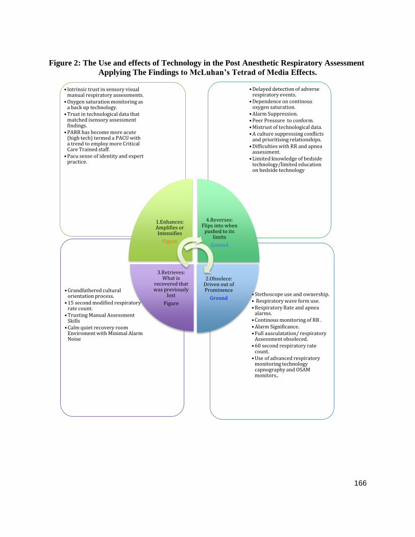
166
Figure 2: The Use and effects of Technology in the Post Anesthetic Respiratory Assessment
Applying The Findings to McLuhan’s Tetrad of Media Effects.
• Stethoscope use and ownership. • Respiratory wave form use. • Respiratory Rate and apnea
alarms. • Continous monitoring of RR . • Alarm Significance. • Full ausculatation/ respiratory
Assessment obsoleced. • 60 second respiratory rate
count. • Use of advanced respiratory
monitoring technology capnography and OSAM monitors..
• Grandfathered cultural orientation process.
• 15 second modified respiratory rate count.
• Trusting Manual Assessment Skills
• Calm quiet recovery room Enviroment with Minimal Alarm Noise
• Delayed detection of adverse respiratory events.
• Dependence on continous oxygen saturation.
• Alarm Suppression. • Peer Pressure to conform. • Mistrust of technological data. • A culture suppressing conflicts
and prioritising relationships. • Difficulties with RR and apnea
assessment. • Limited knowledge of bedside
technology/limited education on bedside technology
• Intrinsic trust in sensory visual manual respiratory assessments.
• Oxygen saturation monitoring as a back up technology.
• Trust in technological data that matched isensory assessment findings.
• PARR has become more acute (high tech) termed a PACU with a trend to employ more Critical Care Trained staff.
• Pacu sense of identity and expert practice.
1.Enhances: Amplifies or Intensifies
Figure
4.Reverses: Flips into when
pushed to its limits
Ground
2.Obsolece: Driven out of Prominence
Ground
3.Retrieves: What is
recovered that was previously
lost
Figure

167
Figure 3: Demographic Characteristics of Study Participants
Characteristics
Participant (n)
Finding
Highest Level of
Education
Certificate in Nursing
Diploma in Nursing or
Associate Degree in
Nursing
Bachelor’s Degree in
Nursing
Master’s in nursing
Other please clarify
2(Dip)
7(BSN)
Specialty Education
Critical Care Course
Post Anesthetic Care
Unit Course
Other
None
6 (Critical Care Course)
3 (PACU Course)
Age
20- 30=3
31-40 =2 41-50 =2
51-60 =2
Bachelors Degree
75%
Dipoma/Associate Degree
25%
Highest Level of Education
1 2
Critical Care
Course 67%
PACU Course
33%
Specialty Education
Critical CareCourse
PACU Course
AGE
20-30 31-40
41-50 51-60

168
Figure 3:
Continued
Demographic Characteristics of Study Participants
Characteristics
Participant (n)
Finding Highest level of
General Nursing
education compared
with Nurses Level of
Specialty Education
Diploma with Critical Care Courses= 1
Diploma with PACU Courses=1
Bachelors with Critical Care Course= 5
Bachelors with PACU Course= 2
Years of Experience in
Nursing
2-5
6-10
11-15
16- 20
>20yrs
2 (2-5yrs) 3 (6-10Yrs)
2 (11-15Yrs)
1 (16-20yrs) 1 (>20yrs)
Years of Experience in
Post Anesthetic
Nursing
<2
6-10
11-15
16-20
2<2
1(2.5yrs)
2(6-10) 2(11-15)
1(16-20yrs)
0
2
4
6
8
10
12
14
2
7 1
5
1
2
PACU Course
Critical CareCourse
00.5
11.5
22.5
33.5
2-5 yrs 6-10 yrs 11-15yrs
16-20yrs >20yrs
N
u
m
b
e
r
o
f
R
N
S
Years of Experience in Nursing
0
0.5
1
1.5
2
2.5
<2years
2-5years
6-10years
11-15years
16- 20years
>20years
N
u
m
b
e
r
o
f
R
N
s
Years of Experince in PACU

169
Figure 3:
Continued
Demographic Characteristics of Study
Participants
Characteristics
Participant (n)
Finding Years of experience in
PACU nursing
compared with
Specialty education
<2 years
With Critical Care Certificate
= 2
2-5 years with Critical care
Certificate = 2
6 -10 with a PACU
Certificate =1
11-15 with a PACU
Certificate =2
11- 15 with Critical Care
Certificate =1
16 – 20 years with Critical
Care Certificate =1
How often do you care
for an adult in early
phase 1 recovery after
the anesthesiologist
leaves the bedside, that
is unconscious with an
LMA or Endotracheal
Airway in Situ:
Never
Daily
More than daily
Weekly
Less than Weekly
8 (daily) 1 (More than daily)
If you have a patient in
early phase 1 with an
LMA or ET tube in situ
how long can they take
to wake and be ready to
extubate in your
experience, for example
A few minutes:
Ten minutes:
Fifteen minutes:
Sometimes longer:
4 (ten mins)
1 (15 minutes)
4 (sometimes longer)
2 2
1
1 1 2
Nu
me
r o
f P
AC
U R
Ns
Years of Experience Vs Specialty Qualification
PACUCourse
Critical CareCourse
LMA left in after anesthetist leaves in
early phase 1
Never
Daily
More than Daily
Longest time to exutabation
A few minutes
Ten minutes
Fifteen minutes
SometimesLonger

170
Figure 3:
Continued
Demographic Characteristics of Study
Participants
Characteristics
Participant (n) Finding
How often do you find
you need to provide
airway support such as
a jaw thrust or chin
lift, or t-piece (02) to
an LMA in the early
phase 1 recovery for
example:
Daily
More than once a day
Once a week
Rarely
Never
3 (daily)
0 (once a week)
1 (rarely (‘x 1 month’) 4 (more than once a day)
How frequently do you
count a respiratory
rate manually during
phase 1?
Q 5 min
1 (“frequently”) 2 (“always”)
3 (“q5 min’s”)
1 (“q10 min’s”) 1 (“at least every patient
x1”)
1 (“this depends on patients’ respiratory status. Q minute
if there are problems. Q 5
minutes until aware. Then Q 10 minutes or more
frequently if there are
problems
How many recovery
rooms have you
worked in?
1
2
3
6 (1 PACU’s)
2 (2 PACU’s)
1 (5 PACU’s)
Use of a Stethoscope P 1 = No
P 2 = Yes P 3 = Yes
P 4 = No
P 5 = No P 6 = No
P 7 = Yes
P 8 = Yes P 9 = Yes
Additional Airway Support Needed in
phase 1 Daily
more than once a day
Once a week
Rarely
never
How frequently PACU RNs Report Counting Manual Respiratory Rates
in Phase 1 q 5 mins
q 10 minutes
frequently'
"always"
at least every patinet x 1'
Depends
How Many Recovery Rooms Worked in
1 PAR
2 PAR
5 PAR
Stethoscope Use
Yes
No

171
Figure 3:
Continued
Demographic Characteristics of Study
Participants
Characteristics
Participant (n)
Finding Participants
Demographics
P (1) P (1) Diploma in Nursing or associate degree.
Specialty Education PACU Course
16-20 years experience in Nursing 11-15 years in Post Anesthetic Nursing
Age 42
........................Has worked in 2 recovery rooms
P (2) P (2) Bachelors Degree in Nursing Specialty Education: PACU Course
Years of Experience in Nursing 11-15 years
Years of Experience in Post Anesthetic Nursing 11-15 Age 43
Has worked in 2 recovery rooms
Participant
Demographics
P (3)
P (3) Bachelors Degree in Nursing
Specialty Education: Critical Care Course Years of Experience in Nursing 2 – 5
Years of Experience in Post Anesthetic Nursing < 2
Age 28 Has worked in 1 Recovery Room
P (4) P (4) Diploma in Nursing Or Associate Degree
Specialty Education: Critical Care Course Years of Experience In Nursing > 20
Years of Experience in Post Anesthetic Nursing 16-20
Age 56 Has Worked in 1 Recovery Room
P (5) P (5) Bachelors Degree in Nursing
Specialty Education: Critical Care Course
Years of Experince in Nursing 11- 15 Years of Experience in Post Anesthetic Nursing 11-15
Age 35
Has worked in 2 Recovery Rooms
P (6) P (6) Bachelors Degree in Nursing
Specialty Education: Critical Care Course
Years of Experience in Nursing 6-10 Years of Experience in Post Anesthetic Nursing 2-5
Age 32
Has worked in 5 Recovery Rooms
P (7) P (7) Bachelors Degree in Nursing Specialty Education: PACU Course
Years if Experience in Nursing 6-10
Years of Experience in Post Anesthetic Nursing 6-10 Age 51
Has Worked in 1 recovery room
P (8) P (8) Bachelors Degree in Nursing Specialty Education: Critical Care Course
Years of Experience in Nursing 6-10
Years of Experince in Post Anesthetic Nursing <2 Age 25
Has worked in 1 Recovery Room
P (9) P (9) Bachelors Degree in Nursing Specialty Education: Critical Care Course
Years of Experience in Nursing 2-5
Years of Experience in Post Anesthetic Nursing 2-5 Age 26
Has worked in 1 Recovery Room

172
Figure 4: Respiratory Assessment Process Flow Diagram
Arrival Walk
• Patient is wheeled on stretcher to the PARR from the operating room by the anethetist and OR nurse. Patient may arrived either intubated, on or off oxygen or extubated on oxygen or on room air.
• This is the 1st 'Arrival Walk' Visualization by the PARR nurse. • PARR Nurse 'prepares' for imminent patient arrival and forms initial impression of respiratory status.
Bedside Hook up
• Arrival at the bedside in PARR: Oxygen is immediately applied, or continued, unless asked not too by anesthesia. • Visualization for breaths is prioritized, condensation on oxygen mask is the first choice for verification of breaths. • Oxygen saturation finger probe applied, followed by BP Cuff and ECG leads. • Nurse immediately identifies and supports ineffective breathing. • Two nurses are required to support intubated patients on arrival.
Assessing
Breaths
• PARR nurse determines breathing pattern and counts respiratory rate manually for typically15 seconds only. • PARR nurse recieves report from the anesthesiologist and the OR Nurse. • Handover not always structured, Often a frenetic time of listening to report, while trying to establish respiratory rate manually,
reporting RR to the anesthetist and ensure patient is hemodynamically stable.
Head to Toe Assessment
• PARR Nurse completes Head to Toe Assessment and starts documentation with interruptions attending to urgent patient needs including pain, respiratory needs or hemodynamic support.
• Anesthesia leaves bedside after 1st set of vital signs recorded, if satisfied with patient condition.
Q 5 Minutes reassessment
• At 5 minutes from arrival PARR nurse repeates Vital Signs Assessment Starting with Respiratory Rate. • Patients are often still intubated, respiratory rate is rapidly transitioning from anesthetic respiratory depression and OR
narcotics and may be reacting to narcotic administration in PARR. • Respiratory rates continue to be re-assessed manually and via continuous Oxygen saturation monitoring. • Patients are often breathing irregularly, at a rate less than 8, and with apneic periods during this early phase.
Q 10 Minutes reassessment
• At Ten minutes the PARR nurse repeates Vital Signs and Head to Toe Assesment. Starting with a Manual Respiratory Rate. • Patients commonly require IV narcotic analgesia during this stage. • Patients may still be intubated or in process of extubation.
15 minutes
reassessment
• Four sets of vital signs have been recorded Q 5 minutes from arrival. 'If stable' this assessement repeates Q 10 minutes until phase 2 criteria are met, and patient transfers to the Ward.
• Patients are usually extubated, but some are still intubated if slow to wake. Apnea is still common at this stage, particularly if painful and requiring narcotic analgesia