Physiological matrix metalloproteinase concentrations in serum during childhood and adolescence,...
15
Physiological matrix metalloproteinase concentrations in serum during childhood and adolescence, using Luminex® Multiplex technology Kathryn M. Thrailkill 1,* , Cindy S. Moreau 1 , Gael Cockrell 1 , Pippa Simpson 1 , Rajiv Goel 1 , Paula North 2 , John L. Fowlkes 1 , and Robert C. Bunn 1 1 Department of Pediatrics, University of Arkansas for Medical Sciences, Little Rock, AR, USA 2 Department of Pathology, University of Arkansas for Medical Sciences, Little Rock, AR, USA Abstract Matrix metalloproteinases are a family of zinc-dependent proteinases which are involved in the breakdown and remodeling of extracellular matrix. As children grow and adolescents reach pubescence, their bodies undergo changes that require age-related morphogenesis of the extracellular matrix, possibly requiring unique patterns of matrix metalloproteinase (MMP) expression during periods of rapid tissue growth (i.e., childhood) or accelerated tissue remodeling and expansion (i.e., adolescence). Therefore, we have characterized age-specific and gender-specific differences in circulating concentrations of MMPs (specifically MMP-1, -2, -3, -8 and -9) in 189 serum samples obtained from healthy subjects, aged 2−18 years. MMP concentrations were measured using Fluorokine® MultiAnalyte Profiling kits and a Luminex® Bioanalyzer, as well as by commercial ELISA. Serum levels of MMP-1, -2, -3, -8, and -9 in healthy pediatric subjects represent log-normal distributions. MMP-2 was significantly negatively correlated with age (r=−0.29; p<0.001), while MMP-3 was significantly positively correlated with age (r=0.38; p<0.001). Although plasma, not serum, is considered the appropriate blood sample for measurement of MMP-8 and -9, serum levels of MMP-8 and -9 were also found to be highly positively correlated with each other (r=0.76; p<0.01). MMP results obtained by Fluorokine® MultiAnalyte Profiling methods correlated well with conventional ELISA methods and use of this technology provided several advantages over ELISA. Keywords collagenase; endopeptidases; gelatinase; particle-based flow cytometry; puberty; stromelysin Introduction Matrix metalloproteinases (MMPs) are an ever-expanding family of zinc-dependent endopeptidases involved in the breakdown, remodeling and physiological homeostasis of extracellular matrix (ECM) (1,2). Currently, there are over 20 human MMP-related enzymes, grouped into several subtypes, including collagenases, gelatinases, stromelysins, membrane- type MMPs (MT-MMPs) and other, less well-characterized, distinct enzymes (3). Targets of these powerful degradative enzymes include structural ECM molecules (i.e., interstitial collagens, laminins, fibronectins, and heparan sulfate proteoglycans), cell-cell adhesion molecules, cell surface receptors, growth factors, cytokines and chemoattractant proteins, as well as other proteinases (2). Because of their potent degradative capacity, the activity of these *Corresponding author: Kathryn M. Thrailkill, MD, Arkansas Children's Hospital, 800 Marshall St., Slot 512−6, Little Rock, AR 72202, USA Phone: +1−501−364−1430, Fax: +1−501−364−6299, E-mail: [email protected]. NIH Public Access Author Manuscript Clin Chem Lab Med. Author manuscript; available in PMC 2008 February 12. Published in final edited form as: Clin Chem Lab Med. 2005 ; 43(12): 1392–1399. NIH-PA Author Manuscript NIH-PA Author Manuscript NIH-PA Author Manuscript
Transcript of Physiological matrix metalloproteinase concentrations in serum during childhood and adolescence,...
Physiological matrix metalloproteinase concentrations in serumduring childhood and adolescence, using Luminex® Multiplextechnology
Kathryn M. Thrailkill1,*, Cindy S. Moreau1, Gael Cockrell1, Pippa Simpson1, Rajiv Goel1,Paula North2, John L. Fowlkes1, and Robert C. Bunn1
1 Department of Pediatrics, University of Arkansas for Medical Sciences, Little Rock, AR, USA
2 Department of Pathology, University of Arkansas for Medical Sciences, Little Rock, AR, USA
AbstractMatrix metalloproteinases are a family of zinc-dependent proteinases which are involved in thebreakdown and remodeling of extracellular matrix. As children grow and adolescents reachpubescence, their bodies undergo changes that require age-related morphogenesis of the extracellularmatrix, possibly requiring unique patterns of matrix metalloproteinase (MMP) expression duringperiods of rapid tissue growth (i.e., childhood) or accelerated tissue remodeling and expansion (i.e.,adolescence). Therefore, we have characterized age-specific and gender-specific differences incirculating concentrations of MMPs (specifically MMP-1, -2, -3, -8 and -9) in 189 serum samplesobtained from healthy subjects, aged 2−18 years. MMP concentrations were measured usingFluorokine® MultiAnalyte Profiling kits and a Luminex® Bioanalyzer, as well as by commercialELISA. Serum levels of MMP-1, -2, -3, -8, and -9 in healthy pediatric subjects represent log-normaldistributions. MMP-2 was significantly negatively correlated with age (r=−0.29; p<0.001), whileMMP-3 was significantly positively correlated with age (r=0.38; p<0.001). Although plasma, notserum, is considered the appropriate blood sample for measurement of MMP-8 and -9, serum levelsof MMP-8 and -9 were also found to be highly positively correlated with each other (r=0.76; p<0.01).MMP results obtained by Fluorokine® MultiAnalyte Profiling methods correlated well withconventional ELISA methods and use of this technology provided several advantages over ELISA.
Keywordscollagenase; endopeptidases; gelatinase; particle-based flow cytometry; puberty; stromelysin
IntroductionMatrix metalloproteinases (MMPs) are an ever-expanding family of zinc-dependentendopeptidases involved in the breakdown, remodeling and physiological homeostasis ofextracellular matrix (ECM) (1,2). Currently, there are over 20 human MMP-related enzymes,grouped into several subtypes, including collagenases, gelatinases, stromelysins, membrane-type MMPs (MT-MMPs) and other, less well-characterized, distinct enzymes (3). Targets ofthese powerful degradative enzymes include structural ECM molecules (i.e., interstitialcollagens, laminins, fibronectins, and heparan sulfate proteoglycans), cell-cell adhesionmolecules, cell surface receptors, growth factors, cytokines and chemoattractant proteins, aswell as other proteinases (2). Because of their potent degradative capacity, the activity of these
*Corresponding author: Kathryn M. Thrailkill, MD, Arkansas Children's Hospital, 800 Marshall St., Slot 512−6, Little Rock, AR 72202,USA Phone: +1−501−364−1430, Fax: +1−501−364−6299, E-mail: [email protected].
NIH Public AccessAuthor ManuscriptClin Chem Lab Med. Author manuscript; available in PMC 2008 February 12.
Published in final edited form as:Clin Chem Lab Med. 2005 ; 43(12): 1392–1399.
NIH
-PA Author Manuscript
NIH
-PA Author Manuscript
NIH
-PA Author Manuscript
enzymes is tightly regulated at all levels (i.e., transcription, post-transcription, secretion,protein processing and tissue localization). For example, proinflammatory cytokines such asinterleukin-1β and tissue necrosis factor-α stimulate MMP production, while other cytokines,hormones, and growth factors such as transforming growth factor-β, interleukin-4, corticoidhormones and insulin-like growth factors (IGFs) down-regulate MMP synthesis (4).
To date, MMPs have been implicated in many pathologic processes characterized by eitherdegradation of connective tissue matrices or pro-inflammatory states, such as rheumatoidarthritis and osteoarthritis, cancer growth and metastases, metabolic bone disease, emphysemaand atherosclerotic heart disease (5–7). Serum or plasma levels of MMPs are altered in variousdiseases, and have been considered as potential clinical markers of disease activity. Forexample, in adult clinical studies, serum MMP-2 levels have been reported to be increased inpatients with liver cirrhosis (8), malignant thyroid cancer (9), and endometriosis (10). Serumor plasma MMP-9 levels appear to be increased in patients with a diversity of cardiovascularrelated inflammatory conditions, including congestive heart failure (11), stroke (12,13),obstructive sleep apnea syndrome (14), and a history of myocardial infarction (15), as well aspatients with amyotrophic lateral sclerosis (16), and acute allograft rejection following livertransplantation (17). Plasma levels of MMP-3 are increased in several connective tissuedisorders, including rheumatoid arthritis (18) and systemic lupus erythematosus (18).
The first description of MMP-like activity was related to the metamorphosis of the tadpole(19). This and many other subsequent discoveries strongly suggest that this well-characterizedfamily of metalloproteinases is also intimately involved in normal tissue growth and ECMremodeling (20). Morphogenesis and tissue growth, remodeling, and repair are sentinel featuresof childhood and adolescence. However, little is known about the production, secretion, andclearance of these important proteinases throughout normal growth and development inhumans. At present, normal values for serum concentrations of commercially assayable MMPsare unavailable for the pediatric age range, limiting our ability to quantitatively comparepediatric disease-specific MMP abnormalities with MMP concentrations in healthy children.Furthermore, without such data, we have little or no understanding of which MMPs may beimportant in mediating events associated with somatic growth and development. Recognizingthat tissue-specific production and/or localization of MMPs is potentially a dynamic process,with unique patterns of expression of these proteinases during periods of rapid tissue growth(i.e., childhood) or accelerated tissue remodeling and expansion (i.e., adolescence), the presentstudy was developed to characterize potential age-specific and/or gender-specific differencesin circulating concentrations of MMPs (specifically MMP-1, -2, -3, -8 and -9) across thepediatric and adolescence age range. Moreover, the intent of this study was to demonstrate theutility of a multiplexed, particle-based flow cytometric assay for the efficient, cost-effectivesimultaneous analysis of a panel of MMPs using a small sample volume.
Materials and methodsResidual serum samples (0.5−2.0 mL) were obtained from otherwise healthy children whowere being tested (through the Arkansas Children's Hospital, Division of Pediatric Allergy andImmunology clinic) to exclude the possibility of a specific food allergy because of a pastanecdotal coincidence of minor illness or urticaria following a particular food exposure.Residual serum samples were maintained only from those patients who were found to have anegative food allergy evaluation. Specifically, a total of 189 samples from children fulfillingthe following inclusion criteria were obtained: 1) age 2−18 years; 2) normal serum total IgElevel; and 3) negative radioallergoimmunosorbent (RAST) testing to one or more foodcategories (results from all tested categories had to be negative for inclusion). At the time ofcollection, patient age, sex, and race were recorded from the hospital database system byclinical laboratory personnel and samples were coded with a unique identifying number. Coded
Thrailkill et al. Page 2
Clin Chem Lab Med. Author manuscript; available in PMC 2008 February 12.
NIH
-PA Author Manuscript
NIH
-PA Author Manuscript
NIH
-PA Author Manuscript

serum samples, along with the coded demographic parameters, were then transported to theresearch laboratory, such that research personnel remained blinded to patient identifyinginformation. Serum samples were stored at −20°C until batch analysis was performed. Theexperimental protocol was approved by the institutional Review Board of the University ofArkansas for Medical Sciences. The requirement for informed consent of subjects was waivedfor this protocol, recognizing that an informed consent document, as a means of identifyingsubjects, would provide an exclusive, unnecessary risk to subject confidentiality.
MMP-1, MMP-2, MMP-3 and MMP-8 were measured simultaneously, while MMP-9 wasmeasured separately in individual human serum samples using Fluorokine® MultiAnalyteProfiling (F-MAP) kits from R&D Systems (Minneapolis, MN, USA). Kits were run on aLuminex® 100™ Bioanalyzer (Luminex Corp., Austin, TX, USA) according to the kitmanufacturer's instructions. Fluorokine® MultiAnalyte Profiling kits contained distinct groupsof microspheres (each group bearing unique fluorescence intensity and a specific MMPantibody), biotinylated MMP antibodies, and phycoerythrin-conjugated streptavidin. Serumsamples were incubated with antibody-coated microspheres, which bind to specific MMPspresent in the serum. Next, microsphere-MMP complexes were washed and incubated withbiotinylated MMP antibodies, which bind to MMPs present on the microspheres. A finalincubation was performed in which phycoerythrin-labeled streptavidin was allowed to bind tobiotinylated MMP antibodies present on microspheres. Microspheres were then loaded into aLuminex® 100™ Bioanalyzer, which quantifies the amount of phycoerythrin fluorescencepresent on each of the distinct microsphere groups. At least 50 individual microspheres werecounted for each MMP, and the median fluorescence intensity was used for subsequentcalculations. Intra-assay (precision within an assay; one sample replicated eight times) andinter-assay precision (precision between different assays; four samples each replicated on 4different days) were: 5.2% and 7.5% for MMP-1; 4.1% and 9.9% for MMP-2; 3.6% and 10.3%for MMP-3; 8.4% and 16.4% for MMP-8; and 3.1% and 8.7% for MMP-9, respectively. Eachof the microsphere sets was reported by the manufacturer to exhibit less than 0.5% cross-reactivity and interference with the other MMP family members studied.
A unique subset of 40 samples per MMP analyte was also assayed for MMP-2, -3, -8 and -9by commercial ELISA to establish the reliability of the Fluorokine® MultiAnalyte Profiling(F-MAP) method compared with the more standard and popular method of measuring MMPs.Specifically, the R&D Systems Quantikine colorimetric sandwich ELISAs that use thismanufacturer's specific MMP polyclonal antibodies were employed. (The R&D SystemsMMP-1 ELISA measures pro-MMP-1, rather than total MMP-1, making a similar F-MAP vs.ELISA comparison for MMP-1 inappropriate.) These 40-sample subsets were randomlygenerated and unique for each analyte, representing a distribution of four samples from eachdecile across the assay range for each MMP (MMP-2, -3, -8 and -9), as obtained by F-MAP.
Statistical analysisLimited demographic data obtained from the medical record (age, sex, and race) were recordedinto an SPSS (SPSS 12.0 for Windows; SPSS, Chicago, IL, USA) data set. Exploratory dataanalysis, including summary statistics (mean, median, range, frequency tables) and plots(scatter plots, boxplots) were used to examine the distribution of and relationship betweenvariables. Subjects were grouped according to patient age into the following cohorts: 2 to ≤8years (pre-pubertal); 8 to ≤10 years (representing ∼Tanner stage 2 puberty); 10 to ≤12 years(∼Tanner stage 2−3 puberty); 12 to ≤15 years (∼Tanner stage 4 puberty); and 15 to ≤18 years(∼Tanner stage 5 puberty). Data were compared for each gender and age cohort separately. Inaddition, data were analyzed using age as an independent variable. A priori, it could be expectedthat MMP measurements would be skewed. The size of the standard deviation values alsosuggested that this was the case. Therefore, logarithms of the MMPs were used for analysis.
Thrailkill et al. Page 3
Clin Chem Lab Med. Author manuscript; available in PMC 2008 February 12.
NIH
-PA Author Manuscript
NIH
-PA Author Manuscript
NIH
-PA Author Manuscript
The distribution of each MMP was examined using histograms. We compared MMP resultsbetween age groups, gender and race using ANOVA with a post hoc Tukey test. Type III sumsof squares were used. Correlations were examined using Pearson correlations and scatterdiagrams. To further aid interpretation, smooth splines were fit showing variation over agegroups (Figure 3).
ResultsA total of 189 samples were obtained. Study subjects ranged in age from 2 to 18 years, with amean±standard error (SE) age of 9.27±0.34 years. The study population included 88 females(46.6%) with a mean age of 9.94±0.50 years and 101 males (53.4%) with a mean age of 8.69±0.45 years. Samples provided a racial distribution for subjects of 80.4% Caucasian, 16.9%African-American, 1.6% Hispanic and 1.1% other/unspecified. Subjects were sub-classifiedaccording to age, with the sample size of each age cohort as follows: 2 to ≤8 years, n=79; 8 to≤10 years, n=29; 10 to ≤12 years, n=22; 12 to ≤15 years, n=34; and 15 to ≤18 years, n=25.
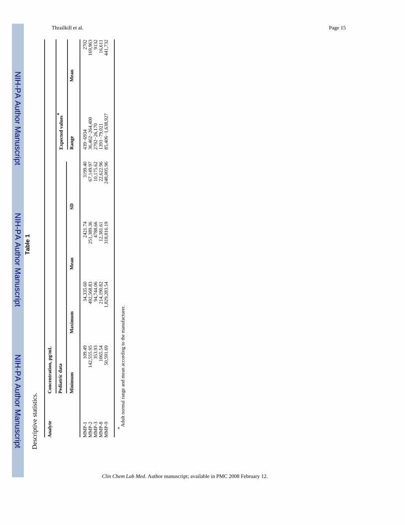
A summary of MMP results for the entire subject group is shown in Table 1. The manufacturer'sexpected values (for an adult population) for each analyte are also shown. Analysis ofhistograms of individual MMP results (Figure 1) suggests that serum levels of MMPs in healthychildren and adolescents represent log-normal distributions.
MMP-1 (collagenase-1), MMP-8 (collagenase-2) and MMP-9 (gelatinase B) results were notsignificantly different from normal ranges reported for these analytes in adult subjects (Table1). In addition, there was no significant difference among MMP-1, MMP-8 or MMP-9 resultsfor different age groups (p>0.20) when the population was examined as a whole. Analysis ofMMP-9 results by gender-race subgroups yielded no statistically significant differences (datanot shown). However, mean serum levels of MMP-8 in African-American males weregenerally higher than MMP-8 levels in other sex-race combinations, and statisticallysignificantly higher than MMP-8 results for Caucasian females (data not shown; p≤0.001).MMP-1 results for Caucasian males in the 8−10-year age group were statistically significantlyhigher than MMP-1 results for Caucasian males in the 12−15-year age group (data not shown;p<0.05).
Two MMPs, MMP-2 (gelatinase A) and MMP-3 (stromelysin-1), were correlated with age.Specifically, MMP-2 concentrations were significantly negatively correlated with age (Figure2; R2=0.086, p<0.001) and when analyzed across the age subsets, MMP-2 results in the 2−8-year age group were statistically higher than MMP-2 results in the 15−18-year group (p<0.05).Overall, the mean MMP-2 result for all pediatric subjects was ∼47% higher than the normativemean value reported for adults (Table 1). When analyzed across specific age groups andstratified according to race and gender, serum MMP-2 results suggest a slight increase inMMP-2 concentration coincident with the expected age of early puberty, occurring earlier inAfrican-American females than in other gender-race combinations (Figure 3) (21).
In contrast, MMP-3 concentrations were significantly positively correlated with age (Figure4; R2=0.148, p<0.001). When examined by individual age groups, mean MMP-3 results in the15−18-year age group were statistically significantly higher than mean MMP-3 results in otherage groups (p<0.05). Moreover, the mean MMP-3 result for all pediatric subjects was ∼48%lower than the normative mean value reported for adults (Table 1). In contrast to results reportedfor adults (18), when evaluated by gender, there was no significant difference among serumMMP-3 results for males vs. females at any age.
MMP-8 (collagenase-2) and MMP-9 (gelatinase B) are both synthesized by differentiatinggranulocytes, stored in circulating neutrophils, and released following neutrophil activation
Thrailkill et al. Page 4
Clin Chem Lab Med. Author manuscript; available in PMC 2008 February 12.
NIH
-PA Author Manuscript
NIH
-PA Author Manuscript
NIH
-PA Author Manuscript

(3). Therefore, serum levels of these MMPs are thought to be influenced by release of MMPsby degranulation of neutrophils during ex vivo blood clotting in the test tube (22). It issignificant, therefore, that serum levels of MMP-8 and MMP-9 were highly positivelycorrelated (Figure 5; R2=0.488; p<0.001).
Assay characteristicsMMP results generated by F-MAP and by ELISA were highly positively correlated for MMP-3(R2=0.97; p<0.001), MMP-8 (R2=0.96; p<0.001) and MMP-9 (R2=0.87; p<0.001; Figure 6).MMP-2 results generated by F-MAP and by ELISA were less tightly correlated (R2=0.45;Figure 6).
DiscussionWe have previously identified and characterized urinary MMP excretion in pediatric patientsand demonstrated that urinary excretion of MMPs is age-specific, with unique alterationsdetected during puberty (23). These data, as well as the work of others, suggest that regulationof MMPs during puberty is very likely distinctive and reflective of exuberant ECM expansionand tissue remodeling during pubertal growth and sexual maturation. For example, highconcentrations of type I collagen breakdown products are detected in the urine of adolescents,and these concentrations correlate positively with growth rate during the pubertal growth spurtand with gender, being higher in females than in males (24). Because MMPs are the principalmediators of collagen turnover, it is important to study the temporal changes in MMPconcentrations throughout childhood and adolescent development, both as a reference sourcefor existing and future pediatric studies of disease-related MMP dysregulation, and to aid inour understanding of the role of MMP activity in normal ECM physiology.
To the best of our knowledge, this report is the first demonstration of the clinical utility of theF-MAP method for simultaneously measuring multiple MMP values in a single, specificallypediatric, serum specimen. Using this technology, we have demonstrated that serum values ofMMP-2 and MMP-3 are age-dependent, and differ significantly from commercial normativeranges for these analytes for adult populations. In addition, similar to our previous studydemonstrating an increase in MMP-2 concentration in urine during the pubertal years (23),MMP-2 data were suggestive of an early to mid-pubertal increase in serum MMP-2 values. Incontrast, we did not identify an age correlation for either MMP-8 or MMP-9. However,recognizing that serum levels of these MMPs reflect release of MMPs following neutrophildegranulation, it is possible that any age correlation was obscured by this effect, and wouldrequire analysis of plasma specimens. In addition, caution should be exercised in interpretingserum values of MMP-8 and MMP-9, as plasma is considered the preferred blood sample formeasurement of these proteases.
While this subject population represented a presumed healthy pediatric and adolescent cohort,the limited demographic information obtained at the time of specimen collection, as well asthe blinded nature of these specimens, makes it possible that this assumption was not uniformlyaccurate. In addition, it is possible that other variables inherent in a pediatric population beyondage, race and sex (i.e., changing body mass index, timing of puberty, minor concurrent illness)would influence MMP results, but could not be factored into this analysis due to the nature ofthe study design. Nevertheless, the large number of samples available for analysis shouldprovide a representative assessment of normative data and dampen any confounding variables.
Because MMPs are produced by many tissues, it is likely that the serum concentrationsmeasured reflect a variety of sources. For example, the balanced regulation of MMPs and theirnaturally occurring inhibitors (tissue inhibitors of metalloproteinases, TIMPs) has been shownto be a critical component of many normal physiologic processes, including cartilage
Thrailkill et al. Page 5
Clin Chem Lab Med. Author manuscript; available in PMC 2008 February 12.
NIH
-PA Author Manuscript
NIH
-PA Author Manuscript
NIH
-PA Author Manuscript

development and endochondral bone formation (25–27), normal bone turnover and repair(28,29), reproductive tissue ECM remodeling during puberty and pregnancy (30–32),menstruation (33), growth of mammary tissues and lactation (34), testicular development(35) and normal spermatogenesis (35). Notably, many of these physiologic processes becomeactive during puberty or are augmented during the pubertal growth spurt. Moreover, pubertalhormones, specifically growth hormone and IGFs (36–39), androgens (40) and estrogen (41,42), have all been shown to regulate MMP expression and/or secretion.
For these studies, we investigated the use of a new methodology to measure multiple MMPsin a single sample of serum. We utilized a Luminex system-based assay that uses a 10×10 beadmatrix, in which each bead set is made up of a specific proportion of red/orange fluorescentdye. The choice of this assay has several advantages over conventional ELISA techniques tomeasure MMPs. First, it is capable of simultaneously measuring multiple MMPs in a singlebiological sample, thereby eliminating significant operator error in sampling (e.g., pipetting)which is possible with multiple MMP analyses. Second, smaller sample volumes are requiredfor multiplexing; thus, this technology is more amenable to the pediatric population, in whichblood volumes may be restricted. Third, our data suggest that when serum is analyzed there isa high degree of correlation between conventional ELISAs and the multiplex assay for MMP-3,MMP-8, and MMP-9; however, comparison of absolute values between assay techniques maynot be valid. Finally, the assays are robust and capable of measuring each MMP over a widerange of values. Other considerations not directly addressed by our study, but which might alsomake this approach more attractive than ELISA, are that the fluorescent readout is more direct,stable, and sensitive than colorimetric methods used for ELISAs, and typically, if measuringmore than six analytes, the multiplex approach can be less expensive (43). Thus, the overallevaluation of this new technology to assay MMP concentrations in human serum shows it tobe reproducible, specific, sensitive, and robust.
In conclusion, particle-based flow cytometry, specifically, F-MAP techniques for measurementof serum concentrations of MMP-1, -2, -3, -8, and -9, has effectively demonstrated age-specificdifferences in the serum concentrations of specific MMPs. These findings suggest that careshould be exercised in interpreting the role of MMPs in the pathophysiology of variouschildhood diseases unless critical pediatric normative data are available and utilized forcomparison.
Acknowledgements
This work was supported by a grant from the Arkansas Children's Hospital Research Institute (ACHRI) Dean's/CUMGResearch Development Fund (#SG-030103-KT) and a grant from the National Institutes of Health to ACHRI (R01-DK62999-KT). The authors also acknowledge the support of the University of Arkansas for Medical Sciences' GeneralClinical Research Center, grant M01 RR14288 for assistance with MMP clinical investigations.
References1. Werb Z. ECM and cell surface proteolysis: regulating cellular ecology. Cell 1997;91:439–42. [PubMed:
9390552]2. Mott JD, Werb Z. Regulation of matrix biology by matrix metalloproteinases. Curr Opin Cell Biol
2004;16:558–64. [PubMed: 15363807]3. Sternlicht MD, Werb Z. How matrix metalloproteinases regulate cell behavior. Annu Rev Cell Dev
Biol 2001;17:463–516. [PubMed: 11687497]4. Van Noort JM, Amor S. Cell biology of autoimmune diseases. Int Rev Cytol 1998;178:127–206.
[PubMed: 9348670]5. Nagase, H. Matrix metalloproteinases. Taylor and Francis; London: 1996. p. 153-204.6. Okada Y, Takeuchi N, Tomita K, Nakanishi I, Nagase H. Immunolocalization of matrix
metalloproteinase 3 (stromelysin) in rheumatoid synovioblasts (B cells): correlation with rheumatoidarthritis. Ann Rheum Dis 1989;48:645–53. [PubMed: 2675782]
Thrailkill et al. Page 6
Clin Chem Lab Med. Author manuscript; available in PMC 2008 February 12.
NIH
-PA Author Manuscript
NIH
-PA Author Manuscript
NIH
-PA Author Manuscript

7. Schoenhagen P, Vince DG, Ziada KM, Kapadia SR, Lauer MA, Crowe TD, et al. Relation of matrix-metalloproteinase 3 found in coronary lesion samples retrieved by directional coronary atherectomyto intravascular ultrasound observations on coronary remodeling. Am J Cardiol 2002;89:1354–9.[PubMed: 12062727]
8. El-Gindy I, El Rahman AT, El-Alim MA, Zaki SS. Diagnostic potential of serum matrixmetalloproteinase-2 and tissue inhibitor of metalloproteinase-1 as non-invasive markers of hepaticfibrosis in patients with HCV related chronic liver disease. Egypt J Immunol 2003;10:27–35. [PubMed:15719620]
9. Pasieka Z, Stepien H, Czyz W, Pomorski L, Kuzdak K. Concentration of metalloproteinase-2 and tissueinhibitor of metalloproteinase-2 in the serum of patients with benign and malignant thyroid tumourstreated surgically. Endocr Regul 2004;38:57–63. [PubMed: 15497929]
10. Huang HF, Hong LH, Tan Y, Sheng JZ. Matrix metalloproteinase 2 is associated with changes insteroid hormones in the sera and peritoneal fluid of patients with endometriosis. Fertil Steril2004;81:1235–9. [PubMed: 15136083]
11. Abou-Raya S, Naim A, Marzouk S. Cardiac matrix remodelling in congestive heart failure: the roleof matrix metalloproteinases. Clin Invest Med 2004;27:93–100. [PubMed: 15202828]
12. Lynch JR, Blessing R, White WD, Grocott HP, Newman MF, Laskowitz DT. Novel diagnostic testfor acute stroke. Stroke 2004;35:57–63. [PubMed: 14671250]
13. Reynolds MA, Kirchick HJ, Dahlen JR, Anderberg JM, McPherson PH, Nakamura KK, et al. Earlybiomarkers of stroke. Clin Chem 2003;49:1733–9. [PubMed: 14500614]
14. Tazaki T, Minoguchi K, Yokoe T, Samson KT, Minoguchi H, Tanaka A, et al. Increased levels andactivity of matrix metalloproteinase-9 in obstructive sleep apnea syndrome. Am J Respir Crit CareMed 2004;170:1354–9. [PubMed: 15361365]
15. Renko J, Kalela A, Jaakkola O, Laine S, Hoyhtya M, Alho H, et al. Serum matrix metalloproteinase-9is elevated in men with a history of myocardial infarction. Scand J Clin Lab Invest 2004;64:255–61.[PubMed: 15222636]
16. Demestre M, Parkin-Smith G, Petzold A, Pullen AH. The pro and the active form of matrixmetalloproteinase-9 is increased in serum of patients with amyotrophic lateral sclerosis. JNeuroimmunol 2005;159:146–54. [PubMed: 15652414]
17. Kuyvenhoven JP, Verspaget HW, Gao Q, Ringers J, Smit VT, Lamers CB, et al. Assessment of serummatrix metalloproteinases MMP-2 and MMP-9 after human liver transplantation: increased serumMMP-9 level in acute rejection. Transplantation 2004;77:1646–52. [PubMed: 15201662]
18. Zucker S, Lysik RM, Zarrabi MH, Greenwald RA, Gruber B, Tickle SP, et al. Elevated plasmastromelysin levels in arthritis. J Rheumatol 1994;21:2329–33. [PubMed: 7699637]
19. Brinckerhoff CE, Matrisian LM. Matrix metalloproteinases: a tail of a frog that became a prince. NatRev Mol Cell Biol 2002;3:207–14. [PubMed: 11994741]
20. Stamenkovic I. Extracellular matrix remodelling: the role of matrix metalloproteinases. J Pathol2003;200:448–64. [PubMed: 12845612]
21. Styne DM. Puberty, obesity and ethnicity. Trends Endocrinol Metab 2004;15:472–8. [PubMed:15541646]
22. Zucker S, Doshi K, Cao J. Measurement of matrix metalloproteinases (MMPs) and tissue inhibitorsof metalloproteinases (TIMP) in blood and urine: potential clinical applications. Adv Clin Chem2004;38:37–85. [PubMed: 15521188]
23. Thrailkill KM, Kumar S, Rosenberg CK, Auten KJ, Fowlkes JL. Characterization of matrixmetalloproteinases in human urine: alterations during adolescence. Pediatr Nephrol 1999;13:223–9.[PubMed: 10353410]
24. Mora S, Prinster C, Proverbio MC, Bellini A, de Poli SC, Weber G, et al. Urinary markers of boneturnover in healthy children and adolescents: age-related changes and effect of puberty. Calcif TissueInt 1998;63:369–74. [PubMed: 9799819]
25. Bord S, Horner A, Beeton CA, Hembry RM, Compston JE. Tissue inhibitor of matrixmetalloproteinase-1 (TIMP-1) distribution in normal and pathological human bone. Bone1999;24:229–35. [PubMed: 10071915]
Thrailkill et al. Page 7
Clin Chem Lab Med. Author manuscript; available in PMC 2008 February 12.
NIH
-PA Author Manuscript
NIH
-PA Author Manuscript
NIH
-PA Author Manuscript

26. Bord S, Horner A, Hembry RM, Compston JE. Stromelysin-1 (MMP-3) and stromelysin-2 (MMP-10)expression in developing human bone: potential roles in skeletal development. Bone 1998;23:7–12.[PubMed: 9662124]
27. Werb Z, Chin JR. Extracellular matrix remodeling during morphogenesis. Ann NY Acad Sci1998;857:110–8. [PubMed: 9917836]
28. Weiss S, Baumgart R, Jochum M, Strasburger CJ, Bidlingmaier M. Systemic regulation of distractionosteogenesis: a cascade of biochemical factors. J Bone Miner Res 2002;17:1280–9. [PubMed:12096842]
29. Uusitalo H, Hiltunen A, Soderstrom M, Aro HT, Vuorio E. Expression of cathepsins B, H, K, L, andS and matrix metalloproteinases 9 and 13 during chondrocyte hypertrophy and endochondralossification in mouse fracture callus. Calcif Tissue Int 2000;67:382–90. [PubMed: 11136537]
30. Fata JE, Ho AT, Leco KJ, Moorehead RA, Khokha R. Cellular turnover and extracellular matrixremodeling in female reproductive tissues: functions of metalloproteinases and their inhibitors. CellMol Life Sci 2000;57:77–95. [PubMed: 10949582]
31. Stygar D, Wang H, Vladic YS, Ekman G, Eriksson H, Sahlin L. Increased level of matrixmetalloproteinases 2 and 9 in the ripening process of the human cervix. Biol Reprod 2002;67:889–94. [PubMed: 12193399]
32. Lenhart JA, Ryan PL, Ohleth KM, Palmer SS, Bagnell CA. Relaxin increases secretion of tissueinhibitor of matrix metalloproteinase-1 and -2 during uterine and cervical growth and remodeling inthe pig. Endocrinology 2002;143:91–8. [PubMed: 11751597]
33. Dong JC, Dong H, Campana A, Bischof P. Matrix metalloproteinases and their specific tissueinhibitors in menstruation. Reproduction 2002;123:621–31. [PubMed: 12006090]
34. Lochter A. Plasticity of mammary epithelia during normal development and neoplastic progression.Biochem Cell Biol 1998;76:997–1008. [PubMed: 10392711]
35. Sang QX, Stetler-Stevenson WG, Liotta LA, Byers SW. Identification of type IV collagenase in rattesticular cell culture: influence of peritubular-Sertoli cell interactions. Biol Reprod 1990;43:956–64. [PubMed: 1963327]
36. Anne-Valerie R, Christelle D, Yannick F, Norbert P, Marc P, Dominique H. Human growth hormonestimulates proteinase activities of rabbit bone cells via IGF-I. Biochem Biophys Res Commun2000;268:875–81. [PubMed: 10679298]
37. Yoon A, Hurta RA. Insulin like growth factor-1 selectively regulates the expression of matrixmetalloproteinase-2 in malignant H-ras transformed cells. Mol Cell Biochem 2001;223:1–6.[PubMed: 11681709]
38. Hui W, Rowan AD, Cawston T. Insulin-like growth factor 1 blocks collagen release and downregulates matrix metalloproteinase-1, -3, -8, and -13 mRNA expression in bovine nasal cartilagestimulated with oncostatin M in combination with interleukin 1alpha. Ann Rheum Dis 2001;60:254–61. [PubMed: 11171688]
39. Lupia E, Elliot SJ, Lenz O, Zheng F, Hattori M, Striker GE, et al. IGF-1 decreases collagen degradationin diabetic NOD mesangial cells: implications for diabetic nephropathy. Diabetes 1999;48:1638–44.[PubMed: 10426384]
40. Li SC, Chen GF, Chan PS, Choi HL, Ho SM, Chan FL. Altered expression of extracellular matrixand proteinases in Noble rat prostate gland after long-term treatment with sex steroids. Prostate2001;49:58–71. [PubMed: 11550211]
41. Liao EY, Luo XH, Deng XG, Wu XP. Effects of 17beta-estradiol on the expression of membranetype 1 matrix metalloproteinase (MT1-MMP) and MMP-2 in human osteoblastic MG-63 cellcultures. J Endocrinol Invest 2001;24:876–81. [PubMed: 11817712]
42. Wingrove CS, Garr E, Godsland IF, Stevenson JC. 17beta-oestradiol enhances release of matrixmetalloproteinase-2 from human vascular smooth muscle cells. Biochim Biophys Acta1998;1406:169–74. [PubMed: 9573355]
43. Vignali DA. Multiplexed particle-based flow cytometric assays. J Immunol Methods 2000;243:243–55. [PubMed: 10986418]
Thrailkill et al. Page 8
Clin Chem Lab Med. Author manuscript; available in PMC 2008 February 12.
NIH
-PA Author Manuscript
NIH
-PA Author Manuscript
NIH
-PA Author Manuscript
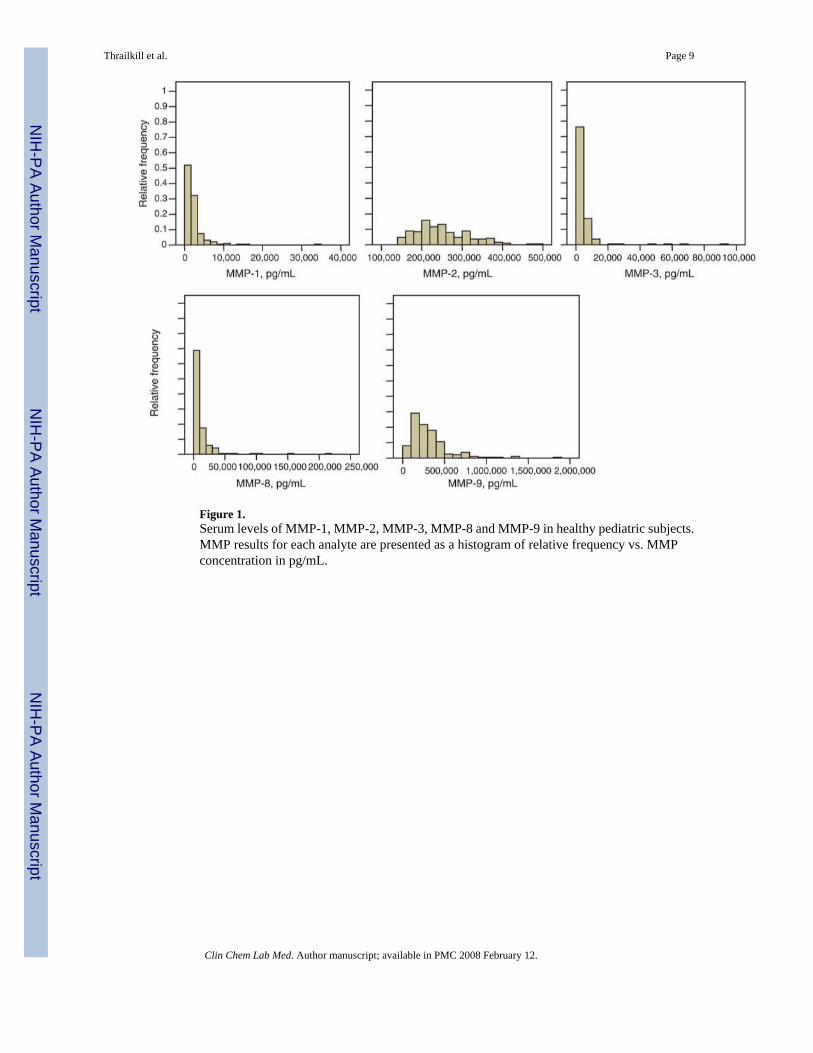
Figure 1.Serum levels of MMP-1, MMP-2, MMP-3, MMP-8 and MMP-9 in healthy pediatric subjects.MMP results for each analyte are presented as a histogram of relative frequency vs. MMPconcentration in pg/mL.
Thrailkill et al. Page 9
Clin Chem Lab Med. Author manuscript; available in PMC 2008 February 12.
NIH
-PA Author Manuscript
NIH
-PA Author Manuscript
NIH
-PA Author Manuscript
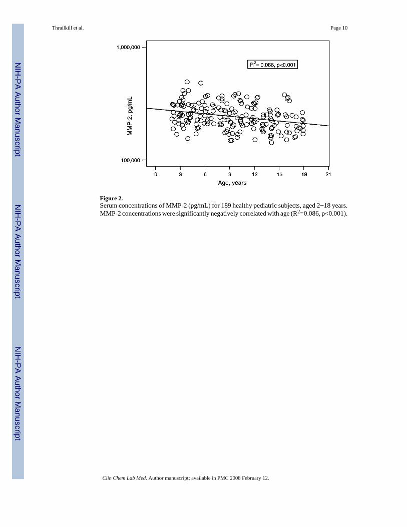
Figure 2.Serum concentrations of MMP-2 (pg/mL) for 189 healthy pediatric subjects, aged 2−18 years.MMP-2 concentrations were significantly negatively correlated with age (R2=0.086, p<0.001).
Thrailkill et al. Page 10
Clin Chem Lab Med. Author manuscript; available in PMC 2008 February 12.
NIH
-PA Author Manuscript
NIH
-PA Author Manuscript
NIH
-PA Author Manuscript
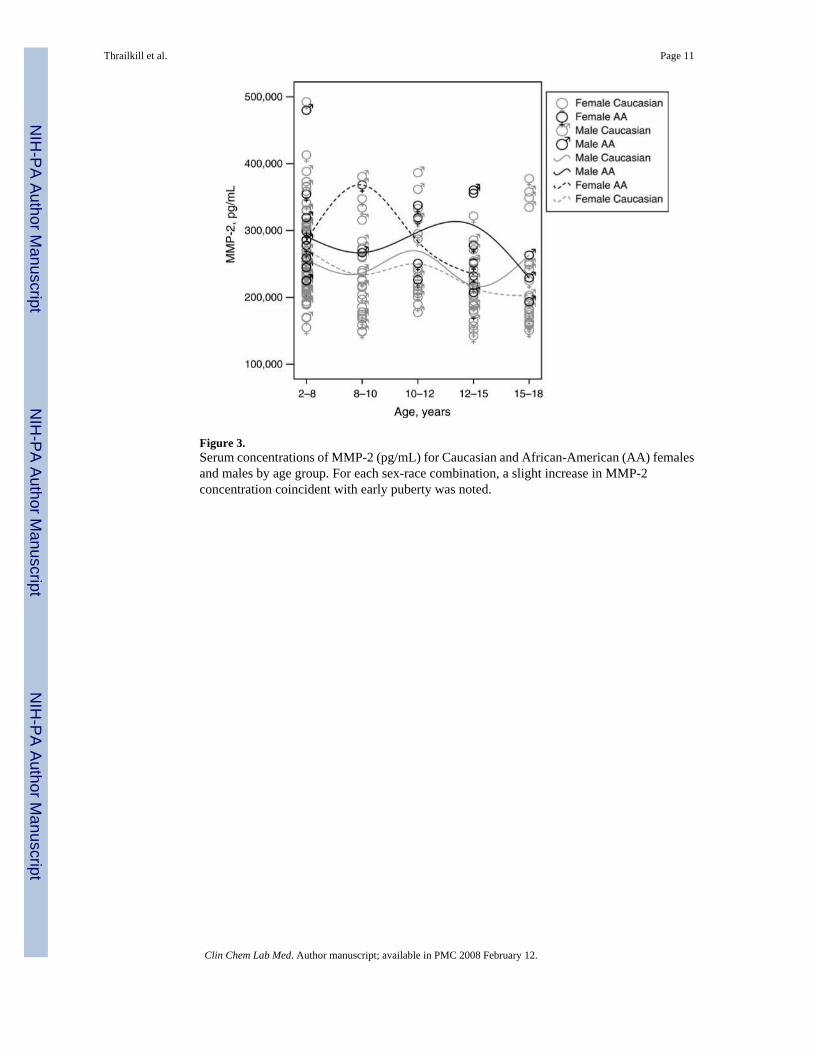
Figure 3.Serum concentrations of MMP-2 (pg/mL) for Caucasian and African-American (AA) femalesand males by age group. For each sex-race combination, a slight increase in MMP-2concentration coincident with early puberty was noted.
Thrailkill et al. Page 11
Clin Chem Lab Med. Author manuscript; available in PMC 2008 February 12.
NIH
-PA Author Manuscript
NIH
-PA Author Manuscript
NIH
-PA Author Manuscript
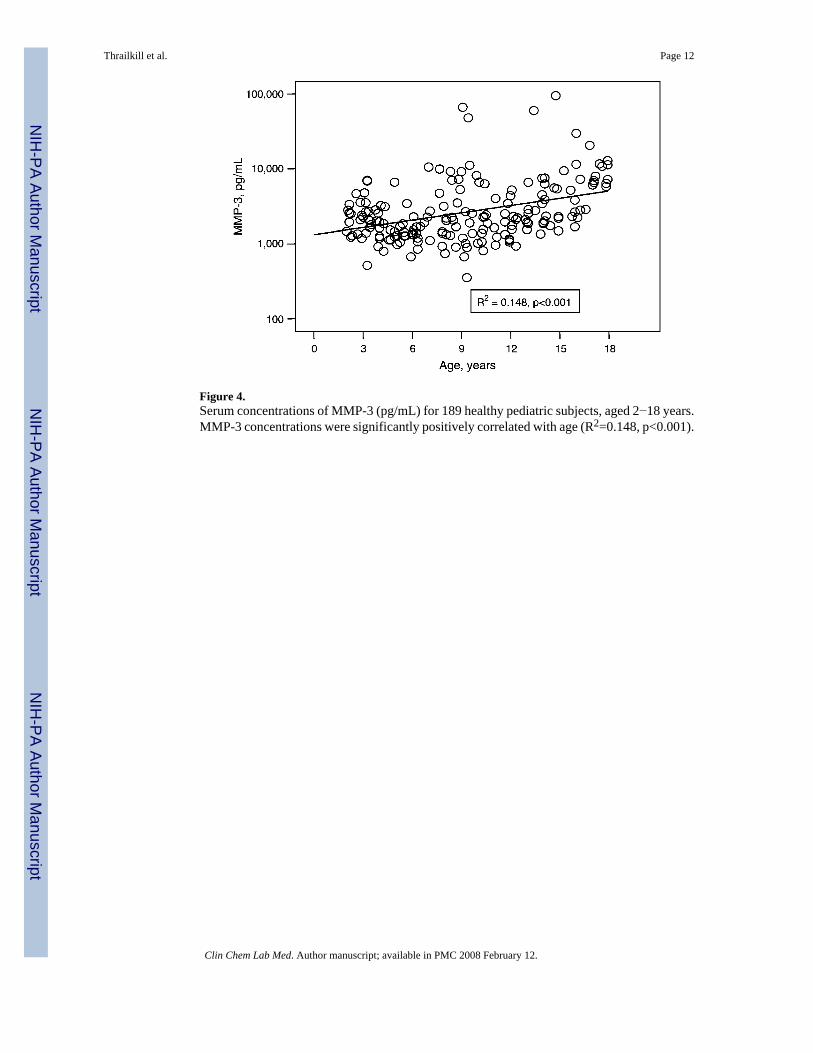
Figure 4.Serum concentrations of MMP-3 (pg/mL) for 189 healthy pediatric subjects, aged 2−18 years.MMP-3 concentrations were significantly positively correlated with age (R2=0.148, p<0.001).
Thrailkill et al. Page 12
Clin Chem Lab Med. Author manuscript; available in PMC 2008 February 12.
NIH
-PA Author Manuscript
NIH
-PA Author Manuscript
NIH
-PA Author Manuscript

Figure 5.Correlation between serum concentrations of MMP-8 and MMP-9 (pg/mL) in 189 healthypediatric subjects, aged 2−18 years. Serum levels of MMP-8 and MMP-9 are highly positivelycorrelated (R2=0.488, p<0.001).
Thrailkill et al. Page 13
Clin Chem Lab Med. Author manuscript; available in PMC 2008 February 12.
NIH
-PA Author Manuscript
NIH
-PA Author Manuscript
NIH
-PA Author Manuscript
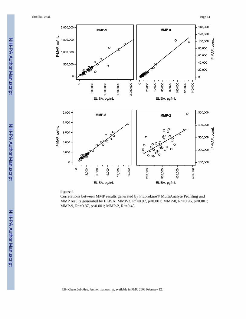
Figure 6.Correlations between MMP results generated by Fluorokine® MultiAnalyte Profiling andMMP results generated by ELISA: MMP-3, R2=0.97, p<0.001; MMP-8, R2=0.96, p<0.001;MMP-9, R2=0.87, p<0.001; MMP-2, R2=0.45.
Thrailkill et al. Page 14
Clin Chem Lab Med. Author manuscript; available in PMC 2008 February 12.
NIH
-PA Author Manuscript
NIH
-PA Author Manuscript
NIH
-PA Author Manuscript
NIH
-PA Author Manuscript
NIH
-PA Author Manuscript
NIH
-PA Author Manuscript
Thrailkill et al. Page 15Ta
ble
1D
escr
iptiv
e st
atis
tics.
Ana
lyte
Con
cent
ratio
n, p
g/m
L
Pedi
atri
c da
taE
xpec
ted
valu
es*
Min
imum
Max
imum
Mea
nSD
Ran
geM
ean
MM
P-1
109.
4934
,335
.60
2421
.74
3199
.40
439−
6934
2702
MM
P-2
142,
555.
9549
2,56
8.83
251,
389.
3667
,149
.97
36,4
62−2
64,4
0016
9,96
3M
MP-
335
3.93
94,7
44.0
647
88.6
610
,175
.62
2792−2
6,17
091
32M
MP-
810
65.5
421
4,19
0.82
12,3
81.6
122
,622
.96
1391−7
9,02
116
,611
MM
P-9
50,5
91.6
91,
829,
283.
5431
8,81
6.19
248,
005.
9685
,406−1
,638
,927
441,
732
* Adu
lt no
rmal
rang
e an
d m
ean
acco
rdin
g to
the
man
ufac
ture
r.
Clin Chem Lab Med. Author manuscript; available in PMC 2008 February 12.