osu6162 to reduce voluntary alcohol - CORE
82
From THE DEPARTEMENT OF CLINICAL NEUROSCIENCE Karolinska Institutet, Stockholm, Sweden THE ROLE OF THE DOPAMINE SYSTEM IN THE ABILITY OF (-)-OSU6162 TO REDUCE VOLUNTARY ALCOHOL DRINKING AND BINGE-EATING IN THE RAT Kristin Feltmann Stockholm 2017
-
Upload
khangminh22 -
Category
Documents
-
view
2 -
download
0
Transcript of osu6162 to reduce voluntary alcohol - CORE

From THE DEPARTEMENT OF CLINICAL NEUROSCIENCE Karolinska Institutet, Stockholm, Sweden
THE ROLE OF THE DOPAMINE SYSTEM IN THE ABILITY OF (-)-OSU6162
TO REDUCE VOLUNTARY ALCOHOL DRINKING AND BINGE-EATING IN THE RAT
Kristin Feltmann
Stockholm 2017

The cover picture has been created by Igor Cervenka.
All previously published papers were reproduced with permission from the publisher.
Published by Karolinska Institutet.
Printed by Eprint AB 2017
© Kristin Feltmann, 2017
ISBN 978-91-7676-898-3

THE ROLE OF THE DOPAMINE SYSTEM IN THE ABILITY OF (-)-OSU6162 TO REDUCE VOLUNTARY ALCOHOL DRINKING AND BINGE-EATING IN THE RAT THESIS FOR DOCTORAL DEGREE (Ph.D.)
By
Kristin Feltmann
Principal Supervisor: Pia Steensland, Ph.D., Assoc. Prof. Karolinska Institutet Department of Clinical Neuroscience Centre for Psychiatry Research Co-supervisor(s): Björn Schilström, M.D., Ph.D., Assoc. Prof. Gothenburg University, Sahlgrenska Academy Department of Neuroscience and Physiology Division of Psychiatry and Neurochemistry Johan Franck, M.D., Ph.D., Prof. Karolinska Institutet Department of Clinical Neuroscience Centre for Psychiatry Research
Opponent: Esa Korpi, M.D., Ph.D., Prof. University of Helsinki Institute of Biomedicine Examination Board: Erika Roman, Ph.D., Assoc. Prof. Uppsala University Department of Pharmaceutical Biosciences Per Andrén, Ph.D., Prof. Uppsala University Department of Pharmaceutical Biosciences Vladana Vukojevic, Ph.D., Assoc. Prof. Karolinska Institutet Department of Clinical Neuroscience


To Vicente


The greatest enemy of knowledge is not ignorance, it is the illusion of knowledge.
Daniel J. Boorstin


ABSTRACT
BACKGROUND AND AIMS: The dopamine system is involved in the reinforcing effects of
both food and alcohol and is thus a potential treatment target for alcohol use disorder (AUD)
and binge-eating disorder (BED).
Alcohol use disorder is characterized by difficulties to control alcohol drinking and by
drinking despite adverse consequences. Few pharmacological treatments are available and
their efficacies are limited. The monoamine-stabilizer (-)-OSU6162 has been identified as a
potential novel treatment for AUD by showing that it reduces alcohol drinking, alcohol
seeking, relapse to seeking and withdrawal symptoms in long-term drinking rats (Steensland
et al, 2012). In the present thesis, the possible underlying mechanisms in the ability of
(-)-OSU6162 to reduce alcohol drinking were investigated.
Binge eating disorder is characterized by episodes of eating large amounts of foods in a
relatively short amount of time, the difficulty to control binge-eating and feelings of shame
and guilt. Recently, lisdexamfetamine was approved for the treatment of BED. Here, the
potential of (-)-OSU6162 as a treatment for BED was investigated.
METHODS: Rats were drinking in the two-bottle choice intermittent-access to 20% ethanol
(IA20E) paradigm for three to ten months. The effects of long-term voluntary alcohol
drinking on dopamine D2 receptor (D2R) expression (paper I) and dopamine output
(paper II) in the nucleus accumbens (NAc) were investigated using qPCR and proximity
ligation assay microdialysis. The effects of (-)-OSU6162 on dopamine in the NAc in long-
term drinking rats were investigated using microdialysis (paper II). Pharmacological
antagonists were used to study the role of the D2R and the serotonin 5-HT2A receptor in the
ability of (-)-OSU6162 to reduce alcohol drinking (paper III). Using a model of binge-eating
and the second-order schedule of reinforcement, the effects of (-)-OSU6162 on binge-like
eating and cue-controlled seeking for chocolate-flavored sucrose were evaluated (paper IV).
RESULTS: Long-term voluntary alcohol drinking downregulated dopamine levels, D2R
expression levels and D2R-D2R homoreceptor complexes, and increased adenosine A2AR-
D2R heteroreceptor complexes in the NAc. Moreover, alcohol drinking reduced the
dopamine response to an alcohol challenge. (-)-OSU6162 increased dopamine levels in the
NAc and normalized the dopamine response to an alcohol challenge. Pre-treatment with a
5-HT2A antagonist, but not a D2 antagonist, prevented the ability of (-)-OSU6162 to reduce
voluntary alcohol intake. (-)-OSU6162 reduced binge-like eating and seeking (second-order
schedule of reinforcement) of palatable food. Furthermore, (-)-OSU6162 infused into the
NAc core reduced food seeking.
CONCLUSIONS: (-)-OSU6162 counteracted the dopaminergic downregulations induced by
long-term alcohol intake. This effect, together with partial agonism at the 5-HT2A receptor
might be involved in the effects of (-)-OSU6162 to reduce voluntary alcohol intake.
(-)-OSU6162 affected behaviors relevant to BED, indicating the potential of (-)-OSU6162 as
a novel treatment for BED.


LIST OF SCIENTIFIC PAPERS
I. Feltmann K, Borroto-Escuela DO, Rüegg J, Pinton L, de Oliveira Sergio T, Narváez M, Jimenez-Beristain A, Ekström T, Fuxe K and Steensland P: Effects of Long-Term Alcohol Drinking on the Dopamine D2 receptor: Gene expression and Heteroreceptor complexes in the Striatum in Rats (under revision in Alcoholism Clinical Experimental Research )
II. Feltmann K*, Fredriksson I*, Wirf M, Schilström B, Steensland P. (2016) The monoamine stabilizer (-)-OSU6162 counteracts downregulated dopamine output in the nucleus accumbens of long-term drinking Wistar rats. Addict Biol 21(2):438-49 *These authors contributed equally
III. Feltmann K, Fredriksson I and Steensland P: The role of the dopamine D2 receptor and serotonin 5-HT2A receptor in the ability of the monoamine stabilizer (-)-OSU6162 to reduce voluntary alcohol intake in long-term drinking rats (Manuscript)
IV. Feltmann K*, Giuliano C*, Everitt BJ, Steensland P, Alsiö J. (2017) The Effects of the Monoamine Stabilizer (-)-OSU6162 on Binge-Like Eating and Cue-Controlled Food-Seeking Behavior in Rats. Neuropsychopharmacology. 2017 Sep 12 *These authors contributed equally
LIST OF SCIENTIFIC PAPERS OUTSIDE THE DOCTORAL THESIS
Steensland P, Fredriksson I, Holst S, Feltmann K, Franck J, Schilström B, Carlsson A. (2012) The monoamine stabilizer (-)-OSU6162 attenuates voluntary ethanol intake and ethanol-induced dopamine output in nucleus accumbens. Biol Psychiatry 72(10):823-31 Egecioglu E, Steensland P, Fredriksson I, Feltmann K, Engel JA, Jerlhag E. (2013) The glucagon-like peptide 1 analogue Exendin-4 attenuates alcohol mediated behaviors in rodents. Psychoneuroendocrinology 38(8):1259-70 Malmlöf T, Feltmann K, Konradsson-Geuken Å, Schneider F, Alken RG, Svensson TH, Schilström B. (2015) Deuterium-substituted L-DOPA displays increased behavioral potency and dopamine output in an animal model of Parkinson's disease: comparison with the effects produced by L-DOPA and an MAO-B inhibitor. J Neural Transm 122(2):259-72 Feltmann K, Konradsson-Geuken Å, De Bundel D, Lindskog M, Schilström B. (2015) Antidepressant drugs specifically inhibiting noradrenaline reuptake enhance recognition memory in rats. Behav Neurosci 129(6):701-8


CONTENTS
1 Introduction .................................................................................................................... 1
1.1 Dopamine transmission ........................................................................................ 1
1.2 The role of dopamine in reward and addiction .................................................... 4
1.3 Global alcohol use ................................................................................................ 6
1.4 Alcohol use disorder ............................................................................................. 6
1.4.1 Diagnosis .................................................................................................. 6
1.4.2 Risk factors ............................................................................................... 8
1.4.3 Treatment ................................................................................................. 8
1.5 The Effects of alcohol on neurotransmitter systems .......................................... 10
1.5.1 Effects of acute alcohol on striatal dopamine release ............................ 12
1.5.2 Effects of chronic alcohol on the dopamine system .............................. 13
1.6 Dopamine transmission as a potential treatment target for AUD ...................... 14
1.7 Binge-eating disorder ......................................................................................... 15
1.7.1 Diagnosis ................................................................................................ 15
1.7.2 Similarities to addiction ......................................................................... 15
1.7.3 Dopamine transmission as a potential treatment target ......................... 16
1.8 The monoamine stabilizer (-)-OSU6162 ........................................................... 16
2 Aims ............................................................................................................................. 19
3 Methodological considerations .................................................................................... 20
3.1 Animals .............................................................................................................. 20
3.1.1 Superiority of males ............................................................................... 20
3.1.2 Outbred rats ............................................................................................ 20
3.2 The drinking model (Paper I-III)........................................................................ 21
3.3 Real-time quantitative reverse transcription PCR (Paper I) .............................. 22
3.4 Proximity ligation assay (Paper I) ...................................................................... 23
3.5 Microdialysis (paper II) ...................................................................................... 25
3.6 Binge-eating model (paper IV) .......................................................................... 27
3.7 Second-order schedule of reinforcement (paper IV) ......................................... 28
3.8 A word on statistics ............................................................................................ 29
3.9 Ethical considerations ........................................................................................ 30
4 Results and Discussion ................................................................................................ 31
4.1 Effects of long-term alcohol drinking on the dopamine system (Paper I
and II) ................................................................................................................. 31
4.1.1 Results .................................................................................................... 31
4.1.2 Discussion .............................................................................................. 32
4.2 The role of dopamine in (-)-OSU6162’s effects to reduce voluntary
alcohol drinking (paper II and III) ..................................................................... 36
4.2.1 Results .................................................................................................... 36
4.2.2 Discussion .............................................................................................. 37
4.3 The potential of (-)-OSU6162 to reduce binge-eating (paper IV) ..................... 41
4.3.1 Results .................................................................................................... 41
4.3.2 Discussion .............................................................................................. 41
5 General conclusions ..................................................................................................... 43
6 Future studies ............................................................................................................... 44
7 Acknowledgements ...................................................................................................... 45
8 References .................................................................................................................... 49

LIST OF ABBREVIATIONS
A2AR
AUD
Adenosine receptor 2A
Alcohol use disorder
BED Binge-eating disorder
CS
DAT
D1R
D2R
Conditioned stimulus
Dopamine transporter
Dopamine receptor D1
Dopamine receptor D2
DOPAC
DSM
Dihydroxyphenylacetic acid
Diagnostic and Statistical Manual of Mental Disorders
EMA
FDA
European Medical Agency
US Food and Drug Administration
fMRI
GABA
Functional magnetic resonance imaging
Gamma-aminobutyric acid
HPA
HVA
HPLC
IA20E
L-DOPA
MSN
MDMA
NAc
Hypothalamic-pituitary-adrenal axis
Homovanillic acid
High performance liquid chromotagraphy
Intermittent-access to 20% ethanol
L-3,4-dihydroxyphenylalanine
Medium spinal neuron
3-4-Methylenedioxymethamphetamine
Nucleus accumbens
NIH
PLA
PPIA
qPCR
US National Institute of Health
Proximity ligation assay
Peptidylprolyl isofmerase A
Quantitative polymerase chain reaction
VTA Ventral tegmental area
WHO World Health Organization


1
1.1
The neurotransmitter dopamine
behavior, reward, motivation and memory. Dopamine neurons originate in the midbrain and
project to
dopaminergic pathways
neurological
Especially, the
important in reward and addiction
Th
cortex
and the hippocampus
Figure 1. The four distinct dopaminergic pathways from cell body to axon terminal (VTAhippocampus(hypothalamus to the pituitary glandThe mesocortical andThe nigrostriatal pathway regulates motor consecretion, Note, that these pathways are based on rodent studies primatesGonzalezThe picture was produced by Users Slashme; The picture was originally in color and the figure legend has been
INTRODUCTION
1.1
The neurotransmitter dopamine
behavior, reward, motivation and memory. Dopamine neurons originate in the midbrain and
project to
dopaminergic pathways
neurological
Especially, the
important in reward and addiction
This pathway
cortex
and the hippocampus
Figure 1. The four distinct dopaminergic pathways from cell body to axon terminal (VTAhippocampushypothalamus to the pituitary gland
The mesocortical andThe nigrostriatal pathway regulates motor consecretion, Note, that these pathways are based on rodent studies primatesGonzalezThe picture was produced by Users Slashme; The picture was originally in color and the figure legend has been
INTRODUCTION
DOPAMINE TRANSMISSIO
The neurotransmitter dopamine
behavior, reward, motivation and memory. Dopamine neurons originate in the midbrain and
project to
dopaminergic pathways
neurological
Especially, the
important in reward and addiction
is pathway
cortex),
and the hippocampus
Figure 1. The four distinct dopaminergic pathways from cell body to axon terminal (VTA to hippocampushypothalamus to the pituitary gland
The mesocortical andThe nigrostriatal pathway regulates motor consecretion, Note, that these pathways are based on rodent studies primates,GonzalezThe picture was produced by Users Slashme; The picture was originally in color and the figure legend has been
INTRODUCTION
DOPAMINE TRANSMISSIO
The neurotransmitter dopamine
behavior, reward, motivation and memory. Dopamine neurons originate in the midbrain and
project to various brain areas
dopaminergic pathways
neurological
Especially, the
important in reward and addiction
is pathway
, the ventral striatum (
and the hippocampus
Figure 1. Dopaminergic pathwaysThe four distinct dopaminergic pathways from cell body to axon terminal
to the hippocampushypothalamus to the pituitary gland
The mesocortical andThe nigrostriatal pathway regulates motor consecretion, mainlyNote, that these pathways are based on rodent studies
, an additionalGonzalez et alThe picture was produced by Users Slashme; The picture was originally in color and the figure legend has been
INTRODUCTION
DOPAMINE TRANSMISSIO
The neurotransmitter dopamine
behavior, reward, motivation and memory. Dopamine neurons originate in the midbrain and
various brain areas
dopaminergic pathways
neurological
Especially, the
important in reward and addiction
is pathway
the ventral striatum (
and the hippocampus
Dopaminergic pathwaysThe four distinct dopaminergic pathways from cell body to axon terminal
the cortehippocampus), hypothalamus to the pituitary gland
The mesocortical andThe nigrostriatal pathway regulates motor con
mainlyNote, that these pathways are based on rodent studies
n additionalet al, 2005)
The picture was produced by Users Slashme; The picture was originally in color and the figure legend has been
INTRODUCTION
DOPAMINE TRANSMISSIO
The neurotransmitter dopamine
behavior, reward, motivation and memory. Dopamine neurons originate in the midbrain and
various brain areas
dopaminergic pathways
neurological disorders, such as schizophrenia, mood disorders and Parkinson’s disease.
Especially, the
important in reward and addiction
is pathway projects from the ventral tegmental
the ventral striatum (
and the hippocampus
Dopaminergic pathwaysThe four distinct dopaminergic pathways from cell body to axon terminal
cortex, nigrostriatal pathway
hypothalamus to the pituitary glandThe mesocortical andThe nigrostriatal pathway regulates motor con
mainly prolactinNote, that these pathways are based on rodent studies
n additional, 2005)
The picture was produced by Users Slashme; The picture was originally in color and the figure legend has been
INTRODUCTION
DOPAMINE TRANSMISSIO
The neurotransmitter dopamine
behavior, reward, motivation and memory. Dopamine neurons originate in the midbrain and
various brain areas
dopaminergic pathways
disorders, such as schizophrenia, mood disorders and Parkinson’s disease.
Especially, the mesocortical and mesolimbic pathway, or mesocorticolimbic pathway,
important in reward and addiction
projects from the ventral tegmental
the ventral striatum (
and the hippocampus
Dopaminergic pathwaysThe four distinct dopaminergic pathways from cell body to axon terminal
x, mainlynigrostriatal pathway
hypothalamus to the pituitary glandThe mesocortical and mesolimbic pathwayThe nigrostriatal pathway regulates motor con
prolactinNote, that these pathways are based on rodent studies
n additional, 2005).
The picture was produced by Users Slashme; The picture was originally in color and the figure legend has been
INTRODUCTION
DOPAMINE TRANSMISSIO
The neurotransmitter dopamine
behavior, reward, motivation and memory. Dopamine neurons originate in the midbrain and
various brain areas
dopaminergic pathways
disorders, such as schizophrenia, mood disorders and Parkinson’s disease.
mesocortical and mesolimbic pathway, or mesocorticolimbic pathway,
important in reward and addiction
projects from the ventral tegmental
the ventral striatum (
and the hippocampus.
Dopaminergic pathwaysThe four distinct dopaminergic pathways from cell body to axon terminal
mainlynigrostriatal pathway
hypothalamus to the pituitary glandmesolimbic pathway
The nigrostriatal pathway regulates motor conprolactin
Note, that these pathways are based on rodent studies n additional pathway
The picture was produced by Users Slashme; The picture was originally in color and the figure legend has been
INTRODUCTION
DOPAMINE TRANSMISSIO
The neurotransmitter dopamine
behavior, reward, motivation and memory. Dopamine neurons originate in the midbrain and
various brain areas
dopaminergic pathways
disorders, such as schizophrenia, mood disorders and Parkinson’s disease.
mesocortical and mesolimbic pathway, or mesocorticolimbic pathway,
important in reward and addiction
projects from the ventral tegmental
the ventral striatum (
.
Dopaminergic pathwaysThe four distinct dopaminergic pathways from cell body to axon terminal
mainly prefrontal cortex), nigrostriatal pathway
hypothalamus to the pituitary glandmesolimbic pathway
The nigrostriatal pathway regulates motor conprolactin.
Note, that these pathways are based on rodent studies pathway
The picture was produced by Users Slashme; The picture was originally in color and the figure legend has been
INTRODUCTION
DOPAMINE TRANSMISSIO
The neurotransmitter dopamine
behavior, reward, motivation and memory. Dopamine neurons originate in the midbrain and
various brain areas
(Fig.
disorders, such as schizophrenia, mood disorders and Parkinson’s disease.
mesocortical and mesolimbic pathway, or mesocorticolimbic pathway,
important in reward and addiction
projects from the ventral tegmental
the ventral striatum (
Dopaminergic pathwaysThe four distinct dopaminergic pathways from cell body to axon terminal
prefrontal cortex), nigrostriatal pathway
hypothalamus to the pituitary glandmesolimbic pathway
The nigrostriatal pathway regulates motor con
Note, that these pathways are based on rodent studies pathway
The picture was produced by Users Slashme; The picture was originally in color and the figure legend has been
INTRODUCTION
DOPAMINE TRANSMISSIO
The neurotransmitter dopamine
behavior, reward, motivation and memory. Dopamine neurons originate in the midbrain and
various brain areas (Dahlström and Fuxe, 1964; Ungerstedt, 1971)
(Fig. 1
disorders, such as schizophrenia, mood disorders and Parkinson’s disease.
mesocortical and mesolimbic pathway, or mesocorticolimbic pathway,
important in reward and addiction
projects from the ventral tegmental
the ventral striatum (where
Dopaminergic pathways The four distinct dopaminergic pathways from cell body to axon terminal
prefrontal cortex), nigrostriatal pathway
hypothalamus to the pituitary gland)mesolimbic pathway
The nigrostriatal pathway regulates motor con
Note, that these pathways are based on rodent studies pathway projecting
The picture was produced by Users Slashme; Patrick J. Lynch; Fvasconcellos/Wikipedia Commons/The picture was originally in color and the figure legend has been
DOPAMINE TRANSMISSIO
The neurotransmitter dopamine (Carlsson,
behavior, reward, motivation and memory. Dopamine neurons originate in the midbrain and
(Dahlström and Fuxe, 1964; Ungerstedt, 1971)
1). These pathways are dysregulated in several
disorders, such as schizophrenia, mood disorders and Parkinson’s disease.
mesocortical and mesolimbic pathway, or mesocorticolimbic pathway,
important in reward and addiction
projects from the ventral tegmental
where
The four distinct dopaminergic pathways from cell body to axon terminal
prefrontal cortex), nigrostriatal pathway
). mesolimbic pathway
The nigrostriatal pathway regulates motor con
Note, that these pathways are based on rodent studies projecting
Patrick J. Lynch; Fvasconcellos/Wikipedia Commons/The picture was originally in color and the figure legend has been
DOPAMINE TRANSMISSIO
(Carlsson,
behavior, reward, motivation and memory. Dopamine neurons originate in the midbrain and
(Dahlström and Fuxe, 1964; Ungerstedt, 1971)
. These pathways are dysregulated in several
disorders, such as schizophrenia, mood disorders and Parkinson’s disease.
mesocortical and mesolimbic pathway, or mesocorticolimbic pathway,
important in reward and addiction (Diana, 2011; Pierce and Kumaresan, 2006; Wise, 2006)
projects from the ventral tegmental
where
The four distinct dopaminergic pathways from cell body to axon terminal prefrontal cortex),
nigrostriatal pathway (substantia nigra to
mesolimbic pathwayThe nigrostriatal pathway regulates motor con
Note, that these pathways are based on rodent studies projecting
Patrick J. Lynch; Fvasconcellos/Wikipedia Commons/The picture was originally in color and the figure legend has been
DOPAMINE TRANSMISSION
(Carlsson,
behavior, reward, motivation and memory. Dopamine neurons originate in the midbrain and
(Dahlström and Fuxe, 1964; Ungerstedt, 1971)
. These pathways are dysregulated in several
disorders, such as schizophrenia, mood disorders and Parkinson’s disease.
mesocortical and mesolimbic pathway, or mesocorticolimbic pathway,
(Diana, 2011; Pierce and Kumaresan, 2006; Wise, 2006)
projects from the ventral tegmental
where the nucleus accumbens (NAc)
The four distinct dopaminergic pathways from cell body to axon terminal prefrontal cortex),
substantia nigra to
mesolimbic pathway is involved in reward, motivation, emotion, memory and cognition. The nigrostriatal pathway regulates motor con
Note, that these pathways are based on rodent studies projecting from different areas to the
Patrick J. Lynch; Fvasconcellos/Wikipedia Commons/The picture was originally in color and the figure legend has been
N
(Carlsson,
behavior, reward, motivation and memory. Dopamine neurons originate in the midbrain and
(Dahlström and Fuxe, 1964; Ungerstedt, 1971)
. These pathways are dysregulated in several
disorders, such as schizophrenia, mood disorders and Parkinson’s disease.
mesocortical and mesolimbic pathway, or mesocorticolimbic pathway,
(Diana, 2011; Pierce and Kumaresan, 2006; Wise, 2006)
projects from the ventral tegmental
the nucleus accumbens (NAc)
The four distinct dopaminergic pathways from cell body to axon terminal prefrontal cortex), mesolimbic pathway
substantia nigra to
involved in reward, motivation, emotion, memory and cognition. The nigrostriatal pathway regulates motor control and the tuberoinfundibular pathway controls
Note, that these pathways are based on rodent studies from different areas to the
Patrick J. Lynch; Fvasconcellos/Wikipedia Commons/The picture was originally in color and the figure legend has been
(Carlsson, 1959; Carlsson
behavior, reward, motivation and memory. Dopamine neurons originate in the midbrain and
(Dahlström and Fuxe, 1964; Ungerstedt, 1971)
. These pathways are dysregulated in several
disorders, such as schizophrenia, mood disorders and Parkinson’s disease.
mesocortical and mesolimbic pathway, or mesocorticolimbic pathway,
(Diana, 2011; Pierce and Kumaresan, 2006; Wise, 2006)
projects from the ventral tegmental
the nucleus accumbens (NAc)
The four distinct dopaminergic pathways from cell body to axon terminal mesolimbic pathway
substantia nigra to
involved in reward, motivation, emotion, memory and cognition. trol and the tuberoinfundibular pathway controls
Note, that these pathways are based on rodent studies from different areas to the
Patrick J. Lynch; Fvasconcellos/Wikipedia Commons/The picture was originally in color and the figure legend has been rewritten
1959; Carlsson
behavior, reward, motivation and memory. Dopamine neurons originate in the midbrain and
(Dahlström and Fuxe, 1964; Ungerstedt, 1971)
. These pathways are dysregulated in several
disorders, such as schizophrenia, mood disorders and Parkinson’s disease.
mesocortical and mesolimbic pathway, or mesocorticolimbic pathway,
(Diana, 2011; Pierce and Kumaresan, 2006; Wise, 2006)
projects from the ventral tegmental
the nucleus accumbens (NAc)
The four distinct dopaminergic pathways from cell body to axon terminal mesolimbic pathway
substantia nigra to
involved in reward, motivation, emotion, memory and cognition. trol and the tuberoinfundibular pathway controls
Note, that these pathways are based on rodent studies from different areas to the
Patrick J. Lynch; Fvasconcellos/Wikipedia Commons/rewritten
1959; Carlsson
behavior, reward, motivation and memory. Dopamine neurons originate in the midbrain and
(Dahlström and Fuxe, 1964; Ungerstedt, 1971)
. These pathways are dysregulated in several
disorders, such as schizophrenia, mood disorders and Parkinson’s disease.
mesocortical and mesolimbic pathway, or mesocorticolimbic pathway,
(Diana, 2011; Pierce and Kumaresan, 2006; Wise, 2006)
projects from the ventral tegmental
the nucleus accumbens (NAc)
The four distinct dopaminergic pathways from cell body to axon terminal mesolimbic pathway
substantia nigra to
involved in reward, motivation, emotion, memory and cognition. trol and the tuberoinfundibular pathway controls
Note, that these pathways are based on rodent studies (Dahlström and Fuxe, 1964; Ungerstedt, 1971)from different areas to the
Patrick J. Lynch; Fvasconcellos/Wikipedia Commons/rewritten.
1959; Carlsson
behavior, reward, motivation and memory. Dopamine neurons originate in the midbrain and
(Dahlström and Fuxe, 1964; Ungerstedt, 1971)
. These pathways are dysregulated in several
disorders, such as schizophrenia, mood disorders and Parkinson’s disease.
mesocortical and mesolimbic pathway, or mesocorticolimbic pathway,
(Diana, 2011; Pierce and Kumaresan, 2006; Wise, 2006)
projects from the ventral tegmental area (VTA) to the
the nucleus accumbens (NAc)
The four distinct dopaminergic pathways from cell body to axon terminal mesolimbic pathway
substantia nigra to
involved in reward, motivation, emotion, memory and cognition. trol and the tuberoinfundibular pathway controls
(Dahlström and Fuxe, 1964; Ungerstedt, 1971)from different areas to the
Patrick J. Lynch; Fvasconcellos/Wikipedia Commons/
1959; Carlsson
behavior, reward, motivation and memory. Dopamine neurons originate in the midbrain and
(Dahlström and Fuxe, 1964; Ungerstedt, 1971)
. These pathways are dysregulated in several
disorders, such as schizophrenia, mood disorders and Parkinson’s disease.
mesocortical and mesolimbic pathway, or mesocorticolimbic pathway,
(Diana, 2011; Pierce and Kumaresan, 2006; Wise, 2006)
area (VTA) to the
the nucleus accumbens (NAc)
The four distinct dopaminergic pathways from cell body to axon terminal mesolimbic pathway
substantia nigra to dorsal striatum
involved in reward, motivation, emotion, memory and cognition. trol and the tuberoinfundibular pathway controls
(Dahlström and Fuxe, 1964; Ungerstedt, 1971)from different areas to the
Patrick J. Lynch; Fvasconcellos/Wikipedia Commons/
1959; Carlsson
behavior, reward, motivation and memory. Dopamine neurons originate in the midbrain and
(Dahlström and Fuxe, 1964; Ungerstedt, 1971)
. These pathways are dysregulated in several
disorders, such as schizophrenia, mood disorders and Parkinson’s disease.
mesocortical and mesolimbic pathway, or mesocorticolimbic pathway,
(Diana, 2011; Pierce and Kumaresan, 2006; Wise, 2006)
area (VTA) to the
the nucleus accumbens (NAc)
The four distinct dopaminergic pathways from cell body to axon terminal mesolimbic pathway
dorsal striatum
involved in reward, motivation, emotion, memory and cognition. trol and the tuberoinfundibular pathway controls
(Dahlström and Fuxe, 1964; Ungerstedt, 1971)from different areas to the
Patrick J. Lynch; Fvasconcellos/Wikipedia Commons/
1959; Carlsson et al
behavior, reward, motivation and memory. Dopamine neurons originate in the midbrain and
(Dahlström and Fuxe, 1964; Ungerstedt, 1971)
. These pathways are dysregulated in several
disorders, such as schizophrenia, mood disorders and Parkinson’s disease.
mesocortical and mesolimbic pathway, or mesocorticolimbic pathway,
(Diana, 2011; Pierce and Kumaresan, 2006; Wise, 2006)
area (VTA) to the
the nucleus accumbens (NAc)
The four distinct dopaminergic pathways from cell body to axon terminal mesolimbic pathway (VTA to the ventral striatum,
dorsal striatum
involved in reward, motivation, emotion, memory and cognition. trol and the tuberoinfundibular pathway controls
(Dahlström and Fuxe, 1964; Ungerstedt, 1971)from different areas to the
Patrick J. Lynch; Fvasconcellos/Wikipedia Commons/
et al
behavior, reward, motivation and memory. Dopamine neurons originate in the midbrain and
(Dahlström and Fuxe, 1964; Ungerstedt, 1971)
. These pathways are dysregulated in several
disorders, such as schizophrenia, mood disorders and Parkinson’s disease.
mesocortical and mesolimbic pathway, or mesocorticolimbic pathway,
(Diana, 2011; Pierce and Kumaresan, 2006; Wise, 2006)
area (VTA) to the
the nucleus accumbens (NAc)
The four distinct dopaminergic pathways from cell body to axon terminal are illustrated: VTA to the ventral striatum,
dorsal striatum
involved in reward, motivation, emotion, memory and cognition. trol and the tuberoinfundibular pathway controls
(Dahlström and Fuxe, 1964; Ungerstedt, 1971)from different areas to the thalamus
Patrick J. Lynch; Fvasconcellos/Wikipedia Commons/
et al, 1957)
behavior, reward, motivation and memory. Dopamine neurons originate in the midbrain and
(Dahlström and Fuxe, 1964; Ungerstedt, 1971)
. These pathways are dysregulated in several
disorders, such as schizophrenia, mood disorders and Parkinson’s disease.
mesocortical and mesolimbic pathway, or mesocorticolimbic pathway,
(Diana, 2011; Pierce and Kumaresan, 2006; Wise, 2006)
area (VTA) to the
the nucleus accumbens (NAc)
are illustrated: VTA to the ventral striatum,
dorsal striatum)
involved in reward, motivation, emotion, memory and cognition. trol and the tuberoinfundibular pathway controls
(Dahlström and Fuxe, 1964; Ungerstedt, 1971)thalamus
Patrick J. Lynch; Fvasconcellos/Wikipedia Commons/CC
, 1957)
behavior, reward, motivation and memory. Dopamine neurons originate in the midbrain and
(Dahlström and Fuxe, 1964; Ungerstedt, 1971)
. These pathways are dysregulated in several
disorders, such as schizophrenia, mood disorders and Parkinson’s disease.
mesocortical and mesolimbic pathway, or mesocorticolimbic pathway,
(Diana, 2011; Pierce and Kumaresan, 2006; Wise, 2006)
area (VTA) to the
is located
are illustrated: VTA to the ventral striatum,
), tuberoinfundibular pathway
involved in reward, motivation, emotion, memory and cognition. trol and the tuberoinfundibular pathway controls
(Dahlström and Fuxe, 1964; Ungerstedt, 1971)thalamus
CC-BY
, 1957) is involved in motor
behavior, reward, motivation and memory. Dopamine neurons originate in the midbrain and
(Dahlström and Fuxe, 1964; Ungerstedt, 1971)
. These pathways are dysregulated in several
disorders, such as schizophrenia, mood disorders and Parkinson’s disease.
mesocortical and mesolimbic pathway, or mesocorticolimbic pathway,
(Diana, 2011; Pierce and Kumaresan, 2006; Wise, 2006)
area (VTA) to the cortex (e.g.
is located
are illustrated: VTA to the ventral striatum,
tuberoinfundibular pathway
involved in reward, motivation, emotion, memory and cognition. trol and the tuberoinfundibular pathway controls
(Dahlström and Fuxe, 1964; Ungerstedt, 1971)thalamus has been
BY-SA
is involved in motor
behavior, reward, motivation and memory. Dopamine neurons originate in the midbrain and
(Dahlström and Fuxe, 1964; Ungerstedt, 1971)
. These pathways are dysregulated in several
disorders, such as schizophrenia, mood disorders and Parkinson’s disease.
mesocortical and mesolimbic pathway, or mesocorticolimbic pathway,
(Diana, 2011; Pierce and Kumaresan, 2006; Wise, 2006)
cortex (e.g.
is located
are illustrated: mesocortical pathway VTA to the ventral striatum,
tuberoinfundibular pathway
involved in reward, motivation, emotion, memory and cognition. trol and the tuberoinfundibular pathway controls
(Dahlström and Fuxe, 1964; Ungerstedt, 1971)has been
SA-4.0,3.0,2.5,2.0,1.0
is involved in motor
behavior, reward, motivation and memory. Dopamine neurons originate in the midbrain and
(Dahlström and Fuxe, 1964; Ungerstedt, 1971) resulting in four
. These pathways are dysregulated in several psychiatric and
disorders, such as schizophrenia, mood disorders and Parkinson’s disease.
mesocortical and mesolimbic pathway, or mesocorticolimbic pathway,
(Diana, 2011; Pierce and Kumaresan, 2006; Wise, 2006)
cortex (e.g.
is located),
mesocortical pathway VTA to the ventral striatum,
tuberoinfundibular pathway
involved in reward, motivation, emotion, memory and cognition. trol and the tuberoinfundibular pathway controls
(Dahlström and Fuxe, 1964; Ungerstedt, 1971)has been
4.0,3.0,2.5,2.0,1.0
is involved in motor
behavior, reward, motivation and memory. Dopamine neurons originate in the midbrain and
resulting in four
psychiatric and
disorders, such as schizophrenia, mood disorders and Parkinson’s disease.
mesocortical and mesolimbic pathway, or mesocorticolimbic pathway,
(Diana, 2011; Pierce and Kumaresan, 2006; Wise, 2006)
cortex (e.g.
), the amygdala
mesocortical pathway VTA to the ventral striatum,
tuberoinfundibular pathway
involved in reward, motivation, emotion, memory and cognition. trol and the tuberoinfundibular pathway controls
(Dahlström and Fuxe, 1964; Ungerstedt, 1971) found
4.0,3.0,2.5,2.0,1.0
is involved in motor
behavior, reward, motivation and memory. Dopamine neurons originate in the midbrain and
resulting in four
psychiatric and
disorders, such as schizophrenia, mood disorders and Parkinson’s disease.
mesocortical and mesolimbic pathway, or mesocorticolimbic pathway,
(Diana, 2011; Pierce and Kumaresan, 2006; Wise, 2006)
cortex (e.g. prefrontal
the amygdala
mesocortical pathway VTA to the ventral striatum,
tuberoinfundibular pathway
involved in reward, motivation, emotion, memory and cognition. trol and the tuberoinfundibular pathway controls
(Dahlström and Fuxe, 1964; Ungerstedt, 1971)found
4.0,3.0,2.5,2.0,1.0
is involved in motor
behavior, reward, motivation and memory. Dopamine neurons originate in the midbrain and
resulting in four
psychiatric and
disorders, such as schizophrenia, mood disorders and Parkinson’s disease.
mesocortical and mesolimbic pathway, or mesocorticolimbic pathway,
(Diana, 2011; Pierce and Kumaresan, 2006; Wise, 2006)
prefrontal
the amygdala
mesocortical pathway amyg
tuberoinfundibular pathway
involved in reward, motivation, emotion, memory and cognition. trol and the tuberoinfundibular pathway controls hormone
(Dahlström and Fuxe, 1964; Ungerstedt, 1971) (Sanchez
4.0,3.0,2.5,2.0,1.0/GFDL
is involved in motor
behavior, reward, motivation and memory. Dopamine neurons originate in the midbrain and
resulting in four
psychiatric and
disorders, such as schizophrenia, mood disorders and Parkinson’s disease.
mesocortical and mesolimbic pathway, or mesocorticolimbic pathway,
(Diana, 2011; Pierce and Kumaresan, 2006; Wise, 2006)
prefrontal
the amygdala
mesocortical pathway amygdala,
tuberoinfundibular pathway
involved in reward, motivation, emotion, memory and cognition. hormone
(Dahlström and Fuxe, 1964; Ungerstedt, 1971)(Sanchez
/GFDL
1
is involved in motor
behavior, reward, motivation and memory. Dopamine neurons originate in the midbrain and
resulting in four
psychiatric and
disorders, such as schizophrenia, mood disorders and Parkinson’s disease.
mesocortical and mesolimbic pathway, or mesocorticolimbic pathway, is
(Diana, 2011; Pierce and Kumaresan, 2006; Wise, 2006)
prefrontal
the amygdala
mesocortical pathway dala,
tuberoinfundibular pathway
involved in reward, motivation, emotion, memory and cognition. hormone
(Dahlström and Fuxe, 1964; Ungerstedt, 1971). In (Sanchez-
/GFDL
1
is involved in motor
behavior, reward, motivation and memory. Dopamine neurons originate in the midbrain and
resulting in four
psychiatric and
disorders, such as schizophrenia, mood disorders and Parkinson’s disease.
is
(Diana, 2011; Pierce and Kumaresan, 2006; Wise, 2006).
prefrontal
the amygdala
mesocortical pathway dala,
tuberoinfundibular pathway
involved in reward, motivation, emotion, memory and cognition. hormone
n -

2
The firing of dopamine neurons in the VTA causes dopamine release in the projection areas,
such as the NAc. Dopamine neurons can fire action potentials in either a tonic single spike or
a burst firing pattern (Bunney et al, 1973; Grace and Bunney, 1984a, b). Whereas tonic firing
causes stable, low concentrations of dopamine (endogenous dopaminergic tone), burst firing
causes large, transient increases in dopamine levels (Gonon and Buda, 1985; Venton et al,
2003).
Figure 2. The dopamine synapse Release: Arrival of an action potential at the axon terminal stimulates calcium entry, which in turn, increases dopamine (DA) synthesis and exocytosis-mediated dopamine release. Released dopamine is taken up by the dopamine transporter (DAT). Synthesis: Dopamine is synthesized from tyrosine. Tyrosine is converted to L-3,4-dihydroxyphenylalanine (L-DOPA) via tyrosine hydroxylase (TH), which is the rate-limiting step. L-DOPA is converted to dopamine via aromatic L- amino acid decarboxylase (AADC). Dopamine is stored in vesicles. Metabolism: Free-available intracellular dopamine is metabolized to dihydroxyphenylacetic acid (DOPAC) via monoamine oxidase (MAO) and aldehyde dehydrogenase (ALDH). Extracellular dopamine is metabolized to 3-Methoxytyramine (3-MT) by catechol-O-methyltransferase (COMT). DOPAC and 3-MT can be further metabolized to homovanillic acid (HVA) via COMT and MAO plus ADLH, respectively. Signaling: Whereas, D1 receptors (D1R) stimulate adenylate cyclase via Gαs/olf, D2 receptors (D2R) inhibit adenylate cyclase via Gαi/o. Adenylate cyclase produces cyclic adenosine monophosphate (cAMP), which in turn activates protein kinase A (PKA). PKA phosphorylates numerous cytoplasmic and nuclear proteins, regulating cell metabolism and ion channel function.
Dopamine diffuses from the synaptic cleft and is rapidly taken up by the dopamine
transporter (DAT) into the axon terminal (Cragg and Rice, 2004; Pickel et al, 1996) or, at a
slower rate, metabolized (Michael et al, 1985) (Fig. 2). A significant amount of dopamine

3
diffuses into the extrasynaptic space (Garris et al, 1994), indicating a role of extrasynaptic
transmission.
Dopamine binds and stimulates dopamine receptors, which are G-protein-coupled receptors
of distinct gene sequences. Dopamine receptors can be divided into type 1 (D1 receptors:
D1R, D5R) and type 2 (D2 receptors: D2R, D3R, D4R) based on their associated
localization, cell signaling (Fig. 2) and affinity for pharmacological compounds. D1 and D2
agonists/antagonists can modulate the activity of each receptor type and differ several
100 folds in their affinity for D1 versus the D2 receptors, but not in their affinity for receptors
within each type (Vallone et al, 2000).
Whereas the D1R and D5R are found postsynaptically, the D2R and D3R are found both
post- and presynaptically (Vallone et al, 2000). Dopamine binding to presynaptic D2
receptors (autoreceptors, Fig. 2), located on axon terminals or soma of dopamine neurons,
inhibits dopamine synthesis (Kehr et al, 1972), release (Gonon and Buda, 1985) and cell
firing (Bunney et al, 1973), contributing to a negative feedback loop.
The D2R is highly expressed in the striatum, the substantia nigra and the VTA (Beaulieu and
Gainetdinov, 2011; Khan et al, 2000; Vallone et al, 2000). Through alternative splicing the
D2R exists in a long and short isoform, differing by an intracellular loop of 29 amino acids
(Dal Toso et al, 1989). The short and long isoform are associated with different cellular
signaling (Lindgren et al, 2003; Senogles, 1994) and thought to be mainly expressed pre- and
postsynaptic, respectively (Khan et al, 1998).
Dopamine receptors form homoreceptor complexes (e.g. D2R-D2R) or heteroreceptor
complexes with various other receptors. Heteroreceptor complexes can be associated with
different cell signaling than homoreceptor complexes. For instance, the D1R-D2R
heteroreceptor complex can activate Gαq-Phospholipase C signaling (Beaulieu and
Gainetdinov, 2011). Furthermore, receptors within the complex can reciprocally agonize or
antagonize each other and thereby, modulate signaling (Fuxe et al, 2014b). For example,
within the A2AR-D2R heteroreceptor complex, activation of one receptor attenuates the
affinity of the interacting receptor for its own ligand. This allosteric antagonism between
receptors within the A2AR-D2R receptor complex reduces Gαi/o mediated signaling and
increases β-Arrestin-mediated signaling, favoring receptor internalization (Fuxe et al, 2014b).
4
1.2 THE ROLE OF DOPAMINE IN REWARD AND ADDICTION
Addiction is conceptualized as a three-stage cycle that affected individuals repeatedly go
through: binge intoxication, withdrawal and drug anticipation. During the development of
addiction, the rewarding effects of the drugs of abuse are reduced, the withdrawal symptoms
worsen and a powerful craving for the drug occurs during drug anticipation. Initial studies
showing that rats self-administer electrical stimulation in the septal area (Olds and Milner,
1954) and the ventral tegmental area (VTA) (Corbett and Wise, 1979; Routtenberg and
Malsbury, 1969) and that drugs of abuse release dopamine in the NAc (Di Chiara and
Imperato, 1988) indicated a role of the mesocorticolimbic dopamine system in binge
intoxication. This system is now thought to play a role in all stages of addiction (Diana, 2011;
George et al, 2012; Koob and Le Moal, 2001; Koob and Volkow, 2010). Below, three main
theories of the role of the dopamine system in reward and addiction are discussed.
One theory of the role of dopamine in reward is the one of reward prediction error (Schultz et
al, 1993; Schultz et al, 1997). Schultz and co-workers have shown that a food reward can
increase the number of dopamine neurons in the VTA showing a phasic response. However,
during training, the reward becomes predictable causing no increase in phasic dopamine
response any longer. Instead, neurons respond to the predicting cue (conditioned stimulus). In
contrast, if an expected reward is omitted dopamine neurons stop phasic firing. Thus,
dopamine is suggested to signal when a received reward mismatches expectations (reward
prediction error). According to this theory, the large increase in phasic dopamine firing
induced by drugs of abuse causes a strong association to predicting stimuli (Schultz, 2002).
Another theory is that of associate learning (Di Chiara, 2002; Di Chiara et al,
1999). This theory states that the dopamine release caused by drugs of abuse (Di Chiara and
Imperato, 1988) is involved in the learning of associating environmental stimuli with drug
taking (conditioned stimuli). Furthermore, drug-induced dopamine release is larger compared
to dopamine release following exposure to natural rewards, such as food (Hernandez and
Hoebel, 1988) or sex (Pfaus et al, 1990). Hence, although dopamine can be involved in
learning of predictive stimuli of natural rewards, the underlying processes are enhanced in
drugs of abuse. In fact, a conditioned stimulus can reinstate previously extinguished drug
seeking in animal models of relapse. In humans, drug-associated cues can induce cravings for
drugs in substance-dependent patients (Garavan et al, 2000).
The theory of incentive salience (Berridge, 2007; Berridge and Robinson, 1998) argues that
dopamine is important for attribution of incentive salience to reward-related stimuli.
According to this theory, the reward can be divided into two discrete components, namely
‘wanting’ and ‘liking’. While dopamine is not necessary to elicit hedonic ‘liking’ reactions in

5
rodents, it is vital for establishing ‘wanting’ or drug-seeking behavior (Berridge, 2007).
However, although human imaging studies could show a correlation between striatal
dopamine release and subjective reward (self-reported high) after methylphenidate
administration (Volkow et al, 2002), researcher promoting this theory argue against a primary
rewarding effect mediated through dopamine. Instead, they suggest that most studies
measuring subjective reward either insufficiently separate wanting from liking or that study
subjects want to be consistent in their response (Berridge, 2007).
The theories presented above do not necessarily exclude each other. Although Dr. Schultz
and Dr. Berridge provide arguments against each other theories (Berridge, 2007; Schultz,
2013), some authors have also tried to combine both theories (McClure et al, 2003).
In addition, there are three other behavioral concepts of addiction, in all of which the
dopamine system seems to be involved. The first concept is a suggested shift from impulsive
to compulsive drug taking (Dalley et al, 2011). Impulsivity, the tendency to act on an
impulse without regarding the consequences, is a risk factor for initial drug taking but is also
increased by drugs of abuse (de Wit, 2009). With long-term drug use, drug taking becomes
compulsive, continues despite adverse consequences and becomes very hard to control (even
when there is a desire to stop drug taking). Serotonin and dopamine play an important role in
impulsivity and compulsivity (Dalley et al, 2011; Fineberg et al, 2010).
The second concept is a shift from positive to negative reinforcement (Wise and
Koob, 2014). Drugs are initially taken for their pharmacological, rewarding effects (positive
reinforcement). However, over time neuroadaptations cause a tolerance to the rewarding
effects of drugs, so that a higher concentration of the drug is needed to obtain the same
effects. Furthermore, long-term drug use causes physical and psychological withdrawal
symptoms and drugs are taken to omit these withdrawal symptoms (negative reinforcement).
The dopamine system plays a role in both the positive reinforcing effects, as well as the
negative reinforcing effects (Wise and Koob, 2014).
The third concept suggests a shift from goal-directed drug taking to habitual drug taking
(Everitt, 2014). Initially, an individual is seeking the drug with the aim to benefit from its
pharmacological effects. If this expected outcome is devalued, drug seeking is diminished
and thus, is goal-directed. Long-term drug use makes the act of drug seeking habitual,
insensitive to devaluation, and controlled by conditioned stimuli. The dopamine system,
especially a shift from the ventral to the dorsal striatum, has been shown to be involved in this
concept (Everitt, 2014). Importantly, these concepts are not mutually exclusive and all
elucidate different aspects of the complex disorder of addiction.
6
1.3 GLOBAL ALCOHOL USE
In Europe, approximately 70-80% of adults are drinking alcohol, a significantly higher
amount than the global average of about 40% (Rehm et al, 2013; Shield et al, 2013; WHO,
2014). In South and Southeast Asia, as well as North Africa/Middle East more than 90% of
people are life-time abstainers from alcohol. Whereas Western Europe and North America
have an average yearly consumption of 15 L and 14 L of pure alcohol per drinker,
respectively, the highest consumption is found in Southern Africa (34 L per drinker) and
Eastern Europe (26 L per drinker) (Shield et al, 2013). In Sweden, the total alcohol
consumption per person is lower, whereas the prevalence of heavy episodic drinking and
alcohol dependence (DSM-IV) is higher than the European average (Ramstedt M., 2014;
Shield et al, 2013; WHO, 2014).
Alcohol use is associated with an increased risk for over 200 health conditions, such as
cardiovascular diseases, infectious diseases, gastrointestinal diseases, injuries and cancer
(WHO, 2014). Moreover, alcohol use can also cause neuropsychiatric conditions, such as
epilepsy, anxiety, depression and alcohol use disorder (AUD) (WHO 2014, Amsterdam).
A recent American study estimated the 12-month and lifetime prevalence of AUD to be 14%
and 29%, respectively (Grant et al, 2015). In addition, alcohol causes harm to children and
spouses of the person drinking, as well as to victims of alcohol-related violence and traffic
accidents caused by driving under influence. Together, these consequences of alcohol
drinking form a huge economic and public health burden on society (van Amsterdam and van
den Brink, 2013). The WHO estimates that 5% of the global health burden of disease and
injury are related to alcohol drinking (WHO, 2014). Moreover, alcohol consumption causes 1
in 7 deaths in men and 1 in 13 deaths in women and 71% of this burden is caused by alcohol
dependence (Rehm et al, 2013).
1.4 ALCOHOL USE DISORDER
1.4.1 Diagnosis
Alcohol use disorder is a chronic psychiatric disorder caused by long-term alcohol drinking.
AUD is characterized by lack of control over alcohol drinking despite negative health and
social consequences. Moreover, in AUD, tolerance to the intoxicating or rewarding effects of
alcohol can develop over time and physical and psychological withdrawal symptoms can
occur, driving further alcohol intake. During abstinence, persons suffering from AUD can
experience intense cravings for alcohol, which can initiate drinking and thereby, contribute to
relapse (Schneekloth et al, 2012). The Diagnostic and Statistical Manual of Mental
Disorders-5 (DSM-5) defines AUD as “a problematic pattern of alcohol use leading to
7
clinically significant impairment or distress, as manifested by at least two of 11 symptoms,
occurring within a 12-month period” (See Table 1).
Previous to DSM-5 (2013), in the DSM-IV, alcohol abuse was distinguished from alcohol
dependence (DSM-IV). In brief, alcohol abuse could be characterized by alcohol-related
social, occupational and legal problems or alcohol use in physically hazardous situations and
alcohol dependence was characterized by tolerance and withdrawal symptoms, loss of control
over alcohol intake and a central role of alcohol drinking in the person’s life. In the DSM-5,
all these were combined into a single construct entitled AUD. The criterion of alcohol-related
legal problems was omitted, the criterion of craving was added and the severity of AUD was
based on the number of criteria fulfilled. Since this change in diagnosis was fairly recent,
most publications cited in this dissertation are based on the DSM-IV diagnosis. Therefore, in
the present thesis, the term alcohol-dependent patients has been used when citing human
studies that used the DSM-IV criteria and AUD was used when generally referring to the
disorder.
Table 1. Diagnostic Criteria for Alcohol use disorder according to DSM-5
1. Alcohol is often taken in larger amounts or over a longer period than was intended.
2. There is a persistent desire or unsuccessful efforts to cut down or control alcohol use.
3. A great deal of time is spent in activities necessary to obtain alcohol, use alcohol, or recover from its effects.
4. Craving, or a strong desire or urge to use alcohol.
5. Recurrent alcohol use resulting in a failure to fulfill major role obligations at work, school, or home.
6. Continued alcohol use despite having persistent or recurrent social or interpersonal problems caused or
exacerbated by the effects of alcohol.
7. Important social, occupational, or recreational activities are given up or reduced because of alcohol use.
8. Recurrent alcohol use in situations in which it is physically hazardous.
9. Alcohol use is continued despite knowledge of having a persistent or recurrent physical or psychological
problem that is likely to have been caused or exacerbated by alcohol.
10. Tolerance, as defined by either of the following:
1. A need for markedly increased amounts of alcohol to achieve intoxication or desired effect.
2. A markedly diminished effect with continued use of the same amount of alcohol.
11. Withdrawal, as manifested by either of the following:
1. The characteristic withdrawal syndrome for alcohol (refer to Criteria A and B of the criteria set for alcohol
withdrawal).
2. Alcohol (or a closely related substance, such as a benzodiazepine) is taken to relieve or avoid withdrawal
symptoms.
The occurrence of at least two symptoms within a 12-month period is required for a diagnosis of alcohol use disorder. These criteria can be used to determine the severity of the disorder (2-3 symptoms = mild, 4-5 symptoms = moderate, 6 or more symptoms = severe).
8
1.4.2 Risk factors
The risk to develop AUD is influenced by individual factors, as well as factors within the
family and the social environment. Especially, a higher alcohol consumption during
adolescence is associated with alcohol dependence in adulthood (McCambridge et al, 2011).
Individual risk factors include impulsivity, conduct problems and negative
affectivity during adolescence (Chartier et al, 2010). Moreover, there is a significant genetic
vulnerability, indicated by a family history of alcohol dependence being a strong risk factor
for the development of AUD (Cotton, 1979). Adoption and twin studies estimate that 50-60%
of the vulnerability to develop AUD might be explained by genetic factors (Cloninger et al,
1981; Goodwin et al, 1974; Heath et al, 2011; Kendler and Baker, 2007; Verhulst et al,
2015). Although a large number of genetic variants have been found, the strength of
association for each variant with AUD is weak (Heath et al, 2011). Thus, similar to other
psychiatric disorders, their contribution to AUD is not well understood, but there appears to
be a cumulative effect.
Moreover, there is a great amount of co-morbidity with other psychiatric disorders, such as
major depressive, bipolar and other substance use disorders (Grant et al, 2015; Kessler et al,
2005). Alcohol might be consumed excessively to alleviate symptoms of these disorders.
However, the development of AUD is not necessarily a reaction to other psychiatric
conditions or symptoms. Instead, common genetic and environmental factors could render the
individual vulnerable to the development of both AUD and other psychiatric disorders.
The childhood environment can also contain important risk factors, such as
physical, emotional and sexual abuse and emotional neglect (Fenton et al, 2013).
Furthermore, other important factors that put a child at increased risk for the development of
AUD is to grow up with parental substance misuse (e.g. heavy drinking) and parental
psychopathology (Peleg-Oren and Teichman, 2006). Apart from being risk factors for AUD
development, early adversity in childhood is a general risk factor for psychiatric disorders
(Green et al, 2010).
Finally, the social environment, including peer- and social relations (e.g. peer pressure), as
well as alcohol availability, influences alcohol drinking and can increase the risk to develop
AUD (Chartier et al, 2010).
1.4.3 Treatment
Alcohol use disorder is frequent in adults below 30 years of age but less frequent in older
adults (Grant et al, 2015) and some people manage to self-remit from AUD without any
treatment (Walters, 2000). However, many people with AUD have a life-long struggle with

9
this disorder and are in need of treatment (Connor and Hall, 2015). Although evidence-based
treatments are available, less than 10% of people with AUD are in treatment (numbers for the
EU: (Alonso et al, 2004)) and below 20% of people with lifetime AUD had ever received
treatment (numbers for the US: (Cohen et al, 2007; Grant et al, 2015)), making AUD one of
the most under-treated psychiatric disorders (Connor and Hall, 2015).
Detoxification programs can aid to safely discontinue heavy alcohol drinking and alleviate
symptoms of the alcohol withdrawal syndrome (Mattick and Hall, 1996). The alcohol
withdrawal syndrome occurs more frequently in severely alcohol-dependent people, a few
hours after they stop drinking and can last for some days. This syndrome can include
sweating, tachycardia, tremor, insomnia, nausea and vomiting, hallucination, anxiety and
seizures. Benzodiazepines can be used to treat alcohol withdrawal syndrome (Soyka et al,
2008). However, detoxification programs are not effective as long-term treatments, since
most patients relapse into excessive alcohol drinking (Mattick and Hall, 1996).
There are a variety of psychological treatments available for AUD, such as 12-step
facilitation (peer support by Alcoholics Anonymous), cognitive behavioral therapy,
motivational enhancement therapy and cue exposure behavioral therapy (for review, see
(Connor and Hall, 2015)). These treatments can help to reduce hazardous drinking, as well as
maintain abstinence.
Pharmacological treatments available and approved for AUD by both the European Medical
Agency (EMA) and the US Food and Drug Administration (FDA) are disulfiram,
acamprosate and naltrexone. In 2013, the EMA approved nalmefene as a treatment for AUD.
Disulfiram and acamprosate might be better suited when alcohol abstinence is the treatment
goal. Naltrexone and nalmefene might be better choices when the aim to reduce heavy
drinking (Jonas et al, 2014; Mason and Lehert, 2012; Rosner et al, 2010a; Rosner et al,
2010b).
Disulfiram irreversibly inhibits the liver enzyme aldehyde dehydrogenase, leading to the
accumulation of aldehyde when alcohol is taken, which causes nausea, flushing, vomiting,
sweating and tachycardia (Soyka et al, 2008). The psychological threat of this unpleasant
reaction is thought to maintain abstinence (Skinner et al, 2014). However, dosing needs to be
supervised as compliance is otherwise low. A recent meta-analysis indicated that open-label,
but not blinded, studies proved disulfiram to be efficacious in maintaining abstinence, which
was explained by the proposed mechanism of the psychological threat (Skinner et al, 2014).
Although disulfiram is associated with a number of side-effects, the meta-analysis reported

10
no increased incidences of deaths or serious adverse side-effects requiring hospitalization
following disulfiram treatment compared to placebo (Skinner et al, 2014).
The mechanism of action of acamprosate is not fully understood, but it is thought to
normalize NMDA transmission, thereby reducing hyperexcitability during withdrawal.
Furthermore, it has been suggested that acamprosate can maintain abstinence by attenuating
cue-induced conditioned reactions and reduce drinking by attenuating alcohol reward (Rosner
et al, 2010a). Acamprosate treatment decreases the risk of returning to drinking and increases
abstinence duration, although effects are moderate. The main reported side-effect from
acamprosate treatment is diarrhea (Rosner et al, 2010a).
Naltrexone is a competitive antagonist at the µ-opioid receptor, which is thought to block the
rewarding effects of alcohol, as well as craving for alcohol (Rosner et al, 2010b). One meta-
analysis (Rosner et al, 2010b) showed that naltrexone reduces the risk of heavy drinking with
moderate efficacy and slightly decreases the number of drinking days, but does not
significantly affect return to any drinking. However, a more recent meta-analysis showed that
naltrexone reduces the return to drinking and was not different from acamprosate (Jonas et al,
2014). Naltrexone causes gastrointestinal and mild psychiatric side-effects that rarely cause
discontinuation of treatment (Rosner et al, 2010b).
Nalmefene antagonizes µ-opioid and δ-opioid receptors and partially agonizes κ-opioid
receptors. Similar to naltrexone, nalmefene is thought to reduce the alcohol’s rewarding
effects, as well as reduce craving for alcohol (Rosner et al, 2010b). The recommendation is to
take nalmefene as an “as-needed” medication, i.e. 1-2 h before an event where the patient
feels at risk of drinking (Jonas et al, 2014). Based on the limited evidence available,
nalmefene reduces the number of heavy drinking days per month and the number of drinks
per drinking day (Jonas et al, 2014).
1.5 THE EFFECTS OF ALCOHOL ON NEUROTRANSMITTER SYSTEMS
Acute alcohol intoxication affects a variety of neurotransmitter systems inducing both
stimulating and sedative effects (Fig. 3, left) (Vengeliene et al, 2008). Mainly, alcohol
increases transmission of the inhibitory transmitter γ-aminobutyric acid (GABA) and
decreases transmission of the excitatory transmitter glutamate, which results in a depressant
effect on the central nervous system. Moreover, an increase in dopamine levels is
contributing to alcohol’s reinforcing effects.
Chronic alcohol intake induces several neuroadaptations that are thought to underlie various
withdrawal symptoms (Fig. 3, right) (Becker and Mulholland, 2014; Korpi et al, 2015).
Especially, a shift to upregulated glutamate transmission and downregulated GABA

11
transmission causes hyperexcitability of the central nervous system (e.g. seizures) during
alcohol withdrawal. Moreover, long-term alcohol intake also affects several other
neurotransmitter systems, which are associated with withdrawal symptoms and can contribute
to the maintenance of alcohol drinking.
Figure 3. Neurochemical effects of acute and chronic alcohol use Left: Acute alcohol intoxication increases and decreases concentrations of various neurotransmitters and stimulates and inhibits various receptors, which are related to a variety of behaviors. Alcohol releases dopamine indirectly (arrows). Right: Chronic alcohol intake upregulates and downregulates several neurotransmitter systems, which is related to various withdrawal symptoms. These information has largely been obtained from animal studies (Becker and Mulholland, 2014; Korpi et al, 2015; Vengeliene et al, 2008) but human imaging studies have confirmed a number of these effects in alcohol-dependent patients (Ravan et al, 2014; Volkow et al, 2017). For sake of clarity, the lines connecting the neurobiological effects with behavior are selected examples of found associations. γ-aminobutyric acid (GABA), N-methyl-D-aspartate receptor (NMDAR), G-protein-activated inwardly rectifying K+ channels (GIRK), Voltage-gated calcium channels (VG Ca2+), nicotinic acetylcholine receptor (nAChR), serotonergic receptor 3 (5-HT3R), corticotropin releasing factor (CRF), Hypothalamic-pituitary-adrenal axis (HPA), κ-opioid receptor (KOR), small conductance (SK), large conductance (BK).
Based on its role in reward the mesocorticolimbic dopamine system has been extensively
studied and is believed to be involved in the development and maintenance of AUD (for
review, see (Engel and Jerlhag, 2014; Jayaram‐Lindström et al, 2016; Ma and Zhu, 2014;
Soderpalm and Ericson, 2013; Tupala and Tiihonen, 2004)). However, in contrast to central
stimulants (e.g. cocaine, amphetamine), alcohol increases dopamine release indirectly
through its actions on other neurotransmitters (Fig. 3). Furthermore, this increase in dopamine
release is rather moderate compared to other drugs of abuse (Di Chiara and Imperato, 1988;
Ericson et al, 1998), which might underlie the fact that alcohol is not a very powerful

12
reinforcer in rodents, which usually do not drink to intoxication. Therefore, it has been
difficult to study AUD in animal models. Moreover, dopamine does not seem crucial for
alcohol reinforcement. This suggestion is indicated by studies showing that dopaminergic
lesions do not affect alcohol self-administration in rats (Fahlke et al, 1994; Lyness and Smith,
1992; Rassnick et al, 1993). However, D2R knockout mice have a reduced alcohol
preference (Phillips et al, 1998), indicating a role of D2R transmission in the reinforcement
of alcohol.
The general lack of validated rodent alcohol models in many studies, as well as the
complexity of the neurochemical effects of alcohol, makes it difficult to understand the
importance of dopamine in the development and maintenance of AUD. Nevertheless, the
effects of acute alcohol intoxication on the dopamine system, as well as dopaminergic
adaptations after chronic alcohol use are presented in the following sections, in order to
provide a concise view of a possible role of the dopamine system in AUD.
1.5.1 Effects of acute alcohol on striatal dopamine release
Acute alcohol administration (oral or injected) increases dopamine release in the NAc in
animals (Di Chiara and Imperato, 1988; Doyon et al, 2005; Ericson et al, 1998; Larsson et al,
2005; Molander and Soderpalm, 2005; Weiss et al, 1996). In humans (healthy volunteers),
oral alcohol intake was initially shown to change [11C]raclopride binding in the NAc, an
indirect measurement of dopamine release (Boileau et al, 2003). However, this change in
binding potential did not correlate to the subjective self-reported level of intoxication
(Boileau et al, 2003).
Intravenous alcohol administration in healthy volunteers did not consistently produce striatal
dopamine release, (Pfeifer et al, 2017; Yoder et al, 2007; Yoder et al, 2005), questioning the
true dose-response relationship of blood alcohol and striatal dopamine release. However, the
same studies also indicate that dopaminergic measures, such as D2R binding in the left NAc
(Yoder et al, 2005) and in several frontal cortex regions (Pfeifer et al, 2017) correlate with
the level of intoxication and liking, respectively.
A recent study in male heavy drinkers showed that beer flavor (conditioned stimulus)
increased right NAc dopamine release and intravenous alcohol (unconditioned stimulus)
increased left NAc dopamine release, which was significantly correlated to self-reported
levels of intoxication (Oberlin et al, 2015). In contrast, Yoder and co-workers recently
showed that alcohol administered intravenously significantly increases striatal dopamine
release in the right ventral striatum in alcohol-dependent individuals, but not in social
drinkers (Yoder et al, 2016).

13
Finally, one study has reported that only in men, but not in women, correlates the alcohol-
induced dopamine release in the ventral striatum with self-reported subjective activation by
alcohol (Urban et al, 2010). Together, these studies indicate that at least in male heavy
drinkers alcohol releases striatal dopamine, correlating with the level of intoxication.
1.5.2 Effects of chronic alcohol on the dopamine system
In detoxified alcohol-dependent humans, reduced striatal D2R binding (Hietala et al, 1994;
Volkow et al, 1996) and reduced central stimulant-induced striatal dopamine release
(Martinez et al, 2005; Volkow et al, 2007), compared to healthy control, has been repeatedly
demonstrated. Moreover, reduced striatal D2R levels have been associated with alcohol
craving and relapse (Heinz et al, 2010; Heinz et al, 1995; Heinz et al, 2004). However, it has
been suggested that reduced striatal D2R binding could reflect a prevailing vulnerability
factor (Volkow et al, 2006).
Animal studies are needed to determine the role of long-term alcohol consumption on levels
of striatal dopamine and D2Rs. However, the literature regarding this issue has been
inconsistent. Repeated alcohol injections reduce extracellular dopamine levels in the ventral
striatum and inhibit VTA dopamine neuron firing during alcohol withdrawal (Diana et al,
1993; Rossetti et al, 1993). Similarly, three to five weeks of continuous involuntary alcohol
access to liquid diet reduces NAc dopamine levels during alcohol withdrawal and rats self-
administer just enough alcohol to restore dopamine levels back to baseline (Weiss et al,
1996). However, increases in striatal dopamine levels in the NAc after repeated alcohol
injections (Smith and Weiss, 1999) have also been reported. Finally, ten weeks of drinking in
the two-bottle choice intermittent-access to 20% ethanol paradigm (the model of voluntary
alcohol drinking used in this thesis) confirmed a downregulation of dopamine levels during
withdrawal (Barak et al, 2011).
The effect of alcohol consumption on striatal D2R levels has been inconsistent in animal
studies. For example, several radioligand studies reported a downregulation of striatal D2R
densities by chronic involuntary alcohol intake (Muller et al, 1980; Rommelspacher et al,
1992; Syvalahti et al, 1988). However, other binding studies of chronic involuntary alcohol
intake reported increased (Hruska, 1988; Lai et al, 1980) or no changes (Hietala et al, 1990),
as well as decreases followed by increases (Hamdi and Prasad, 1992) in striatal D2R
densities. Using voluntary alcohol access of 14 weeks, one study showed increased D2R
radioligand binding in the NAc in alcohol-preferring rats (Sari et al, 2006). Both increases
(Kim et al, 1997) and decreases (Jonsson et al, 2014) of D2R gene expression have been
found, using involuntary and voluntary alcohol intake models, respectively. In addition to

14
effects on D2R expression, both forced (Rothblat et al, 2001) and voluntary (Kashem et al,
2012) long-term alcohol intake induce changes of various proteins in the striatum, involved in
dopamine synthesis and signaling.
1.6 DOPAMINE TRANSMISSION AS A POTENTIAL TREATMENT TARGET FOR AUD
As reviewed in the previous sections, dopamine plays a role in alcohol’s reinforcement and
there are indications for a hypo-functioning dopamine system in AUD, suggesting the
dopamine system as a potential treatment target for AUD. However, although many
dopaminergic manipulations in animals decrease voluntary alcohol intake in animals,
dopaminergic medications have shown inconsistent results in human studies of AUD.
In animals, systemic and intra-NAc administration of dopamine antagonist have shown
conflicting results on voluntary alcohol intake (Dyr et al, 1993), but consistently reduce
alcohol seeking (Czachowski et al, 2001; Hodge et al, 1997; Pfeffer and Samson, 1988;
Rassnick et al, 1992). In humans, typical antipsychotics (D2 antagonists) have shown to
reduce alcohol craving in alcohol-dependent patients (Modell et al, 1993; Swift, 2010).
However, the D2 antagonist flupenthixol increased the rate of relapse in recently detoxified
alcohol-dependent patients (Wiesbeck et al, 2001). Furthermore, typical antipsychotics are
associated with severe side-effects, such as extrapyramidal side-effects. Extrapyramidal side-
effects are less common with atypical antipsychotics, which target dopamine receptors but
also several other receptors. Atypical antipsychotics, such as tiapride, olanzapine and
quetiapine, have shown efficacious in reducing craving and drinking, and increasing length of
abstinence (Hutchison et al, 2001; Swift, 2010).
Animal studies have shown that increasing dopamine output in the NAc (Bass et al, 2013;
Feduccia et al, 2014; Pohorecky and Sweeny, 2012; Samson et al, 1991), as well as
increasing D1 or D2 transmission (Cheng et al, 2017; Dyr et al, 1993; Thanos et al, 2004;
Thanos et al, 2001) reduces voluntary alcohol intake. However, clinically the use of
dopamine agonists, such as bromocriptine, has not been effective in the treatment of alcohol-
dependent patients (Swift, 2010). Nevertheless, one study showed that Modafinil (atypical
DAT inhibitor but also affecting glutamate) increases the time to relapse and number of
abstinent days in alcohol-dependent patients with high impulsivity at baseline, but had the
opposite effect in patients with low impulsivity at baseline (Joos et al, 2013a). Moreover,
Modafinil improves working memory, impulse inhibition (Schmaal et al, 2013) and
impulsive decision making (Schmaal et al, 2014) in alcohol-dependent patients (Joos et al,
2013b).

15
Finally, the partial D2 agonist aripiprazole has been shown to reduce alcohol intake in
alcohol-dependent patients (Martinotti et al, 2009; Martinotti et al, 2007; Voronin et al,
2008), although not in the largest study performed so far (Anton et al, 2008). Furthermore, in
alcohol-preferring rats, aripiprazole only reduced voluntary alcohol intake at doses that also
suppressed locomotor activity (Ingman et al, 2006).
Taken together, although dopamine seems to be involved in acute and chronic alcohol intake,
traditional dopamine agonists and antagonists cannot be used for the treatment of AUD.
However, compounds that target the dopamine system using different mechanisms of action
might become promising novel treatments of AUD.
1.7 BINGE-EATING DISORDER
1.7.1 Diagnosis
In DSM-5 , binge-eating disorder (BED) is recognized as a specific eating disorder diagnosis.
It is defined as recurring episodes (≥once/week for at least three months) of consuming large
amounts of food in a short time (food-bingeing), together with a feeling of loss of control
over eating during the episode. Furthermore, BED is characterized by at least three of the
following: fast eating, eating until uncomfortably full, binge-eating when not being hungry,
eating alone due to embarrassment and feeling disgusted, depressed or guilty after binge-
eating. In addition, there is a feeling of distress about the binge-eating and there is no
compensatory behavior, such as purging or extreme exercising, which are characteristics of
bulimia nervosa.
1.7.2 Similarities to addiction
Binge-eating disorder is characterized by loss of control over food intake, food cravings, as
well as a heightened cue-responsivity (Curtis and Davis, 2014; Schienle et al, 2009; Sobik et
al, 2005). These characteristics of BED resemble substance use disorders (Schreiber et al,
2013). In fact, Cassin and Ranson (2007) conducted interviews with 79 women with BED
and reported that over 90% fulfill the DSM-IV criteria for substance dependence when the
word ‘substance’ is substituted for ‘binge-eating’. Another study of 81 people with BED
identified that 57% meet the criteria for ‘food addiction’ using the ‘Yale food addiction scale’
(YFAS) (Gearhardt et al, 2012). The YFAS is a 25-item self-report measure that is closely
related to the DSM-IV criteria for substance use disorder but where the word ‘substance’ has
been replaced with ‘food’. Nevertheless, some researchers question the concept of ‘food
addiction’ (Ziauddeen and Fletcher, 2013), whereas others argue that it should not be rejected
prematurely (Avena et al, 2012).

16
1.7.3 Dopamine transmission as a potential treatment target
Dopamine has been suggested as a potential treatment target for BED (Berner et al, 2011).
This suggestion is supported by several animal and human studies. The mesocorticolimbic
dopamine system plays an important role in food reward (Wise, 2006). For example, intake
of palatable food, such as sucrose, its taste or cues predicting this food release dopamine in
the NAc (Avena et al, 2006; Bassareo and Di Chiara, 1999; du Hoffmann and Nicola, 2014;
Rada et al, 2005). In addition, chronic binge-like intake of palatable food induces
dopaminergic changes in the NAc, such as reduced D2R binding, as well as addictive-like
behaviors (Adams et al, 2015; Alsio et al, 2010; Avena, 2007, 2010; Avena et al, 2008;
Colantuoni et al, 2001; Robinson et al, 2015).
There are associations of certain polymorphisms of the D2R and the DAT with BED (Bello
and Hajnal, 2010). Moreover, the presentation of food-associated cues increases striatal
dopamine release to a higher extent in obese patients with BED compared to obese patients
without BED, although this study consisted of very few subjects and thus, needs to be
replicated (Wang et al, 2011). Nevertheless, several reviews have recognized a potential role
of dopamine in BED (Bello and Hajnal, 2010; Carlier et al, 2015; Kessler et al, 2016).
Several studies indicate that modulating dopamine transmission might be a treatment strategy
for BED. Increasing dopamine release in the NAc through optogenetics decreases voluntary
sucrose intake (Mikhailova et al, 2016). The dopamine-releasing compound methylphenidate
reduces food consumption in rodents and humans (Davis et al, 2012; Thanos et al, 2015).
Finally, the monoamine-releasing compound lisdexamfetamine is the only FDA-approved
drug for the treatment of BED (McElroy et al, 2016) but is also associated with significant
abuse liability.
Considering the role of dopamine in food reward and BED, targeting dopamine transmission
might be a promising treatment strategy in BED.
1.8 THE MONOAMINE STABILIZER (-)-OSU6162
The monoamine stabilizer (-)-OSU6162 was developed by Nobel laureate Dr. Arvid Carlsson
and co-workers in an attempt to develop antipsychotic medications that do not induce
extrapyramidal side-effects (for review, see (Carlsson and Carlsson, 2006)). One of these
compounds was the partial D2 agonist (-)-3PPP, which has been shown to decrease motor
activity in the presence of high dopamine activity and increases motor activity in the presence
of low dopamine activity. This compound seemed to stabilize motor activity to the level of its
intrinsic activity. However, since the clinical antipsychotic effect was lost after one week, it

has been hypothesized that its intrinsic activity might be too high and thereby,
autoreceptor desensitization
a similar stabilizing pattern of psychomotor activation, i.e. it increases motor activity in
arena
Sonesson
a D2 antagonist
Figure 4For more information about (Sonesson
Despite having
autoreceptor D2R antagonism
(Sonesson
occupancy in non
(-)
tone in primates
mod
contrast to the typical antipsychotic and
induce
occupancies
Although
et al
vivo
lesion model (dopaminergic lesion in one
enhance locomotion and stimulates
ra
plasma prolactin levels
thought to underlie a preferential antagonism at extrasynaptic D2R recept
Carlsson, 2006)
(-
shown to play a role
has been hypothesized that its intrinsic activity might be too high and thereby,
autoreceptor desensitization
a similar stabilizing pattern of psychomotor activation, i.e. it increases motor activity in
arena
Sonesson
D2 antagonist
Figure 4For more information about (Sonesson
Despite having
autoreceptor D2R antagonism
(Sonesson
occupancy in non
)-OSU6162 stabilizes L
tone in primates
model of Parkinson’s disease without causing akinesia
contrast to the typical antipsychotic and
induce
occupancies
Although
et al, 2010; Seeman and Guan, 2007)
vivo
lesion model (dopaminergic lesion in one
enhance locomotion and stimulates
rats (monoamine
plasma prolactin levels
thought to underlie a preferential antagonism at extrasynaptic D2R recept
Carlsson, 2006)
-)-OSU6162 is a partial agonist at the serotonin 5
shown to play a role
has been hypothesized that its intrinsic activity might be too high and thereby,
autoreceptor desensitization
a similar stabilizing pattern of psychomotor activation, i.e. it increases motor activity in
arena-habituated rats and prevents amphetamine
Sonesson
D2 antagonist
Figure 4.For more information about (Sonesson
Despite having
autoreceptor D2R antagonism
(Sonesson
occupancy in non
OSU6162 stabilizes L
tone in primates
el of Parkinson’s disease without causing akinesia
contrast to the typical antipsychotic and
induce
occupancies
Although
, 2010; Seeman and Guan, 2007)
studies showed that (
lesion model (dopaminergic lesion in one
enhance locomotion and stimulates
ts (monoamine
plasma prolactin levels
thought to underlie a preferential antagonism at extrasynaptic D2R recept
Carlsson, 2006)
OSU6162 is a partial agonist at the serotonin 5
shown to play a role
has been hypothesized that its intrinsic activity might be too high and thereby,
autoreceptor desensitization
a similar stabilizing pattern of psychomotor activation, i.e. it increases motor activity in
habituated rats and prevents amphetamine
Sonesson et al
D2 antagonist
. Chemical structureFor more information about (Sonesson et al
Despite having
autoreceptor D2R antagonism
(Sonesson
occupancy in non
OSU6162 stabilizes L
tone in primates
el of Parkinson’s disease without causing akinesia
contrast to the typical antipsychotic and
induce catalepsy
occupancies
Although in vitro
, 2010; Seeman and Guan, 2007)
studies showed that (
lesion model (dopaminergic lesion in one
enhance locomotion and stimulates
ts (monoamine
plasma prolactin levels
thought to underlie a preferential antagonism at extrasynaptic D2R recept
Carlsson, 2006)
OSU6162 is a partial agonist at the serotonin 5
shown to play a role
has been hypothesized that its intrinsic activity might be too high and thereby,
autoreceptor desensitization
a similar stabilizing pattern of psychomotor activation, i.e. it increases motor activity in
habituated rats and prevents amphetamine
et al
D2 antagonist
Chemical structureFor more information about
et al, 1995; Sonesson
Despite having
autoreceptor D2R antagonism
et al
occupancy in non
OSU6162 stabilizes L
tone in primates
el of Parkinson’s disease without causing akinesia
contrast to the typical antipsychotic and
catalepsy
occupancies (Natesan
in vitro
, 2010; Seeman and Guan, 2007)
studies showed that (
lesion model (dopaminergic lesion in one
enhance locomotion and stimulates
ts (monoamine
plasma prolactin levels
thought to underlie a preferential antagonism at extrasynaptic D2R recept
Carlsson, 2006)
OSU6162 is a partial agonist at the serotonin 5
shown to play a role
has been hypothesized that its intrinsic activity might be too high and thereby,
autoreceptor desensitization
a similar stabilizing pattern of psychomotor activation, i.e. it increases motor activity in
habituated rats and prevents amphetamine
et al, 1994)
D2 antagonist
Chemical structureFor more information about
, 1995; Sonesson
Despite having
autoreceptor D2R antagonism
et al
occupancy in non
OSU6162 stabilizes L
tone in primates
el of Parkinson’s disease without causing akinesia
contrast to the typical antipsychotic and
catalepsy
(Natesan
in vitro
, 2010; Seeman and Guan, 2007)
studies showed that (
lesion model (dopaminergic lesion in one
enhance locomotion and stimulates
ts (monoamine
plasma prolactin levels
thought to underlie a preferential antagonism at extrasynaptic D2R recept
Carlsson, 2006).
OSU6162 is a partial agonist at the serotonin 5
shown to play a role
has been hypothesized that its intrinsic activity might be too high and thereby,
autoreceptor desensitization
a similar stabilizing pattern of psychomotor activation, i.e. it increases motor activity in
habituated rats and prevents amphetamine
, 1994)
(for review, see
Chemical structureFor more information about
, 1995; Sonesson
Despite having a
autoreceptor D2R antagonism
et al, 1994; Steensland
occupancy in non
OSU6162 stabilizes L
tone in primates (Tedroff
el of Parkinson’s disease without causing akinesia
contrast to the typical antipsychotic and
catalepsy
(Natesan
in vitro studies have suggested a certain partial agonism
, 2010; Seeman and Guan, 2007)
studies showed that (
lesion model (dopaminergic lesion in one
enhance locomotion and stimulates
ts (monoamine-depleted)
plasma prolactin levels
thought to underlie a preferential antagonism at extrasynaptic D2R recept
.
OSU6162 is a partial agonist at the serotonin 5
shown to play a role
has been hypothesized that its intrinsic activity might be too high and thereby,
autoreceptor desensitization
a similar stabilizing pattern of psychomotor activation, i.e. it increases motor activity in
habituated rats and prevents amphetamine
, 1994). However, (
(for review, see
Chemical structureFor more information about
, 1995; Sonesson
a low D2R affinity
autoreceptor D2R antagonism
, 1994; Steensland
occupancy in non-human primates
OSU6162 stabilizes L
(Tedroff
el of Parkinson’s disease without causing akinesia
contrast to the typical antipsychotic and
catalepsy in rats (measure for extrapyramidal side
(Natesan
studies have suggested a certain partial agonism
, 2010; Seeman and Guan, 2007)
studies showed that (
lesion model (dopaminergic lesion in one
enhance locomotion and stimulates
depleted)
plasma prolactin levels
thought to underlie a preferential antagonism at extrasynaptic D2R recept
OSU6162 is a partial agonist at the serotonin 5
shown to play a role
has been hypothesized that its intrinsic activity might be too high and thereby,
autoreceptor desensitization
a similar stabilizing pattern of psychomotor activation, i.e. it increases motor activity in
habituated rats and prevents amphetamine
. However, (
(for review, see
Chemical structureFor more information about
, 1995; Sonesson
low D2R affinity
autoreceptor D2R antagonism
, 1994; Steensland
human primates
OSU6162 stabilizes L
(Tedroff
el of Parkinson’s disease without causing akinesia
contrast to the typical antipsychotic and
in rats (measure for extrapyramidal side
et al
studies have suggested a certain partial agonism
, 2010; Seeman and Guan, 2007)
studies showed that (
lesion model (dopaminergic lesion in one
enhance locomotion and stimulates
depleted)
plasma prolactin levels (Natesan
thought to underlie a preferential antagonism at extrasynaptic D2R recept
OSU6162 is a partial agonist at the serotonin 5
shown to play a role in its motor
has been hypothesized that its intrinsic activity might be too high and thereby,
autoreceptor desensitization
a similar stabilizing pattern of psychomotor activation, i.e. it increases motor activity in
habituated rats and prevents amphetamine
. However, (
(for review, see
Chemical structures of For more information about chemical synthesis
, 1995; Sonesson et al
low D2R affinity
autoreceptor D2R antagonism
, 1994; Steensland
human primates
OSU6162 stabilizes L-[11C]DOPA influx rate depending on the prevailing dopaminergic
(Tedroff et al
el of Parkinson’s disease without causing akinesia
contrast to the typical antipsychotic and
in rats (measure for extrapyramidal side
et al, 2006)
studies have suggested a certain partial agonism
, 2010; Seeman and Guan, 2007)
studies showed that (-)
lesion model (dopaminergic lesion in one
enhance locomotion and stimulates
depleted)
(Natesan
thought to underlie a preferential antagonism at extrasynaptic D2R recept
OSU6162 is a partial agonist at the serotonin 5
n its motor
has been hypothesized that its intrinsic activity might be too high and thereby,
autoreceptor desensitization. (-
a similar stabilizing pattern of psychomotor activation, i.e. it increases motor activity in
habituated rats and prevents amphetamine
. However, (
(for review, see
of the dopamine stabilizers chemical synthesis
et al
low D2R affinity
autoreceptor D2R antagonism
, 1994; Steensland
human primates
[11C]DOPA influx rate depending on the prevailing dopaminergic
et al, 1998)
el of Parkinson’s disease without causing akinesia
contrast to the typical antipsychotic and
in rats (measure for extrapyramidal side
, 2006)
studies have suggested a certain partial agonism
, 2010; Seeman and Guan, 2007)
)-OSU6162 does not induce contrala
lesion model (dopaminergic lesion in one
enhance locomotion and stimulates
depleted) (Natesan
(Natesan
thought to underlie a preferential antagonism at extrasynaptic D2R recept
OSU6162 is a partial agonist at the serotonin 5
n its motor
has been hypothesized that its intrinsic activity might be too high and thereby,
-)-OSU616
a similar stabilizing pattern of psychomotor activation, i.e. it increases motor activity in
habituated rats and prevents amphetamine
. However, (
(for review, see (Carlsson and Carlsson, 2006)
the dopamine stabilizers chemical synthesis
et al, 1994)
low D2R affinity
autoreceptor D2R antagonism in vivo
, 1994; Steensland
human primates
[11C]DOPA influx rate depending on the prevailing dopaminergic
et al, 1998)
el of Parkinson’s disease without causing akinesia
contrast to the typical antipsychotic and
in rats (measure for extrapyramidal side
, 2006)
studies have suggested a certain partial agonism
, 2010; Seeman and Guan, 2007)
OSU6162 does not induce contrala
lesion model (dopaminergic lesion in one
enhance locomotion and stimulates
(Natesan
(Natesan
thought to underlie a preferential antagonism at extrasynaptic D2R recept
OSU6162 is a partial agonist at the serotonin 5
n its motor
has been hypothesized that its intrinsic activity might be too high and thereby,
OSU616
a similar stabilizing pattern of psychomotor activation, i.e. it increases motor activity in
habituated rats and prevents amphetamine
. However, (-)-
(Carlsson and Carlsson, 2006)
the dopamine stabilizers chemical synthesis
, 1994)
low D2R affinity
in vivo
, 1994; Steensland
human primates
[11C]DOPA influx rate depending on the prevailing dopaminergic
, 1998)
el of Parkinson’s disease without causing akinesia
contrast to the typical antipsychotic and
in rats (measure for extrapyramidal side
, 2006).
studies have suggested a certain partial agonism
, 2010; Seeman and Guan, 2007)
OSU6162 does not induce contrala
lesion model (dopaminergic lesion in one
enhance locomotion and stimulates
(Natesan
et al
thought to underlie a preferential antagonism at extrasynaptic D2R recept
OSU6162 is a partial agonist at the serotonin 5
n its motor-stabilizing effects
has been hypothesized that its intrinsic activity might be too high and thereby,
OSU616
a similar stabilizing pattern of psychomotor activation, i.e. it increases motor activity in
habituated rats and prevents amphetamine
-OSU6162 seems to have no intrinsic activity and is thus,
(Carlsson and Carlsson, 2006)
the dopamine stabilizers chemical synthesis
, 1994).
low D2R affinity
in vivo
, 1994; Steensland et al
human primates (Ekesbo
[11C]DOPA influx rate depending on the prevailing dopaminergic
, 1998) and prevents levodopa
el of Parkinson’s disease without causing akinesia
contrast to the typical antipsychotic and
in rats (measure for extrapyramidal side
studies have suggested a certain partial agonism
, 2010; Seeman and Guan, 2007),
OSU6162 does not induce contrala
lesion model (dopaminergic lesion in one
enhance locomotion and stimulates (instead of inhibits)
(Natesan et al
et al, 2006)
thought to underlie a preferential antagonism at extrasynaptic D2R recept
OSU6162 is a partial agonist at the serotonin 5
stabilizing effects
has been hypothesized that its intrinsic activity might be too high and thereby,
OSU6162 is structural
a similar stabilizing pattern of psychomotor activation, i.e. it increases motor activity in
habituated rats and prevents amphetamine
OSU6162 seems to have no intrinsic activity and is thus,
(Carlsson and Carlsson, 2006)
the dopamine stabilizers chemical synthesis
low D2R affinity in vitro
in vivo, such as increased dopamine release and turnover
et al
(Ekesbo
[11C]DOPA influx rate depending on the prevailing dopaminergic
and prevents levodopa
el of Parkinson’s disease without causing akinesia
contrast to the typical antipsychotic and
in rats (measure for extrapyramidal side
studies have suggested a certain partial agonism
, in vivo
OSU6162 does not induce contrala
lesion model (dopaminergic lesion in one
(instead of inhibits)
et al
, 2006)
thought to underlie a preferential antagonism at extrasynaptic D2R recept
OSU6162 is a partial agonist at the serotonin 5
stabilizing effects
has been hypothesized that its intrinsic activity might be too high and thereby,
2 is structural
a similar stabilizing pattern of psychomotor activation, i.e. it increases motor activity in
habituated rats and prevents amphetamine
OSU6162 seems to have no intrinsic activity and is thus,
(Carlsson and Carlsson, 2006)
the dopamine stabilizers chemical synthesis and pharmacological evaluation of these compounds
in vitro
, such as increased dopamine release and turnover
et al, 2012)
(Ekesbo
[11C]DOPA influx rate depending on the prevailing dopaminergic
and prevents levodopa
el of Parkinson’s disease without causing akinesia
contrast to the typical antipsychotic and D2 antagonist
in rats (measure for extrapyramidal side
studies have suggested a certain partial agonism
in vivo
OSU6162 does not induce contrala
lesion model (dopaminergic lesion in one
(instead of inhibits)
et al, 2006; Sonesson
, 2006)
thought to underlie a preferential antagonism at extrasynaptic D2R recept
OSU6162 is a partial agonist at the serotonin 5
stabilizing effects
has been hypothesized that its intrinsic activity might be too high and thereby,
2 is structural
a similar stabilizing pattern of psychomotor activation, i.e. it increases motor activity in
habituated rats and prevents amphetamine
OSU6162 seems to have no intrinsic activity and is thus,
(Carlsson and Carlsson, 2006)
the dopamine stabilizers and pharmacological evaluation of these compounds
in vitro
, such as increased dopamine release and turnover
, 2012)
(Ekesbo
[11C]DOPA influx rate depending on the prevailing dopaminergic
and prevents levodopa
el of Parkinson’s disease without causing akinesia
D2 antagonist
in rats (measure for extrapyramidal side
studies have suggested a certain partial agonism
in vivo studies indicated no partial agonism. These
OSU6162 does not induce contrala
lesion model (dopaminergic lesion in one-hemisphere)
(instead of inhibits)
, 2006; Sonesson
, 2006). (
thought to underlie a preferential antagonism at extrasynaptic D2R recept
OSU6162 is a partial agonist at the serotonin 5
stabilizing effects
has been hypothesized that its intrinsic activity might be too high and thereby,
2 is structural
a similar stabilizing pattern of psychomotor activation, i.e. it increases motor activity in
habituated rats and prevents amphetamine-induce
OSU6162 seems to have no intrinsic activity and is thus,
(Carlsson and Carlsson, 2006)
the dopamine stabilizers and pharmacological evaluation of these compounds
in vitro, (
, such as increased dopamine release and turnover
, 2012)
et al
[11C]DOPA influx rate depending on the prevailing dopaminergic
and prevents levodopa
el of Parkinson’s disease without causing akinesia
D2 antagonist
in rats (measure for extrapyramidal side
studies have suggested a certain partial agonism
studies indicated no partial agonism. These
OSU6162 does not induce contrala
hemisphere)
(instead of inhibits)
, 2006; Sonesson
. (-)-
thought to underlie a preferential antagonism at extrasynaptic D2R recept
OSU6162 is a partial agonist at the serotonin 5
stabilizing effects
has been hypothesized that its intrinsic activity might be too high and thereby,
2 is structural
a similar stabilizing pattern of psychomotor activation, i.e. it increases motor activity in
induce
OSU6162 seems to have no intrinsic activity and is thus,
(Carlsson and Carlsson, 2006)
the dopamine stabilizers (-)-and pharmacological evaluation of these compounds
, (-)-
, such as increased dopamine release and turnover
, 2012). Indeed, (
et al
[11C]DOPA influx rate depending on the prevailing dopaminergic
and prevents levodopa
el of Parkinson’s disease without causing akinesia
D2 antagonist
in rats (measure for extrapyramidal side
studies have suggested a certain partial agonism
studies indicated no partial agonism. These
OSU6162 does not induce contrala
hemisphere)
(instead of inhibits)
, 2006; Sonesson
-OSU6162’s behavioral stabilizing effect
thought to underlie a preferential antagonism at extrasynaptic D2R recept
OSU6162 is a partial agonist at the serotonin 5
stabilizing effects
has been hypothesized that its intrinsic activity might be too high and thereby,
2 is structurally
a similar stabilizing pattern of psychomotor activation, i.e. it increases motor activity in
induced
OSU6162 seems to have no intrinsic activity and is thus,
(Carlsson and Carlsson, 2006)
-PPP and (and pharmacological evaluation of these compounds
-OSU6162 possesses characteristics of
, such as increased dopamine release and turnover
. Indeed, (
et al, 1999; Neu
[11C]DOPA influx rate depending on the prevailing dopaminergic
and prevents levodopa
el of Parkinson’s disease without causing akinesia (Ekesbo
D2 antagonist
in rats (measure for extrapyramidal side
studies have suggested a certain partial agonism
studies indicated no partial agonism. These
OSU6162 does not induce contrala
hemisphere)
(instead of inhibits)
, 2006; Sonesson
OSU6162’s behavioral stabilizing effect
thought to underlie a preferential antagonism at extrasynaptic D2R recept
OSU6162 is a partial agonist at the serotonin 5-HT2A receptor, which has a
stabilizing effects (Burstein
has been hypothesized that its intrinsic activity might be too high and thereby,
ly similar to (
a similar stabilizing pattern of psychomotor activation, i.e. it increases motor activity in
d hyperlocomotion
OSU6162 seems to have no intrinsic activity and is thus,
(Carlsson and Carlsson, 2006)
PPP and (and pharmacological evaluation of these compounds
OSU6162 possesses characteristics of
, such as increased dopamine release and turnover
. Indeed, (
, 1999; Neu
[11C]DOPA influx rate depending on the prevailing dopaminergic
and prevents levodopa
(Ekesbo
D2 antagonist haloperidol, (
in rats (measure for extrapyramidal side
studies have suggested a certain partial agonism
studies indicated no partial agonism. These
OSU6162 does not induce contrala
hemisphere)
(instead of inhibits) DOPA accumulation in reserpinized
, 2006; Sonesson
OSU6162’s behavioral stabilizing effect
thought to underlie a preferential antagonism at extrasynaptic D2R recept
HT2A receptor, which has a
(Burstein
has been hypothesized that its intrinsic activity might be too high and thereby,
similar to (
a similar stabilizing pattern of psychomotor activation, i.e. it increases motor activity in
hyperlocomotion
OSU6162 seems to have no intrinsic activity and is thus,
(Carlsson and Carlsson, 2006)
PPP and (and pharmacological evaluation of these compounds
OSU6162 possesses characteristics of
, such as increased dopamine release and turnover
. Indeed, (
, 1999; Neu
[11C]DOPA influx rate depending on the prevailing dopaminergic
and prevents levodopa-induced dyskinesia in a monkey
(Ekesbo
haloperidol, (
in rats (measure for extrapyramidal side
studies have suggested a certain partial agonism
studies indicated no partial agonism. These
OSU6162 does not induce contrala
hemisphere) (Nichols
DOPA accumulation in reserpinized
, 2006; Sonesson et al
OSU6162’s behavioral stabilizing effect
thought to underlie a preferential antagonism at extrasynaptic D2R recept
HT2A receptor, which has a
(Burstein
has been hypothesized that its intrinsic activity might be too high and thereby,
similar to (
a similar stabilizing pattern of psychomotor activation, i.e. it increases motor activity in
hyperlocomotion
OSU6162 seems to have no intrinsic activity and is thus,
(Carlsson and Carlsson, 2006)).
PPP and (-)-and pharmacological evaluation of these compounds
OSU6162 possesses characteristics of
, such as increased dopamine release and turnover
. Indeed, (-)-
, 1999; Neu
[11C]DOPA influx rate depending on the prevailing dopaminergic
induced dyskinesia in a monkey
(Ekesbo
haloperidol, (
in rats (measure for extrapyramidal side-effects) even at high D2R
studies have suggested a certain partial agonism
studies indicated no partial agonism. These
OSU6162 does not induce contrala
(Nichols
DOPA accumulation in reserpinized
et al
OSU6162’s behavioral stabilizing effect
thought to underlie a preferential antagonism at extrasynaptic D2R recept
HT2A receptor, which has a
(Burstein
has been hypothesized that its intrinsic activity might be too high and thereby,
similar to (
a similar stabilizing pattern of psychomotor activation, i.e. it increases motor activity in
hyperlocomotion
OSU6162 seems to have no intrinsic activity and is thus,
.
-OSU6162and pharmacological evaluation of these compounds
OSU6162 possesses characteristics of
, such as increased dopamine release and turnover
-OSU6162 has a high D2R
, 1999; Neu
[11C]DOPA influx rate depending on the prevailing dopaminergic
induced dyskinesia in a monkey
et al
haloperidol, (
effects) even at high D2R
studies have suggested a certain partial agonism (Burstein
studies indicated no partial agonism. These
OSU6162 does not induce contralateral rotations in a 6
(Nichols
DOPA accumulation in reserpinized
et al, 1994)
OSU6162’s behavioral stabilizing effect
thought to underlie a preferential antagonism at extrasynaptic D2R recept
HT2A receptor, which has a
et al
has been hypothesized that its intrinsic activity might be too high and thereby,
similar to (-)
a similar stabilizing pattern of psychomotor activation, i.e. it increases motor activity in
hyperlocomotion
OSU6162 seems to have no intrinsic activity and is thus,
OSU6162and pharmacological evaluation of these compounds
OSU6162 possesses characteristics of
, such as increased dopamine release and turnover
OSU6162 has a high D2R
et al
[11C]DOPA influx rate depending on the prevailing dopaminergic
induced dyskinesia in a monkey
et al, 1997)
haloperidol, (
effects) even at high D2R
(Burstein
studies indicated no partial agonism. These
teral rotations in a 6
(Nichols et al
DOPA accumulation in reserpinized
, 1994)
OSU6162’s behavioral stabilizing effect
thought to underlie a preferential antagonism at extrasynaptic D2R recept
HT2A receptor, which has a
et al, 2011; Carlsson
has been hypothesized that its intrinsic activity might be too high and thereby,
)-3PPP
a similar stabilizing pattern of psychomotor activation, i.e. it increases motor activity in
hyperlocomotion
OSU6162 seems to have no intrinsic activity and is thus,
OSU6162and pharmacological evaluation of these compounds
OSU6162 possesses characteristics of
, such as increased dopamine release and turnover
OSU6162 has a high D2R
et al
[11C]DOPA influx rate depending on the prevailing dopaminergic
induced dyskinesia in a monkey
, 1997)
haloperidol, (-)
effects) even at high D2R
(Burstein
studies indicated no partial agonism. These
teral rotations in a 6
et al
DOPA accumulation in reserpinized
, 1994)
OSU6162’s behavioral stabilizing effect
thought to underlie a preferential antagonism at extrasynaptic D2R recept
HT2A receptor, which has a
et al, 2011; Carlsson
has been hypothesized that its intrinsic activity might be too high and thereby,
3PPP
a similar stabilizing pattern of psychomotor activation, i.e. it increases motor activity in
hyperlocomotion (Rung
OSU6162 seems to have no intrinsic activity and is thus,
OSU6162 and pharmacological evaluation of these compounds
OSU6162 possesses characteristics of
, such as increased dopamine release and turnover
OSU6162 has a high D2R
et al, 1997)
[11C]DOPA influx rate depending on the prevailing dopaminergic
induced dyskinesia in a monkey
, 1997)
)-OSU6162 does not
effects) even at high D2R
(Burstein
studies indicated no partial agonism. These
teral rotations in a 6
et al, 2002)
DOPA accumulation in reserpinized
, 1994), as well as induces
OSU6162’s behavioral stabilizing effect
thought to underlie a preferential antagonism at extrasynaptic D2R receptors
HT2A receptor, which has a
, 2011; Carlsson
has been hypothesized that its intrinsic activity might be too high and thereby,
3PPP (Fig. 4)
a similar stabilizing pattern of psychomotor activation, i.e. it increases motor activity in
(Rung
OSU6162 seems to have no intrinsic activity and is thus,
and pharmacological evaluation of these compounds
OSU6162 possesses characteristics of
, such as increased dopamine release and turnover
OSU6162 has a high D2R
, 1997)
[11C]DOPA influx rate depending on the prevailing dopaminergic
induced dyskinesia in a monkey
, 1997). Furthermore, in
OSU6162 does not
effects) even at high D2R
(Burstein et al
studies indicated no partial agonism. These
teral rotations in a 6
, 2002)
DOPA accumulation in reserpinized
, as well as induces
OSU6162’s behavioral stabilizing effect
ors
HT2A receptor, which has a
, 2011; Carlsson
has been hypothesized that its intrinsic activity might be too high and thereby,
(Fig. 4)
a similar stabilizing pattern of psychomotor activation, i.e. it increases motor activity in
(Rung
OSU6162 seems to have no intrinsic activity and is thus,
and pharmacological evaluation of these compounds
OSU6162 possesses characteristics of
, such as increased dopamine release and turnover
OSU6162 has a high D2R
, 1997). Moreover,
[11C]DOPA influx rate depending on the prevailing dopaminergic
induced dyskinesia in a monkey
. Furthermore, in
OSU6162 does not
effects) even at high D2R
et al, 2011; Kara
studies indicated no partial agonism. These
teral rotations in a 6
, 2002)
DOPA accumulation in reserpinized
, as well as induces
OSU6162’s behavioral stabilizing effect
ors (Carlsson and
HT2A receptor, which has a
, 2011; Carlsson
has been hypothesized that its intrinsic activity might be too high and thereby, leading to
(Fig. 4)
a similar stabilizing pattern of psychomotor activation, i.e. it increases motor activity in
(Rung et al
OSU6162 seems to have no intrinsic activity and is thus,
and pharmacological evaluation of these compounds
OSU6162 possesses characteristics of
, such as increased dopamine release and turnover
OSU6162 has a high D2R
. Moreover,
[11C]DOPA influx rate depending on the prevailing dopaminergic
induced dyskinesia in a monkey
. Furthermore, in
OSU6162 does not
effects) even at high D2R
, 2011; Kara
studies indicated no partial agonism. These
teral rotations in a 6
, 2002), does not
DOPA accumulation in reserpinized
, as well as induces
OSU6162’s behavioral stabilizing effect
(Carlsson and
HT2A receptor, which has a
, 2011; Carlsson
leading to
(Fig. 4) and has
a similar stabilizing pattern of psychomotor activation, i.e. it increases motor activity in
et al, 2008;
OSU6162 seems to have no intrinsic activity and is thus,
and pharmacological evaluation of these compounds
OSU6162 possesses characteristics of
, such as increased dopamine release and turnover
OSU6162 has a high D2R
. Moreover,
[11C]DOPA influx rate depending on the prevailing dopaminergic
induced dyskinesia in a monkey
. Furthermore, in
OSU6162 does not
effects) even at high D2R
, 2011; Kara
studies indicated no partial agonism. These
teral rotations in a 6-OHDA
, does not
DOPA accumulation in reserpinized
, as well as induces
OSU6162’s behavioral stabilizing effect
(Carlsson and
HT2A receptor, which has also been
, 2011; Carlsson
leading to
and has
a similar stabilizing pattern of psychomotor activation, i.e. it increases motor activity in
, 2008;
OSU6162 seems to have no intrinsic activity and is thus,
and pharmacological evaluation of these compounds, see
OSU6162 possesses characteristics of
, such as increased dopamine release and turnover
OSU6162 has a high D2R
. Moreover,
[11C]DOPA influx rate depending on the prevailing dopaminergic
induced dyskinesia in a monkey
. Furthermore, in
OSU6162 does not
effects) even at high D2R
, 2011; Kara
studies indicated no partial agonism. These
OHDA
, does not
DOPA accumulation in reserpinized
, as well as induces
OSU6162’s behavioral stabilizing effect
(Carlsson and
lso been
et al
17
leading to
and has
a similar stabilizing pattern of psychomotor activation, i.e. it increases motor activity in
, 2008;
OSU6162 seems to have no intrinsic activity and is thus,
, see
OSU6162 possesses characteristics of
, such as increased dopamine release and turnover
OSU6162 has a high D2R
. Moreover,
[11C]DOPA influx rate depending on the prevailing dopaminergic
induced dyskinesia in a monkey
. Furthermore, in
OSU6162 does not
effects) even at high D2R
, 2011; Kara
studies indicated no partial agonism. These in
OHDA
, does not
DOPA accumulation in reserpinized
, as well as induces
OSU6162’s behavioral stabilizing effect is
(Carlsson and
lso been
et al
17
leading to
and has
a similar stabilizing pattern of psychomotor activation, i.e. it increases motor activity in
, 2008;
OSU6162 seems to have no intrinsic activity and is thus,
, see
OSU6162 possesses characteristics of
, such as increased dopamine release and turnover
OSU6162 has a high D2R
. Moreover,
[11C]DOPA influx rate depending on the prevailing dopaminergic
induced dyskinesia in a monkey
. Furthermore, in
OSU6162 does not
effects) even at high D2R
, 2011; Kara
in
OHDA
, does not
DOPA accumulation in reserpinized
, as well as induces
is
(Carlsson and
lso been
et al,

18
2011). For example, in contrast to the study in rats (Sonesson et al, 1994), (-)-OSU6162 (at
high doses) was able to induce motor activity in reserpinized-mice, which could be blocked
by a 5-HT2A antagonist but not a D2 antagonist. Thus, it has been renamed from its original
classification as a “dopamine stabilizer” to a “monoamine stabilizer”. Finally, (-)-OSU6162 is
clinically safe with a mild side-effect profile (Johansson et al, 2012; Khemiri et al, 2015;
Kloberg et al, 2014; Tedroff et al, 1999) and has been shown to improve symptoms of
Huntington’s disease (Kloberg et al, 2014; Tedroff et al, 1999) and mental fatigue following
stroke (Johansson et al, 2012).
In 2009, it was demonstrated that (-)-OSU6162 increases the threshold of electrical
stimulation required for maintenance of self-administration into the lateral hypothalamus,
indicating an attenuation of reward (Benaliouad et al, 2009). In 2011, my thesis supervisor
Dr. Steensland together with my colleague Dr. Fredriksson started to investigate the potential
of (-)-OSU6162 as a treatment for AUD (Steensland et al, 2012). They showed that
(-)-OSU6162 attenuates voluntary alcohol drinking, motivation to obtain alcohol
(e.g. progressive ratio), cue-induced reinstatement (model of relapse) and withdrawal
symptoms. Before the beginning of my PhD studies, Dr. Schilström and I started to
collaborate with Dr. Steensland. We investigated the effects of (-)-OSU6162 on dopamine
output in the NAc in alcohol-naïve rats. We confirmed that (-)-OSU6162 significantly
increased dopamine release and turnover and showed that (-)-OSU6162 attenuates the
alcohol-induced dopamine output, which we hypothesized to be the underlying mechanism
for the effects on alcohol-mediated behaviors (Steensland et al, 2012).
In summary, the monoamine-stabilizer (-)-OSU6162 is a promising potential novel treatment
for AUD, but very little is known about the mechanisms involved in its effects on alcohol-
mediated behaviors.

19
2 AIMS
The main aim of this thesis was to understand the role of the dopamine system in the
previously found ability of (-)-OSU6162 to reduce voluntary alcohol drinking. A secondary
aim of this thesis was to evaluate if (-)-OSU6162 could be a potential treatment for BED.
Specifically, the following hypotheses were tested:
1. Long-term alcohol drinking downregulates striatal dopamine output and D2 receptor
levels (paper I, II)
2. OSU6162 reduces voluntary alcohol intake via its effects on dopamine output, the D2
and 5-HT2A receptor (paper II, III)
3. OSU6162 reduces sugar binge-eating and seeking (paper IV)
4. OSU6162 reduces sugar seeking via its effects in the striatum (paper IV)

20
3 METHODOLOGICAL CONSIDERATIONS
3.1 ANIMALS
Male Rcc Wistar Han rats were used in the alcohol studies (paper I-III) and male Long-Evans
rats were used in the sucrose study (paper IV).
Importantly, rats were never food- or water-deprived or restricted in any study, with the
exception of a 2-hour food-deprivation in the binge-eating model and food- and water
deprivation during dialysis (final hours of the animals’ life).
3.1.1 Superiority of males
Only male rats were used in this thesis, since voluntary alcohol intake in females varies
during their estrous cycles (Ford et al, 2002; Forger and Morin, 1982). Therefore, it is
difficult to use females to investigate the effects of pharmacological compounds using Latin-
square designs. However, there is a substantial a sex bias in biomedical research, especially in
neuroscience (Beery and Zucker, 2011). Emerging evidence demonstrates crucial
neurobiological sex differences (Cahill, 2006). For example, a study using female rats
revealed important sex-differences regarding the effects of maternal separation and restraint
stress on voluntary alcohol intake (Roman et al, 2004) by comparing the effects to previous
studies that had only used male rats. In fact, at the national institute of health (NIH, US) it is
now a requirement to include both sexes in all studies. In future studies both sexes should be
included to be able to detect sex-differences early on.
3.1.2 Outbred rats
Outbred rats, i.e. rats not specifically bred for alcohol preference, were chosen to obtain
results of translational value to a general population without any genetic predisposition.
Wistar rats drink more than Sprague Dawley rats (Wise, 1975). More specifically, Rcc Han
Wistar rats (Harlan Laboratories, The Netherlands) were chosen since they have shown
higher voluntary alcohol intake and alcohol preference than other Wistar rats (Palm et al,
2011) or than other Wistar Han rats (Goepfrich et al, 2013) in 5&20% ethanol three-bottle
choice and 6% ethanol two-bottle choice paradigms, respectively. These differences between
outbred strains raise the possibility that there is a genetic underlying component on voluntary
alcohol intake. Indeed, we have experienced differences in alcohol intake ranging from a
mean of 2.5 to 6 g/kg/24hrs depending on the batch we obtained. Long-Evans rats were used
in the sucrose intake and operant experiments since they generally perform well on
demanding operant tasks and were used before in the same paradigms in an earlier study
(Giuliano et al, 2012) by the co-author Dr. Giuliano.

21
3.2 THE DRINKING MODEL (PAPER I-III)
We used the intermittent-access to 20% ethanol in a two-bottle choice paradigm (IA20E,
Fig-5) (Simms et al, 2008), which was first introduced by Dr. Wise (Wise, 1973). My
supervisor Dr. Steensland has reintroduced this drinking model (Simms et al. 2008)
demonstrating clear face validity (escalation of voluntary alcohol intake to around 6 g/kg/d,
relevant blood alcohol concentration) and predictive validity (Litten et al, 2013; Simms et al,
2008; Steensland et al, 2007). Subsequent studies confirmed face and predictive validity, as
well as demonstrated construct validity (Barak et al, 2011; Carnicella et al, 2014; Hopf et al,
2010; Khemiri et al, 2015; Steensland et al, 2012).
Since rats are nocturnal, alcohol access started during the dark phase, when the biggest
drinking bouts occurr (Wise, 1975).
Rats were drinking for at least three months, as long-term access is needed to produce
relevant neurobiological changes. This is based on an observation by Dr. Steensland that
varenicline decreased alcohol drinking in rats that had been drinking in the IA20E for
12 weeks, but not in rats that had been drinking for only five weeks in this model
(unpublished observation).
Rats were single-housed to measure individual alcohol intake. We were
weighing the rats daily, in order to get them familiarized with the experimenter. A second aim
of the daily handling was to reduce the amount of stress and anxiety-like behavior induced by
the single housing (Pritchard et al, 2013). In fact, single-housed animals could also
experience less stress than group-housed animals, indicated by one study showing lower
cortisone levels in single-housed animals compared to group-house animals (Roeckner et al,
2017). In our experience the use of filtertops on the animals’ cages seemed to reduce the rats
stress levels while increasing their alcohol consumption.
Figure 5. Two-bottle choice intermittent access to 20% ethanol (IA20E) drinking paradigm Single-housed male rats got access to 20% alcohol and water on three alternating days during the week. Access to alcohol is given shortly after the beginning of the dark phase. Fluid intake is measured after 4 and 24 hours. Alcohol intake is voluntary and escalates during the first weeks. Pharmacological compounds can be administered on Wednesdays to test their acute effect on alcohol intake and a potential rebound effect on alcohol intake on Fridays.

22
Reflections
We observed that each rat’s alcohol intake was individually quite stable after the first few
weeks, but varied between individuals. Thus, we usually had batches with individuals
voluntarily drinking relatively low (1-2 g/kg/24hrs), moderate (2-3 g/kg/24hrs), or high
amounts of alcohol (3.5-6 g/kg/24hrs). Although the overall alcohol intake was higher in
previous studies (Simms et al, 2008; Wise, 1973) compared to this thesis, I consider this
variation in alcohol intake in the present thesis a strength of the model, considering the
individual variance of alcohol intake in the human population. Moreover, it is of interest to
the development of AUD to examine whether moderate amounts of alcohol intake over a
long period of time are able to induce relevant biological changes. Finally, (-)-OSU6162 has
been shown to reduce alcohol intake in rats voluntarily drinking high, but not low, amounts of
alcohol, proving selectivity for moderate and high drinkers (Steensland et al, 2012).
Nevertheless, although the IA20E model shows face, constructive and predictive validity, an
even better model would be one where (at least some) rats continue to escalate their voluntary
alcohol intake to levels of intoxication and physical dependence.
Since both age and experience has an influence on the dopamine system (Ihalainen et al,
1999; Jonsson et al, 2014), we used an age-matched alcohol-naive control group, i.e. rats that
were given water in a two-bottle choice paradigm (paper I-III). These rats were treated in the
exact same way (e.g. weighed daily, bottles changed in the same way as in the alcohol group)
as the alcohol group with the exception of access to alcohol.
3.3 REAL-TIME QUANTITATIVE REVERSE TRANSCRIPTION PCR (PAPER I)
Reverse transcriptase real-time polymerase chain reaction (RTqPCR) is a sensitive technique
to quantify mRNA from cells or tissue even if only very small amounts are available.
We used a two-step method, where mRNA is first converted into cDNA with random primers
and then the resulting cDNA is amplified with specific primers and quantified with SYBR
Green. The SYBR Green dye intercalates with all double-stranded DNA, i.e. with the
mRNA-derived cDNA but also genomic DNA, primer dimers and hairpins. Hence, genomic
DNA was digested prior to cDNA conversion. Moreover, melt-curves were analyzed and
agarose gel of the qPCR-amplified sample and a no-reverse-transcription-control were run to
test for unspecific amplification. Furthermore, to avoid amplification from genomic DNA
primers were designed to detect exon-exon junctions. No sample controls were always added
in each RTqPCR to test for cross-contamination during the preparation of samples. Finally,
multiple reference genes were checked to ensure stable expression, however, only

23
peptidylprolyl isomerase A (PPIA) was used for analysis, since it has been shown to be the
most reliable (Engdahl et al, 2016).
Reflections
The method used for extraction of mRNA and DNA from tissue (tissue lysis and column-
based separation) did not result in very high levels of nucleic acids. Alternative methods such
as chloroform extraction exist, which might produce better yields. However, we wanted to be
able to detect both RNA and DNA to measure both gene expression and CpG methylation
levels. Furthermore, we needed an easy method that could be used for many samples as I had
no prior experience with, for example, chloroform-based extraction. Furthermore, based on
the 260/280 nm absorbance ratios of above 2.0 and 1.8 for RNA and DNA, respectively, the
purity of these nucleic acids seemed satisfactory.
Instead of the intercalating dye method (SYBR-Green), a 5’nuclease assay (TaqMan® or
PrimeTime®) could have been used as a more specific method for quantification of the
amplified product. 5’nuclease assays use sequence-specific probes that attach to the DNA
strand during amplification, releasing a fluorescent probe from its quencher. Moreover,
multiplex-qPCR could have been used to quantify multiple genes in one sample
simultaneously, in case a greater amount of samples are being analyzed. Although this
method needs certain expertise and would have been more expensive, perhaps more brain
regions could have been analyzed.
A more unbiased approach than the selection of target genes based on a prior hypothesis
would have been to start with a microarray. However, our aim was not to get a full picture of
all the genes differentially expressed following long-term drinking. Nevertheless, after a first
array, target hits need to be verified by qPCR.
3.4 PROXIMITY LIGATION ASSAY (PAPER I)
PLA is a sensitive method that can detect and visualize receptor-complexes in brain slices. It
is based on using specific antibodies for each receptor and secondary antibodies connected to
short nucleic acids, which-upon being in close proximity- can form a circle that can be
amplified with PCR (Fig.6). The amplified PCR product can be visualized with a fluorescent
dye, appearing as specific ‘plobs' in the slice. Many studies analyze and quantify only a single
selected z-layer. Instead, we scanned 20 layers and stacked them to one single picture. This
measure avoids bias and variability based on the selection of certain layers.

24
Figure 6. Principle of proximity ligation assay Left: Two primary antibodies, one for each receptor, derived from different species (e.g. mouse and rabbit) are binding to the receptors. Then, secondary antibodies (e.g. goat-derived anti-mouse & anti-rabbit) attached to oligos (PLA probes) are binding to the primary antibodies. Only when the receptors are in close proximity (10 nm) can these oligos from a ring. Right: The DNA-ring is ligated and amplified with PCR. The fluorescent probes are binding to the amplified DNA. In the microscopy slice, each receptor complex will appear as one dot.
Reflections
Today, the PLA technique is the only technique that is used to identify receptor-complexes in
tissue. However, the existence of receptor complexes such as the D2R-A2AR in cells has
been confirmed with bioluminescence resonance energy transfer and co-immunoprecipitation
(Borroto-Escuela et al, 2011; Kamiya et al, 2003). Also, the findings of allosteric receptor-
receptor interactions, i.e. the ligand of one receptor affects the affinity for the ligand of the
interacting receptor (Fuxe et al, 2014a), indicates that a direct interaction might indeed occur
as opposed to a secondary interaction on the secondary messenger level. Moreover, these
allosteric interactions have been reduced by a point mutation (Borroto-Escuela et al, 2010a),
further supporting the idea of direct receptor-receptor interactions. However, this technique
can only identify changes in receptor density. The effects of these changes on receptor
affinity and cell signaling should be investigated.
I have personally not been involved in any optimization since this technique was up and
running in the Fuxe laboratory and has been used to identify changes after chronic cocaine
self-administration (Borroto-Escuela et al, 2017). In fact, I mostly assisted Lucas Pinton and
Dasiel Oscar Borroto-Escuela in the execution of the experiments. Then, Lucas analyzed the
slices of the pictures and I did the statistical analysis. Therefore, I would have to conduct
more PLA experiments to gain true independence and master this technique. Nevertheless, I
was involved in creating the hypothesis and interpretation of the data and the concept of
receptor-complexes fundamentally changed my understanding of neuropharmacology.

25
3.5 MICRODIALYSIS (PAPER II)
Microdialysis allows the quantification, as a measurement over time, of several
neurotransmitters and their metabolites in the brain of the alive, free moving rodent.
The levels of the desired neurotransmitter or metabolite in the brain region of interest together
with the detection limit of the HPLC/electrical cell system, determines the time-resolution.
We used an established sampling frequency of 15 min, since we used off-line sampling and
were most interested in dopamine changes over several hours. However, the high dopamine
levels in the NAc would probably allow for an increased sampling frequency and thus,
increased time-resolution. For example, time-resolutions of 1-min have been achieved
recently with microdialysis in the striatum (Gu et al, 2015).
We constructed the probes ourselves, as this is an inexpensive method to produce a probe at
each desired length. Furthermore, we left the probe including the dialysis membrane inside
the brain for 48 hours before dialysis started. Since insertion of the probe causes some tissue
trauma, this period allows for some tissue recovery before the dialysis and might influence
the detection limit. More commonly, purchased probes are inserted into the brain directly
before microdialysis by using a previously surgically implanted guide cannula. Since the
probe is longer than the guide a fresh trauma is induced and an inflammatory response
initiated potentially increasing the noise and the detection threshold.
High performance liquid chromatography (HPLC) was used to separate neurotransmitters and
metabolites. Concentrations of methanol and octanesulfonic acid were optimized for each
HPLC system to ensure optimal separation. Dopamine, DOPAC, HVA of known
concentration (external standards) were used to enable quantification of these molecules in
the samples.
Reflections
I had previously done microdialysis in the laboratory of Dr. Svensson (under the supervision
of Dr. Schilström), where the HPLC analysis of the samples used in paper II took place.
Thus, I had some experience with the surgical techniques, as well as the HPLC analysis.
However, for the thesis project, I had to set up the surgery and the microdialysis at another
laboratory, where this type of methodology had not been previously used. Mrs. Aronsson and
I prepared one room for surgery and another for the dialysis. In the surgery room, to protect
me from isoflurane exposure, we used a fan connected to a big tube that could suck isoflurane
from the surgical area and transport it to the ventilation system. I also used oxygen from a gas
bottle to mix with the isoflurane, preventing hypoxia during longer surgeries. Since the room

26
was not always only used for surgery, I took great care in cleaning all the surgical areas, the
stereotaxic frame and the handles of the microscope with 70% ethanol. I also always used
clean scrubs and surgical towels above the animal leaving only the head exposed. As no
additional room was available to shave the animal’s hair, I cleaned the head using an ethanol-
soaked cotton ball and using scissors cut the wet hair at the incision area off. After surgery
animals got chow soaked in water to facilitate eating. Onto the box for dialysis, I attached a
moveable arm that had a ring attached, through which I threaded the tubings. To protect the
tubings from being grabbed and chewed by the rat I threaded them through a rubber tube,
whose stiffness also allowed the tubings to go straight towards the ring without bending. The
pump was located higher than the boxes to ensure good flow avoiding pumping against
gravity. The Hamilton syringes were cleaned with dishwater detergent and scrubbed with a
brush after every use to avoid leakages, which would affect the flow.
Moreover, since I used older, bigger animals with a thicker skull compared to the previous
studies, I encountered some problems. For example, I had trouble using the surgical anchor
screws, which also seemed to have changed between delivery batches. I also used isoflurane
for the first time. Using isoflurane enables a better control of the depth of anesthesia
compared to injectable anesthetics and allows for a faster awakening of the animal after
surgery. Finally, I used off-line sampling for the first time, which brings its own challenges,
such as dopamine oxidation, variation in the amount sampled/injected. To avoid dopamine
oxidation 0.5 M perchloric acid was added to the collection tubes. However, two added
standards to correct for variations in flow and injection into the column would have been
useful. Furthermore, due to offline-sampling, I was unable to know, if the dopamine baseline
would be stable before the first injection. Nevertheless, the laboratory where the analysis was
done had a great experience with microdialysis and up and running HPLC-electrochemical
cell systems.
Alcohol was injected during dialysis (Fig. 7 for experimental set-up) to avoid individual
differences in time and amount of alcohol intake. Our original plan was to repeat the
microdialysis study with rats voluntarily drinking alcohol during dialysis, in order to see if
rats would truly restore their reduced dopamine levels back to baseline. This experiment
might produce valuable information, however, the possibility that rats would drink less after
surgery (Wise and James, 1974) and therefore would need a prolonged recovery period needs
to be taken into account. Unfortunately, we had to cancel that follow up experiment because
my animal allergy had become severe.

27
Dialysis was done unilateral, in the right NAc. Based on studies showing lateralized effects of
alcohol in humans (Oberlin et al, 2015), it might have been informative to study both
hemispheres, although rats are probably not as lateralized as humans are.
Figure 7. Set-up of microdialysis experiment (paper II) Vertical arrows indicate injections during dialysis (separated by 60 min). The baseline dopamine values (before injection) and the dopamine output induced by the ethanol injection were compared between the alcohol drinking group and the control group. The effects of (-)-OSU6162 on basal and ethanol-induced dopamine output were evaluated in alcohol-drinking rats.
3.6 BINGE-EATING MODEL (PAPER IV)
The binge-eating model is a relatively simple paradigm to measure binge-eating (eating large
amounts of palatable food in a short time) and the anticipation of palatable food (Fig. 8)
developed by Cottone and coworkers (Cottone et al, 2008).
Figure 8. Binge-eating model During training, rats are daily exposed to a 2 hour-food depri-vation, 10-min chow access (first feeder), followed by 10-min chow access (second feeder). After stabilization of food intake rats get access to palatable food in the second feeder. Food intake is measured by weighing the feeders before and after food access (left and right side of the arrow) On day 1, rats continue to eat a substantial amount of chow and an equal amount (in kcal) of sucrose. During 15 days, rats significantly escalate their sucrose intake (binge-like eating), while reducing the prior chow intake in anticipation of the palatable food (anticipatory negative contrast).
In the binge-eating model the anticipatory negative contrast is interpreted as a voluntary self-
restriction of food in anticipation of the more palatable food. This effect is compared to the
dietary restraint persons suffering from binge-eating disorder (Corwin, 2006). However,
persons with this disorder are usually under great distress, trying to control their eating, but

28
ultimately failing and ‘relapsing’ into binge-eating. It is therefore, questionable if the present
model truly mimics this compulsive behavior and the attempt to control food intake.
Nevertheless, the rats display a binge-like eating pattern of high-caloric food, the core feature
of BED. Other models have used intermittent access to highly palatable food in the home
cage, inducing significant weight gain, an aspect we did not study in the current paper.
However, BED is not necessarily associated with weight gain.
Reflections
I learned this method from Dr. Giuliano who had published her results in 2012 (Giuliano et
al, 2012). I had three groups that were run after each other, which resulted in long days, since
each had a roughly 3-hour procedure. In the original version of the method (Giuliano et al,
2012), rats got an additional 1-h food access before the 2-h food deprivation, which we
excluded. However, we did measure homecage food intake every 24 hours. My group of rats
had a substantial greater amount of spillage than Dr. Giuliano was used to. We collected and
weighted all spillage to account for it when we measured the food intake. Still, there may be
some measurement error based on the spillage as food could get wet or disintegrate into
powder. We trained the animals for one month in the hope that spillage would decrease,
which however, did not happen.
3.7 SECOND-ORDER SCHEDULE OF REINFORCEMENT (PAPER IV)
This is a complex training schedule with the aim to separate the seeking period before the
reward is received from the seeking period after the reward is received (Fig. 9).
Figure 9. The second-order schedule of reinforcement The basis of the second-order schedule is a fixed interval schedule, where upon a press of the active lever rats get a certain amount of pellets paired with a 20-s light stimulus (conditioned stimulus, CS) after a fixed time has elapsed (left box). The fixed interval starts at 1 min and is increased gradually over several days to 15 mins. The fixed interval 15-min schedule becomes a second-order schedule of reinforcement when after each 10 active lever presses a short 1-s CS is introduced. Consequently, the 1-s CS increases the amount of lever presses. After 15-min have elapsed (first interval) the rats are rewarded with 20 chocolate-flavored sucrose pellets. Then, an identical 15-min interval (second interval) and a reward delivery follows. Hence, the first (pre-reward) and second (post-reward) interval can be analyzed separately to identify the effects of compounds on sucrose seeking directly (during the first interval) or indirectly by affecting hedonic aspects during food ingestion (during the second interval).

29
Originally, the second order schedule of reinforcement was developed by Prof. Everitt to
study sexual behavior (Everitt et al, 1989). Since, drugs of abuse or other centrally active
compounds can influence seeking behavior by itself (e.g. cocaine has in itself a motor
stimulatory effect), the second-order schedule is a useful tool to study motivation to obtain
rewards, especially the cue-controlled seeking behavior (Everitt and Robbins, 2000).
Reflections
I also learned this method by Dr. Giuliano (Giuliano et al, 2012). The inherent difficulty with
this behavior compared to other behaviors seems to be that the seeking behavior under this
schedule is less stable between days for each individual. Hence, there is a bigger error and re-
baselining between testing days is harder to assess.
3.8 A WORD ON STATISTICS
In both the alcohol (paper III) and sucrose study (paper IV) we tested pharmacological
compounds using a Latin-square design (i.e. every rat received all doses and thus was its own
control) when pharmacological compounds were tested, to minimize the effects of inter-
individual behavioral differences. When between-group comparisons were required (paper II,
microdialysis study), rats were matched for their individual alcohol intake. Similarly, before
being designed for different treatments or paradigms (paper IV), rats were matched for
baseline responding.
Parametric tests were only used when their assumptions were met, such as Gaussian
distributions (e.g. non-skewed), similar standard deviation between groups. If these
assumptions were not met, non-parametric tests were used or data was square-rot
transformed. For example, in paper II basal levels of dopamine, DOPAC and HVA were
compared between alcohol-drinking and alcohol-naïve rats using Mann-Whitney U test and
in paper IV FI15 and second-order data was square-rot transformed.
The significance level was set at alpha = 0.05, i.e. the chance of observing the finding by
chance is less than 5%. This agreement is based on the aim to keep the risk of a type-I error
low, i.e. falsely rejecting the null-hypothesis (falsely claiming a difference). However, care
should be taken at interpreting non-significant results as they do not proof absence of
differences, i.e. do not proof the null-hypothesis to be true. Moreover, although the
significance level could be kept even lower, which can be necessary on large data sets, this
could increase the risk of a type II error (failing to reject a true null-hypothesis, i.e. missing a
difference).
Whenever possible, I have been blind to the drinking condition (alcohol vs. water), when, for
example, scoring the novel object recognition data (paper II) or extracting RNA and DNA

30
from brain tissue. The PLA experiments have also been analyzed by a person blind to the
drinking condition.
Due to ethical concerns the number of animals used in each study is generally kept to a
minimum. However, the use of small sample sizes, commonly used (including the present
thesis) increases the likelihood of observing extreme, random results. This effect, together
with a publication bias for positive results, might have contributed to generally poor
reproducibility in preclinical neuroscience studies (Button et al, 2013). Hence, it might be
crucial to increase sample sizes, attempt to replicate one’s own and other people’s findings
and publish all findings.
3.9 ETHICAL CONSIDERATIONS
All experiments carried out in the study have been approved by the ethical committees in
Sweden or England.
As all studies involve animals (mammals) it should be carefully evaluated, if these
experiments are relevant and necessary. The aim of this study was to ultimately aid in the
development of novel treatments for AUD and BED. Both disorders cause great individual
suffering and for both disorders better treatments are needed. Therefore, I believe that the
experiments in this thesis were necessary as the study of alcohol’s effect on the brain and the
investigation of compounds’ effect on voluntary alcohol drinking and binge-like eating need
complex, higher-order organisms, such as mammals. However, my hope is that the
interdisciplinary collaboration, a greater understanding of neurobiology and computer models
in the future will ultimately eliminate the need for animal experiments.
Another aspect is that the animals should not suffer in line with EU, British and Swedish
ethical guidelines. Only very few animals displayed signs of sickness and had to be sacrificed
prior to the end of the experiment.
A number of refinement measures were taken in order to reduce the number of animals
required for the experiments. First, rats were handled daily to ensure familiarity with the
experimenter, reduce stress and increase stability in behavioral readout. Second, most
alcohol-drinking rats were subjected to repeated treatments (separated by wash-out periods)
during their life-time. Third, sensitive techniques (PLA, qPCR) and Latin-square designs
were used. Finally, long training periods (alcohol drinking, sucrose seeking) allowed the
behaviors to become individually stable, which is crucial for the use of Latin-square designs.

31
4 RESULTS AND DISCUSSION
4.1 EFFECTS OF LONG-TERM ALCOHOL DRINKING ON THE DOPAMINE SYSTEM (PAPER I AND II)
4.1.1 Results
I evaluated the effects of long-term voluntary alcohol drinking on D2R expression (paper I)
and dopamine output (paper II) in the NAc using rats that had been drinking in the IA20E
model (Fig. 5) and age-matched alcohol-naïve controls. First, alcohol drinking reduced Drd2
gene expression in the NAc (Fig. 11). Second, alcohol drinking significantly decreased
densities of D2R-D2R homoreceptor complexes in the striatum (including the NAc shell) and
increased densities of A2A-D2R heteroreceptor complexes in the NAc shell (Fig. 12).
Together, these results indicate reduced D2R levels and affinity (based on the reciprocal
A2AR-D2R antagonism). Third, alcohol drinking reduced basal extracellular dopamine levels
in the NAc (Table 2) and blunted the dopamine response to an alcohol challenge (Fig. 13).
Figure 11. Alcohol drinking reduces D2R gene expression in the NAc A) Rats that had been drinking in the IA20E model for five months (alcohol, n=12) displayed a significantly lower gene expression of the D2R long isoform (D2L) in the NAc compared to the alcohol-naïve age-matched control group (n=7). *p<0.05, two-way ANOVA followed by Bonferroni post-hoc (from analysis including the dorsal striatum not shown here). B) There was a significant correlation between the individual alcohol intake (mean of seven weeks) and the D2L expression levels (Pearson’s correlation). Gene expression was measured using RTqPCR and is presented as 2^ΔCT values relative to PPIA.
Figure 12. Alcohol drinking changes the densities of D2R homo-and heteroreceptor complexes in the NAc Rats that had been drinking in the IA20E model for 12 weeks were compared to age-matched alcohol naïve controls (n=4/group) for in situ densities of D2R receptor complexes using the proximity ligation assay (PLA, Fig. 6). Alcohol drinking reduced the density of D2R-D2R homoreceptor complexes in the striatum (NAc core, shell and dorsal striatum analyzed together in a two-way ANOVA, significant overall main effect of alcohol drinking) and increased the density of A2AR-D2R heteroreceptor complexes in the NAc shell (right) and dorsal striatum (not shown), ** p<0.01, two-way ANOVA followed by Bonferroni post-hoc.

32
Table 2. Alcohol drinking reduces basal dopamine levels in the NAc Dopamine [fmol/min]
Alcohol-naïve group Alcohol-drinking group
2.5 (2.3-3.3) 1.8* (1.2-2.7)
Using microdialysis, basal dopamine levels in the NAc of rats that had been drinking in the IA20E model for ten months (n=27) and age-matched alcohol-naïve controls (n=7) were measured. After three hours of dialysis, three baseline samples were collected (over 45 min) and the mean of these samples were used to calculate the group medians (interquartile range) provided here. The experimental set-up can be found in Fig. 7. *p<0.05, Mann-Whitney U-Test.
Figure 13. A blunted dopamine response to alcohol in alcohol-drinking rats A) Microdialysis was used to measure the extracellular dopamine levels in the NAc over time. Rats that had been drinking in the IA20E model for ten months (n=7, filled black circles) and an age-matched alcohol-naïve control group (n=5, filled white circles) were subjected to an alcohol injection (2.5 g/kg, i.p.). The experimental set-up can be found in Fig. 7. B) The increase in dopamine levels caused by the alcohol injection was measured as the area under the curve increase between 0 to 45 min relative to time-point 0 min. *p<0.05, Student’s t-test.
4.1.2 Discussion
The effects of long-term voluntary alcohol drinking on dopamine and D2R expression in the
NAc presented above indicate a hypo-functioning dopamine system caused by alcohol
drinking. This hypothesis is supported by findings in alcohol-dependent detoxified patients
showing a reduced D2R radioligand binding (Hietala et al, 1994; Volkow et al, 1996), as well
as a reduced dopamine response to central stimulants (Martinez et al, 2005; Volkow et al,
2007) compared to healthy controls. Moreover, reduced D2R binding has also been found in
cocaine, methamphetamine and heroin abusers (Volkow et al, 2009). Reduced central
stimulant-induced dopamine release has also been found in cocaine (Volkow et al, 2014;
Volkow et al, 1997) and in methamphetamine abusers (Wang et al, 2012). Hence, a hypo-
functioning dopamine system seems to be a common factor of different substance use
disorders, including AUD.

33
Since drugs of abuse consistently release dopamine, repeated counter-adaptive processes of
homeostasis might eventually lead to an allostatic state, in which the dopamine system is
downregulated outside of the homeostatic range. This theory of allostasis of addiction has
been described by Koob and Le Moal (2001), but for a more recent description see (George et
al, 2012). More specifically, a hypo-functioning dopamine system, together with a hyper-
functioning HPA stress axis is thought to underlie a negative emotional state during
withdrawal. This theory is in line with the concept that during the development of addiction
there is a shift from positive to negative reinforcement (Wise and Koob, 2014).
Although the theory of allostasis suggests a drug-induced downregulation of the dopamine
system, reduced D2R levels in the striatum might also be an underlying vulnerability factor,
i.e. a neurobiological characteristic that makes the individual more likely to seek and abuse
drugs. This suggestion is indicated by studies showing that some rat strains bred for alcohol-
preference have reduced D2R levels (McBride et al, 1993; Stefanini et al, 1992). In contrast,
high D2R levels could protect individuals against the development of AUD. This suggestion
is supported by one study showing that unaffected members of a family with AUD have
higher striatal D2R levels than individuals without a family history of AUD. In these
individuals, increased D2R levels correlate with increased frontal lobe metabolism and
positive emotionality (Volkow et al, 2006).
Similar to the results in the present thesis, several rodent alcohol models had previously
shown that chronic alcohol consumption reduces dopamine output in the NAc (Barak et al,
2011; Kashem et al, 2012; Weiss et al, 1996). These studies and the present thesis used
outbred rats randomly allocated to alcohol or water access, making underlying vulnerability
factors less likely to cause group differences. Furthermore, both the study by Barak and co-
workers (2011) and the present thesis used the IA20E paradigm, a model possessing face,
construct and predictive validity (see section 3.2). Whereas the former study used ten weeks
of alcohol exposure, we used ten months of alcohol exposure, indicating that these changes
last and are not masked by an age-related decline in dopamine levels.
The literature has been inconsistent regarding the effects of alcohol consumption on the
dopamine output and the D2R levels in the NAc (for details, see section 1.5.2). However,
most models used forced alcohol intake, which has no face validity and little translational
value. The present thesis shows that long-term alcohol drinking is downregulating D2R-D2R
homoreceptor complexes while upregulating A2AR-D2R heteroreceptor complexes. To the
best of my knowledge, these receptor complex-specific changes induced by alcohol-drinking
have not been reported previously. Furthermore, our collaborator Dr. Borroto-Escuela and

34
his co-workers (2017) have recently shown that chronic cocaine self-administration also
upregulates A2AR-D2R heteroreceptor complexes in the NAc. This result strengthens the
previous suggestion that blocking the A2AR-D2R heteroreceptor complex might provide a
novel treatment strategy to compensate for the reduced D2R availability in substance use
disorders (Kravitz et al, 2015). Considering the present results of a hypo-dopaminergic state
in alcohol drinking and the upregulation of A2AR-D2R heteroreceptor complexes, this might
be a valid treatment strategy for AUD as well.
Finally, the results in the present thesis indicate that the downregulation of D2R-D2R
homoreceptor complexes is caused by a reduction in gene expression of the long isoform of
the D2R. Jonsson and co-workers (2014) showed recently that rats with continuous access to
6% alcohol in a two-bottle choice paradigm display reduced D2R gene expression in the
NAc, compared to age-matched controls, after two and four months, but not at ten months of
voluntary alcohol drinking. More specifically, an age-related decline in dopamine levels
masked the effects of alcohol. However, the alcohol intake was generally low (less than
1g/kg/24h), due to the continuous access and measurements of fluid intake only twice a weak
(Jonsson et al, 2014). In the present thesis, rats displayed a higher alcohol intake than in this
study, indicating that an alcohol-induced downregulation of D2R expression might still be
significant compared to age-matched controls after longer alcohol access than used in the
present study (three to five months).
Findings of several studies suggest that a downregulation in dopamine and D2R levels, as
found in the present thesis, might contribute to the maintenance of alcohol drinking. First, rats
with reduced NAc dopamine levels induced by chronic liquid alcohol diet self-administer just
enough alcohol to restore their dopamine baseline (Weiss et al, 1996). Second, mice with
reduced NAc dopamine levels due to chronic MDMA (‘ecstasy’) treatment show increased
alcohol consumption and preference than saline-treated rats (Izco et al, 2007). Third, in
humans, the alcohol-induced dopamine release in the ventral striatum correlates with the self-
reported levels of intoxication (Oberlin et al, 2015; Urban et al, 2010). Fourth, reduced D2R
levels in the ventral striatum of alcohol-dependent patients are correlated to craving severity
and with greater cue-induced activation of the medial prefrontal cortex and anterior cingulate
measured by fMRI (Heinz et al, 2004). In fact, a downregulated dopamine system in drug
abusers is associated with a reduced activity in the prefrontal, cingulate and orbitofrontal
cortices, which is thought to underlie impaired executive control, impulsivity and
compulsivity, respectively (Volkow et al, 2009). Fifth, low levels of dopamine have been
associated with earlier relapse in alcohol-dependent patients (Guardia et al, 2000). Finally,
increasing the dopaminergic tone reduces voluntary alcohol intake rodents (Bass et al, 2013;
35
Cheng et al, 2017; Dyr et al, 1993; Thanos et al, 2004; Thanos et al, 2001) as does the
dopamine-releasing compound (-)-OSU6162 in the present thesis (see the following section).
The blunted dopamine response to an alcohol challenge found in long-term drinking rats in
the present thesis could potentially indicate a tolerance effect to the rewarding effects of
alcohol. Hence, the present results might further support the notion that dopamine is involved
in the shift from positive to negative reinforcement during the development of AUD.
However, in contrast to my finding, one previous study showed that repeated alcohol gavage
increases the dopamine response in the NAc to an alcohol challenge (Diana et al, 1993).
Similarly, one recent study showed that ten weeks of continuous voluntary alcohol intake in
female alcohol-preferring rats increases the NAc dopamine response to intra-VTA alcohol
infusions (Ding et al, 2016). Moreover, a human imaging study revealed that intravenous
alcohol administration only releases dopamine in the ventral striatum in alcohol-dependent,
but not in social drinkers (Yoder et al, 2016). Although these studies are contrasting my
findings, one study showed that the alcohol-induced dopamine release is attenuated in
MDMA-treated dopamine deficient mice and hypothesized that this result represents an
increased reward threshold for alcohol, which in turn, increases alcohol consumption (Izco et
al, 2007). Furthermore, both methamphetamine and cocaine abusers have a blunted striatal
dopamine response to methylphenidate. For example, a small study reported that absence of
this dopamine response predicted relapse in methamphetamine abusers (Wang et al, 2012).
However, although cocaine-dependent patients had a reduced striatal dopamine response,
they exhibited an increased thalamic dopamine response that was correlated to craving
(Volkow et al, 1997). This finding, together with the thalamus’ function to filter information
from subcortical areas to the cortex (‘gate to consciousness’) and the discovery of
dopaminergic innervation of the thalamus in primates but not rodents (Sanchez-Gonzalez et
al, 2005), challenges the use of rodent models for addiction. Nevertheless, further studies are
needed to test the consistency of a drinking-induced downregulation of the dopamine system
and investigate a potential association with dysphoria and maintenance of alcohol intake.
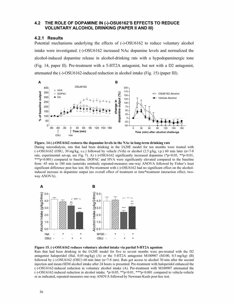
36
4.2 THE ROLE OF DOPAMINE IN (-)-OSU6162’S EFFECTS TO REDUCE VOLUNTARY ALCOHOL DRINKING (PAPER II AND III)
4.2.1 Results Potential mechanisms underlying the effects of (-)-OSU6162 to reduce voluntary alcohol
intake were investigated. (-)-OSU6162 increased NAc dopamine levels and normalized the
alcohol-induced dopamine release in alcohol-drinking rats with a hypodopaminergic tone
(Fig. 14, paper II). Pre-treatment with a 5-HT2A antagonist, but not with a D2 antagonist,
attenuated the (-)-OSU6162-induced reduction in alcohol intake (Fig. 15) (paper III).
Time (min)
% o
f b
ase
lin
e o
utp
ut
-90 -60 -30 0 30 60 90 120 150 1800
50
100
150
200
250
300
350
400
DOPAC
HVA
DA
OSU Veh
OSU6162
**
***
***
** *** ** *** **** * *
*** ** **
Time (min) after alcohol challenge
ch
an
ge in
do
pam
ine o
utp
ut
(%)
0 30 60 90 120 150 180-50
-25
0
25
50
75
100
125
150
175
200
OSU6162-Alcohol
Vehicle-Alcohol
A B
Figure. 14 (-)-OSU6162 restores the dopamine levels in the NAc in long-term drinking rats During microdialysis, rats that had been drinking in the IA20E model for ten months were treated with (-)-OSU6162 (OSU, 30 mg/kg, s.c.) followed by vehicle (Veh) or alcohol (2.5 g/kg, i.p.) 60 min later (n=7-8 rats, experimental set-up, see Fig. 7). A) (-)-OSU6162 significantly increased dopamine (*p<0.05, **p<0.01, ***p<0.001) compared to baseline. DOPAC and HVA were significantly elevated compared to the baseline from -45 min to 180 min (asterisks omitted); repeated-measures one-way ANOVA followed by Fisher’s least significant difference post hoc test. B) Pre-treatment with (-)-OSU6162 had no significant effect on the alcohol-induced increase in dopamine output (no overall effect of treatment or time*treatment interaction effect, two-way ANOVA).
Alc
ohol I
nta
ke (g/k
g/2
4hrs
)
0.0
1.0
2.0
3.0
4.0
5.0
**
***
***
Hal: -
OSU: -
+
-
-
+
+
+
Alc
oho
l Inta
ke (g
/kg
/24
hrs
)
0.0
1.0
2.0
3.0
4.0
5.0
***
**
M100: -
OSU: -
+
-
-
+
+
+
*
A B
Figure 15. (-)-OSU6162 reduces voluntary alcohol intake via partial 5-HT2A agonism Rats that had been drinking in the IA20E model for five to sevent months were pre-treated with the D2 antagonist haloperidol (Hal, 0.05 mg/kg) (A) or the 5-HT2A antagonist M100907 (M100, 0.5 mg/kg) (B) followed by (-)-OSU6162 (OSU) 60 min later (n=7-8 rats). Rats got access to alcohol 30 min after the second injection and mean±SEM alcohol intake after 24 hours is presented. Pre-treatment with haloperidol enhanced the (-)-OSU6162-induced reduction in voluntary alcohol intake (A). Pre-treatment with M100907 attenuated the (-)-OSU6162-induced reduction in alcohol intake. *p<0.05, **p<0.01, ***p<0.001 compared to vehicle-vehicle or as indicated, repeated-measures one-way ANOVA followed by Newman-Keuls post-hoc test.

37
4.2.2 Discussion
The results described in the previous section indicate that the effects of (-)-OSU6162 on the
dopamine system, as well as 5-HT2AR partial agonism, are involved in the reduction in
voluntary alcohol intake by (-)-OSU6162.
The (-)-OSU6162-induced increase in dopamine levels might underlie its previously found
ability to reduce voluntary alcohol intake (Steensland et al, 2012). This suggestion is
supported by a study showing that optogenetically-induced tonic stimulation of dopamine
neurons in the VTA produces stable dopamine elevations in the NAc and dramatically
decreases voluntary alcohol intake in rats that had been drinking in the IA20E model for
seven weeks (Bass et al, 2013). Furthermore, rats self-administer just enough alcohol to
restore the dopamine baseline that had been downregulated by chronic alcohol intake (Weiss
et al, 1996), indicating that an increase in dopamine levels could potentially reduce the need
for this compensation. Furthermore, transient over-expression of D2R in the NAc also
reduces alcohol consumption (Thanos et al, 2004; Thanos et al, 2001). Together, these
studies indicate that increasing dopamine transmission can reduce voluntary alcohol intake.
However, to treat patients pharmacological compounds are needed. Indeed, the dopamine-
releasing drug amphetamine reduces voluntary alcohol intake in animals (Feduccia et al,
2014; Halladay et al, 1999; Pohorecky and Sweeny, 2012), but is obviously a limited
treatment option due to its abuse potential. This abuse potential might be caused by a
significant peak-like dopamine increase induced by amphetamine. The anti-smoking
medication varenicline (agonist/partial agonist at several nicotinic acetylcholine receptor)
reduces alcohol drinking in rats (Steensland et al, 2007) and humans (Litten et al, 2013;
McKee et al, 2009; Mitchell et al, 2012) and was recently shown to increase dopamine output
in the NAc in rats (Feduccia et al, 2014). Similar to (-)-OSU6162 in the present thesis,
varenicline seems to moderately increase dopamine in the NAc of rats and maintains stable
levels for at least 2.5 hours. Especially, the long-lasting (at least four hours) increase in
dopamine levels by (-)-OSU6162 could explain the reductions of 24-hour alcohol intake
(Steensland et al, 2012). In conclusion, the increase in dopaminergic tone by (-)-OSU6162
might underlie the effects of (-)-OSU6162 to reduce alcohol drinking in rats and indicate a
potential for (-)-OSU6162 to reduce alcohol consumption in AUD.
(-)-OSU6162 is thought to increase dopamine by antagonizing D2R autoreceptors (Carlsson
and Carlsson, 2006). In fact, the lack of catalepsy in rats even at 80% of receptor occupancy
(Natesan et al, 2006), as well as the moderate occupancy in humans (Tolboom et al, 2014)
might indicate that (-)-OSU6162 does not bind to synaptic D2Rs. Furthermore, in vitro

38
studies have suggested that (-)-OSU6162 has a low affinity for the D2R and a fast
dissociation rate (Dyhring et al, 2010), which might contribute to its preference for
extrasynaptic D2R. Extrasynaptic receptors have a higher affinity for dopamine than synaptic
receptors since they are usually exposed to lower dopamine concentrations (Carlsson and
Carlsson, 2006).
Dopamine releasing compounds could be abused and cause addiction. However, the findings
of my colleague Dr. Fredriksson that (-)-OSU6162 does not induce conditioned place
preference in alcohol-naïve or alcohol-drinking rats (Paper II), indicate that (-)-OSU6162 is
not rewarding by itself. Furthermore, a fast increase in dopamine levels and not the absolute
dopamine levels seem to be important for drug-induced euphoria, which has been suggested
by studies showing that intravenously injected methylphenidate, but not oral methylphenidate
causes a feeling of ‘high’ (Swanson and Volkow, 2003). Swanson and Volkow suggested that
the intravenous administration mimics phasic dopamine release, while the oral administration
mimics tonic dopamine release. In the present thesis, following treatment with (-)-OSU6162
dopamine levels were slowly increasing and then plateauing for several hours, indicating less
risk for abuse liability. Moreover, none of the alcohol-dependent patients in a proof-of-
concept human laboratory study (Khemiri et al, 2015) reported wanting more of the
(-)-OSU6162 tablets and only one patient reported euphoric effects, although even one patient
in the placebo group did so (unpublished observation).
Increased dopamine levels can activate both D1R and D2R. One study in alcohol-preferring
rats showed that the D2 agonist quinpirole was more effective in reducing voluntary alcohol
intake than a D2 antagonist, although D1 agonist and antagonists also showed some effect
(Dyr et al, 1993). However, in another study quinpirole did not affect alcohol consumption
but increased water consumption (Linseman, 1990). Furthermore, the increased alcohol
intake in dopamine-deficient mice (induced by MDMA) could be attenuated by a D1 agonist,
which in turn, could be abolished by a D1 antagonist (Izco et al, 2007). Hence, both D1 and
D2 transmission could be involved in (-)-OSU6162’s effect on voluntary alcohol intake.
To investigate the role of postsynaptic D2R stimulation in (-)-OSU6162’s ability to reduce
voluntary alcohol intake, I pre-treated the rats with the D2 antagonist haloperidol and
(-)-OSU6162 before access to alcohol was given. This idea is based on a study showing that
haloperidol pre-treatment can attenuate (-)-OSU6162’s stimulating effect on motor activity in
rats with a suspected low dopaminergic tone due to arena-habituation (Carlsson et al, 2011).
However, haloperidol potentiated the (-)-OSU6162-induced decrease in voluntary alcohol
intake in the present thesis. One possibility is that haloperidol reaches the synaptic D2Rs that

39
(-)-OSU6162 cannot reach and thereby, through additional D2R antagonism reduces alcohol
intake. However, we used a low dose of haloperidol that would not affect alcohol or water
intake (pilot study, data not shown) and indeed in the present data set, haloperidol alone had
only a small effect on alcohol intake. In fact, systemically-administered D2 antagonists
reduce water intake at lower doses than those affecting alcohol intake in a two-bottle choice
paradigm (Goodwin et al, 1996; Linseman, 1990; Silvestre et al, 1996). Another possibility is
that haloperidol and (-)-OSU6162 in combination antagonize a bigger part of the population
of D2R autoreceptors, leading to a prolonged dopamine release than either treatment alone.
Based on the found attenuation of the alcohol-induced dopamine release by (-)-OSU6162 in
alcohol-naïve rats (Steensland et al, 2012), we originally hypothesized that (-)-OSU6162 is
reducing the rewarding effects of alcohol via this mechanism. However, in long-term
drinking rats in the present thesis we found the opposite: a trend for a potentiation of the
alcohol-induced dopamine release. Although one could argue that this observation indicates
that (-)-OSU6162 increases alcohol reinforcement, our behavioral data on alcohol drinking
and alcohol seeking argue against this suggestion (Steensland et al, 2012). Furthermore,
varenicline pre-treatment prolonged the alcohol-induced dopamine release in long-term
drinking rats (Feduccia et al, 2014), further suggesting that a potentiation in alcohol-induced
dopamine release might not be associated with increased alcohol intake. I suggest that
(-)-OSU6162 rather normalizes a reduced dopamine response to alcohol caused by long-term
drinking. This effect might reduce the amount of alcohol necessary to restore the
downregulated dopamine baseline and therefore, reduce alcohol drinking.
The different effects of (-)-OSU6162 on the alcohol-induced dopamine release in alcohol-
naïve (Steensland et al, 2012) and long-term drinking rats in the present thesis, could further
indicate that (-)-OSU6162 affects the dopamine system differently depending on the
dopaminergic tone. Nevertheless, this difference in dopaminergic tone might not solely be
caused by alcohol intake, since the rats were of different ages and strains (both Wistar rats,
but different suppliers) in the two studies. Previous studies showed that (-)-OSU6162 has
dopamine-stabilizing properties, such as stabilizing motor activity based on the dopaminergic
tone, preventing dyskinesia induced by dopaminergic compounds without causing akinesia
(Ekesbo et al, 2000; Ekesbo et al, 1997) or stabilizing L-[11C]DOPA influx depending on the
baseline (Tedroff et al, 1998). However, it is not completely understood how (-)-OSU6162
mediates these effects. Potentially, both dopamine-releasing, as well as inhibition of D2R
transmission, possibly at extrasynaptic D2R receptors are involved in these effects, but also
allosteric mechanisms have been suggested by in vitro studies (Kara et al, 2010; Lahti et al,
2007).

40
(-)-OSU6162 is not only a D2 antagonist but also a partial agonist at the 5-HT2AR (Burstein
et al, 2011). The 5-HT2A antagonist M100907 could prevent the (-)-OSU6162-induced
increase in motor activity in reserpinized-mice and arena-habituated rats (Carlsson et al,
2011). Similarly, in the present thesis, M100907 attenuated the (-)-OSU6162 induced
decrease in voluntary alcohol intake, indicating a role of partial 5-HT2A agonism in
(-)-OSU6162’s effect to reduce voluntary alcohol intake. The effects of 5-HT2A antagonists
on dopamine release (Yamamoto and Meltzer, 1992) and dopamine neuron firing (Grenhoff
et al, 1990), indicate that modulating the 5-HT2AR activity could influence NAc dopamine
levels. Therefore, 5-HT2AR partial agonism of (-)-OSU6162 might contribute to its
stabilizing effects on dopamine activity (Ekesbo et al, 1997; Natesan et al, 2006; Rung et al,
2011; Tedroff et al, 1998) and in turn, reduce voluntary alcohol intake (Steensland et al,
2012).
Although this thesis has focused on the ventral striatum (especially the NAc) the effects of
(-)-OSU6162 might also involve the dorsal striatum. A recent chemogenetic study showed
that both stimulation of D1-medium spinal neurons (MSNs) and inhibition of D2-MSNs
reduces voluntary alcohol intake in mice, implicating a role of both the direct and indirect
dopaminergic pathway in alcohol consumption (Cheng et al, 2017). (-)-OSU6162 has been
shown to release dopamine in the dorsal striatum in rats (Sonesson et al, 1994) and displace
the D2R radioligand raclopride in the dorsal striatum of humans (Tolboom et al, 2014). Thus,
the (-)-OSU6162-induced dopamine release or additional extrasynaptic D2R antagonism by
(-)-OSU6162 in the striatum could also underlie the effects of (-)-OSU6162 on alcohol
intake.

41
4.3 THE POTENTIAL OF (-)-OSU6162 TO REDUCE BINGE-EATING (PAPER IV)
4.3.1 Results
(-)-OSU6162 significantly decreased binge-like intake of palatable food, but not the prior
self-restriction of normal chow in anticipation of this food, i.e. (-)-OSU6162 has no effect on
the anticipatory negative contrast (Fig. 16A, for binge-eating model, see Fig. 8). Moreover,
(-)-OSU6162 reduced chow intake in the control group that never got access to palatable food
and therefore consumed higher amounts of chow (data not shown). (-)-OSU6162 reduced the
cue-controlled seeking of palatable food under a second-order schedule of reinforcement
during the first interval, i.e. before the first reward delivery (Fig. 16B). These results were
replicated when (-)-OSU6162 was infused in the NAc core (Fig. 16C).
0 5 10 15 300
10
20
30
40
(-)-OSU6162 (mg/kg, sc)
Ch
ow
or
Su
cro
se
inta
ke (
kcal)
chow (1st feeder)
sucrose (2nd feeder)
***a
cti
ve
le
ve
r p
res
se
s
Veh 5 10 150
200
400
600
800
**
OSU6162 (mg/kg)a
ctiv
e le
ve
r p
res
se
s
Veh 0.5 1.5 50
200
400
600
800
**
OSU6162 (µg/side)
*
A B C
Figure 16. (-)-OSU6162 reduces binge-like eating and cue-controlled seeking of chocolate-flavored sucrose (A) (-)-OSU6162 reduces binge-like intake of chocolate-flavored sucrose pellets, but does not affect the anticipatory negative contrast in prior chow intake (see model Fig. 8). Values represent mean ±SEM food intake (n=10). *p<0.05. ** p<0.01 repeated-measures one-way ANOVA followed by within-subject contrast to vehicle. Systemic (B, n=8)) and intra-NAc core (C, n=15) administration of (-)-OSU6162 significantly reduced seeking of chocolate-flavored sucrose pellets under a second-order schedule of reinforcement during the first interval, before reward delivery (see model Fig. 9). Values represent mean ±SEM lever presses. *p<0.05, **p<0.01 repeated-measures one-way ANOVA followed by within-subject contrast to vehicle.
4.3.2 Discussion
(-)-OSU6162 reduced both the binge-like consumption of palatable food, as well as
motivation to obtain this food as indicated by the effect on cue-controlled seeking. This effect
on both consummatory and appetitive behaviors indicate the potential of (-)-OSU6162 as a
novel treatment for BED.
The effects of (-)-OSU6162 on binge-like eating of sucrose found in this thesis confirm
previous findings of a reduction in voluntary intake of a sucrose solution (Steensland et al,
2012). Furthermore, since (-)-OSU6162 reduces intake of sucrose and alcohol, but not of a
salt water solution, (-)-OSU6162 might specifically reduce the intake of rewarding
substances. However, in the present thesis, (-)-OSU6162 reduced also the intake of regular

42
chow in control rats of the binge-eating paradigm, arguing against a suggested specificity for
palatable food. However, the chow intake in the binge-eating rats was not affected by
(-)-OSU6162 and neither did (-)-OSU6162 affect the consumption of the palatable food
received after each interval of the second-order schedule of reinforcement. Since both this
chow intake and this sucrose intake was low, I suggest that (-)-OSU6162 only affects high
levels of food intake. Similarly, (-)-OSU6162 reduced alcohol intake only in rats voluntarily
drinking high, but not low, amounts of alcohol (Steensland et al, 2012).
The effects of (-)-OSU6162 on sucrose seeking already during the first interval of the second-
order schedule of reinforcement, before the food reward is delivered, indicates that
(-)-OSU6162 is able to directly affect the motivation to obtain the reward. Instead,
compounds that only affect the second interval indicate an indirect effect on motivation
through modulation of the hedonic properties of the reward upon ingestion. Furthermore, the
finding that (-)-OSU6162 is more specific in reducing cue-controlled seeking under a second-
order schedule, but not simpler schedules of reinforcement, in contrast to the D2 antagonist
raclopride (see paper IV), indicates that (-)-OSU6162 might be especially beneficial in BED.
Obese patients with BED display enhanced cue-induced dopamine activation than obese
patients without BED (Wang et al, 2011).
These effects of (-)-OSU6162 on sucrose seeking are in line with the effects of (-)-OSU6162
on alcohol seeking under various schedules revealed by my colleague Dr. Fredriksson
(Steensland et al, 2012). Furthermore, the finding that (-)-OSU6162 does not affect a simpler
schedule of reinforcement (paper IV) further strengthens our observations of lack of general
motor-inhibiting or sedative effects that could account for an unspecific reduction in lever
pressing. For example, (-)-OSU6162 increased the inactive lever press in the cue/priming-
induce reinstatement test of alcohol seeking (Steensland et al, 2012) and did not affect the
latency to respond to a stimulus or collect the reward in a 5-choice serial reaction time task
(unpublished observations by Fredriksson and co-workers).
The fact that (-)-OSU6162 infused into the NAc core replicates the findings of systemic
administration on seeking under a second-order schedule of reinforcement, confirms a role of
dopamine in the NAc on (-)-OSU6162’s effects on cue-controlled food seeking. However,
the exact mechanism, especially the role of dopamine release, D1R and D2R transmission, as
well as 5-HT2A partial agonism, in these found behavioral effects of (-)-OSU6162 should be
further investigated.

43
5 GENERAL CONCLUSIONS
The monoamine stabilizer (-)-OSU6162 has recently been identified as a potential novel
treatment for AUD. (-)-OSU6162 reduces voluntary alcohol drinking, seeking and
withdrawal symptoms in long-term drinking rats (Steensland et al, 2012)
In the present thesis, long-term voluntary alcohol drinking downregulated the dopamine
system and (-)-OSU6162 counteracted such dopaminergic deficits (for proposed model of
mechanism, see Fig. 17). First, the finding that alcohol drinking reduces dopamine levels in
the NAc is in line with previous studies. Second, the alcohol-induced reduction on D2R
expression is adding to the relatively scarce literature on the effects of alcohol drinking on
D2R levels using validated rodent alcohol models. Furthermore, these results indicate that
reduced striatal D2R levels found in alcohol-dependent individuals during abstinence might
be directly caused by alcohol drinking. Third, the effects of alcohol drinking on the density of
D2R receptor complexes in situ have not been investigated previously. Hence, a D2R
receptor complex-specific change has been demonstrated with the present thesis for the first
time. Moreover, increased A2AR-D2R heteroreceptor complexes might provide a novel
treatment target for AUD. Finally, (-)-OSU6162 slowly elevated the dopaminergic tone in the
NAc to a new plateau. This effect is likely underlying the ability of (-)-OSU6162 to reduce
voluntary alcohol drinking. In fact, several studies show that increasing the dopaminergic
tone (e.g. via optogenetics) decreases voluntary alcohol drinking. Finally, pre-treatment with
a 5-HT2A antagonist attenuated the (-)-OSU6162-induced reduction in voluntary alcohol
intake, indicating an additional role of partial agonism at this receptor in (-)-OSU6162’s
effects.
Figure 17. Role of dopamine transmission in the ability of (-)-OSU6162 to reduce alcohol drinking
Left: Acute alcohol intake releases dopamine in the NAc and stimulates synaptic and extrasynaptic receptors. Released dopamine binds to presynaptic D2R autoreceptors, inhibiting further dopamine release Middle: Long-term alcohol drinking downregulates the dopamine system demonstrated by decreased extracellular dopamine levels, reduced densities of D2R-D2R homoreceptor complexes (likely caused by decreased D2R gene expression) and increased densities of A2A-D2R heteroreceptor complexes (probably decreasing D2R affinity through reciprocal receptor antagonism). Right: (-)-OSU6162 (light green dot) increases dopamine release via D2R autoreceptor antagonism (at the axon terminal, increasing dopamine release, and/or at the soma, increasing dopamine firing and release). This slow increase in dopamine levels might abolish the need to drink alcohol in order to restore the dopamine system. (-)-OSU6162 might also antagonize extrasynaptic receptors (homo- or heteroreceptor complexes) located on the medium spinal neurons, which could inhibit alcohol-reinforcement.

44
6 FUTURE STUDIES
Several questions remain regarding the mechanisms of (-)-OSU6162’s reduction in alcohol
drinking and other alcohol-mediated behaviors, which should be investigated in future
studies.
First, the role of the sigma1 receptor in the (-)-OSU6162-induced reduction in voluntary
alcohol intake should be investigated. (-)-OSU6162 has shown a high affinity for the sigma1
receptor (Sahlholm et al, 2013). This receptor can form a complex with the long isoform of
the D2R receptor (Borroto-Escuela et al, 2017; Navarro et al, 2013) and is often a co-receptor
in different D2R heteroreceptor-complexes (Borroto-Escuela et al, 2016; Navarro et al,
2013). Moreover, in paper I, a reduction in the density of sigma1-D2R in the dorsal striatum
by alcohol drinking has been found. A similar reduction has also been found following
chronic cocaine intake (Borroto-Escuela et al, 2017; Navarro et al, 2013) and the sigma1
receptor is thought to play a role in cocaine’s inhibition of D2 signaling (Borroto-Escuela et
al, 2017; Navarro et al, 2013). Furthermore, a sigma1 antagonist was shown to reduce
voluntary alcohol intake (Blasio et al, 2015).
Second, the role of the dorsal striatum in the effects of (-)-OSU6162 to reduce alcohol
drinking, but especially alcohol seeking, should be investigated. (-)-OSU6162 releases
dopamine in the dorsal striatum (Sonesson et al, 1994; Tolboom et al, 2014) and reduces
dyskinesia in a rat model of Parkinson’s disease (Ekesbo et al, 1997). Moreover, the dorsal
striatum is involved in habitual alcohol (Corbit and Janak, 2016; Corbit et al, 2014) and
cocaine seeking (Belin and Everitt, 2008; Murray et al, 2012). Furthermore, as mentioned
above chronic alcohol drinking (paper I) and cocaine intake (Borroto-Escuela et al, 2017;
Navarro et al, 2013) affects the density of D2R heteroreceptor complexes in the dorsal
striatum.
Third, the effect of chronic alcohol intake on signaling and interaction between D2R-
heteroreceptor complexes, such as the A2AR-D2R (Pintsuk et al, 2016) or D2R-5-HT2A
receptor complex (Borroto-Escuela et al, 2010b), should be further investigated. Moreover,
the effect of (-)-OSU6162 treatment on the signaling of these heteroreceptor complexes
should be studied.
Fourth, the role of postsynaptic D1R and D2R in (-)-OSU6162-induced reduction of
voluntary alcohol intake and other alcohol-mediated behaviors should be investigated, as well
as the role of long-term alcohol intake on the D1R receptor.
Fifth, the effect of long-term drinking on the serotonergic system, as well as the effect of
5-HT2A partial agonism on the dopamine system, should be investigated. Finally, the role of
different brain areas, such as the prefrontal cortex, and their interaction to the striatum, should
be investigated in the effect of (-)-OSU6162 on alcohol-mediated behaviors.

45
7 ACKNOWLEDGEMENTS
Many people have contributed directly or indirectly to this thesis. I would like to thank
especially:
My main supervisor Pia Steensland for literally EVERYTHING. You gave me the
opportunity to do a PhD, provided me with an interesting project to work on and believed that
I could do it. I remember especially your encouragement in times of struggle and your help in
finding projects and collaborations here at KI and abroad. You gave me endless support and
were always there on every step of the way. I appreciate all your effort and I am truly proud
of what we accomplished together. I believe thanks to you I have grown a lot and I highly
value this experience to do a PhD. There is nothing like it.
My co-supervisor Björn Schilström for your support during the PhD and for inspiring me to
go into neuropharmacology research in the first place. You taught me a lot about psychiatry
research, which gave me with an invaluable wide-view. Thank you especially for advising me
to stay in psychiatry research and for your hands on support during my first study.
My co-supervisor Johan Franck for providing a great translational research environment and
for stressing the importance of basic and clinical research. Thank you for giving me a wider
perspective and insight on clinical challenges.
It was a special honor to collaborate with Arvid Carlsson. Without you and your generosity,
this thesis would not exist. Your accomplishments in medical science are a real inspiration.
Ida Fredriksson, my fellow PhD-student, for helping me with experiments, exchanging
ideas about science and the struggles with the experiments and for a great time in Italy! You
are very driven and also very kind and I wish you all the best for the future!
Malin Wirf, I am so happy to call you my dear colleague and friend. Your contribution to
our studies was huge and I could not have done it without you. It is so nice to be around you
at work and outside of it. We both became moms at almost the same time and it was nice that
we could share moments together with our kids. It is great to have someone to talk to about
literally everything Kram!
Nitya Jayaram-Lindström for lots of positive energy. You are a warm and funny and
cheered me up lots of days. You also have a great ability as a mentor and helped me figure
out some important decisions for my future career. After talking to you I always felt

46
motivated, inspired and also valuable and capable. Thank you also, for inviting great
speakers, creating an academic environment and widening my scientific perspective.
Other colleagues of the research group: Especially Christoffer Brynte and Anders
Hammarberg for our laughs together, Lotta Skoglund and Maija Konstenius for being
such warm and caring persons and also for being great role models, Joar Guterstam and
Lotfi Khemiri for discussing science, life and kids with me, offering me a clinical
perspective, inspiring me with exceptional talks and always being very kind., Camilla
Hellspong for lunch breaks and advice on daycare, strollers and all the stuff you gave us for
Juno. You are so kind and funny and I am so happy we have you back! and Sarah Holst for
being so positive and for being passionate about animal behavior.
Monica Aronsson, I am so fortunate to have worked with you. You were always there,
listening to all our daily troubles and always giving smart insight and, not less important
coffee ;). You are so nice to be around and I really value your advice. I also thank you,
Linnea Tankred and Thatiane de Oliveira Sergio for your invaluable work. I am happy to
have met you and we would not have known what to do without you. Helder André, thank
you for being supportive and generous in sharing valuable advice and your lab!
Kjell Fuxe for inviting me into your lab. Your never-ceasing enthusiasm about data and
research is very inspiring. I am really glad that you introduced me to the field of
heteroreceptor complexes! Dasiel Borroto Escuela for hard work and many hours on the
microscope, for brilliant scientific insight and for being extremely nice. Thank you for
making me feel welcome in the Fuxe lab!
Barry Everitt for inviting me to your lab! for helping me to find housing, inviting me to a
potluck at your house, for writing great reviews on the field, for your valuable insight and
feedback on paper IV and for the openness and kindness towards me.
Johan Alsiö for coming up with the project in the first place and for your valuable support in
our study. You are a brilliant researcher and I learned a lot from you. I really appreciate your
humor and positive spirit and I can’t wait to meet your daughter.
Chiara Giuliano for everything! Without you I would not have managed to do this project
within six months! You are also one of the most hard-working and kindest persons I ever
met! I especially remember us laughing together, complaining about the tough world of
academia, but I remember also the Spritz Aperols at your place! I wish you all the best!

47
My “office mates” and fellow PhD-students at Cambridge: especially Camilla D’Angelo for
many lunch breaks and memorable nights going out, Zara Goozee and George Vousden for
making my time in the office and outside of it so enjoyable.
Joëlle Rüegg, Louise (Lollo) Sjöholm and Tomas Ekström for inviting me so warmly to
your lab, introducing me to the field of epigenetics and helping with the project. I especially
enjoyed the weekly breakfast presentations at CMM.
My friends in Stockholm: Paula, Javi, Alfredo and Concha for all the laughs and all the
great food! You created truly special memories! To Nadim, Tina, Janina, Sophia,
Annett, Stephanie and Irène for lots of lunches, coffees, dinners and always being there
listening to me and laughing with me!
My sister Katrin and her family: Zahra, Catalina, Eli and Johannes for many Christmases
and summers together!
My previous colleagues: Åsa Konradsson-Geuken, Torun Malmlöf and Carl Björkholm
there is not enough space left for explaining how you have shaped me and I am so glad I met
you, both because of you as persons and scientists!
My mentor Ulrica Westerblad for listening and giving advice.
Johanna Gripenberg for inviting me so warmly into your research group and keeping me in
research. I am looking forward to doing research that directly can make an impact on society!
Other colleagues at STAD (to name a few): Erik von Essen, Tobias Elgán, Frida
Bergenblad, Unda Lönnqvist, Håkan Källmén, Nicklas Kartengren, Pia Kvillemo,
Kamilla Nylund, Maria Ingemarsson and Camilla Jalling. I enjoyed working and having
lunches with you and I really appreciate all the help and support you already have given me.
You really make me feel welcome!
Over the years I worked in many different places: R5 at KS; CPF, CMM, Neuroscience at
KI, St:Eriks in Stockholm, Swetox in Östertälje and the Experimental Psychology
Department in Cambridge. Thanks to all the other great people that have not yet been
properly acknowledged but that provided a nice environment and made me enjoy coming to
work every day
Thanks to my friends from FyFa for nice chats, lunches and great parties! Special thanks to
Igor Cervenka for creating the thesis cover picture!

48
Meinen Eltern Hartwich Feltmann, Ina Feltmann und Ursula Feltmann für eure
Unterstützung und Liebe.
A mi familia española, por todo el cariño y el apoyo durante todos estos años.
Last but not least, I want to thank my husband Vicente! I am unable to write with just a few
lines what you mean to me. You are my everything! I have to thank you for both daily
practical and moral support as well as opening my cultural world! The biggest gift you gave
me is our little sunshine Juno. You two mean more to me than anything in the world!
This work was supported by the Swedish Research Council, Karolinska Institutet’s Research
Funds, the Research Council of the Swedish Alcohol Retailing Monopoly, the Torsten
Söderberg Foundation, the Swedish Brain Foundation, the Swedish Research Council for
Health, Working Life and Welfare, the Swedish Society of Medicine, the Swedish
Pharmaceutical Society, the Swedish Society for Medical Research, AFA Insurance, Swedish
Research Council Formas and the Swedish Mental Health fund.

49
8 REFERENCES
Adams WK, Sussman JL, Kaur S, D'Souza A M, Kieffer TJ and Winstanley CA (2015). Long-term, calorie-restricted intake of a high-fat diet in rats reduces impulse control and ventral striatal D2 receptor signalling - two markers of addiction vulnerability. The European journal of neuroscience 42(12): 3095-3104.
Alonso J, Angermeyer MC, Bernert S, Bruffaerts R, Brugha TS, Bryson H, et al (2004). Use of mental health services in Europe: results from the European Study of the Epidemiology of Mental Disorders (ESEMeD) project. Acta psychiatrica Scandinavica. Supplementum (420): 47-54.
Alsio J, Olszewski PK, Norback AH, Gunnarsson ZE, Levine AS, Pickering C, et al (2010). Dopamine D1 receptor gene expression decreases in the nucleus accumbens upon long-term exposure to palatable food and differs depending on diet-induced obesity phenotype in rats. Neuroscience 171(3): 779-787.
American Psychiatric Association (1994). Substance-Related DisordersDiagnostic and Statistical Manual of Mental Disorders:Washington, DC.
American Psychiatric Association (2013a). Feeding and Eating DisordersDiagnostic and Statistical Manual of Mental Disorders:Washington, DC.
American Psychiatric Association (2013b). Substance-related and Addictive DisordersDiagnostic and Statistical Manual of Mental Disorders:Washington, DC.
Anton RF, Kranzler H, Breder C, Marcus RN, Carson WH and Han J (2008). A randomized, multicenter, double-blind, placebo-controlled study of the efficacy and safety of aripiprazole for the treatment of alcohol dependence. J Clin Psychopharmacol 28(1): 5-12.
Avena NM (2007). Examining the addictive-like properties of binge eating using an animal model of sugar dependence. Experimental and Clinical Psychopharmacology 15(5): 481-491.
Avena NM (2010). The study of food addiction using animal models of binge eating. Appetite 55(3): 734-737.
Avena NM, Bocarsly ME, Rada P, Kim A and Hoebel BG (2008). After daily bingeing on a sucrose solution, food deprivation induces anxiety and accumbens dopamine/acetylcholine imbalance. Physiol Behav 94(3): 309-315.
Avena NM, Gearhardt AN, Gold MS, Wang GJ and Potenza MN (2012). Tossing the baby out with the bathwater after a brief rinse? The potential downside of dismissing food addiction based on limited data. Nature Reviews: Neuroscience 13(7): 514; author reply 514.
Avena NM, Rada P, Moise N and Hoebel BG (2006). Sucrose sham feeding on a binge schedule releases accumbens dopamine repeatedly and eliminates the acetylcholine satiety response. Neuroscience 139(3): 813-820.
Barak S, Carnicella S, Yowell QV and Ron D (2011). Glial cell line-derived neurotrophic factor reverses alcohol-induced allostasis of the mesolimbic dopaminergic system: implications for alcohol reward and seeking. The Journal of neuroscience : the official journal of the Society for Neuroscience 31(27): 9885-9894.
Bass CE, Grinevich VP, Gioia D, Day-Brown JD, Bonin KD, Stuber GD, et al (2013). Optogenetic stimulation of VTA dopamine neurons reveals that tonic but not phasic patterns of dopamine transmission reduce ethanol self-administration. Frontiers in behavioral neuroscience 7: 173.

50
Bassareo V and Di Chiara G (1999). Modulation of feeding-induced activation of mesolimbic dopamine transmission by appetitive stimuli and its relation to motivational state. The European journal of neuroscience 11(12): 4389-4397.
Beaulieu JM and Gainetdinov RR (2011). The physiology, signaling, and pharmacology of dopamine receptors. Pharmacological Reviews 63(1): 182-217.
Becker HC and Mulholland PJ (2014). Neurochemical mechanisms of alcohol withdrawal. Handbook of Clinical Neurology 125: 133-156.
Beery AK and Zucker I (2011). Sex bias in neuroscience and biomedical research. Neurosci Biobehav Rev 35(3): 565-572.
Belin D and Everitt BJ (2008). Cocaine seeking habits depend upon dopamine-dependent serial connectivity linking the ventral with the dorsal striatum. Neuron 57(3): 432-441.
Bello NT and Hajnal A (2010). Dopamine and binge eating behaviors. Pharmacol Biochem Behav 97(1): 25-33.
Benaliouad F, Kapur S, Natesan S and Rompre PP (2009). Effects of the dopamine stabilizer, OSU-6162, on brain stimulation reward and on quinpirole-induced changes in reward and locomotion. European neuropsychopharmacology : the journal of the European College of Neuropsychopharmacology 19(6): 416-430.
Berner LA, Bocarsly ME, Hoebel BG and Avena NM (2011). Pharmacological interventions for binge eating: lessons from animal models, current treatments, and future directions. Curr Pharm Des 17(12): 1180-1187.
Berridge KC (2007). The debate over dopamine's role in reward: the case for incentive salience. Psychopharmacology 191(3): 391-431.
Berridge KC and Robinson TE (1998). What is the role of dopamine in reward: hedonic impact, reward learning, or incentive salience? Brain Research: Brain Research Reviews 28(3): 309-369.
Blasio A, Valenza M, Iyer MR, Rice KC, Steardo L, Hayashi T, et al (2015). Sigma-1 receptor mediates acquisition of alcohol drinking and seeking behavior in alcohol-preferring rats. Behavioural brain research 287: 315-322.
Boileau I, Assaad JM, Pihl RO, Benkelfat C, Leyton M, Diksic M, et al (2003). Alcohol promotes dopamine release in the human nucleus accumbens. Synapse 49(4): 226-231.
Borroto-Escuela DO, Marcellino D, Narvaez M, Flajolet M, Heintz N, Agnati L, et al (2010a). A serine point mutation in the adenosine A2AR C-terminal tail reduces receptor heteromerization and allosteric modulation of the dopamine D2R. Biochemical and Biophysical Research Communications 394(1): 222-227.
Borroto-Escuela DO, Narvaez M, Wydra K, Pintsuk J, Pinton L, Jimenez-Beristain A, et al (2017). Cocaine self-administration specifically increases A2AR-D2R and D2R-sigma1R heteroreceptor complexes in the rat nucleus accumbens shell. Relevance for cocaine use disorder. Pharmacol Biochem Behav 155: 24-31.
Borroto-Escuela DO, Romero-Fernandez W, Tarakanov AO, Ciruela F, Agnati LF and Fuxe K (2011). On the existence of a possible A2A-D2-beta-Arrestin2 complex: A2A agonist modulation of D2 agonist-induced beta-arrestin2 recruitment. Journal of Molecular Biology 406(5): 687-699.
Borroto-Escuela DO, Romero-Fernandez W, Tarakanov AO, Marcellino D, Ciruela F, Agnati LF, et al (2010b). Dopamine D2 and 5-hydroxytryptamine 5-HT((2)A) receptors assemble

51
into functionally interacting heteromers. Biochemical and Biophysical Research Communications 401(4): 605-610.
Borroto-Escuela DO, Wydra K, Pintsuk J, Narvaez M, Corrales F, Zaniewska M, et al (2016). Understanding the Functional Plasticity in Neural Networks of the Basal Ganglia in Cocaine Use Disorder: A Role for Allosteric Receptor-Receptor Interactions in A2A-D2 Heteroreceptor Complexes. Neural Plasticity 2016: 4827268.
Bunney BS, Walters JR, Roth RH and Aghajanian GK (1973). Dopaminergic neurons: effect of antipsychotic drugs and amphetamine on single cell activity. The Journal of pharmacology and experimental therapeutics 185(3): 560-571.
Burstein ES, Carlsson ML, Owens M, Ma JN, Schiffer HH, Carlsson A, et al (2011). II. In vitro evidence that (-)-OSU6162 and (+)-OSU6162 produce their behavioral effects through 5-HT2A serotonin and D2 dopamine receptors. J Neural Transm 118(11): 1523-1533.
Button KS, Ioannidis JP, Mokrysz C, Nosek BA, Flint J, Robinson ES, et al (2013). Power failure: why small sample size undermines the reliability of neuroscience. Nature Reviews: Neuroscience 14(5): 365-376.
Cahill L (2006). Why sex matters for neuroscience. Nature Reviews: Neuroscience 7(6): 477-484.
Carlier N, Marshe VS, Cmorejova J, Davis C and Muller DJ (2015). Genetic Similarities between Compulsive Overeating and Addiction Phenotypes: A Case for "Food Addiction"? Curr Psychiatry Rep 17(12): 96.
Carlsson A (1959). The occurrence, distribution and physiological role of catecholamines in the nervous system. Pharmacological Reviews 11(2, Part 2): 490-493.
Carlsson A and Carlsson ML (2006). A dopaminergic deficit hypothesis of schizophrenia: the path to discovery. Dialogues in Clinical Neuroscience 8(1): 137-142.
Carlsson A, Lindqvist M and Magnusson T (1957). 3,4-Dihydroxyphenylalanine and 5-hydroxytryptophan as reserpine antagonists. Nature 180(4596): 1200.
Carlsson ML, Burstein ES, Kloberg A, Hansson S, Schedwin A, Nilsson M, et al (2011). I. In vivo evidence for partial agonist effects of (-)-OSU6162 and (+)-OSU6162 on 5-HT2A serotonin receptors. J Neural Transm 118(11): 1511-1522.
Carnicella S, Ron D and Barak S (2014). Intermittent ethanol access schedule in rats as a preclinical model of alcohol abuse. Alcohol 48(3): 243-252.
Cassin SE and von Ranson KM (2007). Is binge eating experienced as an addiction? Appetite 49(3): 687-690.
Chartier KG, Hesselbrock MN and Hesselbrock VM (2010). Development and vulnerability factors in adolescent alcohol use. Child and Adolescent Psychiatric Clinics of North America 19(3): 493-504.
Cheng Y, Huang CCY, Ma T, Wei X, Wang X, Lu J, et al (2017). Distinct Synaptic Strengthening of the Striatal Direct and Indirect Pathways Drives Alcohol Consumption. Biol Psychiatry 81(11): 918-929.
Cloninger CR, Bohman M and Sigvardsson S (1981). Inheritance of alcohol abuse. Cross-fostering analysis of adopted men. Arch Gen Psychiatry 38(8): 861-868.

52
Cohen E, Feinn R, Arias A and Kranzler HR (2007). Alcohol treatment utilization: findings from the National Epidemiologic Survey on Alcohol and Related Conditions. Drug and Alcohol Dependence 86(2-3): 214-221.
Colantuoni C, Schwenker J, McCarthy J, Rada P, Ladenheim B, Cadet JL, et al (2001). Excessive sugar intake alters binding to dopamine and mu-opioid receptors in the brain. Neuroreport 12(16): 3549-3552.
Connor JP and Hall W (2015). Alcohol burden in low-income and middle-income countries. Lancet 386(10007): 1922-1924.
Corbett D and Wise RA (1979). Intracranial self-stimulation in relation to the ascending noradrenergic fiber systems of the pontine tegmentum and caudal midbrain: a moveable electrode mapping study. Brain research 177(3): 423-436.
Corbit LH and Janak PH (2016). Changes in the Influence of Alcohol-Paired Stimuli on Alcohol Seeking across Extended Training. Front Psychiatry 7: 169.
Corbit LH, Nie H and Janak PH (2014). Habitual responding for alcohol depends upon both AMPA and D2 receptor signaling in the dorsolateral striatum. Frontiers in behavioral neuroscience 8: 301.
Corwin RL (2006). Bingeing rats: a model of intermittent excessive behavior? Appetite 46(1): 11-15.
Cotton NS (1979). The familial incidence of alcoholism: a review. Journal of Studies on Alcohol 40(1): 89-116.
Cottone P, Sabino V, Steardo L and Zorrilla EP (2008). Opioid-dependent anticipatory negative contrast and binge-like eating in rats with limited access to highly preferred food. Neuropsychopharmacology 33(3): 524-535.
Cragg SJ and Rice ME (2004). DAncing past the DAT at a DA synapse. Trends in Neurosciences 27(5): 270-277.
Curtis C and Davis C (2014). A qualitative study of binge eating and obesity from an addiction perspective. Eat Disord 22(1): 19-32.
Czachowski CL, Chappell AM and Samson HH (2001). Effects of raclopride in the nucleus accumbens on ethanol seeking and consumption. Alcohol Clin Exp Res 25(10): 1431-1440.
Dahlström A and Fuxe K (1964). Evidence for the Existence of Monoamine-Containing Neurons in the Central Nervous System. I. Demonstration of Monoamines in the Cell Bodies of Brain Stem Neurons. Acta Physiologica Scandinavica. Supplementum: SUPPL 232:231-255.
Dal Toso R, Sommer B, Ewert M, Herb A, Pritchett DB, Bach A, et al (1989). The dopamine D2 receptor: two molecular forms generated by alternative splicing. EMBO Journal 8(13): 4025-4034.
Dalley JW, Everitt BJ and Robbins TW (2011). Impulsivity, compulsivity, and top-down cognitive control. Neuron 69(4): 680-694.
Davis C, Fattore L, Kaplan AS, Carter JC, Levitan RD and Kennedy JL (2012). The suppression of appetite and food consumption by methylphenidate: the moderating effects of gender and weight status in healthy adults. Int J Neuropsychopharmacol 15(2): 181-187.
de Wit H (2009). Impulsivity as a determinant and consequence of drug use: a review of underlying processes. Addict Biol 14(1): 22-31.

53
Di Chiara G (2002). Nucleus accumbens shell and core dopamine: differential role in behavior and addiction. Behavioural brain research 137(1-2): 75-114.
Di Chiara G and Imperato A (1988). Drugs abused by humans preferentially increase synaptic dopamine concentrations in the mesolimbic system of freely moving rats. Proc Natl Acad Sci U S A 85(14): 5274-5278.
Di Chiara G, Tanda G, Bassareo V, Pontieri F, Acquas E, Fenu S, et al (1999). Drug addiction as a disorder of associative learning. Role of nucleus accumbens shell/extended amygdala dopamine. Annals of the New York Academy of Sciences 877: 461-485.
Diana M (2011). The dopamine hypothesis of drug addiction and its potential therapeutic value. Front Psychiatry 2: 64.
Diana M, Pistis M, Carboni S, Gessa GL and Rossetti ZL (1993). Profound decrement of mesolimbic dopaminergic neuronal activity during ethanol withdrawal syndrome in rats: electrophysiological and biochemical evidence. Proc Natl Acad Sci U S A 90(17): 7966-7969.
Ding ZM, Ingraham CM, Rodd ZA and McBride WJ (2016). Alcohol drinking increases the dopamine-stimulating effects of ethanol and reduces D2 auto-receptor and group II metabotropic glutamate receptor function within the posterior ventral tegmental area of alcohol preferring (P) rats. Neuropharmacology 109: 41-48.
Doyon WM, Anders SK, Ramachandra VS, Czachowski CL and Gonzales RA (2005). Effect of operant self-administration of 10% ethanol plus 10% sucrose on dopamine and ethanol concentrations in the nucleus accumbens. J Neurochem 93(6): 1469-1481.
du Hoffmann J and Nicola SM (2014). Dopamine invigorates reward seeking by promoting cue-evoked excitation in the nucleus accumbens. The Journal of neuroscience : the official journal of the Society for Neuroscience 34(43): 14349-14364.
Dyhring T, Nielsen EO, Sonesson C, Pettersson F, Karlsson J, Svensson P, et al (2010). The dopaminergic stabilizers pridopidine (ACR16) and (-)-OSU6162 display dopamine D(2) receptor antagonism and fast receptor dissociation properties. European journal of pharmacology 628(1-3): 19-26.
Dyr W, McBride WJ, Lumeng L, Li TK and Murphy JM (1993). Effects of D1 and D2 dopamine receptor agents on ethanol consumption in the high-alcohol-drinking (HAD) line of rats. Alcohol 10(3): 207-212.
Ekesbo A, Andren PE, Gunne LM, Sonesson C and Tedroff J (2000). Motor effects of (-)-OSU6162 in primates with unilateral 6-hydroxydopamine lesions. European journal of pharmacology 389(2-3): 193-199.
Ekesbo A, Andren PE, Gunne LM and Tedroff J (1997). (-)-OSU 6162 inhibits levodopa-induced dyskinesias in a monkey model of Parkinson's disease. Neuroreport 8(11): 2567-2570.
Ekesbo A, Torstenson R, Hartvig P, Carlsson A, Sonesson C, Waters N, et al (1999). Effects of the substituted (S)-3-phenylpiperidine (-)-OSU6162 on PET measurements of [11C]SCH23390 and [11C]raclopride binding in primate brains. Neuropharmacology 38(3): 331-338.
Engdahl E, Dunn N and Fogdell-Hahn A (2016). Investigation of reference gene expression during human herpesvirus 6B infection indicates peptidylprolyl isomerase A as a stable reference gene and TATA box binding protein as a gene up-regulated by this virus. Journal of Virological Methods 227: 47-49.

54
Engel JA and Jerlhag E (2014). Alcohol: mechanisms along the mesolimbic dopamine system. Progress in Brain Research 211: 201-233.
Ericson M, Blomqvist O, Engel JA and Soderpalm B (1998). Voluntary ethanol intake in the rat and the associated accumbal dopamine overflow are blocked by ventral tegmental mecamylamine. European journal of pharmacology 358(3): 189-196.
Everitt BJ (2014). Neural and psychological mechanisms underlying compulsive drug seeking habits and drug memories--indications for novel treatments of addiction. The European journal of neuroscience 40(1): 2163-2182.
Everitt BJ, Cador M and Robbins TW (1989). Interactions between the amygdala and ventral striatum in stimulus-reward associations: studies using a second-order schedule of sexual reinforcement. Neuroscience 30(1): 63-75.
Everitt BJ and Robbins TW (2000). Second-order schedules of drug reinforcement in rats and monkeys: measurement of reinforcing efficacy and drug-seeking behaviour. Psychopharmacology (Berl) 153(1): 17-30.
Fahlke C, Hansen S, Engel JA and Hard E (1994). Effects of ventral striatal 6-OHDA lesions or amphetamine sensitization on ethanol consumption in the rat. Pharmacol Biochem Behav 47(2): 345-349.
Feduccia AA, Simms JA, Mill D, Yi HY and Bartlett SE (2014). Varenicline decreases ethanol intake and increases dopamine release via neuronal nicotinic acetylcholine receptors in the nucleus accumbens. British journal of pharmacology 171(14): 3420-3431.
Fenton MC, Geier T, Keyes K, Skodol AE, Grant BF and Hasin DS (2013). Combined role of childhood maltreatment, family history, and gender in the risk for alcohol dependence. Psychological Medicine 43(5): 1045-1057.
Fineberg NA, Potenza MN, Chamberlain SR, Berlin HA, Menzies L, Bechara A, et al (2010). Probing compulsive and impulsive behaviors, from animal models to endophenotypes: a narrative review. Neuropsychopharmacology 35(3): 591-604.
Ford MM, Eldridge JC and Samson HH (2002). Microanalysis of ethanol self-administration: estrous cycle phase-related changes in consumption patterns. Alcohol Clin Exp Res 26(5): 635-643.
Forger NG and Morin LP (1982). Reproductive state modulates ethanol intake in rats: effects of ovariectomy, ethanol concentration, estrous cycle and pregnancy. Pharmacol Biochem Behav 17(2): 323-331.
Fuxe K, Borroto-Escuela DO, Romero-Fernandez W, Palkovits M, Tarakanov AO, Ciruela F, et al (2014a). Moonlighting proteins and protein-protein interactions as neurotherapeutic targets in the G protein-coupled receptor field. Neuropsychopharmacology 39(1): 131-155.
Fuxe K, Tarakanov A, Romero Fernandez W, Ferraro L, Tanganelli S, Filip M, et al (2014b). Diversity and Bias through Receptor-Receptor Interactions in GPCR Heteroreceptor Complexes. Focus on Examples from Dopamine D2 Receptor Heteromerization. Frontiers in Endocrinology 5: 71.
Garavan H, Pankiewicz J, Bloom A, Cho JK, Sperry L, Ross TJ, et al (2000). Cue-induced cocaine craving: neuroanatomical specificity for drug users and drug stimuli. The American journal of psychiatry 157(11): 1789-1798.

55
Garris PA, Ciolkowski EL, Pastore P and Wightman RM (1994). Efflux of dopamine from the synaptic cleft in the nucleus accumbens of the rat brain. The Journal of neuroscience : the official journal of the Society for Neuroscience 14(10): 6084-6093.
Gearhardt AN, White MA, Masheb RM, Morgan PT, Crosby RD and Grilo CM (2012). An examination of the food addiction construct in obese patients with binge eating disorder. International Journal of Eating Disorders 45(5): 657-663.
George O, Le Moal M and Koob GF (2012). Allostasis and addiction: role of the dopamine and corticotropin-releasing factor systems. Physiol Behav 106(1): 58-64.
Giuliano C, Robbins TW, Nathan PJ, Bullmore ET and Everitt BJ (2012). Inhibition of opioid transmission at the mu-opioid receptor prevents both food seeking and binge-like eating. Neuropsychopharmacology 37(12): 2643-2652.
Goepfrich AA, Gluch C, Friemel CM and Schneider M (2013). Behavioral differences in three Wistar Han rat lines for emotional reactivity, cognitive processing and ethanol intake. Physiol Behav 110-111: 102-108.
Gonon FG and Buda MJ (1985). Regulation of dopamine release by impulse flow and by autoreceptors as studied by in vivo voltammetry in the rat striatum. Neuroscience 14(3): 765-774.
Goodwin DW, Schulsinger F, Moller N, Hermansen L, Winokur G and Guze SB (1974). Drinking problems in adopted and nonadopted sons of alcoholics. Arch Gen Psychiatry 31(2): 164-169.
Goodwin FL, Koechling UM, Smith BR and Amit Z (1996). Lack of effect of dopamine D2 blockade on ethanol intake in selected and unselected strains of rats. Alcohol 13(3): 273-279.
Grace AA and Bunney BS (1984a). The control of firing pattern in nigral dopamine neurons: burst firing. The Journal of neuroscience : the official journal of the Society for Neuroscience 4(11): 2877-2890.
Grace AA and Bunney BS (1984b). The control of firing pattern in nigral dopamine neurons: single spike firing. The Journal of neuroscience : the official journal of the Society for Neuroscience 4(11): 2866-2876.
Grant BF, Goldstein RB, Saha TD, Chou SP, Jung J, Zhang H, et al (2015). Epidemiology of DSM-5 Alcohol Use Disorder: Results From the National Epidemiologic Survey on Alcohol and Related Conditions III. JAMA Psychiatry 72(8): 757-766.
Green JG, McLaughlin KA, Berglund PA, Gruber MJ, Sampson NA, Zaslavsky AM, et al (2010). Childhood adversities and adult psychiatric disorders in the national comorbidity survey replication I: associations with first onset of DSM-IV disorders. Arch Gen Psychiatry 67(2): 113-123.
Grenhoff J, Tung CS, Ugedo L and Svensson TH (1990). Effects of amperozide, a putative antipsychotic drug, on rat midbrain dopamine neurons recorded in vivo. Pharmacology and Toxicology 66 Suppl 1: 29-33.
Gu H, Varner EL, Groskreutz SR, Michael AC and Weber SG (2015). In Vivo Monitoring of Dopamine by Microdialysis with 1 min Temporal Resolution Using Online Capillary Liquid Chromatography with Electrochemical Detection. Analytical Chemistry 87(12): 6088-6094.
Guardia J, Catafau AM, Batlle F, Martin JC, Segura L, Gonzalvo B, et al (2000). Striatal dopaminergic D(2) receptor density measured by [(123)I]iodobenzamide SPECT in the

56
prediction of treatment outcome of alcohol-dependent patients. The American journal of psychiatry 157(1): 127-129.
Halladay AK, Wagner GC, Hsu T, Sekowski A and Fisher H (1999). Differential effects of monoaminergic agonists on alcohol intake in rats fed a tryptophan-enhanced diet. Alcohol 18(1): 55-64.
Hamdi A and Prasad C (1992). Bidirectional changes in striatal D2-dopamine receptor density during chronic ethanol intake. Alcohol 9(2): 133-137.
Heath AC, Whitfield JB, Martin NG, Pergadia ML, Goate AM, Lind PA, et al (2011). A quantitative-trait genome-wide association study of alcoholism risk in the community: findings and implications. Biol Psychiatry 70(6): 513-518.
Heinz A, Beck A, Mir J, Grusser SM, Grace AA and Wrase J (2010). Alcohol Craving and Relapse Prediction: Imaging Studies. In: CM Kuhn and GF Koob, (eds). Advances in the Neuroscience of Addiction.CRC Press:Boca Raton (FL), Chapter 4.
Heinz A, Dettling M, Kuhn S, Dufeu P, Graf KJ, Kurten I, et al (1995). Blunted growth hormone response is associated with early relapse in alcohol-dependent patients. Alcohol Clin Exp Res 19(1): 62-65.
Heinz A, Siessmeier T, Wrase J, Hermann D, Klein S, Grusser SM, et al (2004). Correlation between dopamine D(2) receptors in the ventral striatum and central processing of alcohol cues and craving. The American journal of psychiatry 161(10): 1783-1789.
Hernandez L and Hoebel BG (1988). Food reward and cocaine increase extracellular dopamine in the nucleus accumbens as measured by microdialysis. Life Sci 42(18): 1705-1712.
Hietala J, Salonen I, Lappalainen J and Syvalahti E (1990). Ethanol administration does not alter dopamine D1 and D2 receptor characteristics in rat brain. Neurosci Lett 108(3): 289-294.
Hietala J, West C, Syvalahti E, Nagren K, Lehikoinen P, Sonninen P, et al (1994). Striatal D2 dopamine receptor binding characteristics in vivo in patients with alcohol dependence. Psychopharmacology (Berl) 116(3): 285-290.
Hodge CW, Samson HH and Chappelle AM (1997). Alcohol self-administration: further examination of the role of dopamine receptors in the nucleus accumbens. Alcohol Clin Exp Res 21(6): 1083-1091.
Hopf FW, Chang SJ, Sparta DR, Bowers MS and Bonci A (2010). Motivation for alcohol becomes resistant to quinine adulteration after 3 to 4 months of intermittent alcohol self-administration. Alcohol Clin Exp Res 34(9): 1565-1573.
Hruska RE (1988). Effect of ethanol administration of striatal D1 and D2 dopamine receptors. J Neurochem 50(6): 1929-1933.
Hutchison KE, Swift R, Rohsenow DJ, Monti PM, Davidson D and Almeida A (2001). Olanzapine reduces urge to drink after drinking cues and a priming dose of alcohol. Psychopharmacology (Berl) 155(1): 27-34.
Ihalainen JA, Riekkinen P, Jr. and Feenstra MG (1999). Comparison of dopamine and noradrenaline release in mouse prefrontal cortex, striatum and hippocampus using microdialysis. Neurosci Lett 277(2): 71-74.
Ingman K, Kupila J, Hyytia P and Korpi ER (2006). Effects of aripiprazole on alcohol intake in an animal model of high-alcohol drinking. Alcohol Alcohol 41(4): 391-398.
57
Izco M, Marchant I, Escobedo I, Peraile I, Delgado M, Higuera-Matas A, et al (2007). Mice with decreased cerebral dopamine function following a neurotoxic dose of MDMA (3,4-methylenedioxymethamphetamine, "Ecstasy") exhibit increased ethanol consumption and preference. The Journal of pharmacology and experimental therapeutics 322(3): 1003-1012.
Jayaram‐Lindström N, Ericson M, Steensland P and Jerlhag E (2016). Dopamine and Alcohol Dependence: From Bench to Clinic In: DW Meil, (ed) Recent Advances in Drug Addiction Research and Clinical Applications.InTech.
Johansson B, Carlsson A, Carlsson ML, Karlsson M, Nilsson MKL, Nordquist-Brandt E, et al (2012). Placebo-controlled cross-over study of the monoaminergic stabiliser (−)-OSU6162 in mental fatigue following stroke or traumatic brain injury. Acta Neuropsychiatrica 24(5): 266-274.
Jonas DE, Amick HR, Feltner C, Bobashev G, Thomas K, Wines R, et al (2014). Pharmacotherapy for adults with alcohol use disorders in outpatient settings: a systematic review and meta-analysis. JAMA 311(18): 1889-1900.
Jonsson S, Ericson M and Soderpalm B (2014). Modest long-term ethanol consumption affects expression of neurotransmitter receptor genes in the rat nucleus accumbens. Alcohol Clin Exp Res 38(3): 722-729.
Joos L, Goudriaan AE, Schmaal L, Fransen E, van den Brink W, Sabbe BG, et al (2013a). Effect of modafinil on impulsivity and relapse in alcohol dependent patients: a randomized, placebo-controlled trial. European neuropsychopharmacology : the journal of the European College of Neuropsychopharmacology 23(8): 948-955.
Joos L, Goudriaan AE, Schmaal L, van den Brink W, Sabbe BG and Dom G (2013b). Effect of modafinil on cognitive functions in alcohol dependent patients: a randomized, placebo-controlled trial. Journal of psychopharmacology 27(11): 998-1006.
Kamiya T, Saitoh O, Yoshioka K and Nakata H (2003). Oligomerization of adenosine A2A and dopamine D2 receptors in living cells. Biochemical and Biophysical Research Communications 306(2): 544-549.
Kara E, Lin H, Svensson K, Johansson AM and Strange PG (2010). Analysis of the actions of the novel dopamine receptor-directed compounds (S)-OSU6162 and ACR16 at the D2 dopamine receptor. British journal of pharmacology 161(6): 1343-1350.
Kashem MA, Ahmed S, Sarker R, Ahmed EU, Hargreaves GA and McGregor IS (2012). Long-term daily access to alcohol alters dopamine-related synthesis and signaling proteins in the rat striatum. Neurochem Int 61(8): 1280-1288.
Kehr W, Carlsson A, Lindqvist M, Magnusson T and Atack C (1972). Evidence for a receptor-mediated feedback control of striatal tyrosine hydroxylase activity. Journal of Pharmacy and Pharmacology 24(9): 744-747.
Kendler KS and Baker JH (2007). Genetic influences on measures of the environment: a systematic review. Psychological Medicine 37(5): 615-626.
Kessler RC, Chiu WT, Demler O, Merikangas KR and Walters EE (2005). Prevalence, severity, and comorbidity of 12-month DSM-IV disorders in the National Comorbidity Survey Replication. Arch Gen Psychiatry 62(6): 617-627.
Kessler RM, Hutson PH, Herman BK and Potenza MN (2016). The neurobiological basis of binge-eating disorder. Neurosci Biobehav Rev 63: 223-238.

58
Khan ZU, Gutierrez A, Martin R, Penafiel A, Rivera A and de la Calle A (2000). Dopamine D5 receptors of rat and human brain. Neuroscience 100(4): 689-699.
Khan ZU, Mrzljak L, Gutierrez A, de la Calle A and Goldman-Rakic PS (1998). Prominence of the dopamine D2 short isoform in dopaminergic pathways. Proc Natl Acad Sci U S A 95(13): 7731-7736.
Khemiri L, Steensland P, Guterstam J, Beck O, Carlsson A, Franck J, et al (2015). The effects of the monoamine stabilizer (-)-OSU6162 on craving in alcohol dependent individuals: A human laboratory study. European neuropsychopharmacology : the journal of the European College of Neuropsychopharmacology 25(12): 2240-2251.
Kim MO, Lee YK, Choi WS, Kim JH, Hwang SK, Lee BJ, et al (1997). Prolonged ethanol intake increases D2 dopamine receptor expression in the rat brain. Mol Cells 7(5): 682-687.
Kloberg A, Constantinescu R, Nilsson MK, Carlsson ML, Carlsson A, Wahlstrom J, et al (2014). Tolerability and efficacy of the monoaminergic stabilizer (-)-OSU6162 (PNU-96391A) in Huntington's disease: a double-blind cross-over study. Acta Neuropsychiatrica. Officieel Wetenschappelijk Orgaan van Het IGBP (Interdisciplinair Genootschap voor Biologische Psychiatrie) 26(5): 298-306.
Koob GF and Le Moal M (2001). Drug addiction, dysregulation of reward, and allostasis. Neuropsychopharmacology 24(2): 97-129.
Koob GF and Volkow ND (2010). Neurocircuitry of addiction. Neuropsychopharmacology 35(1): 217-238.
Korpi ER, den Hollander B, Farooq U, Vashchinkina E, Rajkumar R, Nutt DJ, et al (2015). Mechanisms of Action and Persistent Neuroplasticity by Drugs of Abuse. Pharmacological Reviews 67(4): 872-1004.
Kravitz AV, Tomasi D, LeBlanc KH, Baler R, Volkow ND, Bonci A, et al (2015). Cortico-striatal circuits: Novel therapeutic targets for substance use disorders. Brain research 1628(Pt A): 186-198.
Lahti RA, Tamminga CA and Carlsson A (2007). Stimulating and inhibitory effects of the dopamine "stabilizer" (-)-OSU6162 on dopamine D2 receptor function in vitro. J Neural Transm 114(9): 1143-1146.
Lai H, Carino MA and Horita A (1980). Effects of ethanol on central dopamine functions. Life Sci 27(4): 299-304.
Larsson A, Edstrom L, Svensson L, Soderpalm B and Engel JA (2005). Voluntary ethanol intake increases extracellular acetylcholine levels in the ventral tegmental area in the rat. Alcohol Alcohol 40(5): 349-358.
Lindgren N, Usiello A, Goiny M, Haycock J, Erbs E, Greengard P, et al (2003). Distinct roles of dopamine D2L and D2S receptor isoforms in the regulation of protein phosphorylation at presynaptic and postsynaptic sites. Proc Natl Acad Sci U S A 100(7): 4305-4309.
Linseman MA (1990). Effects of dopaminergic agents on alcohol consumption by rats in a limited access paradigm. Psychopharmacology (Berl) 100(2): 195-200.
Litten RZ, Ryan ML, Fertig JB, Falk DE, Johnson B, Dunn KE, et al (2013). A double-blind, placebo-controlled trial assessing the efficacy of varenicline tartrate for alcohol dependence. Journal of addiction medicine 7(4): 277-286.
Lyness WH and Smith FL (1992). Influence of dopaminergic and serotonergic neurons on intravenous ethanol self-administration in the rat. Pharmacol Biochem Behav 42(1): 187-192.

59
Ma H and Zhu G (2014). The dopamine system and alcohol dependence. Shanghai Arch Psychiatry 26(2): 61-68.
Martinez D, Gil R, Slifstein M, Hwang DR, Huang Y, Perez A, et al (2005). Alcohol dependence is associated with blunted dopamine transmission in the ventral striatum. Biol Psychiatry 58(10): 779-786.
Martinotti G, Di Nicola M, Di Giannantonio M and Janiri L (2009). Aripiprazole in the treatment of patients with alcohol dependence: a double-blind, comparison trial vs. naltrexone. Journal of psychopharmacology 23(2): 123-129.
Martinotti G, Di Nicola M and Janiri L (2007). Efficacy and safety of aripiprazole in alcohol dependence. American Journal of Drug and Alcohol Abuse 33(3): 393-401.
Mason BJ and Lehert P (2012). Acamprosate for alcohol dependence: a sex-specific meta-analysis based on individual patient data. Alcohol Clin Exp Res 36(3): 497-508.
Mattick RP and Hall W (1996). Are detoxification programmes effective? Lancet 347(8994): 97-100.
McBride WJ, Chernet E, Dyr W, Lumeng L and Li TK (1993). Densities of dopamine D2 receptors are reduced in CNS regions of alcohol-preferring P rats. Alcohol 10(5): 387-390.
McCambridge J, McAlaney J and Rowe R (2011). Adult consequences of late adolescent alcohol consumption: a systematic review of cohort studies. PLoS Medicine 8(2): e1000413.
McClure SM, Daw ND and Montague PR (2003). A computational substrate for incentive salience. Trends in Neurosciences 26(8): 423-428.
McElroy SL, Hudson J, Ferreira-Cornwell MC, Radewonuk J, Whitaker T and Gasior M (2016). Lisdexamfetamine Dimesylate for Adults with Moderate to Severe Binge Eating Disorder: Results of Two Pivotal Phase 3 Randomized Controlled Trials. Neuropsychopharmacology 41(5): 1251-1260.
McKee SA, Harrison EL, O'Malley SS, Krishnan-Sarin S, Shi J, Tetrault JM, et al (2009). Varenicline reduces alcohol self-administration in heavy-drinking smokers. Biol Psychiatry 66(2): 185-190.
Michael AC, Justice JB, Jr. and Neill DB (1985). In vivo voltammetric determination of the kinetics of dopamine metabolism in the rat. Neurosci Lett 56(3): 365-369.
Mikhailova MA, Bass CE, Grinevich VP, Chappell AM, Deal AL, Bonin KD, et al (2016). Optogenetically-induced tonic dopamine release from VTA-nucleus accumbens projections inhibits reward consummatory behaviors. Neuroscience 333: 54-64.
Mitchell JM, Teague CH, Kayser AS, Bartlett SE and Fields HL (2012). Varenicline decreases alcohol consumption in heavy-drinking smokers. Psychopharmacology (Berl) 223(3): 299-306.
Modell JG, Mountz JM, Glaser FB and Lee JY (1993). Effect of haloperidol on measures of craving and impaired control in alcoholic subjects. Alcohol Clin Exp Res 17(2): 234-240.
Molander A and Soderpalm B (2005). Glycine receptors regulate dopamine release in the rat nucleus accumbens. Alcohol Clin Exp Res 29(1): 17-26.
Muller P, Britton RS and Seeman P (1980). The effects of long-term ethanol on brain receptors for dopamine, acetylcholine, serotonin and noradrenaline. European journal of pharmacology 65(1): 31-37.

60
Murray JE, Belin D and Everitt BJ (2012). Double dissociation of the dorsomedial and dorsolateral striatal control over the acquisition and performance of cocaine seeking. Neuropsychopharmacology 37(11): 2456-2466.
Natesan S, Svensson KA, Reckless GE, Nobrega JN, Barlow KB, Johansson AM, et al (2006). The dopamine stabilizers (S)-(-)-(3-methanesulfonyl-phenyl)-1-propyl-piperidine [(-)-OSU6162] and 4-(3-methanesulfonylphenyl)-1-propyl-piperidine (ACR16) show high in vivo D2 receptor occupancy, antipsychotic-like efficacy, and low potential for motor side effects in the rat. The Journal of pharmacology and experimental therapeutics 318(2): 810-818.
Navarro G, Moreno E, Bonaventura J, Brugarolas M, Farre D, Aguinaga D, et al (2013). Cocaine inhibits dopamine D2 receptor signaling via sigma-1-D2 receptor heteromers. PLoS One 8(4): e61245.
Neu H, Hartvig P, Torstenson R, Fasth KJ, Sonesson C, Waters N, et al (1997). Synthesis of [11C-methyl]-(-)-OSU6162, its regional brain distribution and some pharmacological effects of (-)-OSU6162 on the dopaminergic system studied in the rhesus monkey by positron emission tomography. Nucl Med Biol 24(6): 507-511.
Nichols NF, Cimini MG, Haas JV, Staton BA, Tedroff J and Svensson KA (2002). PNU-96391A (OSU6162) antagonizes the development of behavioral sensitization induced by dopamine agonists in a rat model for Parkinson's disease. Neuropharmacology 43(5): 817-824.
Oberlin BG, Dzemidzic M, Tran SM, Soeurt CM, O'Connor SJ, Yoder KK, et al (2015). Beer self-administration provokes lateralized nucleus accumbens dopamine release in male heavy drinkers. Psychopharmacology (Berl) 232(5): 861-870.
Olds J and Milner P (1954). Positive reinforcement produced by electrical stimulation of septal area and other regions of rat brain. Journal of Comparative and Physiological Psychology 47(6): 419-427.
Palm S, Roman E and Nylander I (2011). Differences in voluntary ethanol consumption in Wistar rats from five different suppliers. Alcohol 45(6): 607-614.
Peleg-Oren N and Teichman M (2006). Young Children of Parents with Substance Use Disorders (SUD): A Review of the Literature and Implications for Social Work Practice. Journal of Social Work Practice in the Addictions 6(1-2): 49-61.
Pfaus JG, Damsma G, Nomikos GG, Wenkstern DG, Blaha CD, Phillips AG, et al (1990). Sexual behavior enhances central dopamine transmission in the male rat. Brain research 530(2): 345-348.
Pfeffer AO and Samson HH (1988). Haloperidol and apomorphine effects on ethanol reinforcement in free feeding rats. Pharmacol Biochem Behav 29(2): 343-350.
Pfeifer P, Tuscher O, Buchholz HG, Grunder G, Vernaleken I, Paulzen M, et al (2017). Acute effect of intravenously applied alcohol in the human striatal and extrastriatal D2 /D3 dopamine system. Addict Biol 22(5): 1449-1458.
Phillips TJ, Brown KJ, Burkhart-Kasch S, Wenger CD, Kelly MA, Rubinstein M, et al (1998). Alcohol preference and sensitivity are markedly reduced in mice lacking dopamine D2 receptors. Nat Neurosci 1(7): 610-615.
Pickel VM, Nirenberg MJ and Milner TA (1996). Ultrastructural view of central catecholaminergic transmission: immunocytochemical localization of synthesizing enzymes, transporters and receptors. Journal of Neurocytology 25(12): 843-856.

61
Pierce RC and Kumaresan V (2006). The mesolimbic dopamine system: the final common pathway for the reinforcing effect of drugs of abuse? Neurosci Biobehav Rev 30(2): 215-238.
Pintsuk J, Borroto-Escuela DO, Pomierny B, Wydra K, Zaniewska M, Filip M, et al (2016). Cocaine self-administration differentially affects allosteric A2A-D2 receptor-receptor interactions in the striatum. Relevance for cocaine use disorder. Pharmacol Biochem Behav 144: 85-91.
Pohorecky LA and Sweeny A (2012). Amphetamine modifies ethanol intake of psychosocially stressed male rats. Pharmacol Biochem Behav 101(3): 417-426.
Pritchard LM, Van Kempen TA and Zimmerberg B (2013). Behavioral effects of repeated handling differ in rats reared in social isolation and environmental enrichment. Neurosci Lett 536: 47-51.
Rada P, Avena NM and Hoebel BG (2005). Daily bingeing on sugar repeatedly releases dopamine in the accumbens shell. Neuroscience 134(3): 737-744.
Ramstedt M. SE, Landberg J., Raninen J. (2014). ANDT-bruket och dess negativa konsekvenser i den svenska befolkningen 2013. STADs rapportserie, 2014 Rapport nummer 55(http://stad.org/sv/publikationer/rapporter).
Rassnick S, Pulvirenti L and Koob GF (1992). Oral ethanol self-administration in rats is reduced by the administration of dopamine and glutamate receptor antagonists into the nucleus accumbens. Psychopharmacology (Berl) 109(1-2): 92-98.
Rassnick S, Stinus L and Koob GF (1993). The effects of 6-hydroxydopamine lesions of the nucleus accumbens and the mesolimbic dopamine system on oral self-administration of ethanol in the rat. Brain research 623(1): 16-24.
Ravan S, Martinez D, Slifstein M and Abi-Dargham A (2014). Molecular imaging in alcohol dependence. Handbook of Clinical Neurology 125: 293-311.
Rehm J, Shield KD, Gmel G, Rehm MX and Frick U (2013). Modeling the impact of alcohol dependence on mortality burden and the effect of available treatment interventions in the European Union. European neuropsychopharmacology : the journal of the European College of Neuropsychopharmacology 23(2): 89-97.
Robinson MJ, Burghardt PR, Patterson CM, Nobile CW, Akil H, Watson SJ, et al (2015). Individual Differences in Cue-Induced Motivation and Striatal Systems in Rats Susceptible to Diet-Induced Obesity. Neuropsychopharmacology 40(9): 2113-2123.
Roeckner AR, Bowling A and Butler TR (2017). Chronic social instability increases anxiety-like behavior and ethanol preference in male Long Evans rats. Physiol Behav 173: 179-187.
Roman E, Ploj K and Nylander I (2004). Maternal separation has no effect on voluntary ethanol intake in female Wistar rats. Alcohol 33(1): 31-39.
Rommelspacher H, Raeder C, Kaulen P and Bruning G (1992). Adaptive changes of dopamine-D2 receptors in rat brain following ethanol withdrawal: a quantitative autoradiographic investigation. Alcohol 9(5): 355-362.
Rosner S, Hackl-Herrwerth A, Leucht S, Lehert P, Vecchi S and Soyka M (2010a). Acamprosate for alcohol dependence. Cochrane Database Syst Rev (9): CD004332.
Rosner S, Hackl-Herrwerth A, Leucht S, Vecchi S, Srisurapanont M and Soyka M (2010b). Opioid antagonists for alcohol dependence. Cochrane Database Syst Rev (12): CD001867.

62
Rossetti ZL, Hmaidan Y, Diana M and Gessa GL (1993). Lack of tolerance to ethanol-induced dopamine release in the rat ventral striatum. European journal of pharmacology 231(2): 203-207.
Rothblat DS, Rubin E and Schneider JS (2001). Effects of chronic alcohol ingestion on the mesostriatal dopamine system in the rat. Neurosci Lett 300(2): 63-66.
Routtenberg A and Malsbury C (1969). Brainstem pathways of reward. Journal of Comparative and Physiological Psychology 68(1): 22-30.
Rung JP, Rung E, Helgeson L, Johansson AM, Svensson K, Carlsson A, et al (2008). Effects of (-)-OSU6162 and ACR16 on motor activity in rats, indicating a unique mechanism of dopaminergic stabilization. J Neural Transm 115(6): 899-908.
Rung JP, Rung E, Johansson AM, Svensson K, Carlsson A and Carlsson ML (2011). Effects of the dopamine stabilizers (S)-(-)-OSU6162 and ACR16 on prolactin secretion in drug-naive and monoamine-depleted rats. Naunyn Schmiedebergs Arch Pharmacol 384(1): 39-45.
Sahlholm K, Arhem P, Fuxe K and Marcellino D (2013). The dopamine stabilizers ACR16 and (-)-OSU6162 display nanomolar affinities at the sigma-1 receptor. Mol Psychiatry 18(1): 12-14.
Samson HH, Tolliver GA, Haraguchi M and Kalivas PW (1991). Effects of d-amphetamine injected into the nucleus accumbens on ethanol reinforced behavior. Brain research bulletin 27(2): 267-271.
Sanchez-Gonzalez MA, Garcia-Cabezas MA, Rico B and Cavada C (2005). The primate thalamus is a key target for brain dopamine. The Journal of neuroscience : the official journal of the Society for Neuroscience 25(26): 6076-6083.
Sari Y, Bell RL and Zhou FC (2006). Effects of chronic alcohol and repeated deprivations on dopamine D1 and D2 receptor levels in the extended amygdala of inbred alcohol-preferring rats. Alcohol Clin Exp Res 30(1): 46-56.
Schienle A, Schafer A, Hermann A and Vaitl D (2009). Binge-eating disorder: reward sensitivity and brain activation to images of food. Biol Psychiatry 65(8): 654-661.
Schmaal L, Goudriaan AE, Joos L, Dom G, Pattij T, van den Brink W, et al (2014). Neural substrates of impulsive decision making modulated by modafinil in alcohol-dependent patients. Psychological Medicine 44(13): 2787-2798.
Schmaal L, Joos L, Koeleman M, Veltman DJ, van den Brink W and Goudriaan AE (2013). Effects of modafinil on neural correlates of response inhibition in alcohol-dependent patients. Biol Psychiatry 73(3): 211-218.
Schneekloth TD, Biernacka JM, Hall-Flavin DK, Karpyak VM, Frye MA, Loukianova LL, et al (2012). Alcohol craving as a predictor of relapse. American Journal on Addictions 21 Suppl 1: S20-26.
Schreiber LR, Odlaug BL and Grant JE (2013). The overlap between binge eating disorder and substance use disorders: Diagnosis and neurobiology. J Behav Addict 2(4): 191-198.
Schultz W (2002). Getting formal with dopamine and reward. Neuron 36(2): 241-263.
Schultz W (2013). Updating dopamine reward signals. Current opinion in neurobiology 23(2): 229-238.
Schultz W, Apicella P and Ljungberg T (1993). Responses of monkey dopamine neurons to reward and conditioned stimuli during successive steps of learning a delayed response task.

63
The Journal of neuroscience : the official journal of the Society for Neuroscience 13(3): 900-913.
Schultz W, Dayan P and Montague PR (1997). A neural substrate of prediction and reward. Science 275(5306): 1593-1599.
Seeman P and Guan HC (2007). Dopamine partial agonist action of (-)OSU6162 is consistent with dopamine hyperactivity in psychosis. European journal of pharmacology 557(2-3): 151-153.
Senogles SE (1994). The D2 dopamine receptor isoforms signal through distinct Gi alpha proteins to inhibit adenylyl cyclase. A study with site-directed mutant Gi alpha proteins. Journal of Biological Chemistry 269(37): 23120-23127.
Shield KD, Rylett M, Gmel G, Gmel G, Kehoe-Chan TA and Rehm J (2013). Global alcohol exposure estimates by country, territory and region for 2005--a contribution to the Comparative Risk Assessment for the 2010 Global Burden of Disease Study. Addiction 108(5): 912-922.
Silvestre JS, O'Neill MF, Fernandez AG and Palacios JM (1996). Effects of a range of dopamine receptor agonists and antagonists on ethanol intake in the rat. European journal of pharmacology 318(2-3): 257-265.
Simms JA, Steensland P, Medina B, Abernathy KE, Chandler LJ, Wise R, et al (2008). Intermittent access to 20% ethanol induces high ethanol consumption in Long-Evans and Wistar rats. Alcohol Clin Exp Res 32(10): 1816-1823.
Skinner MD, Lahmek P, Pham H and Aubin HJ (2014). Disulfiram efficacy in the treatment of alcohol dependence: a meta-analysis. PLoS One 9(2): e87366.
Smith AD and Weiss F (1999). Ethanol exposure differentially alters central monoamine neurotransmission in alcohol-preferring versus -nonpreferring rats. The Journal of pharmacology and experimental therapeutics 288(3): 1223-1228.
Sobik L, Hutchison K and Craighead L (2005). Cue-elicited craving for food: a fresh approach to the study of binge eating. Appetite 44(3): 253-261.
Soderpalm B and Ericson M (2013). Neurocircuitry involved in the development of alcohol addiction: the dopamine system and its access points. Current Topics in Behavioral Neurosciences 13: 127-161.
Sonesson C, Barf T, Nilsson J, Dijkstra D, Carlsson A, Svensson K, et al (1995). Synthesis and evaluation of pharmacological and pharmacokinetic properties of monopropyl analogs of 5-, 7-, and 8-[[(trifluoromethyl)sulfonyl]oxy]-2-aminotetralins: central dopamine and serotonin receptor activity. J Med Chem 38(8): 1319-1329.
Sonesson C, Lin CH, Hansson L, Waters N, Svensson K, Carlsson A, et al (1994). Substituted (S)-phenylpiperidines and rigid congeners as preferential dopamine autoreceptor antagonists: synthesis and structure-activity relationships. J Med Chem 37(17): 2735-2753.
Soyka M, Kranzler HR, Berglund M, Gorelick D, Hesselbrock V, Johnson BA, et al (2008). World Federation of Societies of Biological Psychiatry (WFSBP) Guidelines for Biological Treatment of Substance Use and Related Disorders, Part 1: Alcoholism. World Journal of Biological Psychiatry 9(1): 6-23.
Steensland P, Fredriksson I, Holst S, Feltmann K, Franck J, Schilstrom B, et al (2012). The monoamine stabilizer (-)-OSU6162 attenuates voluntary ethanol intake and ethanol-induced dopamine output in nucleus accumbens. Biol Psychiatry 72(10): 823-831.

64
Steensland P, Simms JA, Holgate J, Richards JK and Bartlett SE (2007). Varenicline, an alpha4beta2 nicotinic acetylcholine receptor partial agonist, selectively decreases ethanol consumption and seeking. Proc Natl Acad Sci U S A 104(30): 12518-12523.
Stefanini E, Frau M, Garau MG, Garau B, Fadda F and Gessa GL (1992). Alcohol-preferring rats have fewer dopamine D2 receptors in the limbic system. Alcohol Alcohol 27(2): 127-130.
Swanson JM and Volkow ND (2003). Serum and brain concentrations of methylphenidate: implications for use and abuse. Neurosci Biobehav Rev 27(7): 615-621.
Swift R (2010). Medications acting on the dopaminergic system in the treatment of alcoholic patients. Curr Pharm Des 16(19): 2136-2140.
Syvalahti EK, Hietala J, Roytta M and Gronroos J (1988). Decrease in the number of rat brain dopamine and muscarinic receptors after chronic alcohol intake. Pharmacology and Toxicology 62(4): 210-212.
Tedroff J, Ekesbo A, Sonesson C, Waters N and Carlsson A (1999). Long-lasting improvement following (-)-OSU6162 in a patient with Huntington's disease. Neurology 53(7): 1605-1606.
Tedroff J, Torstenson R, Hartvig P, Sonesson C, Waters N, Carlsson A, et al (1998). Effects of the substituted (S)-3-phenylpiperidine (-)-OSU6162 on PET measurements in subhuman primates: evidence for tone-dependent normalization of striatal dopaminergic activity. Synapse 28(4): 280-287.
Thanos PK, Robison LS, Steier J, Hwang YF, Cooper T, Swanson JM, et al (2015). A pharmacokinetic model of oral methylphenidate in the rat and effects on behavior. Pharmacol Biochem Behav 131: 143-153.
Thanos PK, Taintor NB, Rivera SN, Umegaki H, Ikari H, Roth G, et al (2004). DRD2 gene transfer into the nucleus accumbens core of the alcohol preferring and nonpreferring rats attenuates alcohol drinking. Alcohol Clin Exp Res 28(5): 720-728.
Thanos PK, Volkow ND, Freimuth P, Umegaki H, Ikari H, Roth G, et al (2001). Overexpression of dopamine D2 receptors reduces alcohol self-administration. J Neurochem 78(5): 1094-1103.
Tolboom N, Berendse HW, Leysen JE, Yaqub M, van Berckel BN, Schuit RC, et al (2014). The Dopamine Stabilizer (-)-OSU6162 Occupies a Subpopulation of Striatal Dopamine D2/D3 Receptors: An [C]Raclopride PET Study in Healthy Human Subjects. Neuropsychopharmacology.
Tupala E and Tiihonen J (2004). Dopamine and alcoholism: neurobiological basis of ethanol abuse. Prog Neuropsychopharmacol Biol Psychiatry 28(8): 1221-1247.
Ungerstedt U (1971). Stereotaxic mapping of the monoamine pathways in the rat brain. Acta Physiologica Scandinavica. Supplementum 367: 1-48.
Urban NB, Kegeles LS, Slifstein M, Xu X, Martinez D, Sakr E, et al (2010). Sex differences in striatal dopamine release in young adults after oral alcohol challenge: a positron emission tomography imaging study with [(1)(1)C]raclopride. Biol Psychiatry 68(8): 689-696.
Vallone D, Picetti R and Borrelli E (2000). Structure and function of dopamine receptors. Neurosci Biobehav Rev 24(1): 125-132.
van Amsterdam J and van den Brink W (2013). The high harm score of alcohol. Time for drug policy to be revisited? Journal of psychopharmacology 27(3): 248-255.

65
Vengeliene V, Bilbao A, Molander A and Spanagel R (2008). Neuropharmacology of alcohol addiction. British journal of pharmacology 154(2): 299-315.
Venton BJ, Zhang H, Garris PA, Phillips PE, Sulzer D and Wightman RM (2003). Real-time decoding of dopamine concentration changes in the caudate-putamen during tonic and phasic firing. J Neurochem 87(5): 1284-1295.
Verhulst B, Neale MC and Kendler KS (2015). The heritability of alcohol use disorders: a meta-analysis of twin and adoption studies. Psychological Medicine 45(5): 1061-1072.
Volkow ND, Fowler JS, Wang GJ, Baler R and Telang F (2009). Imaging dopamine's role in drug abuse and addiction. Neuropharmacology 56 Suppl 1: 3-8.
Volkow ND, Fowler JS, Wang GJ, Ding YS and Gatley SJ (2002). Role of dopamine in the therapeutic and reinforcing effects of methylphenidate in humans: results from imaging studies. European neuropsychopharmacology : the journal of the European College of Neuropsychopharmacology 12(6): 557-566.
Volkow ND, Tomasi D, Wang GJ, Logan J, Alexoff DL, Jayne M, et al (2014). Stimulant-induced dopamine increases are markedly blunted in active cocaine abusers. Mol Psychiatry 19(9): 1037-1043.
Volkow ND, Wang GJ, Begleiter H, Porjesz B, Fowler JS, Telang F, et al (2006). High levels of dopamine D2 receptors in unaffected members of alcoholic families: possible protective factors. Arch Gen Psychiatry 63(9): 999-1008.
Volkow ND, Wang GJ, Fowler JS, Logan J, Gatley SJ, Hitzemann R, et al (1997). Decreased striatal dopaminergic responsiveness in detoxified cocaine-dependent subjects. Nature 386(6627): 830-833.
Volkow ND, Wang GJ, Fowler JS, Logan J, Hitzemann R, Ding YS, et al (1996). Decreases in dopamine receptors but not in dopamine transporters in alcoholics. Alcohol Clin Exp Res 20(9): 1594-1598.
Volkow ND, Wang GJ, Telang F, Fowler JS, Logan J, Jayne M, et al (2007). Profound decreases in dopamine release in striatum in detoxified alcoholics: possible orbitofrontal involvement. The Journal of neuroscience : the official journal of the Society for Neuroscience 27(46): 12700-12706.
Volkow ND, Wiers CE, Shokri-Kojori E, Tomasi D, Wang GJ and Baler R (2017). Neurochemical and metabolic effects of acute and chronic alcohol in the human brain: Studies with positron emission tomography. Neuropharmacology 122: 175-188.
Voronin K, Randall P, Myrick H and Anton R (2008). Aripiprazole effects on alcohol consumption and subjective reports in a clinical laboratory paradigm--possible influence of self-control. Alcohol Clin Exp Res 32(11): 1954-1961.
Walters GD (2000). Spontaneous remission from alcohol, tobacco, and other drug abuse: seeking quantitative answers to qualitative questions. American Journal of Drug and Alcohol Abuse 26(3): 443-460.
Wang GJ, Geliebter A, Volkow ND, Telang FW, Logan J, Jayne MC, et al (2011). Enhanced striatal dopamine release during food stimulation in binge eating disorder. Obesity (Silver Spring) 19(8): 1601-1608.
Wang GJ, Smith L, Volkow ND, Telang F, Logan J, Tomasi D, et al (2012). Decreased dopamine activity predicts relapse in methamphetamine abusers. Mol Psychiatry 17(9): 918-925.

66
Weiss F, Parsons LH, Schulteis G, Hyytia P, Lorang MT, Bloom FE, et al (1996). Ethanol self-administration restores withdrawal-associated deficiencies in accumbal dopamine and 5-hydroxytryptamine release in dependent rats. The Journal of neuroscience : the official journal of the Society for Neuroscience 16(10): 3474-3485.
WHO (2014). Global status report on alcohol and health 2014. Geneva:World Health Organization.
Wiesbeck GA, Weijers HG, Lesch OM, Glaser T, Toennes PJ and Boening J (2001). Flupenthixol decanoate and relapse prevention in alcoholics: results from a placebo-controlled study. Alcohol Alcohol 36(4): 329-334.
Wise RA (1973). Voluntary ethanol intake in rats following exposure to ethanol on various schedules. Psychopharmacologia 29(3): 203-210.
Wise RA (1975). Maximization of ethanol intake in the rat. Advances in Experimental Medicine and Biology 59: 279-294.
Wise RA (2006). Role of brain dopamine in food reward and reinforcement. Philos Trans R Soc Lond B Biol Sci 361(1471): 1149-1158.
Wise RA and James L (1974). Rat ethanol intake: suppression by intracranial surgery and facilitation by intracranial stimulation. Psychopharmacologia 37(4): 179-184.
Wise RA and Koob GF (2014). The development and maintenance of drug addiction. Neuropsychopharmacology 39(2): 254-262.
Yamamoto BK and Meltzer HY (1992). The effect of the atypical antipsychotic drug, amperozide, on carrier-mediated striatal dopamine release measured in vivo. The Journal of pharmacology and experimental therapeutics 263(1): 180-185.
Yoder KK, Albrecht DS, Dzemidzic M, Normandin MD, Federici LM, Graves T, et al (2016). Differences in IV alcohol-induced dopamine release in the ventral striatum of social drinkers and nontreatment-seeking alcoholics. Drug and Alcohol Dependence 160: 163-169.
Yoder KK, Constantinescu CC, Kareken DA, Normandin MD, Cheng TE, O'Connor SJ, et al (2007). Heterogeneous effects of alcohol on dopamine release in the striatum: a PET study. Alcohol Clin Exp Res 31(6): 965-973.
Yoder KK, Kareken DA, Seyoum RA, O'Connor S J, Wang C, Zheng QH, et al (2005). Dopamine D(2) receptor availability is associated with subjective responses to alcohol. Alcohol Clin Exp Res 29(6): 965-970.
Ziauddeen H and Fletcher PC (2013). Is food addiction a valid and useful concept? Obesity Reviews 14(1): 19-28.