The association between smokeless tobacco use and pancreatic adenocarcinoma: a systematic review

Oral tissue alterations associated with the use of smokeless tobacco by teen-agers Part I. Clinical findings
Robert 0. Greer, Jr., D.D.S., Sc.D.,* and Todd C. Paulson, D.D.S.,** Denver, Cola.
LJNIVERSITY OF COLORADO SCHOOL OF DENTISTRY
The use of smokeless tobacco appears to be finding its way onto middle school, high school, and college campuses as a socially acceptable and popular habit. Numerous reports in the literature have described the oral changes that appear to be associated with the use of smokeless tobacco in adults, Such information is unavailable for the lower age groups. A study was therefore undertaken to determine the prevalence and frequency of oral hard- and soft-tissue alterations associated with the use of smokeless
tobacco in a teen-age population. High school students in grades 9 to 12 were evaluated on a random basis. From a total sample of 1,l 19 students, 117 users of smokeless tobacco were identified. Four distinct lesions associated with smokeless tobacco use were identified clinically: (1) hyperkeratotic or erythroplakic lesions of the oral mucosa, (2) gingival or periodontal inflammation, (3) a combination of oral
mucosal lesions and periodontal inflammation, and (4) cervical erosion of the teeth. Among the smokeless tobacco users, 113 were boys and 4 were girls. Fifty-seven (48.7 percent) of the users had soft-tissue lesions and/or periodontal inflammation or erosion of dental hard tissues. Ninety-nine of the 1 17 users were Caucasian, 6 were Hispanic, 1 was black, 1 was Asian, 1 was an American Indian, and 6 failed to identify an ethnic origin. Use ranged from one to twenty “dips” per day, with an average time per dip of 30 minutes. Most users had been dipping for an average of 2 years, and twelve different tobacco brands were
identified.
T obacco has been smoked, chewed, and inhaled in various forms for more than 500 years.‘,? Christen and associates, in a 1982 review of the literature concerning the social history of smokeless tobacco use, traced the historical development and folklore surrounding the use of smokeless tobacco to the time of the first voyage by Columbus to the Americas. The use of smokeless tobacco has been, and remains, a worldwide phenomenon. Its use in the United States has been well documented since the period of the Revolutionary War.’ During the 1800’s, three forms of smokeless tobacco became quite popular in the United States: moist snuff, loose-leaf chewing tobacco, and block or plug chewing tobacco.’ In the
Supported in part by National Institutes of Health Grants DE-063 I3 and CA-2 IO98 and grants from the Colorado Division
of the American Cancer Society and the Sands House Associa-
tion. *Professor and Chairman, Division of Oral Pathology and Oncol-
OkTY. **Instructor, Division of Oral Pathology and Oncology.
nineteenth century the use of smokeless tobacco fell into disfavor, largely because of the work of Koch, Pasteur, Lister, Ehrlich, and others who popularized the “germ theory of infection” and characterized the tobacco-chewing habit as unsanitary. A resurgence in the use of all forms of smokeless tobacco in the United States appeared in the 1970’s. The sales of smokeless tobacco have increased about 11 percent annually since 1974, it is presently estimated that there are 22 million users in the United States a1one.3 Documentation of smokeless tobacco as an adult habit associated with lesions of the oral mucosa has been well delineated in the literature. In the decade of the 1980’s however, smokeless tobacco appears to be finding its way onto middle school, high school, and college campuses as a socially acceptable and vastly popular habit that reflects a masculine image. The revival of tobacco dipping and chewing as a popular social habit among adolescents has aroused renewed interest in the health controversy surround- ing its use.2
Smokeless tobacco is popularly used in one of two forms; either as dipping tobacco (snuff) or as rough-
275
276 Greer and Paulson Oral Surg. September, 1983
_ Ethnic OngIn
Telephone- _ ~~ _ ~. --Asian ~-Caucasian --American Indian __Hispanic
Patlent Age _ _~~ -Black
PLEASE ANSWER THE FOLLOWING QUESTIONS (This mformation will remain confidential)
1. When WBS the InsI time you had a h&mouth ex~mlnation by II denlist? less than 6 mo* ago 6 mos to one year ago 1 2 years ago 2 4 years ago 5 or more years ago
2. Do you ever uw chewing tobwco (snuff)? Yes NO I, “NO”. “ou need not anw,er any more questions PLEASE TAKE THIS FORM TO THE EXAMINER. If “Yes”. please conhnue wth the questmnnalre
3. When did you lint 81ml using chewing tob8ccO?
4. How many limes I day do you “chew”?
5. a. How long do you keep (Uch chew in your mouth? b. On the average. what is the total length of lime you have tobacco in you mouth per day?
6. a. What brand of chewing lobrcco do you usa? b. Do you use this brand all the lime? -Yes -No c. What other brands have you used?
7. DO yo” ever drink alcohol? -Ye* -NO If “Yes”. what do you drink. --Wine ~~~tmles per day _timas per week
and how often? --Beer ~~tllne5 per my -times per week
._.-Hard Liquor -.~t,mes per day ~~.tmes per Week 6. Do you war smoke cigwottw? -“IS -NO
If “Yes”, how many cigarettes do you smoke per day? _.-just a few _-‘h pack
-one pack -._more fhan a pack
Thank you. The dental examiners will complete the remainder. Please remember that the exam you are about to receive is only a screenmg ana IS not a complete dental examination.
; PERIODONTAL CONDITION -__
Treatment needed mlledlafely
Treatment needed lmmedlately
cut chewing tobacco. Snuff dipping consists of plac- ing a pinch of powdered tobacco into the buccal sulcus, whereas the use of chewing tobacco consists of placing leaf tobacco or a plug of tobacco near the inner cheek. A “thaw” is a golf-ball-sized quid of leaf or plug tobacco on which the chewer sucks.3 A “quid” is a small portion of any smokeless tobacco that is held in the mouth for dipping or chewing.
Well-recognized oral mucosal reactions have been documented in persons who use smokeless tobacco in
any of its forms. The relationship of the clinical picture of the adult snuff dipper’s lesions to their histopathologic appearance has been thoroughly studied in Scandinavia,4-6 the United States,‘, * and South Africa.9 These studies are all largely confirma- tory in that they show that oral leukoplakic patches appear in the anatomic region where the smokeless tobacco is most commonly placed. Christen and colleagues1 have reported that smokeless tobacco can produce significant effects on the hard tissue of the

Volume 56 Oral tissue changes associated with smokeless tobacco 277 Number 3
oral cavity in adults, including discoloration of teeth and fillings and abrasion of the incisal and occlusal surfaces of the teeth. They also reported decreased ability to taste and smell, gingival recession, and advanced periodontal disease.
The question of the potential carcinogenicity of smokeless tobacco has received considerable atten- tion in the medical and dental literature, and numer- ous investigators have examined the possible associa- tion of smokeless tobacco with oral cancer, especially verrucous carcinoma.5-‘h Christen2 suggests that there is a supportable link between the use of smokeless tobacco and oral cancer, and he further speculates that more than 600 cases of oral, pharyn- geal, or laryngeal cancer can be traced directly to the use of smokeless tobacco.
Numerous scientists have suggested that there may be a substantial link between the use of smoke- less tobacco and oral epithelial dysplasia.3,4 Sund- Strom and associates” recently reviewed the clinical and histologic characteristics of twenty-three oral carcinomas in the anterior vestibule of snuff-dipping Swedish males with an average age of 76 years. Examples of verrucous carcinomas as well as ulcer- ating, infiltrative, squamous cell carcinomas were encountered in their study. Widespread oral leuko- plakia, dysplasia, and second primary carcinomas were also recorded. Other investigators, however, have found no premalignant or malignant changes related to the use of snuff or chewing tobacco.6, “. I8
Axe11 and associates,‘8 in 1976, were responsible for establishing standardized clinical guidelines for grading the mucosal changes seen in adult snuff dippers. More recently Hirsch and co-workers4 reviewed the clinical, histomorphologic, and histo- chemical features of snuff-induced lesions in fifty habitual adult snuff dippers. They graded the lesions according to the four-point scale developed by Axe11 and found that all of the lesions were hyperkeratotic to some degree, with colors ranging from white to yellow or brown and surface textures that showed variations from slightly wrinkled to deeply furrowed or leathery. These investigators indicated that the presence of dysplastic changes could not be predicted by means of the parameters which characterized the snuff habit clinically. Nonetheless, they were able to document nine instances of dysplasia in their study of fifty adult patients.
Although there has been considerable scientific investigation of the clinical and histomorphologic changes associated with the use of smokeless tobacco in adults, there is no such information available for children and adolescents. This paucity of informa- tion concerning oral hard- and soft-tissue changes
Table II. Sex and age distribution among 117 teen- age users of smokeless tobacco
AGE 14
associated with the use of smokeless tobacco in a teen-age population, coupled with the current resur- gence in the use of all forms of smokeless tobacco in the United States, precipitated the present investiga- tion.
MATERIALS AND METHODS
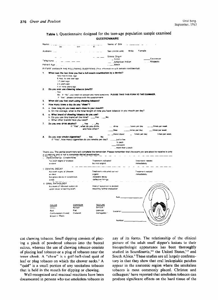
One thousand one hundred nineteen teenagers in grades 9 to 12 of the metropolitan Denver public schools were examined to determine the incidence and frequency of oral-tissue alterations associated with the use of smokeless tobacco. The students completed a questionnaire with eight specifically selected questions designed to identify the number of years with the habit, daily exposure, brand of tobac- co used, site of application, smoking and drinking habits, subjective symptoms, and frequency of dental care (Table I).
Two examiners previously trained in the diagnosis and indexing of oral lesions associated with the use of smokeless tobacco products performed oral hard- and soft-tissue examinations. All examiners per- formed the examinations without seeing the ques- tionnaire completed by the students, so that examin- er bias would not be a factor.
The clinical appearance of lesions induced by smokeless tobacco was graded by a modification of the method developed by Axe11 and associates.18 An exhaustive evaluation of the histomorphologic altera- tions, electron microscopic findings, and histochemi- cal changes seen in the mucosa of teen-age users of smokeless tobacco is the subject of an ongoing scientific investigation, which will be presented as a second stage of this study.

278 Greer and Poulson Oral Surg. September. 1983
Fig. 1. A typical Degree 1 lesion. Note that the lesion is superficial and approximates the color of the surrounding tissue. A very subtle wrinkling is obvious, but there is no discernible thickening. Fig. 2. A classic Degree 2 lesion. The lesion is primarily white, with a moderate amount of wrinkling and several intervening linear zones of erythema. Fig. 3. Degree 3 lesion showing evidence of white corrugated, thickened mucosa with interlacing furrows ranging from pink to red. Fig. 4. Gingival recession and degenerative periodontal disease are significant in this patient, who also has a Degree 1 lesion of the mucobuccal fold and labial mucosa. Fig. 5. This photograph shows the only example of cervical erosion that was documented. Slight cervical erosion is present in both mandibular central incisors. Tobacco is trapped in the area of erosion, affecting the right central incisor. The patient also has a Degree 3 mucosal lesion and tobacco-associated degenerative periodontal disease.
RESULTS Age, sex, and clinical appearance
The total sample of 1 ,119 subjects included 522 males and 597 females. One hundred seventeen persons (10.45 percent of the total sample) admitted to using smokeless tobacco. One hundred thirteen users were males and four were females. Table II
reflects the age and sex distribution of smokeless tobacco users. Fifty-seven (48.7 percent) of smoke- less tobacco users had lesions of the teeth, periodon- tium, or mucosa. The lesions were easily detectable clinically and were graded by a modification of the method established by Axe11 and associates’* in the following manner:
Volume 5h Oral tissue changes associated with smokeless tobacco 279 Number 3
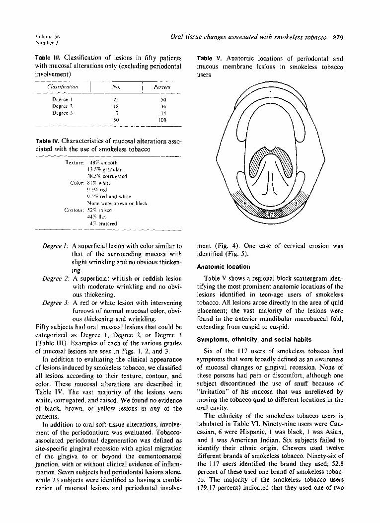
Table III. Classification of lesions in fifty patients with mucosal alterations only (excluding periodontal involvement)
- Classij‘ication No. Percent
Dcgrce I 25 SO Degree 2 18 36 Degree 3 -2 14
50 loo -
Table IV. Characteristics of mucosal alterations asso- ciated with the use of smokeless tobacco
Texture: 48% smooth
13.5% granular
38.5% corrugated
Color: XI% white
9.5%) red
9.5’1; red and white
None were brown or black
Contour: 52’S raised
44%) flat
4% cratered
Degree I: A superficial lesion with color similar to that of the surrounding mucosa with slight wrinkling and no obvious thicken- ing.
Degree 2: A superficial whitish or reddish lesion with moderate wrinkling and no obvi- ous thickening.
Degree 3: A red or white lesion with intervening furrows of normal mucosal color, obvi- ous thickening and wrinkling.
Fifty subjects had oral mucosal lesions that could be categorized as Degree 1, Degree 2, or Degree 3 (Table III). Examples of each of the various grades of mucosal lesions are seen in Figs. 1, 2, and 3.
In addition to evaluating the clinical appearance of lesions induced by smokeIess tobacco, we classified all lesions according to their texture, contour, and color. These mucosal alterations are described in Table IV. The vast majority of the lesions were white, corrugated, and raised. We found no evidence of black, brown, or yellow lesions in any of the patients.
In addition to oral soft-tissue alterations, involve- ment of the periodontium was evaluated. Tobacco- associated periodontal degeneration was defined as site-specific gingival recession with apical migration of the gingiva to or beyond the cementoenamel junction, with or without clinical evidence of inflam- mation. Seven subjects had periodontal lesions alone, while 23 subjects were identified as having a combi- nation of mucosal lesions and periodontal involve-
Table V. Anatomic locations of periodontal and mucous membrane lesions in smokeless tobacco users
ment (Fig. 4). One case of cervical erosion was identified (Fig. 5).
Anatomic location
Table V shows a regional block scattergram iden- tifying the most prominent anatomic locations of the lesions identified in teen-age users of smokeless tobacco. All lesions arose directly in the area of quid placement; the vast majority of the lesions were found in the anterior mandibular mucobuccal fold, extending from cuspid to cuspid.
Symptoms, ethnicity, and social habits
Six of the 117 users of smokeless tobacco had symptoms that were broadly defined as an awareness of mucosal changes or gingival recession. None of these persons had pain or discomfort, although one subject discontinued the use of snuff because of “irritation” of his mucosa that was unrelieved by moving the tobacco quid to different locations in the oral cavity.
The ethnicity of the smokeless tobacco users is tabulated in Table VI. Ninety-nine users were Cau- casian, 6 were Hispanic, 1 was black, 1 was Asian, and 1 was American Indian. Six subjects failed to identify their ethnic origin. Chewers used twelve different brands of smokeless tobacco. Ninety-six of the 117 users identified the brand they used; 52.8 percent of these used one brand of smokeless tobac- co, The majority of the smokeless tobacco users (79.17 percent) indicated that they used one of two

280 Greer and Paulson Oral Surg. September, 1983
Table VI. Racial distribution among teen-age users of smokeless tobacco
Caucasian 66.64%
American Indian
Unidentified
I , I 1 25% 50% 75% 100%
specific brands of tobacco. More than half of the subjects (62 percent) admitted to occasional use of alcohol, although it was difficult to quantitate the amount of alcohol that was used. Only 3 persons who used smokeless tobacco also smoked cigarettes. Eigh- teen subjects gave a positive history of alcohol use, cigarette smoking, and the use of smokeless tobacco. No significant differences with regard to clinical grading of lesions could be found, either between subjects with multiple habits (dipping, smoking, and drinking) and those who used only smokeless tobacco or between subjects who used different brands of smokeless tobacco and those who used one brand only. More than 69 percent of the smokeless tobacco users had had a complete dental examination in the past year. The level of dental care recorded for smokeless tobacco users is shown in Table VII.
EXPOSURE
The average exposure among users of smokeless tobacco with oral sequelae was determined to be 177 minutes per day. In persons who chewed or dipped but had no oral signs or sequelae, an average exposure time of 53 minutes per day was calculated. The duration of use and daily exposure among all users of smokeless tobacco are shown in Tables VIII and IX.
DICUSSION
The patients examined during this investigation represented quite a different population from those who have traditionally been studied. Most had been users of smokeless tobacco for a short period (aver- age use, 2.72 years) when compared with the studies of Roed-Petersen and Pindborg5 (average use, 22
Table VII. Level of dental care (how recently the subject had undergone a full-mouth examination by a dentist)
Less than 6 months 55
6 months to I year 24
I to 2 years 22
2 to 4 years 3
5 or more years IO
Among teen-ape users of smokeless tobacco. 69.3 percent had been to a
dentist in the last \ear.
years) and Axe11 and associates’* (average use, 23.8 years) in adults. We were unable to duplicate the findings of Axe11 and associates’* or of Hirsch and colleagues4 who established four degrees of oral mucosal alteration associated with the use of smoke- less tobacco, the most severe change being a white to yellowish brown, heavily wrinkled lesion with inter- vening deep, red furrows and/or heavy thickening. The reason for this failure is unquestionably related to the fact that nearly all previous authors have evaluated adult populations, where the tobacco users have had a snuff-dipping or tobacco-chewing habit for periods ranging from 16 to 20 years. The three degrees of mucosal change that were noted in our study represent a new classification which we believe should be applied to persons who have used smoke- less tobacco 4 years or less.
Hirsch and associates4 reported that the subjects in their recent study of snuff-induced lesions in adult Scandinavians kept the tobacco quid in their mouths 8.5 + 4.9 hours. Subjects in our study admitted to an average daily exposure of less than 3 hours. Although we were unable to determine a daily consumption in terms of the number of grams of tobacco used, we expect that the consumption in the teen-age popula- tion studied in no way approached the total con- sumption of 14 grams per day reported by Axe11 and othersix or Hirsch and co-workers4 in adult users.
The present study documents that the use of smokeless tobacco among teen-agers is overwhelm- ingly a male habit. Roed-Peterson and Pindborg5 reviewed a sample of 450 adult Danish patients with oral leukoplakia, 32 of whom had snuff-related lesions, and demonstrated that the habit was pre- dominantly seen in males. However, an abundance of reports in the American literature indicate that the habit is widespread among adult females.‘*, Is Seven- ty-five percent of 15,000 American snuff users sur- veyed by Smith and others were females.’ We suspect that this reported female predilection proba- bly represents to some extent a regional population sample bias.
Christen and colleagues3 reported a high preva-

Volume 56 Number 3
Oral tissue changes associated with smokeless tobacco 281
Table VIII. Duration of use of smokeless tobacco
Exposure time in years
Exposure time in years
lence of abrasion on the occlusal and incisal surfaces of teeth among adult tobacco chewers and snuff dippers in the United States. These investigators also reported a high frequency of gingival recession and periodontal destruction associated with the use of smokeless tobacco. Van Wyk9 demonstrated similar findings in South African patients who were con- firmed snuff dippers. We found no evidence of occlusal or incisal abrasion of the teeth in any of the 117 teen-agers who admitted to the use of smokeless tobacco. Although we were able to document tobac- co-associated periodontal deterioration, the ad- vanced periodontal destruction and loss of teeth that have been reported adjacent to regions where the tobacco quid is held in long-term users of smokeless tobacco’2, I9 was not demonstrated. It appears that such severe hard- and soft-tissue changes are related to long-term use of the tobacco product and are features classically seen in an adult population.
Although we did document one instance of cervi-
cal erosion in the study, it was deemed an aberrant finding since no correiation between the erosion and excessive use of smokeless tobacco or a specific brand of tobacco was possible. We favor this as the cause, however, since the erosion was in an otherwise healthy oral cavity and was identified at the anatom- ic site where the tobacco quid was routinely placed.
We found no evidence of tobacco-associated den- tal caries. It has been speculated that the relative paucity of caries seen in heavy chewers of tobacco may largely be due to the accelerated salivary flow that the tobacco stimulates. It is postulated that the accelerated flow causes a physical cleansing action and a mild buffering action that inhibit the aggrega- tion of plaque and cariogenic material. Christen* has further suggested that many smokeless tobacco prod- ucts contain fluoride in levels ranging from 0.9 1 ppm to 2.01 ppm. The fluoride may also be instrumental in suppressing dental caries in users of smokeless

282 Greer and Poulson Oral Surg. September. I983
Table IX. Daily exposure to smokeless tobacco
Daily exposure time in minutes
Taverage 53 min./day
Daily exposure time in minutes
tobacco. There is, however, no universal agreement among investigators that smokeless tobacco is innoc- uous with regard to caries formation. Sitzes20 re- ported that several patients who chewed sweetened smokeless tobacco had evidence of cervical caries.
The present study demonstrates a marked pre- dominance of white male chewers and dippers. This ethnic distribution was quite striking, and we consid- er it unrelated to an ethnic sample bias since 83 percent of the participants in the study were from the Denver public school system, a school system that has had mandatory busing since 1973 with a resul- tant racial balance among the majority of high school student bodies.
In various parts of the world smokeless tobacco has different constituents. Thus, the abrasive quality, tobacco content, chemical components, and manu- facturing process may vary widely.5 In Scandinavia, wet snuff, which is highly alkaline (pH 8 to 9) is used almost exclusively. The dry tobacco used in the United States is not nearly as alkaline. It has been suggested that epithelial changes found in Swedish and Danish snuff users represent direct tissue dam- age probably related to the high alkaline reaction of Scandinavian wet snuff.18 Pindborg and his col- leagueslO accept the theory that the vacuolated cells
seen in the histopathologic material from Swedish and Danish snuff users may be the result of the highly alkaline product. However, the development of characteristic chevron-like keratinized spikes, which they have suggested are histologically charac- teristic and specific for mucosal damage from pipes, snuff, and hooklis or water pipes, must have a different etiology since they are found in cigarette smokers as well as in users of smokeless tobacco. We also suspect that these chevron-like keratinized spikes appear as a function of tobacco exposure over time and very likely may be absent in persons, such as teen-agers, who have used tobacco for a relatively short time.
Pindborg and associateslO have described a charac- teristic pumicelike keratinization of the oral mucosa in persons who are long-term users of smokeless tobacco. This pumicelike change has been character- ized clinically by a homogenous white patch with elevated keratinized striae. The pumice pattern has been seen solely on parts of the oral mucosa which normally are nonkeratinized. In none of our 50 patients who had discernible oral mucosal lesions that were related to the use of smokeless tobacco were we able to demonstrate this characteristic pumicelike appearance. In 13.5 percent of the users

Volume 56 Oral tissue changes associated with smokeless tobacco 283 Number 3
of smokeless tobacco we were able to detect a granular texture to the mucosa which is perhaps similar to what Pindborg has defined as a pumicelike quality. However, of those patients whose mucosae had a granular quality, none had the deep furrowing that is characteristic of the pumice pattern.
Hirsch and associates4 recently attempted to cor- relate snuff habits with the clinical severity of oral lesions as well as with certain superficial and deeply located cell changes in the epithelium and connective tissue. They found that the incidence of keratinized lesions, sialadenitis, and mild dysplasia were higher than previously reported in the literature. These investigators emphasized, however, that the presence of dysplastic changes could not be predicted by means of the parameters which characterize the snuff habit or from the clinical grading of the lesion. The authors noted that the mild dysplasia seen in their study did not necessarily mean that the lesions were precancerous, since similar dysplastic epithelial change may be found in noncancerous lesions according to the World Health Organization’s col- laborative study on oral precancerous lesions.2’ How- ever, in a retrospective study of snuff dipper’s muco- sal alterations, Axe11 and others” demonstrated a clear correlation between snuff dipping and oral cancer in Sweden, a finding supported in earlier studies in various other countries.“. 13. ”
Christen, perhaps more than any single investiga- tor, has adamantly maintained that smokeless tobac- co can cause oral cancer. He supports the concept that verrucous carcinoma is associated with the use of smokeless tobacco by noting that nitrosonornico- tine (NNN), the first organic carcinogen isolated from unburned tobacco, is found in smoking tobacco, chewing tobacco, and snuff in high concentrations (between 0.03 and 90 pg/Gm. of dry tobacco).2.“3 The suggestion that nitrosonornicotine may be the carcinogen responsible for verrucous carcinoma development in users of smokeless tobacco and the suggestion by Pindborg and associates’0 that oral epithelial changes induced by smokeless tobacco may be predicted on the basis of histologic findings deserve further study. Thermal irritation has been implicated as one of the possible etiologic factors associated with dysplastic changes in the mucosa of hookli smokers2, lo However, thermal irritation alone cannot be the sole explanation for the oral changes, since similar lesions are seen in tobacco chewers and snuff dippers.
The early clinical changes that have been noted in the present study are thought to be largely the result of mucosal irritation from the topically applied tobacco product. Bernstein and Carlish24 reported
similar diffuse, filmy, white lesions of the oral mucosa in several patients with histories of excessive use of Listerine mouthwash. In their studies remis- sion of the lesions occurred 2 weeks after the product was discontinued. They developed a control animal model in hamsters to study the effects of prolonged oral contact with Listerine and found that after 42 days of application every animal had developed clinical evidence of diffuse, filmy, white, corrugated lesions of the oral mucosa. The authors postulated that the response was one purely related to physical contact of the product with the mucosa.
In our study half of the patients who admitted to the use of smokeless tobacco had no oral lesions. We were able to elicit from thorough history taking that two of the subjects in our study who had used smokeless tobacco were aware of a “white callus” that disapeared when they stopped using the product, which suggests that the mucosal lesions are revers- ible.
From the response to our question concerning the level of dental care afforded the patients in this study, it was apparent that the vast majority of the patients (69.3 percent) had access to dental care in the form of a routine full-mouth examination during the previous year. We were unable to determine whether tobacco-related mucosal lesions were evi- dent in any of the patients during those examina- tions. The degree of difficulty experienced in recog- nizing Degree 1 lesions can be extreme, even among examiners well versed in recognizing such subtle mucosal alterations. An educational campaign to advise dentists of these early mucosal changes may prove quite beneficial.
Although this study documents that changes in oral tissue are unquestionably associated with the use of smokeless tobacco in teen-agers, additional studies are deemed necessary to study the histomorphologic, electron microscopic, and histochemical changes evi- dent in oral mucosa associated with the precipitant increase in use of smokeless tobacco among Ameri- can teen-agers. Pursuant to that goal, a second phase of this study has been designed to investigate these varied parameters and compare them with previously published information concerning similar changes seen in adult patients.
SUMMARY
The practice of placing a small amount of chewing tobacco or snuff in the oral cavity and leaving it in place for extended periods of time appears to be finding its way onto middle school, high school, and college campuses as a socially acceptable and popu- lar habit. Numerous reports have appeared in the

284 Greer and Paulson
literature describing the oral changes associated with the use of smokeless tobacco in adults. Such informa- tion with respect to adolescents and teen-agers has previously been unavailable. The prevalence and frequency of oral tissue alterations associated with the use of smokeless tobacco was studied in a teen-age population. The relative exposure to the tobacco in terms of minutes per day, the specific brands of tobacco used, the common anatomic sites for placing of the tobacco quid, smoking and drink- ing habits, and subjective symptoms were deter- mined. From a total sample size of 1,119 students, 117 users of smokeless tobacco were identified and 57 persons with oral lesions were evaluated and their lesions classified.
The authors would like to extend their sincere apprecia- tion to Drs. James Lindenmuth, John Coke, and Lawrence Meskin for their assistance in gathering and analyzing data for this study. Without their assistance the task of clinical evaluation could not have been completed. We also acknowledge the support of Ms. Patricia Yamashita in preparing the manuscript.
REFERENCES
I.
2.
3.
4.
5
6.
7.
8.
ChrIsten. A. G., Swanson, B. Z., Clover, E. D., and Hender- son. A. H.: Smokeless Tobacco: The Folklore and Social History of Snuffing, Sneezing, Dipping and Chewing, J. Am. Dent. 2ssoc. 105: 821-829. 1982. Christen, A. G.: The Case Against Smokeless Tobacco: Five Facts for the Health Professional to Consider. J. Am. Dent. Assoc. 101: 464-469, 1980. Christen. A. G.. Armstrong, W. R., and McDaniel, R. K.: Intraoral Leukoplakia, Abrasion. Periodontal Breakdown, and Tooth Loss in a Snuff Dipper, J. Am. Dent. Assoc. 98: 584-586. 1979. Hirsch, .I. M.. Heyden, G., and Thilander. H.: A Clinical, Histomorphological and Histochemical Study on Snuff- Induced Lesions of Varying Severity, J. Oral Pathol. 11: 387-398. 1982. Roed-Peterson, B.. and Pindborg. J. J.: A Study of Danish Snuff-Induced Oral Leukoplaskias, J. Oral Pathol. 2:301-313. 1973. Pindborg. J. J., and Renstrup, G.: Studies in Oral Leukopla- kia. II. Effect of Snuff on Oral Epithelium, Acta Derm. Vcnereol. [Stock.] 43: 271-276. 1963. Smith, J. F., Mincer, H. A., Hopkins, K. P., and Bell, J.: Snuff-Dipper’s Lesion: A Cytological and Pathological Study in a Large Population, Arch. Otolaryngol. 9t: 450-456, 1970. Archard, H. 0.. and Tarpley. T. M.. Jr.: Clinicopathologic and Histochemical Characterization of Submucosal Deposits in Snuff Dipper’s Keratosis. J. Oral Pathol. I: 3-l I, 1972.
9.
IO.
I I.
12.
13.
14.
15.
16.
17.
18.
19.
20. 21.
22.
23.
24.
Oral Surg. September, 1983
van Wyk, C. W.: The Oral Lesion Caused by Snuff: A Clinicopathologic Study, Med. Proc. 11: 531-537, 1965. Pindborg. J. J., Reibel, J., Roed-Petersen, B., and Mehta, F. S.: Tobacco-Induced Changes in Oral Leukoplakic Epitheli- urn, Cancer 45: 2330-2336. 1980. Sundstrom, 8.. MSrnstad, H., and AxelI, T.: Oral Carcinomas Associated With Snuff Dipping, J. Oral Pathol. 11: 245-25 I. 1982. Brown, R. L.. Suh, J. M., Scarborough, J. E., Wilkins, S. A., and Smith, R. R.: Snuff Dipper’s Intraoral Cancer: Clinical Characteristics and Response to Therapy, Cancer 18: 2-13, 1965. Landy, J. J.. and White, H. J.: Buccogingival Carcinoma of Snuff Dippers, Am. Surg. 17: 442-447, 1961. Peacock, E. E., Jr., Greenberg, B. G., and Brawley, B. W.: The Effect of Snuff and Tobacco on the Production of Oral Carcinoma: An Experimental and Epidemiological Study, Ann. Surg. 151: 542-550, 1960. Rosenfeld. L., and Callaway, J.: Snuff Dipper’s Cancer. Am. J. Surg. 106: 840-844, 1963. Vogler, W. R., Lloyd, J. W., and Milmore. B. K.: A Retrospective Study of Etiological Factors in Cancer of the Mouth, Pharynx and Larynx, Cancer 15: 246-258, 1962. Peacock, E. E., Jr., and Bradley, B. W.: An Evaluation of Snuff and Tobacco in the Production of Mouth Cancer, Plast. Reconstr. Surg. 23: 628-635, 1959. Axell, T.. Mornstad, H., and Sundstrdm, B.: The Relation of the Clinical Picture to the Histopathology of Snuff Dipper’s Lesions in a Swedish Population, J Oral Pathol. 5: 229-236, 1976. Christen. A. G.: The Clinical EfFects of Tobacco on Oral Tissue, J. Am. Dent. Assoc. 81: 1378-1382, 1970. Sit7cs. L..: On Chewing Tobacco, ADA News 8: 2, 1977. Kramer, I. R. H.. Lucas, R. B., Pindborg, J. J., and Sobin, L. H.: Definition of Leukoplakia and Related Lesions: An Aid to Studies on Oral Precancer, ORAL SURC. 46: 5 18-539, 1978. Axcll, T.. Mornstad. H.. and Sundstram, B.: Snusning Och Munhalecancer: En Retrospecktiv Studie, Tandlaekartid- ningcn 70:1048-1052, 1978. Hoffman, D.: Chemical Studies on Tobacco Smoke. Nitros- onornicotine: Presence in Tobacco, Formation and Carcinoge- nicity. In Walker, E. A., Bogovskii, P., and Griciute, L. (editors): Environmental N-nitroso Compound Analysis and Formation. IARC Sci. Publ. 14: 307-320, 1976. Bernstein, M. L., and Carlish, R. S.: The Induction of White Lesions in Hamster Cheek Pouches With Mouthwash, Abstract 18. Essay Program, American Academy of Oral Pathlogy, Thirty-Second Annual Meeting, April 23-28, 1978. Fort Lauderdale, Fla.
Reprint requests to:
Dr. Robert 0. Greer, Jr. University of Colorado School of Dentistry 4200 East 9th Ave. Box C28.5 Denver. Colo. 82062
Copyright © 2022 FDOKUMEN