Mourning and melancholia revisited: correspondences between principles of Freudian metapsychology...
23
BioMed Central Page 1 of 23 (page number not for citation purposes) Annals of General Psychiatry Open Access Review Mourning and melancholia revisited: correspondences between principles of Freudian metapsychology and empirical findings in neuropsychiatry Robin L Carhart-Harris* 1 , Helen S Mayberg 2 , Andrea L Malizia 1 and David Nutt 1 Address: 1 Psychopharmacology Unit, University of Bristol, Bristol, UK and 2 Emory University School of Medicine, Atlanta, GA 30322, USA Email: Robin L Carhart-Harris* - [email protected]; Helen S Mayberg - [email protected]; Andrea L Malizia - [email protected]; David Nutt - [email protected] * Corresponding author Abstract Freud began his career as a neurologist studying the anatomy and physiology of the nervous system, but it was his later work in psychology that would secure his place in history. This paper draws attention to consistencies between physiological processes identified by modern clinical research and psychological processes described by Freud, with a special emphasis on his famous paper on depression entitled 'Mourning and melancholia'. Inspired by neuroimaging findings in depression and deep brain stimulation for treatment resistant depression, some preliminary physiological correlates are proposed for a number of key psychoanalytic processes. Specifically, activation of the subgenual cingulate is discussed in relation to repression and the default mode network is discussed in relation to the ego. If these correlates are found to be reliable, this may have implications for the manner in which psychoanalysis is viewed by the wider psychological and psychiatric communities. Background 'When some new idea comes up in science, which is hailed at first as a discovery and is also as a rule dis- puted as such, objective research soon afterwards reveals that after all it was in fact no novelty' [1]. The intention of this paper is to draw attention to consist- encies between Freudian metapsychology and recent find- ings in neuropsychiatry, especially those relating to depression. A case will be made that findings in neuroim- aging and neurophysiology can provide a fresh context for some of the most fundamental theories of psychoanalysis. In his famous paper 'Mourning and melancholia', Freud carried out an elegant application of psychoanalytic the- ory to the illness of depression. It is the task of this paper to parallel the psychological processes described by Freud with the physiological processes identified by modern clinical research in order to furnish a more comprehensive understanding of the whole phenomenon. Under the tutelage of Meynert, Freud began his career as neurologist studying the anatomy and physiology of the medulla. Inspired by a Helmholtzian tradition (1821– 1894) and a 'psycho-physical parallelism' made fashiona- ble by the likes of Hering (1838–1918), Sherrington (1857–1952) and Hughlings-Jackson (1835–1911), Freud began to consider more seriously how a science of movements of energy in the brain might account for psy- Published: 24 July 2008 Annals of General Psychiatry 2008, 7:9 doi:10.1186/1744-859X-7-9 Received: 2 February 2008 Accepted: 24 July 2008 This article is available from: http://www.annals-general-psychiatry.com/content/7/1/9 © 2008 Carhart-Harris et al; licensee BioMed Central Ltd. This is an Open Access article distributed under the terms of the Creative Commons Attribution License (http://creativecommons.org/licenses/by/2.0 ), which permits unrestricted use, distribution, and reproduction in any medium, provided the original work is properly cited.
-
Upload
independent -
Category
Documents
-
view
0 -
download
0
Transcript of Mourning and melancholia revisited: correspondences between principles of Freudian metapsychology...

BioMed CentralAnnals of General Psychiatry
ss
Open AcceReviewMourning and melancholia revisited: correspondences between principles of Freudian metapsychology and empirical findings in neuropsychiatryRobin L Carhart-Harris*1, Helen S Mayberg2, Andrea L Malizia1 and David Nutt1Address: 1Psychopharmacology Unit, University of Bristol, Bristol, UK and 2Emory University School of Medicine, Atlanta, GA 30322, USA
Email: Robin L Carhart-Harris* - [email protected]; Helen S Mayberg - [email protected]; Andrea L Malizia - [email protected]; David Nutt - [email protected]
* Corresponding author
AbstractFreud began his career as a neurologist studying the anatomy and physiology of the nervous system,but it was his later work in psychology that would secure his place in history. This paper drawsattention to consistencies between physiological processes identified by modern clinical researchand psychological processes described by Freud, with a special emphasis on his famous paper ondepression entitled 'Mourning and melancholia'. Inspired by neuroimaging findings in depression anddeep brain stimulation for treatment resistant depression, some preliminary physiologicalcorrelates are proposed for a number of key psychoanalytic processes. Specifically, activation ofthe subgenual cingulate is discussed in relation to repression and the default mode network isdiscussed in relation to the ego. If these correlates are found to be reliable, this may haveimplications for the manner in which psychoanalysis is viewed by the wider psychological andpsychiatric communities.
Background'When some new idea comes up in science, which ishailed at first as a discovery and is also as a rule dis-puted as such, objective research soon afterwardsreveals that after all it was in fact no novelty' [1].
The intention of this paper is to draw attention to consist-encies between Freudian metapsychology and recent find-ings in neuropsychiatry, especially those relating todepression. A case will be made that findings in neuroim-aging and neurophysiology can provide a fresh context forsome of the most fundamental theories of psychoanalysis.In his famous paper 'Mourning and melancholia', Freudcarried out an elegant application of psychoanalytic the-
ory to the illness of depression. It is the task of this paperto parallel the psychological processes described by Freudwith the physiological processes identified by modernclinical research in order to furnish a more comprehensiveunderstanding of the whole phenomenon.
Under the tutelage of Meynert, Freud began his career asneurologist studying the anatomy and physiology of themedulla. Inspired by a Helmholtzian tradition (1821–1894) and a 'psycho-physical parallelism' made fashiona-ble by the likes of Hering (1838–1918), Sherrington(1857–1952) and Hughlings-Jackson (1835–1911),Freud began to consider more seriously how a science ofmovements of energy in the brain might account for psy-
Published: 24 July 2008
Annals of General Psychiatry 2008, 7:9 doi:10.1186/1744-859X-7-9
Received: 2 February 2008Accepted: 24 July 2008
This article is available from: http://www.annals-general-psychiatry.com/content/7/1/9
© 2008 Carhart-Harris et al; licensee BioMed Central Ltd. This is an Open Access article distributed under the terms of the Creative Commons Attribution License (http://creativecommons.org/licenses/by/2.0), which permits unrestricted use, distribution, and reproduction in any medium, provided the original work is properly cited.
Page 1 of 23(page number not for citation purposes)
Annals of General Psychiatry 2008, 7:9 http://www.annals-general-psychiatry.com/content/7/1/9
chological phenomena [2]. It has been argued that Freudnever truly abandoned his physiological roots [3,4] andthat his early flirtations with psycho-physical parallelismcontinued to haunt 'the whole series of [his] theoreticalworks to the very end' [4].
This paper will begin with an overview of some key con-cepts of Freudian metapsychology (libido, cathexis, objectcathexis, the ego, the super ego, the id, the unconscious,the primary and secondary psychical process and repres-sion) and an attempt will be made to hypothesise theirphysiological correlates. This will be followed by a sum-mary of 'Mourning and melancholia' and an extensivelook at relevant findings in neuropsychiatry. Of specialinterest are neuroimaging findings in depression andinduced depressed mood, deep brain stimulation (DBS)of the subgenual cingulate (Brodmann area 25/Cg25) forthe treatment of intractable depression, electrical stimula-tion of medial temporal regions, and regional atrophyand glial loss in the brains of patients suffering frommajor depression.
Before beginning, it is important to make a few brief com-ments on the principle of psycho-physical parallelism.Drawing connections between psychological and biologi-cal phenomena was an approach that Freud was both crit-ical of:
'I shall carefully avoid the temptation to determinepsychical locality in any anatomical fashion' [5].
'Every attempt to discover a localisation of mentalprocesses...has miscarried completely. The same fatewould await any theory that attempted to recognisethe anatomical position of the system [consciousness]– as being in the cortex, and to localise the uncon-scious processes in the subcortical parts of the brain.There is a hiatus here which at present cannot be filled,nor is it one of the tasks of psychology to fill it. Ourpsychical topography has for the present nothing to dowith anatomy' [6].
And receptive to:
'All our provisional ideas in psychology will presuma-bly some day be based on an organic substructure' [7].
The ambiguity in Freud's position can be explained by hiscriticism of the modular or 'segregationist' [8] approachand preference for a more dynamic model [9]. Essentially,Freud was opposed to 'flag polling' the anatomical causesof psychological phenomena but not the drawing of par-allels between psychological and physiological processes:
'It is probable that the chain of physiological events inthe nervous system does not stand in a causal connec-tion with the psychical events. The physiologicalevents do not cease as soon as the psychical onesbegin; on the contrary, the physiological chain contin-ues. What happens in simply that, after a certain pointin time, each (or some) of its links has a psychical phe-nomena corresponding to it. Accordingly, the psychi-cal is a process parallel to the physiological – "adependent concomitant"' [9].
Integrating psychoanalysis with modern neuroscience is adifficult and controversial endeavour. It should be madeclear from the outset what we believe it is possible for thisapproach to achieve. Psychoanalysis can be viewed ontwo levels: a hermeneutic, interpretative or meaning basedlevel; and a metapsychological, mental process based level.The hermeneutic level is inherently subjective. The ques-tion has often been raised whether it is possible to identifyspatiotemporal coordinates of subjective meaning. Thisview was shared by Paul McLean in his seminal book 'Thetriune brain in evolution' [10]:
'Since the subjective brain is solely reliant on the deri-vation of immaterial information, it can never estab-lish an immutable yardstick of its own...Information isinformation, not matter or energy' [10].
It would be incorrect to align this position with dualism.Psychophysical parallelism is a materialist approach thatacknowledges that meaning arises through time betweennetworks of communicative systems. It must be statedthat the evidence cited in this paper cannot logically vali-date psychoanalysis on the hermeneutic level and neitherdoes it provide evidence for the efficacy of psychoanalysisas a treatment modality (see [11] for a review). What webelieve it can do, however, is bring together converginglines of enquiry in support of the Freudian topography ofthe mind. The findings cited below describe changes inphysiological processes paralleling changes in psycholog-ical processes; however, the objective measures do notshed any light on the specific content or meaning heldwithin these processes. Aside from interpretation, muchof Freud's work was spent theorising about dynamic psy-chical processes; energies flowing into and out of mentalprovinces, energy invested, dammed up and dischargedthroughout the mind. It is this metapsychological level ofpsychoanalysis that we believe is most accessible to inte-gration with modern neuroscience.
An introduction to some key terms of Freudian metapsychologyLibido
'Libido means in psycho-analysis in the first instancethe force (thought of as quantitatively variable and
Page 2 of 23(page number not for citation purposes)
Annals of General Psychiatry 2008, 7:9 http://www.annals-general-psychiatry.com/content/7/1/9
measurable) of the sexual drives directed towards anobject – "sexual" in the extended sense required byanalytic theory' [12].
From its earliest recorded use [13] the term 'libido' wasused to connote the principal energy of the nervous sys-tem. Freud differentiated 'libido' from a more general'psychical energy':
'We have defined the concept of libido as a quantita-tively variable force which could serve as a measure ofprocesses and transformations occurring in the field ofsexual excitation. We distinguish this libido in respectof its special origin from the energy which must besupposed to underlie the mental processes in general'[14].
Freud's extended use of the term 'sexual' brought him intoconflict with Jung, who argued that the principal energy ofthe nervous system was not inherently sexual [15]. Argua-bly, the two perspectives are not irreconcilable. We mayview Freud's 'libido' in connection with the motivationaldrive system (see The id below) and the withdrawal andinvestment of cerebral energy (see The ego below). Jung's'psychical energy' can be viewed less specifically as cere-bral energy in general.
CathexisThe German original 'Besetzung' literally translates as'occupation', 'filling' or 'investment'. The neologism'cathexis' was one that Freud was not especially fond of[16]. Freud first used the term on an explicitly physiolog-ical level, referring to neurons 'cathected with a certainquantity [of energy]' [2], systems 'loaded with a sum ofexcitation' [17] and 'provided with a quota of affect' [18].Succinctly, the term 'cathexis' means 'libidinal invest-ment'. It is a vitally important concept for the integrationof Freudian metapsychology with principles of modernneuroscience. In this paper, we discuss changes in haemo-dynamic response and other neurophysiological meas-ures in relation to the withdrawal and investment oflibido.
Object cathexisThe concept of "the object" is used in a broad sense in psy-choanalysis to refer to literal, abstract and symbolicobjects. People, tasks, work and ideas can all serve asobjects. The process of object cathexis can be comparedwith the process of goal-directed cognition, since bothrequire libidinal investment. Based on neuroimaging datain depression (see Neuropsychiatric findings in depres-sion correlated with principles of Freudian metapsychol-ogy below), we propose that activation of the dorsolateralprefrontal cortex (DLPFC) correlates with object cathexis,and reduced DLPFC activation correlates with reduced
object cathexis which manifests in depression as anhedo-nia (see Hypofrontality below). As will be discussed in thenext section, activation of the DLPFC is accompanied by adeactivation in a network of regions known as the default-mode network (DMN) [19]. The DMN is highly activeduring resting cognition. The regions engaged duringactive cognition are referred to here as the object-orientednetwork (ON). We propose that activation in the ON anddeactivation in the DMN correlates with the process ofobject cathexis.
The egoThe German original 'das Ich' literally translates as 'the I'.It is somewhat regrettable that Freud's terms have notbeen translated more literally since the originals have anappeal that is lost in translation. Freud used the conceptof the ego in a number of different ways; a useful way ofgaining a sense of the different applications therefore, is tocite some examples of its use:
1. A referent to the conscious sense of self:
' [I]n each individual there is a coherent organisationof mental processes; and this we call his ego. It is tothis ego that consciousness is attached' [1].
2. An unconscious force maintaining self-cohesion:
'It is certain that much of the ego is itself unconsciousand notably what we may call its nucleus; only a smallpart of it is covered by the term "preconscious"' [20].
3. A nucleus of somatic cohesion:
'The ego is first and foremost a bodily ego' [1].
4. A reservoir of libido:
'Thus we form the idea of there being an original libid-inal cathexis of the ego, from which some is later givenoff to objects' [7].
'The ego is the true and original reservoir of libido'[20].
5. The primary agent of repression:
' [T]he ego is the power that sets repression in motion'[12].
Given the many different functions to the ego, it would becounterintuitive to suggest that it is 'housed' in a singlegiven region of the brain. Based on a large number of neu-roimaging studies, we propose that a highly connectednetwork of regions, principally incorporating the medial
Page 3 of 23(page number not for citation purposes)
Annals of General Psychiatry 2008, 7:9 http://www.annals-general-psychiatry.com/content/7/1/9
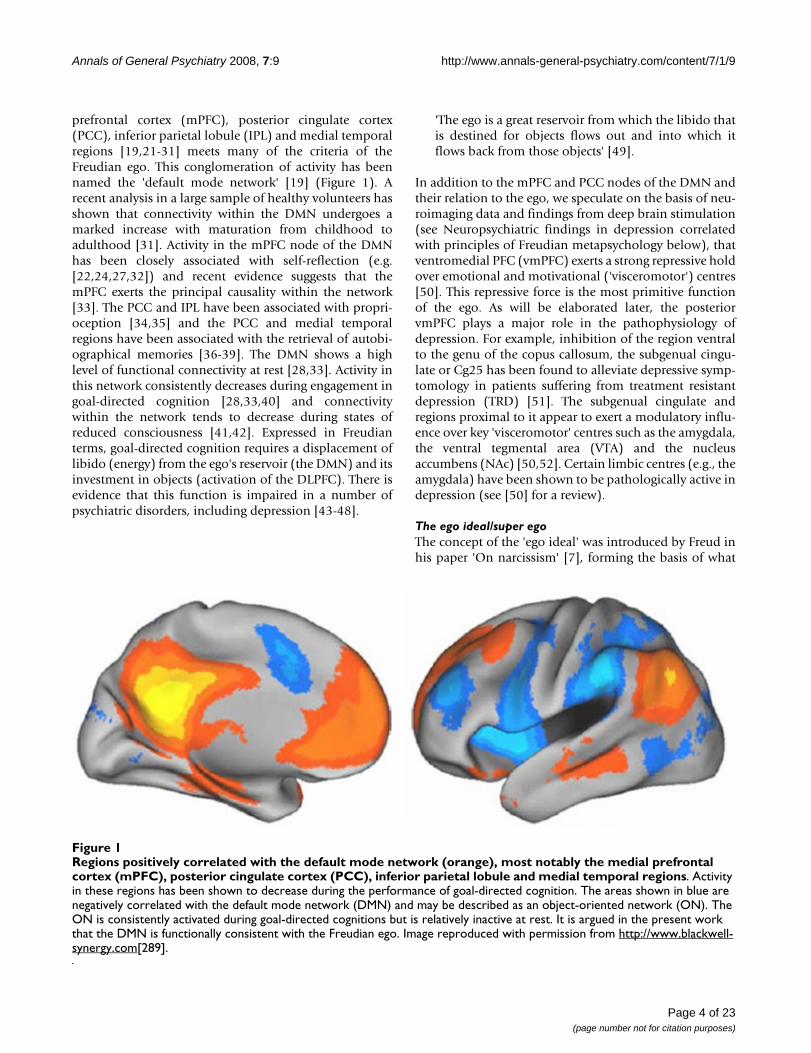
prefrontal cortex (mPFC), posterior cingulate cortex(PCC), inferior parietal lobule (IPL) and medial temporalregions [19,21-31] meets many of the criteria of theFreudian ego. This conglomeration of activity has beennamed the 'default mode network' [19] (Figure 1). Arecent analysis in a large sample of healthy volunteers hasshown that connectivity within the DMN undergoes amarked increase with maturation from childhood toadulthood [31]. Activity in the mPFC node of the DMNhas been closely associated with self-reflection (e.g.[22,24,27,32]) and recent evidence suggests that themPFC exerts the principal causality within the network[33]. The PCC and IPL have been associated with propri-oception [34,35] and the PCC and medial temporalregions have been associated with the retrieval of autobi-ographical memories [36-39]. The DMN shows a highlevel of functional connectivity at rest [28,33]. Activity inthis network consistently decreases during engagement ingoal-directed cognition [28,33,40] and connectivitywithin the network tends to decrease during states ofreduced consciousness [41,42]. Expressed in Freudianterms, goal-directed cognition requires a displacement oflibido (energy) from the ego's reservoir (the DMN) and itsinvestment in objects (activation of the DLPFC). There isevidence that this function is impaired in a number ofpsychiatric disorders, including depression [43-48].
'The ego is a great reservoir from which the libido thatis destined for objects flows out and into which itflows back from those objects' [49].
In addition to the mPFC and PCC nodes of the DMN andtheir relation to the ego, we speculate on the basis of neu-roimaging data and findings from deep brain stimulation(see Neuropsychiatric findings in depression correlatedwith principles of Freudian metapsychology below), thatventromedial PFC (vmPFC) exerts a strong repressive holdover emotional and motivational ('visceromotor') centres[50]. This repressive force is the most primitive functionof the ego. As will be elaborated later, the posteriorvmPFC plays a major role in the pathophysiology ofdepression. For example, inhibition of the region ventralto the genu of the copus callosum, the subgenual cingu-late or Cg25 has been found to alleviate depressive symp-tomology in patients suffering from treatment resistantdepression (TRD) [51]. The subgenual cingulate andregions proximal to it appear to exert a modulatory influ-ence over key 'visceromotor' centres such as the amygdala,the ventral tegmental area (VTA) and the nucleusaccumbens (NAc) [50,52]. Certain limbic centres (e.g., theamygdala) have been shown to be pathologically active indepression (see [50] for a review).
The ego ideal/super egoThe concept of the 'ego ideal' was introduced by Freud inhis paper 'On narcissism' [7], forming the basis of what
Regions positively correlated with the default mode network (orange), most notably the medial prefrontal cortex (mPFC), pos-terior cingulate cortex (PCC), inferior parietal lobule and medial temporal regionsFigure 1Regions positively correlated with the default mode network (orange), most notably the medial prefrontal cortex (mPFC), posterior cingulate cortex (PCC), inferior parietal lobule and medial temporal regions. Activity in these regions has been shown to decrease during the performance of goal-directed cognition. The areas shown in blue are negatively correlated with the default mode network (DMN) and may be described as an object-oriented network (ON). The ON is consistently activated during goal-directed cognitions but is relatively inactive at rest. It is argued in the present work that the DMN is functionally consistent with the Freudian ego. Image reproduced with permission from http://www.blackwell-synergy.com[289].
Page 4 of 23(page number not for citation purposes)

Annals of General Psychiatry 2008, 7:9 http://www.annals-general-psychiatry.com/content/7/1/9
would later become 'the super ego' [1] (German original= 'Das über-Ich'; 'the over-I'). The ego ideal/super egoplays a fundamental role in the aetiology of depression:
'Repression, we have said, proceeds from the ego, wemight say with greater precision that it proceeds fromthe self-respect of the ego' [7].
Freud described this more fully in the following passage:
'The ego ideal is...the target of the self-love which wasenjoyed in childhood by the actual ego. The subject'snarcissism makes its appearance displaced on to thisnew ideal ego, which like the infantile ego finds itselfpossessed of every perfection that is of value. As alwayswhere the libido is concerned, man has here shownhimself incapable of giving up a satisfaction he hadonce enjoyed. He is not willing to forgo the narcissisticperfection of his childhood; and when as he grows up,he is disturbed by the admonitions of others and bythe awakening of his own critical judgement, so thathe can no longer retain that perfection, he seeks torecover it in the new form of an ideal. What he projectsbefore him as his ideal is the substitute for the lost nar-cissism of his childhood in which he was his ownideal' [7].
It is difficult to postulate a neurodynamic correlate of sucha high-level concept as the ego ideal or super ego. The fol-lowing model should therefore be considered speculativeand preliminary. The super ego might be thought of as anumbrella term for high-level cognitions that work toappraise the ego's ability to meet an imagined ideal. Thisideal-ego or 'ego ideal' is acquired through an internalisa-tion of value judgements of others (e.g., one's early caregivers) under social and environmental demands (seeMourning and melancholia below). Through the superego, the ego receives feedback on how closely it corre-sponds with an imagined ideal. If the super ego judges theego as falling short of this ideal, or if the super ego judgesthe ego's or the id's drives as unhealthy or dangerous inthe context of its social environment, then the ego mayrepel these drives, withholding them from consciousness.The implications of the super ego's instruction to represswill be discussed in the next section in relation to depres-sion.
It is highly unlikely that the ego ideal/super ego is housedin any specific region of the brain but we may speculateabout dynamic physiological processes paralleling psy-chological ones. Thus, paralleling the super ego's valuejudgements of the ego may be feedback between theDLPFC of the ON and the mPFC of the DMN. Informa-tion communicated between these two systems (see The
ego above) may parallel the experience of pursuing anideal and judging how successfully it is met.
In relation to the unconscious, punishing aspect of thesuper-ego it might be useful to consider the role of theanterior cingulate (ACC). Activation of the ACC has beenassociated with error detection and guilt [8,53,54]. It maybe significant that a recent analysis of functional connec-tivity in the human cingulate revealed strong connectivitybetween the ACC and the DLPFC [54]. Conversely, Cg25was found to be strongly connected with regions of theDMN such as the OFC. It is possible that feedbackbetween the DLPFC and the mPFC is mirrored at a lowerlevel by feedback between the ACC, OFC and Cg25. Feed-back between the ON and DMN likely takes place via cor-tico-striato-pallido-thalamo-cortical circuitry.
The super ego's control over the ego gives it a uniquepower to influence the motility and expression of thedrives. Impassioned behaviours deemed dangerous to theego in the context of its environment may be deniedexpression by activating Cg25 and the DMN. Integratingthis hypothesis into a model of depression, we can postu-late that activating Cg25 and the DMN controls the fullexpression of affective, mnemonic and motivationalbehaviours promulgated by visceromotor centres. Thus,engaging Cg25 contains limbic activity within paralimbic-thalamic circuits maintained by the Cg25 in reaction tosustained limbic arousal (for relevant models, see[46,50,55-58]).
The idThe German original 'das es' literally translates as 'the it'.As with the German word for the ego (das Ich), the origi-nal word for the id has an appeal that is lost in translation.The id was one of Freud's later concepts, being introducedin his paper 'The ego and the id' [1]. Some have arguedthat the id is synonymous with the unconscious, and it istrue that two are closely related:
'The id and the unconscious are as intimately linked asthe ego and the preconscious' [59].
'The truth is that it is not only the psychically repressedthat remains alien to our consciousness, but also someof the impulses which dominate our ego' [6].
Although the id and the unconscious are related, they alsoretain some important differences, both psychologicallyand physiologically. Essentially, the id refers to the uncon-scious as a system in a topographical sense [60]. Freuddescribed the id as an archaic psychical system governedby primitive drives.
Page 5 of 23(page number not for citation purposes)

Annals of General Psychiatry 2008, 7:9 http://www.annals-general-psychiatry.com/content/7/1/9
'We now distinguish in our mental life (which weregard as an apparatus compounded of several agen-cies, districts or provinces) one region which we callthe ego proper and another which we name the id. Theid is the older of the two; the ego has developed out ofit, like a cortical layer, through the influence of theexternal world. It is in the id that all our primary drivesare at work, all the processes in the id take placeunconsciously' [61].
The function of the id corresponds closely with that of themesocorticolimbic dopamine system [62]. The NAc andVTA are especially sensitive to rewarding stimuli [63].Neuroimaging studies in humans have shown thatrewarding stimuli activate dopaminergic cells in the VTA[64-66] eliciting an increase of dopamine release in theNAc [67]. Jaak Panksepp has described the mesocorticol-imbic dopamine system as the appetitive, motivational or'seeking' system [68]. High voltage electrical stimulationof the NAc in both animals and humans has been foundto elicit pleasurable and sexual responses [68,69] andejaculation in human males has been found to correlatewith activation of the VTA [64].
The unconsciousJames Strachey explained in a footnote to Freud's paper'The unconscious' [6] that the German word for 'uncon-scious' ('das unbewusste') typically translates as 'not con-sciously known' and does not have the unhelpfulconnotation of the English equivalent meaning 'knockedout' or 'comatose'. This information is useful for an under-standing of this difficult concept. Along with repression,the theory of a conscious/unconscious dynamic is one ofthe most important in psychoanalysis. The term uncon-scious is used in both a topographical ('the system uncon-scious') and descriptive sense (e.g., 'renderedunconscious') [60]. When we speak of 'the unconscious',it is usually the topographical meaning that is beingemployed. In this paper, we refer to 'the unconscious' asan archaic psychical system with its own characteristicphenomenology and physiology.
James Uleman comments in the introduction to the book'The new unconscious' [70] that 'the psychoanalyticunconscious is widely acknowledged to be a failure as ascientific theory because evidence of its major compo-nents cannot be observed, measured precisely, or manip-ulated easily'. In order to address this not unreasonablecharge, it is important for those who have 'turned theirear' to the unconscious to devise a method of demonstrat-ing its phenomenology to those who have not. A case willbe made in this paper that the study of consistent phe-nomenologies in a number of different altered states ofconsciousness such as dreaming, acute psychotic states,the aura of temporal lobe epilepsy and psychedelic drug
induced states will provide converging evidences for theexistence of a characteristic psychical system. It is hopedthat identifying the neurophysiological activity parallel-ing the subjective phenomena in these states will providethe necessary scientific breakthrough to finally do awaywith the persuasive impression that the unconscious doesnot exist.
Identifying the correlates of 'primary process' (see The pri-mary and secondary psychical process below) activitiestaking place during wakefulness is extremely difficultgiven the relatively rigid, impervious nature of normalwaking consciousness. The altered states of consciousnessmentioned above are comparatively much more yielding.For example, during transient episodes of 'dreamlike' cog-nition, the normal processes of repression may be dis-turbed, allowing unconscious material to flow intoconsciousness with greater freedom. In a recent review ofhuman intracranial electroencephalography recordings ofrapid eye movement (REM) sleep, acute psychotic states,temporal lobe auras and psychedelic drug states, Carhart-Harris identified bursts of rhythmic theta and slow-waveactivity in the medial temporal regions in all these statesand hypothesised that these discharges of limbic theta arethe signature activity of the unconscious mind, describedby Freud as 'the primary psychical process' [71].
The primary and secondary psychical process'We have found that processes in the unconscious orin the id obey different laws from those in the precon-scious ego. We name these laws in their totality the pri-mary process, in contrast to the secondary processwhich governs the course of events in the precon-scious, in the ego' [59].
Dating back to his early work on dissociative states [72],Freud described two distinct laws or principles governingthe distribution of psychical energy in the mind: (1) thesecondary psychical process of normal waking consciousnesswhich exerts a tonic inhibitory hold over the primary psy-chical process in accordance with the demands of socialcontext; (2) The archaic and ontogenetically and phyloge-netically regressive primary psychical process. The primarypsychical process describes the relatively motile, free-flowing activity of the unconscious mind. The primarypsychical process becomes observable when the forces ofrepression are circumvented by the forces of the uncon-scious. Such episodes are characterised by a fluidity ofassociation – perceptually and cognitively, and a floodingof affect.
This paper takes the position that discharges of rhythmictheta and slow-wave activity from the medial temporallobes to the association cortices are the signature activity
Page 6 of 23(page number not for citation purposes)

Annals of General Psychiatry 2008, 7:9 http://www.annals-general-psychiatry.com/content/7/1/9
of the primary psychical process of the unconscious mind[71].
RepressionFreud described repression in the following ways:
'The theory of repression is the corner-stone on whichthe whole structure of psycho-analysis rests' [7].
' [T]he essence of repression lies simply in turningsomething away, and keeping it at a distance, from theconscious' [6].
' [R]epression is brought to bear invariably on ideaswhich evoke a distressing affect (unpleasure) in theego' [2].
'The repressions behave like dams against the pressureof water' [73].
'The mechanisms of repression...[involve] a withdrawalof the cathexis of energy (or of libido)' [6].
Based on the evidence reviewed below, we propose thatthe Cg25, the orbitofrontal cortex (OFC) and vmPFC exerta strong repressive hold over visceromotor centres, servingto restrain untempered drive and flurries of unconsciousmaterial from discharging into the cortices and being con-sciously registered (Figure 2). It is likely however thatthere are different gradations of repression and that therepressive function takes place more through a set of proc-esses than the action of a specific nucleus. We maintainthat Cg25 exerts the principal suppressive effect on vis-ceromotor centres but it is likely that the vmPFC and OFCfacilitate this action (see The function of the vmPFC andOFC in relation to repression below). We also speculate
that the repressive function is modulated by informationtransmitted through feedback between the ON and theDMN (see The ego idea/super ego above).
'For the ego, the formation of an ideal would be theconditioning factor for repression' [7].
Mourning and melancholiaIn 'Mourning and melancholia' [74], Freud compared theexperience of mourning with the pathological state ofdepression:
'It is well worth notice that, although mourninginvolves grave departures from the normal attitude tolife, it never occurs to us to regard it as a pathologicalcondition and refer to it medical treatment. We rely onit being overcome after a certain lapse of time, and welook upon any interference with it as useless or evenharmful. The distinguishing mental features of melan-cholia, are a profoundly painful sense of dejection, acessation of interest in the outside world, loss ofcapacity to love, inhibition of all activity...a loweringof the self-regarding feelings to a degree that findsutterance in self-reproaches and self-revilings, and cul-minates in a delusional expectation of punishment'[74].
Freud described how both mourning and depressioninvolve a forced withdrawal of object cathexis. Since thiswithdrawal is involuntary, it is experienced as a painfulprocess against which the ego protests. The ego denies theloss and strives to place within its grasp a substitute object– whether real or imaginary, in fantasy or hallucination.In cases of successful recovery, the energetic ties whichonce bound the subject to the object begin to be severed
Functional connectivity of the subgenual cingulate (Cg25)Figure 2Functional connectivity of the subgenual cingulate (Cg25). Yellow/red indicates regions positively correlated with the seed region (i9) and blue indicates regions negatively correlated with the seed region. The seed region, i9, fell within the area of Cg25. This region's network of connectivity incorporated several areas associated with the default mode network (DMN). Although it is not clear in these images, activity in Cg25 was also strongly correlated with activity in the ventral striatum and medial temporal regions. Image reproduced with permission [54].
Page 7 of 23(page number not for citation purposes)
Annals of General Psychiatry 2008, 7:9 http://www.annals-general-psychiatry.com/content/7/1/9
and the libidinal energies that flowed out of the ego andinto the object are displaced into alternative objects.
In depression, the attempted recovery begins in a similarmanner to mourning, with a protest from the ego andsearch for a substitute object. However, failing to find asuitable replacement in the outside world and refusing toconcede that the object is lost, the ego draws within itselfits own cathexes. The energies, which were before sent outfreely from the ego, now return from the object to con-dense and concentrate upon it.
'Thus the shadow of the object fell upon the ego' [74].
In depression, this is experienced as an increase in intro-spection and a reciprocal decrease in interest in the out-side world. The ego, having taken itself as its own object,begins a process of self-evaluation. The self-questioningbecomes fiercely critical as ambivalent feelings felttowards the lost object and self-rapprochement for failingto live up to ideals are targeted at the ego.
'The object cathexis...was brought to an end. But thefree libido was not displaced onto another object; itwas withdrawn into the ego. There, however, it wasnot employed in an unspecified way, but served toestablish an identification of the ego with the aban-doned object. Thus, the shadow of the object fell uponthe ego, and the latter could henceforth be judged bya special agency, as though it were an object, the for-saken object. In this way an object-loss was trans-formed into an ego-loss and the conflict between theego and the loved person into a cleavage between thecritical activity of the ego and the ego as altered byidentification' [74].
Object loss in mourning relates to a literal death; the psy-chological significance of which is well appreciated by themourner and those around him/her. Accordingly, expres-sions of sadness in mourning are viewed as appropriate,healthy and cathartic. In depression however, the negativeaffect that accompanies the condition is often viewed asdisproportionate to the individual's circumstances – bothby the individual him/herself and by others. In contrast tomourning, Freud argued that the intense, ostensibly dis-proportionate level of negative affect experienced indepression is symptomatic of unpleasant and problematicemotions (e.g., love and resentment) that are denied afully conscious actualisation:
' [In depression], one cannot see clearly what it is thathas been lost, and it is all the more reasonable to sup-pose that the patient cannot consciously perceive whathe has lost either. This, indeed, might be so even if thepatient is aware of the loss that has given rise to his
melancholia, but only in the sense he knows whom hehas lost but not what he has lost in him. This wouldsuggest that melancholia is in some way related to anobject-loss which is withdrawn from consciousness, incontradistinction to mourning, in which there is noth-ing about the loss that is unconscious' [74].
If we are to be consistent with Freud's economic theory oflibido [2], the intensity of the mental anguish experiencedin depression is proportionate to the intensity of the emo-tion held back from consciousness, and the severity ofaggression directed towards the self is proportionate tothe severity of aggression that, were it not for repression,would be propelled towards the object:
'Ambivalence gives a pathological cast to mourningand forces it to express itself in the form of self-reproaches to the effect that the mourner himself is toblame for the loss of the loved object, i.e., that he haswilled it... If the love for the object – a love which can-not be given up though the object itself is given up –takes refuge in narcissistic identification, then the hatecomes into operation on this substitutive object, abus-ing it, debasing it, making it suffer and deriving sadis-tic satisfaction from its suffering... It is sadism alonethat solves the riddle of the tendency to suicide, whichmakes the melancholic so interesting – and so danger-ous. So immense is the ego's self-love, which we havecome to recognise as the primal state from whichinstinctual life proceeds, and so vast is the amount ofnarcissistic libido that we see liberated in the threat tolife, that we cannot conceive how the ego can consentto its own destruction. We have known, it is true, thatno neurotic harbours thoughts of suicide which he hasnot turned back upon himself from murderousimpulses against others' [74].
In addition to the anger and resentment that is turnedtowards the ego, the ego is admonished for failing to liveup to expectations. 'Mourning and melancholia' was writ-ten shortly after Freud introduced the idea of 'the egoideal' [17] that would later become 'the super ego' [1]. Asdiscussed in section 1.5, the super ego is a critical agencythat judges the ego in relation to its own ideal.
'The melancholic displays something else besideswhich is lacking in mourning – an extraordinary dim-inution in his self-regard, an impoverishment of hisego on a grand scale. In mourning it is the world thathas become poor and empty; in melancholia it is theego itself. The patient represents his ego to us as worth-less, incapable of any achievement and morally despi-cable; he reproaches himself, vilifies himself andexpects to be punished. He abases himself before eve-
Page 8 of 23(page number not for citation purposes)

Annals of General Psychiatry 2008, 7:9 http://www.annals-general-psychiatry.com/content/7/1/9
ryone and commiserates with his own relatives forbeing connected with someone so unworthy' [74].
The super ego is of central importance in psychoanalytictheory, but it is a much more difficult concept to identifyphysiologically than e.g., libido or cathexis. Freud arguedthat the super ego results from a process that took place ininfancy (the Oedipus complex) as a recapitulation of aprocess that occurred in the development of the species[75]. Through this process, the infant was coerced viaparental and communal authority to renounce its libidi-nal demands. Although the infant's free reign was put toan end, he/she internalised the demands for concessionand turned them into an image of an ideal:
'The broad general outcome of the sexual phase dom-inated by the Oedipus complex may, therefore, betaken to be the forming of a precipitate in the ego, con-sisting of these two identifications in some way unitedwith each other. This modification of the ego retainsits special position; it confronts the other contents ofthe ego as an ego ideal or super ego' [1].
'The super ego retains the character of the father, themore powerful the Oedipus complex was and themore rapidly it succumbed to repression (under theinfluence of authority, religious teaching, schoolingand reading), the stricter will be the domination of thesuper ego over the ego later on – in the form of con-science or perhaps of an unconscious sense of guilt'[1].
' [I]n the undertaking of repression, the ego is at bot-tom following the commands of its super ego – com-mands which, in their turn, originate from influencesin the external world that have found representationin the super ego. The fact remains that the ego hastaken sides with those powers, that in it their demandshave more strength than the instinctual demands ofthe id, and that the ego is the power that sets therepression in motion against the portion of the id con-cerned' [1].
To summarise the key processes involved in depression asoutlined by Freud: the illness is triggered by the loss of anobject imbued with a particularly intense level of libidinalcathexis, there is a forced withdrawal of cathexis, a regres-sion of libido into the ego, a critical judgement of the egobased on its failure to live up to ideals, and a simultaneousattacking of the ego by repressed emotions felt towardsthe lost object.
' [Melancholias] show us the ego divided, fallen apartinto two pieces, one which rages against the second.This second piece is the one which has been altered by
introjection and which contains the lost object. Butthe piece that behaves so cruelly is not unknown to useither. It comprises the conscience, a critical agencywithin the ego, which even in normal times takes up acritical attitude towards the ego, though never sorelentlessly and so unjustifiably' [76].
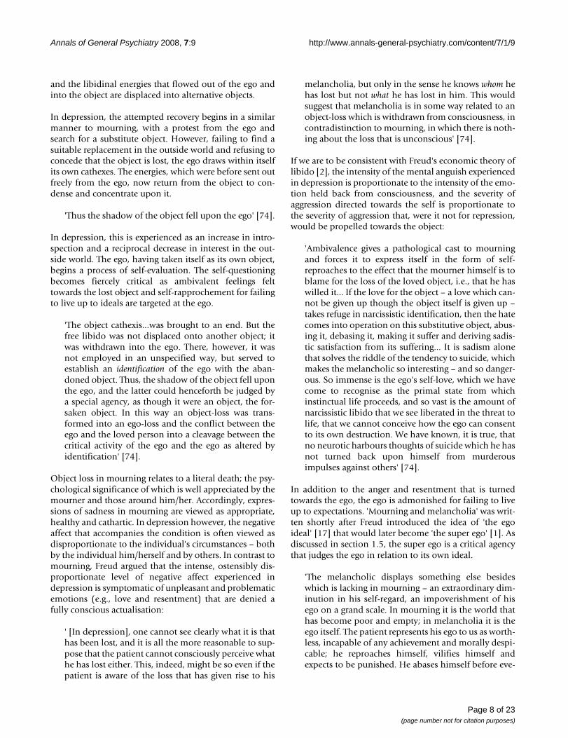
Neuropsychiatric findings in depression correlated with principles of Freudian metapsychologyHypofrontalityOne of the most consistent findings in the neuroimagingof depression is decreased cerebral blood flow (CBF) andglucose metabolism in the PFC, particularly the DLPFC[77-85] (figure 3). The PFC is a large and functionally het-erogeneous structure. Studies of frontal activity in depres-sion have highlighted these differences, with the DLPFC,associated with cognitive and executive functions show-ing decreased activity in depressed states, and the ventralPFC, associated with emotional processing, showingincreased activity during episodes of emotional rumina-tion (see [86] or [50]).
Several studies have found negative correlations betweendepression severity and frontal metabolism [78,81,87-93]. The induction of depressed symptomology in healthyvolunteers and remitted depressed patients has beenfound to correlate reliably with decreases in frontal activ-ity [56,94,95]. Frontal blood flow and metabolism tendsto normalise after spontaneous or treatment-inducedremission [51,78,79,96-105]. These studies highlight thereliability of frontal hypometabolism, particularly in theDLPFC, in neuroimaging studies of depression.
Based on the neuroimaging data we speculate thathypoactivity in the DLPFC is a correlate of withdrawnobject cathexis experienced subjectively as impoverishedmotivation and diminished interest in the matters outsideof the self. A recent functional magnetic resonance imag-ing (fMRI) study reported a positive correlation betweensubjective measures of anhedonia and activity in thevmPFC and OFC (Brodmann areas (BA)10, 11, and 32)[106]. Importantly, an additional relationship was foundbetween anhedonia scores and diminished activation ofthe amygdala and the ventral striatum. As will beexplained in the following section, in depression, Cg25can be envisaged as functioning in a manner analogous toa dam, preventing ascending energies from being investedin the PFC.
' [T]he ego controls the approaches to motility – thatis, to the discharge of excitations into the externalworld...' [107].
Page 9 of 23(page number not for citation purposes)
Annals of General Psychiatry 2008, 7:9 http://www.annals-general-psychiatry.com/content/7/1/9
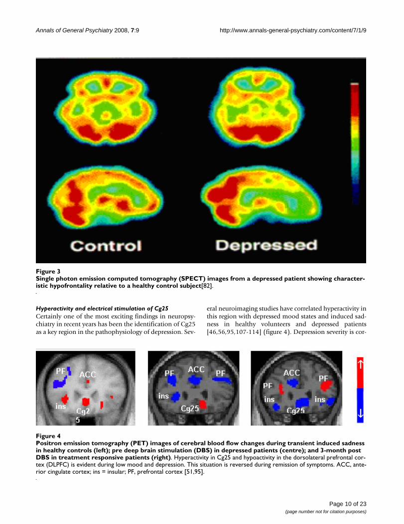
Hyperactivity and electrical stimulation of Cg25Certainly one of the most exciting findings in neuropsy-chiatry in recent years has been the identification of Cg25as a key region in the pathophysiology of depression. Sev-
eral neuroimaging studies have correlated hyperactivity inthis region with depressed mood states and induced sad-ness in healthy volunteers and depressed patients[46,56,95,107-114] (figure 4). Depression severity is cor-
Single photon emission computed tomography (SPECT) images from a depressed patient showing characteristic hypofrontality relative to a healthy control subject [82]Figure 3Single photon emission computed tomography (SPECT) images from a depressed patient showing character-istic hypofrontality relative to a healthy control subject[82].
Positron emission tomography (PET) images of cerebral blood flow changes during transient induced sadness in healthy con-trols (left); pre deep brain stimulation (DBS) in depressed patients (centre); and 3-month post DBS in treatment responsive patients (right)Figure 4Positron emission tomography (PET) images of cerebral blood flow changes during transient induced sadness in healthy controls (left); pre deep brain stimulation (DBS) in depressed patients (centre); and 3-month post DBS in treatment responsive patients (right). Hyperactivity in Cg25 and hypoactivity in the dorsolateral prefrontal cor-tex (DLPFC) is evident during low mood and depression. This situation is reversed during remission of symptoms. ACC, ante-rior cingulate cortex; ins = insular; PF, prefrontal cortex [51,95].
Page 10 of 23(page number not for citation purposes)
Annals of General Psychiatry 2008, 7:9 http://www.annals-general-psychiatry.com/content/7/1/9
related with Cg25 hypermetabolism [115] and increasedfunctional connectivity [46]. Spontaneous and treatment-induced remission of symptoms is associated with signif-icantly decreased Cg25 metabolism[51,100,105,110,113,116-119].
The subgenual cingulate has been the target of ablativesurgeries in the past [120] and, more recently, DBS [51],where high frequency stimulation is used to inhibit activ-ity in target nuclei. The preliminary results of chronicbilateral high frequency stimulation of Cg25 in sixpatients suffering from severe treatment-resistant depres-sion were reported by Mayberg and colleagues [51]. Sig-nificant improvements (a 50% or greater reduction inHamilton depression rating scale (HDRS-17) score) wereseen in five of the six patients at 2-month follow-up withsustained improvements achieved in four patients at 6months. Positron emission tomography (PET) scans ofpatients at 3 and 6 months post stimulation revealeddecreased blood flow in Cg25 and increased blood flow inthe DLPFC. Significant improvements were seen in sleep,energy, interest and psychomotor speed. Patients andtheir families reported 'renewed interest and pleasure insocial and family activities, decreased apathy and anhedo-nia, as well as improved ability to plan, initiate, and com-plete tasks that were reported as impossible prior tosurgery'.
At the 2007 international Neuropsychoanalysis congress inVienna, some first-person accounts relating to acute stim-ulation were reported:
'It isn't like something has been added – no, some-thing has been taken away'.
'It is as if I have just suddenly shifted from a state of allconsuming internal focus to realising that there arenumber of things around to do'.
'When you're depressed the focus is inwards. So ifsomeone tells you, well you aren't the only one whofeels like that, you don't care. With the stimulator, Idon't feel that inward look; it has lifted so I am not sofocused on myself...'.
'It is as though I have been locked in a room with 10screaming children; constant noise, no rest, no escape.Whatever just happened, the children have just left thebuilding'
The 'something...taken away' described in these accountsis consistent with the idea of a release from repression(deactivation of Cg25) and a return to object cathexis(DLPFC activation). The final account is especially inter-esting given that the patient was a father of 5.
Interestingly, sudden and dramatic deactivations of Cg25and functionally related regions of the vmPFC and OFChave recently been recorded after intravenous infusion ofthe dissociative hallucinogen ketamine in healthy humanvolunteers [121]. These deactivations correlated stronglywith dissociative phenomena. Significant activations wereseen in the parahippocampal gyrus, temporal cortex andPCC. Importantly, the regions deactivated by ketamine(OFC and vmPFC) are those postulated in this paper to beinvolved in the process of repression, and the regions acti-vated by ketamine (specifically the medial temporal struc-tures), are those we hypothesise to be involved in theprimary psychical process of the unconscious mind. Aswith the classic psychedelic drugs (e.g., LSD and psilocy-bin), the effects of ketamine have been described as dis-turbing the mechanisms of repression and facilitating therelease of primary process thought [122]. Single doses ofketamine have been found to elicit a short-term antide-pressant effect in depressed patients [123-126] and thedrug has also been used as an adjunct to psychotherapywith reported efficacy in the treatment of alcoholism[122].
The function of the vmPFC and OFC in relation to repressionThe data cited in the previous section supports thehypothesis that Cg25 plays a key role in repression. How-ever, it is likely that Cg25 does not act alone in this regard.For example, activity in the ventral anterior PFC correlatespositively with depression severity and activity in thisregion decreases after effective treatment [50]. The OFC(BA11 and BA47) is activated when subjects try todecrease arousal to erotic films [127] and there is impov-erished activation of BA10 and 11 in paedophile sexoffenders viewing paedophilic material [128]. In healthycontrols viewing the same images, the lateral OFC (BA47)was activated. The lateral OFC has also been found to beactivated during contemplation of moral transgressions[129] and script-induced guilt [130].
Using autobiographical scripts designed to evoke strongemotion, healthy control subjects showed increasedblood flow in the vmPFC during script-induced angercompared to patients with anger attacks who showed animpoverished vmPFC response [131]. ImpoverishedvmPFC activation in anger patients suggests that recruit-ment of this region is necessary for suppression of aggres-sive affect. Significantly lower resting metabolism hasbeen recorded in the OFC of patients with a history ofreactive aggression [132,133] and patients with OFC andmPFC lesions who show an increased risk of reactiveaggression [134-136]. Healthy participants who imaginedresponding in an unrestrained aggressive manner to anassault showed hypoactivity in the OFC but increasedactivity in the same region when imagining restraint
Page 11 of 23(page number not for citation purposes)

Annals of General Psychiatry 2008, 7:9 http://www.annals-general-psychiatry.com/content/7/1/9
[137]. In cases of post traumatic stress disorder, a condi-tion characterised by unsuccessful repression of traumaticmemories, patients exposed to a script-driven reminder ofa personally traumatic experience showed impoverishedactivity in the rostral anterior cingulate compared withcontrols [138]. A related study showed a strong negativecorrelation between emotional scores and vmPFC activa-tion in PTSD patients exposed to script-driven remindersof their traumatic experience [139] implying that impov-erished vmPFC activation facilitates the return of the affectattached to the original trauma.
As will be discussed in the next section, activation of theamygdala is associated with the expression of primitiveemotions such as anger and fear as well as complex auto-biographical recollections [140,141]. It is interestingtherefore that the study by Dougherty and colleagues citedabove discovered an inverse relationship between bloodflow in the vmPFC and amygdala in healthy control sub-jects during script-driven anger but a positive relationshipbetween amygdala and vmPFC activity in patients withanger attacks [131]. These findings imply that patientswith anger attacks suffer from ineffective suppression ofamygdala activation [131]. A number of studies havedemonstrated that activation of the amygdala with con-comitant emotional arousal is very quickly followed byactivation of the OFC [142-145] and – in healthy individ-uals – suppression of the amygdala response [146-148].This suppressive function of the vmPFC/OFC is supportedby a large body of preclinical data [147-160]. It is likelythat this function is impaired in depression, with the sup-pressive/repressive action of the vmPFC/OFC being dom-inated by persistent flurries of limbic arousal [161].
Amygdala hyperactivity and electrical stimulation of medial temporal lobesHyperactivity in the amygdala has been reported in a largenumber of imaging studies of depression[47,87,97,111,162-169]. Increased activity in the amy-gdala has been recorded in studies of induced sadness inhealthy volunteers [169-171]. Amygdala activity has beenfound to correlate positively with depression severity[87,162,166], to show a sustained response to negativeemotional stimuli in depressed patients compared tohealthy controls [161] and to decrease in sensitivity toemotional stimuli after successful antidepressant treat-ment [172,173].
The amygdala has long been recognised to play an impor-tant role in emotion. Bilateral resection of the amygdalahas been found to result in dramatic behavioural changes(Klüver and Bucy syndrome) including emotional blunt-ing, indifference to loved ones, hyperorality and hypersex-uality [140]. Electrical stimulations of the humanamygdala and medial temporal regions have been found
to elicit a range of primitive emotional responses includ-ing: fear, anxiety, anger, aggression, sexual behaviours,déjà vu and autobiographical recollections [141,174-185]:
'I just get the electrical feeling, and it goes all the waythrough me...it makes me do things I don't want to do– I get mad' [10].
'I had a flash of familiar memory, but I don't knowwhat it was... I had a little memory – a scene in a play.They were talking and I could see it... Just seeing it inmy memory...a very familiar memory of a girl talkingto me...that feeling of familiarity – a familiar memory'[177].
A thorough phenomenological review of these experi-ences is necessary for an appreciation of the functional sig-nificance of the medial temporal lobes in relation to theprimary psychical process of the unconscious mind. Suchexperiences have been interpreted by several cliniciansand researchers as examples of primary process activitytaking over from the secondary psychical process of nor-mal waking consciousness [72,174,177,179,180,186-193]:
'Reflected in the seizure-related behaviour may beemotional trauma of early life, negative feelingstowards specific individuals because of past incidentsor situations' [192].
'Repression fails, the usual defence systems crumble,disturbing unconscious material erupts, anxietymounts, and the personality structure becomes inef-fective' [189].
'It is in my view wrong to call the feeling of havingexperienced something before an illusion. It is ratherthat at such moments something is touched on whichwe have already experienced once before' [5].
A review of depth electroencephalography recordingsfrom the medial temporal regions suggests that stimula-tion-induced dreamlike experiences share a common phe-nomenology and neurophysiology (bursts of rhythmictheta and slow-wave activity) with other dreamlike states[71]. It is hoped that converging evidences correlatingneurophysiological activity with qualitatively analysedphenomenological experiences will facilitate a widerunderstanding of the phenomenology and psychophysi-ology of the unconscious mind.
Cg25 connectivityAnatomical studies in primates have revealed dense con-nections between Cg25 and the hypothalamus [194,195]
Page 12 of 23(page number not for citation purposes)
Annals of General Psychiatry 2008, 7:9 http://www.annals-general-psychiatry.com/content/7/1/9
mPFC [196], parahippocampal cortex [197], amygdala,ventral striatum, septal nuclei, dorsomedial caudatenucleus and mediodorsal nucleus of the thalamus; withmoderate connections to the periaqueductal grey and dor-sal raphe nucleus [195,196]. Human tractography andfunctional connectivity analyses support these findings,showing prominent connections between Cg25 and theNAc, amygdala, hypothalamus, OFC and vmPFC[54,198,199]. The connections of Cg25 to a number ofimportant visceromotor centres offering profuse projec-tions to the PFC has led to suggestions that Cg25 plays animportant modulatory role in cortical functioning [195].A recent cytological analysis of the human cingulate cor-tex has revealed an especially dense concentration ofinhibitory receptors in Cg25 [200]. These findings areconsistent with the hypothesis that Cg25 exerts a control-ling influence over visceromotor regions.
Connectivity between Cg25 and the amygdala has beenfound to be especially strong during the viewing of fearfuland threatening faces [201]. The magnitude of disconnec-tivity between these structures predicted anxiety scores ina number of individuals [201]. Resting state connectivitybetween Cg25 and a range of structures including themedial temporal lobes has been found to predict treat-ment response in depressed patients [58] and a strong cor-relation was discovered between subjective measures ofneuroticism and Cg25 and amygdala activation duringthe viewing of emotionally provocative images [202].
In addition to medial temporal structures, other impor-tant visceromotor centres connected with Cg25 includethe NAc [198,203,204] and the VTA [196,205]. Cg25shows an especially high level of functional connectivitywith the NAc at rest [23,54,204]. The NAc and VTA are keynuclei in the mesocorticolimbic dopamine systems, beingcentral to the mechanisms of motivation and reward[10,68]. The VTA has also been found to be strongly acti-vated during ejaculation in male humans [64] cocaineinduced euphoria [65] and the heroin rush [66]. Electricalstimulation of the NAc and the septum has been found toelicit feelings of sexual pleasure and orgasm in humans[69,206,207]. Patients given a self-stimulator connectedto the septal region stimulated themselves repeatedly forhours and protested bitterly when attempts were made totake the device from them [206]. These findings supportthe association of the mesolimbic dopamine system withthe Freudian 'id' [1]. Chronic stimulation of the NAc hasrecently been carried out in three TRD patients [208].Early results suggest that this intervention may be particu-larly effective in relieving symptoms of anhedonia.
Volumetric reductions'The neurosis may last a considerable time and causemarked disturbances, but it may also run a latent
course and be overlooked. As a rule defence retains theupper hand in it; in any case alterations of the ego,comparable to scars, are left behind' [61].
Postmortem and MRI studies have found glial loss andvolume reductions in the PFC in major depressive disor-der (MDD) and bipolar disorder (BPD) [209-214] as wellas extensive losses in Cg25 and proximal paralimbicregions [163,201,215-221]. Unilateral and bilateral volu-metric reductions in the medial temporal regions – prima-rily in the hippocampus, have also been reported indepressed patients [112,201,222-232], as have reductionsin the ventral striatum [233,234].
It is not difficult to surmise that the metabolic work ofrepression has structural ramifications. This paperhypothesises that the volumetric reductions found inpostmortem and neuroimaging studies of depression arerelated to the effects of repression. It is significant that themost severe reductions have been found in Cg25 (48%reductions in 163) the area hypothesised to exert the pri-mary repressive force. One possible mechanism for thevolumetric reductions is glucocorticiod-mediated neuro-toxicity [235]. Dysregulation of the stress related hypoth-alamic-pituitary-adrenal (HPA) axis is consistentlyassociated with depression [236]. Dysregulation of theHPA may be related to hyperactivity in the amygdala[237]. Electrical stimulation of the amygdala increasescortisol release in humans [238]. HPA hyperactivityincreases the likelihood of excitotoxic processes, downreg-ulating glial, and increasing the concentrations of neuro-toxic glucocorticiods and excitiotoxic glutamate [239].The OFC and the vmPFC are dense in glucocorticiodreceptors and glutamate cells, with glutamatergic afferentsascending from the amygdala and hippocampus [240].
It is hypothesised that the volumetric reductions discov-ered in depressed patients, as well as patients sufferingfrom other major psychiatric conditions such as schizo-phrenia [194,208,210,212,215,216,219,241-248] may berelated to the effects of psychological conflict.
DiscussionThe main inspiration behind the primary hypothesis ofthis paper i.e., that Cg25 is centrally involved in repres-sion, was Mayberg's paper on DBS for the treatment ofsevere depression [51]. The findings of this study have aspecial significance for Freudian metapsychology. It hasbeen inferred in this paper that the sudden lifting of neg-ative affect upon stimulation of Cg25 is consistent withthe idea of a release of libido for object cathexis after it hasbeen pathologically 'dammed up' behind a centralrepressing force. However, the therapeutic response tostimulation raises some difficult questions for both psy-choanalysis and psychiatry. One important question con-
Page 13 of 23(page number not for citation purposes)
Annals of General Psychiatry 2008, 7:9 http://www.annals-general-psychiatry.com/content/7/1/9
cerns the economy of psychical energy in relation todepression and mania. Freud discussed the issue of maniatowards the end of 'Mourning and melancholia':
'The impression which several psycho-analytic investi-gators have already put into words is that the contentof mania is no different from that of melancholia, thatboth disorders are wrestling with the same "complex",but that probably in melancholia the ego has suc-cumbed to the complex whereas in mania it has mas-tered it or pushed it aside. Our second pointer isafforded by the observation that all states such as joy,exultation or triumph, which give us the normalmodel for mania, depend on the same economic con-ditions. What has happened here is that, as a result ofsome influence, a large expenditure of psychicalenergy, long maintained or habitually occurring, hasat last become unnecessary, so that it is available fornumerous applications and possibilities of discharge'[74].
Neuroimaging studies of manic patients have shown thatin direct contrast to depression, resting state activity isdecreased in the OFC [249,250] and increased in dorsalfrontal areas during manic episodes [250,251]. It is signif-icant that the early responses to Cg25 stimulation do notappear to switch patients from pathological depression toovert mania. According to Freud's model, manic episodesdepend on a quantity of dammed-up libido being sud-denly made available for object cathexis.
'An important element in the theory of repression isthe view that repression is not an event that occursonce but that it requires a permanent expenditure [ofenergy]. If this expenditure were to cease, the repressedimpulse, which is being fed all the time from itssources, would on the next occasion flow along thechannels from which it had been forced away, and therepression would either fail in its purpose or wouldhave to be repeated an indefinite number of times.Thus it is because drives are continuous in their naturethat the ego has to make its defensive action secure bya permanent expenditure [of energy]' [252].
In the long term it is feasible that abeyance of repressionleads to a therapeutic shift in the energetic equilibrium ofthe mind; but even if this is true, we still need to considerwhy upon release from repression, we do not see a patho-logical release of primitive drive and repressed memory.Are we to assume that electrical stimulation of Cg25removes both the physiological and psychological causesof depression? Even if depression is primarily an energeticphenomenon and the physiological causes are the psycho-logical causes (and vice versa), wouldn't there still remain
memory traces and exogenous stressors facilitating a recallof lost objects, regretted behaviours and eluded ideals?
One possible reason why the effect of Cg25 stimulationappears to have a sustained beneficial effect [253] ratherthan an iatrogenic one [254] may be that inhibiting activ-ity in Cg25 facilitates the disintegration of a wider net-work. For example, it is possible that activation of Cg25supports activation of the DMN and deactivation of Cg25supports deactivation of the DMN and activation of theON. This model would account for the diminished selffocus (ego cathexis) and rejuvenated task focus (objectcathexis) seen upon Cg25 stimulation. It may be possibleto test this formulation through neuroimaging studies ofpatients undergoing Cg25 stimulation. If the ego isdependent on repression, we would expect to seedecreased activity in the DMN, decreased activity in Cg25and increased activity in the ON after stimulation. Prelim-inary evidence lends support to this model [51]. Evidencesupporting the interdependency of the ego and repressionwould of course have important implications for the his-tory of the evolution of human consciousness.
In order to test the validity of the Freudian model, it isimportant that there be thorough psychophenomenolog-ical and neurophysiological analyses of Cg25 and NAcstimulations. Ideally, subjective and objective measuresshould be taken simultaneously, in real time. If, as thispaper predicts, Cg25 is centrally involved in repression,then in addition to dramatic improvements in energy/libido we would also expect to see some adverse responsesto stimulation, such as disinhibited behaviour, patholog-ical drive, perseverance, hostility, aggression, sexual pro-miscuity and a reduced capacity to consider others. Suchbehaviours are commonly associated with ventromedialprefrontal lesions [255-259]. We would also predict thatpatients undergoing Cg25 stimulation would be moresusceptible to temporal lobe phenomena (such as déjàvu) as a result of diminished inhibitory control over exci-tatory medial temporal structures. If electrophysiologicalrecordings are carried out, we would hypothesise thatelectrodes placed within the proximity of septal,supramammillary or hippocampal theta structures woulddisplay characteristic bursts of high voltage rhythmic thetaduring moments of strong emotion ([69], see [71] for areview). We would also predict that intracranial EEGrecordings in bipolar patients would reveal significantchanges in activity paralleling shifts in mood i.e., Cg25hyperactivity/NAc hypoactivity during depression andCg25 hypoactivity/NAc hyperactivity during mania.
It is acknowledged that very little in the way of counterevidence has been cited in this paper challenging thevalidity of the Freudian model. It is likely that severalexamples could be found in Freud's evolving work of
Page 14 of 23(page number not for citation purposes)

Annals of General Psychiatry 2008, 7:9 http://www.annals-general-psychiatry.com/content/7/1/9
hypotheses that do not correspond well with the findingsof modern clinical research. However, it must be empha-sised that what we have brought together in this paper areprincipal concepts of Freudian metapsychology togetherwith principal findings of neuropsychiatry. It is all themore significant therefore that the meeting has been com-plementary.
In order to develop a discussion of the comparative meritsof psychological paradigms, it is worth reminding our-selves of the two main aims of this paper: (1) to proposea series of hypotheses correlating neurophysiologicalprocesses with some fundamental processes of psychoa-nalysis, and (2) to highlight correspondences betweenFreud's writings in 'Mourning and melancholia' and cur-rent findings in depression. How successful these taskshave been will largely depend on two factors: (1) whetherevidences from other fields converge with the evidencesreviewed here, and (2) whether the psychoanalytic per-spective is given credence. There is already ample evidenceto support the role of Cg25, the vmPFC and OFC in sup-pressing primitive affect, but the psychoanalytic signifi-cance of this function has yet to be fully appreciated.
It has been said before that it matters little which psycho-logical discipline we choose to derive our operationalterms; the approach is secondary to the phenomena:
'Listen my friend, the golden tree of life is green, alltheory is grey' [260].
Psychological models do serve a purpose however, but toprovide comprehensive explanations of mental states andbehaviours, effective models must evolve naturally fromtheir phenomena. In a recent letter published in a reputa-ble journal and co-signed by a number of leadingresearchers [261] a proposal was put-forward as part of a'decade of the mind' initiative to work towards a transdis-ciplinary explanation of mental phenomena. The mainpsychological discipline championed by the authors wascognitive psychology. While the essential idea is a com-mendable one, we must ask ourselves seriously whetherthe information processing paradigm is really the bestmodel for carrying out this initiative. The psychologicallimitations of the behavioural model have been recog-nised for several decades but the cognitive approach,which views the human mind as an information processoris currently the favoured model of clinicians and research-ers. If the computer analogy is an accurate representationof the human psyche, then we can feel comfortable goinginto the final years of the 'decade of the mind' that realprogress will be made. If however, the model is at allincomplete, we may need to consult alternative para-digms to assist our empiricism. The information process-ing model has traditionally been put to good use guiding
and informing empirical research. However, severalresearchers are now recognising that the computationalmodel has limitations, especially when it is applied tohuman emotion [62,68,262,263]. What we hope psycho-analysis can bring to the table therefore, is a psychologicalmodel that has its roots set firmly in human experience.We hope psychoanalysis can work alongside cognitivepsychology to provide a more comprehensive under-standing of human experience.
' [T]he mind would often slip through the fingers ofpsychology, if psychology refused to keep a hold onthe mind's unconscious states' [264].
'Psychoanalysis still represents the most coherent andintellectually satisfying view of the mind that we have'[263].
The primary requirement for a scientific psychoanalysis is(and always has been) to confirm beyond reasonabledoubt that the unconscious mind exists and that it is notonly important but essential for an understanding of thehuman mind and behaviour. If, as this paper maintains,the unconscious does exist, then regardless of the wordschosen to define it, the establishment of its phenomenol-ogy as subject matter worthy of scientific investigation isimportant. Deciding how best to test and confirm thehypothesis that the unconscious mind exists will presentus simultaneously with a direction towards studying itsform and physiology. Due to the rigour of repression,depression is not the easiest phenomenon to gain a per-spective on the workings of the unconscious. The psycho-ses provide a better vantage:
'Things that in the neuroses have to be laboriouslyfetched up from the depths are found in the psychoseson the surface, visible to every eye' [265].
' [M]aterial which is ordinarily unconscious can trans-form itself into preconscious material and thenbecome conscious – a thing that happens to a largeextent in psychotic states. From this we infer that themaintenance of certain internal resistances is a sine quanon of normality' [59].
In depression we only assume the existence of the uncon-scious through a process of deduction based on ostensiblyirrational behaviours (e.g., self-harm, violent self-criticismetc). As Freud made clear, there are much better ways ofstudying the unconscious and the free-flowing psychicalenergies that are its signature. Freud first stumbled acrossa realisation of the unconscious through his work on dis-sociative states [72]:
Page 15 of 23(page number not for citation purposes)

Annals of General Psychiatry 2008, 7:9 http://www.annals-general-psychiatry.com/content/7/1/9
' [O]ne received the clearest impression – especiallyfrom the behaviour of subjects after hypnosis – of theexistence of mental processes that one could onlydescribe as "unconscious". The "unconscious" has it istrue, long been under discussion among philosophersas a theoretical concept; but now for the first time, inthe phenomena of hypnotism, it became somethingactual, tangible and subject to experiment' [1].
'To most people educated in philosophy the idea ofanything psychical which is not also conscious is soinconceivable that it seems to them absurd and refut-able simply by logic. I believe this is only because theyhave never studied the relevant phenomena of hypno-sis and dreams, which – quite apart from pathologicalmanifestations – necessitate this view. Their psychol-ogy of consciousness is incapable of solving the prob-lems of dreams and hypnosis' [1].
For Freud, dreams were a way of studying the unconscious– unfettered by waking consciousness but the phenome-nology of dreaming has largely failed to convince scepticsof the existence of the unconscious. Freud acknowledgedthat converging lines of enquiry would be required to con-solidate the insights gained through the study of dreams:
'Thus, the psychological hypotheses to which we areled by an analysis of the process of dreaming must beleft, as it were, in suspense, until they can be related tothe findings of other enquiries which seek to approachthe kernel of the same problem from another angle'[5].
Future work may provide the necessary evidence. Alterna-tive means of studying the unconscious – perhaps by wayof a pharmacological agent such as a psychedelic drug[193,266] may open up fresh angles of enquiry. Freudfamously described the interpretation of dreams as 'theroyal road to a knowledge of the unconscious activities ofthe mind' [5]. However, dreaming occurs in sleep, makingreal-time recitation of subjective phenomena impossible.If we could stimulate the primary psychical process inwaking we would have a more effective method for stud-ying the unconscious:
'Freud once said of dreams that they were the via regiaor royal way to study the unconscious; to an evengreater degree this seems to be true for the LSD experi-ence' [265].
It is anticipated that progress towards a wider appreciationof the psychoanalytic model will first require confirma-tion of the existence of the unconscious mind. We pro-pose that the most effective way of achieving this is tostimulate the primary psychical process in waking con-
sciousness. There is a wealth of evidence to suggest thatthis can be reliably achieved through the use of a psyche-delic drug such as LSD [193,266-285]:
'One must...put it simply, it does seem that all LSDdoes is open the doors to the unconscious' [279].
Using neuroimaging techniques we would predict that theego dissolving, primary process releasing properties of apsychedelic compound would correspond with a shift ineffective connectivity in the DMN, with the medial tem-poral regions (as opposed to the vmPFC) exerting princi-pal causality [33,121,286,287]. Testing this hypothesiswill be difficult, but such challenging procedures are nec-essary if the primary psychical process is to be considereda matter worthy of investigation. Once we are better ableto study the phenomenology of the unconscious, theapplication of our new knowledge to the study and treat-ment of the whole of the mind will follow more easily.
ConclusionThe goal of this paper has been to investigate consistenciesbetween Freudian metapsychology and empirical findingsin neuropsychiatry. A summary of several key psychoana-lytic concepts was given together with some early hypoth-eses about their physiological coordinates. This was doneto facilitate an understanding of Freudian terminologyand allow for the application of these ideas to areas ofclinical interest. Modern clinical research and olderempirical work such as intracranial stimulations was dis-cussed in relation to Freudian metapsychology in order tohighlight correspondences between physiology, phenom-enology and theory. If a new level of scientific verificationis achieved for subjective phenomena of relevance to psy-choanalysis, this will have implications not just for theway in which psychoanalysis is viewed by the wider phil-osophical, psychological and psychiatric communities,but also for those interested in incorporating psychoana-lytic ideas into their own clinical practice.
' [I]t should not be forgotten that in fact [the distinc-tion between the unconscious and conscious dimen-sions of the mind] is not a theory at all but a first stock-taking of the facts of our observations, that it keeps asclose to them as possible' [59].
'Psycho-analysis an Empirical Science – Psychoanalysis isnot, like philosophies, a system starting out from a fewsharply defined basic concepts, seeking to grasp thewhole universe with the help of these and, once it iscompleted, having no room for fresh discoveries orbetter understanding. On the contrary, it keeps closeto the facts in its field of study, seeks to solve theimmediate problems of observation, gropes its wayforward by the help of experience, is always incom-
Page 16 of 23(page number not for citation purposes)
Annals of General Psychiatry 2008, 7:9 http://www.annals-general-psychiatry.com/content/7/1/9
plete and always ready to correct or modify its theo-ries. There is no incongruity (anymore than in the caseof physics or chemistry) if its most general conceptslack clarity and if its postulates are provisional; itleaves their more precise definition to the results offuture work' [288].
Competing interestsThe authors declare that they have no competing interests.
Authors' contributionsThe present paper was inspired by the work of HSM as pre-sented at the 2007 International Neuropsychoanalysiscongress in Vienna. The paper was written by RLC–H withintellectual support and guidance from HSM, ALM andDJN. All authors read and approved the final manuscript.
AcknowledgementsRCH is grateful to Drs S J Wilson and S J C Davies of the University of Bris-tol Psychopharmacology Unit for providing additional intellectual support.
References1. Freud S: The ego and the id 19th edition. London: Vintage; 1923. 2. Freud S: Project for a scientific psychology 1st edition. London: Vintage;
1895. 3. Pribram K, Gill M: Freud's project re-assessed Tiptree, UK: Anchor
Press; 1976. 4. Strachey J: Freud S (1886–1899). Project for a scientific psychology 1st
edition. Edited by: Strachey J. London: Vintage; 1954. 5. Freud S: The interpretation of dreams London: Penguin; 1900. 6. Freud S: The unconscious 14th edition. London: Vintage; 1915. 7. Freud S: On narcissism 14th edition. London: Vintage; 1914. 8. Fletcher P, McKenna PJ, Friston KJ, Frith CD, Dolan RJ: Abnormal
cingulate modulation of fronto-temporal connectivity inschizophrenia. Neuroimage 1999, 9:337-342.
9. Freud S: On aphasia 14th edition. London: Vintage; 1891. 10. MacLean PD: The triune brain in evolution New York: Plenum Press;
1990. 11. Fonagy P: Psychoanalysis today. World Psychiatry 2003, 2:73-80.12. Freud S: Neurosis and psychosis 19th edition. London: Vintage; 1924. 13. Freud S: Draft F 1st edition. London: Vintage; 1894. 14. Freud S: Three essays on the theory of sexuality 7th edition. London: Vin-
tage; 1905. 15. Jung CG: On psychic energy. In On the nature of the psyche New
York: Routledge; 1928. 16. Strachey J: Freud S (1893–1899). The emergence of Freud's fundamental
hypotheses 3rd edition. Edited by: Strachey J. London: Vintage; 1924. 17. Freud S: The psychoneuroses of defence 3rd edition. London: Vintage;
1894. 18. Freud S: Some points for an organic study of hysterical and motor paralyses
1st edition. London: Vintage; 1893. 19. Raichle ME, MacLeod AM, Snyder AZ, Powers WJ, Gusnard DA, Shul-
man GL: A default mode of brain function. Proc Natl Acad Sci USA2001, 98:676-682.
20. Frued : Beyond the pleasure principle 18th edition. London: Vintage;1920.
21. Gusnard DA, Raichle ME: Searching for a baseline: functionalimaging and the human brain. Nat Rev Neurosci 2001, 2:685-694.
22. Gusnard DA, Akbudak E, Shulman GL, Raichle ME: Medial prefron-tal cortex and self-referential mental activity: relation to adefault mode of brain function. Proc Natl Acad Sci USA 2001,98:4259-4264.
23. Greicius MD, Krasnow B, Reiss AL, Menon V: Functional connec-tivity in the resting brain: a network analysis of the defaultmode hypothesis. Proc Natl Acad Sci USA 2003, 100:253-258.
24. Johnson SC, Baxter LC, Wilder LS, Pipe JG, Heiserman JE, PrigatanoGP: Neural correlates of self-reflection. Brain 2002,125:1808-1814.
25. Vogeley K, May M, Ritzl A, Falkai P, Zilles K, Fink GR: Neural corre-lates of first-person perspective as one constituent of humanself-consciousness. J Cogn Neurosci 2004, 16:817-827.
26. Kelley WM, Macrae CN, Wyland CL, Caglar S, Inati S, Heatherton TF:Finding the self? An event-related fMRI study. J Cogn Neurosci2002, 14:785-794.
27. Fossati P, Hevenor SJ, Graham SJ, Grady C, Keightley ML, Craik F,Mayberg H: In search of the emotional self: an fMRI studyusing positive and negative emotional words. Am J Psychiatry2003, 160:1938-1945.
28. Fox MD, Snyder AZ, Vincent JL, Corbetta M, Van Essen DC, RaichleME: The human brain is intrinsically organized into dynamic,anticorrelated functional networks. Proc Natl Acad Sci USA 2005,102:9673-9678.
29. Fransson P: Spontaneous low-frequency BOLD signal fluctua-tions: an fMRI investigation of the resting-state default modeof brain function hypothesis. Hum Brain Map 2005, 26:15-29.
30. Greicius MD, Supekar K, Menon V, Dougherty RF: Resting-statefunctional connectivity reflects structural connectivity in thedefault mode network. Cereb Cortex 2008.
31. Fair DA, Cohen AL, Dosenbach NU, Church JA, Miezin FM, BarchDM, Raichle ME, Petersen SE, Schlaggar BL: The maturing archi-tecture of the brain's default network. Proc Natl Acad Sci USA2008, 105:4028-4032.
32. Buckner RL, Carroll DC: Self-projection and the brain. TrendsCogn Sci 2006, 11:49-57.
33. Uddin LQ, Clare Kelly AM, Biswal BB, Xavier Castellanos F, MilhamMP: Functional connectivity of default mode network compo-nents: correlation, anticorrelation, and causality. Hum BrainMap 2008 in press.
34. Saxe R, Xiao DK, Kovacs G, Perrett DI, Kanwisher N: A region ofright posterior superior temporal sulcus responds toobserved intentional actions. Neuropsychologia 2004,42:1435-1446.
35. Stark M, Coslett H, Saffran E: Impairment of an egocentric mapof locations: Implications for perception and action. CognNeuropsychol 1996, 13:481-523.
36. Maddock RJ, Garrett AS, Buonocore MH: Remembering familiarpeople: the posterior cingulate cortex and autobiographicalmemory retrieval. Neurosci 2001, 104:667-676.
37. Maguire EA, Mummery CJ: Differential modulation of a commonmemory retrieval network revealed by positron emissiontomography. Hippocampus 1999, 9:54-61.
38. Vincent JL, Snyder AZ, Fox MD, Shannon BJ, Andrews JR, Raichle ME,Buckner RL: Coherent spontaneous activity identifies a hip-pocampal-parietal memory network. J Neurophysiol 2006,96:3517-3531.
39. Gilboa A, Winocur G, Grady CL, Hevenor SJ, Moscovitch M:Remembering our past: functional neuroanatomy of recol-lection of recent and very remote personal events. Cereb Cor-tex 2004, 14:1214-1225.
40. Raichle ME, Snyder AZ: A default mode of brain function: a briefhistory of an evolving idea. Neuroimage 2007, 37:1083-1090.
41. Greicius MD, Kiviniemi V, Tervonen O, Vainionpää V, Alahuhta S,Reiss AL, Menon V: Persistent default-mode network connec-tivity during light sedation. Hum Brain Map 2008, 29:839-847.
42. Rombouts SA, Barkhof F, Goekoop R, Stam CJ, Scheltens P: Alteredresting state networks in mild cognitive impairment andmild Alzheimer's disease: an fMRI study. Hum Brain Map 2005,26:231-239.
43. Kennedy DP, Redcay E, Courchesne E: Failing to deactivate: rest-ing functional abnormalities in autism. Proc Natl Acad Sci USA2006, 103:8275-8280.
44. Garrity AG, Pearlson GD, McKiernan K, Lloyd D, Kiehl KA, CalhounVD: Aberrant "default mode" functional connectivity inschizophrenia. Am J Psychiatry 2007, 164:450-457.
45. Zhou Y, Liang M, Tian L, Wang K, Hao Y, Liu H, Liu Z, Jiang T: Func-tional disintegration in paranoid schizophrenia using resting-state fMRI. Schizophr Res 2007, 97:194-205.
46. Greicius MD, Flores BH, Menon V, Glover GH, Solvason HB, KennaH, Reiss AL, Schatzberg AF: Resting-state functional connectiv-ity in major depression: abnormally increased contributionsfrom subgenual cingulate cortex and thalamus. Biol Psychiatry2007, 62:429-437.
47. Anand A, Li Y, Wang Y, Wu J, Gao S, Bukhari L, Mathews VP, KalninA, Lowe MJ: Activity and connectivity of brain mood regulat-
Page 17 of 23(page number not for citation purposes)

Annals of General Psychiatry 2008, 7:9 http://www.annals-general-psychiatry.com/content/7/1/9
ing circuit in depression: a functional magnetic resonancestudy. Biol Psychiatry 2005, 57:1079-1088.
48. Pomarol-Clotet E, Salvador R, Sarró S, Gomar J, Vila F, Martínez A,Guerrero A, Ortiz-Gil J, Sans-Sansa B, Capdevila A, Cebamanos JM,McKenna PJ: Failure to deactivate in the prefrontal cortex inschizophrenia: dysfunction of the default mode network? Psy-chol Med 2008, 29:1-9.
49. Freud S: A difficulty in the path of psycho-analysis 17th edition. London:Vintage; 1917.
50. Drevets WC: Orbitofrontal cortex function and structure indepression. Ann NY Acad Sci 2007, 1121:499-527.
51. Mayberg HS, Lozano AM, Voon V, McNeely HE, Seminowicz D, Ham-ani C, Schwalb JM, Kennedy SH: Deep brain stimulation for treat-ment-resistant depression. Neuron 2005, 45:651-660.
52. Dougherty DD, Shin LM, Rauch SL: Orbitofrontal cortex activa-tion during functional neuroimaging studies of emotioninduction in humans. In The orbitofrontal cortex Edited by: Zald DH,Rauch SL. Oxford, UK: Oxford University Press; 2006.
53. Meyer-Lindenberg AS, Olsen RK, Kohn PD, Brown T, Egan MF, Wein-berger DR, Berman KF: Regionally specific disturbance of dor-solateral prefrontal-hippocampal functional connectivity inschizophrenia. Arch Gen Psychiatry 2005, 62:379-386.
54. Margulies DS, Kelly AM, Uddin LQ, Biswal BB, Castellanos FX, MilhamMP: Mapping the functional connectivity of anterior cingulatecortex. Neuroimage 2007, 37:579-588.
55. Mayberg HS, Brannan SK, Mahurin RK, Jerabek PA, Brickman JS,Tekell JL, Silva JA, McGinnis S, Glass TG, Martin CC, Fox PT: Cingu-late function in depression: a potential predictor of treat-ment response. Neuroreport 1997, 8:1057-1061.
56. Mayberg HS, Liotti M, Brannan SK, McGinnis S, Mahurin RK, JerabekPA, Silva JA, Tekell JL, Martin CC, Lancaster JL, Fox PT: Reciprocallimbic-cortical function and negative mood: converging PETfindings in depression and normal sadness. Am J Psychiatry 1999,156:675-682.
57. Mayberg HS: Modulating dysfunctional limbic-cortical circuitsin depression: towards development of brain-based algo-rithms for diagnosis and optimised treatment. Br Med Bull2003, 65:193-207.
58. Seminowicz DA, Mayberg HS, McIntosh AR, Goldapple K, Kennedy S,Segal Z, Rafi-Tari S: Limbic-frontal circuitry in major depres-sion: a path modeling metanalysis. Neuroimage 2004, 22:409-18.
59. Freud S: An outline of psychoanalysis 23rd edition. London: Vintage;1940.
60. Freud S: New introductory lectures of psychoanalysis 22nd edition. Lon-don: Vintage; 1933.
61. Freud S: Moses and monotheism 23rd edition. London: Vintage; 1939. 62. Solms M, Turnbull O: The brain and the inner world London: Karnac;
2002. 63. Pagnoni G, Zink CF, Montague PR, Berns GS: Activity in human
ventral striatum locked to errors of reward prediction. NatNeurosci 2002, 5:97-98.
64. Holstege G, Georgiadis JR, Paans AM, Meiners LC, Graaf FH van der,Reinders AA: Brain activation during human male ejaculation.J Neurosci 2003, 23:9185-9193.
65. Breiter HC, Gollub RL, Weisskoff RM, Kennedy DN, Makris N, BerkeJD, Goodman JM, Kantor HL, Gastfriend DR, Riorden JP, Mathew RT,Rosen BR, Hyman SE: Acute effects of cocaine on human brainactivity and emotion. Neuron 1997, 19:591-611.
66. Sell LA, Morris J, Bearn J, Frackowiak RS, Friston KJ, Dolan RJ: Acti-vation of reward circuitry in human opiate addicts. Eur J Neu-rosci 1999, 11:1042-1048.
67. Pappata S, Dehaene S, Poline JB, Gregoire MC, Jobert A, Delforge J,Frouin V, Bottlaender M, Dolle F, Di Giamberardino L, Syrota A: Invivo detection of striatal dopamine release during reward: aPET study with [(11)C]raclopride and a single dynamic scanapproach. Neuroimage 2002, 16:1015-1027.
68. Panksepp J: Affective neuroscience New York: Oxford University Press;1998.
69. Heath RG: The role of pleasure in behavior New York: Hoeber; 1964. 70. Hassin RR, Uleman JS, Bargh JA: The new unconscious Oxford, UK:
Oxford University Press; 2005. 71. Carhart-Harris R: Waves of the unconscious: The neurophysi-
ology of dreamlike states and its implications for the psy-chodynamic model of the mind. Neuropsychoanalysis 2007,9:183-211.
72. Breuer J, Freud S: Studies on hysteria. Standard edition of the completeworks of Sigmund Freud Volume 2. London: Vintage :1893-1895.
73. Freud S: Analysis terminable and interminable 23rd edition. London: Vin-tage; 1937.
74. Freud S: Mourning and melancholia 14th edition. London: Vintage;1917.
75. Freud S: Totem and taboo 13th edition. London: Vintage; 1913. 76. Freud S: Group psychology and the analysis of the ego 18th edition. Lon-
don: Vintage; 1921. 77. Bremner JD, Innis RB, Salomon RM, Staib LH, Ng CK, Miller HL, Bro-
nen RA, Krystal JH, Duncan J, Rich D, Price LH, Malison R, Dey H,Soufer R, Charney DS: Positron emission tomography meas-urement of cerebral metabolic correlates of tryptophandepletion-induced depressive relapse. Arch Gen Psychiatry 1997,54(4):364-374.
78. Baxter LR Jr, Schwartz JM, Phelps ME, Mazziotta JC, Guze BH, SelinCE, Gerner RH, Sumida RM: Reduction of prefrontal cortex glu-cose metabolism common to three types of depression. ArchGen Psychiatry 1989, 46:243-250.
79. Bench CJ, Friston KJ, Brown RG, Scott LC, Frackowiak RS, Dolan RJ:The anatomy of melancholia – focal abnormalities of cere-bral blood flow in major depression. Psychol Med 1992,22:607-615.
80. Biver F, Goldman S, Delvenne V, Luxen A, De Maertelaer V, HubainP, Mendlewicz J, Lotstra F: Frontal and parietal metabolic dis-turbances in unipolar depression. Biol Psychiatry 1994,36:381-388.
81. Cohen RM, Gross M, Nordahl TE, Semple WE, Oren DA, RosenthalN: Preliminary data on the metabolic brain pattern ofpatients with winter seasonal affective disorder. Arch Gen Psy-chiatry 1992, 49:545-552.
82. Mayberg HS, Lewis PJ, Regenold W, Wagner HN Jr: Paralimbichypoperfusion in unipolar depression. J Nucl Med 1994,35(6):929-934.
83. Ebert D, Feistel H, Barocka A: Effects of sleep deprivation on thelimbic system and the frontal lobes in affective disorders: astudy with Tc-99m-HMPAO SPECT. Psychiatry Res 1991,40:247-251.
84. Ring HA, Bench CJ, Trimble MR, Brooks DJ, Frackowiak RS, Dolan RJ:Depression in Parkinson's disease. A positron emissionstudy.Br J Psychiatry 1994, 165:333-339.
85. Drevets WC, Ongür D, Price JL: Reduced glucose metabolism inthe subgenual prefrontal cortex in unipolar depression. MolPsychiatry 1998, 3:190-191.
86. Dougherty DD, Rauch SL: Brain correlates of antidepressanttreatment outcome from neuroimaging studies in depres-sion. Psychiatr Clin North Am 2007, 30:91-103.
87. Drevets WC, Videen TO, Price JL, Preskorn SH, Carmichael ST, Rai-chle ME: A functional anatomical study of unipolar depres-sion. J Neurosci 1992, 12:3628-3641.
88. Yazici KM, Kapucu O, Erbas B, Varoglu E, Gülec C, Bekdik CF:Assessment of changes in regional cerebral blood flow inpatients with major depression using the 99mTc-HMPAOsingle photon emission tomography method. Eur J Nucl Med1992, 19(12):1038-1043.
89. Andreason PJ, Altemus M, Zametkin AJ, King AC, Lucinio J, CohenRM: Regional cerebral glucose metabolism in bulimia ner-vosa. Am J Psychiatry 1992, 149:1506-1513.
90. Hirono N, Mori E, Ishii K, Ikejiri Y, Imamura T, Shimomura T, Hashi-moto M, Yamashita H, Sasaki M: Frontal lobe hypometabolismand depression in Alzheimer's disease. Neurology 1998,50:380-383.
91. Mayberg HS, Starkstein SE, Sadzot B, Preziosi T, Andrezejewski PL,Dannals RF, Wagner HN Jr, Robinson RG: Selective hypometab-olism in the inferior frontal lobe in depressed patients withParkinson's disease. Ann Neurol 1990, 28:57-64.
92. Volkow ND, Hitzemann R, Wang GJ, Fowler JS, Wolf AP, Dewey SL,Handlesman L: Long-term frontal brain metabolic changes incocaine abusers. Synapse 1992, 11:184-190.
93. Bonne O, Krausz Y, Shapira B, Bocher M, Karger H, Gorfine M, ChisinR, Lerer B: Increased cerebral blood flow in depressedpatients responding to electroconvulsive therapy. J Nucl Med1996, 37(7):1075-1080.
94. Bremner JD, Vythilingam M, Ng CK, Vermetten E, Nazeer A, OrenDA, Berman RM, Charney DS: Regional brain metabolic corre-lates of alpha-methylparatyrosine-induced depressive symp-
Page 18 of 23(page number not for citation purposes)
http://www.ncbi.nlm.nih.gov/entrez/query.fcgi?cmd=Retrieve&db=PubMed&dopt=Abstract&list_uids=9141092
http://www.ncbi.nlm.nih.gov/entrez/query.fcgi?cmd=Retrieve&db=PubMed&dopt=Abstract&list_uids=9141092
http://www.ncbi.nlm.nih.gov/entrez/query.fcgi?cmd=Retrieve&db=PubMed&dopt=Abstract&list_uids=9141092
http://www.ncbi.nlm.nih.gov/entrez/query.fcgi?cmd=Retrieve&db=PubMed&dopt=Abstract&list_uids=9331351
http://www.ncbi.nlm.nih.gov/entrez/query.fcgi?cmd=Retrieve&db=PubMed&dopt=Abstract&list_uids=9331351
http://www.ncbi.nlm.nih.gov/entrez/query.fcgi?cmd=Retrieve&db=PubMed&dopt=Abstract&list_uids=9107153
http://www.ncbi.nlm.nih.gov/entrez/query.fcgi?cmd=Retrieve&db=PubMed&dopt=Abstract&list_uids=9107153
http://www.ncbi.nlm.nih.gov/entrez/query.fcgi?cmd=Retrieve&db=PubMed&dopt=Abstract&list_uids=9107153
http://www.ncbi.nlm.nih.gov/entrez/query.fcgi?cmd=Retrieve&db=PubMed&dopt=Abstract&list_uids=2784046
http://www.ncbi.nlm.nih.gov/entrez/query.fcgi?cmd=Retrieve&db=PubMed&dopt=Abstract&list_uids=2784046
http://www.ncbi.nlm.nih.gov/entrez/query.fcgi?cmd=Retrieve&db=PubMed&dopt=Abstract&list_uids=1410086
http://www.ncbi.nlm.nih.gov/entrez/query.fcgi?cmd=Retrieve&db=PubMed&dopt=Abstract&list_uids=1410086
http://www.ncbi.nlm.nih.gov/entrez/query.fcgi?cmd=Retrieve&db=PubMed&dopt=Abstract&list_uids=1410086
http://www.ncbi.nlm.nih.gov/entrez/query.fcgi?cmd=Retrieve&db=PubMed&dopt=Abstract&list_uids=7803599
http://www.ncbi.nlm.nih.gov/entrez/query.fcgi?cmd=Retrieve&db=PubMed&dopt=Abstract&list_uids=7803599
http://www.ncbi.nlm.nih.gov/entrez/query.fcgi?cmd=Retrieve&db=PubMed&dopt=Abstract&list_uids=1627045
http://www.ncbi.nlm.nih.gov/entrez/query.fcgi?cmd=Retrieve&db=PubMed&dopt=Abstract&list_uids=1627045
http://www.ncbi.nlm.nih.gov/entrez/query.fcgi?cmd=Retrieve&db=PubMed&dopt=Abstract&list_uids=8195877
http://www.ncbi.nlm.nih.gov/entrez/query.fcgi?cmd=Retrieve&db=PubMed&dopt=Abstract&list_uids=8195877
http://www.ncbi.nlm.nih.gov/entrez/query.fcgi?cmd=Retrieve&db=PubMed&dopt=Abstract&list_uids=1811242
http://www.ncbi.nlm.nih.gov/entrez/query.fcgi?cmd=Retrieve&db=PubMed&dopt=Abstract&list_uids=1811242
http://www.ncbi.nlm.nih.gov/entrez/query.fcgi?cmd=Retrieve&db=PubMed&dopt=Abstract&list_uids=1811242
http://www.ncbi.nlm.nih.gov/entrez/query.fcgi?cmd=Retrieve&db=PubMed&dopt=Abstract&list_uids=7994502
http://www.ncbi.nlm.nih.gov/entrez/query.fcgi?cmd=Retrieve&db=PubMed&dopt=Abstract&list_uids=7994502
http://www.ncbi.nlm.nih.gov/entrez/query.fcgi?cmd=Retrieve&db=PubMed&dopt=Abstract&list_uids=9672886
http://www.ncbi.nlm.nih.gov/entrez/query.fcgi?cmd=Retrieve&db=PubMed&dopt=Abstract&list_uids=9672886
http://www.ncbi.nlm.nih.gov/entrez/query.fcgi?cmd=Retrieve&db=PubMed&dopt=Abstract&list_uids=1527602
http://www.ncbi.nlm.nih.gov/entrez/query.fcgi?cmd=Retrieve&db=PubMed&dopt=Abstract&list_uids=1527602
http://www.ncbi.nlm.nih.gov/entrez/query.fcgi?cmd=Retrieve&db=PubMed&dopt=Abstract&list_uids=1464356
http://www.ncbi.nlm.nih.gov/entrez/query.fcgi?cmd=Retrieve&db=PubMed&dopt=Abstract&list_uids=1464356
http://www.ncbi.nlm.nih.gov/entrez/query.fcgi?cmd=Retrieve&db=PubMed&dopt=Abstract&list_uids=1464356
http://www.ncbi.nlm.nih.gov/entrez/query.fcgi?cmd=Retrieve&db=PubMed&dopt=Abstract&list_uids=1415817
http://www.ncbi.nlm.nih.gov/entrez/query.fcgi?cmd=Retrieve&db=PubMed&dopt=Abstract&list_uids=1415817
http://www.ncbi.nlm.nih.gov/entrez/query.fcgi?cmd=Retrieve&db=PubMed&dopt=Abstract&list_uids=9484357
http://www.ncbi.nlm.nih.gov/entrez/query.fcgi?cmd=Retrieve&db=PubMed&dopt=Abstract&list_uids=9484357
http://www.ncbi.nlm.nih.gov/entrez/query.fcgi?cmd=Retrieve&db=PubMed&dopt=Abstract&list_uids=2375634
http://www.ncbi.nlm.nih.gov/entrez/query.fcgi?cmd=Retrieve&db=PubMed&dopt=Abstract&list_uids=2375634
http://www.ncbi.nlm.nih.gov/entrez/query.fcgi?cmd=Retrieve&db=PubMed&dopt=Abstract&list_uids=2375634
http://www.ncbi.nlm.nih.gov/entrez/query.fcgi?cmd=Retrieve&db=PubMed&dopt=Abstract&list_uids=1636149
http://www.ncbi.nlm.nih.gov/entrez/query.fcgi?cmd=Retrieve&db=PubMed&dopt=Abstract&list_uids=1636149
http://www.ncbi.nlm.nih.gov/entrez/query.fcgi?cmd=Retrieve&db=PubMed&dopt=Abstract&list_uids=8965172

Annals of General Psychiatry 2008, 7:9 http://www.annals-general-psychiatry.com/content/7/1/9
toms: implications for the neural circuitry of depression.JAMA 2003, 289:3125-34.
95. Liotti M, Mayberg HS, McGinnis S, Brannan SL, Jerabek P: Unmask-ing disease-specific cerebral blood flow abnormalities: moodchallenge in patients with remitted unipolar depression. AmJ Psychiatry 2002, 159:1830-1840.
96. Drevets WC: Functional neuroimaging studies of depression:the anatomy of melancholia. Ann Rev Med 1998, 49:341-361.
97. Drevets WC, Raichle ME: Neuroanatomical circuits in depres-sion: implications for treatment mechanisms. PsychopharmacolBull 1992, 28:261-274.
98. Goodwin GM, Austin MP, Dougall N, Ross M, Murray C, O'CarrollRE, Moffoot A, Prentice N, Ebmeier KP: State changes in brainactivity shown by the uptake of 99mTc-exametazime withsingle photon emission tomography in major depressionbefore and after treatment. J Affect Disord 1993, 29:243-253.
99. Martinot JL, Hardy P, Feline A, Huret JD, Mazoyer B, Attar-Levy D,Pappata S, Syrota A: Left prefrontal glucose hypometabolism inthe depressed state: a confirmation. Am J Psychiatry 1990,147:1313-1317.
100. Nobler MS, Sackeim HA, Prohovnik I, Moeller JR, Mukherjee S,Schnur DB, Prudic J, Devanand DP: Regional cerebral blood flowin mood disorders, III. Treatment and clinical response. ArchGen Psychiatry 1994, 51:884-897.
101. Rubin P, Hemmingsen R, Holm S, Møller-Madsen S, Hertel C, PovlsenUJ, Karle A: Relationship between brain structure and func-tion in disorders of the schizophrenic spectrum: single posi-tron emission computerized tomography, computerizedtomography and psychopathology of first episodes. Acta Psy-chiatr Scand 1994, 90:281-289.
102. Trivedi MH, Morris DW, Grannemann BD, Mahadi S: Symptomclusters as predictors of late response to antidepressanttreatment. J Clin Psychiatry 1994, 66(8):1064-1070.
103. Mayberg HS: Limbic-cortical dysregulation: a proposed modelof depression. J Neuropsychiatry Clin Neurosci 1997, 9(3):471-481.
104. Ogura A, Morinobu S, Kawakatsu S, Totsuka S, Komatani A:Changes in regional brain activity in major depression aftersuccessful treatment with antidepressant drugs. Acta PsychiatrScand 1998, 98:54-59.
105. Kennedy SH, Evans KR, Krüger S, Mayberg HS, Meyer JH, McCann S,Arifuzzman AI, Houle S, Vaccarino FJ: Changes in regional brainglucose metabolism measured with positron emission tom-ography after paroxetine treatment of major depression. AmJ Psychiatry 2001, 158:899-905.
106. Keedwell PA, Andrew C, Williams SC, Brammer MJ, Phillips ML: Theneural correlates of anhedonia in major depressive disorder.Biol Psychiatry 2005, 58:843-853.
107. Baker SC, Frith CD, Dolan RJ: The interaction between moodand cognitive function studied with PET. Psychol Med 1997,3:565-578.
108. George MS, Ketter TA, Parekh PI, Horwitz B, Herscovitch P, PostRM: Brain activity during transient sadness and happiness inhealthy women. Am J Psychiatry 1995, 152:341-351.
109. Bench CJ, Friston KJ, Brown RG, Frackowiak RS, Dolan RJ: Regionalcerebral blood flow in depression measured by positronemission tomography: the relationship with clinical dimen-sions. Psychol Med 1993, 23(3):579-590.
110. Pardo JV, Sheikh SA, Schwindt GC, Lee JT, Kuskowski MA, Surerus C,Lewis SM, Abuzzahab FS, Adson DE, Rittberg BR: Chronic vagusnerve stimulation for treatment-resistant depressiondecreases resting ventromedial prefrontal glucose metabo-lism. Neuroimage 2008.
111. Drevets WC, Price JL, Bardgett ME, Reich T, Todd RD, Raichle ME:Glucose metabolism in the amygdala in depression: relation-ship to diagnostic subtype and plasma cortisol levels. Pharma-col Biochem Behav 2002, 71:431-447.
112. Videbech P, Ravnkilde B: Hippocampal volume and depression:a meta-analysis of MRI studies. Am J Psychiatry 2004,161:1957-1966.
113. Dougherty DD, Weiss AP, Cosgrove GR, Alpert NM, Cassem EH,Nierenberg AA, Price BH, Mayberg HS, Fischman AJ, Rauch SL: Cer-ebral metabolic correlates as potential predictors ofresponse to anterior cingulotomy for treatment of majordepression. J Neurosurg 2003, 99:1010-1017.
114. Nofzinger EA, Buysse DJ, Germain A, Price JC, Meltzer CC, MiewaldJM, Kupfer DJ: Alterations in regional cerebral glucose metab-
olism across waking and non-rapid eye movement sleep indepression. Arch Gen Psychiatry 2005, 62:387-396.
115. Osuch EA, Ketter TA, Kimbrell TA, George MS, Benson BE, WillisMW, Herscovitch P, Post RM: Regional cerebral metabolismassociated with anxiety symptoms in affective disorderpatients. Biol Psychiatry 2000, 48:1020-1023.
116. Liotti M, Martin CC, Gao JH, Roby JW, Mayberg HS, Zamarripa F, Jer-abek PA, Fox PT: Xenon effects on regional cerebral blood flowassessed by 15O-H2O positron emission tomography: impli-cations for hyperpolarized xenon MRI. J Mag Res Imag 1997,7:761-764.
117. Wu J, Buchsbaum MS, Gillin JC, Tang C, Cadwell S, Wiegand M, NajafiA, Klein E, Hazen K, Bunney WE Jr, Fallon JH, Keator D: Predictionof antidepressant effects of sleep deprivation by metabolicrates in the ventral anterior cingulate and medial prefrontalcortex. Am J Psychiatry 1999, 156:1149-1158.
118. Mayberg HS, Brannan SK, Tekell JL, Silva JA, Mahurin RK, McGinnis S,Jerabek PA: Regional metabolic effects of fluoxetine in majordepression: serial changes and relationship to clinicalresponse. Biol Psychiatry 2000, 48:830-843.
119. Mayberg HS, Silva JA, Brannan SK, Tekell JL, Mahurin RK, McGinnis S,Jerabek PA: The functional neuroanatomy of the placeboeffect. Am J Psychiatry 2002, 159:728-737.
120. Cosgrove GR, Rauch SL: Psychosurgery. Neurosurg Clin N Am 1995,6(1):167-176.
121. Deakin JF, Lees J, McKie S, Hallak JE, Williams SR, Dursun SM: Gluta-mate and the neural basis of the subjective effects of keta-mine: a pharmaco-magnetic resonance imaging study. ArchGen Psychiatry 2008, 65:154-164.
122. Krupitsky EM, Grinenko AY: Ketamine psychedelic therapy(KPT): a review of the results of ten years of research. J Psy-choactive Drugs 1997, 29:165-183.
123. Berman RM, Cappiello A, Anand A, Oren DA, Heninger GR, CharneyDS, Krystal JH: Antidepressant effects of ketamine indepressed patients. Biol Psychiatry 2000, 47:351-354.
124. Ostroff R, Gonzales M, Sanacora G: Antidepressant effect of ket-amine during ECT. Am J Psychiatry 2005, 162:1385-1386.
125. Zarate CA Jr, Singh JB, Carlson PJ, Brutsche NE, Ameli R, Lucken-baugh DA, Charney DS, Manji HK: A randomized trial of an N-methyl-D-aspartate antagonist in treatment-resistant majordepression. Arch Gen Psychiatry 2006, 63:856-864.
126. Liebrenz M, Borgeat A, Leisinger R, Stohler R: Intravenous keta-mine therapy in a patient with a treatment-resistant majordepression. Swiss Med Week 2007, 137:234-236.
127. Beauregard M, Lévesque J, Bourgouin P: Neural correlates of con-scious self-regulation of emotion. J Neurosci 2001, 21:RC165.
128. Schiffer B, Paul T, Gizewski E, Forsting M, Leygraf N, Schedlowski M,Kruger TH: Functional brain correlates of heterosexual pae-dophilia. Neuroimage 2008, 41:80-91.
129. Finger EC, Marsh AA, Kamel N, Mitchell DG, Blair JR: Caught in theact: the impact of audience on the neural response to mor-ally and socially inappropriate behavior. Neuroimage 2006,33:414-421.
130. Shin LM, Dougherty DD, Orr SP, Pitman RK, Lasko M, Macklin ML,Alpert NM, Fischman AJ, Rauch SL: Activation of anterior paral-imbic structures during guilt-related script-driven imagery.Biol Psychiatry 2000, 48:43-50.
131. Dougherty DD, Rauch SL, Deckersbach T, Marci C, Loh R, Shin LM,Alpert NM, Fischman AJ, Fava M: Ventromedial prefrontal cor-tex and amygdala dysfunction during an anger induction pos-itron emission tomography study in patients with majordepressive disorder with anger attacks. Arch Gen Psychiatry2004, 61:795-804.
132. Raine A, Meloy JR, Bihrle S, Stoddard J, LaCasse L, Buchsbaum MS:Reduced prefrontal and increased subcortical brain function-ing assessed using positron emission tomography in preda-tory and affective murderers. Behav Sci Law 1998, 16:319-332.
133. Goyer PF, Andreason PJ, Semple WE, Clayton AH, King AC, Comp-ton-Toth BA, Schulz SC, Cohen RM: Positron-emission tomogra-phy and personality disorders. Neuropsychopharmacology 1994,10:21-28.
134. Grafman J, Schwab K, Warden D, Pridgen A, Brown HR, Salazar AM:Frontal lobe injuries, violence, and aggression: a report ofthe Vietnam Head Injury Study. Neurology 1996, 46:1231-1238.
135. Anderson SW, Bechara A, Damasio H, Tranel D, Damasio AR:Impairment of social and moral behavior related to early
Page 19 of 23(page number not for citation purposes)
http://www.ncbi.nlm.nih.gov/entrez/query.fcgi?cmd=Retrieve&db=PubMed&dopt=Abstract&list_uids=9509268
http://www.ncbi.nlm.nih.gov/entrez/query.fcgi?cmd=Retrieve&db=PubMed&dopt=Abstract&list_uids=9509268
http://www.ncbi.nlm.nih.gov/entrez/query.fcgi?cmd=Retrieve&db=PubMed&dopt=Abstract&list_uids=1480730
http://www.ncbi.nlm.nih.gov/entrez/query.fcgi?cmd=Retrieve&db=PubMed&dopt=Abstract&list_uids=1480730
http://www.ncbi.nlm.nih.gov/entrez/query.fcgi?cmd=Retrieve&db=PubMed&dopt=Abstract&list_uids=8126311
http://www.ncbi.nlm.nih.gov/entrez/query.fcgi?cmd=Retrieve&db=PubMed&dopt=Abstract&list_uids=8126311
http://www.ncbi.nlm.nih.gov/entrez/query.fcgi?cmd=Retrieve&db=PubMed&dopt=Abstract&list_uids=8126311
http://www.ncbi.nlm.nih.gov/entrez/query.fcgi?cmd=Retrieve&db=PubMed&dopt=Abstract&list_uids=2399999
http://www.ncbi.nlm.nih.gov/entrez/query.fcgi?cmd=Retrieve&db=PubMed&dopt=Abstract&list_uids=2399999
http://www.ncbi.nlm.nih.gov/entrez/query.fcgi?cmd=Retrieve&db=PubMed&dopt=Abstract&list_uids=7944877
http://www.ncbi.nlm.nih.gov/entrez/query.fcgi?cmd=Retrieve&db=PubMed&dopt=Abstract&list_uids=7944877
http://www.ncbi.nlm.nih.gov/entrez/query.fcgi?cmd=Retrieve&db=PubMed&dopt=Abstract&list_uids=7831999
http://www.ncbi.nlm.nih.gov/entrez/query.fcgi?cmd=Retrieve&db=PubMed&dopt=Abstract&list_uids=7831999
http://www.ncbi.nlm.nih.gov/entrez/query.fcgi?cmd=Retrieve&db=PubMed&dopt=Abstract&list_uids=7831999
http://www.ncbi.nlm.nih.gov/entrez/query.fcgi?cmd=Retrieve&db=PubMed&dopt=Abstract&list_uids=9276848
http://www.ncbi.nlm.nih.gov/entrez/query.fcgi?cmd=Retrieve&db=PubMed&dopt=Abstract&list_uids=9276848
http://www.ncbi.nlm.nih.gov/entrez/query.fcgi?cmd=Retrieve&db=PubMed&dopt=Abstract&list_uids=9696515
http://www.ncbi.nlm.nih.gov/entrez/query.fcgi?cmd=Retrieve&db=PubMed&dopt=Abstract&list_uids=9696515
http://www.ncbi.nlm.nih.gov/entrez/query.fcgi?cmd=Retrieve&db=PubMed&dopt=Abstract&list_uids=9696515
http://www.ncbi.nlm.nih.gov/entrez/query.fcgi?cmd=Retrieve&db=PubMed&dopt=Abstract&list_uids=7864258
http://www.ncbi.nlm.nih.gov/entrez/query.fcgi?cmd=Retrieve&db=PubMed&dopt=Abstract&list_uids=7864258
http://www.ncbi.nlm.nih.gov/entrez/query.fcgi?cmd=Retrieve&db=PubMed&dopt=Abstract&list_uids=7901863
http://www.ncbi.nlm.nih.gov/entrez/query.fcgi?cmd=Retrieve&db=PubMed&dopt=Abstract&list_uids=7901863
http://www.ncbi.nlm.nih.gov/entrez/query.fcgi?cmd=Retrieve&db=PubMed&dopt=Abstract&list_uids=7901863
http://www.ncbi.nlm.nih.gov/entrez/query.fcgi?cmd=Retrieve&db=PubMed&dopt=Abstract&list_uids=7696872
http://www.ncbi.nlm.nih.gov/entrez/query.fcgi?cmd=Retrieve&db=PubMed&dopt=Abstract&list_uids=9250944
http://www.ncbi.nlm.nih.gov/entrez/query.fcgi?cmd=Retrieve&db=PubMed&dopt=Abstract&list_uids=9250944
http://www.ncbi.nlm.nih.gov/entrez/query.fcgi?cmd=Retrieve&db=PubMed&dopt=Abstract&list_uids=9768464
http://www.ncbi.nlm.nih.gov/entrez/query.fcgi?cmd=Retrieve&db=PubMed&dopt=Abstract&list_uids=9768464
http://www.ncbi.nlm.nih.gov/entrez/query.fcgi?cmd=Retrieve&db=PubMed&dopt=Abstract&list_uids=9768464
http://www.ncbi.nlm.nih.gov/entrez/query.fcgi?cmd=Retrieve&db=PubMed&dopt=Abstract&list_uids=8179791
http://www.ncbi.nlm.nih.gov/entrez/query.fcgi?cmd=Retrieve&db=PubMed&dopt=Abstract&list_uids=8179791
http://www.ncbi.nlm.nih.gov/entrez/query.fcgi?cmd=Retrieve&db=PubMed&dopt=Abstract&list_uids=8628458
http://www.ncbi.nlm.nih.gov/entrez/query.fcgi?cmd=Retrieve&db=PubMed&dopt=Abstract&list_uids=8628458
Annals of General Psychiatry 2008, 7:9 http://www.annals-general-psychiatry.com/content/7/1/9
damage in human prefrontal cortex. Nat Neurosci 1999,2:1032-1037.
136. Eslinger PJ, Grattan LM: Altered serial position learning afterfrontal lobe lesion. Neuropsychologia 1994, 32:729-739.
137. Pietrini P, Guazzelli M, Basso G, Jaffe K, Graffman J: Neural corre-lates of imaginal aggressive behaviour assessed by positronemission tomography in healthy subjects. Am J Psychiatry 2000,157:1772-1781.
138. Britton JC, Phan KL, Taylor SF, Fig LM, Liberzon I: Corticolimbicblood flow in posttraumatic stress disorder during script-driven imagery. Biol Psychiatry 2005, 57:832-840.
139. Frewen P, Lane RD, Neufeld RW, Densmore M, Stevens T, Lanius R:Neural correlates of levels of emotional awareness duringtrauma script-imagery in posttraumatic stress disorder. Psy-chosom Med 2007, 70:27-31.
140. Terzian H, Ore GD: Syndrome of Kluver and Bucy. Repro-duced in man by bilateral removal of the temporal lobes.Neurology 1955, 5:373-380.
141. Gloor P: Experiential phenomena of temporal lobe epilepsy.Brain 1990, 113:1673-1694.
142. Kawasaki H, Kaufman O, Damasio H, Damasio AR, Granner M,Bakken H, Hori T, Howard MA 3rd, Adolphs R: Single-neuronresponses to emotional visual stimuli recorded in humanventral prefrontal cortex. Nat Neurosci 2001, 4:15-16.
143. Krolak-Salmon P, Hénaff MA, Vighetto A, Bertrand O, Mauguière F:Early amygdala reaction to fear spreading in occipital, tem-poral, and frontal cortex: a depth electrode ERP study inhumans. Neuron 2004, 42:665-676.
144. Streit M, Ioannides AA, Liu L, Wölwer W, Dammers J, Gross J,Gaebel W, Müller-Gärtner HW: Neurophysiological correlatesof the recognition of facial expressions of emotion asrevealed by magnetoencephalography. Brain Res Cogn Brain Res1999, 7:481-491.
145. Streit M, Ioannides A, Sinnemann T, Wölwer W, Dammers J, Zilles K,Gaebel W: Disturbed facial affect recognition in patients withschizophrenia associated with hypoactivity in distributedbrain regions: a magnetoencephalographic study. Am J Psychi-atry 2001, 158:1429-1436.
146. Garcia R, Vouimba RM, Baudry M, Thompson RF: The amygdalamodulates prefrontal cortex activity relative to conditionedfear. Nature 1999, 402:294-296.
147. Hariri AR, Bookheimer SY, Mazziotta JC: Modulating emotionalresponses: effects of a neocortical network on the limbic sys-tem. Neuroreport 2000, 11:43-48.
148. Milad MR, Quirk GJ: Neurons in medial prefrontal cortex signalmemory for fear extinction. Nature 2002, 420:70-74.
149. Morgan MA, Romanski LM, LeDoux JE: Extinction of emotionallearning: contribution of medial prefrontal cortex. NeurosciLett 1993, 163:109-113.
150. Morgan MA, LeDoux JE: Differential contribution of dorsal andventral medial prefrontal cortex to the acquisition andextinction of conditioned fear in rats. Behav Neurosci 1995,109:681-688.
151. LeDoux JE: The emotional brain New York: Simon & Schuster; 1996. 152. LeDoux JE: Emotion circuits in the brain. Ann Rev Neurosci 2000,
23:155-184.153. Quirk GJ, Russo GK, Barron JL, Lebron K: The role of ventrome-
dial prefrontal cortex in the recovery of extinguished fear. JNeurosci 2000, 20:6225-6231.
154. LeDoux JE, Gorman JM: A call to action: overcoming anxietythrough active coping. Am J Psychiatry 2001, 158:1953-1955.
155. Garcia R: Postextinction of conditioned fear: between twoCS-related memories. Learn Mem 2002, 9:361-363.
156. Grace AA, Rosenkranz JA: Regulation of conditioned responsesof basolateral amygdala neurons. Physiol Behav 2002,77:489-493.
157. Herry C, Garcia R: Prefrontal cortex long-term potentiation,but not long-term depression, is associated with the mainte-nance of extinction of learned fear in mice. J Neurosci 2002,22:577-583.
158. Harrison BJ, Pujol J, Ortiz H, Fornito A, Pantelis C, Yücel M: Modu-lation of brain resting-state networks by sad mood induction.PLoS ONE 2008, 3:e1794.
159. Myers KM, Davis M: Behavioral and neural analysis of extinc-tion. Neuron 2002, 36:567-584.
160. Paré D, Quirk GJ, Ledoux JE: New vistas on amygdala networksin conditioned fear. J Neurophysiol 2004, 92:1-9.
161. Siegle GJ, Steinhauer SR, Thase ME, Stenger VA, Carter CS: Can'tshake that feeling: event-related fMRI assessment of sus-tained amygdala activity in response to emotional informa-tion in depressed individuals. Biol Psychiatry 2002, 51:693-707.
162. Drevets WC, Burton H, Videen TO, Snyder AZ, Simpson JR Jr, Rai-chle ME: Blood flow changes in human somatosensory cortexduring anticipated stimulation. Nature 1995, 373:249-252.
163. Drevets WC, Price JL, Simpson JR Jr, Todd RD, Reich T, Vannier M,Raichle ME: Subgenual prefrontal cortex abnormalities inmood disorders. Nature 1997, 386:824-827.
164. Wu JC, Gillin JC, Buchsbaum MS, Hershey T, Johnson JC, Bunney WEJr: Effect of sleep deprivation on brain metabolism ofdepressed patients. Am J Psychiatry 1992, 149:538-543.
165. Mentis MJ, Pietrini P, Polles A: Cerebral glucose metabolism inlate onset depression without cognitive impairment. Neurosci1995, 21:1736.
166. Abercrombie HC, Larson CL, Ward TL: Metabolic rate in theamygdala predicts negative affect and depression severity indepressed patients: an FDG-PET study. Neuroimage 1996,3:S217.
167. Nofzinger EA, Nichols TE, Meltzer CC, Price J, Steppe DA, MiewaldJM, Kupfer DJ, Moore RY: Changes in forebrain function fromwaking to REM sleep in depression: preliminary analyses of[18F]FDG PET studies. Psychiatry Res 1999, 91:59-78.
168. Ketter TA, Kimbrell TA, George MS, Dunn RT, Speer AM, Benson BE,Willis MW, Danielson A, Frye MA, Herscovitch P, Post RM: Effectsof mood and subtype on cerebral glucose metabolism intreatment-resistant bipolar disorder. Biol Psychiatry 2001,49:97-109.
169. Ketter TA, Wang PW: Predictors of treatment response inbipolar disorders: evidence from clinical and brain imagingstudies. J Clin Psychiatry 2002, 63(Suppl 3):21-25.
170. Schneider F, Gur RE, Mozley LH, Smith RJ, Mozley PD, Censits DM,Alavi A, Gur RC: Mood effects on limbic blood flow correlatewith emotional self-rating: a PET study with oxygen-15labeled water. Psychiatry Res 1995, 61:265-283.
171. Schneider F, Grodd W, Weiss U, Klose U, Mayer KR, Nägele T, GurRC: Functional MRI reveals left amygdala activation duringemotion. Psychiatry Res 1997, 76:75-82.
172. Sheline YI, Barch DM, Donnelly JM, Ollinger JM, Snyder AZ, MintunMA: Increased amygdala response to masked emotionalfaces in depressed subjects resolves with antidepressanttreatment: an fMRI study. Biol Psychiatry 2001, 50:651-658.
173. Fu CH, Williams SC, Cleare AJ, Brammer MJ, Walsh ND, Kim J,Andrew CM, Pich EM, Williams PM, Reed LJ, Mitterschiffthaler MT,Suckling J, Bullmore ET: Attenuation of the neural response tosad faces in major depression by antidepressant treatment:a prospective, event-related functional magnetic resonanceimaging study. Arch Gen Psychiatry 2004, 61:877-889.
174. Delgado JR, Hamlin H, Higgins JW, Mahl GF: Behavioral changesduring intracerebral electrical stimulation. AMA Arch NeurolPsychiatry 1956, 76:399-419.
175. Bickford RG, Mulder DW, Dodge HW Jr, Svien HJ, Rome HP:Changes in memory function produced by electrical stimula-tion of the temporal lobe in man. Res Pub Assoc Res Nerv MentalDis 1958, 36:227-40.
176. Baldwin M: Electrical stimulation of the mesial temporalregion. In Electrical studies on the unanesthetized brain Edited by:Ramey ER, Doherty DS. New York: Hoeber; 1960:159-176.
177. Penfield W, Perrot P: The brain's record of auditory and visualexperience. Brain 1963, 86:595-696.
178. Horowitz MJ, Adams JE, Rutkin BB: Visual imagery on brain stim-ulation. Arch Gen Psychiatry 1968, 19:469-486.
179. Ferguson SM, Rayport M, Gardner R, Kass W, Weiner H, Reiser MF:Similarities in mental content of psychotic states, spontane-ous seizures, dreams, and responses to electrical brain stim-ulation in patients with temporal lobe epilepsy. Psychosom Med1969, 31:479-498.
180. Halgren E, Walter RD, Cherlow DG, Crandall PH: Mental phenom-ena evoked by electrical stimulation of the human hippoc-ampus formation and amygdala. Brain 1978, 101:83-117.
181. Wieser HG, ILAE Commission on Neurosurgery of Epilepsy: ILAECommission Report. Mesial temporal lobe epilepsy with hip-pocampal sclerosis. Epilepsia 2004, 45:695-714.
Page 20 of 23(page number not for citation purposes)
http://www.ncbi.nlm.nih.gov/entrez/query.fcgi?cmd=Retrieve&db=PubMed&dopt=Abstract&list_uids=8084427
http://www.ncbi.nlm.nih.gov/entrez/query.fcgi?cmd=Retrieve&db=PubMed&dopt=Abstract&list_uids=8084427
http://www.ncbi.nlm.nih.gov/entrez/query.fcgi?cmd=Retrieve&db=PubMed&dopt=Abstract&list_uids=2276040
http://www.ncbi.nlm.nih.gov/entrez/query.fcgi?cmd=Retrieve&db=PubMed&dopt=Abstract&list_uids=8295722
http://www.ncbi.nlm.nih.gov/entrez/query.fcgi?cmd=Retrieve&db=PubMed&dopt=Abstract&list_uids=8295722
http://www.ncbi.nlm.nih.gov/entrez/query.fcgi?cmd=Retrieve&db=PubMed&dopt=Abstract&list_uids=7576212
http://www.ncbi.nlm.nih.gov/entrez/query.fcgi?cmd=Retrieve&db=PubMed&dopt=Abstract&list_uids=7576212
http://www.ncbi.nlm.nih.gov/entrez/query.fcgi?cmd=Retrieve&db=PubMed&dopt=Abstract&list_uids=7576212
http://www.ncbi.nlm.nih.gov/entrez/query.fcgi?cmd=Retrieve&db=PubMed&dopt=Abstract&list_uids=7816140
http://www.ncbi.nlm.nih.gov/entrez/query.fcgi?cmd=Retrieve&db=PubMed&dopt=Abstract&list_uids=7816140
http://www.ncbi.nlm.nih.gov/entrez/query.fcgi?cmd=Retrieve&db=PubMed&dopt=Abstract&list_uids=9126739
http://www.ncbi.nlm.nih.gov/entrez/query.fcgi?cmd=Retrieve&db=PubMed&dopt=Abstract&list_uids=9126739
http://www.ncbi.nlm.nih.gov/entrez/query.fcgi?cmd=Retrieve&db=PubMed&dopt=Abstract&list_uids=1554042
http://www.ncbi.nlm.nih.gov/entrez/query.fcgi?cmd=Retrieve&db=PubMed&dopt=Abstract&list_uids=1554042
http://www.ncbi.nlm.nih.gov/entrez/query.fcgi?cmd=Retrieve&db=PubMed&dopt=Abstract&list_uids=8748470
http://www.ncbi.nlm.nih.gov/entrez/query.fcgi?cmd=Retrieve&db=PubMed&dopt=Abstract&list_uids=8748470
http://www.ncbi.nlm.nih.gov/entrez/query.fcgi?cmd=Retrieve&db=PubMed&dopt=Abstract&list_uids=8748470
http://www.ncbi.nlm.nih.gov/entrez/query.fcgi?cmd=Retrieve&db=PubMed&dopt=Abstract&list_uids=9522399
http://www.ncbi.nlm.nih.gov/entrez/query.fcgi?cmd=Retrieve&db=PubMed&dopt=Abstract&list_uids=9522399
http://www.ncbi.nlm.nih.gov/entrez/query.fcgi?cmd=Retrieve&db=PubMed&dopt=Abstract&list_uids=4876804
http://www.ncbi.nlm.nih.gov/entrez/query.fcgi?cmd=Retrieve&db=PubMed&dopt=Abstract&list_uids=4876804
http://www.ncbi.nlm.nih.gov/entrez/query.fcgi?cmd=Retrieve&db=PubMed&dopt=Abstract&list_uids=5361744
http://www.ncbi.nlm.nih.gov/entrez/query.fcgi?cmd=Retrieve&db=PubMed&dopt=Abstract&list_uids=5361744

Annals of General Psychiatry 2008, 7:9 http://www.annals-general-psychiatry.com/content/7/1/9
182. Bartolomei F, Barbeau E, Gavaret M, Guye M, McGonigal A, Regis J,Chauvel P: Cortical stimulation study of the role of rhinal cor-tex in deja vu and reminiscence of memories. Neurology 2004,63:858-864.
183. Bancaud J, Brunet-Bourgin F, Chauvel P, Halgren E: Anatomical ori-gin of déjà vu and vivid 'memories' in human temporal lobeepilepsy. Brain 1994, 117:71-90.
184. Barbeau E: Recollection of vivid memories after perirhinalregion stimulations: synchronization in the theta range ofspatially distributed brain areas. Neuropsychologia 2005,43:1329-1337.
185. Vignal JP, Maillard L, McGonigal A, Chauvel P: The dreamy state:hallucinations of autobiographic memory evoked by tempo-ral lobe stimulations and seizures. Brain 2007, 130:88-99.
186. Ostow M: Psychodynamic disturbances in patients with tem-poral lobe disorders. Trans Am Neurol Assoc 1952, 56:79-83.
187. Ostow M: Psychodynamic disturbances in patients with tem-poral lobe disorder. J Mt Sinai Hosp N Y 1954, 20:293.
188. Kubie LS: Some implications for psychoanalysis of modernconcepts of the organization of the brain. Psychoanal Q 1953,22:21-68.
189. Rodin EA, Mulder DW, Faucett RL, Bickford RG: Psychologic fac-tors in convulsive disorders of focal origin. AMA Arch Neurol Psy-chiatry 1955, 74:440.
190. Epstein AW, Ervin F: Psychodynamic significance of seizurecontent in psychomotor epilepsy. Psychosom Med 1956,18:43-55.
191. Mahl GF, Rothenberg A, Delgado JMR, Hamlin H: Psychologicalresponses in the human to intracerebral electrical stimula-tion. Psychosom Med 1964, 26:337-368.
192. Ferguson SM, Rayport M: Id, ego, and temporal lobe revisited.Int Rev Neurobiol 2006, 76:21-31.
193. Cohen S: The Beyond Within. The LSD Story New York: Atheneum;1964.
194. Ongür D, An X, Price JL: Prefrontal cortical projections to thehypothalamus in macaque monkeys. J Comp Neurol 1998,401:480-505.
195. Freedman LJ, Insel TR, Smith Y: Subcortical projections of area25 (subgenual cortex) of the macaque monkey. J Comp Neurol2000, 421:172-188.
196. Ongür D, An X, Price JL: Prefrontal cortical projections to thehypothalamus in macaque monkeys. J Comp Neurol 2000,401:480-505.
197. Kondo H, Saleem KS, Price JL: Differential connections of theperirhinal and parahippocampal cortex with the orbital andmedial prefrontal networks in macaque monkeys. J CompNeurol 2005, 493:479-509.
198. Johansen-Berg H, Gutman DA, Behrens TE, Matthews PM, Rush-worth MF, Katz E, Lozano AM, Mayberg HS: Anatomical connec-tivity of the subgenual cingulate region targeted with deepbrain stimulation for treatment-resistant depression. CerebCortex 2008, 18:1374-1383.
199. Lehéricy S, Ducros M, Moortele PF Van de, Francois C, Thivard L,Poupon C, Swindale N, Ugurbil K, Kim DS: Diffusion tensor fibertracking shows distinct corticostriatal circuits in humans.Ann Neurol 2004, 55:522-9.
200. Palomero-Gallagher N, Mohlberg H, Zilles K, Vogt B: Cytology andreceptor architecture of human anterior cingulate cortex. JComp Neurol 2008, 508:906-926.
201. Pezawas L, Meyer-Lindenberg A, Drabant EM, Verchinski BA, MunozKE, Kolachana BS, Egan MF, Mattay VS, Hariri AR, Weinberger DR: 5-HTTLPR polymorphism impacts human cingulate-amygdalainteractions: a genetic susceptibility mechanism for depres-sion. Nat Neurosci 2005, 8:828-834.
202. Haas BW, Omura K, Constable RT, Canli T: Emotional conflictand neuroticism: personality-dependent activation in theamygdala and subgenual anterior cingulate. Behav Neurosci2007, 121:249-256.
203. Mogenson GJ, Swanson LW, Wu M: Neural projections fromnucleus accumbens to globus pallidus, substantia innomi-nata, and lateral preoptic-lateral hypothalamic area: an ana-tomical and electrophysiological investigation in the rat. JNeurosci 1983, 3:189-202.
204. Di Martino A, Scheres A, Margulies DS, Kelly AM, Uddin LQ, ShehzadZ, Biswal B, Walters JR, Castellanos FX, Milham MP: Functional
connectivity of human striatum: a resting state fMRI study.Cereb Cortex 2008.
205. Leichnetz GR, Astruc J: The efferent projections of the medialprefrontal cortex in the squirrel monkey (Saimiri sciureus).Brain Res 1976, 109:455-472.
206. Heath RG: Electrical stimulation of the brain in man. Am J Psy-chiatry 1963, 120:571-577.
207. Heath RG: Pleasure and brain activity in man. Deep and sur-face electroencephalograms during orgasm. J Nerv Mental Dis1972, 154:3-18.
208. Schlaepfer TE, Cohen MX, Frick C, Kosel M, Brodesser D, AxmacherN, Joe AY, Kreft M, Lenartz D, Sturm V: Deep brain stimulationto reward circuitry alleviates anhedonia in refractory majordepression. Neuropsychopharmacology 2008, 33:368-377.
209. Uranova NA, Zimina IS, Vikhreva OV, Denisov DV, Orlovskaya DD:Morphometric study of ultrastructural alterations of myeli-nated fibres in post-mortem schizophrenia brains. SchizophrRes 1999, 36:85.
210. Johnston-Wilson NL, Sims CD, Hofmann JP, Anderson L, Shore AD,Torrey EF, Yolken RH: Disease-specific alterations in frontalcortex brain proteins in schizophrenia, bipolar disorder, andmajor depressive disorder. The Stanley NeuropathologyConsortium. Mol Psychiatry 2000, 5:142-149.
211. Rajkowska G: Postmortem studies in mood disorders indicatealtered numbers of neurons and glial cells. Biol Psychiatry 2000,48:766-777.
212. Rajkowska G, Miguel-Hidalgo JJ, Wei J, Dilley G, Pittman SD, MeltzerHY, Overholser JC, Roth BL, Stockmeier CA: Morphometric evi-dence for neuronal and glial prefrontal cell pathology inmajor depression. Biol Psychiatry 1999, 45:1085-1098.
213. Bremner JD, Vythilingam M, Vermetten E, Nazeer A, Adil J, Khan S,Staib LH, Charney DS: Reduced volume of orbitofrontal cortexin major depression. Biol Psychiatry 2002, 51:273-279.
214. Cotter D, Mackay D, Chana G, Beasley C, Landau S, Everall IP:Reduced neuronal size and glial cell density in area 9 of thedorsolateral prefrontal cortex in subjects with major depres-sive disorder. Cereb Cortex 2002, 12:386-394.
215. Benes FM, Davidson J, Bird ED: Quantitative cytoarchitecturalstudies of the cerebral cortex of schizophrenics. Arch Gen Psy-chiatry 1986, 43:31-35.
216. Benes FM, McSparren J, Bird ED, San Giovanni JP, Vincent SL: Defi-cits in small interneurons in prefrontal and cingulate corticesof schizophrenic and schizoaffective patients. Arch Gen Psychi-atry 1991, 48:996-1001.
217. Ongür D, Drevets WC, Price JL: Glial reduction in the subgenualprefrontal cortex in mood disorders. Proc Natl Acad Sci USA1998, 95:13290-13295.
218. Orlovskaya DD, Vostrikov VM, Rachmanova VI, Uranova NA:Decreased numerical density of oligodendroglial densitycells in the prefrontal cortex area 9 in schizophrenia andmood disorders: a study of brain collection from the StanleyFoundation Neuropathology Consortium. Schizophr Res 2000,41:105-106.
219. Cotter D, Mackay D, Landau S, Kerwin R, Everall I: Reduced glialcell density and neuronal size in the anterior cingulate cor-tex in major depressive disorder. Arch Gen Psychiatry 2001,58:545-553.
220. Hirayasu Y, Shenton ME, Salisbury DF, Kwon JS, Wible CG, FischerIA, Yurgelun-Todd D, Zarate C, Kikinis R, Jolesz FA, McCarley RW:Subgenual cingulate cortex volume in first-episode psycho-sis. Am J Psychiatry 1999, 156:1091-1093.
221. Botteron KN, Raichle ME, Drevets WC, Heath AC, Todd RD: Volu-metric reduction in left subgenual prefrontal cortex in earlyonset depression. Biol Psychiatry 2002, 51:342-344.
222. MacQueen GM, Campbell S, McEwen BS, Macdonald K, Amano S,Joffe RT, Nahmias C, Young LT: Course of illness, hippocampalfunction, and hippocampal volume in major depression. ProcNatl Acad Sci USA 2003, 100:1387-13892.
223. Sheline YI, Wang PW, Gado MH, Csernansky JG, Vannier MW: Hip-pocampal atrophy in recurrent major depression. Proc NatlAcad Sci USA 1996, 93:3908-3913.
224. Sheline YI, Sanghavi M, Mintun MA, Gado MH: Depression dura-tion but not age predicts hippocampal volume loss in medi-cally healthy women with recurrent major depression. JNeurosci 1999, 19:5034-5043.
Page 21 of 23(page number not for citation purposes)
http://www.ncbi.nlm.nih.gov/entrez/query.fcgi?cmd=Retrieve&db=PubMed&dopt=Abstract&list_uids=8149215
http://www.ncbi.nlm.nih.gov/entrez/query.fcgi?cmd=Retrieve&db=PubMed&dopt=Abstract&list_uids=8149215
http://www.ncbi.nlm.nih.gov/entrez/query.fcgi?cmd=Retrieve&db=PubMed&dopt=Abstract&list_uids=8149215
http://www.ncbi.nlm.nih.gov/entrez/query.fcgi?cmd=Retrieve&db=PubMed&dopt=Abstract&list_uids=9826274
http://www.ncbi.nlm.nih.gov/entrez/query.fcgi?cmd=Retrieve&db=PubMed&dopt=Abstract&list_uids=9826274
http://www.ncbi.nlm.nih.gov/entrez/query.fcgi?cmd=Retrieve&db=PubMed&dopt=Abstract&list_uids=6822855
http://www.ncbi.nlm.nih.gov/entrez/query.fcgi?cmd=Retrieve&db=PubMed&dopt=Abstract&list_uids=6822855
http://www.ncbi.nlm.nih.gov/entrez/query.fcgi?cmd=Retrieve&db=PubMed&dopt=Abstract&list_uids=6822855
http://www.ncbi.nlm.nih.gov/entrez/query.fcgi?cmd=Retrieve&db=PubMed&dopt=Abstract&list_uids=3942472
http://www.ncbi.nlm.nih.gov/entrez/query.fcgi?cmd=Retrieve&db=PubMed&dopt=Abstract&list_uids=3942472
http://www.ncbi.nlm.nih.gov/entrez/query.fcgi?cmd=Retrieve&db=PubMed&dopt=Abstract&list_uids=1747023
http://www.ncbi.nlm.nih.gov/entrez/query.fcgi?cmd=Retrieve&db=PubMed&dopt=Abstract&list_uids=1747023
http://www.ncbi.nlm.nih.gov/entrez/query.fcgi?cmd=Retrieve&db=PubMed&dopt=Abstract&list_uids=1747023
http://www.ncbi.nlm.nih.gov/entrez/query.fcgi?cmd=Retrieve&db=PubMed&dopt=Abstract&list_uids=9789081
http://www.ncbi.nlm.nih.gov/entrez/query.fcgi?cmd=Retrieve&db=PubMed&dopt=Abstract&list_uids=9789081
http://www.ncbi.nlm.nih.gov/entrez/query.fcgi?cmd=Retrieve&db=PubMed&dopt=Abstract&list_uids=8632988

Annals of General Psychiatry 2008, 7:9 http://www.annals-general-psychiatry.com/content/7/1/9
225. Bell-McGinty S, Butters MA, Meltzer CC, Greer PJ, Reynolds CF 3rd,Becker JT: Brain morphometric abnormalities in geriatricdepression: long-term neurobiological effects of illness dura-tion. Am J Psychiatry 2002, 8:1424-1427.
226. Steffens DC, Byrum CE, McQuoid DR, Greenberg DL, Payne ME,Blitchington TF, MacFall JR, Krishnan KR: Hippocampal volume ingeriatric depression. Biol Psychiatry 2003, 48:301-309.
227. Mervaala E, Föhr J, Könönen M, Valkonen-Korhonen M, Vainio P, Par-tanen K, Partanen J, Tiihonen J, Viinamäki H, Karjalainen AK, Lehto-nen J: Quantitative MRI of the hippocampus and amygdala insevere depression. Psychol Med 2000, 30:117-125.
228. Bremner JD, Narayan M, Anderson ER, Staib LH, Miller HL, CharneyDS: Hippocampal volume reduction in major depression. AmJ Psychiatry 2000, 157(1):115-118.
229. Shah PJ, Ebmeier KP, Glabus MF, Goodwin GM: Cortical grey mat-ter reductions associated with treatment-resistant chronicunipolar depression. Controlled magnetic resonance imag-ing study. Br J Psychiatry 1998, 172:527-532.
230. Pearlson GD, Barta PE, Powers RE, Menon RR, Richards SS, AylwardEH, Federman EB, Chase GA, Petty RG, Tien AY: Medial and supe-rior temporal gyral volumes and cerebral asymmetry inschizophrenia versus bipolar disorder. Biol Psychiatry 1997,41:1-14.
231. Bowley MP, Drevets WC, Ongür D, Price JL: Low glial numbers inthe amygdala in major depressive disorder. Biol Psychiatry 2002,52:404-412.
232. Frodl T, Meisenzahl EM, Zetzsche T, Born C, Groll C, Jäger M, Leins-inger G, Bottlender R, Hahn K, Möller HJ: Hippocampal changesin patients with a first episode of major depression. Am J Psy-chiatry 2002, 159:1112-1118.
233. Baumann B, Danos P, Krell D, Diekmann S, Leschinger A, Stauch R,Wurthmann C, Bernstein HG, Bogerts B: Reduced volume of lim-bic system-affiliated basal ganglia in mood disorders: prelim-inary data from a postmortem study. J Neuropsychiatry ClinNeurosci 1999, 11:71-78.
234. Krishnan KR, McDonald WM, Escalona PR, Doraiswamy PM, Na C,Husain MM, Figiel GS, Boyko OB, Ellinwood EH, Nemeroff CB: Mag-netic resonance imaging of the caudate nuclei in depression.Preliminary observations. Arch Gen Psychiatry 1992, 49(7):553-7.
235. Sapolsky RM: Glucocorticoids and hippocampal atrophy inneuropsychiatric disorders. Arch Gen Psychiatry 2000, 57:925-935.
236. Nemeroff CB: The corticotropin-releasing factor (CRF)hypothesis of depression: new findings and new directions.Mol Psychiatry 1996, 1:336-342.
237. Drevets WC: Neuroimaging abnormalities in the amygdala inmood disorders. Ann NY Acad Sci 2003, 985:420-444.
238. Rubin RT, Mandell AJ, Crandall PH: Corticosteroid responses tolimbic stimulation in man: localization of stimulus sites. Sci-ence 1966, 153:767-768.
239. Sheline YI: Neuroimaging studies of mood disorder effects onthe brain. Biol Psychiatry 2003, 54:338-352.
240. Finch DM: Hippocampal, subicular, and entorhinal afferentsand synaptic integration in rodent cingulate cortex. In Neuro-biology of cingulate cortex and the limbic thalamus: a comprehensive hand-book Edited by: Vogt BA, Gabriel M. Boston: Birkhauser; 1993.
241. Falkai P, Bogerts B: Cell loss in the hippocampus of schizo-phrenics. Eur Arch Psychiatry Neurol Sci 1986, 236:154-161.
242. Falkai P, Bogerts B, Rozumek M: Limbic pathology in schizophre-nia: the entorhinal region – a morphometric study. Biol Psychi-atry 1988, 24:515-521.
243. Bogerts B, Häntsch J, Herzer M: A morphometric study of thedopamine-containing cell groups in the mesencephalon ofnormals, Parkinson patients, and schizophrenics. Biol Psychia-try 1983, 18:951-969.
244. Radewicz K, Garey LJ, Gentleman SM, Reynolds R: Increase inHLA-DR immunoreactive microglia in frontal and temporalcortex of chronic schizophrenics. J Neuropathol Exp Neurol 2000,59:137-150.
245. Orlovskaya DD, Vikhreva OV, Zimina IS, Denisov DV, Uranova NA:Ultrastructural dystrophic changes of oligodendroglial cellsin autopsied prefrontal cortex and striatum in schizophre-nia: a morphometric study. Schizophr Res 1999, 36:82-83.
246. Webster MJ, Johnston-Wilson N, Nagata K, Yolken RH: Alterationsin the expression of phosphorylated glial fibrillary acidic pro-teins in the frontal cortex of individuals with schizophrenia,bipolar disorder, and depression. Schizophr Res 2000, 41:106.
247. Honer WG, Falkai P, Chen C, Arango V, Mann JJ, Dwork AJ: Synap-tic and plasticity-associated proteins in anterior frontal cor-tex in severe mental illness. Neurosci 1999, 91:1247-1255.
248. Owen F, Crow TJ, Frith CD, Johnson JA, Johnstone EC, Lofthouse R,Owens DG, Poulter M: Selective decreases in MAO-B activityin post-mortem brains from schizophrenic patients withtype II syndrome. Br J Psychiatry 1987, 151:514-519.
249. Rubin E, Sackeim HA, Prohovnik I, Moeller JR, Schnur DB, MukherjeeS: Regional cerebral blood flow in mood disorders: IV. Com-parison of mania and depression. Psychiatry Res 1995, 61:1-10.
250. Blumberg HP, Stern E, Martinez D, Ricketts S, de Asis J, White T,Epstein J, McBride PA, Eidelberg D, Kocsis JH, Silbersweig DA:Increased anterior cingulate and caudate activity in bipolarmania. Biol Psychiatry 2000, 48:1045-1052.
251. Goodwin GM, Cavanagh JT, Glabus MF, Kehoe RF, O'Carroll RE,Ebmeier KP: Uptake of 99mTc-exametazime shown by singlephoton emission computed tomography before and afterlithium withdrawal in bipolar patients: associations withmania. Br J Psychiatry 1997, 170:426-430.
252. Freud S: Inhibitions, symptoms and anxiety 20th edition. London: Vin-tage; 1926.
253. Neimat JS, Hamani C, Giacobbe P, Merskey H, Kennedy SH, MaybergHS, Lozano AM: Neural stimulation successfully treats depres-sion in patients with prior ablative cingulotomy. Am J Psychiatry2008, 165:687-693.
254. McNeely HE, Mayberg HS, Lozano AM, Kennedy SH: Neuropsycho-logical impact of Cg25 deep brain stimulation for treatment-resistant depression: preliminary results over 12 months. JNerv Mental Dis 2008, 196:405-410.
255. Eslinger PJ, Damasio AR: Severe disturbance of higher cognitionafter bilateral frontal lobe ablation: patient EVR. Neurology1985, 35:1731-1741.
256. Cummings JL: Clinical neuropsychiatry New York: Grune and Stratton;1985.
257. Beer JS, Heerey EA, Keltner D, Scabini D, Knight RT: The regula-tory function of self-conscious emotion: insights frompatients with orbitofrontal damage. J Personal Soc Psychol 2003,85:594-604.
258. Moretti L, Dragone D, di Pellegrino G: Reward and social valua-tion deficits following ventromedial prefrontal damage. JCogn Neurosci 2008.
259. Seeley WW, Menon V, Schatzberg AF, Keller J, Glover GH, Kenna H,Reiss AL, Greicius MD: Dissociable intrinsic connectivity net-works for salience processing and executive control. J Neuro-sci 2007, 27:2349-2356.
260. Goethe JWV: Faust. The first part of the tragedy Hertfordshire, UK:Wordsworth Editions Ltd; 1808.
261. Albus JS, Bekey GA, Holland JH, Kanwisher NG, Krichmar JL, MishkinM, Modha DS, Raichle ME, Shepherd GM, Tononi GA: Proposal fora Decade of the Mind initiative. Science 2007, 317:1321.
262. Sacks O: A leg to stand on London: Duckworth; 1984. 263. Kandel ER: Biology and the future of psychoanalysis: a new
intellectual framework for psychiatry revisited. Am J Psychiatry1999, 156:505-524.
264. Hering E: Uber das Gedachtnis al seine allgemaine Function der organi-sirten Materie Vienna: Imperial Academy of Sciences; 1870.
265. Freud S: An autobiographical study 20th edition. London: Vintage; 1925. 266. Abramson HA: The second international conference on the use of LSD in
psychotherapy New York: The Bobbs-Merrill Company; 1967. 267. Martin AJ: The treatment of twelve male homosexuals with
LSD. Acta Psychother 1962, 10:394-402.268. Grof S: Realms of the human unconscious. Observations from LSD research
London: Souvenir Press; 1975. 269. Sandison RA: Psychological aspects of the LSD treatment of
the neuroses. J Ment Sci 1954, 100:508-515.270. Sandison RA: The contribution of LSD therapy to analytic the-
ory and practice. Bull Br Psychol Soc 1957, 33:24.271. Sandison RA: Certainty and uncertainty in the LSD treatment
of psychoneurosis. In Hallucinogenic drugs and their psychotherapeu-tic use Edited by: Crocket R, Sandison RA, Walk A. London: Lewis HK;1963.
272. Lewis DJ, Sloane RB: Therapy with lysergic acid diethylamide. JClin Exp Psychopathol 1958, 19:19-31.
273. Cutner M: Analytic work with LSD 25. Psychiatr Q 1959,33:715-757.
Page 22 of 23(page number not for citation purposes)
http://www.ncbi.nlm.nih.gov/entrez/query.fcgi?cmd=Retrieve&db=PubMed&dopt=Abstract&list_uids=9828995
http://www.ncbi.nlm.nih.gov/entrez/query.fcgi?cmd=Retrieve&db=PubMed&dopt=Abstract&list_uids=9828995
http://www.ncbi.nlm.nih.gov/entrez/query.fcgi?cmd=Retrieve&db=PubMed&dopt=Abstract&list_uids=9828995
http://www.ncbi.nlm.nih.gov/entrez/query.fcgi?cmd=Retrieve&db=PubMed&dopt=Abstract&list_uids=8988790
http://www.ncbi.nlm.nih.gov/entrez/query.fcgi?cmd=Retrieve&db=PubMed&dopt=Abstract&list_uids=8988790
http://www.ncbi.nlm.nih.gov/entrez/query.fcgi?cmd=Retrieve&db=PubMed&dopt=Abstract&list_uids=8988790
http://www.ncbi.nlm.nih.gov/entrez/query.fcgi?cmd=Retrieve&db=PubMed&dopt=Abstract&list_uids=9990559
http://www.ncbi.nlm.nih.gov/entrez/query.fcgi?cmd=Retrieve&db=PubMed&dopt=Abstract&list_uids=9990559
http://www.ncbi.nlm.nih.gov/entrez/query.fcgi?cmd=Retrieve&db=PubMed&dopt=Abstract&list_uids=9990559
http://www.ncbi.nlm.nih.gov/entrez/query.fcgi?cmd=Retrieve&db=PubMed&dopt=Abstract&list_uids=1627046
http://www.ncbi.nlm.nih.gov/entrez/query.fcgi?cmd=Retrieve&db=PubMed&dopt=Abstract&list_uids=1627046
http://www.ncbi.nlm.nih.gov/entrez/query.fcgi?cmd=Retrieve&db=PubMed&dopt=Abstract&list_uids=1627046
http://www.ncbi.nlm.nih.gov/entrez/query.fcgi?cmd=Retrieve&db=PubMed&dopt=Abstract&list_uids=9118360
http://www.ncbi.nlm.nih.gov/entrez/query.fcgi?cmd=Retrieve&db=PubMed&dopt=Abstract&list_uids=9118360
http://www.ncbi.nlm.nih.gov/entrez/query.fcgi?cmd=Retrieve&db=PubMed&dopt=Abstract&list_uids=5940897
http://www.ncbi.nlm.nih.gov/entrez/query.fcgi?cmd=Retrieve&db=PubMed&dopt=Abstract&list_uids=5940897
http://www.ncbi.nlm.nih.gov/entrez/query.fcgi?cmd=Retrieve&db=PubMed&dopt=Abstract&list_uids=3803399
http://www.ncbi.nlm.nih.gov/entrez/query.fcgi?cmd=Retrieve&db=PubMed&dopt=Abstract&list_uids=3803399
http://www.ncbi.nlm.nih.gov/entrez/query.fcgi?cmd=Retrieve&db=PubMed&dopt=Abstract&list_uids=3167140
http://www.ncbi.nlm.nih.gov/entrez/query.fcgi?cmd=Retrieve&db=PubMed&dopt=Abstract&list_uids=3167140
http://www.ncbi.nlm.nih.gov/entrez/query.fcgi?cmd=Retrieve&db=PubMed&dopt=Abstract&list_uids=6640008
http://www.ncbi.nlm.nih.gov/entrez/query.fcgi?cmd=Retrieve&db=PubMed&dopt=Abstract&list_uids=6640008
http://www.ncbi.nlm.nih.gov/entrez/query.fcgi?cmd=Retrieve&db=PubMed&dopt=Abstract&list_uids=6640008
http://www.ncbi.nlm.nih.gov/entrez/query.fcgi?cmd=Retrieve&db=PubMed&dopt=Abstract&list_uids=3447667
http://www.ncbi.nlm.nih.gov/entrez/query.fcgi?cmd=Retrieve&db=PubMed&dopt=Abstract&list_uids=3447667
http://www.ncbi.nlm.nih.gov/entrez/query.fcgi?cmd=Retrieve&db=PubMed&dopt=Abstract&list_uids=3447667
http://www.ncbi.nlm.nih.gov/entrez/query.fcgi?cmd=Retrieve&db=PubMed&dopt=Abstract&list_uids=7568564
http://www.ncbi.nlm.nih.gov/entrez/query.fcgi?cmd=Retrieve&db=PubMed&dopt=Abstract&list_uids=7568564
http://www.ncbi.nlm.nih.gov/entrez/query.fcgi?cmd=Retrieve&db=PubMed&dopt=Abstract&list_uids=9307691
http://www.ncbi.nlm.nih.gov/entrez/query.fcgi?cmd=Retrieve&db=PubMed&dopt=Abstract&list_uids=9307691
http://www.ncbi.nlm.nih.gov/entrez/query.fcgi?cmd=Retrieve&db=PubMed&dopt=Abstract&list_uids=9307691
http://www.ncbi.nlm.nih.gov/entrez/query.fcgi?cmd=Retrieve&db=PubMed&dopt=Abstract&list_uids=4069365
Annals of General Psychiatry 2008, 7:9 http://www.annals-general-psychiatry.com/content/7/1/9
Publish with BioMed Central and every scientist can read your work free of charge
"BioMed Central will be the most significant development for disseminating the results of biomedical research in our lifetime."
Sir Paul Nurse, Cancer Research UK
Your research papers will be:
available free of charge to the entire biomedical community
peer reviewed and published immediately upon acceptance
cited in PubMed and archived on PubMed Central
yours — you keep the copyright
Submit your manuscript here:http://www.biomedcentral.com/info/publishing_adv.asp
BioMedcentral
274. Rolo A, Krinsky LW, Goldfarb L: LSD as an adjunct to psycho-therapy with alcoholics. J Psychol 1960, 50:85-104.
275. Ling TM: The use of LSD and ritalin in the treatment of neu-rosis. In The second international conference on the use of LSD in psy-chotherapy Edited by: Abramson HA. New York: The Bobbs-MerrillCompany; 1967.
276. Spencer AM: Permissive group therapy with lysergic aciddiethylamide. Br J Psychiatry 1963, 109:37-45.
277. Spencer AM: Modifications in the technique of LSD therapy.Comp Psychiatry 1964, 5:232-252.
278. Abramson HA: Lysergic acid diethylamide (LSD-25): XIX. Asan adjunction to brief psychotherapy with special referenceto ego enhancement. J Psychology 1956, 41:199-229.
279. Abramson HA: The use of LSD in psychotherapy New York: The JosiahMacy Jr Foundation; 1959.
280. Leuner H: Present state of psycholytic therapy and its possi-bilities. In The second international conference on the use of LSD in psy-chotherapy Edited by: Abramson HA. New York: The Bobbs-MerrillCompany; 1967.
281. Osmond H: A comment on some uses of psychotomimetics inpsychiatry. In The second international conference on the use of LSD inpsychotherapy Edited by: Abramson HA. New York: The Bobbs-Mer-rill Company; 1967.
282. Busch AK, Johnson WC: LSD-25 as an aid to psychotherapy. Dis-ease Nerv Sys 1950, 11:241.
283. Grof S: The use of LSD-25 in personality diagnostics and ther-apy of psychogenic disorders. In The second international confer-ence on the use of LSD in psychotherapy Edited by: Abramson HA. NewYork: The Bobbs-Merrill Company; 1967.
284. Grof S: LSD psychotherapy Florida: MAPS; 1980. 285. Eisner BG: Communication to first international congress of
CNIP, Rome. In The use of LSD in psychotherapy New York: TheJosiah Macy Jr Foundation; 1959.
286. Vollenweider FX, Leenders KL, Scharfetter C, Maguire P, StadelmannO, Angst J: Positron emission tomography and fluorodeoxy-glucose studies of metabolic hyperfrontality and psychopa-thology in the psilocybin model of psychosis.Neuropsychopharmacology 1997, 16:357-372.
287. Hermle L, Fünfgeld M, Oepen G, Botsch H, Borchardt D, GouzoulisE, Fehrenbach RA, Spitzer M: Mescaline-induced psychopatho-logical, neuropsychological, and neurometabolic effects innormal subjects: experimental psychosis as a tool for psychi-atric research. Biol Psychiatry 1992, 32:976-991.
288. Freud S: Two encyclopaedia articles 18th edition. London: Vintage;1923.
289. Buckner RL, Andrews-Hanna JR, Schacter DL: The brain's defaultnetwork: anatomy, function, and relevance to disease. AnnNY Acad Sci 2008, 1124:1-38.
Page 23 of 23(page number not for citation purposes)
http://www.ncbi.nlm.nih.gov/entrez/query.fcgi?cmd=Retrieve&db=PubMed&dopt=Abstract&list_uids=9109107
http://www.ncbi.nlm.nih.gov/entrez/query.fcgi?cmd=Retrieve&db=PubMed&dopt=Abstract&list_uids=9109107
http://www.ncbi.nlm.nih.gov/entrez/query.fcgi?cmd=Retrieve&db=PubMed&dopt=Abstract&list_uids=1467389
http://www.ncbi.nlm.nih.gov/entrez/query.fcgi?cmd=Retrieve&db=PubMed&dopt=Abstract&list_uids=1467389