Lineagespecific activities of a multipotent mitochondrion of ...
13
Lineage-specific activities of a multipotent mitochondrion of trypanosomatid flagellates Ingrid Škodová-Sveráková, 1,2† Zdene ˇk Verner, 1,2,3† Tomáš Skalický, 1,4 Jan Votýpka, 1,3 Anton Horváth 2 and Julius Lukeš 1,4,5 * 1 Institute of Parasitology, Biology Centre, C ˇ eské Bude ˇ jovice (Budweis), Czech Republic. 2 Department of Biochemistry, Faculty of Natural Sciences, Comenius University, Bratislava, Slovakia. 3 Department of Parasitology, Faculty of Science, Charles University, Prague, Czech Republic. 4 Faculty of Sciences, University of South Bohemia, C ˇ eské Bude ˇ jovice (Budweis), Czech Republic. 5 Canadian Institute for Advanced Research, Toronto, Ontario, Canada. Summary Trypanosomatids are a very diverse group composed of monoxenous and dixenous parasites belonging to the excavate class Kinetoplastea. Here we studied the respiration of five monoxenous species (Blechomonas ayalai, Herpetomonas muscarum, H. samuelpessoai, Leptomonas pyrrhocoris and Sergeia podlipaevi) introduced into culture, each rep- resenting a novel yet globally distributed and/or species-rich clade, and compare them with well- studied flagellates Trypanosoma brucei, Phytomonas serpens, Crithidia fasciculata and Leishmania tarento- lae. Differences in structure and activities of res- piratory chain complexes, respiration and other biochemical parameters recorded under laboratory conditions reveal their substantial diversity, likely a reflection of different host environments. Phyloge- netic relationships of the analysed trypanosomatids do not correlate with their biochemical parameters, with the differences within clades by far exceeding those among clades. As the S. podlipaevi canonical respiratory chain complexes have very low activities, we believe that its mitochondrion is utilised for pur- poses other than oxidative phosphorylation. Hence, the single reticulated mitochondrion of diverse trypa- nosomatids seems to retain multipotency, with the capacity to activate its individual components based on the host environment. Introduction Trypanosomatid flagellates constitute an obligatory para- sitic group within the class Kinetoplastea, which belongs to the eukaryotic supergroup Excavata (Adl et al., 2012). Due to their amenability for a wide range of methods of forward and reverse genetics, as well as an early knowledge of complete genomes of the three most important species (Trypanosoma brucei, T. cruzi and Leishmania major or TriTryps), they serve as model parasitic protists. Initial biochemical and molecular biology data were obtained from the fast and cheap growing non-pathogenic L. taren- tolae and Crithidia fasciculata (Le Trant et al., 1983; Priest and Hajduk, 1992; Benne, 1993; Speijer et al., 1997). However, within the last decade these species have largely been abandoned in favour of T. brucei, which possesses an efficient RNAi machinery (LaCount et al., 2000). Many other Trypanosoma and Leishmania spp. are being sequenced, yet they are less accessible to functional studies (Ackermann et al., 2012; Akhoundi et al., 2013; Roellig et al., 2013; Rogers et al., 2014), with the strength of the comparative analysis with the related T. brucei being extensively used. Most research is focused at the dixenous trypanosomatids, as they are the causative agents of serious diseases of humans and economically important vertebrates and plants. Trypanosomatids represent one of the most diverse and widespread groups of parasitic protists and are capable of infecting plants, insects, as well as probably any vertebrate host. Only relatively recently, this anticipated diversity has been systematically mapped, and trypanosomatids iso- lated mostly from insects originating from different conti- nents are becoming available for molecular studies. Phylogenetic analyses indicate a subdivision of the family Trypanosomatidae into about a dozen of major clades which, however, do not have a support in morphology (Maslov et al., 2013). The flagellates studied so far by biochemical and molecular methods in any detail fall into just three clades represented by the genera Trypanosoma and Phytomonas and the subfamily Leishmaniinae. Virtu- ally no information is available for the remaining clades, Accepted 28 December, 2014. *For correspondence. E-mail jula@ paru.cas.cz; Tel. (+420) 38 5310351; Fax (+420) 38 5310388. † Equal contribution. Molecular Microbiology (2015) 96(1), 55–67 ■ doi:10.1111/mmi.12920 First published online 30 January 2015 © 2014 John Wiley & Sons Ltd
-
Upload
khangminh22 -
Category
Documents
-
view
3 -
download
0
Transcript of Lineagespecific activities of a multipotent mitochondrion of ...

Lineage-specific activities of a multipotent mitochondrion oftrypanosomatid flagellates
Ingrid Škodová-Sveráková,1,2† Zdenek Verner,1,2,3†
Tomáš Skalický,1,4 Jan Votýpka,1,3 Anton Horváth2
and Julius Lukeš1,4,5*1Institute of Parasitology, Biology Centre, CeskéBudejovice (Budweis), Czech Republic.2Department of Biochemistry, Faculty of NaturalSciences, Comenius University, Bratislava, Slovakia.3Department of Parasitology, Faculty of Science,Charles University, Prague, Czech Republic.4Faculty of Sciences, University of South Bohemia,Ceské Budejovice (Budweis), Czech Republic.5Canadian Institute for Advanced Research, Toronto,Ontario, Canada.
Summary
Trypanosomatids are a very diverse group composedof monoxenous and dixenous parasites belongingto the excavate class Kinetoplastea. Here westudied the respiration of five monoxenous species(Blechomonas ayalai, Herpetomonas muscarum,H. samuelpessoai, Leptomonas pyrrhocoris andSergeia podlipaevi) introduced into culture, each rep-resenting a novel yet globally distributed and/orspecies-rich clade, and compare them with well-studied flagellates Trypanosoma brucei, Phytomonasserpens, Crithidia fasciculata and Leishmania tarento-lae. Differences in structure and activities of res-piratory chain complexes, respiration and otherbiochemical parameters recorded under laboratoryconditions reveal their substantial diversity, likely areflection of different host environments. Phyloge-netic relationships of the analysed trypanosomatidsdo not correlate with their biochemical parameters,with the differences within clades by far exceedingthose among clades. As the S. podlipaevi canonicalrespiratory chain complexes have very low activities,we believe that its mitochondrion is utilised for pur-poses other than oxidative phosphorylation. Hence,the single reticulated mitochondrion of diverse trypa-nosomatids seems to retain multipotency, with the
capacity to activate its individual components basedon the host environment.
Introduction
Trypanosomatid flagellates constitute an obligatory para-sitic group within the class Kinetoplastea, which belongs tothe eukaryotic supergroup Excavata (Adl et al., 2012). Dueto their amenability for a wide range of methods of forwardand reverse genetics, as well as an early knowledge ofcomplete genomes of the three most important species(Trypanosoma brucei, T. cruzi and Leishmania major orTriTryps), they serve as model parasitic protists. Initialbiochemical and molecular biology data were obtainedfrom the fast and cheap growing non-pathogenic L. taren-tolae and Crithidia fasciculata (Le Trant et al., 1983; Priestand Hajduk, 1992; Benne, 1993; Speijer et al., 1997).However, within the last decade these species have largelybeen abandoned in favour of T. brucei, which possessesan efficient RNAi machinery (LaCount et al., 2000). Manyother Trypanosoma and Leishmania spp. are beingsequenced, yet they are less accessible to functionalstudies (Ackermann et al., 2012; Akhoundi et al., 2013;Roellig et al., 2013; Rogers et al., 2014), with the strengthof the comparative analysis with the related T. brucei beingextensively used. Most research is focused at the dixenoustrypanosomatids, as they are the causative agents ofserious diseases of humans and economically importantvertebrates and plants.
Trypanosomatids represent one of the most diverse andwidespread groups of parasitic protists and are capable ofinfecting plants, insects, as well as probably any vertebratehost. Only relatively recently, this anticipated diversity hasbeen systematically mapped, and trypanosomatids iso-lated mostly from insects originating from different conti-nents are becoming available for molecular studies.Phylogenetic analyses indicate a subdivision of the familyTrypanosomatidae into about a dozen of major cladeswhich, however, do not have a support in morphology(Maslov et al., 2013). The flagellates studied so far bybiochemical and molecular methods in any detail fall intojust three clades represented by the genera Trypanosomaand Phytomonas and the subfamily Leishmaniinae. Virtu-ally no information is available for the remaining clades,
Accepted 28 December, 2014. *For correspondence. E-mail [email protected]; Tel. (+420) 38 5310351; Fax (+420) 38 5310388. †Equalcontribution.
Molecular Microbiology (2015) 96(1), 55–67 ■ doi:10.1111/mmi.12920First published online 30 January 2015
© 2014 John Wiley & Sons Ltd

although that is where most of the trypanosomatid diversityseems to be hidden.
To change this highly dixenous species-centred knowl-edge, we searched for trypanosomatids isolated frominsects that fulfil the following criteria: (i) belong to a cladefor which no or very little biochemical and molecular datais available, (ii) are easily cultivable and grow to highconcentrations, and (iii) have been formally describedbased on their spliced leader (SL) and 18S rRNAsequences and morphology (Maslov et al., 2013; Votýpkaet al., 2013; Yurchenko et al., 2014). Using these criteria,we selected Blechomonas ayalai, Herpetomonas mus-carum, H. samuelpessoai, Leptomonas pyrrhocoris andSergeia podlipaevi, which have been isolated from heter-opteran, dipteran or siphonapteran hosts. These flagel-lates have been subjected to a panel of methods to allowtheir direct comparison with trypanosomatids character-ised previously. We wondered whether phylogenetic posi-tion, lifestyles and/or different hosts would be reflected intheir mitochondrial (mt) metabolism.
A text-book mitochondrion utilises Krebs cycle for twopurposes. Firstly, the cycle serves as the metabolic cross-road of carbohydrate and amino acid metabolism via2-oxoglutarate (connected to alanine, aspartate and glu-tamate metabolism), succinyl coenzyme A (connected tovaline, leucine and isoleucine degradation), fumarate(connected to arginine, proline and tyrosine metabolism)or oxaloacetate (connected to alanine, aspartate and glu-tamate metabolism). Secondly, the cycle is a source ofreduced co-factors to feed the mt respiratory chain oxida-tive phosphorylation (OXPHOS) (Scheffler, 2007). Areduced nicotinamid adenine dinucleotide (NADH) isre-oxidised at complex I (= NADH dehydrogenase), a mul-tisubunit structure capable of pumping protons acrossthe inner mt membrane. A flavin adenine dinucleotide(FAD) is regenerated at complex II (= succinate dehydro-genase), without the translocation of protons, while mtFAD-dependent glycerol-3-phosphate dehydrogenase(mtG3PDH) facing the mt intermembrane space consti-tutes part of the glycerol-3-phosphate:dihydroxyacetonephosphate (G3PDH: DHAP) shuttle, connecting respira-tion with glycolysis. Electrons from all three enzymes arechannelled to ubiquinol, from where they flow to complexIII (= cytochrome c reductase). To translocate protonsacross the inner membrane, this complex employs theso-called Q-cycle with electrons being further passed tosoluble cytochrome c located in the intermembranespace. The reduced cytochrome c is then re-oxidised bycomplex IV (= cytochrome c oxidase), the last protonpump in the chain that sinks electrons to molecularoxygen, giving rise to water. The translocated protons areused for either transport of various molecules or ATPsynthesis by complex V (= ATP synthase) (Scheffler,2007).
Trypanosomatid flagellates studied previously displayvarious deviations from the aforementioned classical situ-ation (Verner et al., 2013). If used at all, their Krebs cycleserves as an entry point of the metabolism of amino acidsunder low glucose conditions, as is the case of C. fascicu-lata, L. tarentolae and T. brucei (Bringaud et al., 2006),whereas P. serpens has been reported to completely lackthis central metabolic wheel (Tielens and van Hellemond,2009). Complex I is prone to be lost upon cultivation inglucose-rich medium as was documented in L. tarentolae(Sloof et al., 1994) and C. fasciculata (Speijer et al., 1997).Both procyclic and bloodstream stages of T. bruceipossess complex I; however, its function in mt metabolismremains enigmatic (Verner et al., 2011; Surve et al., 2012;Duarte and Tomás, 2014). In T. brucei, NADH appears tobe primarily oxidised by an alternative rotenone-insensitiveNADH dehydrogenase (Fang and Beattie, 2003; Verneret al., 2013), whereas other NADH dehydrogenases areactive in the laboratory strains of C. fasciculata andL. tarentolae (Verner et al., 2014). Complexes III and IVwere completely lost by P. serpens (Nawathean andMaslov, 2000; Gonzáles-Halphen and Maslov, 2004), ren-dering the functionality of complex I essential for survival ofthis plant pathogen (Cermáková et al., 2007). To sink elec-trons, P. serpens employs an alternative oxidase, anenzyme unable to pump protons (van Hellemond et al.,1998; Chaudhuri et al., 2006). A homologue of this proteinis also present in both life cycle stages of T. brucei [whereit is called TrypanosomeAlternative Oxidase (TAO)], givingrise to a branched electron transport chain (Bringaud et al.,2006; Tielens and van Hellemond, 2009).
Given this variability in mt respiratory chain set up, wedecided to investigate its composition and activities in fivemonoxenous trypanosomatids belonging to novel and/orpreviously not studied phylogenetic clades. To character-ise their mt metabolism and physiology, we have sub-jected them to a battery of tests, the aim of which was toassay for the presence, size and activities of individualrespiratory complexes, measure the activity of mtG3PDH,oxygen consumption and sensitivity to inhibitors, as wellas potential carbon source preferences, allowing us tointegrate the obtained data into the framework of mt andcellular physiology. The emerging pattern is compatiblewith a multipotent mitochondrion, the metabolism of whichhas been tailored to different needs.
Results
Properties of respiratory complexes
Initially, we performed western blot analyses after standarddenaturing sodium dodecyl sulfate polyacrylamide gelelectrophoresis (SDS-PAGE) (Fig. 1A) and Blue Native(BN) gel electrophoreses (Fig. 1B), followed by in-gel
56 I. Škodová-Sveráková et al. ■
© 2014 John Wiley & Sons Ltd, Molecular Microbiology, 96, 55–67

staining of activities of complexes IV and V (Fig. 1C).Immunodetection upon SDS-PAGE was successfully per-formed with antibodies against complexes II (sdh66), III(apo c1), IV (trCOIV) and V (β-ATPase) (Fig. 1A), whereasanalyses under native conditions were successful only withthe last two antibodies. Consequently, the native westernblot analysis was extended by the inclusion of an antibodyagainst the Rieske subunit of complex III (Fig. 1B).
In all studied species SDS-PAGE confirmed the pres-ence of all examined subunits, although with differentsignal intensity. Next, the results were compared with thein-gel activity staining of complexes IV and V (Fig. 1C).B. ayalai showed weak signals for both subunits ofcomplex III and a single strong band in complex IV.S. podlipaevi gave an almost undetectable in-gel activitystaining signal of complex IV accompanied by a veryweak signal of trCOIV in both denatured and native gels.Surprisingly, in S. podlipaevi and B. ayalai, there was aconstant discrepancy between the invariably strongβ-ATPase signal in SDS-PAGE and weak correspondingsignals in native gels (Fig. 1).
Combining BN-PAGE with either western blot analysis orin-gel activity staining gave us information not only aboutthe presence, assembly and the putative formation ofartificial in-gel aggregates of protein complexes but alsoabout the activities of different forms. Although complex IIIis uniformly detected with an apparent molecular weight
slightly below 880 kDa (Fig. 1B), complexes IV and V areusually represented by two and multiple bands respec-tively (Fig. 1B and C). A reliable in-gel activity staining ofcomplex III has yet to be developed (Verner et al., 2014),and thus we were unable to correlate the immunoanalysisof this complex with its activity. However, based on thepresence of a single prominent band in western blot analy-sis upon BN gel electrophoresis, we concluded that thiswas the active form. Except for S. podlipaevi (Fig. 1C),complex IV forms an active band with a molecular weightslightly lower than 880 kDa (Fig. 1C; Suppl. Table S1A). InH. muscarum, H. samuelpessoai and L. pyrrhocoris, asecond strong activity band was observed in the area ofhigher molecular weights, whereas only a very low activityappeared in the same area for S. podlipaevi. However, theimmunodetection signal was very weak in all samples(Fig. 1B). Moreover, a weak 440 kDa signal, which maycorrespond to the monomer of complex IV, was detected inH. muscarum (Fig. 1B). This suggests that the oligomericforms are not equally active (Fig. 1B and C; Suppl.Table S1A and B). The S. podlipaevi signal was theweakest one detected by both techniques.
Next, to resolve subunit composition of the detectedcomplexes, we performed a 2D BN/SDS-PAGE.Coomassie staining upon the 2nd dimension showedthat respiratory complexes of L. pyrrhocoris (Fig. 2),H. samuelpessoai, S. podlipaevi and B. ayalai (data not
Fig. 1. Western blot analyses and histochemical activity staining.A. Western blot analysis of the mitochondrion-enriched fractions upon a regular SDS-PAGE. Antibodies used correspond to sdh66 subunit ofcomplex II (sdh66), apocytochrome c1 of complex III (apoc1), trCOIV subunit of complex IV (trCOIV) and β chain of complex V (β-ATPase).B. Western blot analysis of the mitochondrial fractions upon Blue Native PAGE. Antibodies used correspond to Rieske Fe-S protein of complexIII (Rieske), trCOIV subunit of complex IV (trCOIV) and β chain of complex V (β-ATPase).C. Histochemically stained Blue Native PAGE for complexes IV (cIV, upper panel) and V activities (cV, lower panel). (B) and (C) panels arealigned so that the same region of the gel is visible for complexes IV and V. Areas used for quantification are framed (Suppl. Table S1).Hm = Herpetomonas muscarum; Hs = H. samuelpessoai; Sp = Sergeia podlipaevi; Lp = Leptomonas pyrrhocoris; Ba = Blechomonas ayalai.This figure is available in colour online at wileyonlinelibrary.com.
Multipotent mitochondrion of trypanosomatid flagellates 57
© 2014 John Wiley & Sons Ltd, Molecular Microbiology, 96, 55–67

shown) are resolved in the same order as in other trypa-nosomatids studied so far (Verner et al., 2014). H. mus-carum is the only trypanosomatid with complex IVmigrating at a lower molecular weight (∼ 440 kDa) thancomplex III (data not shown), which is represented by asingle band in the 1st dimension, and forms a discretecolumn of subunits in an area slightly lower than880 kDa. 2D gel analysis resolves two distinct columnsof complex V, which are not equivalent in terms of thequantity of protein (Fig. 2).
Activities of respiratory enzymes
The above-mentioned methods provided informationabout the presence and activities of the respectiveenzymes. Next, we measured the specific OXPHOS enzy-matic activities (mtG3PDH and complexes II–V) by a spec-trophotometric approach to provide quantitative data
(Table 1). Both mtG3PDH and complex II shift electrons tothe respiratory chain by passing them to ubiquinone, con-necting OXPHOS with glycolysis and Krebs cycle respec-tively. The highest mtG3PDH activity was measured in theS. podlipaevi lysate, which has virtually no complex IIactivity (Table 1). H. samuelpessoai has the same activitytwo times lower and complex II activity three times higherthan the corresponding activities in B. ayalai, which areboth relatively high. The last two flagellates, L. pyrrhocorisand H. muscarum, showed no or extremely low mtG3PDHactivity, although their complex II activities were quite high(Table 1).
Complex III participates in the generation of mt mem-brane potential using electron flow from ubiquinone tocytochrome c, its activity being seemingly inverselyrelated to mtG3PDH. Activity of this complex is low inS. podlipaevi and B. ayalai, which happen to have thehighest mtG3PDH activity (Table 1). Given that complexIII shuffles electrons to complex IV, which then sinks themto oxygen, one may predict a correlation between theiractivities. This holds true for H. muscarum, H. samuelpes-soai and L. pyrrhocoris, which have high complex III activ-ity. However, such correlation seems to be lost inB. ayalai, which has very low complex IV activity, and inS. podlipaevi, complex III of which shows very limitedactivity. Finally, the activity of complex V was determinedvia its capacity to hydrolyse ATP and varied substantiallyamong studied species. Quite unexpected is its low activ-ity in H. muscarum, which has high activities of the otherrespiratory complexes (Table 1).
Phylogenetic and clustering analyses ofOXPHOS activities
We wondered whether the measured OXPHOS activitiescorrelate with the phylogenetic positions of studied flagel-lates. For that purpose, the newly obtained data werecombined with those available for the well-studied trypa-nosomatids (Verner et al., 2014), allowing a comparison ofthe hierarchical clustering. Phylogenetic topologies were
Fig. 2. Native versus denaturing 2D analysis ofmitochondria-enriched fractions. Respiratory complexes in themitochondrion-enriched fraction from Leptomonas pyrrhocorisresolved in a 2D gel (see Materials and methods) and stained withCoomassie blue. Molecular weight markers are shown to the right.
Table 1. Specific activities of OXPHOS.
Cell line G3PDH [mU × mg−1] II [U × mg−1] III [mU × mg−1] IV [mU × mg−1] V [U × mg−1]
H. muscarum 3.4 ± 2.6 92.0 ± 19.2 1142.6 ± 137.9 7.7 ± 0.8 302.1 ± 51.3H. samuelpessoai 16.2 ± 3.4 230.0 ± 13.3 1518.1 ± 502.8 3.5 ± 0.5 1503.9 ± 264.2S. podlipaevi 70.8 ± 14.7 2.2 ± 2.2 60.0 ± 24.6 3.1 ± 0.8 463.1 ± 53.7L. pyrrhocoris 0.0 55.3 ± 10.0 1353.0 ± 261.8 2.7 ± 0.9 1078.6 ± 186.7B. ayalai 31.8 ± 4.8 84.0 ± 9.8 595.8 ± 57.1 0.4 ± 0.2 424.5 ± 57.0
Average values ± standard deviation in units (U) × mg−1 for respiratory complexes II and V and in mU mg−1 for glycerol-3-phosphate dehydroge-nase (G3PDH), complexes III and IV are shown. The values are calculated at least from three measurements. The U of appropriate activity isdefined as an amount of enzyme required for conversion of: (i) 1 μmol of 2,6-dichlorophenolindophenol per min for G3PDH, (ii) 1 nmol of2,6-dichlorophenolindophenol per min for complex II; (iii) 1 μmol of cytochrome c for complexes III and IV; (iv) 1 nmol of ATP (releasing of 1 nmolof free phosphate) per min for complex V.
58 I. Škodová-Sveráková et al. ■
© 2014 John Wiley & Sons Ltd, Molecular Microbiology, 96, 55–67
inferred upon the inclusion of 18S rRNA and gGAPDHsequences from the studied flagellates into comprehen-sive alignments of these genes (Fig. 3). Although bothexamined Herpetomonas species belong to the well-known Herpetomonas clade, S. podlipaevi is a sole culti-vable member of the recently established genus Sergeia,B. ayalai belongs to the newly erected and species-richgenus Blechomonas, and L. pyrrhocoris is a cosmopolitanrepresentative of the subfamily Leishmaniinae (Fig. 3).
For visualisation, the measured quantitative data havebeen converted into a heat map. The resulting patterndiffers from the phylogenetic relationships (Fig. 4A). Fol-lowing analysis used the Ward method of hierarchicalclustering (Fig. 4B), in addition to the single and completelinkages. None of the clustering methods gives a topologythat corresponds with the phylogenetic reconstruction(Fig. 4C; Suppl. Fig. S2). In all analyses, the well-studiedT. brucei, C. fasciculata and L. tarentolae clusteredtogether, giving rise to a ‘cold island’. This cluster containedB. ayalai and H. muscarum (Ward method), S. podlipaevialone (single linkage) or B. ayalai and S. podlipaevi (com-plete linkage) (Fig. 4A and B; Suppl. Fig. S1).
There is a clear lack of correlation between the phyloge-netic position of the analysed species and respective clus-tering analysis, based on the measured features of therespiratory chain (Fig. 4C). Indeed, biochemical differ-ences within phylogenetic clades by far exceed thoseamong clades, a situation evidenced by the scatteredpositions of the related species in hierarchical topologies.Both Herpetomonas species (H. muscarum and H. samu-elpessoai) as well as representatives of the subfamilyLeishmaniinae (L. pyrrhocoris, C. fasciculata and L. taren-tolae) apparently differ more within these monophyleticgroups than in-between them. Furthermore, monoxenous[M] or dixenous [D] life strategies do not correlate with theenzymatic activities. The best grouping is obtained usingsingle linkage; however, the monoxenous C. fasciculatainevitably flaws the picture (Suppl. Fig. S1A).
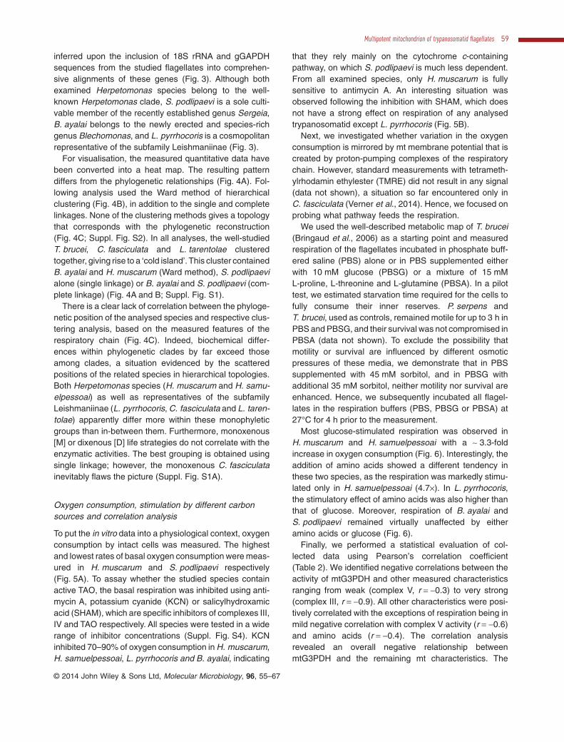
Oxygen consumption, stimulation by different carbonsources and correlation analysis
To put the in vitro data into a physiological context, oxygenconsumption by intact cells was measured. The highestand lowest rates of basal oxygen consumption were meas-ured in H. muscarum and S. podlipaevi respectively(Fig. 5A). To assay whether the studied species containactive TAO, the basal respiration was inhibited using anti-mycin A, potassium cyanide (KCN) or salicylhydroxamicacid (SHAM), which are specific inhibitors of complexes III,IV and TAO respectively. All species were tested in a widerange of inhibitor concentrations (Suppl. Fig. S4). KCNinhibited 70–90% of oxygen consumption in H. muscarum,H. samuelpessoai, L. pyrrhocoris and B. ayalai, indicating
that they rely mainly on the cytochrome c-containingpathway, on which S. podlipaevi is much less dependent.From all examined species, only H. muscarum is fullysensitive to antimycin A. An interesting situation wasobserved following the inhibition with SHAM, which doesnot have a strong effect on respiration of any analysedtrypanosomatid except L. pyrrhocoris (Fig. 5B).
Next, we investigated whether variation in the oxygenconsumption is mirrored by mt membrane potential that iscreated by proton-pumping complexes of the respiratorychain. However, standard measurements with tetrameth-ylrhodamin ethylester (TMRE) did not result in any signal(data not shown), a situation so far encountered only inC. fasciculata (Verner et al., 2014). Hence, we focused onprobing what pathway feeds the respiration.
We used the well-described metabolic map of T. brucei(Bringaud et al., 2006) as a starting point and measuredrespiration of the flagellates incubated in phosphate buff-ered saline (PBS) alone or in PBS supplemented eitherwith 10 mM glucose (PBSG) or a mixture of 15 mML-proline, L-threonine and L-glutamine (PBSA). In a pilottest, we estimated starvation time required for the cells tofully consume their inner reserves. P. serpens andT. brucei, used as controls, remained motile for up to 3 h inPBS and PBSG, and their survival was not compromised inPBSA (data not shown). To exclude the possibility thatmotility or survival are influenced by different osmoticpressures of these media, we demonstrate that in PBSsupplemented with 45 mM sorbitol, and in PBSG withadditional 35 mM sorbitol, neither motility nor survival areenhanced. Hence, we subsequently incubated all flagel-lates in the respiration buffers (PBS, PBSG or PBSA) at27°C for 4 h prior to the measurement.
Most glucose-stimulated respiration was observed inH. muscarum and H. samuelpessoai with a ∼ 3.3-foldincrease in oxygen consumption (Fig. 6). Interestingly, theaddition of amino acids showed a different tendency inthese two species, as the respiration was markedly stimu-lated only in H. samuelpessoai (4.7×). In L. pyrrhocoris,the stimulatory effect of amino acids was also higher thanthat of glucose. Moreover, respiration of B. ayalai andS. podlipaevi remained virtually unaffected by eitheramino acids or glucose (Fig. 6).
Finally, we performed a statistical evaluation of col-lected data using Pearson’s correlation coefficient(Table 2). We identified negative correlations between theactivity of mtG3PDH and other measured characteristicsranging from weak (complex V, r = −0.3) to very strong(complex III, r = −0.9). All other characteristics were posi-tively correlated with the exceptions of respiration being inmild negative correlation with complex V activity (r = −0.6)and amino acids (r = −0.4). The correlation analysisrevealed an overall negative relationship betweenmtG3PDH and the remaining mt characteristics. The
Multipotent mitochondrion of trypanosomatid flagellates 59
© 2014 John Wiley & Sons Ltd, Molecular Microbiology, 96, 55–67

0.1
Paratrypanosoma confusum
Sergeia podlipaevi (CER3)
C. absconditaLeptomonas pyrrhocoris (H10)
L. scantiiL. neopamerae
L. jaderaeL. podlipaeviL. seymouri
C. bombiC. expoeki
L. bifurcataC. confusaC. insperataC. pachysarca
Crithidia fasciculataC. dedva
C. brachyflagellisL. tarcolesL. acusL. spiculata
TU (G15)83C. otongatchi
C. brevicula
L. tenuaC. permixtaLeptomonas sp. (CFM)
C. pragensis
L. moramangoL. barvae
L. costaricensisLeishmania infantumLeishmania majorLeishmania tarentolae
Leishmania mexicana
B. campbelliB. danrayi
B. juanalfonsi
B. maslovi
B. luni
B englundi
Blechomonas ayalai (B08-376)B. wendygibsoni
B. lauriereadiB. pulexsimulantis
B. keelingi
Phytomonas serpensPhytomonas sp. (HART)
Phytomonas sp. (EM1)W. collosoma
W. rigidusW. ravinia
H. trimorphaH. ztiplika
H. wanderleyiH. mirabillis
H. isaaci
Herpetomonas muscarum (MMO-01)Herpetomonas sp. (B08-481B)
H. elegansH. puellarum
Herpetomonas samuelpessoai (GMO-04)
H. samueli
H. costorisH. modestus
A. ambiguus
A. desouzai
A. deaneiS. oncopelti
S. galatiS. culicis
T. rotatoriumT. megaT. grayi Trypanosoma brucei
T. congolenseT. vivax
T. lewisiT. microti
T. cruziT. rangeli
T. corviT. culicavium
Bodo saltans
Leishmaniinae
Herpetom
onas
Phytomonas
Wallacemonas
Blechomonas
Trypanosoma
AngomonasStrigomonas
Sergeia
Leptomonas sp. (ZM)
*
*
*
*
*
*
**
*
*
*
92
*
Fig. 3. Phylogeneticrelationships oftrypanosomatids.Maximum likelihood treebased on a concatenatedalignment of glycosomalglyceraldehyde-3-phosphate dehydrogenaseand 18S rRNA genes.Underlined genera(subfamily) containanalysed trypanosomatidmembers; five speciesanalysed in this study arehighlighted; four speciesstudied elsewhere are inbold and underlined.Maximum bootstrapsupports are marked withasterisks; schematic iconsdepict the insect host.
60 I. Škodová-Sveráková et al. ■
© 2014 John Wiley & Sons Ltd, Molecular Microbiology, 96, 55–67

inverse dependence between glycolytic generation of ATPand mt metabolism is illustrated by the strong inversecorrelation with complex III, a proton pump directly influ-encing number of electrons to be passed to the secondpump, complex IV. The correlation between activity ofcomplex III and glucose-induced respiration implies thatelectrons originating from glucose oxidation pass throughcomplex III, suggesting a lack of capacity of other putativeterminal oxidases to cope with the electron flow. Lastly,the correlation between the amino acid-stimulated respi-
ration and complex V fits perfectly with the bulk of ATPbeing generated in the mitochondrion, rather than by gly-colysis or other pathways (Table 2).
Discussion
Due to the dearth of suitable morphological characters,our current view of trypanosomatid diversity has beenshaped by the 18S rRNA-, SL RNA- and gGAPDH-basedphylogenies. Moreover, several novel and sometimes
Fig. 4. Comparison of clustering analysis ofenzymatic activities with phylogeny. Valuesfrom Table 1 were supplemented with therespective values from Verner et al. (2014),each characteristic was standardised towardsthe highest value and visualised as a heatmap using MS Excel. Rows represent (fromleft to right) G3PDH (G) and respiratorycomplexes II, III, IV and V. CD – classicaldixenous species (T. brucei, P. serpens,L. tarentolae); CM – classical monoxenousspecies (C. fasciculata); NM – newmonoxenous species (B. ayalai, H. muscarum,H. samuelpessoai, L. pyrrhocoris,S. podlipaevi).A. Heat map arrangement corresponding tothe phylogenetic position of analysed species.B. Heat map arrangement corresponding tohierarchical clustering analysis using the Wardmethod for distance calculation.C. Mapping of the Ward method clusteringanalysis onto phylogeny.
Multipotent mitochondrion of trypanosomatid flagellates 61
© 2014 John Wiley & Sons Ltd, Molecular Microbiology, 96, 55–67

very species-rich clades are represented only by environ-mental sequences, with no cultivable strain available(Maslov et al., 2013). Still, our systematic approach hasresulted in the introduction into culture of five species thatrepresent clades that to this time were observed only ininsect hosts. For the chosen five seemingly monoxenoustrypanosomatids, very little or no information is available.Measuring basal respiration and sensitivity to inhibitors ofcanonical and alternative enzymes of respiratory com-plexes, as well as following their mtG3PDH activity andthe composition of, and electron flow through, respiratorycomplexes allowed basic characterisation of mt metabo-lism of the investigated species. The collected data wereused for comparative analysis, into which an additionalfour well-studied trypanosomatids have been included(Suppl. Table S2).
Initially, we would like to stress that the data may rep-resent only a section of mitochondrial metabolic capacitydue to the fact that all the characterisations were per-formed under laboratory conditions. Although we believewe did not encounter dramatic deviations of any enzy-matic activities from the levels occurring in natural envi-
ronment, we cannot exclude this possibility as a directcomparison of in vitro and in vivo activities is not amena-ble for any of the described taxa at the moment. Thedramatic change as a described loss of complex I inlong-term cultivated isolates of L. tarentolae and C. fas-ciculata (Sloof et al., 1994; Speijer et al., 1997) most likelydid not take place, although given these species wereintroduced into laboratory cultures might have biased theobserved results. With this notion in mind the observeddata are put into context.
Fig. 5. Comparative analysis of respiration.A. Basal oxygen consumption in cultivation media. Averagevalues ± standard deviation for at least three measurements areshown.B. Sensitivity of respiration to inhibitors of cytochrome c-containing(KCN and antimycin A) and alternative pathways (SHAM). Averagevalues ± standard deviation for three to 10 measurements areshown. Hm = H. muscarum; Hs = H. samuelpessoai;Sp = S. podlipaevi; Lp = L. pyrrhocoris; Ba = B. ayalai.
Fig. 6. Stimulation of respiration by various carbon sources. Cellswere washed and resuspended in PBS or in PBS containingglucose (PBSG) or mixture of aminoacids (PBSA). Oxygenconsumption was measured after 4 h and standardised towardsrespiration in PBS. Average values ± standard deviation fortriplicates are shown, absolute values of respiration in PBS isshown in brackets following species name. Hm = H. muscarum(4.1 × 10⁻¹¹ ± 2.7 × 10⁻¹² μmol O2 × min−1 × cell−1);Hs = H. samuelpessoai (2.8 × 10⁻¹¹ ± 1.3 × 10⁻¹¹ μmolO2 × min−1 × cell−1); Sp = S. podlipaevi (9.5 × 10⁻¹² ± 1.5 × 10⁻¹² μmolO2 × min−1 × cell−1); Lp = L. pyrrhocoris (1.9 × 10⁻¹¹ ± 4.9 × 10⁻¹²
μmol O2 × min−1 × cell−1); Ba = B. ayalai (1.7 × 10⁻¹¹ ± 3.6 × 10⁻¹²
μmol O2 × min−1 × cell−1).
Table 2. Correlation of measured mitochondrial features.
G II III IV V Resp Glu
II −0.46III −0.90 0.72IV −0.39 0.39 0.46V −0.35 0.69 0.68 0.08Resp −0.43 0.09 0.16 0.51 −0.56Glu −0.82 0.78 0.90 0.73 0.45 0.48AA −0.56 0.74 0.83 0.15 0.97 −0.38 0.60
Pearson’s correlation coefficient was calculated based on averagevalues of following mitochondrial features: G = activity of glycerol-3-phosphate dehydrogenase; II = activity of complex II; III = activity ofcomplex III; IV = activity of complex IV; V = activity of complex V;Resp – overall basal respiration; Glu – glucose-stimulated respiration;AA – amino acid-stimulated respiration. Common correlation division:weak (0.1–0.3), medium (0.4–0.6), strong (0.7–0.8) and very strong(0.9–1.0; underlined). P-values were not calculated given the natureof the input data.
62 I. Škodová-Sveráková et al. ■
© 2014 John Wiley & Sons Ltd, Molecular Microbiology, 96, 55–67

The activity of mtG3PDH is in a negative correlationwith all examined activities of respiratory complexes. Inglycosomes of bloodstream form T. brucei, G3PDHcatalyses NADH-dependent conversion of dihydroxyac-etone phosphate to glycerol-3-phosphate connected witha mt FAD-dependent isoenzyme working in an oppositedirection. In this vein, the T. brucei bloodstream stage isdevoid of the cytochrome c-containing respiration, withmtG3PDH directly linked to TAO (Clarkson et al., 1989).The succinate-producing branch in its glycosomes, previ-ously thought to be absent (Michels et al., 2006), is notsufficient to provide enough oxidised co-factors, hencethe activation of the G3PDH: DHAP shuttle. The rate ofmtG3PDH reflects the high rate of glycolysis, and thenegative correlation with respiratory enzymes suggeststhat the more the cell utilises glycolysis as a source ofATP, the less active mitochondrion it possesses in termsof classical respiration. In mt energy production, a highcomplex II activity reflects the employment of at leastpart(s) of Krebs cycle in trypanosomes. This is furtherdocumented by the situation in P. serpens and blood-stream T. brucei, where the high rate of mtG3PDH indi-cates that the connection between Krebs cycle andrespiration is not operational (Clarkson et al., 1989;Sanchez-Moreno et al., 1992; Verner et al., 2014).
The association between complex II and mtG3PDH isnot reflected by any other OXPHOS enzymes. A relativelyhigh activity of complex III does not necessarily implysimilarly high activity of complex IV. Due to large differ-ences in the activities of complexes III and IV, we postu-late that they do not readily respond to changes in theirpartner’s activity. This conclusion is supported by our pre-vious observation in which RNAi silencing of one complexhad no effect on the activity of the following one and viceversa (Horváth et al., 2005). Similarly, there is no obviouscorrelation between the activities of oxidative phosphor-ylation enzymes and overall respiration.
Complex III forms a single band in all flagellates studiedso far (this study; Speijer et al., 1997; Horváth et al., 2000;Horváth et al., 2005; Verner et al., 2014), although thepattern of complex IV is becoming increasingly convo-luted. With the exception of S. podlipaevi and B. ayalai,several high molecular weight bands are detectable withantibodies against the complex IV subunit (Fig. 1B).However, their in-gel activity (Fig. 1C) does not alwayscorrespond to the intensity of detected signals, suggest-ing that not all agglomerates or oligomers are equallyactive. Combined, the results are compatible with the viewthat the dimer represents the active complex IV with thesmall forms being inactive, which is in agreement withdata available for complex IV in beef heart mitochondria(Antonini et al., 1987). Unique to trypanosomatids is theinvariably slower migration of complex IV as comparedwith complex III.
The presence of TAO in T. brucei and P. serpens(Chaudhuri et al., 1998; 2006; van Hellemond et al., 1998;Verner et al., 2014) and its likely absence in some monox-enous flagellates (this study; Suppl. Fig. S3) supports theevolutionary scenario postulating the presence of terminalalternative oxidase in the common eukaryotic ancestor andits preservation only in few lineages (McDonald et al.,2009). A similar scenario is observed within microsporidiawhere some lineages possess an alternative oxidase,whereas others have lost it (Williams et al., 2010).
The high and low complex IV and mtG3PDH activities ofH. muscarum, respectively, indicate the presence of clas-sical aerobic mitochondrion with the Krebs cycle as prob-ably the main source of electrons for the electron transportchain. As a member of the same flies-parasitising genus,H. samuelpessoai differs from H. muscarum by higherspecific activities of studied respiratory complexes, exceptfor complex IV. Overall low sensitivity to SHAM is acommon feature for all studied genera with the exception ofL. pyrrhocoris, where the inhibition of alternative oxidaseresembles the inhibition observed in P. serpens. The par-tition coefficients of KCN and SHAM are very similar(LogP = −0.78 and 0.7, respectively), and thus we expectthem to cross the membrane under the same conditions.Therefore, the insensitivity to SHAM is most likely notcaused by membrane impermeability for this inhibitor.
In vitro biochemical features and the 18S rRNA/gGAPDH-based phylogenies of species examined in thiswork do not correspond, as was documented by hierarchi-cal clustering analysis. Moreover, we were unable to iden-tify any key biochemical factor correlating with eitherphylogeny or life style, which leaves us with the conclusionthat the obligatory parasitic trypanosomatids are multipo-tent in terms of respiratory chain complexes, and theyseem to have retained all the essential elements of the mtmetabolism over the course of evolution. Realisation of thispotential is dictated by the availability of nutrients from thefood of their final and/or intermediate host(s), and similarflexible or highly variable circumstances.
In summary, phylogenetic relationships of the studiedtrypanosomatids representing novel and potentially verywidespread clades, do not correlate with the level of theirenzymatic activities or mt physiology. Whether the unex-pected variability of metabolic parameters is a conse-quence of diverse life style of these omnipresent parasitesand/or the driver of their extreme success remains to beestablished. As parasites of perhaps up to 10% of allinsect specimen inhabiting this planet (Maslov et al.,2013), trypanosomatids need a multipotent mitochondrionattached to a flexible metabolism. In any case, extendingmolecular and biochemical studies outside the well-known genera Trypanosoma, Leishmania, Phytomonasand Crithidia, as attempted for the first time herein, will bea fertile ground for the mt research and also for our
Multipotent mitochondrion of trypanosomatid flagellates 63
© 2014 John Wiley & Sons Ltd, Molecular Microbiology, 96, 55–67

understanding of the emergence of the medically impor-tant dixenous species from their relatively innocuousmonoxenous kins.
Experimental procedure
Organisms used and growth conditions
The following five species of monoxenous trypanosoma-tids were studied: (i) H. muscarum, strain MMO-01, wasisolated from the hindgut part of the brachycerid fly maleChrysomya putori (Calliphoridae; Diptera) captured in2010 in Ambatolampy, Madagascar (Týc et al., 2013). Theglobally distributed H. muscarum is a type species of thegenus Herpetomonas, one of the most species-rich cladesof monoxenous trypanosomatids parasitising predomi-nantly brachyceran flies. It displays the classical morphol-ogy of the genus Herpetomonas – promastigotes with anarrow flagellar pocket and pre-nuclear kinetoplast areelongated and needle-shaped, whereas a long and narrowflagellar pocket and post-nuclear localisation of the kineto-plast are features characteristic for the less frequentopisthomastigotes. (ii) Another globally distributed speciesof the same genus, Herpetomonas samuelpessoai, strainGMO-04, was isolated from the hindgut part of the brachy-cerid fly male of the subfamily Sarcophaginae (Diptera)collected in 2009 in Kokrobite, Ghana (Týc et al., 2013).Promastigotes of this species are smaller, wider andshorter than those of H. muscarum and opisthomastigotesare rare in culture. (iii) Sergeia podlipaevi, strain CER3,was isolated from the Malpighian tubes and the abdominalmidgut part of the biting midge female Culicoides festivi-pennis (Ceratopogonidae; Diptera) captured in 2000 inMikulov, Czech Republic (Svobodová et al., 2007). This isthe type species of Sergeia, a genus that accommodatesonly two species found in Europe and America. Morpho-logically uniform cells are represented by elongated andneedle-like promastigotes with the tendency to formrosette-like structures in culture. Another representative ofan extremely species-rich clade is (iv) Leptomonas pyr-rhocoris, strain H10, isolated from the abdominal midgutpart of the fire bug female Pyrrhocoris apterus (Pyrrhocori-dae; Heteroptera) and collected in 2008 in Prague, CzechRepublic (Votýpka et al., 2012). In culture, L. pyrrhocoris isinvariably present in the form of highly heterogeneouspromastigotes, varying from oval to long slender cells.The last monoxenous species studied herein is (v)Blechomonas ayalai, strain B08-376, isolated from the fleaCtenophthalmus agyrtes (Ctenophthalmidae; Sipho-naptera) captured in a rodent nest in 2008 in Príbram,Czech Republic (Votýpka et al., 2013). B. ayalai is a typespecies of the recently erected genus Blechomonas thatrepresent a species-rich clade of global distribution (J.V.and J.L., unpubl. data). For B. ayalai a high morphological
heterogeneity is characteristic, ranging from oval-shapedchoanomastigotes to elongated promastigotes.
For long-term maintenance, these trypanosomatidswere kept at 23°C on blood agar plates with an overlay. Forscaled-up growth, the temperature was raised to 27°C.H. samuelpessoai, L. pyrrhocoris and B. ayalai were keptin liquid brain-heart infusion (BHI) medium (Fluka) supple-mented with 10 mg ml−1 hemin (Sigma). S. podlipaevi andH. muscarum were cultivated in M199 + RPMI medium(1:1) (Fluka), supplemented with 10% heat-inactivatedFBS (Fluka), 10 mg ml−1 hemin and sterile urine (1:50), asthis species grows very slowly in BHI medium.
Phylogenetic and clustering analyses
Phylogenetic analyses were performed as describedearlier (Votýpka et al., 2012; Týc et al., 2013). Briefly, theconcatenated 18S rRNA and gGAPDH sequenceswere aligned via Kalign (http://www.ebi.ac.uk/Tools/msa/kalign/), and the resulting alignments were edited manu-ally using BioEdit 7.0.9.0 to remove the fast-evolvingregions of 18S rRNA preventing unambiguous alignment.Final concatenated alignment included 3155 charactersand the accession numbers retrieved from GenBank andused in phylogenetic reconstructions are available uponrequest from authors. Phylogenetic analyses were per-formed with MrBayes 3.2.1 program (Bayesian criteria:rates for six different types of substitution, proportion ofinvariant sites and shape parameter of the gamma cor-rection for the rate heterogeneity with four discrete cat-egories were allowed to vary; the covarion model wasused to allow the rate heterogeneity along the tree; theMarkov chain Monte Carlo was run for five million gen-erations). Clustering analysis of the enzymatic activitieswas performed using free on-line application (http://www.wessa.net/rwasp_hierarchicalclustering.wasp).
Detection of mitochondrial respiratory complexes
Mitochondria-enriched fraction was isolated as describedelsewhere for T. brucei (Horváth et al., 2005), and proteincontent was measured by Bradford method. ForBN-PAGE, mt proteins were lysed by 2% dodecylmaltosidefollowed by 1 h incubation on ice. Upon spinning, 15 μl oflysate was mixed with 1.5 μl Coomassie dye buffer (5%Coomassie Brilliant Blue G-250, 500 mM aminocaproicacid) and loaded on 2–12% gradient BN-PAGE asdescribed previously (Horváth et al., 2005). After electro-phoresis, the gel slices were immediately used for down-stream experiments or stored at −20°C.
Two-dimensional gel analysis
Analysis of respiratory complexes of purified mitochondriawas performed by 3–10% 2D BN/Tricine-SDS-PAGE as
64 I. Škodová-Sveráková et al. ■
© 2014 John Wiley & Sons Ltd, Molecular Microbiology, 96, 55–67

described previously (Horváth et al., 2005). A slice of thegel obtained from the first BN dimension was incubated at37°C in denaturing buffer (1 M Tricine; 1% SDS, 1%2-mercapthoethanol, pH 8.45) for 45 min. The incubationwas followed by resolution of complexes in 10% Tricine-SDS-PAGE. The resulting gel was stained with 0.25%Coomassie Brilliant Blue R-250 in 10% acetic acid and30% methanol.
Immunodetection
Immunodetection was performed as described elsewhere(Verner et al., 2014). Briefly, protein complexes fromBN-PAGE or SDS-PAGE were transferred to nitrocellulosemembranes by wet blotting overnight using anchored20 mA current. Membranes were probed with antibodiesraised against: (i) the sdh66 subunit of succinate dehydro-genase from T. brucei (1:1000) (Korený et al., 2012); (ii)apocytochrome c1 (apo c1) or Rieske protein of complex IIIfrom T. brucei (1:1000) (Horváth et al., 2005); (iii) thetrCOIV subunit of complex IV from L. tarentolae (1:1000)(Maslov et al., 2002), and (iv) β chain of complex V fromT. brucei (1:1000) (kindly provided by A. Zíková). Appropri-ate secondary antibodies (1:2000; Sigma) coupled tohorseradish peroxidase were visualised using an ECL kitaccording to the manufacturer’s protocol (Pierce). Forspecification of band intensities, Manual Rectangle Selec-tion of regions of interest was used (Kodak MolecularImaging).
In-gel activity staining and spectrophotometric assays
The staining was performed as described recently (Verneret al., 2014). For the detection of appropriate enzymeactivities, BN gel slices were incubated overnight in50 mM sodium phosphate, pH 7.2; 0.5 mg ml−1 3,3′-diaminobenzidine; 1 mg ml−1 cytochrome c for complex IVand in 8.5 mM Tris-HCl, pH 7.0; 67 mM glycine; 14 mMmagnesium sulphate; 0.2% lead nitrate; 8 mM adenosine5-triphosphate for complex V. Band intensities wereevaluated as described above.
Complex II activity was measured in 5 μg of mt lysate inSDH-A buffer (25 mM potassium phosphate, pH 7.2;5 mM magnesium chloride; 20 mM succinate disodiumsalt; 0.014 mg ml−1 dichlorophenolindophenol; 65 μMubiquinone Q2) at 600 nm for 5 min. Activities of com-plexes III, IV and V were measured as described else-where (Horváth et al., 2005; Schnaufer et al., 2005) andFAD-dependent mtG3PDH was measured followingŠkodová et al. (2013).
Oxygen consumption and survival experiment
Oxygen consumption was determined with a Clark-type electrode (1302 Microcathode Oxygen Electrode;
Strathkelvin). Basal oxygen uptake was recorded in 0.5 mlof logarithmically growing cell culture at 27°C. Cyanide(KCN), antimycin A and salicylhydroxamic acid (SHAM)were added in 4 min intervals to final concentrations of0.1 mM, 1.2 μg ml−1 and 0.09 mM respectively. For sur-vival experiments, approximately 2 × 10⁷ cells ml−1 werewashed twice in PBS (Fluka) and resuspended in 1 ml ofPBS alone or PBS supplemented with 10 mM glucose(PBSG) or combination of L-threonine, L-proline andL-glutamine (15 mM each; PBSA). Respiration was meas-ured in cells upon their incubation in appropriate buffer for4 h at 27°C. Collected data were processed and analysedusing either Microsoft Excel 2007 or GraphPad Prism 5.
Acknowledgements
We thank Simona Poláková for help with statistics and DavidWildridge for comments on the manuscript. This work wassupported by the Czech Grant Agency (P305/12/2261 and14/23986S), the Bioglobe grant (CZ.1.07/2.3.00/30.0032),the AMVIS grant (LH 12104), the Praemium Academiaeaward to J.L., the Slovak Research and Development Agency(APVV-0286-12), the Scientific Grant Agency of the SlovakMinistry of Education and the Academy of Sciences (1/0664/13) to A.H. and the University of South Bohemia (108/2013/P)to T.S. This work is also the result of the ITMS 26240220071project funded by the ERDF. We acknowledge the use ofresearch infrastructure that has received funding from the EU7th Framework Programme (FP7/2007–2013, grant agree-ment no. 316304).
References
Ackermann, A., Panunzi, L., Cosentino, R.O., Sánchez, D.O.,and Agüero, F. (2012) A genomic scale map of geneticdiversity in Trypanosoma cruzi. BMC Genomics 13: 736.
Adl, S.M., Simpson, A.G.B., Lane, C.E., Lukeš, J., Bass, D.,Bowser, S.S., et al. (2012) The revised classification ofeukaryotes. J Eukaryot Microbiol 59: 429–493.
Akhoundi, M., Baghaei, A., Depaquit, J., and Parvizi, P.(2013) Molecular characterization of leishmania infectionfrom naturally infected sand flies caught in a focus of cuta-neous leishmaniasis (Eastern Iran). J Arthropod Borne Dis7: 122–131.
Antonini, G., Brunori, M., Malatesta, F., Sarti, P., and Wilson,M.T. (1987) Reconstitution of monomeric cytochrome coxidase into phospholipid vesicles yields functionally inter-acting cytochrome aa3 units. J Biol Chem 262: 10077–10079.
Benne, R. (1993) RNA editing in mitochondria of Leishmaniatarentolae and Crithidia fasciculata. Semin Cell Biol 4:241–249.
Bringaud, F., Rivière, L., and Coustou, V. (2006) Energymetabolism of trypanosomatids: adaptation to availablecarbon sources. Mol Biochem Parasitol 149: 1–9.
Chaudhuri, M., Ajayi, W., and Hill, G.C. (1998) Biochemicaland molecular properties of the Trypanosoma brucei alter-native oxidase. Mol Biochem Parasitol 95: 53–68.
Multipotent mitochondrion of trypanosomatid flagellates 65
© 2014 John Wiley & Sons Ltd, Molecular Microbiology, 96, 55–67

Chaudhuri, M., Ott, R.D., and Hill, G.C. (2006) Trypanosomealternative oxidase: from molecule to function. TrendsParasitol 22: 484–491.
Clarkson, A.B., Bienen, E.J., Pollakis, G., and Grady, R.W.(1989) Respiration of bloodstream forms of the parasiteTrypanosoma brucei brucei is dependent on a plant-likealternative oxidase. J Biol Chem 264: 17770–17776.
Cermáková, P., Verner, Z., Man, P., Lukeš, J., and Horváth, A.(2007) Characterization of the NADH:ubiquinone oxidore-ductase (complex I) in the trypanosomatid Phytomonasserpens (Kinetoplastida). FEBS J 274: 3150–3158.
Duarte, M., and Tomás, A.M. (2014) The mitochondrialcomplex I of trypanosomatids – an overview of currentknowledge. J Bioenerg Biomembr 46: 299–311.
Fang, J., and Beattie, D.S. (2003) Identification of a geneencoding a 54 kDa alternative NADH dehydrogenase inTrypanosoma brucei. Mol Biochem Parasitol 127: 73–77.
Gonzáles-Halphen, D., and Maslov, D.A. (2004) NADH-ubiquinone oxidoreductase activity in the kinetoplasts ofthe plant trypanosomatid Phytomonas serpens. ParasitolRes 92: 341–346.
van Hellemond, J.J., Simons, B., Millenaar, F.F., and Tielens,A.G. (1998) A gene encoding the plant-like alternativeoxidase is present in Phytomonas but absent in Leishma-nia spp. J Eukaryot Microbiol 45: 426–430.
Horváth, A., Berry, E.A., Huang, L., and Maslov, D.A. (2000)Leishmania tarentolae: a parallel isolation of cytochromebc1 and cytochrome coxidase. Exp Parasitol 96: 160–167.
Horváth, A., Horáková, E., Dunajcíková, P., Verner, Z.,Pravdová, E., Šlapetová, I., et al. (2005) Downregulation ofthe nuclear-encoded subunits of the complexes III and IVdisrupts their respective complexes but not complex I inprocyclic Trypanosoma brucei. Mol Microbiol 58: 116–130.
Korený, L., Sobotka, R., Kovárová, J., Gnipová, A.,Flegontov, P., Horváth, A., et al. (2012) Aerobic kinetoplas-tid flagellate Phytomonas does not require heme for viabil-ity. Proc Natl Acad Sci USA 109: 3808–3813.
LaCount, D.J., Bruse, S., Hill, K.L., and Donelson, J.E. (2000)Double-stranded RNA interference in Trypanosoma bruceiusing head-to-head promoters. Mol Biochem Parasitol 111:67–76.
Le Trant, N., Meshnick, S.R., Kitchener, K., Eaton, J.W., andCerami, A. (1983) Iron-containing superoxide dismutasefrom Crithidia fasciculata. J Biol Chem 258: 125–130.
McDonald, A.E., Vanlerberghe, G.C., and Staples, J.F. (2009)Alternative oxidase in animals: unique characteristics andtaxonomic distribution. J Exp Biol 212: 2627–2634.
Maslov, D.A., Zíková, A., Kyselová, I., and Lukeš, J. (2002) Aputative novel nuclear-encoded subunit of the cytochromec oxidase complex in trypanosomatids. Mol Biochem Para-sitol 125: 113–125.
Maslov, D.A., Votýpka, J., Yurchenko, V., and Lukeš, J.(2013) Diversity and phylogeny of insect trypanosomatids:all that is hidden shall be revealed. Trends Parasitol 29:43–52.
Michels, P.A.M., Bringaud, F., Herman, M., and Hannaert, V.(2006) Metabolic functions of glycosomes in trypanosoma-tids. Biochim Biophys Acta 1763: 1463–1477.
Nawathean, P., and Maslov, D.A. (2000) The absence ofgenes for cytochrome c oxidase and reductase subunits inmaxicircle kinetoplast DNA of the respiration-deficient planttrypanosomatid Phytomonas serpens. Curr Genet 38:95–103.
Priest, J.W., and Hajduk, S.L. (1992) Cytochrome c reduc-tase purified from Crithidia fasciculata contains an atypicalcytochrome c1. J Biol Chem 267: 20188–20195.
Roellig, D.M., Savage, M.Y., Fujita, A.W., Barnabé, C.,Tibayrenc, M., Steurer, F.J., and Yabsley, M.J. (2013)Genetic variation and exchange in Trypanosoma cruzi iso-lates from the United States. PLoS ONE 8: e56198.
Rogers, M.B., Downing, T., Smith, B.A., Imamura, H.,Sanders, M., Svobodová, M., et al. (2014) Genomic confir-mation of hybridisation and recent inbreeding in a vector-isolated Leishmania population. PLoS Genet 10: e1004092.
Sanchez-Moreno, M., Lasztity, D., Coppens, I., andOpperdoes, F.R. (1992) Characterization of carbohydratemetabolism and demonstration of glycosomes in a Phyto-monas sp. isolated from Euphorbia characias. MolBiochem Parasitol 54: 185–199.
Scheffler, I.E. (2007) Mitochondria, 2nd edn. Hoboken: JohnWiley and Sons, Inc.
Schnaufer, A., Clark-Walker, D.G., Steinberg, A.G., andStuart, K. (2005) The F1-ATP synthase complex in blood-stream stage trypanosomes has an unusual and essentialfunction. EMBO J 24: 4029–4040.
Sloof, P., Arts, G.J., van den Burg, J., van der Spek, H., andBenne, R. (1994) RNA editing in mitochondria ofcultured trypanosomatids: translatable mRNAs for NADH-dehydrogenase subunits are missing. J Bioenerg Biomembr26: 193–203.
Speijer, D., Breek, C.K.D., Muijsers, A.O., Hartog, A.F.,Berden, J.A., Albracht, S.P.J., et al. (1997) Characteriza-tion of the respiratory chain from cultured Crithidia fascicu-lata. Mol Biochem Parasitol 85: 171–186.
Surve, S., Heestand, M., Panicucci, B., Schnaufer, A., andParsons, M. (2012) Enigmatic presence of mitochondrialcomplex I in Trypanosoma brucei bloodstream forms.Eukaryot Cell 11: 183–193.
Svobodová, M., Zídková, L., Cepicka, I., Oborník, M., Lukeš,J., and Votýpka, J. (2007) Sergeia podlipaevi gen. nov., sp.nov. (Trypanosomatidae, Kinetoplastida), a parasite ofbiting midges (Ceratopogonidae, Diptera). Int J Syst EvolMicrobiol 57: 423–432.
Škodová, I., Verner, Z., Bringaud, F., Fabian, P., Lukeš, J.,and Horváth, A. (2013) Characterization of two mitochon-drial FAD-dependent glycerol-3-phosphate dehydroge-nases in Trypanosoma brucei. Eukaryot Cell 12: 1664–1673.
Tielens, A.G.M., and van Hellemond, J.J. (2009) Surprisingvariety in energy metabolism within Trypanosomatidae.Trends Parasitol 25: 482–490.
Týc, J., Votýpka, J., Klepetková, H., Suláková, H., Jirku, M.,and Lukeš, J. (2013) Growing diversity of trypanosomatidparasites of flies (Diptera: Brachycera): frequent cosmo-politism and moderate host specificity. Mol PhylogenetEvol 69: 255–264.
Verner, Z., Cermáková, P., Škodová, I., Kriegová, E.,Horváth, A., and Lukeš, J. (2011) Complex I (NADH:ubiq-uinone oxidoreductase) is active in but non-essential for
66 I. Škodová-Sveráková et al. ■
© 2014 John Wiley & Sons Ltd, Molecular Microbiology, 96, 55–67

procyclic Trypanosoma brucei. Mol Biochem Parasitol 175:196–200.
Verner, Z., Škodová, I., Poláková, S., Durišová-Benkovicová,V., Horváth, A., and Lukeš, J. (2013) Alternative NADHdehydrogenase (NDH2): intermembrane-space-facingcounterpart of mitochondrial complex I in the procyclicTrypanosoma brucei. Parasitology 140: 328–337.
Verner, Z., Cermáková, P., Škodová, I., Kovácová, B., Lukeš,J., and Horváth, A. (2014) Comparative analysis of respira-tory chain and oxidative phosphorylation in Leishmaniatarentolae, Crithidia fasciculata, Phytomonas serpens andprocyclic stage of Trypanosoma brucei. Mol Biochem Para-sitol 193: 55–65.
Votýpka, J., Klepetková, H., Yurchenko, V.Y., Horák, A.,Lukeš, J., and Maslov, D.A. (2012) Cosmopolitan distribu-tion of a trypanosomatid Leptomonas pyrrhocoris. Protist163: 616–631.
Votýpka, J., Suková, E., Kraeva, N., Ishemgulova, A., Duží, I.,Lukeš, J., and Yurchenko, V. (2013) Diversity of Trypano-
somatids (Kinetoplastea: Trypanosomatidae) parasitizingfleas (Insecta: Siphonaptera) and description of a newgenus Blechomonas gen. n. Protist 164: 763–781.
Williams, B.A.P., Elliot, C., Burri, L., Kido, Y., Kita, K., Moore,A.L., and Keeling, P.J. (2010) A broad distribution of thealternative oxidase in microsporidian parasites. PLoSPathog 6: e1000761.
Yurchenko, V., Votýpka, J., Tesarová, M., Klepetková, H.,Kraeva, N., Jirku, M., and Lukeš, J. (2014) Ultrastructureand molecular phylogeny of four new species of monox-enous trypanosomatids from flies (Diptera: Brachycera)with redefinition of the genus Wallaceina. Folia Parasitol61: 97–112.
Supporting information
Additional supporting information may be found in the onlineversion of this article at the publisher’s web-site.
Multipotent mitochondrion of trypanosomatid flagellates 67
© 2014 John Wiley & Sons Ltd, Molecular Microbiology, 96, 55–67