LEM-domain proteins are lost during human spermiogenesis ...
63
HAL Id: hal-01681591 https://hal.archives-ouvertes.fr/hal-01681591 Submitted on 16 Apr 2018 HAL is a multi-disciplinary open access archive for the deposit and dissemination of sci- entific research documents, whether they are pub- lished or not. The documents may come from teaching and research institutions in France or abroad, or from public or private research centers. L’archive ouverte pluridisciplinaire HAL, est destinée au dépôt et à la diffusion de documents scientifiques de niveau recherche, publiés ou non, émanant des établissements d’enseignement et de recherche français ou étrangers, des laboratoires publics ou privés. LEM-domain proteins are lost during human spermiogenesis but BAF and BAF-L persist Razan A. Elkhatib, Marine Paci, Romain Boissier, Guy Longepied, Yasmina Auguste, Vincent Achard, Patrice Bourgeois, Nicolas Levy, Nicolas Branger, Michael J. Mitchell, et al. To cite this version: Razan A. Elkhatib, Marine Paci, Romain Boissier, Guy Longepied, Yasmina Auguste, et al.. LEM- domain proteins are lost during human spermiogenesis but BAF and BAF-L persist. Reproduction, BioScientifica, 2017, 154 (4), pp.387-401. 10.1530/REP-17-0358. hal-01681591
-
Upload
khangminh22 -
Category
Documents
-
view
1 -
download
0
Transcript of LEM-domain proteins are lost during human spermiogenesis ...

HAL Id: hal-01681591https://hal.archives-ouvertes.fr/hal-01681591
Submitted on 16 Apr 2018
HAL is a multi-disciplinary open accessarchive for the deposit and dissemination of sci-entific research documents, whether they are pub-lished or not. The documents may come fromteaching and research institutions in France orabroad, or from public or private research centers.
L’archive ouverte pluridisciplinaire HAL, estdestinée au dépôt et à la diffusion de documentsscientifiques de niveau recherche, publiés ou non,émanant des établissements d’enseignement et derecherche français ou étrangers, des laboratoirespublics ou privés.
LEM-domain proteins are lost during humanspermiogenesis but BAF and BAF-L persist
Razan A. Elkhatib, Marine Paci, Romain Boissier, Guy Longepied, YasminaAuguste, Vincent Achard, Patrice Bourgeois, Nicolas Levy, Nicolas Branger,
Michael J. Mitchell, et al.
To cite this version:Razan A. Elkhatib, Marine Paci, Romain Boissier, Guy Longepied, Yasmina Auguste, et al.. LEM-domain proteins are lost during human spermiogenesis but BAF and BAF-L persist. Reproduction,BioScientifica, 2017, 154 (4), pp.387-401. �10.1530/REP-17-0358�. �hal-01681591�

For Review Only
LEM-domain proteins during human spermiogenesis 12/06/2017 1
1
LEM-domain proteins are lost during human spermiogenesis but BAF and 2
BAF-L persist 3
4
5
Razan A. Elkhatib 1, Marine Paci 1,2, Romain Boissier 3, Guy Longepied 1, Yasmina Auguste 6 1, Vincent Achard 2, 4, Patrice Bourgeois 1, Nicolas Levy 1, Nicolas Branger 3, Michael J 7
Mitchell 1* Catherine Metzler-Guillemain 1,2* 8
9
*(MJ Mitchell and C Metzler-Guillemain are equal senior authors) 10
11 1 Aix Marseille Univ, INSERM, GMGF UMR_S 910, 13385 Marseille, France. 12
2 APHM Hôpital La Conception, Gynépôle, Laboratoire de Biologie de la Reproduction- 13
CECOS, 13385 Marseille cedex 5, France. 14 3 APHM Hôpital La Conception, Service d’Urologie, 13385 Marseille cedex 5, France 15
4Aix-Marseille Univ, Laboratoire de Biogénotoxicologie et Mutagenèse Environnementale, 16
EA 1784 – Fédération de Recherche CNRS n°3098 Ecosystèmes Continentaux et Risques 17
Environnementaux, 13385 Marseille cedex 5, France. 18 19 20 21
22 Short title: 23
LEM-domain and BAF proteins in spermiogenesis 24
25
Page 46 of 105
Manuscript submitted for review to Reproduction

For Review Only
LEM-domain proteins during human spermiogenesis 12/06/2017 2
Abstract - 248 words 26
During spermiogenesis the spermatid nucleus is elongated, and dramatically reduced in size 27
with protamines replacing histones to produce a highly compacted chromatin. After 28
fertilisation, this process is reversed in the oocyte to form the male pronucleus. Emerging 29
evidence, including the coordinated loss of the nuclear lamina (NL) and the histones, supports 30
the involvement of the NL in spermatid nuclear remodelling, but how the NL links to the 31
chromatin is not known. In somatic cells, interactions between the NL and the chromatin have 32
been demonstrated: LEM-domain proteins and LBR interact with the NL and, respectively, 33
the chromatin proteins BAF and HP1. We therefore sought to characterise the lamina-34
chromatin interface during spermiogenesis, by investigating the localisation of six LEM-35
domain proteins, two BAF proteins, and LBR, in human spermatids and spermatozoa. Using 36
RT-PCR, IF and Western blotting, we show that six of the proteins tested are present in 37
spermatids: LEMD1, LEMD2 (a short isoform), ANKLE2, LAP2β, BAF and BAF-L, and 38
three absent: Emerin, LBR and LEMD3. The full-length LEMD2 isoform, required for 39
nuclear integrity in somatic cells, is absent. In spermatids, no protein localised to the nuclear 40
periphery, but five were nucleoplasmic, receding towards the posterior nuclear pole as 41
spermatids matured. Our study therefore establishes that the lamina-chromatin interface in 42
human spermatids is radically distinct from that defined in somatic cells. In ejaculated 43
spermatozoa, we detected only BAF and BAF-L, suggesting that they might contribute to the 44
shaping of the spermatozoon nucleus and, after fertilisation, its transition to the male 45
pronucleus. 46
47
48
49
Key words: LEM-domain/ BAF/ spermiogenesis/ human/ Nuclear Lamina 50
Page 47 of 105
Manuscript submitted for review to Reproduction

For Review Only
LEM-domain proteins during human spermiogenesis 12/06/2017 3
51
Introduction 52
Spermiogenesis is the post-meiotic phase of spermatogenesis when haploid round spermatids 53
undergo a dramatic transformation into mature elongated spermatozoa. This involves nuclear 54
remodelling, chromatin condensation, replacement of nuclear histones with protamines and 55
formation of an acrosomal cap and a flagellum at opposite poles of the spermatid nucleus. 56
Although the precise mechanisms are not yet fully elucidated, it is certain that these processes 57
are in part coordinated by interactions between the nuclear envelope (NE) and specialised 58
cytoskeletal elements of the spermatid, the acroplaxome and the manchette (Kierszenbaum 59
and Tres, 2004). 60
The NE of eukaryotic cells is composed of the two nuclear membranes, the nuclear 61
lamina (NL) and the nuclear pore complexes (NPC) that create channels between the 62
nucleoplasm and the cytoplasm. The outer nuclear membrane (ONM) is continuous with the 63
endoplasmic reticulum (ER), while the inner nuclear membrane (INM) is lined on its 64
nucleoplasmic face by the NL, a peripheral network of type V intermediate filaments, called 65
lamins, involved in chromatin organization, cell cycle regulation, DNA replication, cell 66
differentiation, gene expression and apoptosis (Burke and Stewart, 2013). During 67
spermiogenesis in human and mouse the NL is composed exclusively of B-type lamins, and 68
retreats from the anterior to the posterior pole as the acrosome spreads and the histones are 69
removed from the chromatin (Alsheimer et al. 2004; De Vries et al. 2012; Elkhatib et al. 70
2015). 71
The NL is known to connect to the cytoskeleton through the interaction of SUN and 72
KASH proteins that span the nuclear membranes. In spermatids, the proteins SUN3, SUN4 73
Page 48 of 105
Manuscript submitted for review to Reproduction

For Review Only
LEM-domain proteins during human spermiogenesis 12/06/2017 4
and SUN5 have a localisation coincident with the lamins (Göb et al. 2010; Calvi et al. 2015; 74
Yassine et al. 2015). In mice lacking SUN4, spermatids do not elongate, and spermatozoa 75
have round heads (globozoospermia-like) (Calvi et al. 2015; Pasch et al. 2015). Biallelic loss-76
of-function mutations in SUN5 have been found in men with acephalic spermatozoa syndrome 77
(Zhu et al. 2016; Elkhatib et al. 2017). True globozoospermia is observed in mice and men 78
lacking DPY19L2, an INM protein localised under the acrosome (Harbuz et al. 2011; 79
Koscinski et al. 2011; Pierre et al. 2012). In mice lacking DPY19L2, the acrosome detaches, 80
elongation does not occur and lamins remain throughout the nuclear periphery of spermatozoa 81
(Pierre et al. 2012; Yassine et al. 2015). There is therefore good evidence that the NL has 82
central roles in shaping functional spermatozoa. 83
In contrast, little is known about how the NL interacts with the chromatin during 84
spermiogenesis, despite the identification of proteins that form links between the NL and the 85
chromatin in somatic cells: Lamin B Receptor (LBR), members of the LEM-domain (the 86
Lamina-associated polypeptide 2, Emerin, MAN1 domain) protein family and Barrier-to-87
Autointegration Factor (BAF) (Goldman et al. 2002; Gruenbaum et al. 2005; Schirmer and 88
Foisner, 2007). LBR was the first integral membrane protein of the INM to be identified 89
(Worman et al. 1988). LBR binds lamin B1 and links the lamina to the chromatin through an 90
interaction with the HP1-type heterochromatin proteins (Ye and Worman, 1994; Ye et al. 91
1997). Human LEM-domain proteins are a heterogeneous family of mainly nuclear proteins 92
that share a conserved ~40 amino acid domain, the LEM-domain, named after three LEM-93
domain proteins shown to bind BAF: LAP2, EMERIN and MAN1 (aka LEMD3) (Furukawa, 94
1999; Cai et al. 2001; Lee et al. 2001; Shumaker et al. 2001; Mansharamani and Wilson, 95
2005). In addition to the founding proteins, four further human LEM-domain encoding genes 96
have been described: LEMD1, with testis-predominant transcription (Yuki et al. 2004), 97
LEMD2, required for nuclear integrity (Brachner et al. 2005), ANKLE2, a regulator of BAF 98
Page 49 of 105
Manuscript submitted for review to Reproduction

For Review Only
LEM-domain proteins during human spermiogenesis 12/06/2017 5
phosphorylation with a role in post-mitotic nuclear envelop formation (Lee and Wilson, 99
2004; Asencio et al. 2012) and ANKLE1, an endonuclease possibly involved in DNA repair 100
(Brachner et al. 2012). 101
BAF, a conserved metazoan chromatin protein (Lee and Craigie, 1998) plays a key 102
role in post-mitotic nuclear assembly (Margalit et al. 2007). It forms homodimers that can 103
simultaneously bind dsDNA molecules and a LEM-domain (Shumaker et al. 2001; Segura-104
Totten et al. 2002). BAF is expressed widely, but has a paralogue, barrier-to-autointegration 105
factor-like (BAF-L) that is expressed predominately in testis and pancreas (Tifft et al. 2006; 106
Margalit et al. 2007). Human BAF-L and BAF share 40% amino acid identity. BAF-L was 107
described as specific to mammals (Margalit et al. 2007), but recent additions to the databases 108
indicate that BAF-L is specific to vertebrates. Unlike BAF, BAF-L does not bind DNA. BAF-109
L does form heterodimers with BAF, and it may thus modulate BAF chromatin functions in 110
testis and pancreas (Tifft et al. 2006). 111
Except for LBR and LAP2 isoforms, none of these proteins has been studied during 112
mammalian spermiogenesis. LBR has been localised to the nuclear periphery of elongating 113
spermatids in the rat, where a role in chromatin remodelling during spermiogenesis has been 114
proposed based on the detection, in vitro, of an interaction with Protamine 1 (Mylonis et al. 115
2004). The LAP2 isoforms have also been studied in the rat, where LAP2β predominates in 116
spermatids, but only LAP2α persists in mature spermatozoa (Alsheimer et al. 1998). Thus as a 117
first step towards defining how the NL interfaces with the chromatin during spermiogenesis, 118
we have characterised the expression of LBR, BAF, BAF-L and six LEM-domain proteins in 119
human spermatids and spermatozoa. 120
Material & Methods 121
Patients 122
Page 50 of 105
Manuscript submitted for review to Reproduction

For Review Only
LEM-domain proteins during human spermiogenesis 12/06/2017 6
Sperm samples were obtained from fourteen normospermic men who consulted at our 123
reproduction centre or gamete bank (CECOS - Centre d’Etude et de Conservation des Oeufs 124
et du Sperme) in Marseilles: ten men consulted for couple infertility (9 primary and 1 125
secondary infertility), four men were fertile sperm donors (Table 1). 126
Testicular samples came from four patients with brain death, who were 21, 31, 50 and 65 127
years old. Testicular samples were obtained within a protocol, approved by the French 128
Research Minister, for the collection of human tissue for use in research. We declared our 129
protocol through the Biomedicine Agency in January 2013. Testes were recovered during 130
multi-organ retrieval for transplantation while the patient was under extracorporeal circulation 131
and respiratory assistance. All patients had normal spermatogenesis based on histology 132
analysis and retrieval of numerous spermatozoa after testis dilaceration and one of the four 133
patients had children (T011400063, T011400051, T011400071 and T011500055). 134
135
Semen and testicular samples 136
Semen was collected via masturbation after a period of sexual abstinence of 2-6 days. After 137
30 minutes of liquefaction, semen analysis was performed according to the WHO criteria 138
(WHO Association, 2009), and according to the French David classification for the 139
morphology analysis (Auger et al. 2016). Sperm was diluted in cryoprotectant medium 140
(Spermfreeze; JCD, La Mulatière, France) and transferred into straws (Cryo Bio System, 141
Saint Ouen sur Iton, France). Straws were then suspended in vapour-phase nitrogen before 142
being stored in liquid nitrogen until use. All patients gave an informed consent for the 143
conservation of the remnant sperm in the Germetheque biobank and their use in studies on 144
human fertility in accordance with the Helsinki Declaration of 1975 on human 145
experimentation. The Germetheque Scientific Committee approved the present study design. 146
Page 51 of 105
Manuscript submitted for review to Reproduction

For Review Only
LEM-domain proteins during human spermiogenesis 12/06/2017 7
All sperm samples came from patients with normal sperm parameters according to WHO 147
criteria (Table 1). 148
Testicular cells fixed in 4% paraformaldehyde in phosphate buffered saline (PBS) before 149
freezing from the four patients (T011400063, T011400051, T011400071 and T011500055) 150
were used. 151
152
Spermatozoa RNA extraction 153
Following storage in liquid nitrogen, sample straws were thawed and the sperm was washed 154
twice in 2 ml of PBS, and then suspended in round cell lysis buffer (0.1% SDS, 0.5% Triton 155
X-100 in RNase free H2O). RNA was extracted with 1ml of Tripure (Roche) and precipitated 156
with 20 µg of glycogen as carrier. RNAs were treated with 10 units of DNase I, at room 157
temperature 10 minutes, in 1X reverse transcriptase buffer with 10 mM DTT and 20 units of 158
Protector RNAse inhibitor (Roche). 159
160
RT-PCR and quality control of spermatozoa RNA extracts 161
Before reverse transcription, RNA was purified on chromaspin 100 (Clontech) or Nucleospin 162
RNA XS columns (Macherey Nagel). Concentration was determined using a nanodrop ND-163
1000 spectrophotometer (NanoDrop Technologies, Wilmington, DE, USA). RNAs (400 ng) 164
were reverse transcribed in a 20 µl reaction with random nanomers (75 pmoles) and Expand 165
Reverse Transcriptase (Roche). The resulting cDNA was diluted to 40 µl with water and 1 µl 166
used in subsequent PCR amplification with Q5 High-Fidelity DNA Polymerase (New 167
England Biolabs). 168
Control PCRs were carried out with standard Taq polymerase. We checked for the presence 169
of spermatid RNA derived cDNAs by amplifying the protamine 1 transcript with primers 170
o1680/o1681 annealed at 60 °C. We checked for the lack of contaminating round cell RNA 171
Page 52 of 105
Manuscript submitted for review to Reproduction

For Review Only
LEM-domain proteins during human spermiogenesis 12/06/2017 8
by Polymerase chain reactions using a triplex reaction with primers for PTPRC (CD45), KIT 172
and CDH1 which are positive markers for leukocytes, early testicular germ cells and epithelial 173
cells respectively (Lambard et al. 2004). Triplex primers and concentrations: PTPRC (310 bp) 174
- o4609/o4610, 300 nM; c-Kit (237 bp) - o4611/o4612, 500 nM; CDH1 136 bp - 175
o4613/o4614, 500 nM. PCR programme 94 °C 2 min, 94 °c 30 s, 60 °C 40 s, 72°C 20 s 40 176
cycles, 72 °C 5 min. 177
LEM-domain proteins transcripts were amplified with Q5 Taq polymerase (New England 178
Biolabs) using the PCR conditions described in the product information. The primer pairs and 179
their annealing temperature for each transcript are as follows: LEMD1: o4426/o4427 – 66 °C, 180
LEMD2-3´: o4092/o4093 – 68°C, LEMD2-5´:o4965/o4966 – 69°C, LEMD3-3´: 181
o5023/o4491– 66 °C, LEMD3-5´: o5024/o5026 – 67°C, EMERIN-3´: o5097/o5098 – 69°C, 182
EMERIN-5´: o5095/o5096 – 67°C, ANKLE1-3´: o5027/o4493 – 67°C, ANKLE1-5´: 183
o5028/o5029 – 72°C, ANKLE2-3´: o4495/o4496 – 68°C, ANKLE2-5´: o5111/o5112 – 70°C, 184
ANKLE2_∆1-64: o4527/o4528 – 71°C, LAP2-5´: o5101/o5103 – 68°C LAP2α 3´: 185
o4087/o4088 – 64°C, LAP2β 3´: o5099/o5100 – 68°C, LAP2β-5´: o4087/o4090 – 64°C, 186
BAF: o4094/o4095 – 69°C, BAF-L: o4096/o4097 – 69°C, LBR-3’: o4667/o4668 – 60°C, 187
LBR-5´: o4669/o4671 – 64°C, musLEMD1: o4435/o4434 – 67°C, musBAF-L: o4522/o4523 188
– 67°C, musHMBS: o2216/o2217 – 67°C, PRM1: o1680/o1681 – 60°C. PRM1, a spermatid-189
specific spermatozoa-retained transcript was included as a positive control for spermatozoa 190
RNA. The Q5 GC-enhancer additive was used for all PCRs except for LBR. (Sequences of 191
primers are provided in supplementary Table S1). 192
Constructs to express tagged proteins 193
To express fusion proteins tagged with either 3xc-Myc (Myc) or 3xFlag (Flag), the coding 194
region for each protein was amplified by PCR from human testis cDNA, using specific 195
primers with restriction sites added at the 5´ end. The PCR products were digested at these 196
Page 53 of 105
Manuscript submitted for review to Reproduction

For Review Only
LEM-domain proteins during human spermiogenesis 12/06/2017 9
added sites and ligated to a vector cut with the same enzymes. The vectors were modified 197
pcDNA3.1 vector (Life Technologies) carrying either an N-terminal tag between NheI and 198
HindIII sites or a C-terminal tag between BamHI and XbaI sites. The primers used are shown 199
in Table S1. Flag-ANKLE2, primers: o4573/o4710, cloning sites: KpnI and BamHI. Flag-200
ANKLE2-∆1-64, primers: o4542/o4710, cloning sites: KpnI and BamHI. LEMD2-myc and 201
LEMD2_∆115-259-myc, primer: o5322/o5323, cloning sites: HindIII and KpnI. BAF-Flag, 202
primers: o4388/o4399, cloning sites: KpnI and BamHI. Flag-BAF-L, primers: o5186/o4663, 203
cloning sites: KpnI and XhoI. myc-LEMD1, primers: o4661/o4659, cloning sites KpnI and 204
XhoI. myc-Emerin, primers: o4579/o4580, cloning sites: BamHI and XhoI. myc-LAP2β, 205
primers: o5022/o4615, cloning sites: BamHI and XhoI. All constructs used were confirmed to 206
have the expected sequence by determining the sequence of the full insert and tag. 207
Cell culture and transfection 208
HeLa cells were cultured according to standard procedures. 209
Cells were transfected in two-well Lab-tek chambers using jetPRIME ® short-DNA 210
transfection protocol (Polyplus-transfection) and 500 ng of plasmid DNA according to the 211
manufacturer’s instructions. The medium was replaced after 4 hours of incubation with the 212
transfection mix. 213
Immunocytochemistry 214
Immunocytochemistry was performed on spread spermatozoa and/or testicular germ cells. 215
Thawed spermatozoa were washed two times in PBS, fixed in 2% formaldehyde in PBS for 216
10 min, washed twice in PBS and spread onto polylysine-coated slides by Cytospin 217
(Shandon). Testicular cells were fixed with 2% formaldehyde in PBS for 10 min after 218
disaggregation of testis fragments, washed in PBS and pellets frozen. Testicular cell pellets 219
were thawed and spread onto polylysine slides by Cytospin (Shandon) as previously described 220
(Metzler-Guillemain and Guichaoua, 2000). Slides were fixed 5 minutes in 4% formaldehyde 221
Page 54 of 105
Manuscript submitted for review to Reproduction

For Review Only
LEM-domain proteins during human spermiogenesis 12/06/2017 10
in PBS, rinsed in PBS and permeabilized using 0.1%, 0.3% or 0.5% Triton X-100, 3% normal 222
goat serum in PBS for 30 min. Slides were blocked with 1% BSA, 7% normal goat serum in 223
PBS for 30 min. They were then incubated for 30 min with lectins (Lectin PNA Alexa Fluor 224
594 conjugates, L-21409 Molecular Probes) at a dilution of 1:600 in PBS to mark the 225
acrosome. After washes in PBS under gentle agitation, slides were incubated one to two hours 226
in a moist chamber at 37°C with primary antibodies after several washes in PBS, detection 227
was performed using Alexa fluor secondary antibodies (Invitrogen) at dilutions recommended 228
by manufacturers for 45 min at 37°C. Slides were then rinsed twice in PBS and mounted with 229
25 to 75 ng/ml DAPI in Vectashield mounting medium for microscope analysis. Negative 230
controls without the primary antibody gave no signal for any of the secondary antibodies 231
used. The slides were analyzed using the Zeiss ApoTome.2 microscope (Zeiss, Oberkochen, 232
Germany) equipped with an AxioCam MRm camera. Images were captured and merged with 233
the ZEN software, and were treated using ImageJ software. Phase contrast overlay of 234
ANKLE2 immunofluorescent labelling shows that cell morphology is preserved by this 235
protocol (Fig. S1). 236
Antibodies 237
The affinity of antibodies for their target was validated for IF analysis using HeLa cells 238
transfected with a construct overexpressing a tagged version of the target protein, except for 239
those against LBR and LEMD3, already shown by the supplier to label the nuclear periphery 240
in HeLa cells (Fig. S2). All antibodies used were monoclonal or immunogen affinity-purified 241
polyclonal and gave the expected labelling pattern in somatic cells in disaggregated testes: 242
nuclear periphery for LEMD2, LEMD3, Emerin, LAP2β, LBR and BAF; endoplasmic 243
reticulum for ANKLE2; faint or no labelling for LEMD1 and BAF-L (only spermatid nuclei 244
labelled). We provide representative images of single large fields obtained for each antibody 245
Page 55 of 105
Manuscript submitted for review to Reproduction

For Review Only
LEM-domain proteins during human spermiogenesis 12/06/2017 11
to show the labelling of germ cells and somatic cells observed together in disaggregated testes 246
(Fig. S3). 247
The antibodies used were rabbit anti-LEMD1 (IF dilution: 1/100, SAB3500644 248
Sigma), rabbit anti-LEMD2 specific N-terminus (IF dilution: 1/100, HPA017340 Sigma), 249
rabbit anti-LEMD2 specific C-terminus (IF dilution: 1/50, ab89866 Abcam), mouse anti-250
LEMD3 (IF dilution: 1/50, ab123973 Abcam), mouse anti-EMERIN (IF dilution: 1/100, 251
NCL-EMERIN Leica), mouse anti-EMERIN (IF dilution: 1/100, Sigma AMAb90560), rabbit 252
anti-ANKLE2 (IF dilution: 1/100, Asencio et al. 2012), rabbit anti-LAP2 beta (IF dilution: 253
1/50, HPA008150 sigma), rabbit anti-BAF (IF dilution: 1/600 ab129184 Abcam), rabbit anti-254
BAF-L (IF dilution: 1/100, HPA042635 Sigma), rabbit anti-BAF-L (IF dilution: 1/200, 255
ab122950 Abcam), rabbit anti LBR (IF dilution: 1/500, ab32535 Abcam), mouse anti-FLAG 256
(IF dilution: 1/100, F1804 Sigma), mouse anti-c-MYC (IF dilution: 1/100, sc40 Santa Cruz) , 257
rabbit anti-α tubulin (WB dilution: 1/1000, ab15246 Abcam) and anti-GRP94 (IF dilution: 258
1/400, ab210960) (Table S2). 259
The immunogen peptide used for pre-absorption of anti-BAF (within amino acids 1-260
45) was custom synthesised (Proteogenix, France). The immunogen peptide for BAF-L (aa 261
12-252) was expressed as a fusion protein with the chitin binding protein in E. coli and 262
purified on a chitin column, following the protocol of the Impact Kit (New England Biolabs), 263
except that the BAF-L fusion protein was eluted with 8M Urea and then dialysed against PBS. 264
The BAF-L coding region was amplified with primers o5350/o5351 and cloned between the 265
NdeI and XhoI sites of the pMXB10 vector (New England Biolabs). Pre-absorption 266
protocol*** 267
Protein extraction and western blot analysis 268
Protein was prepared from 12x106 spermatozoa which were separated from round cells as for 269
RNA extraction, and then lysed on ice for 30 min in 100 µl of 0.5% SDS, 50 mM Tris.HCl 270
Page 56 of 105
Manuscript submitted for review to Reproduction

For Review Only
LEM-domain proteins during human spermiogenesis 12/06/2017 12
pH8.0, 5 mM EDTA, 50 mM DTT, 2x complete proteinase inhibitor cocktail (Roche), 271
followed by 30 s of sonication at high setting in a Bioruptor Standard (Diagenode). The 272
effectiveness of the round cell lysis is illustrated by the absence of a signal for Emerin, 273
despite the strong signal in whole testis (Fig. S4). Emerin is strongly detected in testicular 274
somatic cells, but we did not detect it by IF in spermatids or spermatozoa. For testis lysates, 275
60 mg of tissue was dissociated in 300 ml of lysis buffer (1M Tris.HCl pH7.5, 5M NaCl, 0.1 276
mM EGTA, 10% Triton) using ceramic beads in a Magnalyser (Roche), followed by 30 min 277
on ice and 3x 30 s of sonication at high setting in a Bioruptor Standard (Diagenode). For 278
migration, lysate from approximately 2 million spermatozoa or 40 µg of testis protein was 279
mixed with 2x Laemmli sample loading buffer loading buffer (Sigma Aldrich) with 5% β-280
mercaptoethanol, heated to 95 °C for 5 minutes and subjected to SDS-PAGE. Proteins were 281
blotted on to nitrocellulose with a pore size of 0.45 µm (Emerin) or 0.2 µm (BAF, BAF-L). 282
283
Results 284
Spermatid transcript analysis using spermatozoa RNA 285
To characterise gene expression of NL-associated proteins (LBR, LEM-domain proteins, BAF 286
and BAF-L) during human spermiogenesis, we first used RT-PCR to detect the 5´ and 3´ end 287
of the coding region of transcripts, in spermatozoa RNA from five normospermic men: two 288
fertile men and three men from infertile couples. RT-PCR detected transcripts for Emerin, 289
LEMD1, LEMD2, ANKLE2, LAP2α, LAP2β and BAF-L in all tested RNA samples (Fig. 1), 290
but in the case of LEMD2 only the 3´ end of the coding region was detected, indicating that 291
the only transcript present for LEMD2 in spermatids lacks a part of the 5´ coding region 292
present in the widely expressed form. No transcripts were detected for LBR, LEMD3 or 293
ANKLE1 in any sample (Fig. 1). 294
Page 57 of 105
Manuscript submitted for review to Reproduction

For Review Only
LEM-domain proteins during human spermiogenesis 12/06/2017 13
BAF transcripts were detected in one RNA sample R010900491 but not in the other 295
four, suggesting that they are normally absent from spermatozoa (Fig. 1). BAF is encoded by 296
the BANF1 gene, and has five retroposed homologues in the genome. We therefore 297
investigated the nature of the BANF1 cDNA product amplified from R010900491 by 298
sequencing and revealed it to be identical to the BANF1 locus, excluding the possibility that it 299
was derived from one of the BANF1 retroposons. In addition, we did not amplify the same 300
cDNA product from the genomic DNA of R010900491, excluding the possibility that 301
R010900491 carries a recently integrated BANF1 retroposon with 100% nucleotide identity to 302
BANF1. The individual in whose sperm sample we detected BAF transcripts is normospermic 303
and his sperm does not have an exceptional morphology or motility profile. He is nevertheless 304
in an infertile couple, and it will now be important to define the relationship between BAF 305
transcript retention in spermatozoa and gamete quality. We conclude that the BANF1 306
transcript can persist in spermatozoa in a minority of cases, but whether this is related to 307
increased RNA stability, epigenetic variation or the quality of spermatogenesis remains to be 308
elucidated. 309
310
BAF-L and LEMD1 transcripts coincide with spermatids in immature mouse testis. 311
Having shown that transcripts for the testis-predominant genes BANF2 and LEMD1 312
were present in human spermatids, we next investigated transcription from these genes during 313
the first wave of post-natal spermatogenesis in the mouse. We found that transcripts for both 314
genes were detected only at later steps (after 20 dpp) when spermatids first appear in the testis 315
(Fig. 2). We conclude that BAF-L and LEMD1 transcripts predominate in spermatids in 316
mouse and human. 317
318
Alternative transcript for ANKLE2 in spermatids 319
Page 58 of 105
Manuscript submitted for review to Reproduction

For Review Only
LEM-domain proteins during human spermiogenesis 12/06/2017 14
For ANKLE2, we detected a transcript (ANKLE2_Δ1-64) corresponding to several 320
testis ESTs with an alternative first exon situated within the first intron (e.g. HY008086) (Fig. 321
3A). We amplified ANKLE2_Δ1-64 from all five spermatozoa RNA samples (Fig. 3B), and 322
from total RNA of testis and brain, but not small intestine, skeletal muscle, prostate or thymus 323
(Fig. 3C). In the mouse, no EST corresponding to this transcript has been described, and we 324
did not detect a homologous transcript from the mouse Ankle2 gene by RT-PCR with primers 325
from candidate alternative first exons in intron 1 defined by splice donor site prediction (data 326
not shown). We amplified as a single amplicon and then sequenced the full coding region of 327
the ANKLE2_∆1-64 transcript from human testis RNA and deposited its sequence in 328
Genbank (accession KY056762). The ANKLE2_Δ1-64 transcript encodes an ANKLE2 329
isoform lacking the N-terminal 64 amino acids that encode the transmembrane domain of the 330
full-length isoform which are replaced by the short peptide of five amino acids 1MWRSE5. 331
332
ANKLE2_ ∆1-64 does not localise to the endoplasmic reticulum in HeLa cells 333
The full-length ANKLE2 protein has been localised to the nuclear periphery and endoplasmic 334
reticulum (ER) (Asencio et al. 2012). To test the intracellular localization of ANKLE2_Δ1-335
64, we transfected HeLa cells with plasmids designed to express N-terminus Flag-tagged 336
versions of the full-length ANKLE2 and ANKLE2_Δ1-64, under control of the CMV 337
promoter. Following transfection, Flag-ANKLE2 had a granular aspect throughout the 338
cytoplasm that we show co-localises with the ER marker GRP94, consistent with the ER 339
localisation described previously for ANKLE2 (Asencio et al. 2012). In contrast Flag-340
ANKLE2_Δ1-64 was more homogeneously distributed throughout the cytoplasm, and did 341
not coincide with GRP94 (Fig. 4A). We conclude that the N-terminal 64 aa of ANKLE2, 342
which contain the TM, are required to locate ANKLE2 to the ER in HeLa cells. 343
344
Page 59 of 105
Manuscript submitted for review to Reproduction

For Review Only
LEM-domain proteins during human spermiogenesis 12/06/2017 15
ANKLE2 localises to the ER in round spermatids 345
We investigated the localisation of ANKLE2 during spermiogenesis by 346
immunofluorescent analysis of testicular germ cells from four men and ejaculated 347
spermatozoa from three men. An ANKLE2 signal was detected throughout human 348
spermiogenesis, in the cytoplasm of round spermatids, elongating spermatids and testicular 349
spermatozoa***, but was not visible in ejaculated spermatozoa. In round spermatids, it 350
presented as a granular cytoplasmic signal, consistent with the known localisation of 351
ANKLE2 to the ER (Fig. 4B). We confirmed ER localisation for ANKLE2 in round 352
spermatids, by showing that the signal co-localises with the ER-lumen protein GRP94 (Fig. 353
4C). In elongating spermatids, however, there was no longer a perfect coincidence of GRP94 354
and ANKLE2 signals, a possible reflection of the distinct localisation of ANKLE2 (ER 355
cytoplasmic surface) and GRP94 (ER lumen). We saw no evidence of the more diffuse 356
cytoplasmic staining that might correspond to the ANKLE2_∆1-64 isoform that lacks the 357
domain necessary for ER localisation (Fig. 4B, C). 358
359
LEMD2 transcript in spermatids encodes a short isoform 360
To identify the LEMD2 transcript present in human spermatids, we initially screened testis 361
and spermatozoa RNA with RT-PCR assays based on ESTs in the databases that lack the first 362
coding exon of LEMD2 (DC315891, DC367547, BI755923, DB028937 and DC389488), but 363
no product was amplified from testis T011400071 or spermatozoa RNA (data not shown). We 364
next amplified the entire LEMD2 coding region from testis RNA and observed a doublet, a 365
band of the expected size (1.5 kb) and a smaller band (1.1 kb) (Fig. 5A). We cloned and 366
sequenced these fragments. The larger fragment contained the full coding region as predicted 367
while the smaller fragment contained two cDNAs created by alternative splicing from a 368
cryptic splice donor site immediately following codon 114 (c.346) in exon 1, to either exon 2 369
Page 60 of 105
Manuscript submitted for review to Reproduction

For Review Only
LEM-domain proteins during human spermiogenesis 12/06/2017 16
(frameshift) or to exon 3 (non-frameshift) (Fig. 5A). Using specific RT-PCR assays for each 370
transcript we found that both alternatives are present in spermatozoa RNA (shown for the 371
non-frameshift transcript in Fig. 5B). We conclude that the only protein-coding transcript 372
from LEMD2 in spermatids encodes a short version of LEMD2 that we have termed 373
LEMD2_∆115-259 since it lacks amino acids 115-259 of the full-length protein. The cryptic 374
splice site in exon 1 that allows the production of the alternative transcripts is not present in 375
mouse and we were unable to detect corresponding transcripts in the mouse (data not shown). 376
We PCR amplified and cloned the coding region of the LEMD2_∆115-259 transcript as a 377
single amplicon from testis cDNA, sequenced it and deposited it in Genbank (accession 378
KY056761). 379
380
LEMD2_∆∆∆∆115-259 is not directed to nuclear periphery and does not destabilise NE 381
It has been concluded that LEMD2 is important for nuclear integrity, since its depletion from 382
somatic cells leads to altered nuclear morphology (Ulbert et al. 2006), similar to that induced 383
by ectopic expression of the spermatid-specific lamin B3 (Schütz et al. 2005; Elkhatib et al. 384
2015). We therefore investigated the possibility that LEMD2_∆115-259 has a role in 385
destabilising the NE to facilitate nuclear remodelling in spermatids. To do this we expressed 386
both isoforms with a C-terminal Flag-tag in HeLa cells. We observed that while the full-387
length isoform was directed to the nuclear periphery as expected, LEMD2_∆115-259 was not, 388
and was visible in the perinuclear region of the cytoplasm. LEMD2_∆115-259 did not induce 389
any nuclear deformation (Fig. 5C). Taken together with previous work (Brachner et al. 2005) 390
showing that LEMD2 lacking amino acids 130-203 is directed to the nuclear periphery, our 391
results show that at least part of a domain necessary for the import of LEMD2 from the 392
cytoplasm to the nuclear periphery, must be located within aa 115-130 and aa 203-259. In 393
addition, our IF analysis of transfected cells shows that the anti-LEMD2 antibody 394
Page 61 of 105
Manuscript submitted for review to Reproduction

For Review Only
LEM-domain proteins during human spermiogenesis 12/06/2017 17
HPA017340, whose immunogen (aa 243-362) overlaps with amino acids 115-259 of LEMD2 395
is essentially specific for the full-length LEMD2 isoform, and does not detect LEMD2_∆115-396
259 (Fig. 5C). The epitopes detected with high affinity by HPA017340 must therefore all 397
overlap with aa 243-259 of LEMD2. 398
399
Nuclear LEM-domain proteins and LBR not at nuclear periphery in human spermatids 400
In order to explore the nature of the interface between the NL and the chromatin during 401
human spermiogenesis, we performed IF analysis of testicular germ cells from four men with 402
antibodies against six LEM-domain proteins, the two BAF proteins and LBR (antibody 403
descriptions - Table S2). We detected signals on spermatids for LEMD1, LEMD2, ANKLE2, 404
LAP2β, BAF and BAF-L (Fig. 6, Fig. S3 and Table 2), but no signal was observed on 405
spermatids with antibodies against Emerin, LBR or LEMD3, despite strong labelling of the 406
nuclear periphery in somatic cells on the same slide (Fig. S5, S4). 407
For Emerin, we had amplified a transcript from spermatozoa RNA by RT-PCR, and it 408
was therefore unexpected that Emerin was not detected in spermatids. We therefore tested two 409
different anti-Emerin antibodies that labelled the nuclear periphery in somatic cells (Fig. S6). 410
Neither labelled any spermatids or spermatozoa (Leica-Emerin-Ncl and AMAb90560) (Fig. 411
S6). Moreover, in Human Protein Atlas, a further two antibodies, HPA000609 and 412
CAB001545, do not label spermatids. Thus although we have shown that the Emerin 413
transcript is present in spermatids, we conclude that it is not translated and that the Emerin 414
protein is not present in spermatids or spermatozoa. 415
Using antibodies directed against LEMD1 we observed a nucleoplasmic signal in 416
round spermatids, except in the area covered by the acrosome. As spermatid maturation 417
progressed, the fluorescence became limited progressively to the posterior pole of the nuclei, 418
Page 62 of 105
Manuscript submitted for review to Reproduction

For Review Only
LEM-domain proteins during human spermiogenesis 12/06/2017 18
where it remained visible in late elongated spermatids. LEMD1 was not detected, however, on 419
testicular spermatozoa or on ejaculated spermatozoa (Fig. 6). 420
For LEMD2, our RT-PCR analyses suggested that in spermatids LEMD2 was only 421
present as an isoform lacking amino acids 115-259. We therefore analysed testicular germ 422
cells with two antibodies, anti-LEMD2_243-362 (Sigma HPA017340) that detects the full-423
length LEMD2 isoform but not LEMD2_∆115-259 (Fig. 5C and S3), and anti-LEMD2_Cter 424
that detects both isoforms (Fig. S2). Despite strong labelling of the nuclear periphery in 425
somatic cells of disaggregated testis with both antibodies, only anti-LEMD2_Cter labelled the 426
nuclei of spermatids, confirming that in spermatids, LEMD2 is present only as the short 427
isoform LEMD2_∆115-259 (Fig. 6 and Fig. S3). Anti-LEMD2_Cter gave a punctate labelling 428
throughout the spermatid nucleoplasm, indicating that LEMD2_∆115-259 does not localise to 429
the nuclear periphery in spermatids (Fig. 5C). 430
For LAP2β, labelling is nuclear, and predominates at the nuclear periphery in the 431
earliest round, spermatids but the signal shifts to the nucleoplasm at later steps and is not 432
detected in elongating spermatids (Fig. 6). In the rat, the nucleoplasm and NE labelling in 433
round spermatids were previously demonstrated with an antibody that detected all LAP2 434
isoforms (Alsheimer et al. 1998).The anti-BAF-L antibody gave a granular signal 435
concentrated over the nucleoplasm of round spermatids, with fainter fluorescence in the 436
cytoplasm. Labelling was concentrated at the nuclear periphery in elongating spermatids, but 437
was excluded from under the acrosome (Fig. 6). In testicular and ejaculated spermatozoa 438
BAF-L labelling was visible at the posterior pole of the nucleus (Fig. 7A). 439
Although no BAF transcript was detected in spermatozoa RNA from four of five men 440
tested, the anti-BAF antibody (ab129184) labelled the nuclei of spermatids (Fig. 6). We 441
observed a nucleoplasmic localization of BAF in 10% - 55% of round spermatids, 2% - 9% of 442
elongated spermatids, and 1% - 10% of testicular spermatozoa, where BAF was located at the 443
Page 63 of 105
Manuscript submitted for review to Reproduction

For Review Only
LEM-domain proteins during human spermiogenesis 12/06/2017 19
posterior pole of the nucleus (Fig. 6 and Fig. S7A and S5B). BAF signal was observed in 444
ejaculated spermatozoa at the posterior nuclear pole in all 3 patients tested with a mean of 30 445
% of spermatozoa labelled (Fig. 7A). These contradictory results for the expression of the 446
BAF protein and its transcript imply either that the BAF spermatid transcript is a remnant 447
from spermatocytes (BAF protein labelling is seen throughout the spermatocyte nucleus - Fig. 448
S7C), or that de novo BAF transcripts are produced in spermatids but are not retained in 449
spermatozoa. 450
451
BAF and BAF-L are present in human ejaculated spermatozoa 452
Our IF analyses of human spermatids indicate that most of the proteins tested as potential 453
links between the NL and the chromatin are lost during the round spermatid steps and do not 454
persist in spermatozoa. The only exceptions were with the antibodies against BAF and BAF-L 455
that labelled the posterior nuclear pole of testicular and ejaculated spermatozoa. These signals 456
were absent when we pre-absorbed the antibodies with their respective immunising peptide, 457
indicating that they were specific for BAF and BAF-L (Fig. 7A). To further verify that this 458
signal was due to the presence of BAF and BAF-L, we performed Western blot analysis of 459
lysates of purified spermatozoa with the same antibodies (Fig. 7B). The anti-BAF and anti-460
BAF-L antibodies detected a strong band of the expected size, approximately 10 kDa, in 461
protein lysates from spermatozoa. Anti-BAF detected a strong band of the same size in testis 462
lysates, but in the case of BAF-L the band was barely detectable in whole testis, suggesting 463
that it is expressed in spermatids at a low level and becomes concentrated in the spermatozoa 464
fraction (Fig. 7B). We conclude that BAF and BAF-L proteins are present in spermatids 465
throughout human spermiogenesis and are retained in ejaculated spermatozoa. 466
467
Discussion 468
Page 64 of 105
Manuscript submitted for review to Reproduction

For Review Only
LEM-domain proteins during human spermiogenesis 12/06/2017 20
Known somatic lamina-chromatin interface absent from all but earliest spermatids 469
Here we present an overview of how LEM-domain proteins, BAF proteins and LBR, 470
many known to be involved in linking the NL to the chromatin in somatic cells, are organised 471
in human spermatids and spermatozoa. For most of the proteins, except LBR and LAP2β, this 472
is the first study of these proteins during mammalian spermiogenesis. We have studied 473
ANKLE1 and LAP2α expression only at the transcript level. 474
Based on our observations, a general theme emerges that during most of 475
spermiogenesis the lamina-chromatin interface, as defined in somatic cells, does not exist, and 476
the three LEM-domain proteins that we did detect, LEMD1, LEMD2_∆115-259 and LAP2β, 477
do not localise to the nuclear envelope in most round spermatids, but to the nucleoplasm. This 478
can readily be explained for LEMD2_∆115-259 based on our HeLa transfection experiments 479
showing that LEMD2_∆115-259 lacks the domains required for NE localisation, consistent 480
with the mapping of these domains to aa 74-130 and aa 203-378 (Brachner et al. 2005). It is 481
more difficult to explain the nucleoplasmic localisation of LEMD1 and LAP2β. Both contain 482
a transmembrane domain (TM), which has been shown to be necessary for the efficient 483
localisation of LAP2β to the NE (Furukawa et al. 1995). LAP2β is found at the inner nuclear 484
membrane in somatic cells (Foisner and Gerace, 1993) (Fig. S3) and tagged-LEMD1 485
transfected into HeLa cells localises, like Emerin (Tsuchiya et al. 1999), to a perinuclear 486
cytoplasmic structure and the nuclear envelope (Fig. S2). Indeed, LAP2β is peripheral in the 487
earliest round spermatids, but becomes exclusively nucleoplasmic at all later steps (Fig. 6). 488
We suggest that proteins required for the direction of LEMD1 and LAP2β to the NE in 489
somatic cells are unavailable in spermatids or that spermatid-specific proteins direct them to 490
the nucleoplasm. 491
LAP2β is known to localise to the NE in part by interacting with lamin B1 (Furukawa 492
et al. 1998), which is present at the INM in human spermatids (Elkhatib et al. 2015). It is 493
Page 65 of 105
Manuscript submitted for review to Reproduction

For Review Only
LEM-domain proteins during human spermiogenesis 12/06/2017 21
therefore possible that changes in the nuclear lamina block the access of LAP2β to lamin B1. 494
It is unlikely that loss of LAP2β from the NE in spermatids is a direct consequence of lamin 495
B3 recruitment or LEMD2 absence because it has been shown that overexpression of lamin 496
B3 or knockdown of LEMD2 in HeLa cells does not affect the localisation of LAP2β to the 497
NE (Schütz et al. 2005; Ulbert et al. 2006). Nevertheless, the displacement of LAP2β from 498
the nuclear periphery could have a functional significance during spermiogenesis, since BAF-499
L and LEMD1, which are predominantly expressed in human and mouse spermatids (Fig. 2), 500
are also nucleoplasmic in round spermatids (Fig. 6; Fig. S3). 501
The absence of the known somatic lamina-chromatin interface from most spermatids 502
is probably related to the unique extreme chromatin remodelling that occurs during 503
spermiogenesis. It is interesting that the somatic lamina-chromatin interface is no longer 504
required in round spermatids even though there is still a high transcriptional activity to 505
produce the proteins that will allow the round spermatid to become a spermatozoon. Indeed 506
the priority in spermatids may be to detach the chromatin from the lamina to increase 507
accessibility for histone-protamine exchange, since the lamina itself gradually disappears 508
from the nuclear periphery in synchrony with the spread of protamine-packed chromatin 509
(Alsheimer et al. 2004; De Vries et al. 2012; Elkhatib et al. 2015). We do not know whether a 510
specific structure continues to link the histone-packed chromatin to the lamina in spermatids, 511
or whether the lamina becomes completely detached from the chromatin early during 512
spermiogenesis. One way of exploring the former possibility would be to try to identify 513
partners of lamin B1 or lamin B3 in purified round spermatids. 514
515
LBR is not detected in human spermatids 516
We did not detect expression of LBR in spermatids at either the RNA or the protein level. In 517
the mouse, LBR has been localised to the nuclear periphery in elongating spermatids, and 518
Page 66 of 105
Manuscript submitted for review to Reproduction

For Review Only
LEM-domain proteins during human spermiogenesis 12/06/2017 22
based on an interaction in transfected cells with protamine 1, it has been suggested that LBR 519
plays a role in the replacement of histones by protamines during spermiogenesis (Mylonis et 520
al. 2004). Our finding is supported by results with anti-LBR antibody HPA049840 (Human 521
Protein Atlas) that labels the nuclear periphery of only the spermatogonia in the seminiferous 522
tubule. This discrepancy with the mouse may indicate that LBR is expressed differently 523
between rodents and human, or it might arise from a lack of antibody specificity in the mouse 524
study, where the antibody against mouse LBR was raised against the N-terminal amino acids 525
1-201 that includes a conserved highly immunogenic stretch (78SRSRSRSRSRSRSP91) that 526
contains peptides found in more than a hundred different mouse proteins. In contrast, the LBR 527
epitopes detected by the antibody ab32535 (aa 1-60) that we used here, and HPA049840 (aa 528
1-71) that is presented in Human Protein Atlas do not overlap with 78SRSRSRSRSRSRSP91. 529
Whatever the explanation for this discrepancy, our data together with those in Human Protein 530
Atlas provide strong evidence that LBR is not present in human spermatids, and is therefore 531
not involved in the deposition of protamine 1 onto the chromatin during human 532
spermiogenesis. 533
534
Increasing flexibility of the spermatid NL 535
In mouse and human, the lamin B2 isoform, lamin B3, is recruited to the NE during 536
spermiogenesis and has been demonstrated to deform the NE when expressed in cultured cells 537
(Schütz et al. 2005; Elkhatib et al. 2015). This has been interpreted to indicate that the 538
flexibility of the spermatid NE is increased to facilitate the remodelling of the nucleus into an 539
elongated form. We have shown, however, that neither LEMD2_∆115-259 nor ANKLE2_∆1-540
64 share the ability of lamin B3 to deform the NE when transfected into HeLa cells. We have 541
also found that orthologous transcripts for LEMD2_∆115-259 or ANKLE2_∆1-64 are not 542
expressed in the mouse (data not shown). We conclude that the ANKLE2_∆1-64 and 543
Page 67 of 105
Manuscript submitted for review to Reproduction

For Review Only
LEM-domain proteins during human spermiogenesis 12/06/2017 23
LEMD2_∆115-259 transcripts are either biological artefacts or primate-specific measures to 544
regulate ANKLE2 and LEMD2 expression. It has however been shown that the reduction of 545
LEMD2 level in HeLa cells (Ulbert et al. 2006) causes the same type of nuclear deformations 546
as the ectopic expression of lamin B3. Thus the absence of the full-length LEMD2 isoform 547
from the spermatid nucleus, with lamin B3 expression, could be part of the same strategy to 548
produce a flexible NE. We conclude that it is more likely the absence of the full-length 549
conserved LEMD2 isoform, and not the presence of the LEMD2_∆115-259 isoform, that has 550
functional significance for the shaping of the spermatozoon nucleus. 551
. 552
553
BAF and BAF-L may regulate condensation of the paternal chromatin 554
Our finding that both BAF and BAF-L are present in spermatozoa is an exciting one. Indeed, 555
on Western blot analysis, the signal obtained for each protein is at least as strong in 556
spermatozoa as in whole testis, implying that both are abundant in spermatozoa. This is very 557
obvious for BAF-L where we detected a very weak signal in whole testis (Fig. 7B). This 558
implies that the retention of BAF and BAF-L in spermatozoa may be functionally important. 559
BAF and BAF-L both form homodimers, but are able to form heterodimers in vitro and on co-560
transfection into cultured cell lines (Tifft et al. 2006). BAF can bind DNA and LAP2β 561
simultaneously, but BAF-L cannot bind DNA or LAP2β on its own (Tifft et al. 2006). 562
Nevertheless BAF-BAF-L heterodimers are able to bind DNA and LAP2β (Tifft et al. 2006). 563
Furthermore we have shown that ANKLE2, necessary for the recruitment of BAF to the 564
chromatin at mitotic exit (Asencio et al. 2012), is present on the ER in round spermatids. It is 565
therefore of critical significance that both BAF and BAF-L proteins are present in spermatids 566
and spermatozoa, since it opens up the possibility that, through its interaction with BAF, 567
BAF-L could be a component of the sperm chromatin. 568
Page 68 of 105
Manuscript submitted for review to Reproduction

For Review Only
LEM-domain proteins during human spermiogenesis 12/06/2017 24
The specific role of BAF-L during spermiogenesis is unclear, but the formation of 569
BAF-BAF-L heterodimers might serve to prevent BAF homodimer formation on the 570
nucleohistone chromatin of round spermatids. BAF homodimers are able to cross-link two 571
double stranded DNA molecules, form dodecamer complexes with dsDNA, and induce 572
condensation of the chromatin (Zheng et al. 2000; Skoko et al. 2009). In contrast, BAF-BAF-573
L heterodimers can only bind to a single DNA molecule and so could promote a more open 574
chromatin that facilitates the massive histone-protamine exchanges that occur during 575
spermiogenesis and during the formation of the male pronucleus in the oocyte cytoplasm. In 576
support of this hypothesis, the concentration of BAF has been shown to be critical for correct 577
chromatin decondensation of sperm chromatin in Xenopus egg extracts, with as little as a 578
20% increase in BAF concentration inducing excessive chromatin condensation and 579
preventing NE assembly (Segura-Totten et al. 2002). 580
581
BAF-L may regulate timing of NE formation on paternal chromatin in oocytes 582
In mouse and Xenopus oocytes, the formation of a sealed nuclear membrane can occur 583
around the paternal chromatin artificially depleted for nucleosomes, but in the absence of 584
nucleosomes, nuclear pore complexes are not recruited, preventing nuclear import notably of 585
NL components, and a failure of nuclear expansion and zygotic development (Inoue and 586
Zhang, 2014; Zierhut et al. 2014). A mechanism must therefore exist that ensures delay of 587
nuclear membrane assembly on the paternal chromatin until the histones have been integrated. 588
In somatic cells, BAF recruitment to the surface of the chromosomes at mitotic exit is an early 589
and essential step in the reassembly of the NE (Haraguchi et al. 2008). Furthermore BAF can 590
behave like a DNA-sensor and binds rapidly to histone-free dsDNA transferred into the 591
cytoplasm of cultured cells, promoting assembly of NE-like membranes (Kobayashi et al. 592
Page 69 of 105
Manuscript submitted for review to Reproduction

For Review Only
LEM-domain proteins during human spermiogenesis 12/06/2017 25
2015). Thus, another possible role for BAF-L in the oocyte could be to delay the onset of 593
BAF-dependent assembly of the NE until after nucleosome incorporation. 594
595
Conclusion and Perspectives 596
Our study reveals that, except for LAP2b in the earliest round spermatids, none of the 597
components of the NL-chromatin interface defined in somatic cells is present at the nuclear 598
periphery of germ cells during human spermiogenesis, and the definition of the NL-chromatin 599
interface in spermatids, if it exists, now requires the identification of proteins that interact 600
with lamin B1 and lamin B3 in spermatids. 601
An important finding of our study is that BAF and BAF-L are present in ejaculated 602
human spermatozoa, where they could be chromatin components with roles in the formation 603
of the spermatozoon nucleus and its transformation into the male pronucleus. Our study 604
therefore opens new perspectives for the study of paternal chromatin remodelling, during 605
spermiogenesis and immediately after fertilisation, that we will now explore through the study 606
of mice inactivated for BAF-L. We predict that, given BAF's central role in mitosis, and the 607
severe somatic phenotype associated with loss of BAF function (Puente et al. 2011) , it will 608
be extremely unlikely to find BAF mutations causal of isolated male infertility in human. 609
Mutations in BAF-L however, with its spermatid-predominant expression, could cause 610
isolated male infertility and might be found by targeting infertile men whose spermatozoa 611
retain high levels of histones or fail to achieve a functional pronucleus following in vitro 612
fertilisation. We will use the mouse model to better predict the spermatogenic phenotype that 613
might be associated with loss of BAF-L function in men. Studying the roles of BAF and 614
BAF-L in the context of male fertility should provide fresh insights into the relationship 615
between chromatin remodelling and the quality of the sperm nucleus. 616
617
Page 70 of 105
Manuscript submitted for review to Reproduction

For Review Only
LEM-domain proteins during human spermiogenesis 12/06/2017 26
Author’s contribution 618
RAE performed most of the experimental work, and was involved in the manuscript writing. 619
MP and YA were involved in immunofluorescence techniques. CMG, RB, PB, VA and NB 620
played a role in the patient recruitment. GL organised and supervised cell transfection and 621
RT-PCR experiments. MJM performed western blot experiments; CMG was particularly 622
involved in the study conception and the manuscript writing with MJM, RAE and NL. CMG 623
and MJM took direct responsibility for the manuscript. All authors contributed to the writing 624
and revision of the manuscript. 625
626
Funding 627
This work was supported by grants from the Agence de la biomédecine, AOR “AMP, 628
diagnostic prenatal et diagnostic génétique” 2013, Inserm and Aix-Marseille Université. The 629
Germetheque biobank was supported by grants from the ANR (Agence Nationale de la 630
Recherche), the Agence de la biomedicine, the Centre Hospitalier Universitaire of Toulouse 631
and APHM (Assistance Publique Hôpitaux de Marseille). RAE was funded by the Islamic 632
Development Bank and the Lebanese Association for Scientific Research (LASeR). 633
634
635
Conflict of interest 636
All authors declare no conflict of interest 637
638
Acknowledgements 639
We are grateful to the patients who gave their informed consent to the use of their samples for 640
research. We thank doctor J. C. Colavolpe and the “Coordination Hospitalière de Prélèvement 641
Page 71 of 105
Manuscript submitted for review to Reproduction

For Review Only
LEM-domain proteins during human spermiogenesis 12/06/2017 27
d’Organes et de Tissus” team for their help collecting human testis samples, C. Metton and 642
M.J. Fays-Bernardin for technical assistance and Germetheque support. 643
644
References 645
Alsheimer M, Fecher E & Benavente R 1998 Nuclear envelope remodelling during rat 646
spermiogenesis: distribution and expression pattern of LAP2/thymopoietins. Journal of Cell 647
Science 111 2227–2234. 648
Alsheimer M, Liebe B, Sewell L, Stewart CL, Scherthan H & Benavente R 2004 649
Disruption of spermatogenesis in mice lacking A-type lamins. Journal of Cell Science 117 650
1173–1178. 651
Asencio C, Davidson IF, Santarella-Mellwig R, Ly-Hartig TBN, Mall M, Wallenfang 652
MR, Mattaj IW & Gorjánácz M 2012 Coordination of kinase and phosphatase activities by 653
Lem4 enables nuclear envelope reassembly during mitosis. Cell 150 122–135. 654
Auger J, Jouannet P & Eustache F 2016 Another look at human sperm morphology. 655
Human Reproduction (Oxford, England) 31 10–23. 656
Brachner A, Reipert S, Foisner R & Gotzmann J 2005 LEM2 is a novel MAN1-related 657
inner nuclear membrane protein associated with A-type lamins. Journal of Cell Science 118 658
5797–5810. 659
Brachner, A, J Braun, M Ghodgaonkar, D Castor, L Zlopasa, V Ehrlich, J Jiricny, J 660
Gotzmann, S Knasmuller & R Foisner 2012 The endonuclease Ankle1 requires its LEM 661
and GIY-YIG motifs for DNA cleavage in vivo. J Cell Sci 125 1048-1057. 662
Burke B & Stewart CL 2013 The nuclear lamins: flexibility in function. Nature Reviews. 663
Molecular Cell Biology 14 13–24. 664
Cai M, Huang Y, Ghirlando R, Wilson KL, Craigie R & Clore GM 2001 Solution 665
structure of the constant region of nuclear envelope protein LAP2 reveals two LEM-domain 666
Page 72 of 105
Manuscript submitted for review to Reproduction

For Review Only
LEM-domain proteins during human spermiogenesis 12/06/2017 28
structures: one binds BAF and the other binds DNA. The EMBO Journal 20 4399–4407. 667
Calvi A, Wong ASW, Wright G, Wong ESM, Loo TH, Stewart CL & Burke B 2015 668
SUN4 is essential for nuclear remodeling during mammalian spermiogenesis. Developmental 669
Biology 407 321–330. 670
De Vries M, Ramos L, Housein Z & De Boer P 2012 Chromatin remodelling initiation 671
during human spermiogenesis. Biology Open 1 446–457. 672
Elkhatib R, Longepied G, Paci M, Achard V, Grillo J-M, Levy N, Mitchell MJ & 673
Metzler-Guillemain C 2015 Nuclear envelope remodelling during human spermiogenesis 674
involves somatic B-type lamins and a spermatid-specific B3 lamin isoform. Molecular 675
Human Reproduction 21 225–236. 676
Elkhatib, RA, M Paci, G Longepied, J Saias-Magnan, B Courbiere, MR Guichaoua, N 677
Levy, C Metzler-Guillemain & MJ Mitchell 2017 Homozygous deletion of SUN5 in three 678
men with decapitated spermatozoa. Human molecular genetics doi: 10.1093/hmg/ddx200. 679
[Epub ahead of print]. 680
Foisner R & Gerace L 1993 Integral membrane proteins of the nuclear envelope interact 681
with lamins and chromosomes, and binding is modulated by mitotic phosphorylation. Cell 73 682
1267–1279. 683
Furukawa K 1999 LAP2 binding protein 1 (L2BP1/BAF) is a candidate mediator of LAP2-684
chromatin interaction. Journal of Cell Science 112 2485–2492. 685
Furukawa K, Panté N, Aebi U & Gerace L 1995 Cloning of a cDNA for lamina-associated 686
polypeptide 2 (LAP2) and identification of regions that specify targeting to the nuclear 687
envelope. The EMBO Journal 14 1626–1636. 688
Furukawa K, Fritze CE & Gerace L 1998 The major nuclear envelope targeting domain of 689
LAP2 coincides with its lamin binding region but is distinct from its chromatin interaction 690
domain. The Journal of Biological Chemistry 273 4213–4219. 691
Page 73 of 105
Manuscript submitted for review to Reproduction

For Review Only
LEM-domain proteins during human spermiogenesis 12/06/2017 29
Göb E, Schmitt J, Benavente R & Alsheimer M 2010 Mammalian sperm head formation 692
involves different polarization of two novel LINC complexes. PloS One 5 e12072. 693
Goldman RD, Gruenbaum Y, Moir RD, Shumaker DK & Spann TP 2002 Nuclear 694
lamins: building blocks of nuclear architecture. Genes & Development 16 533–547. 695
Gruenbaum Y, Margalit A, Goldman RD, Shumaker DK & Wilson KL 2005 The nuclear 696
lamina comes of age. Nature Reviews. Molecular Cell Biology 6 21–31. 697
Haraguchi T, Kojidani T, Koujin T, Shimi T, Osakada H, Mori C, Yamamoto A & 698
Hiraoka Y 2008 Live cell imaging and electron microscopy reveal dynamic processes of 699
BAF-directed nuclear envelope assembly. Journal of Cell Science 121 2540–2554. 700
Harbuz R, Zouari R, Pierre V, Ben Khelifa M, Kharouf M, Coutton C, Merdassi G, 701
Abada F, Escoffier J, Nikas Y et al. 2011 A recurrent deletion of DPY19L2 causes 702
infertility in man by blocking sperm head elongation and acrosome formation. American 703
Journal of Human Genetics 88 351–361. 704
Inoue A & Zhang Y 2014 Nucleosome assembly is required for nuclear pore complex 705
assembly in mouse zygotes. Nature Structural & Molecular Biology 21 609–616. 706
Kierszenbaum AL & Tres LL 2004 The acrosome-acroplaxome-manchette complex and the 707
shaping of the spermatid head. Archives of Histology and Cytology 67 271–284. 708
Kobayashi S, Koujin T, Kojidani T, Osakada H, Mori C, Hiraoka Y & Haraguchi T 709
2015 BAF is a cytosolic DNA sensor that leads to exogenous DNA avoiding autophagy. 710
Proceedings of the National Academy of Sciences of the United States of America 112 7027–711
7032. 712
Koscinski I, Elinati E, Fossard C, Redin C, Muller J, Velez de la Calle J, Schmitt F, Ben 713
Khelifa M, Ray PF, Kilani Z et al. 2011 DPY19L2 deletion as a major cause of 714
globozoospermia. American Journal of Human Genetics 88 344–350. 715
Lambard S, Galeraud-Denis I, Saunders PTK & Carreau S 2004 Human immature germ 716
Page 74 of 105
Manuscript submitted for review to Reproduction

For Review Only
LEM-domain proteins during human spermiogenesis 12/06/2017 30
cells and ejaculated spermatozoa contain aromatase and oestrogen receptors. Journal of 717
Molecular Endocrinology 32 279–289. 718
Lee MS & Craigie R 1998 A previously unidentified host protein protects retroviral DNA 719
from autointegration. Proceedings of the National Academy of Sciences of the United States of 720
America 95 1528–1533. 721
Lee KK & Wilson KL 2004 All in the family: evidence for four new LEM-domain proteins 722
Lem2 (NET-25), Lem3, Lem4 and Lem5 in the human genome. Symposia of the Society for 723
Experimental Biology 329–339. 724
Lee KK, Haraguchi T, Lee RS, Koujin T, Hiraoka Y & Wilson KL 2001 Distinct 725
functional domains in emerin bind lamin A and DNA-bridging protein BAF. Journal of Cell 726
Science 114 4567–4573. 727
Mansharamani M & Wilson KL 2005 Direct binding of nuclear membrane protein MAN1 728
to emerin in vitro and two modes of binding to barrier-to-autointegration factor. The Journal 729
of Biological Chemistry 280 13863–13870. 730
Margalit A, Brachner A, Gotzmann J, Foisner R & Gruenbaum Y 2007 Barrier-to-731
autointegration factor--a BAFfling little protein. Trends in Cell Biology 17 202–208. 732
Metzler-Guillemain C & Guichaoua MR 2000 A simple and reliable method for meiotic 733
studies on testicular samples used for intracytoplasmic sperm injection. Fertility and Sterility 734
74 916–919. 735
Mylonis I, Drosou V, Brancorsini S, Nikolakaki E, Sassone-Corsi P & Giannakouros T 736
2004 Temporal association of protamine 1 with the inner nuclear membrane protein lamin B 737
receptor during spermiogenesis. The Journal of Biological Chemistry 279 11626–11631. 738
Pasch E, Link J, Beck C, Scheuerle S & Alsheimer M 2015 The LINC complex component 739
Sun4 plays a crucial role in sperm head formation and fertility. Biology Open 4 1792–1802. 740
Pierre V, Martinez G, Coutton C, Delaroche J, Yassine S, Novella C, Pernet-Gallay K, 741
Page 75 of 105
Manuscript submitted for review to Reproduction

For Review Only
LEM-domain proteins during human spermiogenesis 12/06/2017 31
Hennebicq S, Ray PF & Arnoult C 2012 Absence of Dpy19l2, a new inner nuclear 742
membrane protein, causes globozoospermia in mice by preventing the anchoring of the 743
acrosome to the nucleus. Development (Cambridge, England) 139 2955–2965. 744
Puente, XS, V Quesada, FG Osorio, R Cabanillas, J Cadinanos, JM Fraile, GR Ordonez, 745
DA Puente, A Gutierrez-Fernandez, M Fanjul-Fernandez et al. 2011 Exome sequencing 746
and functional analysis identifies BANF1 mutation as the cause of a hereditary progeroid 747
syndrome. American journal of human genetics 88 650-656. 748
Schirmer EC & Foisner R 2007 Proteins that associate with lamins: many faces, many 749
functions. Experimental Cell Research 313 2167–2179. 750
Schütz W, Benavente R & Alsheimer M 2005 Dynamic properties of germ line-specific 751
lamin B3: the role of the shortened rod domain. European Journal of Cell Biology 84 649–752
662. 753
Segura-Totten M, Kowalski AK, Craigie R & Wilson KL 2002 Barrier-to-autointegration 754
factor: major roles in chromatin decondensation and nuclear assembly. The Journal of Cell 755
Biology 158 475–485. 756
Shumaker DK, Lee KK, Tanhehco YC, Craigie R & Wilson KL 2001 LAP2 binds to 757
BAF.DNA complexes: requirement for the LEM domain and modulation by variable regions. 758
The EMBO Journal 20 1754–1764. 759
Skoko D, Li M, Huang Y, Mizuuchi M, Cai M, Bradley CM, Pease PJ, Xiao B, Marko 760
JF, Craigie R et al. 2009 Barrier-to-autointegration factor (BAF) condenses DNA by 761
looping. Proceedings of the National Academy of Sciences of the United States of America 762
106 16610–16615. 763
Tifft KE, Segura-Totten M, Lee KK & Wilson KL 2006 Barrier-to-autointegration factor-764
like (BAF-L): a proposed regulator of BAF. Experimental Cell Research 312 478–487. 765
Tsuchiya Y, Hase A, Ogawa M, Yorifuji H & Arahata K 1999 Distinct regions specify the 766
Page 76 of 105
Manuscript submitted for review to Reproduction

For Review Only
LEM-domain proteins during human spermiogenesis 12/06/2017 32
nuclear membrane targeting of emerin, the responsible protein for Emery-Dreifuss muscular 767
dystrophy. European Journal of Biochemistry / FEBS 259 859–865. 768
Ulbert S, Antonin W, Platani M & Mattaj IW 2006 The inner nuclear membrane protein 769
Lem2 is critical for normal nuclear envelope morphology. FEBS Letters 580 6435–6441. 770
World Health Organisation 2009 WHO laboratory manual for the examination of human 771
semen and sperm-cervical mucus interaction, Cambridge University Press, Cambridge. 772
Worman HJ, Yuan J, Blobel G & Georgatos SD 1988 A lamin B receptor in the nuclear 773
envelope. Proceedings of the National Academy of Sciences of the United States of America 774
85 8531–8534. 775
Yassine S, Escoffier J, Abi Nahed R, Nahed RA, Pierre V, Karaouzene T, Ray PF & 776
Arnoult C 2015 Dynamics of Sun5 localization during spermatogenesis in wild type and 777
Dpy19l2 knock-out mice indicates that Sun5 is not involved in acrosome attachment to the 778
nuclear envelope. PloS One 10 e0118698. 779
Ye Q & Worman HJ 1994 Primary structure analysis and lamin B and DNA binding of 780
human LBR, an integral protein of the nuclear envelope inner membrane. The Journal of 781
Biological Chemistry 269 11306–11311. 782
Ye Q, Callebaut I, Pezhman A, Courvalin JC & Worman HJ 1997 Domain-specific 783
interactions of human HP1-type chromodomain proteins and inner nuclear membrane protein 784
LBR. The Journal of Biological Chemistry 272 14983–14989. 785
Yuki D, Lin Y-M, Fujii Y, Nakamura Y & Furukawa Y 2004 Isolation of LEM domain-786
containing 1, a novel testis-specific gene expressed in colorectal cancers. Oncology Reports 787
12 275–280. 788
Zheng R, Ghirlando R, Lee MS, Mizuuchi K, Krause M & Craigie R 2000 Barrier-to-789
autointegration factor (BAF) bridges DNA in a discrete, higher-order nucleoprotein complex. 790
Proceedings of the National Academy of Sciences of the United States of America 97 8997–791
Page 77 of 105
Manuscript submitted for review to Reproduction

For Review Only
LEM-domain proteins during human spermiogenesis 12/06/2017 33
9002. 792
Zhu F, Wang F, Yang X, Zhang J, Wu H, Zhang Z, Zhang Z, He X, Zhou P, Wei Z et al. 793
2016 Biallelic SUN5 Mutations Cause Autosomal-Recessive Acephalic Spermatozoa 794
Syndrome. American Journal of Human Genetics 99 942-949. 795
Zierhut C, Jenness C, Kimura H & Funabiki H 2014 Nucleosomal regulation of chromatin 796
composition and nuclear assembly revealed by histone depletion. Nature Structural & 797
Molecular Biology 21 617–625. 798
799
Page 78 of 105
Manuscript submitted for review to Reproduction

For Review Only
Figure and Table Legends Figure 1. RT-PCR amplification of transcripts coding LEM-Domain proteins in human from
total spermatozoa RNA, testis RNA and skeletal muscle RNA. RT-PCR analysis of the 5´ and
3´ ends of the coding region were performed separately for all tested transcripts, except for
the short two exon coding regions of BAF and BAF-L which were tested with a single RT-
PCR assay. RT-PCR products are shown migrated on a 3% agarose gel. Spermatozoa RNAs
were extracted from samples from individuals 1-5 in Table 1. The protein encoded by the
target transcript and the primer pairs used are indicated at the right of the gel. The known
spermatid transcript for protamine 1 (PRM1) was amplified as a positive control. The
expected size of each amplicon is shown at the left of the gel. The ladder is 1 kb Plus DNA
Ladder (Life Technologies), and the 200 bp band is indicated with an (*).
Figure 2. RT-PCR analysis of mouse transcripts for LEMD1 and BANF2 on RNA extracted
from staged mouse testis of different postnatal ages (dpp: days post partum). The expected
germ cell content is indicated above the gel by the grey bars. Amplicons from LEMD1 or
BAF-L transcripts were co-amplified with amplicons from the control HMBS transcript by
RT-PCR, and show that LEMD1 and BANF2 transcripts predominate during mouse
spermiogenesis. The proteins encoded by transcripts and the primers pair used are indicated
at the right of the gel. The expected sizes of amplicons are shown at the left of the gel. The
ladder is 1 kb Plus DNA Ladder (Life Technologies), and the 200 bp band is indicated with
an (*).
Figure 3. An alternative ANKLE2 transcript encoding ANKLE2_∆1 -64 is present in
spermatids. (A) Schematic representation of full length ANKLE2 isoform described in
databases, and a testis EST (HY008086) that includes an alternative first exon in intron 1. (B)
Page 79 of 105
Manuscript submitted for review to Reproduction
For Review Only
Specific RT-PCR analysis of the human transcripts encoding the two ANKLE2 isoforms
from spermatozoa RNA (individuals 1-5 in Table 1), testis RNA and skeletal muscle RNA.
(C) RT-PCR analysis of the novel alternative transcript ANKLE2_∆1 -64 in six tissues:
brain, small intestine, skeletal muscle, prostate, thymus and testis. (B and C) PCR products
were migrated on 3% agarose gel. The protein encoded by the target transcripts and the
primer pairs used are indicated at to the right of the gel. The expected size of amplicon is
shown at the left of the gel. The ladder is 1 kb plus (Life Technologies), and the 200 bp band
is indicated with an (*).
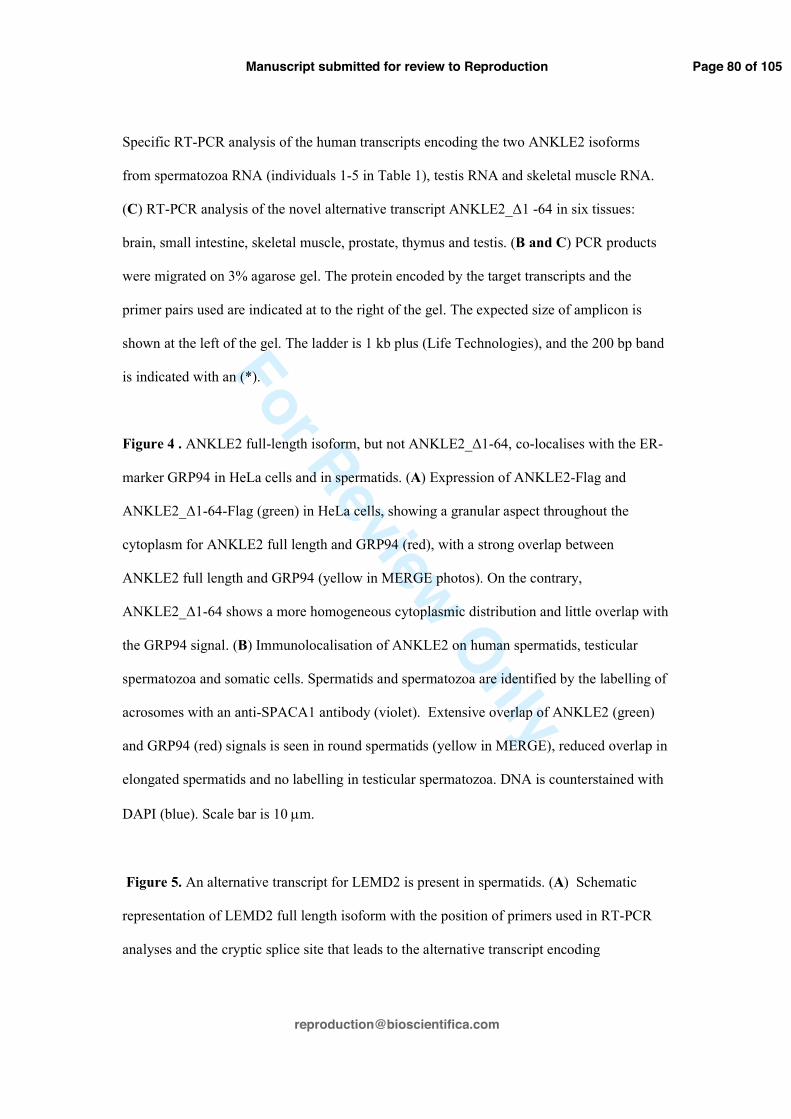
Figure 4 . ANKLE2 full-length isoform, but not ANKLE2_∆1-64, co-localises with the ER-
marker GRP94 in HeLa cells and in spermatids. (A) Expression of ANKLE2-Flag and
ANKLE2_∆1-64-Flag (green) in HeLa cells, showing a granular aspect throughout the
cytoplasm for ANKLE2 full length and GRP94 (red), with a strong overlap between
ANKLE2 full length and GRP94 (yellow in MERGE photos). On the contrary,
ANKLE2_∆1-64 shows a more homogeneous cytoplasmic distribution and little overlap with
the GRP94 signal. (B) Immunolocalisation of ANKLE2 on human spermatids, testicular
spermatozoa and somatic cells. Spermatids and spermatozoa are identified by the labelling of
acrosomes with an anti-SPACA1 antibody (violet). Extensive overlap of ANKLE2 (green)
and GRP94 (red) signals is seen in round spermatids (yellow in MERGE), reduced overlap in
elongated spermatids and no labelling in testicular spermatozoa. DNA is counterstained with
DAPI (blue). Scale bar is 10 µm.
Figure 5. An alternative transcript for LEMD2 is present in spermatids. (A) Schematic
representation of LEMD2 full length isoform with the position of primers used in RT-PCR
analyses and the cryptic splice site that leads to the alternative transcript encoding
Page 80 of 105
Manuscript submitted for review to Reproduction

For Review Only
LEMD2_∆115 -259. Primer o5055 is specific for the LEMD2_∆115 -259 transcript. Primers
o4711 and o4712 were used to amplify the full coding region from testis RNA and generated
two bands (right panel) that were sequenced and the predicted proteins encoded by these
transcripts are represented schematically with the different domains (TM: transmembrane)
mapped to LEMD2 (Brachner et al., 2005). (B) RT-PCR analysis of the alternative transcript
encoding LEMD2_∆115 -259 in human spermatozoa (individuals 1-5 in Table 1), and six
tissues: brain, small intestine, skeletal muscle, prostate, thymus and testis. PCR products
were migrated on 3% agarose gels. The ladder is 1 kb plus (Life technologies), and the 200
bp band is indicated with an (*). (C) Ectopic expression of fusion proteins LEMD2 and
LEMD2_∆115 -259 (green) with C-terminal Flag tag (red) in HeLa cells. Only LEMD2
transfected cells were labelled with anti-LEMD2 (Sigma HPA017340), indicating that this
antibody is specific for the full-length isoform of LEMD2. Scale bar is 10 µm.
Figure 6. Immunolocalisation of LEM-domain proteins and BAF proteins (green) on human
spermatids and testicular spermatozoa. Labelling is shown on successive steps of spermatid
development: early, mid and late round, early elongating and testicular spermatozoa. The
antibodies used are indicated in green. Spermatids are identified using lectin PNA (red) and
DNA counterstained with DAPI (blue). Exposures have been normalised on Sertoli cell
labelling. Scale bar is 10 µm.
Figure 7. Immunolocalisation of BAF and BAF-L on human ejaculated spermatozoa. (A)
Composite montage of spermatozoa labelled with BAF and BAF-L (green). BAF and BAF-L
were detected respectively in 30% and 36,9% of mature ejaculated spermatozoa, at the
posterior pole of the nucleus (upper panels). No labelled spermatozoa were observed when
each antibody was pre-absorbed with its immunogen peptide (lower panels). Acrosomes were
Page 81 of 105
Manuscript submitted for review to Reproduction

For Review Only
labelled using lectin PNA (red) and the nucleus is stained with DAPI (blue). Scale bar is 10
µm. (B) Protein expression of BAF and BAF-L by western blot analysis of human
spermatozoa lysates. Results are shown for human testis (T011400051, T011500055,
T011400062 and T011400071) (20 µg) and three spermatozoa samples (2 x 10⁶
spermatozoa) corresponding to individuals 11-13 in Table 1. The anti-α tubulin antibody was
used, on the same membranes without prior stripping, as a loading control. In order to reveal
the faint signal of BAF-L in whole testis lysates, a long exposure of a segment of the BAF-L
blot (between the grey bars) was compressed vertically and is shown below the α-tubulin
result.
Table 1: Sperm parameters of patients whose spermatozoa were used for RT-PCR, western
blot (WB), immunofluorescence analysis (IF) and selection procedures. F Fertile patients, SI
Secondary infertility, *WHO criteria, **Normal forms (David criteria).
Table 2: The presence (+) or absence (-) of LEM-domain proteins, BAF proteins, LBR and
lamins, and their respective transcripts in human spermatids and spermatozoa, as determined
by us in this study or previously (Elkhatib et al. 2015). nd: not determined; +?: the absence of
an antibody that discriminates between lamin B2 and lamin B3 prevents us from determining
which is present.
Supplementary data Figure Legends
Figure S1: Phase contrast overlay on IF analysis of testicular cells and HeLa cells used for
the localisation of ANKLE2.
Page 82 of 105
Manuscript submitted for review to Reproduction

For Review Only
Figure S2: Affinity tests of antibodies used in immunofluorescence analyses. LEM-domain
proteins and BAF proteins (green) tagged with c-Myc or Flag (red) were ectopically
expressed in HeLa cells. Tagged proteins were detected using antibodies against the target
protein (green) and against the tag (red). The anti-LAP2β antibody was tested on HeLa cells
transfected with a construct expressing tagged versions of either LAP2α (nucleoplasm) or
LAP2β (nuclear periphery), and its failure to label the nucleoplasm in LAP2α transfected
cells shows the antibody does not have high affinity for native LAP2α. DNA is
counterstained with DAPI (blue). Scale bar is 10 µm.
Figure S3: Immunolocalisation of LEM-domain proteins on human spermatids, testicular
spermatozoa and somatic cells showing the difference of localisation of different proteins
(green) between germ cells and somatic cells. Spermatids are identified using lectin PNA
(red) and counterstained with DAPI (blue). Exposures for the different antibodies were
normalised on Sertoli cell labelling. Scale bar is 10 µm.
Figure S4: Western blot analysis of protein lysates from human testis and purified
spermatozoa with the anti-Emerin antibody Sigma AMAb90560. The predicted molecular
weight of Emerin is 29 kDa.
Figure S5: Immunolocalisation of LEMD3, LBR, LEMD2 on cells from disaggregated
human testis. Spermatids and testicular spermatozoa are not labelled, only somatic cells are
labelled (green) with anti-LEMD3 (ab123973), anti-LBR (ab32535) and anti-LEMD2-mid
antibody (Sigma HPA017340). The LEMD2-mid antibody is specific for the full LEMD2
isoform. Spermatids are identified using lectin PNA (red) and DNA is counterstained with
Page 83 of 105
Manuscript submitted for review to Reproduction

For Review Only
DAPI (blue). Exposures for the different antibodies were normalised on Sertoli cell labelling.
Scale bar is 10 µm.
Figure S6: Immunolocalization of Emerin on cells from disaggregated human testis and
ejaculated spermatozoa. Spermatids and spermatozoa are not labelled, only somatic cells are
labelled (green) with two different anti-Emerin antibodies (LEICA-NCL-EMERIN and
Sigma AMAb90560 - green). Ejaculated spermatozoa are presented in a composite montage.
Spermatids are identified using lectin PNA (red) and DNA is counterstained with DAPI
(blue). Exposures for the two antibodies were normalised on Sertoli cell labelling. Scale bar
is 10 µm.
Figure S7: (A) Percentages of germ cells labelled with anti-BAF antibody (ab129184)
(green) on post-meiotic germ cells of 4 patients (T011400051, T011400063, T011400071
and T011500055). (B) Three examples of the labelling round spermatids, elongating
spermatids and testicular spermatozoa with the anti-BAF antibody (green). Nuclei are
labelled with DAPI (blue) and acrosomes with lectin PNA (red). (C) Immunofluorescent
labelling of BAF (green) on three spermatocytes. DNA is counterstained with DAPI (blue).
Table S1: Primer sequences used in RT-PCR, DNA constructs and sequencing. Adaptors
used for constructs are underlined and the name of the restriction enzyme site in the adaptor
is preceded by a + after the target gene name. Target gene prefix: h=human; m=mouse.
Table S2: Antibodies used in immunofluorescence and western blot analyses. For primary
antibodies, IF and Western tests refer to affinity tests by transfection into tissue culture cells
Page 84 of 105
Manuscript submitted for review to Reproduction

For Review Only
(Figure S1). For secondary antibodies, this refers to the absence of IF signal when antibody is
used without a primary antibody.
Page 85 of 105
Manuscript submitted for review to Reproduction

For Review Only
Figure 1. RT-PCR amplification of transcripts coding LEM-Domain proteins in human from total spermatozoa RNA, testis RNA and skeletal muscle RNA. RT-PCR analysis of the 5´ and 3´ ends of the coding region were
performed separately for all tested transcripts, except for the short two exon coding regions of BAF and BAF-L which were tested with a single RT-PCR assay. RT-PCR products are shown migrated on a 3% agarose gel. Spermatozoa RNAs were extracted from samples from individuals 1-5 in Table 1. The protein encoded by the
target transcript and the primer pairs used are indicated at the right of the gel. The known spermatid transcript for protamine 1 (PRM1) was amplified as a positive control. The expected size of each amplicon is shown at the left of the gel. The ladder is 1 kb Plus DNA Ladder (Life Technologies), and the 200 bp band is
indicated with an (*).
228x302mm (300 x 300 DPI)
Page 86 of 105
Manuscript submitted for review to Reproduction

For Review Only
Figure 2. RT-PCR analysis of mouse transcripts for LEMD1 and BANF2 on RNA extracted from staged mouse testis of different postnatal ages (dpp: days post partum). The expected germ cell content is indicated
above the gel by the grey bars. Amplicons from LEMD1 or BAF-L transcripts were co-amplified with amplicons from the control HMBS transcript by RT-PCR, and show that LEMD1 and BANF2 transcripts
predominate during mouse spermiogenesis. The proteins encoded by transcripts and the primers pair used are indicated at the right of the gel. The expected sizes of amplicons are shown at the left of the gel. The
ladder is 1 kb Plus DNA Ladder (Life Technologies), and the 200 bp band is indicated with an (*).
109x68mm (300 x 300 DPI)
Page 87 of 105
Manuscript submitted for review to Reproduction

For Review Only
Figure 3. An alternative ANKLE2 transcript encoding ANKLE2_∆1 -64 is present in spermatids. (A) Schematic representation of full length ANKLE2 isoform described in databases, and a testis EST (HY008086) that
includes an alternative first exon in intron 1. (B) Specific RT-PCR analysis of the human transcripts encoding the two ANKLE2 isoforms from spermatozoa RNA (individuals 1-5 in Table 1), testis RNA and skeletal muscle
RNA. (C) RT-PCR analysis of the novel alternative transcript ANKLE2_∆1 -64 in six tissues: brain, small intestine, skeletal muscle, prostate, thymus and testis. (B and C) PCR products were migrated on 3%
agarose gel. The protein encoded by the target transcripts and the primer pairs used are indicated at to the right of the gel. The expected size of amplicon is shown at the left of the gel. The ladder is 1 kb plus (Life
Technologies), and the 200 bp band is indicated with an (*).
166x163mm (300 x 300 DPI)
Page 88 of 105
Manuscript submitted for review to Reproduction

For Review Only
Figure 4 . ANKLE2 full-length isoform, but not ANKLE2_∆1-64, co-localises with the ER-marker GRP94 in HeLa cells and in spermatids. (A) Expression of ANKLE2-Flag and ANKLE2_∆1-64-Flag (green) in HeLa cells, showing a granular aspect throughout the cytoplasm for ANKLE2 full length and GRP94 (red), with a strong overlap between ANKLE2 full length and GRP94 (yellow in MERGE photos). On the contrary, ANKLE2_∆1-64
shows a more homogeneous cytoplasmic distribution and little overlap with the GRP94 signal. (B) Immunolocalisation of ANKLE2 on human spermatids, testicular spermatozoa and somatic cells. Spermatids
and spermatozoa are identified by the labelling of acrosomes with an anti-SPACA1 antibody (violet). Extensive overlap of ANKLE2 (green) and GRP94 (red) signals is seen in round spermatids (yellow
in MERGE), reduced overlap in elongated spermatids and no labelling in testicular spermatozoa. DNA is counterstained with DAPI (blue). Scale bar is 10 microns.
171x170mm (300 x 300 DPI)
Page 89 of 105
Manuscript submitted for review to Reproduction

For Review Only
Figure 5. An alternative transcript for LEMD2 is present in spermatids. (A) Schematic representation of LEMD2 full length isoform with the position of primers used in RT-PCR analyses and the cryptic splice site
that leads to the alternative transcript encoding LEMD2_∆115 -259. Primer o5055 is specific for the LEMD2_∆115 -259 transcript. Primers o4711 and o4712 were used to amplify the full coding region from
testis RNA and generated two bands (right panel) that were sequenced and the predicted proteins encoded by these transcripts are represented schematically with the different domains (TM: transmembrane) mapped to LEMD2 (Brachner et al., 2005). (B) RT-PCR analysis of the alternative transcript encoding LEMD2_∆115 -
259 in human spermatozoa (individuals 1-5 in Table 1), and six tissues: brain, small intestine, skeletal muscle, prostate, thymus and testis. PCR products were migrated on 3% agarose gels. The ladder is 1 kb
plus (Life technologies), and the 200 bp band is indicated with an (*). (C) Ectopic expression of fusion proteins LEMD2 and LEMD2_∆115 -259 (green) with C-terminal Flag tag (red) in HeLa cells. Only LEMD2
transfected cells were labelled with anti-LEMD2 (Sigma HPA017340), indicating that this antibody is specific for the full-length isoform of LEMD2. Scale bar is 10 microns.
Page 90 of 105
Manuscript submitted for review to Reproduction

For Review Only
256x378mm (300 x 300 DPI)
Page 91 of 105
Manuscript submitted for review to Reproduction

For Review Only
Figure 6. Immunolocalisation of LEM-domain proteins and BAF proteins (green) on human spermatids and testicular spermatozoa. Labelling is shown on successive steps of spermatid development: early, mid and
late round, early elongating and testicular spermatozoa. The antibodies used are indicated in green. Spermatids are identified using lectin PNA (red) and DNA counterstained with DAPI (blue). Exposures have
been normalised on Sertoli cell labelling. Scale bar is 10 µm.
137x114mm (300 x 300 DPI)
Page 92 of 105
Manuscript submitted for review to Reproduction

For Review Only
Figure 7. Immunolocalisation of BAF and BAF-L on human ejaculated spermatozoa. (A) Composite montage
of spermatozoa labelled with BAF and BAF-L (green). BAF and BAF-L were detected respectively in 30% and
36,9% of mature ejaculated spermatozoa, at the posterior pole of the nucleus (upper panels). No labelled
spermatozoa were observed when each antibody was pre-absorbed with its immunogen peptide (lower
panels). Acrosomes were labelled using lectin PNA (red) and the nucleus is stained with DAPI (blue). Scale
bar is 10 microns. (B) Protein expression of BAF and BAF-L by western blot analysis of human spermatozoa
lysates. Results are shown for human testis (T011400051, T011500055, T011400062 and T011400071) (20
µg) and three spermatozoa samples (2 x 10⁶ spermatozoa) corresponding to individuals 11-13 in Table 1.
The anti-α-tubulin antibody was used, on the same membranes without prior stripping, as a loading control.
In order to reveal the faint signal of BAF-L in whole testis lysates, a long exposure of a segment of the BAF-
L blot (between the grey bars) was compressed vertically and is shown below the α-tubulin result.
206x245mm (300 x 300 DPI)
Page 93 of 105
Manuscript submitted for review to Reproduction

For Review Only
F Fertile patients. SI Secondary infertility *WHO criteria, **Normal forms (David criteria)
Patients GT number Spz (M/ml) Motility (a+b/c/d)* Vitality (%) Normal forms **
(%)
Technics performed on spermatozoa
1 F R 011300032 84 35/20/45 78 39 RT-PCR
2 F R 011300029 160 50/10/40 70 37 RT-PCR, IF
3 R 010900491 102 45/20/35 85 45 RT-PCR, IF, WB
4 SI R 010900544 150 50/20/30 80 41 RT-PCR
5 R 010900563 109.6 60/10/30 80 39 RT-PCR
6 R 01090026 44 60/20/20 64 20 RT-PCR
7 R 01090040 33,6 40/10/50 60 22 RT-PCR
8 R 010900236 63,4 30/25/40 81 20 RT-PCR
9 R 010900505 92 25/45/30 82 54 IF
10 F R 011500066 135 30/10/60 65 22 WB
11 F R 011500071 130 30/10/60 59 32 WB
12 R 010900549 195 60/10/30 85 53 WB
13 R 010900409 259,8 30/10/60 82 53 WB
14 R 010900214 159 35/10/55 70 34 WB
Page 95 of 105
Manuscript submitted for review to Reproduction

For Review Only
Transcript (RT-PCR) Protein (IF) Ejac. sperm. Spermatid Ejac. Sperm. LEMD1 + + - LEMD2-full - - - LEMD2-∆115-259 + + - LEMD3 - - - ANKLE2 + + - ANKLE2_∆1-64 + nd - ANKLE1 - nd nd LAP2α + nd nd LAP2β + + - Emerin + - - LBR - - - BAF - + + (30%) BAF-L + + + (37%) Lamin A - - - Lamin C - - - Lamin B1 + + + (30-60%) Lamin B2 - +? - Lamin B3 + +? nd
Page 96 of 105
Manuscript submitted for review to Reproduction

For Review Only
129x97mm (300 x 300 DPI)
Page 97 of 105
Manuscript submitted for review to Reproduction

For Review Only
215x268mm (300 x 300 DPI)
Page 98 of 105
Manuscript submitted for review to Reproduction

For Review Only
140x114mm (300 x 300 DPI)
Page 99 of 105
Manuscript submitted for review to Reproduction

For Review Only
105x74mm (300 x 300 DPI)
Page 100 of 105
Manuscript submitted for review to Reproduction

For Review Only
154x150mm (300 x 300 DPI)
Page 101 of 105
Manuscript submitted for review to Reproduction

For Review Only
122x92mm (300 x 300 DPI)
Page 102 of 105
Manuscript submitted for review to Reproduction

For Review Only10% 2% 10%
0% 20% 40% 60% 80% 100%
testicular spermatozoa elongated spermatids
Round spermatids
T011400071-F
1% 5%
37%
0% 20% 40% 60% 80% 100%
testicular spermatozoa elongated spermatids
Round spermatids
T011500055-F
6% 3%
55%
0% 20% 40% 60% 80% 100%
testicular spermatozoa elongated spermatids
Round spermatids
T011400063-F
7% 9%
35%
0% 20% 40% 60% 80% 100%
testicular spermatozoa elongated spermatids
Round spermatids
T011400051-F
A
C
Figure S7. (A) Percentages of germ cells labelled with anti-BAF antibody (ab129184) (red) on post-meiotic germ cells of 4 patients (T011400051, T011400063, T011400071 and T011500055). (B) Three
examples of the labelling on round spermatids, elongating spermatids and testicular spermatozoa with the anti-BAF antibody (green). Nuclei are labelled with DAPI (blue) and acrosomes with lectin (red). (C)
Immunofluorescent labelling of BAF (green) on a spermatocyte. DNA is counterstained with DAPI (blue). Scale bar is 10 µm.
BAF DAPI
B
Round spermatids Elongated spermatids Testicular spermatozoa
Page 103 of 105
Manuscript submitted for review to Reproduction

For Review Only
Primer number Primer sequence Target Gene Annealing
temperature o1680 AGGTACAGATGCTGTCGCAG h_PRM1 65°C
o1681 AGTCTGGTAACATTCTCAGGC h_PRM1 62°C
o2216 TGAAAGCCTTGTACCCTGGC m_Hmbs 66°C
o2217 GAGTGAACGACCAGGTCCAC m_Hmbs 66°C
o4087 TCTTCTACTCCTCTGCCAAC h_TMPO (LAP2) 61°C
o4088 CCACCAGAGGGAGTAGTTCC h_TMPO (LAP2α) 65°C
o4090 GTTAATGCCTGCAGAGGTCC h_TMPO (LAP2β) 64°C
o4092 TCCAGGACCATTACGTGGAC h_LEMD2 65°C
o4093 CTCGGTCCCAGACACGCTTC h_LEMD2 69°C
o4094 TTGGTGAAGTCCTGGGCAAG h_BANF1 66°C
o4095 GAAGGCATCCGAAGCAGTCC h_BANF1 67°C
o4096 TGGGTGGATGGCATCAGCC h_BANF2 69°C
o4097 CCTCACACTCAGTGGCACC h_BANF2 66°C
o4367 AGGTTCGAATTCGCTAGCGAGATGGACGACATGTCTCC h_BANF2 +NheI 63°C
o4368 ATTTGACGGATCCCAGGAAGCAGGCACACCAC h_BANF2 +BamHI 67°C
o4388 AGGTTCGAATTCGCTAGCGATCAAGATGACAACCTCCC h_BANF1 +EcoRI-NheI 62°C
o4389 ATTTGACGGATCCTCACAAGAAGGCGTCGCACC h_BANF1 +BamHI 69°C
o4399 ATTTGACGGATCCCAAGAAGGCGTCGCACCAC h_BANF1 +BamHI 70°C
o4426 GCCCAATACTACCTTCCACC h_LEMD1 63°C
o4427 AAGCCTCAGGCCATTTTTTGAG h_LEMD1 64°C
o4434 GCCTCAAGTCTCTTCTTGCC m_Lemd1 64°C
o4435 GATTGCATAGACTTCAGACAACC m_Lemd1 61°C
o4491 GGCTTCAATGGAGTGTTGGA h_LEMD3 63°C
o4493 CATAGCAGTGCCCTTGCTTC h_ANKLE1 64°C
o4495 ATGTCCTTGAGTCCCAGCAG h_ANKLE2 65°C
o4496 ACTGAGATCTGGCAGCTGAG h_ANKLE2 65°C
o4500 AACCATGATTGCAGACATTG h_ANKLE2 59°C
o4501 CCCTCACGGAAATGGAATTG h_ANKLE2 62°C
o4502 CATGAGCTGGGGTATCCCTG h_ANKLE2 66°C
o4503 AGTTCGAGACCAGCCTGGTC h_ANKLE2 67°C
o4504 GGAAAACTCCACCTCGAGAG h_ANKLE2 63°C
o4522 AGGTCTCATATGAAGATGGACGACATGTCGCC m_Banf2 66°C
o4523 TTTGACGAATTCTTAGGAAACAGGAGCACCACT m_Banf2 64°C
o4527 CGGTAGACTGTCGGGTCTGG h_ANKLE2 68°C
o4528 AGAGCTGTGACACCTGCCTC h_ANKLE2 68°C
o4542 TTGGCAGGTACCGAAATGACAATGGATGCTCTGTTGG h_ANKLE2_Δ1-64 +KpnI 65°C
o4573 TTGGCAGGTACCATGCTGTGGCCGCGGCTGG h_ANKLE2 +KpnI 65°C
o4579 ATTGCAGGATCCGACAACTACGCAGATCTTTCGG h_EMD +BamHI 67°C
Page 104 of 105
Manuscript submitted for review to Reproduction

For Review Only
o4580 ATTGAACTCGAGTCATGGCTCCCTCTAGAAGG h_EMD +XhoI 64°C
o4609 AAAGTGCTCCTCCAAGCCAG h_PTPRC 59°C
o4610 GCCAGAAATGCTATCAGTGCCTTAG h_PTPRC 60°C
o4611 GCTTCGCGTCCAGACAGG h_KIT 61°C
o4612 CGTGTATTTGCCGGTGTTGG h_KIT 59°C
o4613 CTTTGACGCCGAGAGCTACA h_CDH1 59°C
o4614 TTTGAATCGGGTGTCGAGGG h_CDH1 59°C
o4615 ATGCTCCTCGAGCCATTCAGTTGGATTTTCTAGGGTCAAC h_TMPO (LAP2β) +XhoI 66°C
o4659 ATGGTCCTCGAGTTAACCAAACAGCGACTTATTTTC h_LEMD1 +XhoI 59°C
o4661 AGGTCTGGTACCATGGTGGATGTGAAGTGTCTG h_LEMD1 +KpnI 63°C
o4663 CCTAGACTCGAGCTACAGGAAGCAGGCACACCAC h_BANF2 +XhoI 71°C
o4667 CTGTCGCCTGCCAGCTCTG h_LBR 70°C
o4668 GGATGATGTCCATGGTCGTC h_LBR 63°C
o4669 ACCGAGCCCCTCCTCGCAG h_LBR 72°C
o4671 GGGAGGTGCTGTCGTGGCTC h_LBR 72°C
o4702 GGTTATAAGCTTACAACCTCCCAAAAGCACC h_BANF1 +HindIII 64°C
o4710 AGGTCAGGATCCCTACAGGGCGGCAAGCTCA h_ANKLE2 +BamHI 68°C
o4711 ATGGCTAAGCTTGCCGGCCTGTCGGACCT h_LEMD2 +HindIII 72°C
o4712 TTGGCAGGTACCGGGGCTTATCGCTCTGAGTC h_LEMD2 +KpnI 70°C
o4965 GCTACTGCTCGTCTTCCTGG h_LEMD2 66°C
o4966 GTAGAGTTCATGCAGCAGCTC h_LEMD2 64°C
o5022 ATCCGGGATCCCCGGAGTTCCTGGAAGACC h_TMPO (LAP2β) +BamHI 66°C
o5023 GACACCGTGTCTAAAGATTCGG h_LEMD3 63°C
o5024 GTGCCCACTACTTGTCGATG h_LEMD3 64°C
o5026 GCAAGCTGTGCCAATCGATCA h_LEMD3 67°C
o5027 CAAGGTGCGTCAGATCTTGG h_ANKLE1 64°C
o5028 AATCGACCGACCAGGCGGTG h_ANKLE1 71°C
o5029 AGCGCAGCAGCTCCTCTACT h_ANKLE1 69°C
o5095 CCGAGCTGACCACCTTGCTG h_EMD 69°C
o5096 CTGGGTCTCGTACTCGAAGAT h_EMD 64°C
o5097 GCTGTCCGCCAGTCAGTGAC h_EMD 69°C
o5098 GGTAGTGCGTGATGCTCTGG h_EMD 66°C
o5099 AGCATGCATCTCCTATTCTGCC h_TMPO (LAP2β) 65°C
o5100 GATTGGTCTGCGGCAACTAGCA h_TMPO (LAP2β) 67°C
o5101 CGGACTTCTCCAGTGACGAAGAG h_TMPO (LAP2) 67°C
o5103 CCACAATAGGACCAGGATTCACTCC h_TMPO (LAP2) 67°C
o5111 CCCCCGCCTCAGGTGAAATG h_ANKLE2 69°C
o5112 GGGTCCACATTTCAATCCGGCTTTG h_ANKLE2 69°C
o5186 GGTTATAAGCTTGACGACATGTCTCCCAGGCTG h_BANF2 +HindIII 70°C
Page 105 of 105
Manuscript submitted for review to Reproduction

For Review Only
o5322 ATGGCTAAGCTTATGGCCGGCCTGTCGGACCT h_LEMD2 +HindIII 78°C
o5323 TTGGCAGGTACCTCGCTCTGAGTCAGAGAAGG h_LEMD2 +KpnI 66°C
o5350 ATTTGACCATATGTTCCTCTCCGAACCCATTGGAG h_BANF2 +NdeI 69°C
o5351 ATTTGACCTCGAGCAGGAAGCAGGCACACCAC h_BANF2 +XhoI 69°C
o5352 GTTCCAGGTACCATGCCAAGTAGGAAATTTGCCGATG h_LBR +KpnI 68°C
o5353 AAGTCCGGATCCGTAGATGTATGGAAATATACGGTAGGGCAC h_LBR +BamHI 68°C
o5354 GTTCCAGGTACCCCAAGTAGGAAATTTGCCGATGGTG h_LBR +KpnI 68°C
o5355 AAGTCCGGATCCTTAGTAGATGTATGGAAATATACGGTAGGGCAC h_LBR +BamHI 68°C
Table S1. Primer sequences used in RT-PCR, DNA constructs and sequencing. Adaptors used for constructs are underlined and the name of the restriction enzyme site in the adaptor
is preceded by a + after the target gene name. Target gene prefix: h=human; m=mouse.
Page 106 of 105
Manuscript submitted for review to Reproduction

For Review Only
Antibodies References Epitope or immunogen position IF tests WB tests LEMD1 Sigma SAB3500644 aa 80-130 in isoform 1 (181 aa) YES YES
LEMD2_mid Sigma HPA017340 aa 243-359 (this study) YES NO LEMD2-C-ter ab89866 aa 454-503 YES YES (by abcam)
LEMD3 ab123973 Within human LEMD3 YES (by abcam) YES (by abcam) ANKLE2 Asencio et al.,2012 aa 59-938 YES YES Emerin Leica-NCL-EMERIN 222 amino acid region near N-terminus YES Not recommended Emerin Sigma AMAb90560 63-DALLYQSKGYNDDYY-77 YES NO LAP2� sigma HPA008150 aa 129-187 (this study) YES YES
BAF ab129184 aa 1-45 YES YES (by abcam) BAF-L Sigma HPA042635 aa 12-252 of human BANF2 YES YES LBR ab32535 aa 1-60* YES YES (by abcam)
GRP94 ab210960 aa 655-798 YES YES (by abcam) SPACA1 Sigma HPA026744 aa 93-153 YES (by Sigma) YES (by Sigma)
Flag Sigma F1804 DYKDDDDK YES YES c-Myc Sc-40 408-439 amino acids within C-domain of c-Myc YES YES �Tubulin ab15246 426-ALEKDYEEVGVDSVEGEGEEEGEEY-450 NO YES
Secondary:GAR Invitrogen A11008 Goat Anti Rabbit - Alexa Fluor®488 *YES - Secondary:GAM Invitrogen A11001 Goat Anti Mouse - Alexa Fluor®488 *YES - Secondary:GAM Invitrogen A21121 Goat Anti Mouse - Alexa Fluor®488 *YES - Secondary:GAM Invitrogen A11003 Goat Anti Mouse - Alexa Fluor®546 *YES - Secondary:DAR Invitrogen A31573 Donkey Anti Rabbit – Alexa fluor®647 *YES -
Table S2: Antibodies used in immunofluorescence and western blot analyses. For primary antibodies, IF and Western tests refer to affinity tests by transfection into tissue
culture cells (Figure S1). For secondary antibodies, this refers to the absence of IF signal when antibody is used without a primary antibody.
Page 107 of 105
Manuscript submitted for review to Reproduction