Kuru - VCJD - Priongenesis
24
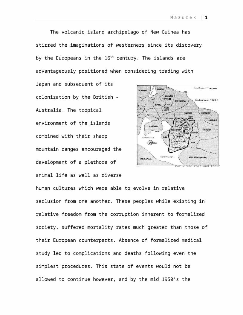
Mazurek | 1 The volcanic island archipelago of New Guinea has stirred the imaginations of westerners since its discovery by the Europeans in the 16 th century. The islands are advantageously positioned when considering trading with Japan and subsequent of its colonization by the British – Australia. The tropical environment of the islands combined with their sharp mountain ranges encouraged the development of a plethora of animal life as well as diverse human cultures which were able to evolve in relative seclusion from one another. These peoples while existing in relative freedom from the corruption inherent to formalized society, suffered mortality rates much greater than those of their European counterparts. Absence of formalized medical study led to complications and deaths following even the simplest procedures. This state of events would not be allowed to continue however, and by the mid 1950’s the
Transcript of Kuru - VCJD - Priongenesis
M a z u r e k | 1
The volcanic island archipelago of New Guinea has
stirred the imaginations of westerners since its discovery
by the Europeans in the 16th century. The islands are
advantageously positioned when considering trading with
Japan and subsequent of its
colonization by the British –
Australia. The tropical
environment of the islands
combined with their sharp
mountain ranges encouraged the
development of a plethora of
animal life as well as diverse
human cultures which were able to evolve in relative
seclusion from one another. These peoples while existing in
relative freedom from the corruption inherent to formalized
society, suffered mortality rates much greater than those of
their European counterparts. Absence of formalized medical
study led to complications and deaths following even the
simplest procedures. This state of events would not be
allowed to continue however, and by the mid 1950’s the
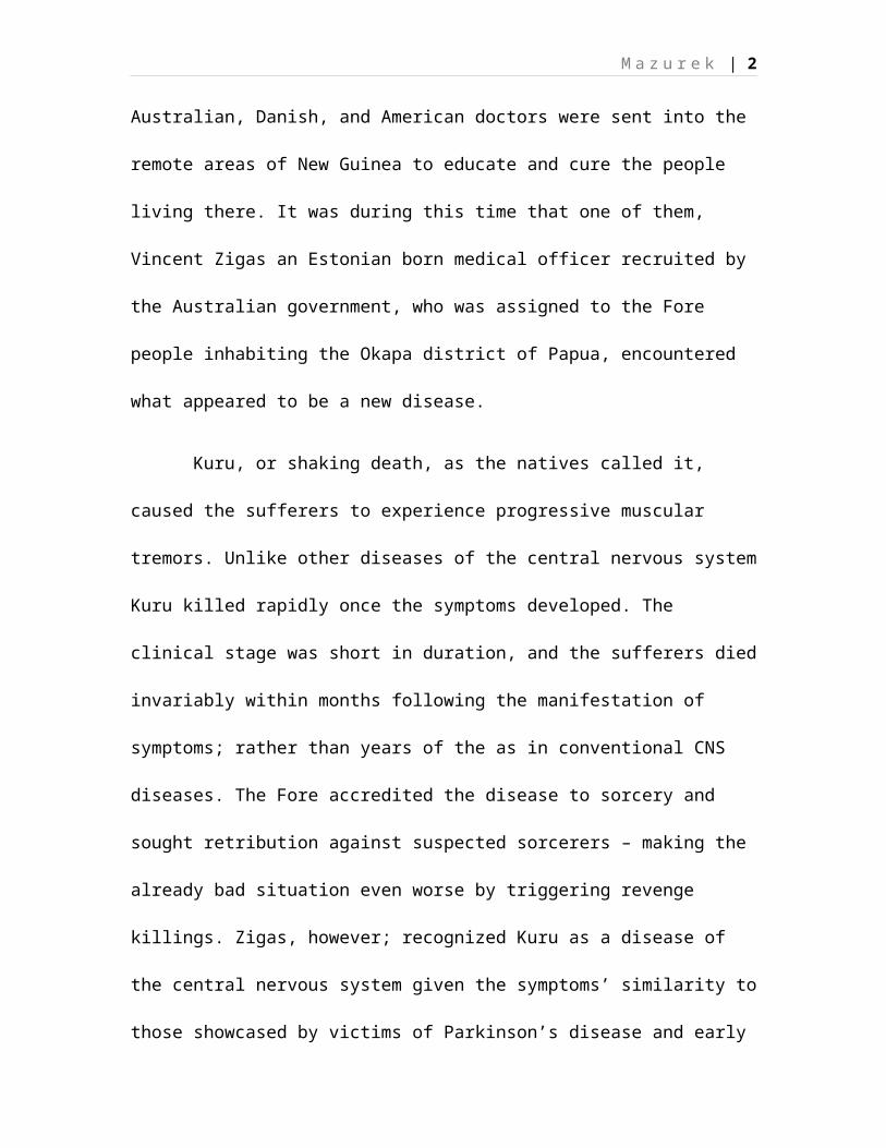
M a z u r e k | 2
Australian, Danish, and American doctors were sent into the
remote areas of New Guinea to educate and cure the people
living there. It was during this time that one of them,
Vincent Zigas an Estonian born medical officer recruited by
the Australian government, who was assigned to the Fore
people inhabiting the Okapa district of Papua, encountered
what appeared to be a new disease.
Kuru, or shaking death, as the natives called it,
caused the sufferers to experience progressive muscular
tremors. Unlike other diseases of the central nervous system
Kuru killed rapidly once the symptoms developed. The
clinical stage was short in duration, and the sufferers died
invariably within months following the manifestation of
symptoms; rather than years of the as in conventional CNS
diseases. The Fore accredited the disease to sorcery and
sought retribution against suspected sorcerers – making the
already bad situation even worse by triggering revenge
killings. Zigas, however; recognized Kuru as a disease of
the central nervous system given the symptoms’ similarity to
those showcased by victims of Parkinson’s disease and early
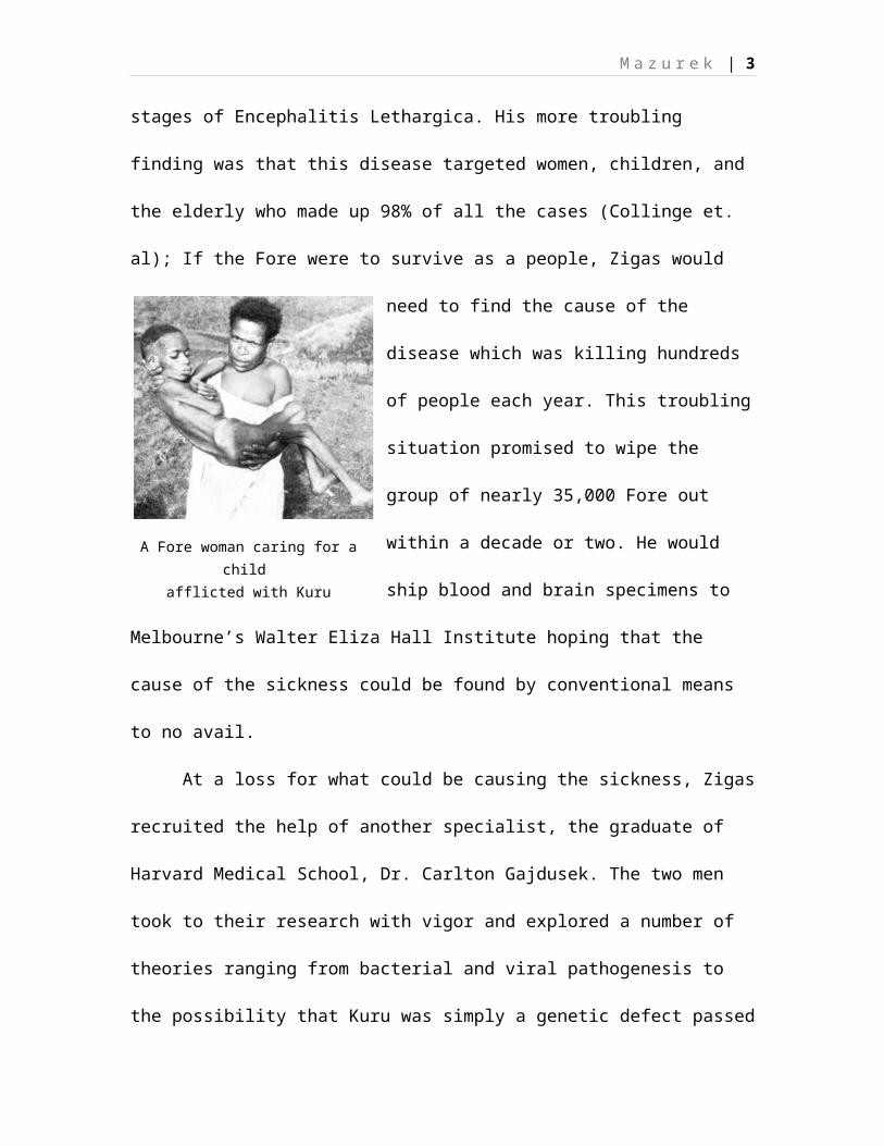
A Fore woman caring for achild
afflicted with Kuru
M a z u r e k | 3
stages of Encephalitis Lethargica. His more troubling
finding was that this disease targeted women, children, and
the elderly who made up 98% of all the cases (Collinge et.
al); If the Fore were to survive as a people, Zigas would
need to find the cause of the
disease which was killing hundreds
of people each year. This troubling
situation promised to wipe the
group of nearly 35,000 Fore out
within a decade or two. He would
ship blood and brain specimens to
Melbourne’s Walter Eliza Hall Institute hoping that the
cause of the sickness could be found by conventional means
to no avail.
At a loss for what could be causing the sickness, Zigas
recruited the help of another specialist, the graduate of
Harvard Medical School, Dr. Carlton Gajdusek. The two men
took to their research with vigor and explored a number of
theories ranging from bacterial and viral pathogenesis to
the possibility that Kuru was simply a genetic defect passed
M a z u r e k | 4
through the familial lines. In the course of their research
they frequently found themselves buying the bodies of the
dead from their families, trading tobacco and tools for
specimens which they hoped would yield a bacterial or viral
agent; inadvertently protecting the sellers from the disease
which moved by unconventional means. (Time)
However, these theories were soon dismissed as no
bacterial or viral agent was noted in the cells of the brain
which the men studied. The genetic factor was also dismissed
as the type of mutation would have either wiped the people
out completely, or would have been found to afflict men in
the prime of their age. The researcher’s first concrete clue
came from the autopsied brains themselves. Kuru sufferers’
brains were riddled with holes, causing the organs to take
on the appearance of a sponge. Something was disintegrating
the brain tissue the question however remained as to what
the agent was. The men noted no fever, nor inflammation,
which discounted the viral theory. No bacterium or multi-
cellular organic infection was present, therefore the
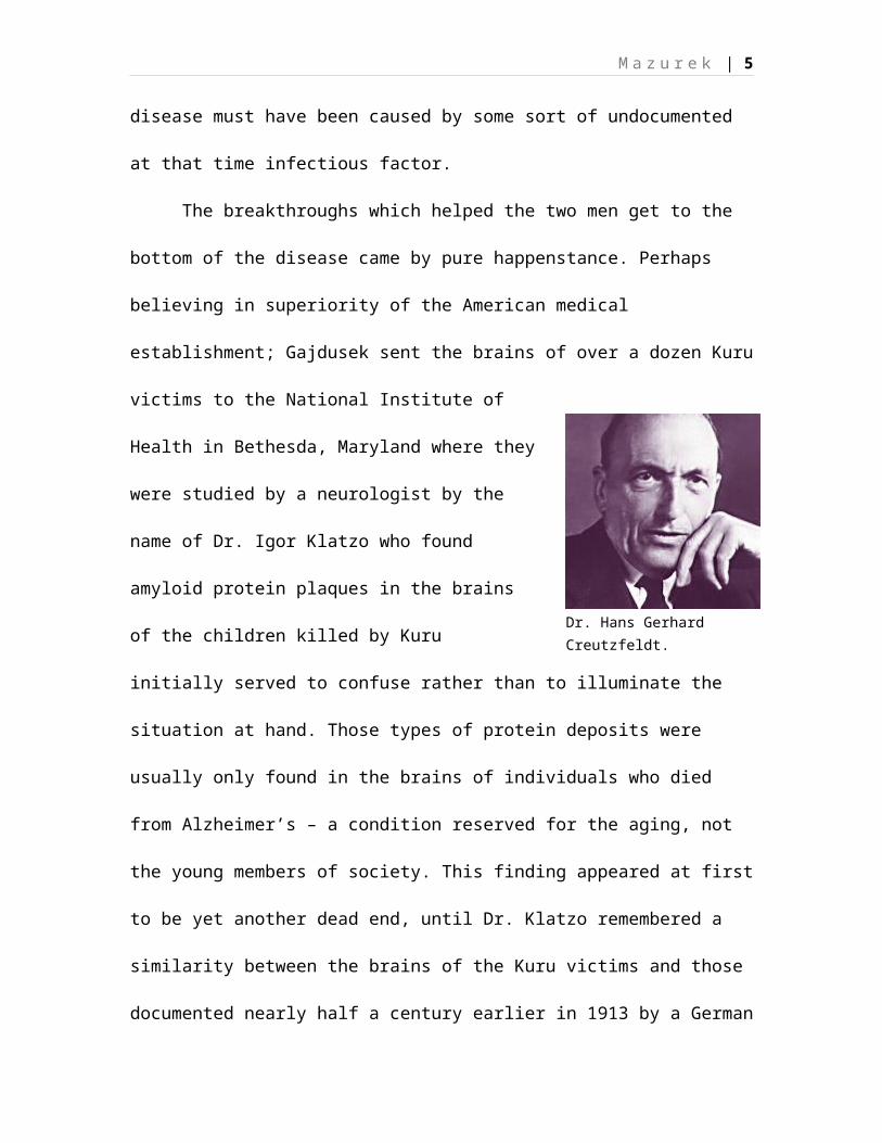
Dr. Hans Gerhard Creutzfeldt.
M a z u r e k | 5
disease must have been caused by some sort of undocumented
at that time infectious factor.
The breakthroughs which helped the two men get to the
bottom of the disease came by pure happenstance. Perhaps
believing in superiority of the American medical
establishment; Gajdusek sent the brains of over a dozen Kuru
victims to the National Institute of
Health in Bethesda, Maryland where they
were studied by a neurologist by the
name of Dr. Igor Klatzo who found
amyloid protein plaques in the brains
of the children killed by Kuru
initially served to confuse rather than to illuminate the
situation at hand. Those types of protein deposits were
usually only found in the brains of individuals who died
from Alzheimer’s – a condition reserved for the aging, not
the young members of society. This finding appeared at first
to be yet another dead end, until Dr. Klatzo remembered a
similarity between the brains of the Kuru victims and those
documented nearly half a century earlier in 1913 by a German
M a z u r e k | 6
doctor by the name of Hans Gerhard Creutzfeldt. The case was
that of a young woman who was showing signs of what was
thought at the time to be dementia.
Bertha Elschker was an orphan who in 1912 was
hospitalized for muscle tremors and spasmodic involuntary
muscle action (Yam 13) her condition quickly deteriorated
and within a few months the once energetic and studious girl
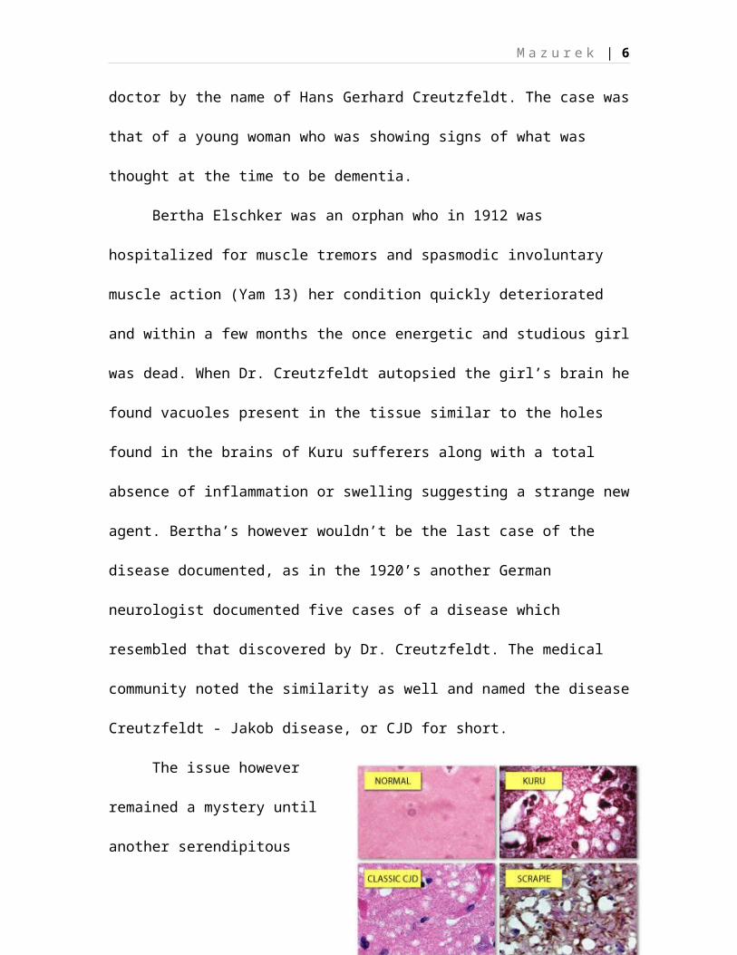
was dead. When Dr. Creutzfeldt autopsied the girl’s brain he
found vacuoles present in the tissue similar to the holes
found in the brains of Kuru sufferers along with a total
absence of inflammation or swelling suggesting a strange new
agent. Bertha’s however wouldn’t be the last case of the
disease documented, as in the 1920’s another German
neurologist documented five cases of a disease which
resembled that discovered by Dr. Creutzfeldt. The medical
community noted the similarity as well and named the disease
Creutzfeldt - Jakob disease, or CJD for short.
The issue however
remained a mystery until
another serendipitous
M a z u r e k | 7
twist of fate brought William Hadlow, an American
veterinarian whose work focused on Scrapie, a central
nervous system disease found in sheep, across a 1959 museum
exhibit of Gajdusek’s Kuru research. Similar to Kuru and
CJD, Scrapie is a degenerative CNS disease; similar to Kuru
and CJD the fatality rate for Scrapie is 100%.
Significantly, much like CJD and Kuru – the brains of the
sheep diagnosed and subsequently killed by Scrapie are
riddled with holes which give them the appearance of
sponges, with no inflammation or anti bodies present in the
tissue. Most importantly, however; Scrapie has been proven
to be passable from one organism to another by inoculation
with stricken nerve tissue in 1939 when two French
veterinarians Jean Cuille and Paul-Louis Chelle transmitted
a mixture made from the spinal cord of a Scrapie positive
sheep into the eye of a healthy one. While the incubation
time was much longer than expected the sheep who received
the shot started showing signs of Scrapie after fifteen
months. To further test their theory the two men made
subdermal and direct-brain injection of the same mixture.
M a z u r e k | 8
Sure enough the inoculated sheep also developed the disease
with the incubation time extending to two years for sub-
dermal inoculation, and contracting to twelve months in case
of the injection into the brain.
Armed with the newfound knowledge Dr. Gajdusek begun to
closely study the cultural practices of the Fore people who
were known for their mortuary cannibalism; he discovered
that most of the preparation and consumption of the dead was
left to women and children as well as the elderly. This
opened a number of avenues for infection with the still
unknown agent as the children frequently played with the
brains of those deceased while their mothers, aunts, and
grandmothers prepared the carcasses for consumption. The
adult men did not partake in the custom and had been by the
virtue of doing so spared from the sickness. It didn’t take
the Australian government long to outlaw the mortuary
cannibalism once the news surfaced leading to a drastic drop
off in the Kuru incidence among the Fore people. The disease
itself hasn’t disappeared entirely, however; as recent
studies conducted in the area have yielded new cases
M a z u r e k | 9
following much longer incubation periods, in some cases more
than 30 years. This can be explained by levels of immunity
to the pathogen, or by “underground” cannibalism persisting
in some portions of the population of the Fore. Importantly,
however; the disease is no longer a major threat to the
peoples’ existence.
Gajdusek’s next breakthrough came when, after departing
with tissue samples, and armed with his newfound knowledge
on the earlier experiments carried out by the French team he
began experimenting on Chimpanzees having secured the
permission to do so in 1961 from Patuxent Wildlife Research
Center in Maryland. In the meantime Gajdusek returned to New
Guinea in order to continue studying Kuru in the Fore, or
perhaps simply because of the freedom he could afford there.
While he was out in the field in 1963 an associate of
Gajdusek’s, Clarence Gibbs injected three chimpanzees with a
mixture made from a Kuru ridden brain. Two years later, the
inoculated Chimpanzees begun to show signs of motor
dysfunction. Shortly thereafter the animals’ state
deteriorated sufficiently that they were euthanized and
M a z u r e k | 10
their brains were autopsied. The telltale vacuoles present
in the human Kuru victims were of course present. This
established the agents’ infectibility and the only remaining
piece of that particular puzzle which remained was to
transfer CJD in a similar fashion. The team was successful
in doing so and by 1968 a number of primates inoculated by
various routes including oral exposure begun showing signs
of CJD establishing its’ relationship with Kuru. (Ferreiro
54)
However, a piece of the puzzle still eluded Gajdusek
and his team against their best efforts, namely the agent
which caused the diseases. The lack of telltale swelling,
fungal growth, and inflammation ruled out the possibility of
bacteria, viruses, and parasitic infection. What could be
causing such severe diseases of people and animals, and more
worrisomely – diseases which could cross species with such
relative ease given only oral inoculation. The answer to
this question would take decades as scientists continued
experimentation with the Scrapie infected tissues of adult
sheep. Various individuals exposed the diseased tissues to
M a z u r e k | 11
extreme heat and cold, ultra violet radiation and in at
least one case study by Dr. Tikvah Alper dating from 1966 –
the bombardment by electrons. Not a single one of the
abovementioned approaches yielded the desired result –
namely the destruction of the Scrapie agent, and insinuated
by extension that those responsible for Kuru and CJD were
similarly resilient to the efforts of men. It seemed that no
matter what sort of punishment the tissue was exposed to,
the infectiousness persisted. These tests strengthened the
argument that the agent responsible is not a virus
considering the above approaches would have annihilated any
virus known to man.
The explanation for the agent’s endurance remained
elusive until a 1967 theory published by a mathematician by
the name of J.S. Griffith which suggested that there is a
possibility that the infectious agent responsible for Kuru,
Scrapie, and CJD among other “transmissible spongiform
encephalopathies” or TSE’s as the group of diseases became
known, could be a humble protein. His theory was met with
staunch resistance if in part to the fact that proteins lack
M a z u r e k | 12
nucleic acids, a component viewed as critical for the
transmission of genetic data between entities (Sommerville &
Bolton). While his theory was not immediately accepted by
the scientific establishment at large, it did provide a
launching point for other researchers in the field of
medicine by discounting the virus and germ theories which
appeared incompatible with the realities of the TSE group,
due to their resistance to destruction and filtering,
combined with a lack of telltale signs of viral infection
present in affected tissues.
The issue was delegated to the filing cabinet however;
perhaps due to what was perceived as the low probability of
the incidence of TSE’s among humans which have been at that
time observed at a rate of about one per one million per
annum in populations other than the Fore tribe which had
become famous for popularizing the issue in common
scientific research. However the world was about to find
itself to have been sitting on a time bomb, in 1971 the
clock finally finished ticking when a slew of CJD related
deaths began striking the recipients of transplants from

M a z u r e k | 13
individuals diagnosed with varying aging diseases, notably
Alzheimer’s. The first case was of a young American woman
who received a cornea transplant and within two years of the
operation thought to be successful, died of CJD due to an
apparent infection transferred by the organ. Things were
just beginning to get worse and by the mid 1980’s adults who
had, as children, received cadaver derived Human Growth
Hormone treatments aimed at curing dwarfism; begun to die
from a CNS disease whose course left their brains turned to
sponges much as CJD does (Walton 104). Between 1963 and
1985 approximately 7,700 U.S. children and 27,000 worldwide
were given the treatment with the hormones yielded this way,
promising a flood of deaths in the coming decade given the
long, sometimes multi-decade incubation period of the
pathogen the name and nature of which was not yet
established. However, now that the disease found its way
into the general population the race to finding it was back
on and the files found their way out of the dusty cabinets.
Scientists launched full force into the issue from vantages
M a z u r e k | 14
both trying to prove the existence of the modified rogue
proteins, and those aiming to dispel it.
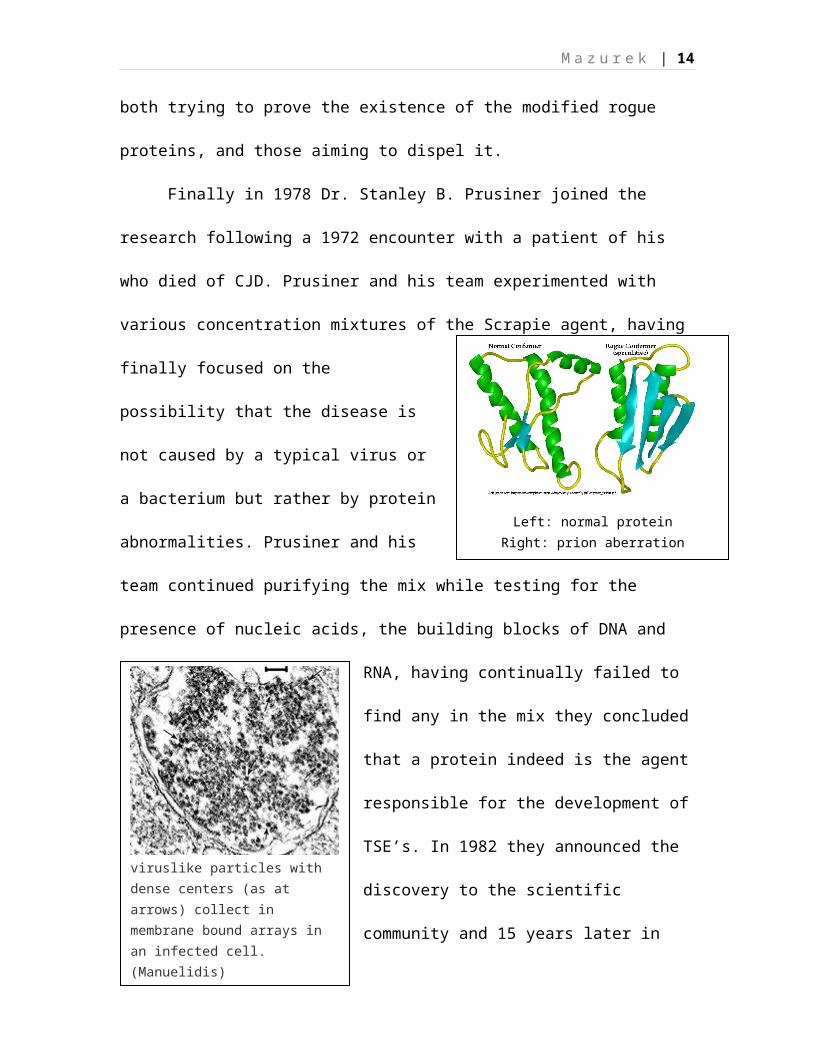
Finally in 1978 Dr. Stanley B. Prusiner joined the
research following a 1972 encounter with a patient of his
who died of CJD. Prusiner and his team experimented with
various concentration mixtures of the Scrapie agent, having
finally focused on the
possibility that the disease is
not caused by a typical virus or
a bacterium but rather by protein
abnormalities. Prusiner and his
team continued purifying the mix while testing for the
presence of nucleic acids, the building blocks of DNA and
RNA, having continually failed to
find any in the mix they concluded
that a protein indeed is the agent
responsible for the development of
TSE’s. In 1982 they announced the
discovery to the scientific
community and 15 years later in
Left: normal proteinRight: prion aberration
viruslike particles with dense centers (as at arrows) collect in membrane bound arrays in an infected cell. (Manuelidis)

M a z u r e k | 15
1997 Dr. Prusiner was awarded the Nobel Award for his
discovery. Prions themselves remain a hotly contested field
of study in present day as different sides of the scientific
establishment; notably a Yale neuropathologist Dr. Laura
Manuelidis who claims that the disease is caused by a virus
and that the proteins are simply its “tracks” as the havoc
is wreaked within the brain of those affected (Manuelidis).
Regardless of the controversy over the prions (short
for protinaceous infective particle) discovered by Dr.
Prusiner there have been far-reaching resolutions passed in
order to protect people from the potential infections caused
by these pathogens. Specifically since they are able to
cross between species while remaining infective albeit with
prolonged incubation periods there has been a call issued by
the World Health Organization for a ban and immediate
cessation of use of any animal feed administered to cattle,
goat, sheep which may contain cadaver-derived materials.
Furthermore WHO released a recommendation that all countries
establish adequate testing and surveillance of animals at
risk for prion diseases and that any such animal found at
M a z u r e k | 16
risk for a prion disease be excluded from the processing for
human and animal consumption considering that certain prion
diseases such as the Bovine Spongiform Encephalopathy appear
to require only casual contact in order to be communicated
between hosts. This creates a humongous risk considering the
long incubation periods and the asymptomatic stages which
can take years, and in some cases – decades. The third
recommendation by the W.H.O. was that the tissues at high
risk for contamination with BSE be excluded entirely from
the diets of humans and pets. These include eyes, brains,
spinal cords, and intestines. However, as the supervision in
this area has been lax there is a strong case made for the
persistence of such practices and the inclusion of the high
risk tissues in “variety meats” and the possibility of them
being included in hamburgers and hot dogs. Furthermore
considering the survival of prions in Scrapie ridden tissues
even after exposure to extreme heat, cold, and at least in
one case – Felmaldachyde, all of which failed to stop the
communicability of Scrapie there is an ever present risk of
contamination of the machinery and tools used for
M a z u r e k | 17
slaughtering and separation of carcasses of asymptomatic
animals – which in turns may compromise the safety of meat
coming from healthy animals. The list of risks taken by the
U.S.A. meat industry doesn’t stop here as since the
introduction, in 1994, of the Advanced Meat Recovery systems
the processing plants have become able to trim nearly 100%
of the meat from the animals, something which they have been
previously unable to do due to the difficulties associated
with removing muscle from the bone. While initially the meat
yielded through this process was not allowed to be labeled
as beef due to what Dr. Greger refers to as the “texture
ranging from ground meat to that of tomato sauce” it took
the USDA less than one year to alter its labeling rules
therefore allowing for these products to be labeled as
“Beef” and included in jerky, sausages, and ground beef as a
volumizing substance. The risk associated with the above
process is of contamination of the meat destined by human
consumption with spinal cord and brain tissues, which as we
now know carry a higher risk for harboring prions, and as
per the W.H.O. recommendations – to be excluded from human
M a z u r e k | 18
consumption. To make the matters even more interesting it
also appears that the U.S.A. beef industry has taken to
feeding calves a milk replacer made out of protein and fat
derived from other cattle. This practice has been already
connected with BSE outbreaks in Germany, Japan, and Denmark.
However the practice continues because the farmers contend
that the substitute is much cheaper than whey protein.
Furthermore given the extremely long incubation periods of
these pathogens there is another added risk of contracting
BSE and vCJD via veal, long as the animal is slaughtered
before showing any serious symptoms, which as shown by
earlier research by Gajdusek can take years.
The laundry list of problems in the U.S. beef industry
continues however while the farmers pretend that BSE hasn’t
invaded the stock meant for human consumption even though
there have been 3 recorded cases between 1993 and 2008
(CDC). Since the 1990’s, vCJD a variant of the earlier
documented CJD was discovered by British researchers at the
University of London, and connected with the consumption of
contaminated tissues of cows affected with BSE. The
M a z u r e k | 19
aforementioned Yale researcher, Dr. Manuelidis has been
quoted that there is a strong case to be made for thousands
of Americans dying every year from the BSE triggered CJD
while being told that the disease is a simple unfortunate
draw by the hand of fate. Concurrent with Dr. Manuelidis’
statement there has been a development of CJD clusters in
the United States the incidence of CJD in which exceeds the
expected 1:1000,000 by as much as a factor of eight. These
clusters range from New Jersey through Florida, with two
originating in Allentown and Lehigh valley Pennsylvania, an
alarming development considering that the prevalence rates
in these clusters range from 1.3 times the expected
occurrence to an astounding 8.7 times the expected
occurrence in the Tampa, FL cluster of CJD.
Finally, in the nod to the Alzheimer’s connection noted
in one of the Fore child’s brain autopsied at the National
Institute of Health. Depending on the stage at which the
sufferer is killed by CJD there is a very good case to be
made for misdiagnosis of the disease. A 1989 sampling of
diagnoses performed by Dr. Maunuelidis and her husband

M a z u r e k | 20
uncovered that of the 46 cases performed 6 were proven to be
CJD when the autopsy was carried out. When one considers
that Alzheimer’s makes up 70 percent of all cases of
Dementia in the United States and is slated to increase
drastically over the next fifteen years there is a point for
concern over the possibility of those 13% misdiagnosed cases
having actually been vCJD. Additionally it appears that
Alzheimer’s has an increased prevalence rate amongst
minority males who can generally be expected to consume more
low quality and “variety” meat than their more affluent
counterparts. (Marcus)
In closing there are the two variants of CJD
themselves. The sporadic which appears to be connected to
its’ sufferers genetically and passed along the familial
lines, and the variant, which manifests itself within the
individuals who consumed meat contaminated by BSE. We
assume, for the time being that the two diseases are removed
cousins. However, it is also equally possible that the
sporadic CJD is actually an outgrowth of variant CJD having
been given sufficient resistance by an individual’s system.
M a z u r e k | 21
Furthermore there is additional risk of passing CJD to one’s
offspring sexually. This would explain the variants and
clusters having tendency of concentrating around families.
While the American consumers continue in their level of
relative ignorance to the food which is augmented with
genetic therapies, some of which may include utilizing
hormones extracted from already dead animals, and remain
indifferent to the inhumane and potentially dangerous
practice of feeding animals…well animals, they also remain
at a much higher risk for the TSE diseases.
M a z u r e k | 22
Works Cited
"CDC - Bovine Spongiform Encephalopathy (BSE)." Centers for
Disease Control and Prevention. Web. 13 Dec. 2010.
<http://www.cdc.gov/ncidod/dvrd/bse/>.
"Do Reports of SCJD Clusters Matter?" Organic Consumers
Association. Web. 20 Dec. 2010.
<http://www.organicconsumers.org/madcow/morgan11304.cfm
>.
Ferreiro, Carmen. Mad Cow Disease (Bovine Spongiform Encephalopathy).
Philadelphia, PA: Chelsea House, 2005. Print.
Greger, Michael. "U.S. Continues to Violate World Health
Organization Guidelines for BSE." (2004). Web. 15 Dec.
2010.
<http://www.organicconsumers.org/madcow/Greger.pdf>.
Manuelidis, Laura. "Alzheimer's and CJD." Official Mad Cow
Disease Home Page. Web. 8 Dec. 2010. <http://www.mad-
cow.org/Alzheimer_cjd.html>.
M a z u r e k | 23
Manuelidis, Laura. "Potentially Pathogenic Virus Found in
Mad Cow Cell." Yale Office of Public Affairs & Communications.
Yale University, 30 Jan. 2007. Web. 11 Dec. 2010.
Marcus, Mary B. "Report: Minorities More Likely to Suffer
Alzheimer's Disease - USATODAY.com." News, Travel, Weather,
Entertainment, Sports, Technology, U.S. & World - USATODAY.com. 3
Sept. 2010. Web. 14 Dec. 2010.
<http://www.usatoday.com/news/health/2010-03-09-
alzheimersstats09_ST_N.htm>.
"Medicine: The Laughing Death." Time 11 Nov. 1957. Web. 12
Dec. 2010.
National Alzheimer's Association. "Latest Alzheimer's
Statistics - U.S. (2010)." Texas Alzheimers Research
Consortium. 2010. Web. 16 Dec. 2010.
<http://www.txalzresearch.org/index.php?
option=com_content&view=article&id=52&Itemid=68>.
Sommerville, Robert A., and David C. Bolton. "NOVA Online |
The Brain Eater | Do Prions Exist?" PBS: Public Broadcasting
M a z u r e k | 24
Service. Web. 11 Dec. 2010.
<http://www.pbs.org/wgbh/nova/madcow/prions.html>.
Walton, Priscilla L. Our Cannibals, Ourselves. Urbana: University
of Illinois, 2004. Print.
Yam, Philip. The Pathological Protein: Mad Cow, Chronic Wasting, and
Other Deadly Prion Diseases. New York: Copernicus, 2003.
Print.