
Journal of the Association of Surgeons of Assam
50
1 JASA, Vol. 22, No. 1, January-April, 2015 Journal of the Association of Surgeons of Assam JASA, Vol. 22, No. 1, January-April, 2015 Editor : Dr H. K. Dutta, MS,M.Ch. Editorial Board Members Dr. N.N. Das, Guwahati Dr. K. Bhuyan, Guwahati Dr. D.K. Sarma, Guwahati Dr. AP Lal, Dibrugarh Dr. M. Saha,Guwahati Editorial Consultants Prof. Rama Kant, Lucknow Prof. Andre Nicolus, France Prof. N.C.Bhattacharyya, Tezpur Dr. S. Singhvi, New Delhi Prof. Arjun Rao, USA Prof. M. Srinivas, New Delhi Prof. K. Das, Bengaluru Dr. R.N. Majumdar, Guwahati Prof. J. Ahmed, Dibrugarh Dr. D. Hazarika, Bongaigaon Prof. A.C. Baro, Jorhat Dr. N. Das, Silchar ISSN 2347-811X Index in : / India Citation Index
-
Upload
khangminh22 -
Category
Documents
-
view
1 -
download
0
Transcript of Journal of the Association of Surgeons of Assam

1JASA, Vol. 22, No. 1, January-April, 2015
Journal of the Association of Surgeons of Assam
JASA, Vol. 22, No. 1, January-April, 2015
Editor :Dr H. K. Dutta, MS,M.Ch.
Editorial Board MembersDr. N.N. Das, Guwahati Dr. K. Bhuyan, GuwahatiDr. D.K. Sarma, Guwahati Dr. AP Lal, Dibrugarh
Dr. M. Saha,Guwahati
Editorial ConsultantsProf. Rama Kant, Lucknow Prof. Andre Nicolus, FranceProf. N.C.Bhattacharyya, Tezpur Dr. S. Singhvi, New DelhiProf. Arjun Rao, USA Prof. M. Srinivas, New DelhiProf. K. Das, Bengaluru Dr. R.N. Majumdar, GuwahatiProf. J. Ahmed, Dibrugarh Dr. D. Hazarika, BongaigaonProf. A.C. Baro, Jorhat Dr. N. Das, Silchar
ISSN 2347-811X
Index in : / IndiaCitation Index

2 JASA, Vol. 22, No. 1, January-April, 2015
CONTENTS
Journal of the Association ofSurgeons of Assam
• Obituary 3
• Doyens of Surgery 4
• Editorial : Dr. H.K. Dutta 5
Review Article :
• Catheter related complications, its prevention and management
Rajeev T. P, Debajit Baishya, Debanga Sarma, S. K. Barua, S. J. Baruah 6
• Consent in Surgical practice: An analysis
Dr. R.C. Brahma, Dr. Swapan Deka Manindranath 14
Original Article :
• Impact of stone clearance in patients with urinary stone disease
and chronic kidney disease
Prof. Rajeev T.P., Dr. Hrishikesh Bordoloi, Dr. Debanga Sarma,
Dr. Sasanka Kumar Barua, Prof. Saumar Jyoti Barua, Dr. Puskal Kumar Bagchi 19
• Maternal nutrition and congenital malformation
Dr. Mauchumi Baruah, Dr. H.K.Dutta, Sri D. Borbora 24
Case Reports :
• Chronic umbilical sinus in an adult as a consequence of
urachal remnant : a case report
Dr. Nirmal Kumar Agarwal 28
• Ceroid Granuloma of the Extra Hepatic Billiary System :
A rare entity causing obstructive jaundice
Dr Gunabhi Ram Das, Dr Dipak K. Sarma, Dr. Rocket C. Brahma,
Dr. Anuj K Sarma, Dr. David Pegu, Dr. Nilaksha Bhattacharjee 31
• Complications following forgotten DJ urinary stent: A case report
Dr Rocket Chandra Brahma, Dr Sidhartha Sankar Konwar 35
• Chronic osteomyelitis of clavicle in an immunocompetent
adult: a case report
Dr. Bikash Jyoti Bordoloi, Dr. Pradip Kumar Baruah,
Dr. Rajarshi Roy, Dr. Bikash Agarwal 38
• Journal Review: 41
• Association News: 44
JASA, Vol. 20, No. 2, May-August 13
Vol. 22, Issue No. 1January-April, 2015

3JASA, Vol. 22, No. 1, January-April, 2015
OBITUARY
Dr. Satya Brata Dutta ChoudhuryASI membership No. 5456 (FL)
Date of Birth: 05.11.1933Expired on: 24.02.2015
1. STOOD 10TH IN FINAL MBBS EXAM2. DRCOG ( LONDON)3. FRCS ( EDINBURG)4. RETIRED PROF AND HEAD OF DEPARTMENT. SILCHAR MEDICAL COLLEGE, ASSAM.5. SELECTED AS EMINENT SURGEON VIDE BOOK REFERENCES ASIA6. PAST PRESIDENT OF ASSOCIATION OF SURGEONS OF ASSAM.

4 JASA, Vol. 22, No. 1, January-April, 2015
DOYENS OF SURGERY
Prof. M.A.Chowdhury
PROF. M.A.CHOWDHURY, a brilliant student since his school days joined Assam Medical College,Dibrugarh in the 3rd batch of MBBS. He passed out in 1954 securing 3rd rank. Subsequently he joined thedepartment of Surgery at Assam Medical College as Registrar. He was the first student to join post graduatecourse in Surgery under Prof. Banerjee. He took Carcinoma esophagus as his thesis subject, but before he couldcomplete it, he proceeded to the U.K. for his FRCS, which he obtained from the Edinburge University. Upon hisreturn from UK, Prof. Chowdhury joined back his alma matter initially as Assistant Professor, then he rose tobecome Professor and Head of the department of surgery, a position he held till his superannuation. He alsoserved as the Principal cum Chief Supdt. of Assam Medical College & Hospital since 27.06.85 till 31.03.88.
Prof. Chowdhury had all the attributes of a great surgeon: clinical acumen, dexterity, courage andthoroughness. As a teacher, he was unconventional, yet a role model for his students. A great pioneer of originalthinking in surgery, he infused in the minds of his students the acumen of a clinician. He never allowed hisstudents to bypass a thorough and methodical clinical examination and he used to say "never be a slave of thelaboratory". His bedside lectures were superb, so were his clinical rounds. He made the most difficult andchallenging surgical procedures look simple and easy. He strictly followed the principles of anti-sepsis andasepsis in the operation theatre and often did not prescribe any antibiotic after routine surgery.
Vividly I remember meeting Prof. Chowdhury, for the first time in 1963, when I was a student of class IX atDibrugarh Govt. High School. My father took me to him for some surgical problem. When he learned that I wasthe first boy of the class, he refused any consultation fee; rather he asked my father to take care of my studiessuggesting that medical profession was a good choice. In course of time, he became a good friend of my father.Many years later, I joined his unit as an intern, and then as house surgeon and subsequently as Registrar.Working under him was the most cherished time of my life and this experience has changed the course of mylife.
Apart from surgery, Prof. Chowdhury was a very knowledgeable person. His theory classes were full ofinteresting anecdotes, philosophical notes and historical accounts of important surgical innovations and usedto stretch for two to three hours. He was also an accomplished Bridge player.
It is matter of pride that many of his students are now shining in their respective fields all over the world.Nothing can be a better tribute than this to this finest surgeon and a great teacher. May the Almighty rest his soulin peace.
Prof. G.M.S. ChowdhuryEx-Professor of Surgery

5JASA, Vol. 22, No. 1, January-April, 2015
Editorial
Innovations in surgery: a historical review
"The abdomen, the chest and the brain will forever be shut from theintrusion of the wise and humane surgeon." So opined Sir John Ericksen,Surgeon Extraordinaire to Queen Victoria in 1837. Today thousands ofsurgical procedures are carried out daily, many in the abdomen, thechest, or the brain.
Surgery, as one of the oldest and most respected fields, built uponcontinuous innovation, has a unique culture and deep tradition. It isthe unsolved problem, or the repetitive failures of existing therapies thatstimulate surgeons to find a better way. Throughout history, surgeonshave been the most prolific medical device innovators. The first iterationof approach was described by Hippocrates in the 4th century B.C. Withinhis school on the island of Kos, he described the first rectal speculum,an enabling technology that supported subsequent endoscopicdevelopments such as the pelvic speculum [1,2].
Not much changed until the advent of focused lighting. In 1805,Philip Bozzini, invented the Lichtleiter, a lighting apparatus supportingvisualization of the bladder and rectum through mirror reflected candle-light [1]. This enabling technology supported critically important futureadvances in lighting. Antoine Jean Desormeaux built on Bozzini'sexternal light source to create and present the first functional cystocopein 1855, earning him the title the "Father of Endoscopy [3-5]. Notcoincidentally, this period also saw the invention of functionalesophagoscopy and hysteroscopy [6,7]. The combination of enablingtechnology and procedure stimulated an expanding period in endoscopicinnovation with the development of multiple new technologies and asignificant impact on patient care.
An increasing number of surgical leaders think that innovationmay be the only way to maintain the quality of their profession. JohnsHopkins' first surgeon-in-chief, William Halsted, M.D., laid thefoundation for surgery in the United States [8]. Dr. Halsted was dedicatedto skill and technique and made many revolutionary contributions tothe field of surgery, including pioneering the first lifesaving surgicaltreatment for breast cancer and developing new operations for intestinaland stomach diseases, gallstone removal, hernia repair and disorders ofthe thyroid gland.
In 1901, Georg Kelling introduced a cystoscope into a dog'sabdominal cavity, performing the first laparoscopy [9]. AlthoughKelling's work generated much interest among the surgical fraternity,

6 JASA, Vol. 22, No. 1, January-April, 2015
the following 50 years were spent on refining the endoscopic techniques.During this period, a great deal of attention was focused on the rapidlyexpanding fields of cardiothoracic and trauma surgery. During the 1960sand 1970s, Kurt Semm helped rekindle interest and enable advances insurgical endoscopy by inventing devices for automatic insufflation,thermocoagulation, and endoscopic irrigation.
During the next few decades, a new period of surgical endoscopyexpansion had begun. Modern minimal access surgery was borne outof vast experiences gained in arthroscopy for orthopedics andlaparoscopy for gynecologic surgery. A French surgeon, PhilippeMouret, operated on a woman suffering from both a gynecologicdisorder and gallstones in 1987. He turned the laparoscope upward andperformed the first laparoscopic cholecystectomy [3]. Francois Duboisof Paris learned of the procedure and he began experiments in bothanimals and humans and presented his work at an academic meetingin Paris. Dubois worked with a French colleague, Jacques Perissat, topublicize efforts in laparoscopic cholecystectomy. Subsequent years sawrapid expansion of technology available for laparoscopy as well assurgeons interested in performing ever more complex operations.
However, the idea that large problems require large incisions so deeplydominated surgical thinking that there was little room to appreciate theadvances of "key-hole" surgery. There are countless historical examplesof the surgical community's reluctance to accept change. From the 1805comment that Bozzini's Lichtleiter was a "magic lantern" to the 1992Caversham Conference where the "tidal wave" of new technology wasnoted to be "threatening western health-care systems," the surgicalcommunity has been consistently antagonistic to innovators [10]. Doubtswere raised as "surgical innovation is part of that threat" and "surgeryneeds to be on its guard against fashion[11]". These thoughts are notunreasonable. As Stirratt et al commented, "new surgical proceduresmust be tested, and that means clinical testing by mortality and morbidity,and psychologic and social testing by outcome for the individual patientand the community [11]. It may also be noted that laparoscopiccholecystectomy was not evaluated initially by randomized controlledtrials but was propelled by anecdotes and case series [12].
Innovation is defined as the act of introducing something new or theuse of a new idea or method [13]. In some instances, it is usedsynonymously with invention, although innovation is more preciselydefined as something thought up or mentally fabricated. Importantly,no technology or its application is entirely new, as no inventor workswithin a vacuum [14]. All definitions of innovation involve both newideas and an act of use or practice. The coupling of new ideas and hands-on use is also a central tenet of surgery, partially explaining the historicalsuccess of surgeons as innovators and the progress, which theirinnovation created. These new ideas may come in the form of technology,technique, or a combination. The process by which surgical innovationapplies new ideas to "hands on" clinical needs is analogous to the processby which translational research applies basic research to clinicalproblems.

7JASA, Vol. 22, No. 1, January-April, 2015
Research is also not the same as innovation. Advancement in thebasic sciences represents critically important progress. This researchcontributes to the fundamental knowledge base and supports futureinvention. However, basic science research is not the same as innovationas it does not require application or intended use. The distance betweenthe two can be thought of as the translational gap.
Surgeons have historically been idea generators and creativepractitioners within their craft. Each clinical case offers unique challengesand requires a degree of creativity. Surgeons understand clinical needsand may anticipate future advances and opportunities.
Nevertheless, for a field that is proud of its innovative roots, and infact, dependent upon them, we often are stingy in our praise for novelideas and procedures. It is clear that personal characteristics of theinnovator and acceptance of the surgical community are just as criticalto innovation as technology and technique. It should be noted that theimpetus for surgical innovation may be changing as surgical care andhealth care, as a whole, are managed with fiscal performance as at leastone primary outcome measure.
Dr. H.K. Dutta
References:1. Rosin D. History. In: Minimal Access Medicine and Surgery. Oxford: Radcliffe Medical,
1993:1-9.2. Litynski GS. Endoscopic surgery: the history, the pioneers. World J Surg. 1999;23:745-
753. [PubMed]3. Gorden A. The History and Development of Endoscopic Surgery. London: Saunders,
1993.4. Lau WY, Leow CK, Li AK. History of endoscopic and laparoscopic surgery. World J
Surg. 1997;21:444-453. [PubMed]5. Desormeaux A.J. De l'Endoscopie, instrument proper a éclairer certaines cavités
intérieures de l'économie. In: Compte rendu des Séances de l'Academie des Sciences.1855.
6. Bevan L. The oesophagoscope. Lancet. 1868;1:470.7. Pantaleoni D. On endoscopic examination of the cavity of the womb. Medical Press
Circular, 1869:26.8. Cosgrove DM. The innovation imperative. J Thorac Cardiovasc Surg. 2000;120:839-
842. [PubMed]9. . Kelling G. Die Tamponade der Bauchhohle mit Luft zur Stillung lebensgefahrlicher
Intestinalblutungen.Med Wochenschr. 1901;48:1480.10. Stirrat G, Ramsay B. Surgical innovation under scrutiny. Lancet. 1993;342:187-188.
[PubMed]11. Stirratt G, et al. The challenge of evaluating surgical procedures. Ann R Coll Surg Engl.
1992;74:80-84.[PMC free article] [PubMed]12. Banta HD. Minimally Invasive Therapy (MIT) in Five European Countries.
Amsterdam: Elsevier, 1993.[PubMed]13. Jones JW, McCullough LB, Richman BW. Ethics of surgical innovation to treat rare
diseases. J Vasc Surg. 2004;39:918-919. [PubMed]14. History of Technology. Encyclopaedia Britannica, 2004.

8 JASA, Vol. 22, No. 1, January-April, 2015
IntroductionInstruments to drain the urinary bladder are among the most ancient
medical devices in the historical record. The earliest descriptions of a urinarycatheter can be found in the Hippocratic text On Diseases (400 BC), in whichbladder drainage was considered a basic skill in the armamentarium of Greekphysicians. The practice of bladder catheterization to permit urine drainagealso found in India, Egypt, and the Byzantine and Roman empires. InAvicenna's Canon of Medicine, mentioned urethral catheterization as ameans to deliver intravesical therapy. Early catheters include hollow tubes made of bronze, paper, animalhide, cloth soaked in wax and lead. In the 16th century, Ambroise Pare usedcatheters made of silver, brass, and copper. In the early 18th century, naturalrubber was first used and the coudé tip was introduced by Mercier. The self-retaining balloon catheter was introduced in the 20th century by Foley, leadingthe way to permit development of catheters of synthetic materials withspecialized coatings and drug-eluting properties.Indication of catheterization
The common indications for the use of a bladder catheter can be broadlydivided into two main categories: - To obtain drainage - To allow the instillation of diagnostic or therapeutic agents. 1) The relief of acute or chronic urinary retention due to either bladder
outlet obstruction or neurogenic bladder dysfunction is the mostcommon indication for urethral catheterization (UC).
2) To monitor urinary output.3) Urinary diversion by a catheter is used to allow healing after lower
urinary tract surgery/trauma
Review Article
Catheter related complications, itsprevention and management
ABSTRACTEstablishment of urinary tract drainage is a basic skills required by
all medical professionals. It is a simple procedure and most widely usedin day to day life. Use of indwelling urinary catheter can lead to variouscomplications most commonly urinary tract infection. Thesecomplications can result in prolong hospitalization, additional hospitalcosts, morbidity and mortality. This article aims to give a basicunderstanding about the indications of catheterization, proper techniqueof catheterization, various catheter related complications, its preventionand to tackle these complications.
Key Words : Catheter; Trauma; Complication
Rajeev T. P1
Debajit Baishya2
Debanga Sarma3
S. K. Barua4
S. J. Baruah5
Dr. Rajeev T. P.ProfessorDepartment of UrologyGauhati Medical College and HospitalBhangagarhGuwahati, Assam.Pin - 781032.Email - [email protected]

9JASA, Vol. 22, No. 1, January-April, 2015
Rajeev T. P at el: Catheter related complication
4) To evacuate the bladder when the urinecontains particulate matter, especially incombination with simultaneous irrigation(post transurethral resection, clot/purulentmaterial evacuation).
5) Collection of microbiologic clean urine(uncooperative patients because of age ormental status or co morbidities that preventvoluntary voiding)
6) Measure postvoid residual urine volumesamples for diagnostic purposes.
7) Urethral catheterization is also used to provideaccess to the bladder for urinary tract imagingstudies such as cystography, which requiresthe instillation of radiographic contrastmaterial.
8) Urethral catheterization with a pressuremonitoring catheter is used during urodynamictesting for physiologic assessment of voidingfunction.
9) It is also used to allow instillation ofpharmacologic agents for local therapy of somebladder pathologies such as chemo/immunotherapy for transitional cell carcinoma(mitomycin, bacillus of Calmette Guerin),interstitial cystitis (dimethyl sulfoxide), andintractable hematuria (e.g., alum, formalininstillation).
Though catheterization is a simple day careprocedure there is various catheter related complicationwhich may lead the patient to morbidity. A correct wayof catheterization prevents most of these complications.So we will shortly discuss here the catheterizationtechnique, catheter related complications, its preventionand how to tackle it.Technique of Catheter Insertion
A clinical history including allergies, urologicpathology, previous urethral surgeries andcatheterization attempts should be taken beforecatheterization. The patient should be in the supineposition at a comfortable height for the physician. Infemale patients a "frog-leg" position is most suitable.Catheterization should be carried out in a sterile fashionwith antiseptic preparation and draping of the patient'smeatal and genital area, the catheter's retention balloonshould be tested. 2% xylocain gel of 20 to 30 mL instilover 3 to 10 seconds and left indwelling a minimum of10 minutes provides less traumatic catheter passageand reduce patient discomfort.
After sterile skin preparation and draping, graspthe shaft of the penis with the nondominant hand
(which is now regarded as contaminated) and hold thepenis at a 90-degree angle or perpendicular to thepatient. This maneuver eliminates the distal orpendulous urethral curvature. Insert the lubricated tipof the catheter into the urethral meatus and gently butfirmly continue to advance the catheter for 7 to 10 cm,while simultaneously bringing the shaft of the penis tothe horizontal plane or parallel to the patient. Continueto advance the catheter, while expecting to feel a slightincrease in resistance as the membranous urethra(external striated sphincter) is traversed. Once the entirelength of the catheter has been introduced (up to thejuncture of the connector or to the two-way bifurcation),wait for spontaneous urine passage that confirmingproper placement of the catheter. If spontaneousdrainage of urine is not seen, gently press on the patient'ssuprapubic area. If despite this maneuver no drainageoccurs, slowly instill 20 mL of saline using a catheter-tipped syringe into the drainage port of the catheterand then slowly aspirate the fluid instilled to clear anyobstruction of the catheter side hole by lubricant or othermaterial. If the catheter is in the bladder, fluid shouldbe aspirated without resistance. If the catheter is stillwithin the urethra, the negative pressure producedduring aspiration will cause collapse of the urethralwall and will not permit the return of the instilled fluid.Only when the position of the catheter has been verifiedshould the retaining balloon be inflated, with theamount of fluid indicated on the catheter. Sterile wateris the preferred solution for balloon inflation. Thecatheter should be attached to a sterile closed bag systemand bag should be placed below the level of the bladderto encourage one-way gravity flow with the tubing asstraight as possible and avoiding kinks that mightimpair drainage that increase the chance of UTIs in upto one third of the patients. The exception to this is inpatients with acute urinary retention with significantbladder distension in which rapid bladder drainagemight precipitate decompression-induced hematuria or"ex vacuo hematuria." In these patients the cathetershould be intermittently clamped and released to permitgradual bladder decompression over 30 to 60 minutes.If the patient is uncircumcised, at this point return theforeskin to its normal reduced position to avoidparaphimosis. Secure the catheter to the patient,allowing for a normal range of motion and withouttension, using adhesive tape.
In infants and children younger than 3 years ofage, when the normal foreskin adhesions have not yetinvoluted, simply align the preputial opening with themeatus to assist catheter insertion.
In female patients, after antiseptic preparation andsterile draping, use the nondominant hand to spread

10 JASA, Vol. 22, No. 1, January-April, 2015
Rajeev T. P at el: Catheter related complication
the patient's labia (now considered contaminated) toreveal the urethral meatus. After lubrication, insert thetip of the catheter and gently advance using a slightlydownward direction, until about half the length of thecatheter has been inserted. Check for urine return andactivate the anchoring mechanism if used.
Difficulties during female catheterization may beencountered for several reasons including the inabilityto locate the urethral meatus due to obesity and age-related changes and less frequently to strictures(postsurgery, radiotherapy, neoplastic causes). In theobese patient, the use of one or more assistants toprovide labial retraction or the use of stirrups can behelpful. In the case of postmenopausal vaginal atrophyor other conditions resulting in the urethral meatusreceding into the introitus, following measures can betaken. Holding the index and middle fingers of thenondominant hand together, slowly slide posterioralong the introitus until the urethra. Using thedominant hand, pass the catheter along the groovemade by the fingers (this serves a dual purpose-it createsa posterior border with the fingertips and provides aguide for the catheter). As the catheter tip crosses themeatus, it can be felt with the fingertips, thus ensuringproper placement. A second maneuver is to use avaginal speculum to aid in the retraction and fixationof the introitus. Finally use a coudé tip catheter angledupward and gently slide the tip along the anteriorvaginal wall in the midline, until it enters the meatus,and then advance into the bladder.
In female children the correct identification of theurethral meatus is essential to avoid unnecessarycatheter contact with the sensitive introitus, leading todiscomfort and possibly loss of cooperation by the child.The meatus is just above the superior margin of theintroitus and frequently hidden by the superior portionof the hymen. Gentle downward pressure on the upperaspect of the hymen with a cotton ball may allowvisualization of the meatus. Failing this maneuver, thecatheter tip should be inserted just above the hymen inthe midline.Catheter related complications, its prevention and howto tackle it1) UTI
It accounts for 40% of all nosocomial infections.Urethral catheters are responsible for up to (80%) ofUTIs in the hospital setting [1].
Risk factors for CAUTIs (catheter associated UTI)include:
- Patients requiring more than 6 days ofcatheterization,
- Female gender,- Active nonurinary infection sites,- Pre existing medical conditions,- Malnutrition,- Renal insufficiency,- Catheter insertion other than in the operating room
having drainage tubing or bag elevated above the levelof UB [2, 3].Prevention of CAUTI
- Avoidance of unnecessary catheterization,- Atraumatic technique of insertion,- The use of a closed collection circuit,- The use of aseptic technique [4].- The application of antibiotic coatings (nitrofurazo
and minocycline/rifampin coatings), silver alloy ontocatheter surfaces and the concept of drug elutionreduces the incidence of asymptomatic bacteriuria [5-7].
But the routine use of catheter coatings is currentlynot supported by the available literature.Not help to prevent UTI:
- The use of antiseptic gels or irrigations of thebladder or collection bag,
- Catheter clamping, or antireflux valves have notbeen shown to prevent CAUTIs [8, 9].How to tackle CAUTI:
Patients with indwelling catheters should be treatedonly if they become symptomatic (e.g., febrile). Urinecultures should be performed before initiatingantimicrobial therapy. The antimicrobial agent shouldbe discontinued within 48 hours of resolution of theinfection [10].
If the catheter has been indwelling for several weeks,encrustation may shelter bacteria from the antimicrobialagent; therefore the catheter should be changed.
When a catheter is to be removed and there is ahigh probability of bacteriuria or the dipstick test ispositive, a culture should be obtained 24 hours beforeremoval [11].
If the probability is low or the dipstick is negative,a culture may not be necessary. The patient should bestarted on empirical antimicrobial therapy such as TMP-SMX or a fluoroquinolone just before decatheterizationand maintained on therapy for 2 days.
A post-therapy culture should be obtained 7 to 10days later to confirm the eradication of the bacteriuria.

11JASA, Vol. 22, No. 1, January-April, 2015
Rajeev T. P at el: Catheter related complication
2) Inability to remove the catheter from the bladderA frequent encountered problem, is the inability to
remove a straight Foley catheter, this may due to- Encrustation,- Entrapment by sutures, or- Inability to disengage/deflate the retaining
balloon.The latter problem may be due to a faulty valve,
inflation channel blockage, or rarely crystallizationwithin the balloon.
If the catheter has been indwelling for a long periodof time, encrustation should be considered and imagingstudies (plain film or ultrasound) will be confirmatory.Prevention
- Sterile water is the preferred solution for ballooninflation as electrolyte or glucose-based solutions canprecipitate and occlude the tubing and valvemechanism.
- In case of long standing catheterization cathetermust be changed 3-4 weekly to prevent encrustationHow to tackle it
- In many instances the encrustation is easilydislodged with gentle traction on the catheter.
- For more significant encrustations, one canconsider using a semirigid ureteroscope and theholmium:YAG laser to remove the stone fragments.
- If the catheter is quite rigidly fixed and in the settingof recent bladder or prostate surgery,semirigidureteroscopy along the catheter and using theholmium:YAG to release the suture have also beendescribed.
- Because the suture materials used in bladder andprostate surgery are often absorbable, waiting for suturedissolution is another option.
- An inability to deflate a Foley balloon can bemanaged using a step wise approach.
a) One should first attempt to place another 1 to 2mL of fluid in the balloon to ensure normalballoon contour. Next step is to cut the inflationport. If the valve is the source of the problem,balloon deflation should then occur.
b) Perforation of balloon can be done bed side byoverinflating the balloon to the level ofperforation and also by chemical instillations(e.g. ether) for intravesical rupture of theballoon, but these procedures not recommendas these maneuver may be painful to the patient,may cause bladder injury and retention of the
balloon fragments, can cause chemical cystitis,UTI, stone formation etc.
c) Insertion of a surgical steel wire (24 or 28 gauge;often included as an obturator for small-caliberureteral catheters) or the stiff end of a 0.035-inch hydrophilic-coated guidewire through thevalve inflation lumen bypasses the site ofobstruction or creates a small perforation of theballoon can be done.
d) Should none of these maneuvers be successful,the use of ultrasound-guided needle punctureof balloon can be done with a long spinal needle(22 gauge) using either a transrectal,transvaginal, or suprapubic surface probe.
e) If all other maneuver failed retained catheterscan be removed with open surgery..
4) Other complications- Hematuria,- Urethral injury- Urethral and meatal strictures,- Urethral perforation, and allergic reactions
including anaphylaxis- Malignant neoplasms (2.3% to 10%),- Stone formation (46% to 53%),- Bladder neck and urethral erosions.
Preventiona) As a general rule, catheter size should be the
smallest size that can accomplish the desireddrainage (i.e., 12 to 14 Fr for clear urine and 20to 24 for thick pus or blood-filled urine).
b) Silicone catheters are indicated when there isrubber/latex sensitivity or allergy and areparticularly suited for patients requiring alonger period of indwelling time.
c) Every catheter should be generously coatedwith sterile lubricant before insertion decreasechance of urethral injury.
d) Only when the position of the catheter has beenverified should the retaining balloon beinflated.
e) Secure the catheter to the patient, allowing fora normal range of motion and without tension,using adhesive tape.
f) In cases where long term catheterizationrequires clean intermittent catheterizationreduce incidence of malignant neoplasms,
12 JASA, Vol. 22, No. 1, January-April, 2015
stone formation, and bladder neck and urethralerosions.[12, 13]
g) In cases of long term catheterization regularupper and lower tract imaging, cystoscopy andrandom biopsy are require in prevention andearly diagnosis of malignant neoplasms, stoneformation
How to tackle itHematuria, urethral injury is occurs in difficult
catheterization (most commonly due to prostaticgrowth, urethral stricture, bladder neck contracture,false passage from previous urethral instrumentation,phimosis or urethral calculi).
Using adequate urethral lubrication and a 16- or18-Fr coudé tip silicone catheter is often successful inpatients with BPH.
If multiple previously unsuccessful attempts havebeen made and urethral trauma is suspected due to theappearance of a bloody urethral discharge, a falsepassage or a stricture is likely. A single atraumaticattempt can be made using a 12-Fr silicon/straight orcoudé tip catheter. If this maneuver is unsuccessful thendepend upon the available facility flexible cystoscopycan be done, allowing a direct visual approach thatcan be both diagnostic and therapeutic and minimizesthe risk of further urethral injury. Under direct vision,the area where the false passage was created or the siteof stricture formation is identified and an attempt ismade to identify the true urethral lumen. Onceidentified, a 0.035 in Teflon-coated guidewire (e.g.,Bentson type) is passed along the urethral lumen andinto the bladder and urethral dilation may be done incase of a tight urethral stricture or bladder neckcontracture over the guidewire. Once the urethrallumen is of adequate diameter to permit placement ofthe catheter , a Councill catheter, which can beadvanced over the guidewire into the bladder, isselected.
If flexible instruments are unavailable and aurethral stricture is suspected, then a blind techniquealthough less desirable, can be used. This techniqueinvolves using filiforms and followers The first step isto pass a small female filiform catheter. In many casesthe initial filiform will frequently end up entrapped inthe stricture membrane or in the false passage, ratherthan following the true lumen. Insertion of additionalfiliforms will then fill the false passage or stricturemembrane until one is successfully directed into thetrue lumen. Once a filiform is successfully passed, malesections (followers) of increasing size are attached tothe filiform to permit sequential dilation of the urethra.
Once dilation to at least 14 to 16 Fr has been achieved,a 12- to 14-Fr catheter is then passed.
Alternatively, one can blindly pass a Councillcatheter to the point of resistance and then attempt toadvance a hydrophiliccoated guidewire. If guidewireadvancement seems to go easily, a 5-Fr open-ended orangled ureteral catheter can be advanced over the wireto permit urine drainage and confirm bladderpositioning. Once bladder positioning of the guidewireis assured, urethral dilation can be performed over theguidewire followed by Councill catheter placement.
If none of these techniques are feasible, asuprapubic catheter should then be insertedComplication related to feeding tube
Feeding tube is routinely used by mosthypospadiologist as well as in infants and smallchildren. But because of their stiffness and length somerare but morbid complications may occur over andabove the above mentioned catheter relatedcomplications eg. ischemic ulcers, urethral strictures,and knotting in the bladder. Infant feeding tubeknotting is a very rare event [13]. It gets knotted whenexcessive length of flexible catheter is inserted in thebladder and forms a loop; subsequently as the catheteris withdrawn a knot can form and tightens onwithdrawal leads to impaction of the catheter.Prevention
It is best to prevent this problem by totally avoidingthe use of infant feeding tube or by introducing onlyshort length of feeding tube to drain the bladder. A 6-10F Foley's catheter may be a better option. A high indexof suspicion is required to diagnose knotting of infantfeeding tube when it cannot be withdrawn easily. Thiswill prevent traumatic urethral injury due to forcibleremoval and subsequent risk of urethral stricture.How to tackle itRemoval of infant feeding tube has been tried in manyways-
- Manual removal of catheter with gentle tractionunder local/general anesthesia
- Iinfant feeding tube may be uncoiled by passing aguide wire through the infant feeding tube andstraightening it, but may fail if the knot is too tight.[15].
- Open or laparoscopic supra pubic cystostomy andremoval of the feeding tubeConclusion:
Though catheterization is a simple procedure,catheter related complications may lead to various comorbidities. These complications can be prevented by
Rajeev T. P at el: Catheter related complication

13JASA, Vol. 22, No. 1, January-April, 2015
well training of our medical and paramedical personsfor safe catheterization in a proper aseptic andantiseptic environment. The catheter balloon shouldbe tested before catheterization and every catheter
should be generously lubricated before insertion, whichare key to prevent majority of catheter relatedcomplications and morbidity.
References:1. Ha US, Cho YH. Catheter-associated urinary tract infections: new aspects of novel urinary catheters. Int J Antimicrob
Agents 2006;28:485-90.2. Maki DG, Tambyah PA. Engineering out the risk of infection with urinary catheters. Emerg Infect Dis. 2001;7:163. Maki DG, Tambyah PA. Engineering out the risk for infection with urinary catheters. Emerg Infect Dis 2001;7(2):342-
7.4. Pratt, R. J., C. M. Pellowe, et al. "epic2: National evidence-based guidelines for preventing healthcare-associated
infections in NHS hospitals in England." J Hosp Infect 2007;65(Suppl 1): S1-S64.5. Maki DG, Ringer M, Alvarado CJ. Prospective randomised trial of povidone-iodine, alcohol, and chlorhexidine for
prevention of infection associated with central venous and arterial catheters. Lancet 1991;338:339-3436. Lee J, Jang J, Kim B, Kim J, Jeong G & Han H. Identification of Lactobacillus sakei and Lactobacillus curvatus by
multiplex PCR-based restriction enzyme analysis. J Microbiol Methods 2004;59: 1-6.7. Stensballe J, Tvede M, Looms D, et al. Infection risk with nitrofurazone-impregnated urinary catheters in trauma
patients: a randomized trial. Ann Intern Med 2007;147:285-938. Tenke, P., B. Kovacs, et al. "European and Asian guidelines on management and prevention of catheter-associated
urinary tract infections." Int J Antimicrob Agents 2008;31(Suppl 1): S68-789. Jahn P, Preuss M, Kernig A, et al. Types of indwelling urinary catheters for long-term bladder drainage in adults.
Cochrane Database Syst Rev 2007;(3):CD004997.10. European and Asian guidelines on management and prevention of catheter-associated urinary tract infections. Int J
Antimicrob Agents 2008;31(Suppl 1): S68-78.11. Tissot E, Limat S, Cornette C, Capellier G. Risk factors for catheter-associated bacteriuria in a medical intensive care
unit. Eur J Clin Microbiol Infect Dis.2001;20:260-212. Clarke, S.A., Samuel, M., & Boddy, S.A. Are prophylactic antibiotics necessary with clean intermittent catheterization?
A randomized controlled trial. Journal of Pediatric Surgery 2005; 40, 568-71..13. Shah PN, Desai R, Macho B. Management of a knotted infant feeding tube in the urinary bladder. Internet J Urol.
2005;2:2.14. Carlson D, Mowery BD. Standards to prevent complications of urinary catheterization in children: Should and
should-knots. J Soc Pediatr Nurs. 1997;2:37-41.
Rajeev T. P at el: Catheter related complication

14 JASA, Vol. 22, No. 1, January-April, 2015
Introduction :The relationship between a doctor and a patient is considered to be
commercial in nature. Such commercial relationship may be contractual ormay not be contractual in nature. In absence of a written contract often thedoctor is dragged to the courts. Hence, in order to avoid litigations it isadvisable for a doctor to have a written agreement between him and hispatient. On the other hand, because of a written agreement the doctor is ableto defend his case better in the courts. A written agreement between a doctorand a patient is covered by legal aspects provided in the Indian ContractAct, 1872. Consent is an important element to legally enforce an agreementbetween a doctor and a patient known as the Contract. The legal definitionof "Consent" is provided in Section 13 of the Indian Contract Act, 1872 whichsays that "Two persons are said to consent when they agree upon the samething in the same person." In other words, there must be consent ad idem tolegally recognize the bilateral transaction between the doctor and the patient.The section 11 of the Indian Contract Act also provides that only those personswho are of and above 18 years of age are competent to enter into a contract. Itis advisable that consent should be obtained preferably in written formatfrom parents or guardian who is below 18 years of age, so that the validity ofthe contract is not challengeable. This basic legal concept was first expressedby Justice Cardozo who stated that, "Every human being of adult years andsound mind has the right to determine what should be done with his body;and the surgeon who performs an operation without his patient's consentcommits an assault for which he is liable in damages"[1]. This paper aims to
Review Article
Consent in Surgical practice: An analysis
ABSTRACTWhenever patient consults a doctor, a relationship is established betweenthe two which is considered to be commercial in nature. To avoid anyfuture litigation, it is always advisable on the part of the doctor to have awritten agreement in the form of consent between him and the patient themoment he performs any diagnostic, therapeutic or surgical procedures.Consent may be implied, expressed or informed consent which may beoral or written. If the doctor performs any procedure without the consentof patient, except in the case of emergency, he may be charged withassault or battery. This paper aims to analyze the legal significance of"Consent" in doctor-patient relationship with special reference to surgicalpractice.
Dr. R.C. Brahma1
MS, FMAS, PGCHMDr. Swapan Deka Manindranath2
LLM, Ph.D
Address :1. Associate Prof. of Surgery AMCH, Dibrugarh2. Associate Prof. of Law Dispur Law College Guwahati
Key Words : Doctor-patient relationship; Informed consent; surgical procedures.

15JASA, Vol. 22, No. 1, January-April, 2015
analyze the legal significance of "Consent" in doctor-patient relationship with special reference to surgicalpractice.
II. Requirement of Consent. Law recognises the significance of Consent in a
doctor-patient relationship to deal with medico-legalcases. If a medical practitioner treats or operates on apatient without consent, such treatment or operationwill be deemed to be intentional interference with thepatient's body without legal sanction which amountsto assault and a battery for which will be entitled torecover damages [1]. The legal consent obtained by amedical practitioner may be either implied or expressedor informed consent. Expressed or informed consentmay be further written or verbal in form.
Implied Consent - The conduct of the greater partof medical practice is on the basis of 'Implied Consent'.Although this is entirely correct it often misleads themedical practitioners to overlook the fact that medicalexamination is a subject of consent, which is necessaryin the appropriate form [1].
Generally, a routine medical examination of apatient does not require a 'formal written consent'because the patient conducts himself in a manner whichimplies consent. When a patient attends a surgeon'sconsulting room or outpatient's department, or whenhe agrees to be admitted to a hospital or nursing home,he thereby implies that he will submit to routinephysical examination like inspection, palpation,percussion and auscultation. Express oral consent mustbe obtained for relatively minor examinations ortherapeutic procedures, preferably in the presence of adisinterested third party. For example examinations ofprivate parts notably rectal, vaginal and withdrawingof blood for diagnostic purposes, express consent inthe form of oral or written consent is necessary alongwith presence of female attendant while examiningfemale patient and male attendant while examiningmale patient. Similarly for more complex diagnosticprocedures like endoscopy, radiological contraststudies and surgical procedures under anaesthesia,written consent should be obtained.
Expressed or Informed Consent -Informed consent is an ongoing process, a two-
way or mutual conversation extending over time,between the registered medical practitioner and thepatient, that prior to the consent having been given, themedical practitioner has informed the patient of thenature of the proposed procedure or treatment of thoserisks and alternative treatment or diagnosis that areasonable patient would consider material to the
decision whether or not to undergo treatment ordiagnosis [1]. Legally, an injection becomes an assaulton the person by the doctor if not given properly andjustifiably so does blood transfusion -its risks or hazardsshould be explained to the patient or attendant, so thatthe consent becomes real.
III. Informed Consent in Surgical PracticeIn surgical practice, there is a clinical duty on a
surgeon to obtain informed consent before thecommencement of treatment. Because of the extremityof their clinical need, patients might agree to surgeryon the basis of no information at all. Agreement of thiskind, however, does not constitute a form of consentwhich is morally or legally acceptable. Unless suchpatients have some understanding of what they areagreeing to, their choices may have nothing to do withplanning their lives and thus do not count asexpressions of autonomy [1]. Every patient undergoingsurgery must have a valid consent. Very often it isnoticed that common consent is taken for both forsurgery and anaesthesia, however legally there shouldbe a separate consent for anaesthesia.
For agreement to count as consent to treatment,patients need to be given appropriate and accurateinformation about:
Their condition and the reasons why itwarrants surgery;
What type of surgery is proposed and how itmight correct their condition;
What the proposed surgery entails in practice; The anticipated prognosis of the proposed
surgery; The expected side-effects of the proposed
surgery; The unexpected hazards of the proposed
surgery; Any alternative and potential successful
treatments for their condition other than theproposed surgery, along with similarinformation about these;
The consequences of no treatment at all.Good professional practice dictates that obtaining
informed consent should occur in circumstances whichare designed to maximise the chances of patientsunderstanding what is said about their condition andproposed treatment, as well as giving them anopportunity to ask questions and express anxieties.Where possible:
Dr. R.C. Brahma at el. Consent in Surgical practice

16 JASA, Vol. 22, No. 1, January-April, 2015
A quiet venue for discussion should be found. Written material in the patient's preferred language
should be provided to supplement verbalcommunication.
Patients should be given time and help toevaluate their own understanding and to cometo their own decision.
The person obtaining the consent shouldideally be the surgeon who will carry out thetreatment. It should not be- as is sometimes thecase - a junior member of staff who has neverconducted such a procedure and thus may nothave enough understanding to counsel thepatient properly.
Surgeons should always attempt to approximatethese conditions, even when they might not becompletely achievable.
Good communication skills go hand in hand withproperly obtaining informed consent for surgery. It isnot good enough just to go through the motion ofproviding patients with information required forconsidered choice. Attention must be paid to:
Whether or not the patient has understood whathas been stated;
Avoiding overtly technical language indescriptions and explanations;
The provision of translators for patients whosefirst language is not English;
Asking patients if they have further questions.
IV. Exceptions to Informed ConsentConsent is must, however, there may be certain
situations when the doctor need not take consent fromthe patient. Such situations are:
(1) When the doctor may conclude that thedisclosure of a risk may pose such a seriouspsychological threat or detriment to the patient as to bemedically contraindicated prompting the patient toforego needed therapy. Such conclusions are based onthe discretion of the doctor and are known as"Therapeutic Privilege" or "Professional Discretion".Under such circumstances, the doctor may make therevelations to the next of kin of the patient or to thefamily physician.
(2) This privilege also applies when a patient isusually sensitive, anxious or emotional. However, ageneral policy of not disclosing information because ofthe presumed hypersensitivity of patient is not anacceptable basis for this privilege.
When the physician's use of 'therapeutic privilege'is challenged, it must be determined whether thephysician followed sound medical judgment inwithholding information.
V. No consent in Emergency casesBesides the use of 'Professional Privilege', a doctor
need not take consent in case of emergency. A case ofemergency may arise in the following situations:
(1) No duty to inform arises in an emergency inwhich the patient is unconscious or otherwiseincapable of giving valid consent and harm of failureto treat is imminent and outweighs any harm threatenedby the proposed treatment (Article 7 of the Conventionof Human Rights and Biomedicine, 1997 held at 4thApril at Oviedo, Spain).
(2) Duty not to inform may also arise when therelative/guardian is/are not available or there is notime to contact them; and
(3) Duty not to inform also arise when there isurgency of situation to save the life or avoid furtherdamage (Article 8 of the Convention of Human Rightsand Biomedicine, 1997). In these circumstances, lawpresumes that consent has been given in 'Good faith'.Doctor is immune to penal proceedings for 'damages','assault' or 'negligence' since he is protected by Section92 of the Indian Penal Code.
This exception shall not extend to:(1) the intentional causing/attempting to cause
death;(2) doing of anything likely to cause death or for
any purpose other than preventing of death or ofgrievous hurt or curing of any grievous disease orinfirmity."
In the case of Dr T.T.Thomas v. Elisa, the doctorwas held liable for not performing an emergencyoperation and the patient died of peritonitis [1]. In thiscase patient was admitted to the particular hospitalwith severe abdominal pain and was diagnosed to besuffering from acute appendicitis. The operation wasnot performed on that day and on subsequent daypatient expired. The plaintiff's case was that had theoperation been performed on that day of admission,the patient's life most probably would have been saved.The defendant surgeon took the plea of refusal by thepatient to give his consent to the operation. Theargument was not accepted as there was no mention inthe case sheet of the patient that consent was soughtand the same was refused by the patient. The court helddoctor negligent in his conduct in as much as the
Dr. R.C. Brahma at el. Consent in Surgical practice

17JASA, Vol. 22, No. 1, January-April, 2015
patient had passed into a critical stage where the primeimportance and the professional duty of a doctor is tosave the life of a patient by doing all that is possible.Sometimes a person may have some hesitation, initiallyto undergo any kind of operation, but when there isonly one alternative to safeguard the health and life ofthe patient the doctor should not evade theresponsibility and he becomes answerable for anyuntoward happening that may take place due to hisinaction. It was held that in case of emergency operationthe doctor cannot wait for the consent of his patientwhere the patient is not in a fit state of mind to giveconsent or to give a conscious answer regarding consentor where the patient is minor.
VI. Right to Refuse Consent:When a patient or his guardian rejects or refuse a
treatment, he should be advised in a discreet, ethical,moral and professional manner about the consequencesof not taking treatment. After that it is open to the patientor his guardian whether they want to continue thetreatment or not. If not, the same may be noted down inthe records of the patients case sheets as "left againstmedical advice" (LAMA) and normally signature of thelegal guardians should be obtained before the patientis discharged.
In normal circumstances if consent is refused, it isan absolute bar for examining or initiating treatment toa patient. If a doctor fails to give correct informationregarding the procedure before consent, he may be suedfor negligence u/s 304 IPC or punished by the MedicalCouncil of India or the State Medical Council forunethical act or professional misconduct.
If he does not take consent at all, he may be chargedwith assault u/s 351 IPC tress pass of privacy
VII. Age for Consent:Most confusing aspect of 'consent' among the
medical fraternity is the issue of 'age of giving legallyvalid consent' for medical purposes. There are threeschools of thought among medical jurists. Accordingto one school of thought, it is 12 years, for routinemedical examination other than operations and otherinvasive medical procedures (relay on Sections 87 and88, IPC). Another school of thought considers it as 12years for all practical purposes irrespective of invasiveor non-invasive medical procedures, and third schoolof thought considers it as 18 years, for operations orinvasive medical procedures, and 12 years for routinemedical procedures. This confusion is evident amongour legislators, when we go through different statutoryprovisions like organ transplantation, blood donation,
abortion, etc. However, for all practical purposes personwith above 18 years with sound and healthy mind maybe considered for giving valid consent.
VIII. Competency for giving consent:'Soundness of mind' and 'maturity of the mind'
may be considered as the most important legalingredient to understand the nature and consequencesto which he is going to give consent. According to sec.90of IPC, consent given under fear, fraud ormisrepresentation of facts or by a person who isignorant of the implications of his consent, or who isunder twelve years of age is invalid. Similarly u/s 92when legal guardians are not available in case ofchildren or minors, teachers, hostel warden,superintendent, Principal or consent from the court oflaw can be sought on behalf of the patients in case ofemergency.
A telephone consent given by the legal guardianof the patient is also valid in case of emergency whenthe patient is unable to give consent due to one reasonor the other, or is a minor, however, one should have athird party listening in as witness. Moreover, it isimperative that the other party be informed that a thirdparty is listening on the line, along with the purposeand identity of that third party and is always advisableto ask for a confirmatory in the form of message or e-mail or some written document for record.
IX. Scope of Consent:When a patient gives a consent to a surgical
procedure or for a specific type of treatment, theperformance of that particular procedure or treatment,the legal validity, the scope of consent is limited towhatever contents were expressed to the patient at thetime of obtaining the consent: however in exceptionalcircumstances an extension of the scope of the consentwould be permissible. The general rule is that, a doctorcommits assault or battery if he exceeds the scope of theconsent given by the patient.
In Ram Bihari Lal v. Dr J.N.Srivastava case, thesurgeon during an operation for appendicectomy foundappendix normal but gallbladder inflamed and likelyto become gangrenous [1]. The patient was underanaesthesia and abdomen was open, surgeon in goodfaith did cholecystectomy without taking consent ofher husband and patient died later. The lower courtdid not hold the surgeon liable as it was in the bestinterest of the patient but the High Court of MadhyaPradesh reversed the judgement.
Dr. R.C. Brahma at el. Consent in Surgical practice

18 JASA, Vol. 22, No. 1, January-April, 2015
X. Conclusion:Surgeons have a legal, as well as moral obligation
to obtain consent for treatment based on appropriatelevels of information. Failure to do so could result in alegal action of battery or negligence. Also bothprofessionally and legally, it is important for surgeonsto understand that a signed consent form is not proofthat valid consent has been properly obtained. It issimply a piece of evidence that consent may have beenattempted. Even when they have provided theirsignature, patients can and do deny that appropriateinformation has been communicated or that the
communication was effective. Surgeons are thereforewell advised to make brief notes of what they have saidto patients about their proposed treatments, especiallyinformation about significant risks. These notes shouldbe placed in the patient's clinical record, moreovervideo recording can also be made about theconversation and explanation between the patients anddoctors regarding the informed consent about theproposed treatment, procedures, expectedcomplications and outcome with prior permission ofthe patient, so that evidence can be produce in the courtor to the competent authorities if there is any litigations.
References:1. Scholendroff v. Society of New York Hospital. 103 NE 92 cited in Roshan Lall Gupta. The Medicolegal Aspects
of Surgery. 1 st edition, Jaypee Brothers Medical Publishers (P) Ltd. New Delhi, 1924; pp. 16-17.2. Prof. (Dr.) R. Chaliah, "Consent-Medical And Legal Aspects". Paper presented at Forensic Medicon 2014,
Guwahati, Assam. 31st January - 2nd February 2014. Organized by 35th Annual National Conference of IndianAcademy of Forensic Medicine.
3. Putul Mahanta. Modern Textbook of Forensic Medicine & Toxicology. Jaypee Brothers Medical Publishers (P)Ltd. New Delhi, p. 35-39
4. Justice K Kannan, Dr K Mathiharan. Modi, A Texbook of Medical Jurisprudence and Toxicology, 24th Edition,Lexis Nexis Publication, A division of Reed Elsevier India Pvt Ltd, Gurgaon, Haryana, India, pp 76-77.
5. R.C.G. Russel, Norman S. Williams & Christopher J.K. Bulstrode (eds.), Bailey & Love's Short Practice ofSurgery, 24th ed., International Students Edition, Holder Education, London in Len Doyal, Surgical ethics, pp.251-256.
6. Dr T.T.Thomas v. Elisa. All India Reporter 1987 Ker 52: (1987) (1) ACJ 192. cited in Modi, A Texbook of MedicalJurisprudence and Toxicology, 24th Edition, Lexis Nexis Publication, A division of Reed Elsevier India Pvt Ltd,Gurgaon, Haryana, India, pp 76-77.
7. Ram Bihari Lal versus Dr J.N.Srivastava. All India Reporter 1986 MP 150: 1985 ACJ 424: 1985 Jab LJ 259. cited inRoshan Lall Gupta.The Medicolegal Aspects of Surgery. 1 st edition, Jaypee Brothers Medical Publishers (P) Ltd.New Delhi, pp. 23.
Dr. R.C. Brahma at el. Consent in Surgical practice

19JASA, Vol. 22, No. 1, January-April, 2015
Introduction :Chronic kidney disease in calculus kidneys is a common problem in
developing countries. Obstructive uropathy as a result of kidney stone diseasehas been contributing to ever increasing numbers of such patients.Obstruction, pyonephrosis and renal failure are a direct consequence ofneglected urinary tract stones. Nephrologists and urologists tend to hesitateto intervene when chronic kidney disease sets in. The diagnosis and
Original Article
Impact of stone clearance in patients withurinary stone disease and chronic kidney
disease
ABSTRACTChronic renal failure (CRF) in calculus kidneys is a common problem indeveloping countries. Obstruction, pyonephrosis and renal failure are adirect consequence of renal stones. Urologists tend to hesitate to intervenewhen chronic renal failure occurs. Eighteen patients with 33 functionalrenal units with urinary stone disease and CKD, without any knownmedical renal disease were taken up for study. The outcome of surgicalintervention was studied by pre-op and post-op GFR values, calculatedusing the MDRD formula based on serum creatinine values.The mean age was 44.8 years(±13.03) and male:female distribution was16:3(6:1). Patients were subdivided into 2 groups based on the GFR atpresentation; Group 1 CKD stageIII had 7 patients while Group 2 CKDIV/V had 11 patients Out of the 33 renal units operated (3 solitaryfunctioning kidneys), pyelo-nephrolithotomy in 8(5+3); URSL for 3;Ureterolithotomy in 11 and PCNL in 3 units. Complete clearance wasachieved in 29(87%) and partial clearance in 4(12%) renal units (non-obstructive residual calculi>4mm). Follow up with estimated GFR valuewas carried out at 3 months after stone clearance. The mean pre-operativeGFR was 25.6ml/min (S.D.±13.43ml/min) and the mean post operativeGFR at 3 months was 38.5ml/min (S.D.±22.97ml/min). The complicationrate was 26%. There was overall significant improvement (P value onpaired t test 0.0346) in the post-operative GFR levels (-12.83ml/min)though when calculated group wise, it was only significant for Grp2.Urolithiasis remains one of the potentially avoidable causes of CKD inmany patients. An earnest effort with timely intervention in early CKDstages may salvage the kidney. In higher grades of CKD the surgicaldecision should rely on minimizing renal injury. Endourologicalprocedure's outcomes rivaled open surgery in selected cases. Early stoneclearance and prevention of recurrence with metabolic evaluation andappropriate dietary modification may prevent grade progression.
Prof. Rajeev T.P.Dr. Hrishikesh BordoloiDr. Debanga SarmaDr. Sasanka Kumar BaruaProf. Saumar Jyoti BaruaDr. Puskal Kumar Bagchi
Address :Dept. UrologyGauhati Medical College HospitalGuwahati - 781032, Assam, IndiaE-mail : [email protected]
Key Words : Chronic kidney disease, Urolithiasis, Renal Stone, ESRD, ChronicRenal Failure

20 JASA, Vol. 22, No. 1, January-April, 2015
Prof. Rajeev T.P. at el: Impact of Stone Clearance in Patients
intervention in patients with chronic kidney disease(CKD) is often delayed due to ignorance of the patient,socioeconomic reasons and reliance on unconventionaltherapy. If prompt radical measures are undertaken insuch azotemic patients, the morbidity and dependencyon dialysis may be reduced and time to progression toend stage renal disease (ESRD) may be delayed.
The effect of various surgical intervention for stonedisease on renal function is an important determinantfor selecting the optimal therapy in patients withCKD[1]. This study is to prospectively evaluate theefficacy and outcome of stone clearance on improvingthe function of renal unit in patients with CKD.PATIENTS AND METHOD
During the period from January 2013 till December2014, 18 cases with evidence of elevated creatininevalues and urinary stone disease without associatedmedical renal disease were prospectively evaluated.Ultrasound study and X-Ray KUB (Kidney UrinaryBladder) and Renal function test (RFT) were routinelyused for screening and serum creatinine values andModification of Diet in Renal Disease (MDRD) formulawas applied to evaluate the Glomerular Filtration Rate(GFR). Patients were divided into 2 groups based onGFR : Group-1 - CKD stages I-III(i.e. GFR more than30ml with renal functional compromise); Group 2 - CKDIV/V ( i.e GFR less than 30 ml/minute/1.73 m). In caseof obstructed and infected systems a preliminarypercutaneous nephrostomy or DJ stenting and culture-specific antibiotic treatment carried out. An IntravenousUrogram (IVU) or Non Contrast Computed Tomogram(NCCT) studies in patients with creatinine above2.5mg/dl were done and stone burden was determinedby multiplying the stone length by the width inmillimeters squared.
Patient characteristics and stone characteristicswere the determining factors for selection of theoperative technique with an aim of relieving obstructionand ensuring complete stone clearance. Open surgeriessuch as pyelolithotomies, nephrolithotomies andureterolithotomies were performed whereeverendourological procedures such as percutaneousnephrolithotomy (PCNL) and Ureteroscopic Lithotripsy(URSL) were deemed not feasible considering the stonebulk, presence of anatomical obstruction, and need formultiple procedures to ensure stone clearance.Auxiliary procedures, including second-look PCNL,ureteroscopy, and extracorporeal shock wavelithotripsy (ESWL) were used to achieve complete stoneclearance. Patients were rendered stone free whenresidual fragments were smaller than 4 mm with orwithout adjuvant treatments. Nephrology consultation
was obtained for all patients prior to planning oftherapy. Postoperative renal function was estimatedusing serum creatinine and MDRD calculated estimatedGFR. Patients with established medical renal diseases,history of previous surgical intervention for stonedisease and presenting with acute on chronic renalfailure were excluded from the study.RESULTS
In the period of study of 24 months, 18 patientswere recruited in the study, and a total of 33 renal unitswere studied including 3 patients who had solitaryfunctioning units. Estimated GFR (eGFR) values werecalculated online using 4 variable MDRD equationusing standardized serum creatinine, age, race, genderat www.MDRD.com.
The male female distribution were 16:3(6:1) andthe mean age of patients was 44.8 years (±13.03) and arange from 20 to 65 years. Group I with CKD stage IIIhad 7 patients while Group II CKD IV & V had 11patients. During pre-operative management to relieveobstruction and in infected units before PCNL, doubleJ stents were placed in 19 (57%) and per cutaneousnephrostomy drainage was done in 10 (30%).Haemodialysis was carried out in 11 patients pre-operatively. Open surgeries were carried out in 23(69%)units where ureterolithotomies were done in 16 (48%)of ureteric stones and 5 pyelolithotomies and 3nephrolithotomies either as a solitary form ofmanagement or in combinations. PCNL to achieveclearance was done in 3 renal units while URSL wasdone in 5 renal units. Blood transfusions pre-operatively or operatively were given with a mean value2.6 units per patient
Complete clearance was achieved in 29(87%) andpartial clearance in 4(12%) renal units where nonobstructive residual calculi (>4mm) remained. Themean creatinine values pre operatively was 3.79mg/dl(S.D.±2.45mg/dl) and post operative 3.17mg/dl(S.D.±3.05mg/dl). The mean post-operative change inserum creatinine value was 0.62mg/dl. The 'P' valueon Paired t test (0.484) is not statistically significant.The mean pre-operative GFR was 25.6ml/min(S.D.±13.43ml/min) and the mean post operative GFRwas 38.5ml/min (S.D.±22.97ml/min), the mean GFRimprovement was significant (-12.83ml/min) on Paired't' test ('P' value 0.0346). The significance of change inGFR value is attributable to the application of theMDRD formula.
The group wise change in GFR values for group I:pre-operatively 40ml/min (S.D.±5.19) and post-operatively 72.16ml/min(S.D.±12.04) and for group II:pre-operatively 16.54ml/min (S.D.±7.44) and post-

21JASA, Vol. 22, No. 1, January-April, 2015
operatively 37.63ml/min(S.D.±28.42). The 'P' value(0.9751 on paired t test) for change in GFR for group I isnot statistically significant while it was statisticallysignificant for group II (0.0231). The creatinine valuesshowed improvement in group II with the mean decreasein 1.1mg/dl while there was insignificant increase ingroup I post operatively (0.14mg/dl
In the study 3 patients with solitary functionalkidneys who underwent intervention, the meanimprovement in creatinine and GFR values were3.95mg/dl and 15.67ml/min. The mean improvementin creatinine value in patients in our series undergoingPCNL was 1.05mg/dl and the mean improvement inGFR was 30ml/min.
The complication rate was 26% on the totalnumber of primary and ancillary procedures. Thecommonest complication was post-operative fever (7patients) which were managed conservatively withsensitivity matched antibiotics and symptomaticmanagement. Three patients had surgical site woundinfection managed conservatively. Intensive care hadto be arranged for 6 post operative patients, 4 of thembelonging to group II. Relook PCNL was performed in1 patient. Cystolithotripsy was performed in 2 patients.DISCUSSION
The changes in epidemiological trends ofurolithiasis and the prevalence of renal failure in thelast few decades have led to rise of such cases inunderdeveloped countries such as India. Current datasuggest 0.2-3.2% of all ESRD cases have stone diseaseas the primary cause[2]. The etiology of CKD in patientswith kidney stone disease is multifactorial and can beattributed to obstruction, urinary tract infection,frequent surgical interventions, and coexisting medicaldisease[3,4]. Neglected urinary stones constitute as theone of the commonest cause of chronic renal failureand 10-15% of patients with calculous diseaseultimately progress to chronic renal failure[5,6].
Gupta et al. justified the need of an aggressiveapproach to clear stones in patients presenting withcalculous nephropathy and azotemia[7]. They alsoshowed that rendering patients stone free with elevatedcreatinine values benefited their renal functionindependent of the relief of obstruction.
Open surgical stone removal is rarely performedin today's practice. There are, however, some patientswith extremely complex staghorn stones, those withcoexistent abnormalities of the collecting system orureter, and those who have failed minimally invasiveapproaches who may require this approach.
Eterovi? and associates evaluated 30 patients whounderwent pyelolithotomy with DTPA and 131 I
Hippuran renal scans preoperatively and 3 monthspostoperatively[8]. Effective Renal Plasma Flow(RPF)increased 72% in the treated kidney at 3 months. GFRincreased 81% at 3 months postoperatively. Balbay andassociates studied 12 patients who underwentnephrolithotomy without pyelolithotomy with DMSAscans and there was a 16% mean decrease in traceruptake in the operated renal unit at 1 month and 10.8%at 3 months; the latter is not statistically significant[9].Similar study by Witherow and Wickham on 19patients with renal insufficiency and creatinineclearance ?20 mL/min, who underwentnephrolithotomy, increased mean creatinine clearancesignificantly from 12.9 to 25.4 mL/min (mean follow-up of 6.3 years), preventing the need for dialysis[10].They also described their experience withnephrolithotomy on 29 patients with solitary kidneys.Serum creatinine levels rose in all patients immediatelyafter the operation; however, at a mean follow-up of 26months, 19 of the 29 patients had return of theircreatinine to baseline, 8 experienced a reduction increatinine, and 2 had a significant increase.
Singh and colleagues performed open surgicalremoval with nephrolithotomy and pyelolithotomieson 70 patients with renal stones and varying degreesof renal insufficiency of which 63 underwentcombination procedure[11]. They measured GFR fromDiethylenetriamine pentaacetate (DTPA) scans 6 to 9months postoperatively and reported mean increasesin this parameter. Al-Kohlany and associates assessedrenal function with Mercaptoacetyltriglycine (MAG3)scans in patients harbouring staghorn stones who weretreated with open surgery, including modifiedAnatrophic nephrolithotomy (ANL), extendedpyelolithotomy, and combined pyelolithotomy/nephrolithotomy[12]. They found no significant declinein the involved renal unit at a mean of 4.9 months afterthese procedures; results were not segregated bytechnique.
In our study open surgeries were carried out in23(69%) units where ureterolithotomies were done in16 (48%) and 5 pyelolithotomies and 3nephrolithotomies either as a solitary form ofmanagement or in combinations. The mean post-operative change in serum creatinine value was0.62mg/dl. The mean pre-operative GFR was 25.6ml/min (S.D.±13.43ml/min) and the mean post-operativeGFR was 38.5ml/min (S.D.±22.97ml/min). The meanGFR improvement was 12.9ml/min. Our resultsmatched those of Witherow and Wickham and wasbetter than Balbay and associates, while Eterovi? andassociates achieved better improvement in GFR aftersurgery.
Prof. Rajeev T.P. at el: Impact of Stone Clearance in Patients

22 JASA, Vol. 22, No. 1, January-April, 2015
Endourological procedures have long beendebated as a better modality for managing obstructingstones with renal dysfunction. PCNL and otherendoscopic procedures such as URSL due to their lowrenal damaging potential, possibly minimizes the lossof renal.
Bilen et al reported the results of 185 patients witha preoperative GFR of 60 mL/min/1.73 m2[13].Treatment of stone disease by PNL improved the GFRand CKD stage, especially in patients with end-stagerenal disease. Canes et al reported that PCNL insolitary kidney was safe and resulted in renal functionpreservation for upto 1 year of follow-up[14]. Kukrejaand associates retrospectively studied the outcome ofPCNL in 87 renal units with various degrees of renalinsufficiency[15]. Overall, 67.9% of patients hadimprovement or stabilization of their renal function atan average follow-up of 2.2 years. Bilen et al7 reportedthe results of 185 patients with a preoperative GFR of60 mL/min/1.73m2. The mean improvement increatinine value in our series undergoing PCNL was1.05mg/dl and the mean improvement in GFR was30ml/min. The improvement in renal function may bedue to relief of the underlying obstruction
There is limited information on the impact ofintervention in patients with solitary kidney with renalinsufficiency. Liou and Streem reported no significantlong-term postoperative change in serum creatinine orestimated GFR in 18 patients with solitary kidney at amean follow-up of 68 months[16]. They did demonstratea significant positive correlation between pretreatmentserum creatinine with the subsequent increase in GFRfor patients with renal insufficiency. In our study 3patients with solitary functional kidneys underwentintervention and the mean improvement in creatinineand GFR values were 3.95mg/dl and 15.67ml/min.
Gupta et al. showed that rendering patients stonefree with elevated creatinine values benefited their renalfunction independent of the relief of obstruction. Theyjustified the need of an aggressive approach to clearstones in patients presenting with calculousnephropathy and CKD and suggested that theprogression to chronic kidney disease due tonephrolithiasis may be mitigated. Singh and colleaguesreported 27% increase in GFR in those with mild CRI(serum creatinine <2.0 mg/dL), 14% in those withmoderate CRI (serum creatinine 2.0 to 4.0 mg/dL), and11% in those with severe CRI (serum creatinine > 4.0mg/dL)[11]. Kukreja and associates reported thatpatients with more severe CKD were at risk fordeveloping end-stage renal disease, all five patients withserum creatinine > 6 mg/dL progressed to ESRD[15].
Their was significant reductions in renal function in25.6% patients with a serum creatinine of 2.0 to 2.9 mg/dL, and 43.5% with a serum creatinine of 3.0 to 5.9 mg/dL. In our study group I i.e. CKD III the GFR valueswere pre-operatively 40ml/min (S.D.±5.19) and post-operatively 72.16ml/min(S.D.±12.04) with a meanchange of 32.17ml which was significant. In the othergroup II with CKD IV/V the GFR values were pre-operatively 16.54ml/min (S.D.±7.44) and post-operatively 37.63ml/min(S.D.±28.42) with a meanchange of 21.09ml/ min which too was significant.The group with severe form of CKD showedimprovement in follow up, though two patients onlonger duration of follow up eventually progressed toESRD.
Canes and associates used a prospective databasewith retrospective chart and concluded that 37% ofpatients improved their CKD class; 56% remainedstable, and only 7% had worsened 1 year afterintervention[14]. The results in our study are similarwith 11% progressing to ESRD. Bilen and colleaguesnoted, that renal function improvement was more likelyto be found in patients who had a higher CKD stagethan in patients with lower stage disease. In our studythe group with higher CKD changes showed betterimprovement in GFR in the early post operative period.
In our study GFR was calculated using serumcreatinine pre and postoperatively. In CKD, it is difficultto estimate differential GFR as 99Tc DTPA renal scanis unreliable and MAG-3 is not widely available. In theguidelines for the diagnosis and classification of CKD,the National Kidney Foundation has recommended theuse of the MDRD equation to predict the renal functionand diagnose CKD[17]. The MDRD equation estimatesthe GFR value by taking into account the serumcreatinine, age, sex, race, and body size and is moreaccurate than the serum creatinine level in assessingthe renal function.
The study had several limitations, including themodest sample size, intermediate-term follow-up, theabsence of stone composition and, perhaps mostimportantly, the absence of a control group. Without acontrol group, it is impossible to determine the true effectof surgical intervention on the natural history of renalfunction in patients with CKD. It would also beimportant to determine whether stone composition (eg,struvite vs calcareous) or morphology (staghorn vsnonstaghorn) is associated with changes in renalfunction. Although this study provides support thatintervention is essential in patients with pre-existingCKD, additional studies are needed to evaluate andcompare amongst various forms of interventions.
Prof. Rajeev T.P. at el: Impact of Stone Clearance in Patients

23JASA, Vol. 22, No. 1, January-April, 2015
CONCLUSIONUrolithiasis remains one of the potentially
avoidable causes of CKD in many patients. CKD due tourolithiasis is a permanent and progressive entity andits assumed end point is ESRD. An earnest effort withtimely intervention in early CKD stages (I to III) maysalvage the kidney. The intent to achieve stone freestatus is foremost and dedicated follow up of suchpatients with adequate control of infection is necessary.Endourological procedure's outcomes rivaled opensurgery in selected cases. Prevention of recurrence with
appropriate metabolic evaluation and dietarymodification can prevent grade progression.
Need of aggressive management cannot be deniedbut higher grade of CKD (stages - III&IV) warrantscautious management. Sincere efforts to relieveobstruction and control infection should be made toarrest progression or delay ESRD. In higher grades ofCKD the surgical decision should rely on minimizingrenal injury. Despite all efforts in the event ofprogression to ESRD, renal replacement therapy is theonly alternative.
REFERENCES[1] Ozden E, Mercimek MN, Bostanci Y, Yakupoglu YK, Sirtbas A, Sarikaya S. Long-term Outcomes of
PercutaneousNephrolithotomy in Patients With ChronicKidney Disease: A Single-center Experience: Urology2012;79: 990-5.
[2] Alexander RT, Hemmelgarn BR, Wiebe N, Bello A, Morgan C, Samuel S, Klarenbach SW, Curhan GC, TonelliM. Kidney stones and kidney function loss: a cohort study; BMJ 2012;345:e5287 doi: 10.1136/bmj.e5287.
[3] Ludwig M, Utsch B, Monnens LA. Recent advances in understanding the clinical and genetic heterogeneity ofDent's disease. Nephrol Dial Transplant 2006;21:2708-17.
[4] Tosetto E, Ghiggeri GM, Emma F, Barbano G, Carrea A, Vezzoli G, et al. Phenotypic and genetic heterogeneityin Dent's disease-the results of an Italian collaborative study. Nephrol Dial Transplant 2006;21:2452-63.
[5] Gupta NP, Kochar SG, Wadhwa SN, Singh SM. Management of patients renal and ureteric calculi presentingwith chromic renal insufficiency. BJU 1985; 57:130.
[6] Bhaduria RPS, Ahlawat R, Sriwastava A, Vijay RK, Banerjie G, Bhandari M. Functional outcome and place ofendourological management of calculous disease in patients with chronic renal failure. Ind J Surg 1993; 55: 619.
[7] Gupta M, Bolton DM, Gupta PN, Stoller ML. Improved renal function following aggressive treatment ofurolithiasis and concurrent mild to moderate renal insufficiency. J Urol 1994; 152: 1086-90.
[8] Eterovi? D, Jureti?-Kusci? L, Capkun V, Duji? Z. Pyelolithotomy improves while extracorporeal lithotripsyimpairs kidney function. J Urol 1999;161:39-44.
[9] Balbay MD, Varo?lu E, Devrim H, et al. Quantitative evaluation of renal parenchymal mass with 99mtechnetiumdimercapto-succinic acid scintigraphy after nephrolithotomy. J Urol 1997;157:1226-8.
[10] Witherow RO'N, Wickham JEA. Nephrostolithotomy in chronic renal failure: Saved from dialysis! BJU 1980;52:419-21.
[11] Singh I, Gupta NP, Hemal AK, et al. Efficacy and outcome of surgical intervention in patients with nephrolithiasisand chronic renal failure. Int Urol Nephrol 2001;33:293-8.
[12] Al-Kohlany KM, Shokeir AA, Mosbah A, et al. Treatment of complete staghorn stones: a prospective randomizedcomparison of open surgery versus percutaneous nephrolithotomy. J Urol 2005;173:469-73.
[13] Bilen CY, Inci K, Kocak B, et al. Impact of percutaneous nephrolithotomy on estimated glomerular filtrationrate in patients with chronic kidney disease. J Endourol 2008;22:895-900.
[14] Canes D, Hegarty NJ, Kamoi K, et al. Functional outcomes following percutaneous surgery in the solitarykidney. J Urol 2009;181:154-60.
[15] Kukreja R, Desai M, Patel SH, Desai MR. Nephrolithiasis associated with renal insufficiency: factors predictingoutcome. J Endourol 2003;17:875-9.
[16] Liou LS, Streem SB. Long-term renal functional effects of shock wave lithotripsy, percutaneous nephrolithotomyand combination therapy: a comparative study of patients with solitary kidney. J Urol 2001;166:36.
[17] Eknoyan G, Levin NW. K/DOQI. clinical practice guidelines forchronic kidney disease: evaluation, classificationand stratification:foreward. Am J Kidney Dis 2002;39:S14.
Prof. Rajeev T.P. at el: Impact of Stone Clearance in Patients

24 JASA, Vol. 22, No. 1, January-April, 2015
Introduction :Disorders resulting from severe iodine deficiency affect more than 400
million people in Asia alone [1]. In India, the classical belt of iodine deficiencydisorders (IDD) includes five north eastern states of India including Assam[2]. An earlier study recorded a high prevalence of endemic goitre among thepopulation of Dibrugarh district in upper Assam [2]. High prevalence ofcongenital malformations was also recorded in certain communities of thedistrict [3]. The present study was conducted to assess the iodine nutritionalstatus and BMI among a section of pregnant women in Dibrugarh and tonote any malformations among the offsprings.
Original Article
Maternal nutrition and congenitalmalformation
ABSTRACTAims : Maternal iodine deficiency causes wide spectrum of disordersand congenital malformations (CM) in the fetus. Earlier studies haveshown higher prevalence of endemic goiter in Dibrugarh district of Assam.The present study is aimed to find out the iodine status among pregnantwomen and its effect on the fetus.Methods : Estimation of urinary iodine (UI) level in casual urine sampleswas done in each trimester in 180 pregnant women (age 18-35 years).Body mass index (BMI) was also assessed in all of them. Detailedinformation was obtained regarding food habits, source of drinking water,type of salt consumed and obstetrical history. Urine samples from 160age-matched non-pregnant women from the same locality were taken ascontrols.Results : Mean and median UI values were normal in all pregnant women.Significantly higher values were noted during the second trimester andamong the older women. Goitre was recorded in 23 women, mostly amongmultigravida and younger women. There was 1 stillborn and 12 pretermbabies. 16 babies had CMs. In the control group 2 women had low UI andhigh TSH values requiring treatment. 23.3% of the mothers had low BMIand 4 of the total 7 major CMs were noted in this group.Conclusion : Normal UI recorded in the study may be because of universalconsumption of iodized salt. Factors other than iodine deficiency maybe responsible for higher incidence of CMs noted in this study. Theprevalence of major malformations among theunderweight mothers were significantly high.
Dr. Mauchumi BaruahDr. H.K.DuttaSri D. Borbora
Dr. H. K. DuttaDept. of Pediatric SurgeryAssam Medical College & Hospital,Dibrugarh,Assam, India. Pin: 786002.E mail: [email protected]
Key Words : congenital malformations; iodine nutrition; iodine deficiency, BMI.

25JASA, Vol. 22, No. 1, January-April, 2015
Materials & methods:180 pregnant women were included in the
study spanning 2 years. Women who had missedabortion during the Ist or early 2nd trimester wereexcluded from the study. History obtained regardingfood habit, type of salt consumed, source of drinkingwater and past obstetrical history. Clinical evaluationof women done to grade the goiter if present. Urinesamples from 160 age-matched women were collectedas controls. Urine iodine estimation was done usingthe Sandell-Kolthoff formula. 50 ml of random urinesample was collected to which 2-3 drops of Tolueneadded to prevent bacterial growth. Stock solutions (1µg iodine/ml) with working standards of 20, 50, 100,200 and 300 were prepared. A pooled urine samplewas prepared for internal quality control assessment.The mean and standard deviation of this sample wascalculated and analyzed with every batch of testsamples. The 95% confidence interval (CI) was thencalculated and used as the operating control range. Astandard curve was constructed by plotting iodineconcentration of each standard on the abscissa againstits optical density on the ordinate. Student's t-test wasemployed to analyze the observations. Maternal BMIwas assessed according to WHO BMI cut-off points andcategorized as low (?18.5 kg/m2), recommended (18.5-22.9), and overweight (> 23 kg/m2).
Results:The source of drinking water of the studied
population was hand pump and tap water. All womenused iodinated salt. Mean and median UI values werewithin normal limits in all women. No significantdifference of UI levels was noted between the primi andmultigravida women and the younger and older womenduring the Ist and 3rd trimesters. However, significantlyhigher values of UI were noted during the secondtrimester and among the older women (26-35 years) (p<0.05) (Table 1). The UI levels were also significantlyhigher in the test population than the control group(p<0.001) (Table 2). Goitre was recorded in 14.1% of thewomen, mostly among the multigravida and youngerwomen. One baby was stillborn. 12 babies were bornpreterm. Malformations were noted in 16 babies. In thecontrol group 2 women had low UI and high TSHvalues requiring treatment. 23.3% of the mothers hadlow BMI and 11.9% of them had babies with CMs. 72.8%of the mothers had recommended BMI and 9.2% of themhad CMs [OR= 1.34(0.44-4.05); p=0.551] ( Table 3).
Dr. Mauchumi Baruah at el: Maternal iodine status and BMI
Table 1: UI levels in various age groups
Time of
gestation
18 – 25 years 26 – 35 yearsSignificanceMean UI
(µgm/l)S.D.
Mean UI
(µgm/l)S.D.
Ist trimester 220 ± 75.91 225 ± 79.11 (p>0.05)
2nd trimester 224.93 ± 85.70 252.04 ± 64.51 (p<0.05)
3rd trimester 242.42 ± 70.58 244.73 ± 77.93 (p>0.05)
Table 2: UI levels among control & studiedpopulation
Group Gestation Mean UI(μg/l)
SD Significance
Controls -- 117.94 ± 45.26 --
PrimigravidaIst Trimester
206.66 ± 78.65 P<0.001(HS)
2nd Trimester223.11
± 83.00 P<0.001(HS)
3rd Trimester247.91
± 68.12 P<0.001(HS)
MultigravidaIst Trimester 228.24 ± 75.96 P<0.001(HS)
2nd Trimester 254.20 ± 65.04 P<0.001(HS)
3rd Trimester 231.33 ± 83.76 P<0.001(HS)
HS - Highly significant
Discussion:One of the most important target groups to the
effects of iodine deficiency from a public health pointof view is pregnant mothers [4]. Pregnancy affectsvirtually all aspects of thyroid hormone function. Themain changes in thyroid function associated withpregnancy are due to an increase in hormonerequirements that begin in the first trimester of gestation.This increase can only be met by a proportional increasein hormone production, something that dependsdirectly upon the availability of iodine. When dietaryiodine is lacking, an adequate physiological adaptationis difficult to achieve and is progressively replaced bypathological alterations that occur in parallel with thedegree and duration of iodine deprivation. Iodinedeficiency has an early effect on neuroblastmultiplication [5]. Lack of thyroid hormones result inamenorrhea, menorrhagia and infertility, frequentmiscarriage, still births and congenital malformations.
Assam falls in the sub-Himalayan endemic zoneof IDD. Although theoretically entirely preventable, IDDstill prevail in the world because of various socio-economical, cultural, and political limitations toadequate programs of iodine supplementation.Prevention of iodine deficiency in Western countries ismost efficiently achieved by programs of salt iodization

26 JASA, Vol. 22, No. 1, January-April, 2015
Dr. Mauchumi Baruah at el: Maternal iodine status and BMI
at the level of one part of iodide to 10,000-50,000 partssalt, depending on the degree of the deficiency and onsalt intake. The present study shows that prevalence ofiodine deficiency has decreased significantly amongthe people who had high prevalence of deficiency fewdecades back. This may be because of universal use ofiodized salt. Currently, 68% of households from areasof the world with previous iodine deficiency have accessto iodized salt [6]. A study on 35,223 school children in28 countries has shown that many previously iodinedeficient parts of the world now have median urinaryiodine concentrations well above 300 ? g/L, which isexcessive and carries the risk of adverse healthconsequences [7].
However, iodised salt is not an ideal means ofdelivering iodine during pregnancy, because of the needto limit salt intake during this period. Injection ofiodized oil given prior to pregnancy have shown toprevent occurrence of neurological syndrome ofendemic cretinism in infants [8]. Multivitamin tabletscontaining iodine has been another method ofsupplementing dietary iodine [9].
As 90% of the ingested iodine is excreted in theurine, the benchmark method for determining the stateof iodine nutrition has been the measurement ofexcretion of iodine in the urine. One study on iodinenutritional status of pregnant women during 2nd and3rd trimesters in a rural Indian population found thatmaternal UIC had limited influence on offspringdevelopmental outcomes [10].
Studies have shown that greater the pre gestationalBMI in mother, the higher the risk for CM in theiroffspring [11]. It was also observed that obese andoverweight mothers have significantly increased riskof neural tube defects (NTD) and cardiovascular defectsin the babies [12]. However, studies are sparse on riskof CMs among women of less than recommended BMI.
One such study reports that risk of CM wassignificantly high among the maternal underweight(BMI?18.5 kg/m2 ) as well as in obese group (BMI?30?kg/ m2) compared with mothers who hadrecommended BMI. Maternal obesity was associatedwith significantly increased risk of ventricular septaldefect (aOR=1.56, P=0.04), cleft lip (aOR=3.71, P=0.04)and eye anomalies (aOR=11.36, P=0.003). Maternalunderweight was associated with significantlyincreased risks of atrial septal defect (aOR=2.86,P=0.02), genital anomalies (aOR=6.30, P=0.009) andhypospadias (aOR=8.77, P=0.02). In the present series,4 babies born to mothers with BMI <18.5 kg/m2 hadCMs, 1 had hypospadias, another had atrial septaldefect, the 3rd child had cystic hygroma in neck andthe 4th had supernumery fingers. The stillborn babyhad myelocele with bilateral telepes. Watkins et al, afterexcluding diabetic mothers and other confoundersfound that compared with average weight women (BMI19.9-22.9), underweight women (BMI <16.5) were lesslikely to have a child with major isolated heart defects(OR=0.64, 95% CI 0.43-0.97) [13]. Moreover,periconceptional multivitamin use was reported to beassociated with a reduced OR for isolated heart defectsamong average-weight women (OR = 0.61, 95% CI =0.36-0.99) and underweight women but not amongoverweight or obese women (OR=1.69, 95% CI = 0.69-3.84) [14].
The present study found high prevalence (9.4%)of CMs among the studied population. Althoughprevalence of CMs among underweight mothers andmother with recommended BMI were comparable (OR=1.34; p=0.5510), the prevalence of major malformationsamong the underweight mothers was significantly high(OR=6.6; p=0.03). Adjustment for confounding factors(e.g. maternal diabetes mellitus and age) did not changethe odds ratios. None of the mothers in the overweightcategory had babies with any malformations.
References:
1. Hetzel BS. Iodine deficiency disorders (IDD) and their eradication. The Lancet 1983;322(8359):1126-9.
2. Epidemiological survey of Endemic goiter and Endemic cretinism. An ICMR Task Force Study. Indian Councilof Medical Research 1989, New Delhi..
3. Dutta HK, Bhattacharyya NC, Sarma JN, Kusre G. Congenital malformations in Assam. J Indian Assoc PediatrSurg 2010;15:54-6.
4. Delange F. The disorders induced by iodine deficiency. Thyroid 1994;4(1):107-28.

27JASA, Vol. 22, No. 1, January-April, 2015
Dr. Mauchumi Baruah at el: Maternal iodine status and BMI
5. Hetzel BS, Mano MT. A review of experimental studies of iodine deficiency during fetal development. TheJournal of Nutrition 1989;119(2):145-51.
6. Delange F, Benoist BD, Pretell E, Dunn JT. Iodine deficiency in the World: where do we stand at the turn of theCentury? Thyroid 2001;11(5):437-47.
7. Buttfield IH and Hetzel BS. Endemic goiter in eastern New Guinea with special reference to the use of iodizedoil in prophylaxis and treatment. Bulletin World Health Organization 1967;36:243-62.
8. Glinner D. The importance of iodine nutrition during pregnancy. Public Health Nutrition. 2007;10(12A):1542-6.
9. Lean MIFA, Lean MEJ, Yajnik CS, Bhat DS, Joshi SM, Raut DA et al. Iodine status during pregnancy in India andrelated neonatal and infant outcomes. Public Health Nutrition 2013;1-10. DOI: http://dx.doi.org/10.1017/S13001201.
10. Martínez FML, Frías JP, Bermejo E, Rodríguez PE, Prieto L and Frías JL. Pre-gestational maternal body massindex predicts an increased risk of congenital malformations in infants of mothers with gestational diabetes.Diabetic Med . 2005;22(6): 775-81. DOI: 10.1111/j.1464-5491.2005.01492.x
11. Stothard KJ, Tennant PWG, Bell R, Rankin J, Maternal Overweight and Obesity and the Risk of CongenitalAnomalies: A Systematic Review and Meta-analysis. JAMA. 2009;301(6):636-50. doi:10.1001/jama.2009.113.
12. Watkins ML, Botto LD. Maternal prepregnancy weight and congenital heart defect in the offspring. Epidemiol2001;12:439-46.
13. Rankin J, Tennant PWG , Stothard KJ, Bythell M, Summerbell CD, Bell R. Maternal body mass index andcongenital anomaly risk:a cohort study. Int J of Obesity 2010;34:1371-80. doi:10.1038/ijo.2010.66.

28 JASA, Vol. 22, No. 1, January-April, 2015
Introduction :Urachal diseases as a result of incomplete obliteration of urachus are
mostly seen in children than adults due to urachal obliteration in earlyinfancy. The urachus or median umbilical ligament is a fibrous cord resultingfrom involution of the allantoic canal. It extends from the dome of bladder tothe posterior umbilicus. A partial or complete failure to obliterate after 5thmonth of gestation can result in urachal abnormalities. The condition isusually discovered in children but a late presentation during adulthood isalways possible. An urachal cyst (UC) with infection being the usual modeof presentation in adults. Urachal Sinus(US) is a rare presentation in adult.Since the first description of urachal abnormality by Cabriolus in 1550, fewcases have been reported in literature. We describe a case of Urachal Sinuspresenting with tender infraumbilical induration, recurrent purulentdischarge and infection in a previously healthy female.Case Report :
A 22 years old local female presented at SOPD with history of recurrentpurulent umbilical discharge associated with occasional lower abdominalpain and mild fever for 18 months. She was treated with multiple courses ofbroad spectrum antibiotics without any improvement. Her generalexamination was normal. Abdominal examination revealed somewhatinflamed umbilicus with little sero-purulent discharge and a cord likestructure felt from the umbilicus towards symphysis pubis for some distance.A working diagnosis of omphalitis or patent urachus was made. Abdominalultrasound scan showed a fibrous tract in midline tissue from umbilicus(Fig-1). An MRI scan confirmed the diagnosis of Urachal Sinuscommunicating proximally with the umbilicus (Fig-2).
Case Report
Chronic Umbilical Sinus in an adult as aconsequence of urachal remnant :
A case report
ABSTRACTUrachal Sinus is one of a spectrum of urachal abnormalities mostcommonly found in children. They are rarely seen in adult because theurachus is normally obliterated in early infancy.We describe a 22 years old female patient who was suffering fromrecurrent infection and discharge from umbilicus for last 1 ½ yrs.Investigation showed a fibrous tract from umbilicus to Urinary bladder,which was confirmed on magnetic resonance scan. She was treated withsurgical excision of the tract from umbilicus to bladder. Histology of theexcised specimen showed chronic inflammation with no evidence ofmalignancy. Post operative recovery was uneventful.
Dr. Nirmal Kumar Agarwal
Address for Correspondence:Dr. Nirmal Kumar Agarwal, MS, FIAGESAssociate Professor of Surgery,Tezpur Medical CollegeTezpur -784153, Assam.Mobile: 98664097914Email: [email protected]
Key Words : Urachus; Urachal remnant; Urachal Sinus.
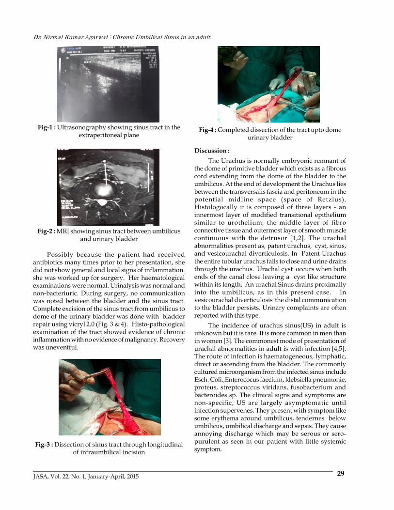
29JASA, Vol. 22, No. 1, January-April, 2015
Fig-1 : Ultrasonography showing sinus tract in theextraperitoneal plane
Fig-2 : MRI showing sinus tract between umbilicusand urinary bladder
Possibly because the patient had receivedantibiotics many times prior to her presentation, shedid not show general and local signs of inflammation.she was worked up for surgery. Her haematologicalexaminations were normal. Urinalysis was normal andnon-bacteriuric. During surgery, no communicationwas noted between the bladder and the sinus tract.Complete excision of the sinus tract from umbilicus todome of the urinary bladder was done with bladderrepair using vicryl 2.0 (Fig. 3 & 4). Histo-pathologicalexamination of the tract showed evidence of chronicinflammation with no evidence of malignancy. Recoverywas uneventful.
Fig-3 : Dissection of sinus tract through longitudinalof infraumbilical incision
Fig-4 : Completed dissection of the tract upto domeurinary bladder
Discussion :The Urachus is normally embryonic remnant of
the dome of primitive bladder which exists as a fibrouscord extending from the dome of the bladder to theumbilicus. At the end of development the Urachus liesbetween the transversalis fascia and peritoneum in thepotential midline space (space of Retzius).Histologocally it is composed of three layers - aninnermost layer of modified transitional epitheliumsimilar to urothelium, the middle layer of fibroconnective tissue and outermost layer of smooth musclecontinuous with the detrusor [1,2]. The urachalabnormalities present as, patent urachus, cyst, sinus,and vesicourachal diverticulosis. In Patent Urachusthe entire tubular urachus fails to close and urine drainsthrough the urachus. Urachal cyst occurs when bothends of the canal close leaving a cyst like structurewithin its length. An urachal Sinus drains proximallyinto the umbilicus, as in this present case. Invesicourachal diverticulosis the distal communicationto the bladder persists. Urinary complaints are oftenreported with this type.
The incidence of urachus sinus(US) in adult isunknown but it is rare. It is more common in men thanin women [3]. The commonest mode of presentation ofurachal abnormalities in adult is with infection [4,5].The route of infection is haematogeneous, lymphatic,direct or ascending from the bladder. The commonlycultured microorganism from the infected sinus includeEsch. Coli.,Enterococus faecium, klebsiella pneumonie,proteus, streptococcus viridans, fusobacterium andbacteroides sp. The clinical signs and symptoms arenon-specific, US are largely asymptomatic untilinfection supervenes. They present with symptom likesome erythema around umbilicus, tendernes belowumbilicus, umbilical discharge and sepsis. They causeannoying discharge which may be serous or sero-purulent as seen in our patient with little systemicsymptom.
Dr. Nirmal Kumar Agarwal : Chronic Umbilical Sinus in an adult

30 JASA, Vol. 22, No. 1, January-April, 2015
Ultrasound can help to make diagnosis in largenumber of cases and a sinus tract can be seen insubcutaneous plane in the midline from umbilicus. MRIshould be done to confirm the diagnosis and definerelationship to surrounding structure. US can becomplicated by abscess formation, rupture into theperitoneal cavity leading to peritonitis, other reportedcomplication include uracho-colonic fistula, stoneformation and neoplastic transformation [6,7]. The risksof urachal malignancy in adult is high and prognosisis poor than that of primary bladder adenocarcinoma[8,9]. Although histologically the innermost layer ofthe urachus is mainly transitional cell, adenocarcinomais the predominant histological type due to metaplasiaoccurring from chronic inflammation [8]. The prognosisis poor as urachal abnormalities are observed rarely inadult and moreover, the urachus is located in aclinically silent area, extraperitonealy in the space ofRetzius. As a result, symptoms and signs ofinflammation as well as tumours are non-specific anddelayed [10].
The treatment of choice for urachal sinus iscomplete excision. For acutely infected casesparticularly abscess formation, a 2 stage procedureinvolving initial incision and drainage, followed by
later excision of the urachal remnant [5]. Traditionalsurgical excision of an urachal remnant involves atransverse or midline infra-umbilical incision. Thelaparoscopic approach for resection of the urachus wasfirst introduced by Trondsen et al [13]. Thelaparoscopic approach offer many advantages likeshort duration of hospital stay, early return to normalactivity, better cosmetic result etc [11,12,13].Conclusion :
In summary urachal disease may only present withabdominal pain without obvious periumbilicalerythema or exudate in adult. A high index of suspicionis required in order to achieve a diagnosis. A triad oflower midline mass, umbilical discharge and sepsis issuggestive, although ultrasound and MRI confirm thediagnosis.
Treatment of the infected urachal remnant in adultshould consist of antibiotic treatment combined withadequate drainage and later followed by completesurgical excision of the remnant including a cuff ofbladder. The laparoscopic approach described lately isa safe and effective alternative to open surgery withgood long term cosmetic result.
References:-1. Hammond G, Yglesias L, Davis JE. The urachus, its anatomy and associated fascia. Anat. Rec 1941;80:271-4.2. Begg RC. The urachus : its Anatomy, Histology and Development. J.Anat 1930; 64:170-83.3. Ashley RA, Inman BA, Routh JC, Rohlinger AL, Husmann DA, Kramer SA. Urachal abnormalities : a longitudinal
study of urachal remnants in children and adults. J. Urol 2007;178: 1615-8.4. Ash A, Gujral R, Raio C. Infected urachal cyst initially misdiagnosed as an incarcerated umbilicus hernia. J.
Emerg Med 2012;42:171-3.5. Yoo KH, Lee SJ, Chang SG. Treatment of infected urachal cysts. Yonsci Med J 2006; 47:423-7.6. Ohgaki M, Higuchi A, Chou H, Takashina K, Kawakami S, Fujita Y, Hegiwara A, Yamagishi H. Acute peritonitis
caused by intraperitoneal rupture of an infected urachal cyst : report of a case. Surg. Today 2003;33:75- 7.7. Flanagan DA, Mellinger JD. Urachal-sigmoid fistula in an adult male. Am Surg 1998; 64:762-3.8. Mazzucchelli R, Scarpelli M, Montironi R. Mucinous adenocarcinoma with superficial stromal invasion and
villous adenoma of urachal remnants : a case report. J. Clinpatho 2003;56: 465-7.9. Ueno T, Hashimoto H, Yokoyama H, Ito M, Kouda K, Kanamaru H : Urachal abnormalities. ultrasonography
and management. J. Paediatr Surg 2003;38: 1203-7.10. Berman SM, Tolia BM, Laor E, Reid RE, Schweizerhof SP, Freed SZ. Urachal remnants in adults. Urology 1988;31:
17.11. Yohannes P, Bruno T, Pathan M, Baltaro R : Laparoscopic radical excision of urachal sinus. J. Endourol 2003,
7:475-479.12. Turial S, Huecksteadt T, Schier F, Fahlenkamp D. Laparoscopic treatment of urachal remnants in children. J Urol
2007;177: 1864-6.13. Okegawa T, Odagane A, Nutahara K, Higashihara E. Laparoscopic management of urachal remnants in adulthood.
Int J Urol, 2006;13: 1466-9.
Dr. Nirmal Kumar Agarwal : Chronic Umbilical Sinus in an adult

31JASA, Vol. 22, No. 1, January-April, 2015
Introduction :Ceroid granuloma is an incidental finding in chronic cholecystitis and
choledocholithiasis with an estimated incidence of 2.1 per 100000 populationper annum and often mimics malignant neoplasms and with a greaterpropensity for perforation or fistula formation due to intramural necrosis[1].Case Report :
A 14 year old female was admitted with complaints of gradual onsetdull aching to intermittent episodes of colicky pain in the right upperabdomen, intermittent low grade fever and progressively increasing jaundicefor 1 month. She had similar episodes of pain abdomen and intermittentfever for last 6 months associated with anorexia, nausea and flatulentdyspepsia. Her bowel habits and micturition were normal. The patient wasmalnourished, pale and deeply icteric. There was no cervical or inguinallymphadenopathy or pedal oedema. She had tenderness over the righthypochondrium, However, there was no muscle guarding, rigidity or ascitesor any palpable lump.
Her haemoglobin level was 9.2 gm%, total bilirubin 15.2 mg/dl (directfraction 10.73 mg/dl) and serum alkaline phosphatase level of 505 IU/l. Herserum albumin level was low at 2.8 g/dl. Ultrasonography (USG) ofabdomen showed a distended gall bladder with microcalculi with normalwall thickness and dilated intrahepatic biliary radicles and common bileduct (CBD) with multiple echogenic structures in distal CBD. A repeat USG
Case Report
Ceroid Granuloma of the Extra Hepatic BilliarySystem : A rare entity causing obstructive
jaundice
ABSTRACTCeroid granuloma is a rare cause of obstructive jaundice and closelymimics malignancy of the extra hepatic biliary system. We report a 14year old female child who was admitted with progressive obstructivejaundice and whose clinical features and radiological investigations weresuggestive of biliary tract malignancy. The patient underwent excision ofthe growth with Roux-en Y hepaticojejunostomy. Histopathologicalexamination found it to be a case of Ceroid Granuloma which has oftenfallen within the purview of 'malignant masquerades'. It is a rare clinicalentity and has been reported to closely mimic malignancy and oftenimpossible to differentiate from malignant neoplasm even withpreoperative imaging and cytological studies. Resection with biliaryenteric bypass is recommended. The patient recovered following theoperation.
Dr Gunabhi Ram Das1
Dr Dipak K. Sarma2
Dr. Rocket C. Brahma2
Dr. Anuj K Sarma1
Dr. David Pegu3
Dr. Nilaksha Bhattacharjee4
1 Assistant ProfessorDepartment of General Surgery, AMCH2 Associate ProfessorDepartment of General Surgery, AMCH3 RegistrarDepartment of General Surgery,,AMCH4 Post Graduate TraineeDepartment of General Surgery, AMCH Key Words : extra hepatic biliary system; Ceroid Granuloma; obstructive
jaundice
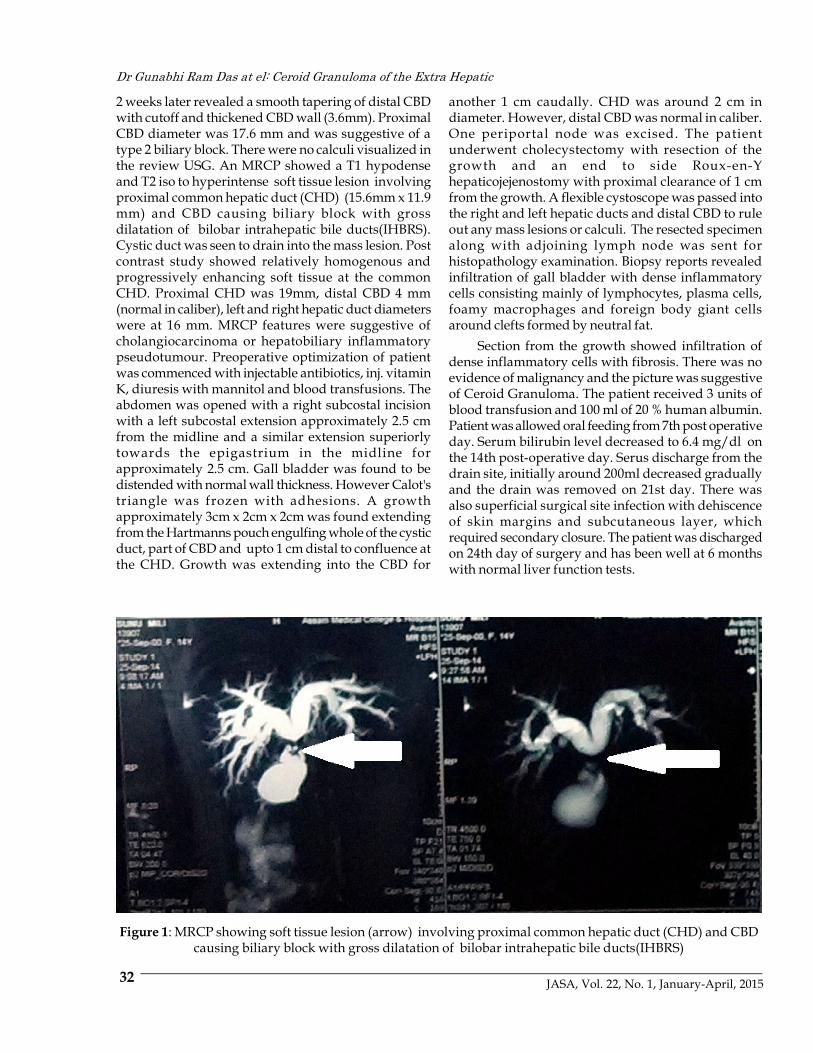
32 JASA, Vol. 22, No. 1, January-April, 2015
2 weeks later revealed a smooth tapering of distal CBDwith cutoff and thickened CBD wall (3.6mm). ProximalCBD diameter was 17.6 mm and was suggestive of atype 2 biliary block. There were no calculi visualized inthe review USG. An MRCP showed a T1 hypodenseand T2 iso to hyperintense soft tissue lesion involvingproximal common hepatic duct (CHD) (15.6mm x 11.9mm) and CBD causing biliary block with grossdilatation of bilobar intrahepatic bile ducts(IHBRS).Cystic duct was seen to drain into the mass lesion. Postcontrast study showed relatively homogenous andprogressively enhancing soft tissue at the commonCHD. Proximal CHD was 19mm, distal CBD 4 mm(normal in caliber), left and right hepatic duct diameterswere at 16 mm. MRCP features were suggestive ofcholangiocarcinoma or hepatobiliary inflammatorypseudotumour. Preoperative optimization of patientwas commenced with injectable antibiotics, inj. vitaminK, diuresis with mannitol and blood transfusions. Theabdomen was opened with a right subcostal incisionwith a left subcostal extension approximately 2.5 cmfrom the midline and a similar extension superiorlytowards the epigastrium in the midline forapproximately 2.5 cm. Gall bladder was found to bedistended with normal wall thickness. However Calot'striangle was frozen with adhesions. A growthapproximately 3cm x 2cm x 2cm was found extendingfrom the Hartmanns pouch engulfing whole of the cysticduct, part of CBD and upto 1 cm distal to confluence atthe CHD. Growth was extending into the CBD for
another 1 cm caudally. CHD was around 2 cm indiameter. However, distal CBD was normal in caliber.One periportal node was excised. The patientunderwent cholecystectomy with resection of thegrowth and an end to side Roux-en-Yhepaticojejenostomy with proximal clearance of 1 cmfrom the growth. A flexible cystoscope was passed intothe right and left hepatic ducts and distal CBD to ruleout any mass lesions or calculi. The resected specimenalong with adjoining lymph node was sent forhistopathology examination. Biopsy reports revealedinfiltration of gall bladder with dense inflammatorycells consisting mainly of lymphocytes, plasma cells,foamy macrophages and foreign body giant cellsaround clefts formed by neutral fat.
Section from the growth showed infiltration ofdense inflammatory cells with fibrosis. There was noevidence of malignancy and the picture was suggestiveof Ceroid Granuloma. The patient received 3 units ofblood transfusion and 100 ml of 20 % human albumin.Patient was allowed oral feeding from 7th post operativeday. Serum bilirubin level decreased to 6.4 mg/dl onthe 14th post-operative day. Serus discharge from thedrain site, initially around 200ml decreased graduallyand the drain was removed on 21st day. There wasalso superficial surgical site infection with dehiscenceof skin margins and subcutaneous layer, whichrequired secondary closure. The patient was dischargedon 24th day of surgery and has been well at 6 monthswith normal liver function tests.
Dr Gunabhi Ram Das at el: Ceroid Granuloma of the Extra Hepatic
Figure 1: MRCP showing soft tissue lesion (arrow) involving proximal common hepatic duct (CHD) and CBDcausing biliary block with gross dilatation of bilobar intrahepatic bile ducts(IHBRS)

33JASA, Vol. 22, No. 1, January-April, 2015
Discussion:Ceroid granuloma of the biliary system is a rare
entity with an incidence of 2.1 per 100000 per annumreported in studies [1]. Ceroid granulomas arecomposed of intramural or mucosal histiocyticinfiltration with pale cytoplasm and yellowish brownwaxy pigments termed as ceroid. The name ceroid isderived from the Greek word 'keros' indicating the waxynature of the same. These ceroid pigments areconsidered to be the early forms of the breakdownpigment Lipofuscin, derived from partial oxidation oflipids and lipoproteins under pathological conditions(cf Lipofuscin which is a naturally occurring wear andtear pigment)[2]. Studies have postulated that bile mayserve as a substrate for formation of these ceroidpigments in a background of recurrent cholecystitis andcholangitis associated with breakdown of theRokitansky-Aschoff sinuses and mucosal ulcerationwith intramural liberation of bile [3]. These arecharacterized by acid fastness and diastase resistance
Dr Gunabhi Ram Das at el: Ceroid Granuloma of the Extra Hepatic
Figure 2: Growth involving CHD, proximal CBD andcystic duct
Figure 3 : Growth resected along with CBD and CHDwith 1 cm proximal clearance
Figure 4 : Excision and Hepaticojejenostomy
Figure 5 : Photomicrograph of specimen showingdense chronic inflammatory infiltrate around clefts
formed by neutral fat
Figure 6 : Photomicrograph (High magnification)showing PAS positive, ceroid laden Granuloma
formation

34 JASA, Vol. 22, No. 1, January-April, 2015
PAS positivity with intense fibrosis and deposition ofcollagen which may even replace the gallbladder, withprogressive dystrophic calcification leading toporcelain gall bladder and/or xanthogranulomatouslesions of the Biliary system.
These lesions are difficult to be differentiated frommalignancy even with pre operative imaging andcytology and complete resection of the stricture resultingfrom the same is recommended [4,5]. Other substratefor ceroidogenesis have been proposed like meconiumand vernix, and cases of ceroid granulomas have alsobeen reported in the female genital tract [6,7].
In our case, the onset was insidious withprogressive obstructive jaundice with features ofcholangitis. MRCP was done for proper delineation ofthe biliary system in light of the conflicting ultrasoundreports initially suggesting cholelithiasis withcholedocholithiasis. The MRCP detected a soft tissuelesion at the proximal CHD also involving proximalCBD causing biliary block. There were no calculi whichraised the suspicion of a neoplasm of the biliary systemcausing obstruction to the bile flow. The patient's poornutritional status along with the findings of MRCP,strongly supported the diagnosis of biliaryadenocarcinoma. Reports in the literature have alsoclaimed that it is often very difficult to distinguishbetween chronic inflammatory conditions and biliaryadenocarcinoma, particularly the Scirrhous type [8].Although MRCP reports suggested a pseudotumour ,such pseudotumours are more common in thegallbladder rather than in the biliary tree. Primarybenign tumours are also rare in the biliary system asthey constitute only 6 percent of all tumours in the biliarysystem. It is often impossible to distinguish betweenbenign and malignant cause of biliary obstruction andsuch patients with an obstructing growth must be
considered to have a malignant disease until provedotherwise [8]. Physical findings in the case of biliarytract growth are also vague and non specific, as in ourpatient, and one must exercise a reasonable amount ofcaution in first ruling out the more sinister of theconditions before considering the benign conditions.
The term 'malignant masquerade' of the ExtraHepatic Biliary tree was brought into practice in 1984by Hadjis and colleague in Hammersmith Londondenoting benign lesions which could not bedistinguished from malignant conditions of the biliarysystem even after extensive pre operative investigations[8]. Several subsequent studies have described suchlesions and if the growth was found resectable, theobstructing lesions were removed and a biliaryreconstruction and bypass in the form ofhepaticojejunostomy was done in majority of the cases.The authors have also concluded that even withmodern modalities of investigations, it is impossible todifferentiate between the benign and malignant lesionsand sometimes even on table frozen section biopsy maybe misleading [8].
In our patient, the growth which was firm to hardin consistency with infiltration into the CHD and CBDmimicked a proximal Cholangiocarcinoma and whichon on-table assessment was found to be resectable. Inlight of the exisiting literature, our decision to achievean excision with a clear margin and a biliary entericanastomosis stands validated. However, it was onlyafter proper histopathology examination thatmalignancy could be ruled out and a diagnosis of aCeroid Granuloma was made. It is a rare but distinctclinical entity which has often been said to mimicmalignancy and has a greater propensity forcomplications like choledocho-duodenal fistula orperforation.
References :1. Duarte Z, Guevara U, Vuong PN. Ceroid granulomas of the gallbladder. A clinicopathologic study of nine cases.
Arch Anat Cytol Pathol. 1994;42(1):5-9.2. Seehafer SS, Pearce DA. You say lipofuscin, we say ceroid: defining autofluorescent storage material. Neurobiol
Aging. 2006;27(4):576-88.3. Takahashi K, Oka K, Hakozaki H, Kojima M. Ceroid-like histiocytic granuloma of gall-bladder --a previously
undescribed lesion--.Acta Pathol Jpn. 1976;26(1):25-46.4. Amazon K, Rywlin AM. Ceroid granulomas of the gallbladder. Am J Clin Pathol. 198;73(1):123-7.5. Krishna RP1, Kumar A, Singh RK, Sikora S, Saxena R, Kapoor VK Xanthogranulomatous inflammatory strictures
of extrahepatic biliary tract: presentation and surgical management. J Gastrointest Surg. 2008;12(5):836-841. doi:10.1007/s11605-008-0478-y.
6. Ooi K1, Riley C, Billson V, Ostör AG. Ceroid granulomas in the female genital system.J Clin Pathol.1995;48(11):1057-9.
7. Pikarsky E1, Maly B, Maly A. Ceroid granuloma of the uterine cervix. Int J Gynecol Pathol. 2002;21(2):191-3.8. Koea J1, Holden A, Chau K, McCall J. Differential diagnosis of stenosing lesions at the hepatic hilus. World J
Surg. 2004;28(5):466-70.
Dr Gunabhi Ram Das at el: Ceroid Granuloma of the Extra Hepatic

35JASA, Vol. 22, No. 1, January-April, 2015
Introduction :The double J ureteral stents have become one of the most basic and
valuable tools in the urological practice both for endoscopic and openurological surgery. Indwelling ureteral stents provide direct drainage ofthe upper urinary tract to the bladder without the need for externaldiversion [1,2]. The indications for insertion of stents into the urinarytract has expanded significantly during the last decade. Stents now areinserted routinely in patients with ureteral obstruction and for preventionof complications following open or endoscopic procedures. Howeverdifferent complications may occur with short or long term use of stents.Initially, very few side effects were reported but later on many publicationsreported that indwelling ureteral stents can cause lower abdominal pain,dysuria, fever, haematuria, low backache and suprapubic pain [3-5].Furthermore, indwelling stents can cause major complications like stentmigration, encrustation, vesicoureteric reflux, stent fracture, secondaryvesical calculus formation or even be forgotten in the patient [6-9].Case Report :
A 27 years old male patient presented to the surgery outpatientdepartment with history of pain in the suprapubic region, occasionalfever, burning micturition for the last 6 months. He took irregularantibiotics and other medications prescribed by the local doctors. Hehad undergone right side uretero-lithotomy for removal of ureteric stonewith DJ stenting about 4 years back at a private hospital, however he lostthe medical records except the ultrasonography report. Despite being
Case Report
Complications following forgotten DJ urinarystent: A case report
ABSTRACTDouble J stent is most widely used by the urologists for drainage of urinefrom upper urinary tract to urinary bladder. Though the insertion of DJstent is to prevent complication following endoscopic and open surgery,the stent itself may cause serious complications like stent migrations,encrustations, sepsis, urinary tract infections\ and stone formation etc.due to delayed removal of the stent or forgotten stent. We report a classicalcase of forgotten stent in a twenty seven year old male patient who hadundergone ureterolithotomy with DJ stent four years back and haddeveloped stent related complications.
Dr Rocket Chandra BrahmaDr Sidhartha Sankar Konwar
Address for correspondence:Dr Rocket Chandra Brahma,MS, FAIS, FMAS, PGCHMAssociate ProfessorDepartment of SurgeryAssam Medical College and HospitalDibrugarh- 786002, AssamMobile-9864051733Email: [email protected]
Key Words : Stent complications; DJ stent; encrustations; forgotten stents;cystolithotomy.

36 JASA, Vol. 22, No. 1, January-April, 2015
advised to come back for check up by the concernedsurgeon he ignored the advice. As he was not awarethat a DJ stent had been inserted and needed to beremoved. His urine analysis revealed plenty of puscells, serum creatinine, blood urea and otherhaematological tests were within normal limits. X-ray KUB revealed calculus in the urinary bladderformed on the coil of DJ stent. Ultrasonographyrevealed mild hydronephrosis of the right kidney, acalculus measuring 33.3mm x 17.4mm was noted inthe urinary bladder, and a thin linear hyperechoicarea also seen extending from the right renal pelvisupto the urinary bladder which was later onconfirmed as DJ stent. Intravenous urographyrevealed delayed excretion of contrast in the rightkidney. The patient was put on broad spectrumantibiotics and posted for suprapubic cystolithotomyunder regional anaesthesia. The vesical calculusencrusted around the coiled DJ stent was removed[Figures 1, 2 & 3]. The post operative period wasuneventful and the patient was discharged on 7thpost operative day. This was a classical case offorgotten DJ stent with encrustation which formed anidus for secondary vesical calculus formation andother stent associated complications like recurrenturinary tract infections, fever and pain lowerabdomen.Discussion :
The double J stent has been a boon and are beingextensively used by the surgeons and urologistspracticing both open and endoscopic procedures. Thestent allows good urinary drainage from kidney tothe urinary bladder and is usually well tolerated bythe patients. Despite its extensive use, DJ stents cangive rise to several complications which may bemanifested as an early and late complication and aremost often related to prolonged indwelling times,especially in forgotten or overlooked stents. Earlycomplications may be stent colic, dysuria,haematuria, vesico-ureteric reflux, urinary tractinfections, pyelonephritis, fever and flank pain. Latecomplications includes hydronephrosis,encrustations, stent migration, stent fracture,spontaneous fragmentation , uretero- arterial anduretero-intestinal fistula [9,10]. In a study conductedby Hao P et al, of 2685 cases of DJ stent used for bothopen and endoscopic procedures, the stents were asafe and useful adjunct if these were removed after 4-6 weeks. In another study, Damiano R et al observed
flank pain (25.3%), encrustation (21.6%), irritablebladder symtoms (18.8%), haematuria ( 18.1%), fever(>104'F in 12.3%) and stent migration (9.5%) in casesof forgotten stents. They also reported that morbidityand complications were minimal when the stentswere kept in situ for less than 3 months, but longerduration stent retention was associated withincreased frequency of encrustation, infections,calculus formation and obstruction of the stentedtract. A literature search revealed that a stent couldremain forgotten upto 8 years post surgery givingrise to various complications [11--13]. In the presentcase also, the stent was left in the urinary tract for 4years after removal of the right sided mid uretericstone and later the part of the stent in the bladderacted as nidus to form a secondary vesical calculusas well as other associated complications like painabdomen, fever, dysuria, encrustation and vesicalcalculus formation.
Most stents can be removed under localanesthesia with a two pronged rigid or flexible biopsyforceps. However managing stent complications likestruck stents due to long forgotten stent, remainedone of the most challenging tasks for the urologists.Those with major proximal stone burden may requirepercutaneous nephrolithotomy ( PCNL) whileureteric fragments can be managed by minimallyinvasive technique such as ureteroscopy and intra-corporeal lithotripsy [14,15]. Forgotten stents shouldbe managed endoscopically only by those who arewell trained in advanced Endourology [16,17]. Opensurgery has a role where multi model endo-urologyprocedures fails, but one must remember that this isalso not easy and there is danger of renal impairmentand sepsis. In the present case, in absence of aqualified urologist, open cysto-lithotomy andremoval of the stent and vesical calculus was doneunder regional anaesthesia. Our report suggests thatsince the stent is a foreign body, it should not be keptinside the body for longer period. Though the DJ stenthas been a boon to the urologists, they shouldremember the serious complications associated withits use and should not be kept for more than threemonths. Moreover to avoid prolonged or forgottenstent, surgeon should maintain a diary or acomputerized stent log and stress the importance offollow up care by proper counseling and also informthe patient and their attendants regarding presenceof a stent and the need for its timely removal.
Dr Rocket Chandra Brahma at el: Complications following forgotten

37JASA, Vol. 22, No. 1, January-April, 2015
Fig-1 : A Supra-pubic cystolithotomy and removal ofencrusted DJ stent.
Fig-2 : Encrusted DJ stent with vesical calculus at thetip of stent.
Fig-3 : Suprapubic-cystolithotomy showing encrustedDJ stent
References :1. Singh I. Indwelling JJ ureteral stents-a current perspective and review of literature. Indian J of Surg 2003; 65: 405-12.2. Nawaz H, Hussain M, Hashmi A, Hussain Z. Experience with indwelling JJ stents. J Pak Med Assoc 1993; 43: 147-9.3. Paz A, Amiel GE, Pick N, Moskovitz B, Nativ O, Potasman I. Febrile complications following insertion of 100 double-
J ureteral stents. J Endourol 2005; 19: 147-50.4. Saltzman B. Ureteral stents: indications, variations and complications. Urol Clin North Am 1988; 15: 481-91.5. Pollard SG, Macfarlane R. Symptoms arising from double J ureteral stents. J Urol 1988; 139: 37-8.6. Memon NA, Talpur AA, Memon JM. Indications and complications of indwelling ureteral stenting at NMCH,
Nawabshah. Pak J of Surg 2007; 23: 187-91.7. Richter S, Ringel A, Shalev, Nissenkorm I. The indwelling ureteric stent: a 'friendly' procedure with unfriendly high
morbidity. Br J Urol Intl 2009; 85: 408-11.8. Singh I. Indwelling JJ ureteral stents-A Current perspective and review of literature, Indian J.Surg 2003; 65:405-12.9. Kehinde EQ, Al-awadi KA,Tawheed A, l-Hunayan A, Ali Y, Mahmood AH. Factors affecting the fate of prolonged
forgotten DJ stents Sc and J Urol Nephrol 2001; 35; 222-7.10. Kar A, Angwalo FF, Jhunjhunwala JS,. Uretero-arterial and Uretero-sigmoid fistula associated with polyethylene
indwelling ureteral stents. J Urol 1984; 132:755-7.11. Hao P, Li W, Song C, Yan J,Song B, Li I. Clinical evaluation of double pigtail stent in patients with upper urinary tract
diseases report of 2685 cases, J Endourol 2008, 22(i) 65-7.12. Igujatovic I, Kovic IS,Ttrapping of double J stent in the urinary tract eight years after extracorporeal shock wave
lithotripsy, Int Urol Nephrol 2000;32(1)29-31.13. Chun Kai C, Ching Chia L, Hung Lung K, Yii Her C, Chun-Hsiving H, Ming Chen S, double J stent forgotten for 7
years. A case report. Kohsiung J Med Sci 2003; 19(2) 84-7.14. Perera ND, Wijewardena M, removal of severely encrusted forgotten ureteral stents by minimally invasive technique.
Ceylon Med J 2002; 47:27.15. Borborough PG, Kane CJ. Current management of severely encrusted ureteral stents with a long associated stone
burden J Urol 2000; 164:648-50.16. Shandera KC, Minimally invasive management of the calcified ureteral stent. Tech Urol 1997; 3:54-7.17. Somers WJ. Management of forgotten retained tn dwelling ureteral stents. J Urol 1996; 47:431-5.
Dr Rocket Chandra Brahma at el: Complications following forgotten

38 JASA, Vol. 22, No. 1, January-April, 2015
Introduction :Osteomyelitis of long bones is commonly encountered in regular
orthopaedic practice but osteomyelitis of clavicle is rare, with an incidenceranging from 0% in mixed-aged population to 7% in children. The mid-portion or medial half of clavicle is a common site of this lesion, althoughany portion of clavicle can be affected. Chronic osteomyelitis of clavicleusually follows a local invasive procedure; however a primarypresentation is possible. Chronic osteomyelitis being a biofilm basedinfection, surgical intervention is usually required [1]. Proper dead spacemanagement combined with prolonged antibiotics can lead to favourableresults. The rarity of this condition and the dearth of sufficient literaturehave prompted us to report this case.Case Report :
A 25 year old gentleman reported to outpatient department oforthopaedics of our institute with pain and chronic discharging sinusover the right clavicle. He gave a history of trauma to the same site sixmonths back; 2 months later a swelling appeared at the site and had beengradually increasing in size. Incision and drainage was done by generalsurgeon in some local hospital. On examination, there was serousdischarge from right clavicle at the junction of middle and distal third ofclavicle with no restriction of movement of the affected shoulder. X- rayshowed a sequestrum in the middle third of clavicle with irregularsclerosis and lysis in adjoining areas. The findings were confirmed on aMRI scan. Curettage and sequestrectomy was performed under generalanaesthesia and the sample was sent for histopathological examination,PCR for TB and repeat culture sensitivity. The wound healed in 3 weeks
Case Report
Chronic osteomyelitis of clavicle in animmunocompetent adult: a case report
ABSTRACTHaematogenous osteomyelitis of clavicle is rare and there is scant literatureregarding the behaviour and prognosis of this condition. We present acase of a 25 year old male who had presented to the outpatient departmentof Orthopaedics in our institute with complaints of pain and dischargingsinus for last 4 months. Curettage and sequestrectomy was done. Thecase is being presented because of its rarity in occurrence.
Dr. Bikash Jyoti Bordoloi1
Dr. Pradip Kumar Baruah2
Dr. Rajarshi Roy3
Dr. Bikash Agarwal3
Address for correspondence:Dr. Bikash Jyoti BordoloiEmail: [email protected]: 9864010379
Key Words : Clavicle, osteomyelitis; immunocompetent; sequestrectomy.
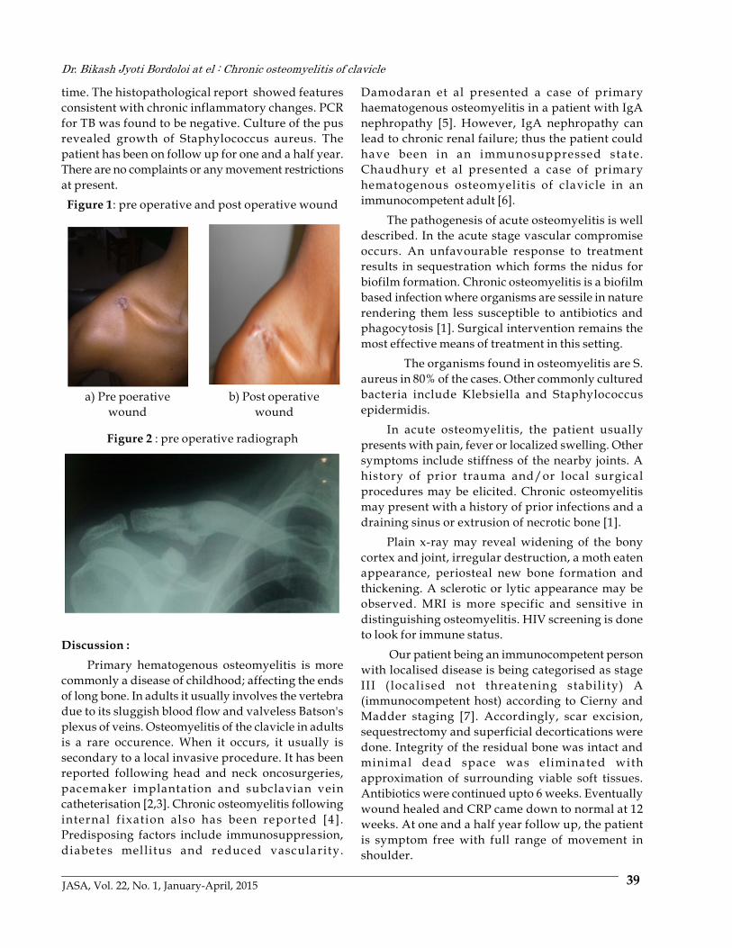
39JASA, Vol. 22, No. 1, January-April, 2015
time. The histopathological report showed featuresconsistent with chronic inflammatory changes. PCRfor TB was found to be negative. Culture of the pusrevealed growth of Staphylococcus aureus. Thepatient has been on follow up for one and a half year.There are no complaints or any movement restrictionsat present.Figure 1: pre operative and post operative wound
a) Pre poerativewound
b) Post operativewound
Figure 2 : pre operative radiograph
Discussion :Primary hematogenous osteomyelitis is more
commonly a disease of childhood; affecting the endsof long bone. In adults it usually involves the vertebradue to its sluggish blood flow and valveless Batson'splexus of veins. Osteomyelitis of the clavicle in adultsis a rare occurence. When it occurs, it usually issecondary to a local invasive procedure. It has beenreported following head and neck oncosurgeries,pacemaker implantation and subclavian veincatheterisation [2,3]. Chronic osteomyelitis followinginternal fixation also has been reported [4].Predisposing factors include immunosuppression,diabetes mellitus and reduced vascularity.
Damodaran et al presented a case of primaryhaematogenous osteomyelitis in a patient with IgAnephropathy [5]. However, IgA nephropathy canlead to chronic renal failure; thus the patient couldhave been in an immunosuppressed state.Chaudhury et al presented a case of primaryhematogenous osteomyelitis of clavicle in animmunocompetent adult [6].
The pathogenesis of acute osteomyelitis is welldescribed. In the acute stage vascular compromiseoccurs. An unfavourable response to treatmentresults in sequestration which forms the nidus forbiofilm formation. Chronic osteomyelitis is a biofilmbased infection where organisms are sessile in naturerendering them less susceptible to antibiotics andphagocytosis [1]. Surgical intervention remains themost effective means of treatment in this setting.
The organisms found in osteomyelitis are S.aureus in 80% of the cases. Other commonly culturedbacteria include Klebsiella and Staphylococcusepidermidis.
In acute osteomyelitis, the patient usuallypresents with pain, fever or localized swelling. Othersymptoms include stiffness of the nearby joints. Ahistory of prior trauma and/or local surgicalprocedures may be elicited. Chronic osteomyelitismay present with a history of prior infections and adraining sinus or extrusion of necrotic bone [1].
Plain x-ray may reveal widening of the bonycortex and joint, irregular destruction, a moth eatenappearance, periosteal new bone formation andthickening. A sclerotic or lytic appearance may beobserved. MRI is more specific and sensitive indistinguishing osteomyelitis. HIV screening is doneto look for immune status.
Our patient being an immunocompetent personwith localised disease is being categorised as stageIII ( localised not threatening stability) A(immunocompetent host) according to Cierny andMadder staging [7]. Accordingly, scar excision,sequestrectomy and superficial decortications weredone. Integrity of the residual bone was intact andminimal dead space was eliminated withapproximation of surrounding viable soft tissues.Antibiotics were continued upto 6 weeks. Eventuallywound healed and CRP came down to normal at 12weeks. At one and a half year follow up, the patientis symptom free with full range of movement inshoulder.
Dr. Bikash Jyoti Bordoloi at el : Chronic osteomyelitis of clavicle
40 JASA, Vol. 22, No. 1, January-April, 2015
Conclusion :Primary osteomyelitis of clavicle in an
immunocompetent patient is a rare occurrence.Chronic osteomyelitis are biofilm based infections.Surgical debridement is the most effective means oftreatment. Reconstruction depends on dead spacecreated and integrity of the residual bone. Effectivesurgical management with prolonged antibiotics canlead to good results.Bibliography :1. Donlan RM, Costerton JW. Biofilms: survival
mechanisms of clinically relevantmicroorganisms. Clin Microbiol Rev, 2002;15(2):167-193.
2. Allessi DM., Sercarz JA., Calcaterra TC.Osteomyelitis of the clavicle. Arch OtolaryngolHead Neck Surg 1988; 114:1000-2.
3. Krespi YP, Monsell EM, Sisson GA.Osteomyelitis of the clavicle. Ann OtolRhynol Laryngol 1983; 92:525-7.
4. Balakrishnan C, Vashi C. Post traumaticosteomyelitis of the clavicle: A case report andreview of literature. Can J Plast Surg2008;16(2):89-91.
5. Damodaran A, Rohit A, Abraham G et al.Case report: rare occurrence of Pseudomonasaeruginosa osteomyelitis in the right claviclein a patient with IgA nephropathy [v1; refstatus: indexed, http://11000r.es/37r]nF1000Research 2014; 3:268 (doi:10.12688/f1000research.3891.1).
6. Chaudhary P. Chronic osteomyelitis of clavicle.Health Renaissance 2011; 9(2):125-7.
7. Cierny G, Mader JT, Pennick JJ. A clinicalstaging for adult osteomyelitis in adults. ClinOrthop Relat Res. 2003; 414:7-24.
Dr. Bikash Jyoti Bordoloi at el : Chronic osteomyelitis of clavicle
41JASA, Vol. 22, No. 1, January-April, 2015
Journal Review
Compiled by:Editor
Tverdal A - There is insufficient epidemiological evidence on the relationshipbetween type of coffee and the risk of prostate cancer. An inverse relationshipbetween number of cups per day and the risk of prostate cancer was presentonly for the boiled coffee type.
Methods:
The risk of prostate cancer by use of boiled vs not boiled coffee wereassessed in a prospective study of 224,234 men 20-69 years.
5740 incident prostate cancers were identified.
Results :
With no coffee as reference group the hazard ratios of <1-4, 5-8 and9+ cups per day of boiled coffee only were 0.84 (0.73-0.96), 0.80 (0.70-0.92) and 0.66 (0.55-0.80), P-trend=0.00.
The corresponding figures for not boiled coffee were 0.89 (0.80-0.99),0.91 (0.81-1.02) and 0.86 (0.74-1.00), P-trend=0.22.
2. Laparoscopic vertical sleeve gastrectomysignificantly improves short term weight loss ascompared to laparoscopic adjustable gastric bandplacement in morbidly obese adolescent patients
The Prostate, 01/27/2015Cook MB, et al.
Within the U.S., there exists significant geographic variability in prostatecancer incidence rates. Although there are large geographic differences inBWIRRs, temporal trends are fairly stable. This may indicate that primaryfactors affecting prostate cancer incidence rates vary geographically but affectboth black and white men to a similar degree.
Methods
Prostate cancer counts and population denominators by race (black,white), age, calendar year, and U.S. census division, for the period1999-2008, were extracted from NPCR and SEER.
Authors calculated age-standardized incidence rates (ASR) andestimated annual percent changes (EAPC) by race and censusdivision.
They assessed black-to-white incidence rate ratios (BWIRR) by census
1.Boiled coffee consumption and the riskof prostate cancer: follow-up of 224,234Norwegian men 20-69 yearsBritish Journal of Cancer, 12/24/2014 Clinical Article
42 JASA, Vol. 22, No. 1, January-April, 2015
division and by calendar period.
Results
This analysis included 1,713,471 prostatecancer cases and 1,217 million person-years.
Black ASRs ranged from 176 per 100,000 person-years in Mountain division to 259 in MiddleAtlantic.
BWIRRs ranged from 1.20 in Western divisionsto 1.72 in Southeastern divisions.
EAPCs indicated that prostate cancer incidenceis not decreasing in East South Central, unlikeall other divisions.
White EAPCs displayed similar variations bycensus division, resulting in modest temporalchanges in BWIRRs.
3. Morbidity after Total Gastrectomy:Analysis of 238 Patients
Luke V. Selby, Emily A. Vertosick, Daniel D.Sjoberg, Mark A. Schattner, Yelena Y. Janjigian, MurrayF. Brennan, Daniel G. Coit, Vivian E. Strong,
Journ of the American College of Surgeon. DOI:http://dx.doi.org/10.1016/j.jamcollsurg.2015.01.058
Background
Surgical quality improvement requires well-defined benchmarks and accurate reporting ofpostoperative adverse events, which have not been welldefined for total gastrectomy.
Study Design
Detailed post-operative outcomes on 238 patientswho underwent curative intent total gastrectomy from2003 - 2012 were reviewed by a dedicated surgeon chartreviewer to establish the 90-day patterns of adverseevents.
Results
Of the 238 patients with stage I - III gastricadenocarcinoma who underwent curative intent totalgastrectomy, the median age was 66, and 68% weremale. Median body mass index was 28, and 68% ofpatients had at least one medical comorbidity. Forty threepercent of our patients received neoadjuvantchemotherapy, and 34% received post-operativeadjuvant chemotherapy. Over the 90 day study period,30-day mortality was 2.5% (6/238), and 90-day
mortality was 2.9% (7/238). At least one post-operativeadverse event was documented in 62% of patients, with28% of patients experiencing a major adverse eventrequiring invasive intervention. The readmission ratewas 20%. Anemia was the most common adverse event(20%), followed by wound complications (18%). Themost common major adverse event was esophagealanastomotic leak, which required invasive interventionin 10% of patients.
Conclusions
This analysis has defined comprehensive 90-daypatterns in post-operative adverse events followingcurative intent total gastrectomy in a Westernpopulation. This benchmark allows surgeons tomeasure, compare, and improve outcomes and informedconsent for this surgical procedure.
4. Tumor factors predictive ofresponse to hypofractionatedradiotherapy in a randomized trialfollowing breast conservingtherapy
Annals of Oncology, 03/03/2014 Bane AL, et al.
This study aims to determine whether tumor grade,molecular subtype and hypoxia predict response tohypofractionated vs. standard radiotherapy (RT)following breast conserving surgery (BCS) for nodenegative breast cancer in a randomised controlled trial(RCT). In women enrolled in the HWBI trial followingBCS tumor molecular subtype predicted LR. Howevertumor grade, molecular subtype and hypoxia did notpredict response to hypofractionation suggesting thatpatients with node negative breast tumors of all gradesand molecular subtypes may be safely treated withhypofractionated RT regimens.
Methods
Formalin fixed paraffin embedded (FFPE) tumorblocks were available on 989 of 1234 patientsenrolled in the Hypofractionation Whole BreastIrradiation (HWBI) Trial.
A central pathology review and assessment oftumor grade using the Nottingham gradingsystem was performed.
Tumors were classified by molecular subtypeas luminal A, luminal B, HER2 enriched, basal-like or unclassified using a six-biomarker panel;ER, PR, HER-2, Ki67, CK5/6 and EGFR.

43JASA, Vol. 22, No. 1, January-April, 2015
Tumors were also classified as hypoxic basedon the expression of HIF1?, CAIX or GLUT-1.
The primary end point was local recurrence(LR).
Results
Median follow-up was 12 years.
In the multivariable Cox model molecular subtype was the only factor predictive of LR, the 10 year cumulativeincidence was 4.5% for luminal A and basal-like, 7.9% for luminal B and 16.9% for HER-2 enriched tumors(p<0.01).
Tumor grade, molecular subtype or hypoxia did not predict response to hypofractionation.

44 JASA, Vol. 22, No. 1, January-April, 2015
HON. SECRETARY`S REPORT from ASSAM STATE CHAPTER ofASI
Respected esteemed members of Association of Surgeons of Assam (Assam State Chapter of ASI),
Rongali Bihu Greetings to you all. It gives me immense pleasure in presenting before you the brief report of ourchapter till March, 2015.
Present executive committee led by our senior and active member Dr. R.N.Mazumder was elected during oursilver jubilee annual conference held last year from 7th to 9th November, 2014 and officially took over charge onJanuary 10th,2015. at a function held at our chapter office , Guwahati. On the same they we sat for our firstexecutive body meeting and planned future activities of our chapter along with a tentative academic calendar forthe coming year.
I like to highlight before you the activities of our Association during this short span of time.
The SILVER JUBILEE ANNUAL CONFERENCE of Assam chapter "ASACON 2015" was held at GuwahatiMedical College, Guwahti, Assam from 7th to 9th November 2014. It was a memorable event with full of academics,surgical workshop and hands on training courses under the guidance and active participation of eminent na-tional and local faculties. I take this opportunity to thank our immediate past President Dr. Satish Sukla sir for hisgracious presence and encouragement during the session.
Dr.G C Jain, Past President of ASA and former GC member of ASI was accorded, during last conference with"Life time achievement award" for his contribution to the society and growth of the association by Assam Chapterfor the year 2014.
At the initiative and continuous stimulation of Dr.Santhosh John Abraham, President of the Association ofSurgeons of India, the Guwahati city branch, along with Assam State Chapter had organised ASI North East ZonalCME on 7th & 8th March'15. Surgeons and post graduate students from all the north eastern states had attendedthe event. The two days programme consisted with PG master Class, CME lectures by eminent teachers of medicalInstitutions from all over the country and NE region in particular. A competition paper and e poster presentationsession was conducted amongst the students. The best e poster was presented by Dr.Sandeep Ghose fromNEIGRIMS, Shillong and the best free paper was presented by Dr.Babitha N from RIMS, Imphal. In the talent huntcompetition (ASI TYSA), Dr.Abhisekh Patil from Silchar Medical College, Assam has been qualified to participatein the national level representing North eastern zone. One hundred and eighty registered delegates attended theprogramme and all the delegates and faculty were awarded with CME credit hours as per MCI guideline.
Nagaon Branch of The Association of Surgeons of Assam organized a CME on 25th October 2014 at Nagaon.The Scientific session was moderated by Dr. K.N. Goswami and Dr. J.N. Gohain. The presentation started withvideo presentation starting from simple laparoscopic cholecystectomy to advanced laparoscopic Nephrectomy byDr. Foridul Hussain. He also presented on advanced technique of TURP and shared some of his experiences. Dr.M.A. Sarkar made a presentation on different aspects of etiology, diagnosis and plan of treatment of pancreatitis.
Dr. R.N. Majumder, Chairman Assam Chapter chaired a session on Burn Care and Prevention organized byburn care foundation, Guwahati held on 14th Feb. 2015.
Our Tezpur city branch has organized a seminar on 15th. Feb 2015. and our Chairman Dr. R.N. Mazumderwas present during the session to encourage activities of the branch. Different aspects of Bariatric surgery and
Association News

45JASA, Vol. 22, No. 1, January-April, 2015
latest development of laparoscopic Hernia repair were discussed by Dr. Manoj Choudhury and S A Majid respec-tively as faculty members in presence of a large gathering.
Dibrugarh branch has organized a seminar on 12 December,2014 at Dibrugarh with special focus on urologi-cal aspect of surgery. Few case reports on Intra-testicular Rhabdomyosarcoma, Urachal Cyst, Pheochromocytomaand Uncommon Foreign Body in urinary bladder were presented by the faculties and post graduate students. Theseminar was well attended by members and eminent surgeons from the region.
Our undivided Sibsagar District Branch organised two seminars jointly with Doctors` club, Jorhat, at Jorhat -one on 23rd Jan, 2015 on " Management of Haemorrhoids - Medical, Surgical and Controversies and the other on7th Feb, 2015 on Surgical management of Chronic Pancreatitis and the Speakers were Dr. Kamal Chandra Deyfrom same chapter and Dr. Pranjal Deka, M.Ch. GI Surgery from Guwahati respectively. An audio visual presen-tation on Medico legal Issues was also presented during the session. Our member Dr. S.J.Das delivered a publicawareness speech at Cancer Awareness programme organized by Concept Junior College, Titabor, on the occasionof World Cancer Day.
Most recently 3rd Advanced Laparoscopic Surgery Workshop cum CME was organized by Nazareth Hospi-tal, Shillong, Meghalaya, in collaboration with Meghalaya surgical Association, Association of Surgeons of Assamand Association of Minimal Access Surgeons of India (AMASI) on 28th and 29th. March 2015. We feel proud to bea part of this academic bonanza.
Recently on 24th Feb.,2015 we lost our beloved past President of our chapter retired Professor Dr. S.B. DuttaChoudhury creating a vacuum in the entire north eastern part of India.
In the mean time, we have prepared an academic calendar for next part of our tenure, which includes differentpublic awareness programme to enable us to reach the populace, besides holding annual conference at Jorhat &midterm CME at Hojai, Surgical workshops and branch CMEs at different branches involving more and moremember including members from the periphery. We have also included awareness programme to popularisenational welfare programme like male sterilisation procedure ( like vasectomy /NSV) in a bid to discharge ourresponsibility towards country`s welfare & development. We have planned to work for enrolment of new ASImembers from our state.
We are regularly publishing a quarterly Journal named JASA of high Standard which is recently indexed inCopernicus and Indian Citation Index. Special thanks to Editor and Editorial board of JASA for this achievement.The news letter "Surgery Papyrus" started last year will be continued as quarterly publication.
Before I conclude, let me offer my sincere thanks to you all for selecting me as Hon Secretary of the Association.We seek your co operation, guidance and suggestions to serve our dear Organisation better.
Thanking you all
Dr. Pulakananda BharaliHon. Secretary.
Assam Chapter of ASI
46 JASA, Vol. 22, No. 1, January-April, 2015

47JASA, Vol. 22, No. 1, January-April, 2015

48 JASA, Vol. 22, No. 1, January-April, 2015

49JASA, Vol. 22, No. 1, January-April, 2015

50 JASA, Vol. 22, No. 1, January-April, 2015