
ISB recommendations on the reporting of intersegmental ...
36
HAL Id: hal-02442619 https://hal.archives-ouvertes.fr/hal-02442619 Submitted on 16 Jan 2020 HAL is a multi-disciplinary open access archive for the deposit and dissemination of sci- entific research documents, whether they are pub- lished or not. The documents may come from teaching and research institutions in France or abroad, or from public or private research centers. L’archive ouverte pluridisciplinaire HAL, est destinée au dépôt et à la diffusion de documents scientifiques de niveau recherche, publiés ou non, émanant des établissements d’enseignement et de recherche français ou étrangers, des laboratoires publics ou privés. ISB recommendations on the reporting of intersegmental forces and moments during human motion analysis Timothy R. Derrick, Antonie J. van den Bogert, Andrea Cereatti, Raphaël Dumas, Silvia Fantozzi, Alberto Leaedini To cite this version: Timothy R. Derrick, Antonie J. van den Bogert, Andrea Cereatti, Raphaël Dumas, Silvia Fantozzi, et al.. ISB recommendations on the reporting of intersegmental forces and moments during human motion analysis. Journal of Biomechanics, Elsevier, 2020, 99, 35 p. 10.1016/j.jbiomech.2019.109533. hal-02442619
-
Upload
khangminh22 -
Category
Documents
-
view
3 -
download
0
Transcript of ISB recommendations on the reporting of intersegmental ...

HAL Id: hal-02442619https://hal.archives-ouvertes.fr/hal-02442619
Submitted on 16 Jan 2020
HAL is a multi-disciplinary open accessarchive for the deposit and dissemination of sci-entific research documents, whether they are pub-lished or not. The documents may come fromteaching and research institutions in France orabroad, or from public or private research centers.
L’archive ouverte pluridisciplinaire HAL, estdestinée au dépôt et à la diffusion de documentsscientifiques de niveau recherche, publiés ou non,émanant des établissements d’enseignement et derecherche français ou étrangers, des laboratoirespublics ou privés.
ISB recommendations on the reporting of intersegmentalforces and moments during human motion analysis
Timothy R. Derrick, Antonie J. van den Bogert, Andrea Cereatti, RaphaëlDumas, Silvia Fantozzi, Alberto Leaedini
To cite this version:Timothy R. Derrick, Antonie J. van den Bogert, Andrea Cereatti, Raphaël Dumas, Silvia Fantozzi,et al.. ISB recommendations on the reporting of intersegmental forces and moments during humanmotion analysis. Journal of Biomechanics, Elsevier, 2020, 99, 35 p. �10.1016/j.jbiomech.2019.109533�.�hal-02442619�

Journal Pre-proofs
ISB recommendations on the reporting of intersegmental forces and momentsduring human motion analysis
Timothy R. Derrick, Antonie J. van den Bogert, Andrea Cereatti, RaphaelDumas, Silvia Fantozzi, Alberto Leardini
PII: S0021-9290(19)30787-0DOI: https://doi.org/10.1016/j.jbiomech.2019.109533Reference: BM 109533
To appear in: Journal of Biomechanics
Received Date: 26 June 2019Revised Date: 14 November 2019Accepted Date: 16 November 2019
Please cite this article as: T.R. Derrick, A.J. van den Bogert, A. Cereatti, R. Dumas, S. Fantozzi, A. Leardini, ISB recommendations on the reporting of intersegmental forces and moments during human motion analysis, Journal of Biomechanics (2019), doi: https://doi.org/10.1016/j.jbiomech.2019.109533
This is a PDF file of an article that has undergone enhancements after acceptance, such as the addition of a cover page and metadata, and formatting for readability, but it is not yet the definitive version of record. This version will undergo additional copyediting, typesetting and review before it is published in its final form, but we are providing this version to give early visibility of the article. Please note that, during the production process, errors may be discovered which could affect the content, and all legal disclaimers that apply to the journal pertain.

ISB recommendations on the reporting of intersegmental forces and moments during human
motion analysis
Authors:
Timothy R Derricka, Antonie J van den Bogertb, Andrea Cereattic, Raphael Dumasd,
Silvia Fantozzie, Alberto Leardinif,
aIowa State University, Ames, Iowa, USA, [email protected]
bCleveland State University, Cleveland, Ohio, USA, [email protected]
cUniversity of Sassari, Sassari, Italy, [email protected]
dUniversity of Lyon, Lyon, France, [email protected]
eUniversity of Bologna, Bologna, Italy, [email protected]
fIRCCS Istituto Ortopedico Rizzoli, Bologna, Italy, [email protected]
Keywords
Locomotion, gait, methods, torque, biomechanics
1
Abstract
The International Society of Biomechanics (ISB) has charged this committee with
development of a standard similar in scope to the kinematic standard proposed in Wu et al., 2002
and Wu et al., 2005. Given the variety of purposes for which intersegmental forces and moments
are used in biomechanical research, it is not possible to recommend a particular set of analysis
standards that will be acceptable in all applications. Instead, it is the purpose of this paper to
recommend a set of reporting standards that will result in an understanding of the differences
between investigations and the ability to reproduce the research. The end products of this
standard are 1) a critical checklist that can be used during submission of manuscripts and
abstracts to insure adequate description of methods, and 2) a web based visualization tool that
can be used to alter the coordinate system, normalization technique and internal/external
perspective of intersegmental forces and moments during walking and running so that the shape
and magnitude of the curves can be compared to one’s own data.
Introduction
Progress in any field of inquiry relies on the ability of researchers to compare previously
published results and replicate research. As complexity of design and analysis increases this
becomes more challenging. The nature of human motion research is such that direct
measurement techniques are rarely available and often inadequate to measure internal loading
during activities of daily living and exercise. We often rely on layers of models to estimate these
loads and apply the models in a variety of ways. Results from in-silico, i.e. computer based
simulations, in-vitro, i.e. anatomical specimens, and in-vivo measures are produced in specific
research centers, but then reported at national and international levels, in congresses and in
2
journals, to be shared within the scientific community. There is a need to establish a shared
knowledge base, to benefit populations of interest, and ultimately to improve the life of
individuals (patients, athletes, workers, etc.). In order to effectively communicate the results of
these studies, calculations must be done correctly and reported clearly, with the goal that the
research can be understood and replicated without ambiguity. Relevant dissemination of results
must be according to standard mechanics, consistent with human body anatomy, and
comprehensible by any professional involved, no matter the medical, engineering, technical or
industrial background of the reader. In our field of study confusion exists on these matters, with
evident errors in a number of published papers, and incomprehension and questionable
interpretation of many available results. This hinders the ability of researchers to take advantage
of the shared knowledge base. A number of review papers have investigated explicit protocols
and techniques for human motion analysis, but only a few specific research topics such as finite
element modelling (Burkhart et al. 2013), multi-segment foot kinematics analysis (Bishop et al.
2012) and soft tissue artefact description (Cereatti et al., 2017) have received recommendations..
In this regard, the International Society of Biomechanics (ISB) attempted standardization for the
description of joint kinematics in two papers (Wu et al. 2002, 2005), which have received more
than 1300 and 1600 citations respectively (as of March 2019, Scopus).
Fundamental quantities of interest in human motion research are the intersegmental
forces and moments acting at the joints. These forces and moments represent the net loads that
act at a joint. The resultant forces should not be considered physical interactions that occur
within the joint as they are often many times smaller than the actual joint contact forces, which
include the contribution of muscles (Scott and Winter, 1990). Both force and moment vectors are
usually decomposed into three components and transformed into a relevant three-dimensional
3
coordinate system for presentation purposes. This can be accomplished by projecting the vectors
onto the corresponding axes. These axes will be referred to as the superior-inferior, anterior-
posterior and medial-lateral axes. Intersegmental moments can be referred to by their action:
internal-external rotation, adduction-abduction and flexion-extension; the plane in which the
moment acts: transverse, frontal and sagittal; or by the axis of rotation: superior-inferior,
anterior-posterior and medial-lateral. Intersegment moments can be analysed on their own or
used in the further estimation of muscle forces and joint contact forces. However, intersegmental
forces are not the total force acting at a joint and therefore have limited utility on their own
(except for specific cases such as kinetic analysis in the prosthetic joints in amputees, Dumas et
al., 2017) but are necessary for the estimation of joint moments and joint contact forces.
There are a number of decisions that need to be made in the collection and analysis stages
and these must be described in any dissemination stage because they affect the calculated values
and the interpretation of the results. Among these are the anthropometric modelling, joint center
estimation, smoothing/filtering, method of calculation, coordinate system, evaluation perspective
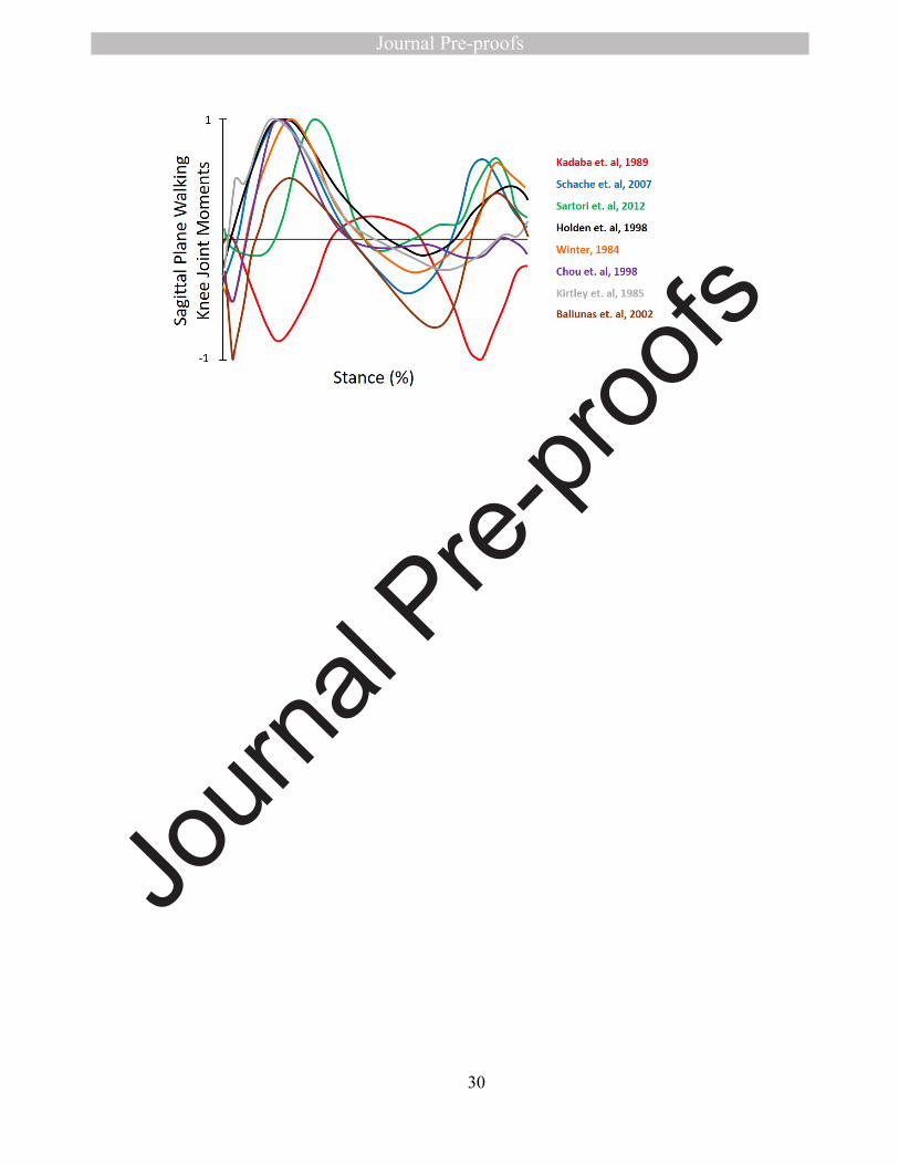
(internal or external), and normalization. As an example of the inherent variety of results
different methodological choices can make, sagittal plane knee joint moments during walking are
presented in Figure 1 from eight research studies on healthy adults. It is presumed that these
curves were all calculated correctly yet various methods and coordinate systems were utilized,
thus altering the shape of the curves. Failure to adequately describe the methods will result in
data that cannot be interpreted by the reader nor replicated by the research community.
The aims of the present paper are to discuss the major issues in the definition, calculation,
and interpretation of intersegmental forces and moments in human motion analysis, and to make
final recommendations on these matters with guidance from relevant papers in the literature. The

4
goal is to eliminate the most frequent sources of error and confusion in the field of human
motion analysis so that research can be correctly interpreted and replicated. We are not putting
forth these recommendations in an attempt to standardize the methods of estimating
intersegmental forces and moments, rather we hope that it is seen as an attempt to standardize the
reporting of such methods, after careful consideration of procedures and calculations have been
applied.
Anthropometric Model
The relationships between kinetic variables (force and moment) and kinematic variables
(linear and angular velocity and acceleration) are governed by the anthropometric properties of
mass and moment of inertia about the center of mass.
∑F=ma
where, ∑F is the sum of the external forces applied to a given human body segment
m is the segment mass,
a is the linear acceleration of the center of mass.
∑M = Icm𝝎 + 𝝎 × 𝐈𝐜𝐦𝝎
where, ∑M is the sum of the external moments acting on a given human body segment,
Icm is the inertia matrix with respect to the center of mass,
ω is the angular velocity vector,
is the angular acceleration vector.𝝎
The summations on the left hand side of these equations include terms due to gravity, external
forces, and intersegmental forces and moments. The intersegmental forces and moments are
generally solved recursively (Winter 2009).
5
Since intersegmental forces and moments are derived from these rigid body equations,
their computation requires the estimation of segment mass, the position of the center of mass
(CoM), and its inertia tensor (moments and products of inertia). All of these quantities must be
transformed into a common coordinate system prior to estimation of the intersegmental forces
and moments. Body segment inertial parameters (BSIPs) can be obtained using regression
equations based on subject's segment length and body mass (De Leva, 1996a), geometrical
models (Yeadon, 1990; Hanavan, 1964), optimization of parameters via COP errors in various
postures (Chen, et al. 2011), or, when available, directly from subject-specific medical imaging
(Ganley et al., 2004; Mungiole et al., 1999; Cheng et al., 2000). These estimations of BSIPs are
the basis for rigid body models. The assumption that the estimates are constant are a source of
uncertainty in model output.
For most regression equations, the position of the segment center of mass is given with
respect to the proximal and distal endpoints, which define the segment length (De Leva, 1996a)
or with respect to a number of anatomical landmarks (Zatsiorsky et al., 1990). Consequently, the
determination of the BSIPs also depend on estimation of the joint centers positions. Adjusted
scaling equations have been proposed for computing 3D inverse dynamics in which BSIPs are
expressed in standardized definitions of the relevant anatomical axes (Dumas et al., 2007, Wu et
al., 2002, Wu et al., 2005).
Due to specific anthropometric characteristics, BSIPs estimated in different populations
can lead to different values (Nguyen et al., 2014). Uncertainties in the identification of BSIPs can
play a critical role in reliable joint moment estimation, especially when analyzing motor
activities involving high accelerations, such as in running (Krabbe et al., 1997) or when the
population under examination has special anthropometric features, such as amputees (Sawers and
6
Hahn, 2010). To the contrary, when analyzing level walking at natural speeds, a minor influence
of uncertainties in BSIPs is expected, particularly at the more distal joints in the stance phase
(Rao et al., 2006; Camomilla et al., 2017).
Summary and Recommendations
The anthropometric model used to estimate body segment parameters must be detailed in
order for results to be replicated. This includes procedures for estimating moments of inertia,
mass, and center of mass locations. The sample for which regression equations were established
should be consistent with the subjects being studied. This becomes especially important as linear
and angular accelerations increase and for specific populations that may have substantially
different BSP’s (e.g. children, amputees...).
Joint centers
To compute the intersegmental joint moments, a reduction point, that is the point with
respect to which the system of forces is reduced, is required. This point is classically defined as a
joint center. In most of the human movement analysis protocols proposed in the literature,
adjacent bony segments are conceptually assumed to be connected by spherical pairs, and their
relative motion is described by three joint angles about the three anatomical axes defining the
joint coordinate system and passing through this joint center (Wu et al. 2002, 2005). Then, when
the joint allows only a small rotation about one axis (resisted degrees of freedom (DoF), e.g.
adduction-abduction at the knee or elbow joint), it can be assumed, in a first approximation, that
the relevant moment represents the action of the main anatomical joint restraints (i.e. articular
contacts and ligaments) (O'Connor et al., 1998). On the other hand, in case of large rotations
about one joint axis and neglecting friction (unresisted DoF, e.g., flexion-extension, adduction-

7
abduction and internal-external rotation at the hip or shoulder joints), the resultant of articular
contact forces passes through this joint center and therefore it can be assumed that the relevant
moment represent the actions of the muscle-tendon units (Challis et al., 1996). To interpret the
joint moments with this rationale, it is required that the origin of the joint anatomical axes
coincides with the reduction point (joint center) and that the joint moments are expressed then
about these joint axes.
Joint centers are commonly defined by using regression equations from palpated external
anatomical landmarks (Bell et al., 1990), using functional approaches (Leardini et al., 1999 ),
multi-body kinematic optimization techniques (Begon et al., 2018) or using medical imaging
techniques (Della Croce et al., 2005). The latter approach, although very accurate and able to
create individualized musculoskeletal models, requires access to expensive and cumbersome
measurement systems, time-consuming post-processing, highly-specific and multidisciplinary
expertise, and in some cases, involves exposure to ionizing radiation. This explains why medical
imaging techniques are frequently used as gold standards in biomechanics for the development
and validation of models and techniques, but their use is limited in the fields of clinical
movement analysis and sport applications.
It has been largely demonstrated that joint moment estimates are very sensitive to errors
in the joint center and that this inaccuracy affects the calculations to a larger extent than other
concurring factors such as errors on BSIPs (Camomilla et al., 2017). For instance, a hip joint
anterior and lateral mislocation of 30 mm, which can be expected in typical of routine gait
analyses (Hara et al., 2016), can cause a mean error of about 1.43 and 1.38% bodyweight x
height in the flexion–extension and abduction–adduction moment components of the
corresponding range, respectively (Stagni et al., 2000). In general, the strengths of the functional
8
method are that the calculated center of rotation is subject specific, side specific i.e. right
different from left, and not influenced by the presence of bony or joint deformity. Nor is it
influenced by differences in body segment proportions, as expected for gender, age, genetic
traits, etc. On the other hand, its implementation requires the subject to perform, either passively
or actively, a sufficiently wide joint angular excursion. Conversely, regression methods can be
applied when movement restrictions are present. However, their accuracy and repeatability
strongly depend on the original regression model, and are affected by uncertainties in anatomical
landmark identification (Sangeux, 2015). The hip joint center is certainly the most critical lower
extremity joint due to the large distance from the palpable pelvic landmark. It has been
determined that the functional approach has errors between 10 to 20 mm whereas regression
methods find errors between 15 to 30 mm (Leardini et al., 1999; Cereatti et al., 2009; Sangeux et
al., 2014; Harrington et al., 2007).
Summary and Recommendations
Since joint center positions are used to define the moment arm of the forces acting on the
segment under analysis, the manner in which they are identified will influence the estimation of
intersegmental moments. Furthermore, because joint centers are commonly used to define
segment length, they also affect body segment inertial parameters. It is therefore fundamental to
use valid methods, and to clearly state these methods, for joint centers determination.
Signal Processing
Correct application and complete reporting of signal processing methods are crucial when
dealing with kinematic and kinetic data. Of first concern is the sampling, which must be of an
adequate rate to insure the frequencies present in the motion are completely captured. At a

9
minimum, the sampling rate must be greater than twice the highest frequency in the signal. A
sampling frequency below this threshold will not only miss higher frequencies, the higher
frequencies will fold back into the data and result in a contaminated signal (Edwards et al.,
2017). This minimum sampling rate insures no information is lost but if peak values need to be
accurately digitized the signal must be sampled at a much higher rate (5 to 10 times the highest
frequency in the signal) or the digitized signal must be reconstructed using resampling
techniques (Hamill et al., 1997). In general, movements that contain collisions are composed of
higher frequencies and therefore must be digitized at a higher rate.
Kinematic data must be differentiated to calculate velocities and again to calculate
accelerations in preparation for use in the equations of inverse dynamics. This double
differentiation process amplifies the time series recording by the square of the frequency
(Antonsson and Mann, 1985). Thus, high frequency noise can dominate the acceleration signal if
proper processing procedures are not utilized. Smoothing techniques attempt to attenuate
frequencies that comprise the noise while leaving the true signal unaffected. Various methods
such as splines, time domain filters and frequency domain filters have been used to accomplish
this. Selection of the frequencies that are being attenuation may be done using a set value or
algorithms that objectively identify the cutoff frequencies (Jackson, 1979; Giakas and
Baltzopoulos, 1999).
There is a concern that the frequency content of kinematic and kinetic data should be in
agreement. Several researchers (Bisseling and Hoff, 2006; Kristianslund et al., 2012; Edwards et
al., 2011) have shown that a disparity in the frequency content can cause artifacts in the
intersegmental moments that cannot be explained by the dynamics of the activity. This
necessitates the same cutoff frequencies for kinetic and kinematic data filtering. However, this

10
poses a problem with impact forces that can be attenuated by low-pass cutoff frequencies that are
in the range necessary to reduce noise in the kinematic data. Decisions concerning the cutoff
frequency need to be made based on the purpose of the experiment and the variables being
measured.
Summary and Recommendations
Both kinematic and kinetic sampling frequencies must be clearly identified. The method
of smoothing should be identified and the degree of smoothing (typically in the form of the
frequency response) should be noted. They technique used to differentiate the data and any
specialized techniques such as optimized cutoffs, resampling of data and procedures to minimize
artifact should be detailed and cited.
Method of Calculation
There are two equivalent methods to describe the dynamics of a mechanical system,
namely the Newton-Euler and Lagrange formulations. In terms of interpretation, the differences
between the two are generally procedural rather than substantive. Note that in biomechanics, few
human joints involve translational DoF greater than a few millimeters, thus, most of the time, the
Lagrange equations of motion only result in moments. The Newton-Euler method is simpler and
gives access to the full three-dimensional intersegmental force and moment vectors, including
the moments for resisted degrees of freedom, such as knee adduction-abduction (Winter, 2009).
Lagrange methods are especially useful when joint models that are more complex than spherical
or hinge joints are needed, but do not solve for loads associated with resisted degrees of freedom
(van den Bogert et al., 2013). Equivalent moments result from using the Newton-Euler equations
of motion projected onto the DoF axes (projection with a dot product). These moments about the
11
joint DoF are directly related to the joint power (they just need to be multiplied by the DoF
angular velocity) and can be described as "motor" or internal joint moments. In musculoskeletal
modelling, these are typically the moments involved in the computation of the musculotendon
forces while the other components of the intersegmental moments are assumed to represent the
actions of ligaments and contact forces (Delp et al., 2007). Although theoretically equivalent,
these two methods may produce small deviations because of differences in soft tissue artifact
propagation.
Both the Newton-Euler and the Lagrange equations (Eberhard, 2006) lead to inverse
dynamics procedures, meaning the intersegmental forces and moments are derived from the
kinematics. In forward dynamics procedures, a muscle-driven or torque-driven model is used to
estimate intersegmental moments from a neural signal obtained via electromyography,
optimization procedures, or a combination (Buchanan, 2005).
When inertial components are absent or negligible, a static analysis was used to roughly
estimate intersegmental forces and moments (Fantozzi et al., 2012). This simplified method
consists of multiplying the ground reaction force vector by its moment arm at each joint and has
been described as the 'ground reaction technique'. This method assumes that segment
accelerations and/or the body segment inertial parameters are negligible, large errors can arise if
this assumption is not met (Wells, 1981).
Summary and Recommendations
In general, static analysis of the human body should be restricted to static or near-static
situations. Newton-Euler and Lagrange formulations of intersegmental moments are
mathematically equivalent but the method should be identified because their sensitivity to signal

12
processing methods can be different. Forward or inverse dynamics procedures also need to be
specified. If muscle-driven forward dynamics are used additional methods detailing the
estimation of muscle forces are necessary.
Coordinate System
Intersegmental forces and moments have been presented in a variety of coordinate
systems: global (also known as inertial or laboratory), proximal, distal and the joint coordinate
system (Schache and Baker, 2007). In general, presentation of intersegment forces and moments
in the global coordinate system should be avoided. Unlike segment coordinate systems, the
intersegmental forces and moments presented in the global coordinate system will be affected by
changes in the direction of motion.
The joint coordinate system (JCS) is appealing if kinematics are also presented in the
JCS, but caution should be used because the axes of the JCS are not orthogonal. Projection onto
non-orthogonal axes is problematic when the moment norm is to be computed or when the 3D
vector is to be retrieved. If a JCS is used, the projection using a dot product (as opposed to a non-
orthogonal projection) is preferred (Kristianslund et al., 2014). Those projected moments can be
multiplied by the rates of change in the JCS angles to obtain a mechanically consistent joint
power analysis. Also these orthogonally projected moments obtained from the Newton-Euler
method will be identical to moments obtained from the Lagrange method where the JCS
mechanism is explicitly modeled.
Proximal or distal segment coordinate systems (Figure 2) are useful in answering
particular research questions. For instance, during the estimation of tibial tissue stresses, the
intersegmental forces and moments would be expressed in a proximal segment coordinate system

13
at the ankle or a distal segment coordinate system at the knee so they are in a coordinate system
that is suitable for further analysis, e.g. with a finite element model (Derrick et al., 2016).
Segment coordinate systems (and the joint coordinate systems derived from them) should be
defined using ISB standards (Wu et al., 2002, 2005). If non-standard coordinate systems are
used, they should be fully specified in terms of anatomical landmarks or other suitable
definitions.
The choice of a coordinate system used to report intersegmental forces and moments can
dramatically affect the interpretation of data. Note that during walking (Figure 3) and running
(Figure 4) the sagittal plane moments and the vertical forces are similar between proximal and
distal coordinate systems but there are some relatively large differences in the other planes and
axes.
The choice of intersegmental coordinate systems should be consistent with the kinematics
and the anthropometric model. Consistency will take two forms: mathematical and
informational. Mathematical consistency is necessary to prevent inaccuracies that result from
calculations with variables in more than one coordinate system. For instance, if moments of
inertia from the anthropometric model were calculated in a segment coordinate system they
should not be multiple by angular accelerations in a global or joint coordinate system. Likewise,
subsequent calculations using intersegment forces and moments such as joint powers and
apparent joint stiffness must have a consistent coordinate system to be accurate. Care should also
be taken when estimating muscle moment arms using a musculoskeletal model. The kinematics
applied to the model should be in the same coordinate system as the intersegmental moments if
they are to be used in a common calculation. Informational consistency suggests that all
quantities presented in a paper should be in the same coordinate system unless there is a

14
justifiable reason. This will remove ambiguity and instil confidence in the reader or reviewer that
proper procedures have been followed.
Summary and Recommendations
The choice of the coordinate system (global coordinate system, proximal segment
coordinate system, distal segment coordinate system, or joint coordinate system) highly
influences the intersegmental forces and moments. It is therefore essential that the coordinate
system used to interpret the intersegmental forces and moments be carefully considered and
reported. Much thought and debate has gone into standardizing kinematic coordinate systems
(Wu et al., 2002/2005) and the motivation for using JCS’s for intersegmental forces and
moments holds. Unless a rationale exists these previously defined kinematic coordinate systems
should also be used to present intersegmental forces and moments. While describing coordinate
systems, the directional signs of the forces should be defined (e.g., superior, anterior and lateral
are positive) as well as for the moments (e.g., flexion, adduction, and internal rotation are
positive) if these parameters are utilized. If moment signe areThis is preferred over defining the
x, y and z axes of the coordinate system because it gives additional information to the reader.
Internal or external perspective
Intersegmental forces and moments can be viewed from two perspectives. From an
external point of view the intersegmental forces and moments represent the result of forces
acting on the body as well as centrifugal and Coriolis actions arising from motion of the body
segments. From an internal point of view these variables represent forces and moments that
originate from within the body and act to resist external load and to maintain posture or
accelerate the segments. Anatomical structures crossing the joint such as skin, fat, fascia,
15
muscles, ligaments, together with friction and contact between the articular surfaces produce the
mechanical action. Both perspectives are equally valid and in fact the numerical value of the
result is equal and opposite because the joint system is in balance. From the internal point of
view an extension knee moment is present during mid-stance to prevent knee collapse but from
an external point of view there exists a flexion knee moment caused by the external ground
reaction force (passing posteriorly to the joint center in the static analysis perspective).
The decision to present internal or external intersegmental forces and moments is often
made based on the researcher’s view of the source of the moments. Internal moments are
considered to be primarily caused by muscles when the joint is not near the end range of motion.
This presents a problem at the knee joint because the result of all the muscles spanning the knee
may produce a relatively small adduction-abduction moment, the primary source is considered to
be ligaments and articular surfaces. This has led many researchers to present adduction-
abduction moments at the knee using an external perspective when this is the primary variable of
analysis (Telfer et al., 2017). This leads to additional confusion when the moments are referred to
by their action. From an external perspective, an adduction moment at the knee is one in which
the external forces are tending to cause the knee to adduct (ground reaction force passing
medially to the joint center in the static analysis perspective), potentially tearing the lateral
ligaments. However, from an internal perspective, an adduction moment at the knee is one in
which the balance of muscles, ligaments and articular contact tend to adduct the knee. These
differing perspectives can, and often do, lead to confusion for a reader trying to interpret knee
function.
Summary and Recommendations

16
Whether intersegmental forces and moments are presented as internal or external can be
determined by the research question being asked but may also be dependent on the perspective
that the researcher is trying to convey. A clear statement of this perspective is essential to
communicating concepts in the paper.
Normalization
In clinical movement analysis, demographic/anthropometric characteristics (i.e. age,
height, body mass, gender) and the velocity ranges, with which the motor task is executed,
influence the amplitude of the kinematic and kinetic variables and if not properly treated may act
as confounding factors (Moisio et al, 2003; Senden et al., 2012; Andriacchi et al., 1977; Lelas et
al. 2003). If these variables are not equivalent between the groups that are being compared they
may need to be controlled statistically (covariate analysis) or their effect removed from the
analysis (normalization). When a repeated measures study design is employed such that the
normalization parameters do not change over time, the normalization process will not alter the
statistical results. However normalization may still be warranted so that results can be
conveniently compared to other studies.
Normalization procedures reduce the variance between individuals when comparing the
intersegmental forces and moments among individuals. Most commonly, forces are divided by
body weight or body mass, while moments are divided by these variables or quantities that result
in non-dimensional values such as body weight multiplied by height or body weight multiplied
by limb or foot length. For instance, before normalization, lower extremity peak moments during
gait were statistically different between males and females in ten cases, but normalizing by body
mass reduced this number to 6 and normalizing by body weight times height further reduced the
number to 2 (Moisio et al., 2003). Pinzone et al. (2016) and Hof (1996) have also shown
17
advantages to using non-dimensional normalization. Additional reductions in residuals may be
attained by considering more joint specific distance measurements or non-linear adjustments
(Wannop et al., 2012, Lee et al., 2015).
Summary and Recommendations
Normalization of data is often necessary if groups are dissimilar on specific variables
such as mass or height. This is especially useful in the context of gait laboratories in which
individual data are frequently compared to a database. Although normalization will not change
the statistical evaluation of a repeated measures study it may still be useful when comparing
values to other studies. Normalization procedures need to be clearly outlined in the methods of
the paper and normalization values such as average mass and height (or leg length) should be
reported. Ranges of these values can also be useful so that researchers can avoid extrapolation of
results. There is no simple method to normalize by the walking or running velocity therefore it is
necessary to report average velocity or other variables that may influence the intersegmental
forces and moments. This will assist researchers in explaining differences between studies.
Conclusions
Many options for the estimation and presentation of intersegmental forces and moments
have been presented. These variations should not be considered correct or incorrect because each
may be superior to the others in the context of the research paradigm and the questions being
asked. However, as members of this field of research, we must insure that no ambiguity exists in
the presentation of results. This is critical to an efficient evolution of a body of knowledge. We
must be able to relate the curves and values presented by past researchers to the functional
movement of the human body so that we can evaluate the results of the study, verify our own

18
research data, and ultimately create new theories and form new hypotheses. Realizing that there
are differences in the detail of methodological information required in differing dissemination
formats and in biomechanical vs clinical journals we make two suggestions: 1) even conference
abstracts should include the coordinate system used and the internal/external perspective –
interpretation of the results requires this minimum amount of information and 2) take advantage
of the liberal policies journals have generally adopted that allow addition information to be
posted online. In partial fulfilment of the goal of improved clarity in the scientific arena, we
have identified three tangible items, in addition to this article, that we hope will help to fulfill
this goal:
1. Reviewer/author checklist for presentation of intersegmental forces and moments.
(Appendix A).
2. Online visualization tool for comparison of typical walking and running
intersegmental forces and moments with adjustable coordinate systems,
normalization methods and internal/external perspectives (ISB Website).
3. Software transparency. A request to the major biomechanics software companies
to make easily available the items in the checklist so that users can access this
information in a single location in the software.
Acknowledgements
The authors would like to thank Dr. Glen Lichtwark for his assistance in the organization
of this committee and the current President of the International Society of Biomechanics, Dr.
Joseph Hamill, for his leadership in forming the committee.

19

20
References (APA Style)
1. Andriacchi, T. P., Ogle, J. A., & Galante, J. O. (1977). Walking speed as a basis for
normal and abnormal gait measurements. Journal of biomechanics, 10(4), 261-268.
2. Antonsson, E. K., & Mann, R. W. (1985). The frequency content of gait. Journal of
biomechanics, 18(1), 39-47.
3. Baliunas, A. J., Hurwitz, D. E., Ryals, A. B., Karrar, A., Case, J. P., Block, J. A., &
Andriacchi, T. P. (2002). Increased knee joint loads during walking are present in
subjects with knee osteoarthritis. Osteoarthritis and cartilage, 10(7), 573-579.
4. Begon, M., Andersen, M. S., & Dumas, R. (2018). Multibody kinematics optimization
for the estimation of upper and lower limb human joint kinematics: a systematized
methodological review. Journal of biomechanical engineering, 140(3), 030801.
5. Bell, A. L., Brand, R. A., & Pedersen, D. R. (1987). Prediction of hip joint center
location from external landmarks. Journal of biomechanics, 20(9), 913.
6. Bell, A. L., Pedersen, D. R., & Brand, R. A. (1990). A comparison of the accuracy of
several hip center location prediction methods. Journal of biomechanics, 23(6), 617-
621.
7. Bishop, C., Paul, G., & Thewlis, D. (2012). Recommendations for the reporting of foot
and ankle models. Journal of biomechanics, 45(13), 2185-2194.
8. Bisseling, R. W., & Hof, A. L. (2006). Handling of impact forces in inverse
dynamics. Journal of biomechanics, 39(13), 2438-2444.
9. Buchanan, T. S., Lloyd, D. G., Manal, K., & Besier, T. F. (2005). Estimation of muscle
forces and joint moments using a forward-inverse dynamics model. Medicine &
Science in Sports & Exercise, 37(11), 1911-1916.
10. Burkhart, T. A., Andrews, D. M., & Dunning, C. E. (2013). Finite element modeling
mesh quality, energy balance and validation methods: A review with recommendations
associated with the modeling of bone tissue. Journal of biomechanics, 46(9), 1477-
1488.
11. Camomilla, V., Cereatti, A., Cutti, A. G., Fantozzi, S., Stagni, R., & Vannozzi, G.
(2017). Methodological factors affecting joint moments estimation in clinical gait
analysis: a systematic review. Biomedical engineering online, 16(1), 106.
21
12. Cappozzo, A. (1984). Gait analysis methodology. Human Movement Science, 3(1-2),
27-50.
13. Cereatti, A., Bonci, T., Akbarshahi, M., Aminian, K., Barré, A., Begon, M., ... & Lin,
C. C. (2017). Standardization proposal of soft tissue artefact description for data
sharing in human motion measurements. Journal of Biomechanics, 62, 5-13.
14. Cereatti, A., Donati, M., Camomilla, V., Margheritini, F., & Cappozzo, A. (2009). Hip
joint centre location: an ex vivo study. Journal of biomechanics, 42(7), 818-823.
15. Challis, J. H., & Kerwin, D. G. (1996). Quantification of the uncertainties in resultant
joint moments computed in a dynamic activity. Journal of Sports Sciences, 14(3), 219-
231.
16. Cheng, C. K., Chen, H. H., Chen, C. S., Lee, C. L., & Chen, C. Y. (2000). Segment
inertial properties of Chinese adults determined from magnetic resonance
imaging. Clinical biomechanics, 15(8), 559-566.
17. Chen, S. C., Hsieh, H. J., Lu, T. W., & Tseng, C. H. (2011). A method for estimating
subject-specific body segment inertial parameters in human movement analysis. Gait &
posture, 33(4), 695-700.
18. Chou, L. S., & Draganich, L. F. (1998). Increasing obstacle height and decreasing toe-
obstacle distance affect the joint moments of the stance limb differently when stepping
over an obstacle. Gait & posture, 8(3), 186-204.
19. Davis III, R. B., Ounpuu, S., Tyburski, D., & Gage, J. R. (1991). A gait analysis data
collection and reduction technique. Human movement science, 10(5), 575-587.
20. De Leva, P. (1996a). Adjustments to Zatsiorsky-Seluyanov's segment inertia
parameters. Journal of biomechanics, 29(9), 1223-1230.
21. De Leva, P. (1996b). Joint center longitudinal positions computed from a selected
subset of Chandler's data. Journal of biomechanics, 29(9), 1231-1233.
22. Della Croce, U., Cappozzo, A., Kerrigan, D.C. (1999) Pelvis and lower limb anatomical
landmark calibration precision and its propagation to bone geometry and joint angles.
Medical and Biological Engineering and Computing 37(2), 155-161
23. Della Croce, U., Cappozzo, A., & Kerrigan, D. C. (1999). Pelvis and lower limb
anatomical landmark calibration precision and its propagation to bone geometry and
joint angles. Medical & biological engineering & computing, 37(2), 155-161.
22
24. Delp, S. L., Anderson, F. C., Arnold, A. S., Loan, P., Habib, A., John, C. T., ... &
Thelen, D. G. (2007). OpenSim: open-source software to create and analyze dynamic
simulations of movement. IEEE transactions on biomedical engineering, 54(11), 1940-
1950.
25. Dempster, W. T. (1955). Space requirements of the seated operator, geometrical,
kinematic, and mechanical aspects of the body with special reference to the limbs.
Michigan State Univ East Lansing.
26. Derrick, T.R., Edwards, W.B., Fellin, R.E., & Seay, J.F. (2016) An integrative
modeling approach for the efficient estimation of cross sectional tibial stresses during
locomotion. Journal of biomechanics 49(3), 429-435.
27. Dillon, M. P., Barker, T. M., & Pettet, G. (2008). Effect of inaccuracies in
anthropometric data and linked-segment inverse dynamic modeling on kinetics of gait
in persons with partial foot amputation. Journal of Rehabilitation Research &
Development, 45(9).
28. Dumas, R., Brånemark, R., & Frossard, L. (2017). Gait analysis of transfemoral
amputees: errors in inverse dynamics are substantial and depend on prosthetic
design. IEEE transactions on neural systems and rehabilitation engineering, 25(6),
679-685.
29. Dumas, R., Cheze, L., & Verriest, J. P. (2007). Adjustments to McConville et al. and
Young et al. body segment inertial parameters. Journal of biomechanics, 40(3), 543-
553.
30. Edwards, W. B., Derrick, T. R., & Hamill, J. (2017). Time Series Analysis in
Biomechanics. In Handbook of Human Motion (pp. 1-24). Springer International
Publishing.
31. Edwards, W. B., Troy, K. L., & Derrick, T. R. (2011). On the filtering of
intersegmental loads during running. Gait & posture, 34(3), 435-438.
32. Eberhard, P., & Schiehlen, W. (2006). Computational dynamics of multibody systems:
history, formalisms, and applications. Journal of computational and nonlinear
dynamics, 1(1), 3-12.
33. Fantozzi, S., Garofalo, P., Cutti, A. G., & Stagni, R. (2012). 3D joint moments in
transfemoral and transtibial amputees: When is the “Ground Reaction Vector
23
Technique” an alternative to inverse dynamics?. Journal of Mechanics in Medicine and
Biology, 12(04), 1250061.
34. Ganley, K. J., & Powers, C. M. (2004). Anthropometric parameters in children: a
comparison of values obtained from dual energy x-ray absorptiometry and cadaver-
based estimates. Gait & posture, 19(2), 133-140.
35. Giakas, G., & Baltzopoulos, V. (1997). A comparison of automatic filtering techniques
applied to biomechanical walking data. Journal of Biomechanics, 30(8), 847-850.
36. Hamill, J., Caldwell, G. E., & Derrick, T. R. (1997). Reconstructing digital signals
using Shannon's sampling theorem. Journal of Applied Biomechanics, 13(2), 226-238.
37. Hanavan Jr, E. P. (1964). A mathematical model of the human body (No. AFIT-GA-
PHYS-64-3). Air Force Aerospace Medical Research Lab Wright-Patterson AFB OH.
38. Hara, R., McGinley, J., Briggs, C., Baker, R., & Sangeux, M. (2016). Predicting the
location of the hip joint centres, impact of age group and sex. Scientific reports, 6,
37707.
39. Harrington, M. E., Zavatsky, A. B., Lawson, S. E. M., Yuan, Z., & Theologis, T. N.
(2007). Prediction of the hip joint centre in adults, children, and patients with cerebral
palsy based on magnetic resonance imaging. Journal of biomechanics, 40(3), 595-602.
40. Hof, A. L. (1996). Scaling gait data to body size. Gait & posture, 3(4), 222-223.
41. Jackson, K. M. (1979). Fitting of mathematical functions to biomechanical data. IEEE
transactions on biomedical engineering, (2), 122-124.
42. Holden, J. P., & Stanhope, S. J. (1998). The effect of variation in knee center location
estimates on net knee joint moments. Gait & posture, 7(1), 1-6.
43. Kadaba, M. P., Ramakrishnan, H. K., Wootten, M. E., Gainey, J., Gorton, G., &
Cochran, G. V. B. (1989). Repeatability of kinematic, kinetic, and electromyographic
data in normal adult gait. Journal of Orthopaedic Research, 7(6), 849-860.
44. Kirtley, C., Whittle, M. W., & Jefferson, R. J. (1985). Influence of walking speed on
gait parameters. Journal of biomedical engineering, 7(4), 282-288.
45. Krabbe, B., Farkas, R., & Baumann, W. (1997). Influence of inertia on intersegment
moments of the lower extremity joints. Journal of biomechanics, 30(5), 517-519.
24
46. Kristianslund, E., Krosshaug, T., & van den Bogert, A. J. (2012). Effect of low pass
filtering on joint moments from inverse dynamics: implications for injury
prevention. Journal of biomechanics, 45(4), 666-671.
47. Kristianslund E., Krosshaug T., Mok K.M., McLean S., & van den Bogert A.J. (2014).
Expressing the joint moments of drop jumps and sidestep cutting in different reference
frames – does it matter? Journal of biomechanics, 47(1), 193-199.
48. Leardini, A., Cappozzo, A., Catani, F., Toksvig-Larsen, S., Petitto, A., Sforza, V.,
Cassanelli, G., Giannini, S., 1999. Validation of a functional method for the estimation
of hip joint centre location. Journal of Biomechanics 32, 99–103.
49. Lee, D., Oh, S. E., Lee, I. K., Sim, T., Joo, S. B., Park, H. J., & Mun, J. H. (2015).
Comparison of three normalization methods for 3D joint moment in the asymmetric
rotational human movements in golf swing analysis. Journal of Biosystems
Engineering, 40(3), 289-295.
50. Lelas, J. L., Merriman, G. J., Riley, P. O., & Kerrigan, D. C. (2003). Predicting peak
kinematic and kinetic parameters from gait speed. Gait & posture, 17(2), 106-112.
51. Moisio, K. C., Sumner, D. R., Shott, S., & Hurwitz, D. E. (2003). Normalization of
joint moments during gait: a comparison of two techniques. Journal of biomechanics,
36(4), 599-603.
52. Mungiole, M., & Martin, P. E. (1990). Estimating segment inertial properties:
comparison of magnetic resonance imaging with existing methods. Journal of
biomechanics, 23(10), 1039-1046.
53. Nguyen, T. C., & Reynolds, K. J. (2014). The effect of variability in body segment
parameters on joint moment using Monte Carlo simulations. Gait & posture, 39(1),
346-353.
54. O’Connor, J.J., Lu, T.W., Wilson, D.R., Feikes, J., & Leardini, A. (1998). Review:
diarthrodial joints-kinematic pairs, mechanisms or flexible structures? Computer
Methods in Biomechanics and Biomedical Engineering, 1, 123–150.
55. Pinzone, O., Schwartz, M. H., & Baker, R. (2016). Comprehensive non-dimensional
normalization of gait data. Gait & posture, 44, 68-73.
25
56. PubMed search: “(biomechanics(tw) OR motion(tw) OR kinematics(tw)) AND
recommendations(ti)”
57. Rao, G., Amarantini, D., Berton, E., & Favier, D. (2006). Influence of body segments’
parameters estimation models on inverse dynamics solutions during gait. Journal of
biomechanics, 39(8), 1531-1536.
58. Sangeux, M. (2015). On the implementation of predictive methods to locate the hip
joint centres. Gait & posture, 42(3), 402-405.
59. Sangeux, M., Pillet, H., & Skalli, W. (2014). Which method of hip joint centre
localisation should be used in gait analysis? Gait & posture, 40(1), 20-25.
60. Sartori, M., Reggiani, M., Farina, D., & Lloyd, D. G. (2012). EMG-driven forward-
dynamic estimation of muscle force and joint moment about multiple degrees of
freedom in the human lower extremity. PloS one, 7(12), e52618.
61. Sawers, A. B., & Hahn, M. E. (2010). The potential for error with use of inverse
dynamic calculations in gait analysis of individuals with lower limb loss: a review of
model selection and assumptions. JPO: Journal of Prosthetics and Orthotics, 22(1), 56-
61.
62. Schache, A. G., & Baker, R. (2007). On the expression of joint moments during
gait. Gait & posture, 25(3), 440-452.
63. Scott, S. H., & Winter, D. A. (1990). Internal forces of chronic running injury
sites. Medicine and science in sports and exercise, 22(3), 357-369.
64. Senden, R., Meijer, K., Heyligers, I. C., Savelberg, H. H. C. M., & Grimm, B. (2012).
Importance of correcting for individual differences in the clinical diagnosis of gait
disorders. Physiotherapy, 98(4), 320-324.
65. Stagni, R., Leardini, A., Cappozzo, A., Benedetti, M. G., & Cappello, A. (2000).
Effects of hip joint centre mislocation on gait analysis results. Journal of
biomechanics, 33(11), 1479-1487.
66. Telfer, S., Lange, M. J., & Sudduth, A. S. (2017). Factors influencing knee adduction
moment measurement: a systematic review and meta-regression analysis. Gait &
posture, 58, 333-339.

26
67. van den Bogert A.J., Geijtenbeek T., Even-Zohar O., Steenbrink F., & Hardin E.C.
(2013). A real-time system for biomechanical analysis of human movement and muscle
function. Medical and Biological Engineering and Computing 51, 1069-1077.
68. Wahid, F., Begg, R., McClelland, J. A., Webster, K. E., Halgamuge, S., & Ackland, D.
C. (2016). A multiple regression normalization approach to evaluation of gait in total
knee arthroplasty patients. Clinical Biomechanics, 32, 92-101.
69. Wannop, J. W., Worobets, J. T., & Stefanyshyn, D. J. (2012). Normalization of ground
reaction forces, joint moments, and free moments in human locomotion. Journal of
applied biomechanics, 28(6), 665-676.
70. Wells, R. P. (1981). The projection of the ground reaction force as a predictor of
internal joint moments. Bulletin of prosthetics research, 10, 15-19.
71. Winter, D. A. (1984). Kinematic and kinetic patterns in human gait: variability and
compensating effects. Human movement science, 3(1-2), 51-76.
72. Winter, D.A. (2009) Biomechanics and Motor Control of Human Movement, 4th
Edition, Wiley & Sons.
73. Wu, G., Siegler, S., Allard, P., Kirtley, C., Leardini, A., Rosenbaum, D., ... & Schmid,
O. (2002). ISB recommendation on definitions of joint coordinate system of various
joints for the reporting of human joint motion—part I: ankle, hip, and spine. Journal of
biomechanics, 35(4), 543-548.
74. Wu, G., Van der Helm, F. C., Veeger, H. D., Makhsous, M., Van Roy, P., Anglin, C., ...
& Werner, F. W. (2005). ISB recommendation on definitions of joint coordinate
systems of various joints for the reporting of human joint motion—Part II: shoulder,
elbow, wrist and hand. Journal of biomechanics, 38(5), 981-992.

27
Conflict of Interest Statement
All authors were fully involved in the study and preparation of the manuscript.
The material within has not been and will not be submitted for publication
elsewhere. We declare that we have no financial or personal relationships with
other people or organizations that could inappropriately influence our work.

28
Appendix A. Example checklist for the reporting of intersegmental forces and moments.

29
Figure 1. A selection of sagittal plane walking knee joint moments. Each curve is normalized to its own maximum absolute value. Various coordinate systems and methods of calculation result in an assortment of curve shapes. Average citations for these research papers is 478 (Google Scholar, May, 2019).
Figure 2. Segment coordinate systems for the pelvis, thigh, leg and foot segments. For the right leg these coordinate systems are defined by positive values pointing anterior, proximal, and lateral. Positive moments are clockwise about the axis while looking in the positive direction (right hand rule).
Figure 3. Ensemble averages of intersegmental forces (top) and moments (bottom) of eight subjects and five trials of walking (1.3 m/s). Both proximal (red) and distal (blue) coordinate systems are represented. Filled area represents ±1 standard deviation of the proximal coordinate system. Kinematic and kinetic data were low-pass filtered at 6 Hz. Segment masses estimated using Dempster (1955). Segment moments of inertia and center of mass locations estimated using Hanavan (1964). Hip joint center estimated using Bell, Brand and Pederson (1989). Forces and moments were estimated using inverse dynamics with the Newton-Euler equations.
Figure 4. Ensemble averages of intersegmental forces (top) and moments (bottom) of eight subjects and five trials of running (3.5 m/s). Both proximal (red) and distal (blue) coordinate systems are represented. Filled area represents ±1 standard deviation of the proximal coordinate system. Running kinematic and kinetic data were low-pass filtered at 10 Hz. Segment masses estimated using Dempster (1955). Segment moments of inertia and center of mass locations estimated using Hanavan (1964). Hip joint center estimated using Bell, Brand and Pederson (1989). Forces and moments were estimated using inverse dynamics with the Newton-Euler equations.
30

31

32

33