Intestinal Alkalization As a Possible Preventive Mechanism in Irinotecan (CPT11)-induced Diarrhea1
10
[CANCER RESEARCH 62, 179 –187, January 1, 2002] Intestinal Alkalization As a Possible Preventive Mechanism in Irinotecan (CPT-11)-induced Diarrhea 1 Tadashi Ikegami, Linan Ha, Kazuhiko Arimori, Patricia Latham, Kunihiko Kobayashi, Susan Ceryak, Yasushi Matsuzaki, and Bernard Bouscarel 2 Departments of Medicine [T. I., P. L., B. B.], Pharmacology [L. H., S. C.], Pathology [P. L.], and Biochemistry and Molecular Biology [B. B.], The George Washington University Medical Center, N. W. Washington, D.C. 20037; Department of Pharmacy, Miyazaki Medical College, Miyazaki, Japan 889-1692 [K. A.]; Saitama Cancer Center, Saitama, Japan 336-0011 [K. K.]; and Institute of Clinical Medicine, University of Tsukuba, Tsukuba-City, Japan 305-0024 [Y. M.] ABSTRACT The therapeutic efficacy of irinotecan (CPT-11), a DNA topoisomerase inhibitor, is often limited by the induction of severe late-onset diarrhea. This prodrug and its active metabolite, 7-ethyl-10-hydroxy-camptothecin (SN-38), have a labile -hydroxy-lactone ring that undergoes pH-dependent reversible hydrolysis. At physiological pH and higher, equilibrium favors the less toxic carboxylate form, whereas at acidic pH, the more potent lactone form is favored. We have reported previously that the initial uptake rate of CPT-11 and SN-38 by intestinal cells was significantly different between the respective lactone and carboxylate form. Results from the present study in HT-29 cells further demonstrate the correlation between the CPT-11/SN-38 initial uptake rate and the induced toxicity, cell cycle alteration, apoptosis, and colony- forming efficiency. The exposure of HT-29 cells to SN-38 for a limited period of time (<2 h) was sufficient to induce these events. Because the decreased initial uptake of SN-38 carboxylate resulted in a reduced cellular toxicity, we postulated that the CPT-11-induced diarrhea was preventable by influencing the equilibrium toward the carboxylate form and, thus, reducing its intestinal uptake. In the golden Syrian hamster model, p.o. sodium bicarbonate sup- plementation (5 mg/ml in drinking water) led to alkalization of the intestinal contents. In addition, this alkalization resulted in the reduction of the his- topathological damage to the mucosa of the small and large intestine, as well as a 20% reduction of the intestinal SN-38 lactone concentration of animals receiving CPT-11 (20 –50 mg/kg 7 days). Taken together, these results from in vitro and in vivo studies support intestinal alkalization by sodium bicar- bonate supplementation as a preventive mechanism against CPT-11-induced diarrhea. In addition, this provides a strong rationale for the usage of this measure as an adjunct to CPT-11 treatment. INTRODUCTION CPT-11 3 is a water-soluble derivative of camptothecin, an anti- tumor alkaloid isolated from Camptotheca acuminata, and presents a wide spectrum of antitumor activity through the inhibition of DNA topoisomerase I (1). This chemotherapeutic agent is broadening its clinical impact because it has shown clinical responses for many malignancies, such as advanced colon cancers, for which CPT-11 therapy has been approved worldwide, but also lung, gastric, pancre- atic, cervical, ovarian, leukemia, and lymphoma (2–7); and survival benefits for these patients (8). Leukopenia and diarrhea are the two major side effects of patients receiving CPT-11. Leucopoenia duration can be minimized and, therefore, controlled by administration of granulocyte colony stimulating factor (9). However, delayed diarrhea occurs in the majority of the treated patients. Several studies have reported this side effect to be grade 3 (severe) or 4 (life threatening), according to the National Cancer Institute Common Toxicity Criteria, in 40% of the patients (10), underlining diarrhea as the major limitation in the therapeutic use of CPT-11 (11, 12). CPT-11/SN-38-induced diarrhea may occur in one or both of two different temporal settings. The first setting, early acute, is observed immediately after CPT-11 infusion. This phenomenon could be re- lated to the early metabolism of the drug and is short lasting and prevented or rapidly suppressed with atropine administration (13). The second setting, late onset, occurs usually after an average period of 6 days, the mechanism of which is unknown. Recent reports have underlined the significant role of diarrhea and dehydration in the early death of patients treated with CPT-11 (11). Drugs like loperamide, acetorphan, and budesonide have been used to slow intestinal motility and decrease water and electrolyte move- ment through the bowel (14 –16). High dose of loperamide is consid- ered as standard treatment in Europe and the United States but presents a limited level of success (17). Additional approaches are being explored to overcome the intestinal toxicity of CPT-11, includ- ing the use of antibiotics or bacterial lipopeptide (JBT3002) to stim- ulate mucosal immunity and maintain mucosal architecture (18), as well as fish oil and glutamine supplementation to decrease the intes- tinal side effect (19, 20). Furthermore, although the mechanism of action is less clear, Kampo, the Chinese herbal medicine, has been used successfully in reducing diarrhea induced by irinotecan (14, 21). CPT-11 is hydrolyzed by hepatic carboxylesterase to SN-38 (22). SN-38 has at least a 1000-fold more potent antitumor effect than CPT-11, as shown in vitro (1). In the liver, a portion of SN-38 undergoes subsequent conjugation by UDP-glucuronyltransferase to SN-38G (23). CPT-11, SN-38, and SN-38G are excreted into bile (23). Although there are no known quantitative reports in human, studies in the rat model suggest that the proportion of CPT-11, SN-38, and SN-38G secreted in bile is 83, 4.5, and 12.5%, respectively (24). Once in the intestine, SN-38G can be deconjugated in the cecum and colon to SN-38 by bacterial -glucuronidase (25). CPT-11, SN-38, and SN-38G are believed to be reabsorbed into the enterohepatic circulation to a certain extent by intestinal cells (25). Among the different CPT-11 metabolites, SN-38 has been consid- ered not only as the most potent anticancer agent but also to be the cause of the treatment-related diarrhea (26). It has been suggested that enterocolitis caused by high levels of SN-38 and/or CPT-11 retained for long periods of time in the intestine was the direct cause of diarrhea associated with CPT-11 administration in athymic mouse (27). Furthermore, in patients with diminished or no UDP-glucuro- nyltransferase activity, such as in those with either Crigler-Najjar syndrome I or Gilbert’s syndrome, the incidence of CPT-11-induced diarrhea is greater (26, 28), suggesting that decreased hepatic glucu- ronide conjugation results in an increased secretion of unconjugated SN-38 with, as a consequence, an increased concentration of luminal SN-38. This in turn could induce tissue injury and, therefore, diarrhea. CPT-11, SN-38, and SN-38G have a labile -hydroxy-3-lactone Received 3/9/01; accepted 11/1/01. The costs of publication of this article were defrayed in part by the payment of page charges. This article must therefore be hereby marked advertisement in accordance with 18 U.S.C. Section 1734 solely to indicate this fact. 1 Presented at the annual meeting of the American Gastroenterological Association in May 2000. Supported in part by Daiichi Pharmaceutical Co., Ltd.; Yakult Honsha Co., Ltd.; and Mitsubishi Tokyo Pharmaceutical Co., Ltd. 2 To whom requests for reprints should be addressed, at 2300 Eye Street, N. W. Ross Hall, Room 523, Washington, D. C. 20037; Phone: (202) 994-2114; Fax: (202) 994-3435; E-mail: [email protected]. 3 The abbreviations used are: CPT-11, irinotecan hydrochloride; SN-38, 7-ethyl- 10-hydroxy-camptothecin; SN-38G, 7-ethyl-10-hydroxy-camptothecin glucuronide; PI, propidium iodide; GFP, green fluorescent protein; MTT, 3-(4,5-dimethylthiazol-2-yl)-2,5- diphenyltetrazolium bromide; PS, phosphatidylserine; BW, body weight; HPLC, high- performance liquid chromatography. 179 Research. on August 17, 2015. © 2002 American Association for Cancer cancerres.aacrjournals.org Downloaded from
Transcript of Intestinal Alkalization As a Possible Preventive Mechanism in Irinotecan (CPT11)-induced Diarrhea1
[CANCER RESEARCH 62, 179–187, January 1, 2002]
Intestinal Alkalization As a Possible Preventive Mechanism in Irinotecan(CPT-11)-induced Diarrhea1
Tadashi Ikegami, Linan Ha, Kazuhiko Arimori, Patricia Latham, Kunihiko Kobayashi, Susan Ceryak,Yasushi Matsuzaki, and Bernard Bouscarel2
Departments of Medicine [T. I., P. L., B. B.], Pharmacology [L. H., S. C.], Pathology [P. L.], and Biochemistry and Molecular Biology [B. B.], The George Washington UniversityMedical Center, N. W. Washington, D.C. 20037; Department of Pharmacy, Miyazaki Medical College, Miyazaki, Japan 889-1692 [K. A.]; Saitama Cancer Center, Saitama, Japan336-0011 [K. K.]; and Institute of Clinical Medicine, University of Tsukuba, Tsukuba-City, Japan 305-0024 [Y. M.]
ABSTRACT
The therapeutic efficacy of irinotecan (CPT-11), a DNA topoisomeraseinhibitor, is often limited by the induction of severe late-onset diarrhea. Thisprodrug and its active metabolite, 7-ethyl-10-hydroxy-camptothecin (SN-38),have a labile �-hydroxy-lactone ring that undergoes pH-dependent reversiblehydrolysis. At physiological pH and higher, equilibrium favors the less toxiccarboxylate form, whereas at acidic pH, the more potent lactone form isfavored. We have reported previously that the initial uptake rate of CPT-11and SN-38 by intestinal cells was significantly different between the respectivelactone and carboxylate form. Results from the present study in HT-29 cellsfurther demonstrate the correlation between the CPT-11/SN-38 initial uptakerate and the induced toxicity, cell cycle alteration, apoptosis, and colony-forming efficiency. The exposure of HT-29 cells to SN-38 for a limited periodof time (<2 h) was sufficient to induce these events. Because the decreasedinitial uptake of SN-38 carboxylate resulted in a reduced cellular toxicity, wepostulated that the CPT-11-induced diarrhea was preventable by influencingthe equilibrium toward the carboxylate form and, thus, reducing its intestinaluptake. In the golden Syrian hamster model, p.o. sodium bicarbonate sup-plementation (5 mg/ml in drinking water) led to alkalization of the intestinalcontents. In addition, this alkalization resulted in the reduction of the his-topathological damage to the mucosa of the small and large intestine, as wellas a 20% reduction of the intestinal SN-38 lactone concentration of animalsreceiving CPT-11 (20–50 mg/kg � 7 days). Taken together, these results fromin vitro and in vivo studies support intestinal alkalization by sodium bicar-bonate supplementation as a preventive mechanism against CPT-11-induceddiarrhea. In addition, this provides a strong rationale for the usage of thismeasure as an adjunct to CPT-11 treatment.
INTRODUCTION
CPT-113 is a water-soluble derivative of camptothecin, an anti-tumor alkaloid isolated from Camptotheca acuminata, and presents awide spectrum of antitumor activity through the inhibition of DNAtopoisomerase I (1). This chemotherapeutic agent is broadening itsclinical impact because it has shown clinical responses for manymalignancies, such as advanced colon cancers, for which CPT-11therapy has been approved worldwide, but also lung, gastric, pancre-atic, cervical, ovarian, leukemia, and lymphoma (2–7); and survivalbenefits for these patients (8). Leukopenia and diarrhea are the twomajor side effects of patients receiving CPT-11. Leucopoenia durationcan be minimized and, therefore, controlled by administration ofgranulocyte colony stimulating factor (9). However, delayed diarrhea
occurs in the majority of the treated patients. Several studies havereported this side effect to be grade 3 (severe) or 4 (life threatening),according to the National Cancer Institute Common Toxicity Criteria,in �40% of the patients (10), underlining diarrhea as the majorlimitation in the therapeutic use of CPT-11 (11, 12).
CPT-11/SN-38-induced diarrhea may occur in one or both of twodifferent temporal settings. The first setting, early acute, is observedimmediately after CPT-11 infusion. This phenomenon could be re-lated to the early metabolism of the drug and is short lasting andprevented or rapidly suppressed with atropine administration (13).The second setting, late onset, occurs usually after an average periodof 6 days, the mechanism of which is unknown. Recent reports haveunderlined the significant role of diarrhea and dehydration in the earlydeath of patients treated with CPT-11 (11).
Drugs like loperamide, acetorphan, and budesonide have been usedto slow intestinal motility and decrease water and electrolyte move-ment through the bowel (14–16). High dose of loperamide is consid-ered as standard treatment in Europe and the United States butpresents a limited level of success (17). Additional approaches arebeing explored to overcome the intestinal toxicity of CPT-11, includ-ing the use of antibiotics or bacterial lipopeptide (JBT3002) to stim-ulate mucosal immunity and maintain mucosal architecture (18), aswell as fish oil and glutamine supplementation to decrease the intes-tinal side effect (19, 20). Furthermore, although the mechanism ofaction is less clear, Kampo, the Chinese herbal medicine, has beenused successfully in reducing diarrhea induced by irinotecan (14, 21).
CPT-11 is hydrolyzed by hepatic carboxylesterase to SN-38 (22).SN-38 has at least a 1000-fold more potent antitumor effect thanCPT-11, as shown in vitro (1). In the liver, a portion of SN-38undergoes subsequent conjugation by UDP-glucuronyltransferase toSN-38G (23). CPT-11, SN-38, and SN-38G are excreted into bile(23). Although there are no known quantitative reports in human,studies in the rat model suggest that the proportion of CPT-11, SN-38,and SN-38G secreted in bile is 83, 4.5, and 12.5%, respectively (24).Once in the intestine, SN-38G can be deconjugated in the cecum andcolon to SN-38 by bacterial �-glucuronidase (25). CPT-11, SN-38,and SN-38G are believed to be reabsorbed into the enterohepaticcirculation to a certain extent by intestinal cells (25).
Among the different CPT-11 metabolites, SN-38 has been consid-ered not only as the most potent anticancer agent but also to be thecause of the treatment-related diarrhea (26). It has been suggested thatenterocolitis caused by high levels of SN-38 and/or CPT-11 retainedfor long periods of time in the intestine was the direct cause ofdiarrhea associated with CPT-11 administration in athymic mouse(27). Furthermore, in patients with diminished or no UDP-glucuro-nyltransferase activity, such as in those with either Crigler-Najjarsyndrome I or Gilbert’s syndrome, the incidence of CPT-11-induceddiarrhea is greater (26, 28), suggesting that decreased hepatic glucu-ronide conjugation results in an increased secretion of unconjugatedSN-38 with, as a consequence, an increased concentration of luminalSN-38. This in turn could induce tissue injury and, therefore, diarrhea.
CPT-11, SN-38, and SN-38G have a labile �-hydroxy-3-lactone
Received 3/9/01; accepted 11/1/01.The costs of publication of this article were defrayed in part by the payment of page
charges. This article must therefore be hereby marked advertisement in accordance with18 U.S.C. Section 1734 solely to indicate this fact.
1 Presented at the annual meeting of the American Gastroenterological Association inMay 2000. Supported in part by Daiichi Pharmaceutical Co., Ltd.; Yakult Honsha Co.,Ltd.; and Mitsubishi Tokyo Pharmaceutical Co., Ltd.
2 To whom requests for reprints should be addressed, at 2300 Eye Street, N. W. RossHall, Room 523, Washington, D. C. 20037; Phone: (202) 994-2114; Fax: (202) 994-3435;E-mail: [email protected].
3 The abbreviations used are: CPT-11, irinotecan hydrochloride; SN-38, 7-ethyl-10-hydroxy-camptothecin; SN-38G, 7-ethyl-10-hydroxy-camptothecin glucuronide; PI,propidium iodide; GFP, green fluorescent protein; MTT, 3-(4,5-dimethylthiazol-2-yl)-2,5-diphenyltetrazolium bromide; PS, phosphatidylserine; BW, body weight; HPLC, high-performance liquid chromatography.
179
Research. on August 17, 2015. © 2002 American Association for Cancercancerres.aacrjournals.org Downloaded from

ring, which undergoes reversible hydrolysis at a rate, which is mainlypH dependent (29). While under acidic conditions, formation of thelactone form is favored; at physiological pH and higher, the lactoneform is unstable, and the equilibrium favors hydrolysis to open thelactone ring and yield the carboxylate form. The carboxylate form isa less potent inhibitor of topoisomerase I and has much weakerantitumor activity than its lactone counterpart. Recent studies fromour laboratory have shown that CPT-11 and SN-38 lactone were bothpassively transported, whereas their respective carboxylate formswere actively transported in isolated intestinal cells (30). The uptakerate of CPT-11 and SN-38 lactone and carboxylate, respectively, issimilar from duodenum to colon (31). However, the respective intes-tinal uptake rate of CPT-11 and SN-38 lactone is �10 times greaterthan that of the carboxylate form.
The principle role of topoisomerase I is the relaxation of DNArequired for transcription and replication. As reported previously forcamptothecin, active replication is a requirement for SN-38-inducedcell toxicity (32–34). The stabilization by SN-38 of the topoisomeraseI complex and its collision with the DNA replication fork may lead tothe generation of permanent strand breaks thought to be responsiblefor the induction of cell death (35).
The aim of the present study was to clarify in vitro the relationshipbetween the respective uptake of CPT-11, SN-38, and SN-38G lac-tone and carboxylate and the series of events associated with celldeath, such as cell cycle alteration, apoptosis, and growth inhibition.The human colon adenocarcinoma HT-29 cell line was used to allowfor long-term studies that are not possible using the isolated entero-cyte model. In order to further test our hypothesis drawn from our invitro studies, we investigated the effect of intestinal alkalization byp.o. sodium bicarbonate supplementation on CPT-11-induced diarrheaand intestinal cell injury in vivo in the golden Syrian hamster model.
MATERIALS AND METHODS
Cell Culture Conditions. The human colon adenocarcinoma HT-29 cellline (American Type Culture Collection, Manassas, VA) was grown in DMEMcontaining 10% fetal bovine serum, 50 units/ml penicillin G, and 50 �g/mlstreptomycin at pH 7.4. The cells were maintained at 37°C in a humidifiedatmosphere of 5% CO2.
Reagents. CPT-11, SN-38, and SN-38G were kindly provided by YakultHonsha (Tokyo, Japan). Radiolabeled SN-38 ([14C] SN-38) was kindly pro-vided by Daiichi Pharmaceutical (Tokyo, Japan) and Yakult Honsha. SN-38and SN-38G were dissolved in DMSO while CPT-11 was dissolved in PBS asdescribed previously (30). To prepare the respective lactone and carboxylateform, CPT-11, SN-38, and SN-38G were incubated overnight at room tem-perature in 50 mM PBS at pH 3.0 and 9.0, respectively. GFP-labeled An-nexin-V was obtained from Clontech (Palo Alto, CA). PI, sodium bicarbonateand sucrose were obtained from Sigma Chemical Co. (St. Louis, MO).
Determination of the Cellular Uptake of [14C] SN-38. The uptake of[14C] SN-38 by HT-29 cells was studied as described previously (30) using24-well tissue culture plates (Corning, Inc., Corning, NY). The initial rate ofuptake of SN-38 was derived from the linear regression analysis of therespective regression line obtained from the plot of the uptake as a function oftime (30, 36). The respective initial rate of uptake was plotted against thecorresponding concentration of the agent, and the data were fitted by leastsquares nonlinear regression analysis using the equation V � (Vmax � S)/(Km � S) � Kd � Vmax, where V represents the initial rate of uptake, Vmax isthe maximum rate of uptake, Km is the apparent Michaelis constant, Kd is therate of diffusion, and S is the respective concentration of SN-38.
Cell Cycle Analysis. For flow cytometric analysis of DNA content,1 � 106 HT-29 cells in exponential growth phase were treated with increasingconcentrations (0.1–5 �M) of SN-38 lactone or carboxylate for 2 h. Afterremoval of the drug, the cells were further incubated for 24 h and then washedthree times with PBS. The cells were harvested and fixed with 70% ethanol for24 h, washed with PBS, treated with 1 mg/ml RNase, and stained with 50�g/ml PI for 30 min. DNA content was determined on a flow cytometer
(FacSort, Becton Dickinson, San Jose, CA). PI was excited at 488 nm, andfluorescence was analyzed at 620 nm (FL2). PI fluorescence signal (fluores-cence pulse area versus pulse width) was used to exclude doublets andaggregates from analysis. The percentage of cells in the S, G0-G1, and G2-Mregions were determined with a minimum of 105 cells, using ModFIT software(Verity Software House, Topsham, ME).
Determination of HT-29 Cell Death
Cytotoxicity Assay. Rapid colorimetric assay for mitochondrial dehydro-genase activity (MTT assay) was used to estimate the drug-induced cytotox-icity as described previously (30).
DNA Fragmentation. HT-29 cells, at a density of 1 � 106 cells in 60-mmdishes, were treated with increasing concentrations (0.1–2 �M) of SN-38lactone or carboxylate for 2 h and then further incubated for 72 h in freshculture medium. At the end of this period, the cells were washed with PBSthree times and then collected by trypsinization and centrifugation. The cellpellets were lysed in 10 mM Tris-HCl (pH 7.4) containing 10 mM EDTA and0.5% Triton X-100 for 10 min at 4°C. The supernatant collected by centrifu-gation at 15,000 rpm for 20 min was successively incubated with 0.4 mg/mlRNase A and 0.4 mg/ml proteinase K at 37°C for 1 and 2 h, respectively. TheDNA was electrophoresed in a 1.5% agarose gel in Tris-borate buffer at 20 Vfor �4 h, stained with ethidium bromide (1 mg/ml), and visualized using anEagle Eye II transilluminator (Stratagene, La Jolla, CA).
GFP-labeled Annexin-V and PI Staining. The apoptotic HT-29 cellswere determined using GFP-labeled annexin-V and in accordance with themanufacturer’s instructions (Clontech). In brief, after treatment, the cells wereharvested by trypsinization, and 1 � 106 cells were stained with 1 �g/mlannexin-V-GFP and 50 �g/ml PI. Binding affinity for each dye was deter-mined by flow cytometric analysis using flow cytometry. Excitation was 488nm, and the emission filters used were 530 nm (FL1) for GFP and 620 nm(FL2) for PI, respectively. The cell population having the lower PI intensity butthe higher GFP intensity was defined as apoptotic. The cell population withboth high PI and GFP intensity was defined as necrotic. Fluorescence cutoff forthe FL1 and FL2 channel was defined using HT-29 cells permeabilized with0.1% Triton X-100-containing PBS.
Clonogenic Assay. The HT-29 cells were treated with increasing concen-trations of CPT-11 (1–10 �M), SN-38 (10–500 nM), and SN-38G (5 �M)lactone or carboxylate for 2 h. After removal of the drug by washing twice withPBS, the cells were further incubated for 24 h under control conditions. Thecells were then washed in fresh medium and trypsinized. Cells (500) wereseeded in triplicate in 60-mm culture dishes containing 3 ml of medium. Thecolonies were grown for 2 weeks, washed with PBS, fixed with 80% methanol,stained with methylene blue (0.04%), and counted using an Eagle Eye IItransilluminator and quantitative software (Stratagene). During colony growth,the culture medium was replaced every 3 days. Cloning efficiency for un-treated HT-29 cells was �78.2%.
CPT-11 and SN-38 Determination. The SN-38 and CPT-11 lactone andcarboxylate concentration in both culture medium and intestinal content wasdetermined by HPLC equipped with a fluorescence detector and according tothe method described by Rivory and Roberts (37) with minor modifications(38). Using an excitation and emission wavelength of 355 and 515 nm,respectively, the concentration for the respective lactone and carboxylate formof SN-38 and CPT-11 was determined and quantified as a percentage of thetotal. The CPT-11 and SN-38 solutions were 98% lactone form at pH 3.0 and97% carboxylate form at pH 9.0. At pH 6.2 and 7.6, the respective lactone formof SN-38 was �70 and 25%.
Animal Model Study. Eight-week-old male golden Syrian hamsters (bodyweight: 100–120 grams) were fed a standard rodent chow diet and maintainedon a 12:12 h light-dark cycle at constant temperature and humidity. One groupof hamsters was administered 5 mg/ml sodium bicarbonate in a 3% sucrosesolution while the control group received only the sucrose solution for 2 daysbefore daily i.p. injection of 100 �l of either CPT-11 (20 mg/kg) or the vehiclefor a 7-day period. CPT-11 was dissolved in sterilized distilled water andincubated at 95°C for 10 min, then diluted with PBS and filtered through a0.22-�m pore-size Millex-GV syringe filter unit (Millipore, Bedford, MA). Ascontrol, one group of hamsters only received the vehicle (100 �l of PBS) usedto solubilize CPT-11. At the end of the experimental period, the animals wereweighed and anesthetized with sodium pentobarbital (50 mg/kg i.p.), and the
180
PREVENTION OF CPT-11-INDUCED DIARRHEA
Research. on August 17, 2015. © 2002 American Association for Cancercancerres.aacrjournals.org Downloaded from
whole intestine was removed immediately. After collection of the intestinalcontents, the intestine was washed with normal saline, fixed in 10% formal-dehyde, processed, and stained with H&E for microscopic analysis.
Intestinal pH and Water Determination. The pH of the jejunal, ileal, cecal,and colonic contents were determined after p.o. administration of increasingconcentrations [0 (CTL), 0.5, 5, and 20 mg/ml] of sodium bicarbonate in 3%sucrose-containing water for 7 days. The pH was determined after homogenizationof the samples in 0.9% saline. Homogenization of fecal samples was accomplishedby pulverization with a mortar. The pH was determined using a micro pH meter(HM-17MX; Toa Electrics, Ltd., Tokyo, Japan). Each determination was per-formed at least three times to confirm the reproducibility.
The colonic content of the hamster was collected in a preweighed glassbeaker and dried in a vacuum oven at 80°C for 48 h. The percentage of colonicwater content was calculated from the difference between the respective wetand dry weight of the colonic content. During the experiment, the animals werehumanely treated according to the guidelines for animal handling at the GeorgeWashington University.
Statistics. Except as otherwise indicated, results were expressed as mean�SE. The statistical significance of the means was determined by eitherone-way ANOVA or Student’s t test.
RESULTS
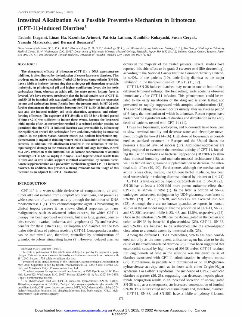
Initial Cellular Uptake Rate of SN-38. The respective initialcellular uptake rate of SN-38 lactone and carboxylate by HT-29 was
plotted as a function of the respective concentration and reported inFig. 1. The uptake rate of SN-38 lactone was higher than that ofSN-38 carboxylate at every concentration tested (P � 0.05 at 0.5 �M,and P � 0.005 at 1 and 2 �M). In addition, the uptake of SN-38carboxylate was saturable and characterized by a Vmax of 0.169pmol � 106 cells�1 � min�1 and a Km of 0.383 �M. Under the sameconditions and within the range of concentrations tested, the uptakeof SN-38-lactone was not saturable.
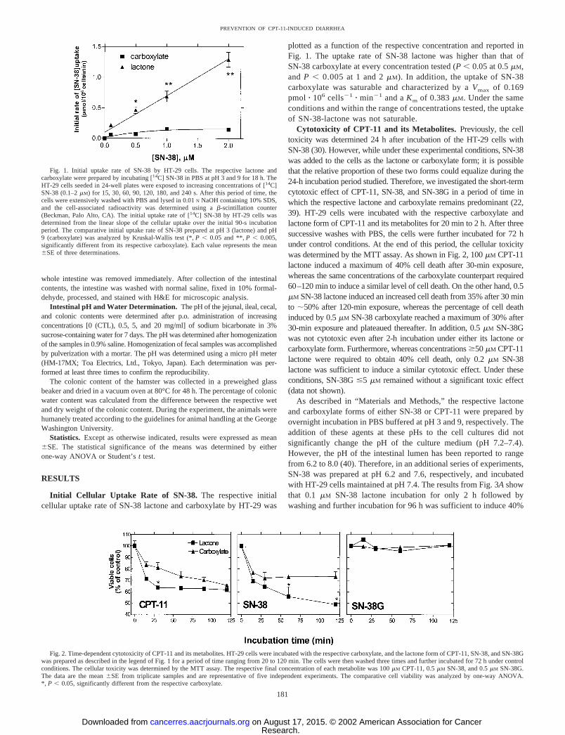
Cytotoxicity of CPT-11 and its Metabolites. Previously, the celltoxicity was determined 24 h after incubation of the HT-29 cells withSN-38 (30). However, while under these experimental conditions, SN-38was added to the cells as the lactone or carboxylate form; it is possiblethat the relative proportion of these two forms could equalize during the24-h incubation period studied. Therefore, we investigated the short-termcytotoxic effect of CPT-11, SN-38, and SN-38G in a period of time inwhich the respective lactone and carboxylate remains predominant (22,39). HT-29 cells were incubated with the respective carboxylate andlactone form of CPT-11 and its metabolites for 20 min to 2 h. After threesuccessive washes with PBS, the cells were further incubated for 72 hunder control conditions. At the end of this period, the cellular toxicitywas determined by the MTT assay. As shown in Fig. 2, 100 �M CPT-11lactone induced a maximum of 40% cell death after 30-min exposure,whereas the same concentrations of the carboxylate counterpart required60–120 min to induce a similar level of cell death. On the other hand, 0.5�M SN-38 lactone induced an increased cell death from 35% after 30 minto �50% after 120-min exposure, whereas the percentage of cell deathinduced by 0.5 �M SN-38 carboxylate reached a maximum of 30% after30-min exposure and plateaued thereafter. In addition, 0.5 �M SN-38Gwas not cytotoxic even after 2-h incubation under either its lactone orcarboxylate form. Furthermore, whereas concentrations �50 �M CPT-11lactone were required to obtain 40% cell death, only 0.2 �M SN-38lactone was sufficient to induce a similar cytotoxic effect. Under theseconditions, SN-38G �5 �M remained without a significant toxic effect(data not shown).
As described in “Materials and Methods,” the respective lactoneand carboxylate forms of either SN-38 or CPT-11 were prepared byovernight incubation in PBS buffered at pH 3 and 9, respectively. Theaddition of these agents at these pHs to the cell cultures did notsignificantly change the pH of the culture medium (pH 7.2–7.4).However, the pH of the intestinal lumen has been reported to rangefrom 6.2 to 8.0 (40). Therefore, in an additional series of experiments,SN-38 was prepared at pH 6.2 and 7.6, respectively, and incubatedwith HT-29 cells maintained at pH 7.4. The results from Fig. 3A showthat 0.1 �M SN-38 lactone incubation for only 2 h followed bywashing and further incubation for 96 h was sufficient to induce 40%
Fig. 1. Initial uptake rate of SN-38 by HT-29 cells. The respective lactone andcarboxylate were prepared by incubating [14C] SN-38 in PBS at pH 3 and 9 for 18 h. TheHT-29 cells seeded in 24-well plates were exposed to increasing concentrations of [14C]SN-38 (0.1–2 �M) for 15, 30, 60, 90, 120, 180, and 240 s. After this period of time, thecells were extensively washed with PBS and lysed in 0.01 N NaOH containing 10% SDS,and the cell-associated radioactivity was determined using a �-scintillation counter(Beckman, Palo Alto, CA). The initial uptake rate of [14C] SN-38 by HT-29 cells wasdetermined from the linear slope of the cellular uptake over the initial 90-s incubationperiod. The comparative initial uptake rate of SN-38 prepared at pH 3 (lactone) and pH9 (carboxylate) was analyzed by Kruskal-Wallis test (*, P � 0.05 and **, P � 0.005,significantly different from its respective carboxylate). Each value represents the mean�SE of three determinations.
Fig. 2. Time-dependent cytotoxicity of CPT-11 and its metabolites. HT-29 cells were incubated with the respective carboxylate, and the lactone form of CPT-11, SN-38, and SN-38Gwas prepared as described in the legend of Fig. 1 for a period of time ranging from 20 to 120 min. The cells were then washed three times and further incubated for 72 h under controlconditions. The cellular toxicity was determined by the MTT assay. The respective final concentration of each metabolite was 100 �M CPT-11, 0.5 �M SN-38, and 0.5 �M SN-38G.The data are the mean �SE from triplicate samples and are representative of five independent experiments. The comparative cell viability was analyzed by one-way ANOVA.*, P � 0.05, significantly different from the respective carboxylate.
181
PREVENTION OF CPT-11-INDUCED DIARRHEA
Research. on August 17, 2015. © 2002 American Association for Cancercancerres.aacrjournals.org Downloaded from
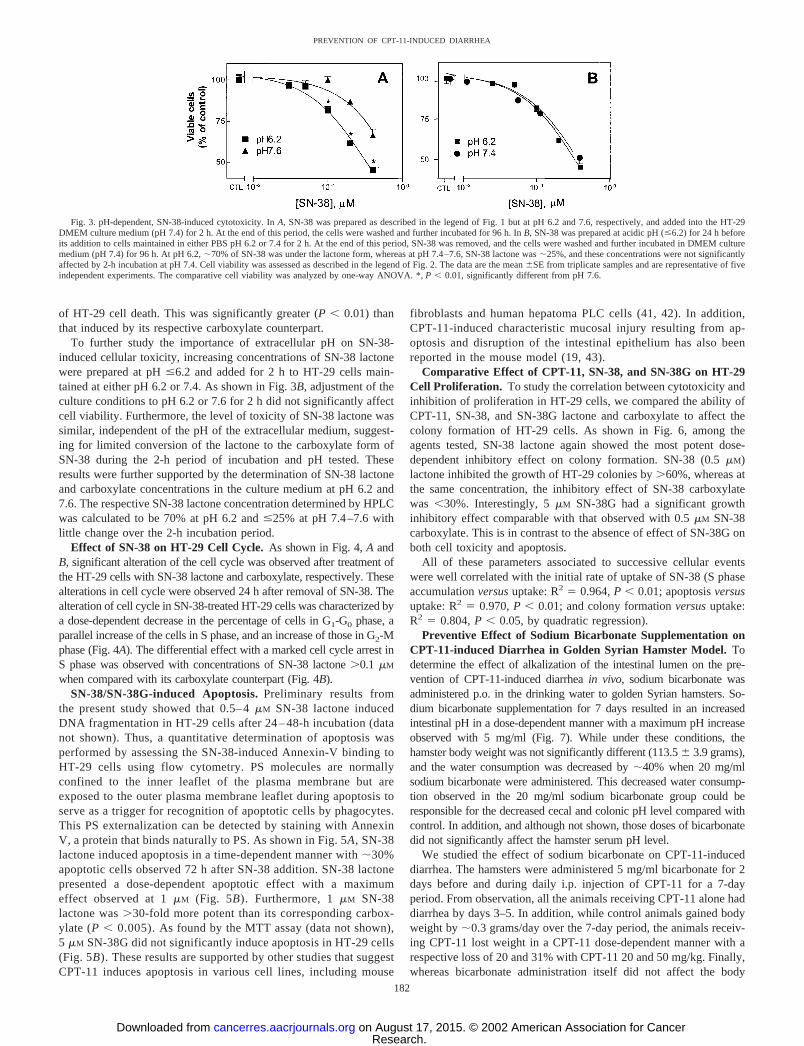
of HT-29 cell death. This was significantly greater (P � 0.01) thanthat induced by its respective carboxylate counterpart.
To further study the importance of extracellular pH on SN-38-induced cellular toxicity, increasing concentrations of SN-38 lactonewere prepared at pH �6.2 and added for 2 h to HT-29 cells main-tained at either pH 6.2 or 7.4. As shown in Fig. 3B, adjustment of theculture conditions to pH 6.2 or 7.6 for 2 h did not significantly affectcell viability. Furthermore, the level of toxicity of SN-38 lactone wassimilar, independent of the pH of the extracellular medium, suggest-ing for limited conversion of the lactone to the carboxylate form ofSN-38 during the 2-h period of incubation and pH tested. Theseresults were further supported by the determination of SN-38 lactoneand carboxylate concentrations in the culture medium at pH 6.2 and7.6. The respective SN-38 lactone concentration determined by HPLCwas calculated to be 70% at pH 6.2 and �25% at pH 7.4–7.6 withlittle change over the 2-h incubation period.
Effect of SN-38 on HT-29 Cell Cycle. As shown in Fig. 4, A andB, significant alteration of the cell cycle was observed after treatment ofthe HT-29 cells with SN-38 lactone and carboxylate, respectively. Thesealterations in cell cycle were observed 24 h after removal of SN-38. Thealteration of cell cycle in SN-38-treated HT-29 cells was characterized bya dose-dependent decrease in the percentage of cells in G1-G0 phase, aparallel increase of the cells in S phase, and an increase of those in G2-Mphase (Fig. 4A). The differential effect with a marked cell cycle arrest inS phase was observed with concentrations of SN-38 lactone �0.1 �M
when compared with its carboxylate counterpart (Fig. 4B).SN-38/SN-38G-induced Apoptosis. Preliminary results from
the present study showed that 0.5– 4 �M SN-38 lactone inducedDNA fragmentation in HT-29 cells after 24 – 48-h incubation (datanot shown). Thus, a quantitative determination of apoptosis wasperformed by assessing the SN-38-induced Annexin-V binding toHT-29 cells using flow cytometry. PS molecules are normallyconfined to the inner leaflet of the plasma membrane but areexposed to the outer plasma membrane leaflet during apoptosis toserve as a trigger for recognition of apoptotic cells by phagocytes.This PS externalization can be detected by staining with AnnexinV, a protein that binds naturally to PS. As shown in Fig. 5A, SN-38lactone induced apoptosis in a time-dependent manner with �30%apoptotic cells observed 72 h after SN-38 addition. SN-38 lactonepresented a dose-dependent apoptotic effect with a maximumeffect observed at 1 �M (Fig. 5B). Furthermore, 1 �M SN-38lactone was �30-fold more potent than its corresponding carbox-ylate (P � 0.005). As found by the MTT assay (data not shown),5 �M SN-38G did not significantly induce apoptosis in HT-29 cells(Fig. 5B). These results are supported by other studies that suggestCPT-11 induces apoptosis in various cell lines, including mouse
fibroblasts and human hepatoma PLC cells (41, 42). In addition,CPT-11-induced characteristic mucosal injury resulting from ap-optosis and disruption of the intestinal epithelium has also beenreported in the mouse model (19, 43).
Comparative Effect of CPT-11, SN-38, and SN-38G on HT-29Cell Proliferation. To study the correlation between cytotoxicity andinhibition of proliferation in HT-29 cells, we compared the ability ofCPT-11, SN-38, and SN-38G lactone and carboxylate to affect thecolony formation of HT-29 cells. As shown in Fig. 6, among theagents tested, SN-38 lactone again showed the most potent dose-dependent inhibitory effect on colony formation. SN-38 (0.5 �M)lactone inhibited the growth of HT-29 colonies by �60%, whereas atthe same concentration, the inhibitory effect of SN-38 carboxylatewas �30%. Interestingly, 5 �M SN-38G had a significant growthinhibitory effect comparable with that observed with 0.5 �M SN-38carboxylate. This is in contrast to the absence of effect of SN-38G onboth cell toxicity and apoptosis.
All of these parameters associated to successive cellular eventswere well correlated with the initial rate of uptake of SN-38 (S phaseaccumulation versus uptake: R2 � 0.964, P � 0.01; apoptosis versusuptake: R2 � 0.970, P � 0.01; and colony formation versus uptake:R2 � 0.804, P � 0.05, by quadratic regression).
Preventive Effect of Sodium Bicarbonate Supplementation onCPT-11-induced Diarrhea in Golden Syrian Hamster Model. Todetermine the effect of alkalization of the intestinal lumen on the pre-vention of CPT-11-induced diarrhea in vivo, sodium bicarbonate wasadministered p.o. in the drinking water to golden Syrian hamsters. So-dium bicarbonate supplementation for 7 days resulted in an increasedintestinal pH in a dose-dependent manner with a maximum pH increaseobserved with 5 mg/ml (Fig. 7). While under these conditions, thehamster body weight was not significantly different (113.5 � 3.9 grams),and the water consumption was decreased by �40% when 20 mg/mlsodium bicarbonate were administered. This decreased water consump-tion observed in the 20 mg/ml sodium bicarbonate group could beresponsible for the decreased cecal and colonic pH level compared withcontrol. In addition, and although not shown, those doses of bicarbonatedid not significantly affect the hamster serum pH level.
We studied the effect of sodium bicarbonate on CPT-11-induceddiarrhea. The hamsters were administered 5 mg/ml bicarbonate for 2days before and during daily i.p. injection of CPT-11 for a 7-dayperiod. From observation, all the animals receiving CPT-11 alone haddiarrhea by days 3–5. In addition, while control animals gained bodyweight by �0.3 grams/day over the 7-day period, the animals receiv-ing CPT-11 lost weight in a CPT-11 dose-dependent manner with arespective loss of 20 and 31% with CPT-11 20 and 50 mg/kg. Finally,whereas bicarbonate administration itself did not affect the body
Fig. 3. pH-dependent, SN-38-induced cytotoxicity. In A, SN-38 was prepared as described in the legend of Fig. 1 but at pH 6.2 and 7.6, respectively, and added into the HT-29DMEM culture medium (pH 7.4) for 2 h. At the end of this period, the cells were washed and further incubated for 96 h. In B, SN-38 was prepared at acidic pH (�6.2) for 24 h beforeits addition to cells maintained in either PBS pH 6.2 or 7.4 for 2 h. At the end of this period, SN-38 was removed, and the cells were washed and further incubated in DMEM culturemedium (pH 7.4) for 96 h. At pH 6.2, �70% of SN-38 was under the lactone form, whereas at pH 7.4–7.6, SN-38 lactone was �25%, and these concentrations were not significantlyaffected by 2-h incubation at pH 7.4. Cell viability was assessed as described in the legend of Fig. 2. The data are the mean �SE from triplicate samples and are representative of fiveindependent experiments. The comparative cell viability was analyzed by one-way ANOVA. *, P � 0.01, significantly different from pH 7.6.
182
PREVENTION OF CPT-11-INDUCED DIARRHEA
Research. on August 17, 2015. © 2002 American Association for Cancercancerres.aacrjournals.org Downloaded from

weight gain as compared with control, it reduced the loss of weight ofthe hamsters receiving CPT-11 (20–50 mg/kg) by �50%.
The histological analysis of the section of the colon from thehamsters treated with CPT-11 showed severe, acute, and ulcerativecolitis. There was necro-inflammatory debris in small foci within theepithelial cell layer with focal loss of cells. Numerous neutrophilswere seen among epithelial cells and edematous lamina propria (Fig.8A). On the other hand, the sections of colon from the hamster treatedwith CPT-11, in which bicarbonate was administered p.o. throughoutthe CPT-11 treatment, showed a moderate acute colitis with focalcryptitis that was less than that seen with CPT-11 alone. There wasonly occasional neutrophil aggregate in a crypt in the epithelial celllayer (Fig. 8A). These changes were not present in the group receivingthe sodium bicarbonate supplementation alone.
This increased intestinal damage induced by 20–50 mg/kg CPT-11was associated with a 70–75% increase in colonic water content. Al-though bicarbonate alone did not alter the hamster’s colonic water con-tent, it significantly (P � 0.05) reduced that induced by CPT-11 by�30% (Fig. 8B). From observation, while the feces of hamsters receiving
CPT-11 were liquid, those of hamsters treated with CPT-11 and receivingbicarbonate supplementation closely resemble the feces of the controlhamsters. Since following CPT-11 treatment alone as compared withCPT-11 coadministration with bicarbonate, the hamsters had a loss ofbody weight, the intestinal damage could, at least in part, be responsiblefor the decreased intestinal nutrient uptake in the CPT-11-treated group.
We also studied the effect of bicarbonate supplementation on theintestinal SN-38 and CPT-11 concentrations. Bicarbonate administra-tion (5 mg/ml) in the drinking water resulted in �20% decrease inintestinal SN-38 lactone concentration, whereas CPT-11 carboxylatewas increased by �15% when compared with control hamsters re-ceiving a daily dose of 20 mg/kg CPT-11 alone. These results supportthe above described decreased SN-38 lactone-induced cell injury anddiarrhea after bicarbonate administration.
DISCUSSION
The present study is the first to clearly demonstrate that an in-creased intestinal pH after bicarbonate p.o. administration was asso-
Fig. 4. Dose-dependent cell cycle alteration induced by the respective lactone and carboxylate form of SN-38. HT-29 cells were exposed to increasing concentrations (0.1–5 �M) of SN-38lactone and carboxylate for 2 h. After the removal of SN-38 (24 h), the cells were collected by trypsinization, fixed with 70% ethanol, stained with PI (50 �g/ml), and subjected to flow cytometryfor cell cycle analysis. A, histogram plot of the intensity gained versus FL-2 channel area. In B, the results, analyzed by the modFIT program (Verity Software House, Topsham, ME) as describedunder “Materials and Methods,” were plotted as a function of the concentration of SN-38. The data are the means �SE from two independent experiments performed in duplicate. Thecomparative dose-dependent cell cycle phase was analyzed by Student’s t test (*, P � 0.05, **, P � 0.005, significantly different from the respective carboxylate).
183
PREVENTION OF CPT-11-INDUCED DIARRHEA
Research. on August 17, 2015. © 2002 American Association for Cancercancerres.aacrjournals.org Downloaded from

ciated with a reduction in the intestinal SN-38 lactone concentration,as well as cellular damage and diarrhea induced by CPT-11 in thegolden Syrian hamster model. A daily injection of CPT-11 at a doseof 20–50 mg/kg BW induced diarrhea by days 3–5 and was lethal byday 5 at a dose �100 mg/kg BW. Under these conditions, p.o.administration of 5 mg/ml sodium bicarbonate in the drinking watersignificantly reduced both the fecal water content, as well as theintestinal tissue damage induced by 20–50 mg/kg BW CPT-11. Theseresults are further supported by a Phase II clinical trial, suggesting abeneficial effect of sodium bicarbonate supplementation against CPT-11-induced severe delayed diarrhea (44).
SN-38 derived from the prodrug CPT-11 is suggested to exert itsantitumor effect well before its elimination from the systemic circu-lation and secretion into bile (23, 45). Once secreted into bile, how-ever, SN-38 has been shown in rats to accumulate intracellularly inintestinal epithelial cells (46). It is believed to be responsible fordiarrhea attributed to CPT-11 administration in nude mice (27). Thesestudies suggest that reduced accumulation of SN-38 in intestinal cellscan diminish the severity of diarrhea and, consequently, increase theclinical usefulness of CPT-11 (19).
The present study supports CPT-11/SN-38 lactone-induced diar-rhea to be the result of intestinal injury rather than increased intestinalsecretion as reported previously in humans (47). Our results are inagreement with recent reports both in animal models (27, 43), as wellas in cancer patients (48), in which lethal small-intestinal injury wasassociated to CPT-11-induced diarrhea. Therefore, one of the pro-
posed mechanisms for bicarbonate action is to reduce both the intes-tinal concentration of SN-38 lactone and its induced tissue damage,resulting in reduced diarrhea.
The protective mechanism of bicarbonate p.o. administration in-volves, at least in part, a pH-dependent increased conversion of
Fig. 5. Flow cytometric analysis of SN-38-induced apoptotic celldeath. Time course of SN-38-induced apoptosis in HT-29 cells. HT-29cells in 60-mm dishes (1 � 106 cells) were incubated with 0.5 �M SN-38as described in the legend of Fig. 2. The percentage of apoptotic cells(bottom right quadrant) over time was determined by GFP-annexin-Vbinding assay using flow cytometry. Cells positive for both annexin-Vand PI (top right quadrant) were defined as necrotic. The 0- and 72-hpanels represent the flow cytometric determination at 0 and 72 h,respectively, after exposure of the cells to SN-38 and were plotted as theintensity gained by FL-1 channel (X-axis; GFP-annexin-V) versus theintensity gained by FL-2 channel (Y-axis; PI). The graph on the rightrepresents the percentage of apoptotic cells determined from the flowcytometric analysis of the apoptotic effect over time. *, P � 0.005,significantly different from control (0 h). B, dose-dependent apoptoticeffect of SN-38 and SN-38G in HT-29 cells at 72 h. Percentage ofapoptotic cells after incubation with increasing concentrations of therespective lactone or carboxylate form was determined by flow cytom-etry using both GFP-annexin-V and PI. The necrotic cells observed with5 �M SN-38 were reported for comparison. All data are the mean �SEfrom two independent experiments performed in duplicate. The com-parative annexin-V binding in the presence and absence of SN-38 andSN-38G, respectively, was analyzed by one-way ANOVA. *, P � 0.005,significantly different from the respective carboxylate form.
Fig. 6. Growth inhibition by colony-forming assay. HT-29 cells were incubated for 2 hwith increasing concentrations of CPT-11 (1–10 �M), SN-38 (0.01–0.5 �M), or SN-38G(5 �M) lactone and carboxylate, respectively. After removal of the agent, the cells werefurther incubated for 24 h, collected by trypsinization, and then seeded in fresh culturemedium (500 cells/dish). Colonies grown in the dish after 2 weeks were counted. Data arethe means �SE from three independent experiments. The comparative colony numberobtained in the presence and absence of CPT-11, SN-38, and SN-38G, respectively, wasanalyzed by one-way ANOVA. *, P � 0.005, significantly different from the respectivecarboxylate form.
184
PREVENTION OF CPT-11-INDUCED DIARRHEA
Research. on August 17, 2015. © 2002 American Association for Cancercancerres.aacrjournals.org Downloaded from

CPT-11/SN-38 lactone to carboxylate, as shown in the present study.It is generally accepted that the �-hydroxy lactone ring is an absoluterequirement for CPT-derived molecules to have in vitro and in vivoactivities (49–51). However, although circumventing the opening ofthe lactone ring is one of the strategies proposed in order to optimizethe antitumor activity of this drug (52), the lactone ring is unstable atphysiological pH and above and can open to the much less activecarboxylate form. Therefore, it is our hypothesis that the reducedincidence and severity of CPT-11-induced diarrhea is attributable, atleast in part, to the opening of the lactone ring in the intestinal lumen.The rationale for this theory is based on our findings in both HT-29cells and in enterocytes isolated from hamster small and large intes-tine (30). Results of these studies show that the initial uptake rate ofthe lactone form of SN-38 by HT-29 cells was significantly greaterthan that of its carboxylate counterpart (Fig. 1). Furthermore, incontrast to the lactone form, for which uptake remained unchanged,the transport of SN-38 carboxylate was carrier mediated and couldspecifically and significantly be inhibited by the addition of dinitro-phenol (30). Finally, the initial uptake rate presents a good correlationwith the cytotoxicity of SN-38 in HT-29 cells (see “Results” andRef. 30).
This difference in the cytotoxicity of CPT-11 and SN-38 in HT-29cells can probably be attributed to the respective differential rate ofconversion between the lactone and carboxylate form of these agents.Indeed, Akimoto et al. (53) have reported that the constant of hydrol-ysis of SN-38 (50% conversion from lactone to carboxylate) was13.5 h and 33 min at pH 6 and pH 7.4, respectively. Furthermore,Rivory et al. (39) have reported that conversion of CPT-11 lactone tocarboxylate in vivo was rapid, whereas SN-38 was present predomi-nantly as the lactone form at all times. The HPLC determination in thecell culture medium indicated that even after 2-h incubation at pH 7.4,SN-38 prepared at pH 6.2 and 7.6 remained �70 and �25% aslactone, respectively. This limited conversion of the lactone to thecarboxylate form was further supported by the fact that similar tox-icity was observed independently of the pH of the incubation mediumduring the 2-h incubation period of the cells with SN-38 lactone.
As reported in the present study, the lactone form of SN-38 wasmore potent to induce both S and G2-M phase cell cycle arrest andapoptosis than its carboxylate counterpart. These results are consistentwith what has been reported previously with camptothecin (33, 34).The lack of G1 arrest is probably attributable to the p53 mutation in
the HT-29 cells and the loss of the p53-modulated G1 check pointsuggested previously by Goldwasser et al. (32). The delay or blockageof, at least, G2 phase after SN-38 treatment may allow for cellularDNA repair (32, 54). This has been suggested previously by variousauthors including Shao et al. (54), who have shown that UCN-01,which abrogates the G2-M checkpoint, increased camptothecin-induced cell toxicity. It is also worthwhile to mention that the HT-29G2-M checkpoint is more sensitive to SN-38 than the S checkpoint,because the G2-M phase arrest is observed at lower SN-38 concen-trations than those required to induce an arrest in S phase (Fig. 4B).These results may underline some interregulatory mechanism(s) be-tween these two checkpoints and support the observations with camp-
Fig. 7. Change in intestinal luminal pH after oral sodium bicarbonate supplementation.The pH of the jejunal, ileal, cecal, and colonic contents was determined after p.o.administration of increasing concentrations [0 (CTL), 0.5, 5, and 20 mg/ml] of sodiumbicarbonate in 3% sucrose-containing water for 7 days. The pH was determined inintestinal homogenates prepared by the addition of 0.9% saline and by using a micro pHmeter (HM-17MX; Toa Electrics, Ltd.). Each determination was performed at least threetimes to confirm the reproducibility. The data are the mean �SE from three independentexperiments. The comparative intestinal pH in the presence and absence of bicarbonatewas analyzed by Student’s t test. *, P � 0.05; **, P � 0.005, significantly different fromcontrol.
Fig. 8. The effect of sodium bicarbonate supplementation on CPT-11-induced diarrheain the golden Syrian hamster model. In A, the respective colon sections from a represent-ative hamster receiving either 20 mg/kg CPT-11 i.p. injected daily alone (CPT-11 alone)or in combination with 5 mg/ml bicarbonate (CPT-11 � bicarbonate) p.o. administered 2days before and during the 7-day CPT-11 treatment were fixed in 10% formalin. Alongwith the respective controls (Control, vehicle alone; Bicarbonate, bicarbonate alone), thesamples were processed and stained with H&E. B, change in the percentage of the colonicwater content; the colonic water content was determined 7 days after daily i.p. adminis-tration of CPT-11 (20–50 mg/kg) or the vehicle (PBS) in the absence (CTL) or presenceof p.o. administration of 5 mg/ml sodium bicarbonate (NaHCO3) in 3% sucrose-contain-ing drinking water for 7 days. The bicarbonate administration was started 2 days beforethe initial i.p. injection. The colonic content was dried by incubation for 48 h at 80°C ina vacuum oven. The water content was determined by subtracting the dry weight from thetotal (wet) weight. Data are the means �SE from three independent experiments. Thecomparative colonic water content was analyzed by one-way ANOVA. *, P � 0.01,significantly different from CTL group; **, P � 0.05, significantly different from CPT-11group. Under these conditions, the intestinal SN-38 lactone concentration was decreasedby �20%, and CPT-11 carboxylate was increased by 15% when the hamsters receivedbicarbonate compared with CPT-11 i.p. administration alone.
185
PREVENTION OF CPT-11-INDUCED DIARRHEA
Research. on August 17, 2015. © 2002 American Association for Cancercancerres.aacrjournals.org Downloaded from
tothecin reported previously by Goldwasser et al. (32). Therefore, ourpresent results demonstrate that the number of apoptotic cells inducedby the lactone form of SN-38 was also greater than that induced by itscarboxylate counterpart. This suggests that SN-38 can induce celldeath in a DNA damage-dependent fashion overcoming any possiblecellular rescue attributable to cell cycle arrest and increased DNArepair.
Furthermore, it was recently reported in this cell line that thereplication protein A2 (RPA2) is an important protein of the replica-tion complex for initiation and maintenance of S phase. In addition,this protein, which plays a role in signaling mechanisms that coordi-nate DNA replication and cell cycle, can be phosphorylated by phar-macological concentrations of camptothecin (55). Because thesechanges are related to the induction of DNA damage (replication forkcollisions) in replicating DNA (34, 35), it is speculated that thespecific changes in cell cycle can be a reflection of the magnitude ofthe DNA damage induced by CPT-11/SN-38. Therefore, the cell cycleanalysis could be a convenient parameter to assess the pharmacolog-ical activity of CPT-11/SN-38, as far as the replication-arresting DNAdamage is concerned.
Interestingly, in the present study, 5 �M SN-38G showed a signif-icant growth inhibitory effect, even though no effect was observedusing either the MTT or Annexin-V binding assay. Because it isbelieved that SN-38G does not induce any significant cytotoxic effect(56), these findings suggest the possible deconjugation of the glucu-ronide group from SN-38 during the prolonged culture period inHT-29 cells. In addition, it remains possible that short-term assays,such as the MTT assay and apoptosis determination, may not besensitive enough to determine the cumulative cellular effect of SN-38G. The possibility that SN-38G can exhibit cellular toxicity after aprolonged period of culture raises the question of the clinical involve-ment of SN-38G in CPT-11-induced diarrhea.
The bicarbonate-induced improvement of tissue damage in thelarge and also in small intestine is somewhat puzzling, in light of thein vitro data showing no effect on overall toxicity of SN-38 withchanges in pH of the incubation medium from 6.9 to 7.4 for 2 h.Indeed, whereas the transit time in human large intestine has beenreported to be around 10 h, that of the small intestine averages 2–4 h(57), and these values should be shorter in the hamster model (58).Therefore, the period allowed for the conversion of the SN-38/CPT-11from lactone to carboxylate in the small intestine should have been tooshort to observe any protective effect against tissue damage. This,therefore, raises the question of possible additional effects of in-creased luminal bicarbonate besides increasing the conversion of thedrug from the lactone to its carboxylate form.
In summary, the toxic effect of SN-38 lactone was maximum evenafter exposure of the cells for a period of time as short as 2 h.Furthermore, the pH-dependent opening of the SN-38 �-hydroxylactone ring to its carboxylate form resulted in a decreased HT-29apoptosis and growth inhibition, which were well correlated with theinitial cellular uptake rate of this drug. These findings can explain, atleast in part, the protective effect of intestinal alkalization on CPT-11-induced diarrhea in vivo.
However, the present study raises an important issue of the role thatincreased intestinal alkalization may have on the potency of CPT-11on colorectal cancer therapy. Although this question cannot be fullyaddressed at the present time, without additional studies, differentelements of a response can be proposed. First, CPT-11 therapy isgenerally limited to advanced colorectal cancer patients with the goalto ablate metastasis associated to internal organs, including liver andkidney (8, 59). Furthermore, colon carcinoma is a rigid tumor and,therefore, only the cells on the surface of the tumor can absorb thedrug from the luminal side. Thus, this emphasizes the importance of
the drug delivery from the blood supply. Under these conditions,elevated pH in the colonic lumen may not play a major role in thealteration of the drug absorption by the tumor cells. Therefore, it is thehypothesis of the authors that the efficacy of CPT-11 should not beparticularly affected in colorectal cancer by the administration ofbicarbonate.
In conclusion, it is believed that the control of late-onset diarrheaholds the key to the effective use of CPT-11, which, in turn, willimprove survival benefit and quality of life for patients with malig-nancies (10). p.o. administration of bicarbonate appears to be a noveland effective protective mechanism against CPT-11/SN-38-associatedtissue injury and side effects. Although the mechanism by whichintestinal alkalization abrogates CPT-11-induced side effects needsfurther clarification in humans, results from our in vitro and in vivostudies suggest a pH-dependent facilitation of SN-38 carboxylateformation and a decreased intestinal epithelial cell accumulation.Finally, this approach may prove to be inexpensive, safe, and usefulto increase the efficacy of CPT-11 in cancer patient therapy.
ACKNOWLEDGMENTS
We thank Muneaki Hidaka, Department of Pharmacy, Miyazaki MedicalCollege, Miyazaki, Japan, for the HPLC determination of SN-38 and CPT-11lactone: carboxylate ratio in the culture medium and hamster intestine. We alsothank Joe Cotruvo for his technical assistance.
REFERENCES
1. Kawato, Y., Aonuma, M., Hirota, Y., Kuga, H., and Sato, K. Intracellular roles ofSN-38, a metabolite of the camptothecin derivative CPT-11, in the antitumor effect ofCPT-11. Cancer Res., 51: 4187–4191, 1991.
2. Kudoh, S., Fujiwara, Y., Takada, Y., Yamamoto, H., Kinoshita, A., Ariyoshi, Y.,Furuse, K., and Fukuoka, M. Phase II study of irinotecan combined with cisplatin inpatients with previously untreated small-cell lung cancer. West Japan Lung CancerGroup. J. Clin. Oncol., 16: 1068–1074, 1998.
3. Irvin, W. P., Price, F. V., Bailey, H., Gelder, M., Rosenbluth, R., Durivage, H. J., andPotkul, R. K. A phase II study of irinotecan (CPT-11) in patients with advancedsquamous cell carcinoma of the cervix. Cancer, 82: 328–333, 1998.
4. Verschraegen, C. F., Levy, T., Kudelka, A. P., Llerena, E., Ende, K., Freedman, R. S.,Edwards, C. L., Hord, M., Steger, M., Kaplan, A. L., Kieback, D., Fishman, A., andKavanagh, J. J. Phase II study of irinotecan in prior chemotherapy-treated squamouscell carcinoma of the cervix. J. Clin. Oncol., 15: 625–631, 1997.
5. Shimizu, Y., Umezawa, S., and Hasumi, K. Successful treatment of clear celladenocarcinoma of the ovary (OCCA) with a combination of CPT-11 and mitomycinC. Gan To Kagaku Ryoho, 23: 587–593, 1996.
6. Pitot, H. C., Wender, D. B., O’Connell, M. J., Schroeder, G., Goldberg, R. M., Rubin,J., Mailliard, J. A., Knost, J. A., Ghosh, C., Kirschling, R. J., Levitt, R., andWindschitl, H. E. Phase II trial of irinotecan in patients with metastatic colorectalcarcinoma. J. Clin. Oncol., 15: 2910–2919, 1997.
7. Ota, K., Ohno, R., Shirakawa, S., Masaoka, T., Okada, K., Ohashi, Y., and Taguchi,T. Late phase II clinical study of irinotecan hydrochloride (CPT-11) in the treatmentof malignant lymphoma and acute leukemia. The CPT-11 Research Group forHematological Malignancies. Gan To Kagaku Ryoho, 21: 1047–1055, 1994.
8. Cunningham, D., Pyrhonen, S., James, R. D., Punt, C. J., Hickish, T. F., Heikkila, R.,Johannesen, T. B., Starkhammar, H., Topham, C. A., Awad, L., Jacques, C., andHerait, P. Randomised trial of irinotecan plus supportive care versus supportive carealone after fluorouracil failure for patients with metastatic colorectal cancer. Lancet,352: 1413–1418, 1998.
9. Fukuoka, M., Takada, M., Masuda, N., Furuse, K., Saijo, N., Ikegami, H., andNishiwaki, Y. Chemotherapy for small cell lung cancer. Nihon Kyobu ShikkanGakkai Zasshi, 31: 225–231, 1993.
10. Hecht, J. R. Gastrointestinal toxicity of irinotecan. Oncology (Huntingt), 12: 72–78,1998.
11. Sargent, D. J., Niedzwiecki, D., O’Connell, M. J., and Schilsky, R. L. Recommen-dation for caution with irinotecan, fluorouracil, and leucovorin for colorectal cancer.N. Engl. J. Med., 345: 144–145, 2001.
12. Rougier, P., Bugat, R., Douillard, J. Y., Culine, S., Suc, E., Brunet, P., Becouarn, Y.,Ychou, M., Marty, M., Extra, J. M., Bonneterre, J., Adenis, A., Seitz, J. F., Ganem,G., Namer, M., Conroy, T., Negrier, S., Merrouche, Y., Burki, F., Mousseau, M.,Herait, P., and Mahjoubi, M. Phase II study of irinotecan in the treatment of advancedcolorectal cancer in chemotherapy-naive patients and patients pretreated with flu-orouracil-based chemotherapy. J. Clin. Oncol., 15: 251–260, 1997.
13. Gandia, D., Abigerges, D., Armand, J. P., Chabot, G. G., Da Costa, L., De Forni, M.,Mathieu Boue, A., and Herait, P. CPT-11-induced cholinergic effects in cancerpatients. J. Clin. Oncol., 11: 196–197, 1993.
14. Takasuna, K., Kasai, Y., Kitano, Y., Mori, K., Kobayashi, R., Hagiwara, T., Kakihata,K., Hirohashi, M., Nomura, M., and Nagai, E. Protective effects of kampo medicines
186
PREVENTION OF CPT-11-INDUCED DIARRHEA
Research. on August 17, 2015. © 2002 American Association for Cancercancerres.aacrjournals.org Downloaded from
and baicalin against intestinal toxicity of a new anticancer camptothecin derivative,irinotecan hydrochloride (CPT-11), in rats. Jpn. J. Cancer Res., 86: 978–984, 1995.
15. Lenfers, B. H., Loeffler, T. M., Droege, C. M., and Hausamen, T. U. Substantialactivity of budesonide in patients with irinotecan (CPT-11) and 5-fluorouracil induceddiarrhea and failure of loperamide treatment. Ann. Oncol., 10: 1251–1253, 1999.
16. Saliba, F., Hagipantelli, R., Misset, J. L., Bastian, G., Vassal, G., Bonnay, M., Herait,P., Cote, C., Mahjoubi, M., Mignard, D., and Cvitkovic, E. Pathophysiology andtherapy of irinotecan-induced delayed-onset diarrhea in patients with advanced colo-rectal cancer: a prospective assessment. J. Clin. Oncol., 16: 2745–2751, 1998.
17. Rothenberg, M. L., Cox, J. V., DeVore, R. F., Hainsworth, J. D., Pazdur, R., Rivkin,S. E., Macdonald, J. S., Geyer, C. E., Jr., Sandbach, J., Wolf, D. L., Mohrland, J. S.,Elfring, G. L., Miller, L. L., and Von Hoff, D. D. A multicenter, phase II trial ofweekly irinotecan (CPT-11) in patients with previously treated colorectal carcinoma.Cancer, 85: 786–795, 1999.
18. Shinohara, H., Killion, J. J., Kuniyasu, H., Kumar, R., and Fidler, I. J. Prevention ofintestinal toxic effects and intensification of irinotecan’s therapeutic efficacy againstmurine colon cancer liver metastases by oral administration of the lipopeptide JBT3002. Clin. Cancer Res., 4: 2053–2063, 1998.
19. Hardman, W. E., Moyer, M. P., and Cameron, I. L. Fish oil supplementation enhancedCPT-11 (irinotecan) efficacy against MCF7 breast carcinoma xenografts and amelio-rated intestinal side-effects. Br. J. Cancer, 81: 440–448, 1999.
20. Savarese, D., Al Zoubi, A., and Boucher, J. Glutamine for irinotecan diarrhea. J. Clin.Oncol., 18: 450–451, 2000.
21. Sakata, Y., Suzuki, H., and Kamataki, T. Preventive effect of TJ-14, a kampo(Chinese herb) medicine, on diarrhea induced by irinotecan hydrochloride (CPT-11).Gan To Kagaku Ryoho, 21: 1241–1244, 1994.
22. Rivory, L. P., Bowles, M. R., Robert, J., and Pond, S. M. Conversion of irinotecan(CPT-11) to its active metabolite, 7-ethyl-10-hydroxycamptothecin (SN-38), by hu-man liver carboxylesterase. Biochem. Pharmacol., 52: 1103–1111, 1996.
23. Atsumi, R., Suzuki, W., and Hakusui, H. Identification of the metabolites of irino-tecan, a new derivative of camptothecin, in rat bile and its biliary excretion. Xeno-biotica, 21: 1159–1169, 1991.
24. Chu, X. Y., Kato, Y., Niinuma, K., Sudo, K. I., Hakusui, H., and Sugiyama, Y.Multispecific organic anion transporter is responsible for the biliary excretion of thecamptothecin derivative irinotecan and its metabolites in rats. J. Pharmacol. Exp.Ther., 281: 304–314, 1997.
25. Takasuna, K., Hagiwara, T., Hirohashi, M., Kato, M., Nomura, M., Nagai, E., Yokoi,T., and Kamataki, T. Involvement of �-glucuronidase in intestinal microflora in theintestinal toxicity of the antitumor camptothecin derivative irinotecan hydrochloride(CPT-11) in rats. Cancer Res., 56: 3752–3757, 1996.
26. Iyer, L., King, C. D., Whitington, P. F., Green, M. D., Roy, S. K., Tephly, T. R.,Coffman, B. L., and Ratain, M. J. Genetic predisposition to the metabolism ofirinotecan (CPT-11). Role of uridine diphosphate glucuronosyltransferase isoform1A1 in the glucuronidation of its active metabolite (SN-38) in human liver micro-somes. J. Clin. Investig., 101: 847–854, 1998.
27. Araki, E., Ishikawa, M., Iigo, M., Koide, T., Itabashi, M., and Hoshi, A. Relationshipbetween development of diarrhea and the concentration of SN-38, an active metab-olite of CPT-11, in the intestine and the blood plasma of athymic mice followingintraperitoneal administration of CPT-11. Jpn. J. Cancer Res., 84: 697–702, 1993.
28. Wasserman, E., Myara, A., Lokiec, F., Goldwasser, F., Trivin, F., Mahjoubi, M.,Misset, J. L., and Cvitkovic, E. Severe CPT-11 toxicity in patients with Gilbert’ssyndrome: two case reports. Ann. Oncol., 8: 1049–1051, 1997.
29. Fassberg, J. and Stella, V. J. A kinetic and mechanistic study of the hydrolysis ofcamptothecin and some analogues. J. Pharm. Sci., 81: 676–684, 1992.
30. Kobayashi, K., Bouscarel, B., Matsuzaki, Y., Ceryak, S., Kudoh, S., and Fromm, H.pH-dependent uptake of irinotecan and its active metabolite, SN-38, by intestinalcells. Int. J. Cancer, 83: 491–496, 1999.
31. Kobayashi, K., Bouscarel, B., Matsuzaki, Y., Ceryak, S., and Fromm, H. Uptakemechanism of irinotecan (CPT-11) and its metabolite (SN-38) by hamster intestinalcells. Gastroenterology, 114: G2578 (Abst. No) A626, 1998.
32. Goldwasser, F., Shimizu, T., Jackman, J., Hoki, Y., O’Connor, P. M., Kohn, K. W.,and Pommier, Y. Correlations between S and G2 arrest and the cytotoxicity ofcamptothecin in human colon carcinoma cells. Cancer Res., 56: 4430–4437, 1996.
33. Holm, C., Covey, J. M., Kerrigan, D., and Pommier, Y. Differential requirement ofDNA replication for the cytotoxicity of DNA topoisomerase I and II inhibitors inChinese hamster DC3F cells. Cancer Res., 49: 6365–6368, 1989.
34. Hsiang, Y. H., Lihou, M. G., and Liu, L. F. Arrest of replication forks by drug-stabilized topoisomerase I-DNA cleavable complexes as a mechanism of cell killingby camptothecin. Cancer Res., 49: 5077–5082, 1989.
35. Tsao, Y. P., Russo, A., Nyamuswa, G., Silber, R., and Liu, L. F. Interaction betweenreplication forks and topoisomerase I-DNA cleavable complexes: studies in a cell-freeSV40 DNA replication system. Cancer Res., 53: 5908–5914, 1993.
36. Bouscarel, B., Nussbaum, R., Dubner, H., and Fromm, H. The role of sodium in theuptake of ursodeoxycholic acid in isolated hamster hepatocytes. Hepatology, 21:145–154, 1995.
37. Rivory, L. P., and Robert, J. Reversed-phase high-performance liquid chromato-graphic method for the simultaneous quantitation of the carboxylate and lactone formsof the camptothecin derivative irinotecan, CPT-11, and its metabolite SN-38 inplasma. J. Chromatogr. B Biomed. Appl., 661: 133–141, 1994.
38. Arimori, K., Kuroki, N., Kumamoto, A., Tanoue, N., Nakano, M., Kumazawa, E.,Tohgo, A., and Kikuchi, M. Excretion into gastrointestinal tract of irinotecan lactoneand carboxylate forms and their pharmacodynamics in rodents. Pharm. Res., 18:814–822, 2001.
39. Rivory, L. P., Chatelut, E., Canal, P., Mathieu-Boue, A., and Robert, J. Kinetics of thein vivo interconversion of the carboxylate and lactone forms of irinotecan (CPT-11)and of its metabolite SN-38 in patients. Cancer Res., 54: 6330–6333, 1994.
40. Charman, W. N., Porter, C. J., Mithani, S., and Dressman, J. B. Physiochemical andphysiological mechanisms for the effects of food on drug absorption: the role of lipidsand pH. J. Pharm. Sci., 86: 269–282, 1997.
41. Suzuki, A., Iwasaki, M., Kato, M., and Wagai, N. Sequential operation of ceramidesynthesis and ICE cascade in CPT-11-initiated apoptotic death signaling. Exp. CellRes., 233: 41–47, 1997.
42. Suzuki, A., and Kato, M. Chemotherapeutic agent CPT-11 induces the new expres-sion of the apoptosis initiator to the cytoplasm. Exp. Cell Res., 227: 154–159, 1996.
43. Ikuno, N., Soda, H., Watanabe, M., and Oka, M. Irinotecan (CPT-11) and character-istic mucosal changes in the mouse ileum and cecum. J. Natl. Cancer Inst. (Bethesda),87: 1876–1883, 1995.
44. Takeda, Y., Kobayashi, K., Akiyama, Y., Soma, T., Handa, S., Kudoh, S., and Kudo,K. Prevention of irinotecan (CPT-11)-induced diarrhea by oral alkalization combinedwith control of defecation in cancer patients. Int. J. Cancer, 92: 269–275, 2001.
45. Slatter, J. G., Schaaf, L. J., Sams, J. P., Feenstra, K. L., Johnson, M. G., Bombardt,P. A., Cathcart, K. S., Verburg, M. T., Pearson, L. K., Compton, L. D., Miller, L. L.,Baker, D. S., Pesheck, C. V., and Lord, R. S. Pharmacokinetics, metabolism, andexcretion of irinotecan (CPT-11) following I.V. infusion of [(14)C]CPT-11 in cancerpatients. Drug Metab. Dispos., 28: 423–433, 2000.
46. Abigerges, D., Armand, J. P., Chabot, G. G., Da Costa, L., Fadel, E., Cote, C., Herait,P., and Gandia, D. Irinotecan (CPT-11) high-dose escalation using intensive high-dose loperamide to control diarrhea. J. Natl. Cancer Inst. (Bethesda), 86: 446–449,1994.
47. Bleiberg, H., and Cvitkovic, E. Characterisation and clinical management of CPT-11(irinotecan)-induced adverse events: the European perspective. Eur. J. Cancer, 32A:S18–S23, 1996.
48. Kobayashi, K., Shinbara, A., Kamimura, M., Takeda, Y., Kudo, K., Kabe, J., Hibino,S., Hino, M., Shibuya, M., and Kudoh, S. Irinotecan (CPT-11) in combination withweekly administration of cisplatin (CDDP) for non-small-cell lung cancer. CancerChemother. Pharmacol., 42: 53–58, 1998.
49. Rodrigues, C. M., Fan, G., Ma, X., Kren, B. T., and Steer, C. J. A novel role forursodeoxycholic acid in inhibiting apoptosis by modulating mitochondrial membraneperturbation. J. Clin. Investig., 101: 2790–2799, 1998.
50. Adams, D. J., Dewhirst, M. W., Flowers, J. L., Gamcsik, M. P., Colvin, O. M.,Manikumar, G., Wani, M. C., and Wall, M. E. Camptothecin analogues with en-hanced antitumor activity at acidic pH. Cancer Chemother. Pharmacol., 46: 263–271,2000.
51. Wall, M. E., and Wani, M. C. Camptothecin. Discovery to clinic. Ann. N.Y. Acad.Sci., 803: 1–12, 1996.
52. Lesueur-Ginot, L., Demarquay, D., Kiss, R., Kasprzyk, P. G., Dassonneville, L.,Bailly, C., Camara, J., Lavergne, O., and Bigg, D. C. Homocamptothecin, an E-ringmodified camptothecin with enhanced lactone stability, retains topoisomerase I-targeted activity and antitumor properties. Cancer Res., 59: 2939–2943, 1999.
53. Akimoto, K., Kawai, A., and Ohya, K. Kinetic studies of the hydrolysis and lacton-ization of camptothecin and its derivatives, CPT-11 and SN-38, in aqueous solution.Chem. Pharm. Bull., 42: 2135–2138, 1994.
54. Shao, R. G., Cao, C. X., Shimizu, T., O’Connor, P. M., Kohn, K. W., and Pommier,Y. Abrogation of an S-phase checkpoint and potentiation of camptothecin cytotox-icity by 7-hydroxystaurosporine (UCN-01) in human cancer cell lines, possiblyinfluenced by p53 function. Cancer Res., 57: 4029–4035, 1997.
55. Shao, R. G., Cao, C. X., Zhang, H., Kohn, K. W., Wold, M. S., and Pommier, Y.Replication-mediated DNA damage by camptothecin induces phosphorylation ofRPA by DNA-dependent protein kinase and dissociates RPA:DNA-PK complexes.EMBO J., 18: 1397–1406, 1999.
56. Kaneda, N., and Yokokura, T. Nonlinear pharmacokinetics of CPT-11 in rats. CancerRes., 50: 1721–1725, 1990.
56. Caride, V. J., Prokop, E. K., Troncale, F. J., Buddoura, W., Winchenbach, K., andMcCallum, R. W. Scintigraphic determination of small intestinal transit time: com-parison with the hydrogen breath technique. Gastroenterology, 86: 714–720, 1984.
57. Caride, V. J., Prokop, E. K., Troncale, F. J., Buddoura, W., Winchenbach, K., andMcCallum, R. W. Scintigraphic determination of small intestinal transit time: com-parison with the hydrogen breath technique. Gastroenterology, 86: 714–720, 1984.
58. Stubbs, J. B., Valenzuela, G. A., Stubbs, C. C., Croft, B. Y., Teates, C. D., Plankey,M. W., and McCallum, R. W. A noninvasive scintigraphic assessment of the colonictransit of nondigestible solids in man. J. Nucl. Med., 32: 1375–1381, 1991.
59. Rougier, P., Van Cutsem, E., Bajetta, E., Niederle, N., Possinger, K., Labianca, R.,Navarro, M., Morant, R., Bleiberg, H., Wils, J., Awad, L., Herait, P., and Jacques, C.Randomised trial of irinotecan versus fluorouracil by continuous infusion afterfluorouracil failure in patients with metastatic colorectal cancer. Lancet, 352: 1407–1412, 1998.
187
PREVENTION OF CPT-11-INDUCED DIARRHEA
Research. on August 17, 2015. © 2002 American Association for Cancercancerres.aacrjournals.org Downloaded from
2002;62:179-187. Cancer Res Tadashi Ikegami, Linan Ha, Kazuhiko Arimori, et al. Irinotecan (CPT-11)-induced DiarrheaIntestinal Alkalization As a Possible Preventive Mechanism in
Updated version
http://cancerres.aacrjournals.org/content/62/1/179
Access the most recent version of this article at:
Cited articles
http://cancerres.aacrjournals.org/content/62/1/179.full.html#ref-list-1
This article cites 57 articles, 25 of which you can access for free at:
Citing articles
http://cancerres.aacrjournals.org/content/62/1/179.full.html#related-urls
This article has been cited by 4 HighWire-hosted articles. Access the articles at:
E-mail alerts related to this article or journal.Sign up to receive free email-alerts
Subscriptions
Reprints and
To order reprints of this article or to subscribe to the journal, contact the AACR Publications
Permissions
To request permission to re-use all or part of this article, contact the AACR Publications
Research. on August 17, 2015. © 2002 American Association for Cancercancerres.aacrjournals.org Downloaded from