Internal jugular vein cannulation: How much safety can we offer?
11
r e v c o l o m b a n e s t e s i o l . 2 0 1 5; 4 3(1) :76–86 Revista Colombiana de Anestesiología Colombian Journal of Anesthesiology w ww.revcolanest.com.co Scientific and Technological Research Internal jugular vein cannulation: How much safety can we offer? William F. Amaya Zu ˜ niga (MD) a,∗ , Fernando Raffán Sanabria (MD) b , Claudia Ni ˜ no de Mejía (MD) c , Eduardo Hermida (MD) d , Jorge Alvarado Sánchez (MD) e , María Conchita Solórzano f , Raphael Hernando Parrado Rodriguez g , Leonardo José León Nu ˜ nez h a Cardiovascular and Transplant Anesthesiologist, Departament of Anesthesiology, Hospital Universitario Fundación Santa Fe de Bogotá; Clinical Professor, Universidad de los Andes, Bogotá, Colombia b Intensive and Transplant Anesthesiologist, Department of Anesthesiology, Hospital Universitario Fundación Santa Fe de Bogotá; Clinical Professor, Universidad de los Andes, Universidad el Bosque; Coordinator of the Transplant and Transfusion Committee of the Colombian Society of Anesthesiology, Bogotá, Colombia c Section Chief of Neuroanesthesiology, Neuroanesthesiologist, Department of Anesthesiology, Hospital Universitario Fundación Santa Fe de Bogotá; Clinical Professor, Universidad de los Andes, Universidad el Bosque, Bogotá, Colombia d Pediatric Anesthesiologist, Department of Anesthesiology, Hospital Universitario Fundación Santa Fe de Bogotá, Bogotá, Colombia e Resident in the Anesthesia and Resuscitation Program, Hospital Universitario Fundación Santa Fe de Bogotá, Universidad el Bosque, Bogotá, Colombia f Special Intern in Anesthesiology, Universidad de los Andes, Hospital Universitario Fundación Santa Fe de Bogotá, Bogotá, Colombia g Anesthesiology Intern, Universidad de los Andes, Hospital Universitario Fundación Santa Fe de Bogotá, Bogotá, Colombia h Clinical Epidemiologist, Bogotá, Colombia a r t i c l e i n f o Article history: Received 17 December 2012 Accepted 29 September 2014 Available online 18 November 2014 Keywords: Ultrasonography Catheters Patient safety Catheterization, Central venous Anesthesia a b s t r a c t Introduction: Central venous catheterization, performed by the anatomical landmark tech- nique, has a mechanical complication rate between 5% and 19%. This technique has been modified and new approaches have been implemented aiming to improve patient safety. With the introduction of ultrasonography in the clinical practice, and recently in central venous catheter insertion, the rate of complications has dropped over time. Objective: To measure the clinical application of the algorithm “Successful ultrasound- guided internal jugular vein cannulation”. Methods: A descriptive, prospective, case series study. Patients over 18 years of age were selected, and the informed consent documentation was filled out appropriately. Patients with masses, anatomical abnormalities, insertion site infections and coagulopathy (Inter- national Normalized Ratio [INR] ≥ 2.0, platelet count ≤50.000) were excluded. Central venous cannulation was performed under ultrasound guidance in accordance with safety Please cite this article as: Zu ˜ niga WFA, Sanabria FR, de Mejía CN, Hermida E, Sánchez JA, Solórzano MC, et al. Canalización venosa yugular interna: que tanta seguridad podemos llegar a ofrecer?. Rev Colomb Anestesiol. 2015;43:76–86. ∗ Corresponding author at: Cll 119 # 7-75 Departamento de Anestesiología, Fundación Santa Fe de Bogotá, Bogotá, DC, Colombia. E-mail address: [email protected] (W.F.A. Zu ˜ niga). 2256-2087/© 2012 Sociedad Colombiana de Anestesiología y Reanimación. Published by Elsevier España, S.L.U. All rights reserved. Document downloaded from http://www.revcolanest.com.co day 08/07/2016. This copy is for personal use. Any transmission of this document by any media or format is strictly prohibited.
-
Upload
independent -
Category
Documents
-
view
1 -
download
0
Transcript of Internal jugular vein cannulation: How much safety can we offer?
r e v c o l o m b a n e s t e s i o l . 2 0 1 5;4 3(1):76–86
Revista Colombiana de AnestesiologíaColombian Journal of Anesthesiology
w ww.revcolanest .com.co
Scientific and Technological Research
Internal jugular vein cannulation: How much
safety can we offer?�
William F. Amaya Zuniga (MD)a,∗, Fernando Raffán Sanabria (MD)b,Claudia Nino de Mejía (MD) c, Eduardo Hermida (MD)d, Jorge Alvarado Sánchez (MD) e,María Conchita Solórzano f, Raphael Hernando Parrado Rodriguezg,Leonardo José León Nunezh
a Cardiovascular and Transplant Anesthesiologist, Departament of Anesthesiology, Hospital Universitario Fundación Santa Fe de Bogotá;
Clinical Professor, Universidad de los Andes, Bogotá, Colombiab Intensive and Transplant Anesthesiologist, Department of Anesthesiology, Hospital Universitario Fundación Santa Fe de Bogotá;
Clinical Professor, Universidad de los Andes, Universidad el Bosque; Coordinator of the Transplant and Transfusion Committee of the
Colombian Society of Anesthesiology, Bogotá, Colombiac Section Chief of Neuroanesthesiology, Neuroanesthesiologist, Department of Anesthesiology, Hospital Universitario Fundación Santa Fe
de Bogotá; Clinical Professor, Universidad de los Andes, Universidad el Bosque, Bogotá, Colombiad Pediatric Anesthesiologist, Department of Anesthesiology, Hospital Universitario Fundación Santa Fe de Bogotá, Bogotá, Colombiae Resident in the Anesthesia and Resuscitation Program, Hospital Universitario Fundación Santa Fe de Bogotá, Universidad el Bosque,
Bogotá, Colombiaf Special Intern in Anesthesiology, Universidad de los Andes, Hospital Universitario Fundación Santa Fe de Bogotá, Bogotá, Colombiag Anesthesiology Intern, Universidad de los Andes, Hospital Universitario Fundación Santa Fe de Bogotá, Bogotá, Colombiah Clinical Epidemiologist, Bogotá, Colombia
a r t i c l e i n f o
Article history:
Received 17 December 2012
Accepted 29 September 2014
Available online 18 November 2014
Keywords:
Ultrasonography
Catheters
Patient safety
Catheterization, Central venous
Anesthesia
a b s t r a c t
Introduction: Central venous catheterization, performed by the anatomical landmark tech-
nique, has a mechanical complication rate between 5% and 19%. This technique has been
modified and new approaches have been implemented aiming to improve patient safety.
With the introduction of ultrasonography in the clinical practice, and recently in central
venous catheter insertion, the rate of complications has dropped over time.
Objective: To measure the clinical application of the algorithm “Successful ultrasound-
guided internal jugular vein cannulation”.
Methods: A descriptive, prospective, case series study. Patients over 18 years of age were
selected, and the informed consent documentation was filled out appropriately. Patients
with masses, anatomical abnormalities, insertion site infections and coagulopathy (Inter-
national Normalized Ratio [INR] ≥ 2.0, platelet count ≤50.000) were excluded. Central
venous cannulation was performed under ultrasound guidance in accordance with safety
� Please cite this article as: Zuniga WFA, Sanabria FR, de Mejía CN, Hermida E, Sánchez JA, Solórzano MC, et al. Canalización venosayugular interna: que tanta seguridad podemos llegar a ofrecer?. Rev Colomb Anestesiol. 2015;43:76–86.
∗ Corresponding author at: Cll 119 # 7-75 Departamento de Anestesiología, Fundación Santa Fe de Bogotá, Bogotá, DC, Colombia.E-mail address: [email protected] (W.F.A. Zuniga).
2256-2087/© 2012 Sociedad Colombiana de Anestesiología y Reanimación. Published by Elsevier España, S.L.U. All rights reserved.
Document downloaded from http://www.revcolanest.com.co day 08/07/2016. This copy is for personal use. Any transmission of this document by any media or format is strictly prohibited.

r e v c o l o m b a n e s t e s i o l . 2 0 1 5;4 3(1):76–86 77
of the Fundación Santa Fe de Bogotá University Hospital (HUFSFB). Adjustment and vali-
dation of the algorithm was done according to an expert consensus in our department. A
descriptive univariate analysis was conducted, and efficacy was determined on the basis
of the number of attempts to achieve successful venous cannulation, and the incidence of
complications.
Results: This series included 38 patients with a mean age of 62 years. In 97.4% of the cases,
successful venous cannulation was achieved on the first attempt. Guidewire displacement
was observed in one case, requiring a second attempt. The posterior jugular vein wall was
punctured in two patients (5.2%), with no associated arterial vascular injury or pneumoth-
orax.
Conclusions: This algorithm resulted in a high rate of successful first attempts and the pre-
vention of potential complications, improving operational standards and healthcare quality
for the patients.
© 2012 Sociedad Colombiana de Anestesiología y Reanimación. Published by Elsevier
España, S.L.U. All rights reserved.
Canalización venosa yugular interna: que tanta seguridad podemosllegar a ofrecer?
Palabras clave:
Ultrasonografìa
Catèteres
Seguridad del Paciente
Cateterìsmo Venoso Central
Anestesia
r e s u m e n
Introducción: La canulación venosa central por técnica de reparos anatómicos presenta com-
plicaciones mecánicas entre 5–19%, por tal motivo se han modificado e implementado
técnicas buscando disminuir los riesgos para el paciente. La introducción de la ultrasono-
grafía en la práctica clínica y más recientemente en la colocación de catéteres venosos
centrales, ha disminuido la incidencia de complicaciones.
Objetivo: Evaluar la aplicación clínica del algoritmo “Adecuada inserción de catéteres venosos
yugulares internos guiados por ultrasonografía”.
Metodología: Estudio descriptivo prospectivo de serie de casos. Se seleccionaron pacientes
mayores de 18 anos de edad, con el consentimiento informado completamente diligen-
ciado. Los criterios de exclusión fueron pacientes con masas, alteraciones anatómicas o
infecciones en el sitio de punción, trastornos de coagulación (Índice Normalizado Interna-
cional INR ≥ 2,0 y conteo plaquetario ≤50.000). La canulación venosa central fue realizada
con técnica ultrasonofigura considerando las recomendaciones de seguridad que se tienen
en el departamento de anestesia del Hospital Universitario Fundación Santa Fe de Bogotá
(HUFSFB), los ajustes y validación del algoritmo guía se realizaron según el consenso de
expertos en procedimientos invasivos y ultrasonografía. Se realizó análisis descriptivo uni-
variado y la eficacia fue determinada por el número de punciones necesarias para una
adecuada canulación vascular y la incidencia de complicaciones.
Resultados: La serie de casos fue de 38 pacientes con una edad promedio de 62 anos. En el
97,4% de los casos el paso fue realizado en el primer intento. En un paciente se evidenció
desplazamiento inadecuado de la guía por lo que fue necesario repetir la punción. En 2
pacientes (5,2%) se presentó punción de la pared posterior del vaso sin que esto se hubiese
correlacionado con presencia de lesión vascular arterial o neumotórax.
Conclusiones: La implementación del algoritmo guía, permitió una alta tasa de éxito en el
primer intento y la prevención de complicaciones potenciales, mejorando los estándares
operacionales, brindando una mayor calidad en el cuidado y atención de los pacientes.
© 2012 Sociedad Colombiana de Anestesiología y Reanimación. Publicado por Elsevier
España, S.L.U. Todos los derechos reservados.
Introduction
Based on the technique designed by Seldinger1 and thedescription by English of percutaneous internal jugular veincatheterization,2 various strategies have been developed and
implemented with the aim of achieving adequate endovascu-lar positioning and confirmation, thus reducing the incidenceof complications that often result in increase morbidity, evendeath.
The classical landmark technique, based on the presumedlocation of the vessels of the neck from the identification
Document downloaded from http://www.revcolanest.com.co day 08/07/2016. This copy is for personal use. Any transmission of this document by any media or format is strictly prohibited.

78 r e v c o l o m b a n e s t e s i o l . 2 0 1 5;4 3(1):76–86
of the external anatomical structures, is considered a blindtechnique. Although it is widely used and is inherent toour medical practice, mechanical complication rates ran-ging between 5% and 19% were reported in the UnitedStates in 2003. These have been found to be related tooperator experience, population group (children and elderlypatients), anatomical considerations (obese patients, anatom-ical variants, thrombosis), comorbidities (coagulopathies,emphysema), number of attempts by operator, prior necksurgery, and a history of failed punctures.3–11
With the introduction of ultrasound in clinical practicefor the placement of central venous catheters, the incidenceof complications has dropped, optimizing placement timeand the number of attempts. Despite increased safety andease, this technique is not free from adverse events.3,4,10
This has led to the development of management guidelinesand protocols, in an attempt at standardizing increasinglyaccurate procedures with the least number of associatedcomplications.3,4,12,13
Organizations such as the Agency for Healthcare Researchand Quality and the National Institute for Clinical Excellencehave recommended the use of ultrasound for the placementof central venous catheters as one of the practices designed toimprove patient safety and care.14,15 Some authors have evenencouraged the broad use of ultrasound, not limiting it onlyto the field of radiology, in order to bring significant benefitsto other specialties.8,16
At the Department of Anaesthesia of the Fundación SantaFe de Bogotá University Hospital, skill training has been opti-mized, strengthening the operational development of thistechnique. The objective of this study was to evaluate the clini-cal application of a guiding algorithm for vascular cannulationdeveloped at this institution, based on the evidence that showsthe best results and the lowest complications rate.
Methods
Prospective descriptive case series study
The protocol was submitted to the institutional ethics com-mittee and the HUFSFB Anaesthesia Department for approval.The subjects were patients undergoing elective or emergencysurgical procedures requiring invasive central venous pres-sure monitoring.
The inclusion criteria were patients over 18 years of agewith informed consent appropiately filled. The exclusion crite-ria were patients with masses, anatomical abnormalities,puncture site infections, or coagulation disorders (INR ≥ 2.0and platelet count ≤50.000).
A consensus of experts in invasive procedures and ultra-sound from the Department of Anaesthesia was broughttogether to develop an algorithm for central venous cannu-lation under ultrasound guidance (Fig. 1). Adjustments andvalidation of this procedure were based on the HUFSFB safetyrecommendations and management guidelines.
The data collection process was performed using a formdesigned for that purpose. The SPSS 19® software was usedfor the univariate analysis, describing proportions and centraltrend measurements.
“Successful ultrasound-guided internal jugularvein catheter insertion” algorithm
1. Patient positioning and catheter laterality
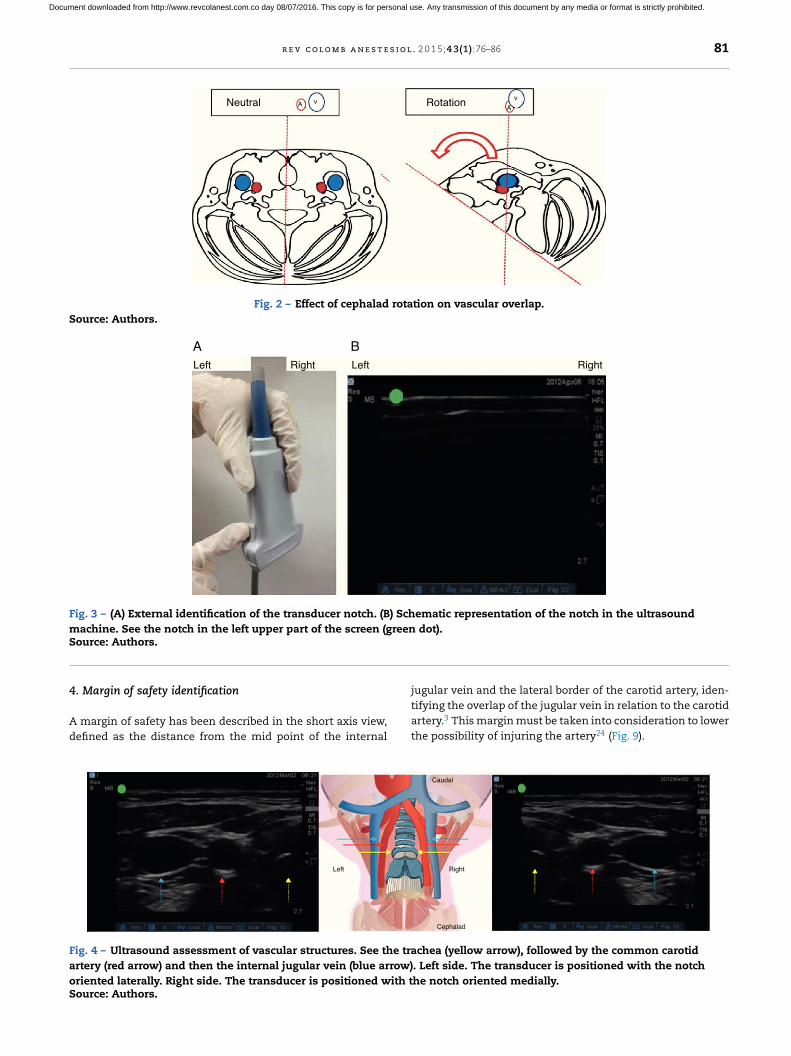
Trendelemburg positioning is recommended, with the headin neutral position or with the least contralateral rotationpossible;17,18 the operator stands at the head of the patientwith the ultrasound equipment on the ipsilateral side of thepuncture area. Prior studies have shown increased vascularoverlap associated with contralateral cephalad rotation.19–22
(Fig. 2) Wang et al. reported important loss of up to 72% of thesafety margin with increased vascular overlap when 90◦ rota-tion is used. These data have contributed to explain arterialvascular injury during puncture.
The notch in the probe helps orient laterality on the patientimage and its corresponding display on the ultrasound screen.When the notch is identified (Fig. 3A), the image is found onthe ultrasound screen (the green dot in our case) in order toserve as a guide to the imaged side and its schematic repre-sentation on the screen (Fig. 3B).
Correct probe placement allows identification of anatomi-cal structures for adequate assessment of the tracheal ringsand vascular structures (Fig. 4), lowering the probability ofpuncture error.
2. Two-dimensional (2D) ultrasound scan
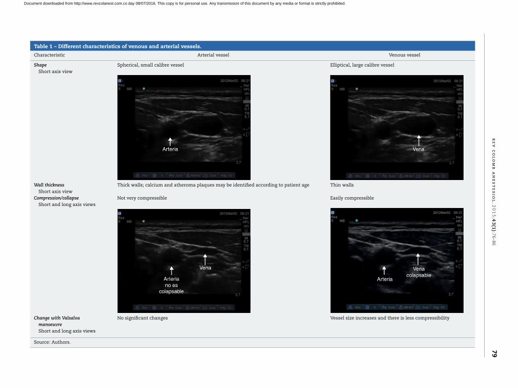
Initial vascular differentiation is done using the 2D mode toestablish the distinct characteristics of the venous and arterialvessels. It is recommended to assess the short axis, the longaxis and the oblique axis views (Table 1) in order to identifyanatomical relationships between the structures and assessfor the presence of thrombi or masses that may interfere withthe cannulation.
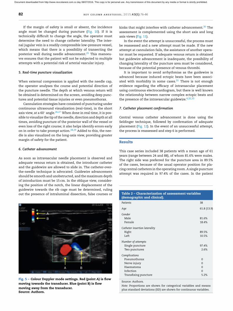
3. Colour Doppler and Pulsed Doppler assessment
Before conducting these assessments, it is important tobecome familiar with the settings of the equipment. In ourcase, for Colour Doppler we set the colour that will identifythe flow that moves towards the transducer and the flow thatmoves away from the transducer (Fig. 5).
For Pulsed Doppler assessment, an incidence angle of30–60◦ was considered. Angles of less than 60◦ must be thegoal in order to avoid velocity estimation errors.23
With the determination of blood cell mass flow veloc-ity and direction using Colour Doppler and Pulsed Dopplerassessment, venous and arterial identification is significantlyenhanced. The Colour Doppler assessment is performed at anangle of 30–60◦ in a caudal direction. On the image, the flowmoving towards the transducer is flow coming from the heartand is displayed in red. The flow that appears in blue is bloodmoving away from the transducer, in this case flow comingfrom the brain and ending in the heart (Fig. 6).
At that same point, if the assessment angle is switched to acephalad direction (120–150◦), the flow appears red, depictingblood coming from the brain, while blue flow corresponds to
Document downloaded from http://www.revcolanest.com.co day 08/07/2016. This copy is for personal use. Any transmission of this document by any media or format is strictly prohibited.
r
e
v
c
o
l
o
m
b
a
n
e
s
t
e
s
i
o
l
.
2
0
1
5;4
3(1
):76
–8
6
79
Table 1 – Different characteristics of venous and arterial vessels.
Characteristic Arterial vessel Venous vessel
Shape
Short axis viewSpherical, small calibre vessel Elliptical, large calibre vessel
Wall thickness
Short axis viewThick walls; calcium and atheroma plaques may be identified according to patient age Thin walls
Compression/collapse
Short and long axis viewsNot very compressible Easily compressible
Change with Valsalva
manoeuvre
Short and long axis views
No significant changes Vessel size increases and there is less compressibility
Source: Authors.
Document downloaded from http://www.revcolanest.com.co day 08/07/2016. This copy is for personal use. Any transmission of this document by any media or format is strictly prohibited.
80 r e v c o l o m b a n e s t e s i o l . 2 0 1 5;4 3(1):76–86
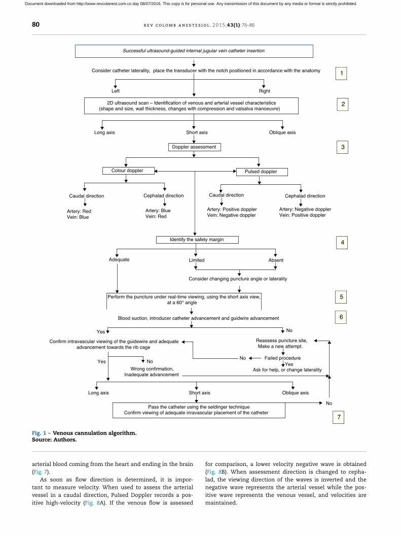
Long axis Short axis Oblique axis
Doppler assessment
Caudal direction
Consider changing puncture angle or laterality
Blood suction, introducer catheter advancement and guidwire advancement
No
Ask for help, or change laterality
Yes
Failed procedure No
Left Right
Yes No
No
1
2
3
4
5
6
7
Colour doppler Pulsed doppler
Cephalad direction
Artery: Red
Vein: Blue
Artery: Blue
Vein: Red
Caudal direction Cephalad direction
Artery: Positive doppler
Vein: Negative doppler
Artery: Negative doppler
Vein: Positive doppler
Identify the safety margin
Adequate Limited Absent
Long axis Short axis Oblique axis
Yes
Consider catheter laterality, place the transducer with the notch positioned in accordance with the anatomy
Confirm intravascular viewing of the guidewire and adequate
advancement towards the rib cage
Reassess puncture site,
Make a new attempt.
Wrong confirmation,
Inadequate advancement
Pass the catheter using the seldinger technique
Confirm viewing of adequate inravascular placement of the catheter
Perform the puncture under real-time viewing, using the short axis view,
at a 60° angle
2D ultrasound scan – Identification of venous and arterial vessel characteristics
(shape and size, wall thickness, changes with compression and valsalva manoeuvre)
Successful ultrasound-guided internal jugular vein catheter insertion
Fig. 1 – Venous cannulation algorithm.Source: Authors.
arterial blood coming from the heart and ending in the brain(Fig. 7).
As soon as flow direction is determined, it is impor-tant to measure velocity. When used to assess the arterialvessel in a caudal direction, Pulsed Doppler records a pos-itive high-velocity (Fig. 8A). If the venous flow is assessed
for comparison, a lower velocity negative wave is obtained(Fig. 8B). When assessment direction is changed to cepha-lad, the viewing direction of the waves is inverted and thenegative wave represents the arterial vessel while the pos-itive wave represents the venous vessel, and velocities aremaintained.
Document downloaded from http://www.revcolanest.com.co day 08/07/2016. This copy is for personal use. Any transmission of this document by any media or format is strictly prohibited.
r e v c o l o m b a n e s t e s i o l . 2 0 1 5;4 3(1):76–86 81
A vv
ANeutral Rotation
Fig. 2 – Effect of cephalad rotation on vascular overlap.
Source: Authors.
RightRight LeftLeft
A B
Fig. 3 – (A) External identification of the transducer notch. (B) Schematic representation of the notch in the ultrasound
machine. See the notch in the left upper part of the screen (green dot).Source: Authors.
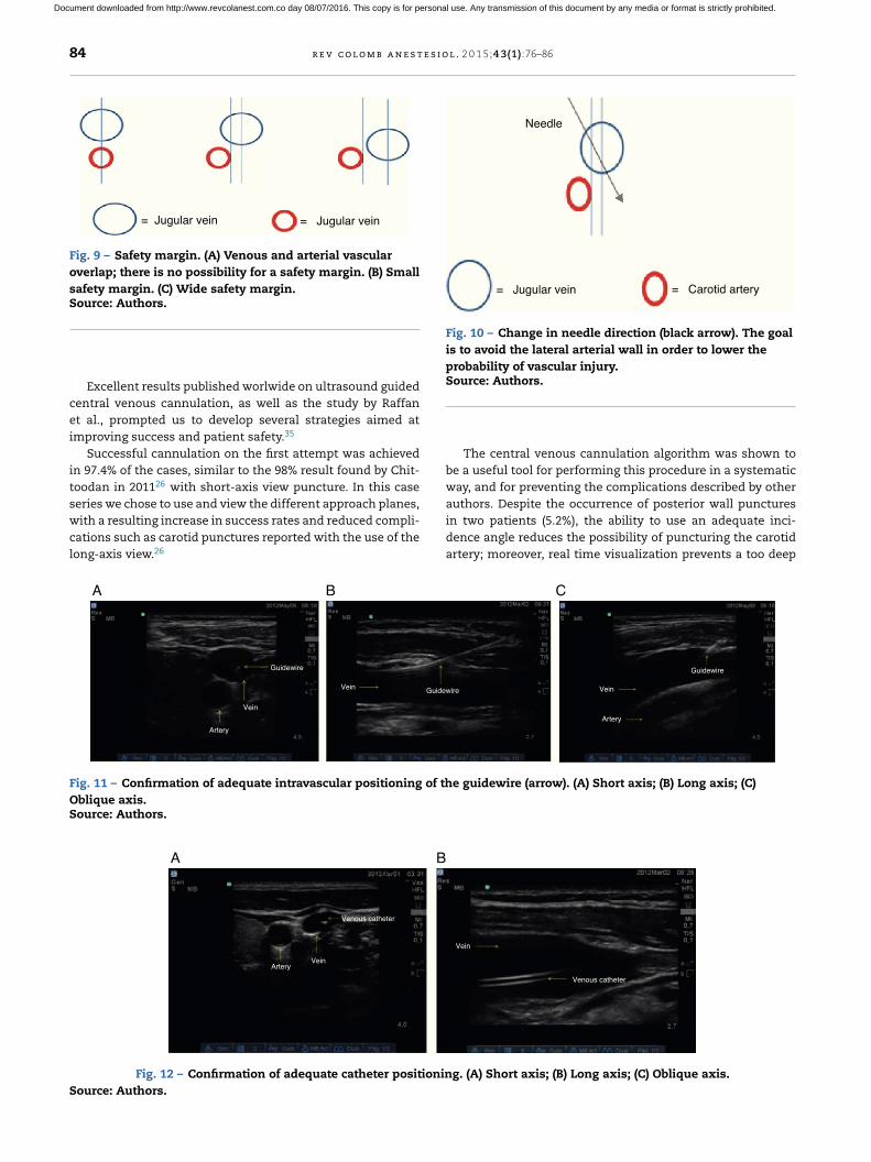
4. Margin of safety identification
A margin of safety has been described in the short axis view,defined as the distance from the mid point of the internal
jugular vein and the lateral border of the carotid artery, iden-tifying the overlap of the jugular vein in relation to the carotidartery.3 This margin must be taken into consideration to lowerthe possibility of injuring the artery24 (Fig. 9).
Caudal
Left Right
Cephalad
Fig. 4 – Ultrasound assessment of vascular structures. See the trachea (yellow arrow), followed by the common carotid
artery (red arrow) and then the internal jugular vein (blue arrow). Left side. The transducer is positioned with the notch
oriented laterally. Right side. The transducer is positioned with the notch oriented medially.Source: Authors.
Document downloaded from http://www.revcolanest.com.co day 08/07/2016. This copy is for personal use. Any transmission of this document by any media or format is strictly prohibited.
82 r e v c o l o m b a n e s t e s i o l . 2 0 1 5;4 3(1):76–86
If the margin of safety is small or absent, the incidenceangle must be changed during puncture (Fig. 10). If it istechnically difficult to change the angle, the operator mustdetermine the need to change catheter laterality. The inter-nal jugular vein is a readily compressible low-pressure vessel,which means that there is a possibility of transecting theposterior wall during needle advancement.25 This manoeu-vre ensures that the patient will not be subjected to multipleattempts with a potential risk of arterial vascular injury.
5. Real-time puncture visualization
When external compression is applied with the needle cap,the operator analyses the course and potential direction ofthe puncture needle. The depth at which venous return willbe obtained is determined on the screen, avoiding deep punc-tures and potential tissue injuries or even pneumothorax.
Cannulation strategies have consisted of puncturing undercontinuous ultrasound visualization (real-time), in the shortaxis view, at a 60◦ angle.26,27 When done in real time, it is pos-sible to visualize the tip of the needle, direction and depth at alltimes, avoiding puncture of the posterior wall of the vessel oreven loss of the right course; it also helps identify errors earlyon in order to take prompt action.28,29 Added to this, the nee-dle is also visualized on the long-axis view, providing greatermargin of safety for the patient.
6. Catheter advancement
As soon as intravascular needle placement is observed andadequate venous return is obtained, the introducer catheterand the guidewire are allowed to slide in. The catheter-over-the-needle technique is advocated. Guidewire advancementshould be smooth and unobstructed, and the maximum depthof introduction must be 15 cm. In the oblique view, consider-ing the position of the notch, the linear displacement of theguidewire towards the rib cage must be determined, rulingout the presence of intraluminal dissection, false courses or
A
A B
B
Fig. 5 – Colour Doppler mode settings. Red (point A) is flow
moving towards the transducer. Blue (point B) is flow
moving away from the transducer.Source: Authors.
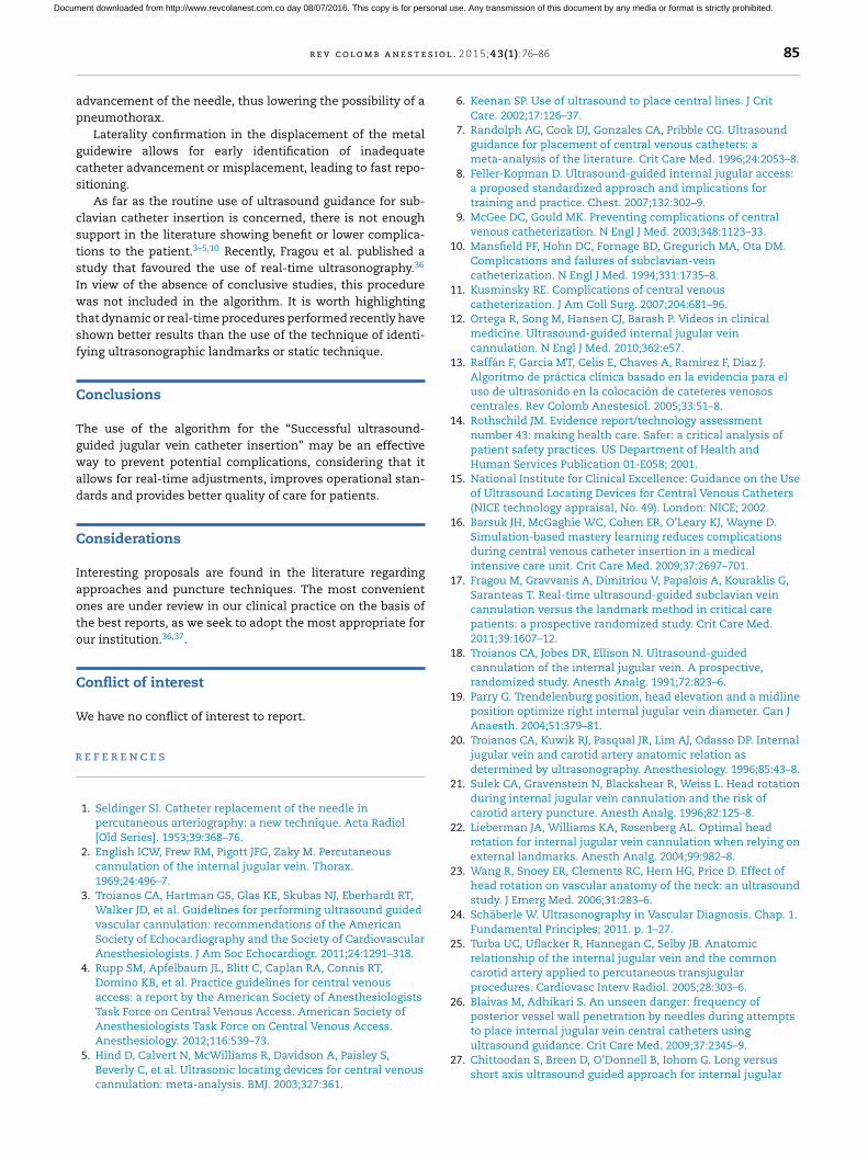
kinks that might interfere with catheter advancement.30 Theassessment is complemented using the short axis and longaxis views (Fig. 11).
In the event the attempt is unsuccessful, the process mustbe reassessed and a new attempt must be made. If the newattempt at cannulation fails, the assistance of another opera-tor must be requested. If adequate venous return is obtainedbut guidewire advancement is inadequate, the possibility ofchanging laterality of the puncture area must be considered,because of the potential presence of venous thrombi.
It is important to avoid arrhythmias as the guidewire isadvanced because induced ectopic beats have been associ-ated with morbidity in some cases.31 There is not enoughevidence regarding the efficacy of intravascular placementusing continuous electrocardiogram, but there is well knownthe relationship between narrow complex ectopic beats andthe presence of the intravascular guidewire.4,32,33
7. Catheter placement confirmation
Central venous catheter advancement is done using theSeldinger technique, followed by confirmation of adequateplacement (Fig. 12). In the event of an unsuccessful attempt,the process is reassessed and step 6 is performed.
Results
This case series included 38 patients with a mean age of 61years (range between 24 and 88), of whom 81.6% were males.The right side was preferred for the puncture area in 89.5%of the cases, because of the usual operator position for pla-cing central catheters in the operating room. A single punctureattempt was required in 97.4% of the cases. In the patient
Table 2 – Characterization of assessment variables(demographic and clinical).
Patients 38
Age 61.8 (13.9)
Gender
Male 81.6%Female 18.4%
Catheter insertion laterality
Right 89.5%Left 10.5%
Number of attempts
Single puncture 97.4%Two punctures 2.6%
Complications
Pneumothorax 0Nerve injury 0Haematoma 0Infection 0Transfixing puncture 5.2%
Source: Authors.
Note: Proportions are shown for categorical variables and meansplus standard deviations (SD) are shown for continuous variables.
Document downloaded from http://www.revcolanest.com.co day 08/07/2016. This copy is for personal use. Any transmission of this document by any media or format is strictly prohibited.

r e v c o l o m b a n e s t e s i o l . 2 0 1 5;4 3(1):76–86 83
Caudal direction
30-60 degrees
Artery Vein
BA
Fig. 6 – Caudal-orientation Colour Doppler ultrasound assessment. (A) See the 30–60◦ angle of incidence between the axis of
the transducer and the vessels of the neck. (B) Flow moving towards the transducer is arterial and is shown in red; flow in
blue is blood going to the heart, i.e. venous flow.Source: Authors.
Artery Vein
120-150 degrees
Cephalad directionBA
Fig. 7 – Cephalad-oriented Colour Doppler ultrasound assessment. (A) The angle of incidence changes to 120–150◦ between
the axis of the transducer and the vessels of the neck. (B) In this case, flow moving towards the transducer is cerebral
venous return, and is shown in red; flow in blue is blood going to the brain, i.e. arterial flow.Source: Authors.
who required two attempts, cephalad advancement of theguidewire was observed and it was decided to repeat the punc-ture (Table 2).
In 2 cases (5.2%) there was evidence of posterior vesselwall puncture unrelated with haematoma or infection. Therewere no complications like pneumothorax, air embolism,nerve injury, haematoma or infection. The assisted operatormodality was applied in 100% of the cases, meaning tutorsupervision of the resident or intern performing the proce-dure.
Discussion
Despite the significant advantages reported in the world liter-ature, in 2005 Girard et al.34 studied the use of ultrasound forguiding vascular cannulation in a level III university hospital.As part of the results, they observed that only 15% of the prac-titioners used ultrasound guidance in more than 60% of theirattempts at central vascular cannulation, showing a relativelyinfrequent use despite the favourable evidence.35
Artery
Vein
Artery
Vein
Carotid artery assessment using caudally-oriented
colour doppler
Jugular vein assessment using caudally-oriented
pulsed doppler
A B
Fig. 8 – Pulsed Doppler ultrasound assessment. (A) Arterial vessel; the blood cell mass moves towards the transducer,
creating a high velocity positive spectrum (30 cm/s in this case). (B) Venous vessel; a low-velocity negative spectrum is
observed (10 cm/s in this case), corresponding to the flow of blood cell mass away from the transducer.Source: Authors.
Document downloaded from http://www.revcolanest.com.co day 08/07/2016. This copy is for personal use. Any transmission of this document by any media or format is strictly prohibited.
84 r e v c o l o m b a n e s t e s i o l . 2 0 1 5;4 3(1):76–86
= Jugular vein = Jugular vein
Fig. 9 – Safety margin. (A) Venous and arterial vascular
overlap; there is no possibility for a safety margin. (B) Small
safety margin. (C) Wide safety margin.Source: Authors.
Excellent results published worlwide on ultrasound guidedcentral venous cannulation, as well as the study by Raffanet al., prompted us to develop several strategies aimed atimproving success and patient safety.35
Successful cannulation on the first attempt was achievedin 97.4% of the cases, similar to the 98% result found by Chit-toodan in 201126 with short-axis view puncture. In this caseseries we chose to use and view the different approach planes,with a resulting increase in success rates and reduced compli-cations such as carotid punctures reported with the use of thelong-axis view.26
Needle
= Jugular vein = Carotid artery
Fig. 10 – Change in needle direction (black arrow). The goal
is to avoid the lateral arterial wall in order to lower the
probability of vascular injury.Source: Authors.
The central venous cannulation algorithm was shown tobe a useful tool for performing this procedure in a systematicway, and for preventing the complications described by otherauthors. Despite the occurrence of posterior wall puncturesin two patients (5.2%), the ability to use an adequate inci-dence angle reduces the possibility of puncturing the carotidartery; moreover, real time visualization prevents a too deep
Guidewire
Vein
Artery
Artery
VeinGuidewire
Guidewire
Vein
A B C
Fig. 11 – Confirmation of adequate intravascular positioning of the guidewire (arrow). (A) Short axis; (B) Long axis; (C)
Oblique axis.Source: Authors.
Venous catheter
VeinArtery
Vein
Venous catheter
A B
Fig. 12 – Confirmation of adequate catheter positioning. (A) Short axis; (B) Long axis; (C) Oblique axis.
Source: Authors.
Document downloaded from http://www.revcolanest.com.co day 08/07/2016. This copy is for personal use. Any transmission of this document by any media or format is strictly prohibited.
r e v c o l o m b a n e s t e s i o l . 2 0 1 5;4 3(1):76–86 85
advancement of the needle, thus lowering the possibility of apneumothorax.
Laterality confirmation in the displacement of the metalguidewire allows for early identification of inadequatecatheter advancement or misplacement, leading to fast repo-sitioning.
As far as the routine use of ultrasound guidance for sub-clavian catheter insertion is concerned, there is not enoughsupport in the literature showing benefit or lower complica-tions to the patient.3–5,10 Recently, Fragou et al. published astudy that favoured the use of real-time ultrasonography.36
In view of the absence of conclusive studies, this procedurewas not included in the algorithm. It is worth highlightingthat dynamic or real-time procedures performed recently haveshown better results than the use of the technique of identi-fying ultrasonographic landmarks or static technique.
Conclusions
The use of the algorithm for the “Successful ultrasound-guided jugular vein catheter insertion” may be an effectiveway to prevent potential complications, considering that itallows for real-time adjustments, improves operational stan-dards and provides better quality of care for patients.
Considerations
Interesting proposals are found in the literature regardingapproaches and puncture techniques. The most convenientones are under review in our clinical practice on the basis ofthe best reports, as we seek to adopt the most appropriate forour institution.36,37.
Conflict of interest
We have no conflict of interest to report.
r e f e r e n c e s
1. Seldinger SI. Catheter replacement of the needle inpercutaneous arteriography: a new technique. Acta Radiol[Old Series]. 1953;39:368–76.
2. English ICW, Frew RM, Pigott JFG, Zaky M. Percutaneouscannulation of the internal jugular vein. Thorax.1969;24:496–7.
3. Troianos CA, Hartman GS, Glas KE, Skubas NJ, Eberhardt RT,Walker JD, et al. Guidelines for performing ultrasound guidedvascular cannulation: recommendations of the AmericanSociety of Echocardiography and the Society of CardiovascularAnesthesiologists. J Am Soc Echocardiogr. 2011;24:1291–318.
4. Rupp SM, Apfelbaum JL, Blitt C, Caplan RA, Connis RT,Domino KB, et al. Practice guidelines for central venousaccess: a report by the American Society of AnesthesiologistsTask Force on Central Venous Access. American Society ofAnesthesiologists Task Force on Central Venous Access.Anesthesiology. 2012;116:539–73.
5. Hind D, Calvert N, McWilliams R, Davidson A, Paisley S,Beverly C, et al. Ultrasonic locating devices for central venouscannulation: meta-analysis. BMJ. 2003;327:361.
6. Keenan SP. Use of ultrasound to place central lines. J CritCare. 2002;17:126–37.
7. Randolph AG, Cook DJ, Gonzales CA, Pribble CG. Ultrasoundguidance for placement of central venous catheters: ameta-analysis of the literature. Crit Care Med. 1996;24:2053–8.
8. Feller-Kopman D. Ultrasound-guided internal jugular access:a proposed standardized approach and implications fortraining and practice. Chest. 2007;132:302–9.
9. McGee DC, Gould MK. Preventing complications of centralvenous catheterization. N Engl J Med. 2003;348:1123–33.
10. Mansfield PF, Hohn DC, Fornage BD, Gregurich MA, Ota DM.Complications and failures of subclavian-veincatheterization. N Engl J Med. 1994;331:1735–8.
11. Kusminsky RE. Complications of central venouscatheterization. J Am Coll Surg. 2007;204:681–96.
12. Ortega R, Song M, Hansen CJ, Barash P. Videos in clinicalmedicine. Ultrasound-guided internal jugular veincannulation. N Engl J Med. 2010;362:e57.
13. Raffán F, Garcia MT, Celis E, Chaves A, Ramirez F, Diaz J.Algoritmo de práctica clínica basado en la evidencia para eluso de ultrasonido en la colocación de cateteres venososcentrales. Rev Colomb Anestesiol. 2005;33:51–8.
14. Rothschild JM. Evidence report/technology assessmentnumber 43: making health care. Safer: a critical analysis ofpatient safety practices. US Department of Health andHuman Services Publication 01-E058; 2001.
15. National Institute for Clinical Excellence: Guidance on the Useof Ultrasound Locating Devices for Central Venous Catheters(NICE technology appraisal, No. 49). London: NICE; 2002.
16. Barsuk JH, McGaghie WC, Cohen ER, O’Leary KJ, Wayne D.Simulation-based mastery learning reduces complicationsduring central venous catheter insertion in a medicalintensive care unit. Crit Care Med. 2009;37:2697–701.
17. Fragou M, Gravvanis A, Dimitriou V, Papalois A, Kouraklis G,Saranteas T. Real-time ultrasound-guided subclavian veincannulation versus the landmark method in critical carepatients: a prospective randomized study. Crit Care Med.2011;39:1607–12.
18. Troianos CA, Jobes DR, Ellison N. Ultrasound-guidedcannulation of the internal jugular vein. A prospective,randomized study. Anesth Analg. 1991;72:823–6.
19. Parry G. Trendelenburg position, head elevation and a midlineposition optimize right internal jugular vein diameter. Can JAnaesth. 2004;51:379–81.
20. Troianos CA, Kuwik RJ, Pasqual JR, Lim AJ, Odasso DP. Internaljugular vein and carotid artery anatomic relation asdetermined by ultrasonography. Anesthesiology. 1996;85:43–8.
21. Sulek CA, Gravenstein N, Blackshear R, Weiss L. Head rotationduring internal jugular vein cannulation and the risk ofcarotid artery puncture. Anesth Analg. 1996;82:125–8.
22. Lieberman JA, Williams KA, Rosenberg AL. Optimal headrotation for internal jugular vein cannulation when relying onexternal landmarks. Anesth Analg. 2004;99:982–8.
23. Wang R, Snoey ER, Clements RC, Hern HG, Price D. Effect ofhead rotation on vascular anatomy of the neck: an ultrasoundstudy. J Emerg Med. 2006;31:283–6.
24. Schäberle W. Ultrasonography in Vascular Diagnosis. Chap. 1.Fundamental Principles; 2011. p. 1–27.
25. Turba UC, Uflacker R, Hannegan C, Selby JB. Anatomicrelationship of the internal jugular vein and the commoncarotid artery applied to percutaneous transjugularprocedures. Cardiovasc Interv Radiol. 2005;28:303–6.
26. Blaivas M, Adhikari S. An unseen danger: frequency ofposterior vessel wall penetration by needles during attemptsto place internal jugular vein central catheters usingultrasound guidance. Crit Care Med. 2009;37:2345–9.
27. Chittoodan S, Breen D, O’Donnell B, Iohom G. Long versusshort axis ultrasound guided approach for internal jugular
Document downloaded from http://www.revcolanest.com.co day 08/07/2016. This copy is for personal use. Any transmission of this document by any media or format is strictly prohibited.
86 r e v c o l o m b a n e s t e s i o l . 2 0 1 5;4 3(1):76–86
vein cannulation: a prospective randomised controlled trial.Med Ultrason. 2011;13:21–5.
28. Blaivas M, Brannam L, Fernandez E. Short-axis versuslong-axis approaches for teaching ultrasound-guidedvascular access on a new inanimate model. Acad Emerg Med.2003;10:1307–11.
29. Augoustides JG, Horak J, Ochroch AE, Vernick WJ, Gambone AJ,Weiner J. A randomized controlled clinical trial of real-timeneedle-guided ultrasound for internal jugular venouscannulation in a large university anesthesia department. JCardiothorac Vasc Anesth. 2005;19:310–5.
30. Rabindranath KS, Kumar E, Rabindranath KS, Kumar E, ShailR, Vaux E. Use of real-time ultrasound guidance for theplacement of hemodialysis catheters: a systematic reviewand meta-analysis of randomized controlled trials. Am JKidney Dis. 2011;58:964–70.
31. Gillman LM, Blaivas M, Lord J, Al-Kadi A, Kirkpatrick AW.Ultrasound confirmation of guidewire position may eliminateaccidental arterial dilatation during central venouscannulation. Scand J Trauma Resusc Emerg Med. 2010;18:39.
32. Bhutta ST, Culp WC. Evaluation and management of centralvenous access complications. Tech Vasc Interv Radiol.2011;14:217–24.
33. Lee JH, Bahk JH, Ryu HG, Jung CW, Jeon Y. Comparison of thebedside central venous catheter placement techniques:landmark vs electrocardiogram guidance. Br J Anaesth.2009;102:662–6. Epub 2009 March 26.
34. Jeon Y, Ryu HG, Yoon SZ, Kim JH, Bahk JH. Transesophagealechocardiographic evaluation of ECG-guided central venouscatheter placement. Can J Anaesth. 2006;53:978–83.
35. Girard TD, Schectman JM. Ultrasound guidance during centralvenous catheterization: a survey of use by house staffphysicians. J Crit Care. 2005;20:224–9.
36. Raffán F, Guerrero C. La canalización de la vena yugularinterna guiada por ultrasonografía en pacientes sometidos atrasplante hepático. Rev Colomb Anestesiol. 2001;29:295–300.
37. Dilisio R, Mittnacht AJ. The “medial-oblique” approach toultrasound-guided central venous cannulation-maximize theview, minimize the risk. J Cardiothorac Vasc Anesth.2012;26:982–4.
Document downloaded from http://www.revcolanest.com.co day 08/07/2016. This copy is for personal use. Any transmission of this document by any media or format is strictly prohibited.