Growth inhibition of mammalian cells by eosinophil cationic protein
Upload
independentCategory
view
6download
0
b i o c h e m i c a l p h a r m a c o l o g y 7 3 ( 2 0 0 7 ) 3 9 4 – 4 0 4
Inhibition of the intestinal absorption of bile acids usingcationic derivatives: Mechanism and repercussions
Marta Vicens a, Rocio I.R. Macias a, Oscar Briz a, Alfonso Rodriguez a,Mohamad Y. El-Mir a, Manuel Medarde b, Jose J.G. Marin a,*aDepartment of Physiology and Pharmacology, Campus Miguel de Unamuno, University of Salamanca, 37007 Salamanca, SpainbDepartment of Pharmaceutical Chemistry, Campus Miguel de Unamuno, University of Salamanca, 37007 Salamanca, Spain
a r t i c l e i n f o
Article history:
Received 14 September 2006
Accepted 13 October 2006
Keywords:
ASBT
Enterohepatic circulation
Ileum
Intestine
Liver
Polyamine
Transport
a b s t r a c t
To pharmacologically interrupt bile acid enterohepatic circulation, two compounds named
BAPA-3 and BAPA-6, with a steroid structure and 1 or 2 positive charges, were obtained by
conjugation of N-(3-aminopropyl)-1,3-propanediamine with one or two moieties of glyco-
cholic acid (GC). Both BAPA-3 and BAPA-6 inhibited Na+-dependent taurocholate (TC) uptake
by Xenopus laevis oocytes expressing rat Asbt, with Ki values of 28 and 16 mM, respectively.
BAPA-3 reduced Vmax without affecting Km. In contrast, BAPA-6 increased Km, with no effect
on Vmax. Uptake of [14C]-GC by the last 10 cm of the rat ileum, perfused in situ over 60 min,
was inhibited to a similar extent by unlabeled GC, BAPA-3 and BAPA-6. However, the
intestinal absorption of these compounds was lower (BAPA-6) or much lower (BAPA-3)
than that of GC. When administered orally to mice, both compounds (BAPA-3 > BAPA-6)
reduced the bile acid pool size, which was accompanied by up-regulation of hepatic Cyp7a1
and Hmgcr and intestinal Osta/Ostb. A tendency towards a decreased expression of hepatic
Ntcp and an enhanced expression of intestinal Asbt was also observed. Serum biochemical
parameters were not affected by treatment with these compounds, except for a moderate
increase in serum triglyceride concentrations. In sum, our results suggest that these
compounds, in particular BAPA-3, are potentially useful tools for inhibiting the intestinal
absorption of bile acids in a non-competitive manner.
# 2006 Elsevier Inc. All rights reserved.
avai lable at www.sc iencedi rec t .com
journal homepage: www.e lsev ier .com/ locate /b iochempharm
1. Introduction
Hypercholesterolemia is one of the most important risk
factors for the development of cardiovascular diseases owing
to the increased probability of atheromatose lesions [1]. This
justifies the enormous efforts carried out in the last decades
to obtain new cholesterol-lowering drugs. One of the
strategies that have produced the best results has been the
* Corresponding author. Tel.: +34 923 294674; fax: +34 923 294669.E-mail address: [email protected] (Jose J.G. Marin).
Abbreviations: ASBT, apical sodium-dependent bile acid transporterCDCA, chenodeoxycholic acid; GC, glycocholic acid; NTCP, Na+-taurochTC, taurocholic acid0006-2952/$ – see front matter # 2006 Elsevier Inc. All rights reserveddoi:10.1016/j.bcp.2006.10.014
one based on the inhibition of the key enzyme involved in
cholesterol synthesis, i.e., hydroxymethyl glutaryl coenzyme
A reductase (HMGCR), by statins. However, in spite of its
efficacy, this family of compounds is not the definitive
solution. The long-term side effects are only partially
understood and its good tolerability cannot be extended to
children with genetic disorders affecting cholesterol home-
ostasis [2].
; BAPA, bile acid-polyamine derivative; BSEP, bile salt export pump;olate-cotransporting polypeptide; OST, organic solute transporter;
.

Fig. 1 – Schematic representation of the molecular structure
of conjugates of N-(3-aminopropyl)-1,3-propanediamine
with one or two glycocholic acid moieties to obtain BAPA-
3 and BAPA-6, respectively.
b i o c h e m i c a l p h a r m a c o l o g y 7 3 ( 2 0 0 7 ) 3 9 4 – 4 0 4 395
Alternative pharmacological approaches include the sti-
mulation of cholesterol biotransformation into bile acids
subsequent to an increased loss of these compounds due to
their sequestering in the intestinal lumen using specific gels or
resins. Cholesterol-7a-hydroxylase (CYP7A1), the key enzyme
in bile acid synthesis [3], is down regulated by bile acids [4].
Thus, by depleting the bile acid pool an enhancement in the
expression of cholesterol-7a-hydroxylase occurs, which is
subsequently accompanied by enhanced removal of choles-
terol, which is then biotransformed into bile acids [5].
Although the conceptual basis of this therapy is interesting,
the pharmaceuticals available are difficult to manipulate and
administer. Moreover they are not devoid of adverse con-
sequences [6,7].
Since the molecular mechanisms accounting for intest-
inal bile acid re-absorption are being elucidated [8,9], the
alternative of favouring the foecal elimination of these
steroids by inhibiting the plasma membrane carriers
responsible for bile acid uptake has emerged as a promising
alternative. The apical sodium-dependent bile acid trans-
porter (ASBT), located at the brush-border membrane of
epithelial cells in the ileal mucosa, is a member of the family
10 of solute carriers encoded by the gene SLC10A2 [10]. Since
this is the main transport system involved in the active
reabsorption of bile acids during the intestinal transit of their
enterohepatic circulation, the inhibition of bile acid uptake
by this carrier would result in increased fecal loss of bile
acids.
Several investigators and pharmaceutical companies have
developed ASBT-inhibitors with very different chemical
structures. Among them, the following can be mentioned
as examples: SC-635 [11], S-8921 [12], 2164U90 [13], 2,3-
disubstituted-4-phenylquinolines [14]. Regarding the use of
bile acid derivatives, several studies carried out in the 70 s
using everted hamster gut sac as an experimental model
revealed that cationic bile salt derivatives were able to
interact with the ileal bile acid transport system in such a way
as to inhibit the transport of natural bile salts [15]. This
interaction seemed to involve two recognition components;
one includes the steroid moiety, the other a coulombic
interaction between the anionic bile salt and a cationic
membrane site [16].
In later studies, using isolated perfused rat liver [17] and in
vivo measurement of the absorption from the jejunum and
ileum of anesthetized guinea pigs [18], it was shown that fully
positively charged bile acid derivatives or zwitterionic bile acid
derivatives were not appreciably taken up by the liver or the
intestine.
More recently, once the transporters involved in these
processes were cloned and expressed in cell lines, it has been
possible to investigate substrate specificity of sodium/bile acid
co-transporters that are the main responsible for bile acid
uptake by the liver (Na+-taurocholate co-transporting poly-
peptide, NTCP, gene symbol SLC10A1) and the ileum (ASBT).
Studies carried out in rabbit orthologues have suggested that
the side chain of bile acids is important for their interaction
with the recognition site of ASBT and NTCP [19]. Regarding
human ASBT, a recent study suggests that C-24 conjugation
and steroidal hydroxylation pattern modulate native bile acid
interaction with human ASBT, with the effect of conjugation
dominating that of steroidal hydroxylation. Moreover the
results of that study indicate that bile acid binding to human
ASBT may be the rate-limiting step in the apical transport of
bile acids [20].
Based on the results mentioned above, the conceptual base
of the present work was that a negative charge in the lateral
chain is needed for natural bile acids to interact with the main
intestinal carrier accounting for bile acid uptake from the
intestinal lumen, i.e., ASBT [18]. This prompted us to design
and synthesize steroids with different sizes and net positive
charges (Fig. 1) and to evaluate their ability to inhibit ASBT-
mediated bile acid intestinal absorption and to affect several
physiological aspects related to the enterohepatic circulation
of these compounds.
2. Methods and materials
2.1. Reagents
FITC-Dextran-40 kDa, N-(3-aminopropyl)-1,3-propanedia-
mine (PA) and sodium salts of glycocholic acid (GC), tauro-
cholic acid (TC) and chenodeoxycholic acid (CDCA) more than
95% pure by thin-layer chromatography were purchased from
Sigma–Aldrich (Madrid, Spain). [14C]-GC (specific radioactivity
46.7 mCi/mmol) and [3H]-TC (specific radioactivity 3.0 Ci/
mmol) were obtained from Perkin-Elmer Life Science (Izasa,
Barcelona, Spain). All other chemicals were from Merck
Eurolab (Barcelona, Spain). They were of high purity and were
used as purchased without any further purification. The GC
and PA conjugates named BAPA-3 and BAPA-6 whose
molecular structures are shown in Fig. 1, were obtained by
an adaptation of a previously described procedure for the
b i o c h e m i c a l p h a r m a c o l o g y 7 3 ( 2 0 0 7 ) 3 9 4 – 4 0 4396
synthesis of amide derivatives of bile acids [21] and purified to
more than 95% by semi-preparative liquid chromatography
using chloroform/methanol/acetic acid/water (65:24:15:9) (v/v)
as the solvent system. The purity of final products was
checked by high performance liquid chromatography in
reverse phase.
2.2. Animals
Male Wistar CF rats and CD1 mice (Animal House, University
of Salamanca, Spain) and mature female frogs (Xenopus laevis;
Regine Olig, Hamburg, Germany) were fed on commercial-
pelleted rat, mouse or Xenopus food (Panlab, Madrid, Spain).
Lighting was controlled by a timer that permitted light
between 8:00 a.m. and 8:00 p.m. All animals received humane
care as outlined in ‘‘Institutional Animal Care and Use
Committee Guidebook’’ (2nd ed., 2002). Experimental proto-
cols were approved by the Ethical Committee for Laboratory
Animals of the University of Salamanca.
2.3. Uptake studies in Xenopus laevis oocytes
After anaesthetizing the frogs by intramuscular administra-
tion of 12.5 mg ketamine in the leg (Imalgene 500; Rhone
Merieux, Barcelona, Spain), the harvesting and preparation of
oocytes were carried out as described elsewhere [22]. The
oocytes were then microinjected with TE buffer (1 mM EDTA,
10 mM Tris, pH 8.0) alone (Control) or with 9 ng of the mRNA of
the rat orthologue of ASBT (Asbt). This was synthesized using
the T7 mMessage mMachine Ultrakit (Ambion, bioNova,
Madrid, Spain) and a recombinant plasmid obtained by
subcloning the DNA corresponding to the ORF of rat Asbt
between the EcoRI and HindIII sites of the pSPORT 1 plasmid.
The transferred DNA was obtained from pCMV5/rAsbt
plasmid kindly supplied by Dr. Paul Dawson (Wake Forest
University School of Medicine, Winston-Salem, NC). Oocytes
were used 2 days after RNA injection, when – on the basis of
preliminary experiments on the time-course of functional
expression for this carrier – the uptake rate was highest (data
not shown).
Uptake studies were carried out using groups of 8–10
oocytes per data point. Experiments were repeated three times
using different frogs. The oocytes were washed with sub-
strate-free uptake medium and incubated with 100 ml of
uptake medium (100 mM NaCl or 100 mM cholineCl, 2 mM KCl,
1 mM CaCl2, 1 mM MgCl2 and 10 mM Hepes/Tris, pH 7.5)
containing the desired amount of substrate and inhibitor to be
tested at 25 8C for the indicated time. Uptake was stopped by
the addition of 4 ml ice-cold uptake medium. The oocytes were
washed three more times before being collected and indivi-
dually placed in vials. They were digested with 200 ml of 10%
(w/v) SDS before measuring the radioactivity due to radi-
olabeled substrate [22].
2.4. In situ single-pass perfusion of the rat ileum
Overnight fasted animals were anaesthetized with sodium
pentobarbital (Nembutal N.R., Abbot, Madrid, Spain) (50 mg/
kg body weight, i.p.), prior to performing the surgical
preparation. A catheter was placed in the common bile duct
to drain bile throughout the experimental period. The
pylorus was ligated to prevent any passage of stomach
contents to the small intestine and two catheters were
implanted in the ileum: (i) The inflow catheter at 10 cm away
from the ileal-cecal junction in the ileum. (ii) The outflow
catheter was implanted at ileal-cecal junction. Using a
peristaltic pump, the ileal segment located between both
catheters was washed with warmed (37 8C) perfusion
medium (120 mM NaCl, 5 mM KCl, 0.65 mM MgSO4�7H2O,
1.17 mM KH2PO4, 1.29 mM CaCl2�2H2O, 25 mM NaHCO3,
100 mg/l gentamycin, pH 7.40) at 0.2 ml/min, until the
effluent was colourless (approximately 10 min). Then, an
additional period of 10 min perfusion was allowed before
starting the experimental period with a 15 min administra-
tion period during which the perfusion medium was
replaced by a similar one containing substrate alone or
together with inhibitors (0–15 min). Fresh perfusion medium
was used to perfuse the ileal segment for the next 45 min
period. Throughout the experimental period, outflowing
perfusate was collected in pre-weighed vials every 10 min.
Volume was determined gravimetrically and the uptake of
the compounds was measured by the difference in contents
between the inflow and the outflow perfusate. Using
appropriate calibration curves for each compound, GC,
BAPA-3 and BAPA-6 were measured by enzymatic detection
of the 3a-hydroxyl group [23], which is present in these three
compounds.
2.5. In vivo experiments in mice
To investigate the effect on the bile acid pool, this was labeled
as follows: 3-month-old male CD1 mice of approximately 30–
40 g body weight received an intragastric administration of
0.23 mCi [3H]-TC in 500 ml saline (time 0 h). After 24 h, the
animals received in a similar way 20 mmol BAPA-3 or BAPA-6
or vehicle alone (500 ml saline:ethanol, 9:1, v/v). Mice were
fasted from 48 to 72 h and then anaesthetized with sodium
pentobarbital (i.p., 0.5 mg/10 g body weight) before under-
going laparotomy, blood extraction (�1 ml) from the cava
vein, and subsequent removal of the liver, the gallbladder,
the small intestine and the mesenterium. These organs were
washed with ice-cold saline, weighed, and homogenized in
saline (1:2; w/v). Radioactivity in the homogenate was
measured and used as an internal standard to follow the
extraction procedure. In brief, after addition of two volumes
of ethanol to the homogenized tissues and incubation at
65 8C for 2 h, bile acids were extracted in the supernatant
resulting from centrifugation at 2200 � g for 10 min. The
process was repeated with the pellet and the two super-
natants pooled together and filtered through filter paper. Bile
acid concentrations were measured enzymatically [23]. Bile
acid pool size was calculated after correcting by the yield of
the extraction procedure using values of radioactivity
measured in the initial homogenate and in the final
extraction solution. The average yield was 93.6%. In control
animals, the average amount of radioactivity in the homo-
genate was 65.4% of the dose given 3 days before. In separate
similar experiments, the mice did not receive radiolabeled
bile acids and they were used to determine serum biochem-
ical parameters by routine biochemical analysis and the liver

Table 1 – Gene-specific primers used for quantitative real-time RT-PCR analysis of mouse mRNA
Target DNA Forward primer (50–30) Reverse primer (50–30) Product size (bp) Accession number
Slc10a1 GGCCACAGACACTGCGCT AGTGAGCCTTGATCTTGCTGAACT 101 NM_011387
Slc10a2 TTGCACAGCACAAGCAGTGA TGCATTGAAGTTGCTCTCAGGT 103 NM_011388
OSTa CAGATCGCTTGCTCACCTCC GGTCCAAGCCACTCTCCTCA 200 NM_145932
OSTb GATGCGGCTCCTTGGAATTA TTCGATTTCTGTTTGCCAGGAT 103 NM_178933
Cyp7a1 TGAGACCTCCGGGCCTTC CGTTAGATATCCGGCTTCAAACA 110 NM_007824
Cyp27a1 GGCCTGGATAGGGCTCATAGT TCCAGGAGCGTCCATCTCA 102 NM_024264
Hmgcr TGCCTGGATGGGAAGGAGTA TCGAGTCATCCCATCTGCAA 138 XM_127496
Slc10a1, Ntcp (Na+-taurocholate cotransporting polypeptide); Slc10a2, Asbt (apical sodium-dependent bile acid transporter); OST, organic solute
transporter; Cyp7a1, cholesterol 7a-hydroxylase; Cyp27a1, sterol 27-hydroxylase; Hmgcr, 3-hydroxy-3-methylglutaryl-coenzyme A reductase.
Fig. 2 – (A) Time-course of [3H]-taurocholic acid ([3H]-TC)
uptake by Xenopus laevis oocytes microinjected with
vehicle (TE buffer, triangles) or with 9 ng of rat Asbt mRNA
(squares) and incubated 2 days later with 10 mM [3H]-TC in
the presence (open symbols) or absence (closed symbols)
of sodium. (B) Effect of substrate concentration (S) on
specific sodium-dependent [3H]-TC uptake (V) as defined
as the difference in uptake in the presence and absence of
sodium in the incubation medium of oocytes expressing
rat Asbt mRNA. Uptake of [3H]-TC by oocytes injected with
TE buffer alone was subtracted in each condition. Uptake
values are mean W S.D. from measurements carried out in
30 oocytes from three different frogs per data-point.
b i o c h e m i c a l p h a r m a c o l o g y 7 3 ( 2 0 0 7 ) 3 9 4 – 4 0 4 397
and the small intestine were employed to measure gene
expression.
2.6. Quantitation of gene expression by real-time RT-PCR
Since bile acids play a crucial role in the regulation of enzymes
and transporters involved in bile acid and cholesterol
homeostasis the degree of expression of some of these
proteins was indirectly determined by measuring steady-
state levels of their mRNA. Freshly obtained mouse tissues
were immersed in the RNAlater RNA-stabilization reagent
(Ambion, bioNova Cientifica, Madrid) and stored at �80 8C
until use. Total RNA was isolated from these samples using
RNeasy spin columns from Qiagen (Izasa, Barcelona). After
treatment with RNase-free DNase I (Roche Diagnostics,
Barcelona), RNA was quantified fluorimetrically with the
RiboGreen RNA-Quantitation kit (Molecular Probes, Leiden,
The Netherlands). DNA was synthesized using random
nonamers and avian myeloblastosis virus reverse transcrip-
tase (enhanced Avian RT-PCR kit; Sigma–Genosys, Cam-
bridge, UK). Primer oligonucleotides obtained from Sigma–
Genosys were designed with the assistance of Primer Express
software (Perkin-Elmer Applied Biosystems, Madrid) for DNA
fragments in described sequences, and their specificity was
checked using BLAST. Their nucleotide sequences for mouse
genes are shown in Table 1 and those for human ASBT, OSTa
and OSTb have been reported elsewhere [24]. Real-time
quantitative PCR was then performed using AmpliTaq Gold
polymerase (Perkin-Elmer Applied Biosystems) in an ABI-
Prism 5700 Sequence Detection System (Perkin-Elmer
Applied Biosystems). The thermal cycling conditions were:
one cycle at 95 8C for 10 min followed by 40 cycles at 95 8C for
15 s and at 60 8C for 60 s. Detection of amplification products
was carried out using SYBR Green I (Perkin-Elmer Applied
Biosystems). The absence of artifacts or non-specific products
of PCR, which was checked using 2.5% agarose gel electro-
phoresis and melting temperature curves, permitted the use
of SYBR Green I detection in all cases. The results of mRNA
abundance for each target gene in each sample were
normalized on the basis of its 18S rRNA content, which was
measured with the TaqMan1 Ribosomal RNA Control
Reagents kit (Perkin-Elmer Applied Biosystms).
2.7. Statistical analysis
Results are expressed as mean � S.D. To calculate the
statistical significance of the differences among groups
the Student’s t-test or the Bonferroni method of multiple-
range testing were used, as appropriate. Regression lines
in kinetic studies were calculated by the least squares
method.

b i o c h e m i c a l p h a r m a c o l o g y 7 3 ( 2 0 0 7 ) 3 9 4 – 4 0 4398
3. Results
3.1. Studies in Xenopus laevis oocytes expressing rat Asbt
Studies on the time-course of TC uptake by Xenopus laevis
oocytes revealed that the expression of rat Asbt and the
presence of sodium in the extracellular medium markedly
enhanced this process (Fig. 2A). Moreover, these results
suggested that 10 min was the right time to determine TC
uptake under the initial velocity (V) conditions. Under these
circumstances, TC uptake was dependent on substrate
concentrations (S), resulting in a typical plot for saturable
processes (Fig. 2B). Indeed, the best fit was a Michaelis–Menten
equation whose Vmax and Km values were approximately 11–
25 pmol/oocyte/10 min and 30–70 mM, respectively. When
BAPA-3 or BAPA-6 was added to the incubation medium, TC
uptake was markedly reduced. Kinetic studies based on Dixon
approaches [25] were carried out to characterize this inhibi-
tion. In a first step, TC uptake at varying substrate concentra-
tions was measured in the presence or absence of a fixed
amount of BAPA-3 or BAPA-6. Upon plotting V versus V/S and
S/V versus S, statistically significant (P < 0.001) correlations
were found (Fig. 3). These plots suggested that BAPA-3 induced
a decrease in Vmax without affecting Km, which is the typical
behavior for a non-competitive inhibitor. In contrast, BAPA-6
Fig. 3 – Effect of the presence of 25 mM BAPA-3 (A and B) or BAP
affinity constant (Km) obtained by plotting the results of specific s
vs. substrate concentrations (S) in two different ways: V vs. V/S
(B and D). Uptake values are mean W S.D. from measurements ca
point. S.D. bars are not seen because they are smaller than the
induced a competitive inhibition, as suggested by the absence
of effect on Vmax, together with an enhancement in the value
ofKm. To calculate the inhibition constant (Ki), a different set of
experiments was carried out, measuring TC uptake by oocytes
incubated with varying concentrations of BAPA-3 or BAPA-6
and two different substrate concentrations (Fig. 4). By plotting
1/V versus inhibitor concentrations, it was possible to obtain
two curves (both P < 0.001) whose intersection and extrapola-
tion to the X-axis permitted us to calculate Ki for BAPA-3 and
BAPA-6. These values were 28 and 16 mM, respectively.
3.2. Bile acid uptake by the in situ perfused rat ileum
Preparations of in situ perfused rat ileum maintained their
viability throughout the experimental period, as indicated by
the relative steady-state in the outflow perfusion rate (Fig. 5A).
Moreover, in some experiments the absence of marked
changes in epithelial integrity and net water balance between
the luminal content and the animal was confirmed by
including in the perfusion medium the non-absorbable
fluorescent compound FITC-dextran-40 kDa. The concentra-
tion of this compound in the outflow remained relatively
steady and similar to that seen in the inflow perfusate
throughout the perfusion period (data not shown). When [14C]-
GC was administered over 15 min, the radioactivity found in
A-6 (C and D) on maximal velocity of transport (Vmax) and
odium-dependent [3H]-taurocholic acid ([3H]-TC) uptake (V)
to calculate Vmax (A and C), and S/V vs. S to calculate Km
rried out in 30 oocytes from three different frogs per data-
symbols.

Fig. 4 – Effect of the presence of varying concentrations of BAPA-3 (A and B) or BAPA-6 (C and D) on specific sodium-
dependent [3H]-taurocholic acid ([3H]-TC) uptake (V), which was measured using two different substrate concentrations
(10 and 30 mM). Uptake values are mean W S.D. from measurements carried out in 30 oocytes from three different frogs per
data-point. When S.D.-bars are not seen is because they are smaller than the symbols. The absolute value of the inhibition
constant (Ki) was calculated by the intersection of both curves.
b i o c h e m i c a l p h a r m a c o l o g y 7 3 ( 2 0 0 7 ) 3 9 4 – 4 0 4 399
the outflowing pefusate for the 60 min that followed the start
of the administration period was 46% of the dose administered
(0.15 mmol) (Fig. 5A). Thus, under these experimental circum-
stances the absorption of [14C]-GC was calculated to be
0.081 mmol. This was confirmed by determination of the
radioactivity in bile samples collected during this time, which
was 98% of the amount of [14C]-GC removed from the ileal
perfusate (data not shown). When [14C]-GC was administered
together with 1.5 mmol of unlabeled GC, BAPA-3 or BAPA-6, a
marked inhibition in [14C]-GC absorption was found (Fig. 5B).
Measurement by an enzymatic technique [23], as described
above, of the difference between the amount of these
compounds in the inflow and outflow perfusate revealed that
the absorption of BAPA-6 was lower than that of GC, whereas
that of BAPA-3 was much lower (Fig. 5C).
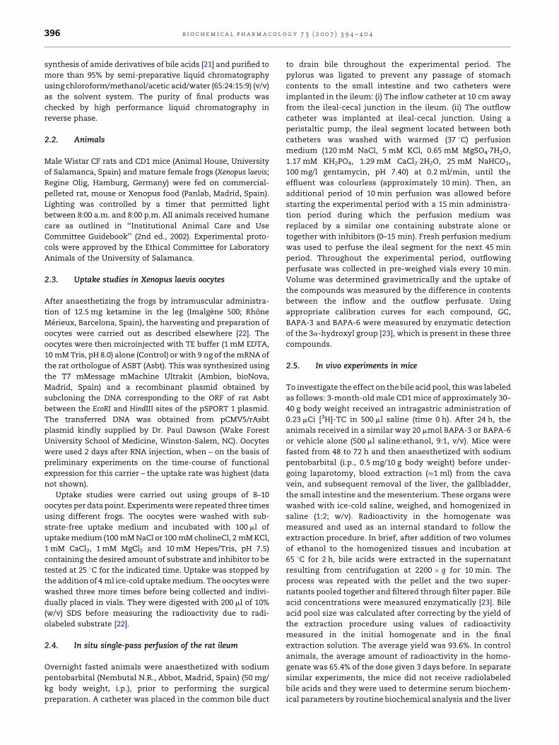
3.3. Effect of administration of BAPA-3 and BAPA-6 tomice in vivo
To investigate the in vivo physiological repercussions in mice
of the inhibition of bile acid absorption by the intestine, the
bile acid pool size, biochemical parameters and the expression
levels of the key enzymes and transporters involved in bile
acid homeostasis were measured 2 days after intra-gastric
administration of BAPA-3 or BAPA-6. Both compounds were
able to reduce the bile acid pool size without affecting serum
biochemical parameters, except for a moderate increase in
serum triglyceride concentrations, which was only statisti-
cally significant for BAPA-6 (Table 2). Similar hypertriglycer-
idemia has been also observed to accompany several
strategies able to lower the transhepatic flux of bile acids
[26]. Regarding the effects on liver expression (Fig. 6), no
significant change in the levels of mRNA for Cyp27a1 was
found. A tendency for Ntcp to be reduced was observed, but
the differences were not significant. However, the expression
of Cyp7a1 and Hmgcr was clearly increased after treatment
with BAPA-3, and less markedly also with BAPA-6. In the small
intestine, a progressive increase from proximal to distal
regions in the expression of Asbt, Osta and Ostb was found
(Fig. 7). The regional distribution of these transporters was
not affected by BAPA-3 or BAPA-6, but the expression of
these transporters was enhanced by both compounds, mainly
in the distal segment, i.e., that closest to the ileo-caecal
valve. The up-regulation induced by BAPA-3 was more marked
than that induced by BAPA-6. In both cases, these were
Osta� Ostb� Asbt (Fig. 7).
4. Discussion
Several pharmacological strategies aimed at reducing the bile
acid pool size are available. Some of them are based on the use

Fig. 5 – (A) Recovery of volume (open circles) and
radioactivity (closed circles) due to [14C]-glycocholic acid
([14C]-GC) in the outflow perfusate from the last 10 cm of
the rat ileum perfused in situ at 200 ml/min with perfusion
medium (PM) alone or containing 0.15 mmol [14C]-GC over
15 min. (B) Cumulative [14C]-GC uptake by the rat ileum
perfused in situ with 0.15 mmol [14C]-GC alone or together
with 1.5 mmol GC, BAPA-3 or BAPA-6. (C) Cumulative
uptake of inhibitors according to their concentrations
measured enzymatically in the PM before and after
passing through the ileum. Values are mean W S.D. from
five different preparations. *P < 0.05 as compared with
controls. yP < 0.05 as compared with GC. The Bonferroni
method of multiple range testing was used for
comparisons.
b i o c h e m i c a l p h a r m a c o l o g y 7 3 ( 2 0 0 7 ) 3 9 4 – 4 0 4400
of sequestrant agents able to trap bile acids in the intestine
and partly prevent their intestinal re-absorption. However, the
efficiency of these formulations is moderate, their handling
and administration present some difficulties and their use is
frequently accompanied by undesirable side effects [6,7]. The
present study constitutes an additional contribution to the
numerous efforts carried out by several groups to develop a
variety of compounds aimed at enhancing the fecal excretion
of bile acids by reducing their intestinal re-absorption
[11,13,14].
One of the interesting findings of the present study
concerns the mechanism of inhibition caused by BAPA-3
and BAPA-6 on bile acid transport by rat Asbt. The fact that
BAPA-3 induced non-competitive inhibition whereas BAPA-6
induced competitive inhibition was surprising. Owing to
similarity of these compounds with natural bile acids as
regards their steroid moiety, competition with the active site
of rat Asbt could be expected. However, the non-competitive
inhibition observed for the di-cationic compound BAPA-3
suggests a more complex interaction with this transporter.
Our results are consistent with, although they do not prove,
the existence in this carrier protein of a regulatory site,
different to the one involved in transport activity, to which
BAPA-3 could bind and modify bile acid transport. The
question therefore arises as to whether the site of Asbt able
to interact with BAPA-3 is also sensitive to endogenous
compounds, such as natural bile acids, food components or
drugs, and whether this plays a role in regulating the overall
transport capability under physiological and pathological
circumstances. The findings that BAPA-6 induced competitive
inhibition of bile acid transport by Asbt and that when given
through the in situ perfused rat ileum a certain amount of
BAPA-6 was taken up by the intestine, although to a lesser
extent than natural bile acids, suggest that this compound
may be taken up in part via Asbt. In contrast, accordingly with
this hypothesis, BAPA-3, which induced non-competitive
inhibition of Asbt, was poorly absorbed by the intestine.
Moreover, the possibility that these compounds may interact
with natural bile acids to form lipophilic ion-pairs which may
influence the recognition of bile acids by relevant intestinal
transporters cannot be ruled out.
Since the highest expression of bile acid transporters was
found to be located in the distal ileum (Fig. 7), we used this
segment of the rat small intestine to investigate the ability of
BAPA-3 and BAPA-6 to inhibit intestinal bile acid absorption.
Using a modification of the in situ perfused rat ileum
preparation previously described [27–29], we observed here
that both BAPA-3 and BAPA-6 were able to inhibit GC uptake.
These results are consistent with the reduction in the bile acid
pool size induced by the administration of these compounds
to mice. The repercussions of this effect include an increased
expression of enzymes responsible for cholesterol and bile
acid biosynthesis in the liver. Regarding the expression of
transporters, the main change was a compensatory response
of the ileal mucosa to increase its ability to transfer bile acids
from epithelial cells toward the portal blood by up-regulation
of Asbt and, more markedly, the Osta/Ostb heterodimer,
which is responsible for the exit of bile acids across the basal
plasma membrane [9]. These results are consistent with the
concept that bile acids induce negative feedback regulation of
ileal bile acid transporters [30]. By preventing intestinal
uptake of endogenous bile acids and hence interaction with
nuclear receptors [31], BAPA-3 and BAPA-6 are able to induce
up-regulation of these transporters. The fact that together
with the increased expression of enzymes favouring choles-
terol metabolism the expression of transporters associated
with the intestinal bile acid absorption was also increased by
the treatment could be disadvantageous to the objective of
lowering enterohepatic recycling of bile acids and hence
hepatic cholesterol level. This suggests that combined
therapy with drugs inhibiting mechanisms involved in
feedback regulation of ileal expression of ASBT would
probably enhance the efficacy of therapies based on the
administration of inhibitors of the intestinal absorption of
bile acids.
Drug-induced reduction in the overall intestinal absorption
of bile acids could take place at different steps, which include:
(i) Interaction with ASBT at the brush-border apical plasma
membrane of the ileal mucosa [32,33]. (ii) Inhibition of the
efflux across the basal plasma membrane, a process that is
believed to be mediated by the heterodimer OSTa/OSTb, with a
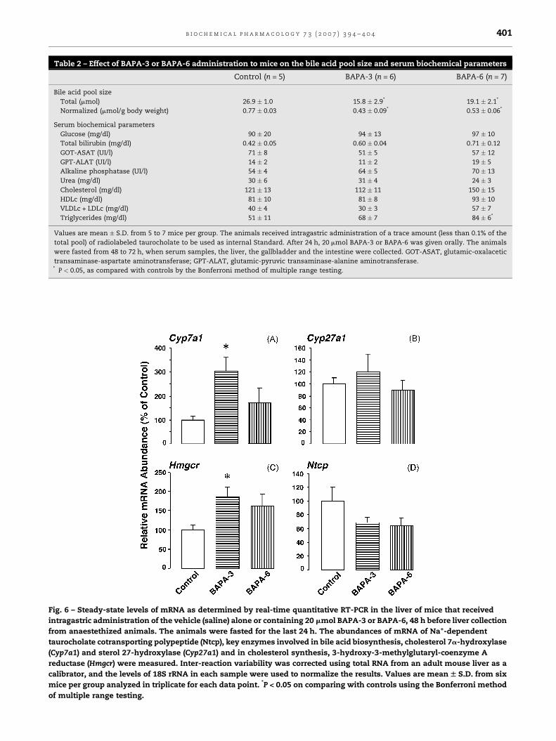
Table 2 – Effect of BAPA-3 or BAPA-6 administration to mice on the bile acid pool size and serum biochemical parameters
Control (n = 5) BAPA-3 (n = 6) BAPA-6 (n = 7)
Bile acid pool size
Total (mmol) 26.9 � 1.0 15.8 � 2.9* 19.1 � 2.1*
Normalized (mmol/g body weight) 0.77 � 0.03 0.43 � 0.09* 0.53 � 0.06*
Serum biochemical parameters
Glucose (mg/dl) 90 � 20 94 � 13 97 � 10
Total bilirubin (mg/dl) 0.42 � 0.05 0.60 � 0.04 0.71 � 0.12
GOT-ASAT (UI/l) 71 � 8 51 � 5 57 � 12
GPT-ALAT (UI/l) 14 � 2 11 � 2 19 � 5
Alkaline phosphatase (UI/l) 54 � 4 64 � 5 70 � 13
Urea (mg/dl) 30 � 6 31 � 4 24 � 3
Cholesterol (mg/dl) 121 � 13 112 � 11 150 � 15
HDLc (mg/dl) 81 � 10 81 � 8 93 � 10
VLDLc + LDLc (mg/dl) 40 � 4 30 � 3 57 � 7
Triglycerides (mg/dl) 51 � 11 68 � 7 84 � 6*
Values are mean � S.D. from 5 to 7 mice per group. The animals received intragastric administration of a trace amount (less than 0.1% of the
total pool) of radiolabeled taurocholate to be used as internal Standard. After 24 h, 20 mmol BAPA-3 or BAPA-6 was given orally. The animals
were fasted from 48 to 72 h, when serum samples, the liver, the gallbladder and the intestine were collected. GOT-ASAT, glutamic-oxalacetic
transaminase-aspartate aminotransferase; GPT-ALAT, glutamic-pyruvic transaminase-alanine aminotransferase.* P < 0.05, as compared with controls by the Bonferroni method of multiple range testing.
Fig. 6 – Steady-state levels of mRNA as determined by real-time quantitative RT-PCR in the liver of mice that received
intragastric administration of the vehicle (saline) alone or containing 20 mmol BAPA-3 or BAPA-6, 48 h before liver collection
from anaestethized animals. The animals were fasted for the last 24 h. The abundances of mRNA of Na+-dependent
taurocholate cotransporting polypeptide (Ntcp), key enzymes involved in bile acid biosynthesis, cholesterol 7a-hydroxylase
(Cyp7a1) and sterol 27-hydroxylase (Cyp27a1) and in cholesterol synthesis, 3-hydroxy-3-methylglutaryl-coenzyme A
reductase (Hmgcr) were measured. Inter-reaction variability was corrected using total RNA from an adult mouse liver as a
calibrator, and the levels of 18S rRNA in each sample were used to normalize the results. Values are mean W S.D. from six
mice per group analyzed in triplicate for each data point. *P < 0.05 on comparing with controls using the Bonferroni method
of multiple range testing.
b i o c h e m i c a l p h a r m a c o l o g y 7 3 ( 2 0 0 7 ) 3 9 4 – 4 0 4 401
Fig. 7 – Steady-state levels of mRNA as determined by real-time quantitative RT-PCR in the small intestine of mice that
received intragastric administration of the vehicle (saline) alone (control, open bars) or containing 20 mmol BAPA-3 (closed
bars) or BAPA-6 (grey bars), 48 h before small intestine collection from anaestethized animals. The animals were fasted for
the last 24 h. The abundances of mRNA of apical sodium-dependent bile acid transporter (Asbt) and the two components of
the heterodimeric organic solute transporter (Osta/Ostb) were measured. Inter-reaction variability was corrected using total
RNA of whole small intestine from an adult mouse as a calibrator, and the levels of 18S rRNA in each sample were used to
normalize the results. Values are mean W S.D. from six mice per group analyzed in triplicate for each data point. *P < 0.05 on
comparing with controls using the Bonferroni method of multiple range testing.
b i o c h e m i c a l p h a r m a c o l o g y 7 3 ( 2 0 0 7 ) 3 9 4 – 4 0 4402
minor contribution of MRP3 [34,35], but in which the
involvement of anion exchange transport systems [36],
probably belonging to the family of the multispecific organic
anion transporting polypeptides (OATPs) [37], has been also
suggested. (iii) Disruption of the dynamics of bile acid
intracellular transit and binding to cytosolic proteins, such
as the bile acid-binding protein (BABP) [32,38,39], may also play
a role. Whether BAPA-3 may interact with more than one of
these mechanisms in polarized in vivo epithelial cells, which
may have an influence on the net loss of bile acid from the
enterohepatic circulation and affect the regulation of enzymes
and transporters, cannot be ruled out.
In summary, the results of the present study suggest that
these compounds, in particular BAPA-3, owing to its strong
ability to inhibit ASBT-mediated bile acid transport and their
low absorption by the intestine, are potentially useful tools to
carry out pharmacological manipulations of the enterohepatic
circulation of endogenous bile acids.
Conflict of interest
In the period of research leading up to this publication we have
not received any financial support that may affect in any way

b i o c h e m i c a l p h a r m a c o l o g y 7 3 ( 2 0 0 7 ) 3 9 4 – 4 0 4 403
the conclusions of our article. Moreover, the authors have no
direct or indirect commercial interest in any company that
might be financially affected by the conclusions of the present
article.
Acknowledgements
The authors thank Dr. Paul Dawson (Wake Forest University
School of Medicine, Winston-Salem, North Carolina) for his
generous supply of recombinant plasmids. Thanks are also
due to L. Munoz, J.F. Martin, J. Villoria, N. Gonzalez and E.
Vallejo for care of the animals. Secretarial help by M.
Hernandez, technical help by E. Cruz and the revision of the
English spelling, grammar and style of the manuscript by N.
Skinner are also gratefully acknowledged. This study was
supported in part by the Junta de Castilla y Leon (Grant SA017/
03 and Grant SA013/04), Spain. Fondo de Investigaciones
Sanitarias, Ministerio de Sanidad y Consumo, Spain, co-
funded by the FEDER-FSE Program of the E.U. (Grant 01/1043).
Ministerio de Ciencia y Tecnologıa, Plan Nacional de Inves-
tigacion Cientıfica, Desarrollo e Innovacion Tecnologica (Grant
BFI2003-03208), Spain. The group is member of the Network for
Cooperative Research on Membrane Transport Proteins (REIT),
co-funded by the Ministerio de Educacion y Ciencia, Spain and
the European Regional Development Fund (ERDF) (Grant
BFU2005-24983-E/BFI) and belongs to the Centro de Investiga-
cion Biomedica en Red (CIBER) for Hepatology and Gastro-
enterology Research (Instituto de Salud Carlos III, Spain).
r e f e r e n c e s
[1] Pedersen TR. Aggressive lipid-lowering therapy: a clinicalimperative. Eur Heart J 1998;Suppl:M15–21.
[2] Tonstad S. Role of lipid-lowering pharmacotherapy inchildren. Paediatr Drugs 2000;2:11–22.
[3] Javitt NB. Bile acid synthesis from cholesterol: regulatoryand auxiliary pathways. FASEB J 1994;8:1308–11.
[4] Shefer S, Hauser S, Bekersky I, Mosbach EH. Biochemicalsite of regulation of bile acid biosynthesis in the rat. J LipidRes 1970;11:404–11.
[5] Shefer S, Nguyen LB, Salen G, Ness GC, Tint GS, Batta AK,et al. Regulation of cholesterol 7 alpha-hydroxylase byhepatic 7 alpha-hydroxylated bile acid flux and newlysynthesized cholesterol supply. J Biol Chem 1991;266:2693–6.
[6] Kuo PT, Wilson AC, Kostis JB, Moreyra AE. Effects ofcombined probucol-colestipol treatment for familialhypercholesterolemia and coronary artery disease. Am JCardiol 1986;57:43H–8H.
[7] Eghdamian B, Ghose K. Mode of action and adverse effectsof lipid lowering drugs. Drugs Today 1998;34:943–56.
[8] Wong MH, Oelkers P, Craddock AL, Dawson PA. Expressioncloning and characterization of the hamster ilealsodium-dependent bile acid transporter. J Biol Chem1994;269:1340–7.
[9] Dawson PA, Hubbert M, Haywood J, Craddock AL, ZerangueN, Christian WV, et al. The heteromeric organic solutetransporter alpha-beta, Ostalpha-Ostbeta, is anileal basolateral bile acid transporter. J Biol Chem2005;280:6960–8.
[10] Hagenbuch B, Dawson P. The sodium bile salt cotransportfamily SLC10. Pflugers Arch 2004;447:566–70.
[11] Yang PC, Campion JG, Tam CYJ, Nachowiak DA, Brown MA,Wagner GM, et al. The effect or SC-635, a potent inhibitor orthe ileal apical sodium co-dependent bile acid transporter,on cholesterol metabolism in rats. In: Van BergeHenegouwen GP, Keppler D, Leuschner U, Paumgartner G,editors. Biology of bile acids in health and disease.Dortrecht, Netherlands: Kluwer Academic Publishers; 2001.p. 183–97.
[12] Higaki J, Hara S, Takasu N, Tonda K, Miyata K, Shike T, et al.Inhibition of ileal Na+/bile acid cotransporter byS-8921 reduces serum cholesterol and preventsatherosclerosis in rabbits. Arterioscler Thromb Vasc Biol1998;18:1304–11.
[13] Root C, Smith CD, Winegar DA, Brieaddy LE, Lewis MC.Inhibition of ileal sodium-dependent bile acid tyransportby 2164U90. J Lipid Res 1995;36:1106–15.
[14] Tollefson MB, Vernier WF, Huang HC, Chen FP, Reinhard EJ,Beaudry J, et al. A novel class of apical sodium co-dependent bile acid transporter inhibitors: the 2,3-disubstituted-4-phenylquinolines. Bioorg Med Chem Lett2000;10:277–9.
[15] Firpi A, Walker JT, Lack L. Interactions of cationic bile saltderivatives with the ileal bile salt transport system. J LipidRes 1975;16:379–85.
[16] Bundy R, Mauskopf J, Walker JT, Lack L. Interaction ofuncharged bile salt derivatives with the ileal bile salttransport system. J Lipid Res 1977;18:389–95.
[17] Anwer MS, OMaille ERL, Hofmann AF, Dipietro RA,Michelotti E. Influence of side-chain charge on hepatictransport of bile acids and bile acid analogues. Am J Physiol1985;249:G479–88.
[18] Lack L, Tantawi A, Halavy C, Rockett D. Positionalrequirements for anionic charge for ileal absortion of bilesalt analogues. Am J Physiol 1984;246:G745–9.
[19] Kramer W, Stengelin S, Baringhaus KH, Enhsen A, Heuer H,Becker W, et al. Substrate specificity of the ileal and thehepatic Na+/bile acid cotransporters of the rabbit. I.Transport studies with membrane vesicles and cell linesexpressing the cloned transporters. J Lipid Res1999;40:1604–17.
[20] Balakrishnan A, Wring SA, Coop A, Polli JE. Influence ofcharge and steric bulk in the C-24 region on the interactionof bile acids with human apical sodium-dependent bileacid transporter. Mol Pharmaceut 2006;3:282–92.
[21] Tserng KY, Hachey DL, Klein PD. An improved procedurefor the synthesis of glycine and taurine conjugates of bileacids. J Lipid Res 1977;18:404–7.
[22] Briz O, Serrano MA, Rebollo N, Hagenbuch B, Meier PJ,Koepsell H, et al. Carriers involved in targeting thecytostatic bile acid-cisplatin derivatives cis-diammine-chloro-cholylglycinate-platinum(II) and cis-diammine-bisursodeoxycholate-platinum(II) toward liver cells. MolPharmacol 2002;61:853–60.
[23] Mashige U, Imai K, Osuga T. Simple and sensitive assay ofserum total bile acids. Clin Chim Acta 1976;70:79–86.
[24] Ballestero MR, Monte MJ, Briz O, Jimenez F, Gonzalez-SanMartin F, Marin JJG. Expression of transporters potentiallyinvolved in the targeting of cytostatic bile acid derivativesto colon cancer and polpyps. Biochem Pharmacol2006;72:729–38.
[25] Dixon M, Edwin C. Enzymes inhibition and activation. In:Enzyme3rd ed., London, UK: Longman Group Ltd.; 1979.Chapter VIII, pp. 322–99.
[26] Houten SM, Watanabe M, Auwerx J. Endocrine functions ofbile acids. EMBO J 2006;25:1419–25.
[27] Macias RIR, El-Mir MY, Monte MJ, Serrano MA, Garcıa MJ,Marin JJG. Cholephilic characteristics of a new cytostaticcomplex of cisplatin with glycocholate (Bamet-R2). J ControlRel 1999;57:161–9.

b i o c h e m i c a l p h a r m a c o l o g y 7 3 ( 2 0 0 7 ) 3 9 4 – 4 0 4404
[28] Palomero MF, Herrera MC, Macias RIR, El-Mir MY,Villanueva GR, Marin JJG. In vivo cholephilic characteristicsof the new cytostatic drug bischolylglycinate-platinum(II)(Bamet-H2). Int J Phamaceut 1998;172:79–88.
[29] Sauer P, Stiehl A, Fitscher BA, Riedel HD, Benz C, Kloters-Plachky P, et al. Downregulation of ileal bile acid absorptionin bile-duct-ligated rats. J Hepatol 2000;33:2–8.
[30] Neimark E, Chen F, Li X, Shneider BL. Bile acid-inducednegative feedback regulation of the human ileal bile acidtransporter. Hepatology 2004;40:149–56.
[31] Houten SM, Auwerx J. The enterohepatic nuclear receptorsare major regulators of the enterohepatic circulation of bilesalts. Ann Med 2004;36:482–91.
[32] Kramer W, Girbig F, Gutjahr U, Kowaslewski S, Jouvenal K,Muller G, et al. Intestinal bile acid absorption Na+-dependent bile acid transport activity in rabbit smallintestine correlates with the coexpression of an integral93 KDa and a peripheral 14 KDa bile acid bindingmembrane protein along the duodenum–ileum axis. J BiolChem 1993;268:18035–46.
[33] Kanamoto R, Kinoshita K, Maruyama T, Seki T, Iwami K.Expression of ileal Na+-dependent bile acids transportergene in transposed ileum of rat small intestine. NutritionRes 1999;19:1009–16.
[34] Inokuchi A, Hinoshita E, Iwamoto Y, Kohno K, Kuwano M,Uchiumi T. Enhanced expression of the human multidrugresistance protein 3 by bile salt in human enterocytes. Atranscriptional control of a plausible bile acid transporter. JBiol Chem 2001;276:46822–9.
[35] Rost D, Mahner S, Sugiyama Y, Stremmel W. Expressionand localization of the multidrug resistance-associatedprotein 3 in rat small and large intestine. Am J Physiol2002;282:G720–6.
[36] Weinberg SL, Burckhardt G, Wilson FA. Taurocholatetransport by rat intestinal basolateral membrane vesicles.Evidence for the presence of an anion exchange transportsystem. J Clin Invest 1986;78:44–50.
[37] Walters HC, Craddock AL, Fusegawa H, Willingham MC,Dawson PA. Expression, transport properties, andchromosomal location of organic anion transportersubtype 3. Am J Physiol 2000;279:G1188–200.
[38] Lin MC, Kramer W, Wilson FA. Identification of cytosolicand microsomal bile acid-binding proteins in rat ilealenterocytes. J Biol Chem 1990;265:14986–95.
[39] Gong YZ, Everett ET, Schwartz DA, Norris JS, Wilson FA.Molecular cloning, tissue distribution an expression of a14 kDa bile acid binding protein from rat ileal cytosol. ProcNatl Acad Sci USA 1994;91:4741–5.
Copyright © 2022 FDOKUMEN