ijair-volume-6-issue-1-xvi-january-march-2019.pdf - IARA
394
Volume 6, Issue 1 (XVI) ISSN 2394 - 7780 January - March 2019 International Journal of Advance and Innovative Research (Conference Special) Indian Academicians and Researchers Association www.iaraedu.com
-
Upload
khangminh22 -
Category
Documents
-
view
0 -
download
0
Transcript of ijair-volume-6-issue-1-xvi-january-march-2019.pdf - IARA
Volume 6, Issue 1 (XVI) ISSN 2394 - 7780 January - March 2019
International Journal of
Advance and Innovative Research (Conference Special)
Indian Academicians and Researchers Association
www.iaraedu.com
National Conference on
“INNOVATIVE RESEARCH IN SCIENCE AND TECHNOLOGY” IRST-2019
Organized by Marathwada Shikshan Prasarak Mandal’s
R. B. Attal Art’s, Science & Commerece College Georai, Dist. Beed
on
16th February 2019
Publication Partner
Indian Academicians and Researcher’s Association
Marathwada Shikshan Prasarak Mandal’s
R. B. Attal Art’s, Science & Commerece College Georai, Dist. Beed
Affiliated to Dr. Babasaheb Ambedkar Marathwada University
Re-accredited with “B” Grade (CGPA 2.78) by NAAC
ISO 9001: 2008 Certified College
Guest Editors of Special Issue Dr. Ranjitsingh K. Nimbalkar
Principal R. B. Attal Art’s, Science & Commerece College
Georai
Editorial Board Major V. P. Sangale Dr. A. M. Budrukkar Dr. S. N. Solanke
Dr. R. V. Shikhare Dr. B. D. Rupnar Dr. P. P. Pangarikar Mr. R. B. Pagore
About College Marathwada shikshan Prasarak Mandal felicitated by the government of Maharashtra by awarding the “ Best Educational Institute” in the Maharashtra State in 2001.
Marathwada Shiksan Prasark Mandal’s R. B. Attal Arts, Science and Commerce College was established in the year 1971 in the most backward taluka Georai. The affiliating University of the college is Dr. Babasaheb Ambedkar Marathwada University Aurangabad. Initially, Arts, Science & Commerce faculties were introduced in the same academic year. Making it a multi faculty educational unit, the college spread over 27 acres upland. And as well-furnished decorative buildings independent Library building, Hostel and newly constructed Indoor Stadium and 400 meters Track. At the same time the college has progress qualitatively by providing a large number of eminent successful alumni in every walk of life. College Awarded by Dr. B.A.M. University with “Ideal Examination Centre” in 2017-2018.
About IARA Indian Academicians and Researchers Association ( IARA ) is an educational and scientific research organization of Academicians, Research Scholars and practitioners responsible for sharing information about research activities, projects, conferences to its members. IARA offers an excellent opportunity for networking with other members and exchange knowledge. It also takes immense pride in its services offerings to undergraduate and graduate students. Students are provided opportunities to develop and clarify their research interests and skills as part of their preparation to become faculty members and researcher. Visit our website www.iaraedu.com for more details.
Objective of Conference The Conference is aimed to get innovative ideas from the expert in the field of science and there by spreading the ideas amongst students, teachers and scientist. The science scenario has progressed through research and development. The development of science must be shared and spread. On sharing the knowledge it not only increases but multiples several folds. The college teachers have to pass this knowledge to the students and therefore it become important for them to update their knowledge bank. The main purpose of the conference is to spread the light of knowledge amongst student, teachers, scholars, industry experts and scientists.

A BRIEF ABOUT ORGANIZING COMMITTEES
Patrons Hon. Shri. Prakash Solanke
President, M. S. P. Mandal, Aurangabad
Hon. Shri. Satish Chavan Secretary, M. S. P. Mandal, Aurangabad
Hon. Shri. Shivajirao Pandit Member, CDC
Hon. Shri. Amarsinha Pandit Vice President, M. S. P. Mandal, Aurangabad
Member, CDC
Organizer Secretary Dr. Ranjitsingh K. Nimbalkar
Principal, R. B. Attal Art’s, Science & Commerece College Georai
Convener Dr. Bhagat S. S.
Co - Convener Dr. Shikhare R. V.
Major Sangale V. P.
Coordinators Dr. Rupnar B. D. Dr. Badhe S. G.
Treasurers Dr. Solanke S. N. Mr. Shirsat A. J.
Organizing Committee Dr. Ingle R. D.
Dr. Pangrikar P. P.
Dr. Budrukkar A. M. Mr. Madavi B. B.
PREFACE
Dear Distinguished Delegates, Colleagues and Guest, Science Faculty of R. B. Attal College is Organizing One Day National Conference on “Innovative Research in Science and Technology” on 16 February 2019.
Marathwada Shikshan Prasarak Mandal’s R.B. Attal College has consistently kept its promises for quality education for the masses in the region addressing to the issues of social responsibility, students development and progression and research. Research has been one of the most prominent activities undertaken by the faculties on the campus.
All Science departments of R.B. Attal College are involved in outstanding research and effective teaching-learning process. It has always been proactive in all student centric activities. The present conference entitled “Innovative Research in Science and Technology (IRST) - 2019” is also a part of its efforts for bringing about academic discussion on the recent development in Chemical Sciences, Material Sciences, Biological Sciences and multidisciplinary approach in Science and Technology.
I feel doubly happy to write this message as I also happen to be a part of the science stream. I take this privilege to welcome all the participants to the conference.
Best wishes!
Dr. Ranjitsingh K. Nimbalkar
Principal R. B. Attal Art’s, Science & Commerece College
Georai
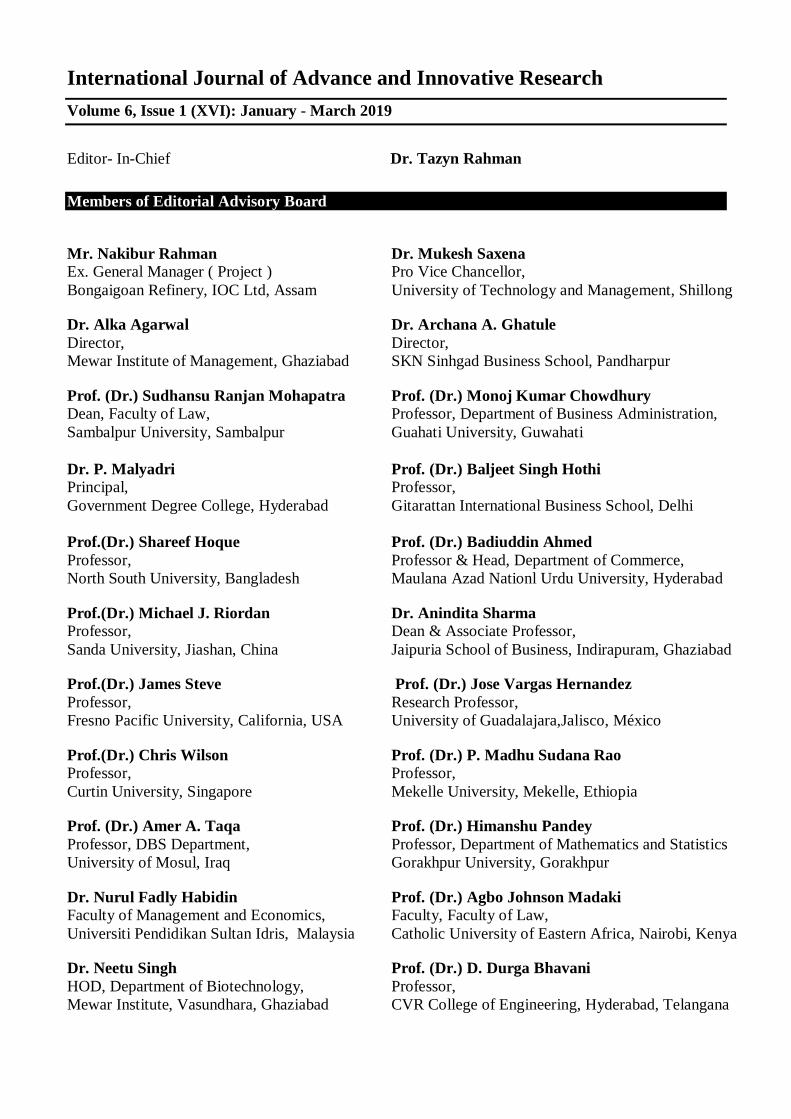
International Journal of Advance and Innovative Research Volume 6, Issue 1 (XVI): January - March 2019
Editor- In-Chief Dr. Tazyn Rahman
Members of Editorial Advisory Board
Mr. Nakibur Rahman Ex. General Manager ( Project ) Bongaigoan Refinery, IOC Ltd, Assam
Dr. Mukesh Saxena Pro Vice Chancellor, University of Technology and Management, Shillong
Dr. Alka Agarwal Director, Mewar Institute of Management, Ghaziabad
Dr. Archana A. Ghatule Director, SKN Sinhgad Business School, Pandharpur
Prof. (Dr.) Sudhansu Ranjan Mohapatra Dean, Faculty of Law, Sambalpur University, Sambalpur
Prof. (Dr.) Monoj Kumar Chowdhury Professor, Department of Business Administration, Guahati University, Guwahati
Dr. P. Malyadri Principal, Government Degree College, Hyderabad
Prof. (Dr.) Baljeet Singh Hothi Professor, Gitarattan International Business School, Delhi
Prof.(Dr.) Shareef Hoque Professor, North South University, Bangladesh
Prof. (Dr.) Badiuddin Ahmed Professor & Head, Department of Commerce, Maulana Azad Nationl Urdu University, Hyderabad
Prof.(Dr.) Michael J. Riordan Professor, Sanda University, Jiashan, China
Dr. Anindita Sharma Dean & Associate Professor, Jaipuria School of Business, Indirapuram, Ghaziabad
Prof.(Dr.) James Steve Professor, Fresno Pacific University, California, USA
Prof. (Dr.) Jose Vargas Hernandez Research Professor, University of Guadalajara,Jalisco, México
Prof.(Dr.) Chris Wilson Professor, Curtin University, Singapore
Prof. (Dr.) P. Madhu Sudana Rao Professor, Mekelle University, Mekelle, Ethiopia
Prof. (Dr.) Amer A. Taqa Professor, DBS Department, University of Mosul, Iraq
Prof. (Dr.) Himanshu Pandey Professor, Department of Mathematics and Statistics Gorakhpur University, Gorakhpur
Dr. Nurul Fadly Habidin Faculty of Management and Economics, Universiti Pendidikan Sultan Idris, Malaysia
Prof. (Dr.) Agbo Johnson Madaki Faculty, Faculty of Law, Catholic University of Eastern Africa, Nairobi, Kenya
Dr. Neetu Singh HOD, Department of Biotechnology, Mewar Institute, Vasundhara, Ghaziabad
Prof. (Dr.) D. Durga Bhavani Professor, CVR College of Engineering, Hyderabad, Telangana

Prof. (Dr.) Shashi Singhal Professor, Amity University, Jaipur
Prof. (Dr.) Aradhna Yadav Professor, Krupanidhi School of Management, Bengaluru
Prof. (Dr.) Alireza Heidari Professor, Faculty of Chemistry, California South University, California, USA
Prof.(Dr.) Robert Allen Professor Carnegie Mellon University, Australia
Prof. (Dr.) A. Mahadevan Professor S. G. School of Business Management, Salem
Prof. (Dr.) S. Nallusamy Professor & Dean, Dr. M.G.R. Educational & Research Institute,Chennai
Prof. (Dr.) Hemant Sharma Professor, Amity University, Haryana
Prof. (Dr.) Ravi Kumar Bommisetti Professor, Amrita Sai Institute of Science & Technology, Paritala
Dr. C. Shalini Kumar Principal, Vidhya Sagar Women’s College, Chengalpet
Dr. Syed Mehartaj Begum Professor, Hamdard University, New Delhi
Prof. (Dr.) Badar Alam Iqbal Adjunct Professor, Monarch University, Switzerland
Dr. Darshana Narayanan Head of Research, Pymetrics, New York, USA
Prof.(Dr.) D. Madan Mohan Professor, Indur PG College of MBA, Bodhan, Nizamabad
Dr. Rosemary Ekechukwu Associate Dean, University of Port Harcourt, Nigeria
Dr. Sandeep Kumar Sahratia Professor Sreyas Institute of Engineering & Technology
Dr. P.V. Praveen Sundar Director, Shanmuga Industries Arts and Science College
Dr. S. Balamurugan Director - Research & Development, Mindnotix Technologies, Coimbatore
Dr. Manoj P. K. Associate Professor, Cochin University of Science and Technology
Dr. Dhananjay Prabhakar Awasarikar Associate Professor, Suryadutta Institute, Pune
Dr. Indu Santosh Associate Professor, Dr. C. V.Raman University, Chhattisgath
Dr. Mohammad Younis Associate Professor, King Abdullah University, Saudi Arabia
Dr. Pranjal Sharma Associate Professor, Department of Management Mile Stone Institute of Higher Management, Ghaziabad
Dr. Kavita Gidwani Associate Professor, Chanakya Technical Campus, Jaipur
Dr. Lalata K Pani Reader, Bhadrak Autonomous College, Bhadrak, Odisha
Dr. Vijit Chaturvedi Associate Professor, Amity University, Noida
Dr. Pradeepta Kishore Sahoo Associate Professor, B.S.A, Institute of Law, Faridabad
Dr. Marwan Mustafa Shammot Associate Professor, King Saud University, Saudi Arabia
Dr. R. Navaneeth Krishnan Associate Professor, Bharathiyan College of Engg & Tech, Puducherry

Dr. Mahendra Daiya Associate Professor, JIET Group of Institutions, Jodhpur
Dr. G. Valarmathi Associate Professor, Vidhya Sagar Women's College, Chengalpet
Dr. Parbin Sultana Associate Professor, University of Science & Technology Meghalaya
Dr. M. I. Qadir Assistant Professor, Bahauddin Zakariya University, Pakistan
Dr. Kalpesh T. Patel Principal (In-charge) Shree G. N. Patel Commerce College, Nanikadi
Dr. Brijesh H. Joshi Principal (In-charge) B. L. Parikh College of BBA, Palanpur
Dr. Juhab Hussain Assistant Professor, King Abdulaziz University, Saudi Arabia
Dr. Namita Dixit Associate Professor, ITS Institute of Management, Ghaziabad
Dr. V. Tulasi Das Assistant Professor, Acharya Nagarjuna University, Guntur, A.P.
Dr. Nidhi Agrawal Associate Professor, Institute of Technology & Science, Ghaziabad
Dr. Urmila Yadav Assistant Professor, Sharda University, Greater Noida
Dr. Ashutosh Pandey Assistant Professor, Lovely Professional University, Punjab
Dr. M. Kanagarathinam Head, Department of Commerce Nehru Arts and Science College, Coimbatore
Dr. Subha Ganguly Scientist (Food Microbiology) West Bengal University of A. & F Sciences, Kolkata
Dr. V. Ananthaswamy Assistant Professor The Madura College (Autonomous), Madurai
Dr. R. Suresh Assistant Professor, Department of Management Mahatma Gandhi University
Dr. S. R. Boselin Prabhu Assistant Professor, SVS College of Engineering, Coimbatore
Dr. V. Subba Reddy Assistant Professor, RGM Group of Institutions, Kadapa
Dr. A. Anbu Assistant Professor, Achariya College of Education, Puducherry
Dr. R. Jayanthi Assistant Professor, Vidhya Sagar Women's College, Chengalpattu
Dr. C. Sankar Assistant Professor, VLB Janakiammal College of Arts and Science
Dr. Manisha Gupta Assistant Professor, Jagannath International Management School
Copyright @ 2019 Indian Academicians and Researchers Association, Guwahati All rights reserved. No part of this publication may be reproduced or transmitted in any form or by any means, or stored in any retrieval system of any nature without prior written permission. Application for permission for other use of copyright material including permission to reproduce extracts in other published works shall be made to the publishers. Full acknowledgment of author, publishers and source must be given. The views expressed in the articles are those of the contributors and not necessarily of the Editorial Board or the IARA. Although every care has been taken to avoid errors or omissions, this publication is being published on the condition and understanding that information given in this journal is merely for reference and must not be taken as having authority of or binding in any way on the authors, editors and publishers, who do not owe any responsibility for any damage or loss to any person, for the result of any action taken on the basis of this work. All disputes are subject to Guwahati jurisdiction only.
International Journal of Advance and Innovative Research Volume 6, Issue 1 (XVI) : January – March 2019
CONTENTS
Research Papers
LiBr-CATALYZED ONE-POT SYNTHESES OF DIHYDROPYRANO [2, 3-c] PYRAZOLES
Imran Shaikh, Mohammad Shaikh, Mubarak H. Shaikh, Sunil S. Bhagat
1 – 10
SYNTHESIS,CHARACTERIZATION,ANTIBACTERIAL AND ANTIFUNGAL STUDIES OF BINUCLEAR METAL COMPLEXES OF ZN(II)FE(II) AND MN(II) VIA INTER-COMPLEX REACTION
Dr. Mahananda A. Raut
11 – 17
ALGAL DIVERSITY OF KANKALESHOWAR TEMPLE POND IN BEED CITY
S. M. Talekar and A. N. Shelke
18 – 19
DIVERSITY, DISTRIBUTIONAND POPULATION DENSITY OF FRESH WATER ZOOPLANKTON FROM SHIVANA-TAKLI RESERVOIR OF KANNAD TALUKA, MAHARASHTRA STATE
K. B. Temkar, B. N. Jadhav, S. A. Shaikh, D. S. Kharate, P. S. Kharate, M. S. Kharate, and V. R.Lakwal
20 – 25
VISCOSITY STUDIES FOR THE BINARY MIXTURES OF ACETALDEHYDE WITH ETHANOL OVER THE ENTIRE RANGE OF ALL COMPOSITIONS AT 298.15, 308.15 AND 318.15 K.
Dr. S. B. Maulage, S. V. Gayakwad and Dr. R. G. Machale
26 – 29
PHYTOCHEMICAL STUDIES ON THE STEM BARK OF ANTHOCEPHALUS CADAMBA (ROXB.) MIQ
Revansiddha Dhotre, Maheboob Shaikh and Mamtaram Kare
30 – 33
STUDIES ON PHYSICO-CHEMICAL ANALYSIS OF SOIL SAMPLES IN BEED DISTRICT, MAHARASHTRA
V. V. Naiknaware and G. M. Dhond
34 – 35
STUDIES ON PHYSICO-CHEMICAL PARAMETERS OF WATER AND ZOOPLANKTON DIVERSITY OF LOWER DUDHANA DAM, PARBHANI DISTRICT. MAHARASHTRA
Nimbalkar R. K., Pawar D. A. and Sondge D. S.
36 – 42
BLENDING ELECTRONICS WITH THE HUMAN BODY
P. D. Gaikwad
43 – 45
STUDY OF ECCENTRICITY-BASED TOPOLOGICAL INDICES OF LOCAL ANESTHETIC DRUGS
N. K. Raut and G. K. Sanap
46 – 50
AGRONOMY OF ZEA MAYS UNDER DIFFERENT CULTIVATION PRACTICES
Shubhangi Khandare and Narayan Pandhure
51 - 53
STUDY OF NEUROSECRETION IN AMOEBOTAENIA JADHAVAE N. SP. FROM GALLUS DOMESTICUS AT SOYGAON, DIST. AURANGABAD, M.S. INDIA
B. S. Thorat
54 – 56
STUDY OF SOME PHYSICAL PROPERTIES OF CdO THIN FILMS
Vijay B. Sanap and B. H. Pawar
57 – 59
FUZZIFICATION OF LINEAR SPACES
Vinod Kulkarni and Vijay Sangle
60 – 63
INNOVATIVE RESEARCH ON WASTE MANAGEMENT SYSTEM IN MRS. KESHARBAI SONAJIRAO KSHIRSAGAR ALIAS KAKU COLLEGE, BEED IN CONCERN WITH GREEN AUDIT
A. N. Shelke
64 – 67
SYNTHESIS, CHARACTERIZATION AND BIOLOGICAL POTENTIAL OF 1,3,4 THIADIAZOLE CONTAINING SCHIFF BASE LIGAND AND ITS METAL COMPLEXES
Ajay M Patil, Sharad P Moharir, Aashish O Dhokte, Sunil R Mirgane
68 – 72
SYNTHESIS, CHARACTERIZATION AND ANTIMICROBIAL ANALYSIS OF VARIOUS SUBSTITUTED 3-(3-(5-bromothiophen-2-yl)-1-(4-fluorophenyl)-1H-pyrazol-4-yl)-1-(2-hydroxyphenyl)prop-2-en-1-one
Amol J. Shirsat, Balaji D. Rupnar, Sunil S. Bhagat, Ajit K. Dhas, Gopal K. Kakade
73 – 76
APPLICATION OF MOBILE TECHNOLOGY IN LIBRARIES
Pagore R. B.
77 – 79
CAN CATALYZED ECO-COMPATIBLE APPROACH FOR THE SYNTHESIS OF NOVEL 1,5-BENZODIAZEPINES AS AN PROMISING ANTIMICROBIAL AGENTS
Asha V. Chate and Charansingh H. Gill
80 – 87
MMUULLTTIIWWAAVVEELLEENNGGTTHH AASSTTRROONNOOMMYY:: TTOOOOLL TTOO PPRROOBBEE TTHHEE UUNNIIVVEERRSSEE
B. T. Tate
88 – 92
MILD AND EFFICIENT SYNTHESIS OF BENZOTHIAZOLE USING ZNFE2O4 AS HETEROGENEOUS CATALYST IN WATER
Rupnar B. D., Shirsat A. J., Bhagat S. S. and Pawar R. P
93 – 95
TOLERANT TOMATO (SOLANUM LYCOPERSICUM ) DEVELOPMENT FOR LEAF CURL VIRUS USING T-REP GENE
Bandewar S. T., Kunchge N. S., Pangrikar P. P. and A. M. Chavan
96 – 99
SYNTHESIS AND SPECTRAL ANALYSIS OF 4-CHLORO-2-(4,5-DIHYDRO-5-(3-NITROPHENYL)-1-1H-PYRAZOL-4-YL)-1H-PYRAZOL-3-YL)PHENOLS DERIVATIVES
Bhakare S. D., Badadhe P. V., Suryawanshi D. M., Bankar S. R., Chavhan N. M.
100 – 103
PHYTOCHEMICAL STUDIES IN LEAF DRUG Tylophora indica (Burm. f.) Merr
Rupali Biradar and Vikas Gambhire
104 – 106
PHYTOCHEMICAL STUDIES IN LEAF DRUGWOODFORDIA FRUTICOSA (L.) KURZ
Rupali Biradar1 and Vikas Gambhire
107 – 109
BIOCHEMICAL STUDIES OF CESTODEPARASITE MONIEZIA (B) OF CAPRA HIRCUS FROM BEED DISTRICT
A. M. Budrukkar
110 – 111
COMPARISON BETWEEN MATERIAL SYNTHESIS BY SOLID STATE REACTION AND SOL-GEL AUTOCOMBUSTION METHOD
C. M. Kale
112 – 115
POTENTIOMETRIC STUDY OF GABAPENTIN- COPPER (II) AND AMINO ACID COMPLEXES: DETERMINATION OF STABILITY CONSTANT
Omprakash S. Chavan, Kailash R. Borude and Bhimrao C. Khade
116 – 120
ICHTHYOFAUNAL DIVERSITY OF MASOLI RESERVIOR (PARBHANI DISTRICT), MAHARASHTRA, INDIA
Chilgar O. S. and H. S. Jagtap
121 – 124
MANAGEMENT OF MINOR INJURIES OF BIRDS-AS ONE WAY OF CONSERVATION OF ENVIRONMRNT
Chandrakant V. Sirsat
125 – 129
MORPHOTAXONOMIC STUDIES AND MEDICINAL USES OF SOME OF THE MEMBERS OF FAMILY EUPHORBIACEAE FROM SHRI SHIVAJI COLLEGE CAMPUS
Deshmukh Sadiya Sultana Khursheed Ahmed
130 – 134
EFFECT OF MEDICINAL PLANTS AND ANTAGONISTSON SEED MYCOFLORA ,SEED GERMINATION AND VIGOUR INDEX OF SUNFLOWER
G. B. Honna
135 – 138
MULTICOMPONENT SYNTHESIS OF SPIRO[4H-PYRAN-OXINDOLE] DERIVATIVES USING BIMETALLIC CATALYST ZIRCONIUM TETRACHLORIDE AND MAGNESIUM PERCHLORATE
Giribala M. Bondle
139 – 143
EVALUATION OF ANTIMICROBIAL AND ANTIFUNGAL PROPERTIES OF 3-((1H-INDOL-3-YL)(1,3-DIPHENYL-1H-PYRAZOL-4-YL)METHYL)-1H-INDOLE SYNTHESIZED USING NOVEL CLAY CATALYST
Ismail Shaikh, Rajendra Satpute, Syed Abed, Sheshrao Pawar
144 – 146
PREVALENCE OF CESTODE PARASITES FROM SOME FRESH WATER FISHES OF AURANGABAD DISTRICT (M.S.) INDIA
Jadhav R. K., Nimbalkar R. K., and Jadhav B. N.
147 – 149
ONE POT SYNTHESIS OF 2-AMINO PYRANES USING AMMONIUM CARBONATE AS AN EFFICIENT CATALYST
Jayant P. Sonar, S. D. Pardeshi, S. A. Dokhe, P. R. Pagare, A. M. Zine and S. N. Thore
150 – 153
SOLVENT FREE ONE-POT SYNTHESIS OF VARIOUS PYRANOPYRAZOLES CATALYZED BY AMMONIUM CHLORIDE
Khandu D Warad, Ramkrushna P Pawar, Chandrashekhar G Devkate and Rajiv Khobre
154 – 156
STUDIES ON THE RELATIONSHIP BETWEEN THE WATER QUALITY PARAMETERS AND FISH PRODUCTION IN LAKE OFKANKALESHWARBEED CITY (M.S.) INDI
Dr. M. K. Kale and P. P. Gaike
157 – 161
AN EFFICIENT, GREEN KNOEVENAGEL CONDENSATION FOR THE SYNTHESIS OF NEW 5-(4-((2-PHENYLTHIAZOL-4-YL) METHOXY) BENZYLIDENE)-3-(4-SUBSTITUTED PHENYL) THIAZOLIDINE-2,4-DIONES
Manisha R. Bhosle
162 – 168
STUDY OF ELECTRONIC WASTE MANAGEMENT IN INDIA: ISSUES AND STRATEGIES
Mayur P Deshmukh and Manjusha M Deshmukh
169 – 173

RECENT DEVELOPMENTS IN MATHEMATICS FOR SCIENCE AND TECHNOLOGY
Fasiyoddin Inayat Momin and Syeda Farra Fatima Nahri
174 – 175
HYDROTHERMAL SYNTHESIS OF NANOSTRUCTURED ZnO AND ITS PHOTOCATALYTIC PERFORMANCE STUDY TOWARDS METHYLENE BLUE DEGRADATION
Shrinivas C. Motekar
176 – 180
PECHMANN CONDENSATION CATALYSED BY OXALIX ACID UNDER MICRO WAVE IRRADIATION
Deepak R. Nagargoje
181 – 183
AN EFFICIENT ONE-POT STRATEGIES FOR THE SYNTHESIS OF [1, 3] OXAZINEDERIVATIVES USING L-PROLINE AS CATALYST
Balasaheb V. Shitole and Nana V. Shitole
184 – 186
SYNTHESIS OF CARBONATES FROM CHLOROMETHYL CHLOROFORMATES AND ITS SOME APPLICATIONS
Navnath D. Gunjal, Somnath S. Gholap, Sunil S. Bhagat
187 – 192
GREEN AND EFFICIENT SYNTHESIS OF Α-AMINOPHOSPHONATE
Vijay P. Pagore, Priti N. Bajad, Balaji D. Rupnar, Rajendra P. Pawar
193 – 195
EFFECT OF PLANT EXTRACTS ON GERMINATION OF OIL SEEDS
Prashant P. Pangrikar and Sudhir N. Solanke
196 – 197
A COMPARATIVE STUDY OF EFFECT OF NATURAL AND ITS COUNTERPART COMMERCIAL MEDICINE ON DEFENCE ORIENTED REACTION
Smt. Pawar S.P., Adhyapak M.S. and Ubale S. B.
198 – 199
ASSESSMENT OF WATER QUALITY AT THE POLLUTED AREA OF TERNA RIVER IN OSMANABAD DISTRICT MAHARASHTRA STATE INIDA
Shoeb Peerzade, S. V. Thakur, R. L. Ware and Sayed Abed
200 – 202
POTENTIOMETRIC STUDY OF COMPLEXATION OF LABETALOL WITH TRANSITION METAL IONS IN ETHANOL-WATER MEDIA
Ramesh Ware, Shoeb Peerzade, Kishor Koinkar, AJ Khan, Shailendrasingh Thakur
203 – 206
SYNTHESIS, CHARACTERISATION AND ANTIBACTERIAL EVOLUTIONS OF LANTHANIDE COMPLEXES WITH MIXED LIGANDS
M. A. Sakhare, Y. N. Bharate, S. A. Survase and O. S. Chavan
207 – 211
ANTI FUNGAL POTENTIALITY OF SOME SELECTED MEDICINAL PLANTS AGAINST FRUIT ROT PATHOGENS
Sawant S. G.
212 – 214
STUDIES ON SOME SOIL PREDATORY NEMATODES (MONICHIDA)OF MARATHWADA REGION
Shaikh Unaiza Nazneen, J. D. Shaikh and T. T. Shaikh
215 – 219
SYNTHESIS AND BIOLOGICAL SCREENING OF NOVEL PYRAZOLE AND ISOOXAZOLE DERIVATIVES
D. W. Shinde, S. S.Bhagat, D. R. Nagargoje and C. H. Gill
220 – 224
SYNTHESIS, CHARACTERIZATION AND ANTIMICROBIAL ANALYSIS OF VARIOUS SUBSTITUTED 2-(5-(3-(5-BROMOTHIOPHEN-2-YL)-1-PHENYL-1H-PYRAZOL-4-YL)-1H-PYRAZOL-3-YL) PHENOLS
Sunil S. Bhagat, Deepak R. Nagargoje, Dadasaheb W. Shinde, Dattatraya S. Ghotekar
225 – 227
MIXED LIGAND COMPLEXES OF ZINC METAL ION WITH DRUG CEFOTAXIME DRUG AND AMINO ACIDS IN AQUEOUS MEDIA
Shailendrasingh Thakur, Mazahar Farooqui, Ramesh Ware
228 – 232
AN OVERVIEW ON BIOLOGICAL IMPORTANCE AND SYNTHETIC METHODOLOGY OF TRIAZOLOPYRIMIDINES
Dr. Santosh S. Undare and Yogesh N. Bharate
233 – 239
GENETIC STUDIES ON COTTON DERIVED THROUGH INTROGRESSION
Ashish Gulhane and M. S. Wadikar
240 – 242
AN OVERVIEW ON SOME β -LACTUM RING CONTAINING THIRD GENERATION CEPHALOSPHORINES
Yogesh N. Bharate, Mahadeo A. Sakhare, Sanjeevan A. Survase and Satish B. Jadhav
243 – 245
POPULATION DYNAMICS OF CESTODE PARSITES IN COLUMBA LIVIA FROM BEED DISTRICT, INDIA
A. M. Budrukkar and R. K. Nimbalkar
246 – 248
POLYTENE CHROMOSOME FROM SALIVARY GLAND OF CHIRONOMUS CIRCUMDATUS
A. V. Gunjal and R. J.Chavan
249 – 250
ANALYSIS OF REFLECTION COEFFICIENT OF FOOD PRESERVATIVE UREA AND POTASSIUM META-BISULPHITE (KMS) USING IMPEDANCE SPECTROSCOPY
Badhe S. G., Prajapati T. A. and Helambe S. N.
251 – 253
STUDY OF SOME ETHENO MEDICINAL PLANTS OF OSMANABAD DISTRICT USED TO CURE DIABETES MELLITUS
Ram S .Bajgire
254 – 255
ETIDRONIC ACID AN EFFICIENT CATALYST FOR THE SYNTHESIS OF Β-ENAMINONES IN AQUEOUS MEDIA
Balaji Madje and Jagdish Bharad
256 – 258
FIRST PRINCIPLES STUDY OF LYSINE AMINO ACID-SINGLE WALLED CARBON NANOTUBES INTERACTIONS
Ravindra Karde, Avinash Ingale, Prashant Pardeshi, Baliram Lone
259 – 264
REVIEW ON NUTRITIONAL VALUES OF ASIAN CATFISH, CLARIAS BATRACHUS: IDEAL SPECIES FOR AQUACULTURE IN RURAL AREA
Jagtap H. S.
265 – 266
APPLICATION OF NATURAL DYE EXTRACTED FROM BEET AND SPINACH AGAINST SILK FIBRE OF BOMBYX MORI
Nagawanshi M. N. and Kirdat P. V.
267 – 271
ABO AND RH BLOOD GROUP FREQUENCY DISTRIBUTION AMONG SHINDE TAKLI POPULATION OF PARBHANI DISTRICT, MAHARASHTRA
Nimbalkar R. K. , Pawar D. A. and S. M. Vadgule
272 – 276
ANALYSIS OF SOYABEAN OIL USED IN CAFETERIAS TO STUDY ADULTERATION BY REGULAR ANALYTICAL PARAMETERS AND UV SPECTROPHOTOMETER
S. V. Mahamuni, S. S. Pise, V. B. Sankpal and N. S. Shinde
277 – 281
IN VITRO STUDIES IN COCHLOSPERMUM RELIGIOSUM (LINN.)
Jyoti Ghodke, Swati Shilwant, Narayan Pandhure and Sudhir Solanke
282 – 285
AGRONOMY OF MEDICAGO SATIVA TO DIFFERENT METHODS OF CULTIVATION
Narayan Pandhure, Laxman Shimple and Prashant P. Pangrikar
286 – 288
PRODUCTION, EXTRACTION, PURIFICATION AND APPLICATION OF BACTERIAL EXOPOLYSACCHARIDE
Samruddhi P. Joshi and Prashant P. Dixit
289 – 293
ZOOPLANKTONIC ANALYSIS AND AQUATIC POLLUTION LOAD OF VANJARWADI RESERVOIR DIST BEED 431122 (MS)
Prashant V. Patil and Anirudha M. Budrukkar
294 – 295
DISTRIBUTION OF SEDGES NEAR JAIKWADI DAM IN AURANGABAD DISTRICT OF MAHARASHTRA
Sudhir Solanke, Raffique Shaikh and Ravi Patil
296 – 298
LITERATURE REVIEW ON FRUIT QUALITY IDENTIFICATION SYSTEM
Rahul J. Mhaske and Prapti Deshmukh
299 – 302
ACETONIDE PROTECTION OF DIOLS USING IODINE AND DIMETHOXYPROPANE
Ravibhushan S. Kulkarni, Prashant P. Dixit, Kishan P. Haval
303 – 304
BUTTERFLY DIVERSITY OF PARBHANI, MAHARASHTRA STATE, INDIA
S. M. Yeole
305 – 307
E – WASTE MANAGEMENT
S. B. Birajdar and P. V. Kulkarni
308 – 311
AN ASSESSMENT OF SOME WATER QUALITY PARAMETERS OF SHIVNA TAKLI DAM TQ. KANNAD, DIST. AURANGABAD
Sanghai P. K1. Kshirsagar A. A. and Ingole, S. B.
312 – 313
ON MODELING AND COMPLETE SOLUTIONS TO GENERAL FIX POINT PROBLEMS IN MULTI-SCALE SYSTEMS & APPLICATION WITH EXAMPLES
Dr. Shaikh Mohammed Sirajuddin Mohammed Salimuddin and Prof. Mohmed Zafar Saber
314 – 318
FUNGAL INCIDENCE ON GROUNDNUT SEED FROM DIFFERENT LOCALITY OF MARATHAWADA
Shrikant B. Mane, Prashant P. Pangrikar and Ashok M. Chavan
319 – 322
ANTIBACTERIAL ACTIVITY OF SOME MEDICINALLY IMPORTANT PLANT LEAF EXTRACT
Shrikant B. Mane, Jadhao A. S., Kate A. S., Dhandge and S. R., Damare N. D.
323 – 327
AN EFFICIENT SYNTHESIS OF TRIAZOLOQUINAZOLINONES AND BENZIMIDAZOLOQUINAZLINONES PROMOTED BY MAGNESIUM SULPHATE AS A GREEN ALTERNATIVE
Somnath S. Gholap, Vinod R. Kadu and Nilesh D. Gaikawad
328 – 334
STUDY OF PHYSICO-CHEMICAL PARAMETERS OF TERNA RESERVOIR, MAHARASHTRA
Dr. Somwanshi J. L., Shri Kulkarni G. A. and Dr. K. H. Rajput
335 – 336
MEASURING ACCURACY AND PERFORMANCE OF TEXT-TO-SPEECH BY SUBJECTIVE AND OBJECTIVE METHODS
Suhas Mache and Sunil Nimbhore
337 – 340
ISOLATION, EXTRACTION OF COMMERCIALLY VALUABLE PROTEASE ENZYME BY USING AGRO-WASTE SUBSTRATE BY SOLID STATE FERMENTATION (SSF)
Syeda Tasleem S. Gani and Aithal S. C
341 – 345
ASSESSMENT OF ENDOPHYTIC DIVERSITY FROM DALBERGIA SISSOO FROM DIFFERENT REGIONS OF AURANGABAD (MAHARASHTRA)
Taware A. S. and Rajurkar S. K.
346 – 349
MICROMORPHOLOGICAL STUDIES ON SOME TEPHROSIA SPECIES
T. A. Gitte and A. S. Dhabe
350 – 356
ATTRACTVITY RESULTS FOR QUADRATIC FUNCTIONAL DIFFERENTIAL EQUATION
V. T. Ghuge and A. B. Mundhe
357 – 364
SYNTHESIS OF AZODYES BASED ON 8-HYDROXY QUINOLINE WITH ITS CHARACTERIZATIONS, ANTIFUNGAL AND ANTIMICROBIAL ACTIVITY
Prashant Vibhute and Sachin A. Khiste
365 – 368
QUANTITATIVE ESTIMATION OF BIOACTIVE PHYTOCONSTITUENTS PRESENT IN DALBERGIA LANCEOLARIA SUBSP. PANICULATA (ROXB.) THOTH. METHANOLIC LEAF AND BARK EXTRACT
Wankhade, M. S1., Solanke S. N. and Pangikar P. P.
369 - 373
International Journal of Advance and Innovative Research Volume 6, Issue 1 (XVI): January - March, 2019
1
ISSN 2394 - 7780
LiBr-CATALYZED ONE-POT SYNTHESES OF DIHYDROPYRANO [2, 3-c] PYRAZOLES
Imran Shaikh1, Mohammad Shaikh2, Mubarak H. Shaikh3, Sunil S. Bhagat4 1,4Department of Chemistry, R B Attal Arts Science and Commerece College, Georai
2,3Department of Chemistry, Dr. Babasaheb Ambedkar Marathwada University, Aurangabad
ABSTRACT A new facile, greenand efficient protocol was developed for synthesis of Dihydropyrano [2,3-c] pyrazolesusing LiBr as an efficient, eco-friendly catalyst.Compared to other methods, this new method consistently has advantages, including excellent yields, short reaction time, mild reaction conditionsand reusability of catalyst.The synthesized Dihydropyrano [2,3-c] pyrazoleswere analyzed for ADME properties.
Keywords: Dihydropyrano [2,3-c] pyrazoles;LiBr; Green protocol, ADME properties.
INTRODUCTION In pharmaceutical industries solvents play an important role for organic transformation and production of active pharmaceutical ingredient (API) have a direct impact on the environment because of its large volume consumption, limited recovery due to volatile nature and residual disposal problem. According to the regulatory agencies and international conference for harmonization (ICH) guideline there are limitation to use the class-1 and class-2 solvents in pharmaceutical product due to hazardous, toxic and carcinogenic nature. It has been recommended to use the class-3 solvent particularly for manufacturing of drug intermediate and finished product.[1]Therefore, replacing of such conventional solvents, with more environmentally benign media is one of the important tasks to meet the current Green Chemistry requirement, and a subject of significant academic and commercial interest.[2] By focusing on current demand of green chemistry, a variety of unconventional solvents, such as water,[3] ionic liquids,[4] polyethylene glycol,[5]supercritical fluids[6] and fluorousmedia[7] have been extensively used and studied well. Although the use of these solvents has certain limitations, such as the incompatibility of reactive reagents or substrates in water, high prices and insufficient data about the toxicity and bio-compatibility for ionic liquids, the requirement of sophisticated equipment for supercritical fluids. Therefore, the search of alternative and eco-friendly reaction media for organic transformation has become the considerable interest of researchers.
Lithium bromide is a stable, relatively safe and readily available low-cost reagent having unique mild Lewis acid properties. It has a wide variety of utility in different chemical transformations including Biginelli condensation, Knoevenagel condensation, Ehrlich-Sachs reaction, Friedel-Crafts reaction, rearrangement of epoxides and preparation of acylals and xanthenes.8In most of these reported reactions, LiBr is almost neutral9 and also does not form any corrosive or harsh by-products during aqueous workup, unlike strong and expensive catalysts. However, there are no examples of use of lithium bromide as catalyst for synthesis of pyrano-pyrazole derivatives
In the current scenario of global warming issues, regulatory agencies and pollution control authorities are having a serious concern, about the waste disposal and air pollution generated by chemical and pharmaceutical industries. Due to the huge demand of fine chemicals, drug intermediate and drug molecule. It is necessary to develop the cost effective, robust and eco-friendly processto develop MCRs. To minimize the waste generation, operational simplicity and atom economy is a great interest of scientific community in recentyears. Therefore,there is a need to design a synthetic route for organic transformation using three or more components in one-pot operation with minimum waste generation. One-pot synthesis MCRs often take shorter reaction time, minimum utilities, use of energy and manpower with consistent higher product yields,compare to multi-step synthesis.[10]MCRs constitute large series of structurally related drug-like molecules, leads to identification and optimization in drug discovery program. Considering these advantages, over the multi-step synthesis, the design of new MCRs with environmental friendly method is a bigchallenge to the scientific community at the forefront area of green chemistry.[11]
As per green chemistry protocol transformation of organic reactions in aqueous media is a big challenging and attractive taskas water is an environmentally benign solvent. Water is abundant in nature, easily available, cheap, and user friendly andsustainability of exothermic reactions.Synthesisof organic reaction in aqueous media offer more benefit like, rate determining, faster reaction and products insolubility, which help for the product isolation in pure form by simple filtration which is more advantageous and beneficial over conventional organic solvents.
International Journal of Advance and Innovative Research Volume 6, Issue 1 (XVI): January - March, 2019
2
ISSN 2394 - 7780
In the class of heterocyclic molecules, multifunctional4H-pyran and their derivatives are important class of which composed most important core of various natural products[12]and photochromic materials.[13] Due to their wide range of biological potency such as antimicrobial,[14]antiproliferative,[15]anticancer,[16]and antioxidant properties.[17]It can be used to cure neurological disorder like Alzheimer’s disease,Huntington’s disease, neurodegenerative disease Parkinson’s disease schizophrenia, and treatment of, including amyotrophic lateral sclerosis, AIDS associated dementia, Down syndrome and myoclonus.[18]Multifuctionalized 4H-Pyran derivatives also shows potential calcium channel antagonists properties, which are structurally similar to biologically active 1,4-dihydropyridines.[19]The nitrile functionality in 4H-pyran derivatives is important synthon, for the synthesis of different bioactive heterocyclic compounds such as pyranopyrazoles, lactones, pyridones, 1,4-dihydropyridines and aminopyrimidines.[20]In organic chemistry, Pyrazole is an important heterocyclic analogue which plays a vital role in many pharmaceutical and agrochemical drugs molecules and intermediates.
In medicinal chemistry and drug designing, Dihydropyrano [2,3-c]pyrazoles is became the first choice of researchers and scientistdue to its potential biological activity, and therefore become the interesting template for medicinal chemistry research. Most class of these compounds, are well known forantioxidant,[21]antimicrobial,[22] insecticidal,[23]molluscicidal,[24] analgesic,[25]anti-inflammatory agents[26] and some of their analogues act as vasodilators, hypotensive,[27]hypoglycemic and anticancer agents.[28]They are also potential inhibitors of human Chk1 kinase.[29]Furthermore, they play a significant role as crucial synthetic intermediates.[30]
Thus, considering the differentpotential therapeutic activity of pyrano [2,3-c]pyrazoles, heterocyclic compounds, various methodologies for synthesis of Dihydropyrano[2,3-c]pyrazoleshave been reported in the literature.These reportedmethodologies have shown good results in many instances.However, some of synthetic strategies have limitations interms of using metal catalyst, expensive reagents, long reaction time,environmental hazard solvents, harsh reaction conditions, tedious workupprocedure, unsatisfactory yield and use of homogeneouscatalyst which are difficult in separation from reaction mixture.In spite of many reported methods for the synthesis of Dihydropyrano[2,3-c]pyrazole derivatives, the development of a new synthetic strategy using easily accessible catalyst and mildand sustainable reaction condition still demand a lot of attention.
Recently, four-component reactions of aldehydes, 1,3-dicarbonyl compounds, malononitrile, and hydrazine have been developed for the synthesis of pyranopyrazoles usingtriphenylphosphine,[31]urea,[32]ionic liquid,[33]water containinga catalytic amount of piperidine,[34]CTACl,[35]heteropolyacids,[36]microwave,[37] piperazine,[38]N-methylmorpholine,[39]L-proline,[40] alumina,[41]per-6-amino-β-cyclodextrin,[42]sodium benzoate,[43]amberlyst A21,[44]glycine,[45]imidazole,[46] and I2
[47]Although thesemethods are quite satisfactory, some of them suffer from the absenceof green chemistry and have been associated with severalshortcomings, such as the use of volatile and hazardous organicsolvents, low yields, extended reaction time, high temperature andtedious procedure for the preparation of catalysts. Thus, thedevelopmentof general, economically and environmentally benignsynthetic methodologies for these heterocycles is highly desirable.
OBJECTIVE Considering the significance of heterocyclic compounds likeDihydropyrano[2,3-c]pyrazoles derivatives in pharmaceutical and medicinal fields, the development of simple, eco-friendly and low cost protocol for the synthesis of this molecules is still the great interest of scientific community and researchers. Hence, with this inspiration we thought to develop new and efficient route for the synthesis of Dihydropyrano [2,3-c]pyrazolesusing LiBr as an efficient, eco-friendly catalystunder environmentally friendly conditions.
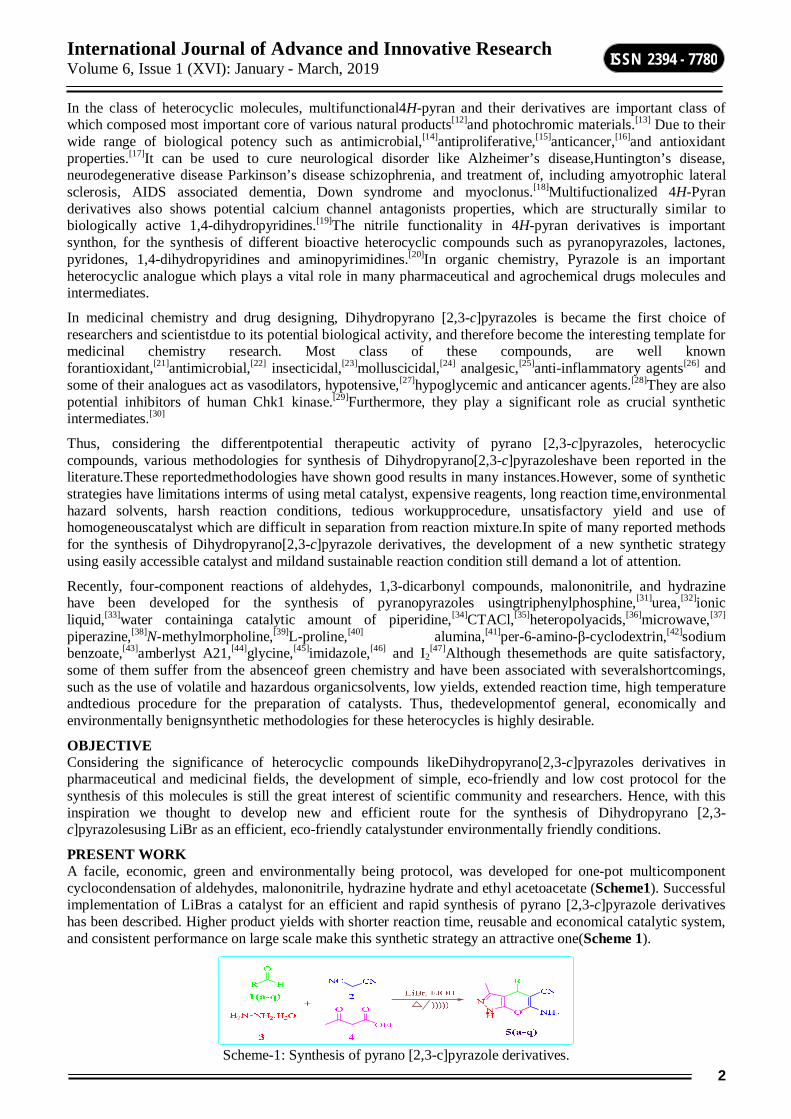
PRESENT WORK A facile, economic, green and environmentally being protocol, was developed for one-pot multicomponent cyclocondensation of aldehydes, malononitrile, hydrazine hydrate and ethyl acetoacetate (Scheme1). Successful implementation of LiBras a catalyst for an efficient and rapid synthesis of pyrano [2,3-c]pyrazole derivatives has been described. Higher product yields with shorter reaction time, reusable and economical catalytic system, and consistent performance on large scale make this synthetic strategy an attractive one(Scheme 1).
Scheme-1: Synthesis of pyrano [2,3-c]pyrazole derivatives.

International Journal of Advance and Innovative Research Volume 6, Issue 1 (XVI): January - March, 2019
3
ISSN 2394 - 7780
RESULTS AND DISCUSSION In search of an efficient catalyst and the best experimental reaction conditions, initially we carried out the reaction between benzaldehyde (1) (1 mmol), malononitrile (2) (1 mmol), hydrazine hydrate(3a) (1 mmol) and ethyl acetoacetate(4) (1 mmol) has been considered as a model reaction.
Scheme-2: Standard model reaction
Initially when the reaction was carried out in absence of the catalyst, the product formed in trace amount (Table1, entry 1). During the initial study, various acid catalysts were screened, owing to theirwidespread catalytic applications in organic synthesis.For this purpose, we tried various Lewis acid catalyst like AlCl3, FeCl3, ZnCl2afforded the product in 57, 59and 60% yields respectively(Table1, entries 2-4).Then we decided to use bromides of alkali metals like LiBr, NaBr, KBr and CsBr. It was observed that when NaBr, KBr and CsBr used as a catalyst, the rate of the reaction very small and product obtained in lower yield (Table1, entry6-8). In comparison,lithium bromide proved to be an excellent catalyst, furnishing the product inexcellent yield (Table1, entry5) and therefore was chosen as a catalyst of choicefor further optimization studies.
Tabl-1: Effect of catalysta Entry Catalyst Time (Min) Yieldb (%)
1 - 180 Trace 2 AlCl3 120 57 3 FeCl3 120 59 4 ZnCl2 120 60 5 LiBr 30 95 6 NaBr 120 55 7 KBr 120 52 8 CsBr 120 50
aReaction conditions: Aldehyde (1 mmol), malononitrile (1 mmol), hydrazine hydrate (1 mmol) and ethylacetoacetate (1 mmol) and catalyst in 5 mL Ethanol at 60°C. bIsolated yield.
Therefore, to accomplish this goal and considering the significance of green chemistry concept, to check the effect of temperature, model reaction was carried out initially at neat condition for appropriate time. But, formation of the desired product was not observed (Table 2, entries 1). In subsequent optimization experiments, efforts were directed towards the use polar protic and polar aprotic solvent at different temperatures. To our surprise, reaction in aqueous media at reflux conditions proceed towards the desired product in 40 %yield (Table 2, entry 2). Similarly, reaction carried out in polar aprotic solvents like acetonitrile, tetrahydrofuran, dimethyl sulfoxide and DMF, product formed in 42, 45, 46 and 48%respectively(Table 2, Entry 3-6).Further, reaction carried out in polar protic solvents like IPA, Methanol and ethanol, product formed in 70, 75 and 95% yield respectively (Table 2, Entry 7-9). Among the tested solvents, ethanol was superior over the othersolvents in terms of both product yield and reaction time (Table 2, entry 9). Furthermore, reaction carried out in EtOH:H2O mixture (Table 2, Entry 12) and ethanol at different temperature and found out that at 60°C product formed in excellent yield (Table 2, Entry 10). Therefore, from this study we found that, ethanol at 60°Cwas the best suitable solvent to carried out reaction with excellent yield.
Table-2: Screening of solventa
Entry Solvent Temp (C) Time (Min) Yieldb (%) 1 Neat 100 180 Trace 2 Water Reflux 180 40 3 CH3CN Reflux 180 42 4 THF Reflux 180 45 5 DMSO Reflux 180 46
International Journal of Advance and Innovative Research Volume 6, Issue 1 (XVI): January - March, 2019
4
ISSN 2394 - 7780
6 DMF Reflux 180 48 7 IPA Reflux 120 70 8 Methanol Reflux 60 75 9 Ethanol Reflux 30 95 10 Ethanol 60 30 95 11 Ethanol 40 60 85 12 EtOH:H2O Reflux 60 80
aReaction conditions: Aldehyde (1 mmol), malononitrile (1 mmol), hydrazine hydrate (1 mmol) and ethylacetoacetate (1 mmol) and LiBr (20mol%) in 5 mL Solvent. bIsolated yield.
To determine the appropriate concentration of the catalystLiBr, we investigated the model reaction at different concentrationsof LiBr such as 5, 10, 15, 20 and 25 mol%. Theproduct formed in 60, 72, 85, 95 and 95% yields respectively(Table3, entries 1-5). As increase in concentration of catalyst from 20 to 25 mol% does not increase the yield of product. This indicates that 20 mol% of LiBr is sufficient forthe reaction by considering yield of product(Table3, entry 4).
Table-3: Optimization of Catalysta Entry LiBr (mol%) Time (Min) Yieldb (%)
1 5 60 60 2 10 60 72 3 15 60 85 4 20 30 95 5 25 30 95
aReaction conditions: Aldehyde (1 mmol), malononitrile (1 mmol), hydrazine hydrate (1 mmol) and ethylacetoacetate (1 mmol) and catalyst in 5 mL Ethanol at 60°C. bIsolated yield.
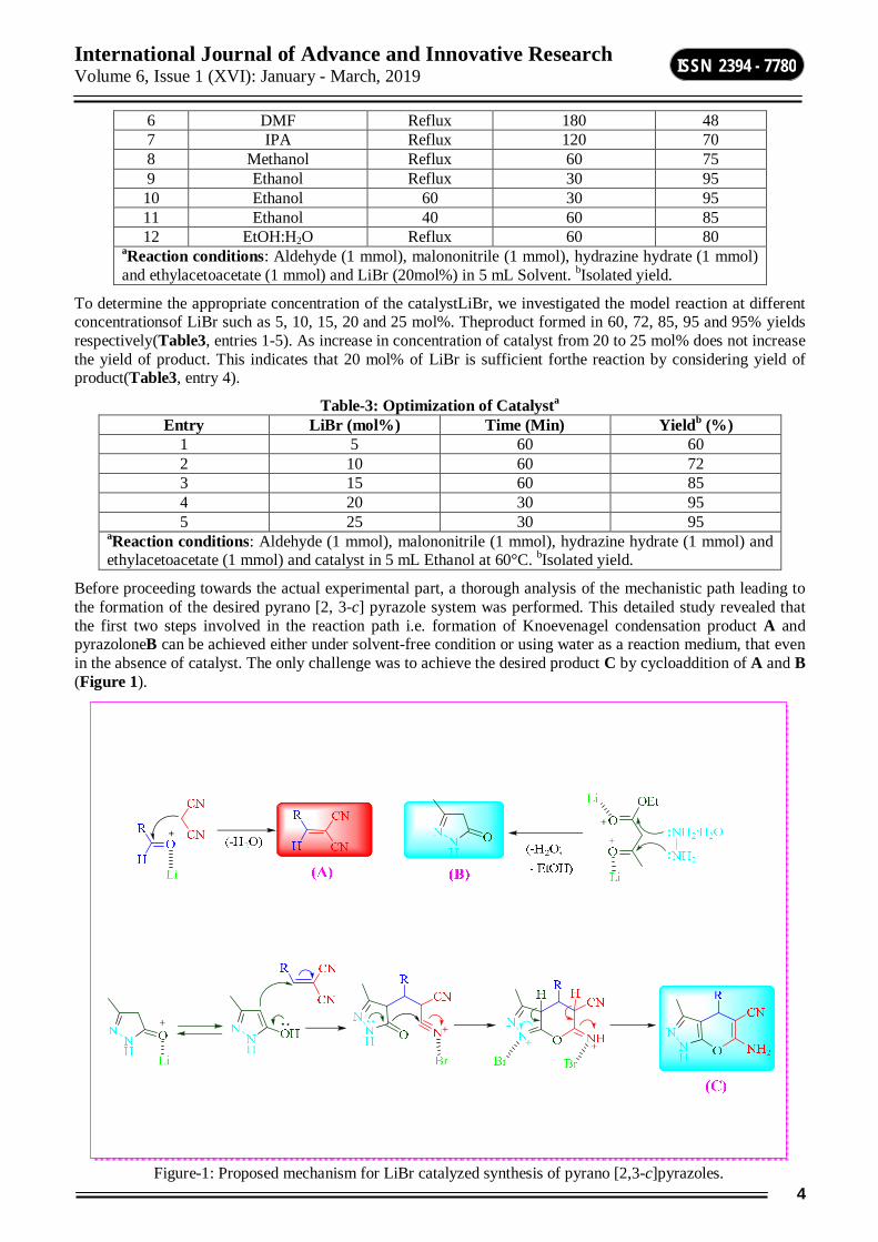
Before proceeding towards the actual experimental part, a thorough analysis of the mechanistic path leading to the formation of the desired pyrano [2, 3-c] pyrazole system was performed. This detailed study revealed that the first two steps involved in the reaction path i.e. formation of Knoevenagel condensation product A and pyrazoloneB can be achieved either under solvent-free condition or using water as a reaction medium, that even in the absence of catalyst. The only challenge was to achieve the desired product C by cycloaddition of A and B (Figure 1).
Figure-1: Proposed mechanism for LiBr catalyzed synthesis of pyrano [2,3-c]pyrazoles.
International Journal of Advance and Innovative Research Volume 6, Issue 1 (XVI): January - March, 2019
5
ISSN 2394 - 7780
Reason behind the success of LiBr bringing the reaction in its favor may be the small size of lithium cation which interact effectively with small negative charged atoms like oxygen. Lithium bromide is a salt of small cation and large anion. They can’t interact effectively, though crystal lattice is quite easy to break. Indeed, lithium cation has the highest hydration energy of all alkali metal cations. Altogether, it means, that in solvents with oxygen atoms (alcohols, esters, acetone) lithium cation effectively bounds to solvent molecules, leaving crystal lattice, and bromide has to follow, resulting in some solubility of lithium bromide in solvents with negatively charged oxygen and to lesser extent, nitrogen (for example pyridine). Diagrammatic representation depicting plausible mechanism for LiBr catalyzed synthesis of pyrano [2,3-c]pyrazoles is rationalized with the help of Figure 1.
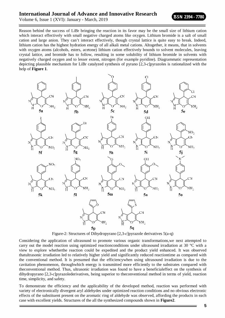
Figure-2: Structures of Dihydropyrano [2,3-c]pyrazole derivatives 5(a-q)
Considering the application of ultrasound to promote various organic transformations,we next attempted to carry out the model reaction using optimized reactionconditions under ultrasound irradiation at 30 °C with a view to explore whetherthe reaction could be expedited and the product yield enhanced. It was observed thatultrasonic irradiation led to relatively higher yield and significantly reduced reactiontime as compared with the conventional method. It is presumed that the efficiencywhen using ultrasound irradiation is due to the cavitation phenomenon, throughwhich energy is transmitted more efficiently to the substrates compared with theconventional method. Thus, ultrasonic irradiation was found to have a beneficialeffect on the synthesis of dihydropyrano [2,3-c]pyrazolederivatives, being superior to theconventional method in terms of yield, reaction time, simplicity, and safety.
To demonstrate the efficiency and the applicability of the developed method, reaction was performed with variety of electronically divergent aryl aldehydes under optimized reaction conditions and no obvious electronic effects of the substituent present on the aromatic ring of aldehyde was observed, affording the products in each case with excellent yields. Structures of the all the synthesized compounds shown in Figure2.
International Journal of Advance and Innovative Research Volume 6, Issue 1 (XVI): January - March, 2019
6
ISSN 2394 - 7780
Table-4: Synthesis of dihydropyrano [2,3-c]pyrazolederivatives5a-q
Compound Ultrasound methoda Conventional methodb Melting point (°C)d Time (Min) Yield (%)c Time (Min) Yield (%)c
5a 15 95 30 95 241-243 5b 15 92 35 92 208-210 5c 20 94 35 94 207-209 5d 20 90 40 90 231-233 5e 25 92 45 92 143-144 5f 15 93 35 93 231-233 5g 15 87 30 87 179-180 5h 15 91 30 91 175-177 5i 20 93 40 93 221-223 5j 20 93 35 93 223-224 5k 20 88 40 88 192-195 5l 20 89 40 89 250-252
5m 20 84 45 84 190-191 5n 20 86 35 86 233-235 5o 25 90 45 90 165-168 5p 25 91 45 91 235-238 5q 25 89 45 89 234-237
aAldehyde (1 mmol), malononitrile (1 mmol), hydrazine hydrate (1 mmol) and ethylacetoacetate (1 mmol) and LiBr (20mol%) in 5 mL Ethanol under ultrasound irradiation bAldehyde (1 mmol), malononitrile (1 mmol), hydrazine hydrate (1 mmol) and ethylacetoacetate (1 mmol) and LiBr (20mol%) in 5 mL Ethanol under conventional heating. cIsolated yields; dMelting points match with literature values.
COMPUTATIONAL STUDY Insilicon ADME prediction A computational study of all the synthesized Dihydropyrano [2,3-c]pyrazole derivatives 5(a-q) was performed for prediction of ADME properties and the value obtained is presented in Table 5. It is observed that, the compounds exhibited a good % ABS (% absorption) ranging from 62.92to 78.73%. Furthermore, none of the compounds violated Lipinski’s rule of five (miLogP ≤ 5). A molecule likely to be developed as an orally active drug candidate should show no more than one violation of the following four criteria: miLogP (octanol-water partition coefficient) ≤ 5, molecular weight ≤ 500, number of hydrogen bond acceptors ≤ 10 and number of hydrogen bond donors ≤ 5.[48] The larger the value of the drug likeness model score, the higher is also probability that the particular molecule will be active. All the tested compounds followed the criteria for orally active drug and therefore, these compounds may have a good potential for eventual development as oral agents.
Table-5: Pharmacokinetic parameters important for good oral bioavailability
Comp-ounds % ABS TPSA
(A2) n-
ROTB MV MW Mi LogP
n-ON
n-OHNH
Lipinski violation
Drug-likeness model score
Rule - - - - < 500 ≤ 5 < 10 < 5 ≤ 1 - 5a 78.73 87.73 1 223.38 252.28 1.44 5 3 0 -0.16 5b 78.73 87.73 1 239.94 266.30 1.89 5 3 0 -0.26 5c 75.54 96.97 2 248.92 282.30 1.50 6 3 0 0.05 5d 78.73 87.73 1 236.91 286.72 2.12 5 3 0 0.29 5e 78.73 87.73 1 236.91 286.72 2.07 5 3 0 0.19 5f 78.73 87.73 1 228.31 270.27 1.61 5 3 0 0.13 5g 78.73 87.73 1 241.26 331.17 2.23 5 3 0 -0.23 5h 78.73 87.73 1 241.26 331.17 2.25 5 3 0 -0.06 5i 71.75 107.96 1 231.40 268.28 0.96 6 4 0 0.24 5j 62.92 133.56 2 246.71 297.27 1.35 8 3 0 -0.22 5k 62.92 133.56 2 246.71 297.27 1.38 8 3 0 -0.11 5l 62.92 133.56 2 246.71 297.27 1.40 8 3 0 -0.18
5m 72.36 106.20 3 274.47 312.33 1.09 7 3 0 0.43
International Journal of Advance and Innovative Research Volume 6, Issue 1 (XVI): January - March, 2019
7
ISSN 2394 - 7780
5n 68.56 117.19 2 256.94 298.30 0.78 7 4 0 0.51 5o 77.62 90.97 2 269.28 295.35 1.55 6 3 0 -0.25 5p 74.20 100.87 1 204.95 242.24 0.70 6 3 0 -0.23 5q 78.73 87.73 1 214.09 258.31 1.34 5 3 0 -0.11
EXPERIMENTAL General Methods All the reagents and solvents used for the synthesis were purchased from Sigma Aldrich, Spectrochem and Molychem and were used as such without further purification. The melting points of all compounds were determined on a Toshniwal apparatus and are uncorrected. IR spectra were recorded on a Shimadzu FTIR-8400S spectrophotometer using KBr pellets. 1H and 13C NMR spectra were recorded in DMSO-d6 using TMS as an internal standard on a Bruker spectrophotometer, respectively. Mass spectra of representative compounds were recorded on JEOL SX-102 spectrometer at 70 eV. Elemental microanalyses were carried out on a Carlo Erba1108 CHN analyzer. Thin layer chromatography was performed on pre-coated silica gel 60 F254 aluminium sheets (E. Merck, Germany) using various solvents systems and spots were identified by UV light and Iodine.
General procedure for the synthesis Dihydropyrano[2,3-c]pyrazoles5(a-q) Conventional method A mixture of aromatic aldehyde1(a-q)(1 mmol), malononitrile (2) (1 mmol), hydrazine hydrate3 (1 mmol), ethyl acetoacetate (4) (1 mmol)andLiBr (20mol%)in ethanol (5 mL) were taken in a 50 mL round-bottomed flask. The resulting mixture was stirred at 60°C for a period as indicated in Table4. After completion of the reaction (monitored by TLC), the reaction mixture poured on ice. Solid obtained was collected by simple filtration and washed successively with warm water. The crude product was purified by crystallization from ethanol. The products 5(a-q) were confirmed by comparing the physical and spectral data with those of the reported compounds. Ultrasound method A mixture of aromatic aldehyde1(a-q)(1 mmol), malononitrile (2) (1 mmol), hydrazine hydrate3 (1 mmol), ethyl acetoacetate (4) (1 mmol)and LiBr (20mol%) in ethanol (5 mL) were taken in a 50 mL round-bottomed flask. The reaction flask was placed in the ultrasonic cleaner bath with the surface of reactants slightly lower than the water level and irradiated at 30°C for the period of time indicated in Table 4. After completion of the reaction (monitored by TLC), the reaction mixture poured on ice. Solid obtained was collected by simple filtration and washed successively with warm water. The crude product was purified by crystallization from ethanol. The products 5(a-q) were confirmed by comparing the physical and spectral data with those of the reported compounds. Spectral data 6-amino-3-methyl-4-phenyl-1,4-dihydropyrano[2,3-c]pyrazole-5-carbonitrile (5a): IR (KBr) ν cm-1: 3321, 3398 (NH2), 2193 (C≡N), 1654 (C=C).1H NMR (400 MHz, DMSO-d6) δ ppm: 2.06 (s,3H, CH3), 4.59 (s, 1H, CH), 5.48 (s, 1H, NH), 7.23-7.47 (m,5H, Ar-H), 10.48 (bs, 2H, NH2). 13C NMR (400 MHz, DMSO-d6) δ ppm: 159.50, 157.02, 153.85, 134.85, 134.69, 127.38, 119.71, 96.54, 94.70, 57.77, 53.77, 34.74, 8.82. Mass (LC-MS) m/z: 251.2 (M-). Elemental analysis for C14H12N4O: C, 66.65; H, 4.79; N, 22.21. Found: C, 66.57; H, 4.63; N, 22.13. 6-Amino-4-(4-methoxyphenyl)-3-methyl-1,4-dihydropyrano[2,3-c]pyrazole-5-carbonitrile (5b): 1H NMR (400 MHz, DMSO-d6) 1.76 (s, 3H, -CH3), 3.71 (s, 3H, -OCH3), 4.51 (s, 1H), 6.79 (s, 2H, -NH2), 6.84 (d, 2H, J = 8.0 Hz), 7.04 (d, 2H, J = 8.0 Hz), 12.04 (s, 1H, -NH). 13C NMR (50 MHz, DMSO-d6+CDCl3) δ.8.8, 34.7, 53.8, 57.7, 94.7, 96.5, 112.5, 119.7, 127.4, 134.7, 134.8, 153.8, 157.0, 159.5. Mass (ES-MS) m/z 283.2 (M+). 6-amino-4-(4-chlorophenyl)-3-methyl-1,4-dihydropyrano[2,3-c]pyrazole-5-carbonitrile (5d): Yellow solid, IR (KBr):3484.11, 3346.76, 3231.27, 2228.22 cm-1, 1H NMR (300 MHz, DMSO): δ 1.77 (s, 3H, CH3), 4.67 (s, 1H, -CH), 6.55 (bs, 2H, -NH2), 7.35-7.37 (dd, 2H, Ar-H, J = 6.0 Hz), 8.09-8.12(dd, 2H, Ar-H, J = 9.0 Hz), 11.95 (s, 1H, -NH),13C NMR (75 MHz,CDCl3): δ 10.21, 36.33, 97.37, 120.99, 128.67, 129.54, 131.96, 135.99, 143.47, 155.19, 161.27; Elemental Anal: C, (55.36%); H, (4.65%); N, (23.06%), Calcd. For C14H11ClN4O:C, (55.39%); H, (4.62%); N, (23.09%). 6-Amino-4-(4-fluorophenyl)-3-methyl-1,4-dihydropyrano[2,3-c]pyrazole-5-carbonitrile (5f):White powder; 1H NMR (300 MHz, DMSO-d6): δ= 1.79 (s, 3H), 4.64 (s, 1H), 6.92 (s, 2H), 7.10-7.30 (m, 4H), 12.13 (s, 1H);13C NMR (75 MHz, DMSO-dd): δ= 9.78, 35.47, 38.69, 38.96, 39.24, 45.01, 49.29, 57.07, 97.53, 115.07, 115.36, 120.77, 129.33, 129.43, 135.67, 140.68, 140.72, 154.72, 159.37, 160.85. IR (neat):1395, 1491, 1591, 2198, 3090, 3226. MS (ESI):m/z= 271.1 (M+H)+.
International Journal of Advance and Innovative Research Volume 6, Issue 1 (XVI): January - March, 2019
8
ISSN 2394 - 7780
6-amino-4-(4-bromophenyl)-3-methyl-1,4-dihydropyrano[2,3-c]pyrazole-5-carbonitrile (5h): Yellow solid, IR (KBr): 3486.37, 3431.74, 3271.99, 2193.51 cm-1,1H NMR (200 MHz, DMSO): δ 1.82 (s, 3H, -CH3), 4.56 ( s, 1H, CH), 6.57 ( bs, 2H, -NH2), 7.10-7.14 (dd, 2H, Ar-H, J = 12.0 Hz), 7.42-7.46 (dd, 2H, Ar-H, J = 12.0 Hz), 11.96 (s, 1H, -NH), 13C NMR (50MHz, DMSO):δ =9.75, 36.93, 57.07, 96.75, 119.85, 120.52, 120.52, 129.39, 131.08, 136.52, 143.33, 154.71 and 160.77; Elemental Anal: C, (50.77%); H, (3.35%); N, (16.92%), Calcd. For C14H11BrN4O: C, (50.76%); H, (3.38%); N, (16.95%). 6-amino-4-(4-hydroxyphenyl)-3-methyl-1,4-dihydropyrano[2,3-c]pyrazole-5-carbonitrile (5i): Yellow solid; IR (KBr): 3459.99, 3253.86, 3126.13, 2223.29 cm-1, 1H NMR (200 MHz, DMSO): δ 1.81 (s, 3H, -CH3), 4.44 (s, 1H, -CH), 6.48 ( bs, 2H, -NH2), 6.71 (dd, 2H, Ar-H), 6.94(dd, 2H, Ar-H), 9.06 (bs, 1H, -OH),11.88 (s, 1H, -NH), Elemental Anal: C, (58.94%); H, (5.30%); N, (24.55%), Calcd. ForC14H12N4O2: C, (58.92%); H, (5.31%); N, (24.58%), 6-amino-3-methyl-4-(4-nitrophenyl)-1,4-dihydropyrano[2,3-c]pyrazole-5-carbonitrile (5l): Yellow solid; IR (KBr): 3386.61, 3307.11, 3177.13, 2189.52, 1643.37cm-1, 1H NMR (300 MHz, DMSO): δ 1.76 (s,3H, -CH3), 4.65 (s, 1H, -CH), 6.35 (bs, 2H, -NH2), 7.33-7.36 (dd, 2H, Ar-H, J = 9.0 Hz), 8.08-8.11 (dd, 2H, Ar-H J = 9.0 Hz), 11.90 (s, 1H, -NH), 13C NMR (75 MHz,CDCl3): δ 10.16, 36.73, 58.01, 96.39, 120.49, 123.88, 128.73, 136.43, 146.88, 151.22, 155.18, 161.17,Elemental Anal: C(53.50%); H, (4.49%); N, (26.74%),Calcd. For C14H11N5O3: C, (53.50%), H, (4.49%); N, (26.74%). 6-amino-4-(3,4-dimethoxyphenyl)-3-methyl-1,4-dihydropyrano[2,3-c]pyrazole-5-carbonitrile (5m): Yellow solid, IR (KBr):3413.28, 3350.11, 3176.29, 2186.78 cm-1;1H NMR (300 MHz, DMSO): δ 1.76 (s, 3H, -CH3), 3.71 (s, 6H, (OCH3)3), 4.45 ( s, 1H, -CH), 6.37 ( bs, 2H, -NH2), 6.75-6.78 (dd, 2H, Ar-H, J = 9.0 Hz), 7.02-7.04 (dd, 2H, Ar-H), 11.84 (s, 1H, -NH), Elemental Anal: C, (58.35%); H, (5.81%); N, (21.26%), Calcd. ForC16H16N4O3: C, (58.37%); H, (5.84%); N, (21.25%). 6-amino-4-(4-hydroxy-3-methoxyphenyl)-3-methyl-1,4-dihydropyrano[2,3-c]pyrazole-5-carbonitrile (5n): Yellow solid, IR (KBr): 3490.79, 3413.72, 3275.81, 2195.64 cm-1, 1H NMR (200 MHz, DMSO): δ 1.85 (s, 3H, -CH3), 3.79 (s, 3H, -OCH3), 4.47 (s, 1H, -CH), 6.18 (bs, 2H, -NH2), 6.66 (m, 2H, Ar-H), 7.86 (s, 1H, Ar-H), 8.46 (bs, 1H, -OH), 11.82 (s, 1H, -NH),13C NMR (50MHz, DMSO):δ = 9.34, 9.78, 10.53, 26.15, 44.56, 53.37, 56.42, 62.08, 63.70, 68.39, 81.24, 86.18, 97.46, 110.90, 114.92, 119.74, 120.73, 134.91, 136.54, 140.13, 145.01, 147.17, 151.02, 154.79, 180.42, 186.13, 193.02, 196.73, 202.20, 211.13; Elemental Anal: C, (57.13%); H, (5.43%); N, (22.21%), Calcd. For C15H14N4O3: C, (57.16%); H, (5.42%); N, (22.19%). 6-amino-4-(4-(dimethylamino)phenyl)-3-methyl-1,4-dihydropyrano[2,3-c]pyrazole-5-carbonitrile (5o): Yellow solid, IR (KBr):3441.70, 3142.41, 2173.69cm-1, 1H NMR (300 MHz, DMSO): δ 1.78 (s,3H, -CH3), 2.86 ( s, 6H, -(N(CH3)2), 4.41 ( s, 1H, -CH), 6.01-6.62 (m, 4H, Ar-H, -NH2), 6.94-6.97 (dd, 2H, Ar-H, J = 9.0 Hz), 8.08-8.11 (dd, 2H, Ar-H, J = 9.0 Hz), 11.91 (s, 1H, NH), 13C NMR (75 MHz,CDCl3)(Fig. 4.13): δ 10.22, 35.97, 58.83, 98.43, 112.61, 121.34, 128.37, 132.30, 135.91, 149.56, 155.26, 160.95, Elemental Anal: C, (61.52%); H, (6.45%); N, (26.90%), Calculated. For C16H17N5O: C, (61.52%); H, (6.45%); N, (26.90%). COMPUTATIONAL STUDY ADME Properties The success of a drug is determined not only by good efficacy but also by an acceptable ADME (absorption, distribution, metabolism and excretion) profile. In the present study, we have calculated molecular volume (MV), molecular weight (MW), logarithm of partition coefficient (miLogP), number of hydrogen bond acceptors (n-ON), number of hydrogen bonds donors (n-OHNH), topological polar surface area (TPSA), number of rotatable bonds (n-ROTB) and Lipinski’s rule of five[49] using Molinspiration online property calculation toolkit.[50] Absorption (% ABS) was calculated by: % ABS = 109-(0.345×TPSA)[51] Drug-likeness model score (a collective property of physic-chemical properties, pharmacokinetics and pharmacodynamics of a compound is represented by a numerical value) was computed by MolSoft[52] software.
CONCLUSION In summary, a facile, economic, echo friendly and green protocol developed for one-pot multicomponent cyclo-condensation of aldehydes, malononitrile, hydrazine hydrate and ethyl acetoacetate is established. Application of LiBras a catalyst for the synthesis of pyrano [2, 3-c] pyrazoleshas been exploited first time. The reaction conditions are mild accepting several functional groups present in the molecules and all reactions proceed under essentially neutral conditions, thus reducing the possibility of many unwanted side reactions. In addition, present method offers marked improvements with regard to product yield, reaction time, and greenness of procedure, avoiding hazardous organic solvents/toxic catalysts and provides a better, clean and practical alternative route of synthesis to the existing protocols.The synthesized Dihydropyrano [2,3-c] pyrazoleswere evaluated for ADME properties.
International Journal of Advance and Innovative Research Volume 6, Issue 1 (XVI): January - March, 2019
9
ISSN 2394 - 7780
REFERENCES [1] ICH guide lines,Impurities: Residual Solvents Impurities: Residual Solvents ICH:
Q3C,https://www.ich.org/fileadmin/Public_Web_Site.
[2] Clarke, C. J.;Tu, W. C.; Levers, O.; Brohl, A.; Hallett, J. P. Chem Rev2018, 118, 747.
[3] Butler, R. N.; Coyne, A. G. Chem Rev2010, 110, 6302.
[4] Chaturvedi, D. Curr Org Synth2011, 8, 438.
[5] Vafaeezadeh, M.;Hashemi, M. M. J MolLiq2015, 207, 73.
[6] Oakes, R. C.; Clifford, A. A.; Rayner, C. M. J ChemSoc Perkin Trans1, 2001, 917.
[7] Shanab, K.; Neudorfer, C.; Schirmer, E.; Spreitzer, H.Curr Org Chem2013, 17, 1179.
[8] Yadav,D.K.; Patel,R.; Srivastava,V.P.;Watal, G.; Yadav,L.D.S.;Chin. J. Chem. 2011, 29, 118.
[9] Dekhane, D.V.;Pawar, S.S.; Gupta, S.V.;Shingare, M.S.;Thore, S.N. Chin. Chem. Lett. 2010, 21, 519.
[10] Haji, M. Beilstein J Org Chem2016, 12, 1269.
[11] Rotstein, B. H.; Zaretsky, S.; Rai, V.; Yudin, A. K. Chem Rev2014, 114, 8323.
[12] Wickel, S. M.; Citron, C. A.; Dickschat, J. S. Eur J OrgChem2013, 2906.
[13] Wang, S.; Qi, Q. Z.; Li, C. P.; Ding, G. H.; Kim, S. H. Dyes Pigm2011, 89, 188.
[14] Makawana, J. A.; Patel, M. P.; Patel, R. G. Arch Pharm2012, 345, 314.
[15] Venkatesham, A.; Rao, R. S.; Nagaiah, K.; Yadav, J. S.; RoopaJ. G.; Basha, S. J.; Sridhar, B.; Addlagatta, A. Me. ChemComm2012, 3, 652.
[16] Patil, S. A.; Wang, J.; Li, X. S.; Chen, J. J.; Jones, T. S.; Hosni-Ahmed, A.; Patil, R.; Seibel, W. L.; Li, W.; Miller, D. D. Bioorg Med Chem Lett2012, 22, 4458.
[17] Saundane, A. R.; Vijaykumar, K.; Vaijinath, A. V. Bioorg Med Chem Lett2013, 23, 1978.
[18] Konkoy, C. S.; Fick, D. B.; Cai, S. X.; Lan, N. C.; Keana, J. F. W. PCT Int. Appl. WO2001/075123, 2001.
[19] Kang, S. S.; Cooper, G.; Dunne, S. F.; Luan, C. H.; Surmeier, D. J.; Silverman, R. B. Bioorg MedChem2013, 21, 4365.
[20] Kumar, D.; Reddy, V. B.; Sharad, S.; Dube, U.; Kapur, S. Eur J MedChem2009, 44, 3805.
[21] Mahmoud, N. F. H.; El-bordany, E. A.; Elsayed, G. A. Journal of Chemistry, 2017, 2017, 1.
[22] AmbethkarS.; Padmini, V.; Bhuvanesh, N. Journal of Advance Research, 2015, 6, 975.
[23] Ismail, Z. H.; Aly, G. M.; El-Degwi, M. S.; Heiba, H. I.; Ghorab, M. M. Egypt JBiotechnol2003, 13, 73.
[24] (a) Abdelrazek, F. M.; Metz, P.; Metwally, N. H.; El-Mahrouky, S. F. Arch. Pharm.2006, 339, 456; (b)Abdelrazek, F. M.; Metz, P.; Kataeva, O.; Jaeger, A.; El-Mahrouky, S. F. Arch Pharm2007, 340, 543.
[25] Kuo, S. C.; Huang, L. J.; Nakamura, H. J Med Chem1984, 27, 539.
[26] Zaki, M. E. A.; Soliman, H. A.; Hiekal, O. A.; Rashad, A. E. Z. Naturforsch C2006, 61, 1.
[27] Ahluwalia, V. K.; Dahiya, A.; Garg, V. Indian J Chem1997, 36, 88.
[28] (a) Nadia, M. R.; Nahed, Y. K.; Fahmyb, A.; El-Sayeda, A. A. F. Pharm Chem2010, 2, 400; (b) Wang, J. L.; Liu, D.; Zheng, Z. J.; Shan, S.; Han, X.; Srinivasula, S. M.; Croce, C. M.; Alnemri, E. S.; Huang, Z. Proc Natl Acad Sci USA2000, 97, 7124.
[29] Foloppe, N.; Fisher, L. M.; Howes, R.; Potter, A.; Robertson, A. G. S.; Surgenor, A. E. Bioorg MedChem2006, 14, 4792.
[30] Stachulski, A. V.; Berry, N. G.; Low, A. C. L.; Moores, S. L.; Row, E.; Warhurst, D. C.; Adagu, I. S.; Rossignol, J. F. J Med Chem2006, 49, 1450.
[31] Khodja, I. M.; Fisli, A.; Lebhour, O.; Boulcina, R.; Boumoud, B.; Debache, A. Letters in Organic Chemistry2016, 13, 85.

International Journal of Advance and Innovative Research Volume 6, Issue 1 (XVI): January - March, 2019
10
ISSN 2394 - 7780
[32] Li, W.; Ruzi, R.; Ablajan, K.; Ghalipt, Z. Tetrahedron2017, 73, 164.
[33] Zakeri, M.; Nasef, M. M.; Kargaran, T.; Ahmad, A.; Lotf, E. A.; Asadi, J. Res ChemIntermed2017, 43, 717.
[34] Vasuki, G.; Kumaravel, K. Tetrahedron Lett2008, 49, 5636.
[35] Wu, M.; Feng, Q.; Wan, D.; Ma, J. Synth Commun2013, 43, 1721.
[36] Heravi, M. M.; Ghods, A.; Derikvand, F.; Bakhtiari, K.; Bammoharram, F. F. J Iran ChemSoc2010, 7, 615.
[37] Parmar, N. J.; Barad, H. A.; Pansuriya, B. R.; Talpada, N. P. RSC Advances2013, 3, 8064.
[38] Peng, Y.; Song, G.; Dou, R. Green Chem2006, 8, 573.
[39] Lehmann, F.; HolmMand Laufer, S. J Comb Chem2008, 10, 364.
[40] Mecadon, H.; Rohman, Md. R.; Kharbangar, I.; Laloo, B. M.; Kharkongor, I.; Rajbangshi, M.; Myrboh, B. Tetrahedron Lett2011, 52, 3228.
[41] Mecadon,H.; Rohman, M. R.; Rajbangshi, M.; Myrboh, B. Tetrahedron Lett2011, 52, 2523.
[42] Kanagaraj, K.; Pitchumani, K. Tetrahedron Lett2010, 51, 3312.
[43] Kiyania, H.; Samimib, H. A.; Ghorbania, F.; Esmaielia, S. CurrChem Lett2013, 2, 197.
[44] Bihani, M.; Bora, P. P.; Bez, G.; Askari, H. ACS Sustainable ChemEng2013, 1, 440.
[45] Madhusudana Reddy, M. B.; Jayashan Kara, V. P.; Pasha, M. A. Synth Commun2010, 40, 2930.
[46] Siddekha, A.; Nizam, A.; Pasha, M. A. Spectrochim Acta Part A2011, 81, 431.
[47] Madhusudana Reddy, M. B.; Pasha, M. A. Indian J Chem2012, 51, 537.
[48] Ertl, P.; Rohde,B.; Selzer,P. J. Med. Chem.2000,43, 3714.
[49] Lipinski,C. A.; Lombardo,L.;Dominy,B. W.; Feeney,P. J.Adv. Drug Deliv. Rev.2001,46, 3.
[50] MolinspirationChemoinformaticsBrastislava, Slovak Republic, Available from: http://www.molinspiration.com/cgi-bin/properties 2014.
[51] Zhao,Y. H.; Abraham,M. H.; Le,J.; Hersey,A.; Luscombe,N. C.; Beck,G.;Sherborne, B.; Cooper,I.Pharm. Res. 2002, 19, 1446.
[52] Drug-likeness and molecular property prediction, available from: http://www.molsoft.com/mprop/

International Journal of Advance and Innovative Research Volume 6, Issue 1 (XVI): January - March, 2019
11
ISSN 2394 - 7780
SYNTHESIS,CHARACTERIZATION,ANTIBACTERIAL AND ANTIFUNGAL STUDIES OF BINUCLEAR METAL COMPLEXES OF ZN(II)FE(II) AND MN(II) VIA INTER-COMPLEX REACTION
Dr. Mahananda A. Raut Department of Chemistry, Prathisthan Mahaviadhylaya, Paithan, Dist Aurangabed
ABSTRACT Binuclear Schiff base complexes of Zn(II),Fe (II) and Mn(II) were prepared by inter-complex reaction between the corresponding metal complexes of 3-ethoxy Salicyaldehyde and 2-amino 3-hydroxy pyridine. The complexes were characterized by elemental analysis and estimation of metals. Thermal behavior of the complexes was studied by TG-DTA analysis. Structures of the complexes were elucidated by spectroscopic methods like, infrared spectroscopy, UV-visible spectroscopy, mass spectrometry and 1HNMR spectroscopy. The powder X-ray diffraction study suggested crystalline nature of the complexes with tetragonal geometry. Magnetic moments and electronic spectra reveal tetrahedral structure of the complexes. Antibacterial activity of complexes were studied against Gram-positive bacteria, Staphylococcus aureus, Bacillus subtilis and Gram-negative bacteria, Salmonella typhi, Escherishia coli by agar cup method. Their antifungal activity was also tested against Aspergillus niger, Penicillium chrysogenum, Fusarium moneliforme and Aspergillus flavus by poison plate method using potato dextrose agar medium at one percent concentration. All complexes show considerable antimicrobial activity.
Keywords: Schiff base, inter-complex reaction, binuclear complex, biological activity
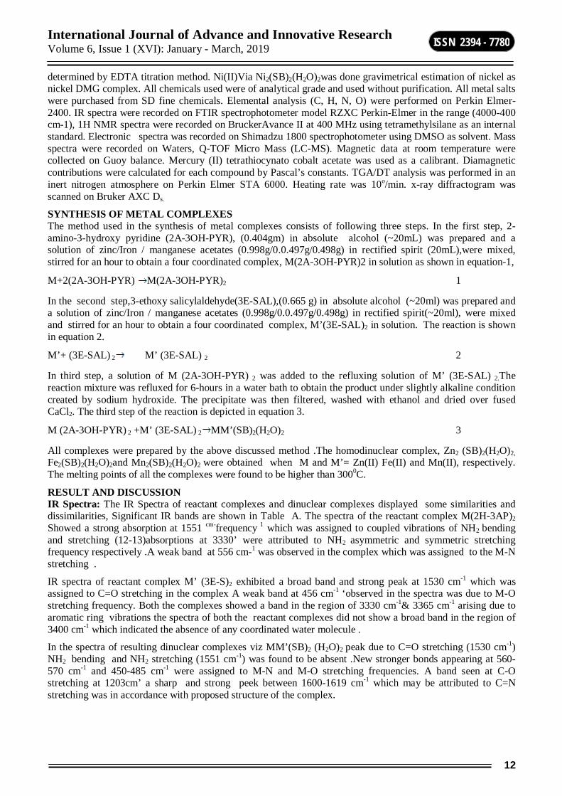
INTRODUCYION Mixed metal complexes differ from traditional complexes in the sense that they are having at least two different or same metals associated with two different ligands (metal organic ligands) the presence of more than one type of ligands in a complex increases chances of variation in properties expected for the complex .this makes the researcher interested in the synthesis of mixed metal complexes with varying properties.[1] Synthesis and characterization of mixed metal complexes is gaining importance day by day. The increased interest in this research area has motivated many researchers to get involved in this field. In recent years many publication are devoted to synthesis and characterization of mixed metal as well as ligands complexes. [2-6] The Schiff base complexes were also used as drugs and they possess a wide variety of antimicrobial activity against bacteria, fungi and it also inhibits the growth of certain type of tumors.[7-8] The complexes formed by coordination with metal ions, have the tendency to coordinate further or react with other complexes ,then they may act as metal organic ligand (MOL). The donor atoms are unable to coordinate with the same metal ions duo to steric factors. This unutilized functionality is drawn on another metal ion forming poly nuclear complex [9-10]
Here a complex containing some unutilized functionality in ligand is considered as a ligand and named as metal organic ligand (MOL). This MOL when allowed to react with metal ions result in the formation of mixed metal complexes. We report here, a novel approach of synthesizing mixed metal complexes. It has been hypothesized that the coordinated ligands of two metal chelates can be reacted to obtain a new metal chelate. In the present work, we have allowed to react two such complexes under the conditions that permit coordinated NH2 to react with the coordinated CHO group. Here an ionic bonds of the precursor do not dissociate and metal-ligand bonding in both the complexes remained intact [11]. Due to the reaction between coordinated amino and aldehyde groups, Schiff base were formed. The imine nitrogen of the Schiff base was allowed to coordinate with the metal nearby while the deficiency created at the metal ion on aldehyde end was sufficed by aquo-ligands liberated during imine formation. The resultant binuclear complex thus has one of the metal ions in di aquo form. When the metal ion in the reacting complexes was different, the resultant complex was mixed metal complex [12].
MATERIALS AND METHOD Reagents: 2-amino 3-hydroxy pyridine and 3-ethoxy salicyaldehyde (>99.0%) were purchased from S.D. Fine Chemicals. Nickel acetate, copper acetate, cobalt acetate sodium hydroxide and solvents (>99.0%) were purchased from E-Marck Ltd, Mumbai (India).The purification was done according to the needs through known procedures.
Measurements:. Elemental analysis (C, H, N & O) was done using Perkin Elmer, series II, 2400 CHNS/O Analyzer. The metal content of the complexes were determined by EDTA titration method after decomposition of the metal complexes with an acid mixture of HCLO4, H2SO4 and HNO3 (1:1.5:2.5) The amount of Cu(II) and Co(II)from homo dinuclear complex of Cu(II) and Co(II) Viz Cu2(SB)2(H2O)2and Co2(SB)2(H2O)2 was
International Journal of Advance and Innovative Research Volume 6, Issue 1 (XVI): January - March, 2019
12
ISSN 2394 - 7780
determined by EDTA titration method. Ni(II)Via Ni2(SB)2(H2O)2was done gravimetrical estimation of nickel as nickel DMG complex. All chemicals used were of analytical grade and used without purification. All metal salts were purchased from SD fine chemicals. Elemental analysis (C, H, N, O) were performed on Perkin Elmer-2400. IR spectra were recorded on FTIR spectrophotometer model RZXC Perkin-Elmer in the range (4000-400 cm-1), 1H NMR spectra were recorded on BruckerAvance II at 400 MHz using tetramethylsilane as an internal standard. Electronic spectra was recorded on Shimadzu 1800 spectrophotometer using DMSO as solvent. Mass spectra were recorded on Waters, Q-TOF Micro Mass (LC-MS). Magnetic data at room temperature were collected on Guoy balance. Mercury (II) tetrathiocynato cobalt acetate was used as a calibrant. Diamagnetic contributions were calculated for each compound by Pascal’s constants. TGA/DT analysis was performed in an inert nitrogen atmosphere on Perkin Elmer STA 6000. Heating rate was 10ο/min. x-ray diffractogram was scanned on Bruker AXC Ds.
SYNTHESIS OF METAL COMPLEXES The method used in the synthesis of metal complexes consists of following three steps. In the first step, 2-amino-3-hydroxy pyridine (2A-3OH-PYR), (0.404gm) in absolute alcohol (~20mL) was prepared and a solution of zinc/Iron / manganese acetates (0.998g/0.0.497g/0.498g) in rectified spirit (20mL),were mixed, stirred for an hour to obtain a four coordinated complex, M(2A-3OH-PYR)2 in solution as shown in equation-1,
M+2(2A-3OH-PYR) M(2A-3OH-PYR)2 1
In the second step,3-ethoxy salicylaldehyde(3E-SAL),(0.665 g) in absolute alcohol (~20ml) was prepared and a solution of zinc/Iron / manganese acetates (0.998g/0.0.497g/0.498g) in rectified spirit(~20ml), were mixed and stirred for an hour to obtain a four coordinated complex, M’(3E-SAL)2 in solution. The reaction is shown in equation 2.
M’+ (3E-SAL) 2 M’ (3E-SAL) 2 2
In third step, a solution of M (2A-3OH-PYR) 2 was added to the refluxing solution of M’ (3E-SAL) 2.The reaction mixture was refluxed for 6-hours in a water bath to obtain the product under slightly alkaline condition created by sodium hydroxide. The precipitate was then filtered, washed with ethanol and dried over fused CaCl2. The third step of the reaction is depicted in equation 3.
M (2A-3OH-PYR) 2 +M’ (3E-SAL) 2 MM’(SB)2(H2O)2 3
All complexes were prepared by the above discussed method .The homodinuclear complex, Zn2 (SB)2(H2O)2, Fe2(SB)2(H2O)2and Mn2(SB)2(H2O)2 were obtained when M and M’= Zn(II) Fe(II) and Mn(II), respectively. The melting points of all the complexes were found to be higher than 3000C.
RESULT AND DISCUSSION IR Spectra: The IR Spectra of reactant complexes and dinuclear complexes displayed some similarities and dissimilarities, Significant IR bands are shown in Table A. The spectra of the reactant complex M(2H-3AP)2 Showed a strong absorption at 1551 cm-frequency 1 which was assigned to coupled vibrations of NH2 bending and stretching (12-13)absorptions at 3330’ were attributed to NH2 asymmetric and symmetric stretching frequency respectively .A weak band at 556 cm-1 was observed in the complex which was assigned to the M-N stretching .
IR spectra of reactant complex M’ (3E-S)2 exhibited a broad band and strong peak at 1530 cm-1 which was assigned to C=O stretching in the complex A weak band at 456 cm-1 ‘observed in the spectra was due to M-O stretching frequency. Both the complexes showed a band in the region of 3330 cm-1& 3365 cm-1 arising due to aromatic ring vibrations the spectra of both the reactant complexes did not show a broad band in the region of 3400 cm-1 which indicated the absence of any coordinated water molecule .
In the spectra of resulting dinuclear complexes viz MM’(SB)2 (H2O)2 peak due to C=O stretching (1530 cm-1) NH2 bending and NH2 stretching (1551 cm-1) was found to be absent .New stronger bonds appearing at 560-570 cm-1 and 450-485 cm-1 were assigned to M-N and M-O stretching frequencies. A band seen at C-O stretching at 1203cm’ a sharp and strong peek between 1600-1619 cm-1 which may be attributed to C=N stretching was in accordance with proposed structure of the complex.
International Journal of Advance and Innovative Research Volume 6, Issue 1 (XVI): January - March, 2019
13
ISSN 2394 - 7780
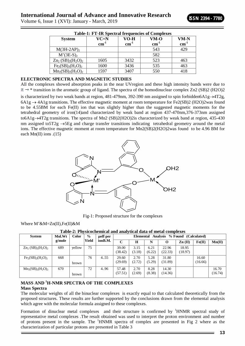
Table-1: FT-IR Spectral frequencies of Complexes System VC=N
cm-1 VO-H cm-1
VM-O cm-1
VM-N cm-1
M(3H-2AP)2 543 429 M’(3E-S)2 582
Zn2 (SB)2(H2O)2 1605 3432 523 463 Fe2(SB)2(H2O)2 1600 3436 535 463 Mn2(SB)2(H2O)2 1597 3407 550 418
ELECTRONIC SPECTRA AND MAGNETIC STUDIES All the complexes showed absorption peaks in the near UVregion and these high intensity bands were due to
* transition in the aromatic group of ligand. The spectra of the homodinuclear complex Zn2 (SB)2 (H2O)2 is characterized by two weak bands at region, 481-479nm, 392-390 nm assigned to spin forbidden6A1g4T2g, 6A1g 4A1g transitions. The effective magnetic moment at room temperature for Fe2(SB)2 (H2O)2was found to be 4.55BM for each Fe(II) ion that was slightly higher than the suggested magnetic moments for the tetrahedral geometry of iron(14)and characterized by weak band at region 437-470nm,376-373nm assigned to6A1g4T2g transitions. The spectra of Mn2 (SB)2(H2O)2is characterized by weak band at region, 435-430 nm assigned to5T2g 5Eg and charge transfer transitions indicating tetrahedral geometry around the metal ions. The effective magnetic moment at room temperature for Mn2(SB)2(H2O)2was found to be 4.96 BM for each Mn(II) ions .(15)
N
N
O
O N
N
O
O
M
OH2
OH2
M'
Fig-1: Proposed structure for the complexes
Where M’&M=Zn(II),Fe(II)&M
Table-2: Physicochemical and analytical data of metal complexes System Mol.Wt
g/mole Color %
Yield µeff per ionB.M.
Elemental Analysis % Found (Calculated) C H N O Zn (II) Fe(II) Mn(II)
Zn2 (SB)2(H2O)2 689 yellow 75 39.00 (38.42)
3.15 (3.18)
6.21 (6.22)
22.96 (22.33)
18.95 (18.97)
Fe2(SB)2(H2O)2 668 brown
76 4..55 29.60 (29.69)
2.70 (2.72)
5.28 (5.29)
31.80 (31.89)
16.60 (16.66)
Mn2(SB)2(H2O)2 670 brown
72 4..96 57.48 (57.51)
2.70 (2.69)
8.28 (8.38)
14.30 (14.36)
16.70 (16.74)
MASS AND 1H-NMR SPECTRA OF THE COMPLEXES Mass Spectra The molecular weights of all the binuclear complexes is exactly equal to that calculated theoretically from the proposed structures. These results are further supported by the conclusions drawn from the elemental analysis which agree with the molecular formula assigned to these complexes.
Formation of dinuclear metal complexes and their structure is confirmed by 1HNMR spectral study of representative metal complexes .The result obtained was used to interpret the proton environment and number of protons present in the sample. The 1HNMR spectra of complex are presented in Fig 2 where as the characterization of particular protons are presented in Table 3
International Journal of Advance and Innovative Research Volume 6, Issue 1 (XVI): January - March, 2019
14
ISSN 2394 - 7780
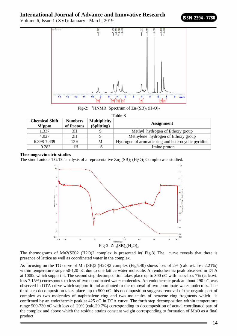
Fig-2: 1HNMR Spectrum of Zn2(SB)2 (H2O)2
Table-3 Chemical Shift
‘δ’ppm Numbers of Protons
Multiplicity (Splitting) Assignment
1.337 3H S Methyl hydrogen of Ethoxy group 4.027 2H S Methylene hydrogen of Ethoxy group
6.398-7.439 12H M Hydrogen of aromatic ring and heterocyclic pyridine 9.283 1H S Imine proton
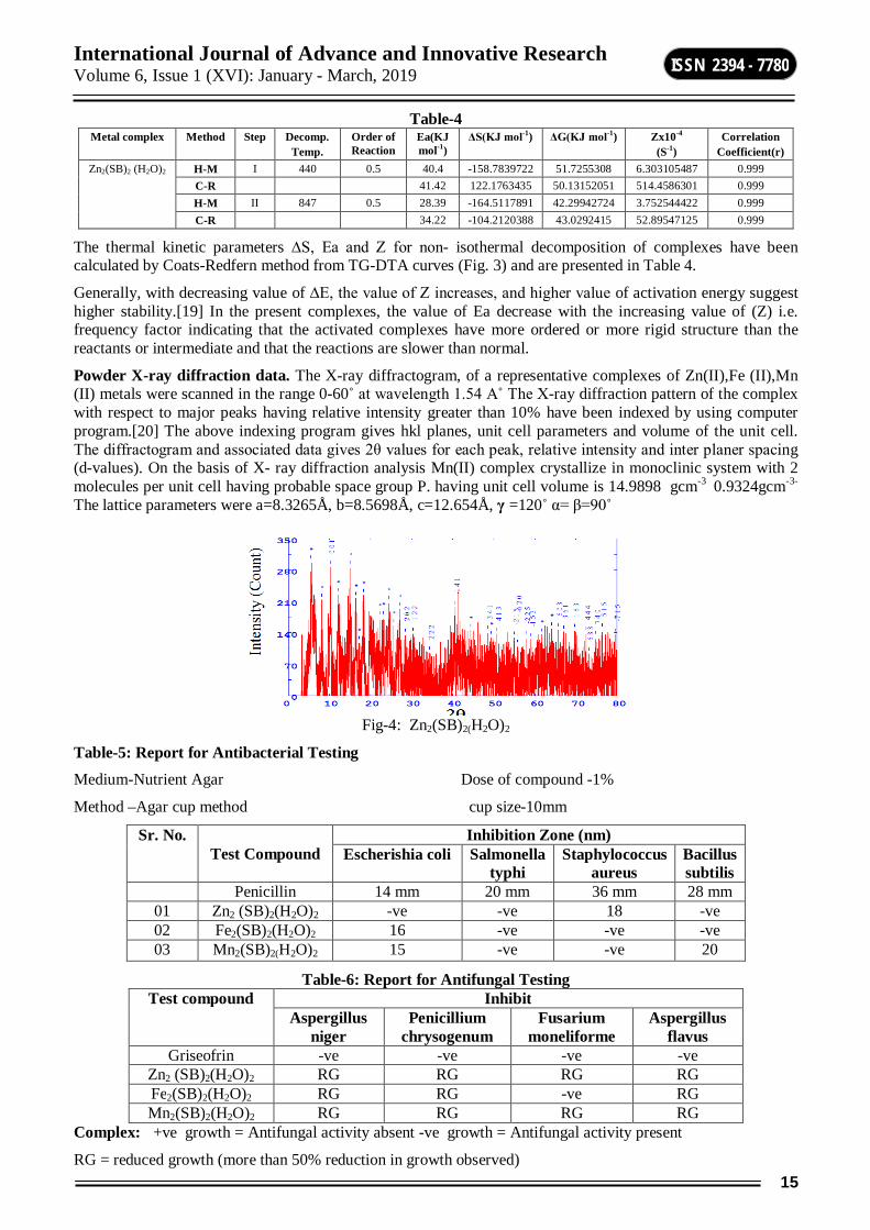
Thermogravimetric studies The simultanious TG/DT analysis of a representative Zn2 (SB)2 (H2O)2 Complexwas studied.
Fig-3: Zn2(SB)2(H2O)2
The thermograms of Mn2(SB)2 (H2O)2 complex is presented in( Fig.3) The curve reveals that there is presence of lattice as well as coordinated water in the complex.
As focusing on the TG curve of Mn (SB)2 (H2O)2 complex (Fig5.40) shows loss of 2% (calc wt. loss 2.21%) within temperature range 50-120 oC due to one lattice water molecule. An endothermic peak observed in DTA at 1000c which support it. The second step decomposition takes place up to 300 oC with mass loss 7% (calc.wt. loss 7.15%) corresponds to loss of two coordinated water molecules. An endothermic peak at about 290 oC was observed in DTA curve which support it and attributed to the removal of two coordinate water molecules. The third step decomposition takes place up to 500 oC this decomposition suggests removal of the organic part of complex as two molecules of naphthalene ring and two molecules of benzene ring fragments which is confirmed by an endothermic peak at 425 oC in DTA curve. The forth step decomposition within temperature range 500-730 oC with loss of 29% (calc.29.7%) corresponding to decomposition of actual coordinated part of the complex and above which the residue attains constant weight corresponding to formation of MnO as a final product.
International Journal of Advance and Innovative Research Volume 6, Issue 1 (XVI): January - March, 2019
15
ISSN 2394 - 7780
Table-4 Metal complex Method Step Decomp.
Temp. Order of Reaction
Ea(KJ mol-1)
ΔS(KJ mol-1) ΔG(KJ mol-1) Zx10-4 (S-1)
Correlation Coefficient(r)
Zn2(SB)2 (H2O)2 H-M I 440 0.5 40.4 -158.7839722 51.7255308 6.303105487 0.999 C-R 41.42 122.1763435 50.13152051 514.4586301 0.999 H-M II 847 0.5 28.39 -164.5117891 42.29942724 3.752544422 0.999 C-R 34.22 -104.2120388 43.0292415 52.89547125 0.999
The thermal kinetic parameters ∆S, Ea and Z for non- isothermal decomposition of complexes have been calculated by Coats-Redfern method from TG-DTA curves (Fig. 3) and are presented in Table 4.
Generally, with decreasing value of ∆E, the value of Z increases, and higher value of activation energy suggest higher stability.[19] In the present complexes, the value of Ea decrease with the increasing value of (Z) i.e. frequency factor indicating that the activated complexes have more ordered or more rigid structure than the reactants or intermediate and that the reactions are slower than normal.
Powder X-ray diffraction data. The X-ray diffractogram, of a representative complexes of Zn(II),Fe (II),Mn (II) metals were scanned in the range 0-60˚ at wavelength 1.54 A˚ The X-ray diffraction pattern of the complex with respect to major peaks having relative intensity greater than 10% have been indexed by using computer program.[20] The above indexing program gives hkl planes, unit cell parameters and volume of the unit cell. The diffractogram and associated data gives 2θ values for each peak, relative intensity and inter planer spacing (d-values). On the basis of X- ray diffraction analysis Mn(II) complex crystallize in monoclinic system with 2 molecules per unit cell having probable space group P. having unit cell volume is 14.9898 gcm-3 0.9324gcm-3-
The lattice parameters were a=8.3265Å, b=8.5698Å, c=12.654Å, γ =120˚ α= β=90˚
Fig-4: Zn2(SB)2(H2O)2
Table-5: Report for Antibacterial Testing
Medium-Nutrient Agar Dose of compound -1%
Method –Agar cup method cup size-10mm
Sr. No. Test Compound
Inhibition Zone (nm) Escherishia coli Salmonella
typhi Staphylococcus
aureus Bacillus subtilis
Penicillin 14 mm 20 mm 36 mm 28 mm 01 Zn2 (SB)2(H2O)2 -ve -ve 18 -ve 02 Fe2(SB)2(H2O)2 16 -ve -ve -ve 03 Mn2(SB)2(H2O)2 15 -ve -ve 20
Table-6: Report for Antifungal Testing Test compound Inhibit
Aspergillus niger
Penicillium chrysogenum
Fusarium moneliforme
Aspergillus flavus
Griseofrin -ve -ve -ve -ve Zn2 (SB)2(H2O)2 RG RG RG RG Fe2(SB)2(H2O)2 RG RG -ve RG Mn2(SB)2(H2O)2 RG RG RG RG
Complex: +ve growth = Antifungal activity absent -ve growth = Antifungal activity present
RG = reduced growth (more than 50% reduction in growth observed)
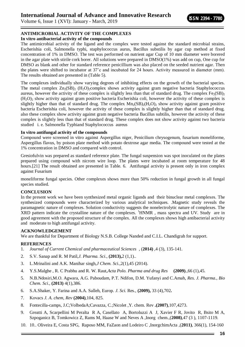
International Journal of Advance and Innovative Research Volume 6, Issue 1 (XVI): January - March, 2019
16
ISSN 2394 - 7780
ANTIMICROBIAL ACTIVITY OF THE COMPLEXES In vitro antibacterial activity of the compounds The antimicrobial activity of the ligand and the complex were tested against the standard microbial strains, Escherishia coli, Salmonella typhi, staphylococcus aurus, Bacillus substilis by agar cup method at fixed concentration of 1% in DMSO. The test was performed on nutrient agar Cup of 10 mm diameter were borered in the agar plate with stirile cork borer. All solutions were prepared in DMSO(1%) was add on cup, One cup for DMSO as blank and other for standard reference penicillium was also placed on the seeded nutrient agar. Then the plates were shifted to incubator at 37˚c and incubated for 24 hours. Activity measured in diameter (mm). The results obtained are presented in (Table 5).
The complexes individually show varying degrees of inhibiting effects on the growth of the bacterial species. The metal complex Zn2(SB)2 (H2O)2complex shows activity against gram negative bacteria Staphylococcus aureus, however the activity of these complex is slightly less than that of standard drug. The complex Fe2(SB)2 (H2O)2 show activity against gram positive bacteria Escherishia coli, however the activity of these complex is slightly higher than that of standard drug. The complex Mn2(SB)2(H2O)2 show activity against gram positive bacteria Escherishia coli, however the activity of these complex is slightly higher than that of standard drug. also these complex show activity against gram negative bacteria Bacillus subtilis, however the activity of these complex is slightly less than that of standard drug. These complex does not show activity against two bacteria studied i. e. Salmonella Typhiand Staphylococcus aureus
In vitro antifungal activity of the compounds Compound were screened in vitro against Aspergillus niger, Penicilium chrysogenum, fusarium moneliforme, Aspergillus flavus, by poison plate method with potato dextrose agar media. The compound were tested at the 1% concentration in DMSO and compared with control.
Gresiofulvin was prepared as standard reference plate. The fungal suspension was spot inoculated on the plates prepared using compound with nicrom wire loop. The plates were incubated at room temperature for 48 hours.[21] The result obtained are presented in Table. 6. Antifungal activity is present only in iron complex against Fusarium
moneliforme fungal species. Other complexes shows more than 50% reduction in fungal growth in all fungal species studied.
CONCLUSION In the present work we have been synthesized metal organic ligands and their binuclear metal complexes. The synthesized compounds were characterized by various analytical techniques. .Magnetic study reveals the paramagnetic nature of complexes. Solution conductivity suggests the nonelectrolytic nature of complexes. The XRD pattern indicate the crystalline nature of the complexes. 1HNMR , mass spectra and UV. Study are in good agreement with the proposed structure of the complex. All the complexes shows high antibacterial activity and moderate to high antifungal activity.
ACKNOWLEDGEMENT We are thankful for Department of Biology N.S.B. College Nanded and C.I.L. Chandigrah for support.
REFERENCES 1. Journal of Current Chemical and pharmaceutical Sciences , (2014) ,4 (3), 135-141. 2. S.V. Sanap and R. M Patil,J. Pharma. Sci., (2013),2 (1,1).. 3. L.Mrinalini and A.K. Manihar singh,J Chem. Sci.,2(1),45 (2014). 4. Y.S.Malghe , R. C Prabhu and R. W. Raut,Acta Polo. Pharma and drug Res (2009).,66 (1),45. 5. N.B.Ndosiri,M.O. Agwara, A.G. Paboudam, P.T. Ndifon, D.M. Yufanyi and C.Amah, Res. J. Pharma., Bio
Chem. Sci., (2013) 4(1),386. 6. S.A.Shaker, Y. Farina and A.A. Salleh, Europ. J. Sci. Res., (2009), 33 (4),702. 7. Kovacs J. A. chem, Rev (2004),104, 825. 8. Fontecilla-camps, J.C;VolbedaA;Cavazza, C.;Nicolet ,Y. chem. Rev .(2007),107,4273. 9. Greatti A, Scarpellini M Peralta R A, Casellato A, Bortoluzzi A J, Xavier F R, Jovito R, Bsito M A,
Szpoganicz B, Tomkowicz Z, Rams M, Haase W and Neves A ,Inorg chem.,(2008),47 (3 ), 1107-1119. 10. 10.. Oliveira E, Costa SPG, Raposo MM, FaZaon and Lodeiro C ,InorgchimActa ,(2011), 366(1), 154-160

International Journal of Advance and Innovative Research Volume 6, Issue 1 (XVI): January - March, 2019
17
ISSN 2394 - 7780
11. Dobrokhotova Z, Emelina A Sidorov A, etal.,(2011).Synthesis and characterization of Li (1)- M (ii) (M=Co, Ni) heterometallic complexes as molecular precursors forLiMO2.polyhedron, 30: 132-141.
12. Dobrokhotova Z, Emelina A SidorovA,et al ., (2011) . Synthesis and characterization of Li (1)-M (ii) (M=Co, Ni) heterometallic complexes as molecular precursors forLiMO2.polyhedron, 30: 132-141.
13. V.D. Bhatt, SR Ram ChemicalSciences Jornal, Vol. (2012): CSJ-63.19
14. M. Islam,B. Hossain and M. Reza (2003). Antimicrobial studies of mixed ligand transition metal complexes of maleic acid and heterocyclic amine bases .journal of Medical science ,3:289-293.
15. V.D.Bhatt,S.R.Ram,ChemicalScienceTransactions,(2013)Accepted.
16. R. Boca, M. Gembicky M, Herchel R, et al., (2003). Ferromagnetism in a dinuclear nickel (ii) complex containing triethylenetetramine and tricyanomethanide. Inorganic
17. Bhatt V.D.and Ray A, Synth met., (1998), 92(2), 115-120.
18. V.D. Bhatt, SR Ram ChemicalSciences Jornal, Vol. (2012). CSJ-63.
19. P.S. Mane, S.D. Saluke, S.G. Shiradkar, T.K. Chondheker, Asian. J. Chem., (2001); 13: 1573-1576
20. J. R. Carvajal, T.Roisnel, A. Winplotr, Graphic Tool for Powder Differaction Laboratories leon brillouin 91191 gif suryvette cedex France( 2004).
21. Swamy D,Pachling S,Bhagat T, RASAYAN J. Chem,(2001)
22. R. J. Cruickshank, P. Duguid, R. S. wain, Publisher Churchill Lilingstone, Medical Microbiology 1: (1998).
International Journal of Advance and Innovative Research Volume 6, Issue 1 (XVI): January - March, 2019
18
ISSN 2394 - 7780
ALGAL DIVERSITY OF KANKALESHOWAR TEMPLE POND IN BEED CITY
S. M. Talekar1 and A. N. Shelke2
1P.G. Department of Botany, Mrs K. S. K. College, Beed, Dist-Beed 2P.G. Department of Zoology, Mrs K. S. K. College, Beed, Dist-Beed
ABSTRACT Beed is a glorious historical city in Maharastra, history says that the Kankaleshowar temple pond more than 500 years old and are still being used for daily ritual pursuits of the the concerned temple. The water body of this pond supporting the growth of different species of aquatic fauna and flora including algae. Algae are most abundant autotrophic element of aquatic ecosystem. They play an important role as to mention the biological oxygen demand of fresh as well as polluted water ecosystem. Temple pond ecosystem generally contaminated due to human activity viz. bathing, clothing, damping unwanted materials. In present study algal diversity and Physical parameters of Kankaleshower temple pond reviles that the dominance of Chlorophyceae with 29 taxa belong to 10 genra and flowed by Cyanophyceae 22 texa with 08 genera, Bascillariophyceae with 16 species belongs to 05 genera and Euglenophyceae 08 texa with 02 genera.
INTRODUCTION The exact geographical location of Beed district is at 16.65oN 74.13oE. it has a mean elevation of 530 meters (1738 feet). Beed district is located on the Deccan plateau. In the district, the main rivers are Manjara, Bendusara and Sindfana. The Balaghat range is close by. The soil of the area is rough and rocky largely consisting of basalt. Thin deposits of fertile black soil are found in the northern part and in the south at the western bank of Bendusara. The district experiences semi-arid, warm and dry climate, summers are lengthy, extending from the middle of February to June. Average temperature in summer vary between 31oC to 40oC. Winters are short with temperature between 12oC to 20oC. Rains are inadequate and take place only during the monsoon from mid June to September. The average annual rainfall is 666mm.
The present investigation was carried out on Algale Diversity of kankaleshowar Temple Pond in Beed city of Maharastra. Because of sum reaserchers have paid their attention on algal diversity of waste water, lake water, reservoir, river ecosystem in Beed distict (Ashtekar 1980,Talekar 2009, Talekar and Jadhav 2009,Yadve 2010, Devggude and Talekar 2017) but no one work on pond ecosystem.
MAREIALS AND METHODES Algal samples were collected at monthly intervals randomly during June 2017 to May 2018 in Acid washed collection bottles. Floating, Planktonic and attached substratum algal samples were collected separately in collection bottles. Plankton net was used to collect Planktonic algae. After collection, algal samples were brought immediately to the Laboratory. The algal samples were preserved in 4% formalin for further taxonomic investigations. The fresh as well as preserved algal forms were observed under microscope and identified. Identification of algal taxa was performed by referring to the standard literature on algae (Smith 1950, Prescott 1951, Desikachary 1959, Randhawa 1959, Pal et. al., 1962, Ramanathan 1962, Krieger and Gerloff 1965, Philipose 1967, Kamat, N.D. 1975). Air temperature and water temperature were recorded by using centigrade mercury thermometer at the time of sampling, similarly pH count also taken at the time of collection of samples.
RESULT AND DISCUSION In present study total 75 algal taxa were encountered belong to 25 genera. Out of these Chlorophyceae with 29 species belong to 10 genra and flowed by Cyanophyceae 22 texa with 08 genera, Bascillariophyceae with 16 species belongs to 05 genera and Euglenophyceae 08 texa with 02 genera. Similar kind of observation was made by Magar 2008 in fresh water of Girna dam of Nashik district, Andhale 2008 fresh water flora of Jayakwadi Bird Sanctuary. Talekar 2016 in dairy waste water in Beed city Average air temperature count 30o and water temperature count 28o in study period . The average pH value of water is 7.5 and acidic in nature this work is similar to Physico-chemical analysis of Pimpalwandi reservoirs (Khopti) Shelke 2016 .
Table-01: Class wises dominance of algal taxa in kankaleshowar Temple Pond. Sr. No. Name of Class Genera Species
01 Chlorophyceae 10 29 02 Bascillariophyceae 05 16 03 Euglenophyceae 02 08 04 Cyanophyceae 08 22
Total 25 75
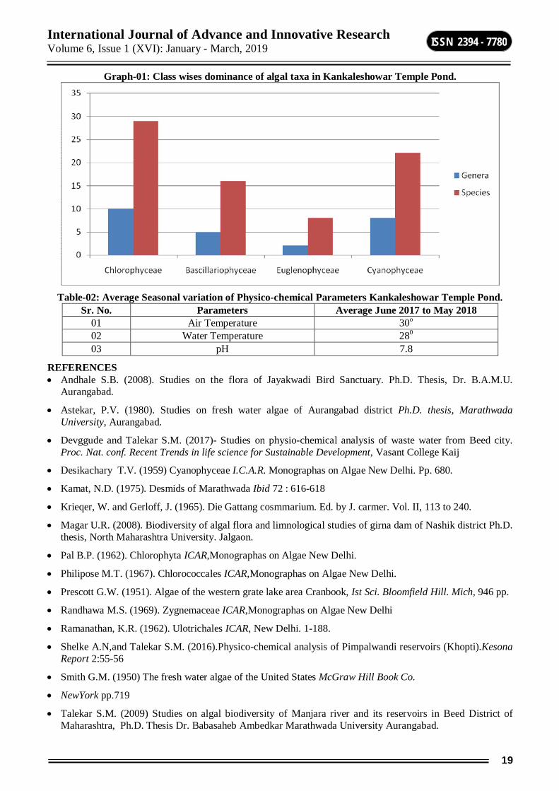
International Journal of Advance and Innovative Research Volume 6, Issue 1 (XVI): January - March, 2019
19
ISSN 2394 - 7780
Graph-01: Class wises dominance of algal taxa in Kankaleshowar Temple Pond.
Table-02: Average Seasonal variation of Physico-chemical Parameters Kankaleshowar Temple Pond.
Sr. No. Parameters Average June 2017 to May 2018 01 Air Temperature 30o 02 Water Temperature 280
03 pH 7.8
REFERENCES Andhale S.B. (2008). Studies on the flora of Jayakwadi Bird Sanctuary. Ph.D. Thesis, Dr. B.A.M.U.
Aurangabad.
Astekar, P.V. (1980). Studies on fresh water algae of Aurangabad district Ph.D. thesis, Marathwada University, Aurangabad.
Devggude and Talekar S.M. (2017)- Studies on physio-chemical analysis of waste water from Beed city. Proc. Nat. conf. Recent Trends in life science for Sustainable Development, Vasant College Kaij
Desikachary T.V. (1959) Cyanophyceae I.C.A.R. Monographas on Algae New Delhi. Pp. 680.
Kamat, N.D. (1975). Desmids of Marathwada Ibid 72 : 616-618
Krieqer, W. and Gerloff, J. (1965). Die Gattang cosmmarium. Ed. by J. carmer. Vol. II, 113 to 240.
Magar U.R. (2008). Biodiversity of algal flora and limnological studies of girna dam of Nashik district Ph.D. thesis, North Maharashtra University. Jalgaon.
Pal B.P. (1962). Chlorophyta ICAR,Monographas on Algae New Delhi.
Philipose M.T. (1967). Chlorococcales ICAR,Monographas on Algae New Delhi.
Prescott G.W. (1951). Algae of the western grate lake area Cranbook, Ist Sci. Bloomfield Hill. Mich, 946 pp.
Randhawa M.S. (1969). Zygnemaceae ICAR,Monographas on Algae New Delhi
Ramanathan, K.R. (1962). Ulotrichales ICAR, New Delhi. 1-188.
Shelke A.N,and Talekar S.M. (2016).Physico-chemical analysis of Pimpalwandi reservoirs (Khopti).Kesona Report 2:55-56
Smith G.M. (1950) The fresh water algae of the United States McGraw Hill Book Co.
NewYork pp.719
Talekar S.M. (2009) Studies on algal biodiversity of Manjara river and its reservoirs in Beed District of Maharashtra, Ph.D. Thesis Dr. Babasaheb Ambedkar Marathwada University Aurangabad.

International Journal of Advance and Innovative Research Volume 6, Issue 1 (XVI): January - March, 2019
20
ISSN 2394 - 7780
DIVERSITY, DISTRIBUTIONAND POPULATION DENSITY OF FRESH WATER ZOOPLANKTON FROM SHIVANA-TAKLI RESERVOIR OF KANNAD TALUKA, MAHARASHTRA STATE
K. B. Temkar1, B. N. Jadhav3, S. A. Shaikh2, D. S. Kharate4, P. S. Kharate5, M. S. Kharate6, and V. R.Lakwal7 1,3Department of Zoology, Vinayakrao Patil College, Vaijapur, Aurangabad
2Department of Zoology, Sunderrao Solanke Mahavidyalaya, Majalgaon, Beed 5Department of Plant Molecular Biology and Biotechnology, Indira Gandhi Krishi, Vishwavidyalaya, Raipur, Chhattisgarh
6Department of Botany, Vinayakrao Patil College, Vaijapur, Aurangabad 7Department of Zoology, Shri Muktanand College, Gangapur, Aurangabad
ABSTRACT The present study was assesseddiversity, distribution and population density of fresh water zooplankton from Shivana-Takli reservoir. This reservoir is located atLatitude 74° 40’ 15” North and Longitude 20° 03’ 20” East on a tributary of Shivana river near village Takali in Kannadtahsil.Samples were collectedusing simple conical tow plankton net (40 µm bolting nylon).During present observations 61freshwater zooplankton species belonging to 18 families 20 genera were recorded. The results clearly showed that species of Rotifera>Copepoda>Cladocera>Ostracoda. The present study concluded that the Shivana-Takli reservoir rich in freshwater zooplankton diversity and the dominance of rotifera and copepoda indicating the eutrophication of present water body.
Keywords: Zooplankton, Diversity,Population density, Shivana-Takli reservoir, Rotifera.
INTRODUCTION Zooplankton are small animals that float freely in the water column of lakes and oceans and whose distribution is primarily determined by water currents and mixing. The zooplankton community of most lakes ranges in size from a few tens of microns (Protozoa) to >2 mm (macrozooplankton). In terms of biomass and productivity, the dominant groups of zooplankton in most lakes are Crustacea and Rotifera and these protocols emphasize those groups. Zooplankton play a pivotal role in aquatic food webs because they are important food for fish and invertebrate predators and they graze heavily on algae, bacteria, protozoa, and other invertebrates. Zooplankton communities are typically diverse (>20 species) and occur in almost all lakes and ponds. Zooplankton are rarely important in rivers and streams because they cannot maintain positive net growth rates in the face of downstream losses.
The relationship between livings and their environment is inevitable, continuous and reciprocal. The study of these interactions under the natural conditions constitutes the science of ecology. The linkages between these wide varieties of diverse habitats and ecosystems are essential for the maintenance of food webs, migration routes and increased productivity.Zooplankton are susceptible to variations in a wide number of environmental factors including water temperature, light, chemistry (particularly pH, oxygen, salinity, toxic contaminants), food availability (algae, bacteria), and predation by fish and invertebrates. It is generally desirable to have as much information on these variables as possible. Clearly, this will frequently be practical. Some variables are relatively easy to measure (e.g. temperature), but others are more difficult (e.g. fish-predation intensity, toxic contaminants). Many environmental factors affect zooplankton only at extreme levels (e.g. toxic contaminants, salinity, oxygen) and will not be important in all lakes. Ideally, most sample collections should be accompanied by measures of water temperature, pH, and algal biomass (chlorophyll-a concentration or phytoplankton biomass).
The Shivana-Takli is medium irrigation project is constructed in the Godavari basin in Aurangabad district of Maharashtra. The project comprises of 4408 m long earth dam with the maximum height of 14.77 m and 172 m long masonry dam having maximum height of 29.50 m with 78 m long spillway across river Shivana near Takli village in Kannad Taluka of Aurangabad district. The canal system consists of 10.53 km long Left Bank Canal (LBC) to irrigate 2320 ha of Irrigable Command Area (ICA) and 56.20 km long Right Bank Canal (RBC) to cover the Irrigable Command Area (ICA) of 4069 ha. The project on completion will create the annual ultimate irrigation potential of 6389 ha in Kannad and Vaijapur Talukas of Aurangabad district. The project will also provide 3.591 Mm3 water for domestic use for 40 villages in Aurangabad district. The CCA of the project is also 6389 hectare.
The project was approved by the Planning Commission, Govt. of India for an estimated cost of Rs. 34.76 crore in 1995. The construction work on the project started in 1982. In the year 2004-05 the project was brought under AIBP. The project is completed during 2008-09 and the Latest estimmated cost of the whole project is
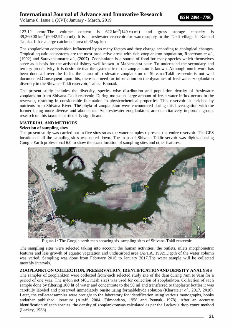
International Journal of Advance and Innovative Research Volume 6, Issue 1 (XVI): January - March, 2019
21
ISSN 2394 - 7780
123.12 crore.The volume content is 622 km3(149 cu mi) and gross storage capacity is 39,360.00 km3 (9,442.97 cu mi). It is a freshwater reservoir for water supply to the Takli village in Kannad Taluka. It has a large catchment area of 42 sq. km.
The zooplankton composition influenced by so many factors and they change according to ecological changes. Tropical aquatic ecosystems are the most productive areas with rich zooplankton population, Robertson et al., (1992) and Saravankumaret al., (2007). Zooplankton is a source of food for many species which themselves serve as a basis for the artisanal fishery well known in Maharashtra state. To understand the secondary and tertiary productivity, it is desirable that the systematic of the zooplankton is known. Although much work has been done all over the India, the fauna of freshwater zooplankton of Shivana-Takli reservoir is not well documented.Consequent upon this, there is a need for information on the dynamics of freshwater zooplankton diversity in the Shivana-Takli reservoir, Taluka Kannad.
The present study includes the diversity, species wise distribution and population density of freshwater zooplankton from Shivana-Takli reservoir. During monsoon, large amount of fresh water influx occurs in the reservoir, resulting in considerable fluctuation in physicochemical properties. This reservoir in enriched by nutrients from Shivana River. The phyla of zooplankton were encountered during this investigation with the former being more diverse and abundance. As freshwater zooplanktons are quantitatively important group, research on this taxon is particularly significant.
MATERIAL AND METHODS Selection of sampling sites The present study was carried out in five sites so as the water samples represent the entire reservoir. The GPS location of all the sampling sites was noted down. The maps of Shivana-Taklireservoir was digitized using Google Earth professional 6.0 to show the exact location of sampling sites and other features.
Figure-1: The Google earth map showing six sampling sites of Shivana-Takli reservoir
The sampling sites were selected taking into account the human activities, the outlets, inlets morphometric features and less growth of aquatic vegetation and undisturbed area (APHA, 1992).Depth of the water column was varied. Sampling was done from February 2016 to January 2017.The water sample will be collected monthly intervals.
ZOOPLANKTON COLLECTION, PRESERVATION, IDENTIFICATIONAND DENSITY ANALYSIS The samples of zooplankton were collected from each selected study site of the dam during 7am to 9am for a period of one year. The nylon net (40μ mesh size) was used for collection of zooplankton. Collection of each sample done by filtering 100 lit of water and concentrate to the 50 ml and transferred to theplastic bottles,it was carefully labeled and preserved immediately onsite using formaldehyde solution (Kharate,et al., 2017, 2018). Later, the collectedsamples were brought to the laboratory for identification using various monographs, books andother published literature (Altaff, 2004, Edmondson, 1958 and Pennak, 1978). After an accurate identification of each species, the density of zooplanktonwas calculated as per the Lackey’s drop count method (Lackey, 1938).

International Journal of Advance and Innovative Research Volume 6, Issue 1 (XVI): January - March, 2019
22
ISSN 2394 - 7780
Formula used for the calculation of population density
N = n × v/V
Where,
N = Total no. of organisms/ lit of water filtered.
n = Number of zooplankton counted in 1 ml of plankton sample.
v = Volume of concentrated plankton sample in ml.
V = Volume of total water filtered through in lit.
RESULTS AND DISCUSSION In the present study 61 species of fresh water zooplankton belonging to 18 families and 20 genera from water samples of Shivana-Takli reservoir were recorded.
Table-1: Checklist of freshwater zooplankton species of Shivana-Takli reservoir during February 2016 to January 2017.
Sr. No. Group Name of the Species 01 Rotifera Anuraeopsisfissa(Gosse, 1851) 02 Anuraeopsisnavicula(Rousselet, 1892) 03 Asplanchnabrightwelli(Gosse, 1850) 04 Asplanchnapriodonta(Gosse,1850) 05 Asplanchnaintermedia(Hudson, 1886) 06 Brachionusangularis(Gosse,1851) 07 Brachionusbidentata(Jokubsky,1912) 08 Brachionusbudapestinesis(Daday, 1885) 09 Brachionuscalyciflorus(Pallas, 1834) 10 Brachionuscaudatus(Barrois and Daddy,1894) 11 Brachionusdiversicornis(Daday, 1883) 12 Brachionusfalcatus(Zacharias, 1898) 13 Brachionusforficule(Weirzejski,1891) 14 Brachionusquadridentatus(Hermann, 1783) 15 Brachionusrubens(Ehrenberg, 1838) 16 Keratellacochlearis(Gosse, 1851) 17 Keratellatropica(Apstein, 1907) 18 Notholcalebis(Gosse, 1887) 19 Lecanepapuana(Murray, 1913) 20 Lecaneluna(Muller,1776) 21 Filinialongiseta(Ehrenberg, 1834) 22 Filiniaterminalis(Plate, 1886) 23 Polyarthra major (Burckhardt, 1900) 24 Trichotriatetractis(Ehrenberg,1830) 25 Copepoda Apocyclopsdengizicus(Lepeschkin, 1900) 26 Cletocamptusalbuquerquensis(Herrick, 1895) 27 Eodiaptomusjaponicus(Burckhardt,1913) 28 Eucyclopssperatus(Lilljeborg, 1901) 29 Heliodiaptomusviduus(Gurney, 1916) 30 Mesocyclopsaspericornis(Daday, 1906) 31 Mesocyclopshyalinus(Rehberg, 1880) 32 Mesocyclopsleuckarti(Claus, 1857) 33 Neodiaptomuslindbergi(Brehm, 1951) 34 Neodiaptomusschmakeri(Poppe& Richard, 1892) 35 Paracyclopfermbrialis(Fischer, 1853) 36 Sinodiaptomusindicus(Sewell, 1934) 37 Thermocyclopshyalinus(Rehberg, 1880) 38 Trpocyclopprasinus(Fischer,1886) 39 Cladocera Diaphanosomasarsi(Richard, 1895)
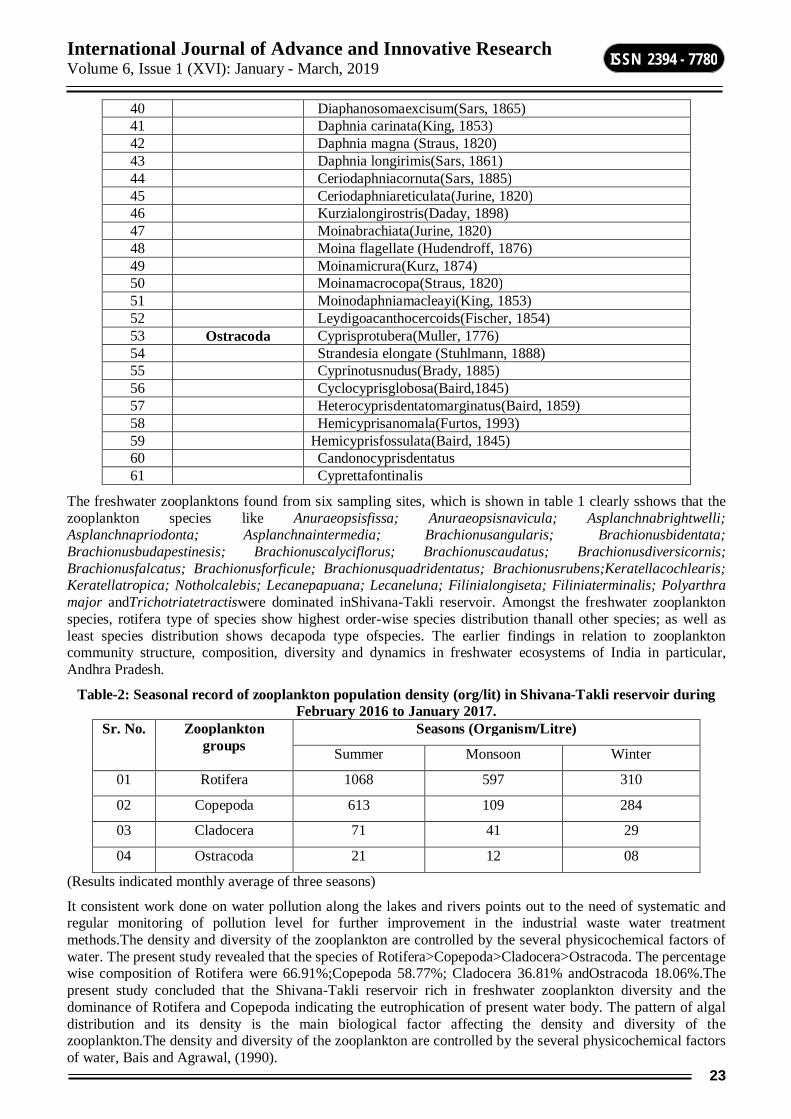
International Journal of Advance and Innovative Research Volume 6, Issue 1 (XVI): January - March, 2019
23
ISSN 2394 - 7780
40 Diaphanosomaexcisum(Sars, 1865) 41 Daphnia carinata(King, 1853) 42 Daphnia magna (Straus, 1820) 43 Daphnia longirimis(Sars, 1861) 44 Ceriodaphniacornuta(Sars, 1885) 45 Ceriodaphniareticulata(Jurine, 1820) 46 Kurzialongirostris(Daday, 1898) 47 Moinabrachiata(Jurine, 1820) 48 Moina flagellate (Hudendroff, 1876) 49 Moinamicrura(Kurz, 1874) 50 Moinamacrocopa(Straus, 1820) 51 Moinodaphniamacleayi(King, 1853) 52 Leydigoacanthocercoids(Fischer, 1854) 53 Ostracoda Cyprisprotubera(Muller, 1776) 54 Strandesia elongate (Stuhlmann, 1888) 55 Cyprinotusnudus(Brady, 1885) 56 Cyclocyprisglobosa(Baird,1845) 57 Heterocyprisdentatomarginatus(Baird, 1859) 58 Hemicyprisanomala(Furtos, 1993) 59 Hemicyprisfossulata(Baird, 1845) 60 Candonocyprisdentatus 61 Cyprettafontinalis
The freshwater zooplanktons found from six sampling sites, which is shown in table 1 clearly sshows that the zooplankton species like Anuraeopsisfissa; Anuraeopsisnavicula; Asplanchnabrightwelli; Asplanchnapriodonta; Asplanchnaintermedia; Brachionusangularis; Brachionusbidentata; Brachionusbudapestinesis; Brachionuscalyciflorus; Brachionuscaudatus; Brachionusdiversicornis; Brachionusfalcatus; Brachionusforficule; Brachionusquadridentatus; Brachionusrubens;Keratellacochlearis; Keratellatropica; Notholcalebis; Lecanepapuana; Lecaneluna; Filinialongiseta; Filiniaterminalis; Polyarthra major andTrichotriatetractiswere dominated inShivana-Takli reservoir. Amongst the freshwater zooplankton species, rotifera type of species show highest order-wise species distribution thanall other species; as well as least species distribution shows decapoda type ofspecies. The earlier findings in relation to zooplankton community structure, composition, diversity and dynamics in freshwater ecosystems of India in particular, Andhra Pradesh.
Table-2: Seasonal record of zooplankton population density (org/lit) in Shivana-Takli reservoir during February 2016 to January 2017.
Sr. No. Zooplankton groups
Seasons (Organism/Litre)
Summer Monsoon Winter
01 Rotifera 1068 597 310
02 Copepoda 613 109 284
03 Cladocera 71 41 29
04 Ostracoda 21 12 08
(Results indicated monthly average of three seasons)
It consistent work done on water pollution along the lakes and rivers points out to the need of systematic and regular monitoring of pollution level for further improvement in the industrial waste water treatment methods.The density and diversity of the zooplankton are controlled by the several physicochemical factors of water. The present study revealed that the species of Rotifera>Copepoda>Cladocera>Ostracoda. The percentage wise composition of Rotifera were 66.91%;Copepoda 58.77%; Cladocera 36.81% andOstracoda 18.06%.The present study concluded that the Shivana-Takli reservoir rich in freshwater zooplankton diversity and the dominance of Rotifera and Copepoda indicating the eutrophication of present water body. The pattern of algal distribution and its density is the main biological factor affecting the density and diversity of the zooplankton.The density and diversity of the zooplankton are controlled by the several physicochemical factors of water, Bais and Agrawal, (1990).
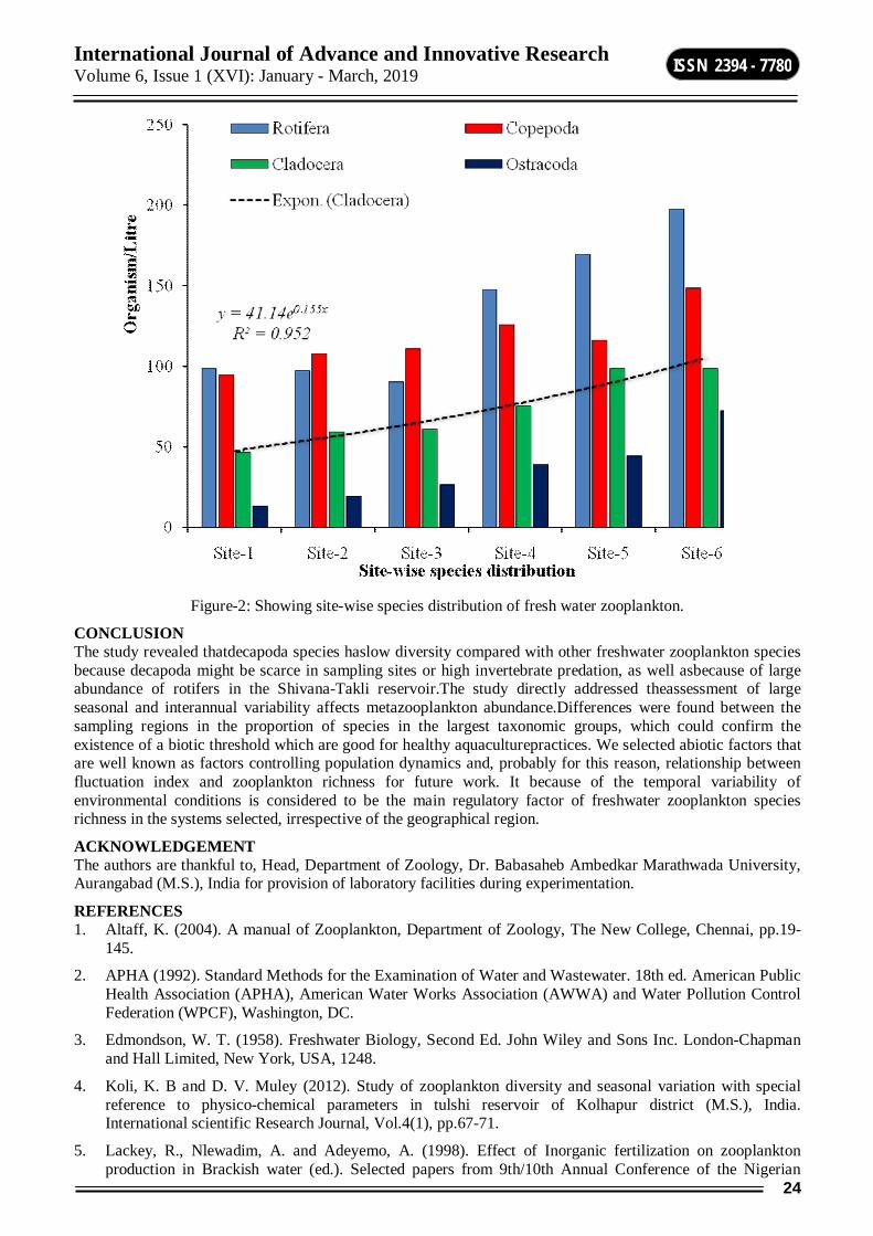
International Journal of Advance and Innovative Research Volume 6, Issue 1 (XVI): January - March, 2019
24
ISSN 2394 - 7780
Figure-2: Showing site-wise species distribution of fresh water zooplankton.
CONCLUSION The study revealed thatdecapoda species haslow diversity compared with other freshwater zooplankton species because decapoda might be scarce in sampling sites or high invertebrate predation, as well asbecause of large abundance of rotifers in the Shivana-Takli reservoir.The study directly addressed theassessment of large seasonal and interannual variability affects metazooplankton abundance.Differences were found between the sampling regions in the proportion of species in the largest taxonomic groups, which could confirm the existence of a biotic threshold which are good for healthy aquaculturepractices. We selected abiotic factors that are well known as factors controlling population dynamics and, probably for this reason, relationship between fluctuation index and zooplankton richness for future work. It because of the temporal variability of environmental conditions is considered to be the main regulatory factor of freshwater zooplankton species richness in the systems selected, irrespective of the geographical region.
ACKNOWLEDGEMENT The authors are thankful to, Head, Department of Zoology, Dr. Babasaheb Ambedkar Marathwada University, Aurangabad (M.S.), India for provision of laboratory facilities during experimentation.
REFERENCES 1. Altaff, K. (2004). A manual of Zooplankton, Department of Zoology, The New College, Chennai, pp.19-
145.
2. APHA (1992). Standard Methods for the Examination of Water and Wastewater. 18th ed. American Public Health Association (APHA), American Water Works Association (AWWA) and Water Pollution Control Federation (WPCF), Washington, DC.
3. Edmondson, W. T. (1958). Freshwater Biology, Second Ed. John Wiley and Sons Inc. London-Chapman and Hall Limited, New York, USA, 1248.
4. Koli, K. B and D. V. Muley (2012). Study of zooplankton diversity and seasonal variation with special reference to physico-chemical parameters in tulshi reservoir of Kolhapur district (M.S.), India. International scientific Research Journal, Vol.4(1), pp.67-71.
5. Lackey, R., Nlewadim, A. and Adeyemo, A. (1998). Effect of Inorganic fertilization on zooplankton production in Brackish water (ed.). Selected papers from 9th/10th Annual Conference of the Nigerian
International Journal of Advance and Innovative Research Volume 6, Issue 1 (XVI): January - March, 2019
25
ISSN 2394 - 7780
Association for Aquatic Sciences. Theme: Sustainable Utilization of Aquatic/Wetland Resources, held at University of Agriculture Abeokuta, Ogun state, Nigeria, pp.75-82.
6. Pennak, R. W. (1953). Fresh Water Invertebrates of the United States.
7. Robertson, A. I. and Blabber, S. J. M. (1992) Plankton, Epibenthos and Fish Communities. Tropical Mangrove Ecosystems, American Geophysical Union, Washington, DC, USA 173-224.
8. Saravanakumar, A., Rajkumar, M., Seshserebiah, J. and Thivakaran, G. A. (2007) Abundance and seasonal variations of zooplankton in the arid zone mangroves of Gulf of Kachchh-Gujarat, West coast of India. Pakistan Journalof Biological Sciences, 10: 3525-3532.
9. Kharate D. S., Lakwal, V. R. and Mokashe, S. S. (2017) The Crustacean Zooplankton Abundance and Population Density in Bhatye Creek, Ratnagiri, Maharashtra, India, International Journal for Research in Applied Science & Engineering Technology (IJRASET), Vol-5, Issue XII, pp. 1386-1392.
10. Kharate D. S., Lakwal, V. R. and Mokashe, S. S. (2018) The Crustacean Zooplankton Diversity fromBhatye Creek of Ratnagiri Coast, MaharashtraState, International Journal for Research in Applied Science & Engineering Technology (IJRASET), Vol-6, Issue-I, pp. 1345-1359.
International Journal of Advance and Innovative Research Volume 6, Issue 1 (XVI): January - March, 2019
26
ISSN 2394 - 7780
VISCOSITY STUDIES FOR THE BINARY MIXTURES OF ACETALDEHYDE WITH ETHANOL OVER THE ENTIRE RANGE OF ALL COMPOSITIONS AT 298.15, 308.15 AND 318.15 K.
Dr. S. B. Maulage, S. V. Gayakwad and Dr. R. G. Machale Mrs. K. S. K. Arts, Science & Commerce College, Beed
ABSTRACT Viscosities and some thermodynamic parameters of binary mixtures of acetaldehyde with ethanol over the entire range of all compositions is measured as a function of mole fraction at 298.15, 308.15 and 318.15 K. Viscosity deviations (Δŋ), molar volumes Vm, excess molar volumes VE and excess free energies of activation of viscous flow ΔG*E of these binary mixtures have been calculated from the experimental data. Viscosity deviations, excess molar volumes and excess free energies of activation of viscous flow were calculated and correlated Redlich-Kister polynomial equation. The molecular interactions existing between the components were also discussed.
Keywords: Viscosity, Viscosity deviation, Excess molar volume, Acetaldehyde.
INTRODUCTION Study of the effect of temperature on viscosity and density of binary liquid mixtures of acetaldehyde with ethanol is a little reported previously. One of the objectives of this study therefore was to produce the data on the effect of temperature on the density and viscosity of given binary liquid mixtures. Furthermore, the volumetric studies of binary liquid mixtures and their analysis in terms of interpretative models constitute a very interesting subject [1-2]. The characteristic study of these mixtures through their thermodynamic, volumetric and transport properties is important from the point of understanding mixing behavior of these mixtures [3-7]. Hence, the viscosities and some thermodynamic parameters of binary mixtures of acetaldehyde with ethanol at different temperatures (298.15, 308.15 and 318.15 K) have been determined in the present paper. Study of effect of temperature on the viscosity of a liquid is important and has been studied by some researchers. Liquid mixtures consisting of aldehydes and alcohols are of great importance in the field of industries such as in Petrochemical, Pharmaceutical and Dye [8, 9]. A thorough knowledge of transport properties of non-aqueous solutions is essential in many chemical and industrial applications [10]. The studies of excess properties such as deviation in viscosity, excess molar volume, excess Gibbs free energy of activation of viscous flow molecular interactions of binary mixtures are useful in understanding the nature of intermolecular interactions between two liquids [11-12]. Binary liquid mixtures due to their unusual behavior have attracted considerable attention due to their importance from both theoretical and practical point of view because these mixtures are used in titration, calorimetry and reaction calorimetry, among other uses [14].
Density (ρ) and viscosity (ŋ) of binary mixtures of Acetaldehyde and ethanol are reported at various temperatures 298.15, 308.15 and 318.15 K. Deviation in viscosity (Δŋ), molar volume (Vm), excess molar volume (VE) and excess Gibbs free energy of activation of viscous flow (ΔG*E) have been calculated from the experimentally measured data. Calculated values of deviation in viscosity and excess functions were fitted to the Redlich-Kister polynomial equation and the results analyzed in terms of molecular interactions.
EXPERIMENTAL METHODOLOGY The chemicals (acetaldehyde and ethanol) used for the current investigation were obtained from SD fine chemicals India. These chemicals used were of analytical grade (AR) of minimum purity of 99.9 % and purities were cross checked by density determination at different temperatures. The densities of pure components and binary mixtures were measured by using a single-arm pycometer which was calibrated at the working temperatures with double distilled water. The sensitivity of the pycnometer corresponded to a precision in density of 1x10⁻³ gm cm–3. The binary liquid mixtures of different known concentrations were prepared in stopper measuring flasks. The weight of the sample was measured using electronic digital balance with an accuracy of ± 0.0001 gm. An Ubbelohde viscometer (of 20 ml capacity) was used in the viscosity measurement and efflux time was determined using a digital clock to within ± 0.01 Sec. The experimental temperature was controlled using kinematic viscosity bath with an accuracy of ± 0.10K.
RESULTS AND DISCUSSION The density, viscosity of different binary mixtures of acetaldehyde with ethanol at 298.15, 308.15 and 318.15 K and calculated data of deviation in viscosity (Δŋ), molar volume (Vm), excess molar volume (VE) and excess Gibbs free energy of activation of viscous flow (ΔG*E) are given in tables (1,2,3) as below.
International Journal of Advance and Innovative Research Volume 6, Issue 1 (XVI): January - March, 2019
27
ISSN 2394 - 7780
Table-1: The density, viscosity, deviation in viscosity (Δŋ), excess molar volume (VE) & excess Gibbs free energy of activation of viscous flow (ΔG*E) at 298.15 K.
X1 ρ ŋ Δŋ Vm VE ΔG*E 0 0.7848 1.0812 0 40.4077 0 1905.367
0.1389 0.7848 1.1538 0.6198 41.8085 -0.0214 2609.977 0.2664 0.7848 1.2291 0.9301 43.0779 -0.0426 2914.407 0.3837 0.7848 1.3013 1.0837 44.5745 -0.0646 3014.475 0.4920 0.7849 1.3743 1.1277 46.4861 -0.0862 3032.475 0.5922 0.7849 1.4478 1.1369 48.0358 -0.1015 2981.42 0.6854 0.7849 1.5243 1.0626 49.7818 -0.1038 2843.696 0.7721 0.7850 1.5955 0.9586 51.6842 -0.0842 2667.581 0.8531 0.7850 1.6692 0.7622 53.8526 -0.0658 2438.353 0.9289 0.7850 1.7382 0.3590 56.1623 -0.0412 2055.836
1 0.7851 1.8081 0 58.7025 0 1503.315
Table: 2: The density, viscosity, deviation in viscosity (Δŋ), excess molar volume (VE) and excess Gibbs free energy of activation of viscous flow (ΔG*E) at 308.15 K.
X1 ρ ŋ Δŋ Vm VE ΔG*E 0 0.7762 0.8953 0 41.3013 0 2010.688
0.1389 0.7759 1.0083 0.5546 42.7021 -0.0091 2715.298 0.2664 0.7756 1.0836 0.8649 43.9715 -0.0303 3019.728 0.3837 0.7753 1.1558 1.0165 45.4681 -0.0523 3119.796 0.4920 0.7750 1.2288 1.0625 47.3797 -0.0739 3137.796 0.5922 0.7747 1.3023 1.0717 48.9294 -0.0892 3086.741 0.6854 0.7744 1.3788 0.9974 50.6754 -0.0915 2949.017 0.7721 0.7742 1.4511 0.8964 52.5778 -0.0719 2772.902 0.8531 0.7739 1.5237 0.697 54.7462 -0.0535 2543.674 0.9289 0.7736 1.5927 0.2978 57.0559 -0.0289 2263.245
1 0.7735 1.6858 0 59.5961 0 1608.636
Table: 3: The density, viscosity, deviation in viscosity (Δŋ), excess molar volume (VE) and excess Gibbs free energy of activation of viscous flow (ΔG*E) at 318.15 K.
X1 ρ ŋ Δŋ Vm VE ΔG*E 0 0.7611 0.8179 0 42.2848 0 2123.012
0.1389 0.7606 0.8741 0.4683 43.6856 -0.0011 2827.622 0.2664 0.7600 0.9463 0.8086 44.955 -0.015 3132.052 0.3837 0.7595 1.0265 0.9622 46.4516 -0.037 3232.12 0.4920 0.7590 1.0965 1.0062 48.3632 -0.0586 3250.12 0.5922 0.7584 1.1628 1.0184 49.9129 -0.0739 3256.215 0.6854 0.7579 1.2495 0.9411 51.6589 -0.0762 3165.213 0.7721 0.7574 1.3138 0.8321 53.5613 -0.0566 2968.354 0.8531 0.7569 1.3854 0.6437 55.7297 -0.0382 2755.698 0.9289 0.7563 1.4534 0.2475 58.0394 -0.0136 2421.354
1 0.7558 1.5472 0 60.5796 0 1720.96
The excess molar volumes and viscosity deviations of the binary mixtures of acetaldehyde and ethanol using experimentally measured values of density and viscosity have been evaluated by using equations 1 and 2 respectively.
------------ (1)
------------ (2)
Where, let x1 and x2 be the mole fractions calculated from mass fractions. M1 and M2 are molar masses, ρ1 and ρ2 are densities, ŋ1 and ŋ2 are the viscosities of pure components 1 and 2 respectively. ρm and ŋm are the density and viscosity of the mixture.
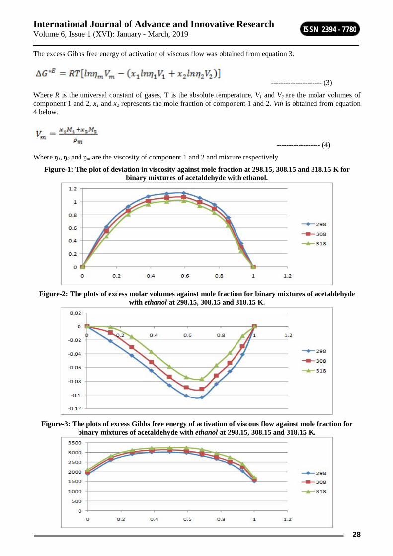
International Journal of Advance and Innovative Research Volume 6, Issue 1 (XVI): January - March, 2019
28
ISSN 2394 - 7780
The excess Gibbs free energy of activation of viscous flow was obtained from equation 3.
--------------------- (3)
Where R is the universal constant of gases, T is the absolute temperature, V1 and V2 are the molar volumes of component 1 and 2, x1 and x2 represents the mole fraction of component 1 and 2. Vm is obtained from equation 4 below.
------------------ (4)
Where ŋ1, ŋ2 and ŋm are the viscosity of component 1 and 2 and mixture respectively
Figure-1: The plot of deviation in viscosity against mole fraction at 298.15, 308.15 and 318.15 K for binary mixtures of acetaldehyde with ethanol.
Figure-2: The plots of excess molar volumes against mole fraction for binary mixtures of acetaldehyde
with ethanol at 298.15, 308.15 and 318.15 K.
Figure-3: The plots of excess Gibbs free energy of activation of viscous flow against mole fraction for
binary mixtures of acetaldehyde with ethanol at 298.15, 308.15 and 318.15 K.
International Journal of Advance and Innovative Research Volume 6, Issue 1 (XVI): January - March, 2019
29
ISSN 2394 - 7780
Deviation of physical properties of liquid mixtures from the ideal behavior is the measure of the interaction between the molecules which is attributed to either adhesive or cohesive forces. The plot of deviation in viscosity against mole fraction for binary mixtures of acetaldehyde with ethanol found to be positive at all temperatures. The positive values of the deviation in viscosity (Δŋ) suggest the existence of strong intermolecular interactions upon mixing in ethanol. This leads to suggest that interactive force are responsible for these positive interactions (18). The variation of excess volumes with the mole fraction (X1) of acetaldehyde and ethanol at (303.15, 308.15 and 313.15) K are represented in figures-2. The excess molar volume values of the mixtures are negative and decreases when temperature increases. This shows that the excess molar volumes are always negative for all the studied temperatures. Treszczanowicz et al. and Roux and Desnoyers [19-20] suggested that VE is the resultant contribution from several opposing effects.
CONCLUSION The density, viscosity, deviation in viscosity, excess molar volume and excess Gibbs free energy of activation of viscous flow for the binary systems of acetaldehyde with ethanol at 298.15, 308.15 and 318.15 K has been reported. The deviation in viscosity of the binary systems of Acetaldehyde with ethanol are found to be negative and decreases with temperature while excess molar volumes are positive for all binary systems. There is intermolecular interaction among the components of the binary mixtures leading to possible hydrogen bond formation of the type Ȫ---H-----O between unlike molecules confirming intermolecular hydrogen bond formation between acetaldehyde and ethanol mixtures. Excess molar volumes (VE) and the viscosity deviations (Δŋ) were used to predict the intermolecular interactions in the mixtures. McAllister's three-body-interaction model was used to correlate the kinematic viscosity of the systems. The excess volume and viscosity deviation data were fitted by means of the Redlich-kister equation. It was found that in all cases the experimental data obtained, matches with the McAllister model and Redlich-Kister equation with a high degree of precision.
REFERENCES 1. Lange’s Handbook of Chemistry 10th edition, 1525 – 1528. 2. Ewing, M.B., Levian,B.J., Marsh,K.N. Journal of Chemical Thermodynamics, 2, 689 - 691(1970)
3. B. R. Kumar, B. Satyanarayana, S. A. Banu, K. A. Jyoti, T. S. Jyostna, N. Satyanarayana. Ind. J. Pure & Appl. Phys., 2009, 47, 511.
4. S. Parveen, M. Yasmin, M. Gupta, J. P. Shukla. Int. J. Thermodyn., 2010, 13(2), 59.
5. S. Singh, B. P. S. Sethi, R. C. Katyal, V. K. Rattan. J. Chem. Eng. Data., 2004, 49, 1373.
6. R. A. Clara, A. C. G. Marigliano, V. V. Campos, H. N. Solimo. Fluid Phase Equilib., 2010, 293, 151. 7. B. Gonzalez, N. Calvar, E. Gomez, A. Dominguez. J. Chem. Thermodyn., 2007, 39, 1578.
8. Suryanarayana C. V., J. Acoust Soc India, 1983, 13, 9. 9. Fletcher A., J. Phys Chem., 1969, 73, 2217.
10. B. Sathyanarayana, B. Ranjithkumar, T. S. Jyostna, N. Satyanarayana. J. Chem. Thermodyn., 2007, 39, 16 .
11. Fedeles, O. Ciocirlan, O. Iulian. U. P. B. Sci. Bull. B., 2009, 71(4) , 99. 12. M. L. J. Kijevcanin, V. Z. Kostic, I. R. Radovic, B. D. Djordjevic, S. P. Serbanovic. Chem. Ind. Chem
Eng., 2008, 14(4), 223.
13. A. G. Peshwe, B. R. Arbad, S. P. Pachaling. Int. J. Chem. Sci., 2009, 7(3), 1505. 14. V. Serheyev. Chem. Chem. Tech., 2011 5(3), 241.
15. Roux,A., Desnoyers, J.: Association models for alcohol – water mixtures. Indian Acad. Proc., Chem.Soc.98,435-439(1978)
16. Maham,Y., Hepler,L.G., Mather,A.E., Hakin,A.W., Marriot.R.M.: Molar heat Capacities of alkanolamines from 299.1 to 397.8 K Group additivity and molecular connectivity analyses J.Chem.Soc., Faraday Trans,93,1747-1750(1997).
17. S. S. Patil, S. R. Mirgane. Rasayan J. Chem., 2011, 4(2), 445.
18. O. Redlich, A. T. Kister. Ind. Eng. Chem., 1948, 40(2), 345. 19. S. C. Bhatia, R. Rani, R. Bhatia. J. Mol. Liq., 2011, 159, 132.
20. S. L. Oswal, H. S. Desai. Fluid Phase Equilib., 1999, 161, 191.
International Journal of Advance and Innovative Research Volume 6, Issue 1 (XVI): January - March, 2019
30
ISSN 2394 - 7780
PHYTOCHEMICAL STUDIES ON THE STEM BARK OF ANTHOCEPHALUS CADAMBA (ROXB.) MIQ
Revansiddha Dhotre, Maheboob Shaikh and Mamtaram Kare Department of Botany, Pratishthan Mahavidyalaya, Paithan, Aurangabad
ABSTRACT Anthocephalus cadamba is one of such ayurvedic remedy that has been mentioned in many Indian medicinal literatures. It is crucially significant as it has the largest number of phytochemicals and secondary metabolites having pharmacological and biological properties.The present work deals with development and standardization of phytochemical screening for quantification of stem bark extract of medicinal plant of Anthocephalus cadamba (Roxb.)Miq. The scientific parameter is necessary to identify the exact plant material and to find its quality and purity. The present study deals with various preliminary phytochemical screening of various successive extracts such as qualitative chemical analysis. The alkaloidal content of stem bark of the title plant has been determined. These studies indicated the possible information for correct identification and standardization of this plant material.
Keywords: Anthocephalus cadamba, Phytoconstituents, Phytochemical Screening, Bark Extract, Solvents.
INTRODUCTION There are a number of floras in use for medicinal purposes over the past several centuries. Countries such as China, India, and Egypt are well known for the active usage of medicinal plants in the treatment of various incurable diseases. India is the largest producer of medicinal herbs in the world due to which it is often called a botanical paradise. Ayurvedic science is deeply rooted in India and its neighboring countries. It was developed even before the medieval period, when people had little knowledge of science. There are a number of ancient therapeutic measures based on medicinal plants that have been developed in India (Dwevedi 2015)(Zaidan MR 2005)(Ahmad I 1998)(Bhakuni DS 1969). They can cure several diseases and ailments such as diabetes, cardiovascular disorders, cancer, and liver damage (Dwevedi 2015) (Ríos JL 2005) (Alekhya V 2013) (Kirana C 2003).
Anthocephalus cadamba (Rubiaceae) commonly known as ‘Kadamba’. The other names of the plant are Neolamarckia cadamba, Nauclea cadamba (Roxb.), Samama cadamba (Roxb.) Kuntze, Anthocephalus morindifolius Korth., Nauclea megaphylla S. Moore,Neonauclea megaphylla (S. Moore) S. Moore, etc. Other vernacular names have been listed in the Table1. The tree is highly regarded religiously and culturally in India being sacred to the Lord Krishna. Radha and Krishna conducted their love play in the hospitable and sweet-scented shade of the Kadamba tree (Arvind Bijalwan 2014). Shiva and Parvati came to Sahyadri and there a child was born to the divine couple. Since the birth took place under a Kadamba tree, the child was named Kadamba, and was placed in charge of the Sahyadri region. The word Kadamba lends its name to the Kadamba Dynasty which ruled from Banavasi in what is now the state of Karnataka from 345 AD to 525 AD (Arvind Bijalwan 2014)(Forest 2000).
Anthocephalus cadamba is large tree with a broad umbrella-shaped crown and straight cylindrical bole. The branches are characteristically arranged in tiers. The tree may reach a height of 45 m with a stem diameter of 100–160 cm and sometimes it has a small buttress up to 2 m high. The bark is grey, smooth and very light in young trees, but rough and longitudinally fissured in old trees. The branches spread horizontally and drop at the tip. The leaves are glossy green, opposite, simple sessile to petiolate, ovate to elliptical (15–50 cm long by 8–25 cm wide). In young fertilised trees, the leaves are much larger, subordinate at base and acuminate at apex; the stipules are interpetiolar, narrowly triangular and deciduous. The fruitlets are numerous, somewhat fleshy, with their upper parts containing 4 hollow or solid structures. The fruit occurs in small, fleshy capsules packed closely together to form a fleshy yellow-orange infructescence containing approximately 8000 seeds. The seeds somewhat are trigonal or irregular shaped, not winged (Kanninen 2011).
Md. Abu Shuaib Rafshanjani et. al. (2014) they isolated from stem barks of Anthocephalus cadamba the active principles and to evaluate possible synergistic affects among the extract components for their cytotoxic properties (Md, Shumaia and Md 2014). Alekhya V. et. al. (2013)they reported from leaves and seeds of Anthocephalus cadambabest sources for obtaining natural antioxidants for various medicinal uses and ascorbic acid also present (Alekhya, et al. 2013). The objective of this work is too carried out to explore the phytochemical constituents of the solvent extractof A. cadamba.

International Journal of Advance and Innovative Research Volume 6, Issue 1 (XVI): January - March, 2019
31
ISSN 2394 - 7780
MATERIAL AND METHOD Plant Material: The stem bark of Anthocephalus cadamba (Roxb.) Miq. was collected from Shivaji park, Paithan. The plant materials was identified and Authenticated by Dr. M. A. Kare, Department of Botany, Pratishthan Mahavidyalaya, Paithan.
Preparation of Extract: The stem bark of Anthocephalus cadamba (Roxb.)Miq. shaded dried, and then these are made into coarsely powdered form using dry grinder. The powdered bark of the plant (180gm.) was packed in soxhlet apparatus and continuously extracted with petroleum ether (40-600C) till complete extraction, after completion of extraction the solvent was removed by distillation and then concentrated extract obtained was dried under reduced pressure using rotatory evaporator at temperature not exceeding 400C and then give moderate heating on water bath.A yellowish extract approximate 1 gm. was obtained. From the drug petroleum ether was removed and the defatted drug was extracted with methanol (95%) till complete extraction, after completion of extraction the solvent was removed by distillation and then concentrated extract obtained dried under reduced pressure at temperature not exceeding 400C and then give moderate heating on water bath. The methanolic extract obtained was dark yellow in color, weighed about 42.8 gm. The methanolic extract was kept in Petridis and it was stored in desiccators at cool place (Mukherjee 2001).
RESULT AND DISCUSSION Phytochemical Test of Anthocephalus Cadamba
Test Methanolic Extract Petro. Ether Chloroform Acetone Tannins + - + + Phenols + - - -
Alkaloids + - - - Saponins + - - - Iridoids - + - +
Quercetin - - - - Kaempferol - - - -
Catechin - - - - Coumarin - + + +
6,7-Dimethoxy coumarin - - + + 5-Methoxy genistein - - + -
Anthocyanin - + - - Proanthocyanin - + - - Carbohydrates + - - +
Flavonoids + - + - Glycosides + - + -
Proteins - - + +
In plants phytochemicals are naturally present. They give colour, flavor, smell and texture. A part from that, phytochemicals could prevent diseases including cancer and cardiovascular diseases and inhibit pathogenic microorganisms. Nowadays the use of medicinal plants rapidly increases in medicine (Renu 2005).Phytochemical evaluation of methanolic extract of Anthocephalus cadamba (Roxb.)Miq.showed the presence of Tannins, Phenols, Alkaloids, Saponins, Iridoids, Quercetin, Kaempferol, Catechin, Coumarin, 6,7-Dimethoxy coumarin, 5-Methoxy genistein, Anthocyanin, Proanthocyanin, Carbohydrates, Flavonoids, Glycosides and Proteins.
Phytochemical screening of the bark showed some differences in the presence of phytoconstituents which are known to have importance in medicine(Sukumaran S 2011)(Kiruba S 2011)(Jeeva S 2011)(J. M. Jeeva S 2012)(Johnson M 2012)(AR, et al. 2014). The preliminary screening tests may be useful in the detection of the bioactive principles and subsequently may lead to the drug discovery and development(Joselin J 2013)(S. B. Joselin J 2012)(Florence AR 2012)(J. S. Joselin J 2014)(AR, et al. 2014).
Saponins are found only in petroleum ether extract. Chloroform extract revealed the presence of carbohydrates and saponins. Ethanol extract showed the presence of alkaloids, flavonoids, glycosides, phenols, steroids, terpenoids and quinones whereas acetone extract showed the availability of alkaloids, flavonoids, phytosterols,
International Journal of Advance and Innovative Research Volume 6, Issue 1 (XVI): January - March, 2019
32
ISSN 2394 - 7780
phenols and proteins.Such alkaloids were effective against ovarian, brain, breast, lung cancer etc(Sato N 1998)(Hsiang YH 1985)(Chen AY 1994)(Multon E 1989)and several of its semisynthetic analogues are 9-Nitro-CPT, 10-hydroxy-9-dimethylaminomethyl - CPT, 7-Ethyl- 10 -hydroxy-camptothecin (SN-38), are applied as clinical anticancer drugs in USA, Europe and Japan(Godwin S 1959). Other alkaloids include indicine, indicine N- oxide, thalicarpine and tetrandrine(Hartwell JL 1969). Flavonoids are also reported to have inhibitory action on growth and proliferation of different types of tumors(Netto 2007).
CONCLUSION Various extracts was subjected to Pharmcoganostic Evaluation for the identification of variousPhytoconstituents and rest of extracts were utilized for pharmacological screening. Phytochemical evaluation extract of Anthocephalus cadamba (Roxb.)Miq.showed the presence of Tannins, Phenols, Alkaloids, Saponins, Iridoids, Quercetin, Kaempferol, Catechin, Coumarin, 6,7-Dimethoxy coumarin, 5-Methoxy genistein, Anthocyanin, Proanthocyanin, Carbohydrates, Flavonoids, Glycosides and Proteins.
ACKNOWLEDGEMENT The authors are grateful to Principal, Pratishthan Mahavidyalaya, Paithan, for providing the necessary laboratory facilities and we are also thankful with our deepest core of heart to Dr. M. A. Kare for his valuable guidance.
REFERENCE Ahmad I, Mehmood Z, Mohammad F. "Screening of some Indian medicinal plants for their antimicrobial
properties." J Ethnopharmacol 62 (1998): 183-193.
Alekhya V, Deepan T, Sahoo S, Dhanaraju MD. "Preliminary phytochemical screening and evaluation of in vitro antioxidant activity of Anthocephalous cadamba by using solvent extracts." Eur J Biol Sci 5 (2013): 34-37.
Alekhya, V, T Deepan, Sahoo Shaktiprasanna, and MD Dhanaraju. "Preliminary Phytochemical Screening and Evaluation of In vitro Antioxidant Activity of Anthocephalous cadamba by Using Solvent Extracts." European Journal of Biological Sciences 5, no. 1 (2013): 34-37.
AR, Florence, Joselin J, Sukumaran S, and Jeeva S. "Screening of Phytochemical Constituents from Certain Flower Extracts." International Journal of Pharmacy Review & Research 4, no. 3 (2014): 152-159.
Arvind Bijalwan, Manmohan J. R. Dobriyal, Bhartiya J. K. "A potential fast growing tree for Agroforestry and Carbon Sequestration in India: Anthocephalus cadamba (Roxb.) Miq." American Journal of Agriculture and Forestry 2, no. 6 (2014): 296-301.
Bhakuni DS, Dhar ML, Dhar MM, Dhawan BN, Mehrotra BN. "Screening of Indian plants for biological activity." Indian J Exp Biol 7 (1969): 250-262.
Chen AY, Liu LF. "Design of topoisomerase inhibitors to overcome MDRI- mediated drug resistance." Advanced Pharmacology, (New York) 29 (1994): 245-256.
Dwevedi, Alka, Sharma, Kuldeep, Yogesh, K, Sharma. "Cadamba: A miraculous tree having enormous pharmacological implications." Pharmacognosy Reviews 9, no. 18 (2015): 107-113.
Florence AR, Joselin J, Jeeva S. "Intra-specific variation of bioactive principles in select members of the genus Clerodendrum L." Journal of Chemical and Pharmaceutical Research 4, no. 11 (2012): 4908-4914.
Forest, Department. The Additional Principal Chief Conservator of Forests. Pune: Research, Education & Training Maharashtra (Pune), 2000.
Godwin S, Smith AF, Horning EC. "Alkaloids of Ochrosia elliptica." J. Amer. Chem Soc 81 (1959): 1903-1908.
Hartwell JL, Abbott BJ. "Antineoplastic principles in plants: Recent developments in the field." Adv Pharma- col Chemother 7 (1969): 117-209.
Hsiang YH, Hertzberg R, Hecht S, Liu LF. "Camptothecin induces protein linked DNA breaks via mammalian DNA topoisomerase I." J. Biol. Chem 260 (1985): 14837-14878.
Jeeva S, Johnson M, Aparna JS, Irudayaraj V. "Preliminary phytochemical and antibacterial studies on flowers of selected medicinal plants." International Journal of Medicinal and Aromatic Plants 1, no. 2 (2011): 107-114.
International Journal of Advance and Innovative Research Volume 6, Issue 1 (XVI): January - March, 2019
33
ISSN 2394 - 7780
Jeeva S, Johnson M. "Anti-bacterial and phytochemical studies on methanolic extracts of Begonia flaccifera Bedd. flower." Asian Pacific Journal of Tropical Biomedicine 1, no. S1 (2012): S151-S154.
Johnson M, Aparna JS, Jeeva S, Sukumaran S, Anantham B. "Preliminary phytochemical studies on the methanolic flower extracts of some selected medicinal plants from India." Asian Pacific Journal of Tropical Biomedicine 1, no. S1 (2012): S79-S82.
Joselin J, Jenitha S, Brintha TSS, Jeeva S, Sukumaran S, Geetha VS. "Phytochemical and FT-IR Spectral analysis of certain Bamboo species of South India." Biodiversity, Biopropecting and Development 1, no. 103 (2014): 1-2.
Joselin J, Shynin Brintha TS, Florence AR and Jeeva S. "Screening of select ornamental flowers of the family Apocyanaceae for phytochemical constitutions." Asian Pacific Journal of Tropical Disease 2 (2012): 1-6.
Joselin J, Shynin Brintha TS, Florence AR, Jeeva S. "Phytochemical evaluation of Bignoniaceae flowers." Journal of Chemical and Pharmaceutical Research 5, no. 4 (2013): 106-111.
Kanninen, Haruni Krisnawati Maarit Kallio Markku. Anthocephalus cadamba Miq. Ecology, silviculture and productivity. Indonesia: CIFOR, Bogor, Indonesia., 2011.
Kirana C, McIntosh GH, Record IR, Jones GP. "Antitumor activity of extract of Zingiber aromaticum and its bioactive sesquiterpenoid zerumbone." Nutr Cancer 45 (2003): 218-225.
Kiruba S, Mahesh M, Nisha SR, Miller Paul Z, Jeeva S. "Phytochemical analysis of the flower extracts of Rhododendron arboreum Sm.ssp. nilagiricum (Zenker) Tagg ." Asian Pacific Journal of Tropical Biomedicine, 2011: S284-S286.
Md, Abu, Shuaib, Rafshanjani, Parvin Shumaia, and Abdul, Kader Md. "Antimicrobial and Preliminary Cytotoxic effects of Ethanol extract and its fractions of Anthocephalus cadamba (Roxb.) Miqstem bark." International Journal of Pharmacy & Life Sciences 5, no. 12 (2014): 4038-4044.
Mukherjee, P, K. Quality Control of Herbal Drugs. Nwe Delhi: Business Horizons Pharmaceutical Publishers, 2001.
Multon E, Riou JF, Lefevre D, Ahomadegbe JC, Tiou G. "Topoisomerase II- mediated DNA cleavage activity induced by ellipticines on the human tumor cell line N 417." Biochem Pharmacol 38 (1989): 2077-2086.
Netto, CC. "Cranberry and its phytochemicals: A review of In vitro anticancer studies." J. Nutr 137 (2007): 186S-193S.
Renu, S. "Useful Metabolites From Plant Tissue Cultures." Biotechnology 4, no. 2 (2005): 79-93.
Ríos JL, Recio MC. "Medicinal plants and antimicrobial activity." J Ethnopharmacol 100 (2005): 80-84.
Sato N, Mizumoto K, Kusumoto M, Niiyama H, Maehara N, Ogawa T, Tanaka M. "9-Hydroxyellipticine inhibits telomerase activity in human pancreatic cancer cells." FEBS Lett 44 (1998): 318-321.
Sukumaran S, Kiruba S, Mahesh M, Nisha SR, Miller Paul Z, Ben CP, Jeeva S. "Phytochemical constituents and antibacterial efficacy of the flowers of Peltophorum pterocarpum(DC,) Baker ex Heyne." Asian Pacific Journal of Tropical Medicine 4, no. 9 (2011): 735-738.
Zaidan MR, Noor Rain A, Badrul AR, Adlin A, Norazah A,. "In vitro screening of five local medicinal plants for antibacterial activity using disc diffusion method." Trop Biomed 22 (2005): 165-170.
International Journal of Advance and Innovative Research Volume 6, Issue 1 (XVI): January - March, 2019
34
ISSN 2394 - 7780
STUDIES ON PHYSICO-CHEMICAL ANALYSIS OF SOIL SAMPLES IN BEED DISTRICT, MAHARASHTRA
V. V. Naiknaware1 and G. M. Dhond2 1Department of Chemistry, Swa. Sawarkar College, Beed
2Department of Zoology, Swa. Sawarkar College, Beed
ABSTRACT Each growth of crop most important role quality and type of soil is fertility of different crops. Good recovery or average of agricultural production is generally major role depends on soil quality. The present study has been undertaken to investigate physic-chemical parameter of soil samples collected from different villages of Beed District, Maharashtra, India. The soil characterization was carried out for the parameters of like temperature, PH , Electrical conductivity, Total organic Carbon, Nitrogen, Phosphorus(P2O5), Potassium(K2O). As per study of these soil samples leads to the conclusion of the nutrients quality of soil of different in different villages in Beed District. The present study shows thataverage of all the villages have medium or high mineral content. This information will help to farmer solve the problem and utilization of fertilizer in their field either an organic and chemical fertilizer for increase the yields of crop. Certain external factor control plant growth, air, temperature, mechanical support, nutrients, and water. Plant needs elements for their growth and completion of life cycle of plants. Growth of all plants is essential of elements of elements like Carbon, Nitrogen, Oxygen, Potassium, Phosphorus, Hydrogen etc.
INTRODUCTION Soil is one of the most important resources are of the nature. Soil recourses are of vital role for survival and welfare of the people. One of the most severe and wide spread problems facing the agriculture, industry is the degradation of soil quality due to salinity. Soil may be defined as a thin layer of earth’s crust which serves as a natural medium for the growth of plants. Soil provides a medium for plant growth to meet our food and fiber need. Soil filters water, decomposes waste, stores heat and exchanges gases and hence have great bearing on environmental balance (Bear, 1976). Soil test based nutrients management has emerged as a key issue in effort to increase agriculture productivity and production since optional uses nutrients, based on soil analysis can improve crop productivity and minimizing wastage of the nutrients, thus minimizing impact on environmental leading to bias through optical production. Soil is natural body on which agriculture product grows and it has fragile ecosystem. Fertility of soil is one of the most important factors which regulate growth and yield of crops. Due to an imbalance and an inadequate use of fertilizers, improper irrigation and various cultural practices, the soil quality is depleting rapidly (Pandeeswari N 2012). The present work is undertaken to study the physic-chemical analysis of soil samples collected from different villages of Beed district Maharashtra. In present study characterization of soil was characterized various parameters like Temperature, PH, Electrical conductivity, Total organic carbon, Nitrogen, Potassium(K2O),Phosphorus(P2O5), etc. This study leads us to the conclusion of the nutrients quality of soil of different villages of Beed District
MATERIAL AND METHODS The soil samples were collected from eight different villages in Beed tehsil of Beed district in the Marathwada region of Maharashtra. The samples were collected in the depth of 0 to 30 cm from the surface of land. Soils were completely air dried and passed through 2mm sieve and stored in properly labeled plastic bags for analysis. The soil samples were collected in the month of Jan. 2016. The eight samples cites from Beed district are Shidod, Palwan, Babhulwadi, Waybhatwadi, (K)pangari, Pimpalner, Nagapur and Jirewadi. Analysis of the Physico-chemical parameters of the soil samples were dissolved in distilled water and allowed to settle down the particles. The temperature of all samples was measured by thermometer in 0C. The PH of the suspension was determined using PH meter. Electrical conductivity (EC) of the soil was determined in the filtrate of water extract using conductivity meter(Chandra 2009). Percentage of organic carbon content was determined by adopting chromic acid wet digestion method. Nitrogen, Phosphorus, and potassium are determined by standard procedure. Results were compared with standard values.
RESULTS AND DISCUSSION The Physico-chemical analysis of different parameter of soil samples collected from different villages of Beed District is given in above Table No. 1. The temperature of important parameter to as helps sowing seed in soil for growth of crop (Oisen S.R. 1954).
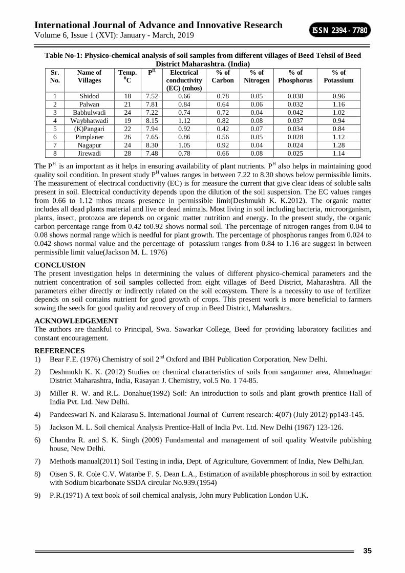
International Journal of Advance and Innovative Research Volume 6, Issue 1 (XVI): January - March, 2019
35
ISSN 2394 - 7780
Table No-1: Physico-chemical analysis of soil samples from different villages of Beed Tehsil of Beed District Maharashtra. (India)
Sr.No.
Name of Villages
Temp. 0C
PH Electrical conductivity (EC) (mhos)
% of Carbon
% of Nitrogen
% of Phosphorus
% of Potassium
1 Shidod 18 7.52 0.66 0.78 0.05 0.038 0.96 2 Palwan 21 7.81 0.84 0.64 0.06 0.032 1.16 3 Babhulwadi 24 7.22 0.74 0.72 0.04 0.042 1.02 4 Waybhatwadi 19 8.15 1.12 0.82 0.08 0.037 0.94 5 (K)Pangari 22 7.94 0.92 0.42 0.07 0.034 0.84 6 Pimplaner 26 7.65 0.86 0.56 0.05 0.028 1.12 7 Nagapur 24 8.30 1.05 0.92 0.04 0.024 1.28 8 Jirewadi 28 7.48 0.78 0.66 0.08 0.025 1.14
The PH is an important as it helps in ensuring availability of plant nutrients. PH also helps in maintaining good quality soil condition. In present study PH values ranges in between 7.22 to 8.30 shows below permissible limits. The measurement of electrical conductivity (EC) is for measure the current that give clear ideas of soluble salts present in soil. Electrical conductivity depends upon the dilution of the soil suspension. The EC values ranges from 0.66 to 1.12 mhos means presence in permissible limit(Deshmukh K. K.2012). The organic matter includes all dead plants material and live or dead animals. Most living in soil including bacteria, microorganism, plants, insect, protozoa are depends on organic matter nutrition and energy. In the present study, the organic carbon percentage range from 0.42 to0.92 shows normal soil. The percentage of nitrogen ranges from 0.04 to 0.08 shows normal range which is needful for plant growth. The percentage of phosphorus ranges from 0.024 to 0.042 shows normal value and the percentage of potassium ranges from 0.84 to 1.16 are suggest in between permissible limit value(Jackson M. L. 1976)
CONCLUSION The present investigation helps in determining the values of different physico-chemical parameters and the nutrient concentration of soil samples collected from eight villages of Beed District, Maharashtra. All the parameters either directly or indirectly related on the soil ecosystem. There is a necessity to use of fertilizer depends on soil contains nutrient for good growth of crops. This present work is more beneficial to farmers sowing the seeds for good quality and recovery of crop in Beed District, Maharashtra.
ACKNOWLEDGEMENT The authors are thankful to Principal, Swa. Sawarkar College, Beed for providing laboratory facilities and constant encouragement.
REFERENCES 1) Bear F.E. (1976) Chemistry of soil 2nd Oxford and IBH Publication Corporation, New Delhi.
2) Deshmukh K. K. (2012) Studies on chemical characteristics of soils from sangamner area, Ahmednagar District Maharashtra, India, Rasayan J. Chemistry, vol.5 No. 1 74-85.
3) Miller R. W. and R.L. Donahue(1992) Soil: An introduction to soils and plant growth prentice Hall of India Pvt. Ltd. New Delhi.
4) Pandeeswari N. and Kalarasu S. International Journal of Current research: 4(07) (July 2012) pp143-145.
5) Jackson M. L. Soil chemical Analysis Prentice-Hall of India Pvt. Ltd. New Delhi (1967) 123-126.
6) Chandra R. and S. K. Singh (2009) Fundamental and management of soil quality Weatvile publishing house, New Delhi.
7) Methods manual(2011) Soil Testing in india, Dept. of Agriculture, Government of India, New Delhi,Jan.
8) Oisen S. R. Cole C.V. Watanbe F. S. Dean L.A., Estimation of available phosphorous in soil by extraction with Sodium bicarbonate SSDA circular No.939.(1954)
9) P.R.(1971) A text book of soil chemical analysis, John mury Publication London U.K.

International Journal of Advance and Innovative Research Volume 6, Issue 1 (XVI): January - March, 2019
36
ISSN 2394 - 7780
STUDIES ON PHYSICO-CHEMICAL PARAMETERS OF WATER AND ZOOPLANKTON DIVERSITY OF LOWER DUDHANA DAM, PARBHANI DISTRICT. MAHARASHTRA
Nimbalkar R. K., Pawar D. A. and Sondge D. S. Department of Zoology, Deogiri College, Aurangabad
ABSTRACT The major role of zooplankton in an aquatic ecosystem is of energy transfer between phytoplankton and fishes. The present study deals with the physico- chemical parameters of water and diversity of zooplankton from Lower Dudhana dam District Parbhani Maharashtra. Study period was one year between January 2018 and December 2018. Lower dudhana dam near Kedar Wakdi provides fish food for humans mainly of Labeo rohita, Cirrahuns mirigala, Catla catla etc. Total 31 species of zooplankton were collected of which rotifera 12 species; cladocera 8 species, copepod 8 species and ostracoda 3 species were observed during study period of one year. The rotifera was dominated followed by copepoda, cladocera and ostracoda from the study area in one year. Throughout study duration of one year on monthly basis physico- chemical parameters such as water temperature, pH, Salinity, Dissolved Oxygen, TDS, Conductivity, Turbidity, Free CO2 concentration and BOD were analysed. The diversity indices of zooplanktons in relation to physico- chemical parameters also calculated.
Keywords: Zooplankton, Diversity, Lower Dudhna Dam, Kedar Wakdi.
INTRODUCTION In an aquatic ecosystem such as pond two types of producers macrophytes which are rooted or large floating plants and phytoplankton those are minute floating plants generally algae or green bacteria as distributed throughout the pond as deep as light penetrates. Water in a greenish colour observed when phytoplanktons are abundant in number. Two types of microconsumers, zooplankton and benthos (bottom forms) appears in the aquatic ecosystem, their main role is transfer of energy from lower trophic level to higher trophic level in a food chain.
Three groups of freshwater habitats can be considered as standing water or lentic ecosystems which include lakes and ponds, running water or lotic ecosystems which include streams, rivers and springs and third group of freshwater habitats is wetlands, where water levels are not same seasonally and annually or water level fluctuate up and down. Examples of wetlands are marshes and swamps. Small portion of the Earth surface is occupied by freshwater habitats compared to terrestrial and marine habitat, but freshwater habitats are more important to humans than their relative area [1].
The physico- chemical parameters of water are mainly responsible for the distribution and diversity of zooplankton species in an aquatic ecosystem [2]. In an aquatic food web between autotrophs and heterotrophs zooplanktons creates a main link in the transfer of energy at secondary level [3].
The physico- chemical parameters of water and interactions among them are very important to study the growth, reproduction, distribution, composition and diversity of aquatic organisms [4, 5]. The basic physico- chemical parameters affecting the aquatic environments are pH, dissolved oxygen, temperature, conductivity of water and nutrients [6]. The highest rate of organic matter decomposition which increases the higher BODs values in an ecosystem [7]. Communities of zooplankton have been documented by the studies of [8] from some fresh water bodies in Kolhapur District related to pollution. Researchers documented the zooplankton community and physico- chemical parameters of Kham River, Aurangabad [17], Ambe Ghosale Lake of Thane city [18] and Narangi Sarangi dam of Vaijapur District Aurangabad of Maharashtra [19].
The aim of the present investigation was to study the physico- chemical parameters of the water along with zooplankton diversity and diversity indices of the zooplanktons of the lower dudhna dam near Kedar Wakdi, District Parbhani Maharashtra.
MATERIALS AND METHODS Study Area In Parbhani district near Kedar Wakdi village, 10 Km away from Selu lower dudhana dam was developed. It is one of the significant water system extends in Maharashtra. As it is developed on Dudhana River alleged lower Dudhana dam. It’s on 19°31' 51'' N and 76°23' 34'' E latitude and longitude respectively. The dam has two trenches, right channel and left waterway for irrigation motive. Sampling station was selected of lower Dudhana nearest to Kedar Wakdi dam to study the physico- chemical parameters of water and diversity of zooplanktons.
International Journal of Advance and Innovative Research Volume 6, Issue 1 (XVI): January - March, 2019
37
ISSN 2394 - 7780
Collection, Preservation and Identification On a monthly basis from sampling station water samples were collected for the study of physico-chemical parameters and zooplankton community for a period of one year from January 2018 to December 2018. Plankton net having 60 micro meters mesh size was used for collection of planktons. Every time through the plankton net 100 litres of water was sieved. The plastic containers of 1-L capacity were used for collections, before use of the plastic containers these were rinsed with sampling water. Filled plastic containers were sealed and transported to laboratory for analysis of physico- chemical parameters.
All the times water samples were collected from same location and in morning between 7.00 am to 10.00 am. Collected samples were preserved in 4% formalin and stained with Bengal Rose. The standard protocols of [9, 10] were used for the analysis of physico-chemical parameters of the water samples. TDS meter is used to record the TDS of water on the spot. pH, temperature, Dissolved Oxygen and Electrical Conductance were analysed and recorded on the spot immediately after collection of water samples by using multi 340i /set water analysis kit.
Under the microscope freshwater zooplankton species were studied and standard protocols of [11, 12] were used for the identification of zooplanktons. Drop method was used to count the planktons. Plankton counting chamber was used for quantitative analysis and Stereomicroscope for observations. Wide mouthed pipette was used to take 1 ml of sample in counting cell and allowed to settle about 4- 5 minutes and after the counting was done. Four counting’s was done for each sample of the plankton and average was calculated from it. Total planktons present in one litre of water sample were estimated by [13, 14].
Statistical Analysis and Diversity Indices Different diversity indices such as Dominance_D, Simpson_1-D, Shannon-H, Evenness and Margalef were calculated by software PAST, v 3.0.
RESULTS AND DISCUSSION Physico- Chemical Parameters In present study on monthly basis water samples were collected and analysed for physico- chemical parameters of water collected from sampling station of lower dudhana dam near Kedar Wakdi between January 2018 and December 2018. All the physico- chemical parameters of water from sampling station are represented in Table 1.
Water temperature (0 C) Lowest water temperature 240C was recorded in month of January and highest water temperature 28.20C was observed in month of May.
pH The values of pH recorded from sampling station was in the range of 7.2±0.2 and 8.95±0.2, pH 7.2 was observed minimum in month of May, while maximum pH 8.95 was recorded in month of August.
Salinity (mg/L) Salinity from water samples of sampling station was recorded in the range of 0.637 mg/L and 0.954 mg/ L, salinity lowest and highest was observed in September and March months respectively. Low salinity 0.637 mg/L in month of September and highest salinity 0.954 mg/L in month of March was recorded from sampling station respectively.
Dissolved Oxygen (mg/L) The concentration of dissolved oxygen in water is depends on the physical, chemical and biochemical processes existing in aquatic systems. Minimum dissolved oxygen 10.5mg/L and maximum value of dissolved oxygen 16.8 mg/L was recorded from sampling station in month of January and December respectively.
Conductivity (μ S/cm) The conductivity of water from sampling station was observed in the range of 156 μ S/cm and 290μ S/cm. Minimum conductivity of water was recorded in month of January, while in month of July conductivity was maximum.
Turbidity (NTU) Maximum value of turbidity recorded from sampling station was 180 (NTU) and minimum value of turbidity was 90 (NTU) in months of September and January respectively.
Free CO2 (mg/L) The free CO2 concentrations were observed in the range of 13.4 mg/L and 28 mg/L. The low free CO2 concentration from sampling station 13.4 mg/L and high free CO2 concentration 28 mg/L was recorded in month
International Journal of Advance and Innovative Research Volume 6, Issue 1 (XVI): January - March, 2019
38
ISSN 2394 - 7780
of February and August respectively. Our results support the statement of [15] that Dissolved Oxygen and Free CO2 concentrations are reciprocal.
BOD (mg/L) BOD range of water was 3.95 mg/L to 8.24 mg/L. Minimum BOD (3.95 mg/L) and maximum BOD (8.24 mg/L) from lower dudhana dam water was recorded in months of May and December respectively.
TDS (mg/L) The TDS range of water samples was 0.685 mg/L to 0.97 mg/L. The minimum TDS 0.685 mg/L and maximum TDS 0.97 mg/L was recorded in months of April and May respectively.
Diversity of Zooplanktons During the study period of one year from January 2018 to December 2018 total 31species of zooplankton belonging to four orders namely rotifera (12 species), cladocera (8 species), copepoda (8 species) and ostracoda (3 species) were listed in Table 2.
Rotifera In the study period of one year from January 2018 to December 2018, total 12 species of rotifera belonging to 7 genera were collected and are listed in table 2.The range of population density for rotifers was 545 ind/L and 834 ind/L was recorded and shown in table 3.The minimum population density 545 ind/L and maximum population density 834 ind/L were recorded in months of July and April respectively. The species dominance was minimum (0.8982) in July month, while maximum (0.9309) was recorded in month of April. The Simpson diversity index was highest (0.1018) during July and low (0.06911) in month of April. The Shannon diversity index (H) was maximum (0.2096) in July while minimum (0.1545) was recorded in month of April. The highest species evenness (0.6166) and low species evenness (0.5835) was recorded in months of July and April respectively. The Margalef species richness (R1) maximum (0.1573) in month of July and minimum (0.1479) in month of April was recorded.
Cladocera Total 8 species of cladocera belonging to 6 genera were collected in one-year study period and are listed in table 2. Population density range for cladocera was observed between 497 ind/L and 858 ind/L. Minimum and maximum population densities were recorded in months of October and July respectively, and are shown in table 3. The species dominance was minimum (0.8895) in October, while maximum (0.9327) was recorded in month of July.
The minimum values of Simpson diversity index (0.06731), Shannon diversity index (0.1513), species evenness (0.5817) and Margalef species richness (0.1473) were recorded in month of July, while maximum values of Simpson diversity index (0.1105), Shannon diversity index (0.2234), species evenness (0.6252) and Margalef species richness (0.1595) were recorded in month of October.
Copepoda Totally 8 species of copepoda belonging to 7 genera were collected and are listed in table 2. Copepoda population density during study period was varies in between 538 ind/L and 924 ind/L, minimum population density 538 ind/L and maximum population density 924 ind/L was recorded in months of March and June respectively and are presented in Table 3. The species dominance was minimum (0.897) in March, while maximum (0.9363) was recorded in month of June. The minimum values of Simpson diversity index (0.06281), Shannon diversity index (0.1432), species evenness (0.577) and Margalef species richness (0.1457) were recorded in month of June, while maximum values of Simpson diversity index (0.103), Shannon diversity index (0.2115), species evenness (0.6178) and Margalef species richness (0.1576) were recorded in month of March.
Ostracoda Three species of ostracoda belonging to three different genera was collected in study period and are shown in table 2. Population density range for ostracoda varies in between 356 ind/L and 686 ind/L during study period. The minimum and maximum population densities 356 ind/L and 686 ind/L were recorded in months of February and April respectively. The species dominance was high (0.9173) during month of April, while low (0.8526) in month of February. The maximum values of Simpson diversity index (0.1474), Shannon diversity index (0.279), species evenness (0.6609) and Margalef species richness (0.1678) were found in February month, while minimum values of Simpson diversity index (0.08273), Shannon diversity index (0.1781), species evenness (0.5975) and Margalef species richness (0.1521) were observed in month of April.
In a food web of aquatic ecosystems zooplanktons infest the central or focal position and they assist expressively, the biological productivity of the freshwater ecosystem [20]. According to [21, 22& 23] the
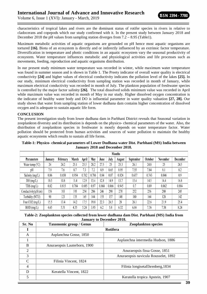
International Journal of Advance and Innovative Research Volume 6, Issue 1 (XVI): January - March, 2019
39
ISSN 2394 - 7780
characteristics of tropical lakes and rivers are the dominant status of rotifer species in rivers in relative to cladocerans and copepods which our study confirmed with it. In the present study between January 2018 and December 2018 the pH values from sampling station diverges from 7.2 – 8.95 (Table1).
Maximum metabolic activities of aquatic organisms are grounded on pH hence most aquatic organisms are tortured [16]. Biota of an ecosystem is directly and or indirectly influenced by an extrinsic factor temperature. Diversification in temperature and photic conditions in an aquatic ecosystem vary the seasonal productivity of ecosystem. Water temperature influences metabolic and physiological activities and life processes such as movements, feeding, reproduction and aquatic organisms distribution.
In our present study minimum water temperature was recorded in winter, while maximum water temperature was found in summer season and is shown in Table 1. The Preety indicator of overall water quality is electrical conductivity [24] and higher values of electrical conductivity indicates the pollution level of the lakes [25]. In our study, minimum electrical conductivity from sampling station was recorded in month of January, while maximum electrical conductivity was recorded in month of July. The plankton population of freshwater species is controlled by the major factor salinity [26]. The total dissolved solids minimum value was recorded in April while maximum value was recorded in month of May in our study. Higher dissolved oxygen concentration is the indicator of healthy water body and DO is influential parameter in water quality valuation [27, 28]. Our study shows that water from sampling station of lower dudhana dam contains higher concentration of dissolved oxygen and is adequate to sustain aquatic life form.
CONCLUSION The present investigation study from lower dudhana dam in Parbhani District reveals that Seasonal variation in zooplankton diversity and its distribution is depends on the physico- chemical parameters of the water. Also, the distribution of zooplankton species in freshwater is mostly depends on water temperature factor. Water pollution should be protected from human activities and sources of water pollution to maintain the healthy aquatic ecosystems which results to sustain all life forms.
Table-1: Physico- chemical parameters of Lower Dudhana water Dist. Parbhani (MS) India between January 2018 and December 2018.
Table-2: Zooplankton species collected from lower dudhana dam Dist. Parbhani (MS) India from
January to December 2018. Sr. No Taxonomic group / Genus Zooplankton species
I Rotifera A Asplanchna Gosse, 1850 1 Asplanchna intermedia Hudson, 1886 B Anuraeopsis Lauterborn, 1900 2 Anuraeopsis fissa Gosse, 1851 3 Anuraeopsis navicula Rousselet, 1892 C Filinia Vincent, 1824 4 Filinia longisetaEhrenberg,1834 D Keratella Vincent, 1822 5 Keratella tropica Apstein, 1907

International Journal of Advance and Innovative Research Volume 6, Issue 1 (XVI): January - March, 2019
40
ISSN 2394 - 7780
E Branchionus Pallas, 1776 6 Branchionus rubens Ehrenberg, 1838 7 Branchionus calyciflorus Pallas, 1776 8 Branchionus quadridentatus Hermann, 1783 9 Branchionus bidentata Anderson, 1889 10 Branchionus budapestinesis Daday, 1885 F Lecane Nitzsch, 1827 11 Lecane papuana Murray, 1913 G Notholca Gossse, 1886 12 Notholca lebis Gosse, 1887 II Cladocera A Daphnia Muller, 1785 13 Daphnia carinata King, 1853 14 Daphnia magna Straus, 1820 B Moina Baird, 1850 15 Moina flagellate Hudendroff, 1876 16 Moina brachiata Jurine, 1820 C Ceriodaphnia Danna, 1853 17 Ceriodaphnia reticulata Jurine, 1820 D Diaphanasoma Fischer, 1850 18 Diaphanasoma excisum Sars, 1885 E Moinodaphnia Herrick, 1887 19 Moinodaphnia macleayi King, 1853 F Leydigo Fischer, 1854 20 Leydigo acanthocercoids Fischer, 1854 III Copepoda A Eucyclops Claus, 1893 21 Eucyclops speratus Lilljeborg, 1901 B Mesocyclops Claus, 1893 22 Mesocyclops hyalinus Rehberg, 1880 23 Mesocyclops aspericornis Daday, 1906 C Sinediaptomus Kiefer, 1937 24 Sinediaptomus indicus Sewell, 1934 D Heliodiaptomus Kiefer, 1932 25 Heliodiaptomus viduus Gurney, 1916 E Neodiaptomus Kiefer, 1932 26 Neodiaptomus lindbergi Brehm, 1951 F Apocyclops Lindberg, 1942 27 Apocyclops dengizicus Lepeschkin, 1900 G Cletocamptus Schmankevitch, 1875 28 Cletocamptus albuquerquensis Herrick, 1895 IV Ostracoda A Cypris Muller, 1776 29 Cypris protubera Muller, 1776 B Cypretta Vavra, 1895 30 Cypretta fontinalis C Hemicypris Sars, 1903 31 Hemicypris anomala Furtos, 1993

International Journal of Advance and Innovative Research Volume 6, Issue 1 (XVI): January - March, 2019
41
ISSN 2394 - 7780
Table-3: Diversity indices of zooplankton from lower dudhana dam Dist. Parbhani (MS) India from January to December 2018
REFERENCES 1. Odum E. P. and Barrett G. W. Fundamentals of Ecology. Fifth edition, pp. 27,424.
2. Harikrishnan K and Abdul Azis PK. (1989). Ecology of the Neyyar Reservoir- A Preliminary Report: In Proceedings of Kerala Science Congress, Cochin, 40-145.
3. Deivanai K., Arunprasath S., Rajan M. K. and Baskaran S. (2004). Biodiversity of phyto and zooplankton in relation to water quality parameters in a sewage polluted pond at Ellayirampannai, Virudhunagar District. In the Proceedings of National Symposium on Biodiversity Resources Management and Sustainable Use, Organised by the Center for Biodiversity and Forest Studies, Madurai Kamaraj University, Madurai.
4. Murugan A. S. and Prabaharan C. (2012). Fish Diversity in Relation to Physico- Chemical characteristics of Kamala Basin of Darbhanga District, Bihar. India. International Journal of Pharmaceutical and Biological Archive. 3(1):211-217
5. Ningule K. B. and Ovhal S. D. (2016). Study of Zooplankton from Sangvi Reservoir, Patoda Dist. Beed. (M.S.) India. World Journal of Pharmacy and Pharmaceutical Sciences. 5(7), 940-947.
6. Boney A. D. (1989). Phytoplankton: Great Britain. Chapman and Hall. London. p.118
7. Sanap R. R., Mohite A. K., Pingle S. D., and Gunale V. R. (2006). Evaluation of Water Qualities of Godavari River with reference to Physico- chemical Parameters, Dist. Nashik (M.S.). Indian Pollution Research, 25(4), 775-778.

International Journal of Advance and Innovative Research Volume 6, Issue 1 (XVI): January - March, 2019
42
ISSN 2394 - 7780
8. Khatavkar S. D., Kulkarni A. Y. and Goel P. G. (1989). Limnological Study on Two Lentic Water Bodies at Kolhapur with Reference to Pollution. IJEP, 9, 198-203.
9. Trivedy R. K, Goel P. K. (1986). Chemical and Biological Methods for Water Pollution Studies, Environment Publication, Karad.
10. APHA, AWWA and WEF. (2005). Standard Methods for the Examination of Water and Waste Water (21st ed.) New York, Washington, DC: Jointly prepared and published by the American Public Health Association, American Water Works Association and Water Environment Federation.
11. Battish S. K. (1992). Freshwater Zooplankton of India. Oxford and IBH Publication Co. New Delhi. 1-231.
12. Edmondson WT, Freshwater Biology, 2nd Edition John Wiley and Sons, Inc, New York, 1959, 1248.
13. Santhanam R., Velayutham P., Jegatheesan G. (1989). A Manual of Freshwater Ecology.
14. Altaff K. (2004). A Manual of Zooplankton. University Grants Commission, New Delhi. 1- 155.
15. Mohapatra S. P. (1987). Heavy Metal Concentrations in Industrial Effluents Changed to the Thana Creek. Indian Journal of Environment Protection, 7(4), 284-286.
16. Wang W, Wang A, Chen L, Liu Y, Sun R. (2002) Effects of pH on Survival, Phosphorus Concentration, Adenylate Energy Charge and Na+-K+ ATPase Activities of Penaeus chinensis Osbeck Juveniles. Aquatic Toxicology; 60:75-83.
17. Shinde S. S., Kamtikar V. N., Muley S. P., Nimbalkar R. K. (2011). Studies on Physico-Chemical Parameters of Water and Zooplanktons Diversity in Kham River, Aurangabad District (MS) India. Biosci. Disc; 2(2):207-213.
18. Nimbalkar R. K., Kamtikar V. N., Shinde S. S., Wadikar M. S.(2013). Studies on zooplankton diversity in relation to water quality of Ambe Ghosale Lake of Thane city (MS) India. Biosci. Disc; 4(1):124-127.
19. Nimbalkar R. K., Pawar. D. A. (2018) Species Diversity of Zooplankton and Physico- chemical Parameters of Narangi Sarangi Dam of Vaijapur, dist. Aurangabad, Maharashtra. International Journal of Zoology Studies. Vol.3 (2); pp.325-330.
20. Wetzell R. G. (2001). Limnology: Lake and River Ecosystem, 3rd edition. Academic Press. ISBN-12-744760-1.
21. Akinbuwa O., Adeniyi I. F. (1996). Seasonal Variation, Distribution and Interrelationships of Rotifers in Opa Reservoir, Nigeria. Afr. J. Ecol.34:351-363.
22. Imoobe, T. O. T., Akoma, O. C. (2009). Spatial Variation in the Composition and Abundance of Zooplankton in the Bihar Dar Gulf of Lake Tana, Ethiopia. Afr. J. Ecol; 48: 72-77.
23. Imoobe, T. O. T. (2011). Diversity and Seasonal Variation of Zooplankton in Okhuo River, a Tropical Forest River in Edo State, Nigeria. Centrepoint Journal 17(1): 37-51.
24. Abbassi S. A., Arya D. S., Hameed A. S., Abbassi N. (1996). Water Quality of a Typical River of Punnurpuzha, Kerala. Pollution Research; 15: 163-166.
25. Kadam S. D. (1990). Environmental Study of Lake Rankala, Jaynatinala and Lake Kotitirth from Kolhapur City. Environmental Ecology; 8: 95-97.
26. Odum E. P. (1971). Fundamentals of Ecology. 3rd edition, W. B. Saunders. Philadelphia; 8: 229-320.
27. Bilgrami K. S., Datta Munshi J. S. (1979). Limnological Survey and Impact of Human Activities on the River Ganges (Barauni to Farakka Range). A Technical Report. Post-graduate Dept of Botany, Bhagalpur University, Bhagalpur, India.
28. Fakruzzaman M., Zaman M. (1996). Preliminary Investigation on the Physico-chemical Characteristics of Some Ponds in Central Barind regions, Bangladesh. Limnologia; 3:18-22.
International Journal of Advance and Innovative Research Volume 6, Issue 1 (XVI): January - March, 2019
43
ISSN 2394 - 7780
BLENDING ELECTRONICS WITH THE HUMAN BODY
P. D. Gaikwad Department of Physics, Sunderrao Solanke Mahavidyalaya Majalgaon, Dist. Beed
ABSTRACT Electrical devices are currently being engineered that blend directly within organs and tissues. The resulting advances have been made possible by the emergence of conformal and soft electronic materials that can flexible human body. This article discusses flexible and electrically active materials have played therein.
Keywords: conductive polymers, flexible materials and Electronics.
INTRODUCTION Flexible Materials and Electronics Flexible and conductive polymers will unequivocally reshape the field of bioelectronics and herald a new age in modern electronics. Flexible bioelectronics have emerged, which enable an unusually facile and intimate integration with biological tissues, such as skin, heart, and brain. Notably, they have been applied as brain–machine interfaces[1-2]However, there are many challenges that need to be addressed, since the thermal stability, solvent resistance, and conductivity of polymer materials still differ a lot from their inorganic counterparts.[3]. Polymer materials that are stronger than steel, optically transparent, flexible, and highly conductive have been developed through the unification of nanomaterials and polymers.[4-7] .
Figure-1: Smart flexible systems required for cybernetics
Recently living tissues within intricate nanowire-based materials to create hybrid constructs that are half man, half machine,[8-9] . Special materials with special properties required for proper interfacing between the electrical and biological components. Smart flexible systems required for cybernetics as shown in figure 1.PANI’s good biocompatibility and high conductivity, it also holds great promise for use in the Engineering of electro active tissues.[10-11]
EXPERIMENTAL Chemicals used during synthesis of PANI matrix. The electrolyte solution was prepared in distilled water with 0.2 M aniline (99%) was stored in the refrigerated until use. The dopants Poly vinyl sulfonic acid (PVS) purchased from Aldrich (25 wt % solution in water). The applied current density, 1 mA/cm2, and the 1.0 pH were kept constant during synthesis of composite films. After synthesis the polymer, coated electrodes were rinsed thoroughly in distilled water, dried in cold air and then use for subsequent characterization.
Electro polymerization of aniline . The electropolymerization of aniline was carried out by potentiometric (Galvanostatic) technique in one compartment electrochemical cell with constant current density for a known polymerization period. All three electrodes were placed vertically in cell. An 80 ml solution was used for each reaction. The monomer and supporting electrolytes dopants were mixed together to form the electrolyte solution. The pH of the electrolyte was measured by a calibrated pH meter (LI 120 pH meter, ELICO). The pH of the electrolyte was adjusted by adding nitric acid and/or sodium hydroxide.
International Journal of Advance and Innovative Research Volume 6, Issue 1 (XVI): January - March, 2019
44
ISSN 2394 - 7780
RESULTS AND DISCUSSION Influence of dopant
150
200
250
300
350
400
-50 200 450 700 950 1200Time (sec)
Pten
tial (
mV
vs A
g/A
gCl)
Figure-2: Potential-time curve of PANI films with dopant PVS.
The potential-time curve recorded during of synthesis of PANI films with dopants PVS is shown in Figure 2.
In case of potentiometric deposition, during first few seconds in the beginning of reaction, the potential increases suddenly probably indicate the difficult formation of dimmers and oligomers, after this potential becomes almost constant suggesting that building up of the film proceeds according to the same reaction along the full thickness of the polymer.
SEM studies of PANI with PVS dopant The scanning electron micrograph was recorded using JEOL, JSM-6360A SEM machine. The Scanning electron micrograph of synthesized PANI-PVS matrix is shown in Fig.2
Fig-3: Scanning electron micrograph of synthesized PANI with PVS dopant
The fine microspheroidal surface was observed with very good uniformity and porosity for PVS
CONCLUSION The SEM of the PANI-PVS matrix showed fine microspheroidal surface with very good uniformity and porosity porous which is suitable for immobilization of biocomponent and can be used for interfacing material for cybernetics..
ACKNOWLEDGEMENT Authors are thankful to authorities of University Grants Commission, Punei, for providing the financial assistance.

International Journal of Advance and Innovative Research Volume 6, Issue 1 (XVI): January - March, 2019
45
ISSN 2394 - 7780
REFERENCES 1. D. Khodagholy, J. N. Gelinas, T. Thesen, W. Doyle, O. Devinsky, G. G. Malliaras, G. Buzsaki, Nat.
Neurosci. 2015, 18, 310.
2. J. Viventi, D. H. Kim, L. Vigeland, E. S. Frechette, J. A. Blanco,Y. S. Kim, A. E. Avrin, V. R. Tiruvadi, S. W. Hwang, A. C. Vanleer,D. F. Wulsin, K. Davis, C. E. Gelber, L. Palmer, J. Van der Spiegel,J. Wu, J. L. Xiao, Y. G. Huang, D. Contreras, J. A. Rogers, B. Litt,Nat. Neurosci. 2011, 14, 1599.
3. G. P. Crawford, Flexible Flat Panel Displays, John Wiley & Sons,Chichester, West Sussex, England; Hoboken, NJ 2005.
4. K. Y. Chun, Y. Oh, J. Rho, J. H. Ahn, Y. J. Kim, H. R. Choi, S. Baik,Nat. Nanotechnol. 2010, 5, 853.
5. Y. Bai, W. W. Yang, Y. Sun, C. Q. Sun, Sens. Actuators, B 2008, 134, 471.
6. S. Gong, W. Schwalb, Y. W. Wang, Y. Chen, Y. Tang, J. Si,B. Shirinzadeh, W. L. Cheng, Nat. Commun. 2014, 5, 3132.
7. S. Gong, D. T. H. Lai, Y. Wang, L. W. Yap, K. J. Si, Q. Q. Shi,N. N. Jason, T. Sridhar, H. Uddin, W. L. Cheng, ACS Appl. Mater. Interfaces 2015, 7, 19700.
8. B. Tian, J. Liu, T. Dvir, L. Jin, J. H. Tsui, Q. Qing, Z. Suo, R. Langer,D. S. Kohane, C. M. Lieber, Nat. Mater. 2012, 11, 986.
9. X. C. Dai, W. Zhou, T. Gao, J. Liu, C. M. Lieber, Nat. Nanotechnol.2016, 11, 776.
10. P D Gaikwad, D J Shirale, V K Gade, P A Savale, K P Kakde, H J Kharat and M D Shirsat, Transaction of The SAEST 41(2006) 52-58
11. V. Guarino, M. A. Alvarez-Perez, A. Borriello, T. Napolitano,L. Ambrosio, Adv. Healthcare Mater. 2013, 2, 218.
12. D. F. Xu, L. Fan, L. F. Gao, Y. Xiong, Y. F. Wang, Q. F. Ye, A. X. Yu,H. L. Dai, Y. X. Yin, J. Cai, L. N. Zhang, ACS Appl. Mater. Interfaces 2016, 8, 17090.
International Journal of Advance and Innovative Research Volume 6, Issue 1 (XVI): January - March, 2019
46
ISSN 2394 - 7780
STUDY OF ECCENTRICITY-BASED TOPOLOGICAL INDICES OF LOCAL ANESTHETIC DRUGS
N. K. Raut1 and G. K. Sanap2
1Department of Physics, Sunderrao Solanke Mahavidyalaya, Majagaon, Dist.Beed 2Department of Mathematics, Sunderrao Solanke Mahavidyalaya, Majagaon, Dist.Beed
ABSTRACT A molecular graph is a simple graph related to the structure of a chemical compound. Let G be a simple and connected graph with the vertex set V(G) and edge set E(G). In a connected graph G, is the eccentricity of vertex v , is the distance between v and a vertex farthest from v in G. Local anesthetic drugs consists of 3-parts: Lipophilic group (-Co-) or amide (-HNC-) linkage and hydrophilic group. In this paper the eccentricity-based topological indices of local anesthetic drugs are investigated.
Keywords: Eccentricity, local anesthetic drugs, QSPR, topological indices.
1. INTRODUCTION A graph is a mathematical object that consists of set of vertices or nodes, and a set of edges that connect these nodes. A molecular graph is a simple graph such that its vertices correspond to atoms and the edges to the bonds. The simplest topological indices do not recognize double bonds and atom types and ignore hydrogen atoms and defined for connected undirected molecular graphs only [1]. The basic assumption is that different molecular structures have different chemical properties and similar molecular structures have similar molecular properties. A topological index is a numerical parameter mathematically derived from the graph structure [2]. A topological index is actually a numerical quantity associated with chemical constitution purporting for correlation of chemical structure with many physicochemical properties, chemical reactivity or you can say that biological activity [3]. In the medicine computation model, the structure of a drug is expressed as graph, where each vertex expresses an atom and each edge represents a chemical bond between these atoms [4].Topological indices are abundantly being used in the QSPR and QSAR researches. The topological indices in drugs are studied by [5-11]. A local anesthetic (LA) is a medication that causes reversible absence of pain sensation. Local anesthetics can be used as individual medicine or as a component of many combination medications [12]. Drug is any substance presented for treating, curing or preventing disease in human beings or in animals. Local anesthetics may have 3-types of systemic effects, namely: central nervous, vegetative and cardiovascular [13].Local anesthetics are drugs which upon topical application or local injection cause reversible loss of sensory perception especially of pain in a localized area of the body. The basic chemical structure of a local anesthetic molecule consists of 3 parts:
1. Lipophilic group, 2. Intermediate bond and 3. Hydrophilic group. Let G be a graph with vertex set V(G) and edge set E(G). Let deg (u) denotes the degree of the vertex u in G. The eccentricity of a vertex u in V(G), denoted by ecc(u), and is defined as:
ecc(u) = maxd(u, v) v ∈ V(G). The maximum eccentricity over all vertices of G is called the diameter of G and denoted by D(G). The eccentricity–based topological indices are studied for molecular graphs by [14-19]. The multiplicative version of fifth atom bond connectivity index ABC5(G), the second multiplicative Zagreb index, *
2(G) , fifth atom bond connectivity index ABC5(G), eccentricity-based Geometrical-arithmetic index GA4(G), Harmonic Eccentric index HEI(G), fourth Zagreb index Zg4(G), average eccentricity index aver(G),third multiplicative Zagreb index *
3(G),and sixth Zagreb index Zg6(G) are defined as [20-21]:
1) Fifth multiplicative atom bond connectivity index ABC5(G) =
2) Second multiplicative Zagreb index
3) Fifth atom bond connectivity index ABC5(G) =
4) Eccentricity-based Geometrical-arithmetic index GA4(G) =
5) Harmonic Eccentric index HEI(G) =
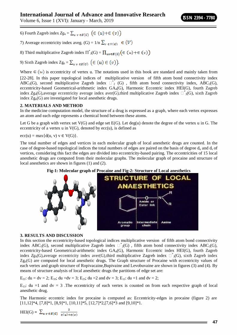
International Journal of Advance and Innovative Research Volume 6, Issue 1 (XVI): January - March, 2019
47
ISSN 2394 - 7780
6) Fourth Zagreb index Zg4 =
7) Average eccentricity index aveg. (G) = 1/n
8) Third multiplicative Zagreb index *3(G) =
9) Sixth Zagreb index Zg6 = .
Where is eccentricity of vertex u. The notations used in this book are standard and mainly taken from [22-28]. In this paper topological indices of multiplicative version of fifth atom bond connectivity index ABC5(G), second multiplicative Zagreb index *
2 (G) , fifth atom bond connectivity index, ABC5(G), eccentricity-based Geometrical-arithmetic index GA4(G), Harmonic Eccentric index HEI(G), fourth Zagreb index Zg4(G),average eccentricity average index aver(G),third multiplicative Zagreb index *
3(G), sixth Zagreb index Zg6(G) are investigated for local anesthetic drugs.
2. MATERIALS AND METHOD In the medicine computation model, the structure of a drug is expressed as a graph, where each vertex expresses an atom and each edge represents a chemical bond between these atoms.
Let G be a graph with vertex set V(G) and edge set E(G). Let deg(u) denote the degree of the vertex u in G. The eccentricity of a vertex u in V(G), denoted by ecc(u), is defined as
ecc(u) = maxd(u, v) v ∈ V(G).
The total number of edges and vertices in each molecular graph of local anesthetic drugs are counted. In the case of degree-based topological indices the total numbers of edges are paired on the basis of degree du and dv of vertices, considering this fact the edges are divided into eccentricity-based pairing. The eccentricities of 15 local anesthetic drugs are computed from their molecular graphs. The molecular graph of procaine and structure of local anesthetics are shown in figures (1) and (2).
Fig-1: Molecular graph of Procaine and Fig-2: Structure of Local anesthetics
3. RESULTS AND DISCUSSION In this section the eccentricity-based topological indices multiplicative version of fifth atom bond connectivity index ABC5(G), second multiplicative Zagreb index *
2(G) , fifth atom bond connectivity index ABC5(G), eccentricity-based Geometrical-arithmetic index GA4(G), Harmonic Eccentric index HEI(G), fourth Zagreb index Zg4(G),average eccentricity index aver(G),third multiplicative Zagreb index *
3(G), sixth Zagreb index Zg6(G) are computed for local anesthetic drugs. The Graph structure of Procaine with eccentricity values of each vertex and graph structure of Ropivacaine,Bupivaine and Levobuvaine are shown in figures (3) and (4). By means of structure analysis of local anesthetic drugs the partitions of edge set are:
E22: du = dv = 2; E33; du =dv = 3; E23; du =2 and dv = 3; E12: du =1 and dv = 2;
E13: du =1 and dv = 3 .The eccentricity of each vertex is counted on from each respective graph of local anesthetic drug.
The Harmonic eccentric index for procaine is computed as: Eccentricity-edges in procaine (figure 2) are [11,12]*4, [7,8]*1, [8,9]*1, [10,11]*5, [12,7]*2,[7,6]*3 and [9,10]*1.
HEI(G) =
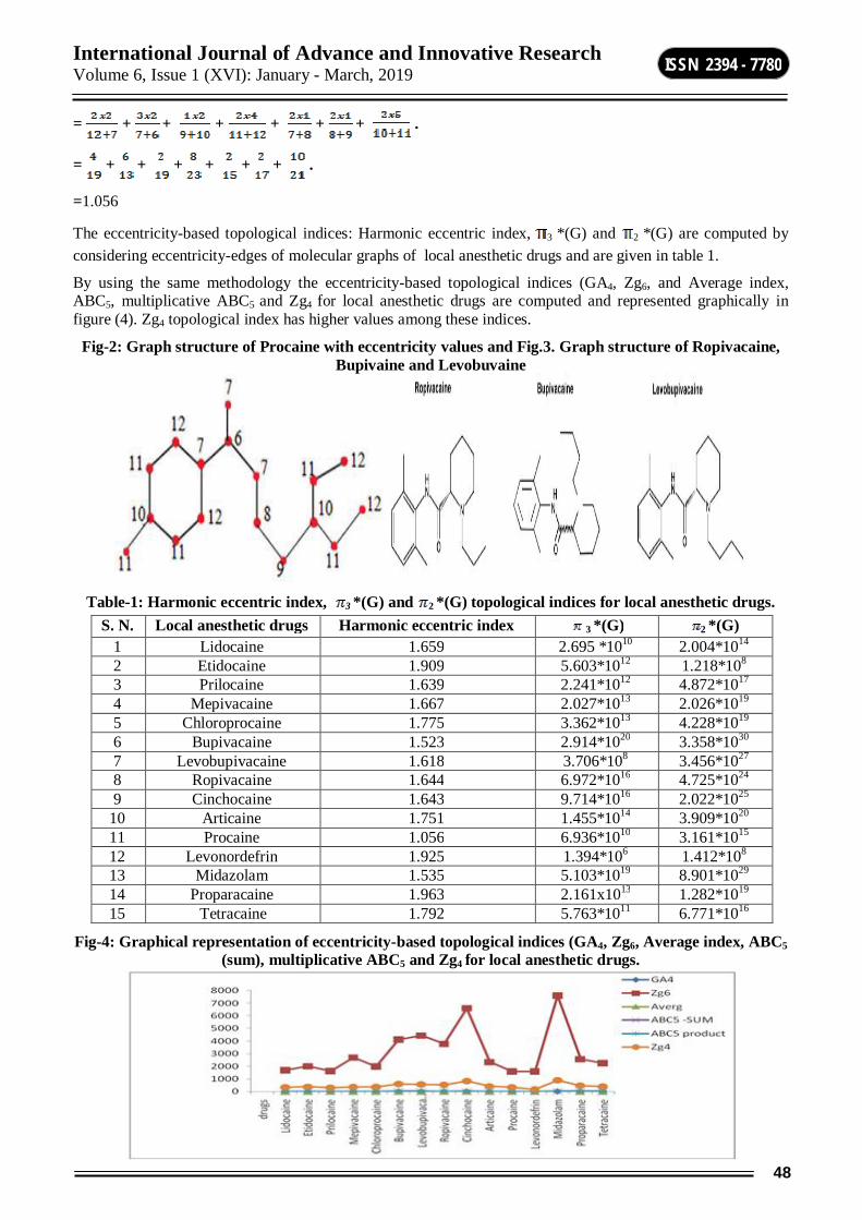
International Journal of Advance and Innovative Research Volume 6, Issue 1 (XVI): January - March, 2019
48
ISSN 2394 - 7780
= + + + + + + .
= + + + + + + .
=1.056
The eccentricity-based topological indices: Harmonic eccentric index, 3 *(G) and 2 *(G) are computed by considering eccentricity-edges of molecular graphs of local anesthetic drugs and are given in table 1.
By using the same methodology the eccentricity-based topological indices (GA4, Zg6, and Average index, ABC5, multiplicative ABC5 and Zg4 for local anesthetic drugs are computed and represented graphically in figure (4). Zg4 topological index has higher values among these indices.
Fig-2: Graph structure of Procaine with eccentricity values and Fig.3. Graph structure of Ropivacaine, Bupivaine and Levobuvaine
Table-1: Harmonic eccentric index, 3 *(G) and 2 *(G) topological indices for local anesthetic drugs.
S. N. Local anesthetic drugs Harmonic eccentric index 3 *(G) 2 *(G) 1 Lidocaine 1.659 2.695 *1010 2.004*1014
2 Etidocaine 1.909 5.603*1012 1.218*108
3 Prilocaine 1.639 2.241*1012 4.872*1017 4 Mepivacaine 1.667 2.027*1013 2.026*1019
5 Chloroprocaine 1.775 3.362*1013 4.228*1019 6 Bupivacaine 1.523 2.914*1020 3.358*1030 7 Levobupivacaine 1.618 3.706*108 3.456*1027 8 Ropivacaine 1.644 6.972*1016 4.725*1024 9 Cinchocaine 1.643 9.714*1016 2.022*1025
10 Articaine 1.751 1.455*1014 3.909*1020 11 Procaine 1.056 6.936*1010 3.161*1015 12 Levonordefrin 1.925 1.394*106 1.412*108 13 Midazolam 1.535 5.103*1019 8.901*1029 14 Proparacaine 1.963 2.161x10¹³ 1.282*1019 15 Tetracaine 1.792 5.763*1011 6.771*1016
Fig-4: Graphical representation of eccentricity-based topological indices (GA4, Zg6, Average index, ABC5 (sum), multiplicative ABC5 and Zg4 for local anesthetic drugs.
International Journal of Advance and Innovative Research Volume 6, Issue 1 (XVI): January - March, 2019
49
ISSN 2394 - 7780
4. CONCLUSION The eccentricity-based HEI(G) has been computed for local anesthetic drugs .The eccentricity-based topological indices: multiplicative version of fifth atom bond connectivity index ABC5(G), second multiplicative Zagreb index *
2 (G) , fifth atom bond connectivity index, ABC5(G), eccentricity-based Geometrical-arithmetic index GA4(G), fourth Zagreb index Zg4(G),average eccentricity average index aver(G), third multiplicative Zagreb index *
3(G), sixth Zagreb index Zg6(G) are studied for local anesthetic drugs. Local anesthetics can be used as individual medicine or as a component of many combinations medications. The eccentricity-based topological indices are used in QSAR/QSPR studies and design of new drugs.
REFERENCES 1. Shetty, B.S., Lokesha, V., Ranjini, P.S.,and Das, K.C. (2012).Computing some topological indices of smart
polymer, Digest Journal of Nanomaterials and Biostuctures,Vol.7,No.3,July-Sepember,1097-1102.
2. Kulli, V. R., (2018). Multiplicative connectivity reverse indices of two families of dendrimer nanostars, International Journal of Current Research in Life Sciences,Vol. 07, No. 02,February, 1102-1108.
3. Hayat, S., Imran, M. (2015).Computation of certain topological indices of Nanotubes, Journal of Computational and Theoretical Nanoscience, Vol.12, 1-7.
4. Gao, W.,Farahani,M.R. (2016).The Forgotten topological index of some drug structures, Acta Medica Mediterranea, 32: 579, 579-584.
5. Gao, W.(2016).Eccentric Related Indices of an Infinite Class of Nanostar Dendrimers, Journal of Chemical and Pharmaceutical Research, 8(4):1187-1190.
6. Zahid, M.A., Baig, A.Q., Naeem, M.,and Azhar, M.R. (2018).Eccentricity-based topological indices of a cyclic octahedron structure ,Mathematics ,6,141, MDPI,1-15.
7. Gao, W., Farahani, M. R., Shi, Li (2016).The forgotten topological index of some drug structures, Acta Medica Mediterranea, 32: 579-585.
8. Gao, W., Siddiqui, M.K., Imran, M., Jamil, M.K., and Farahani, M.R. (2016).Forgotten topological indices of Chemical structure in drugs, Saudi Pharmaceutical Journal,24,258-264.
9. Balasubramanian, K., Balachandran, S., Raja Balachandar, S., and Venkatesh, S.G. (2017). Computing eccentric distance sum of denrimers and nanotubes, International Journal of Pharmacy & Technology ,IJPT|, April-| Vol. 9 | Issue No.1 | 28728-28738.
10. Kanna, M.R.R.,and Jagdeesh, R. (2017).Topological indices of paracetamol,International Journal of Pure and Applied mathematics,Vol.117,No.13,343-352.]Krishnan, Ebbesen et al., Nature ,388,(2001)241.
11. Yamuna, M., and Diyya, T. (2018). Drug pKa value predicting using graph eccentricity,International Journal of Green Pharmacy,July-Septmember ,(12*3)168-174.
12. Plotycya, S., Dubenska, L., Blazheyevsiy, M., Pysarevska, S.,and Sarahman, O. (2016). Determination of Local anesthetics of amide group in pharmaceutical preparations by cyclic voltametry, Wiley Online Library,Electrolysis,28,1-8.
13. Sirbu R.,et al, (2016). Local anesthetics-substance with multiple applications in medicine, Europian Journal of Interdisciplinary studies, January-April ,Vol.2,Issue 1,17-26.
14. Songhori, M. (2012).Eccentric connectivity index of graphs of fullerene graphs, Journal of Mathematical Nanoscience,Vol.2,No.2,21-27.
15. Gao, W., Chen, Y.,and Wang, W. (2017).The topological variable computation for a special type of cycloalkane ,Hindwi,Journal of chemistry,1-8.Article ID 6534758.
16. Zaheed, M.A.,Baig, A.Q., Naeem, M. ,and Azhar, M.R. (2018).Eccentricity-based topological indices of a cyclic octahedron structure, Mathematics,6,141,1-15,MDPI.
17. Farooq, R., Nazir, N., Malik, M.A.,and Arfan, M. (2015). Eccentricity-based topological indices of a dendrimer, Journal of Optoecltronics and Advanced materials,Vol.17,No.11-12,November-December ,1799-1807.
18. Raut, N.K. (2018).F-polynomial and fourth Zagreb polynomial of a molecular graph, International Journal of Science and Research (IJSR),ISSN(Online):2319-7064,Volume 7,Issue 4,April ,1-2.
International Journal of Advance and Innovative Research Volume 6, Issue 1 (XVI): January - March, 2019
50
ISSN 2394 - 7780
19. Raut,N.K.(2018).Eccentricity-based Geometrical-Arithmetic indices of Dendrimers,International Journal of Science and Research(IJSR),Volume 7,Issue 4,April,612-614.
20. Gao, W., Chem, Y.and Wang, W. (2017).The topological variable computation for a special type of cycloalkanes ,Hinawi,Journalof Chemistry, 1-8.
21. Bhanumathi M., and Easu Julia Rani, K. (2017). On Multiplicative K-Eccentric Indices and Multiplicative K Hyper- Eccentric Indices of Graphs,International Journal of Engineering Science, Advanced Computing and Bio-Technology Vol. 8, No. 2, April – June , 42 – 54.
22. Diestel, R. (1997-2000). Graph theory, Electronic edition, Springer-Verlog, New York..
23. West, D.B. (2009).Introduction to graph theory, second edition, PHI, Learning Private Ltd. New Delhi, 67-80.
24. Harary, F. Graph theory, Addison, Wesley, Reading MA, 1971.
25. Vasudev,C.,(2006). Graph theory with applications, New Age, International publishers, New Delhi.
26. Diudea,M.V., Gutman I.,and Lorentz,J., (1999).Molecular Topology, NOVA, Science Publishers Inc.
27. Balakrishnan R., and Renganathan,K. (2000).A textbook of Graph theory, Springer-Verlag, New York.
28. Bondy,J.A., and Murthy,U.S.R. (1982).Graph theory with applications, North-Holland, Fifth printing, ISBN:0-444-19451-7.
International Journal of Advance and Innovative Research Volume 6, Issue 1 (XVI): January - March, 2019
51
ISSN 2394 - 7780
AGRONOMY OF ZEA MAYS UNDER DIFFERENT CULTIVATION PRACTICES
Shubhangi Khandare and Narayan Pandhure Deogiri College, Aurangabad
Department of Botany, Dr. Babasaheb Ambedkar Marathwada University, Aurangabad
ABSTRACT Maize (Zea mays. L.) belongs to family is one of the important forage crop used in Marathwada. During the piece of work the effect of different cultivation methods on growth performance of forage crop Maize was studied. Cultivation practices utilized were Mulching, Raised bed, Shed nets, Ridges and furrows along with control. These four treatments were carried out along with three replicates. Mulching produced the tallest plant, along with highest number of leaves per plant. These plants were emerged out early with 100% rate of seed germination. Control showed poor performance. Thus it could be concluded that, mulching method is best for the production of forage crop maize in rain feed area of Marathwada.
Keywords: Maize, Mulching, Shed net, Raised bed, Furrow and Ridge.
INTRODUCTION Maize (Zea- mays) is one of the most important, high yielding forage crops supporting the livelihood of million people across the world. It is a cheap form of starch and is a major energy source for animal feed (ICAR, 2006). Maize also known as corn is the world’s third most important grain crop after rice and wheat (ICAR, 2006). It is widely grown for food, feed, and fuel, and optimal yield will be required to meet increasing demand due to world population growth and increased fodder usage. Maize can be grown on wide verity of soil but performs best on well drained deep loams and silt loam containing adequate organic matter and available nutrients (Nasir, M., 2000). It is necessarily that the pH of the soil does not deviate from the range 7.5 to 8.5 (Molatud, R.L. and L. K. Marige , 2009).
Maize plants particularly of the seedling stage are susceptible to salinity and 90% relatively of 1.8dsm-1 (Aikinss, H. M; & Joseph,2006). Maize is also sensitive to water logging. Accordingly, provision of adequate drainage is essential for economic production. Maize seed germinate 4-5 days after sowing under warm, moist conditions. When temperature is less than optimum, 14 to 16 days may be necessary for emergence of seedlings (Alessi;J. and J.F.Power,1971).
In Marathwada, maize is produced for consumption both for human and livestock. The green leaves and stalks are used to feed domestic animals. One of the problems experienced by the farmers is lodging. In dense population most plants remain barren; ear and ear size remains smaller, crop become susceptible to lodging, disease and pest, while plant population at sub-optimum level resulted lower yield per unit area (ICAR, 2006 ). High plant population leads to lodging of maize plants (Aikinss, H. M; & Joseph, 2006). Pesent study was under taken to determine the effect of cultivation practices on growth performance of maize.
Secondly temperature changes in the field can be reflected in rapid transpiration and these results in scarcity of water. Aurangabad features a semiarid climate (Alessi, J. and J.F.Power, 1771 ). Annual mean temperatures in Aurangabad range from 17 to 33 °C, with the most comfortable time to visit in the winter – October to February Most of the rainfall occurs in the monsoon season from June to September. Average annual rainfall is 710 mm. The city is often cloudy during the monsoon season and the cloud cover may remain together for days. Hence conditions are suitable for the growth of maize.
The present experiments were conducted to investigate effect of different planting methods on growth of forage crop maize. Keeping in a view above facts a study has been designed with following objectives:
1. To determine percentage of germination and time taken for germinate by maize seeds during different cultivation practices.
2. To investigate impact of different cultivation method on performance of yield.
2. MATERIAL AND METHODS 2.1 Experimental site The experiments were conducted in Botanical garden in the Botany Department of Marathwada University Aurangabad and at the village Revgaon of district Jalna.
International Journal of Advance and Innovative Research Volume 6, Issue 1 (XVI): January - March, 2019
52
ISSN 2394 - 7780
2.2. Experimental Design and treatment The experiment was carried out in a Randomized complete block design (RCBD) with three replicates. The size of each individual plot was 1.5x2.1 M2=3.15M2. The distance between plots, plant, rows, and blocks would be 70cm, 30cm, 70cm, and 1m respectively. Treatments were assigned randomly to each plot and each treatment appeared only once in each block. The experiments were conducted using different cultivation methods viz. Mulching, Shed net, Raised bed, Furrow and Ridge along with three replicates.
2.3 Data collection Data were collected on the following parameters
1) Height of plant after 15 days and after 30 days
2) Fresh weight of plant after 30days.
3) RESULT AND DISCUSSION Plant growth refers to irreversible increase in organ or whole plant size (length, area, volume and weight), while plant development refers to processes related to cell differentiation, organ initiation, member appearance, and extends to plant senescence (Streck et al., 2003). During the present investigation various cultivation methods were employed for growth of maize viz. Mulching, Raised bed, Shed nets, Ridges and furrows along with control. It was recorded that, using mulching method heights of the plants recorded were 29.5 cm., 28.8 cm and 30.3 cm. Using other methods recorded heights of the plants were less compare to the mulchigng method after 15 days. The average heights recorded was 29.53333 cm. Heights were recorded after 30 days which were 48.5, 50 And 47.8. The average heights recorded was 48.7666 cm. Secondly average fresh weights of the plants recorded were 266 with mulching, 87.00 gms with raised seed beds, 120.0 with net and 92.00 with furrows and rows. The value of control recorded was 80.00. (Table.1- 2)
Table-1: Height of plant after 15 and 30 days (In cm.) Cultivation method After 15 Days After 30Days Obs.1 Obs.2 Obs.3 Average Obs.1 Obs.2 Obs.3 Average Mulching 29.5 28.8 30.3 29.53333 48.5 50 47.8 48.7666 Raised bed 18.3 19.7 20.5 19.5 29 28.5 29 28.8333 Shed net 18.8 17.6 19.5 18.63333 35.4 36.2 34.6 35.4 Furrow and ridge 19.4 18.5 19 18.96667 38.2 34.5 37 36.56667 Control 15.6 14.4 13.2 14.43333 30.1 29.3 31.2 30.2
Table-2: Fresh weight of 10 plants after 30 days (In gms.) Cultivation method Obs.1 Obs.2 Obs.3 Average
Mulching 266.60 258.00 268.00 264. 2 Raised bed 86. 89 87 87.3333 Shed net 121 113 126 120.00
Furrow and ridge 91 89 98 92.66667 Control 83 81 78 80.66667
Graph: Effect of different cultivation methods on fresh weight of Maize.
From the recorded results it could be stated that, mulching is best method for the growth of the plants compare to other methods like raised seed beds, shed nets and furrows and ridges. Due to application of this method, moisture could be held up in the soil which has been covered with plastic sheet. Secondly temperature is the governing factor which is sufficient for germination. This favours the growth of roots. Roots are the main organs for the absorption of the nutrients from the soil. Hence healthy growth of seedlings and crop takes place.
International Journal of Advance and Innovative Research Volume 6, Issue 1 (XVI): January - March, 2019
53
ISSN 2394 - 7780
Maize (Zea mays L.) development is primarily driven by temperature, with air temperature being theoretical to enhance maize development from emergence to physiological maturity (Cutforth and Shaykewich, 1990). Muchow (1990) showed that seed growth may be directly influenced by air temperature. Different sowing dates might cause favorable environmental conditions from emergence to seed filling. Fischer (1985) showed that the thermal time requirement needed by a specific growth stage is more or less constant.
Marathwada is rain feed area where only kharif crops could be grown. Hence there is severe deficit of forage crops. Unless and until you don’t have green forage for cattle’s there maintenance is obscure. One could have to adopt such novel techniques in scarcity of water.
4. CONCLUSIONS The growth performance of maize is greatly affected by the different cultivation practices. From the recorded result it could be concluded that, mulching method for plantation of forage crop is better than other methods. This method is viable as requirement of water is less and productivity is more. Thus, for similar agro ecologies of Marathwada, this mulching method recommended for higher yield of forage crop.
ACKNOWLEDGEMENT Authors are thankful to Dr. Babasaheb Ambedkar Marathwada University, Aurangabad, for financial assistance.
REFERENCES Aikinss. H. M; Afuakwa, J, J. and Baidoo, D (2006) . Effect of planting depth on maize stand establishment.
journal of the chana institution of Engineers,4(2):20-25
Alessi; J. and J. F. Power , 1971. Corn emergence in relation to soil temperature and seedling depth. Agron.J, 63:717-719.
Hamdy A., Karam F., Katerji N., Mastrorilli M., van Hoorn I.W. Effect of salinity on water stress, growth , and yield of maize an d sun flower.
ICAR, 2006. Hand book of Agriculture, council of Agricultural Research, New Delhi.
Molatud, R.L. and L.K.Marige (2009). The effect of Maize seed size and depth of planting on seedling emergence and seed vigor. School of Agricultural environment
Nasir, M. 2000. The effects of different plant population on yield and yield components of different maize varieties. M. Sc. (Hons) Thesis, Dept. of Agron. KPK Agric. Univ, Peshawar, Pakistan.
Trenton, F., S. Stanger and G.L. Joseph. 2006. Optimum plant population of Bacillus thuringiensis and non Bacillus thuringiensis corn in Wisconsin. Agron. J., 98: 914-921.
Trenton, F.S. and G.L. Joseph. 2007. Corn stalk response to plant population and the Bt–European corn borer trait. Agron. J., 99: 657-664.
International Journal of Advance and Innovative Research Volume 6, Issue 1 (XVI): January - March, 2019
54
ISSN 2394 - 7780
STUDY OF NEUROSECRETION IN AMOEBOTAENIA JADHAVAE N. SP. FROM GALLUS DOMESTICUS AT SOYGAON, DIST. AURANGABAD, M.S. INDIA
B. S. Thorat Department of Zoology, Vasant Mahavidyalaya, Kaij, Dist. Beed
ABSTRACT The present paper deals with a description and neurosecretion of new species of the genus Amoebotaenia jadhavae n.sp.from Gallus domesticus at Soygaon.T.S.of scolex shows central nervous System and neurosecretory cells and mature segments shows L.N.C. and neurosecretory cells.
Keywords: Neurosecretion, Amoebotaenia, Gallus domesticus.
INTRODUCTION The genus Amoebotaenia was erected by Cohn in 1900 from Charadrius pluvialis as Amoebotaenia brevis.The present paper deals with description and neurosecretion of new species Amoebotaenia jadhavae n.sp. from Gallus domesticus at Soygaon in the month of June,1998.
MATERIAL AND METHODS Eleven intestines of Hen Gallus domesticus were dissected ,five of them were heavily infected with cestode parasites.Identical parasites of cestodes were separated and observed under the microscope,they were identified as Amoebotaenia jadhavae n.sp.Few of these cestodes were fixed in Bouin’s fluid for neurosecretion.The material was washed, dehydrated through graded alcohols and embedded in paraffin wax (58-60oc).Blocks were cut at 7mu,slides were stained with Chrome-alum-haematoxylin phloxin method (Gomori,1941).The best slides were selected for the study of neurosecretion of Amoebotaenia jadhavae n.sp.
OBSERVATION AND DISCUSSION In the whole nervous system of Amoebotaenia jadhavae n.sp.central nervous system and peripheral nervous system are clearly observed.In the present investigation the lateral branches of the central cephalic mass are clearly observed.The cephalic mass (cerebral ganglia) consist of a bipolar part with more unipolar cells,moderate bipolar cells and very few multipolar cells, all with neurosecretory material.In the sucker region there are some unipolar cells moderate bipolar and multipolar cells are very few in number.In the T.S. of scolex near the suckers,the unipolar cells are empty i.e.without neurosecretory material,perhaps it has been oozed out at the time of some action of the parasite,where as other types of cells i.e.bipolar and multipolar are fall with neurosecretory material.T.S. of scolex and neurosecretory cells and mature segments shows L.N.C. and neurosecretory cells.The lateral longitudinal nerve cords are bounded from outside by nerve cells in the scolex region.These cords are thick and contain bundles of nerve fibres.The study of segments revealed the presence of lateral nerves in this region also,bounded by central nucleated cells,which are darkely stained.There are neurosecretory cells,which are located to periphery of the segment.These cells very vary in size,shape & structure and named accordingly unipolr,bipolar and multipolar cells.The unipolar having round shape,single axon and are small in size,bipolar cells are spindle shaped,medium in size and bears two axons,multipolar cells are irregular in shape,many axons and large in size.All the neurosecretorycells have thin cell walls.
ACKNOWLEDGEMENT The author are thankful to the Professor & Head,Department of Zoology,Dr.B.A.Marathwada University,Aurangabad.
REFERANCES B.V.Jadhav,R.M.Khadap and B.S.Thorat.(2003): Anew species of the genus Amoebotaenia (Cohn,1900)
from Gallus domesticus at Aurangabad.
Indian J.Helminth (N.S.)Vol.21,2003 pp.68-70.
B.S.Thorat ,B.V.Jadhav.(2009): ): Anew species of the genus Amoebotaenia (Cohn,1900) from Gallus domesticus at Soygaon.Dist.Aurangabad.(M.S.India).
Life Science Bulletin,6(2) 2009pp.179-181.
B.S.Thorat.(2015): Study of Neurosecretion in Davainea shindei n.sp. from Gallus domesticus at Vaijapur. .Dist.Aurangabad.(M.S.India).
Journal of Basic Sciences,2015.Special Issu on BIOIPPE,75-77.
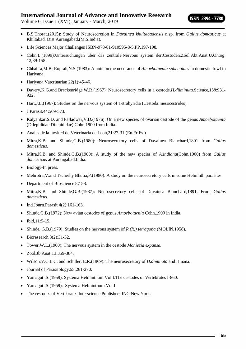
International Journal of Advance and Innovative Research Volume 6, Issue 1 (XVI): January - March, 2019
55
ISSN 2394 - 7780
B.S.Thorat.(2015): Study of Neurosecretion in Davainea khultabadensis n.sp. from Gallus domesticus at Khiltabad. Dist.Aurangabad.(M.S.India).
Life Sciences Major Challenges ISBN-978-81-910595-8-5.PP.197-198.
Cohn,L.(1899):Untersuchungen uber das zentrale.Nervous system der.Cestoden.Zool.Abt.Anat.U.Ontog. 12,89-158.
Chhabra,M.B; Ruprah,N.S.(1983): A note on the occurance of Amoebotaenia sphenoides in domestic fowl in Hariyana.
Hariyana Vaterinarian 22(1):45-46.
Davery,K.G.and Breckenridge,W.R.(1967): Neurosecretory cells in a cestode,H.diiminuta.Science,158:931-932.
Hart,J.L.(1967): Studies on the nervous system of Tetrahyridia (Cestoda:mesocestrides).
J.Parasit.44:569-573.
Kalyankar,S.D. and Palladwar,V.D.(1976): On a new species of ovarian cestode of the genus Amoebotaenia (Dilepididae:Dilepididae) Cohn,1900 from India.
Anales de la fawlted de Veterinaria de Leon,21:27-31.(En.Fr.Es.)
Mitra,K.B. and Shinde,G.B.(1980): Neurosecretory cells of Davainea Blanchard,1891 from Gallus domesticus.
Mitra,K.B. and Shinde,G.B.(1980): A study of the new species of A.indiana(Cohn,1900) from Gallus domesticus at Aurangabad,India.
Biology-In press.
Mehrotra,V.and Tscherhy Bhutia,P.(1980): A study on the neurosecretory cells in some Helminth parasites.
Department of Bioscience 87-88.
Mitra,K.B. and Shinde,G.B.(1987): Neurosecretory cells of Davainea Blanchard,1891. From Gallus domesticus.
Ind.Journ.Parasit 4(2):161-163.
Shinde,G.B.(1972): New avian cestodes of genus Amoebotaenia Cohn,1900 in India.
Ibid,11:5-15.
Shinde, G.B.(1979): Studies on the nervous system of R.(R.) tetragona (MOLIN,1958).
Bioresearch,3(2):31-32.
Tower,W.L.(1900): The nervous system in the cestode Moniezia expansa.
Zool.Jb.Anat;13:359-384.
Wilson,V.C.L.C. and Schiller, E.R.(1969): The neurosecretory of H.diminuta and H.nana.
Journal of Parasitology,55.261-270.
Yamaguti,S.(1959): Systema Helminthum.Vol.I.The cestodes of Vertebrates I-860.
Yamaguti,S.(1959): Systema Helminthum.Vol.II
The cestodes of Vertebrates.Interscience Publishers INC;New York.
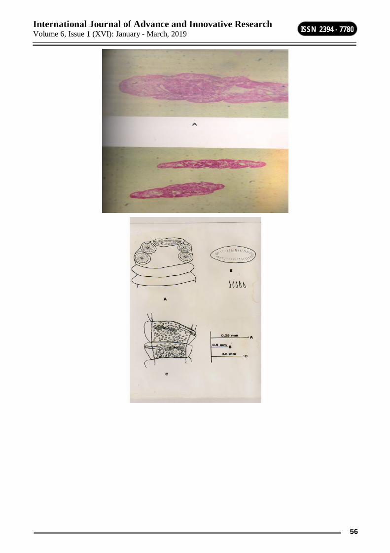
International Journal of Advance and Innovative Research Volume 6, Issue 1 (XVI): January - March, 2019
56
ISSN 2394 - 7780
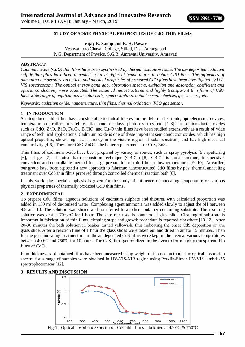
International Journal of Advance and Innovative Research Volume 6, Issue 1 (XVI): January - March, 2019
57
ISSN 2394 - 7780
STUDY OF SOME PHYSICAL PROPERTIES OF CdO THIN FILMS
Vijay B. Sanap and B. H. Pawar
Yeshwantrao Chavan College, Sillod, Dist. Aurangabad P. G. Department of Physics, S.G.B. Amravati University, Amravati
ABSTRACT Cadmium oxide (CdO) thin films have been synthesized by thermal oxidation route. The as- deposited cadmium sulfide thin films have been annealed in air at different temperatures to obtain CdO films. The influences of annealing temperature on optical and physical properties of prepared CdO films have been investigated by UV-VIS spectroscopy. The optical energy band gap, absorption spectra, extinction and absorption coefficient and optical conductivity were evaluated. The obtained nanostructured and highly transparent thin films of CdO have wide range of applications in solar cells, smart windows, optoelectronic devices, gas sensors; etc.
Keywords: cadmium oxide, nanostructure, thin films, thermal oxidation, TCO gas sensor.
1 INTRODUCTION Semiconductor thin films have considerable technical interest in the field of electronic, optoelectronic devices, temperature controllers in satellites, flat panel displays, photo-resistors, etc. [1-3].The semiconductor oxides such as CdO, ZnO, BaO, Fe2O3, BiClO, and Cu2O thin films have been studied extensively as a result of wide range of technical applications. Cadmium oxide is one of these important semiconductor oxides, which has high optical properties, shows high transparency in the visible region of solar spectrum, and has high electrical conductivity [4-6]. Therefore CdO-ZnO is the better replacements for CdS, ZnS.
Thin films of cadmium oxide have been prepared by variety of routes, such as spray pyrolysis [5], sputtering [6], sol gel [7], chemical bath deposition technique (CBDT) [8]. CBDT is most common, inexpensive, convenient and controllable method for large preparation of thin films at low temperatures [9, 10]. At earlier, our group have been reported a new approach to fabricate nanostructured CdO films by post thermal annealing treatment over CdS thin films prepared through controlled chemical reaction bath [8].
In this work, the special emphasis is given for the study of influence of annealing temperature on various physical properties of thermally oxidized CdO thin films.
2 EXPERIMENTAL To prepare CdO films, aqueous solutions of cadmium sulphate and thiourea with calculated proportion was added in 130 ml of de-ionized water. Complexing agent ammonia was added slowly to adjust the pH between 9.5 and 10. The solution was stirred and transferred to another container containing substrate. The resulting solution was kept at 70±2ºC for 1 hour. The substrate used is commercial glass slide. Cleaning of substrate is important in fabrication of thin films, cleaning steps and growth procedure is reported elsewhere [10-12]. After 20-30 minutes the bath solution in beaker turned yellowish, thus indicating the onset CdS deposition on the glass slide. After a reaction time of 1 hour the glass slides were taken out and dried in air for 15 minutes. Then for the post annealing treatment in air, the as-deposited CdS films were kept in the oven at various temperatures between 400ºC and 750ºC for 10 hours. The CdS films get oxidized in the oven to form highly transparent thin films of CdO.
Film thicknesses of obtained films have been measured using weight difference method. The optical absorption spectra for a range of samples were obtained in UV-VIS-NIR region using Perklin-Elmer UV-VIS lambda-35 spectrophotometer [12].
3 RESULTS AND DISCUSSION
Fig-1: Optical absorbance spectra of CdO thin films fabricated at 450°C & 750°C

International Journal of Advance and Innovative Research Volume 6, Issue 1 (XVI): January - March, 2019
58
ISSN 2394 - 7780
Fig.1 shows the optical absorbance spectra of CdO thin films in the wavelength range from 200 to 1100nm for various annealing temperatures. It is observed that the maximum absorption occurs between 350-450nm and remains constant after 600nm. A nearly sharp fall in absorbance is an identification of good crystallinity of films. It is observed that the films obtained at higher annealing temperature shows slightly higher absorbance as compared to the films obtained at lower annealing temperature. These results slight changes in optical band-gap of the film and it may be due to the small grain size of the polycrystalline films.
Fig-2: Plot of (αhν) 2 and hν of CdO thin films fabricated at 450°C & 750°C.
The optical energy band gap (Eg) was determined by plotting (αhν)2 versus hν and then extrapolating the straight line portion to the energy axis at α =0 (Fig. 4). The band gap energy obtained for each film is different. It was observed that the band gap of CdO films obtained at higher annealing temperature is 2.45eV whereas it is 2.32eV for the films obtained at lower annealing temperature. This difference of 0.13eV in optical band-gap of CdO films may be due to the difference in grain size.
Fig-3: Plot of R.I. (n) and wavelength of CdO thin films fabricated at 450°C & 750°C
Fig.3 shows the plot between refractive index (R.I.) and wavelength of CdO thin films fabricated at 450°C & 750°C. The average value of R.I. observed for higher and lower annealing temperature is 2.102 and 2.097 respectively.
Fig-4: Plot of extinction coefficient of CdO thin films fabricated at 450°C & 750°C
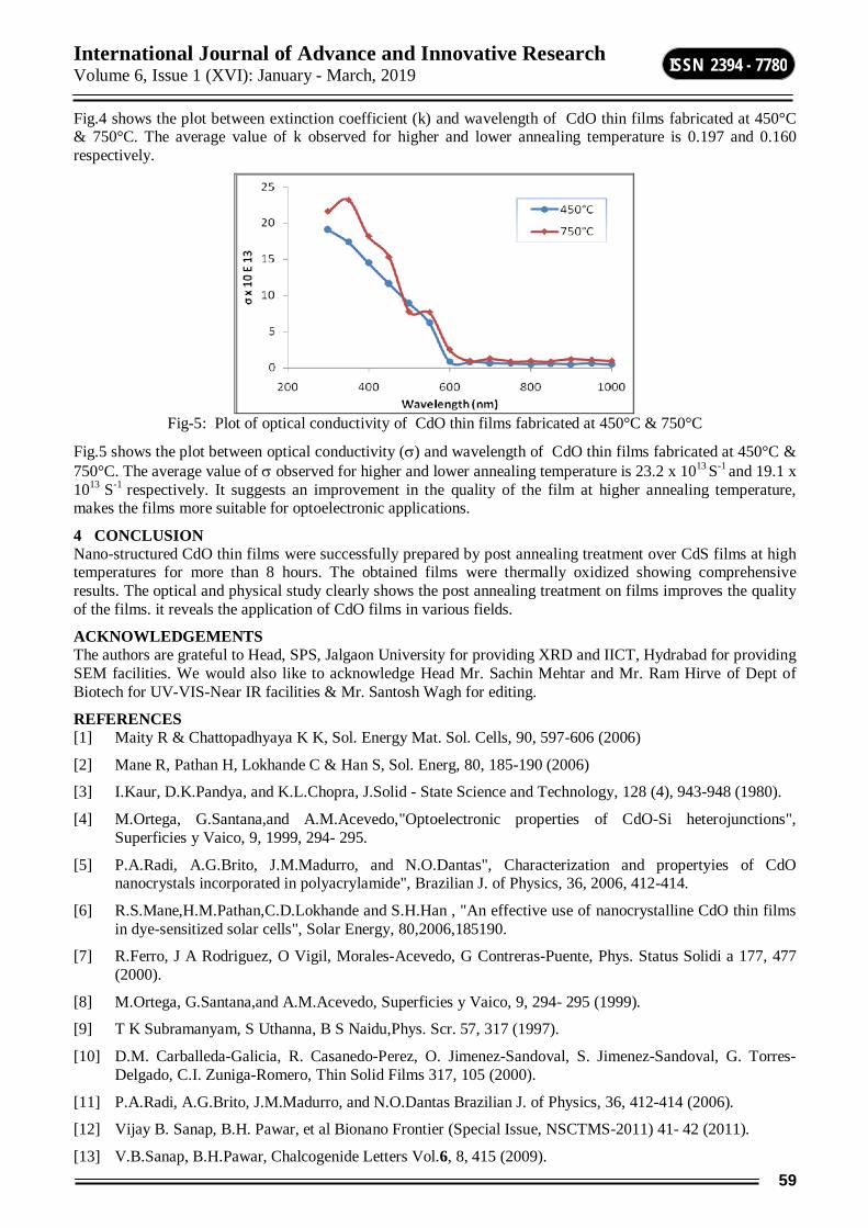
International Journal of Advance and Innovative Research Volume 6, Issue 1 (XVI): January - March, 2019
59
ISSN 2394 - 7780
Fig.4 shows the plot between extinction coefficient (k) and wavelength of CdO thin films fabricated at 450°C & 750°C. The average value of k observed for higher and lower annealing temperature is 0.197 and 0.160 respectively.
Fig-5: Plot of optical conductivity of CdO thin films fabricated at 450°C & 750°C
Fig.5 shows the plot between optical conductivity () and wavelength of CdO thin films fabricated at 450°C & 750°C. The average value of observed for higher and lower annealing temperature is 23.2 x 10¹³ S-1 and 19.1 x 10¹³ S-1 respectively. It suggests an improvement in the quality of the film at higher annealing temperature, makes the films more suitable for optoelectronic applications.
4 CONCLUSION Nano-structured CdO thin films were successfully prepared by post annealing treatment over CdS films at high temperatures for more than 8 hours. The obtained films were thermally oxidized showing comprehensive results. The optical and physical study clearly shows the post annealing treatment on films improves the quality of the films. it reveals the application of CdO films in various fields.
ACKNOWLEDGEMENTS The authors are grateful to Head, SPS, Jalgaon University for providing XRD and IICT, Hydrabad for providing SEM facilities. We would also like to acknowledge Head Mr. Sachin Mehtar and Mr. Ram Hirve of Dept of Biotech for UV-VIS-Near IR facilities & Mr. Santosh Wagh for editing.
REFERENCES [1] Maity R & Chattopadhyaya K K, Sol. Energy Mat. Sol. Cells, 90, 597-606 (2006)
[2] Mane R, Pathan H, Lokhande C & Han S, Sol. Energ, 80, 185-190 (2006)
[3] I.Kaur, D.K.Pandya, and K.L.Chopra, J.Solid - State Science and Technology, 128 (4), 943-948 (1980).
[4] M.Ortega, G.Santana,and A.M.Acevedo,"Optoelectronic properties of CdO-Si heterojunctions", Superficies y Vaico, 9, 1999, 294- 295.
[5] P.A.Radi, A.G.Brito, J.M.Madurro, and N.O.Dantas", Characterization and propertyies of CdO nanocrystals incorporated in polyacrylamide", Brazilian J. of Physics, 36, 2006, 412-414.
[6] R.S.Mane,H.M.Pathan,C.D.Lokhande and S.H.Han , "An effective use of nanocrystalline CdO thin films in dye-sensitized solar cells", Solar Energy, 80,2006,185190.
[7] R.Ferro, J A Rodriguez, O Vigil, Morales-Acevedo, G Contreras-Puente, Phys. Status Solidi a 177, 477 (2000).
[8] M.Ortega, G.Santana,and A.M.Acevedo, Superficies y Vaico, 9, 294- 295 (1999).
[9] T K Subramanyam, S Uthanna, B S Naidu,Phys. Scr. 57, 317 (1997).
[10] D.M. Carballeda-Galicia, R. Casanedo-Perez, O. Jimenez-Sandoval, S. Jimenez-Sandoval, G. Torres-Delgado, C.I. Zuniga-Romero, Thin Solid Films 317, 105 (2000).
[11] P.A.Radi, A.G.Brito, J.M.Madurro, and N.O.Dantas Brazilian J. of Physics, 36, 412-414 (2006).
[12] Vijay B. Sanap, B.H. Pawar, et al Bionano Frontier (Special Issue, NSCTMS-2011) 41- 42 (2011).
[13] V.B.Sanap, B.H.Pawar, Chalcogenide Letters Vol.6, 8, 415 (2009).
International Journal of Advance and Innovative Research Volume 6, Issue 1 (XVI): January - March, 2019
60
ISSN 2394 - 7780
FUZZIFICATION OF LINEAR SPACES
Vinod Kulkarni1 and Vijay Sangle2 1Department of Mathematics, Swa. Sawarkar Mahavidyalaya, Beed
2Department of Mathematics, R. B. Attal College, Georai, Dist. Beed
ABSTRACT Analyze the concept of fuzzy linear spaces (FLS) and we have proposed the redefined notion of fuzzy linear spaces and have established that the proposed definition is more general and appealing than that of Nanda and Biswas. The notion of product (*) of two fuzzy linear spaces has been proposed and it has been observed that the product is again a fuzzy linear space under the new definition. In other words, we can say that these structures are preserved under the product (*). We observe that it is more general than its classical counter part.
1. INTRODUCTION The concept of fuzzy l inear spaces was introduced by Sudarsan Nanda in 1986 and was again redefined by Biswas in 1989. It is expected that several r esults from linear a lgebra and funct ional analys is can be extended to the concept of fuzzy sett ing. Nanda propounded the not ion of fuzzy l inear spaces in a l inear space as fol lows:
2. FUZZY LINEAR SPACE Let F be a fuzzy f ield in a field (X, + , . ) with membership function F( ). Let Y be a linear space over F and V be a fuzzy subset of Y with membership function V(x). Then, V is called as a fuzzy l inear space in Y if the following postu lates ar e satisf ied:
(i) V(x + y) minV(x),V(y) , x , y Y
(i i) V(x) minF( ),V(x), λ F and x Y
(i i i) V(0) = 1
In case F is an ordinary f ield then, F ( ) = 1 and hence
V(x) min1,V(x), F and x Y
= V(x)
Hence, for F to be an ordinary field, the ( i i) postu late may be cons idered as
V(x) V(x), F and xY
Now we will analyze the def init ion of fuzzy l inear space introduced by Nanda .
Let us cons ider the case when F and V both ar e classica l set . Then , we have F ( ) = 1, V(x) = 1 and V(y) = 1 for a ll x , y F
and λ F.
Hence, from condit ion ( i), we have
V( x + y ) = 1 x + y V
Thus, we get that x , y V x + y V.
Further , from condit ion ( i i), we get
V(λx) ≥ min 1,1 = 1
i. e. V(λx) = 1 x V. That is , x V, λ F λx V.
It follows that V is c losed under addit ion and scalar mult ip lication.
Thus, on the basis of above discuss ion we arr ive at the conclus ion that the def init ion of fuzzy l inear space has been cons idered in such a way that when F and V both ar e cons idered as an ordinary subset, V turns out to be a subspace of Y.
Alternatively , For all x , y Y. and , F, we have
International Journal of Advance and Innovative Research Volume 6, Issue 1 (XVI): January - March, 2019
61
ISSN 2394 - 7780
V(x + y) minV(x),V(y)
minminF( ),V(x),minF(),V(y)---(1)
If V and F both are ordinary subset then, we get
V(x) = 1 = V(y) and F(λ) = 1 = F ()
i. e. V(x + µy) ≥ minmin1,1,min1,1 = 1
i. e. V(x + µy) = 1 x + y V
That is for a l l λ, µ F and x, y V x + y V . It follows that V is a subspace of Y. So, aga in we are in a pos it ion to say that the not ion of fuzzy l inear space has been cons idered in such a way that when F and V both are taken to be an ordinary subset under above def init ion of fuzzy linear space V turns out to be a subspace of Y.
In case when F is an ordinary field in X, then F() = F( ) = 1 . And hence in this cas e condition (1) r educes to
V(x + y) minmin1,V(x),min1,V(y)
= min V(x),V(y)
i. e. V(x + y) min V(x), V(y)
The concept of fuzzy l inear space was r edef ined by R.Biswas in 1989 as he was not satisf ied by the def init ion of fuzzy l inear space proposed by Nanda . Biswas redef ined the not ion of fuzzy l inear space, as follows:
3. REDEFINED FUZZY LINEAR SPACE Let F be a fuzzy f ield in a field (X, +, . ) with membership function F( ). Let Y be a linear space over F and V is a fuzzy subset of Y with membership funct ion V(x). Then, V is sa id to be a fuzzy l inear space in Y if the following condit ions are satis f ied:
(i) V(x + y) min V(x),V(y), x , y Y
(i i) V(x) min F( ),V(x), F , x Y
Now our aim is to f ind out the r eason for delet ing the (i i i) condit ion in the def init ion of fuzzy l inear space proposed by Biswas . In our opinion the r eason behind this is the propos it ion 4.5 in the paper of Nanda . It sta tes that- V is a fuzzy l inear space in Y(over a fuzzy f ield F in X) if and only if
V(x + y) minmin F( ), V(x),minF(),V(y)---(1)
for a ll , F and x , y Y
On putt ing = = 1, we get
V(x + y)=V(1x+1y) minminF(1),V(x),minF(1),V(y)
= minmin1,V(x), min 1,V(y)
= minV(x),V(y)
i. e V(x + y) min V(x),V(y), x , y Y
Aga in on putt ing = 0 in (1), we get
V(x + 0y) = V(x + 0) minV(x), V(0)
= minV(x), 1
= V(x)
minF( ),V(x)
i. e V(x) minF( ),V(x), F , x Y ---(2)
Further , if F is an ordinary f ield in X , then F( ) = 1
International Journal of Advance and Innovative Research Volume 6, Issue 1 (XVI): January - March, 2019
62
ISSN 2394 - 7780
Now, from equation (2), we get
V(x) min 1,V(x)
= V(x)
i. e V(x) V(x), F = X and xY
Also, V(0) = V(x-x) = V(1x -1x)
minV(1x), V(-1x)
minminF(1),V(x), V(x)
= min V(x), V(x)
= V(x)
i. e V(0) V(x)
Thus, we observe that the (i i i) condit ion in the def init ion of Nanda is not deducib le from the propos it ion 4.5 and so it has been dropped.
We may l ike to redef ine the concept of fuzzy linear spaces in the following way:
4. IMPROVED DEFINITION OF FUZZY LINEAR SPACE Let F be a fuzzy f ield in a f ield (X, +, . ). Let Y be a l inear space over F and V is a fuzzy subset of Y. Then V be called as a fuzzy l inear space in Y if the following condit ion a re satisf ied:
(i) V(x + y) V (x). V(y), x, yY
(ii) V(x) F( .V(x), F and xY
If F is an ordinary f ield ( in par t icu lar if F = X), then F ( ) = 1. Then the, ( i i) condit ion reduces to
V(x) V(x), Y
Now we claim that our def init ion of fuzzy l inear space is more general than that of Biswas .
For , if V is a fuzzy l inear space in Y in the sense of Biswas , then
V(x + y) minV(x),V(y) V(x).V(y), x, yY
and V(x) min F( ),V(x) F( ).F(x), F , xY
It follows that V is a fuzzy l inear space according to our def init ion when it is a fuzzy l inear space according to Biswas .
Next, we suppose that V is a fuzzy l inear space in Y according to our def init ion.
That is V(x + y) V(x).V(y)
And V(x) F( ).V(x)
But V(x).V(y) minV(x),V (y)
and F( ).V(x) minF( ),V(x)are not t rue.
Therefore, V is not a fuzzy linear space in view of Biswas , while it is a fuzzy l inear space under our def init ion.
5. MULTIPLICATION OF FUZZY LINEAR SPACES Let Y be a l inear space over a fuzzy f ield F and U and V are two fuzzy l inear spaces in Y. Then, we def ine mult ip lication (*) between any two fuzzy l inear spaces a s follows:
(U*V)(x) = U(x).V(x) ∀ x ∊ Y
Evident ly U*U U
For (U*U)(x) = U(x).U(x)
International Journal of Advance and Innovative Research Volume 6, Issue 1 (XVI): January - March, 2019
63
ISSN 2394 - 7780
= [U(x)]2
≤ U(x), ∀(x) ∊ Y
Therefore U*U U
THEOREM The product of two fuzzy l inear spaces in a l inear space Y over a fuzzy field F in a f ield X is aga in a fuzzy l inear space in Y.
PROOF: Let us assume that V1 and V2 be any two fuzzy l inear spaces in a l inear space Y over an ordinary field F in X. Then, for a l l x, yY and X, we have
V1(x + y) V1(x).V1(y) and V2(x + y) V2(x).V2(y)
Also V1(x) V1(x) and V2(x) V2(x)
Now our aim is to show that the product (*) of V 1 and V2 i. e. V1* V2 is again a fuzzy l inear space in Y. The product (*) of fuzzy l inear spaces in Y is def ined as
(V1 * V2) (x) = V1(x).V2(x)
Then, we have
(V1 * V2) (x + y) = V1(x + y).V2(x + y)
V1(x).V1(y).V2(x).V2(y)
= V1(x).V2(x).V1(y).V2(y)
= (V1 * V2) (x). (V1* V2)(y)
(V1 * V2)(x + y) V1(x + y).(V1 * V2) (y)
Also (V1 * V2) (x) = V1(x).V2 (x)
V1(x).V2 (x)
= (V1 * V2) (x)
i. e. (V1 * V2) (x) (V1 * V2) (x)
This establishes the claim that the product of linear spaces V1 and V2 i. e. V1 * V2 is a lso a fuzzy l inear space in Y.
REFERENCES 1. Zadeh, L.A [1965] “Fuzzy Sets”, Informat ion and Control, 8,pp338-353
2. Nanda, Sudarshan [1986] “Fuzzy Fields and Fuzzy Linear Spaces” Fuzzy Sets and Systems,9,pp 89-84
3. Rosenfeld, A [1971] “Fuzzy Groups’,J.Math.Ana l.Appl.35 Pp 512-517.
4. Biswas,R [1989] “Fuzzy Field and Linear Spaces Redefined”, Fuzzy Sets and Systems,33,pp.257-259
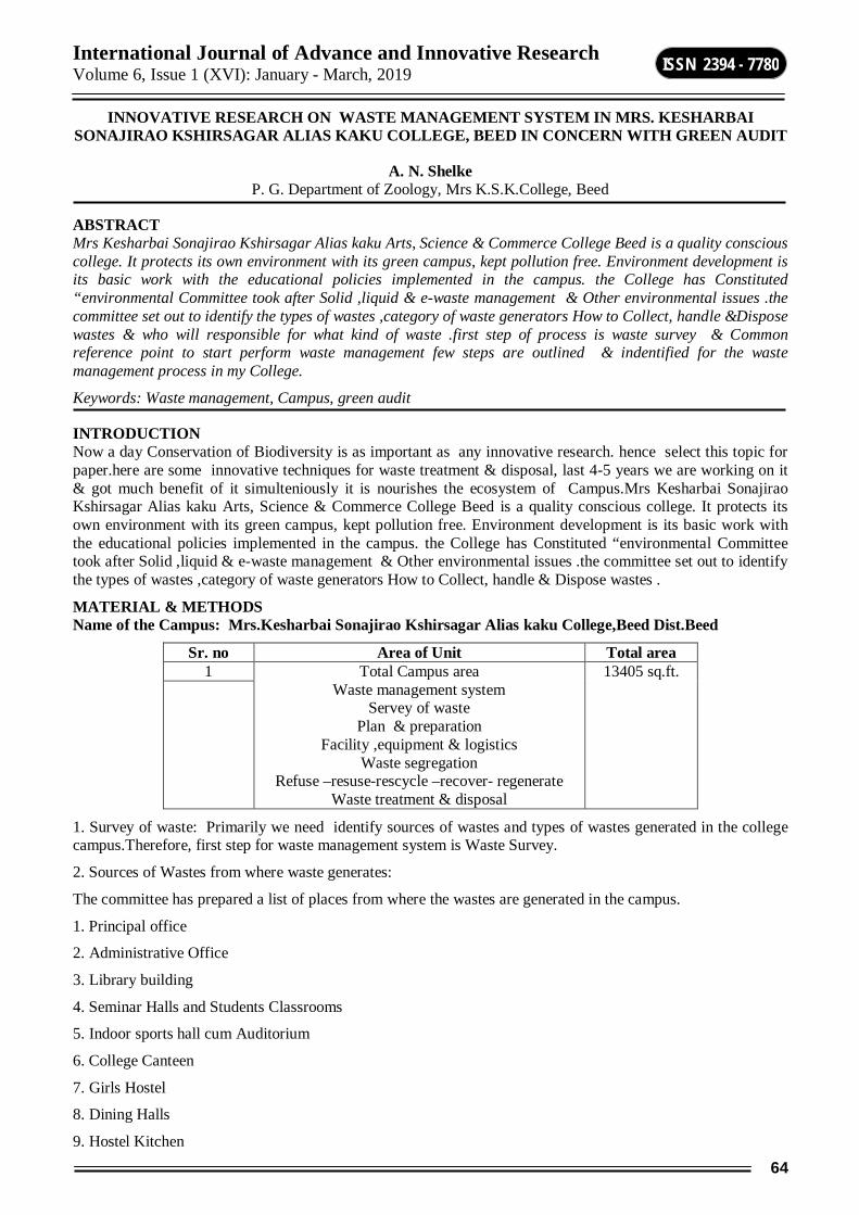
International Journal of Advance and Innovative Research Volume 6, Issue 1 (XVI): January - March, 2019
64
ISSN 2394 - 7780
INNOVATIVE RESEARCH ON WASTE MANAGEMENT SYSTEM IN MRS. KESHARBAI SONAJIRAO KSHIRSAGAR ALIAS KAKU COLLEGE, BEED IN CONCERN WITH GREEN AUDIT
A. N. Shelke
P. G. Department of Zoology, Mrs K.S.K.College, Beed
ABSTRACT Mrs Kesharbai Sonajirao Kshirsagar Alias kaku Arts, Science & Commerce College Beed is a quality conscious college. It protects its own environment with its green campus, kept pollution free. Environment development is its basic work with the educational policies implemented in the campus. the College has Constituted “environmental Committee took after Solid ,liquid & e-waste management & Other environmental issues .the committee set out to identify the types of wastes ,category of waste generators How to Collect, handle &Dispose wastes & who will responsible for what kind of waste .first step of process is waste survey & Common reference point to start perform waste management few steps are outlined & indentified for the waste management process in my College.
Keywords: Waste management, Campus, green audit
INTRODUCTION Now a day Conservation of Biodiversity is as important as any innovative research. hence select this topic for paper.here are some innovative techniques for waste treatment & disposal, last 4-5 years we are working on it & got much benefit of it simulteniously it is nourishes the ecosystem of Campus.Mrs Kesharbai Sonajirao Kshirsagar Alias kaku Arts, Science & Commerce College Beed is a quality conscious college. It protects its own environment with its green campus, kept pollution free. Environment development is its basic work with the educational policies implemented in the campus. the College has Constituted “environmental Committee took after Solid ,liquid & e-waste management & Other environmental issues .the committee set out to identify the types of wastes ,category of waste generators How to Collect, handle & Dispose wastes .
MATERIAL & METHODS Name of the Campus: Mrs.Kesharbai Sonajirao Kshirsagar Alias kaku College,Beed Dist.Beed
Sr. no Area of Unit Total area 1 Total Campus area
Waste management system Servey of waste
Plan & preparation Facility ,equipment & logistics
Waste segregation Refuse –resuse-rescycle –recover- regenerate
Waste treatment & disposal
13405 sq.ft.
1. Survey of waste: Primarily we need identify sources of wastes and types of wastes generated in the college campus.Therefore, first step for waste management system is Waste Survey.
2. Sources of Wastes from where waste generates:
The committee has prepared a list of places from where the wastes are generated in the campus.
1. Principal office
2. Administrative Office
3. Library building
4. Seminar Halls and Students Classrooms
5. Indoor sports hall cum Auditorium
6. College Canteen
7. Girls Hostel
8. Dining Halls
9. Hostel Kitchen

International Journal of Advance and Innovative Research Volume 6, Issue 1 (XVI): January - March, 2019
65
ISSN 2394 - 7780
10. Hostel Grocery Stores Room / Crockery Store Room
11. Health Centre
12. Play Ground
13. Various Departments
14. Laboratories.
3. Types of Wastes and existing practice of waste disposal After finding the places from where waste is generated, the next step is to dispose the different types of wastes (wet, dry and hazardous waste) as per the plan mentioned in the table below.
Type of waste Disposed at common waste bins Municipality
use it for composting
Dumped in road side, and street corners /
vacant plots
Send to Waste Pickers for recycling
Wet Waste Vegetable peels
Fruit peels Rotten fruits and vegetables
Leftover food Used tea / tea bags Used coffee ground
Egg shells Expired other food items Mango kernel & any seed
Coconut fibre Used flowers / dry flowers
Spoiled spices Floor sweeping dust
Garden shrubs Dry Waste
Soap covers / pockets / sachets Empty shampoo bottles, Empty
perfume bottles / containers of deodorants
/ shaving creams
Empty Chemical Bottles Milk covers
Used door mats/door mats Used tooth brush
Chocolate wrappers Butter wrappers Used mop cloth
Ghee / oil pockets / cans Package / polythene covers /
Plastic covers
Newspapers / card boards cosmetics containers
Styrofoam Broken stationery like used pens,
pencil sharpener
Empty cans of floor cleaners Kurkure / Lays packets

International Journal of Advance and Innovative Research Volume 6, Issue 1 (XVI): January - March, 2019
66
ISSN 2394 - 7780
Sachets (of shampoo, creams etc.) Bisleri kind of water bottles Used tooth paste tubes etc.
Metal tins, and cans (e.g Pepsi Coke cans)
Small tubs like the ones used for yogurt,
cheese, jam
Hazardous Waste Mosquito repellent refill bottles/
Mosquito repellent mats
Expired medicines Tablet covers / Syrups bottles
Any medical discard Sanitary napkins
Children’s diapers Used razor / razor blades
Old batteries Fused bulbs / tubes / electrical
items
Broken glasses / ceramics Empty cans of toilet cleaners
Expired cosmetics Cockroach killers / spray cans
Old printer cartridge / CDs Waste generated at Laboratories
after proper treatment
Old Electronic items / parts Pieces of wires, old chargers, old
pen drives
WET WASTE METHOD Waste especially from kitchen, such as vegetable refuses, food scraps etc. are called wet waste. Send them for composting using one of the methods such as aerobic or anaerobic methods.
DRY WASTE METHOD Basic principles of dry waste management
1) 4RS: Refuse ,Reduce, Reuse and Recycle.
Refuse: Do not buy anything which we do not really need.
Reduce - Reduce the amount of garbage generated. Alter our lifestyle so that minimum garbage is generated.
Reuse - Reuse everything to its maximum after properly cleaning it. Make Secondary use of different articles.
Recycle – Keep things which can be recycled to be given to rag pickers or waste pickers (bhangarwala).Convert the recyclable garbage into manures or other useful products.
WASTE SEGREGATION Store biodegradable and non biodegradable solid waste in different bins. Recycle of all the components with minimum labor and cost. 3) Different treatments for different types of solid wastes: One must apply the techniques which are suitable to the given type of waste. 4) Treatment at nearest possible point: The solid waste should be treated in as decentralized manner as possible. The waste generated should be treated preferably at the site of generation. Dry waste must undergo shifting for picking out the recyclable to be passed on to the recyclers. The residual reject are sent for incineration in & eco friendly in incinerator.

International Journal of Advance and Innovative Research Volume 6, Issue 1 (XVI): January - March, 2019
67
ISSN 2394 - 7780
E WASTE MANAGEMENT we have design a room in the campus which we called material recovery centre in (MRC).the Discarded E –waste from all the office in campus shall send to MRC & stored until it sent to e waste recyclers. In College Campus for e waste should begin at the point of generation .This Can be done by waste minimization. Techniques & By sustainable products design. Waste minimization in college involves adapting various management.
RESULT the above methods are very essesial now a days for Biodiversity Conservation. This can be performed at various levels in Nation at every space even in city ,rural areas & industrial areas. if it perform at every level it will Saves the Earth and conserves energy.
CONCLUSION Waste management involves the collection and disposal of both hazardous and non-hazardous wastes from all the department of College. We have many benefits of proper garbage disposal like as This practice is highly lucrative. & Keeps the environment clean and fresh also it Reduces environmental pollution. with Waste management you will earn money.
REFERANCES 1) The Green Audit Committee of Mrs.Kesharbai Sonajirao Kshirsagar Alias kaku College,Beed Dist.Beed
2) All Department of Mrs.Kesharbai Sonajirao Kshirsagar Alias kaku College,Beed Dist.Beed.
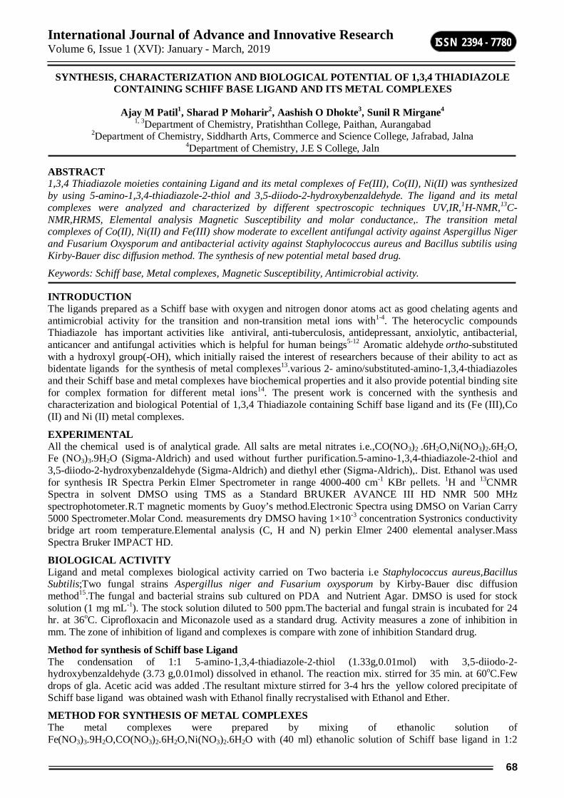
International Journal of Advance and Innovative Research Volume 6, Issue 1 (XVI): January - March, 2019
68
ISSN 2394 - 7780
SYNTHESIS, CHARACTERIZATION AND BIOLOGICAL POTENTIAL OF 1,3,4 THIADIAZOLE CONTAINING SCHIFF BASE LIGAND AND ITS METAL COMPLEXES
Ajay M Patil1, Sharad P Moharir2, Aashish O Dhokte3, Sunil R Mirgane4
1, 3Department of Chemistry, Pratishthan College, Paithan, Aurangabad 2Department of Chemistry, Siddharth Arts, Commerce and Science College, Jafrabad, Jalna
4Department of Chemistry, J.E S College, Jaln
ABSTRACT 1,3,4 Thiadiazole moieties containing Ligand and its metal complexes of Fe(III), Co(II), Ni(II) was synthesized by using 5-amino-1,3,4-thiadiazole-2-thiol and 3,5-diiodo-2-hydroxybenzaldehyde. The ligand and its metal complexes were analyzed and characterized by different spectroscopic techniques UV,IR,1H-NMR,13C-NMR,HRMS, Elemental analysis Magnetic Susceptibility and molar conductance,. The transition metal complexes of Co(II), Ni(II) and Fe(III) show moderate to excellent antifungal activity against Aspergillus Niger and Fusarium Oxysporum and antibacterial activity against Staphylococcus aureus and Bacillus subtilis using Kirby-Bauer disc diffusion method. The synthesis of new potential metal based drug.
Keywords: Schiff base, Metal complexes, Magnetic Susceptibility, Antimicrobial activity.
INTRODUCTION The ligands prepared as a Schiff base with oxygen and nitrogen donor atoms act as good chelating agents and antimicrobial activity for the transition and non-transition metal ions with1-4. The heterocyclic compounds Thiadiazole has important activities like antiviral, anti-tuberculosis, antidepressant, anxiolytic, antibacterial, anticancer and antifungal activities which is helpful for human beings5-12 Aromatic aldehyde ortho-substituted with a hydroxyl group(-OH), which initially raised the interest of researchers because of their ability to act as bidentate ligands for the synthesis of metal complexes13.various 2- amino/substituted-amino-1,3,4-thiadiazoles and their Schiff base and metal complexes have biochemical properties and it also provide potential binding site for complex formation for different metal ions14. The present work is concerned with the synthesis and characterization and biological Potential of 1,3,4 Thiadiazole containing Schiff base ligand and its (Fe (III),Co (II) and Ni (II) metal complexes.
EXPERIMENTAL All the chemical used is of analytical grade. All salts are metal nitrates i.e.,CO(NO3)2 .6H2O,Ni(NO3)2.6H2O, Fe (NO3)3.9H2O (Sigma-Aldrich) and used without further purification.5-amino-1,3,4-thiadiazole-2-thiol and 3,5-diiodo-2-hydroxybenzaldehyde (Sigma-Aldrich) and diethyl ether (Sigma-Aldrich),. Dist. Ethanol was used for synthesis IR Spectra Perkin Elmer Spectrometer in range 4000-400 cm-1 KBr pellets. 1H and 13CNMR Spectra in solvent DMSO using TMS as a Standard BRUKER AVANCE III HD NMR 500 MHz spectrophotometer.R.T magnetic moments by Guoy’s method.Electronic Spectra using DMSO on Varian Carry 5000 Spectrometer.Molar Cond. measurements dry DMSO having 1×10⁻³ concentration Systronics conductivity bridge art room temperature.Elemental analysis (C, H and N) perkin Elmer 2400 elemental analyser.Mass Spectra Bruker IMPACT HD.
BIOLOGICAL ACTIVITY Ligand and metal complexes biological activity carried on Two bacteria i.e Staphylococcus aureus,Bacillus Subtilis;Two fungal strains Aspergillus niger and Fusarium oxysporum by Kirby-Bauer disc diffusion method15.The fungal and bacterial strains sub cultured on PDA and Nutrient Agar. DMSO is used for stock solution (1 mg mL-1). The stock solution diluted to 500 ppm.The bacterial and fungal strain is incubated for 24 hr. at 36oC. Ciprofloxacin and Miconazole used as a standard drug. Activity measures a zone of inhibition in mm. The zone of inhibition of ligand and complexes is compare with zone of inhibition Standard drug.
Method for synthesis of Schiff base Ligand The condensation of 1:1 5-amino-1,3,4-thiadiazole-2-thiol (1.33g,0.01mol) with 3,5-diiodo-2-hydroxybenzaldehyde (3.73 g,0.01mol) dissolved in ethanol. The reaction mix. stirred for 35 min. at 60oC.Few drops of gla. Acetic acid was added .The resultant mixture stirred for 3-4 hrs the yellow colored precipitate of Schiff base ligand was obtained wash with Ethanol finally recrystalised with Ethanol and Ether.
METHOD FOR SYNTHESIS OF METAL COMPLEXES The metal complexes were prepared by mixing of ethanolic solution of Fe(NO3)3.9H2O,CO(NO3)2.6H2O,Ni(NO3)2.6H2O with (40 ml) ethanolic solution of Schiff base ligand in 1:2
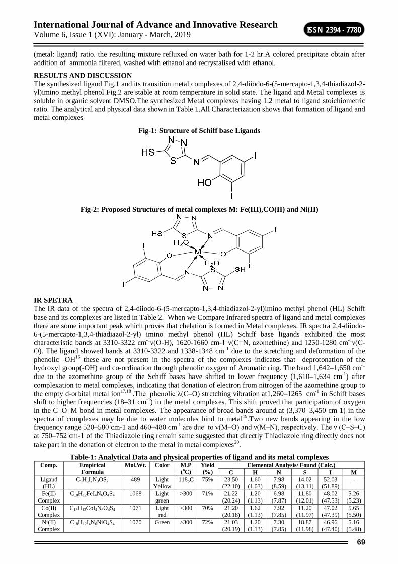
International Journal of Advance and Innovative Research Volume 6, Issue 1 (XVI): January - March, 2019
69
ISSN 2394 - 7780
(metal: ligand) ratio. the resulting mixture refluxed on water bath for 1-2 hr.A colored precipitate obtain after addition of ammonia filtered, washed with ethanol and recrystalised with ethanol.
RESULTS AND DISCUSSION The synthesized ligand Fig.1 and its transition metal complexes of 2,4-diiodo-6-(5-mercapto-1,3,4-thiadiazol-2-yl)imino methyl phenol Fig.2 are stable at room temperature in solid state. The ligand and Metal complexes is soluble in organic solvent DMSO.The synthesized Metal complexes having 1:2 metal to ligand stoichiometric ratio. The analytical and physical data shown in Table 1.All Characterization shows that formation of ligand and metal complexes
Fig-1: Structure of Schiff base Ligands
Fig-2: Proposed Structures of metal complexes M: Fe(III),CO(II) and Ni(II)
IR SPETRA The IR data of the spectra of 2,4-diiodo-6-(5-mercapto-1,3,4-thiadiazol-2-yl)imino methyl phenol (HL) Schiff base and its complexes are listed in Table 2. When we Compare Infrared spectra of ligand and metal complexes there are some important peak which proves that chelation is formed in Metal complexes. IR spectra 2,4-diiodo-6-(5-mercapto-1,3,4-thiadiazol-2-yl) imino methyl phenol (HL) Schiff base ligands exhibited the most characteristic bands at 3310-3322 cm-1ν(O-H), 1620-1660 cm-1 ν(C=N, azomethine) and 1230-1280 cm-1ν(C-O). The ligand showed bands at 3310-3322 and 1338-1348 cm–1 due to the stretching and deformation of the phenolic -OH16 these are not present in the spectra of the complexes indicates that deprotonation of the hydroxyl group(-OH) and co-ordination through phenolic oxygen of Aromatic ring. The band 1,642–1,650 cm-1 due to the azomethine group of the Schiff bases have shifted to lower frequency (1,610–1,634 cm-1) after complexation to metal complexes, indicating that donation of electron from nitrogen of the azomethine group to the empty d-orbital metal ion17,18 .The phenolic λ(C–O) stretching vibration at1,260–1265 cm-1 in Schiff bases shift to higher frequencies (18–31 cm-1) in the metal complexes. This shift proved that participation of oxygen in the C–O–M bond in metal complexes. The appearance of broad bands around at (3,370–3,450 cm-1) in the spectra of complexes may be due to water molecules bind to metal19.Two new bands appearing in the low frequency range 520–580 cm-1 and 460–480 cm-1 are due to ν(M–O) and ν(M–N), respectively. The ν (C–S–C) at 750–752 cm-1 of the Thiadiazole ring remain same suggested that directly Thiadiazole ring directly does not take part in the donation of electron to the metal in metal complexes20.
Table-1: Analytical Data and physical properties of ligand and its metal complexes Comp. Empirical
Formula Mol.Wt. Color M.P
(oC) Yield (%)
Elemental Analysis/ Found (Calc.) C H N S I M
Ligand (HL)
C9H5I2N3OS2 489 Light Yellow
118oC 75% 23.50 (22.10)
1.60 (1.03)
7.98 (8.59)
14.02 (13.11)
52.03 (51.89)
-
Fe(II) Complex
C18H12FeI4N6O4S4 1068 Light green
>300 71% 21.22 (20.24)
1.20 (1.13)
6.98 (7.87)
11.80 (12.01)
48.02 (47.53)
5.26 (5.23)
Co(II) Complex
C18H12CoI4N6O4S4 1071 Light red
>300 70% 21.20 (20.18)
1.62 (1.13)
7.92 (7.85)
11.20 (11.97)
47.02 (47.39)
5.65 (5.50)
Ni(II) Complex
C18H12I4N6NiO4S4 1070 Green >300 72% 21.03 (20.19)
1.20 (1.13)
7.30 (7.85)
18.87 (11.98)
46.96 (47.40)
5.16 (5.48)

International Journal of Advance and Innovative Research Volume 6, Issue 1 (XVI): January - March, 2019
70
ISSN 2394 - 7780
1H NMR and 13C NMR Spectra The 1H-NMR spectra of ligand were recorded in DMSO-solution using TMS as a standard (Table 3). The spectra of ligands shows singlet at δ 7.19-8.00 ppm due to aromatic proton while azomethine proton resonate at singlet δ 8.8 ppm the phenolic OH has signal singlet at δ 11.51 ppm and Thiadiazole containing –SH group shows singlet at δ 13.17 ppm 21,22. 13C NMR of Ligand, peak appeared at δ159-161ppm imine group(-C=N-),peak 181 ppm Due to C-SH bonding in Thiadiazole.123.96-141.74 ppm due to aromatic carbon,150-170 ppm peak due to Ar-OH group23.
Table-2: Infrared Spectra of the Schiff base and Complexes in Cm-1 Compound νOH/H2O νC-O νC=N νM-N νM-O νC-S-C ν-C=N-N=C νN-N
Ligand 3320 1267 1651 ___ ___ 751 1479 1028 Fe(III) Complex 3495 1300 1637 468 574 753 1475 1022 Co(II) Complex 3475 1271 1647 478 520 752 1477 1028 Ni(II) Complex 3402 1295 1649 470 576 756 1481 1023
Table-3: 1H NMR Signals (δ, ppm) and their assignments
Compound 1H NMR Signals (δ,ppm) and their assignments Ligand(HL) 11.51 (s,1H,Ar-OH), 8.8 (s,1H,CH=N),7.19-8.00 (s,2H,Ar-CH), 13.17 (s,1H,SH)
MASS SPECTRA Mass Spectra of ligands shows peak at m/z 489 which is M+H peak at 100% intensity this peak support to the elucidation structure of the ligand.
MAGNETIC SUSCEPTIBILITY AND MOLAR CONDUCTANCE The magnetic susceptibility and molar conductance observed at room temperature. The entire synthesized metal complexes are paramagnetic in nature having free electron. Molar conductance values of metal complexes were observed at 1×10⁻³M DMSO Solution. The studies show negligible molar conductance value in range 8-12 ohm-
1cm2mol-1. It is observed that all metal complexes are non-electrolytic in nature 24,25.
ELECTRONIC ABSORPTION SPECTRA The electronic spectral data of the ligands and metal complexes in DMSO are given in Table 4. The nature and geometry are analyzed from the electronic spectra data. The band appearing at 218-315 is due to transition of benzene ring of the ligand. The other band due to free ligands 320-375 nm due to transition for azomethine moieties and phenolic -OH. These band shifts longer wavelength due to Synthesis of Schiff base metal complexes14,26 The spectra and magnetic moment value of the Fe(III) complexes display band Due to charge transfer transition from ligands to metal27.The magnetic moment value for CO (II) complexes is 5.08 B.M is near to octahedral complex spectra shows two band at 480-520 nm and 702-712 nm shows that octahedral geometry of CO (II) complex28,29 .Electronic spectra of Ni(II) complexes shows three band 940 nm,552 nm and 405 nm suggest octahedral geometry20,30-31.
Table-4: Electronic spectral Magnetic and Molar conductance Data Compounds Wavelength in nm Magnetic moment
μeff (BM) Molar conductance (ohm-1 cm2 mol-1)
Ligand C9H5I2N3OS2 260,370 -- 6.68 C18H12FeI4N6O4S4 396-401,460-456,591-596 5.81 11.9 C18H12CoI4N6O4S4 310-350,480-520,70-712 4.91 9.8 C18H12I4N6NiO4S4 376-408,552,940 3.12 11.10
ANTIMICROBIAL ACTIVITY The antimicrobial activity on selected two gram positive bacteria S. aureus and B.Subtlis two fungi A. niger and F.Oxysporum was carried out. All of the tested synthesized ligand and metal complexes showed good biological activity against microorganism. The bactericidal and fungicidal investigation data of the compounds are given in Tables 5. The investigation shows that Ni (II) and CO(II) shows more good bactericidal and fungicidal activity than Fe(II)Complexes and Ligand hence activity of metal complexes increases due to chelation in metal complexes increase in delocalization of π electron on chelating ring and enhance the penetration of complexes in lipid membrane of microorganism and blocks the binding site enzymes of harmful microorganism there are some other factors also like lipophilicity,hydrophilicity solubility,Conductivity and bonding between M-L that increases the activity of complexes31-35.
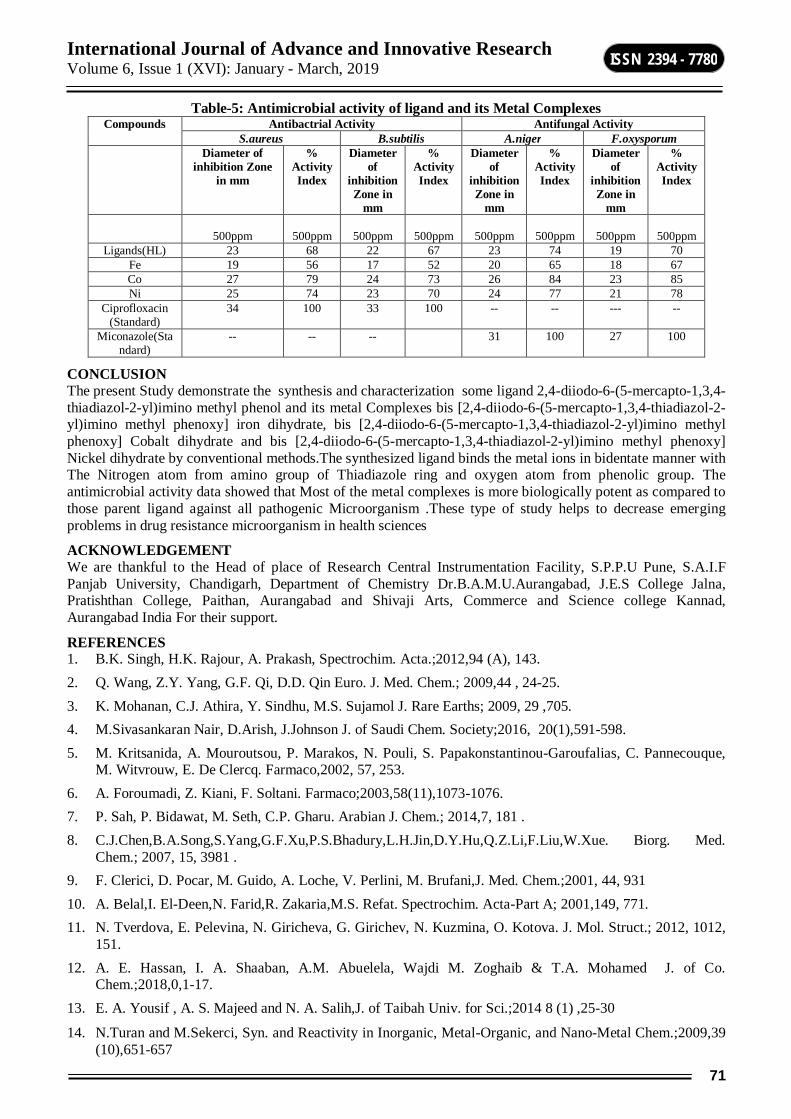
International Journal of Advance and Innovative Research Volume 6, Issue 1 (XVI): January - March, 2019
71
ISSN 2394 - 7780
Table-5: Antimicrobial activity of ligand and its Metal Complexes Compounds Antibactrial Activity Antifungal Activity
S.aureus B.subtilis A.niger F.oxysporum Diameter of
inhibition Zone in mm
% Activity Index
Diameter of
inhibition Zone in
mm
% Activity Index
Diameter of
inhibition Zone in
mm
% Activity Index
Diameter of
inhibition Zone in
mm
% Activity Index
500ppm
500ppm
500ppm
500ppm
500ppm
500ppm
500ppm
500ppm
Ligands(HL) 23 68 22 67 23 74 19 70 Fe 19 56 17 52 20 65 18 67 Co 27 79 24 73 26 84 23 85 Ni 25 74 23 70 24 77 21 78
Ciprofloxacin (Standard)
34 100 33 100 -- -- --- --
Miconazole(Standard)
-- -- -- 31 100 27 100
CONCLUSION The present Study demonstrate the synthesis and characterization some ligand 2,4-diiodo-6-(5-mercapto-1,3,4-thiadiazol-2-yl)imino methyl phenol and its metal Complexes bis [2,4-diiodo-6-(5-mercapto-1,3,4-thiadiazol-2-yl)imino methyl phenoxy] iron dihydrate, bis [2,4-diiodo-6-(5-mercapto-1,3,4-thiadiazol-2-yl)imino methyl phenoxy] Cobalt dihydrate and bis [2,4-diiodo-6-(5-mercapto-1,3,4-thiadiazol-2-yl)imino methyl phenoxy] Nickel dihydrate by conventional methods.The synthesized ligand binds the metal ions in bidentate manner with The Nitrogen atom from amino group of Thiadiazole ring and oxygen atom from phenolic group. The antimicrobial activity data showed that Most of the metal complexes is more biologically potent as compared to those parent ligand against all pathogenic Microorganism .These type of study helps to decrease emerging problems in drug resistance microorganism in health sciences
ACKNOWLEDGEMENT We are thankful to the Head of place of Research Central Instrumentation Facility, S.P.P.U Pune, S.A.I.F Panjab University, Chandigarh, Department of Chemistry Dr.B.A.M.U.Aurangabad, J.E.S College Jalna, Pratishthan College, Paithan, Aurangabad and Shivaji Arts, Commerce and Science college Kannad, Aurangabad India For their support.
REFERENCES 1. B.K. Singh, H.K. Rajour, A. Prakash, Spectrochim. Acta.;2012,94 (A), 143. 2. Q. Wang, Z.Y. Yang, G.F. Qi, D.D. Qin Euro. J. Med. Chem.; 2009,44 , 24-25. 3. K. Mohanan, C.J. Athira, Y. Sindhu, M.S. Sujamol J. Rare Earths; 2009, 29 ,705. 4. M.Sivasankaran Nair, D.Arish, J.Johnson J. of Saudi Chem. Society;2016, 20(1),591-598. 5. M. Kritsanida, A. Mouroutsou, P. Marakos, N. Pouli, S. Papakonstantinou-Garoufalias, C. Pannecouque,
M. Witvrouw, E. De Clercq. Farmaco,2002, 57, 253. 6. A. Foroumadi, Z. Kiani, F. Soltani. Farmaco;2003,58(11),1073-1076. 7. P. Sah, P. Bidawat, M. Seth, C.P. Gharu. Arabian J. Chem.; 2014,7, 181 . 8. C.J.Chen,B.A.Song,S.Yang,G.F.Xu,P.S.Bhadury,L.H.Jin,D.Y.Hu,Q.Z.Li,F.Liu,W.Xue. Biorg. Med.
Chem.; 2007, 15, 3981 . 9. F. Clerici, D. Pocar, M. Guido, A. Loche, V. Perlini, M. Brufani,J. Med. Chem.;2001, 44, 931 10. A. Belal,I. El-Deen,N. Farid,R. Zakaria,M.S. Refat. Spectrochim. Acta-Part A; 2001,149, 771. 11. N. Tverdova, E. Pelevina, N. Giricheva, G. Girichev, N. Kuzmina, O. Kotova. J. Mol. Struct.; 2012, 1012,
151. 12. A. E. Hassan, I. A. Shaaban, A.M. Abuelela, Wajdi M. Zoghaib & T.A. Mohamed J. of Co.
Chem.;2018,0,1-17. 13. E. A. Yousif , A. S. Majeed and N. A. Salih,J. of Taibah Univ. for Sci.;2014 8 (1) ,25-30
14. N.Turan and M.Sekerci, Syn. and Reactivity in Inorganic, Metal-Organic, and Nano-Metal Chem.;2009,39 (10),651-657
International Journal of Advance and Innovative Research Volume 6, Issue 1 (XVI): January - March, 2019
72
ISSN 2394 - 7780
15. A.W.Bauer , D.M Perry, Kirby, AMA Arch Intern Med.;1959, 104(2), 208–216.
16. K.Nakamoto, Infrared and Raman Spectra of Inorganic and Coordination Compounds5th ed. John Wiley and Sons, Part A & B, New York.1998.
17. H.Temel,S.Ilhan,M.Aslanoglu, A.Kilic,E.Tas,J Chin Chem Soc.;2006, 53,1027-1031.
18. D.Shukla , L.K.Gupta, S.Chandra, Spectrochim Acta.;2008,71(A),746–750
19. G.G.Mohamed,M.M.Omar,A.M.Hindy,Turk J. Chem.;2008, 30,361-382.
20. M.A.Neelakantan,S.S.Marriappan,J.Dharmaraja,T.Jeyakumar,K.Muthukumaran,. Spectrochim Acta.; 2008,71(A), 628-635.
21. R.C.Maurya,P.Patel,S.Rajput,Synthesis and Reactivity in Inorganic and Metal-Organic Chem.;2003 33(5), 801-816.
22. R.B.Rastogi,M.Yadav,K.Singh,Synth. React. Inorg. Met.-Org. Chem.;2001, 31 (6), 1011-1022.
23. M.M.Abd-Elzaher, S.A.Moustafa, A.A.Labib, H.A.Mousa, M.M.Ali, and A.E.Mahmoud, Applied Organometallic Chemistry;2012, 26(5), 230-236.
24. S.N.Sampal, S.V.Thakur, A.S.Rajbhoj and S.T.Gaikwad ,Asian J. Chem.;2017,30(2), 398 - 40.
25. W.J. Geary Coord. Chem. Rev.; 1971 7, 81.
26. S.Y.Ucan and M.Ucan, Synth. React. Inorg. Met. Org. Nano-Metal Chem.;2005, 35, 417-421.
27. A. Çapan, S. Uruş, M. Sönmez,Journal of Saudi Chemical Society;2017, 22 (6), 757-766
28. S.M.Abu-El-Wafa,N.A.El-Wakiel,R.M.Ssa,R.A.Mansour,J.Coordination Chem.; 2005, 58, 683-694.
29. H.Temel,S.Ilhan,M.ekerci,R.Ziyadanog,Spectros. Lett.; 2002, 35 (2), 219-228.
30. Lever ABP Inorganic electronic spectroscopy, 2nd edn. Elsevier, New York. 1984.
31. C,J.Ballhausen,Introduction to Ligand Field Theory, McGraw-Hill, New York,;1962,256.
32. Z.H.Chohan,A.Munawar,C.T.Supuran.Metal Based Drugs;2001,8,137-143
33. W.G.Hanna and M.M.Moawad,Transit. Metal Chem.; 2005, 26(6),644-651.
34. V.P.Singh, A.Katiyar, BioMetals;2008,21(4), 491-501.
35. F.Azam,S.Singh,S.L.Khokhra, O.Prakash,J. Zhejiang Univ. Sci. B.2007,8(6),446-452.
International Journal of Advance and Innovative Research Volume 6, Issue 1 (XVI): January - March, 2019
73
ISSN 2394 - 7780
SYNTHESIS, CHARACTERIZATION AND ANTIMICROBIAL ANALYSIS OF VARIOUS SUBSTITUTED 3-(3-(5-bromothiophen-2-yl)-1-(4-fluorophenyl)-1H-pyrazol-4-yl)-1-(2-
hydroxyphenyl)prop-2-en-1-one
Amol J. Shirsat1, Balaji D. Rupnar2, Sunil S. Bhagat3, Ajit K. Dhas4, Gopal K. Kakade5 1,2,3Department of Chemistry, R. B. Attal Arts, Science & Commerce College, Georai, Beed
4Department of Chemistry, Deogiri College, Aurangabad 5Department of Chemistry, Arts, Commerce & Science College, Kille- Dharur, Beed
ABSTRACT We have developed a protocol for the synthesis of some novel chalcones, also used a green, efficient and rapid procedure for the synthesis, this synthesis done by the condensation of pyrazole aldehyde and O-hydroxyketone, in the presence of KOH in EtOH. All the synthesized products were characterized by NMR, IR and Mass spectral data also. These synthesized compounds have been screened for their antimicrobial activity against Gram –ve and gram +ve microorganisms. A few of them shows moderate antimicrobial activity.
Keywords: Aldehyde, O-hydroxyketone, Pyrazole, Chalcone, Condensation, KOH, EtOH.
INTRODUCTION Chalcones are natural products which are found in a variety of plant species with the general formula Ar-CH=CH-CO-Ar in which the two aromatic rings are joined by α, β-unsaturated carbonyl system. These are rich in edible plants and are considered as precursors of flavonoids and isoflavonoids. Chalcones have been generally prepared by Claisen-Schmidt (Aldol) condensation reaction of aromatic aldehydes with aryl ketones in presence of suitable agents. They show diverse chemical reactions and act as precursor for the synthesis of various heterocyclic compounds1 like benzodiazepine, thiadiazines, isoxazoles, quinolinones, benzofuranones, tetrahydro-2-chromens flavones2 etc. Chalcones and their derivatives have attracted greater attention towards it due to several pharmacological applications. They shows a broad spectrum of pharmacological activities, like antibacterial3,4, antimicrobial5, anti-inflammatory6,7, antifungal8,9, antimalarial10-13, anticancer 14,15, antioxidant16, antiprotozoal (antileishmanial and antitrypanosomal)17, antifilarial18, larvicidal 19, anticonvulsant20 activities have been reported yet. Also they have shown inhibition of the enzymes, especially mammalian alpha-amylase21, monoamine oxidase (MAO)22 and cyclo-oxygenase (COX) 23. Having a lot of pharmacological activity and their synthetic utility, chemists are attracted towards chalcones to develop a lot of synthetic methodologies for their synthesis around the world.
MATERIALS AND METHODS For the synthesis of the compounds, all used chemicals were obtained specially from Sigma Aldrich and SD Fine chemicals. By using simple open capillaries Melting points were recorded and are uncorrected. By using 400 MHz NMR Spectrophotometer, 1H NMR spectra were recorded in this analysis DMSO-d6 used as solvent and TMS as an internal standard. By using FT-IR Spectrophotometer Model RZX (Perkin Elmer) the infra-red spectra were recorded. On Macromass mass spectrophotometer (Waters) mass spectra were recorded using electro-spray method (ES). On TLC purity of the all synthesized compounds were checked. TLC silica gel coated plates were obtained from Merck in which stationary phase and mobile phase were mixture of hexane / ethyl acetate (80:20).
GENERAL PROCEDURE Mixture of 1 (0.01 mole) and 2 (0.01 mole) was dissolved in 50 ml of ethanol, and contents were placed in ice bath at 0οC. To maintaining temperature below 5οC, 2g KOH pellets were added in this reaction mixture. This reacting mixture was stirred for 48 hr at room temperature. After 48 hours this reaction mixture was poured in ice cold water and acidified with 2M HCl then yellow solid was obtained and filtered for separation, also washed with cold water. Product was recrystallized in ethanol. By using this typical experimental procedure, other analogs were prepared of this series. The physical data of the compounds 3(a-h) were recorded in Table 1. Their structures have been confirmed by analyzing method like 1HNMR, Mass and IR spectra.
IR (3c) (cm-1):832(C-Cl), 1021(C-F), 1247(C-O), 1530(C=C), 1563(C=N), 1637(C=O), 3134(O-H). 1H NMR (3c) (DMSO-d6)δ ppm: 6.8054-6.8954(s, 2H, Ar-H), 6.9874-7.0034(s, 1H, Ar-H,), 7.0657-7.0857(m, 2H, Ar-H), 7.0974-7.2145(m, 1H, CH=C-), 7.4125-7.5921(m, 2H, Ar-H) 7.6851-7.7521(m, 2H, Ar-H), 7.8745-7.9241(d, 1H, Ar-H, J= 19.8 Hz), 8.6984(s, 1H, pyrazole-H), 12.3974(s, 1H, Ar–OH).
ES-MS (3c) (m/z):503(M+1), 505(M+3).

International Journal of Advance and Innovative Research Volume 6, Issue 1 (XVI): January - March, 2019
74
ISSN 2394 - 7780
IR (3f) (cm-1):832(C-Cl), 1011(C-F), 1223(C-O), 1511(C=C), 1561(C=N), 1640 (C=O), 2970(O-H). 1H NMR (3f) (DMSO-d6)δ ppm: 2.4121-2.4524(s, 3H, -CH3), 6.8324(s, 1H, Ar-H), 7.3956-7.4001 (d, 1H, Ar-H. J=1.8 Hz), 7.4256-7.5069(m, 1H, Ar-H), 7.5498-7.5949(m, 2H, Ar-H), 7.7089-7.7224(d, 1H, CH=C-, J=5.4 Hz), 7.9678-7.9956(m, 2H, Ar-Hz), 7.9172-7.9367(m, 1H,Ar-H), 8.0063(s, 1H,Ar-H), 8.9964(s, 1H, pyrazole-H), 12.5987(s, 1H, Ar–OH).
ES-MS (3f) (m/z):519(M+1), 521(M+3).
R1R2
R3
OH
CH3
O
EtOH/KOH
RT,48 hr.
1(a-h) 2 3(a-h)
R4
OH
O
NN
S
NN
S R4
R3
R2
R1
OHC+
Br
FF
Br
Scheme 1: Synthesis of series of various (E)-3-(3-(5-bromothiophen-2-yl)-1-(4-fluorophenyl)-1H-
pyrazol-4-yl)-1-(2-hydroxyphenyl)prop-2-en-1-one
Table-1: Physical data of compounds 3(a-h) Comp. R1 R2 R3 M.P. (oC) Yield (%)
3a H H H 148-150 78 3b H H CH3 168-170 77 3c H H Cl 158-160 79 3d Cl H Cl 208-210 70 3e H H F 192-194 66 3f H CH3 Cl 152-154 82 3g H H Br 200-202 69 3h CH3 H CH3 154-156 76
RESULT AND DISCUSSION Eight chalcone derivatives were synthesized successfully with good yields. All newly synthesized compounds were analyzed from melting point range, IR, 1H NMR, Mass spectral analysis. All newly synthesized compounds were screened for antimicrobial activity using disc diffusion method.
Antimicrobial activity: Compounds 3(a-h) were screened for their antimicrobial activity against Gram positive (Salmonella typh, Enterobacter aerogenes, Escherichia coli, Pseudomonas aerogenosa, Salmonella abony, Shigella boydii ) and Gram negative pathogens(Bacillus subtilis, Megaterium Bacillus, Staphylococcus aureus, Bacillus cereus) by paper disc diffusion method using tetracyclin as a reference standard drug. Using Nystatin as standard drug, antifungal activity was screened against Candida albicans, Saccharomyces cerevisiae, Aspergillus niger at 100 μg/ml concentration. Muller Hinton agar was the culture media. The zone of inhibition was measured in mm, after the 24 hr of incubation at 37οC. Microbial data for 3(a-h) are summarized in Table 2.
Table-2: Antimicrobial Analysis Data of 3(a-h)
Bacterial pathogens Fungal pathogen Gram negative pathogen Gram positive pathogen
Com
poun
ds
Salm
onel
la
typh
i
Ent
erob
acte
r ae
roge
nes
Esc
heri
chia
co
li Ps
eudo
mon
as
aero
geno
sa
Salm
onel
la
abon
y
Shig
ella
boy
dii
Bac
illus
su
btili
s
Bac
illus
M
egat
eriu
m
Stap
hylo
cocc
us a
ureu
s
Bac
illus
ce
reus
Can
dida
al
bica
ns
Sacc
haro
myc
es c
erev
isia
e
Asp
ergi
llus
nige
r
3a 07 - 10 09 13 - 07 10 07 08 - 13 08 3b 08 06 08 - 11 05 06 - 08 11 09 - 10 3c 06 09 - 08 18 - 10 - 07 10 - - 10

International Journal of Advance and Innovative Research Volume 6, Issue 1 (XVI): January - March, 2019
75
ISSN 2394 - 7780
*Standard for bacterial pathogens-tetracyclin, for fungal pathogens-nystatin
CONCLUSION All eight compounds were synthesized successfully; these newly synthesized compounds were screened for their antimicrobial activity against Gram positive as well as Gram negative bacterial strains and against fungal pathogens. The synthesized compounds show moderate activity as compared to standard drugs. The obtained data through the present work shows a good agreement between the experimental and computed spectral data.
ACKNOWLEDGEMENT The authors are thankful to The Principal Dr. R.K. Nimbalkar, for providing laboratory facilities. The authors are also thankful to Director, SAIF/CIL, Panjab University, Chandigarh for providing spectral data and also thankful to Dr. Prashant Dixit, Department of Microbiology, Dr. Babasaheb Ambedkar Marathwada University Sub-Center, Osmanabad for antimicrobial as well as antifungal analysis.
REFERENCES 1. J. Li, Name Reactions in Heterocyclic Chemistry, Wiley-Interscience Publication, 2005. pp. 262–265.
2. R.A. Dixon, N.L. Paiva, Stress-induced phenylpropanoid metabolism, Plant cell , 1995, 7, 1085–1097.
3. Hamdi N, Fischmeister C, Puerta MC, Valerga P, A rapid access to new coumarinyl chalcone and substituted chromeno[4,3-c]pyrazol-4(1H)-ones and their antibacterial and DPPH radical scavenging activities. Medicinal Chemistry Research, 2010,19:1-16.
4. Bhatia NM, Mahadik KR, Bhatia MS, QSAR analysis of 1,3-diaryl-2-propen-1-ones and their indole analogs for designing potent antibacterial agents. Chemical Papers, 2009, 63:456-463.
5. Yayli N, Ucuncu O, Yasar A, Kucuk M, Akyuz E, Karaoglu SA, Synthesis and biological activities of N-alkyl derivatives of o-, m-, and p-nitro (E)-4-azachalcones and stereoselective photochemistry in solution with theoretical calculations. Turkish Journal of Chemistry, 2006, 30:505-514.
6. Yadav HL, Gupta P, Pawar PS, Singour PK, Patil UK, Synthesis and biological evaluation of antiinflammatory activity of 1,3-diphenyl propenone derivatives. Medicinal Chemistry Research, 2010, 19:1-8.
7. Zhang XW, Zhao DH, Quan YC, Sun LP, Yin XM, Guan LP, Synthesis and evaluation of anti-inflammatory activity of substituted chalcone derivatives. Medicinal Chemistry Research, 2010, 19:403-412.
8. Bag S, Ramar S, Degani MS, Synthesis and biological evaluation of α,β-unsaturated ketone as potential antifungal agents. Medicinal Chemistry Research, 2009, 18:309-316.
9. Lahtchev KL, Batovska DI, Parushev SP, Ubiyvovk VM, Sibirny AA, Antifungal activity of chalcones: A mechanistic study using various yeast strains. European Journal of Medicinal Chemistry, 2008, 43:2220-2228.
10. Motta LF, Gaudio AC, Takahata Y, Quantitative structure-activity relationships of a series of chalcone derivatives (1,3-diphenyl-2-propen-1-one) as anti-Plasmodium falciparum agents (antimalaria agents). Internet Electronic Journal of Molecular Design, 2006, 5:555-569.
11. Awasthi SK, Mishra N, Kumar B, Sharma M, Bhattacharya A, Mishra LC, Bhasin VK, Potent antimalarial activity of newly synthesized substituted chalcone analogs in vitro. Medicinal Chemistry Research, 2009, 18:407-420.
12. Cheng MS, Shili R, Kenyon G, A solid phase synthesis of chalcones by Claisen-Schmidt condensations. Chinese Chemical Letters, 2000, 11:851-854.
13. Lim SS, Kim HS, Lee DU, In vitro antimalarial activity of flavonoids and chalcones. Bulletin of the Korean Chemical Society, 2007, 28:2495-2497.
3d 09 07 15 09 30 12 09 11 10 12 16 19 25 3e - 13 12 10 16 - - 07 - 07 15 - 11 3f 05 - 09 - - 05 - - 06 - - - 09 3g - 19 - 11 - 16 07 - - 06 - 27 10 3h 06 10 08 07 13 - 09 10 11 11 13 08 12
DMSO - - - - - - - - - - - - - STND. 22 20 20 33 21 26 25 20 30 25 24 20 25

International Journal of Advance and Innovative Research Volume 6, Issue 1 (XVI): January - March, 2019
76
ISSN 2394 - 7780
14. Achanta G, Modzelewska A, Feng L, Khan SR, Huang P, A boronic-chalcone derivative exhibits potent anticancer activity through inhibition of the proteasome. Molecular Pharmacology, 2006, 70:426-433.
15. Romagnoli R, Baraldi PG, Carrion MD, Cara CL, Cruz-Lopez O, Preti D, Design, synthesis and biological evaluation of thiophene analogues of chalcones. Bioorganic and Medicinal Chemistry, 2008, 16:5367-5376.
16. Vasil’ev RF, Kancheva VD, Fedorova GF, Batovska DI, Trofimov AV, Antioxidant activity of chalcones: The chemiluminescence determination of the reactivity and the quantum chemical calculation of the energies and structures of reagents and intermediates. Kinetics and Catalysis, 2010, 51:507-515.
17. Lunardi F, Guzela M, Rodrigues AT, Corre R, Eger-Mangrich I, Steindel M, Grisard EC, Assreuy J, Calixto JB, Santos ARS, Trypanocidal and leishmanicidal properties of substitution-containing chalcones. Antimicrobial Agents and Chemotherapy, 2003, 47:1449-1451.
18. Awasthi SK, Mishra N, Dixit SK, Singh A, Yadav M, Yadav SS, Rathaur S, Antifilarial activity of 1,3diarylpropen-1-one: Effect on glutathione-S-transferase, a phase-II detoxification enzyme. American Journal of Tropical Medicine and Hygiene, 2009, 80:764-768.
19. Begum NA, Roy N, Laskar RA, Roy K, Mosquito larvicidal studies of some chalcone analogues and their derived products: structure-activity relationship analysis. Medicinal Chemistry Research, 2010, 19:1-14.
20. Kaushik S, Kumar N, Drabu S, Synthesis and anticonvulsant activities of phenoxychalcones. The Pharma Research, 2010, 3:257-262.
21. Najafian M, Ebrahim-Habibi A, Hezareh N, Yaghmaei P, Parivar K, Larijani B, Trans-chalcone: a novel small molecule inhibitor of mammalian alpha-amylase. Molecular Biology Reports, 2010, 10:271-274.
22. Chimenti F, Fioravanti R, Bolasco A, Chimenti P, Secci D, Rossi F, Yanez M, Francisco OF, Ortuso F, Alcaro S, Chalcones: A valid scaffold for monoamine oxidases inhibitors. Journal of Medicinal Chemistry, 2009,10:1-8.
23. Zarghi A, Zebardast T, Hakimion F, Shirazi FH, Rao PNP, Knaus EE, Synthesis and biological evaluation of 1,3-diphenylprop-2-en-1-ones possessing a methanesulfonamido or an azido pharmacophore as cyclooxygenase1/-2 inhibitors. Bioorganic and Medicinal Chemistry, 2006, 14:7044-7050.
International Journal of Advance and Innovative Research Volume 6, Issue 1 (XVI): January - March, 2019
77
ISSN 2394 - 7780
APPLICATION OF MOBILE TECHNOLOGY IN LIBRARIES
Pagore R. B. Librarian, R. B. Attal College, Georai Dist. Beed
ABSTRACT Information communication technology changes the role of library to traditional library to digital libraries; it’s also impact on the library services. Digitization provides the faster access to information and also changes their services by accepting the technological change. In the modern era mobile phones become an essential part of day to day life and changing the way of communication in the world. Mobile technology helps to libraries towards strength, user relation, and orientation and library services to its user. Libraries may provide remote access to users who were considered unlikely to connect because of absence of a medium. This paper highlight the use of mobile technology especially Smartphone based library services, its purpose, application, prerequisites; skills required for its implementation, advantages, disadvantages, challenges faced etc.
Keywords: Digital Libraries, Mobile Technology, Mobile Application, Smartphone based library services.
INTRODUCTION The world wide mobile phones subscription are at billions-equivalent to half global population. With Smartphone becoming the very important in the lives of the population’s majority, the industry has undergone an evolution in 2017, with competition becoming even stiffer. Smartphone designs have been revolutionized in the past few years, thereby shifting the popularity rating of different companies. This statistics are substantial evidence that people everywhere interact with the information.
Libraries are nonprofit social institute that connect people with people and user with information. The traditional library services are now moving to mobile library information services. Mobile technologies have made communication and information access very convent and timely to users. Mobile devices and services offer tremendous flexibility for those who want to take advantage of library services. With a simple 3G and 4G connection, a user lying on a beach can access e-book and multimedia content from a local library, Smartphone’s can access network and content can be continually streamed over a network, providing content on demand and making it unnecessary to maintain a paper copy of the material. Google is developing of mobile firs and the desktop second. Apple is in the midst of making its desktop computer behave more like its mobile devices aside from offering convenience mobile technology present new opportunities for libraries promote access and expand reach. The adaption of mobile technology alerts the traditional relation between libraries and their users and introduces novel challenges to reader privacy. University and college in various countries are successfully providing library and information services through mobile phone.
The internet and networking of libraries information Centre have facilitated information access 24x7 at one’s fingertip. The library provides access to the online library catalogue, maps, contacts, news and travel information through mobile phones. Libraries of Athabasca, Alberta, Canada, American Universities, Boston College, Duke University, Texas, Dr. Babasaheb Ambedkar Marathwada University Library, etc. offer mobile interfaces, application and services over mobile phones.
LIBRARY SERVICES THAT CAN BE PROVIDED TO USERS VIA MOBILE TECHNOLOGY ARE SMS Notification The SMS facility can be used as a medium for the communication of reference queries, latest news, event and notices in libraries. SMS text messages can be sent to the customer in order to the overdue of books, new books arrival list, subscribe journals, loan request, renew of books. The user can also be informed of the hold item which is ready to be collected from the library through SMS. Libraries can broadcast text messages to group of client for promoting services such as new databases, extended library opening hours or a new series of hands on workshops.
Distance education and E-learning The distance education and e learning share many common features such as remote learners, course materials, emphasis on self-study, learning at once own pace and time, etc. however, e-learning by delivering digital course material. The digital nature of course material brings much flexibility to the learners. Libraries should redesign the services, which are mostly need by users for interaction, communication and information sharing. Library services should also blend with teaching and research practices of education institutes, of group of users whom they serve. Mobile devices also help for accessing e-journals and e- books freely available in the internet.
International Journal of Advance and Innovative Research Volume 6, Issue 1 (XVI): January - March, 2019
78
ISSN 2394 - 7780
Database Browsing Libraries provide access to a variety of its resources and databases. The user can just enter search term and see result that is designed specifically for mobile viewing. The service include OPAC, MOPAC, different searches
E-Resources with mobile interface In digital environment publisher supply not only print material also delivering e-books that are accessible via mobile phone in the form of text and audio. It offer access to variety of databases and e-resources like e-books, e-journals, web databases, dissertation, audio books, music, video, images and article databases which can be used on mobile. OCLC’s Net library, Librovox, Plucker Mobile Pocket of Amazon these are some examples of who provide e-resources.
Library Websites Information communication technology has revolutionized our society in a great manner. Libraries are required to redesign their web pages as mobile optimized interactive and participative library website to provide dynamic information services to users on a 24x7 basis via mobile devices like mobile Smartphone.
MOPAC MOPAC is a smart phone based Book Search APP offers a low-cost cloud-based platform to all the libraries in the world to share book data. All library patrons worldwide get easy to use App for free book search.
Online Full Text Service A full-text database is a compilation of documents or other information in the form of a database in which the complete text of each referenced document is available for online viewing, printing, or downloading. Our library uses the INFLIBNET N-LIST CONSORTIA and remote access facility through Dr. Babasaheb Ambedkar Marathwada University library to access online resources
Reference service Many times the researcher enquire for reference questions, they want some definitions, abstract services or useful reference sources now widely available as mobile reference tools for stakeholders.
Mobile Devices use in libraries 1. Personal Digital Assistance
2. Smart Phones
3. Android Cell Phones
4. iPods and Audio/ Video Players/ Tablets
Skill required for library staff to providing mobile library service 1. Training and orientation of library staff
2. Interaction with user via smart phones and web pages
3. Information about mobile devices (hardware and software )
4. To train staff about protection, privacy, and levels of security
5. Collect workshop for library staff about internet services, emails, library website.
CONCLUSION The advancement in information communication technology, the mobile has become a part of everyone’s life. Smartphones are popular in the both developed and developing country to fulfil the communication need of human. The device like smartphone that enable user to connect, communicate and innovate. Mobile services have potential to facilitate the teaching and learning process in a great way. Mobile applications can support learning by making library resources more omnipresent, by bringing new users to the library through increased accessibility to the library resources, and by creating a new way to enhance connections between patrons and libraries. This increased use of mobile phones provides an untapped resource for delivering library resources to patrons. The mobile web is the next step for libraries in providing universal access to resource and information.
REFERENCE 1. Barile, Lori. 2010. “Mobile technologies for libraries: a list of mobile applications and resources for
development”, College & Research Libraries News, vol. 72, no. 4, p. 222-228.
2. Booth, C. 2009. “Information innovation: Tracking student interest in emerging library technologies at Ohio University”, ACRL Report. [http://www.ala.org/ala/mgrps/divs/acrl/publications/digital/ii-booth.pdf.]
International Journal of Advance and Innovative Research Volume 6, Issue 1 (XVI): January - March, 2019
79
ISSN 2394 - 7780
3. Hahn, Jim. 2008. “Mobile learning for the twenty-first century librarian”, Reference Services Review, 36(3), pp 272-288.
4. Hanson, Cody W. 2011. “Libraries and the Mobile Web”, //Special Issue of Library Technology Reports, v. 47, no. 2 (February/March 2011)
5. Archana Saxena 2013. “IMPACT OF MOBILE TECHNOLOGY ON LIBRARIES: A DESCRIPTIVE STUDY”, //International Journal of Digital Library Services, Vol .3 Oct-Dec.2013, Issue- 4, PP. 1-13
6. Sudhir Ramdas Nagarkar
7. Sudhir Ramdas Nagarkar
8. Sudhir Ramdas Nagarkar
9. Sudhir Ramdas Nagarkar
10. Nagarkar S. R. “use of mobile technology in library services”, // Indian stream research journal, Vol. 3, Dec-2013, Issue-11, PP. 1-4
11. John Paczkowski, “OMFG: 4.1 Billion Text Messages Sent Every Day in U.S.”Digital Daily (Oct. 8, 2009), http://digitaldaily.allthingsd.com/20091008/omfg-4-1-billion-textmessages-sent-every-day-in-us/
12. Mary Meeker et al., “Economy + Internet Trends” Morgan Stanley, Web 2.0 Summit, Oct.20,2009,p.33,http://www.morganstanley.com/institutional/techresearch/pdfs/MS_Economy_Internet_Trends_102009_FINAL.pdf
13. Pagore R. B. 2018. “Digital Libraries and Search Engines”, Research Journey, Feb-2018, Issue-LI, PP. 223-235
14. Sharma Dhara. 2014, “Application of Mobile Technology in Library Services: An Overview”, International journal of Information and Library Science. Vol.3, Issue-1, PP. 17-24
15. Maideen Sheik.2017. “Mobile Technologies for Academic Libraries: An overview ”Emerging trends in Library and information science” PP. 124-128 www.researchgate.net/publication/315516134
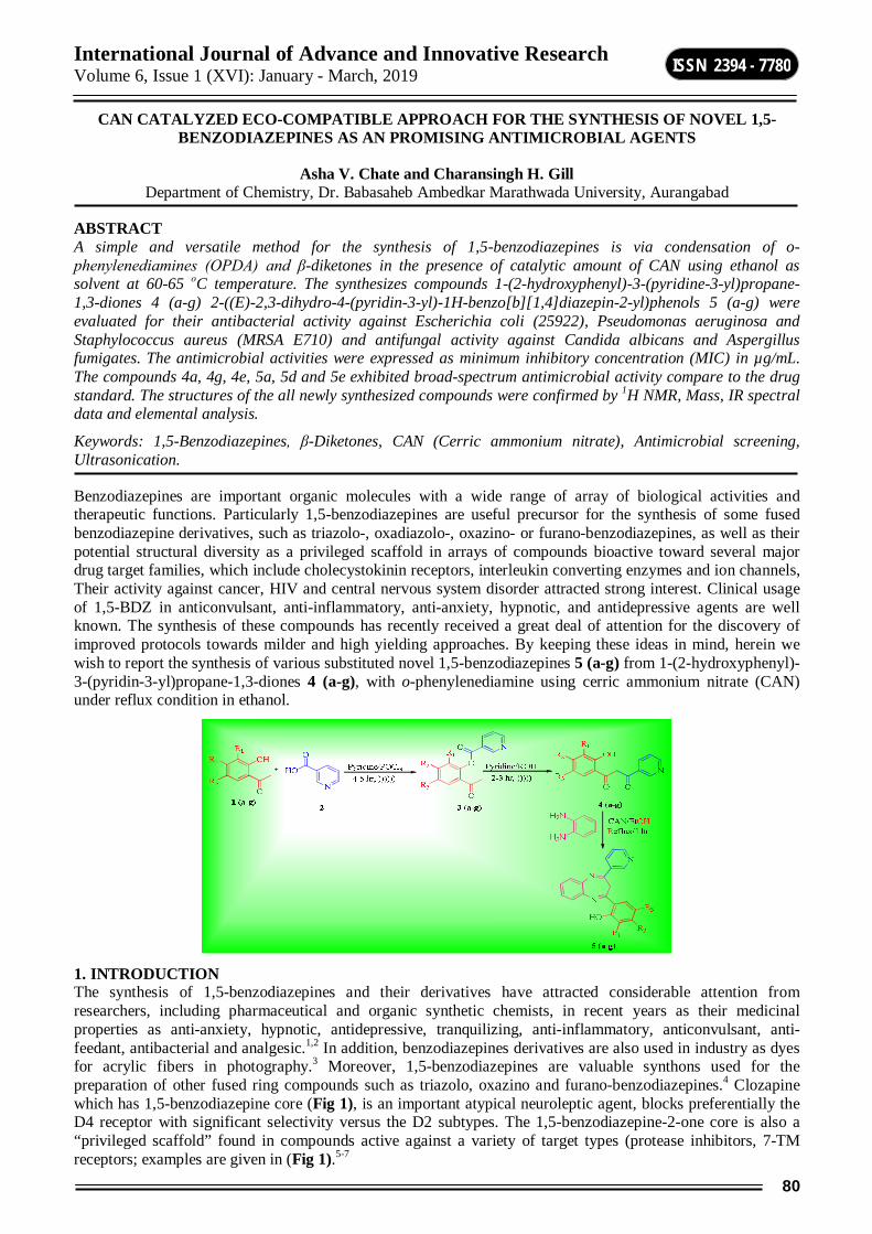
International Journal of Advance and Innovative Research Volume 6, Issue 1 (XVI): January - March, 2019
80
ISSN 2394 - 7780
CAN CATALYZED ECO-COMPATIBLE APPROACH FOR THE SYNTHESIS OF NOVEL 1,5-BENZODIAZEPINES AS AN PROMISING ANTIMICROBIAL AGENTS
Asha V. Chate and Charansingh H. Gill Department of Chemistry, Dr. Babasaheb Ambedkar Marathwada University, Aurangabad
ABSTRACT A simple and versatile method for the synthesis of 1,5-benzodiazepines is via condensation of o-phenylenediamines (OPDA) and β-diketones in the presence of catalytic amount of CAN using ethanol as solvent at 60-65 οC temperature. The synthesizes compounds 1-(2-hydroxyphenyl)-3-(pyridine-3-yl)propane-1,3-diones 4 (a-g) 2-((E)-2,3-dihydro-4-(pyridin-3-yl)-1H-benzo[b][1,4]diazepin-2-yl)phenols 5 (a-g) were evaluated for their antibacterial activity against Escherichia coli (25922), Pseudomonas aeruginosa and Staphylococcus aureus (MRSA E710) and antifungal activity against Candida albicans and Aspergillus fumigates. The antimicrobial activities were expressed as minimum inhibitory concentration (MIC) in µg/mL. The compounds 4a, 4g, 4e, 5a, 5d and 5e exhibited broad-spectrum antimicrobial activity compare to the drug standard. The structures of the all newly synthesized compounds were confirmed by 1H NMR, Mass, IR spectral data and elemental analysis.
Keywords: 1,5-Benzodiazepines, β-Diketones, CAN (Cerric ammonium nitrate), Antimicrobial screening, Ultrasonication.
Benzodiazepines are important organic molecules with a wide range of array of biological activities and therapeutic functions. Particularly 1,5-benzodiazepines are useful precursor for the synthesis of some fused benzodiazepine derivatives, such as triazolo-, oxadiazolo-, oxazino- or furano-benzodiazepines, as well as their potential structural diversity as a privileged scaffold in arrays of compounds bioactive toward several major drug target families, which include cholecystokinin receptors, interleukin converting enzymes and ion channels, Their activity against cancer, HIV and central nervous system disorder attracted strong interest. Clinical usage of 1,5-BDZ in anticonvulsant, anti-inflammatory, anti-anxiety, hypnotic, and antidepressive agents are well known. The synthesis of these compounds has recently received a great deal of attention for the discovery of improved protocols towards milder and high yielding approaches. By keeping these ideas in mind, herein we wish to report the synthesis of various substituted novel 1,5-benzodiazepines 5 (a-g) from 1-(2-hydroxyphenyl)-3-(pyridin-3-yl)propane-1,3-diones 4 (a-g), with o-phenylenediamine using cerric ammonium nitrate (CAN) under reflux condition in ethanol.
1. INTRODUCTION The synthesis of 1,5-benzodiazepines and their derivatives have attracted considerable attention from researchers, including pharmaceutical and organic synthetic chemists, in recent years as their medicinal properties as anti-anxiety, hypnotic, antidepressive, tranquilizing, anti-inflammatory, anticonvulsant, anti-feedant, antibacterial and analgesic.1,2 In addition, benzodiazepines derivatives are also used in industry as dyes for acrylic fibers in photography.3 Moreover, 1,5-benzodiazepines are valuable synthons used for the preparation of other fused ring compounds such as triazolo, oxazino and furano-benzodiazepines.4 Clozapine which has 1,5-benzodiazepine core (Fig 1), is an important atypical neuroleptic agent, blocks preferentially the D4 receptor with significant selectivity versus the D2 subtypes. The 1,5-benzodiazepine-2-one core is also a “privileged scaffold” found in compounds active against a variety of target types (protease inhibitors, 7-TM receptors; examples are given in (Fig 1).5-7
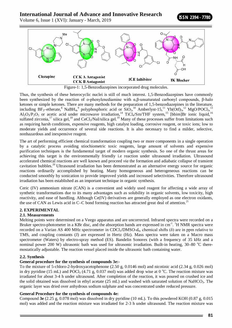
International Journal of Advance and Innovative Research Volume 6, Issue 1 (XVI): January - March, 2019
81
ISSN 2394 - 7780
Figure-1: 1,5-Benzodiazepines incorporated drug molecules.
Thus, the synthesis of these heterocyclic nuclei is still of much interest. 1,5-Benzodiazepines have commonly been synthesized by the reaction of o-phenylenediamine with α,β-unsaturated carbonyl compounds, β-halo ketones or simple ketones. There are many methods for the preparation of 1,5-benzodiazepines in the literature, including BF3–etherate,8 NaBH4,9 polyphosphoric acid or SiO2,10 Amberlyst-15,11 Yb(Otf)3,12 MgO/POCl3,13 Al2O3/P2O5 or acetic acid under microwave irradiation,14 TiCl4/Sm/THF system,15 [bbim]Br ionic liquid,16
sulfated zirconia,17 silica gel,18 and CeCl3/NaI/silica gel.19 Many of these processes suffer from limitations such as requiring harsh conditions, expensive reagents, high catalyst loading, corrosive reagent, or toxic ions; low to moderate yields and occurrence of several side reactions. It is also necessary to find a milder, selective, nonhazardous and inexpensive reagent.
The art of performing efficient chemical transformation coupling two or more components in a single operation by a catalytic process avoiding stiochiometric toxic reagents, large amount of solvents and expensive purification techniques is the fundamental target of modern organic synthesis. So one of the thrust areas for achieving this target is the environmentally friendly i.e reaction under ultrasound irradiation. Ultrasound accelerated chemical reactions are well known and proceed via the formation and adiabatic collapse of transient cavitation bubbles.20 Ultrasound irradiation has been demonstrated as an alternative energy source for organic reactions ordinarily accomplished by heating. Many homogeneous and heterogeneous reactions can be conducted smoothly by sonication to provide improved yields and increased selectivities. Therefore ultrasound irradiation has been established as an important technique in organic synthesis.
Ceric (IV) ammonium nitrate (CAN) is a convenient and widely used reagent for affecting a wide array of synthetic transformations due to its many advantages such as solubility in organic solvents, low toxicity, high reactivity, and ease of handling. Although Ce(IV) derivatives are generally employed as one electron oxidants, the use of CAN as Lewis acid in C–C bond forming reaction has attracted great deal of attention.21
2. EXPERIMENTAL 2.1. Measurements Melting points were determined on a Veego apparatus and are uncorrected. Infrared spectra were recorded on a Bruker spectro-photometer in a KBr disc, and the absorption bands are expressed in cm-1. 1H NMR spectra were recorded on a Varian AS 400 MHz spectrometer in CDCl3/DMSO-d6, chemical shifts (δ) are in ppm relative to TMS, and coupling constants (J) are expressed in Hertz (Hz). Mass spectra were taken on a Macro mass spectrometer (Waters) by electro-spray method (ES). Bandelin Sonorex (with a frequency of 35 kHz and a nominal power 200 W) ultrasonic bath was used for ultrasonic irradiation. Built-in heating, 30–80 οC there-mostatically adjustable. The reaction vessel placed inside the ultrasonic bath containing water.
2.2. Synthesis General procedure for the synthesis of compounds 3e:- To the mixture of 5-chloro-2-hydroxyacetophenone (2.50 g, 0.0146 mol) and nicotinic acid (2.34 g, 0.026 mol) in dry pyridine (15 mL) and POCl3 (4.71 g, 0.037 mol) was added drop wise at 0 oC. The reaction mixture was irradiated for about 3-4 h under ultrasound. After completion of the reaction, it was poured on crushed ice and the solid obtained was dissolved in ethyl acetate (25 mL) and washed with saturated solution of NaHCO3. The organic layer was dried over anhydrous sodium sulphate and was concentrated under reduced pressure.
General Procedure for the synthesis of compounds 4e: Compound 3e (2.25 g, 0.078 mol) was dissolved in dry pyridine (10 mL). To this powdered KOH (0.87 g, 0.015 mol) was added and the reaction mixture was irradiated for 2-3 h under ultrasound. The reaction mixture was

International Journal of Advance and Innovative Research Volume 6, Issue 1 (XVI): January - March, 2019
82
ISSN 2394 - 7780
poured on ice cold water and acidified with Conc. HCl. The yellow solid obtained was filtered off and crystallized from ethanol to obtain pure product.
1-(2-hydroxyphenyl)-3-(pyridine-3-yl)propane-1,3-dione 4a: Yellow solid. Mp 109-111 οC. 1H NMR (400 MHz, CDCl3-d6): = 6.32 (d, 1H, Ar-H), 6.89 (dd, 1H, Ar-H), 7.10 (dd, 1H, Ar-H), 7.31 (m, 1H, Ar-H), 7.54 (dd, 1H, Ar-H), 7.56 (d, 1H, Ar-H), 8.49 (m, 1H, Ar-H), 8.99 (dd, 1H, Ar-H), 9.26 (s, 1H, Vinylic Proton), 12.67 (s, 1H, OH), 15.45 (s, 1H, Enolic-OH). EC-MS: 242.05 (M+1). IR (KBr) cm-1: 3340 (-OH), 2933(Ar-H), 1705 (C=O), 1612 (C=N). Elemental analysis Calcd. for C14H11NO3. C, 69.70; H, 4.60; N, 5.81. Found: C, 69.44; H, 4.38; N, 5.52.
1-(3,5-dichloro-2-hydroxyphenyl)-3-(pyridine-3-yl)propane-1,3-dione 4b: Yellow solid. Mp 105-107 οC. 1H NMR (400 MHz, CDCl3-d6): = 6.93 (dd, 1H, Ar-H), 7.12 (d, 1H, Ar-H), 7.26 (d, 1H, Ar-H), 7.47 (dd, 1H, Ar-H), 7.92 (m, 1H, Ar-H), 8.23 (s, 1H, Vinylic Proton), 8.99 (d, 1H, Ar-H), 12.72 (s, 1H, OH), 15.35 (s, 1H, Enolic-OH). EC-MS: 310.03 (M+1) & 312.06 (M+3). IR (KBr) cm-1: 3350 (-OH), 2952(Ar-H), 1698 (C=O), 1612 (C=N), 761 (Ar-Cl). Elemental analysis Calcd. for C14H9Cl2NO3. C, 54.22; H, 2.93; N, 4.52. Found: C, 53.94; H, 2.65; N, 4.25.
1-(2-hydroxy-3,5-dimethylphenyl)-3-(pyridine-3-yl)propane-1,3-dione 4c: Yellow solid. Mp 130-132 οC. 1H NMR (400 MHz, CDCl3-d6 : = 2.40 (s, 3H, CH3), 2.51 (s, 3H, CH3), 6.82 (dd, 1H, Ar-H), 6.92 (d, 1H, Ar-H), 7.06 (d, 1H, Ar-H), 7.34 (dd, 1H, Ar-H), 7.82 (m, 1H, Ar-H), 8.53 (s, 1H, Vinylic Proton), 8.89 (d, 1H, Ar-H), 12.93 (s, 1H, OH), 15.59 (s, 1H, Enolic-OH). EC-MS: 270.08 (M+1). IR (KBr) cm-1: 3348 (-OH), 2943(Ar-H), 1709 (C=O), 1614 (C=N). Elemental analysis Calcd. for C16H15NO3. C, 71.36; H, 5.61; N, 5.20. Found: C, 70.98; H, 5.44; N, 4.93.
1-(5-chloro-2-hydroxy-4-methylphenyl)-3-(pyridine-3-yl)propane-1,3-dione 4d: Yellow solid. Mp 123-125 οC. 1H NMR (400 MHz, CDCl3-d6): = 2.54 (s, 3H, CH3), 6.85 (dd, 1H, Ar-H),), 6.90 (s, 1H, Ar-H), 7.61 (s, 1H, Ar-H), 7.82 (dd, 1H, Ar-H), 8.51 (m, 1H, Ar-H), 8.89 (s, 1H, Vinylic Proton), 9.37 (d, 1H, Ar-H), 12.04 (s, 1H, OH), 15.52 (s, 1H, Enolic-OH). EC-MS: 290.10 (M+1) & 392.06 (M+3). IR (KBr) cm-1: 3342 (-OH), 2943(Ar-H), 1702 (C=O), 1610 (C=N), 757 (Ar-Cl). Elemental analysis Calcd. for C15H12ClNO3. C, 62.19; H, 4.17; N, 4.83. Found: C, 61.93; H, 3.99; N, 4.55.
1-(5-chloro-2-hydroxyphenyl)-3-(pyridine-3-yl)propane-1,3-dione 4e: Yellow solid. Mp 131-133 οC. 1H NMR (400 MHz, DMSO-d6): = 6.79 (dd, 1H, Ar-H), 6.89 (d, 1H, Ar-H), 7.14 (dd, 1H, Ar-H), 7.36 (dd, 1H, Ar-H), 7.57 (d, 1H, Ar-H), 8.53 (m, 1H, Ar-H), 8.98 (s, 1H, Vinylic Proton), 9.34 (d, 1H, Ar-H), 12.64 (s, 1H, OH), 15.41 (s, 1H, Enolic-OH). EC-MS: 276.05 (M+1) & 378.09 (M+3). IR (KBr) cm-1: 3345(-OH), 2949 (Ar-H), 1700 (C=O), 1615 (C=N), 759 (Ar-Cl). Elemental analysis Calcd. for C14H10ClNO3. C, 60.99; H, 3.66; N, 5.08. Found: C, 61.24; H, 3.37; N, 4.78.
1-(5-fluoro-2-hydroxyphenyl)-3-(pyridine-3-yl)propane-1,3-dione 4f: Yellow solid. Mp 117-120 οC. 1H NMR (400 MHz, DMSO-d6): = 5.98 (dd, 1H, Ar-H), 7.08 (d, 1H, Ar-H), 7.12 (dd, 1H, Ar-H), 7.48 (d, 1H, Ar-H), 7.58 (dd, 1H, Ar-H), 7.96 (m, 1H, Ar-H), 8.49 (dd, 1H, Ar-H), 8.79 (s, 1H, Vinylic Proton), 12.61 (s, 1H, OH), 15.57 (s, 1H, Enolic-OH). EC-MS: 270.09 (M+1). IR (KBr) cm-1: 3398 (-OH), 1572 ((C=C), 1219 (C=O), 1488 (C=N). Elemental analysis Calcd. for C14H10FNO3. C, 64.86; H, 3.89; N, 5.40. Found: C, 64.58; H, 3.62; N, 5.89.
1-(2-hydroxy-5-methylphenyl)-3-(pyridine-3-yl)propane-1,3-dione 4g: Yellow solid. Mp 116-118 οC. 1H NMR (400 MHz, CDCl3-d6): = 2.31 (s, 3H, CH3), 6.85 (dd, 1H, Ar-H), 7.26 (dd, 1H, Ar-H), 7.43 (d, 1H, Ar-H), 7.46 (d, 1H, Ar-H), 7.55 (dd, 1H, Ar-H), 8.24 (m, 1H, Ar-H), 8.77 (s, 1H, Vinylic Proton), 9.16 (d, 1H, Ar-H), 11.80 (s, 1H, OH), 15.49 (s, 1H, Enolic-OH). EC-MS: 238.04 (M+1). IR (KBr) cm-1: 3498 (-OH), 1575 (C=C), 1215 (C=O), 1490 (C=N). Elemental analysis Calcd. for C15H13NO3. C, 70.58; H, 5.13; N, 5.49. Found: C, 70.96; H, 4.85; N, 5.20.
General procedure for the synthesis of 1,5-benzodiazepines In a typical experiment, CAN (10 % mmol) was dissolved into the mixture of diketones (1.0 mol) and o-phenylenediamine (1.0 mol) in ethanol (5 mL) in a 25-mL round-bottom flask equipped with a stopper. The reaction mixture was irradiated under ultrasonication at 60-65 οC for the desired time as shown in (Table 1). The completion of reaction was monitered by TLC. (20:80 EtOAc in petroleum ether as eluent). After completion, the reaction mixture was poured over crushed ice & the solid obtained was filtered, dried to afford the corresponding 1,5-Benzodiazepines.
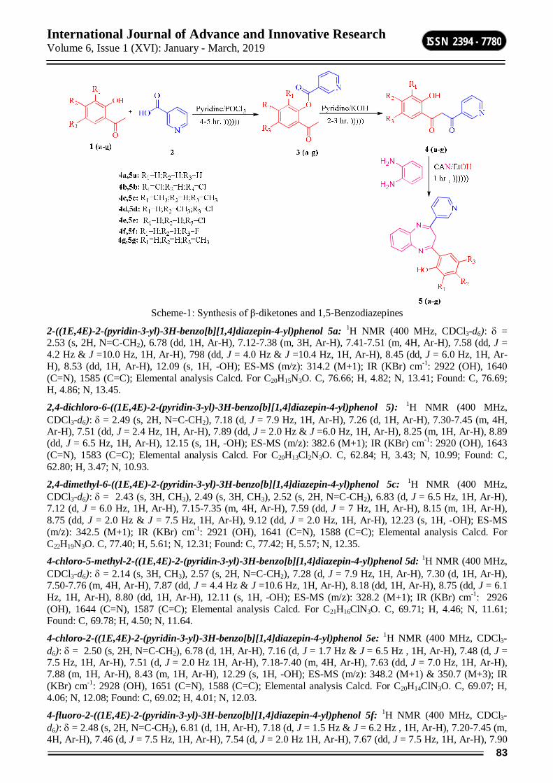
International Journal of Advance and Innovative Research Volume 6, Issue 1 (XVI): January - March, 2019
83
ISSN 2394 - 7780
Scheme-1: Synthesis of β-diketones and 1,5-Benzodiazepines
2-((1E,4E)-2-(pyridin-3-yl)-3H-benzo[b][1,4]diazepin-4-yl)phenol 5a: 1H NMR (400 MHz, CDCl3-d6): = 2.53 (s, 2H, N=C-CH2), 6.78 (dd, 1H, Ar-H), 7.12-7.38 (m, 3H, Ar-H), 7.41-7.51 (m, 4H, Ar-H), 7.58 (dd, J = 4.2 Hz & J =10.0 Hz, 1H, Ar-H), 798 (dd, J = 4.0 Hz & J =10.4 Hz, 1H, Ar-H), 8.45 (dd, J = 6.0 Hz, 1H, Ar-H), 8.53 (dd, 1H, Ar-H), 12.09 (s, 1H, -OH); ES-MS (m/z): 314.2 (M+1); IR (KBr) cm-1: 2922 (OH), 1640 (C=N), 1585 (C=C); Elemental analysis Calcd. For C20H15N3O. C, 76.66; H, 4.82; N, 13.41; Found: C, 76.69; H, 4.86; N, 13.45.
2,4-dichloro-6-((1E,4E)-2-(pyridin-3-yl)-3H-benzo[b][1,4]diazepin-4-yl)phenol 5): 1H NMR (400 MHz, CDCl3-d6): = 2.49 (s, 2H, N=C-CH2), 7.18 (d, J = 7.9 Hz, 1H, Ar-H), 7.26 (d, 1H, Ar-H), 7.30-7.45 (m, 4H, Ar-H), 7.51 (dd, J = 2.4 Hz, 1H, Ar-H), 7.89 (dd, J = 2.0 Hz & J =6.0 Hz, 1H, Ar-H), 8.25 (m, 1H, Ar-H), 8.89 (dd, J = 6.5 Hz, 1H, Ar-H), 12.15 (s, 1H, -OH); ES-MS (m/z): 382.6 (M+1); IR (KBr) cm-1: 2920 (OH), 1643 (C=N), 1583 (C=C); Elemental analysis Calcd. For C20H13Cl2N3O. C, 62.84; H, 3.43; N, 10.99; Found: C, 62.80; H, 3.47; N, 10.93.
2,4-dimethyl-6-((1E,4E)-2-(pyridin-3-yl)-3H-benzo[b][1,4]diazepin-4-yl)phenol 5c: 1H NMR (400 MHz, CDCl3-d6): = 2.43 (s, 3H, CH3), 2.49 (s, 3H, CH3), 2.52 (s, 2H, N=C-CH2), 6.83 (d, J = 6.5 Hz, 1H, Ar-H), 7.12 (d, J = 6.0 Hz, 1H, Ar-H), 7.15-7.35 (m, 4H, Ar-H), 7.59 (dd, J = 7 Hz, 1H, Ar-H), 8.15 (m, 1H, Ar-H), 8.75 (dd, J = 2.0 Hz & J = 7.5 Hz, 1H, Ar-H), 9.12 (dd, J = 2.0 Hz, 1H, Ar-H), 12.23 (s, 1H, -OH); ES-MS (m/z): 342.5 (M+1); IR (KBr) cm-1: 2921 (OH), 1641 (C=N), 1588 (C=C); Elemental analysis Calcd. For C22H19N3O. C, 77.40; H, 5.61; N, 12.31; Found: C, 77.42; H, 5.57; N, 12.35.
4-chloro-5-methyl-2-((1E,4E)-2-(pyridin-3-yl)-3H-benzo[b][1,4]diazepin-4-yl)phenol 5d: 1H NMR (400 MHz, CDCl3-d6): = 2.14 (s, 3H, CH3), 2.57 (s, 2H, N=C-CH2), 7.28 (d, J = 7.9 Hz, 1H, Ar-H), 7.30 (d, 1H, Ar-H), 7.50-7.76 (m, 4H, Ar-H), 7.87 (dd, J = 4.4 Hz & J =10.6 Hz, 1H, Ar-H), 8.18 (dd, 1H, Ar-H), 8.75 (dd, J = 6.1 Hz, 1H, Ar-H), 8.80 (dd, 1H, Ar-H), 12.11 (s, 1H, -OH); ES-MS (m/z): 328.2 (M+1); IR (KBr) cm-1: 2926 (OH), 1644 (C=N), 1587 (C=C); Elemental analysis Calcd. For C21H16ClN3O. C, 69.71; H, 4.46; N, 11.61; Found: C, 69.78; H, 4.50; N, 11.64.
4-chloro-2-((1E,4E)-2-(pyridin-3-yl)-3H-benzo[b][1,4]diazepin-4-yl)phenol 5e: 1H NMR (400 MHz, CDCl3-d6): = 2.50 (s, 2H, N=C-CH2), 6.78 (d, 1H, Ar-H), 7.16 (d, J = 1.7 Hz & J = 6.5 Hz , 1H, Ar-H), 7.48 (d, J = 7.5 Hz, 1H, Ar-H), 7.51 (d, J = 2.0 Hz 1H, Ar-H), 7.18-7.40 (m, 4H, Ar-H), 7.63 (dd, J = 7.0 Hz, 1H, Ar-H), 7.88 (m, 1H, Ar-H), 8.43 (m, 1H, Ar-H), 12.29 (s, 1H, -OH); ES-MS (m/z): 348.2 (M+1) & 350.7 (M+3); IR (KBr) cm-1: 2928 (OH), 1651 (C=N), 1588 (C=C); Elemental analysis Calcd. For C20H14ClN3O. C, 69.07; H, 4.06; N, 12.08; Found: C, 69.02; H, 4.01; N, 12.03.
4-fluoro-2-((1E,4E)-2-(pyridin-3-yl)-3H-benzo[b][1,4]diazepin-4-yl)phenol 5f: 1H NMR (400 MHz, CDCl3-d6): = 2.48 (s, 2H, N=C-CH2), 6.81 (d, 1H, Ar-H), 7.18 (d, J = 1.5 Hz & J = 6.2 Hz , 1H, Ar-H), 7.20-7.45 (m, 4H, Ar-H), 7.46 (d, J = 7.5 Hz, 1H, Ar-H), 7.54 (d, J = 2.0 Hz 1H, Ar-H), 7.67 (dd, J = 7.5 Hz, 1H, Ar-H), 7.90
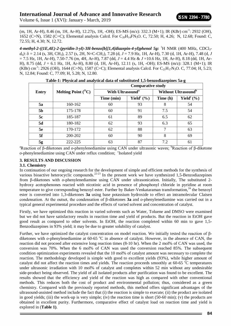
International Journal of Advance and Innovative Research Volume 6, Issue 1 (XVI): January - March, 2019
84
ISSN 2394 - 7780
(m, 1H, Ar-H), 8.46 (m, 1H, Ar-H), 12.27(s, 1H, -OH); ES-MS (m/z): 332.3 (M+1); IR (KBr) cm-1: 2932 (OH), 1652 (C=N), 1582 (C=C); Elemental analysis Calcd. For C20H14FN3O. C, 72.50; H, 4.26; N, 12.68; Found: C, 72.55; H, 4.30; N, 12.72.
4-methyl-2-((1E,4E)-2-(pyridin-3-yl)-3H-benzo[b][1,4]diazepin-4-yl)phenol 5g: 1H NMR (400 MHz, CDCl3-d6): = 2.14 (s, 3H, CH3), 2.57 (s, 2H, N=C-CH2), 7.28 (d, J = 7.9 Hz, 1H, Ar-H), 7.30 (d, 1H, Ar-H), 7.48 (d, J = 7.5 Hz, 1H, Ar-H), 7.50-7.76 (m, 4H, Ar-H), 7.87 (dd, J = 4.4 Hz & J =10.6 Hz, 1H, Ar-H), 8.18 (dd, 1H, Ar-H), 8.75 (dd, J = 6.1 Hz, 1H, Ar-H), 8.80 (d, 1H, Ar-H), 12.11 (s, 1H, -OH); ES-MS (m/z): 328.1 (M+1); IR (KBr) cm-1: 2926 (OH), 1644 (C=N), 1587 (C=C); Elemental analysis Calcd. For C21H17N3O. C, 77.04; H, 5.23; N, 12.84; Found: C, 77.09; H, 5.28; N, 12.80.
Table-1: Physical and analytical data of substituted 1,5-benzodiazepines 5a-g
Entry Melting Point (ΟC) Comparative study
With Ultrasounda Without Ultrasoundb Time (min) Yieldc (%) Time (h) Yieldc (%)
5a 160-162 60 93 8 54 5b 175-178 60 91 7.5 54 5c 185-187 61 89 6.5 62 5d 180-182 62 93 6.3 65 5e 170-172 62 88 7 63 5f 200-202 60 90 8 69 5g 222-225 63 87 7.2 61
aReaction of β-diketones and o-phenylenediamine using CAN under ultrasonic waves; bReaction of β-diketone o-phenylenediamine using CAN under reflux condition; cIsolated yield
3. RESULTS AND DISCUSSION 3.1. Chemistry In continuation of our ongoing research for the development of simple and efficient methods for the synthesis of various bioactive heterocyclic compounds.22-23 In the present work we have synthesized 1,5-Benzodiazepines from β-diketones with o-phenylenediamine using CAN under ultrasonication. Initially, The substituted 2-hydroxy acetophenones reacted with nicotinic acid in presence of phosphonyl chloride in pyridine at room temperature to give corresponding benzoyl ester. Further by Baker-Venkataraman transformation,24 the benzoyl ester is converted into 1,3-diketones 3a using base potassium hydroxide to effect an intramolecular Claisen condensation. At the outset, the condensation of β-diketones 3a and o-phenylenediamine was carried out in a typical general experimental procedure and the effects of varied solvent and concentration of catalyst.
Firstly, we have optimized this reaction in varied solvents such as Water, Toluene and DMSO were examined but we did not have satisfactory results in reaction time and yield of products. But the reaction in EtOH gave good result as compared to other solvents. In EtOH, the reaction completed within 60 min to gave 1,5-Benzodiazepines in 93% yield; it may be due to greater solubility of catalyst.
Further, we have optimized the catalyst concentration on model reaction. We initially tested the reaction of β-diketones with o-phenylenediamine at 60-65 οC in absence of catalyst. However, in the absence of CAN, the reaction did not proceed after extensive long reaction times (8-10 hr). When the 2 mol% of CAN was used, the conversion was 70%. When the 6 mol% of CAN was used the conversion reached 85%. The subsequent condition optimization experiments revealed that the 10 mol% of catalyst amount was necessary to complete the reaction. The methodology developed is simple with good to excellent yields (93%), while higher amount of catalyst did not affect the reaction times and yields. The reaction proceeds smoothly at 60-65 οC temperatures under ultrasonic irradiation with 10 mol% of catalyst and completes within 52 min without any undesirable side-product being observed. The yield of all isolated products after purification was found to be excellent. The results showed that the efficiency and yield of the reaction was high as compared with other conventional methods. This reduces both the cost of product and environmental pollution; thus, considered as a green chemistry. Compared with the previously reported methods, this method offers significant advantages of the ultrasound-assisted method include the fact that (i) the reaction is simple to execute; (ii) the product are isolated in good yields; (iii) the work-up is very simple; (iv) the reaction time is short (50-60 min); (v) the products are obtained in excellent purity. Furthermore, comparative effect of catalyst load on reaction time and yield is explored in (Table 1).
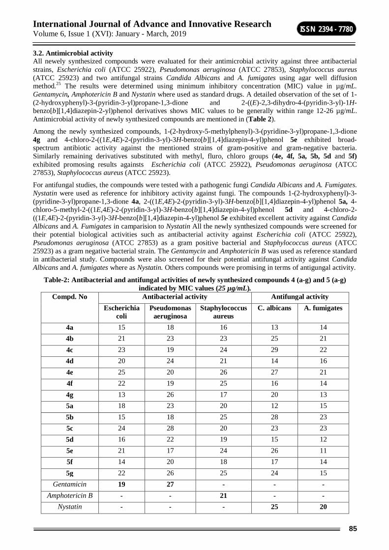
International Journal of Advance and Innovative Research Volume 6, Issue 1 (XVI): January - March, 2019
85
ISSN 2394 - 7780
3.2. Antimicrobial activity All newely synthesized compounds were evaluated for their antimicrobial activity against three antibacterial strains, Escherichia coli (ATCC 25922), Pseudomonas aeruginosa (ATCC 27853), Staphylococcus aureus (ATCC 25923) and two antifungal strains Candida Albicans and A. fumigates using agar well diffusion method.25 The results were determined using minimum inhibitory concentration (MIC) value in µg/mL. Gentamycin, Amphotericin B and Nystatin where used as standard drugs. A detailed observation of the set of 1-(2-hydroxyphenyl)-3-(pyridin-3-yl)propane-1,3-dione and 2-((E)-2,3-dihydro-4-(pyridin-3-yl)-1H-benzo[b][1,4]diazepin-2-yl)phenol derivatives shows MIC values to be generally within range 12-26 µg/mL. Antimicrobial activity of newly synthesized compounds are mentioned in (Table 2).
Among the newly synthesized compounds, 1-(2-hydroxy-5-methylphenyl)-3-(pyridine-3-yl)propane-1,3-dione 4g and 4-chloro-2-((1E,4E)-2-(pyridin-3-yl)-3H-benzo[b][1,4]diazepin-4-yl)phenol 5e exhibited broad-spectrum antibiotic activity against the mentioned strains of gram-positive and gram-negative bacteria. Similarly remaining derivatives substituted with methyl, fluro, chloro groups (4e, 4f, 5a, 5b, 5d and 5f) exhibited promosing results againsts Escherichia coli (ATCC 25922), Pseudomonas aeruginosa (ATCC 27853), Staphylococcus aureus (ATCC 25923).
For antifungal studies, the compounds were tested with a pathogenic fungi Candida Albicans and A. Fumigates. Nystatin were used as reference for inhibitory activity against fungi. The compounds 1-(2-hydroxyphenyl)-3-(pyridine-3-yl)propane-1,3-dione 4a, 2-((1E,4E)-2-(pyridin-3-yl)-3H-benzo[b][1,4]diazepin-4-yl)phenol 5a, 4-chloro-5-methyl-2-((1E,4E)-2-(pyridin-3-yl)-3H-benzo[b][1,4]diazepin-4-yl)phenol 5d and 4-chloro-2-((1E,4E)-2-(pyridin-3-yl)-3H-benzo[b][1,4]diazepin-4-yl)phenol 5e exhibited excellent activity against Candida Albicans and A. Fumigates in camparision to Nystatin All the newly synthesized compounds were screened for their potential biological activities such as antibacterial activity against Escherichia coli (ATCC 25922), Pseudomonas aeruginosa (ATCC 27853) as a gram positive bacterial and Staphylococcus aureus (ATCC 25923) as a gram negative bacterial strain. The Gentamycin and Amphotericin B was used as reference standard in antibacterial study. Compounds were also screened for their potential antifungal activity against Candida Albicans and A. fumigates where as Nystatin. Others compounds were promising in terms of antigungal activity.
Table-2: Antibacterial and antifungal activities of newly synthesized compounds 4 (a-g) and 5 (a-g) indicated by MIC values (25 µg/mL).
Compd. No Antibacterial activity Antifungal activity Escherichia
coli Pseudomonas
aeruginosa Staphylococcus
aureus C. albicans A. fumigates
4a 15 18 16 13 14 4b 21 23 23 25 21 4c 23 19 24 29 22 4d 20 24 21 14 16 4e 25 20 26 27 21 4f 22 19 25 16 14 4g 13 26 17 20 13 5a 18 23 20 12 15 5b 15 18 25 28 23 5c 24 28 20 23 23 5d 16 22 19 15 12 5e 21 17 24 26 11 5f 14 20 18 17 14 5g 22 26 25 24 15
Gentamicin 19 27 - - - Amphotericin B - - 21 - -
Nystatin - - - 25 20
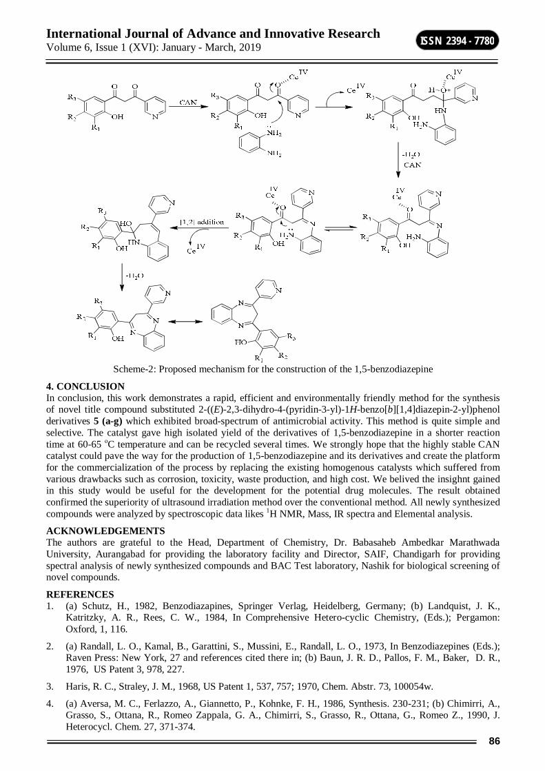
International Journal of Advance and Innovative Research Volume 6, Issue 1 (XVI): January - March, 2019
86
ISSN 2394 - 7780
Scheme-2: Proposed mechanism for the construction of the 1,5-benzodiazepine
4. CONCLUSION In conclusion, this work demonstrates a rapid, efficient and environmentally friendly method for the synthesis of novel title compound substituted 2-((E)-2,3-dihydro-4-(pyridin-3-yl)-1H-benzo[b][1,4]diazepin-2-yl)phenol derivatives 5 (a-g) which exhibited broad-spectrum of antimicrobial activity. This method is quite simple and selective. The catalyst gave high isolated yield of the derivatives of 1,5-benzodiazepine in a shorter reaction time at 60-65 οC temperature and can be recycled several times. We strongly hope that the highly stable CAN catalyst could pave the way for the production of 1,5-benzodiazepine and its derivatives and create the platform for the commercialization of the process by replacing the existing homogenous catalysts which suffered from various drawbacks such as corrosion, toxicity, waste production, and high cost. We belived the insighnt gained in this study would be useful for the development for the potential drug molecules. The result obtained confirmed the superiority of ultrasound irradiation method over the conventional method. All newly synthesized compounds were analyzed by spectroscopic data likes 1H NMR, Mass, IR spectra and Elemental analysis.
ACKNOWLEDGEMENTS The authors are grateful to the Head, Department of Chemistry, Dr. Babasaheb Ambedkar Marathwada University, Aurangabad for providing the laboratory facility and Director, SAIF, Chandigarh for providing spectral analysis of newly synthesized compounds and BAC Test laboratory, Nashik for biological screening of novel compounds.
REFERENCES 1. (a) Schutz, H., 1982, Benzodiazapines, Springer Verlag, Heidelberg, Germany; (b) Landquist, J. K.,
Katritzky, A. R., Rees, C. W., 1984, In Comprehensive Hetero-cyclic Chemistry, (Eds.); Pergamon: Oxford, 1, 116.
2. (a) Randall, L. O., Kamal, B., Garattini, S., Mussini, E., Randall, L. O., 1973, In Benzodiazepines (Eds.); Raven Press: New York, 27 and references cited there in; (b) Baun, J. R. D., Pallos, F. M., Baker, D. R., 1976, US Patent 3, 978, 227.
3. Haris, R. C., Straley, J. M., 1968, US Patent 1, 537, 757; 1970, Chem. Abstr. 73, 100054w.
4. (a) Aversa, M. C., Ferlazzo, A., Giannetto, P., Kohnke, F. H., 1986, Synthesis. 230-231; (b) Chimirri, A., Grasso, S., Ottana, R., Romeo Zappala, G. A., Chimirri, S., Grasso, R., Ottana, G., Romeo Z., 1990, J. Heterocycl. Chem. 27, 371-374.
International Journal of Advance and Innovative Research Volume 6, Issue 1 (XVI): January - March, 2019
87
ISSN 2394 - 7780
5. Tranquillini, M. E., Cassara, P. G., Corsi, M., Curotto, G., Donati, D.; Finizia, G., Pentassuglia, G., Polinelli, S., Tarzia, G., Ursini, A., Van Amsterdam, F. T. M., 1997, Arch. Pharm. 330, 353.
6. Batchelor, M. J., Bebbington, D., Bemis, G. W., Fridman, W. H., Gillespie, R. J., Golec, J. M. C., Gu, Y., Lauffer, D. J., Livingston, D. J., Matharu, S. S., Mullican, M. D., Murcko, M. A., Murdoch, R., Nyce, P. L., Robidoux, A. L. C., 1997, PCT Int. Appl. WO 9722619 A2 19970626; Bemis, G. W., Golec, J. M. C., Lauffer, D. J., Mullican, M. D., Murcko, M. A., Livingston, D. J. (ICE), PCT Int. Appl. WO 9535308.
7. Claremon, D. A., Freidinger, R. M., Liverton, N., Selnick, H. G., Smith, G. R., PCT Int. Appl. WO 9640656; Selnick, H. G., Liverton, N. J., Baldwin, J. J., Butcher, J. W., Smith, G. R., Tebben, A. J., Jurkiewicz, N. K., Lynch, J. J., Salata, J. J.,Sanguinetti, M. C., Siegl, P. K. S., Slaughter, D. E., Vyas, K., 1997, J. Med. Chem. 40, 3865.
8. Herbert, J. A. L., Suschitzky, H. J., 1974, Chem. Soc. Perkin Trans. 1, 2657-2661.
9. Morales, H. R., Bulbarela, A., Contreras, R., 1986, Heterocycles. 24, 135-139.
10. Jung, D. I., Choi, T. W., Kim, Y. Y., Kim, I. S., Park, Y. M.; Lee, Y. G., Jung, D. H., 1999, Synth. Commun. 29, 1941-1951.
11. Yadav, J. S., Reddy, B. V. S., Eshwaraian, B., Anuradha, K., 2002, Green Chem. 4, 592.
12. Curini, M., Epifano, F., Marcotullio, M. C., Rosati, O., 2001, Tetrahedron Lett. 42, 3193-3195.
13. Balakrishna, M. S., Kaboundin, B., 2001, Tetrahedron Lett. 42, 1127-1129.
14. (a) Kaboudin, B., Navaee, K., 2001, Heterocycles. 55, 1443-1446; (b) Pozarentzi, M., Stephanidou, S. J., Tsoleridis, C. A., 2002, Tetrahedron Lett. 43, 1755-1758; (c) Kidwai, M., Ruby Venkataramanan, R., 2004, Chem. Heterocycl. Compd. 40, 631-634.
15. Ma, Y., Zhang, Y., 2002, Synth. Commun. 32, 165-167.
16. Jarikote, D. V., Siddique, S. A., Rajagopal, R., Daniel, T., Lahoti, R. J., Srinivasan, K. V., 2003, Tetrahedron Lett. 44, 1835-1838.
17. Reddy, B. M.; Sreekanth, P. M., 2003, Tetrahedron Lett. 44, 4447-4449.
18. Kodomari, M., Noguchi, T., Aoyama, T., 2004, Synth. Commun. 34, 1783-1790.
19. Sabitha, G., Reddy, G. S. K. K., Reddy, K. B., Reddy, N. M., Yadav, J. S., 2004, Adv. Synth. Catal. 346, 921-923.
20. (a) Gaplovsky, A., Gaplovsky, M., Toma, S., Luche, J. L., 2000, J. Org. Chem. 65, 8444- 8447; (b) Suslick, K. S., 1988, In ultrasound, its Chemical, physical and biological effects, VCH, Weinheim, 287-303; (c) Rajagopal, R., Jarikote, D. V., Srinivasan, K. V., 2002, Chem. Commun. 616-617; (d) Deshmukh, R. R., Rajagopal, R., Srinivasan, K. V., 2001, Chem. Commun. 1544-1545.
21. (a) Hwu, J. R., King, K. Y., 2001, Curr. Sci. 81, 1043-1053; (b) Nair, V., Panicker, S. B., Nair, L. G., George, T. G., Augustine, A., 2003, Synlett. 156-165; (c) Dhakshinmoorty, A., 2003, Synlett. 3014; (d) Varala, R., Enugala, R., Sreelatha, N. S. R., Adapa, S. R., Synlett. 2006, 1009; (e) Varala, R., Sreelatha N., Adapa, S. R., 2006, Synlett. 1549-1555; (f) Nair, V., Mathew, J., Prabhakaran, J., 1997, Chem. Soc. Rev. 127; (g) Nair, V., Balagopal, L., Rajan, R., Mathew, J., 2004, Acc. Chem. Res. 37, 21-30; (h) Nair, V., Deepathi, A., 2007, Chem. Rev. 107, 1862-1891
22. (a) Joshi, R. S.; Mandhane, P. G.; Diwakar, S. D.; Dabhade, S. K.; Gill, C. H., 2010, Bioorg. Med. Chem. Letts 20, 3721-3725; (b) Jadhav, G. R., Shaikh, M. U., Shingare, M. S., Gill, C. H., 2008, J. Het. Chem. 45(1), 1287-1292; (c) Jadhav, G. R., Kale, R. P., Shaikh, M. U., Ghawalkar, A. R., Nagargoje, D. R., Shiradkar, M., Gill, C. H., 2008, Bioorg. & Med. Chem. Letts. 16, 6244-6247.
23. (a) Joshi, R. S., Mandhane, P. G., Diwakar, S. D., Gill, C. H., 2010, Ultrason. Sonochem. 17, 298-300; (b) Mandhane, P. G., Joshi, R. S., Nagargoje, D. R., Gill, C. H., 2010, Tett. Letts. 51, 1490-1492.
24. Wheeler, T. S., 1963, Organic Syntheses; Wiley New York, Vol. N, 418.
25. Nathan, P., et al., 1978, Laboratory Methods for Selection of Topical antimicrobial Agents to treat infected Burn Wounds 1978, 4, 177-187.

International Journal of Advance and Innovative Research Volume 6, Issue 1 (XVI): January - March, 2019
88
ISSN 2394 - 7780
MMUULLTTIIWWAAVVEELLEENNGGTTHH AASSTTRROONNOOMMYY:: TTOOOOLL TTOO PPRROOBBEE TTHHEE UUNNIIVVEERRSSEE
B. T. Tate Department of Physics, Balbhim College, Beed
ABSTRACT It is impossible to study all astronomical objects at a time, that is why one of the main task of astronomers is to search and find those particular objects that will provide a lot of understanding of the physics of objects and phenomena. In last few decades it is observed that we could see the universe in only one window that is optical. But now a day due to modern observing facility we can see the universe throughout all the wavelengths of electromagnetic spectrum. In this paper we are going to discuss astronomical data archives and data analysis tools to probe the universe in all wavelengths .This task are being achieved by astronomical surveys; having observed large areas of the sky, one can select interesting objects by definite criteria. Surveys are the backbones of astronomy, and therefore the engine of discovery. They’re of cultural importance, as a result of this they satisfy the need to map our surroundings, and provide a sense for wherever we tend to live.
Keywords: Astronomical data archives, Software’s .
INTRODUCTION The major characteristic of modern astronomy is to study multiwavelength (MW) (from γ-ray to radio) and big data (data acquisition, storage and analysis). Now a day astronomical databases and archives contain billions of objects observed at different wavelengths, both galactic and extragalactic, and the vast amount of data on them allows new studies and discoveries [1]. Radiations coming from astronomical objects are the only tool to study them, and it is found that astronomical object emits all wavelengths in electromagnetic spectrum from radio waves to gamma rays. Emission of Radio waves having temperature less than 10 K are coming from cosmic background radiation ,scattering of free electrons in interstellar plasmas, cold gas and dust between the stars, regions near white dwarfs and neutron stars, supernova remnants, dense regions of interstellar space such as the galactic center, cold molecular clouds. Microwaves having temperature same as that of radio waves shows background radiation of the universe (remnant of Big Bang). Infrared have the temperature 10 – 10 3 K comes from cool stars, star-forming regions, interstellar dust warmed by starlight, planets, comets and asteroids. as per as visible light is concerned 10 3 – 10 4 K Planets, Stars ,Galaxies, Nebulae. Ultraviolet radiation comes from Supernova remnants ,very hot star, quasars and has temperature range 10 4 – 10 6 K. X-rays are high energetic radiations with temperature 10 6 – 10 8 K and they emits from regions of hot, shocked gas, gas in clusters of galaxies, neutron stars ,supernova remnants, and the hot outer layers of stars. Finally gamma ray emission takes place from interstellar clouds where cosmic rays collide with hydrogen nuclei , disks of material surrounding black holes, pulsars or neutron stars , gamma rays has highest energy with temperature more than 108K.
DATA ACQUISITION Multiwavelength data can be acquired from different arxives some of them discussed here
1. Nasa Extragalactic Database (NED) It is one of the a comprehensive database of multiwavelength data for extragalactic objects, providing a scientific and systematic information of hundreds of large sky surveys and thousands of research articles. It gives information of entire electromagnetic spectrum from gamma rays to radio waves. As new observations are added through publications, they are cross-identified or statistically related with previous data and integrated into a unified database to simplify queries and retrieval. It has connectivity with the NASA astrophysics mission archives (MAST, HEASARC, and IRSA) to the astrophysics literature via ADS, and to other data centers around the world. The database additionally includes a master index of various astrophysical objects, associations, positions, redshifts, spectra, redshift-independent distances, images, photometry, diameters, and elaborate notes. With this it gives information regarding derived quantities which includes Galactic extinction, velocity corrections, quick-look luminosities ,Hubble flow distances and scales, cosmological corrections and spectral energy distributions (SEDs). Objects information can be found by name, near name or near position and references can be queried by author and by object name.
2. High Energy Astrophysics Science Archive Research Center (HEASARCH) HEASARC is the High energy archive for NASA’s missions studying electromagnetic radiation from extremely energetic cosmic phenomena ranging from black holes to the Big Bang. Since its merger with the Legacy Archive for Microwave Background Data Analysis (LAMBDA) in 2008, the HEASARC archive contains data obtained by high-energy astronomy missions observing in the extreme-ultraviolet (EUV), X-ray, and gamma-
International Journal of Advance and Innovative Research Volume 6, Issue 1 (XVI): January - March, 2019
89
ISSN 2394 - 7780
ray bands, as well as data from space missions, balloons, and ground-based facilities that have studied the relic cosmic microwave background (CMB) radiation in the sub-mm, mm and cm bands.
3. Chandra Data Archive (CDA) The Chandra Data Archive (CDA) plays a central role in the operation of the Chandra X-ray Center (CXC) by providing support to the astronomical community in accessing Chandra data. The CDA offers access to digital archives through powerful query engines, including VO-compliant interfaces and also serves as a permanent storage repository of contributed data products by authors who have processed images or other pertinent and valuable datasets that are essential to their publications. We can access the Chandra Data Archive through ChaSeR: Search & Retrieval interface for scientists, allowing specification of detailed selection criteria. Chandra Fast Image is a simplified quick search tool for Chandra X-ray images and other data for the general public and FTP: Direct FTP access to the primary and secondary data products for all observations that are publicly released.
4. Hubble Legacy Archive (HLA) HLA is indented to optimize science from the Hubble Space Telescope (HST) by providing data in the infrared, visible and ultraviolet. .The HLA is a joint project of the Space Telescope European Coordinating Facility (ST-ECF), Space Telescope Science Institute (STScI), and the Canadian Astronomy Data Centre (CADC).
The goal of the HLA is to provide high level products, online for immediate access, data quality data discovery and system properties[2].
5. NASA/IPAC Infrared Science Archive (IRSA) IRSA is dedicated to serve calibrated science products from NASA's infrared and sub millimeter missions, together with 2MASS, IRAS, ISO, SWAS and MSX surveys, observatory missions. IRSA provides online and machine-friendly tools for efficient access to these data sets.
To know additional about what data are available through IRSA, view following lists of IRSA's holdings divided into catalogs, images, and spectra.
COSMOS Evolution Survey
ISO Infrared Space Observatory
Spitzer Space Telescope
IRAS Infrared Astronomical Satellite
2MASS Two Micron All Sky Survey
IRTS Infrared Telescope in Space
WISE Wide-field Infrared Survey
Herschel Space Observatory
SWAS Sub millimeter Wave Astronomy Satellite
AND others like BLAST, BOLOCAM, MSX, PTF
6. Very Large Array (VLA) Very Large Array (VLA) is a centimeter-wavelength radio astronomy observatory situated at central New Mexico on the Plains of San Agustin, between the cities of Magdalena and Datil at about (80 km) west of Socorro. The VLA contains twenty-seven 25-meter radio telescopes deployed in a Y-shaped array and all the equipment, instrumentation, and computing power to function as an interferometer. Over the last 20 years, the VLA has proved to be one of the most successful and widely used radio telescopes. The Expanded Very Large Array (EVLA) is a technical upgrade using the same antennas with a new receiving system that covers the entire frequency range 1-50 GHz in ten bands [3].Astronomers using the VLA takes observations of black holes and protoplanetary disks around young stars, finded magnetic filaments and traced complex gas motions at the Milky Way's center and provided new knowledge about the physical mechanisms that produce radio emission.
7. Giant Metrewave Radio Telescope (GMRT) National Center for Radio Astronomy (NCRA) is unique facility for radio astronomical research using the metrewavelengths range of the radio spectrum, known as the Giant Metrewave Radio Telescope (GMRT), it is located near Pune at a distance of about 80 km. GMRT consists of 30 fully steerable gigantic parabolic dishes of

International Journal of Advance and Innovative Research Volume 6, Issue 1 (XVI): January - March, 2019
90
ISSN 2394 - 7780
45m diameter and it is spread over distances of up to 25 km [4]. The GMRT is designed to operate at six frequencies 50MHz, 150MHz, 235MHz, 325MHz, 610MHz and 1000-1450MHz.
8. ASTROSAT AstroSat is a India’s first space observatory, was launched in 2015 and it is multi wavelength space observatory which observes universe in the optical, ultraviolet, low and high energy X-ray regions of the spectrum[5]. Whereas most other scientific satellites are capable of observing a narrow range of wavelength band. All major astronomy Institutions throughout the world and some Universities in India are participating in these observations. It has five major instruments with totally different objectives.
i) The Ultraviolet Imaging Telescope (UVIT): It is constructed for observing the sky in the Visible, Near Ultraviolet (NUV) and Far Ultraviolet (FUV) regions of the electromagnetic spectrum
ii) Large Area X-ray Proportional Counter (LAXPC): It is designed to study the variations in the emission of X-rays from sources like, Active Galactic Nuclei (AGN) ,X-ray binaries and other cosmic sources.
iii) Soft X-ray Telescope (SXT) is designed for studying how the X-ray spectrum of 0.3-8 keV range coming from distant celestial bodies varies with time.
iv) Cadmium Zinc Telluride Imager (CZTI), functioning in the X-ray region, extends the capability of the satellite to sense X-rays of high energy in 10-100 keV range.
v) Scanning Sky Monitor (SSM), is intended to scan the sky for long term monitoring of bright X-ray sources in binary stars, and for the detection and location of sources that become bright in X-rays for a short duration of time.
9. The Fermi Gamma-ray Space Telescope (FGST) Formerly called the Gamma-ray Large Area Space Telescope (GLAST), is a space observatory being used to perform gamma-ray astronomy observations from low Earth orbit. The main instrument of FGST is the Large Area Telescope (LAT), with which astronomers mostly intend to perform an all-sky survey studying astrophysical and cosmological phenomena such as active galactic nuclei, pulsars, other high-energy sources and dark matter.
Fig: Multiwavelength (Optical, Infrared and X-ray) View of Galaxy M31
Image Source-http://sci.esa.int/science-e-media/img/36/M31_COMPO_A_screen.jpg
DATA ANALYSIS TEQNIQUES 1. Image Reduction and Analysis Facility (IRAF) Is a software package was developed by National Optical Astronomy Observatory (NOAO) that is used for reduction of astronomical data that is images in pixel array form. IRAF is a well known system with many applications which is in wide use within the astronomical community [6]. It is available for all major operating systems, though it is written for UNIX-like operating systems; we can use it on Microsoft Windows also. It is primarily used on Linux distributions, with a growing share of Mac OS X users. IRAF commands (known as tasks) are organized into package structures. Extra packages may be added to IRAF. Packages may contain other packages. There are many packages available by NOAO and external developers usually focusing on a specific branch of research or facility.
International Journal of Advance and Innovative Research Volume 6, Issue 1 (XVI): January - March, 2019
91
ISSN 2394 - 7780
2. Chandra Interactive Analysis of Observations (CIAO) It is system of tools and applications developed by Chandra X-ray Center Science Data Systems (SDS) and Data Systems (DS) teams to assist users in the reduction and analysis of x-ray data from Chandra X-ray Observatory [7]. CIAO is a comprehensive software suite designed with processing Chandra data in mind [8]. We use CIAO tools in the first stages of preparing Chandra imagery for scientific and public use [9]. The CIAO design allows close interconnection of tools. As an example the modeling and fitting tool Sherpa is central of the CIAO system. Sherpa performs fitting and modeling of data in N dimensions, plotting tool ChIPS provides high-quality screen and hardcopy plots from both interactive and scriptable interfaces. In order to allow users of Chandra data to use pre-existing tools, all CIAO tools read and write many formats, as well as FITS images and tables and IRAF imh files.
3. The Science Analysis System (SAS) The Science Analysis System (SAS) is an assortment of tasks, scripts and libraries, specially designed for the reduction and analysis of data collected by the XMM-Newton observatory. It is found that XMM-Newton data are offered in two formatso, Observation Data Files (ODF), i.e. FITS format. And Pipeline Processing System (PPS) products
Even if one starts the analysis of an XMM-Newton datasets with the PPS products, the SAS is important to extract standard (spectra, light curves) and customized science products. Moreover, SAS permits the users to reproduce the reduction pipelines run to get the PPS products from the ODFs files.
4. The Astronomical Image Processing System (AIPS) It is a software package which supports the reduction and analysis of data taken from radio telescopes. It is originally written in FORTRAN 66 but it is has used FORTRAN 77 since 1989. At very first AIPS was installed on a MODCOMP computer, but the package's portability has led to it being installed on many different systems. Pre-compiled versions are now a day is available for users of Linux, Mac OS and Solaris. In 1983, when AIPS was selected as the primary data reduction package for the Very Long Baseline Array (VLBA), the scope of the AIPS effort was expanded to embrace all stages of radio interferometric calibration, both continuum and spectral line. The AIPS package contains a full suite of calibration and editing functions for both VLA and VLBI data, including interactive and batch methods for editing visibility data.
5. Common Astronomy Software Applications (CASA) This package was developed with the primary aim to support the data post-processing needs of the next generation of radio astronomial telescopes such as VLA and ALMA The package can process both interferometric and single dish data. The CASA consists of a group of C++ tools binded together under an iPython interface as data reduction tasks. This structure provides flexibility to process the data via task interface or as a python script. CASA is developed by an international consortium of scientists based at the National Radio Astronomical Observatory (NRAO), the European Southern Observatory (ESO), the Academia Sinica Institute of Astronomy and Astrophysics (ASIAA), the National Astronomical Observatory of Japan (NAOJ), the CSIRO division for Astronomy and Space Science (CASS), and the Netherlands Institute for Radio Astronomy (ASTRON) under the guidance of NRAO.
6. HEASARC Software This software is used for data analysis in high energies like X-ray and gamma rays, it is supported by following additional software’s. Most of this software’s are designed for professional researchers and advanced students for the analysis of scientific astronomical observations in FITS format.
HEASARC Software Packages SAOImage DS9 - Astronomical Imaging and Data Visualization Application: DS9 supports FITS images
and binary tables, multiple frame buffers, region manipulation, and many scale algorithms and color maps.
Sky View-In-A-Jar - Running Sky View on your own computer.
XANADU - Suite of spectral (xspec), timing (xronos), and image (ximage) analysis programs.
XSELECT - Multipurpose tool for filtering event files and generating images, spectra, and light curves. Distributed as part of the HEASARC package on the download page.
XSTAR - Program for calculating physical conditions and emission spectra in photo ionized gases.
FITSIO - A subroutine library for reading and writing FITS files for C and FORTRAN programmers.
FTOOLS - General FITS file utility programs and mission-specific data analysis tools.
International Journal of Advance and Innovative Research Volume 6, Issue 1 (XVI): January - March, 2019
92
ISSN 2394 - 7780
FV - Interactive editor and viewer for astronomical data files in FITS format. Also provides access to Hera.
HEAsoft - A unified release of the FTOOLS and XANADU packages.
Hera - Run the FTOOLS and XANADU software on the Hera servers at the HEASARC, without having to install the software packages locally.
Maki - Displays fields of view for various instruments on FITS images. Currently supports the Suzaku XIS & HXD, Chandra ACIS & HRC, XMM EPIC, and RXTE PCA detectors.
PIMMS - Program to estimate count rates from fluxes or vice versa, or to estimate count rates in one instrument from those measured in another.
Profit - GUI tool for visualizing and modeling high-resolution spectra.
CONCLUSION By studying the universe across the electromagnetic spectrum we will get lot of understanding of objects in space. The light from each part of the electromagnetic spectrum brings us important and distinctive information. X-rays and gamma rays bring us information regarding high energy phenomena like black holes, hot gas, supernova remnants and neutron stars. Ultraviolet light reveals hot stars and quasars, where as visible light shows us warmer stars, planets, nebulae, and galaxies. In the infrared we see cool stars, regions of star birth, cool dusty regions of space, and the core of our galaxy. Radiation in the radio region shows us cold molecular clouds and therefore the radiation left over from the big bang, which gives information regarding birth of the universe.
REFERENCES [1] Mickaelian A. M. (2006), Astronomy Reports, Volume 60, No. 9, pp 857–877
[2] Jenkner, H., Doxsey, R. E., Hanisch, R. J., Lubow, S. H., Miller, W. W., III, & White, R. L.(2006), Astronomical Data Analysis Software and Systems XV ASP Conference Series, Vol. 351.
[3] Jacob W. M. Baars et. al., (2009) , Proceedings of the IEEE, Vol. 97, No. 8.
[4] Swarup G., Ananthakrishanan S., Subrahmanya C. R., Rao A. P., Kulkarni V. K.and Kapahi V. K.(1997), in high sensitivty radio astronomy , Cambridge University Press.
[5] Singh, K. P., Tandon, S. N., Agrawal, P. C., Antia, H. M.,Manchanda, R. K. et al. (2014),Proc. SPIE,9144, 15.
[6] Tody, Douglas. (1993). IRAF in the nineties. A.S.P. Conference Series. 52.
[7] Nicholas P. Lee. (2011) Astronomical Data Analysis Software and Systems XX, Astronomical Society of the Pacific Conference Series,Vol. 442.
[8] Fruscione et al., 2006, Proceedings of the SPIE, Volume 6270, id. 62701V.
[9] Joseph DePasquale, Kimberly Arcand andPeter Edmonds (2015), Studies in Media and Communication, Vol. 3, No. 2.

International Journal of Advance and Innovative Research Volume 6, Issue 1 (XVI): January - March, 2019
93
ISSN 2394 - 7780
MILD AND EFFICIENT SYNTHESIS OF BENZOTHIAZOLE USING ZNFE2O4 AS HETEROGENEOUS CATALYST IN WATER
Rupnar B. D.1, Shirsat A. J.2, Bhagat S. S.3 and Pawar R. P4 1, 2, 3Department of Chemistry, R. B. Attal College, Georai 4Department of Chemistry, Deogiri College, Aurangabad
ABSTRACT A mild and efficient protocol has been developed for the synthesis of benzothiazole, from aldehyde and 2-aminothiophenol in presence of ZnFe2O4 as heterogeneous catalyst in water. High yield, simple workup procedure and mild reaction condition are main feature of this protocol.
Keywords: heterogeneous catalyst, benzothiazole, ZnFe2O4, efficient.
INTRODUCTION Benzothiazole is a privileged bicyclic ring system, that possess unique pharmacophores and are well known in drug discovery due to their significant biological activities like antimalarials, antimicrobial, antitubercular, antitumour, anticonvulsant, anthelmintic, analgesic and anti-inflammatory activity.[1] Furthermore, benzothiazole highly found in numerous natural products, which have helpful biological activities. Due to their importance in pharmaceutical utilities, the synthesis of various benzothiazole derivatives is of considerable interests. Synthesis of privileged benzothiazoles through an economical and environment friendly method is always desirable because benzothiazole moieties are of paramount interest in medicinal chemistry due to their antitumor, anticancer, and antimicrobial activities.[2]
Green technology actively look for new, safer, alternative solvents as well as catalysts to replace common widely used organic solvents and catalyst that present inherent toxicity and high volatility, leading to evaporation of volatile organics to the atmosphere [3].
The reaction achieve in heterogeneous catalysts is another objective of green chemistry protocol in order to generate sustainable chemical transformations [4]. Heterogeneous catalysis is favored in industrial processes compared to homogeneous catalysis as the extraction of product and catalyst recovery is easier in heterogeneous catalysis [5]. Based on green chemistry desires, the development of new strategies for relatively nontoxic and recycling of this catalyst, which minimize the energy and time required in achieving separations, can result in significant economic and environmental benefits.[6]
Based on the importance of these compounds various catalysts have been described for the synthesis of benzothiazole. However some protocols suffer from one or more disadvantages like tedious work-up procedure, poor yield, prolonged reaction time, expensive and toxic catalysts and solvents. Therefore, development of new methods for synthesis of benzothiazole is important and much in demand.
ZnFe2O4is highly stable bimetallic heterogeneous catalyst in which Fe3+cation and counter anionic O2- act as Lewis acid and base respectively [7]. Herein we report the application of ZnFe2O4 nanoparticles for the library synthesis of benzothiazole in water.
2. EXPERIMENTAL 2.1 General All chemical and reagents were purchased from Sigma Aldrich India and spectrochem Chemical companies in high purity which was used without further purification. Infrared (IR) spectra in KBr were recorded using a Perkin-Elmer FT-IR spectrometer 65. 1H NMR spectra were recorded on BrukarAvance II 400 MHz FT-NMR spectrometer in DMSO-d6 as a solvent and chemical shift values are recorded in units δ (ppm) relative to tetramethylsilane (Me4Si) as an internal standard. Abbreviations used for NMR signals are s = singlet, d = doublet, t = triplet, and m = multiplet. Melting points were determined in open capillaries using an electrothermal Mk3 apparatus. The progresses of the reactions were monitored by TLC (Thin Layer Chromatography).
CHO
R
NH2SH
S
N
R
ZnFe2O4
RT, Water 1-2h
Scheme 1
International Journal of Advance and Innovative Research Volume 6, Issue 1 (XVI): January - March, 2019
94
ISSN 2394 - 7780
2.2 General Procedure for the synthesis of benzothiazole A mixture of aromatic aldehyde (1 mmol), 2-aminothiophenol (1 mmol) and ZnFe2O4 in catalytic amount (30 mol%) was taken in 2 ml of water. Reaction mixture was stirred at room temperature for a period of 1-2 hours (Scheme 1). The progress of reaction was monitored by thin layer chromatography (ethyl acetate: hexane 4:2). After completion of reaction, the reaction mixture was cooled to room temperature, ethyl acetate (5 mL) was added to the reaction mixture and the solid catalyst was separated from the mixture by filtering through a sintered funnel. The recovered catalyst was washed with water and acetone, dried in desiccators and used for another consecutive reaction. Product was collected by evaporation of solvent in a rotary evaporator and the solid residue was finally recrystallized from ethanol. The products were confirmed by melting point, FTIR, 1H NMR with reported work.
RESULT AND DISCUSSION In the current protocol, we have used 2-aminothiophenol and benzaldehyde as model substrates for the optimization of the reaction condition. The reaction conditions were optimized with respect to the quantity of catalyst (Table 1) and the solvent (Table 2) by studying the condensation of 2-aminothiophenol (1mmol) with benzaldehyde (1mmol).To determine the catalyst loading, a model reaction of 2-aminothiophenol and benzaldehyde with the different percentage of ZnFe2O4 in water was carried out. The reaction occurred smoothly in the presence of 30% ZnFe2O4 as a catalyst and water as a solvent at room temperature, affording product with 90% yield. Increasing the amount of catalyst, more than 30% showed no substantial improvement in the yield (Table 1). In the absence of ZnFe2O4, the reaction in water was incomplete even after an extended reaction time.
Table-1: optimization of catalyst loading Entry Amount of ZnFe2O4(Mol %) Time (h) Yield %
1 ----- 3 Trace 2 10 3 50 3 20 3 70 4 30 1.5 90 5 40 3 80 6 50 3 80
In order to determine effect of solvent and solvent free reaction condition on the reaction conversion using 30 mol% of ZnFe2O4 as catalyst at room temperature. Various solvents like DMF, EtOH, CH3CN, and water was examined. Among the various solvents (Table 2, entries 1-4) and solvent-free conditions (Table 2, entry 5), water was selected to be the best reaction media for its higher yield and shorter reaction time (Table 2, entry 4). The results of the optimization experiments are summarized in Table 2.
Table-2: Optimization of solvent Entry Solvent Time (h) Yield %
1 DMF 3 40 2 EtOH 3 70 3 CH3CN 3 45 4 water 1.5 90 5 ----- 3 50
With the optimized parameters in hand we have also performed wide substrate study with different aromatic aldehydes for the synthesis of benzothiozole derivatives (Table 3). Good to excellent yields of the desired product were obtained with electron withdrawing and electron donating group on aromatic aldehydes.
Table-3: Synthesis of benzothiazole under optimized reaction conditions Entry R Time (h) Yield % MP observed (oC)
1 H 1.5 90 220-221 2 4-NO2 1 93 205-207 3 4-Cl 1.2 91 200-202 4 4-F 1.5 92 204-206 5 4-Br 1.3 88 201-202 7 4-OH 1.5 87 215-217 8 3-OH 2 85 185-187 9 4-Me 1.3 89 227-229
10 4-OMe 1.5 88 175-177
International Journal of Advance and Innovative Research Volume 6, Issue 1 (XVI): January - March, 2019
95
ISSN 2394 - 7780
SOME SELECTED SPECTRAL DATA OF THE PRODUCTS 2-phenylbenzo[d]thiazole(1). White cream solid; Yield 90 %; mp 220-221 °C; FT-IR (KBr) νmax /cm-1 3066, 2919, 1599 (C=N), 1579, 1500, 1404, 1297, 1204, 1086, 1014, 834, 810, 747 1H NMR (400 MHz, CDCl3, ppm) δ 7.27-8.03 (m, 9 H, Ar-H).
2-(4-chlorophenyl)benzo[d]thiazole(3) White solid; Yield 91 %; mp 200-202 °C; FT-IR (KBr) νmax /cm-1 3066, 2919, 1599 (C=N), 1579, 1500, 1404, 1297, 1204, 1086, 1014, 834, 810, 747 1H NMR (400 MHz, CDCl3, ppm) δ 7.27-8.09 (m, 8 H, Ar-H).
4. CONCLUSION We have developed an environmentally benign protocol that is advantageous for the rapid and high yielding syntheses of benzothiazole by using ZnFe2O4 from 2-aminothiophenol and benzaldehyde in water medium. The advantages of this protocol are recyclability of catalyst, use of microwave and in water.
REFERENCES 1. R. Paramashivappa, P. Phani Kumar, P. V. Subba Rao and A. Srinivasa Rao, Bioorg. Med. Chem. Lett.,
2003, 13, 657-660.
2. P. Xiang, T. Zhou, L. Wang, C.-Y. Sun, J. Hu, Y.-L. Zhao and L. Yang, Molecules, 2012, 17, 873-883.
3. I.T. Horvath, Green Chem. 2008,10, 1024-1028.
4. A. Corma and H. Garcia, Catal. Today, 1997, 38, 257-261.
5. I. F. J. Vankelecom and P. A. Jacobs, D. E. De Vos, Wiley - VCH Verlag GmbH, Weinheim, 2000. 19-42.
6. L. Wang, X. Yu, X. Feng, M. Bao, Org. Lett. 2012, 14, 2418-2423.
7. C. T. Wang, R. J. Willey., J. Catal. 2001, 202, 211-216.
International Journal of Advance and Innovative Research Volume 6, Issue 1 (XVI): January - March, 2019
96
ISSN 2394 - 7780
TOLERANT TOMATO (SOLANUM LYCOPERSICUM ) DEVELOPMENT FOR LEAF CURL VIRUS USING T-REP GENE
Bandewar S. T., Kunchge N. S., Pangrikar P. P. and A. M. Chavan Department of Botany, Rajarshi Shahu Arts, Commerce and Science College, Pathri
ABSTRACT Tomatos highly tolerant to tomato leaf curl virus disease (ToLCVD) were developed by genetic engineering using truncated replicase (T-rep) gene. The gene construct developed by scientists at IARI using conserved sequences of ToLCV New Delhi virus was cloned in pCambia 2301 plant expression vector. A binary vector carrying the antisense T-rep gene (Truncated sequence, 479 bp) along with the NPTII gene and GUS gene cloned and transformed in Agrobacterium LBA 4404 strain. Transgenic tomato plants were developed using Agrobacterium mediated genetic transformation system. High level of tolerance and inheritability of the transgene was observed following challenge inoculation with the tomato leaf curl virus disease using whitefly (Bemisia tabaci). The mechanism of resistance appears RNA-mediated inhibition of ToLCV disease.
Keywords: Agrobacterium, Tomato, Transformation, whitefly, TOLCV etc.
INTRODUCTION ToLCV is one of the most devastating viral diseases of cultivated tomato (Solanum lycopersicon) in tropical and subtropical regions of the world. Affected plants produce either no fruit or a few small fruits, causing more than 90% yield loss in severe cases. In India alone five distinct geminiviruses have been reported to cause the disease. The disease causes severe leaf curling, cupping of leaf lamina and overall stunting of growth in tomato. Several laboratories, worldwide have tried to introduce resistance to ToLCV by transforming tomato with viral derived resistance. Nevertheless, only partial resistance to the virus has been obtained.
The present study was undertaken for developing trait stable transgenic resistance with broader coverage against Tomato Leaf Curl Virus Disease (ToLCVD) using RNAi based transgenic approach. Here efforts have been made to develop transgenic tomato using T-rep gene and evaluation of the second generation plants was carried out for ToLCV resistance by challenge inoculation using whitefly vector.
MATERIAL AND METHODS Here we describe the strategy for cross inhibition of ToLCV replication by siRNAs targeted to various conserved regions of the AC1 gene. The multiple siRNAs have been used to target the AC1 gene, including a small overlapping AC4 gene essential for pathogenicity and having silencing suppression activity.
For this study the gene construct T-rep was obtained from Indian agriculture Research Institute, New Delhi. Transgenic tomato development and evaluation was planned using highly ToLCVD susceptible breeding line obtained from Bejo Sheetal Seeds Pvt. Ltd. Tomato seeds of breeding line TomD4 were obtained in sufficient quantity to use for developing transgenic tomato using Agrobacterium mediated genetic transformation system. Further this line was used as susceptible control during all experimental stages of transgenic tomato evaluation.
TRANSGENIC TOMATO DEVELOPMENT Transgenic tomato plants were developed using Agrobacterium mediated genetic transformation containing gene construct; pCAMBIA2301 with T-rep antisense gene along with NPTII gene as selectable marker gene (for antibiotic kanamycin as plant selection marker) & GUS gene as scorable marker gene was used for transformation of Tomato (var. TomD4). Nine promising transgenic tomato lines were selected based on PCR confirmations and natural tolerance of the transgenic plants in controlled condition in T0 generation. T1 generation progenies were developed from these lines, total genomic DNA was isolated by CTAB method with some modifications originally described by (Sambrook et al. 1989). 2-3 juvenile leaves were taken from primary transgenic plants Total DNA was dissolved in 30 ul of TE (pH 8.0), 2ul DNA was used to confirm the DNA quality using agarose gel electrophoresis
PCR analysis was performed using gene specific primers for NPTII and GUS gene amplification and for T-rep gene gene cassette specific primers were designed. PCR reactions were set up using approximately 80-100 ng template DNA in total 25 ul PCR reaction volume. Non transgenic plant DNA control, PCR reaction mix without DNA, plasmid DNA as positive control were used as controls with the test PCR samples. Reaction mix was containing 10x buffer, 1U Taq DNA polymerase (Invitrogen), 10mM DNTP’s 25mM MgCl2.
PCR was performed using the program of initial denaturation at 940C for 5 min. then 35 cycles of denaturation at 94 0C for 1 min, annealing 57 0C for T-rep, 600C for NPTII and 630C for GUS gene and extension at 720C
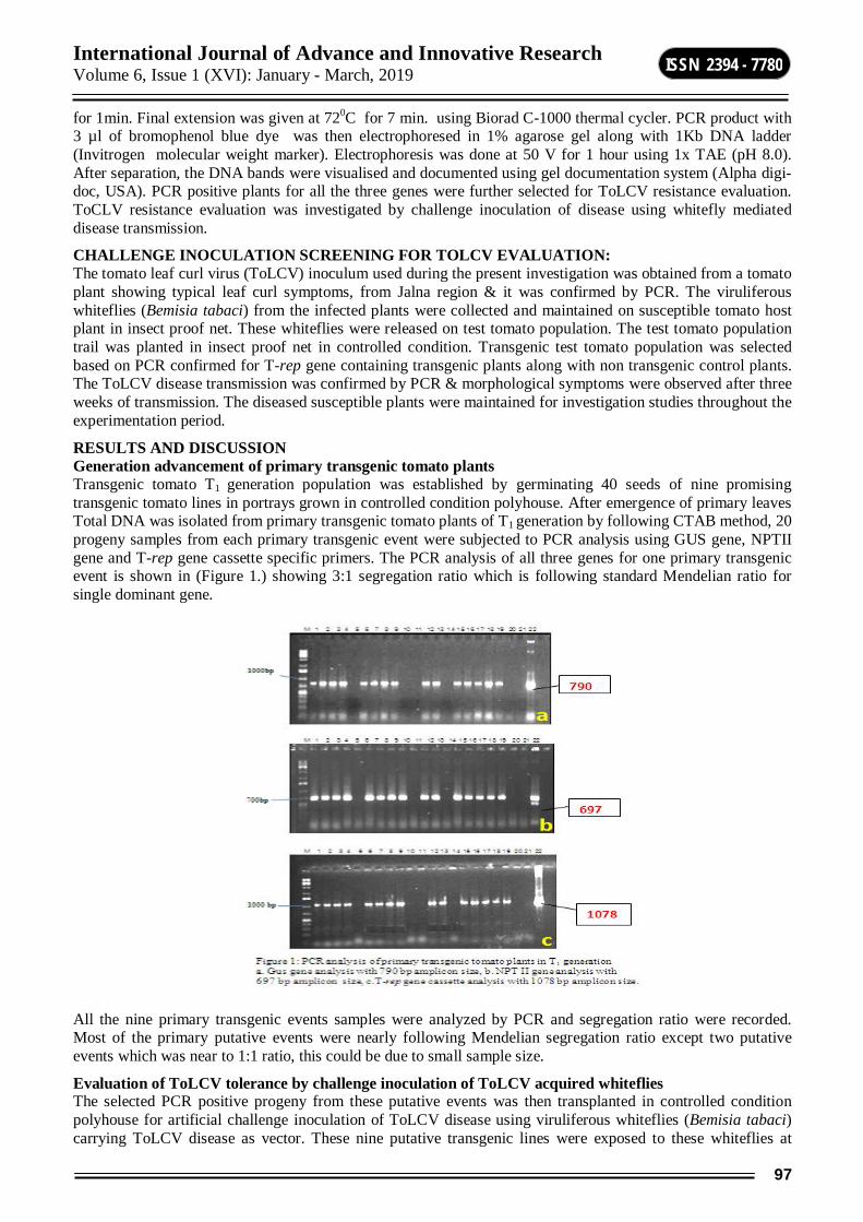
International Journal of Advance and Innovative Research Volume 6, Issue 1 (XVI): January - March, 2019
97
ISSN 2394 - 7780
for 1min. Final extension was given at 720C for 7 min. using Biorad C-1000 thermal cycler. PCR product with 3 µl of bromophenol blue dye was then electrophoresed in 1% agarose gel along with 1Kb DNA ladder (Invitrogen molecular weight marker). Electrophoresis was done at 50 V for 1 hour using 1x TAE (pH 8.0). After separation, the DNA bands were visualised and documented using gel documentation system (Alpha digi-doc, USA). PCR positive plants for all the three genes were further selected for ToLCV resistance evaluation. ToCLV resistance evaluation was investigated by challenge inoculation of disease using whitefly mediated disease transmission.
CHALLENGE INOCULATION SCREENING FOR TOLCV EVALUATION: The tomato leaf curl virus (ToLCV) inoculum used during the present investigation was obtained from a tomato plant showing typical leaf curl symptoms, from Jalna region & it was confirmed by PCR. The viruliferous whiteflies (Bemisia tabaci) from the infected plants were collected and maintained on susceptible tomato host plant in insect proof net. These whiteflies were released on test tomato population. The test tomato population trail was planted in insect proof net in controlled condition. Transgenic test tomato population was selected based on PCR confirmed for T-rep gene containing transgenic plants along with non transgenic control plants. The ToLCV disease transmission was confirmed by PCR & morphological symptoms were observed after three weeks of transmission. The diseased susceptible plants were maintained for investigation studies throughout the experimentation period.
RESULTS AND DISCUSSION Generation advancement of primary transgenic tomato plants Transgenic tomato T1 generation population was established by germinating 40 seeds of nine promising transgenic tomato lines in portrays grown in controlled condition polyhouse. After emergence of primary leaves Total DNA was isolated from primary transgenic tomato plants of T1 generation by following CTAB method, 20 progeny samples from each primary transgenic event were subjected to PCR analysis using GUS gene, NPTII gene and T-rep gene cassette specific primers. The PCR analysis of all three genes for one primary transgenic event is shown in (Figure 1.) showing 3:1 segregation ratio which is following standard Mendelian ratio for single dominant gene.
All the nine primary transgenic events samples were analyzed by PCR and segregation ratio were recorded. Most of the primary putative events were nearly following Mendelian segregation ratio except two putative events which was near to 1:1 ratio, this could be due to small sample size.
Evaluation of ToLCV tolerance by challenge inoculation of ToLCV acquired whiteflies The selected PCR positive progeny from these putative events was then transplanted in controlled condition polyhouse for artificial challenge inoculation of ToLCV disease using viruliferous whiteflies (Bemisia tabaci) carrying ToLCV disease as vector. These nine putative transgenic lines were exposed to these whiteflies at
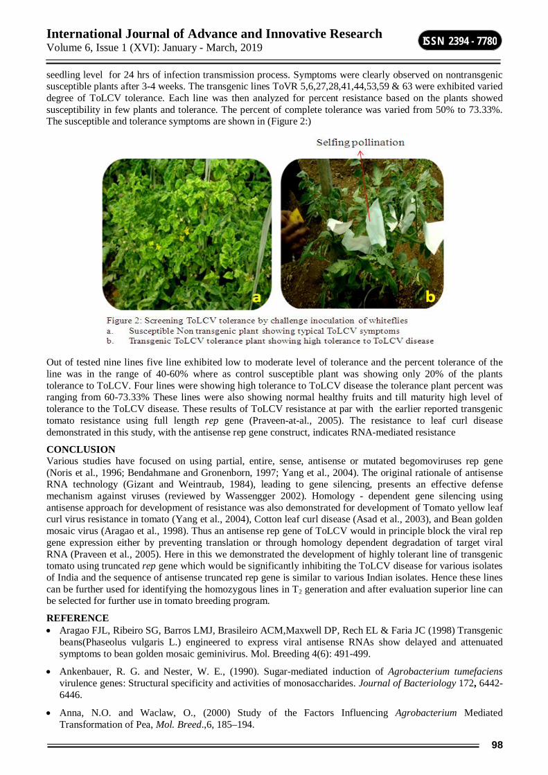
International Journal of Advance and Innovative Research Volume 6, Issue 1 (XVI): January - March, 2019
98
ISSN 2394 - 7780
seedling level for 24 hrs of infection transmission process. Symptoms were clearly observed on nontransgenic susceptible plants after 3-4 weeks. The transgenic lines ToVR 5,6,27,28,41,44,53,59 & 63 were exhibited varied degree of ToLCV tolerance. Each line was then analyzed for percent resistance based on the plants showed susceptibility in few plants and tolerance. The percent of complete tolerance was varied from 50% to 73.33%. The susceptible and tolerance symptoms are shown in (Figure 2:)
Out of tested nine lines five line exhibited low to moderate level of tolerance and the percent tolerance of the line was in the range of 40-60% where as control susceptible plant was showing only 20% of the plants tolerance to ToLCV. Four lines were showing high tolerance to ToLCV disease the tolerance plant percent was ranging from 60-73.33% These lines were also showing normal healthy fruits and till maturity high level of tolerance to the ToLCV disease. These results of ToLCV resistance at par with the earlier reported transgenic tomato resistance using full length rep gene (Praveen-at-al., 2005). The resistance to leaf curl disease demonstrated in this study, with the antisense rep gene construct, indicates RNA-mediated resistance
CONCLUSION Various studies have focused on using partial, entire, sense, antisense or mutated begomoviruses rep gene (Noris et al., 1996; Bendahmane and Gronenborn, 1997; Yang et al., 2004). The original rationale of antisense RNA technology (Gizant and Weintraub, 1984), leading to gene silencing, presents an effective defense mechanism against viruses (reviewed by Wassengger 2002). Homology - dependent gene silencing using antisense approach for development of resistance was also demonstrated for development of Tomato yellow leaf curl virus resistance in tomato (Yang et al., 2004), Cotton leaf curl disease (Asad et al., 2003), and Bean golden mosaic virus (Aragao et al., 1998). Thus an antisense rep gene of ToLCV would in principle block the viral rep gene expression either by preventing translation or through homology dependent degradation of target viral RNA (Praveen et al., 2005). Here in this we demonstrated the development of highly tolerant line of transgenic tomato using truncated rep gene which would be significantly inhibiting the ToLCV disease for various isolates of India and the sequence of antisense truncated rep gene is similar to various Indian isolates. Hence these lines can be further used for identifying the homozygous lines in T2 generation and after evaluation superior line can be selected for further use in tomato breeding program.
REFERENCE Aragao FJL, Ribeiro SG, Barros LMJ, Brasileiro ACM,Maxwell DP, Rech EL & Faria JC (1998) Transgenic
beans(Phaseolus vulgaris L.) engineered to express viral antisense RNAs show delayed and attenuated symptoms to bean golden mosaic geminivirus. Mol. Breeding 4(6): 491-499.
Ankenbauer, R. G. and Nester, W. E., (1990). Sugar-mediated induction of Agrobacterium tumefaciens virulence genes: Structural specificity and activities of monosaccharides. Journal of Bacteriology 172, 6442-6446.
Anna, N.O. and Waclaw, O., (2000) Study of the Factors Influencing Agrobacterium Mediated Transformation of Pea, Mol. Breed.,6, 185–194.

International Journal of Advance and Innovative Research Volume 6, Issue 1 (XVI): January - March, 2019
99
ISSN 2394 - 7780
Asad S, Haris WA, Bashir A, Zafar Y, Malik KA, Malik NN & Lichtenstei CP (2003)
Transgenic tobacco expressing geminiviral RNAs are resistant to the serious viral pathogen causing cotton leaf curl disease. Arch. Virol. 148(12): 2341–2352
Baulcombe DC (1996) Mechanisms of pathogen-derived resistance viruses in transgenic plants. Plant Cell 8: 1833–1844
Bendahmane M & Gronenborn B (1997) Engineering resistance against tomato yellow leaf curl virus (TYLCV) using antisense RNA. Plant Mol. Biol. 33: 351–357.
Brunt A, Crabtree K, Dallwitz M, Gibbs A & Watson L (1996)Viruses of Plants.
Description and Lists from the VIDE Database, CAB International, Wallingford, UK, p. 165
Dougherty WG & Parks TD (1995) Transgenes and gene suppression: Telling us something new?. Curr. Opin. Cell Biol. 7:399–405
Flavell RB (1994) Inactivation of gene expression in plants as a consequence of specific sequence duplication. Proc. Natl.Acad. Sc. USA 91: 3490–3496
Frary A & Van Eck J (2005) Organogenesis from transformed tomato explants. Methods Mol. Biol. 286: 141–150
Genetic Engineering. In: Rechcigl NA & Rechcigl JE (eds) Environmentally Safe Approaches to Crop Disease Control. Lewis publishers CRC press, Boca Raton, Florida (pp 333–368)
Gizant J & Weintraub H (1984) Inhibition of thymidine kinase gene expression by antisense RNA: a molecular approach to genetic analysis. Cell 36: 1007–1015
Goodwin J, Chapman K, Swaney S, Park TD, Wernsman EA & Dougherty WG (1996) Genetic and biochemical dissection of transgenic RNA-mediated virus resistance. Plant Cell 8:95–105
Horsch, R. B., Fraley, R. T., Rogers, S. G.,Sanders, P. R., Lloyd, A. and Hoffman, N., (1984). Inheritance off unctional foreign genes in plants.Science 223, 496-498.
Jefferson, R. A., Kavangh, T.A. and BevanM. W., (1987). GUS fusions: _-glucuronidase as a sensitive and versatile gene fusion marker in higher plants. EMBO J. 6, 3901–3907.
Kottearachchi, N. S., Kertbundit, S. and Juricek, M., (2000). Agrobacterium mediated transformation of Cucumis melo with replicase gene from papaya ring spot virus and regeneration of transformed plants.Tropical Agricultural Research and Extention, 3 (2), 94-97.
Moghaieb, R.E.A., Saneoka, H., Youssef,S. S., EL-Sharkawy, A.M. and Fujita, K., (2006). Improvement of salt Tolerance in Tomato plant(Lycoperiscon esculentum) by transformation with ectoine biosynthetic genes.Transgenicplant journal, 1(1), 228-232.
Murashige, T. and Skoog, F., (1962). Arevised medium for rapid growthand bioassays with tobacco tissueculture. Physiologia Plantarum, 15,473-379.
Praveen S, Dasgupta A & Varma A (2004) Phylogenetic analysis and homologies of the replicase of tomato leaf curl geminiviruses: implications for obtaining pathogen derived resistance. Virus Gene 28(1): 197–201
Praveen S, Mishra AK & Dasgupta A (2005) Antisense suppression of replicase gene expression recovers tomato plants from leaf curl infection. Plant Sci. 168: 1011–1014
Prematilake, D.P., Power, J.B. and Davey,M.R., (2002). Genetic transformation of cultivated tomato (Lycopersicon esculentum) with Agrobacterium. Annals of the SriLanka Department of Agriculture, 4,207-214.
Van Roekel, J. S. C., Damm, B., Melchers,L. S., and Hoekema, A., (1993)Factors Influencing Transformation Frequency of Tomato(Lycoperisicon esculentum), PlantCell Rep., 12, 644–647.
Wu, Y.F., Chen, Y., Liang, X.M., andWang, X.Z., (2005) An Experimental Assesment of The Factors Influencing Agrobacterium-Mediated Transformation inTomato, Plant Physiology., 53 (2),252-256.
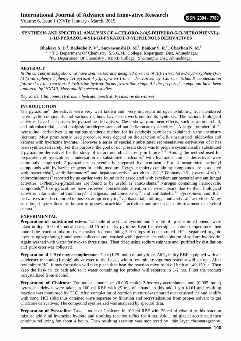
International Journal of Advance and Innovative Research Volume 6, Issue 1 (XVI): January - March, 2019
100
ISSN 2394 - 7780
SYNTHESIS AND SPECTRAL ANALYSIS OF 4-CHLORO-2-(4,5-DIHYDRO-5-(3-NITROPHENYL)-1-1H-PYRAZOL-4-YL)-1H-PYRAZOL-3-YL)PHENOLS DERIVATIVES
Bhakare S. D.1, Badadhe P. V2., Suryawanshi D. M.3, Bankar S. R.4, Chavhan N. M.5
1, 3, 4, 5PG Department Of Chemistry, S.S.G.M., College, Kopargaon, Dist. Ahmednagar 2PG Department Of Chemistry , RBNB College, Shrirampur Dist. Ahmednagar
ABSTRACT In the current investigation, we have synthesized and designed a series of (E)-1-(5-chloro-2-hydroxyphenyl)-3-(3-(3-nitrophenyl-1-phenyl-1H-pyrazol-4-yl)prop-2-en-1-one derivatives by Claisen- Schmidt condensation followed by the reaction of hydrazine hydrate forms pyrazoline rings. All the prepared compound have been analyzed by 1HNMR, Mass and IR spectral studies.
Keywords: Chalcones, Hydrazine hydrate, Spectral, Pyrazoline derivatives
INTRODUCTION The pyrazoline1 derivatives were very well known and very important nitrogen exhibiting five membered heterocyclic compounds and various methods have been work out for its synthesis. The various biological activities have been posses by pyrazoline derivatives. These shows prominent effects, such as antimicrobial, anti-microbacterial, , anti-analgesic antidepressant and anti-inflammatory activities2. A large number of 2-pyrazoline derivatives using various synthetic method for its synthesis have been explained in the chemistry literature. Most prominently used procedure were depend on the reaction of α,β- unsaturated aldehydes and ketones with hydrazine hydrate. However a series of specially substituted representatives derivatives of it has been synthesized rarely. For this purpose the goal of our present study was to prepare systematically substituted 2-pyrazoline derivatives for the study of its antimicrobial activity in future.3, 4 Among the method used for preparation of pyrazolines condensation of substituted chalcones4 with hydrazine and its derivatives were commonly employed. 2-pyrazolenes conveniently prepared by treatment of α β unsaturated carbonyl compounds with hydrazine reagents in acidic medium. Pyrazole moiety containing compounds are associated with bactericidal5, antiinflammatory6 and hepatoprotective7 activities. 2-(1,3-Diphenyl-1H- pyrazol-4-yl)-3-chlorochromones8 reported by us earlier were found to be associated with excellent antibacterial and antifungal activities. 1-Phenyl-2-pyrazolines are found to be useful as antioxidants.9 Nitrogen containing heterocyclic compounds10 like pyrazolines have received considerable attention in recent years due to their biological activities like anti- inflammatory,11 analgesic, anticonvulsant,12 and antidiabetic.13 Pyrazolines and their derivatives are also reported to possess antiprotcolytic,14 antibacterial, antifungal and antiviral15 activities. Many substituted pyrazolines are known to possess acaricidal16 activities and are used in the treatment of cerebral edema.17
EXPERIMENTAL Preparation of substituted esters: 1.2 mole of acetic anhydride and 1 mole of p-substituted phenol were taken in dry 100 ml conical flask; add 15 ml of dry pyridine. Kept for overnight at room temperature, then poured the reaction mixture over crushed ice containing 5-10 drops of concentrated HCl. Separated organic layer using separating funnel were collected and washed with 1percent ice cold solution of sodium hydroxide. Again washed with water for two- to three times. Then dried using sodium sulphate and purified by distillation and pure ester was collected.
Preparation of 2-Hydroxy acetophenone: Take (1.25 mole) of anhydrous AlCl3 in dry RBF equipped with air condenser then add (1 mole) above ester to the flask , within few minute vigorous reaction will set up . After few minute HCl fumes formation will take place then heat the reaction mixture in oil bath at 140-1500 c. Then keep the flask in ice bath add to it water containing ice product will separate in 1-2 hrs. Filter the product recrystallized from alcohol.
Preparation of Chalcone: Equimolar amount of (0.005 mole) 2-hydroxy-acetophnone and (0.005 mole) pyrazole aldehyde were taken in 100 ml RBF with 25 mL of ethanol to this add 1 gm KOH and resulting reaction was monitored by TLC. After completion of reaction mixture was poured over crushed ice and acidify with conc. HCl solid thus obtained were separate by filtration and recrystalization from proper solvent to get Chalcone derivatives. The compound synthesized was analyzed by spectral data.
Preparation of Pyrazoline: Take 1 mole of Chalcone in 100 ml RBF with 20 ml of ethanol to this reaction mixture add 2 ml hydrazine hydrate and resulting reaction reflux for 4 hrs. Add 1 ml glacial acetic acid then continue refluxing for about 4 hours. Then resulting reaction was monitored by thin layer chromatography.
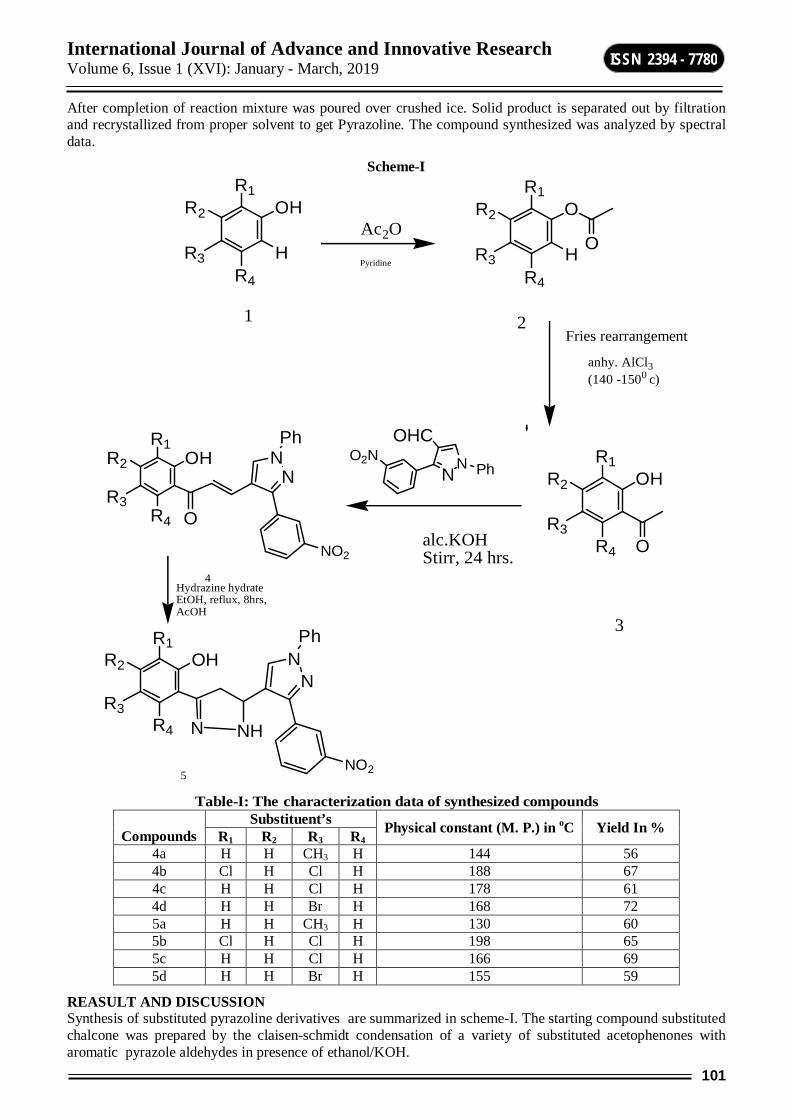
International Journal of Advance and Innovative Research Volume 6, Issue 1 (XVI): January - March, 2019
101
ISSN 2394 - 7780
After completion of reaction mixture was poured over crushed ice. Solid product is separated out by filtration and recrystallized from proper solvent to get Pyrazoline. The compound synthesized was analyzed by spectral data.
Scheme-I R1
OH
HR4
R3
R2Ac2O
Pyridine
R1O
H
R2
R4
R3O
Fries rearrangement
anhy. AlCl3 (140 -1500 c)
R1OHR2
R4
R3O
NN
OHC
alc.KOHStirr, 24 hrs.
R1OH
R4
R3
R2N
NPh
O
Hydrazine hydrate EtOH, reflux, 8hrs,AcOH
R1OH
R4
R3
R2N
NPh
N NH
1 2
3
4
5
PhO2N
NO2
NO2
Table-I: The characterization data of synthesized compounds
Compounds Substituent’s Physical constant (M. P.) in oC Yield In % R1 R2 R3 R4
4a H H CH3 H 144 56 4b Cl H Cl H 188 67 4c H H Cl H 178 61 4d H H Br H 168 72 5a H H CH3 H 130 60 5b Cl H Cl H 198 65 5c H H Cl H 166 69 5d H H Br H 155 59
REASULT AND DISCUSSION Synthesis of substituted pyrazoline derivatives are summarized in scheme-I. The starting compound substituted chalcone was prepared by the claisen-schmidt condensation of a variety of substituted acetophenones with aromatic pyrazole aldehydes in presence of ethanol/KOH.
International Journal of Advance and Innovative Research Volume 6, Issue 1 (XVI): January - March, 2019
102
ISSN 2394 - 7780
O H
N
N
H
NN
NO-
O
Hb
Ha
Hx
Cl
The chalcones (4a-d) were characterized by IR, 1HNMR, Mass Spectrometry. The 1H NMR spectra of pyrazolines 4c exhibit characteristic feature signals due to the ABX system. The Ha proton which is eclipsed by Hx proton indicates an apparent doublet of doublet at 3.59. The Hb proton shows 3.06 doublet of a doublet. The Hx proton appeared downfield at δ 5.12 as a doublet of a doublet. There are two broad bands appears due to alcoholic –OH with N of pyrazoline ring and another due to –NH with O of nitro group.
4c: IR(in cm-1) 3133.4 ( O-H), 1650.6 (C=O),749.31 (C-Cl),1596.3 (C=C),1619.8 (C=C) , 1540 & 1350 (-NO2).
1H NMR:10.91 δ (-OH), 6.04 δ ( d, olefinic 1H), 6.93 δ (d, olefinic 1H), 6.97-7.74 δ (m, aromatic 12H) , 8.00 δ (s, pyrazole 1H).
MASS:M/Z (m+1) (m+2)= 446, 448.
5c: IR(in cm-1) 3158.20 (-NH), 1540 & 1350 (-NO2), 751.0 (C-Cl), 1601.0 , (C=C), 3137.0 cm-1 (-O-H) 1H NMR: 3.06 δ (dd, pyrazoline 1H),3.59 δ (dd, pyrazoline 1H) 6.97-7.74 δ (m, aromatic 12H) ,8.00 δ ( s, pyrazole 1H), 10.91 δ s, -OH), 5.12 δ (dd , pyrazoline 1H).
MASS- M/Z: (m+1) (m+2)= 460, 462.
CONCLUSION AND FUTURE ASPECTS A simple, efficient and general method has been developed for the preparation of substituted pyrazoline derivatives. The synthesized titled compound has satisfactory structure which was confirmed by spectral tools. We have obtained the good yield. In future we are going to screen its biological activities .
ACKNOWLEDGEMENTS The authors are thankful to Dr. A. B. Nikumbh Incharge Principal SSGM College Kopargaon for providing laboratory facilities and for constant encouragement. The Authors are also thankful to head of the chemistry department for spectral analysis.
REFERENCES [1] Cottet f, Marrul M, Lefebvre O and Schlosser M, “ Recommendable routes to trifluoromethyl substituted
pyridine and quinoline carboxylic acid” Eur J Org Chem, Vol.2003 issue 8, 1559-1568, 2003.
[2] Ghotekar D S, Joshi R S, Mandhane P G, Bhagat S S, & Gill C H, Synthesis of some biologically important fluorinated 3-chlorochromones and 1,5-benzothiazepines as antimicrobial and antifungal agents”, Ind J of Chem, Vol-49B, pp 1267-1270, 2010.
[3] Chavhan NM, Badadhe PV, Joshi RS Mandhane PG and Gill CH, “Synthesis and antibacterial screening of some fluoroflavones’’ IJHC, Vol. 19, 163-166, 2009.
[4] N. M. Chavhan*1, P. V. Badadhe2, S. N. Shelke1 “Synthesis and screening of biological activities of some pyrazoline derivatives”, International Journal of Innovative Research in Science, Engineering and Technology, Vol. 4 (2), pp-417-421, 2015.
[5] Bhat BA, Dhar KL, Puri SC, Saxsena AK, Shanmugavel M and Qazi GN,” Synthesis and biological evaluation of chalcones and their derived pyrazoles as potential cytotoxic agents” Bioorg Med Chem Lett, 15, 3177, 2005.

International Journal of Advance and Innovative Research Volume 6, Issue 1 (XVI): January - March, 2019
103
ISSN 2394 - 7780
[6] Saundane A R & Prabhakar W, “Synthesis, antimicrobial & antioxidant activities of some indole analogues containing naphyridine and pyrimidonaphthyridine systems”, Ind J of Chem, Vol-51B, pp 1593-1606, 2012.
[7] Singh J P, Dulwat M, Jaitawat N, Chundawat S S, Devpura A & Dulwat S S, “ Microwave enhanced Claisen-Schmidt condensation: A green route to chalcones”, Ind J Chem, Vol- 51B, pp 1623-1627, 2012.
[8] Shelke S N, Mhaske G R, Bonifacio V D B, Gawande M B, “ Green Synthesis & anti-infectives of fluorinated pyrazoline derivatives’, Bioorg Med Chem Lett, Vol-22, pp5727-5730 2012.
[9] Bhagat S S, Ghotekar D S, Badadhe P V, Chavan N M, Dixit P P and Gill C H, “Synthesis and antimicrobial screening of pyrazolines and benzothiazepines incorporated with thiophene” Indian J Het Chem, 20, 355-358, 2011.
[10] Badadhe P V, Chavan N M, Mandhane P G, Joshi R S, Nagargoje D R and Gill C H, “Synthesis and characterization of some novel isoxazolines and pyrazolines as potent antimicrobial agents” Indian J Chem, 50B, 879, 2011.
[11] N. M. Chavhan*, “Environmentally Benign Ultrasound Promoted Synthesis Of Some Important Pyrazoline Derivatives As Antibacterial And Antifungal Agents”, IJRSET, Vol. 4 (4), pp-3279-3284, 2015.
[12] S Y Patil, R J Oswal, A S Sayare, S L Landge & R V Antre, “Synthesis, characterization & Antibacterial evaluation of novel 2-pyrazoline derivatives”, Der Pharma Chemica Vol- 4(1) pp 33-38, 2012.
[13] V V Kotla, V K Dalavai & V R Chundari, “ Synthesis & biological activity studies of some novel pyrazoline derivatives”, Der Pharma Chemica, Vol 4(5), pp 2003-2008, 2012.
[14] M R Patel, B L Dodiya, R M Chetiya, K A Joshi, P B Vekaria, A H Bapodara & H S Joshi, “Synthesis & Antimicrobial evaluation of pyrazoline derivatives”, Int J Chem Tech Research, Vol-3(2), pp 967-974, 2011.
[15] Acharya B N, Saraswat D, Tiwari M, Shrivastva A K, Ghorpade R, Bapna S & Kaushik M P, “ Synthesis & antimicrobial evaluation of 1,2,5-trisubstituted pyrazolines”, Eur J Med Chem, Vol-45, 430-438, 2010.
[16] A V Chate, R S Joshi, P G Mandhane, & C H Gill, “ Synthesis, characterization & antimicrobial screening of some novel chalcones & their derivatives”, Ind J of Chem, Vol-51B, pp 1642-1648, 2012.
[17] Diwakar S D, Bhagat S S, Shingare M S and Gill C H, “ Synthesis and Antimicrobial screening of some novel 2-(5-(4-(1H-Benzo[d][1,2,3]triazol-1-yl)phenyl-4,5-dihydro-1H-pyrazol-3-yl)phenols incorporated by triazole moiety” Bioorg Med Chem Lett, 18, 4678, 2008.
International Journal of Advance and Innovative Research Volume 6, Issue 1 (XVI): January - March, 2019
104
ISSN 2394 - 7780
PHYTOCHEMICAL STUDIES IN LEAF DRUG Tylophora indica (Burm. f.) Merr
RupaliBiradar 1 and Vikas Gambhire2 1Department of Botany, Indraraj Arts, Commerce and Science College, Sillod, Aurangabad
2Department of Botany, The Institute of Science, Mumbai
ABSTRACT Tylophora indica (Burm.f.) Merr.istwining, perennial, slender, much branched undershurb. It belngs to family Asclepiadaceae. Its leaves are medicinally exploited to treat several diseases and disorders. Phytochemical studies in this leafy drug are carried out to standardize and detect the adulteration in it. The phytochemical studies include details of characters of leaf powder like colour, odour, taste, Alkaloids, Anthraqinone, Iridoids, Saponins, Steroids, Tannins (Qualitative) and dry matter, bulk density, nitrogen, Water soluble nitrogen, crude protein, crude fat, crude fiber, total ash, acid insoluble ash, acid soluble ash, water insoluble ash, water soluble ash, calcium, reducing sugar, total sugar, non-reducing sugar, cellulose, gross energy, phosphorus,extractive values in 10 solvents (Quantitative). The above parameters can be applied to standardize this leaf drug.
Keywords: Phytochemical studies, Adulterations, Qualitative, Quantitative
INTRODUCTION Tylophora indica (Burm.f.) Merr.is twining, perennial, slender, much branched undershurb. It belngs to family Asclepiadaceae. Its leaves are medicinally important and are used to cure Allergy, Dermatitis, Rheumatism (Gupta and Bal, 1956; Shivpuri et al, 1972), Arthritis, Cold, Dysentery, Expectorant, Hay fever (CSIR 1948-1976), Bronchial asthma (Bielory and Lupoli, 1999), Syphilitic rheumatism, Whooping cough (Nadkarni, 1976). It is also used as Blood purifier (Nadkarni, 1976), Diaphoretic (CSIR 1948-1976) and Laxative (CSIR 1948-1976). The leaves are often adulterated with other leaf samples. During present investigation an attempt was made to standardize the leaves ofTylophora indica (Burm.f.) Merr.byusing phytochemical characters.
MATERIALS AND METHODS The leaf samples were collected from the medium sized authentically identified plant species from different localities of Marathwada. The leaves were removed carefully by hand pricking without damaging the plants. The leaves were collected in polythene bags and brought to the laboratory within 2-5 hours. Some leaves were preserved in 70% alcohol for their dermatology and anatomical work. Other were initially dried in shade and later in oven at 60oC till constant weight, then made in to fine powder and stored in sealed plastic container for further analysis (Sadasivam and Manickem, 2008).
RESULTS Phytochemical characters of leaf powder A) Physical characters (Table 1)
Sr. No. Character Expression (%) 01 Colour Green 02 Odour Disagreeable 03. Taste Astringent
B) Qualitative Characters (Table 2)
Sr. No. Character Expression 01 Alkaloids + 02 Anthraquinones - 03. Iridoids - 04 Saponins + 05 Steroids + 06 Tannins +
C ) Quantitative Characters (Table 3)
Sr. No. Character Expression (%) 01 Dry matter 22 02 Bulk Density 0.282 mg/cm3 03 Nitrogen 2.33 04 Water soluble nitrogen 1.5
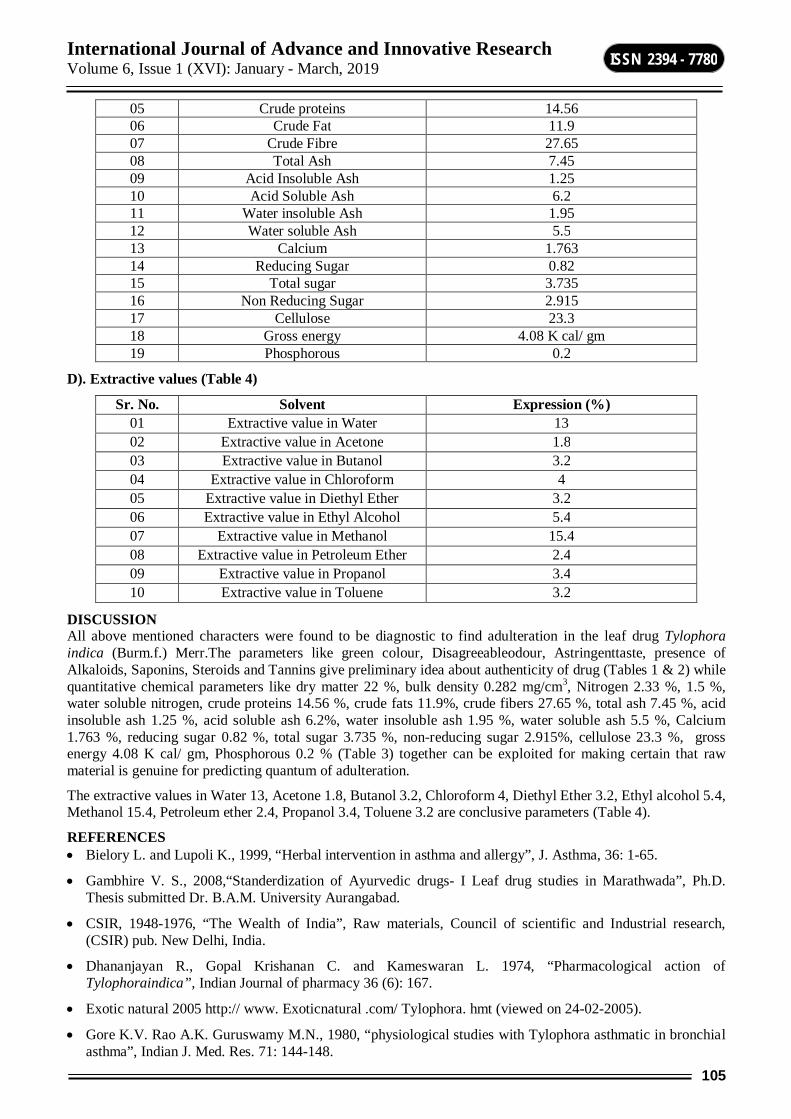
International Journal of Advance and Innovative Research Volume 6, Issue 1 (XVI): January - March, 2019
105
ISSN 2394 - 7780
05 Crude proteins 14.56 06 Crude Fat 11.9 07 Crude Fibre 27.65 08 Total Ash 7.45 09 Acid Insoluble Ash 1.25 10 Acid Soluble Ash 6.2 11 Water insoluble Ash 1.95 12 Water soluble Ash 5.5 13 Calcium 1.763 14 Reducing Sugar 0.82 15 Total sugar 3.735 16 Non Reducing Sugar 2.915 17 Cellulose 23.3 18 Gross energy 4.08 K cal/ gm 19 Phosphorous 0.2
D). Extractive values (Table 4)
Sr. No. Solvent Expression (%) 01 Extractive value in Water 13 02 Extractive value in Acetone 1.8 03 Extractive value in Butanol 3.2 04 Extractive value in Chloroform 4 05 Extractive value in Diethyl Ether 3.2 06 Extractive value in Ethyl Alcohol 5.4 07 Extractive value in Methanol 15.4 08 Extractive value in Petroleum Ether 2.4 09 Extractive value in Propanol 3.4 10 Extractive value in Toluene 3.2
DISCUSSION All above mentioned characters were found to be diagnostic to find adulteration in the leaf drug Tylophora indica (Burm.f.) Merr.The parameters like green colour, Disagreeableodour, Astringenttaste, presence of Alkaloids, Saponins, Steroids and Tannins give preliminary idea about authenticity of drug (Tables 1 & 2) while quantitative chemical parameters like dry matter 22 %, bulk density 0.282 mg/cm3, Nitrogen 2.33 %, 1.5 %, water soluble nitrogen, crude proteins 14.56 %, crude fats 11.9%, crude fibers 27.65 %, total ash 7.45 %, acid insoluble ash 1.25 %, acid soluble ash 6.2%, water insoluble ash 1.95 %, water soluble ash 5.5 %, Calcium 1.763 %, reducing sugar 0.82 %, total sugar 3.735 %, non-reducing sugar 2.915%, cellulose 23.3 %, gross energy 4.08 K cal/ gm, Phosphorous 0.2 % (Table 3) together can be exploited for making certain that raw material is genuine for predicting quantum of adulteration.
The extractive values in Water 13, Acetone 1.8, Butanol 3.2, Chloroform 4, Diethyl Ether 3.2, Ethyl alcohol 5.4, Methanol 15.4, Petroleum ether 2.4, Propanol 3.4, Toluene 3.2 are conclusive parameters (Table 4).
REFERENCES Bielory L. and Lupoli K., 1999, “Herbal intervention in asthma and allergy”, J. Asthma, 36: 1-65.
Gambhire V. S., 2008,“Standerdization of Ayurvedic drugs- I Leaf drug studies in Marathwada”, Ph.D. Thesis submitted Dr. B.A.M. University Aurangabad.
CSIR, 1948-1976, “The Wealth of India”, Raw materials, Council of scientific and Industrial research, (CSIR) pub. New Delhi, India.
Dhananjayan R., Gopal Krishanan C. and Kameswaran L. 1974, “Pharmacological action of Tylophoraindica”, Indian Journal of pharmacy 36 (6): 167.
Exotic natural 2005 http:// www. Exoticnatural .com/ Tylophora. hmt (viewed on 24-02-2005).
Gore K.V. Rao A.K. Guruswamy M.N., 1980, “physiological studies with Tylophora asthmatic in bronchial asthma”, Indian J. Med. Res. 71: 144-148.
International Journal of Advance and Innovative Research Volume 6, Issue 1 (XVI): January - March, 2019
106
ISSN 2394 - 7780
Gupta M.P., T.D. Arias, M. Correa and S. S. Lamba, 1997, “Ethnopharmacognostic observation on panamian Medicinal plant 1.”, Q.J. Crude Drug Res; 17(314): 115-130.
Gupta B. and Bal S.N., 1956, “Pharmacognostic studies of Tylophpraindica”, J. Sci. Industr. Res. 15C: 111.
Haranath, PSRK and Shamalakumari S., 1975, “Experimental study on mode of action of Tylophoraasthmatica in bronchial asthma”, IJMR 62 (5); 661-670.
Holistic, 2004, “Http://www.holisticoline.com./ Remedies/ Diabetes/ diabetes Ayurveda. Html Viewed on 28-10-2004.
Nadkarni K. M. , 1976, ( Reprint ed. 2002), “ Indian Materia Neesa Vol. I ”, Popular Prakashan , Bombay, India. Pp. 1252-1253.
Naik V.N. 1998, “MarathwadyatilSamanyaVanaushadhi”, AmrutPrakashan, Aurangabad, (M.S.), India.
Naik V.N., 1998,“Flora of Marathwada”, AmrutPrakashan, Auranagabad.
Patel P.K., 2004, “Ethno Medicinal study of Patan District, North Gujarat”, Agrobios Newsletter, Vol II, issue.
Sadasivam S. and Manickam A., 2008, “Biochemical Methods”,New age International Publishers, New Delhi
Shivpuri D. N. , Singhal S. C. and Prakash D., 1972, “ Treatment of Asthma with an alcoholic extract of Tylophoraindica : a cross over” Double blind study, Ann Allergy 30 ; 407-412.
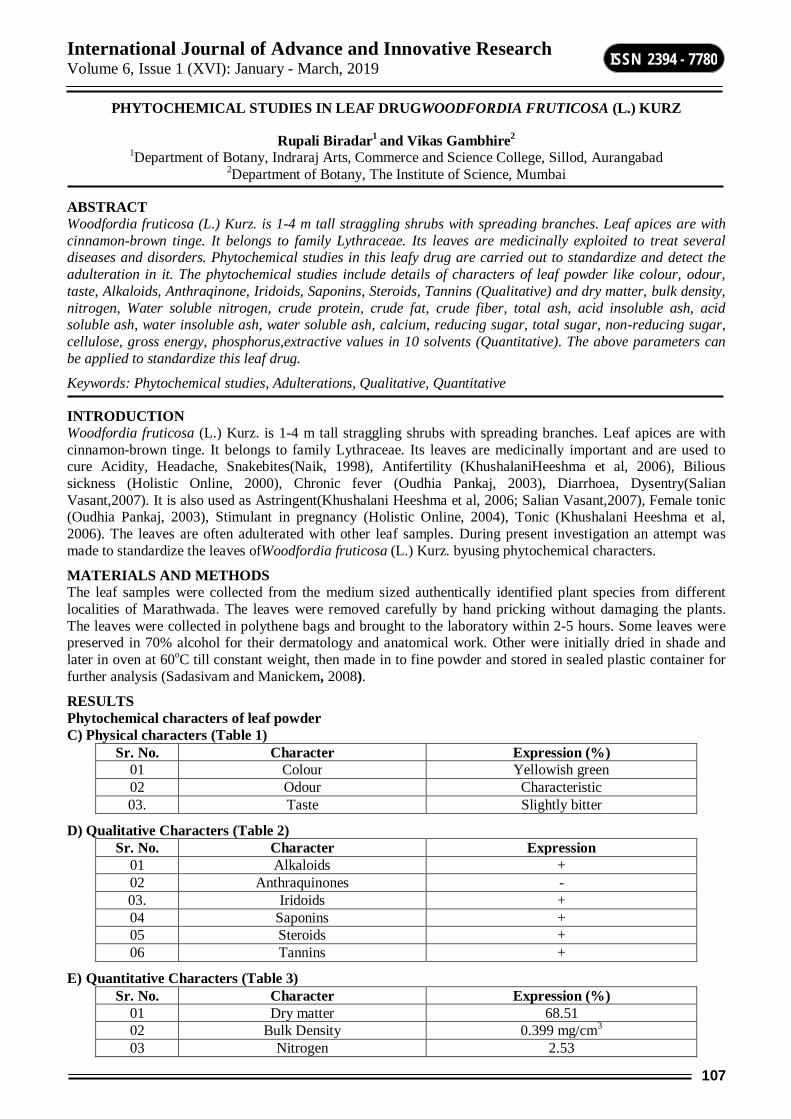
International Journal of Advance and Innovative Research Volume 6, Issue 1 (XVI): January - March, 2019
107
ISSN 2394 - 7780
PHYTOCHEMICAL STUDIES IN LEAF DRUGWOODFORDIA FRUTICOSA (L.) KURZ
Rupali Biradar1 and Vikas Gambhire2 1Department of Botany, Indraraj Arts, Commerce and Science College, Sillod, Aurangabad
2Department of Botany, The Institute of Science, Mumbai
ABSTRACT Woodfordia fruticosa (L.) Kurz. is 1-4 m tall straggling shrubs with spreading branches. Leaf apices are with cinnamon-brown tinge. It belongs to family Lythraceae. Its leaves are medicinally exploited to treat several diseases and disorders. Phytochemical studies in this leafy drug are carried out to standardize and detect the adulteration in it. The phytochemical studies include details of characters of leaf powder like colour, odour, taste, Alkaloids, Anthraqinone, Iridoids, Saponins, Steroids, Tannins (Qualitative) and dry matter, bulk density, nitrogen, Water soluble nitrogen, crude protein, crude fat, crude fiber, total ash, acid insoluble ash, acid soluble ash, water insoluble ash, water soluble ash, calcium, reducing sugar, total sugar, non-reducing sugar, cellulose, gross energy, phosphorus,extractive values in 10 solvents (Quantitative). The above parameters can be applied to standardize this leaf drug.
Keywords: Phytochemical studies, Adulterations, Qualitative, Quantitative
INTRODUCTION Woodfordia fruticosa (L.) Kurz. is 1-4 m tall straggling shrubs with spreading branches. Leaf apices are with cinnamon-brown tinge. It belongs to family Lythraceae. Its leaves are medicinally important and are used to cure Acidity, Headache, Snakebites(Naik, 1998), Antifertility (KhushalaniHeeshma et al, 2006), Bilious sickness (Holistic Online, 2000), Chronic fever (Oudhia Pankaj, 2003), Diarrhoea, Dysentry(Salian Vasant,2007). It is also used as Astringent(Khushalani Heeshma et al, 2006; Salian Vasant,2007), Female tonic (Oudhia Pankaj, 2003), Stimulant in pregnancy (Holistic Online, 2004), Tonic (Khushalani Heeshma et al, 2006). The leaves are often adulterated with other leaf samples. During present investigation an attempt was made to standardize the leaves ofWoodfordia fruticosa (L.) Kurz. byusing phytochemical characters.
MATERIALS AND METHODS The leaf samples were collected from the medium sized authentically identified plant species from different localities of Marathwada. The leaves were removed carefully by hand pricking without damaging the plants. The leaves were collected in polythene bags and brought to the laboratory within 2-5 hours. Some leaves were preserved in 70% alcohol for their dermatology and anatomical work. Other were initially dried in shade and later in oven at 60oC till constant weight, then made in to fine powder and stored in sealed plastic container for further analysis (Sadasivam and Manickem, 2008).
RESULTS Phytochemical characters of leaf powder C) Physical characters (Table 1)
Sr. No. Character Expression (%) 01 Colour Yellowish green 02 Odour Characteristic 03. Taste Slightly bitter
D) Qualitative Characters (Table 2) Sr. No. Character Expression
01 Alkaloids + 02 Anthraquinones - 03. Iridoids + 04 Saponins + 05 Steroids + 06 Tannins +
E) Quantitative Characters (Table 3) Sr. No. Character Expression (%)
01 Dry matter 68.51 02 Bulk Density 0.399 mg/cm3 03 Nitrogen 2.53
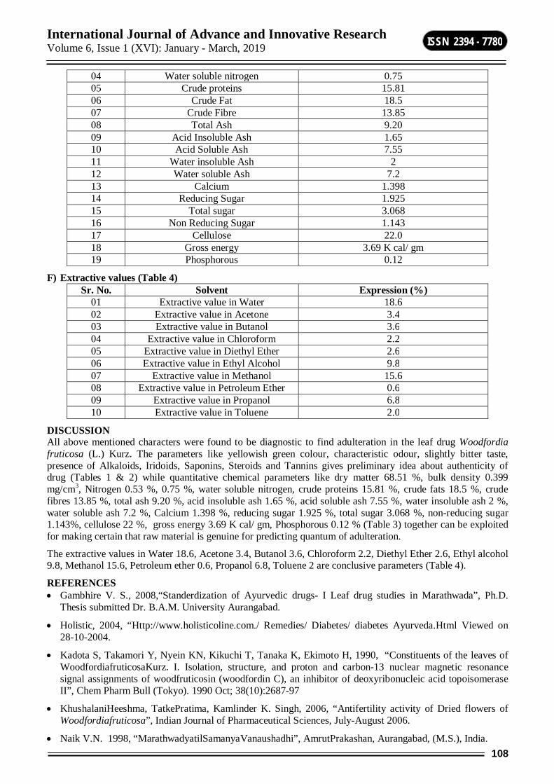
International Journal of Advance and Innovative Research Volume 6, Issue 1 (XVI): January - March, 2019
108
ISSN 2394 - 7780
04 Water soluble nitrogen 0.75 05 Crude proteins 15.81 06 Crude Fat 18.5 07 Crude Fibre 13.85 08 Total Ash 9.20 09 Acid Insoluble Ash 1.65 10 Acid Soluble Ash 7.55 11 Water insoluble Ash 2 12 Water soluble Ash 7.2 13 Calcium 1.398 14 Reducing Sugar 1.925 15 Total sugar 3.068 16 Non Reducing Sugar 1.143 17 Cellulose 22.0 18 Gross energy 3.69 K cal/ gm 19 Phosphorous 0.12
F) Extractive values (Table 4) Sr. No. Solvent Expression (%)
01 Extractive value in Water 18.6 02 Extractive value in Acetone 3.4 03 Extractive value in Butanol 3.6 04 Extractive value in Chloroform 2.2 05 Extractive value in Diethyl Ether 2.6 06 Extractive value in Ethyl Alcohol 9.8 07 Extractive value in Methanol 15.6 08 Extractive value in Petroleum Ether 0.6 09 Extractive value in Propanol 6.8 10 Extractive value in Toluene 2.0
DISCUSSION All above mentioned characters were found to be diagnostic to find adulteration in the leaf drug Woodfordia fruticosa (L.) Kurz. The parameters like yellowish green colour, characteristic odour, slightly bitter taste, presence of Alkaloids, Iridoids, Saponins, Steroids and Tannins gives preliminary idea about authenticity of drug (Tables 1 & 2) while quantitative chemical parameters like dry matter 68.51 %, bulk density 0.399 mg/cm3, Nitrogen 0.53 %, 0.75 %, water soluble nitrogen, crude proteins 15.81 %, crude fats 18.5 %, crude fibres 13.85 %, total ash 9.20 %, acid insoluble ash 1.65 %, acid soluble ash 7.55 %, water insoluble ash 2 %, water soluble ash 7.2 %, Calcium 1.398 %, reducing sugar 1.925 %, total sugar 3.068 %, non-reducing sugar 1.143%, cellulose 22 %, gross energy 3.69 K cal/ gm, Phosphorous 0.12 % (Table 3) together can be exploited for making certain that raw material is genuine for predicting quantum of adulteration.
The extractive values in Water 18.6, Acetone 3.4, Butanol 3.6, Chloroform 2.2, Diethyl Ether 2.6, Ethyl alcohol 9.8, Methanol 15.6, Petroleum ether 0.6, Propanol 6.8, Toluene 2 are conclusive parameters (Table 4).
REFERENCES Gambhire V. S., 2008,“Standerdization of Ayurvedic drugs- I Leaf drug studies in Marathwada”, Ph.D.
Thesis submitted Dr. B.A.M. University Aurangabad.
Holistic, 2004, “Http://www.holisticoline.com./ Remedies/ Diabetes/ diabetes Ayurveda.Html Viewed on 28-10-2004.
Kadota S, Takamori Y, Nyein KN, Kikuchi T, Tanaka K, Ekimoto H, 1990, “Constituents of the leaves of WoodfordiafruticosaKurz. I. Isolation, structure, and proton and carbon-13 nuclear magnetic resonance signal assignments of woodfruticosin (woodfordin C), an inhibitor of deoxyribonucleic acid topoisomerase II”, Chem Pharm Bull (Tokyo). 1990 Oct; 38(10):2687-97
KhushalaniHeeshma, TatkePratima, Kamlinder K. Singh, 2006, “Antifertility activity of Dried flowers of Woodfordiafruticosa”, Indian Journal of Pharmaceutical Sciences, July-August 2006.
Naik V.N. 1998, “MarathwadyatilSamanyaVanaushadhi”, AmrutPrakashan, Aurangabad, (M.S.), India.

International Journal of Advance and Innovative Research Volume 6, Issue 1 (XVI): January - March, 2019
109
ISSN 2394 - 7780
Naik V.N., 1998,“Flora of Marathwada”, AmrutPrakashan, Auranagabad.
Patel P.K., 2004, “Ethno Medicinal study of Patan District, North Gujarat”, AgrobiosNewsletter, Vol II, issue.
Oudhia Pankaj, 2003, “Interactions with the herb collectors of Gandai region, Chhattisgarh, India having rich traditional Medicinal knowledge about useful herb Dhawai (Woodfordiafruticosa)”, Botanical.com
Sadasivam S. and Manickam A., 2008, “Biochemical Methods”,New age International Publishers, New Delhi
SalianVasant M, 2007, “Dhawai (Woodfordia fruticosa)”www.piercenet.com

International Journal of Advance and Innovative Research Volume 6, Issue 1 (XVI): January - March, 2019
110
ISSN 2394 - 7780
BIOCHEMICAL STUDIES OF CESTODEPARASITE MONIEZIA (B) OF CAPRA HIRCUS FROM BEED DISTRICT
A. M. Budrukkar Department of Zoology, R. B. Attal College, Georai
ABSTRACT This paper contains biochemical study of cestode genus Moniezia (Blanchard) 1891of Capra hircus to understand their host parasite relationship. The protein contents in cestodes were estimated by the method given byGornell et al. (1994) and lipid content were estimated by the Barner`s and Blackstock method (1973).
Keywords: Moniezia, Cestode Parasite, Capra hircus
PROTEIN ESTIMATION The intestine of Capra hircus were examined at laboratory. The identical parasites were sorted out with the help of microscope. Small pieces of infected host intestine were also collected for the protein estimation. The protein content in the cestode parasites was estimated by Brand (1966) and Gornell et al. (1994) method. The worm were dried on blotting paper to remove water and taken wet weight of the tissue. The material was transferred in to previously weighted watch glass and kept in oven at 60°C for 24 hrs. Dried material was made into powder form. This powder weighed 250 mgs on balance. This material was grind with the help of morter pastle. Added with 5 ml of 10% TCA. Material was transferred to test tube and centrifuged for10 min. at 2000 rpm. Discard the supernatant and taken the residue add 1 ml of distilled water and 3 ml of Biuret solution. The tube was kept for half hour until lavender colour is developed. Colour reading was noted on colorimeter at 530 mm to note
Optical density
O. D. of Unknown tissue 1000
___________________ x mg of Protein x _________
O. D. Of known tissue weight of taken tissue
O. D. of unknown tissue = 0.35
O. D. of known tissue = 0.55
mg. of protein = 10
0.35 x 10 1000
_______ x ________
0.55 250
= 25.45mg/gm wet weight of the tissue.
By the same procedure amount of protein in the host intestine was estimated. The results showed that the intestine possessed 30.43 mg/gm wet weight of the tissue. These two comparisons reveal that Moniezia absorbed 25.45 mg/gm.
Lipid estimation: The intestine dissected and were found to be infected with the cestode. Parasite and host intestine kept in watch glass. This material was taken on blotting paper to remove excess of water and then it was weighted on balance to obtained wet weight of tissue. Tissue then kept at 80°C to completely dry. Tissue was powdered with the help of mortar pastle. Lipid was estimated by the Barner`s and Black stock method (1973). The lipid content was very high in the worms as compared to the host. The lipid level in Moniezia was 24.30 mg/100 mg ±S.D. whereas 20.85 was in the host i.e. Capra hircus
Glycogen estimation: To estimate glycogen in cestode as well as in host intestine, the tissue was dried on blotting paper to remove excess water. Material kept at 60°C for 24 hours. The 100 gms of dry material were homogenized in mortal pastle then added 5% TCA to it and was transferred in centrifuge tube. Material was digested in boiling water bath for 15 minutes. Cool and centrifuged for 15 minutes at 2000 rpm. One ml of supernatant was taken in tube and added with 3 ml of sulphuric acid and cooled for 15 minutes. Mixture shaken well, then readings was taken in colorimeter at 530μ.

International Journal of Advance and Innovative Research Volume 6, Issue 1 (XVI): January - March, 2019
111
ISSN 2394 - 7780
100 x U
Percentage of glycogen = _________
1.11 x S
U = O. D. of the unknown test solution
S = O. D. of the known test solution
1.11 = Conversion factor of glucose to glycogen
100 x 0. 43
Percentage of glycogen = __________
1.11 x 2
= 19.36 mg/100ml of solution.
The glycogen contain in host tissue was 19.81 mg/100ml of solution.
CONCLUTION The results revealed that the percentage of lipid is high in the parasite than their host and also high as compared to glycogen and protein. Cestodes are depends upon the host for the lipid source. Results indicate that distinctiveness host parasite relationship.
REFERENCES Aldrich. D. V., CHANDLER A. C. AND DAUGHRTY J. W. (1954): Intermediarty protein metabolism in
helminthes.
BAYLIS H. A. (1928): Systema Helminthology, Yamaguti Vol. II. The cestode of vertebrates.
BRAND T. VON. (1966): Biochemistry of parasite, Academic press New York.
BBALERAO G. D. (1932): A General Account of Helminth Parasites affecting Domestic Animal in india with methods of Collection, Preservation, staining, etc Indian Jour. Vet. Sci and Animal Husbandry 2: 1- 28.
KEMP A, VANRITS and HAIJNINGEN A.J. M. (1954): A Colorimetric method for the determination of glycogen in the tisuue. Biochemi. J. 56:646 648.

International Journal of Advance and Innovative Research Volume 6, Issue 1 (XVI): January - March, 2019
112
ISSN 2394 - 7780
COMPARISON BETWEEN MATERIAL SYNTHESIS BY SOLID STATE REACTION AND SOL-GEL AUTOCOMBUSTION METHOD
C. M. Kale Department of Physics, Indraraj Arts, Commerce and Science College, Sillod, Aurangabad
ABSTRACT Materials scientists have invested a long period for the synthesis of magnetic oxides. There are several methods available now a day for the synthesis of magnetic oxides. The properties of ferrite are known to depend upon preparation technique and preparation parameters. It has been reported that the properties of material prepared by two different techniques are different. By changing the preparative technique one can bring changes in the properties of a material. Synthesis of nano grain size particles proves to be one of the most interesting and important technique in the field of material science, as the small grain size particles have some of the interesting properties when compared to bulk particles in material processing and technological applications. In this paper, synthesis of material by solid state reaction method and sol-gel auto-combustion method are studied with their advantages and disadvantages in detal.
Keywords: Material synthesis, Solid state reaction, sol-gel, temperature, nanoferrite.
1. INTRODUCTION In recent years, major progress has been achieved in the era of nanoscience and nanotechnology. Nano-materials play very important role in our life. Material synthesis is the most important step in developing new materials with varying properties. There are number of methods available for the synthesis of various materials [1]. In this paper the standard ceramic and sol-gel autocombustion methods of preparation are briefly discussed. Both the method was comparatively discussed with their advantages and disadvantages. Selection of any synthesis method depends on the specifications required in the final product. The factors that influence the selection of methods and materials are - desired properties, cost of materials and its availability. A critical comparison of methods is required to make the best choice for given boundary conditions of targeted final material properties, raw materials, cost investment, and processing.
Several synthesis methods for preparation of polycrystalline sample are available in literature. Some of them well-known synthesis techniques are quoted below:
1. Solid state reaction method (SSR),
2. Sol-gel technique,
3. Co-precipitation technique,
4. Hydrothermal method, etc
1. Solid state reaction method (SSR) Solid solution ceramic samples are synthesized by mixing two or more solid compounds. The characteristics of starting constituents are retained and the desired mixture is formed. Basically, solids do not react at room temperature and hence, in order to complete the reaction, they are heated at higher temperatures [2]. Synthesis of ceramic powders by the conventional solid-state reaction method and repeated heating with intermediate mechanical or hand-grinding process is almost always essential to produce a single-phase powder to attain chemical homogeneity. In this technique, first of all starting precursors are weighed according to the stoichiometry of the compound with due consideration for impurity and moisture contents. The starting reactant chemicals selection depends on the reaction conditions and predictable nature of the product. The reactants are dried thoroughly before weighing. Reaction rate increases due to increase in surface area hence, fine grained precursors are used. Starting precursor materials are thoroughly mixed and then grinding it in an alcoholic or wet media to control the particle size and to make the homogeneous mixture. For this purpose, grinding operation is performed which helps in reducing the particlesize [3]. Process of reducing the size of particles affects the homogeneity and purity of the material. Distilled water is used as wetting medium with appropriate purity. A solid state reaction is accomplished between the starting materials at appropriate temperature. This process is known as firing or calcination. Calcination causes the starting materials to interact by inter-diffusion of their ions and resulting in a homogeneous body. Calcination also controls the shrinkage during sintering. After calcination, preferred shape is given to the powder then it is densified through sintering process. Sintering is a process of heating the material in a sintering furnace below its melting point until its particles adhere to
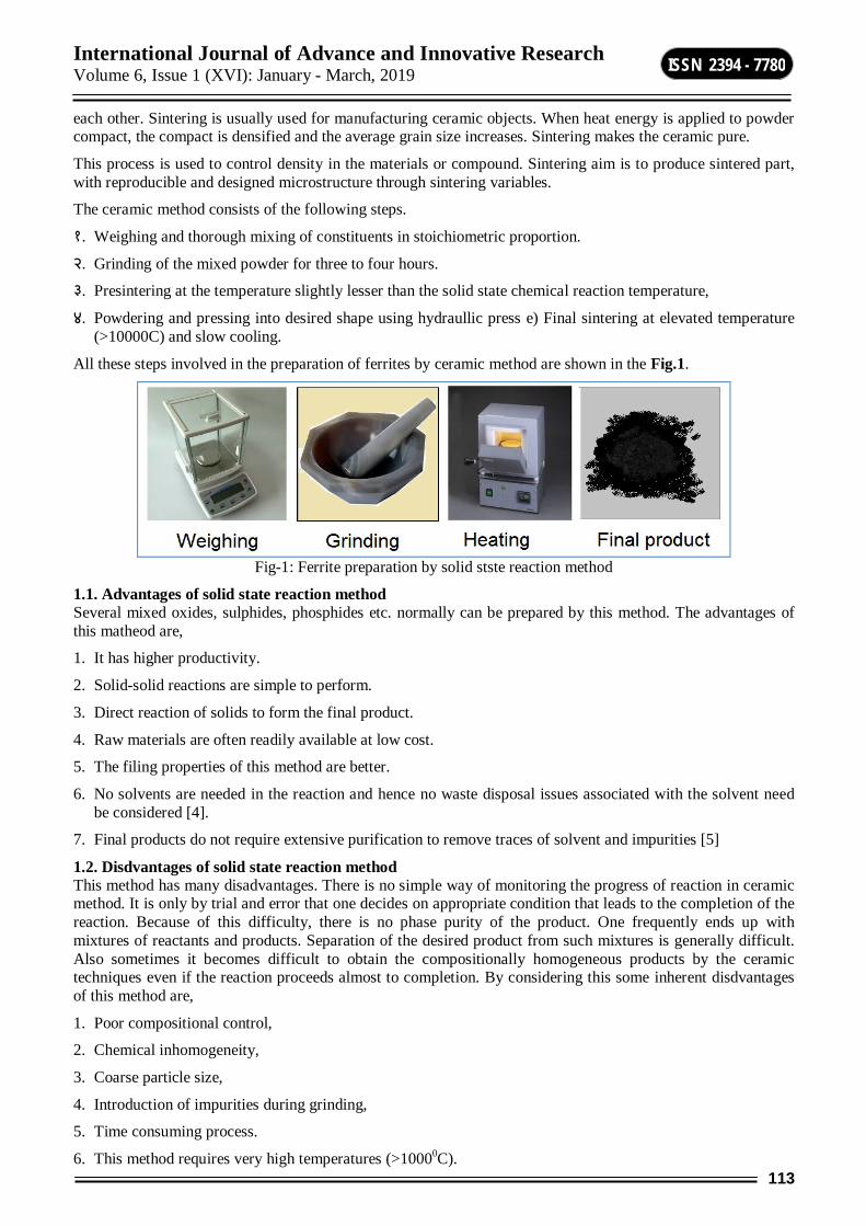
International Journal of Advance and Innovative Research Volume 6, Issue 1 (XVI): January - March, 2019
113
ISSN 2394 - 7780
each other. Sintering is usually used for manufacturing ceramic objects. When heat energy is applied to powder compact, the compact is densified and the average grain size increases. Sintering makes the ceramic pure.
This process is used to control density in the materials or compound. Sintering aim is to produce sintered part, with reproducible and designed microstructure through sintering variables.
The ceramic method consists of the following steps.
१. Weighing and thorough mixing of constituents in stoichiometric proportion.
२. Grinding of the mixed powder for three to four hours.
३. Presintering at the temperature slightly lesser than the solid state chemical reaction temperature,
४. Powdering and pressing into desired shape using hydraullic press e) Final sintering at elevated temperature (>10000C) and slow cooling.
All these steps involved in the preparation of ferrites by ceramic method are shown in the Fig.1.
Fig-1: Ferrite preparation by solid stste reaction method
1.1. Advantages of solid state reaction method Several mixed oxides, sulphides, phosphides etc. normally can be prepared by this method. The advantages of this matheod are,
1. It has higher productivity.
2. Solid-solid reactions are simple to perform.
3. Direct reaction of solids to form the final product.
4. Raw materials are often readily available at low cost.
5. The filing properties of this method are better.
6. No solvents are needed in the reaction and hence no waste disposal issues associated with the solvent need be considered [4].
7. Final products do not require extensive purification to remove traces of solvent and impurities [5]
1.2. Disdvantages of solid state reaction method This method has many disadvantages. There is no simple way of monitoring the progress of reaction in ceramic method. It is only by trial and error that one decides on appropriate condition that leads to the completion of the reaction. Because of this difficulty, there is no phase purity of the product. One frequently ends up with mixtures of reactants and products. Separation of the desired product from such mixtures is generally difficult. Also sometimes it becomes difficult to obtain the compositionally homogeneous products by the ceramic techniques even if the reaction proceeds almost to completion. By considering this some inherent disdvantages of this method are,
1. Poor compositional control,
2. Chemical inhomogeneity,
3. Coarse particle size,
4. Introduction of impurities during grinding,
5. Time consuming process.
6. This method requires very high temperatures (>10000C).

International Journal of Advance and Innovative Research Volume 6, Issue 1 (XVI): January - March, 2019
114
ISSN 2394 - 7780
2. Sol-gel autocombustion method (SSR) Sol gel method is an important mean of preparing inorganic oxides. It is a chemical method and involving both chemical and physical processes. A sudden increase in viscosity is the common feature in sol-gel processing, indicating the onset of gel formation. Sol-gel is the multi step process involving chemlcal and physical processes associated with hydrolysis, polymerization, gelatlon, condensation, drying and densification [6]. This process generally starts with the mixing of metal alkoxides or salts in water or in a suitable solvent (usually an alcohol) at ambient or slightly elevated temperatures [7]. Schematic diagram of various steps involved in the sol gel process are shown in Fig. 2.
Fig-2: Ferrite preparation by sol-gel method
In sol gel process, controlling the pH of starting solution is very much important to avoid the precipitation as well as to form the homogenous gel, which can achieved by the addition of base or acidic solutions [8]. Apart from the above, organic compounds with hydrophilic functional groups (hydroxides or carboxylates) in small molecules such as citric acid, succinic acid, oxalic acid, tartaric acid, acrylic acid, etc. and polymers like polyacrylic acid (PAA) and polyvinyl pynolidone (PVP) can be used with metal ion sources to form the sol as well as control the particle size and uniformity of the products [9-10]. Chelation of metal ions by carboxylic acid groups lead to a homogeneous distribution of the constituent ions in the obtained gel [11]. The gel intermediate is further heated between 150 "C and 300 "C to eliminate volatile organic components, excess water, etc., which results the dried intermediate powders. Single phase nanocrystalline metal oxides are obtained after calcining of dried gel powder at 400-8000C depends on the precursor chemical nature [12-13].
2.1. Advantages of sol-gel method The important advantages of the sol-gel method are. 1. Lower processing temperature 2. Smaller particle size and morphological control in poivder synthesis. 3. Sintering at low temperature also possible. 4. Better homogeneity 5. High phase purity compared to traditional ceramic method. 6. More uniform phase distribution in multi component systems like ferrites. 2.2. Disadvantages of sol-gel method 1. Raw materials for this process is expensive (in the case of metal alkoxides) compared to mineral based metal
ion sources. 2. Products would contain high carbon content when organic reagents are used in preparative steps and this
would inhiblt densification during sintering. 3. Since several steps are involved, close monitoring of the process is needed. CONCLUSIONS The comparision between solid state reaction method and sol-gel autocombustion method leads the conclusions that, both the method are good on their applicability of the materials. Conventional solid state reaction method; do not provide a high level of homogeneity in the product compounds of spinel ferrites.it required high temperatute. But this method is cost effective.Where as the sol-gel auto-combustion method is best to speed up the synthesis of complex materials. It is a simple process, a significant saving in time and energy consumption over the traditional methods, and requires low sintering temperature. This method is employed to obtain improved powder characteristics, low homogeneity and have a nano-particle size, there by influencing structural, electrical, and magnetic properties of spinel ferrites. Small crystalline size of the resultants may have an important influence on the properties of the materials prepared.
International Journal of Advance and Innovative Research Volume 6, Issue 1 (XVI): January - March, 2019
115
ISSN 2394 - 7780
REFERENCES [1]. Richa Srivastava, B. C. Yadav, International Journal of Green Nanotechnology, 4 (2012) 141-154.
[2]. C.M. Kale, P.P.Bardapurkar, S.J.Shukla, K.M.Jadhav, J. Magn. Magn. Mater., 331, (2013) 220-224
[3]. M. Yokoyarna, E. Ohta, T. Sato, T. Komaba and T. Sato, J. Phys- IV France 7 (1997) 521
[4]. D. C. Dittmer, Chem. Ind. (1997) 779.
[5]. D Bradly, Chemistry in Britain, September 2002 p 42.
[6]. C. J. Brinker, G. W. Scherer. Sol - gel science: The Physics and the chemistry of sol-gel processing, Academic Press, Inc. London, ( 1990)
[7]. L. L. Hench. J. K. West. Chem. Rev. 90 1 (1990) 33
[8]. L. J. Fu. H. Liu, C. Li, Y. P. Wu: E. Rahm, R. Holze. H. Q. Wu, Progress in Materials Science 50 (2005) 881-928
[9]. H. Liu, Y. P. Wu , E. Rahm, R. Holze, H. Q. Wu. J Solid State Elelrochem, 8 (2004) 450-466
[10]. L. Predoana, A. Barau, M. Zaharescu, H. Vassilchina, N. Velinova, B. Banov, A. Mornchilov, J. European Cera. Soc. 27 (2007) 1137-1 142
[11]. B. J. Hwang, R. Santhanam, D. G. Liu, J. Power Sources, 97-98 (2001) 443-446
[12]. Y. D. Zhong, X. B. Zhao, G. S. Cao, J. P. Tu, T. J. Zhu, J Alloys Compd., 420 (2006) 298-305
[13]. J. T. Son, H. G. am, J. Power Sources, 147 (2005) 220-226.
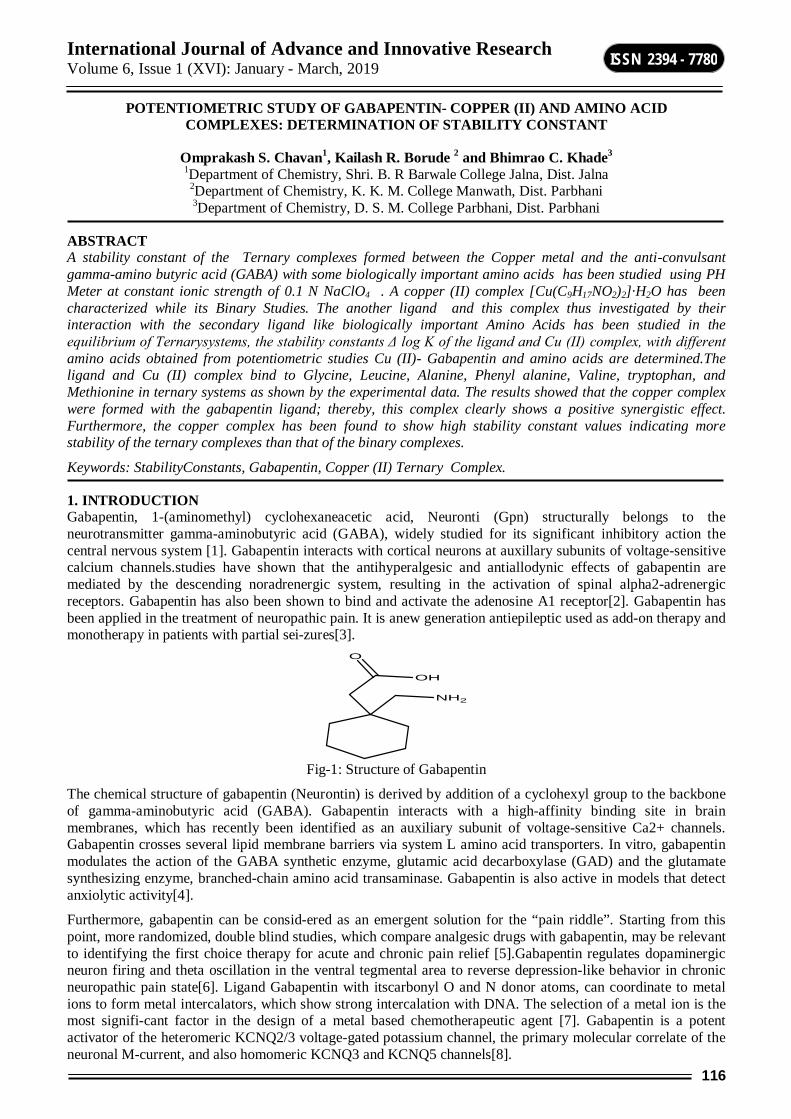
International Journal of Advance and Innovative Research Volume 6, Issue 1 (XVI): January - March, 2019
116
ISSN 2394 - 7780
POTENTIOMETRIC STUDY OF GABAPENTIN- COPPER (II) AND AMINO ACID COMPLEXES: DETERMINATION OF STABILITY CONSTANT
Omprakash S. Chavan1, Kailash R. Borude 2 and Bhimrao C. Khade3
1Department of Chemistry, Shri. B. R Barwale College Jalna, Dist. Jalna
2Department of Chemistry, K. K. M. College Manwath, Dist. Parbhani 3Department of Chemistry, D. S. M. College Parbhani, Dist. Parbhani
ABSTRACT A stability constant of the Ternary complexes formed between the Copper metal and the anti-convulsant gamma-amino butyric acid (GABA) with some biologically important amino acids has been studied using PH Meter at constant ionic strength of 0.1 N NaClO4 . A copper (II) complex [Cu(C9H17NO2)2]·H2O has been characterized while its Binary Studies. The another ligand and this complex thus investigated by their interaction with the secondary ligand like biologically important Amino Acids has been studied in the equilibrium of Ternarysystems, the stability constants Δ log K of the ligand and Cu (II) complex, with different amino acids obtained from potentiometric studies Cu (II)- Gabapentin and amino acids are determined.The ligand and Cu (II) complex bind to Glycine, Leucine, Alanine, Phenyl alanine, Valine, tryptophan, and Methionine in ternary systems as shown by the experimental data. The results showed that the copper complex were formed with the gabapentin ligand; thereby, this complex clearly shows a positive synergistic effect. Furthermore, the copper complex has been found to show high stability constant values indicating more stability of the ternary complexes than that of the binary complexes.
Keywords: StabilityConstants, Gabapentin, Copper (II) Ternary Complex.
1. INTRODUCTION Gabapentin, 1-(aminomethyl) cyclohexaneacetic acid, Neuronti (Gpn) structurally belongs to the neurotransmitter gamma-aminobutyric acid (GABA), widely studied for its significant inhibitory action the central nervous system [1]. Gabapentin interacts with cortical neurons at auxillary subunits of voltage-sensitive calcium channels.studies have shown that the antihyperalgesic and antiallodynic effects of gabapentin are mediated by the descending noradrenergic system, resulting in the activation of spinal alpha2-adrenergic receptors. Gabapentin has also been shown to bind and activate the adenosine A1 receptor[2]. Gabapentin has been applied in the treatment of neuropathic pain. It is anew generation antiepileptic used as add-on therapy and monotherapy in patients with partial sei-zures[3].
NH2
O
OH
Fig-1: Structure of Gabapentin
The chemical structure of gabapentin (Neurontin) is derived by addition of a cyclohexyl group to the backbone of gamma-aminobutyric acid (GABA). Gabapentin interacts with a high-affinity binding site in brain membranes, which has recently been identified as an auxiliary subunit of voltage-sensitive Ca2+ channels. Gabapentin crosses several lipid membrane barriers via system L amino acid transporters. In vitro, gabapentin modulates the action of the GABA synthetic enzyme, glutamic acid decarboxylase (GAD) and the glutamate synthesizing enzyme, branched-chain amino acid transaminase. Gabapentin is also active in models that detect anxiolytic activity[4].
Furthermore, gabapentin can be consid-ered as an emergent solution for the “pain riddle”. Starting from this point, more randomized, double blind studies, which compare analgesic drugs with gabapentin, may be relevant to identifying the first choice therapy for acute and chronic pain relief [5].Gabapentin regulates dopaminergic neuron firing and theta oscillation in the ventral tegmental area to reverse depression-like behavior in chronic neuropathic pain state[6]. Ligand Gabapentin with itscarbonyl O and N donor atoms, can coordinate to metal ions to form metal intercalators, which show strong intercalation with DNA. The selection of a metal ion is the most signifi-cant factor in the design of a metal based chemotherapeutic agent [7]. Gabapentin is a potent activator of the heteromeric KCNQ2/3 voltage-gated potassium channel, the primary molecular correlate of the neuronal M-current, and also homomeric KCNQ3 and KCNQ5 channels[8].

International Journal of Advance and Innovative Research Volume 6, Issue 1 (XVI): January - March, 2019
117
ISSN 2394 - 7780
Copper is a crucial cofactor in tumor angiogenesis processes and is a bio-essential and bio-relevant element, which is an inseparable component of many enzymes, including superoxide dismutase, tyrosinase, cerulo-plasmin, etc. [9, 10]. Gabapentin is a novel anticonvulsant drug. The anticonvulsant mechanism of gabapentin is not known. Gabapentin is not a substrate of BCAA-T, but it exhibited a potent competitive inhibition of both cytosolic and mitochondrial forms of brain BCAA-T. Inhibition of BCAA-T by this drug was reversible. Km values for the branched-chain amino acids (BCAA) L-leucine, L-isoleucine, and L-valine (0.6-1.2 mM), suggesting that gabapentin may significantly reduce synthesis of glutamate from BCAA in brain by acting on BCAA-T[11].Copper is a transition metal ion are integral parts of enzymes and play an important rolein the biological system, such as to trigger a reaction, control reaction mechanism, stabilizeprotein structure, maintain structure of cell walls etc. Latest information indicates regulation ofmetabolism and growth of animal cell is dependent upon the mobilization of divalent and trivalent metal ions. It is widely distributed throughout the body[12].
A stable protein drug complex with a dominant role in stor-age and drug disposition may be formed by a significant interaction of any drug with a protein. Therefore, understanding the mechanism of in-teraction of a bioactive compound with BSA, a well-studied protein, is important [13].As the amino acids are the essential part of the proteins it will be worth considering them as one of the ligand.
2. MATERIALS AND THE METHODS All chemicals and solvents used for synthesis were commercially available, reagent grade and were used without further purification. Sol-vents and starting materials were supplied by Sigma Aldrich or Merck Chemical Companies and used without further purification.The concentration of metal ionswas estimated by the standard procedures[14].Amino acids from Merck (Germany) and H.D. Fine (India) were prepared by dissolving A.R.grade sample in 80% (v/v) ethanol – water medium.Solution of the Drug Gabapentinwere prepared by dissolving sample as received in 80% (v/v)ethanol-water medium. Drugs samples in pure form were obtained from pharmacy industries.The Methodology were used in the study of ternary metal complexes by thePotentiometric titration technique, involves the titrations of carbonate free solution of againstStandard sodium hydroxide, where drug Gabapentin (D) and amino acids (R) are the ligands.The ionic strength of the solutions was maintained constant i.e. 0.1 M by adding appropriateamount of 1M sodium perchlorate solution. The metal solution of 0.02 Molar concentration and that of the ligand with 0.01 Molar concentration was prepared by taking nitrates of the metal and recrystallized drug respectively. The titrations were carried out at 27 0Cin an inertAtmosphere by bubbling oxygen free nitrogen gas through an assembly containing the electrodeTo expel out CO2.The experimental procedure, in the study of ternary metal complexes by thePotentiometric titration technique, involves the titration of carbonate free solution of in 80 %(v/v) ethanol-water, were corrected by method of Vansittart and Hass. The formation constant ofTernary complexes were determined by computational programmed SCOGS to minimize theStandardderivation. The different systems of Acid, Acid –ligand, Acid –ligand –Metal and Acid-ligand-Metal-Amino –acid have studied by potentiometric titrations against standard NaOH.
3. RESULTS AND DISCUSSION 3.1. Binary Complexes In this study, Copper (II) was used to react with gabapentin and obtained the following results. which was found in accordance to the results obtained in one of the earlier study [15].The proton ligand constant and metal ligand stability constant of drug Gabapentinand amino acids with Copper (II) determined in 80 % ( v/v) ethanol-water mixture at 27°C and ionic strength µ= 0.1 M NaCl04 are given in Table No.1.
Legands pK1 pK2 Copper Logk1 Logk2
Gabapentin 4.81933 10.0856 12.2113 11.5822 Glycine 2.77000 9.74000 9.69000 8.98000 Leucine 3.81000 10.34000 8.07030 --
Glutamine 3.01000 9.28000 9.40000 7.89000 Glutamicacid 3.13600 5.89870 10.9800 8.64000 Methionine 3.12000 9.60000 9.64000 8.67000
Phenyl alanine 3.14000 9.30000 8.99000 7.67000 Valine 3.21000 9.80240 10.0100 8.48000
Table No.1
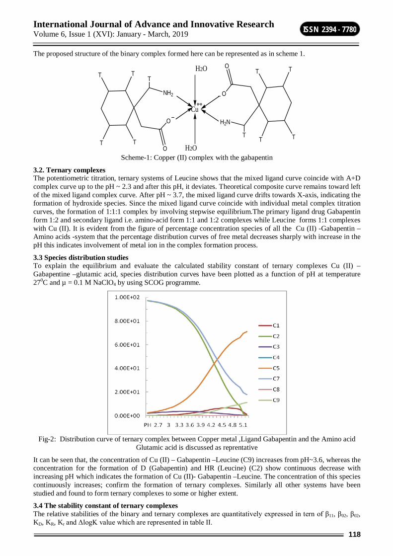
International Journal of Advance and Innovative Research Volume 6, Issue 1 (XVI): January - March, 2019
118
ISSN 2394 - 7780
The proposed structure of the binary complex formed here can be represented as in scheme 1.
O
T
T
T
T
T
NH2
O
O
T
T
T
T
T
H2N
O
Cu
H2O
H2O Scheme-1: Copper (II) complex with the gabapentin
3.2. Ternary complexes The potentiometric titration, ternary systems of Leucine shows that the mixed ligand curve coincide with A+D complex curve up to the pH ~ 2.3 and after this pH, it deviates. Theoretical composite curve remains toward left of the mixed ligand complex curve. After pH ~ 3.7, the mixed ligand curve drifts towards X-axis, indicating the formation of hydroxide species. Since the mixed ligand curve coincide with individual metal complex titration curves, the formation of 1:1:1 complex by involving stepwise equilibrium.The primary ligand drug Gabapentin form 1:2 and secondary ligand i.e. amino-acid form 1:1 and 1:2 complexes while Leucine forms 1:1 complexes with Cu (II). It is evident from the figure of percentage concentration species of all the Cu (II) -Gabapentin –Amino acids -system that the percentage distribution curves of free metal decreases sharply with increase in the pH this indicates involvement of metal ion in the complex formation process.
3.3 Species distribution studies To explain the equilibrium and evaluate the calculated stability constant of ternary complexes Cu (II) –Gabapentine –glutamic acid, species distribution curves have been plotted as a function of pH at temperature 270C and µ = 0.1 M NaClO4 by using SCOG programme.
Fig-2: Distribution curve of ternary complex between Copper metal ,Ligand Gabapentin and the Amino acid
Glutamic acid is discussed as reprentative
It can be seen that, the concentration of Cu (II) – Gabapentin –Leucine (C9) increases from pH~3.6, whereas the concentration for the formation of D (Gabapentin) and HR (Leucine) (C2) show continuous decrease with increasing pH which indicates the formation of Cu (II)- Gabapentin –Leucine. The concentration of this species continuously increases; confirm the formation of ternary complexes. Similarly all other systems have been studied and found to form ternary complexes to some or higher extent.
3.4 The stability constant of ternary complexes The relative stabilities of the binary and ternary complexes are quantitatively expressed in tern of β11, β02, β02, KD, KR, Kr and ΔlogK value which are represented in table II.
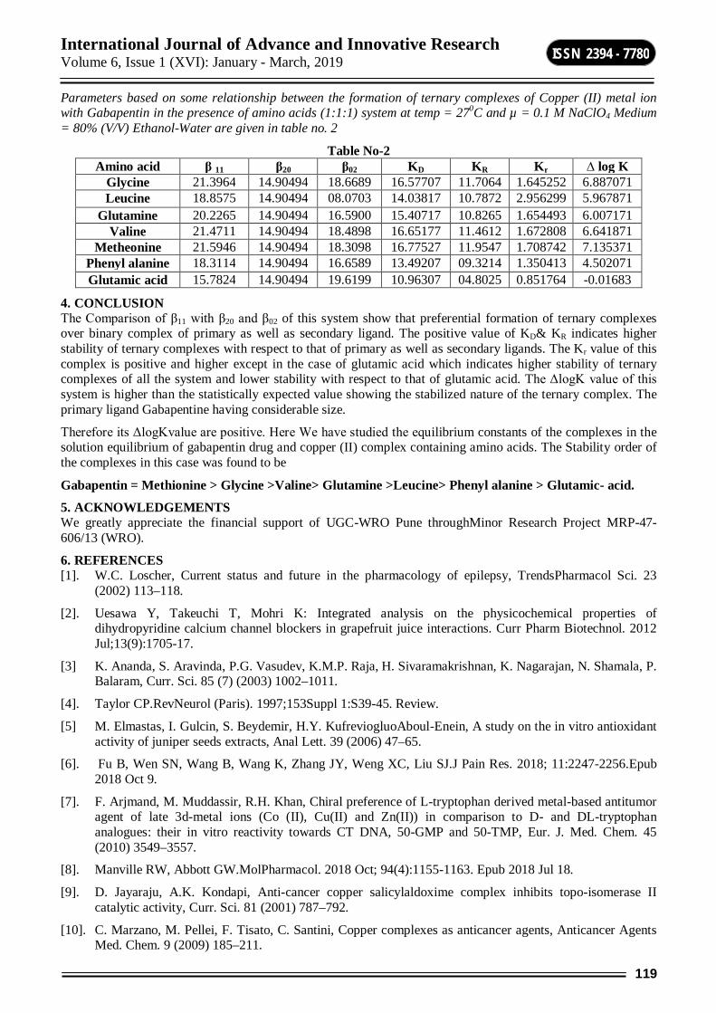
International Journal of Advance and Innovative Research Volume 6, Issue 1 (XVI): January - March, 2019
119
ISSN 2394 - 7780
Parameters based on some relationship between the formation of ternary complexes of Copper (II) metal ion with Gabapentin in the presence of amino acids (1:1:1) system at temp = 270C and µ = 0.1 M NaClO4 Medium = 80% (V/V) Ethanol-Water are given in table no. 2
Table No-2 Amino acid β 11 β20 β02 KD KR Kr ∆ log K
Glycine 21.3964 14.90494 18.6689 16.57707 11.7064 1.645252 6.887071 Leucine 18.8575 14.90494 08.0703 14.03817 10.7872 2.956299 5.967871
Glutamine 20.2265 14.90494 16.5900 15.40717 10.8265 1.654493 6.007171 Valine 21.4711 14.90494 18.4898 16.65177 11.4612 1.672808 6.641871
Metheonine 21.5946 14.90494 18.3098 16.77527 11.9547 1.708742 7.135371 Phenyl alanine 18.3114 14.90494 16.6589 13.49207 09.3214 1.350413 4.502071 Glutamic acid 15.7824 14.90494 19.6199 10.96307 04.8025 0.851764 -0.01683
4. CONCLUSION The Comparison of β11 with β20 and β02 of this system show that preferential formation of ternary complexes over binary complex of primary as well as secondary ligand. The positive value of KD& KR indicates higher stability of ternary complexes with respect to that of primary as well as secondary ligands. The Kr value of this complex is positive and higher except in the case of glutamic acid which indicates higher stability of ternary complexes of all the system and lower stability with respect to that of glutamic acid. The ΔlogK value of this system is higher than the statistically expected value showing the stabilized nature of the ternary complex. The primary ligand Gabapentine having considerable size.
Therefore its ΔlogKvalue are positive. Here We have studied the equilibrium constants of the complexes in the solution equilibrium of gabapentin drug and copper (II) complex containing amino acids. The Stability order of the complexes in this case was found to be
Gabapentin = Methionine > Glycine >Valine> Glutamine >Leucine> Phenyl alanine > Glutamic- acid.
5. ACKNOWLEDGEMENTS We greatly appreciate the financial support of UGC-WRO Pune throughMinor Research Project MRP-47-606/13 (WRO).
6. REFERENCES [1]. W.C. Loscher, Current status and future in the pharmacology of epilepsy, TrendsPharmacol Sci. 23
(2002) 113–118.
[2]. Uesawa Y, Takeuchi T, Mohri K: Integrated analysis on the physicochemical properties of dihydropyridine calcium channel blockers in grapefruit juice interactions. Curr Pharm Biotechnol. 2012 Jul;13(9):1705-17.
[3] K. Ananda, S. Aravinda, P.G. Vasudev, K.M.P. Raja, H. Sivaramakrishnan, K. Nagarajan, N. Shamala, P. Balaram, Curr. Sci. 85 (7) (2003) 1002–1011.
[4]. Taylor CP.RevNeurol (Paris). 1997;153Suppl 1:S39-45. Review.
[5] M. Elmastas, I. Gulcin, S. Beydemir, H.Y. KufreviogluoAboul-Enein, A study on the in vitro antioxidant activity of juniper seeds extracts, Anal Lett. 39 (2006) 47–65.
[6]. Fu B, Wen SN, Wang B, Wang K, Zhang JY, Weng XC, Liu SJ.J Pain Res. 2018; 11:2247-2256.Epub 2018 Oct 9.
[7]. F. Arjmand, M. Muddassir, R.H. Khan, Chiral preference of L-tryptophan derived metal-based antitumor agent of late 3d-metal ions (Co (II), Cu(II) and Zn(II)) in comparison to D- and DL-tryptophan analogues: their in vitro reactivity towards CT DNA, 50-GMP and 50-TMP, Eur. J. Med. Chem. 45 (2010) 3549–3557.
[8]. Manville RW, Abbott GW.MolPharmacol. 2018 Oct; 94(4):1155-1163. Epub 2018 Jul 18.
[9]. D. Jayaraju, A.K. Kondapi, Anti-cancer copper salicylaldoxime complex inhibits topo-isomerase II catalytic activity, Curr. Sci. 81 (2001) 787–792.
[10]. C. Marzano, M. Pellei, F. Tisato, C. Santini, Copper complexes as anticancer agents, Anticancer Agents Med. Chem. 9 (2009) 185–211.

International Journal of Advance and Innovative Research Volume 6, Issue 1 (XVI): January - March, 2019
120
ISSN 2394 - 7780
[11]. Goldlust A, Su TZ, Welty DF, Taylor CP, Oxender DL. Effects of anticonvulsant drug gabapentin on the enzymes in metabolic pathways of glutamate and GABA. Epilepsy Res 22: 1-11.
[12]. Walkar W.R, Reeves R.R, Brosnan M, Coleman G.D, Bioinorganic chemistry,1977;7:271.
[13] S.L. Zhang, G.L.V. Damu, L. Zhang, R.X. Geng, C.H. Zhou, Eur. J. Med. Chem. 55674 (2012) 164–175.
[14]. Vogel A.I, “A Text Book of Quantitative Inorganic Analysis, Pergamon Green and Co.Ltd, London, 1957; 539.]
[15]. Bhimrao C. Khade, Studies of Metal complexes of Drug Furosemide and Amino acids with Copper (II) ; IJPR, vol.5, issue 3 March 2015.
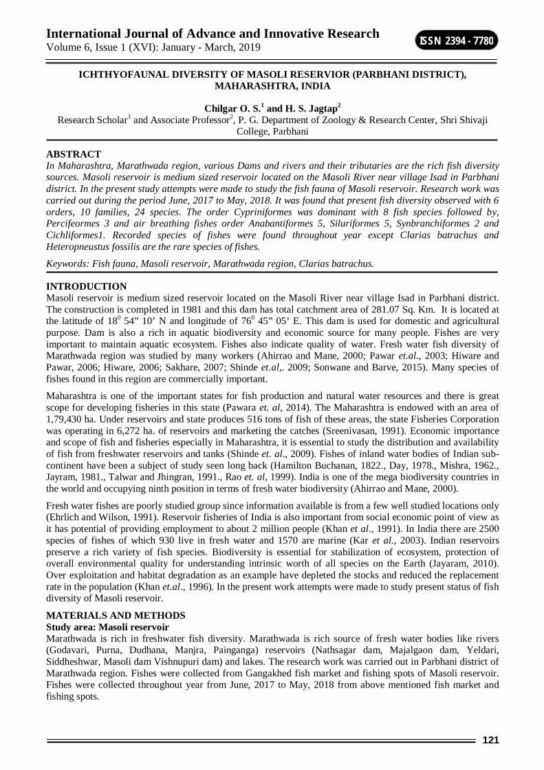
International Journal of Advance and Innovative Research Volume 6, Issue 1 (XVI): January - March, 2019
121
ISSN 2394 - 7780
ICHTHYOFAUNAL DIVERSITY OF MASOLI RESERVIOR (PARBHANI DISTRICT), MAHARASHTRA, INDIA
Chilgar O. S.1 and H. S. Jagtap2 Research Scholar1 and Associate Professor2, P. G. Department of Zoology & Research Center, Shri Shivaji
College, Parbhani
ABSTRACT In Maharashtra, Marathwada region, various Dams and rivers and their tributaries are the rich fish diversity sources. Masoli reservoir is medium sized reservoir located on the Masoli River near village Isad in Parbhani district. In the present study attempts were made to study the fish fauna of Masoli reservoir. Research work was carried out during the period June, 2017 to May, 2018. It was found that present fish diversity observed with 6 orders, 10 families, 24 species. The order Cypriniformes was dominant with 8 fish species followed by, Percifeormes 3 and air breathing fishes order Anabantiformes 5, Siluriformes 5, Synbranchiformes 2 and Cichliformes1. Recorded species of fishes were found throughout year except Clarias batrachus and Heteropneustus fossilis are the rare species of fishes.
Keywords: Fish fauna, Masoli reservoir, Marathwada region, Clarias batrachus.
INTRODUCTION Masoli reservoir is medium sized reservoir located on the Masoli River near village Isad in Parbhani district. The construction is completed in 1981 and this dam has total catchment area of 281.07 Sq. Km. It is located at the latitude of 180 54” 10’ N and longitude of 760 45” 05’ E. This dam is used for domestic and agricultural purpose. Dam is also a rich in aquatic biodiversity and economic source for many people. Fishes are very important to maintain aquatic ecosystem. Fishes also indicate quality of water. Fresh water fish diversity of Marathwada region was studied by many workers (Ahirrao and Mane, 2000; Pawar et.al., 2003; Hiware and Pawar, 2006; Hiware, 2006; Sakhare, 2007; Shinde et.al,. 2009; Sonwane and Barve, 2015). Many species of fishes found in this region are commercially important.
Maharashtra is one of the important states for fish production and natural water resources and there is great scope for developing fisheries in this state (Pawara et. al, 2014). The Maharashtra is endowed with an area of 1,79,430 ha. Under reservoirs and state produces 516 tons of fish of these areas, the state Fisheries Corporation was operating in 6,272 ha. of reservoirs and marketing the catches (Sreenivasan, 1991). Economic importance and scope of fish and fisheries especially in Maharashtra, it is essential to study the distribution and availability of fish from freshwater reservoirs and tanks (Shinde et. al., 2009). Fishes of inland water bodies of Indian sub-continent have been a subject of study seen long back (Hamilton Buchanan, 1822., Day, 1978., Mishra, 1962., Jayram, 1981., Talwar and Jhingran, 1991., Rao et. al, 1999). India is one of the mega biodiversity countries in the world and occupying ninth position in terms of fresh water biodiversity (Ahirrao and Mane, 2000).
Fresh water fishes are poorly studied group since information available is from a few well studied locations only (Ehrlich and Wilson, 1991). Reservoir fisheries of India is also important from social economic point of view as it has potential of providing employment to about 2 million people (Khan et al., 1991). In India there are 2500 species of fishes of which 930 live in fresh water and 1570 are marine (Kar et al., 2003). Indian reservoirs preserve a rich variety of fish species. Biodiversity is essential for stabilization of ecosystem, protection of overall environmental quality for understanding intrinsic worth of all species on the Earth (Jayaram, 2010). Over exploitation and habitat degradation as an example have depleted the stocks and reduced the replacement rate in the population (Khan et.al., 1996). In the present work attempts were made to study present status of fish diversity of Masoli reservoir.
MATERIALS AND METHODS Study area: Masoli reservoir Marathwada is rich in freshwater fish diversity. Marathwada is rich source of fresh water bodies like rivers (Godavari, Purna, Dudhana, Manjra, Painganga) reservoirs (Nathsagar dam, Majalgaon dam, Yeldari, Siddheshwar, Masoli dam Vishnupuri dam) and lakes. The research work was carried out in Parbhani district of Marathwada region. Fishes were collected from Gangakhed fish market and fishing spots of Masoli reservoir. Fishes were collected throughout year from June, 2017 to May, 2018 from above mentioned fish market and fishing spots.
International Journal of Advance and Innovative Research Volume 6, Issue 1 (XVI): January - March, 2019
122
ISSN 2394 - 7780
Fishes were systematically identified up to species level using taxonomic keys of Jayram (1981), Jhingran (1991) and Qureshi (1983) and preserved in 4% formalin in laboratory. Fishes were recorded and kept in research laboratory for further studies.
RESULTS AND DISCUSSION During the present study 24 species of fresh water fishes belonging to 06 orders, 10 families were recorded from Masoli reservoir. Number of species and their status is given in table1.
Indian major carps, murrels, catfishes and other palatable fishes were recorded. These fishes were commonly used as food fishes. The order Cyprinifomes is dominant having 8 species followed order Perciformes 3 species by Air breathing fishes order Anabantiformes 5 species, Siluriformes 5 species, Synbranchiformes 2 species, and Cichliformes 01 species. Some of the reported exotic species in Maharashtra are Oreochromis mossambica (Kharat et.al., 2003., Singh and Lakra, 2011., Sugunan, 1995).
Ahirrao and Mane (2000) recorded 32 fish species belonging to 25 genera and 08 families from Parbhani district Maharashtra. Hiware and Pawar recorded 43 fish species from Nathsagar Dam Paithan in Aurangabad district. Sakhare (2007) reported 29 fish belonging to 20 genera falling in 4 orders from Yeldari reservoir of Parbhani district. Pawar et. al., (2003) observed 11 species belonging to 5 orders from Sirur Dam near Mukhed, Nanded district (M.S.). Sonawane and Barve (2015) reported 23 species of 20 genera, 10 families and 8 orders in which order cypriniformes was dominant with 9 species from the Lower Dudhana dam district- Parbhani (M.S) India. Hiware (2006) studied ichthyofauna of four district of Marathwada region of Maharashtra. Results reported by above mentioned workers are more or less similar with the present works.
CONCLUSION There is rich fish diversity in Masoli reservoir, Parbhani district of Maharashtra. Fishes maintain aquatic ecosystem hence there is need for conservation strategies. Over fishing and immature fishing are main causes of loss of many fish species. Seasonal fluctuation, anthropogenic activities, climate change (extreme heat and cold), invasion of exotic species, dry drought, and water pollution are some causes for complete and partial loss of many fresh water fishes. Many fish species are already become extinct while some of them are endangered. Present work reveals that air breathing fishes like Clarias batrachus and Heterpnestus fossils become rare in Masoli reservoir. To maintain fish diversity in Masoli reservoir there is need for conservation of air breathing fishes. Total number of fresh water fish species recorded during the present study indicates rich fish diversity in Masoli reservoir.
ACKNOWLEDGEMENT The authors are thankful to Principal, Shri Shivaji College, Parbhani (M.S.) India for providing laboratory facilities.
REFERENCES Ahirrao, S.D. and A.S. Mane (2000). The diversity of Ichthyofauna, taxonomy and fisheries from freshwater
of Parbhani, Dist. Maharashtra State, J. Aqua. Biol., 15(1&2): 40-43.
Day, F.S. (1978). The Fishes of India, William and Sons Ltd., London.
Ehrlich, P.R. and Wilson E.O., Biodiversity studies science and policy. Sci., (1991), 253, 758-762.
Hamilton-Buchanan (1822) An account of the fishes found in the river Ganges and its branches, Edinburgh &London,VIII + 405 pp.39.
Hiware, C. J. 2006. Ichthyofauna from four districts of Marathwada region, Maharashtra, India. Zoos’ Print Journal. 21(1): 2137-2139.
Hiware, C.J. and Pawar, R.T. (2006) Ichthyofauna of Paithan Reservoir (Nathsagar Dam) in Aurangabad Dist. of Marathwada region Maharashtra, Ecology and Environment, APH Publishing Corporation, New Delhi.
Jayaram K.C., The Freshwater fishes of the Indian Region. Vol. II (2010).
Jayaram, K.C. (1981). The Freshwater Fishes of India. A Handbook Zoological Survey of India, Calcutta.
Kar, D.A. Kumar, C. Bohra and L.K. Shing, (Eds)2003.fishes of Barak drainage, Mizoram and Tripura; In: Environment, pollution and management, APH publishing corporation, new Delhi, 604: 203-211.
Khan MS, Lee, Patrick KY, Cramphorn J and Zakaria Ismail Mohd (1996) Fresh water fishes of the Pahang River Basin, Malaysia. Wetland International J. Asia Pacific Publication. 112.
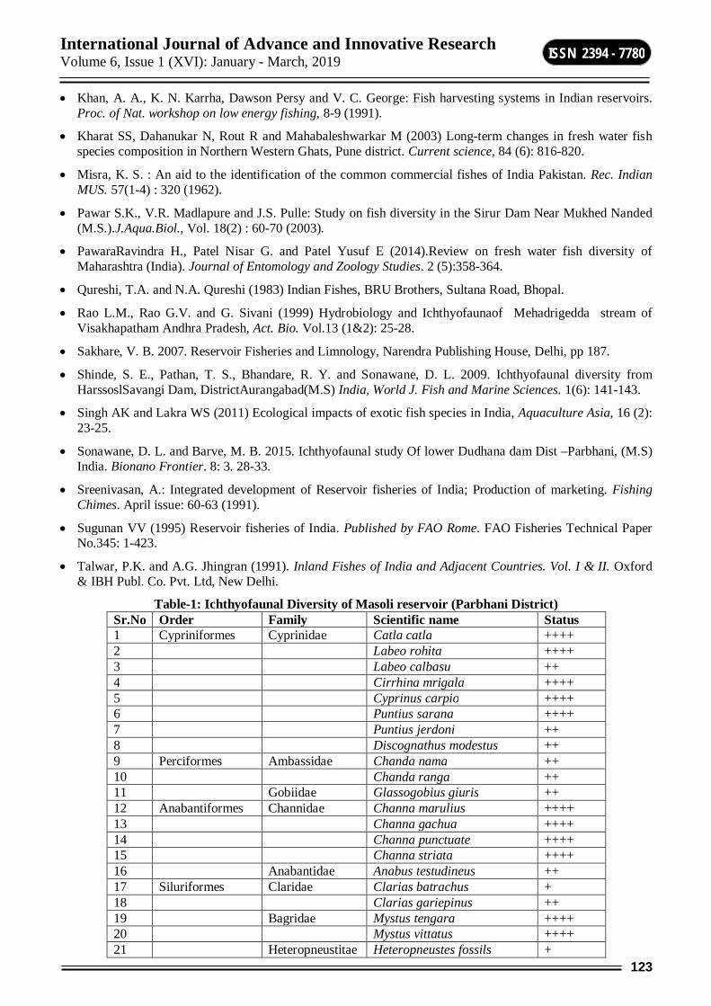
International Journal of Advance and Innovative Research Volume 6, Issue 1 (XVI): January - March, 2019
123
ISSN 2394 - 7780
Khan, A. A., K. N. Karrha, Dawson Persy and V. C. George: Fish harvesting systems in Indian reservoirs. Proc. of Nat. workshop on low energy fishing, 8-9 (1991).
Kharat SS, Dahanukar N, Rout R and Mahabaleshwarkar M (2003) Long-term changes in fresh water fish species composition in Northern Western Ghats, Pune district. Current science, 84 (6): 816-820.
Misra, K. S. : An aid to the identification of the common commercial fishes of India Pakistan. Rec. Indian MUS. 57(1-4) : 320 (1962).
Pawar S.K., V.R. Madlapure and J.S. Pulle: Study on fish diversity in the Sirur Dam Near Mukhed Nanded (M.S.).J.Aqua.Biol., Vol. 18(2) : 60-70 (2003).
PawaraRavindra H., Patel Nisar G. and Patel Yusuf E (2014).Review on fresh water fish diversity of Maharashtra (India). Journal of Entomology and Zoology Studies. 2 (5):358-364.
Qureshi, T.A. and N.A. Qureshi (1983) Indian Fishes, BRU Brothers, Sultana Road, Bhopal.
Rao L.M., Rao G.V. and G. Sivani (1999) Hydrobiology and Ichthyofaunaof Mehadrigedda stream of Visakhapatham Andhra Pradesh, Act. Bio. Vol.13 (1&2): 25-28.
Sakhare, V. B. 2007. Reservoir Fisheries and Limnology, Narendra Publishing House, Delhi, pp 187.
Shinde, S. E., Pathan, T. S., Bhandare, R. Y. and Sonawane, D. L. 2009. Ichthyofaunal diversity from HarssoslSavangi Dam, DistrictAurangabad(M.S) India, World J. Fish and Marine Sciences. 1(6): 141-143.
Singh AK and Lakra WS (2011) Ecological impacts of exotic fish species in India, Aquaculture Asia, 16 (2): 23-25.
Sonawane, D. L. and Barve, M. B. 2015. Ichthyofaunal study Of lower Dudhana dam Dist –Parbhani, (M.S) India. Bionano Frontier. 8: 3. 28-33.
Sreenivasan, A.: Integrated development of Reservoir fisheries of India; Production of marketing. Fishing Chimes. April issue: 60-63 (1991).
Sugunan VV (1995) Reservoir fisheries of India. Published by FAO Rome. FAO Fisheries Technical Paper No.345: 1-423.
Talwar, P.K. and A.G. Jhingran (1991). Inland Fishes of India and Adjacent Countries. Vol. I & II. Oxford & IBH Publ. Co. Pvt. Ltd, New Delhi.
Table-1: Ichthyofaunal Diversity of Masoli reservoir (Parbhani District) Sr.No Order Family Scientific name Status 1 Cypriniformes Cyprinidae Catla catla ++++ 2 Labeo rohita ++++ 3 Labeo calbasu ++ 4 Cirrhina mrigala ++++ 5 Cyprinus carpio ++++ 6 Puntius sarana ++++ 7 Puntius jerdoni ++ 8 Discognathus modestus ++ 9 Perciformes Ambassidae Chanda nama ++ 10 Chanda ranga ++ 11 Gobiidae Glassogobius giuris ++ 12 Anabantiformes Channidae Channa marulius ++++ 13 Channa gachua ++++ 14 Channa punctuate ++++ 15 Channa striata ++++ 16 Anabantidae Anabus testudineus ++ 17 Siluriformes Claridae Clarias batrachus + 18 Clarias gariepinus ++ 19 Bagridae Mystus tengara ++++ 20 Mystus vittatus ++++ 21 Heteropneustitae Heteropneustes fossils +
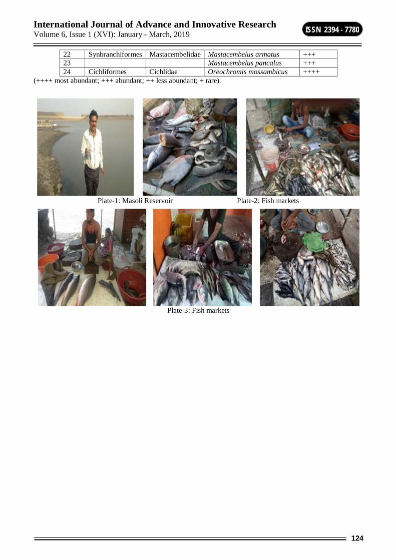
International Journal of Advance and Innovative Research Volume 6, Issue 1 (XVI): January - March, 2019
124
ISSN 2394 - 7780
22 Synbranchiformes Mastacembelidae Mastacembelus armatus +++ 23 Mastacembelus pancalus +++ 24 Cichliformes Cichlidae Oreochromis mossambicus ++++
(++++ most abundant; +++ abundant; ++ less abundant; + rare).
Plate-1: Masoli Reservoir Plate-2: Fish markets
Plate-3: Fish markets
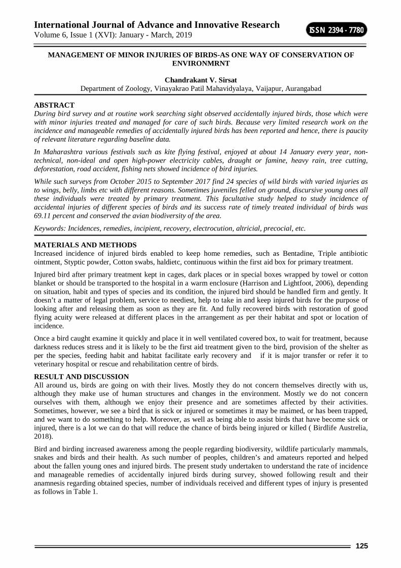
International Journal of Advance and Innovative Research Volume 6, Issue 1 (XVI): January - March, 2019
125
ISSN 2394 - 7780
MANAGEMENT OF MINOR INJURIES OF BIRDS-AS ONE WAY OF CONSERVATION OF ENVIRONMRNT
Chandrakant V. Sirsat Department of Zoology, Vinayakrao Patil Mahavidyalaya, Vaijapur, Aurangabad
ABSTRACT During bird survey and at routine work searching sight observed accidentally injured birds, those which were with minor injuries treated and managed for care of such birds. Because very limited research work on the incidence and manageable remedies of accidentally injured birds has been reported and hence, there is paucity of relevant literature regarding baseline data.
In Maharashtra various festivals such as kite flying festival, enjoyed at about 14 January every year, non-technical, non-ideal and open high-power electricity cables, draught or famine, heavy rain, tree cutting, deforestation, road accident, fishing nets showed incidence of bird injuries.
While such surveys from October 2015 to September 2017 find 24 species of wild birds with varied injuries as to wings, belly, limbs etc with different reasons. Sometimes juveniles felled on ground, discursive young ones all these individuals were treated by primary treatment. This facultative study helped to study incidence of accidental injuries of different species of birds and its success rate of timely treated individual of birds was 69.11 percent and conserved the avian biodiversity of the area.
Keywords: Incidences, remedies, incipient, recovery, electrocution, altricial, precocial, etc.
MATERIALS AND METHODS Increased incidence of injured birds enabled to keep home remedies, such as Bentadine, Triple antibiotic ointment, Styptic powder, Cotton swabs, haldietc, continuous within the first aid box for primary treatment.
Injured bird after primary treatment kept in cages, dark places or in special boxes wrapped by towel or cotton blanket or should be transported to the hospital in a warm enclosure (Harrison and Lightfoot, 2006), depending on situation, habit and types of species and its condition, the injured bird should be handled firm and gently. It doesn’t a matter of legal problem, service to neediest, help to take in and keep injured birds for the purpose of looking after and releasing them as soon as they are fit. And fully recovered birds with restoration of good flying acuity were released at different places in the arrangement as per their habitat and spot or location of incidence.
Once a bird caught examine it quickly and place it in well ventilated covered box, to wait for treatment, because darkness reduces stress and it is likely to be the first aid treatment given to the bird, provision of the shelter as per the species, feeding habit and habitat facilitate early recovery and if it is major transfer or refer it to veterinary hospital or rescue and rehabilitation centre of birds.
RESULT AND DISCUSSION All around us, birds are going on with their lives. Mostly they do not concern themselves directly with us, although they make use of human structures and changes in the environment. Mostly we do not concern ourselves with them, although we enjoy their presence and are sometimes affected by their activities. Sometimes, however, we see a bird that is sick or injured or sometimes it may be maimed, or has been trapped, and we want to do something to help. Moreover, as well as being able to assist birds that have become sick or injured, there is a lot we can do that will reduce the chance of birds being injured or killed ( Birdlife Austrelia, 2018).
Bird and birding increased awareness among the people regarding biodiversity, wildlife particularly mammals, snakes and birds and their health. As such number of peoples, children’s and amateurs reported and helped about the fallen young ones and injured birds. The present study undertaken to understand the rate of incidence and manageable remedies of accidentally injured birds during survey, showed following result and their anamnesis regarding obtained species, number of individuals received and different types of injury is presented as follows in Table 1.
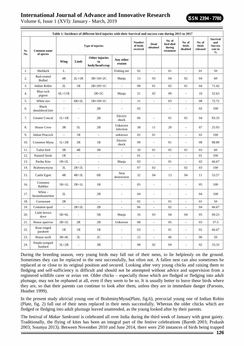
International Journal of Advance and Innovative Research Volume 6, Issue 1 (XVI): January - March, 2019
126
ISSN 2394 - 7780
Table 1: Incidence of different bird injuries with their Survival and success rate during 2015 to 2017
Sr. No.
Common name of species
Type of injuries Number of birds received
Dead obtained
No. of bird died
during treatment
No. of birds
disabled
No. of birds
released
Survival and
Success rate in
%
Wing Limb Other injuries
to body/head/crop
Any other reason
1. Shelduck L - Fishing net 02 - 01 - 01 50
2. Red-vented Bulbul 4R 2L+1R 3B+1H+2C Manja 13 03 04 02 04 60
3. Indian Robin 2L 1R 2B+2H+1C - 08 01 02 01 04 71.42
4. Blue rock pigeon 6L+11R 2B+2C Manja 21 02 09 - 10 52.63
5. White eye - 6R+2L 1B+1H+1C - 11 - 03 - 08 72.72
6. Black shouldered kite - - 2B - 02 - - 02 100
7. Greater Coucal 1L+1R - 2B Electric shock 06 - 01 01 04 83.33
8. House Crow 3R 5L 2B Unknown infection 38 11 20 - 07 25.93
9. Indian Peacock - 1R - unknown 02 01 - - 02 100
10. Common Myna 1L+2R 2R 1B Electric shock 09 - 01 - 08 88.89
11. Tailor bird 3R 4R 3B - 10 01 05 01 03 60
12. Painted Stork 1R - - - 01 - - - 01 100
13. Pariha Kite 1R+2L - - Manja 03 - 01 - 02 66.67
14. Brahmnymyna 3L 2R+2L - - 07 02 - 02 03 100
15. Cattle Egret 4R 4R+3L 6B Nest destruction 32 04 13 04 11 53.57
16. Common Babbler 1R+1L 2R+1L 1B - 05 - - - 05 100
17. White -brestedwaterhen 2L 2B - 04 - - - 04 100
18. Cormorant 2R - - - 02 - 01 - 01 50
19. Common quail - 2R+2L 2B - 06 - 02 - 04 66.67
20. Little brown dove 5R+6L - 5B Manja 16 03 04 04 05 69.23
21. House sparrow 2R+2L 2R 2B Unknown 08 - 05 - 03 37.5
22. Rose ringed parakeet 1R 1R 1B - 03 - 01 - 02 66.67
23. House swift 3R+4L 3L 2C - 12 - 06 - 06 50
24. Purple-rumped Sunbird 3L+2R - 3B - 08 02 04 - 02 33.33
During the breeding season, very young birds may fall out of their nests, to lie helplessly on the ground. Sometimes they can be replaced in the nest successfully, but often not. A fallen nest can also sometimes be replaced at or close to its original position and secured. Looking after very young chicks and raising them to fledging and self-sufficiency is difficult and should not be attempted without advice and supervision from a registered wildlife carer or avian vet. Older chicks – especially those which are fledged or fledging into adult plumage, may not be orphaned at all, even if they seem to be so. It is usually better to leave these birds where they are, so that their parents can continue to look after them, unless they are in immediate danger (Parsons, Heather 1999).
In the present study altricial young one of BrahminyMyna(Plate, fig.6), precocial young one of Indian Robin (Plate, fig. 2) fall out of their nests replaced in their nests successfully. Whereas the older chicks which are fledged or fledging into adult plumage leaved unattended, as the young looked after by their parents.
The festival of Makar Sankranti is celebrated all over India during the third week of January with great gaiety. Traditionally, the flying of kites has been an integral part of the festive celebrations (Bareth 2003; Prakash 2003; Soumya 2013). Between November 2010 and June 2014, there were 250 instances of birds being trapped
International Journal of Advance and Innovative Research Volume 6, Issue 1 (XVI): January - March, 2019
127
ISSN 2394 - 7780
in Chinese manja strings in the city of Bangalore. These comprised 268 birds belonging to about 10–13 species. Volunteers and other associated agencies rescued them (N=250 instances). Among the birds that were rescued, Black Kites were the single most commonly affected species making up nearly 70% of the birds found dangling from manja strings (Babu S et.al 2015).
In this study, instances of birds trap in Chinese manja strings was also observed specifically Blue rock pigeon, Pariha Kite and Little brown dove in the study area. All these birds removed successfully from the net of Chineasemanja strings, in this instance some bird found minor injuries all those treated ayurvedically and allopathically, and released after recovery in their habitat. Besides, aquatic bird such as Shelduck, found trapped to long lengths of fishing net line that also removed from the fish net with the help of fisherman– if not so it may cause death of the bird. It took a little effort, but made a big difference for the awareness among fisherman and survival of birds.
Anonymous (2012) worked on Ayurveda healing and reported that herbs are not dangerous to birds, ayurveda treatment is the best for birds. Apply Haldi (Turmeric) if the bird is injured. Buy a piece of haldi and then grind it. Do not use haldi powder from the market. Haldi acts as a disinfectant.Ginger (Adrak) juice added to the bird drinking water at night prevents loose motion when they are sick. Mix some yellow turmeric powder (haldi) in the yogurt. This should help with the Motion sickness.
There are many other ways that birds can become injured, and they may not be so obvious. Electrocutions is one of them. Such injuries due to electric shock observed in case of Common Myna and Greater Coucal. In this instance Greater Coucal had have minor shock, found lied insipiently on the ground, caught it and observed none of the injuries, but it’s both limb digits were curved so hard, immediately after massage it was recovered, which took little effort to save life of the coucal which flyed in the sky by drinking water, sat on tree and turned back to thanks. But electrocution to Common Myna ( Plate, fig.5) eye witness observed death by high power electric shock early in the morning, carcase fall on the ground near electric pole observed too much electric shock damage to leg, tongue observed black and no any other injury to the body. Another way that cause injury to birds was road accidents, such death observed in Indian peacock.
Hand-feeding a sick bird using a warmed hand feeding product may be attempted prior to tube-feeding. Hand feeding will be more time-consuming but less stressful for the patient if accepted. Hand-feeding can be an ideal way to supplement birds that revert to baby begging and feeding behavior when ill. However, the administration of an adequate quantity of supplement at each feeding requires that the patient be strong enough to demonstrate an adequate feeding response, and that the person feeding be experienced enough to avoid causing aspiration of the material (Harrison and Lightfoot, 2006).
During the study such type of hand feeding required for House crow (Plate, Fig.3), Red-vented Bulbul (Plate, Fig.1), Rose ringed parakeet, White eye and Common Babbler with adequate quantity of food depending on habit of the bird.
Degerns (1997) described the initial goal in treating contaminated or infected wound is the removal of devitalized tissue, foreign material and bacteria. The feathers surrounding the wound should be gently plucked or trimmed to allow more thorough cleansing and to prevent feather matting during the healing phases. Wound lavage will remove foreign material, reduce bacterial numbers and rehydrate soft tissues.
In the present work injuries of birds such as House sparrow, House Swift, Purple-rumped Sunbird, White -breasted waterhen treated with bentidine and Red-vented Bulbul with haldi (Plate, fig.1). Whereas wound of Indian peacock and Painted Stork by triple antibiotic ointment.
Anamnesis regarding species, number of the birds received and different types of injury is based on species predisposition, the highest incidences of kite string injuries were observed in Blue-rock Pigeons (116, 69%) followed by Common Pariah Kites (21, 12.5%), Barn Owls (4, 2.38%), Indian Peahen (3, 1.78%) and Cattle Egrets (3, 1.78%). There were two cases each (1.19%) of Black-eared Kite, Indian Peafowl, Comb Duck, Greylag Goose and Black Ibis followed by one case each (0.59%) of Woolly-necked Stork, Honey Buzzard, Painted Stork, Khakhicampbell Duck, Domestic Duck, Changeable Hawk- Eagle, Peregrine Falcon, Montagu’s Harrier, Demoiselle Crane, Indian Fowl and Domestic Turkey (Patel AJ 2013).
Present study governing all types of incidences caused by kite flying Chinese manja string, non-technical, non-ideal and open high-power electricity cables, draught or famine, heavy rain, tree cutting, deforestation, road accident and fishing nets. The highest incidences of injuries observed in House Crows (38, 19.29%) due to heavy rainfall and municipal corporation dumping ground contaminated wastes, followed by Cattle Egrets (32, 16.24%) because of tree cutting, Blue Rock Pigeon (21, 10.67%), Little Brown Doves (16, 8.12%), Red-vented
International Journal of Advance and Innovative Research Volume 6, Issue 1 (XVI): January - March, 2019
128
ISSN 2394 - 7780
Bulbuls (13, 6.6%) and Pariha Kites (3, 1.52%) mainly due to kite flying Chinese manja string. There were high incidences of injuries found in House Swift (12,6.09%), White Eye (11,5.58%), Tailor bird (10,5.08%), Common Myna (9,4.57%), House sparrow (8,4.06%), Indian Robin (8,4.06%), Purple-rumped Sunbird (8,4.06%), Brahminy Myna (7,3.55%), Common Quail (6,3.04%), Greater Coucal (6,3.04%), Common Babbler (5,2.53%) White breasted waterhen (4,2.03%), Rose ringed Parakeet (3,1.52%), Black Shouldered Kite (2,1.01%) Cormorant (2,1.01%) Indian Peacock (2,1.01%) Painted stork (1,0.5%). Among these most of the injuries were unknown. However there were two cases of electrocutions observed one in Common Myna which was dead and other Greater Coucal which was recovered.
The incidence of bird injuries was more in the Blue-rock Pigeons (116, 69%) followed by Common Pariah Kites (21, 12.5%). Similarly, in a study, among 3826 injured birds during kite flying festival, the incidence was more in Blue-rock Pigeons (2618, 68%) followed by Common Pariah Kites (749, 19%) (Tiwari et al., 2011).
Out of 168 clinical cases, left wing injuries were 55 (32.73%), right wing 44 (26.19%) and bilateral 12 (7.14%). In 49 White rumped Vultures with kite string injuries, 24 (49%) left wing, 15 (31%) right wing and 7 (14%) bilateral injuries were recorded (Kavechiyaet al., 2012).
Flying acuity was assessed by allowing the bird to fly in a large closed area. Complete return of flight was observed in majority of the birds within 20 days. In 27 cases partial flight could be restored. 38 birds died during the post-operative period. In 20 birds flight could not be restored. It was observed that all the birds with fresh wound/fracture showed excellent results. On the whole, good surgical results were observed in 49% cases (83 out of 168) followed by fair in 17% (29 out of 168) and failure in 34% (58 out of 168) cases. 65% birds (110 out of 168) with complete recovery and good flying acuity were released in the environment at different places as per their natural habitat. In 12% birds (20 out 168) flying acuity could not be restored and became permanently disabled. Such birds were sent to different permanent shelters according to their species run by forest department and different N.G.Os. During this study, the survival rate of the birds was 77% (130 out of 168) and the success rate was (110 out of 168) 65% (Patel AJ 2013).
In the present work manageable remedies of accidentally injured incipient bird recovery was very successful. There was no any single case of fracture was found. Whatever the injuries all were minor. Therefore, survival success rate was very high (197, 69.11%). Species specific survival success rate was found very highest (100%) in Black shouldered kite, Indian Peacock, Painted Stork, Brahmny myna, Common Babbler and White -breasted waterhen. Among the high incidences of injured birds showed excellent results in Common Myna (88.89%), Greater Coucal (83.33%), Indian Robin (72.72%), White eye (71.42%, Plate, Fig.4), Little brown dove (69.23%), followed by 66.67% in Pariha Kite, Common quail and Rose ringed parakeet. In 7.61% birds (15 out of 197) flying acuity could not be restored and became permanently disabled. Such birds were sent to different permanent shelters according to their habitat.
There is a need of dedicated efforts to organize treatment camps for management of injured birds in the most scientific manner, involving government bodies, non-government organizations and veterinarians especially trained for treatment of injured birds. Further, a tertiary centre for critical care of birds is also necessary.
REFERENCES Anonymous (2012).https://hinduismglance.wordpress.com
Babu S, SubramanyaS andDilawarM (2015). Kite flying: Effect of Chinese manja on birds in Bangalore, India. Indian BIRDS 10 (1): 13–18
Bareth N (2003). India celebrates kite flying. BBC News World Edition. URL: http://news. bbc.co.uk/2/hi/south Asia/2657419.stm. (Accessed on 12 August 2013.)
Birdlife Australia (2018).Helping sick or injured birds(Birdlife.orh.au).
Degernes LA (1997). Trauma medicine. In: Avian Medicine: Principles and Application.
HarrisonGJ, and LightfootT (2006). Emergency and critical care. In: Clinical Avian Medicine., Spix publishing Inc., Palm Beach, FL., 7: 1-19.
Kavechiya VP, Patel PB, ShashtriKB, Chaudhary DA, Barvalia DR, Raval JD and RavalNJ (2012). Incidences of emergencies and their management in white-rumped vulture (Gyps bvengalensis) – study on 107 cases. XXXVIth Annual Congress of Indian Society for Veterinary Surgery, Anand, Gujarat, India.
Parsons, Heather (1999). Caring for Australian Native Birds. Kangaroo Press: Sydney.

International Journal of Advance and Innovative Research Volume 6, Issue 1 (XVI): January - March, 2019
129
ISSN 2394 - 7780
Patel AJ (2013).studies on surgical management of injured birds during kite flying festival, thesis, Anand Agricultural University, Anand Gujrat, India.
Prakash A (2003). Kites in India. Normad Heritage Trust (2003), Mumbai. Press Trust of India (PTI). 2015. Maharashtra bans ‘manja‘ kite strings to save birds. Indian Express 24 April 2015. URL: http://indianexpress.com/article/india/maharashtra/ maharashtra-bans-manja-kite-strings-to-save-birds/. (Accessed on 24 April 2015).
RitchieBW, Harrison GJ and HarrisonLR (eds.), Abridged Ed., Wingers Publishing, Lake Worth, FI, 33466.,16: 417133.
SoumyaE (2013). Kites that won’t let birds fly. Post Noon. URL: http://postnoon.com/2013/01/12/kites-that-wont-let-birds-fly/102179.(Accessed on 14 August 2013).
Tiwari DK, DarMUD, Parikh PV, PatilDB, Jhala SK, Joy N, Shukla T, Patel P and Kavechiya V (2011). Incidence and management of birds injured during uttarayan festival: Report of two years. XXXVthAnnual Congress of Indian Society for Veterinary Surgery. Kolkata, W. B., India.
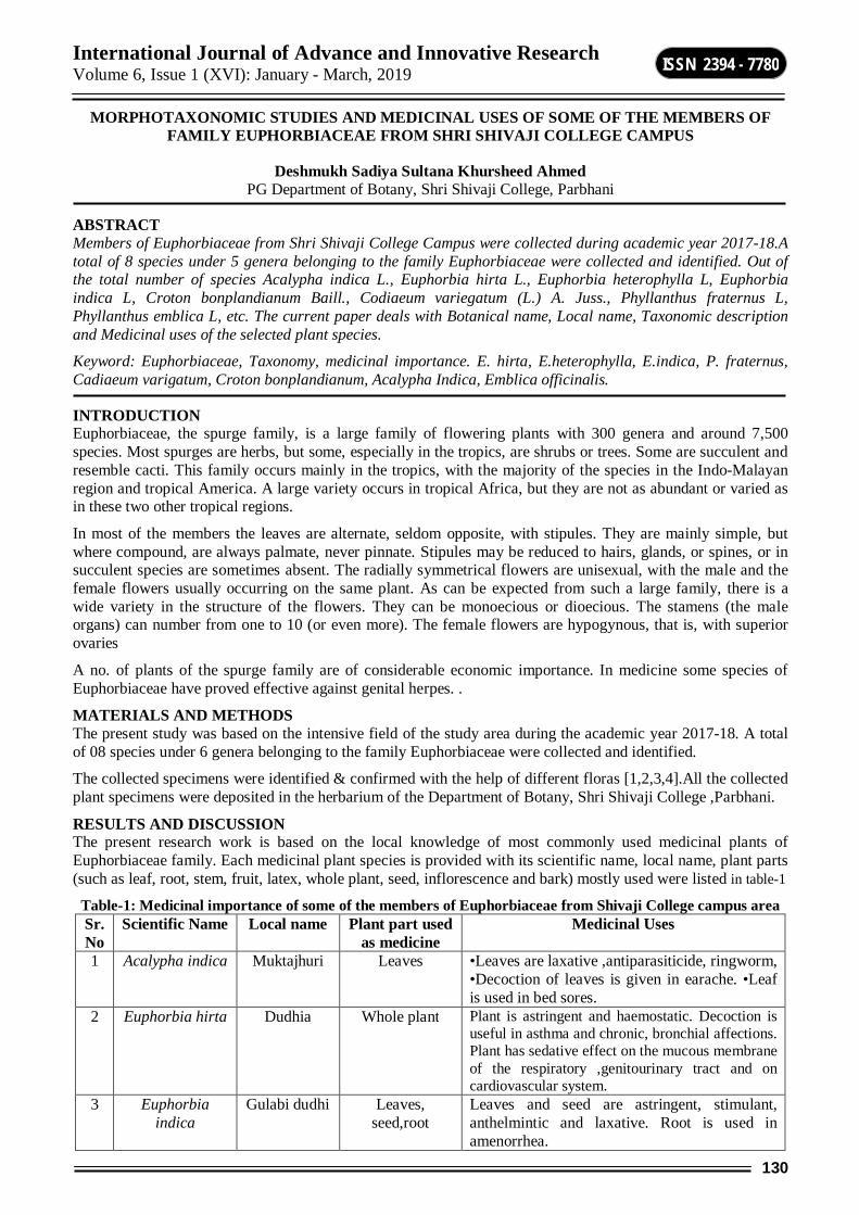
International Journal of Advance and Innovative Research Volume 6, Issue 1 (XVI): January - March, 2019
130
ISSN 2394 - 7780
MORPHOTAXONOMIC STUDIES AND MEDICINAL USES OF SOME OF THE MEMBERS OF FAMILY EUPHORBIACEAE FROM SHRI SHIVAJI COLLEGE CAMPUS
Deshmukh Sadiya Sultana Khursheed Ahmed PG Department of Botany, Shri Shivaji College, Parbhani
ABSTRACT Members of Euphorbiaceae from Shri Shivaji College Campus were collected during academic year 2017-18.A total of 8 species under 5 genera belonging to the family Euphorbiaceae were collected and identified. Out of the total number of species Acalypha indica L., Euphorbia hirta L., Euphorbia heterophylla L, Euphorbia indica L, Croton bonplandianum Baill., Codiaeum variegatum (L.) A. Juss., Phyllanthus fraternus L, Phyllanthus emblica L, etc. The current paper deals with Botanical name, Local name, Taxonomic description and Medicinal uses of the selected plant species.
Keyword: Euphorbiaceae, Taxonomy, medicinal importance. E. hirta, E.heterophylla, E.indica, P. fraternus, Cadiaeum varigatum, Croton bonplandianum, Acalypha Indica, Emblica officinalis.
INTRODUCTION Euphorbiaceae, the spurge family, is a large family of flowering plants with 300 genera and around 7,500 species. Most spurges are herbs, but some, especially in the tropics, are shrubs or trees. Some are succulent and resemble cacti. This family occurs mainly in the tropics, with the majority of the species in the Indo-Malayan region and tropical America. A large variety occurs in tropical Africa, but they are not as abundant or varied as in these two other tropical regions.
In most of the members the leaves are alternate, seldom opposite, with stipules. They are mainly simple, but where compound, are always palmate, never pinnate. Stipules may be reduced to hairs, glands, or spines, or in succulent species are sometimes absent. The radially symmetrical flowers are unisexual, with the male and the female flowers usually occurring on the same plant. As can be expected from such a large family, there is a wide variety in the structure of the flowers. They can be monoecious or dioecious. The stamens (the male organs) can number from one to 10 (or even more). The female flowers are hypogynous, that is, with superior ovaries
A no. of plants of the spurge family are of considerable economic importance. In medicine some species of Euphorbiaceae have proved effective against genital herpes. .
MATERIALS AND METHODS The present study was based on the intensive field of the study area during the academic year 2017-18. A total of 08 species under 6 genera belonging to the family Euphorbiaceae were collected and identified.
The collected specimens were identified & confirmed with the help of different floras [1,2,3,4].All the collected plant specimens were deposited in the herbarium of the Department of Botany, Shri Shivaji College ,Parbhani.
RESULTS AND DISCUSSION The present research work is based on the local knowledge of most commonly used medicinal plants of Euphorbiaceae family. Each medicinal plant species is provided with its scientific name, local name, plant parts (such as leaf, root, stem, fruit, latex, whole plant, seed, inflorescence and bark) mostly used were listed in table-1
Table-1: Medicinal importance of some of the members of Euphorbiaceae from Shivaji College campus area Sr. No
Scientific Name Local name Plant part used as medicine
Medicinal Uses
1 Acalypha indica Muktajhuri Leaves •Leaves are laxative ,antiparasiticide, ringworm, •Decoction of leaves is given in earache. •Leaf is used in bed sores.
2 Euphorbia hirta Dudhia Whole plant Plant is astringent and haemostatic. Decoction is useful in asthma and chronic, bronchial affections. Plant has sedative effect on the mucous membrane of the respiratory ,genitourinary tract and on cardiovascular system.
3 Euphorbia indica
Gulabi dudhi Leaves, seed,root
Leaves and seed are astringent, stimulant, anthelmintic and laxative. Root is used in amenorrhea.

International Journal of Advance and Innovative Research Volume 6, Issue 1 (XVI): January - March, 2019
131
ISSN 2394 - 7780
4 Euphorbia heterophylla
Wild sparge Root, leaves
fresh or dried leaves is taken as a purgative and laxative to treat stomach-ache and constipation, and to expel intestinal worms..The leaf extract is taken to treat body pain.The latex and preparations of the leaves and root are applied to treat skin tumours. The roots are cathartic, emetic and galactogogue. They are used in small doses in the treatment of gonorrhoea and to increase milk production in breast-feeding women.
5 Croton bonplandianum
Kala bhangra Root, stem, bark, leaves
It contain Alkaloids. It used to treat chlorela and antiseptic
6 Coliaeum variegatum
Patabahar Leaf sap It is herbal medicine to treat gastric ulcers.
7 Emblica officinalis
Amla Root bark, Leaves, Fruit
The root bark is useful in ulcerative stomatitis and gastrohelcosis. The bark is useful in gonorrhoea, jaundice, diarrhoea and myalgia. The leaves are useful in conjuctuvitis, inflammation, dyspepsia, diarrhoea and dysentery. The fruits are useful in diabetes, cough, asthma, bronchitis, cephalalgia, ophthalmopathy, dyspepsia, colic, flatulence, hyperacidity, peptic ulcer, erysipelas, skin diseaes, leprosy, haematemesis, inflammations, anaemia, emaciation, hepatopathy, jaundice, strangury, diarrhoea, dysentery, haemorrhages, leucorrhoea, menorrhagia, cardiac disorders, intermittent fevers and greyness of hair. It is the principal constituent of the famous Ayurvedic restorative tonic called CHAYAVAN PRASH
8 Phyllanthus fraternus
Bhuiavali Root, leaves, Fruits
Root taken against jaundice, decoction of root and leaves is used to treat Malaria. Leaves are strongly diuretic ,Facilitate childbirth, fever, dysentery. Fruits are used in the treatment of ulcers, wounds, sores, scabies, ringworm & other skin problems.
The examined plant materials collected from the study area using the identification methods and medicinal information was accumulated are described below.
1. Acalypha Indica .L. Taxonomic description: An annual, erect herb, up to 1 m high. Leaves 2.5-7.5 cm long, ovate or rhomboid-ovate, crenate-serrate. Flowers in numerous lax, erect, elongated axillary spikes, the male minute, clustered near the summit of the spike, the females scattered, surrounded by a large, dentate, cuneiform bracts. Capsules small, hispid
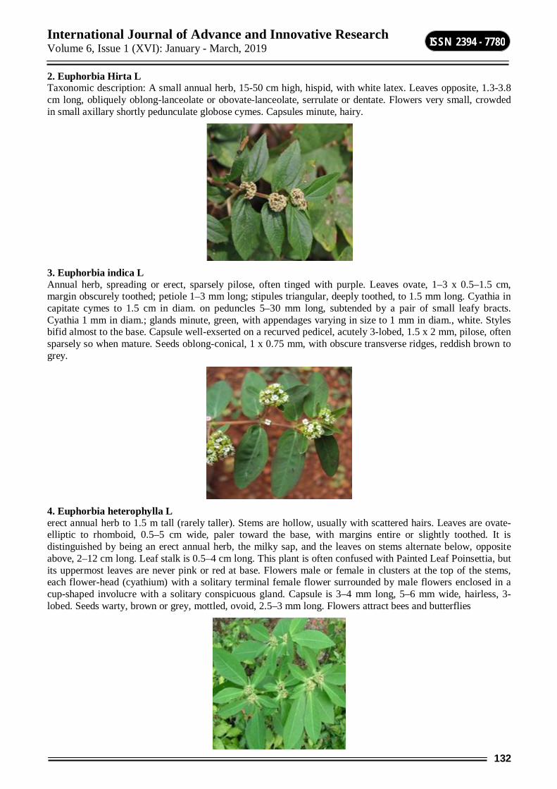
International Journal of Advance and Innovative Research Volume 6, Issue 1 (XVI): January - March, 2019
132
ISSN 2394 - 7780
2. Euphorbia Hirta L Taxonomic description: A small annual herb, 15-50 cm high, hispid, with white latex. Leaves opposite, 1.3-3.8 cm long, obliquely oblong-lanceolate or obovate-lanceolate, serrulate or dentate. Flowers very small, crowded in small axillary shortly pedunculate globose cymes. Capsules minute, hairy.
3. Euphorbia indica L Annual herb, spreading or erect, sparsely pilose, often tinged with purple. Leaves ovate, 1–3 x 0.5–1.5 cm, margin obscurely toothed; petiole 1–3 mm long; stipules triangular, deeply toothed, to 1.5 mm long. Cyathia in capitate cymes to 1.5 cm in diam. on peduncles 5–30 mm long, subtended by a pair of small leafy bracts. Cyathia 1 mm in diam.; glands minute, green, with appendages varying in size to 1 mm in diam., white. Styles bifid almost to the base. Capsule well-exserted on a recurved pedicel, acutely 3-lobed, 1.5 x 2 mm, pilose, often sparsely so when mature. Seeds oblong-conical, 1 x 0.75 mm, with obscure transverse ridges, reddish brown to grey.
4. Euphorbia heterophylla L erect annual herb to 1.5 m tall (rarely taller). Stems are hollow, usually with scattered hairs. Leaves are ovate-elliptic to rhomboid, 0.5–5 cm wide, paler toward the base, with margins entire or slightly toothed. It is distinguished by being an erect annual herb, the milky sap, and the leaves on stems alternate below, opposite above, 2–12 cm long. Leaf stalk is 0.5–4 cm long. This plant is often confused with Painted Leaf Poinsettia, but its uppermost leaves are never pink or red at base. Flowers male or female in clusters at the top of the stems, each flower-head (cyathium) with a solitary terminal female flower surrounded by male flowers enclosed in a cup-shaped involucre with a solitary conspicuous gland. Capsule is 3–4 mm long, 5–6 mm wide, hairless, 3-lobed. Seeds warty, brown or grey, mottled, ovoid, 2.5–3 mm long. Flowers attract bees and butterflies
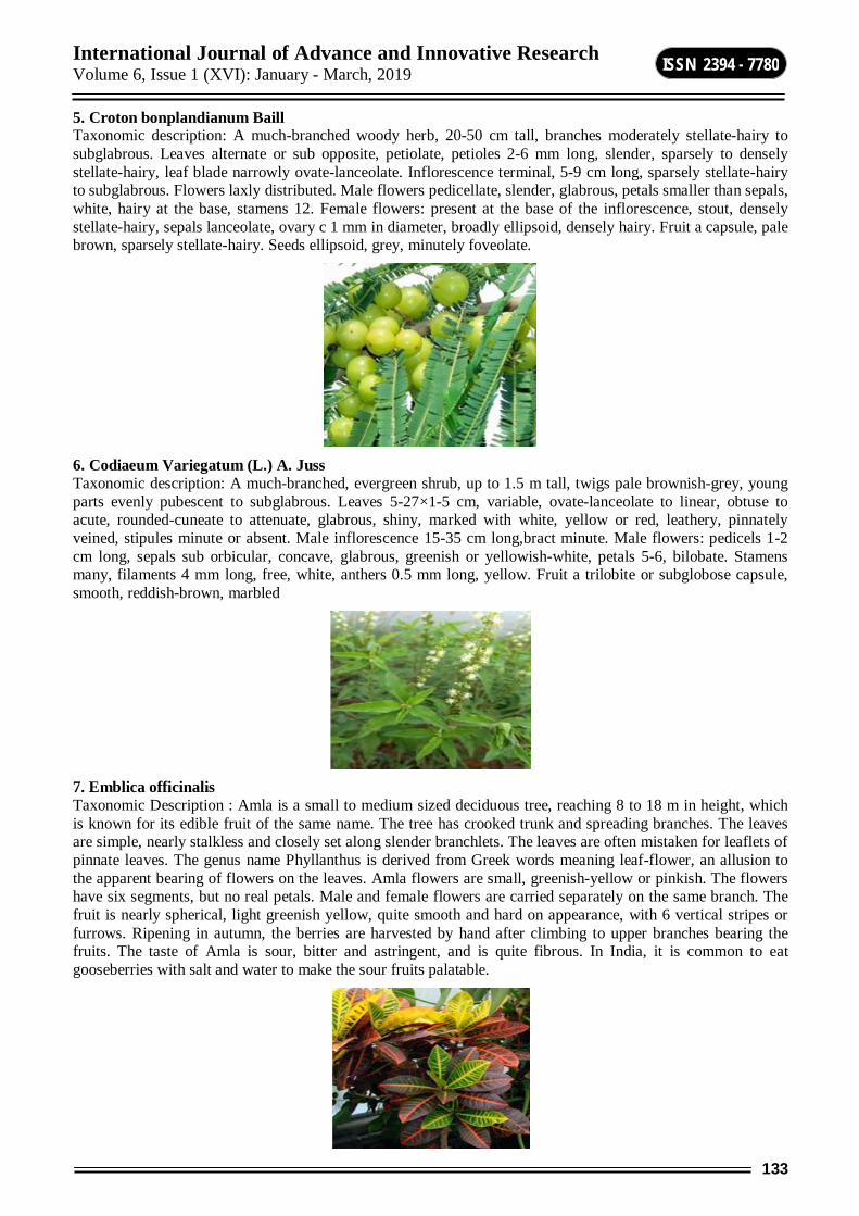
International Journal of Advance and Innovative Research Volume 6, Issue 1 (XVI): January - March, 2019
133
ISSN 2394 - 7780
5. Croton bonplandianum Baill Taxonomic description: A much-branched woody herb, 20-50 cm tall, branches moderately stellate-hairy to subglabrous. Leaves alternate or sub opposite, petiolate, petioles 2-6 mm long, slender, sparsely to densely stellate-hairy, leaf blade narrowly ovate-lanceolate. Inflorescence terminal, 5-9 cm long, sparsely stellate-hairy to subglabrous. Flowers laxly distributed. Male flowers pedicellate, slender, glabrous, petals smaller than sepals, white, hairy at the base, stamens 12. Female flowers: present at the base of the inflorescence, stout, densely stellate-hairy, sepals lanceolate, ovary c 1 mm in diameter, broadly ellipsoid, densely hairy. Fruit a capsule, pale brown, sparsely stellate-hairy. Seeds ellipsoid, grey, minutely foveolate.
6. Codiaeum Variegatum (L.) A. Juss Taxonomic description: A much-branched, evergreen shrub, up to 1.5 m tall, twigs pale brownish-grey, young parts evenly pubescent to subglabrous. Leaves 5-27×1-5 cm, variable, ovate-lanceolate to linear, obtuse to acute, rounded-cuneate to attenuate, glabrous, shiny, marked with white, yellow or red, leathery, pinnately veined, stipules minute or absent. Male inflorescence 15-35 cm long,bract minute. Male flowers: pedicels 1-2 cm long, sepals sub orbicular, concave, glabrous, greenish or yellowish-white, petals 5-6, bilobate. Stamens many, filaments 4 mm long, free, white, anthers 0.5 mm long, yellow. Fruit a trilobite or subglobose capsule, smooth, reddish-brown, marbled
7. Emblica officinalis Taxonomic Description : Amla is a small to medium sized deciduous tree, reaching 8 to 18 m in height, which is known for its edible fruit of the same name. The tree has crooked trunk and spreading branches. The leaves are simple, nearly stalkless and closely set along slender branchlets. The leaves are often mistaken for leaflets of pinnate leaves. The genus name Phyllanthus is derived from Greek words meaning leaf-flower, an allusion to the apparent bearing of flowers on the leaves. Amla flowers are small, greenish-yellow or pinkish. The flowers have six segments, but no real petals. Male and female flowers are carried separately on the same branch. The fruit is nearly spherical, light greenish yellow, quite smooth and hard on appearance, with 6 vertical stripes or furrows. Ripening in autumn, the berries are harvested by hand after climbing to upper branches bearing the fruits. The taste of Amla is sour, bitter and astringent, and is quite fibrous. In India, it is common to eat gooseberries with salt and water to make the sour fruits palatable.
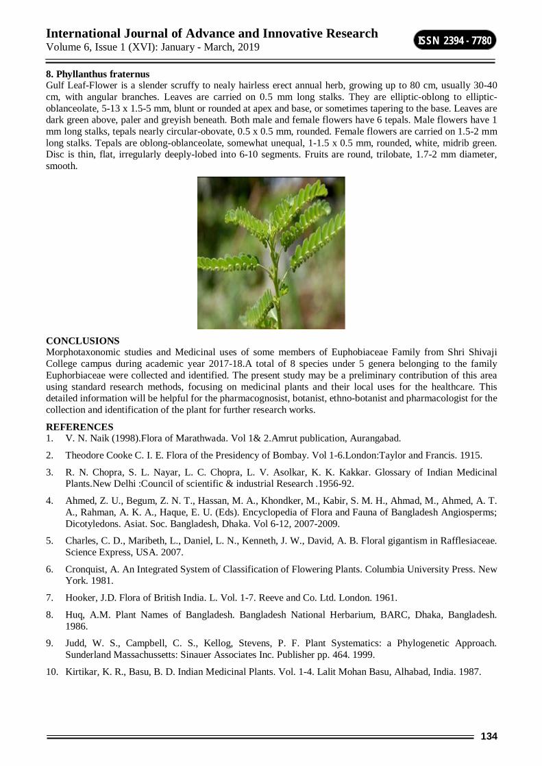
International Journal of Advance and Innovative Research Volume 6, Issue 1 (XVI): January - March, 2019
134
ISSN 2394 - 7780
8. Phyllanthus fraternus Gulf Leaf-Flower is a slender scruffy to nealy hairless erect annual herb, growing up to 80 cm, usually 30-40 cm, with angular branches. Leaves are carried on 0.5 mm long stalks. They are elliptic-oblong to elliptic- oblanceolate, 5-13 x 1.5-5 mm, blunt or rounded at apex and base, or sometimes tapering to the base. Leaves are dark green above, paler and greyish beneath. Both male and female flowers have 6 tepals. Male flowers have 1 mm long stalks, tepals nearly circular-obovate, 0.5 x 0.5 mm, rounded. Female flowers are carried on 1.5-2 mm long stalks. Tepals are oblong-oblanceolate, somewhat unequal, 1-1.5 x 0.5 mm, rounded, white, midrib green. Disc is thin, flat, irregularly deeply-lobed into 6-10 segments. Fruits are round, trilobate, 1.7-2 mm diameter, smooth.
CONCLUSIONS Morphotaxonomic studies and Medicinal uses of some members of Euphobiaceae Family from Shri Shivaji College campus during academic year 2017-18.A total of 8 species under 5 genera belonging to the family Euphorbiaceae were collected and identified. The present study may be a preliminary contribution of this area using standard research methods, focusing on medicinal plants and their local uses for the healthcare. This detailed information will be helpful for the pharmacognosist, botanist, ethno-botanist and pharmacologist for the collection and identification of the plant for further research works.
REFERENCES 1. V. N. Naik (1998).Flora of Marathwada. Vol 1& 2.Amrut publication, Aurangabad.
2. Theodore Cooke C. I. E. Flora of the Presidency of Bombay. Vol 1-6.London:Taylor and Francis. 1915.
3. R. N. Chopra, S. L. Nayar, L. C. Chopra, L. V. Asolkar, K. K. Kakkar. Glossary of Indian Medicinal Plants.New Delhi :Council of scientific & industrial Research .1956-92.
4. Ahmed, Z. U., Begum, Z. N. T., Hassan, M. A., Khondker, M., Kabir, S. M. H., Ahmad, M., Ahmed, A. T. A., Rahman, A. K. A., Haque, E. U. (Eds). Encyclopedia of Flora and Fauna of Bangladesh Angiosperms; Dicotyledons. Asiat. Soc. Bangladesh, Dhaka. Vol 6-12, 2007-2009.
5. Charles, C. D., Maribeth, L., Daniel, L. N., Kenneth, J. W., David, A. B. Floral gigantism in Rafflesiaceae. Science Express, USA. 2007.
6. Cronquist, A. An Integrated System of Classification of Flowering Plants. Columbia University Press. New York. 1981.
7. Hooker, J.D. Flora of British India. L. Vol. 1-7. Reeve and Co. Ltd. London. 1961.
8. Huq, A.M. Plant Names of Bangladesh. Bangladesh National Herbarium, BARC, Dhaka, Bangladesh. 1986.
9. Judd, W. S., Campbell, C. S., Kellog, Stevens, P. F. Plant Systematics: a Phylogenetic Approach. Sunderland Massachussetts: Sinauer Associates Inc. Publisher pp. 464. 1999.
10. Kirtikar, K. R., Basu, B. D. Indian Medicinal Plants. Vol. 1-4. Lalit Mohan Basu, Alhabad, India. 1987.

International Journal of Advance and Innovative Research Volume 6, Issue 1 (XVI): January - March, 2019
135
ISSN 2394 - 7780
EFFECT OF MEDICINAL PLANTS AND ANTAGONISTSON SEED MYCOFLORA ,SEED GERMINATION AND VIGOUR INDEX OF SUNFLOWER
G. B. Honna Department of Botany, Shrisiddheshwar College Majalgaon Di-Beed
ABSTRACT Sunflower (Helianthus annus L.) seeds were treated with fungal and bacterial antagonists.The seeds were soaked in fungal spores the bacterial suspension for 15 minutes.The treated seeds were incubated for 7-8 days.The percentage incidence of mycoflora, percentage of seed germination and vigour index were calculated in treated and controlled seeds.In treated seeds the percentage incidence of seed mycoflora decreases where as percentage of seed germination and vigour index increases as compaired to control.
The seeds were also treated with plant extracts like AzadirachtaindicaA. Juss.,Ocimum sanctum L., Withaniasomnifera (L.) Dunal. Polyalthialongifolia (sonner.), Lantana camera (L). and Zingiberofficinale (Rosc.) Among theseeffective AzadirachtaindicaA. Juss., was more effective than other plants.
The seeds are treated with biocontrol agent like Trichodermaharzianum, Trichodermaviride, Pseudomonasfluorescens and Bacillus subtilis, Among these Trichodermaharzianum and pseudomonas fluorescens were most effective
Keywords: Sunflower, Medicinalplants, Antagonists, Mycoflora, Vigour index.
INTRODUCTION India is striving hard to increase agricultural production with a view to accelerate food production to feed the ever increasing population though an integratedapproach towards the applicatoion of farm technology (Neergaard, 1970; Dharamvir, 1974).Seed play an important role in disseminating pathogenic organism to areas from hitherto, they have been absent. To check the spreads of such pathogen, seed health testing procedure is necessary.
India is the third largest producer of oil seeds in the world. It ranks first in the production of ground nut and sesame.
Oil seeds are grown in an area about 20 million hectares of which nearly 84% areas is rain fed. The vegetable oil is obtained from oil seed crop like sunflower.
About 90-95% areas under Oil seeds remain rain fed of which about 80% area comes under dry land where irrigation facilities do not exist at all. It has been observed that often absence of rains at critical growth stages of kharif oil seed crops, before maturity, causes significant reduction in yields and oil content.
Fats and oil are important ingredients of human food. Vegetable oil is extracted from seeds and fruits of different crops and trees.(Butt & Ali,2005). Sunflower (Helianthus annuus L.) an important member of the family Asteraceaeand is one of the major oil seed crops grown for edible oil in the world (Anon, 2007).
In India sunflower is an important oil seed crop.Popularly known as ‘Surjmukhi . It is one of the fastest growing oil seed crop in India. Sunflower was introduced in India as an oil seed crop for the first time in 1969. It is a drought tolerant crop due to it’s deep taproot, there fore, it is best substitute to all rain feed commercial crop.
Indian sunflower seed production ranges between 10-15 lakh tons. The major producer states are Karnataka (35%), Andhra Pradesh (30%),Maharashtra (15%),Panjab (4%) and Haryana (4%).
Sunflower seeds contain 40-50% oil, 23% protein and constitute excellent source of unsaturated fats, crude protein and fiber and important nutrients like vitamin E, Copper, Zinc, Selenium, B-Complex vitamin.
Seeds are generally associated with certain saprophytic or parasitic micro-organisms which perpetuate in the seed lots on the advent of favorable conditions. Seeds are associated with pathogens like fungi, bacteria, nematodes etc. Pathogens present in almost any seed lot of economically important crop which may be disastrous if introduced into disease free areas. Therefore, seed must be “Substantially free” from inoculum with high level of germination and purity before sowing.
According to recent report of the World Health Organization (WHO), 1-4 % of the world’s grain production is lost due to microbial spoilage.
International Journal of Advance and Innovative Research Volume 6, Issue 1 (XVI): January - March, 2019
136
ISSN 2394 - 7780
Sunflower seed constitute essential components of agriculture.About 90 percent of all food crop are propagated through seeds. They act as passive carries of fungi, bacteria, viruses and nematodes.
Bakers (1972) defined seed borne pathogens and a large number of pathogens belonging to 90 fungal and 5 bacterial genera are seed transmitted (Phatak, 1980 and Tomlinson, 1987).
Among the various microorganisms associated with seeds, fungi play an important role in determining the quality of grains and seeds (Mirocha et al., 1976, Dennis, 1977 and Gupta, 1994).
Seed borne microorganisms considerably effect agricultural production in the field as well as reduce storage life of seed. In several cases such mycoflora is found to affect adversely the seed germination, vigour quality and quantity of oil .(ward and Diener,1961;Kadian and Suryanarayana,1972).
All India coordinated Research Project (AICRP) under Indian Agricultural Research Institute (IARI) has carried out interdisciplinary multilocational research since 1967, gradually leads to the standardization of appropriate production technology for different agro-ecological conditions. Accordingly, Indian agro-ecological condition has been divided into five zones i.e. Northern hill zone, Northern plane zone, Central zone, Southern zone and North-Eastern zone.
In the process of seed bio-deterioration the moulds have been found to cause qualitative and quantative changes in chemical composition of the seed,poisoning food and making them unsuitable for human and animal consumption.production of enzymes and toxins by the moulds have been found to be correlated with the degree of bio-detrrioration.The major post-harvest bio-deterioration of sunflower was found to be fungi,which results in decrease germination . Hence an attempt has been made to increase the seed germination.
The seeds are treated with biocontrol agent like Trichodermaharzianum, Trichodermaviride, Pseudomonasfluorescens and Bacillus subtilis, Among these Trichodermaharzianum and pseudomonas fluorescens were most effective.
The seeds were also treated with plant extracts like AzadirachtaindicaA. Juss.,Ocimum sanctum L., Withaniasomnifera (L.) Dunal. Polyalthialongifolia (sonner.), Lantana camera (L). andZingiberofficinale (Rosc.) . Among theseeffective AzadirachtaindicaA. Juss.,was more effective than other plants.
MATERIAL AND METHODS Sunflower seeds were treated with fungal and bacterial antagonists. The seeds were soaked in fungal spores and bacterial suspension for 15 minutes. The treated seeds were incubated for 7-8 days. The percentage incidence of mycoflora,percentage of seed germination and vigour index were calculated in treated and controlled seeds. In treated seeds the percentage incidence of seed mycoflora decreases whereas of seed germination and vigour index increases as compaired to control.
Seeds were treated with antagonists fungi and bacteria. The antagonist fungi like Trichoderma species. The antagonists bacteria used were Bacillus subtilis and pseudomonas fluorescens.
SEED TREATMENT WITH TRICHODERMA SPECIES a) Seed treatment with Trichodermaspecies 200 g of seeds were coated with 100mL aqueous spore suspension of Trichoderma species (8×10⁹ spores/mL) by adding 1 mL of 0.5% carboxyl methyl cellulose (CMC) as sticker and 20 g of Bentonite powder as filler for seed dressing. Treated seeds were incubated for 7-8 days. The percent incidence of fungi, seed germination and vigour index were observed in seed samples.
b) Seed treatment with Bacterial species The method of Weller and Cook (1983) was followed for seed bacterization. Pseudomonas fluorescensandBacillussubtilis were separately grown in succinate broth for 24 hours at 2810C under shaking condition and finally centrifuged at 7000 rpm for 15 minutes at 40C. The supernatant was discarded and pellets were washed with SDW and resuspended to obtain a population density of 107cfu/mL. This suspension was mixed with 1% carboxyl methyl cellulose (CMC). Seeds were allowed to air dry overnight under aseptic condition after coating with CMC slurry of bacterial culture. Care was taken to avoid clumping of seeds. Seeds coated with slurry of CMC (without bacteria) served as control. The seeds were incubated on sterile blotter paper. The percent mycoflora, seed germination and vigour index were observed in seed samples.
iv) Effect of plant extract on seed mycoflora, germination and vigour index During the present study six common plants namely AzadirachtaindicaA. Juss., Ocimum sanctum L., Withaniasomnifera (L.) Dunal Polyalthialongifolia (sonner). Thw; Lantana camera (L.) Zingiberofficinale
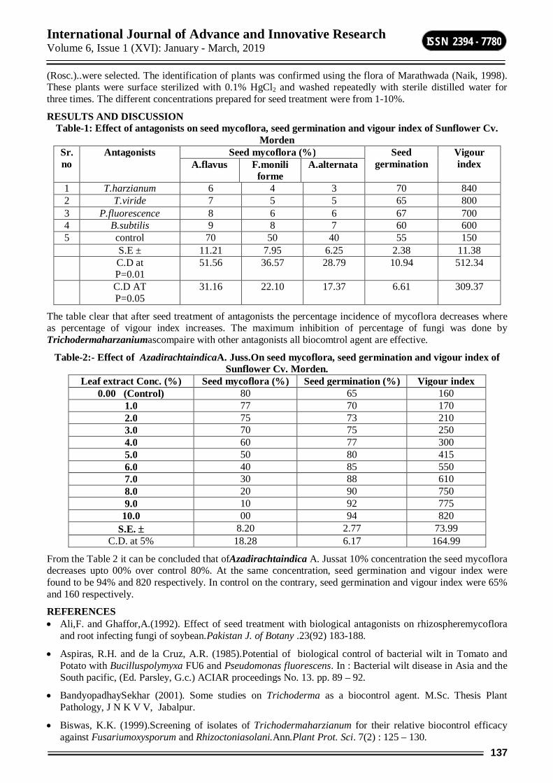
International Journal of Advance and Innovative Research Volume 6, Issue 1 (XVI): January - March, 2019
137
ISSN 2394 - 7780
(Rosc.)..were selected. The identification of plants was confirmed using the flora of Marathwada (Naik, 1998). These plants were surface sterilized with 0.1% HgCl2 and washed repeatedly with sterile distilled water for three times. The different concentrations prepared for seed treatment were from 1-10%.
RESULTS AND DISCUSSION Table-1: Effect of antagonists on seed mycoflora, seed germination and vigour index of Sunflower Cv.
Morden Sr. no
Antagonists Seed mycoflora (%) Seed germination
Vigour index A.flavus F.monili
forme A.alternata
1 T.harzianum 6 4 3 70 840 2 T.viride 7 5 5 65 800 3 P.fluorescence 8 6 6 67 700 4 B.subtilis 9 8 7 60 600 5 control 70 50 40 55 150 S.E ± 11.21 7.95 6.25 2.38 11.38 C.D at
P=0.01 51.56 36.57 28.79 10.94 512.34
C.D AT P=0.05
31.16 22.10 17.37 6.61 309.37
The table clear that after seed treatment of antagonists the percentage incidence of mycoflora decreases where as percentage of vigour index increases. The maximum inhibition of percentage of fungi was done by Trichodermaharzaniumascompaire with other antagonists all biocomtrol agent are effective.
Table-2:- Effect of AzadirachtaindicaA. Juss.On seed mycoflora, seed germination and vigour index of Sunflower Cv. Morden.
Leaf extract Conc. (%) Seed mycoflora (%) Seed germination (%) Vigour index 0.00 (Control) 80 65 160
1.0 77 70 170 2.0 75 73 210 3.0 70 75 250 4.0 60 77 300 5.0 50 80 415 6.0 40 85 550 7.0 30 88 610 8.0 20 90 750 9.0 10 92 775
10.0 00 94 820 S.E. 8.20 2.77 73.99
C.D. at 5% 18.28 6.17 164.99
From the Table 2 it can be concluded that ofAzadirachtaindica A. Jussat 10% concentration the seed mycoflora decreases upto 00% over control 80%. At the same concentration, seed germination and vigour index were found to be 94% and 820 respectively. In control on the contrary, seed germination and vigour index were 65% and 160 respectively.
REFERENCES Ali,F. and Ghaffor,A.(1992). Effect of seed treatment with biological antagonists on rhizospheremycoflora
and root infecting fungi of soybean.Pakistan J. of Botany .23(92) 183-188.
Aspiras, R.H. and de la Cruz, A.R. (1985).Potential of biological control of bacterial wilt in Tomato and Potato with Bucilluspolymyxa FU6 and Pseudomonas fluorescens. In : Bacterial wilt disease in Asia and the South pacific, (Ed. Parsley, G.c.) ACIAR proceedings No. 13. pp. 89 – 92.
BandyopadhaySekhar (2001). Some studies on Trichoderma as a biocontrol agent. M.Sc. Thesis Plant Pathology, J N K V V, Jabalpur.
Biswas, K.K. (1999).Screening of isolates of Trichodermaharzianum for their relative biocontrol efficacy against Fusariumoxysporum and Rhizoctoniasolani.Ann.Plant Prot. Sci. 7(2) : 125 – 130.

International Journal of Advance and Innovative Research Volume 6, Issue 1 (XVI): January - March, 2019
138
ISSN 2394 - 7780
Chande D.S and Chowdhary S.R (1995). Antagonism of Trichodermalongibrachitum,againstmicrofungi isolated from the phylloplane of soybean. Indian J. Mycol.Pl.Path 25(1&2):125.
Chang, R.R. Baker,O.Kliefeld and I.Chet (1986) Increased growth of plants in the presence of the biological control agent, Trichodermaharzianum.plants Dis, 70:145-148.
Claudia Calistru,Michelle,MC Lean and Patricia Berjak (1997).In vitro studies on the potential for biological control of Aspergillusflavus and Fusariummoniliforme by Trichodermaspecies.Mycopathologia137:115-124.
Dennis,C. and Webster,J.(1971). Antagonistic properties of species group of TrichodermaIII.Hyphal interaction Trans.Br.mycol.soc. 57:363-369.
Gahil, V.P. and Vala, D.G. (1996).Effect of extract of some medicinal plants on growth of Fusariummoniliforme.Indian J. Mycol. Pl.Pathol.26(1) : 110 – 111.
Hajra, K.K., Khatua, D.C. and Mukherjee, N. (1992).Antagonistic bacteria against fungal pathogens.J.Mycopathol. Res. 30(1) : 68 – 70.
KausikBiswas, Ishitachattopadhyay, Ranjit,Banerjee,K. and UdayBandopadhyay (2002). Biological activities and medicinal properties of neem(Azadirachtaindica)current sci,Vol.82(11):1376.
Lalitha, V. and Veesha, K.A. (2006).Antagonistic effect of Trichodermakoningii on important seed borne pathogens of Paddy.Asian Jr. ofMicrobiol.Biotech.Env. Sc. Vol. 8, No.(1) : 483 – 488..
PurnimaDargan and Sexena, S.K.(2002).Effect of plant extract of withaniasomnifera on fruit rot of tomata caused by Aspergillusniger in presence of Drosophila busckii.Indian phytopath 55(1):112-113.
Rajathilagam, R. and Kannabirun, B. (2001).Antagonistic effect of Trichodermaviride against anthracnose fungus Colletotrichumcapsici.IndianPhytopath. 54(1) : 135 – 136.
Sitansu Pan and SomeshwarBhagat (2007).Antagonistic potential of Trichoderma and Gliocladium spp. from West Bengal.J. Mycol. Pl.Pathol.Vol. 37, No. 2.
XueBaodi, Juan, L. and Y ongxuan, C. (1995).Studies on antagonism of Trichoderma. Sp. Against 6 pathogenic fungi and biocontrol.JNanjingAgric. Uni. 18: 31-36.
International Journal of Advance and Innovative Research Volume 6, Issue 1 (XVI): January - March, 2019
139
ISSN 2394 - 7780
MULTICOMPONENT SYNTHESIS OF SPIRO[4H-PYRAN-OXINDOLE] DERIVATIVES USING BIMETALLIC CATALYST ZIRCONIUM TETRACHLORIDE AND MAGNESIUM PERCHLORATE
Giribala M. Bondle Department of Chemistry, Dr. BabasahebAmbedkar Marathwada University, Aurangabad
ABSTRACT Structurally diverse spiro-heterocycles with fused systems incorporating medicinally privileged systems have been synthesized by an efficient and convenient synthetic method involving three component reaction of dimedone, substituted isatin and malononitrile using composite catalyst ZrCl4:Mg(ClO4)2 in a molar ratio(1:2) respectively at room temperature. Simple reaction conditions andeasy work up procedure that resulted into simple isolation and purification of products by non-chromatographic methods.
Keywords: Isatin, dimedone, spiro[4H-pyran-oxindole], bimetallic catalyst
INTRODUCTION Innovation of straight synthetic pathways and easy access to more complex products are of the main incentives existing behind the recent researches in organic synthesis. To meet these goals, chemists have been attempting specially to develop multicomponent reactions (MCR).1 Since these reactions offer significant advantages over conventional linear type syntheses, as more often are recognized cost-effective and comparatively fast routes though generating less chemical waste.2Consequently, they are regarded as viable synthetic routes toward both economical and environmental benefits of chemical transformations. In this view, designing of MCRs without using toxic catalysts under neat reaction conditions isworthwhile for complementing the significant characters of MCRs, so as to satisfy the green chemistry principles.3Generation of a bimetallic catalyst can leads to changes in available sites either by the generation of new geometries or by reducing the formation of metal adsorbate complexes.Thus changes in either the electronic or geometric properties of an active site may alter the absorption energies of the reactants and products leading to higher activity,4 suppression of side reactions, resulting in improved selectivity. To make a contribution to these ongoing research fields connected with our interest in the synthesis of spiro-oxindole compounds.5
The indole ring system is probably the most well-known heterocycle, a common and important feature of various natural products and medicinal agents.6It constitutes the core of spiro-oxindoles, a recurring subclass of indole alkaloids in nature, which exhibit highly pronounced biological activities so as deserve to occupy a unique place among pharmacological agents.7Gelsemine, pseudotabersonine, morroniside, formosanine, isoformosanine, and mitraphylline are representatives of the alkaloids, which incorporate spiro-oxindole ring systems.8Certain members of this class, possessing significant bioactivities, have prompted many attempts toward synthesis of structurally related mimetics in search for new drug-like lead molecules.9For example, spirotryprostatin A and B, two natural alkaloids isolated from the fermentation broth of Aspergillus fumigates, have been identified as novel inhibitors of microtubule assembly, while pteropodine and isopteropodine have been shown to modulate the function of muscarinic serotonin receptors.10,11On the other hand, pyran-containing heterocycles have been found to have various biological activities.12For examples, zanthosimuline I is active against multidrug resistant KB-VI cancer cells, while huajiaosimuline II exhibits a selectivecytotoxicity profile showing the greatest activity with estrogen receptor-positive ZR-75-1 breast cancer cells.13 In view of these important profiles enlightened by the recognition that molecules comprised of two or more heterocyclic nucleuses often possess heightened pharmacological activities,14remarkable attention has been aimed at synthesis of the spiro [4H-pyran-oxindole] ring system. Interest in the synthesis of these compounds was increased further by the fact that indolesspirofused at the 3-position generally show intensive biological properties. 15The main synthetic avenue and direct method for assembling of spiro[4H-pyran-oxindole] compounds is based on the three-component condensations of two (usually different) 1,3-dicarbonyl compounds, or alternatively their synthetic equivalents, with isatin derivatives. These reactions are generally slow processes by themselves at room temperature, so several methods describing activation by heating and assistance in aqueous media by using various catalysts, such as ammonium chloride,16ethylenediamine diacetate,17surfactant metal carboxylates,18L-proline,19triethylbenzyl ammonium (TEBA) salt,20and β-cyclodextrin21were devised so far to facilitate the synthesis. Beside traditional heating methods, microwave dielectric heating employing InCl3 under solvent-free conditions has been similarly used to shorten the reaction times significantly.22 Alternatively, Et3N-catalyzed three-component condensations23 and piperidine assisted two-component reactions were invoked to give spirofusedpyran-oxindole scaffold in refluxing ethanol solutions24and also magnesium perchlorate25catalysed three component reaction.All the above mentioned
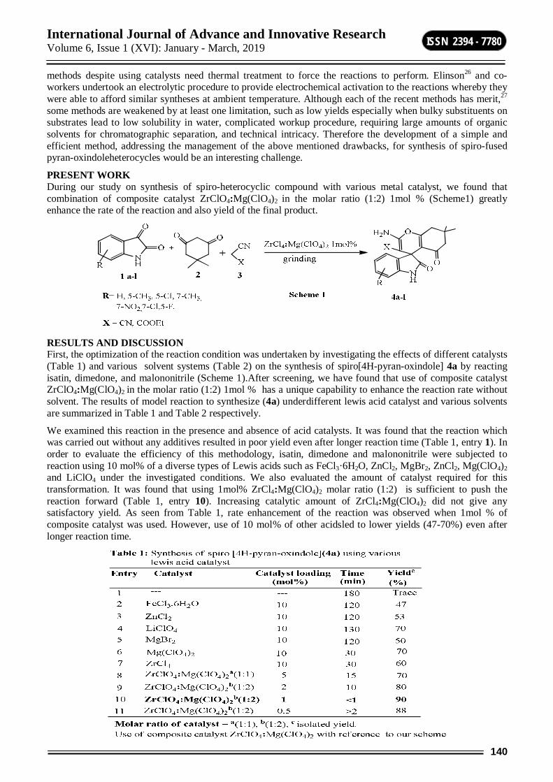
International Journal of Advance and Innovative Research Volume 6, Issue 1 (XVI): January - March, 2019
140
ISSN 2394 - 7780
methods despite using catalysts need thermal treatment to force the reactions to perform. Elinson26 and co-workers undertook an electrolytic procedure to provide electrochemical activation to the reactions whereby they were able to afford similar syntheses at ambient temperature. Although each of the recent methods has merit,27 some methods are weakened by at least one limitation, such as low yields especially when bulky substituents on substrates lead to low solubility in water, complicated workup procedure, requiring large amounts of organic solvents for chromatographic separation, and technical intricacy. Therefore the development of a simple and efficient method, addressing the management of the above mentioned drawbacks, for synthesis of spiro-fused pyran-oxindoleheterocycles would be an interesting challenge.
PRESENT WORK During our study on synthesis of spiro-heterocyclic compound with various metal catalyst, we found that combination of composite catalyst ZrClO4:Mg(ClO4)2 in the molar ratio (1:2) 1mol % (Scheme1) greatly enhance the rate of the reaction and also yield of the final product.
RESULTS AND DISCUSSION First, the optimization of the reaction condition was undertaken by investigating the effects of different catalysts (Table 1) and various solvent systems (Table 2) on the synthesis of spiro[4H-pyran-oxindole] 4a by reacting isatin, dimedone, and malononitrile (Scheme 1).After screening, we have found that use of composite catalyst ZrClO4:Mg(ClO4)2 in the molar ratio (1:2) 1mol % has a unique capability to enhance the reaction rate without solvent. The results of model reaction to synthesize (4a) underdifferent lewis acid catalyst and various solvents are summarized in Table 1 and Table 2 respectively.
We examined this reaction in the presence and absence of acid catalysts. It was found that the reaction which was carried out without any additives resulted in poor yield even after longer reaction time (Table 1, entry 1). In order to evaluate the efficiency of this methodology, isatin, dimedone and malononitrile were subjected to reaction using 10 mol% of a diverse types of Lewis acids such as FeCl3·6H2O, ZnCl2, MgBr2, ZnCl2, Mg(ClO4)2 and LiClO4 under the investigated conditions. We also evaluated the amount of catalyst required for this transformation. It was found that using 1mol% ZrCl4:Mg(ClO4)2 molar ratio (1:2) is sufficient to push the reaction forward (Table 1, entry 10). Increasing catalytic amount of ZrCl4:Mg(ClO4)2 did not give any satisfactory yield. As seen from Table 1, rate enhancement of the reaction was observed when 1mol % of composite catalyst was used. However, use of 10 mol% of other acidsled to lower yields (47-70%) even after longer reaction time.
International Journal of Advance and Innovative Research Volume 6, Issue 1 (XVI): January - March, 2019
141
ISSN 2394 - 7780
Furthermore, screening of solvents with bimetallic catalyst was also investigated which concludes that use of various solvents not show a remarkable effect on yield of the product. Thus neat reaction condition is more suitable condition for this reaction.
As shown in Table 3, it was found that this procedure works with a wide variety of substrates. Seven types of substituted isatins, one dimedone with malononitrile or ethyl cyanoacetate were used in this reaction (Scheme 1). But the reaction with malononitrile was finished faster than with ethyl cyanoacetate which may be owing to the difference of the activity between the two active methylene reagents.
The most probable mechanism of this reaction includes a fast Knoevenagel condensation between isatin and CH-acidic cyanoacetic ester derivatives in the presence of composite catalyst in the first step and a Micheal addition of diketones to the unsaturated nitrile, the product of Knoevenagel condensation, in the second stage and then the cycloaddition of the hydroxyl group to the cyano moiety to form the desired product. After the reaction was over (TLC), the resulting solid was filtered and washed with aqueous ethanol solution to yield pure substituted spiro[4H-pyran-oxindole] 4a-l. All the products were crystalline and characterized based on their melting points, elemental analysis, and spectral data.
International Journal of Advance and Innovative Research Volume 6, Issue 1 (XVI): January - March, 2019
142
ISSN 2394 - 7780
CONCLUSION In conclusion, we have reported an environmentally friendly three-component, efficient, clean and simple method for the synthesis of some new spirooxindole derivatives using readily available starting materials.Use of composite catalyst without solvent is important alternative to the use of volatile organic solvents. The advantages of this new methods are short reaction times, operational simplicity, use of IL media, high yields, easy work up procedures, nontoxic, easily available, recyclable and inexpensive catalyst.
EXPERIMENTAL General procedure for the synthesis of Spiro [4H-pyranoxindole] A mixture ofisatin1(1mmol), dimedone2 (1mmol) and malononitrile3 (1mmol) was added together in a mortar and pestle with 1mol % of the composite of ZrCl4:Mg(ClO4)2 in molar ratio(1:2). The reaction mixture was grinded until disappearance of the starting materials (under 5 min). After completion of the reaction, the residue was washed with 2×10 mL of either water or diethyl ether. Washing the solid residue with ethanol (10mL, 95.5%) has given remarkably pure powders of product.
Spectral data of some representative compounds 2-amino-7,7-dimethyl-2',5-dioxo-5,6,7,8-tetrahydrospiro[chromene-4,3'-indoline]-3-carbonitrile(4a)White solid, IR (KBr) cm-1: 3380, 3190, 3010, 2960, 2200, 1730, 1620, 1350, 1227. 1HNMR (400 MHz, DMSO-d6): δ 1.00 (s, 3H, CH3), 1.03 (s, 3H, CH3), 2.09 (d, J =16.0 Hz, 1H, CHAHB), 2.13 (d, J = 16.0 Hz, 1H, CHAHB), 2.56 (s, 2H, CH2), 6.78 (d, J = 7.6 Hz, 1H, ArH), 6.88 (t, J = 7.4 Hz, 1H, ArH), 6.98 (d, J = 6.8 Hz, 1H, ArH), 7.14 (t, J = 8.2 Hz, 1H, ArH), 7.12 (s, 2H, NH2), 10.39 (s, 1H,NH); 13CNMR (100 MHz, DMSO-d6) δ 27.5, 28.1, 32.4, 47.3, 50.4, 57.9, 109.7, 111.2, 117.8, 122.1, 123.5, 128.6,134.9, 142.5, 159.2, 164.6, 178.5, 195.3; m/z = 335(358.1) [M+ Na] .
2-amino-5',7,7-trimethyl-2',5-dioxo-5,6,7,8-tetrahydrospiro[chromene-4,3'-indoline]-3-carbonitrile(4b) White solid 1HNMR (400 MHz,DMSO-d6) δ 1.00 (s, 3H, CH3), 1.02 (s, 3H, CH3), 2.12 (m,2H, CH2), 2.19 (s, 3H, Ar-CH3), 2.55 (m, 2H, CH2), 6.67(d, J = 7.2 Hz, 1H, ArH), 6.78 (s, 1H, ArH), 6.93 (d, J = 8.4Hz, 1H, ArH), 7.19 (s, 2H, NH2), 10.27 (s, 1H, NH). 13CNMR (100 MHz, DMSO-d6) δ 21.1, 27.7, 27.9, 32.4, 47.3, 50.5, 58.2, 109.4, 111.3, 117.8, 124.1, 128.9, 130.9, 135.0, 140.1, 159.2, 164.5, 178.4, 195.3; m/z = 349 (M+).
2-amino-5-chloro-7,7-dimethyl-2',5-dioxo-5,6,7,8-tetrahydrospiro[chromene-4,3'-indoline]-3-carbonitrile(4c) White solid, 1HNMR(400MHz, DMSO-d6): δ 1.00 (s, 3H, CH3), 1.01 (s, 3H, CH3), 2.14 (s, 2H, CH2), 2.49-2.56 (m, 2H, CH2), 6.78 (d, J =8.0 Hz, 1H, ArH), 7.08 (s, 1H, ArH), 7.17 (d, J = 5.6 Hz,1H, ArH), 7.29 (s, 2H, NH2), 10.51 (s, 1H, NH). 13CNMR (100 MHz, DMSO-d6): δ 27.7, 27.9, 32.4, 47.5, 50.4, 57.1, 110.6, 111.1, 117.7, 123.7, 126.1, 128.5, 136.9, 141.5, 159.3, 165.1, 178.3, 195.6; m/z = 369.08.
Ethyl-2-amino-7,7-dimethyl-2',5-dioxo-5,6,7,8-tetrahydrospiro[chromene-4,3'-indoline]-3-carbonitrile (4g) White solid ,1HNMR (400MHz, DMSO-d6) δ 0.79 (t, J = 6.8 Hz, 3H, CH3), 0.94 (s,3H, CH3), 1.01 (s, 3H, CH3), 2.00 (d, J = 16.0 Hz, 1H,CHAHB), 2.14 (d, J = 16.0 Hz, 1H, CHAHB), 2.52 (m, 2H,CH2), 3.69 (q, J = 6.8 Hz, 2H, CH2), 6.66 (d, J = 7.6 Hz, 1H,ArH), 6.75 (t, J = 7.2 Hz, 1H, ArH), 6.82 (d, J = 7.2 Hz, 1H,ArH), 7.03 (t, J = 7.2 Hz, 1H, ArH), 7.84 (s, 2H, NH2), 10.12(s, 1H, NH); 13C NMR (100MHz, DMSO-d6) δ 13.6, 27.1,28.2, 32.0, 47.1, 51.1, 59.3, 76.8, 108.6, 113.6, 121.0, 122.7,127.6, 136.4, 144.5, 159.6, 162.8, 168.1, 180.2, 195.1; m/z = 309.12.
2-amino-5-fluoro-7,7-dimethyl-2',5-dioxo-5,6,7,8-tetrahydrospiro[chromene-4,3'-indoline]-3-carbonitrile (4l) Light pink solid, IR (KBr) cm-1: 3359, 3299, 3161, 2963, 2190, 1726, 1647, 1345, 1223. 1HNMR (400MHz, DMSO-d6): δ (ppm) 1.00 (3H, s, CH3), 1.04 (3H, s,CH3), 2.07 (1H, d, J=15.9 Hz, CHAHB), 2.15 (1H, d, J=16.0 Hz, CHAHB),2.51 (2H, m, CH2), 6.62–6.78 (3H, m, ArH), 7.70 (2H, s, NH2), 10.24(1H, s, NH). 13CNMR (100MHz, DMSO-d6): (ppm) 27.35 (CH3), 28.11 (CH3), 33.12, 47.35 (CH2), 51.2 (CH2), 59.12, 110.15, 12.28, 117.81 (CN), 123.09, 124.45, 129.58, 133.81, 144.52, 160.01, 167.52, 179.45 (C=O, amide), 196.25 (C=O); m/z = 353 (M+).
REFERENCES [1] (a) Mont, N.; Teixido, J.; Borrell, J.I.; Kappe, C. O.; Tetrahedron Lett.2003, 44, 5385. (b) Bagley, M. C.;
Dale, J. W.; Bower; J. Chem. Commun. 2002, 1682. (c) Simon, C.; Constantieux, T.; Rodriguez, J.; Eur. J. Org. Chem. 2004, 24, 4957. (d) Huang, Y. J.; Yang, F. Y.; Zhu, C. J.; J. Am. Chem. Soc.2005, 127, 16386.
[2] (a) Domling, A.; Ugi, I.; Angew.Chem. Int. Ed. 2000, 39, 3168. (b) Domling, A.; Chem. Rev.2006, 17, 106.
[3] (a) Martins, M. A. P.; Frizzo, C. P.; Moreira, D. N.; Zanatta, N.; Bonacorso, H.; Chem. Rev. 2008, 108,

International Journal of Advance and Innovative Research Volume 6, Issue 1 (XVI): January - March, 2019
143
ISSN 2394 - 7780
2015. (b) Holbery, J. D.; Turner, M. B.; Reichart, W. M.; Rogers, R. D.; Green Chem. 2003, 5,731.
[4] Sachtler W.M.H.;VanSanten R. A.;Adv. Catal. 1977; 26:69-119.
[5] Dautzenberg F. M.; Helle J. N.; Biloen P.; Sachtler W. M. H.; J. Catal. 1980; 63:119.
[6] Rad-Moghadam, K.; Youseftabar-Miri, L.; Synlett. 2010, 1969.
[7] Hoalihan, W. J.; Remers,W. A.; Brown, R. K. Indoles: Part 1; Wiley: New York, NY, 1992.
[8] (a) Ma, J.; Hecht, S. M.; Chem. Commun. 2004, 1190. (b) Edmondson, S.; Danishefsky, S. J.; Sepp-Lorenzinol, L.; Rosen, N.; J. Am. Chem. Soc. 1999, 121, 2147. (c) Smith, W. P.; Sollis, L. S.; Howes, D.P.; Cherry C. P.; Starkay, D. I.; Cobley, N. K.; J. Med. Chem. 1998, 41, 787.
[9] (a) Early, W. G.; Oh, T.; Overman, L. E. Tetrahedron Lett. 1988, 29, 3785; (b) Ban, Y.; Seto, M.; Oishi, T. Chem. Pharm. Bull. 1975, 23, 2605; (c) Ban, Y.; Taga, N.; Oishi, T. Tetrahedron Lett. 1974, 2, 187.
[10] (a) Dania, A.; Singh, R.;Khaturia, S.; Merienne, C.; Morgant, G.; Loupy, A.; Bioorg. Med. Chem. 2006, 14, 2409. (b) Sebahhar, P. R.; Williams, R. M. J.; Am. Chem. Soc. 2000, 122, 5666.
[11] Kang, T. H.; Matsumoto, K.; Murakami, Y.; Takayama, H.; Kitajima, M.; Aimini, N.; Watanabe, H.; Eur. J. Pharmacol. 2002, 444, 39.
[12] Kulkarni, S. K.; Kaul, P. N. Indian J. Exp. Biol. 1980, 13, 270.
[13] Chen, T. S.; Wu, S. J.; Tsai, I. L.; Pezzuto, J. M.; Lu, M. C.; Chai, H.; Suh, N.;Teng, C. M.; J. Nat. Prod. 1994, 57, 1206.
[14] (a) Hilton, S. C.; Ho, T. C. T.; Pljevaljcic, G.; Jones, K.; Org. Lett. 2000, 2, 2639. (b) Fresneda, P. M.; Molina, P.; Bleda, J. A.; Tetrahedron.2001, 57, 2355.
[15] (a) Da-Silva, J. F. M.; Garden, S. J.; Pinto, A. C.; J. Braz. Chem. Soc. 2001,12, 273. (b) Joshi K. C.; Jain, R.; Sharma, K.;J. Ind. Chem. Soc. 1988, 115, 202.
[16] Dabiri, M.; Bahramnnejad, M.; Baghbanzadeh, M.; Tetrahedron. 2009, 65, 9443.
[17] Lee, Y. R.; Hari, G. S. Synthesis2010, 3, 453.
[18] Wang, L. M.; Jiao, N.; Qiu, J.; Yu, J. J.; Liu, J. Q.; Guo, F. L.; Liu. Y.; Tetrahedron. 2010, 66, 339.
[19] Yuling, L.; Hui, C.; Chunling, S.; Daqing, S.; Shunjun, J.; J. Comb. Chem. 2010, 12, 231.
[20] Zhu, S. L.; Ji, S. J.; Zhang, Y.; Tetrahedron. 2007, 63, 9365.
[21] Sridhar, R.; Srinivas, B.; Madhav, B.; Reddy, V. P.; Nageswar, Y. V. D.; Rao, K. R.; Can. J. Chem. 2009, 87, 1704.
[22] Shanthi, G.; Subbulakshmi, G.; Perumal, P. T. Tetrahedron.2007, 63, 2057.
[23] Litvinov, Y. M.; Mortikov, V. Y.; Shestopalov, A. M.; J. Comb. Chem. 2008, 10, 741.
[24] El-Ahl, A. A.; Metwally, M. A.; J. Chem. Res. 1994, 14.
[25] C. Wu, R. Shen, J. Chen, and C. Hu; Bull. Korean Chem. Soc. 2013, Vol. 34, 8, 2431.
[26] Elinson, M. N.; Ilovaisky, A. I.; Dorofeev, A. S.; Merkulov, V. M.; Stepanov, N. O.; Miloserdov, F. M.; Ogibin, Y. N.; Nikishin, G. I.; Tetrahedron. 2007, 63, 10543.
[27] Moradi, R.; Ziarani, G. M.; Lashgari N.; Arkivoc. 2017, i, 148-201.
International Journal of Advance and Innovative Research Volume 6, Issue 1 (XVI): January - March, 2019
144
ISSN 2394 - 7780
EVALUATION OF ANTIMICROBIAL AND ANTIFUNGAL PROPERTIES OF 3-((1H-INDOL-3-YL)(1,3-DIPHENYL-1H-PYRAZOL-4-YL)METHYL)-1H-INDOLE SYNTHESIZED USING NOVEL
CLAY CATALYST
Ismail Shaikh1, Rajendra Satpute2, Syed Abed3, Sheshrao Pawar4
1, 4P. G. Department of Chemistry, ShriAnand College Pathardi, Dist. Ahmednagar 2Rajaram College Kolhapur, Dist. Kolhapur
3Government College of Arts and Science Aurangabad, Dist. Aurangabad
ABSTRACT This article gives information about study of antibacterial and antifungal properties of synthesized 3-((1H-indol-3-yl)(1,3-diphenyl-1H-pyrazol-4-yl)methyl)-1H-indole using novel clay catalyst.Bis-indolyl pyrazolyl methane has appeared as structurally novel antifungal and antibacterial and activity. Therefore 3-((5-chloro-3-methyl-1-phenyl-1H-pyrazol-4-yl)(1H-indol-3-yl)methyl)-1-H-indole was synthesized by addition of 1, 3-diphenyl-1H-pyrazole-4-carboxaldehyde with indole.
Keywords: Novel Clay,Bis indolyl Pyrazolyl methane, antibacterial, antifungal activity, 1, 3-diphenyl-1H-pyrazole-4-carboxaldehyde.
INTRODUCTION Infectious diseases initiated by microbes such as bacteria and fungi are one of the leading causes of disease and mortality and the major cause for the increase in microbial infections is the resistance developed by these microbial organisms, particularly gram-positive bacteria S. aureus and species of the genus enterococcus towards existing antimicrobial drugs [1]. The appearance and spread of antimicrobial resistance has become one of the most serious public health anxieties across the world. Antimicrobial resistance refers to micro-organism that has developed the ability to inactivate, exclude or block the inhibitory or lethal mechanism of the antimicrobial agents [2]. Vibrindole has been demonstrated for the first time to exhibit antibacterial activity against S. aureus, S. albus and B. subtills. Gentamycin is in use as a standard antibacterial drug [3]. Predominantly bis(indolyl)methanes are the most active cruciferous ingredients, showing a wide array of pharmacological activities [4]. Substituted indole derivatives possess antibacterial activity [5]. S. aureus, the chief culprit, is also a common source of community developed infections, and causes illnesses that range from minor skin infections and abscesses to life-threatening diseases such as severe pneumonia, meningitis, joint infections, and heart and blood stream infections [6]. Bis indolyl methane exhibit antimicrobial and antifungal activities [7]. Analgesic and anti-inflammatory [8].Anticancer [9].Bis(indolyl)methanes were obtained by reactions of indole with various aldehydes in the presence of several bronsted and lewis acid catalysts such as LiClO4 [10], montmorillonite clay K-10 [11-12], NBS [13],ZrCl4 [14], Zeolite [15].But many of these methods have several drawbacks such as use of expensive reagents, longer reaction times, cumbersome workup, and low product yields. But in the present work, we replaced this catalyst by low cost cheaply available Novel clay catalyst.
2. EXPERIMENTAL PROTOCOLS 2.1 Chemistry All chemicals were acquired from major chemical suppliers as high or highest purity grade and without further purification. The melting points are uncorrected TLC is run in n- hexane and methyl acetate in required amount. FT-IR was noted in KBr, HNMR in CDCl3 from Central Instrumentation Facility, Savitribai Phule Pune University, and Pune. X-Ray Powder diffraction (XRD) was noted from department of Physics, Savitribai Phule Pune University. Energy-dispersive X-Ray Spectroscopy (EDS) and Field Emission Scanning Electron Microscope (FESEM) by using device Nova Nano SEM 450 UOP were documented from Central Instrumentation Facility, Savitribai Phule Pune University, and Maharashtra All the synthesized drugs were used for antibacterial test procedures, All necessary controls like drug control, vehicle control, agar control, organism control, known antibacterial drugs control, entirely MTCC cultures were confirmed against above mentioned known and unknown drugs, Mueller hinton broth was used as nutrient medium to propagate and dilute the drug suspension for the test bacteria, inoculum size for test strain was adjust to 108cfu [Colony Forming Unit] per milliliter by comparing the turbidity, Subsequent common standard strains were used for screening of antibacterial and antifungal activities. The strains were acquired from Institute of Microbial Technology, Chandigarh.
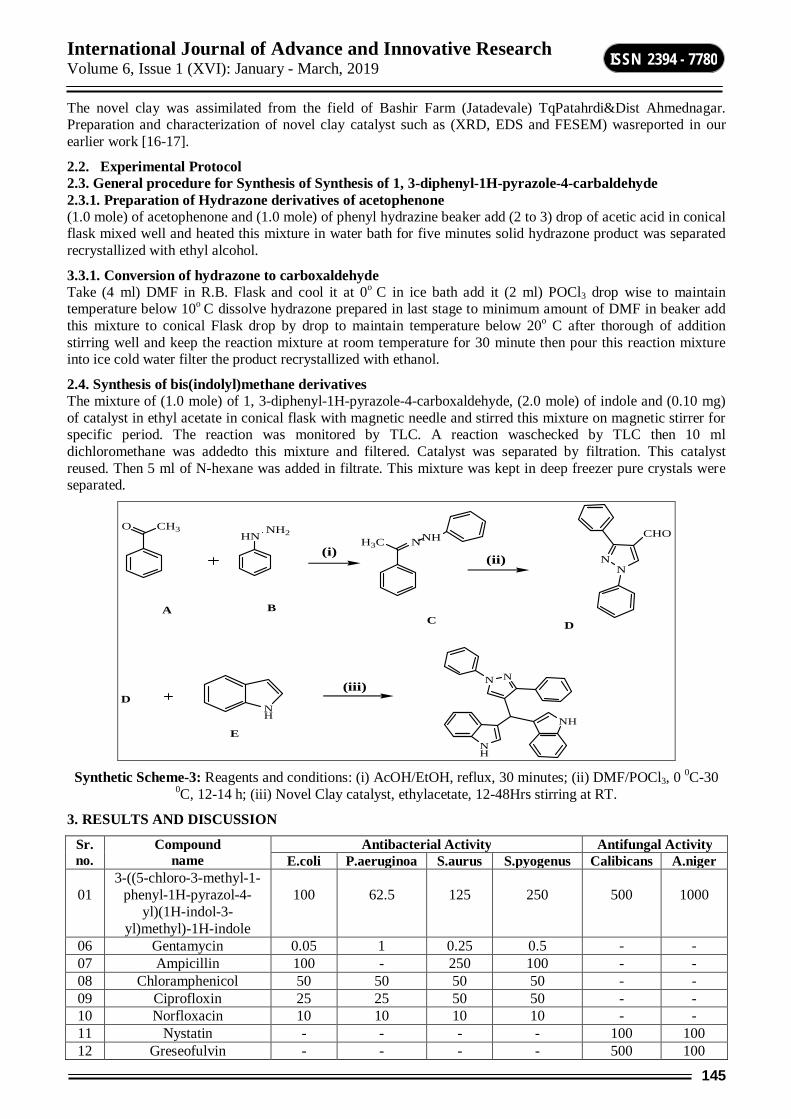
International Journal of Advance and Innovative Research Volume 6, Issue 1 (XVI): January - March, 2019
145
ISSN 2394 - 7780
The novel clay was assimilated from the field of Bashir Farm (Jatadevale) TqPatahrdi&Dist Ahmednagar. Preparation and characterization of novel clay catalyst such as (XRD, EDS and FESEM) wasreported in our earlier work [16-17].
2.2. Experimental Protocol 2.3. General procedure for Synthesis of Synthesis of 1, 3-diphenyl-1H-pyrazole-4-carbaldehyde 2.3.1. Preparation of Hydrazone derivatives of acetophenone (1.0 mole) of acetophenone and (1.0 mole) of phenyl hydrazine beaker add (2 to 3) drop of acetic acid in conical flask mixed well and heated this mixture in water bath for five minutes solid hydrazone product was separated recrystallized with ethyl alcohol.
3.3.1. Conversion of hydrazone to carboxaldehyde Take (4 ml) DMF in R.B. Flask and cool it at 0o C in ice bath add it (2 ml) POCl3 drop wise to maintain temperature below 10o C dissolve hydrazone prepared in last stage to minimum amount of DMF in beaker add this mixture to conical Flask drop by drop to maintain temperature below 20o C after thorough of addition stirring well and keep the reaction mixture at room temperature for 30 minute then pour this reaction mixture into ice cold water filter the product recrystallized with ethanol.
2.4. Synthesis of bis(indolyl)methane derivatives The mixture of (1.0 mole) of 1, 3-diphenyl-1H-pyrazole-4-carboxaldehyde, (2.0 mole) of indole and (0.10 mg) of catalyst in ethyl acetate in conical flask with magnetic needle and stirred this mixture on magnetic stirrer for specific period. The reaction was monitored by TLC. A reaction waschecked by TLC then 10 ml dichloromethane was addedto this mixture and filtered. Catalyst was separated by filtration. This catalyst reused. Then 5 ml of N-hexane was added in filtrate. This mixture was kept in deep freezer pure crystals were separated.
O CH3
H3C NNH
NN
CHO
NH
NH
NH
NN
HNNH2
A BC D
D
(i)(ii)
(iii)
E
Synthetic Scheme-3: Reagents and conditions: (i) AcOH/EtOH, reflux, 30 minutes; (ii) DMF/POCl3, 0 0C-30
0C, 12-14 h; (iii) Novel Clay catalyst, ethylacetate, 12-48Hrs stirring at RT.
3. RESULTS AND DISCUSSION
Sr. no.
Compound name
Antibacterial Activity Antifungal Activity E.coli P.aeruginoa S.aurus S.pyogenus Calibicans A.niger
01
3-((5-chloro-3-methyl-1-phenyl-1H-pyrazol-4-
yl)(1H-indol-3-yl)methyl)-1H-indole
100
62.5
125
250
500
1000
06 Gentamycin 0.05 1 0.25 0.5 - - 07 Ampicillin 100 - 250 100 - - 08 Chloramphenicol 50 50 50 50 - - 09 Ciprofloxin 25 25 50 50 - - 10 Norfloxacin 10 10 10 10 - - 11 Nystatin - - - - 100 100 12 Greseofulvin - - - - 500 100
International Journal of Advance and Innovative Research Volume 6, Issue 1 (XVI): January - March, 2019
146
ISSN 2394 - 7780
3-((5-chloro-3-methyl-1-phenyl-1H-pyrazol-4-yl)(1H-indol-3-yl)methyl)-1H-indole shows antibacterial activity for bacteria E. coli at 100 mg/ml, P .aeruginosa at 62.5 mg/ml, S. aureus 125 mg/ml and S. pyogenus 250 mg/ml. Amplicillin for E. coli 100 mg/ml, S. aureus 250 mg/ml and S. pyogenus 100 mg/ml. hence it found that amplicillin and this compound shows similar activity for bacteria E. coli and better reactivity than S. Aureus. similar way standard drug Gentamycin for E. coli 0.05 mg/ml, P. aeruginosa 1 mg/ml, S. aureus 0.25 mg/ml and S. pyogenus 0.5 mg/ml. Standard drug Chloramphenicol for E. coli 50 mg/ml, P. aeruginosa 50 mg/ml, S. aureus 50 mg/ml and S. pyogenus 50 mg/ml. Standard drug Ciprofloxacin for E. coli 25 mg/ml, P. aeruginosa 25 mg/ml, S. aureus 50 mg/ml and S. pyogenus 50 mg/ml. Standard drug Norfloxacin for E. coli 10 mg/ml, P. aeruginosa 10 mg/ml, S. aureus 10 mg/ml and S. pyogenus 10 mg/ml. it is found that 3-((5-chloro-3-methyl-1-phenyl-1H-pyrazol-4-yl)(1H-indol-3-yl)methyl)-1H-indole shows less reactivity than all standard drugs.
Antifungal activity of compound 3-((5-chloro-3-methyl-1-phenyl-1H-pyrazol-4-yl)(1H-indol-3-yl)methyl)-1H-indole for fungus C. albicans MTCC 227 is 500 mg/ml, A. niger MTCC 282 is 1000 mg/ml and minimal fungicidal concentration for standard drug Nystatin 100 mg/ml, A. niger 100 mg/ml, A. clavatus 100 mg/ml. Standard drug Nystatin 500 mg/ml, A. niger 100 mg/ml, A. clavatus 100 mg/ml. it is detected that this compound shows comparable reactivity as greseofulvin for fungi C. Albicans.
ACKNOWLEDGEMENT Auther are thankful toDr.Mazahar Farooqui (Principal, RafiqZakaria College for Women, Naukhanda, Aurangabad and Dean, faculty of science& technology, Dr.BabasahebAmbedkarMarathwada University, Aurangabad) for his constant support and guidance.
REFERENCE [1] M. Grare, M. Mourer, S. Fontanay, J.B. Regnouf-de-Vains, C. Finance, R.E. Duval, J. of Antimi. Chem.
60 (2007) 575-581.
[2] Tolaro, K.; Tolaro, A.; Found.ofMicrob., 3 (1993) 326.
[3] Hong C, Firestone G L and Bjeidanes L F, Biochem. Pharmaco., 2002, 63, 1085.
[4] G. Sivaprasad, P.T. Perumal, V.R. Prabavathy, N. Mathivanan, Bioorg. Med. Chem. Lett,. 16 (2006) 6302–6305.
[5] Yamuna, E.; Kumar, R. A.; Zeller, M.; Prasad, K. J. R. Eur. J. Med. Chem., 47,(2012) 228–238.
[6] B. John, Experimental Staph Vaccine Broadly Protective in Animal Studies, Nat.Inst. of Health News,. 1999.
[7] G. Sivaprasad, P.T. Perumal, V.R. Prabavathy, N. Mathivanan, Bioorg. Med. Chem. Lett., 16 (2006) 6302-6305.
[8] K. Sujatha, P.T. Perumal, D. Muralidharan, and M. Rajendra, Ind. J. of Chem., 48 (2), (2009) 267-272.
[9] Hong, C.; Firestone, G. L.; Bjeldance, L. F. Biochem. Pharmacol., 2002, 63, 1085.
[10] J.S. Yadav, B.V. S. Reddy, C.V.S. R. Murthy, G.M. Kumar, C. Madan, Synthesis, 2001, 783–787.
[11] Maiti, M.; Bhattacharyya, P. J. Chem. Res. 1997, 424
[12] Chakrabarty, M.; Basak, R.; Harigaya, Y.; Ghosh, N. Tetrahedron Lett. 2002, 43, 4075.
[13] Koshima, H. and W. Matsuaka, J. Heterocycl. Chem., 39 (2002) 1089-1091.
[14] Zhang, Z.H., L. Yin and Y.cM. Wang, 2005. 20. Bandgar, B.P., S.V. Bettigeri and N.S. Joshi, Synthesis.12:(2004) 1949-1954.
[15] Karthik M, Tripathi A K, Gupta N M, Palanichamy M., Murugesan V. Catal. Commu.,2004, 5, 371-375.
[16] Ismail Shaikh, SheseraoPawar, Muktar Shaikh, Syed Abed., Res. J. of pharm. Bio. And Chem. Sci., 7-4, (2016) 2932-2938.
[17] Ismail Shaikh, Mazahar Farooqui, GajananSanap, Syed Abed, Inter. J. of Sci. Res. in Sci., Eng. and Tech., 4-3, (2018) 35-39.

International Journal of Advance and Innovative Research Volume 6, Issue 1 (XVI): January - March, 2019
147
ISSN 2394 - 7780
PREVALENCE OF CESTODE PARASITES FROM SOME FRESH WATER FISHES OF AURANGABAD DISTRICT (M.S.) INDIA
Jadhav R. K.1, Nimbalkar R. K.2, and Jadhav B. N.3 1,3Department of Zoology, V. P. College, Vaijapur, Dist. Aurangabad
2Department of Zoology, R. B. Attal Arts, Science and Commerce College, Georai, Dist. Beed
ABSTRACT Present investigation deal with study of cestode parasite from some fresh water fishes Mastacembalusarmatus, Mystusseenghala, Wallagoattu, Channapuntatus, Clariusbatrachus, Cirrihana mrigala etc of Aurangabad district. During the period of 12 months (Monsoon Winter and Summer Season) i.e. from, June, 2015 – May 2016, total 284 fresh water fishes were examined for cestode infections, out of which 33 samples were positive for cestode infection. The percentage of prevalence being 11.61 %.
Keywords: Fresh water fishes, cestode parasites, percentage of prevalence.etc
INTRODUCTION Fishes are most popular group of animals about 40,000 species of fishes are known that live in different aquatic habitat. Fish is good of excellent nutritional value providing high quality Protein, Vitamins, Minerals, including Vitamins A and D, Phosphorus, Magnesium and Iodine, in fish its Protein is easily digestible. Economically fishes are useful to man like as a food, oil, leathers, medicines, disease control fish meal and fish manure. People involved in fisheries, aquaculture and fish trade, fish are a source of income. India is among the 17 mega diversity countries [1] and hosts as many as 55 families of freshwater fish [2, 8].
The environment factors including climate, season and rainfall play important role in the development of cestode parasite due to the environmental factor the natures of cestode infection of different group of live stock have been studied by researcher from particular region of the country. Parasitic diseases of fish are very common throughout the world. Infections which are caused by viruses, bacteria and parasites among fishes in natural and man-made culture system are harmful for fish health and growth. Seasonal fluctuation, locality, age, size and sex of the host also determine the parasitic community diversity and burden[3]. Fishes parasitized by helminth parasites, which reduce the food value of host fish. Study of helminth parasites is therefore an urgent necessity today. Helminth infections are very common in people who consume improperly cooked meat, unhygienic habits and poor sanitation. These helminthic infection leads to various disorders i.e. anaemia. Population investigation is necessary to provide data for the prediction of integrated methods to achieve the regulation of numbers of harmful parasites [10, 11] Notable contribution made by Dobson [6], Dogiel[4], Euzeby [5], Anderson [01], Moller, H [10] and Rajeshwar Rao[11].Parasites of fish constitute one of the major problems to fish health. Besides the direct losses caused by mortality, parasites have a considerable impact on growth, resistance to other stressing factors, susceptibility to predation, marketability and pave way for secondary infections.[9]
MATERIAL AND METHODS The freshwater fishes i.e. Mastacembalusarmatus, Mystusseenghala, Wallagoattu, Channapuntatus, Clariusbatrachus, Cirrihana mrigala etc were collected from various localities (Tehsil) of Aurangabad district i.e. Paithan, Kannad, Vaijapur, Gangapur, Phulambri ,KaigaonToka etc during period June 2015 to May 2016. The fishes were bring in laboratory and were collected from gut, collected cestode after washing firstly fixed in 4 % formalin. Keys of Yamaguti1959), (1961) were used for the identification of helminths.
RESULT AND DISCUSSION During the period of 12 months (Monsoon Winter and Summer Season) i.e. from, June, 2015 – May 2016, total 284 fresh water fishes i.e. Mastacembalusarmatus, Mystusseenghala, Wallagoattu, Channapuntatus, Clariusbatrachus, Cirrihana mrigala etc were examined for cestode infections, out of which 33 samples were positive for cestode infection. The percentage of prevalence being 11.61 %.
Month wise analysis showed that maximum prevalence was duringMay-2016, followed by Feb, Oct,April,Aug,Nov,March, Dec, July, Sept., January, Jun ( 20%,16%, 16%, 13.33%, 11.53%, 10.71%, 10%, 10%., 9.09%, 00%, 00%).
Sr.no Month and Year
No. of dissected host
No.of infected Host
No.of parasites collected
Prevalence %
Location
01 June-2015 15 00 00 00 Phulambri 02 July -2015 20 02 03 10 Vaijapur
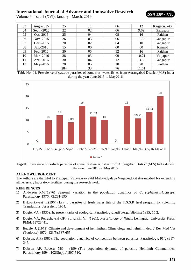
International Journal of Advance and Innovative Research Volume 6, Issue 1 (XVI): January - March, 2019
148
ISSN 2394 - 7780
03 Aug -2015 25 03. 06 12 KaigaonToka 04 Sept. -2015 22 02 06 9.09 Gangapur 05 Oct.-2015 25 04 08 16 Paithan 06 Nov.-2015 26 03 06 11.53 Gangapur 07 Dec.-2015 20 02 04 10 Gangapur 08 Jan.-2016 15 00 00 00 Kannad 09 Feb.-2016 30 05 12 16 Paithan 10 Mar.-2016 28 03 09 10.71 Vaijapur 11 Apr.-2016 30 04 12 13.33 Gangapur 12 May-2016 28 05 10 20 Paithan
284 33 76 11.61 Table No- 01: Prevalence of cestode parasites of some freshwater fishes from Aurangabad District (M.S) India
during the year June 2015 to May2016.
Fig-01: Prevalence of cestode parasites of some freshwater fishes from Aurangabad District (M.S) India during
the year June 2015 to May2016.
ACKNOWLEDGEMENT The authors are thankful to Principal, Vinayakrao Patil Mahavidyalaya Vaijapur,Dist Aurangabad for extending all necessary laboratory facilities during the research work.
REFERENCES 1) Anderson RM.(1976) Seasonal variation in the population dynamics of Caryophyllacuslacticeps.
Parasitology 1976; 72:281-395.
2) Bykovskayaet al.(1964) key to parasites of fresh water fish of the U.S.S.R Isrel program for scientific Translations, Jerusalem, 1964.
3) Dogiel VA. (1935)The present tasks of ecological Parasitology.TudPatergofBiolInst 1935; 15:2.
4) Dogiel VA, Petrushevski GK, Polyanski YI, (1961). Parasitology of fishes. Leningrad: University Press; PMid: 13723441.
5) Euzeby J. (1972) Climate and development of helminthes: Climatology and helminth dev. J Rev Med Vet (Toulouse) 1972; 123(5):637-655.
6) Dobson, A.P.(1985): The population dynamics of competition between parasites. Parasitology, 91(2):317-347.
7) Dobson AP, Roberts MG. (1994).The population dynamic of parasitic Helminth Communities. Parasitology 1994; 102(Suppl.):507-510.
International Journal of Advance and Innovative Research Volume 6, Issue 1 (XVI): January - March, 2019
149
ISSN 2394 - 7780
8) Froese R and D Pauly, (2015) Fishbase. www.fishbase.org version (04/2015).
9) I. Kundu, P.K. Bandyopadhyay and D.R., Mandal (2015). Prevalence of helminth parasites infecting Channa puntatus Bloch, 1793 from Nadia district of West Bengal, Journal of Agriculture and Veterinary Science, (8), 2015, 41-46.
10) Kennedy, C.R. (1978): Ecological aspects of Parasitology North Holland Publishing Company. Amsterdam, Oxford.
11) Kennedy CR. (1974). A checklist of British and Irish freshwater fish parasites with notes on their distribution fish Biol1974; 6(5):613-644.
12) Moller H. (1978). The effect of salinity and temperature in the development and survival of fish parasites. Jr of Fish Biol1978; 12:311-324.
13) Rao R, Ramkrishna V. (1982). The seasonal variations of Helminth Parasites of Ranatigrinain Hyderabad district. Geobios 1982; (10):34-36.
14) Yamaguti, S. (1959): SystemaHelminthum Vol.3.The cestode of vertebrates.Interscience Publ. New York & London.1-160
15) Yamaguti, S. (1961):SystemaHelminthum Vol.3.The nematodes of vertebrates. Parts I &II.Interscience publishers Inc., New York, pp.1261.
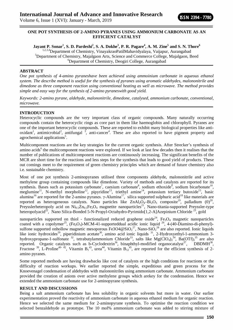
International Journal of Advance and Innovative Research Volume 6, Issue 1 (XVI): January - March, 2019
150
ISSN 2394 - 7780
ONE POT SYNTHESIS OF 2-AMINO PYRANES USING AMMONIUM CARBONATE AS AN EFFICIENT CATALYST
Jayant P. Sonar1, S. D. Pardeshi2, S. A. Dokhe3, P. R. Pagare4, A. M. Zine5 and S. N. Thore6 1,2,3,4Department of Chemistry, VinayakraoPatilMahavidyalaya, Vaijapur, Aurangabad
5Department of Chemistry, Majalgaon Arts, Science and Commerce College, Majalgaon, Beed 6Department of Chemistry, Deogiri College, Aurangabad
ABSTRACT One pot synthesis of 4-amino pyraneshave been achieved using ammoinium carbonate in aqueous ethanol system. The describe method is useful for the synthesis of pyranes using aromatic aldehydes, malononitrile and dimedone as three component reaction using conventional heating as well as microwave. The method provides simple and easy way for the synthesis of 2-amino pyraneswith good yield.
Keywords: 2-amino pyrane, aldehyde, malononitrile, dimedone, catalysed, ammonium carbonate, conventional, microwave.
INTRODUCTION Heterocyclic compounds are the very important class of organic compounds. Many naturally occurring compounds contain the heterocyclic rings as core part in them like haemoglobin and chlorophyll. Pyranes are one of the important heterocyclic compounds. These are reported to exhibit many biological properties like anti-oxidant1, antimicrobial2, antifungal 3, anti-cancer4. These are also reported to have pigment property and agrochemical applications5.
Multicomponent reactions are the key strategies for the current organic synthesis. After Strecker’s synthesis of amino acids6 the multicomponent reactions were explored. If we look at last few decades then it realises that the number of publications of multicomponent reactions are continuously increasing. The significant benefits of the MCR are short time for the reactions and less steps for the synthesis that leads to good yield of products. These out comings meet to the requirement of green chemistry principles which are demand of future chemistry also i.e. sustainable chemistry.
Most of one pot synthesis 2-aminopyranes utilised three components aldehyde, malononitrile and active methylene group containing compounds like dimedone. Variety of methods and catalysts are reported for its synthesis. Bases such as potassium carbonate7, caesium carbonate8, sodium ethoxide9, sodium bicarbonate10, meglumine11, N-methyl morpholine12, pipyridine13, triethyl amine14, potassium tertiary butoxide15, basic alumina16 are reported for the 2-amino pyranes. γ-Alumina17, silica supported sulphuric acid18 like materials are reported as heterogeneous catalysts. Nano particles like ZnAl2O4–Bi2O3 composite19, palladium (0)20, Preysslerheteropoly acid on Ni0.5Zn0.5Fe2O4 magnetite nanoparticles21, Nano-titania-supported Preyssler-type heteropolyacid22, Nano Silica-Bonded 5-N-Propyl-Octahydro-Pyrimido[1,2-A]Azepinium Chloride 23, gold
nanoparticles supported on thiol - functionalized reduced graphene oxide24, Fe3O4 magnetic nanoparticles coated with a copolymer25, (Fe2O3)-MCM-41-supporteddual acidic ionic liquid 26, 4-(40-Diamino-di-phenyl)-sulfone supported onhollow magnetic mesoporous Fe3O4@SiO2
27, Nano-SiO228 are also reported. Ionic liquids
like ionic hydroxides29, piperidinium acetate30, amino acid ionic liquids 31, 2-Hydroxyethyl-1-ammonium 3-hydroxypropane-1-sulfonate 32, tetrabutylammonium Chloride33, salts like Mg(ClO4)2
34, Ba(OTf)235 are also
reported. Organic catalysts such as b-Cyclodextrin36, binaphthyl-modified organocatalyst37, DBDMH38, Fructose 39, L-Proline40- 42, Vitamin B1
43, urea44, Vitamin B1245, are reported for the efficient synthesis of 2-
amino pyranes.
Some reported methods are having drawbacks like cost of catalysts or the high conditions for reactions or the difficulty of reaction workups. We earlier reported the simple, expeditious and green process for the Knoevenagel condensation of aldehydes with malononitriles using ammonium carbonate. Ammonium carbonate provided the creation of anions over active methylene groups which arekey for the condensation. Hence we extended the ammonium carbonate use for 2-aminopyrane synthesis.
RESULT AND DISCUSSIONS Being a salt ammonium carbonate has less solubility in organic solvents but more in water. Our earlier experimentation proved the reactivity of ammonium carbonate in aqueous ethanol medium for organic reaction. Hence we selected the same medium for 2-aminopyrane synthesis. To optimise the reaction condition we selected benzaldehyde as prototype. The 10 mol% ammonium carbonate was added to stirring mixture of
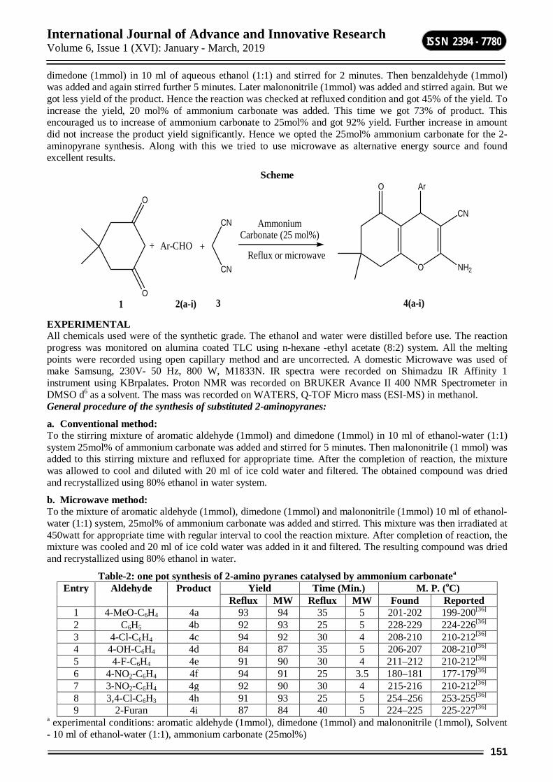
International Journal of Advance and Innovative Research Volume 6, Issue 1 (XVI): January - March, 2019
151
ISSN 2394 - 7780
dimedone (1mmol) in 10 ml of aqueous ethanol (1:1) and stirred for 2 minutes. Then benzaldehyde (1mmol) was added and again stirred further 5 minutes. Later malononitrile (1mmol) was added and stirred again. But we got less yield of the product. Hence the reaction was checked at refluxed condition and got 45% of the yield. To increase the yield, 20 mol% of ammonium carbonate was added. This time we got 73% of product. This encouraged us to increase of ammonium carbonate to 25mol% and got 92% yield. Further increase in amount did not increase the product yield significantly. Hence we opted the 25mol% ammonium carbonate for the 2-aminopyrane synthesis. Along with this we tried to use microwave as alternative energy source and found excellent results.
Scheme
Ar-CHO+
O
O
CN
CN Ammonium Carbonate (25 mol%)
Reflux or microwaveO
CN
NH2
ArO
+
1 2(a-i) 3 4(a-i) EXPERIMENTAL All chemicals used were of the synthetic grade. The ethanol and water were distilled before use. The reaction progress was monitored on alumina coated TLC using n-hexane -ethyl acetate (8:2) system. All the melting points were recorded using open capillary method and are uncorrected. A domestic Microwave was used of make Samsung, 230V- 50 Hz, 800 W, M1833N. IR spectra were recorded on Shimadzu IR Affinity 1 instrument using KBrpalates. Proton NMR was recorded on BRUKER Avance II 400 NMR Spectrometer in DMSO d6 as a solvent. The mass was recorded on WATERS, Q-TOF Micro mass (ESI-MS) in methanol. General procedure of the synthesis of substituted 2-aminopyranes:
a. Conventional method: To the stirring mixture of aromatic aldehyde (1mmol) and dimedone (1mmol) in 10 ml of ethanol-water (1:1) system 25mol% of ammonium carbonate was added and stirred for 5 minutes. Then malononitrile (1 mmol) was added to this stirring mixture and refluxed for appropriate time. After the completion of reaction, the mixture was allowed to cool and diluted with 20 ml of ice cold water and filtered. The obtained compound was dried and recrystallized using 80% ethanol in water system.
b. Microwave method: To the mixture of aromatic aldehyde (1mmol), dimedone (1mmol) and malononitrile (1mmol) 10 ml of ethanol-water (1:1) system, 25mol% of ammonium carbonate was added and stirred. This mixture was then irradiated at 450watt for appropriate time with regular interval to cool the reaction mixture. After completion of reaction, the mixture was cooled and 20 ml of ice cold water was added in it and filtered. The resulting compound was dried and recrystallized using 80% ethanol in water.
Table-2: one pot synthesis of 2-amino pyranes catalysed by ammonium carbonatea Entry Aldehyde Product Yield Time (Min.) M. P. (oC)
Reflux MW Reflux MW Found Reported 1 4-MeO-C6H4 4a 93 94 35 5 201-202 199-200[36] 2 C6H5 4b 92 93 25 5 228-229 224-226[36] 3 4-Cl-C6H4 4c 94 92 30 4 208-210 210-212[36] 4 4-OH-C6H4 4d 84 87 35 5 206-207 208-210[36] 5 4-F-C6H4 4e 91 90 30 4 211–212 210-212[36] 6 4-NO2-C6H4 4f 94 91 25 3.5 180–181 177-179[36] 7 3-NO2-C6H4 4g 92 90 30 4 215-216 210-212[36] 8 3,4-Cl-C6H3 4h 91 93 25 5 254–256 253-255[36] 9 2-Furan 4i 87 84 40 5 224–225 225-227[36]
a experimental conditions: aromatic aldehyde (1mmol), dimedone (1mmol) and malononitrile (1mmol), Solvent - 10 ml of ethanol-water (1:1), ammonium carbonate (25mol%)

International Journal of Advance and Innovative Research Volume 6, Issue 1 (XVI): January - March, 2019
152
ISSN 2394 - 7780
REPRESENTATIVE SPECTRAL DATA 2-amino-4-(4-chlorophenyl)-5,6,7,8-tetrahydro-7,7-dimethyl-5-oxo-4H-chromene-3-carbonitrile
IR (KBr, cm-1): 3393,3337, 2966, 2213, 1680, 1608, 1372, 1216. 1H NMR (400 MHz, DMSO-d6): δ (ppm) 0.94 (s, 3H), 1.00 (s, 3H), 1.15 (m, 1H), 1.96-2.23 (m,3H), 4.17 (s, 1H), 6.4 (s, 2H), 7.14 (d, J = 8 Hz, 2H), 7.31 (d, J = 8 Hz, 2H). Mass: 329 (M+1)
CONCLUSION The ammonium carbonate in aqueous ethanol system is found to be good for the synthesis of 2-amino pyrane compounds. It provides an easy way for one pot three component reaction in both conventional refluxing and microwave assisted synthesis. The short time reaction and easy workup process are the merits of this reaction.
ACKNOWLEDGEMENT The authors are thankful to the Principal, VinayakraoPatilMahavidyalaya, Vaijapur for providing the laboratory facilities and the Director, SAIF, Chandigarh for providing the spectral analysis.
REFERENCES 1. T. Symeonidis, M. Chamilos, D. J. Hadjipavlou-Litina, M. Kallitsakis, K.E. Litinas, Bioorg. Med. Chem.
Lett., 2009, 19, 1139–1142.
2. H.G. Kathrotiya, M.P. Patel, Med. Chem. Res., 2012, 21, 3406–3416.
3. L. Alvey, S. Prado, B. Saint-Joanis, S. Michel, M. Koch, S.T.Cole, F. Tillequin, Y.L. Janin, Eur. J. Med. Chem., 2009, 44, 2497–2505.
4. S.X. Cai, J. Drewe, W. Kemnitzer, Anti-Cancer Agents Med. Chem., 2009, 9, 437–456.
5. D. Kumar, V. B. Reddy, S. Sharad, U. Dube, S. Kapur, Euro. J. Med Chem, 2009, 44, 3805-3809
6. A.Strecker, Ann. Chem. Pharm., 1850, 75, 27–45.
7. Reza Heydari, RohollahRahimi, MehrnooshKangani, AfshinYazdani-Elah-Abadi, MojtabaLashkari, ActaChemica Iasi, 2017, 25(2), 163-178
8. Vinod V. Throat, Satish A. Dake; Maya V. Katariya and Rajendra P. Pawar, Der ChemicaSinica, 2015, 6(6),37-50
9. Md. Korban Ali, JobayetHossain, Md. Moniruzzama, International Journal of Advanced Research in Chemical Science (IJARCS), 2016, 3(1), 39-45
10. NeerajPathak, Jayshree Parikh and Ram Vishun Prasad, Journal of Chemistry and Chemical Sciences, 2018, 8(3), 390-403.
11. Rui-Yun Guo, Zhi-Min An, Li-Ping Mo, Rui-Zhi Wang, Hong-Xia Liu, Shu-Xia Wang, Zhan-Hui Zhang, ACS Comb. Sci. 2013, 15, 557−563.
12. SeyedehMahbobehMahdavi, AzizollahHabibi, HadiDolati, Seyed Mohammad Shahcheragh Soroush Sardari, Parisa Azerang, , Iran. J. Pharmaceu. Res., 2018, 17(4), 1229-1239.
13. Nilesh J. Thumar and Manish P. Patel, ARKIVOC, 2009, (xiii), 363-380.
14. Akbar Mobinikhaledi, NaserForoughifar, TahereMosleh and Ahmad Hamta, Iran. J. Pharmaceu. Res., 2014, 13 (3), 873-879
15. N. K. Rao, T. N. Rao, B. Parvatamma, K. P. Devi, S. C. Setty, Bull. Chem. Soc. Ethiop., 2018, 32(1), 133-138.
16. Ji Tai Li, Wen ZhiXu, Li Chao Yang , Tong Shuang Li, Syn. Comm., 2004, 34(24), 4565-457.
17. L. Edjlali, R. Hosseinzdeh-Khanmiri, Iran. J. Sci. Technol. Trans. Sci., 2016, 40, 151-156.
18. S. Sheik Mansoor, K. Aswin, K. Logaiya, S.P.N. Sudhan, J. Taibah Univ. Sci., 2014, 8, 265-275.
19. MajidGhashang, Res. Chem. Intermed., 2015, 42 (5), 4191- 4205
20. MithuSaha, Amarta Kumar Pal, Advances in Nanoparticles, 2012, 1, 61-70
21. Ali Javid, FaridMoeinpour, Bull. Chem. Soc. Ethiop., 2018, 32(3), 501-511.
22. DavoodAzarifar, Seyed-Mola Khatami, RaziehNejat-Yami, J. Chem. Sci., 2014, 126(1), 95-101.
International Journal of Advance and Innovative Research Volume 6, Issue 1 (XVI): January - March, 2019
153
ISSN 2394 - 7780
23. RobabehBaharfar, SakinehAsghari And NargesShariati, J. Chil. Chem. Soc., 2015, 60(2), 2900-2904.
24. HosseinNaeimi, Maryam FarahnakZarabi, Res. Chem. Intermed., 2018, 44, 3227-3247.
25. MahdiehKeshavarz, MasumehAbdoli-Senejani, SeyedehFatemehHojati, ShivadokhtAskari, ReacKinetMech Cat, 2018, 124(2), 757-766.
26. ShahnazRostamizadeh, NegarZekri, Res ChemIntermed, 2015, 42(3), 2329-2341.
27. JavadSafaei-Ghomi, NasrinEnayat-Mehri, FahimeEshteghal, J. Saudi Chem. Soc., 2018, 22, 485–495.
28. EbrahimMollashahi, Mohammad Nikraftar, J. Saudi Chem. Soc., 2018, 22, 42-48.
29. Kiran F. Shelke and Ravi E. Khadse, Der PharmaChemica, 2015, 7(1), 191-196.
30. A. Indrasena, Sd. Riyaz, A. Naidu And P.K. Dubey, Asian J. Chem., 2014, 26(8), 2221-2225.
31. Paula Ossowicz, ZbigniewRozwadowski, MarcinGano, Ewa Janus, Pol. J. Chem. Tech., 2016, 18(4), 90-95.
32. MoonesHonarmand, AndromachiTzani, Anastasia Detsi, Journal of the Iranian Chemical Society, 2018, https://doi.org/10.1007/s13738-018-1537-2
33. HosseinMehrabi, NafisehKamali, J. Iran . Chem. Soc., 2012, 9, 599–605.
34. MasoudMohammadiZeydi and SomayehAhmadi, Orient. J. Chem., 2016, 32(4), 2215-2220.
35. Anil Kumar, M. SudershanRao, Green Chem.Lett. and Rev., 2012, 5(3), 283-290.
36. Jun Lu, Xue-wen Fu, Ge Zhang, Chun Wang, Res. Chem. Intermed., 2015, 42(2), 417-424.
37. Chang Won Suh and Dae Young Kim, Bull. Korean Chem. Soc. 2014, 35(1), 98-102.
38. YasaminSaadati, F. SeyedehHojati, Maan Al Naddaf, Mohammad Keshe,
39. J. Chem. Pharmace. Res., 2018, 10(5), 108-112.
40. SayyedehShadfarPourpanah, SayyedMostafaHabibi-Khorassani , Mehdi Shahraki, Chin. J. Cat., 2015, 36, 757-763.
41. Noha M. HilmyElnagdi, NouraSaad Al-Hokbany, Molecules 2012, 17, 4300-4312.
42. Farahnaz K. Behbahani, FereshtehAlipour, GU J Sci, 2015, 28(3), 387-393.
43. PoursattarMarjani Ahmad, EbrahimiSaatluoBahman; NouriFariba, Iran. J. Chem. Chem. Eng., 2018, 37(1), 149-157.
44. Devidas S. Bhagat, Jagadish L. Wawre, Ashok R. Yadav, Pintu G. Pathare, Laszlo Kotai and Rajendra P. Pawar, Eur. Chem. Bull., 2017, 6(5), 211-214.
45. GoutamBrahmachari and Bubun Banerjee, ACS Sustainable Chem. Eng., 2014, 2, 411-422.
46. Mohammad Dodangeh, Malek-TaherMaghsoodlou, MehrnooshKangani, FaridehPaymozd, NourallahHazeri, J. Nutraceuticals and Food Sci., 2016, 1(2), 1-10.
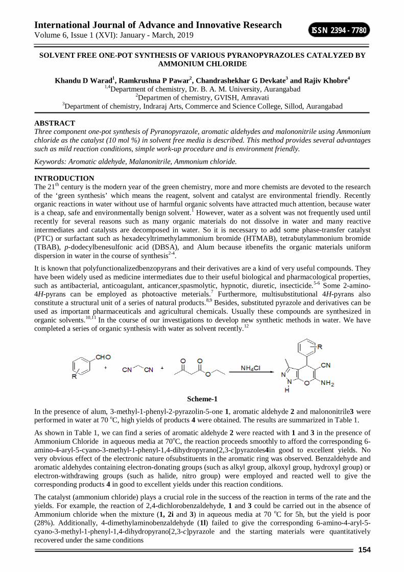
International Journal of Advance and Innovative Research Volume 6, Issue 1 (XVI): January - March, 2019
154
ISSN 2394 - 7780
SOLVENT FREE ONE-POT SYNTHESIS OF VARIOUS PYRANOPYRAZOLES CATALYZED BY AMMONIUM CHLORIDE
Khandu D Warad1, Ramkrushna P Pawar2, Chandrashekhar G Devkate3 and Rajiv Khobre4 1,4Department of chemistry, Dr. B. A. M. University, Aurangabad
2Departmen of chemistry, GVISH, Amravati 3Department of chemistry, Indraraj Arts, Commerce and Science College, Sillod, Aurangabad
ABSTRACT Three component one-pot synthesis of Pyranopyrazole, aromatic aldehydes and malononitrile using Ammonium chloride as the catalyst (10 mol %) in solvent free media is described. This method provides several advantages such as mild reaction conditions, simple work-up procedure and is environment friendly.
Keywords: Aromatic aldehyde, Malanonitrile, Ammonium chloride.
INTRODUCTION The 21th century is the modern year of the green chemistry, more and more chemists are devoted to the research of the ‘green synthesis’ which means the reagent, solvent and catalyst are environmental friendly. Recently organic reactions in water without use of harmful organic solvents have attracted much attention, because water is a cheap, safe and environmentally benign solvent.1 However, water as a solvent was not frequently used until recently for several reasons such as many organic materials do not dissolve in water and many reactive intermediates and catalysts are decomposed in water. So it is necessary to add some phase-transfer catalyst (PTC) or surfactant such as hexadecyltrimethylammonium bromide (HTMAB), tetrabutylammonium bromide (TBAB), p-dodecylbenesulfonic acid (DBSA), and Alum because itbenefits the organic materials uniform dispersion in water in the course of synthesis2-4.
It is known that polyfunctionalizedbenzopyrans and their derivatives are a kind of very useful compounds. They have been widely used as medicine intermediates due to their useful biological and pharmacological properties, such as antibacterial, anticoagulant, anticancer,spasmolytic, hypnotic, diuretic, insecticide.5-6 Some 2-amino-4H-pyrans can be employed as photoactive meterials.7 Furthermore, multisubstitutional 4H-pyrans also constitute a structural unit of a series of natural products.8,9 Besides, substituted pyrazole and derivatives can be used as important pharmaceuticals and agricultural chemicals. Usually these compounds are synthesized in organic solvents.10,11 In the course of our investigations to develop new synthetic methods in water. We have completed a series of organic synthesis with water as solvent recently.12
Scheme-1
In the presence of alum, 3-methyl-1-phenyl-2-pyrazolin-5-one 1, aromatic aldehyde 2 and malononitrile3 were performed in water at 70 oC, high yields of products 4 were obtained. The results are summarized in Table 1.
As shown in Table 1, we can find a series of aromatic aldehyde 2 were reacted with 1 and 3 in the presence of Ammonium Chloride in aqueous media at 70oC, the reaction proceeds smoothly to afford the corresponding 6-amino-4-aryl-5-cyano-3-methyl-1-phenyl-1,4-dihydropyrano[2,3-c]pyrazoles4in good to excellent yields. No very obvious effect of the electronic nature ofsubstituents in the aromatic ring was observed. Benzaldehyde and aromatic aldehydes containing electron-donating groups (such as alkyl group, alkoxyl group, hydroxyl group) or electron-withdrawing groups (such as halide, nitro group) were employed and reacted well to give the corresponding products 4 in good to excellent yields under this reaction conditions.
The catalyst (ammonium chloride) plays a crucial role in the success of the reaction in terms of the rate and the yields. For example, the reaction of 2,4-dichlorobenzaldehyde, 1 and 3 could be carried out in the absence of Ammonium chloride when the mixture (1, 2i and 3) in aqueous media at 70 oC for 5h, but the yield is poor (28%). Additionally, 4-dimethylaminobenzaldehyde (1l) failed to give the corresponding 6-amino-4-aryl-5-cyano-3-methyl-1-phenyl-1,4-dihydropyrano[2,3-c]pyrazole and the starting materials were quantitatively recovered under the same conditions
International Journal of Advance and Innovative Research Volume 6, Issue 1 (XVI): January - March, 2019
155
ISSN 2394 - 7780
Table-1: Synthesis of pyrano[2,3-c]pyrazoles catalyzed by Ammonium chloride in aqueous media. Entry Ar Yield (%)a Mp (oC)
Found Reported8,10,12f
4a C6H5 80 170-171 168-170 4b 4-CH3C6H4 82 177-178 176-178 4c 4-CH3OC6H4 81 171-172 170-172 4d 3,4-(OCH2O)C6H3 81 175-176 174-176 4e 4-HOC6H4 80 210-212 211-212 4f 2-ClC6H4 90 145-146 144-146 4g 3-ClC6H4 88 158-160 158-159 4h 4-ClC6H4 90 175-176 174-175 4i 2,4-Cl2C6H3 90 185-186 182-184 4j 3-NO2C6H4 85 190-191 188-190 4k 4-NO2C6H4 88 195-196 194-196 4l 4-(CH3)2NC6H4 no reaction
Isolated yield.
EXPERIMENTAL SECTION General Procedures. aldehydes were purified by distilation before use. IR spectra were recorded on aBio-Rad FTS-40 spectrometer (KBr). 1H NMR spectra were measured on a Bruker AVANCE 400 (400 MHz) spectrometer using TMS as internal reference and CDCl3 as solvent. Elemental analyses were determined using Perkin-Elmer 2400 II elemental analyzer.
General procedure for the preparation of 4. A mixture of 3-methyl-1-phenyl-2-pyrazolin-5-one (1, 2mmol), aromatic aldehyde (2, 2mmol), malononitrile (3, 2mmol), and Ammonium Chloridee (10 mol%) in water (40 mL) was stirred at 70 oC for 2 hours. Then the mixture was cooled to room temperature, solid was filtered off and washed with H2O (40mL). The crude products were purified by recrystallization by ethanol (90%) to give 4. Data of compounds are shown below:
6-Amino-5-cyano-3-methyl-1,4-diphenyl-1,4-dihydropyrano[2,3-c]pyrazole (4a). IR (KBr):νmax= 3472, 3320, 2195, 1660, 1590, 1264, 1125, 1027, 753 cm-1.1H NMR: δ =1.93 (s, 3H,CH3), 4.68 (s, 1H, 4-H), 4.75 (s, 2H, NH2), 7.16-7.32 (m, 10H, ArH) ppm. Anal.Calcd. for C20H16N4O: C 73.15, H 4.91, N 17.06; found C 73.19, H 5.01, N 17.1.
RESULT AND DISCUSSION Synthesis of 6-amino-4-aryl-5-cyano-3-methyl-1-phenyl-1,4-dihydropyrano[2,3-c]pyrazoles from 3-methyl-1-phenyl-2-pyrazolin-5-one, aromatic aldehydes and malononitrile using Alum as the catalyst (10 mol %) in green solvent media is described. Variety of pyrazoles were attempted to investigate the scope of the method. As shown in table, different types of substituted pyrazole derivatives were obtained in good yields.
CONCLUSION Here, we have developed a green and efficient method for the synthesis of 6-amino-4-aryl-5-cyano-3-methyl-1-phenyl-1,4-dihydropyrano[2,3-c]pyrazoles from 3-methyl-1-phenyl-2-pyrazolin-5-one, aromatic aldehydes and malononitrile using Ammonium chloride as the catalyst (10 mol%) in green solvent media is described. This method provides several advantages such as mild reaction conditions, simple work-up procedure and is environment friendly. In addition, water is chosen as a green solvent.
REFERENCES 1. Li, C. J.; Chan, T. H. Organic Reaction in Aqueous Media, Wiley: New York, 1997. 2. Foye, W. O. Prinicipi di ChimicaFarmaceuticaPiccin, Padova: Italy, 1991, p 416. 3. Adreani, L. L.; Lapi, E. Boll. Chim. Farm. 1960, 99, 583; Chem. Abstr. 1961, 55, 2668d. 4. Zhang, Y. L.; Chen, B. Z.; Zheng, K. Q.; Xu, M. L.; Lei, X. H. Chinese ActaPharmaceuticaSinica1982, 17,
17; Chem. Abstr. 1982, 96, 135383e. 5. Bonsignore, L.; Loy, G..; Secci, D.; Calignano, A. Eur. J. Med. Chem. 1993, 28, 517. 6. (a) Witte, E. C.; Neubert, P.; Roesch, A. Ger. Offen DE. 1986, 3427985; Chem. Abstr. 1986, 104, 224915f.
(b) Wang, J. L.; Liu, D.; Zhang, Z. J.; Shan, S.; Han, X.; Srinivasula, S. M.; Croce, C. M.; Alnemri, E. S.; Huang, Z. Proc. Natl. Acad. Sci. U. S. A. 2000, 97, 7124. (c) Monamed, Y. A.; Zahran, M. A.; Ali, M. M.; ElAgrody, A. M.; El-Said, U. H. J. Chem. Res (S). 1995, 322.
International Journal of Advance and Innovative Research Volume 6, Issue 1 (XVI): January - March, 2019
156
ISSN 2394 - 7780
7. Armetso, D.; Horspool, W. M.; Martin, N.; Ramos, A.; Seaone, C. J. Org. Chem. 1989, 54, 3069.
8. Hatakeyama, S.; Ochi, N.; Numata; H.; Takano, S. J. Chem. Soc., Chem. Commun. 1988, 1202.
9. (a) Gonzalez, R.; Martin, N.; Seoane; C.; Soto, J. J. Chem. Soc., Perkin Trans. 1. 1985, 202. (b) Kamaljit, S. J.; Harjit, S. Tetrahedron 1996, 52, 14273.
10. (a) Shi, D. Q.; Mou, J.; Zhuang, Q. Y.; Niu, L.H.; Wu, N.; Wang, X. S. Synth. Commun. 2004, 34, 4557. (b) Zhou, J. F.; Tu, S. J.; Gao, Y.; Qi, M.Chinese J. Org. Chem. 2001,21, 742.
11. Kamaljit, S.; Jasbi, S.; Harjit, S. Tetrahedron 1996, 52, 14273.
12. (a) Jin, T. S.; Zhang, J. S.; Xiao, J. C.; Wang, A.Q.; Li, T. S. Synlett2004, 866. (b) Jin, T. S.; Wang, A.Q.; Wang, X.; Zhang, J. S.; Li, T. S. Synlett2004, 871. (c) Jin, T. S.; Zhang, J. S.; Guo, T. T.; Wang, A.Q.; Li, T. S. Synthesis 2004, 2001. (d) Jin, T. S.; Wang, A.Q.; Cheng, Z. L.; Zhang, J. S.; Li, T. S. Synth. Commun. 2005, 35, 2339. (e) Jin, T. S.; Liu, L. B.; Zhao, Y.; Li, T. S. Synth. Commun. 2005, 35, 2379. (f) Jin, T. S.; Wang, A. Q.; Cheng, Z. L.; Zhang, J. S.; Li, T. S. Synth. Commun. 2005, 35,137. (g) Jin, T. S.; Liu, L. B.; Zhao, Y.; Li, T. S. Synth. Commun. 2005, 35, 1859. (h) Jin, T. S.; Zhang, J. S.; Wang, A.Q.; Zhang, F. S. Chinese J. Org. Chem. 2005, 25, 335.
International Journal of Advance and Innovative Research Volume 6, Issue 1 (XVI): January - March, 2019
157
ISSN 2394 - 7780
STUDIES ON THE RELATIONSHIP BETWEEN THE WATER QUALITY PARAMETERS AND FISH PRODUCTION IN LAKE OFKANKALESHWARBEED CITY (M.S.) INDI
Dr. M. K. Kale and P. P. Gaike Department of Zoology, V.P. College, Patoda Dist. Beed
ABSTRACT Heavy metal pollution occurs in lakes, ponds and rivers in different ways such as disposal of industrial effluents, sewage and modern agricultural practices. This study was performed to assess the water quality parameters, and heavy metals (Fe. Zn, Cu, Pb) accumulated in two fish species of Kankaleshwar lake. Increase in heavy metals in fresh water bodies can pose severe threats to humans and aquatic ecosystem. Heavy metals and physic-chemical parameters were analysed using standard procedures during the monsoon season. The results revealed that physico-chemical parameters like temperature, pH, EC, DO, BOD significantly (p<0.06) increased during monsoon with limited differences of heavy metals between the two species. Although the metal concentrations measured in fish muscle are low, high levels of Zn and Cu were observed. The concentrations of heavy metals in edible parts of fish were within the permissible levels and are safe for the human consumption. However, the results of the study clearly indicates the biomagnification of metals in the lakes.
INTRODUCTION Water is the most vital resource of all kinds of life as it forms a medium in which physical, chemical transformations especially those of biological significance takes place. Water pollution has become a global problem due to industrial effluents, city sewage, chemical fertilisers of agriculture and various religious activities Bajpai (2002), Variya Rajesh (2010), VarsaniAlpa (2010). The increasing pace of developmental activities and extensive use of water resources are subject to the quality and hydrobiology of freshwater resources Mishra (1993). Water which is a delicate part of the environment, when polluted can be totally deoxygenated and life in it becomes impossible. The present work has taken up to study the physicochemical properties two lakes during monsoon season. Lakes are more sensitive to pollution inputs because lakes flush out their contents relatively slowly. The threats facing the lake include eutrophication, over-exploitation of fisheries, introduced exotic species, and climate change. The population in the catchment is growing rapidly, with lake itself attracting people because of the economic opportunities it offers. Domestic and industrial wastewater, soil erosion in the catchment area, agricultural wastes and atmospheric deposition are the major nutrient sources to the lake. Various physico-chemical parameters like temperature, pH, EC, DO, BOD are regulating the planktonic biomass and productivity of the freshwater bodies (Adeyemo et al., 2008). The fresh water ecosystem contaminated with a wide range of pollutants is a matter of concern worldwide. The accumulation of heavy metals in aquatic ecosystem had posed a serious threat to the aquatic fauna surviving in the ecosystem. At present, the pollution has become a serious threat, and has brought hazards to the growing population as well as the earth/environment (Yousafzai et al., 2010). The speedy urbanization and industrialization has led to increased disposal of pollutants like heavy metals, radio nucleotides, and various types of organic and inorganic substances into the environment (Ghosh et al., 2007).. It has been cited that the heavy metals constitute the major pollutants in the environment. The natural aquatic sys-tems have been extensively be contaminated with heavy metals released from domestic, industrial and other man-made activities (Velez and Montaro, 1998). Heavy metal contaminations have devastating effects on the ecological balance of the recipient environment and a diversity of aquatic organisms. Heavy metal pollution in the lake must have influence on the quality of the fishes (Bolis et al., 1985). The bioaccumulation of heavy metals in living organisms and biomagnification describe the processes and pathways of pollutants from one trophic level to another (Barlas, 1997). These metals alter the physiological activities and biochemical parameters both in tissues and blood (Basa and Rani, 2003). Being the complexity of heavy metal bioaccumulation of fish, it is important to study the heavy metal accumulation in different commercial fishes for the food safety (Barron, and Albeke, 2000). This could help us to understand the accumulation pattern of heavy metals and relationship between the water quality parameters and fish production in the lakes. On the perusal of literature, the present study was undertaken to analyze the physico-.chemical properties of the Kankaleshwarlake water, the effect on the heavy metal and biochemical parameters of the cultivable fish Labeorohitaand Tilapia ti-lapia. and the relationship between the water quality parameters and fish production.
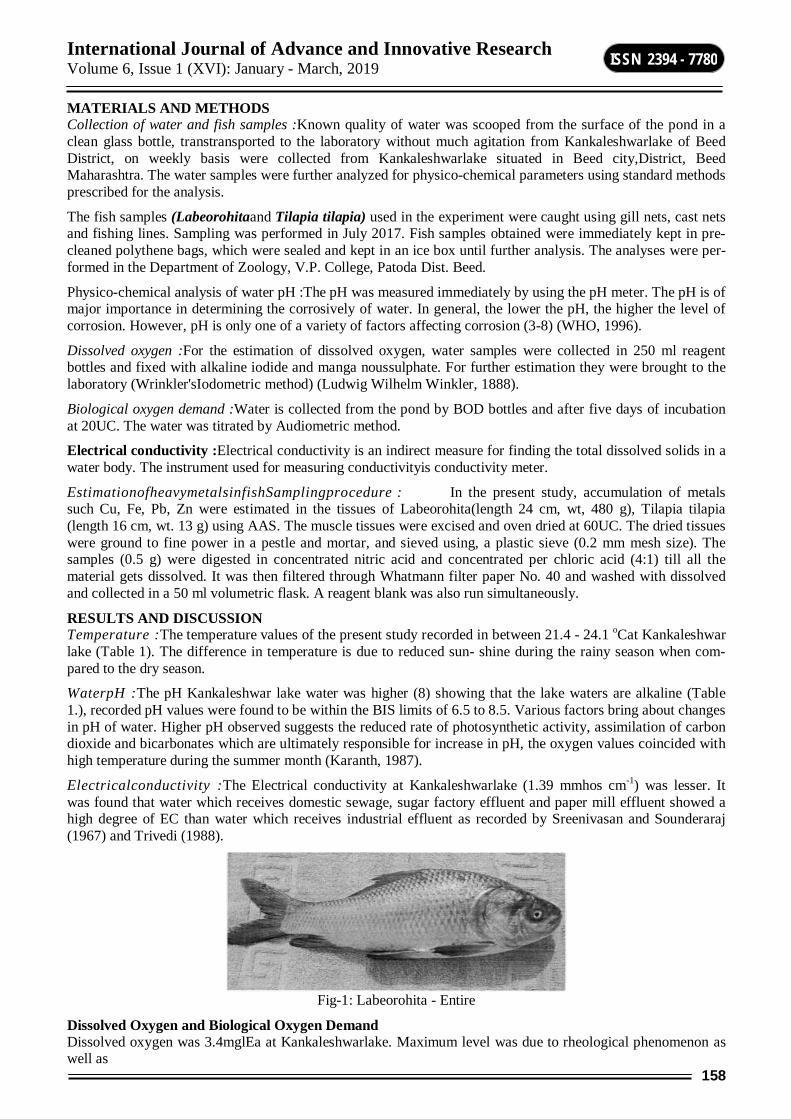
International Journal of Advance and Innovative Research Volume 6, Issue 1 (XVI): January - March, 2019
158
ISSN 2394 - 7780
MATERIALS AND METHODS Collection of water and fish samples :Known quality of water was scooped from the surface of the pond in a clean glass bottle, transtransported to the laboratory without much agitation from Kankaleshwarlake of Beed District, on weekly basis were collected from Kankaleshwarlake situated in Beed city,District, Beed Maharashtra. The water samples were further analyzed for physico-chemical parameters using standard methods prescribed for the analysis.
The fish samples (Labeorohitaand Tilapia tilapia) used in the experiment were caught using gill nets, cast nets and fishing lines. Sampling was performed in July 2017. Fish samples obtained were immediately kept in pre-cleaned polythene bags, which were sealed and kept in an ice box until further analysis. The analyses were per-formed in the Department of Zoology, V.P. College, Patoda Dist. Beed.
Physico-chemical analysis of water pH :The pH was measured immediately by using the pH meter. The pH is of major importance in determining the corrosively of water. In general, the lower the pH, the higher the level of corrosion. However, pH is only one of a variety of factors affecting corrosion (3-8) (WHO, 1996).
Dissolved oxygen :For the estimation of dissolved oxygen, water samples were collected in 250 ml reagent bottles and fixed with alkaline iodide and manga noussulphate. For further estimation they were brought to the laboratory (Wrinkler'sIodometric method) (Ludwig Wilhelm Winkler, 1888).
Biological oxygen demand :Water is collected from the pond by BOD bottles and after five days of incubation at 20UC. The water was titrated by Audiometric method.
Electrical conductivity :Electrical conductivity is an indirect measure for finding the total dissolved solids in a water body. The instrument used for measuring conductivityis conductivity meter.
EstimationofheavymetalsinfishSamplingprocedure : In the present study, accumulation of metals such Cu, Fe, Pb, Zn were estimated in the tissues of Labeorohita(length 24 cm, wt, 480 g), Tilapia tilapia (length 16 cm, wt. 13 g) using AAS. The muscle tissues were excised and oven dried at 60UC. The dried tissues were ground to fine power in a pestle and mortar, and sieved using, a plastic sieve (0.2 mm mesh size). The samples (0.5 g) were digested in concentrated nitric acid and concentrated per chloric acid (4:1) till all the material gets dissolved. It was then filtered through Whatmann filter paper No. 40 and washed with dissolved and collected in a 50 ml volumetric flask. A reagent blank was also run simultaneously.
RESULTS AND DISCUSSION Temperature :The temperature values of the present study recorded in between 21.4 - 24.1 oCat Kankaleshwar lake (Table 1). The difference in temperature is due to reduced sun- shine during the rainy season when com-pared to the dry season.
WaterpH :The pH Kankaleshwar lake water was higher (8) showing that the lake waters are alkaline (Table 1.), recorded pH values were found to be within the BIS limits of 6.5 to 8.5. Various factors bring about changes in pH of water. Higher pH observed suggests the reduced rate of photosynthetic activity, assimilation of carbon dioxide and bicarbonates which are ultimately responsible for increase in pH, the oxygen values coincided with high temperature during the summer month (Karanth, 1987).
Electricalconductivity :The Electrical conductivity at Kankaleshwarlake (1.39 mmhos cm-1) was lesser. It was found that water which receives domestic sewage, sugar factory effluent and paper mill effluent showed a high degree of EC than water which receives industrial effluent as recorded by Sreenivasan and Sounderaraj (1967) and Trivedi (1988).
Fig-1: Labeorohita - Entire
Dissolved Oxygen and Biological Oxygen Demand Dissolved oxygen was 3.4mglEa at Kankaleshwarlake. Maximum level was due to rheological phenomenon as well as
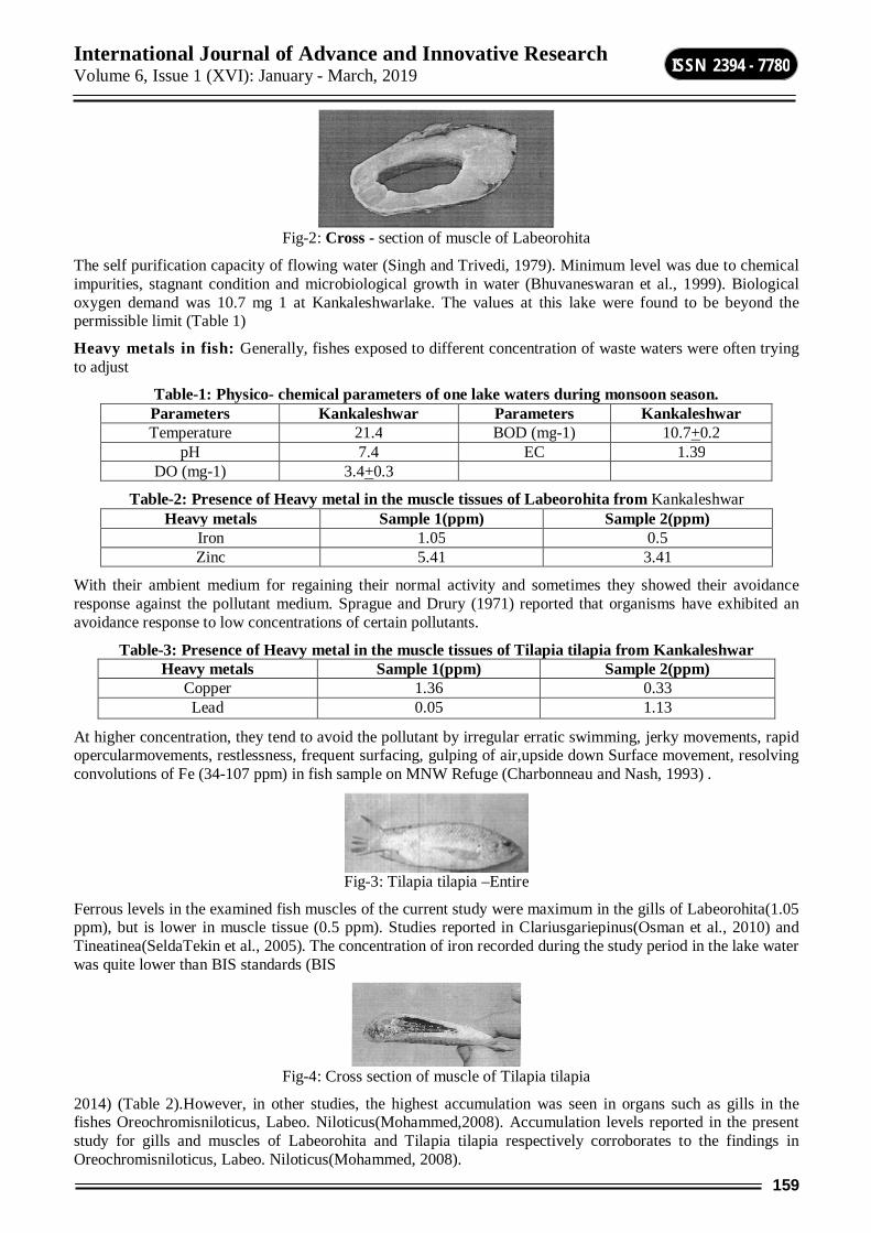
International Journal of Advance and Innovative Research Volume 6, Issue 1 (XVI): January - March, 2019
159
ISSN 2394 - 7780
Fig-2: Cross - section of muscle of Labeorohita
The self purification capacity of flowing water (Singh and Trivedi, 1979). Minimum level was due to chemical impurities, stagnant condition and microbiological growth in water (Bhuvaneswaran et al., 1999). Biological oxygen demand was 10.7 mg 1 at Kankaleshwarlake. The values at this lake were found to be beyond the permissible limit (Table 1)
Heavy metals in fish: Generally, fishes exposed to different concentration of waste waters were often trying to adjust
Table-1: Physico- chemical parameters of one lake waters during monsoon season. Parameters Kankaleshwar Parameters Kankaleshwar Temperature 21.4 BOD (mg-1) 10.7+0.2
pH 7.4 EC 1.39 DO (mg-1) 3.4+0.3
Table-2: Presence of Heavy metal in the muscle tissues of Labeorohita from Kankaleshwar Heavy metals Sample 1(ppm) Sample 2(ppm)
Iron 1.05 0.5 Zinc 5.41 3.41
With their ambient medium for regaining their normal activity and sometimes they showed their avoidance response against the pollutant medium. Sprague and Drury (1971) reported that organisms have exhibited an avoidance response to low concentrations of certain pollutants.
Table-3: Presence of Heavy metal in the muscle tissues of Tilapia tilapia from Kankaleshwar Heavy metals Sample 1(ppm) Sample 2(ppm)
Copper 1.36 0.33 Lead 0.05 1.13
At higher concentration, they tend to avoid the pollutant by irregular erratic swimming, jerky movements, rapid opercularmovements, restlessness, frequent surfacing, gulping of air,upside down Surface movement, resolving convolutions of Fe (34-107 ppm) in fish sample on MNW Refuge (Charbonneau and Nash, 1993) .
Fig-3: Tilapia tilapia –Entire
Ferrous levels in the examined fish muscles of the current study were maximum in the gills of Labeorohita(1.05 ppm), but is lower in muscle tissue (0.5 ppm). Studies reported in Clariusgariepinus(Osman et al., 2010) and Tineatinea(SeldaTekin et al., 2005). The concentration of iron recorded during the study period in the lake water was quite lower than BIS standards (BIS
Fig-4: Cross section of muscle of Tilapia tilapia
2014) (Table 2).However, in other studies, the highest accumulation was seen in organs such as gills in the fishes Oreochromisniloticus, Labeo. Niloticus(Mohammed,2008). Accumulation levels reported in the present study for gills and muscles of Labeorohita and Tilapia tilapia respectively corroborates to the findings in Oreochromisniloticus, Labeo. Niloticus(Mohammed, 2008).

International Journal of Advance and Innovative Research Volume 6, Issue 1 (XVI): January - March, 2019
160
ISSN 2394 - 7780
Zinc: Zinc is an essential trace metal for both retarded growth, loss of taste and hypogonadism, leading to decreased fertility (Sivaperumal, 2007). The FAO maximum guideline for Zn is 30 ppm (FAO 1983). Thus, the concentrations of Zn in the fish samples were within the FAO guideline. The values are shown in (Table 2). The decrease of zinc during monsoon was due to its uptake by macrophysics, and its adsorption in to clay particles and then final sedimentation (Ali Fisher, 2005). Accumulation reported in the present study for gills and muscles are comparable to O. niloticusand C. punctatus. Theconcentrations of Zn (5.41 ppm) was seen in the gill of Labeorahita, While the value of 3.41 ppm was measure in the muscle tissue to Labeorothita. were within the permissible limits.
Lead : Present study reports that lead concentrations of 0.05 ppm was observed in the muscle tissue of Tilapia tilapia, while the concentration of 1.13 ppm was detected in the muscle tissue of Tilapia tilapia (Table 3). The lead concentration was more than BIS Standards (0.05 mgl)' High levels of lead often occur in water bodies near highways and large cities due to gasoline combustion (Banat et al., 1998). The higher level of Pb in water can be attributed to industrial and agricultural discharges as well as leaded petrol from fishing boats (Mason, 2002).
Copper: Copper is essential as a micronutrient for fish and aquatic life, widely used as a very effective al-gaecide and molluseicide (Saeed, 1999; Shaker et al.. 2000). Copper revealed its minimum accumulation in the gills and muscles of the two species. The concentration of Cu in the fish sample was (1.36 ppm) the muscle tissue of Tilapia tilapia while the lowest level of 0.33 ppm was detected in the muscle tissue of Tilapia tilapia. However, the value of 1.36 ppm was below the FAO guideline of 30 ppm. Thus the concentrations of Cu in the fish samples analysed were all FAO recommended guide-line (FAO 1983) (Table 3). The Cu accumulation is within the permissible limits in fish tissues (20 ppm, WHO, 2015).
CONCLUSION Based on the results of the present investigation, the levels of metals bioaccumuiated in tissues of Labeorohitaand Tilapia tilapia did not exceed the permissible limits set for heavy metals by FAO, FEPA and WHO. Therefore, these fishes did not pose any threat to human upon their consumption.
In the present study, due to the bioaccumulation of heavy metals like Iron and Zinc in the tissues of Labeorohitaof Kankaleshwar lake fish, there are a variations in the biochemical contents. High bioaccumulation of Iron and Zinc results in elevation of carbohydrates and depletion of fats and proteins.
Similarly in the present study in Kankaleshwar lake fish, due to the bioaccumulation of heavy metals like Copper and Lead in the tissues of Tilapia tilapia, there is a variation in the biochemical content. High bioaccumulation of Copper results in elevation of fats and proteins and depletion of carbohydrates. High bioaccumulation of Lead results in elevation of carbohydrates and depletion of fat and protein.
REFERENCES - Ali, M. H. H., Fishar, M.R: Accumulation of trace metals in some benthic invertebrate and fish species
relevant to their concentration in water and sediment of Lake Qarun, Egypt, Egypt J. Aqua. Res., 31, 289 - 302 (2005).
- Bajpai, A., Pani, S., Jain, R.K. and Mishra, S.M: Heavy metal con-tamination through ido immersion ina tropical lake,Eco. Environ. Conserv., 8, 157 - 159 (2002).
- Banat, I.M., Hassan, E.S., El-Shahawi, M.S. and Abu-Hilal, A.H: Post-gulf-war assessment of nutrients, heavy metal ions, hydrocarbons, and bacterial pollution levels in the United Arab Emirates coastal waters, Environ. Int., 24, 109 - 116 (1998).
- Barlas, N: A pilot study of heavy metal concentration in various environments, fishes in the upper Sakarya River Basin, Turkey Environ. Toxicol..14, 367- 373 (1997).
- Barron, M.G. and Albeke, S: Calcium Control of Zinc Uptake in Rainbow Trout, Aqua. Toxicol.50, 257 - 264 (2000).
- BasaSiraj, P. and Usha Rani, A: Cadmium induced antioxidant defense mechanism in freshwater teleost Oreochromismossambicus(tilapia), EcoToxicol. Environ. Saf.,2, 218 - 221 (2003).
- Bhagwant, S. and Elahee, K.B: Pathologic gill lesions in two edible lagoon fish species, Mulloidichtysflavolineatusand Mugilcephalus, from the ofpoudre d' or, mauritis, Western Indian Ocean J. Mar. ScL, 1, 35 - 42 (2002).
- Bhuvaneswaran, N., G. Santhalakshmi and S. Rajeswari: Water quality of river Adyar in Chennai city - The river a boon or a bane, Int. J. Environ. Protect., 19, 412 - 415 (1999).

International Journal of Advance and Innovative Research Volume 6, Issue 1 (XVI): January - March, 2019
161
ISSN 2394 - 7780
- BIS, 2014. Specifications for drinking water IS: 10:500 Bureau of Indian Standards, New Delhi.
- Bolis, C.L., Cambria A. and Famam, M.: Effects of Acid Stress on Fish Gills. In: Toxins, Drugs and Pollutants in Marine Mammals (L. Zadunaisky and R. Gilles, Eds.), Springer Verlag, Berlin, 122-129 (1984).
- Charbonneau, C. S. and Nash, T: Contaminants Program, Mingo National Wildlife Refuge (Region 3), Contaminants Survey Results, US Fish and Wildlife Service, Columbia, 1993.
- Karanth, K.R: Ground Water Assessment Development and Management, Tata McGraw Hill Publishing Company Ltd., New Delhi, 725-726 (1987).
- Mason, C.F: Biology of freshwater pollution, 4th Ed. Essex Univ. England, 387 p (2002).
- Mishra, P.C. and Trivedy, R.K: Ecology and Pollution of Indian Lakes and Reservoirs Ashish, New Delhi, 347 p (1993).
- Mohammed, F.A: Bioaccumulation of selected metals and histo-pathological alterations in tissues of Oreochromisniloticusand Lutes niloticusfrom lake Nasser, Egypt, Global Veterinaria, 4, 205 - 218 (2008).
- Osman, A., Alaa, G.M.. Werner Kloas: Water quality and heavy metal monitoring in water sediments and tissues of African cat fish, Clariasgariepinus(Burchell, 1822) from the river Nile, Egypt, J. Environ. Protect., 1, 389 - 400 (2010).
- Purandara, B.K., Vararajan, N. and Jayashree K: Study Regarding Lake Water Pollution with Heavy Metals in Nagpur City (India), Poll., Res, 22, 189 (2003).
- Rompala, J.M..Rutosky, F.W. and Putnam, D.J: Concentrations of Environmental Contaminants from Selected Waters in Pennsylvania, US Pish and Wildlife Service. Special Scientific Report, State College. Pennsylvania (1984).
- Saeed, S.M: A study on factors affecting fish production from certain fish farm in the Delta, M.Sc, Thesis, Ain Shames University, Egypt (1999).
- SeldaTekin.-O. zan AE Ismail Kir: Comparative study on the accumulation of heavy metals in different organs of tench and plero-cercoids of its endoparasiteLigula intestinalis, Parasitol. Res., 97.156 - 159 (2005).
- Shaker. I.M..Ez-El-Rigal, A., and El Ghohashy, H: Effects of EDTA pm reducing of copper toxicity in water and OreochromisniloticusEgypt. J Appl. Sci., 15, 119-131 (2000).
- Sivapermal.P..Sankar, J.V. and Nair Viswanathan, P.G: Heavy Metal Concentrations in Pish.
- Shellfish and Fish Products from Internal Markets of India vis-a-vis International Standards, Food Chem.. 102. 612 - 620 (2007).
- Sreenivasan, A. and Soundeiaraj, R: Effects of certain wastes on the water quality and fisheries of rivers Cauvery and Bhavani, J. Environ. Health. 9. 13 - 21 (1967).
- Trivedy, R.K: Studies on the biological characteristics of the river Krishna in Maharastra with reference to human activity and pollution, Technical Report submitted to Dept. of Environment, Ministry of Environ. Forest and Wild Life, New Delhi (1988).
- USEPA (United states Environmental Protection Agency), Executive summary of the National Water Quality Inventory: 1994 Report to Congress. EPA841-S-94-002. December 1995, Office of Water, Washington, DC, USA.,pp: 209 (1994).
- Variya Rajesh: Impact of Ganesh Idol Immersion on Tapi River at UmaraOvara, M.Sc, dissertation. Veer Narmad South Gujarat University, Surat (2010).
- VarsaniAlpa: Impact of Ganesh Idol Immersion on Tapi River at Ashwani Kumar Ovara.M.Sc. dissertation, Veer Narmad South Gujarat University, Surat (2010).
- Velez, D. and Montoro, R: Arsenic speciation in manufactured seafood products: A review, J Food Protect., 9, 1240 - 1245 (1998).
- WHO World Health Organization, Guidelines for drinking water, Geneva (2015).
- WHO: Guidelines for drinking-water quality, 2nd ed. Vol. 2., Health criteria and other supporting information, World Health Organization, Geneva (2015).
International Journal of Advance and Innovative Research Volume 6, Issue 1 (XVI): January - March, 2019
162
ISSN 2394 - 7780
AN EFFICIENT, GREEN KNOEVENAGEL CONDENSATION FOR THE SYNTHESIS OF NEW 5-(4-((2-PHENYLTHIAZOL-4-YL) METHOXY) BENZYLIDENE)-3-(4-SUBSTITUTED PHENYL)
THIAZOLIDINE-2,4-DIONES
Manisha R. Bhosle Department of Chemistry, Dr. Babasaheb Ambedkar Marathwada University, Aurangabad
ABSTRACT A rapid, efficient and environmentally benign methodology has been developed for the synthesis of new 5-(4-((2-phenylthiazol-4-yl)methoxy)benzylidene)-3-(4-substituted phenyl)thiazolidine-2,4-diones (6a-g) by carrying the Knoevenagel condensation of 4-((2-phenylthiazol-4-yl)methoxy)benzaldehyde (4) and 2,4- thiazolidinediones (5a-g) at room temperature in presence of diisopropyl ethyl ammonium acetate (DIPEAc) as a catalyst/medium. The fascinating scope of this synthetic strategy is that products are rapidly obtained with non tedious isolation work up and moderate to good yields. Reusability of DIPEAc with consistent activity for at least four cycles is also established, indicating DIPEAc as a greener reaction medium.
Keywords: Diisopropyl ethyl ammonium acetate (DIPEAc); Green synthesis; Knoevenagel condensation; 2,4-Thiazolidinediones; Thiazole
INTRODUCTION Thiazolidinedione scaffold and its derivatives are an important class of heterocyclic compounds. They serves as basic pharmacophore for various biological profiles i.e. it displays anticonvulsants,3 antimicrobial,4 tuberculostatic,5 antitumor,6 immunostimulatory, antiarthritic and oncostatic activities.7 Among those biologically interesting thiazolidinediones, 5-arylidene-2,4-thiazolidinediones are important structural elements in medicinal chemistry and are found to possess significant antihyperglycemic activitie.8 Structure-activity relationship (SAR) studies on 2,4-thiazolidinediones have shown that the substituent at 5th position of the 2,4-thiazolidinedione ring system influences the pharmacological activities.9 A series of 5-arylidenyl 2, 4-thiazolidinediones are under clinical trials as potential phospho lipase A2 inhibitor, dual COX-2/5- LOX inhibitor and anti-inflammatory agents.10
The ethereal linkage and 2,4- thiazoldinedione are essential in the molecular framework of various antidiabetic drugs11 like, pioglitazone, troglitazone and rosiglitazone (Figure 1). Thiazoles occupy special place in the realm of natural and synthetic organic chemistry, because of their therapeutic and pharmacological properties.12 A large number of thiazole derivatives have received considerable attention as potential anti-inflammatory, anticonvulsant and anti tubercular agents.13
Figure-1: Biologically active 2,4-thiazolidinedione and thiazole pharmacophore
Hybrid molecules have recently come under focus due to their promising physical, chemical, and biological properties. Hybrid molecules are chemical units composed of two (or more) structural domains in which the characteristics of various constituents have been altered to give rise to altogether new properties.14 The concept of hybrid molecules or molecular hybridization is now being increasingly used by pharmaceutical chemists in their quest for potent new drugs as evidenced by the large number of recent literature reports on the synthesis of new bioactive hybrid molecules with the goal of creating new chemical entities more medically effective than their precursors.15,16
In view of the pharmacological importance of 2,4-thiazolidinediones and thiazoles here it was thought worthwhile to bridge 2,4-TZDs and thiazoles by ethereal linkage and construct some new 5-(4-((2-phenylthiazol-4-yl)methoxy)benzylidene)-3-(4-substituted phenyl)thiazolidine-2,4-diones with the hope to obtain the compounds with intensified activities.

International Journal of Advance and Innovative Research Volume 6, Issue 1 (XVI): January - March, 2019
163
ISSN 2394 - 7780
Knoevenagel condensation of 2,4-thiazolidinediones and aldehydes has found to be one of the key steps. Various methods were reported for the Knoevenagel condensation to synthesize 5-arylidine-2,4-thiazolidinediones.17 Knoevenagel condensation is carried using organic bases like aliphatic amines or their corresponding salts, and amino acids like glycine and L-proline.18 Knoevenagel condensation of different aldehydes and 2,4-thiazolidinediones has been reported in organic media in presence of catalysts like zeolites,19 alum,20 piperidine,21 piperidine benzoate,22 potassium carbonate23 and sodiumacetate.24 The known protocols for carrying the Knoevenagel condensations have one or other kind of drawbacks like need of toxic, hazardous and flammable media, expensive catalysts, prolonged reaction time and heating.
For the discovery of new lead structures in drug discovery, based on high throughput screening, synthetic methodologies are required which deliver highly diverse derivatives in a timely manner. Under these circumstances, organic reactions using sustainable reaction media such as ionic liquids (ILs), avoiding the use of volatile organic solvents, have attracted a great deal of attention of synthetic organic chemist. ILs are not only easily available, environmentally safe, but also excellent catalysts and medium having weakly coordinating ions, i.e. organic cation and inorganic/organic anion.25 ILs having nonvolatility, nonflammability, and are endowed with unique properties such as high thermal/chemical stability.26 ILs act as ‘neoteric solvents’ for a broad range of chemical and industrial processes. Recently, ILs have been found to be useful as “green” media for numerous organic transformations.27 The ability to dissolve many organic and inorganic substances makes ILs eco-friendly reaction media/catalysts.28
Considering the synthetic expediency of green chemistry and DIPEAc as catalyst/medium and in continuation of our earlier interest to develop novel environment friendly methodologies for the synthesis of pharmaceutically important scaffolds,29 here microwave assisted synthetic protocol is developed for the Knoevenagel condensations of 4-((2-phenylthiazol-4-yl)methoxy)benzaldehyde (4) and N-substituted 2,4-thiazolidinediones (5a-g) synthesis of new 5-(4-((2-phenylthiazol-4-yl)methoxy)benzylidene)-3-(4-substituted phenyl)thiazolidine-2,4-diones (6a-g).
RESULTS AND DISCUSSION Avoiding organic solvents during the reactions in organic synthesis leads to a clean, efficient and economical technology. There is an increasing interest in the use of environmentally benign reagents and methods. In the present investigation, the Knoevenagel condensation (Scheme 1) was carried out under conventional and green media i.e. ionic liquid. The condensation of 4-((2-phenylthiazol-4-yl)methoxy)benzaldehyde and active methylene compounds 2,4- thiazolidinediones (5a-g) in presence of DIPEAc were stirred for 1 h to produce the Knoevenagel product. The products obtained in good yields with high purity without using any solvent. (Table 1).
4-((2-Phenylthiazol-4-yl)methoxy)benzaldehyde (4), required as starting precursor for getting the titled molecules was freshly prepared, starting from known reactant 4-(chloromethyl)-2-phenylthiazole (3). 4-(Chloromethyl)-2-phenylthiazole (3) was synthesized using reported procedure16 i.e. by allowing the interaction of thiobenzamide (1) and 1,3-dichloro acetone (2) in ethanol under reflux. Chloromethyl thiazole on condensation with 4-hydroxybenzyldehyde in N,N-dimethyl formamide in the presence of K2CO3 at room temperature gave the required 4-((2-phenylthiazol-4-yl)methoxy)benzaldehyde (4) with quantitative yield (97%).
Scheme-1: Synthesis of 5-(4-((2-phenylthiazol-4-yl)methoxy)benzylidene)-3-(4-substituted phenyl)thiazolidine-
2,4-diones (6a-g)
International Journal of Advance and Innovative Research Volume 6, Issue 1 (XVI): January - March, 2019
164
ISSN 2394 - 7780
In order to establish the best experimental conditions, we have considered the condensation of 4-((2-phenylthiazol-4-yl)methoxy)benzaldehyde (4) and 2,4-thiazolidinedione (5a) as a standard model reaction. Before developing the above route, the Knoevenagel condensation of 4-((2-phenylthiazol-4-yl)methoxy)benzaldehyde (4) and 2,4-thiazolidinedione (5a) was performed separately in conventional medium, pipiridinium acetate in ethanolic medium and sodium acetate in acetic acid under reflux for 9-10h and obtained 5-(4-((2-phenylthiazol-4 yl)methoxy)benzylidene)thiazolidine-2,4-dione (6a) with poor yield (Table 1). Further attempt was made to carry the above model reaction replacing ethanol by DMF/dimethyl acetamide and sodium ethoxide as a base. There was not much improvement in the product yield and reaction time (Table 1).
Scheme-2: Standard model reaction
Therefore to reduce the reaction time and to improve the product yield, Knoevenagel condensation of 4-((2-phenylthiazol-4 yl)methoxy)benzaldehyde (4) and 2,4- thiazolidinedione (5a) was carried under green medias separately in deep eutectic medium, glycerol, PEG-400 and DIPEAc. It was observed that, there was improvement in the product yield and considerable reduction in the time required for the completion of the reaction. Condensation has been found to run successfully in presence of the green media and gave the titled product 5-(4-((2-phenylthiazol-4 yl)methoxy)benzylidene)thiazolidine-2,4-dione (6a) with better yields, 69, 61, 64 and 72% respectively (Table 1). From these findings it has been confirmed that the condensation of (4) and (5a) was found to take place efficiently and safely if carried using DIPEAc as catalyst and medium at room temperature.
The reaction has also been performed under neat conditions (without catalyst) reflux ethanol. It was observed that there was no reaction in the absence of DIPEAc. To investigate the amount of DIPEAc required for the formation of (6a), the model reaction was performed varying the amount of DIPEAc in the range of 20, 40, 60, 80 and 100 mole % in medium ethanol. It was found that when we used 20 and 40 mol% DIPEAc there was no appreciable transformation. However, by increasing the concentration of DIPEAc the yield of (6a) has also been increased (Table 1, entry 10). The optimum concentration of DIPEAc required was 100 mole %, therefore we used DIPEAc as catalyst and medium (Table 1).
RECYCLING OF CATALYST To check reusability of the catalyst, the reaction was performed between (4) and (5a) under the optimized reaction condition. DIPEAc was separated from the reaction mixture by the following procedure. After completion of the reaction, water and ethyl acetate were added to the reaction mixture. The product (6a) was extracted with ethyl acetate. As the DIPEAc is highly water soluble it goes in to the aqueous layer. Evaporation of aqueous layer under reduced pressure provided the catalyst (DIPEAc). Catalysts purity was checked by 1H NMR for subsequent reactions. Reusability of the DIPEAc was tested for four consecutive cycles, but a decrease in the catalytic activity of DIPEAc was observed after the third cycle (Figure 2).
Figure-2: Reuse and recovery of DIPEAc and its effect on yield.
International Journal of Advance and Innovative Research Volume 6, Issue 1 (XVI): January - March, 2019
165
ISSN 2394 - 7780
With all the above optimized conditions in hand, an attempt was made to neatly condense 4-((2-phenylthiazol-4 yl)methoxy)benzaldehyde, (4) and 2,4- thiazolidinediones (5a-g) in presence of DIPEAc at room temperature and obtained the desired products 5-(4-((2-phenylthiazol-4-yl)methoxy)benzylidene)-3-(4-substituted phenyl)thiazolidine-2,4-diones (6a-g) with excellent yields (Table 2).
PLAUSIBLE REACTION MECHANISM The catalytical role of DIPEAc proposed for the condensation aldehyde and 2,4-thiazolidinone.16 Literature reveals that DIPEAc having N-H proton which could be responsible for the acceleration of Knoevenagel condensation. Oxygen of carbonyl carbon of aldehyde and 2,4-thiazolidinedione interact with a proton of DIPEAc which could be increases the electrophilicity of aldehyde and nucleophilicity of active methylene carbon. Thus the generated nucleophile would attack the electrophilic carbon of the aldehyde resulting in the aldol adduct which on sequential dehydration leads to Knoevenagel products.
Scheme-3: Plausible reaction mechanism
Table-1: Optimization of catalysts for the synthesis of (6a)a
Entry Catalysts Conditions Time Yield (%) 1 Piperidine Ethanol/Δ 9-10h 53 2 Piperidinium acetate Ethanol/Δ 9-10h 59 3 Sodium acetate Acetic acid/Δ 9-10h 47 4 Sodium ethoxide DMF/Δ 9-10h 50 5 Sodium ethoxide DMAc/Δ 9-10h 55 6 Deep eutectic solvent (5 mL) Stirr/RT 1h 69 7 Glycerol (5 mL) Stirr/RT 1h 61 8 PEG-400 (5 mL) Stirr/RT 1h 64 9 DIPEAc (5 mL) Stirr/RT 1h 72
10 DIPEAc/EtOH 20, 40, 60, 80 and 100 mole % Stirr/RT 1h 62, 62, 63, 69, 72
11 Neat Ethanol/Δ - No reaction aReaction conditions: 4 (3 mmol) and 5a (3 mmol)
Table-2: Synthesis of 5-(4-((2-phenylthiazol-4-yl)methoxy)benzylidene)-3-(4-substituted phenyl)thiazolidine-2,4-diones (6a-g).a
Entry Compound R Yield (%)b Melting point (C) 1 6a H 72 270-272 2 6b C6H5 70 239-241 3 6c p-CH3 C6H4 68 255-257 4 6d o-CH3 C6H4 65 226-227 5 6e p-F C6H4 71 203-204 6 6f p-OCH3 C6H4 69 247-249 7 6g o-Cl C6H4 65 195-197 aReaction conditions: 4-((2-phenylthiazol-4-yl)methoxy)benzaldehyde (4) (3 mmol), 2,4-
thiazolidinedione (5a) (3 mmol) and DIPEAc (5 mL) bIsolated Yield:
International Journal of Advance and Innovative Research Volume 6, Issue 1 (XVI): January - March, 2019
166
ISSN 2394 - 7780
CONCLUSION We have developed a rapid, expedient and environmentally benign DIPEAc catalysed Knoevenagel condensation to produce new 5-(4-((2-phenylthiazol-4-yl)methoxy) benzylidene)-3-(4-substituted phenyl) thiazolidine-2,4-diones. Consequently, our findings will endow with a great impact on chemists for assisting investigations in the 2,4-thiazolidinedione field in search of novel and greener methodologies.
ACKNOWLEDGMENTS The authors are thankful to Professor Ramrao A. Mane for his invaluable discussions and guidance. One of the authors, Manisha R. Bhosle, is thankful to Dr. Babasaheb Ambedkar Marathwada University authorities for awarding Jahagirdar Research Fellowship. The authors are also thankful to Central Drug Research Institute (CDRI), Lucknow for spectral analysis.
EXPERIMENTAL General: All the chemicals used were of laboratory grade. Melting points of all the synthesized compounds were determined in open capillary tubes and are uncorrected. 1H NMR spectra were recorded with a Bruker Avance 400 spectrometer operating at 400 MHz and 200 MHz using DMSO-d6 or CDCl3 solvent and tetramethylsilane (TMS) as the internal standard and chemical shift in δ ppm. Mass spectra were recorded on a Sciex, Model; API 3000 LCMS/MS Instrument. The purity of each compound was checked by TLC using silica-gel, 60F254 aluminum sheets as adsorbent and visualization was accomplished by iodine/ultraviolet light.
Synthesis of 4-((2-Phenylthiazol-4-yl)methoxy)benzaldehyde (4) A mixture of chloromethylthiazole (3) (10 mmol), powdered potassium carbonate (20 mmol), and 4-hydroxybenzaldehyde (10 mmol) was added to N,N-dimethyl formamide (20-30 mL). The reaction mixture was then stirred for 5-6 h at r.t. After completion of the reaction, the reaction mixture was poured on crushed ice. Thus obtained solid was filtered, washed with water, and crystallized from ethanol. Yield: 97%, M.P. 100-102oC.
Spectral analysis of 4-((2-Phenylthiazol-4-yl)methoxy) benzaldehyde (4).
IR (KBr, υ cm-1) Characteristic absorptions: 3117 (Ar-H stretch), 2830 (-C-H stretch), 1745 (C=O) and 1266 (C-O-C stretch). MS (Scanning mode, ESI+): m/z (% intensity): 295.9 (M+, 100). 1H-NMR (300 MHz, CDCl3, δ ppm): 5.33 (s, 2H, CH2), 7.14-7.50 (m, 6H, Ar-H and thiazolyl-H), 7.82-8.00 (m, 4H, Ar-H) and 9.89 (s, 1H, -CHO). Synthesis of 5-(4-((2-phenylthiazol-4-yl)methoxy)benzylidene)-3-thiazolidine-2,4-dione (6a) Conventional method A mixture of 4-((2-phenylthiazol-4-yl)methoxy)benzaldehyde (4) (2 mmol), 2,4-thiazolidinedione (5a) (2.2 mmol) and DIPEAc (5 mL) was stirred at room temperature. The progress of the reaction was monitored by thin layer chromatography. After 1 h of stirring, it was poured on crushed ice. Thus obtained solid was filtered, washed with water, and crystallized from ethanol and DMF. General procedure for the synthesis of diisopropylethylammonium acetate (DIPEAc) A mixture of glacial acetic acid (0.02 mol) and N-ethyl-N-isopropylpropan-2-amine (0.02 mol) was stirred at 0-10 ºC for 30 min. to obtain diisopropylethylammonium acetate as a viscous liquid.26
Following is a spectral data of one of the representative products from the series, 5-(4-((2-phenylthiazol-4-yl) methoxy) benzylidene)-3-thiazolidine-2,4-dione (6a).
IR (KBr, υ cm-1) Characteristic absorptions: 3433 (N-H stretch), 3275 (Ar-H stretch), 2924 (C-H stretch), 1639 (C=O stretch), 1560 (C=C stretch), 1330, 1342 and 829 (due to thiazole ring). MS (Scanning mode, ESI+): m/z (% intensity): 395 (M+, 100), 1H-NMR (300 MHz, CDCl3, δ ppm): 5.31 (s, 2H, CH2), 7.11 (s, 1H, olefinic), 7.26-7.44 (m, 6H, Ar-H and thiazolyl-H) and 7.80-8.07 (m, 4H, Ar-H).13C-NMR (75 MHz, CDCl3, δ ppm): 65.53, 114.26, 114.36, 117.15, 117.45, 121.39, 124.78, 125.91, 127.00, 128.67, 129.85, 130.05, 131.55, 132.78, 142.53, 152.84, 157.14, 159.85, 167.38 and 167.54. Elemental Analysis: Anal. Calcd. for C20H14N2S2: C, 60.90; H, 3.58; N, 7.10; S, 16.26 Found: C, 60.86; H, 3.16; N, 7.56 and S, 16.47. REFERENCES 1. Corbett, J. W.; Ko, S. S.; Rodgers, J. D.; Gearhart, L. A.; Magnus, N. A.; Bacheler, L. T.; Diamond, S.;
Jeffrey, S.; Klabe, R. M.; Cordova, B. C.; Garber, S.; Logue, K.; Trainor, G. L.; Anderson, P. S.; Erickson-Viitanen, S. K. J. Med. Chem. 2000, 43, 2019.
2. Franzen, R. G. J. Comb. Chem. 2000, 2, 195.
International Journal of Advance and Innovative Research Volume 6, Issue 1 (XVI): January - March, 2019
167
ISSN 2394 - 7780
3. (a) Brown, F. C. Chem. Rev. 1961, 61, 463. (b) Lesyk, R. B.; Zimenkovsky B. S. Curr. Org. Chem. 2004, 8, 1547.
4. Grasso, S.; Chimirri, A.; Monforte, P.; Fenech, G. Farmaco [Sci.] 1984, 39, 505.
5. (a) Tuncbilek, M.; Bozdag, O.; Ayhan-Kilcigil, G.; Ceylan, M.; Waheed, A.; Verspohl, E. J.; Ertan, R.Farmaco. 2003, 58, 79. (b) Bozdag, O.; Verspohl, E. J.; Ertan, R. Arzneim-Forsch Drug. Res. 2000, 50, 539.
6. Panigrahy, D.; Singer, S.; Shen, L. Q.; Butterfield, C. E.; Freedman, D. A.; Chen, E. J.; Moses, M. A.; Kilroy, S.; Duensing, S.; Fletcher, C.; Fletcher, J. A.; Hlatky, L.; Hahnfeldt, P.; Folkman, J.; Kaipainen1, A. J. Clin. Invest. 2002, 110, 923.
7. Moretti, R. M.; Marelli, M. M.; Motta, M.; Limonta, P. Int. J. Cancer 2001, 92, 733.
8. Donald, E.; Carpenter, R. J.; Imbordino, M. T.; Maloney, J. A.; Moeslein, M. R.; Scott, A. Org. Proces. Res. & Dev. 2002, 6, 721.
9. (a) Mohareb, R. M.; Fleita, D. H. Heteroatom Chem. 2002, 13, 258. (b) Herrding, D. A.; Christmann, L. T.; Clark, T. J.; Holmes, D. J.; Rittenhouse, S. F.; Takata, D. T.; Venslavsky, J. W. Bioorg. Med. Chem. Lett. 2003, 13, 3771.
10. Hulin, B.; Clark, D. A.; Goldstein, S. W.; McDermott, R. E.; Dambek, P. J.; Kappeler, W. H.; Lamphere, C. H.; Lewis, D. M.; Rizzi, J. P. J. Med. Chem. 1992, 35, 1853.
11. (a) Liu, K. G.; Smith, J. S.; Ayscue, A. H.; Henke, B. R.; Lambert, M. H.; Leesnitzer, L. M.; Stenbach, D. D. Boiorg. Med. Chem. Lett. 2001, 11, 2385. (g) Berger, J.; Leibowitz, M. D.; Doebber, T. W.; Elbrecht, A.; Zhang, B.; Zhou, G.; Biswas, C.; Cullinan, C. A.; Burger, G. D.; Mostley, R.; Marquis, R.; Sahoo, S. P.; Tolman, R. L.; Smith, R. G.; Moller, D. E. J. Biol. Chem. 1999, 274, 6718. (f) Cantello B C C, Cawthrone M A, Cottam G P, Duff P T, Haigh D, Hindley R M, Lister C A, Smith S A and Thurlby P L 1994 J. Med. Chem. 37 3977
12. Stevens, M. J. Med. Chem. 2002, 45, 744.
13. Siddiqui, N.; Arshad, M. F.; Ahsan, W.; Alam, M. S. Int. J. Pharm. Sci. Drug Res. 2009, 1, 3, 136.
14. (a) Meunier, B. Acc. Chem. Res. 2008, 41, 69; (b) Viegas-Junior, C.; Danuello, A.; da Silva Bolzani, V.; Barreiro, E. J.; Fraga, C. A. Curr. Med. Chem. 2007, 14, 1829–1852; (c) Arnaud, C. H. Chem. Eng. News 2007, 85, 46.
15. (d) Tukulula, M.; Sharma, R.-K.; Meurillon, M.; Mahajan, A.; Naran, K.; Warner, D.; Huang, J.; Mekonnen, B.; Chibale, K. ACS Med. Chem. Lett. 2013, 4, 128. (b) Wang, Y.; Damu, G. V. L.; Ledv, J.-S.; Geng, R.-X.; Yang, D.-C.; Zhou, C.-H. Bioorg. Med. Chem. Lett. 2012, 22, 5363;
16. (c) Manohar, S.; Khan, S. I.; Rawat, D. S. Chem. Biol. Drug Des. 2011, 78, 124; (d) Woo, L. W. L.; Bubert, C.; Purohit, A.; Potter, B. V. L. ACS Med. Chem. Lett. 2011, 2, 243; (h) Singh, S. B.; Tiwari, K.; Verma, P. K.; Srivastava, M.; Tiwari, K. P.; Singh, J. Supramol. Chem. 2013, 25, 255.
17. (a) Venkatanarayana, M.;.Dubey, P. K. Synth. Comm. 2012, 1, 1746. (b) Jawale, D. V.; Pratap, U. R.; Mane R. A. Bull. Korean Chem. Soc. 2011, 32, 7.
18. Gadekar, L. S.; Arbad, B. R.; Lande, M. K. Org. Chem.: An Ind. J. 2008, 4, 458.
19. Shelke, K. F.; Sapkal, S. B.; Kakade, G. K.; Sadaphal, S. A.; Shingate, B. B.; Shingare, M. S. Green Chem. Lett. Rev. 2010, 3, 17.
20. Bhattarai, B. R.; Kafle, B.; Hwang, J. S.; Khadka, D.; Lee, S. M.; Kang, J. S.; Hamd, S. W.; Han, I. O.; Park, H.; Cho, H. Bioorg. Med. Chem. Lett. 2009, 19, 6161.
21. Liu, Z.; Huang, Y.; Zhang, W.; Ma, L.; Li, J.; Wang, X.; Li, J.; Shen, J. J. Comb. Chem. 2008, 10, 632.
22. Yoshitata, O.; Teruo, M.; Mishiko, N.; Motoyuki, J.; Norio, K. Chem. Pharm. Bull. 1992, 40, 907.
23. Tuncbine, M.; Dundar, B.; Ayhan-Kilcigil, G.; Ceylan, M.; Waheed, A.; Verspohl, E. J.; Ertan, R. I. Farmaco 2003, 58, 79.
24. Adams, D. J.; Dyson, P. J.; Tavener, S. J. Chemistry in Alternative Media; Wiley: New York, 2004.
25. Wilkes, J. S. Green Chem., 2002, 4, 73-80.

International Journal of Advance and Innovative Research Volume 6, Issue 1 (XVI): January - March, 2019
168
ISSN 2394 - 7780
26. (a) Fraga-Dubreuil, J.; Bazureau, J. P. Tetrahedron 2003, 59, 6121. (b) Plechkova, N. V.; Seddon, K. R. Chem. Soc. Rev. 2008, 37, 123. (c) Deng, Y. Q.; Shi, F.; Beng, J. J.; Kun, Q. J. Mol. Catal. A: Chem. 2001, 33, 165. (d) Ganeshpure, P. A.; George, G.; Das, J. J. Mol. Catal. A: Chem. 2008, 182, 279. (e) Xu, F.; Chen, H. Y.; Zhang, H. B.; Zhou, X. H.; Cheng, G. Z. J. Mol. Catal. A: Chem. 2009, 9, 307.
27. (a) Jiang, T.; Gao, H.; Han, B.; Zhao, G.; Chang, Y.; Wu, W.; Gao, L.; Yang, G. Tetrahedron Lett. 2004, 45, 2699-2701. (b) Chandran, R.; Kalaipriya, M.; Ramakrishnan, U.; Radhakrishnan, S.; Seeram, R. RSC Adv. 2012, 2, 11657. (c) Wilkes, J. S. Green Chem., 2002, 4, 73-80.
28. Chandran, R.; Kalaipriya, M.; Ramakrishnan, U.; Radhakrishnan, S.; Seeram, R. RSC Adv. 2012, 2, 11657.
29. (a) Bhosle, M. R.; Khillare, L. D.; Dhumal, S. T.; Mane, R. A. Chinese Chem. Lett. 2016, 27, 370-374. (b) Bhosle, M. R.; Mali, J. R.; Mulay, A. A.; Mane, R. A. Heteroatom Chem. 2012, 23, 2. (c) Bhosle, M. R.; Khillare, L. D.; Dhumal, S. T.; Mane, R. A. Lett Org Chem, 2016, 13, 148-155.
International Journal of Advance and Innovative Research Volume 6, Issue 1 (XVI): January - March, 2019
169
ISSN 2394 - 7780
STUDY OF ELECTRONIC WASTE MANAGEMENT IN INDIA: ISSUES AND STRATEGIES
Mayur P Deshmukh1 and Manjusha M Deshmukh2 1Department of Computer Science, Balbhim Art’s, Science & Commerce College, Beed
Research Scholars2, Dr. BabasahebAmbedkarMarathwada University, Aurangabad
ABSTRACT The current practices of e-waste management in India suffer from a number of drawbacks like the difficulty in invention, unhealthy conditions of informal recycling, inadequate legislation, poor awareness and reluctance on part of the corporate to address the critical issues. The consequences are that (i) toxic materials enter the waste stream with no special precautions to avoid the known adverse effects on the environment and human health and (ii) resources are wasted when economically valuable materials are dumped or unhealthy conditions are developed during the informal recycling. The paper highlights the associated issues and strategies to address this emerging problem, in the light of initiatives in India. The paper presents a waste management system with shared responsibility for the collection and recycling of electronic wastes amongst the manufacturers / assemblers, importers, recyclers, regulatory bodies and the consumers.
Keywords: E-Waste, toxic materials, NGO’s, CRT, LCD
1. INTRODUCTION The electronic industry is the world’s largest and fastest growing manufacturing industry (Radha, 2002; DIT, 2003). During the last decade, it has assumed the role of providing a forceful leverage to the socio - economic and technological growth of a developing society. The consequence of its consumer oriented growth combined with rapid product obsolescence and technological advances are a new environmental challenge - the growing menace of “Electronics Waste” or “e waste” that consists of obsolete electronic devices. It is an emerging problem as well as a business opportunity of increasing significance, given the volumes of e-waste being generated and the content of both toxic and valuable materials in them. The fraction including iron, copper, aluminum, gold and other metals in e-waste is over 60%, while plastics account for about 30% and the hazardous pollutants comprise only about 2.70% (Widmer et al., 2005). Solid waste management, which is already a mammoth task in India, is becoming more complicated by the invasion of e-waste, particularly computer waste. E-waste from developed countries find an easy way into developing countries in the name of free trade (Toxics Link, 2004) is further complicating the problems associated with waste management. The paper highlights the associated issues and strategies to address this emerging problem, in the light of initiatives in India.
2. E – WASTE IN INDIA As there is no separate collection of e-waste in India, there is no clear data on the quantity generated and disposed of each year and the resulting extent of environmental risk. The preferred practice to get rid of obsolete electronic items in India is to get them in exchange from retailers when purchasing a new item. The business sector is estimated to account for 78% of all installed computers in India (Toxics Link, 2003). Obsolete computers from the business sector are sold by auctions. Sometimes educational institutes or charitable institutions receive old computers for reuse. It is estimated that the total number of obsolete personal computers emanating each year from business and individual households in India will be around 1.38 million. According to a report of Confederation of Indian Industries, the total waste generated by obsolete or broken down electronic and electrical equipment in India has been estimated to be 1,46,000 tons peryear (CII, 2006).
The results of a field survey conducted in the Chennai, a metroplolitan city of India to assesstheaverage usage and life of the personal computers (PCs), television (TV) and mobile phoneshowed that the average household usage of the PC ranges from 0.39 to 1.70 depending on theincome class (Shobbana Ramesh and Kurian Joseph, 2006). In the case of TV it varied from1.07 to 1.78 and for mobile phones it varied from 0.88 to 1.70. The low-income households usethe PC for 5.94 years, TV for 8.16 years and the mobile phones for 2.34 years while, the upperincome class uses the PC for 3.21 years, TV for 5.13 years and mobile phones for 1.63 years.Although the per-capita waste production in India is still relatively small, the total absolutevolume of wastes generated will be huge. Further, it is growing at a faster rate. The growth rateof the mobile phones (80%) is very high compared to that of PC (20%) and TV (18%). Thepublic awareness on e-wastes and the willingness of the public to pay for e-waste management asassessed during the study based on an organized questionnaire revealed that about 50% of thepublic are aware of environmental and health impacts of the electronic items. The willingness ofpublic to pay for e-waste management ranges from 3.57% to 5.92% of the product cost for PC,3.94 % to 5.95 % for TV and 3.4 % to 5 % for the mobile phones.
International Journal of Advance and Innovative Research Volume 6, Issue 1 (XVI): January - March, 2019
170
ISSN 2394 - 7780
Additionally considerable quantities of e-waste are reported to be imported (Agarwal, 1998;Toxics Link, 2004). However, no confirmed figures available on how substantial are thesetrans boundary e-waste streams, as most of such trade in e-waste is camouflaged and conductedunder the pretext of obtaining ‘reusable’ equipment or ‘donations’ from developed nations. Thegovernment trade data does not distinguish between imports of new and old computers andperipheral parts and so it is difficult to track what share of imports are used electronic goods.
3. INFLUENCES OF E – WASTES Electronic wastes can cause widespread environmental damage due to the use of toxic materials in the manufacture of electronic goods (Mehra, 2004). Hazardous materials such as lead, mercury and hexavalent chromium in one form or the other are present in such wastes primarily consisting of Cathode ray tubes (CRTs), Printed board assemblies, Capacitors, Mercury switches and relays, Batteries, Liquid crystal displays (LCDs), Cartridges from photocopying machines, Selenium drums (photocopier) and Electrolytes. Although it is hardly known, e-waste contains toxic substances such as Lead and Cadmium in circuit boards; lead oxide and Cadmium in monitor Cathode Ray Tubes (CRTs); Mercury in switches and flat screen monitors; Cadmium in computer batteries; polychlorinated biphenyls (PCBs) in older capacitors and transformers; and brominated flame retardants on printed circuit boards, plastic casings, cables and polyvinyl chloride (PVC) cable insulation that releases highly toxic dioxins and furans when burned to retrieve Copper from the wires. All electronic equipment’s contain printed circuit boards which are hazardous because of their content of lead (in solder), brominated flame retardants (typically 5-10 % by weight) and antimony oxide, which is also present as a flame retardant (typically 1- 2% by weight) (Devi et al, 2004).
Landfilling of e wastes can lead to the leaching of lead into the ground water. If the CRT is crushed and burned, it emits toxic fumes into the air (Ramachandra and Saira, 2004). These products contain several rechargeable battery types, all of which contain toxic substances that can contaminate the environment when burned in incinerators or disposed of in landfills. The cadmium from one mobile phone battery is enough to pollute 600 m3 of water (Trick, 2002). The quantity of cadmium in landfill sites is significant, and considerable toxic contamination is caused by the inevitable medium and long-term effects of cadmium leaking into the surrounding soil (Envocare, 2001). Because plastics are highly flammable, the printed wiring board and housings of electronic products contain brominated flame retardants, a number of which are clearly damaging to human health and the environment.
4. STATUS OF E-WASTE MANAGEMENT IN INDIA Despite a wide range of environmental legislation in India there are no specific laws orguidelines for electronic waste or computer waste (Devi et al., 2004). As per the HazardousWaste Rules (1989), e-waste is not treated as hazardous unless proved to have higherconcentration of certain substances. Though PCBs and CRTs would always exceed theseparameters, there are several grey areas that need to be addressed. Basel Convention has Wasteelectronic assemblies in A1180 and mirror entry in B1110, mainly on concerns of mercury, leadand cadmium.Electronic waste is included under List-A and List-B of Schedule-3 of theHazardous Wastes (Management & Handling) Rules, 1989 as amended in 2000 & 2003. Theimport of this waste therefore requires specific permission of the Ministry of Environment and forests.
As the collection and re-cycling of electronic wastes is being done by the informal sector inthe country at present, the Government has taken the following action/steps to enhanceawareness about environmentally sound management of electronic waste (CII, 2006):
Several Workshops on Electronic Waste Management was organised by the Central PollutionControl Board (CPCB) in collaboration with Toxics Link, CII etc.
Action has been initiated by CPCB for rapid assessment of the E-Waste generated in majorcities of the country.
A National Working Group has been constituted for formulating a strategy for E-Waste management.
A comprehensive technical guide on "Environmental Management for Information
Technology Industry in India" has been published and circulated widely by the Department ofInformation Technology (DIT), Ministry of Communication and Information Technology.
Demonstration projects has also been set up by the DIT at the Indian Telephone Industries forrecovery of copper from Printed Circuit Boards.

International Journal of Advance and Innovative Research Volume 6, Issue 1 (XVI): January - March, 2019
171
ISSN 2394 - 7780
Although awareness and readiness for implementing improvements is increasing rapidly, themajor obstacles to manage the e wastes safely and effectively remain. These include
The lack of reliable data that poses a challenge to policy makers wishing to design an e-wastemanagement strategy and to an industry wishing to make rational investment decisions.
Only a fraction of the e waste (estimated 10%) finds its way to recyclers due to absence of anefficient take back scheme for consumers,
The lack of a safe e waste recycling infrastructure in the formal sector and thus reliance on thecapacities of the informal sector pose severe risks to the environment and human health.
The existing e waste recycling systems are purely business-driven that have come about without any government intervention. Any development in these e waste sectors will have tobe built on the existing set-up as the waste collection and pre-processing can be handledefficiently by the informal sector, at the same time offer numerous job opportunities.
The Swiss State Secretariat for Economic Affairs mandated the Swiss Federal Laboratories forMaterials Testing and Research (EMPA) to implement the programme “Knowledge Partnershipsin e-Waste Recycling" and India is one of the partner countries. The programme aims atimproving e-waste management systems through Knowledge Management and CapacityBuilding. It has analyzed e-waste recycling frameworks and processes in different parts of theworld (Switzerland, India, China, South Africa) in its first phase (2003-04) and all results of theproject are documented on the website http://www.ewaste.ch/.
5. E-WASTE MANAGEMENT STRATEGIES The best option for dealing with E wastes is to reduce the volume. Designers should ensure that the product is built for re-use, repair and/or upgradeability. Stress should be laid on use of less toxic, easily recoverable and recyclable materials which can be taken back for refurbishment, remanufacturing, disassembly and reuse. Recycling and reuse of material are the next level of potential options to reduce e-waste (Ramachandra and Saira, 2004). Recovery of metals, plastic, glass and other materials reduces the magnitude of e-waste. These options have a potential to conserve the energy and keep the environment free of toxic material that would otherwise have been released.
It is high time the manufactures, consumers, regulators, municipal authorities, state governments, and policy makers take up the matter seriously so that the different critical elements depicted in Figure 1 are addressed in an integrated manner. It is the need of the hour to have an “e waste-policy” and national regulatory frame work for promotion of such activities. An e Waste Policy is best created by those who understand the issues. So it is best for industry to initiate policy formation collectively, but with user involvement. Sustainability of e-waste management systems has to be ensured by improving the effectiveness of collection and recycling systems (e.g., public–private-partnerships in setting up buy-back or drop-off centers) and by designing-in additional funding e.g., advance recycling fees.
Figure-1: E-Waste Policy in India

International Journal of Advance and Innovative Research Volume 6, Issue 1 (XVI): January - March, 2019
172
ISSN 2394 - 7780
5.1 E-waste Recycling Many discarded machines contain usable parts which could be salvaged and combined with other used equipment to create a working unit. It is labor intensive to remove, inspect and test components and then reassemble them into complete working machines. Institutional infrastructures, including e-waste collection, transportation, treatment, storage, recovery and disposal, need to be established, at national and/or regional levels for the environmentally sound management of e-wastes. These facilities should be approved by the regulatory authorities and if required provided with appropriate incentives. Establishment of e-waste collection, exchange and recycling centers should be encouraged in partnership with governments, NGOs and manufacturers.
Environmentally sound recycling of e-waste requires sophisticated technology and processes, which are not only very expensive, but also need specific skills and training for the operation. Proper recycling of complex materials requires the expertise to recognize or determine the presence of hazardous or potentially hazardous constituents as well as desirable constituents (i.e. those with recoverable value), and then be able to apply the company’s capabilities and process systems to properly recycle both of these streams. Appropriate air pollution control devices for the fugitive and point source emissions are required. Guidelines are to be developed for environmentally sound recycling of E Wastes. Private Sector are coming forward to invest in the e-waste projects once they are sure of the returns.
5.2 Capacity building, training and awareness programs The future of e-waste management depends not only on the effectiveness of local government, the operator of recycling services, but also on the attitude of citizens, and on the key role of manufactures and bulk consumers to shape and develop community participation. Lack of civic sense and awareness among city residents will be a major hurdle to keep e- waste out of municipal waste stream. Collaborative campaigns are required to sensitize the users and consumers should pay for recycling of electronic goods.
Consumers are to be informed of their role in the system through a labeling requirement for items. Consumers to be educated to buy only necessary products that utilize some of the emerging technologies (i.e. lead-free, halogen-free, recycled plastics and from manufacturers or retailers that will `take-back' their product) to be identified through eco-labeling.
Awareness raising programs and activities on issues related to the environmentally sound management (ESM), health and safety aspects of e-wastes in order to encourage better management practices should be implemented for different target groups. Technical guidelines for the ESM of e-wastes should be developed as soon as possible.
6. CONCLUSION Solid waste management, which is already a massive task in India, is becoming more complicated by the attack of e-waste, particularly computer waste. There exists an urgent need for a detailed assessment of the current and future scenario including quantification, characteristics, existing disposal practices, environmental impacts etc. Institutional infrastructures, including e-waste collection, transportation, treatment, storage, recovery and disposal, need to be established, at national and/or regional levels for the environmentally sound management of e-wastes. Establishment of e-waste collection, exchange and recycling centers should be encouraged in partnership with private entrepreneurs and manufacturers.
Model facilities employing environmentally sound technologies and methods for recycling and recovery are to be established. Criteria are to be developed for recovery and disposal of E Wastes. Policy level interventions should include development of e-waste regulation, control of import and export of e-wastes and facilitation in development of infrastructure. An effective take-back program providing incentives for producers to design products that are less wasteful, contain fewer toxic components, and are easier to disassemble, reuse, and recycle may help in reducing the wastes. It should set targets for collection and reuse/recycling, impose reporting requirements and include enforcement mechanisms and deposit/refund schemes to encourage consumers to return electronic devices for collection and reuse/recycling. End-of life management should be made a priority in the design of new electronic products.
REFERENCES: [1] Radha G. (2002). A Study of the Performance of the Indian IT Sector’ at www.nautilus.org accessed on
21st June 2005.
[2] Widmer R, Heidi Oswald-Krapf , DeepaliSinha-Khetriwal, Max Schnellmann, Heinz Bo¨ni (2005), Global perspectives on e-waste, Environmental Impact Assessment Review 25 436– 458
International Journal of Advance and Innovative Research Volume 6, Issue 1 (XVI): January - March, 2019
173
ISSN 2394 - 7780
[3] Toxics Link. (2004). E-Waste in Chennai Time is running out, www.toxicslink.org
[4] Toxic links (2003). Scrapping the Hi-Tech Myth Computer Waste in India, www.toxiclink.org accessed on June 2006.
[5] CII (2006). “E-waste management”, Green Business Opportunities, Vol.12, Issue 1, Confederation of Indian Industry, Delhi.
[6] Shobana Ramesh and Kurian Joseph (2006). Electronic waste generation and management in an Indian city, Journal of Indian Association for Environmental Management, Vol. 33, No.2, pp 100-105
[7] Agarwal R. (1998) India: The World's Final Dumpyard!, January, Basel Action News, Vol.1 at www.ban.org accessed on 14th September 2006.
[8] Alastair I. (2004) Mapping Environmental Justice in Technology Flows: Computer Waste Impacts in Asia Global Environmental Politics 4:4, Massachusetts Institute of TechnologyAsia-Pacific Regional Scoping Workshop on Environmentally Sound Management of Electronic Wastes, 19-22 November, 2002, Tianjin, China
[9] Ammons J and Sarah B. (2003) ‘Eliminating E-waste: Recycling through Reverse Production’ at www.lionhrtpub.com accessed on 7th September 2005.
[10] Devi B.S, Shobha S. V, Kamble R. K. (2004). E-Waste: The Hidden harm of Technological Revolution, Journal IAEM, Vol.31, pp.196-205.
[11] DIT (2003). Environmental management for Information Technology industry in India, Department of Information Technology, Government of India, pp.122-124.
[12] Envocare (2001). Mobile Phone Recycling at www.envocare.co.uk accessed on 28th August 2005
[13] Mehra H.C. (2004). PC waste leaves toxic taste, The Tribune, 22nd March.
[14] Ramachandra T.V and Saira V. K. (2004). Environmentally sound options for waste management, Envis Journal of Human Settlements, March 2004.
[15] Sergio J. and Tohru M. (2005) Waste management of electric and electronic equipment: comparative analysis of end-of-life strategies, J. Mater. Cycles Waste Manag. 7:24–32
[16] Trick J. (2002). A mobile is not just for Christmas, Tuesday, 24th December 2002, http://news.bbc.co.uk accessed on 19th August 2005.
[17] Kurian Joseph, Electronic Waste management in India Issues and Strategies,Eleventh International Waste Management and Landfill Symposium, Sardinia 2007,Centre for Environmental Studies, Anna University, Chennai, India
International Journal of Advance and Innovative Research Volume 6, Issue 1 (XVI): January - March, 2019
174
ISSN 2394 - 7780
RECENT DEVELOPMENTS IN MATHEMATICS FOR SCIENCE AND TECHNOLOGY
Fasiyoddin Inayat Momin and Syeda Farra Fatima Nahri Department of Mathematics, Milliya Arts, Science and Management Science College, Beed
ABSTRACT
Mathematics plays an important role in various disciplines of sciences so called it is the mother of all sciences. Especially it plays a vital role in science and technology. Integral transform is one of the important part of mathematics. There are so many integral transforms, each having its own importance. In this paper we have discussed about the Sadik transform and its applications, which is recently introduced by Sadikali Shaikh.
Keywords: Integral transform, Sadik transform, Control theory
I. INTRODUCTION The concept of an integral transform originated from the celebrated Fourier integral formula. The importance of integral transforms is that they provide powerful operational methods for solving initial value problems and initial –boundary value problems for linear differential and integral equations. Integral transforms have been popularly used for solving many problems in engineering, physics, electronics, astronomy and all the applied sciences from almost two centuries. The beauty of the integral transforms technique is that the critical problems convert into simple one. There are so many integral transforms developed by different mathematicians.
II. PRELIMINARIES Definition. Integral transform The integral transform of a function defined in is denoted by
where given function of two variables
and , is called the kernel of the transform. The operator I is an integral transform operator. In the integral transform, there are different types of kernels, but most popular kernel is of the exponential type. The most popular integral transform with exponential type is the Laplace transform. By changing the kernel, different types of integral transforms are developed by the different mathematecians.The following table shows all the integral transforms of exponential type.
Integral Transform Kernel 1.Laplace transform 2.Elzaki transform
3.Sumudu transform
4.Tarig transform
5.Kamal transform
6. Aboodh transform
7.Natural transform
III. SADIK TRANSFORM Recently a new integral transform called Sadik transform is introduced by Dr.Sadikali Shaikh, Head dept.of mathematics Maulana Azad Arts, Science and Commerce College, Aurangabad (MS) in 2018. The beauty of this transform is that all the transform shown in the table are the particular cases of this transform.
Definition. Sadik Transform If
i) is piecewise continuous on the interval for any
ii) , when ,for any real constant and some positive constant and .
International Journal of Advance and Innovative Research Volume 6, Issue 1 (XVI): January - March, 2019
175
ISSN 2394 - 7780
Then Sadik transform of is defined by
, where is complex variable, is any non zero real numbers and
is any real number.
IV.THE BEAUTY OF SADIK TRANSFORM By changing the values of and ,the Sadik transform not only converts into the Laplace ,Sumudu,Elzaki,Tarig,Kamal,Aboodh transforms but also will be convert into those integral transforms which are actually not present in the literature and till yet not be proposed by anyone[2].Sadik transform is now very powerful tool because after applying the Sadik transform you have a choice whether you wish to proceed by Sadik transform or any other existing, on existed integral transform just by fixing values of and according to a convenience and situation of the problem. Using Sadik transform we can solve ordinary and partial differential differential equations conveniently
V.APPLICATIONS OF SADIK TRANSFORM 1. Sadik transform generate v-domain instead of s-domain-domain of Sadik transform has the strength to
handle the problems in extreme simplified way[3]
2. We can get the transfer function of the linear system of differential equation by taking Sadik transform(assuming zero initial condition)[3]
3. Sadik transform works effectively to solve dynamical problems in control theory. Instead of the Laplace transform we may use Sadik transform for better understanding the problems in control theory.[3]
VI .CONCLUSION Sadik transform is very powerful transform among all the integral transforms of exponential type kernel. Sadik transform is very applicable in dynamic system. Inventor of Sadik transform, Dr.Sadikali Shaikh provided a platform to young researcher for parallel research in the same. This transform will play a vital role in the development of science and technology
VI.REFRENCES 1. Lokenath Debanth, D.Bhatta.Integral transforms and their applications, Second edition,A Chapman and Hall
Book (2015)
2. Sadikali Latif Shaikh, “Introducing a new integral transform: Sadik transform” American international jou.of research in science, technology, engineering and mathematics, 22(1), March-May 2018, pp100-102
3. Sadikali Latif Shaikh, “Sadik transform in control theory” International jou.of innovative and research technology, Vol.3, issue5,May 2018,pp396-398

International Journal of Advance and Innovative Research Volume 6, Issue 1 (XVI): January - March, 2019
176
ISSN 2394 - 7780
HYDROTHERMAL SYNTHESIS OF NANOSTRUCTURED ZnO AND ITS PHOTOCATALYTIC PERFORMANCE STUDY TOWARDS METHYLENE BLUE DEGRADATION
Shrinivas C. Motekar
Department of Chemistry, Sunderrao Solanke Mahavidyalaya, Majalgaon, Dist: Beed
ABSTRACT In the present work the synthesis of ZnO nanoparticles by the hydrothermal synthesis method is analyzed. Zinc Oxide (ZnO) is a promising and technological material due to its unique physical and chemical properties. It is an II-VI semiconductor with wide band gap of 3.37 eV. It has potential applications in many areas such as dye degradation optoelectronic devices, solar cells, chemical sensors, piezoelectric devices, spintronics etc. In this Structural and morphological properties were characterized by X-ray diffraction (XRD) and scanning and transmission electron microscopy techniques (SEM and TEM). The estimated crystallite size in synthesized samples ranged from 34 to 62 nm. The morphologies obtained presented particle size variation of 40 to 170 nm. Here we have shown that after using a statistical experimental design, it leads us to a fast and reliable optimization of the synthesis parameters for obtaining small size ZnO nanoparticles with EDTA as capping agent. The photocatalytic activity for degradation of methylene blue (MB)) dye was performed under direct sun light using as synthesized ZnO.
Keywords: Hydrothermal synthesis method, Photocatalyst, Dye degradation
INTRODUCTION In order to fulfill our daily needs and for making the life comfortable man made progress in technology. But it had resulted on the growth rate of industries in all sectors like, chemical, textile, steel and power. With growing industrialization and population, the pollution of water and air has become major universal problem [1]. The available conventional methods for the effective remediation of pollutants are not cost effective. In this regard scientific community is in search of suitable methods which have the advantages over current techniques. Present technologies for controlling pollution are costly and they have several limitations. In our country, the government has forced the stringent legislation to control the pollution. For controlling and treating pollution [2] the scientists are searching novel methods which are easy and affordable. Heterogeneous semiconductor photocatalysis has emerged recently as a potential technique for controlling pollution [3] as well as in solving energy related issues [4]. Zinc oxide (ZnO) is a semiconductor material that in recent years has attracted much attention due to its physical and electronic properties, such as wide direct band gap (∼3.37 eV), large exciton binding energy (∼60 meV), and excellent thermal stability. These properties have led to multiple applications of ZnO in various technological areas, such as optoelectronics, cosmetology, medicine, and industry [5, 6]. In addition, ZnO is a low toxicity, biocompatible and biodegradablematerial; these properties make possible its use in photocatalysis for water treatment [7]. On the other hand, ZnO in powder form and with nanometric dimensions presents a great potential for its application in solar cells [8], photocatalysts [9], varistors [10], and gas sensors [11], among others.There are multiple synthesis techniques for producing ZnO nanoparticles, which determine the dimensions and morphologies of the particles. Among the most reported techniques in the literature, we can mention sol-gel [12], direct and hydrothermal precipitation [13–15], aerosol process [16], sonochemical [17], microemulsions [18], mechanochemical process [19], and spray pyrolysis [13].
Herein, we have synthesized nanostructured ZnO using novel hydrothermal synthesis method. The photocatalytic performance of prepared ZnO nanostructures was investigated by following the degradation of aqueous methylene blue (MB) dye under natural sunlight.
2 EXPERIMENTAL SECTIONS 2.1 Materials The chemicals used were of reagent grade, from Sigma- Aldrich. These were used as such without any further purification. All solutions used for the synthesis were prepared with double distilled water (D.W.).
2.2. Preparation of ZnO Nanopowder by novel hydrothermal synthesis method All chemicals were analytical grade and used as received without further purification. Zinc acetate (Zn(CH3COOH)2 2H2O), Ethylene Diamine Tetra acetic acid (EDTA (HOOCCH2)2NCH2)2 and sodium hydroxide (NaOH) for ZnO synthesis. For synthesis of ZnO nanoparticles, 10 mmols of zinc acetate was dissolved in 20 ml deionised water. Capping agent EDTA (2.5 mmoles, 0.7306 gm) was dissolved by stirring and warming in 20 ml of D.I. water. This EDTA solution was added dropwise to zinc acetate solution with constant stirring. Stirring was continued for 10 mins after complete addition. In another beaker 20 mmols of
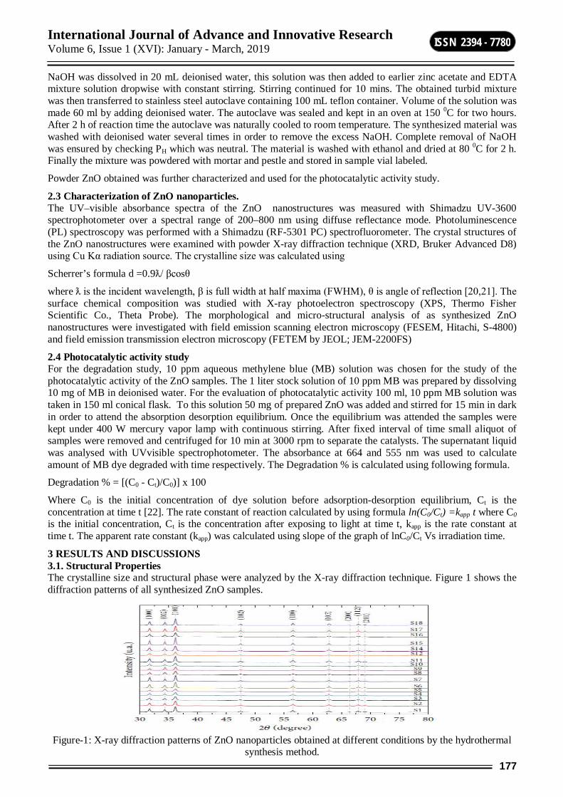
International Journal of Advance and Innovative Research Volume 6, Issue 1 (XVI): January - March, 2019
177
ISSN 2394 - 7780
NaOH was dissolved in 20 mL deionised water, this solution was then added to earlier zinc acetate and EDTA mixture solution dropwise with constant stirring. Stirring continued for 10 mins. The obtained turbid mixture was then transferred to stainless steel autoclave containing 100 mL teflon container. Volume of the solution was made 60 ml by adding deionised water. The autoclave was sealed and kept in an oven at 150 0C for two hours. After 2 h of reaction time the autoclave was naturally cooled to room temperature. The synthesized material was washed with deionised water several times in order to remove the excess NaOH. Complete removal of NaOH was ensured by checking PH which was neutral. The material is washed with ethanol and dried at 80 0C for 2 h. Finally the mixture was powdered with mortar and pestle and stored in sample vial labeled.
Powder ZnO obtained was further characterized and used for the photocatalytic activity study.
2.3 Characterization of ZnO nanoparticles. The UV–visible absorbance spectra of the ZnO nanostructures was measured with Shimadzu UV-3600 spectrophotometer over a spectral range of 200–800 nm using diffuse reflectance mode. Photoluminescence (PL) spectroscopy was performed with a Shimadzu (RF-5301 PC) spectrofluorometer. The crystal structures of the ZnO nanostructures were examined with powder X-ray diffraction technique (XRD, Bruker Advanced D8) using Cu Kα radiation source. The crystalline size was calculated using
Scherrer’s formula d =0.9ƛ/ βcosθ
where ƛ is the incident wavelength, β is full width at half maxima (FWHM), θ is angle of reflection [20,21]. The surface chemical composition was studied with X-ray photoelectron spectroscopy (XPS, Thermo Fisher Scientific Co., Theta Probe). The morphological and micro-structural analysis of as synthesized ZnO nanostructures were investigated with field emission scanning electron microscopy (FESEM, Hitachi, S-4800) and field emission transmission electron microscopy (FETEM by JEOL; JEM-2200FS)
2.4 Photocatalytic activity study For the degradation study, 10 ppm aqueous methylene blue (MB) solution was chosen for the study of the photocatalytic activity of the ZnO samples. The 1 liter stock solution of 10 ppm MB was prepared by dissolving 10 mg of MB in deionised water. For the evaluation of photocatalytic activity 100 ml, 10 ppm MB solution was taken in 150 ml conical flask. To this solution 50 mg of prepared ZnO was added and stirred for 15 min in dark in order to attend the absorption desorption equilibrium. Once the equilibrium was attended the samples were kept under 400 W mercury vapor lamp with continuous stirring. After fixed interval of time small aliquot of samples were removed and centrifuged for 10 min at 3000 rpm to separate the catalysts. The supernatant liquid was analysed with UVvisible spectrophotometer. The absorbance at 664 and 555 nm was used to calculate amount of MB dye degraded with time respectively. The Degradation % is calculated using following formula.
Degradation % = [(C0 - Ct)/C0)] x 100
Where C0 is the initial concentration of dye solution before adsorption-desorption equilibrium, Ct is the concentration at time t [22]. The rate constant of reaction calculated by using formula ln(C0/Ct) =kapp t where C0 is the initial concentration, Ct is the concentration after exposing to light at time t, kapp is the rate constant at time t. The apparent rate constant (kapp) was calculated using slope of the graph of lnC0/Ct Vs irradiation time.
3 RESULTS AND DISCUSSIONS 3.1. Structural Properties The crystalline size and structural phase were analyzed by the X-ray diffraction technique. Figure 1 shows the diffraction patterns of all synthesized ZnO samples.
Figure-1: X-ray diffraction patterns of ZnO nanoparticles obtained at different conditions by the hydrothermal
synthesis method.
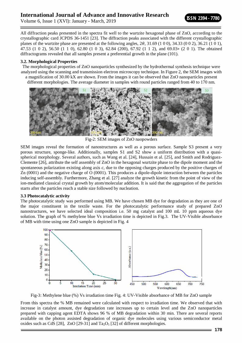
International Journal of Advance and Innovative Research Volume 6, Issue 1 (XVI): January - March, 2019
178
ISSN 2394 - 7780
All diffraction peaks presented in the spectra fit well to the wurtzite hexagonal phase of ZnO, according to the crystallographic card JCPDS 36-1451 [23]. The diffraction peaks associated with the different crystallographic planes of the wurtzite phase are presented at the following angles, 2휃, 31.69 (1 0 0), 34.33 (0 0 2), 36.21 (1 0 1), 47.53 (1 0 2), 56.50 (1 1 0), 62.80 (1 0 3), 62.84 (200), 67.92 (1 1 2), and 69.03∘ (2 0 1). The obtained diffractograms revealed that all samples present a preferential growth in the plane (101).
3.2. Morphological Properties The morphological properties of ZnO nanoparticles synthesized by the hydrothermal synthesis technique were
analyzed using the scanning and transmission electron microscopy technique. In Figure 2, the SEM images with a magnification of 30.00 kX are shown. From the images it can be observed that ZnO nanoparticles present different morphologies. The average diameter in samples with round particles ranged from 40 to 170 nm.
Fig-2: SEM images of ZnO naopowders
SEM images reveal the formation of nanostructures as well as a porous surface. Sample S3 present a very porous structure, sponge-like. Additionally, samples S1 and S2 show a uniform distribution with a quasi-spherical morphology. Several authors, such as Wang et al. [24], Hussain et al. [25], and Smith and Rodriguez-Clemente [26], attribute the self assembly of ZnO in the hexagonal wurtzite phase to the dipole moment and the spontaneous polarization existing along axis 푐, due to the opposing charges produced by the positive charges of Zn (0001) and the negative charge of O (0001). This produces a dipole-dipole interaction between the particles inducing self-assembly. Furthermore, Zhang et al. [27] analyze the growth kinetic from the point of view of the ion-mediated classical crystal growth by atom/molecular addition. It is said that the aggregation of the particles starts after the particles reach a stable size followed by nucleation.
3.3 Photocatalytic activity The photocatalytic study was performed using MB. We have chosen MB dye for degradation as they are one of the major constituent in the textile waste. For the photocatalytic performance study of prepared ZnO nanostructures, we have selected ideal composition i.e. 50 mg catalyst and 100 mL 10 ppm aqueous dye solution. The graph of % methylene blue Vs irradiation time is depicted in Fig.3. The UV-Visible absorbance of MB with time using one ZnO sample is depicted in Fig. 4
Fig-3: Methylene blue (%) Vs irradiation time Fig. 4: UV-Visible absorbance of MB for ZnO sample
From this spectra the % MB remained were calculated with respect to irradiation time. We observed that with increase in catalyst amount, dye degradation rate increases up to certain level and the ZnO nanoparticles prepared with capping agent EDTA shows 96 % of MB degradation within 30 min. There are several reports available on the photon assisted degradation of organic dye molecules using various semiconductor metal oxides such as CdS [28], ZnO [29-31] and Ta2O5 [32] of different morphologies.
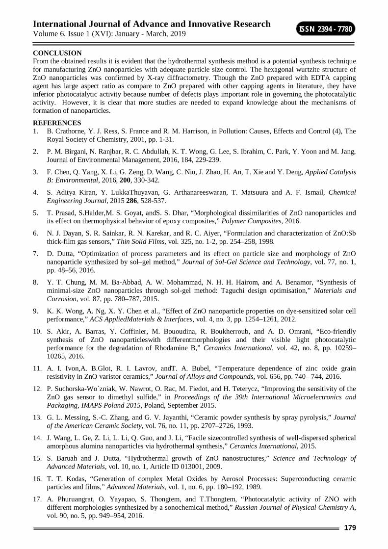
International Journal of Advance and Innovative Research Volume 6, Issue 1 (XVI): January - March, 2019
179
ISSN 2394 - 7780
CONCLUSION From the obtained results it is evident that the hydrothermal synthesis method is a potential synthesis technique for manufacturing ZnO nanoparticles with adequate particle size control. The hexagonal wurtzite structure of ZnO nanoparticles was confirmed by X-ray diffractometry. Though the ZnO prepared with EDTA capping agent has large aspect ratio as compare to ZnO prepared with other capping agents in literature, they have inferior photocatalytic activity because number of defects plays important role in governing the photocatalytic activity. However, it is clear that more studies are needed to expand knowledge about the mechanisms of formation of nanoparticles.
REFERENCES 1. B. Crathorne, Y. J. Ress, S. France and R. M. Harrison, in Pollution: Causes, Effects and Control (4), The
Royal Society of Chemistry, 2001, pp. 1-31.
2. P. M. Birgani, N. Ranjbar, R. C. Abdullah, K. T. Wong, G. Lee, S. Ibrahim, C. Park, Y. Yoon and M. Jang, Journal of Environmental Management, 2016, 184, 229-239.
3. F. Chen, Q. Yang, X. Li, G. Zeng, D. Wang, C. Niu, J. Zhao, H. An, T. Xie and Y. Deng, Applied Catalysis B: Environmental, 2016, 200, 330-342.
4. S. Aditya Kiran, Y. LukkaThuyavan, G. Arthanareeswaran, T. Matsuura and A. F. Ismail, Chemical Engineering Journal, 2015 286, 528-537.
5. T. Prasad, S.Halder,M. S. Goyat, andS. S. Dhar, “Morphological dissimilarities of ZnO nanoparticles and its effect on thermophysical behavior of epoxy composites,” Polymer Composites, 2016.
6. N. J. Dayan, S. R. Sainkar, R. N. Karekar, and R. C. Aiyer, “Formulation and characterization of ZnO:Sb thick-film gas sensors,” Thin Solid Films, vol. 325, no. 1-2, pp. 254–258, 1998.
7. D. Dutta, “Optimization of process parameters and its effect on particle size and morphology of ZnO nanoparticle synthesized by sol–gel method,” Journal of Sol-Gel Science and Technology, vol. 77, no. 1, pp. 48–56, 2016.
8. Y. T. Chung, M. M. Ba-Abbad, A. W. Mohammad, N. H. H. Hairom, and A. Benamor, “Synthesis of minimal-size ZnO nanoparticles through sol-gel method: Taguchi design optimisation,” Materials and Corrosion, vol. 87, pp. 780–787, 2015.
9. K. K. Wong, A. Ng, X. Y. Chen et al., “Effect of ZnO nanoparticle properties on dye-sensitized solar cell performance,” ACS AppliedMaterials & Interfaces, vol. 4, no. 3, pp. 1254–1261, 2012.
10. S. Akir, A. Barras, Y. Coffinier, M. Bououdina, R. Boukherroub, and A. D. Omrani, “Eco-friendly synthesis of ZnO nanoparticleswith differentmorphologies and their visible light photocatalytic performance for the degradation of Rhodamine B,” Ceramics International, vol. 42, no. 8, pp. 10259–10265, 2016.
11. A. I. Ivon,A. B.Glot, R. I. Lavrov, andT. A. Bubel, “Temperature dependence of zinc oxide grain resistivity in ZnO varistor ceramics,” Journal of Alloys and Compounds, vol. 656, pp. 740– 744, 2016.
12. P. Suchorska-Wo´zniak, W. Nawrot, O. Rac, M. Fiedot, and H. Teterycz, “Improving the sensitivity of the ZnO gas sensor to dimethyl sulfide,” in Proceedings of the 39th International Microelectronics and Packaging, IMAPS Poland 2015, Poland, September 2015.
13. G. L. Messing, S.-C. Zhang, and G. V. Jayanthi, “Ceramic powder synthesis by spray pyrolysis,” Journal of the American Ceramic Society, vol. 76, no. 11, pp. 2707–2726, 1993.
14. J. Wang, L. Ge, Z. Li, L. Li, Q. Guo, and J. Li, “Facile sizecontrolled synthesis of well-dispersed spherical amorphous alumina nanoparticles via hydrothermal synthesis,” Ceramics International, 2015.
15. S. Baruah and J. Dutta, “Hydrothermal growth of ZnO nanostructures,” Science and Technology of Advanced Materials, vol. 10, no. 1, Article ID 013001, 2009.
16. T. T. Kodas, “Generation of complex Metal Oxides by Aerosol Processes: Superconducting ceramic particles and films,” Advanced Materials, vol. 1, no. 6, pp. 180–192, 1989.
17. A. Phuruangrat, O. Yayapao, S. Thongtem, and T.Thongtem, “Photocatalytic activity of ZNO with different morphologies synthesized by a sonochemical method,” Russian Journal of Physical Chemistry A, vol. 90, no. 5, pp. 949–954, 2016.

International Journal of Advance and Innovative Research Volume 6, Issue 1 (XVI): January - March, 2019
180
ISSN 2394 - 7780
18. ¨ O. A. Yildirim and C. Durucan, “Synthesis of zinc oxide nanoparticles elaborated by microemulsion method,” Journal of Alloys and Compounds, vol. 506, no. 2, pp. 944–949, 2010.
19. L. Shen, N. Bao,K.Yanagisawa, K. Domen, A. Gupta, and C.A. Grimes, “Direct synthesis of ZnO nanoparticles by a solutionfree mechanochemical reaction,”Nanotechnology, vol. 17, no. 20, article no. 013, pp. 5117–5123, 2006.
20. S. S. Arbuj, R. R. Hawaldar, U. P. Mulik, B. N. Wani, D. P. Amalnerkar and S. B. Waghmode, Materials Science and Engineering: B, 2010, 168, 90-94.
21. M. S. Solmaz Aghdasi, Iranian Journal of Catalysis 2016, 6(5), 481-487
22. P. Sangpour, F. Hashemi and A. Z. Moshfegh, The Journal of Physical Chemistry C, 2010, 114, 13955-13961.
23. Z. Wen, L. Zhu, Z. Zhang, and Z. Ye, “Fabrication of gas sensor based onmesoporous rhombus-shaped ZnOrod arrays,” Sensors and Actuators B: Chemical, vol. 208, pp. 112–121, 2015.
24. J. Wang, Y. Qi, Z. Zhi, J. Guo, M. Li, and Y. Zhang, “A selfassemblymechanismfor sol-gel derived ZnO thin films,” Smart Materials and Structures, vol. 16, no. 6, pp. 2673–2679, 2007.
25. S. Hussain, T. Liu, M. Kashif et al., “Effects of reaction time on the morphological, structural, and gas sensing properties of ZnO nanostructures,” Materials Science in Semiconductor Processing, vol. 18, no. 1, pp. 52–58, 2014.
26. A. Smith and R. Rodriguez-Clemente, “Morphological differences in ZnO films deposited by the pyrosol technique: effect of HCl,” Thin Solid Films, vol. 345, no. 2, pp. 192–196, 1999.
27. Q. Zhang, S.-J. Liu, and S.-H. Yu, “Recent advances in oriented attachment growth and synthesis of functional materials: concept, evidence, mechanism, and future,” Journal of Materials Chemistry, vol. 19, no. 2, pp. 191–207, 2009.
28. S. K. Apte, S. N. Garaje, S. S. Arbuj, B. B. Kale, J. O. Baeg, U. P. Mulik, S. D. Naik, D. P. Amalnerkar and S. W. Gosavi, J. Mater. Chem. 2011, 21, 19241
29. S. Arbuj, N. Rumale, A. Pokle, J. D. Ambekar, S. B. Rane, U. P. Mulikand, and D. P. Amalnerkar, Sci. Adv. Mater. 6, 269 (2014).
30. K. R. Chandrasekhar, J. D. Ambekar, S. B. Rane, and S. S. Arbuj, J. Nanoeng. Nanomanuf. 5, 77 (2015).
31. P. S. Badgujar, S. S. Arbuj, J. M. Mali, S. B. Rane, and U. P. Mulik, J. Nanoeng. Nanomanuf. 4, 65 (2014).
32. S. P. Deshpande, M. S. Tamboli, S. S. Arbuj, U. P. Mulik, and D. P. Amalnerkar, J. Nanoeng. Nanomanuf. 4, 215 (2014).
International Journal of Advance and Innovative Research Volume 6, Issue 1 (XVI): January - March, 2019
181
ISSN 2394 - 7780
PECHMANN CONDENSATION CATALYSED BY OXALIX ACID UNDER MICRO WAVE IRRADIATION
Deepak R. Nagargoje Department of Chemistry, A. S. C. College, Mokhada Dist. Palghar
ABSTRACT An energy efficient protocol has been developed for coumarin synthesis using Pechmann condensation under Microwave irradiation. The reaction has been catalysed by oxalic acid without solvent. The present synthetic protocol offers a convenient, clean and fast alternative for synthesis of coumarin derivatives within short time.
Keywords: coumarins; pechmann condensation; oxalic acid; microwave irradiation
INTRODUCTION Coumarin derivatives continue to be investigated over the years due to their importance in organic and medicinal chemistry because of their biological activities[1]. Coumarin and its derivatives are associated with various biological activities viz. anti-inflammatory [2], anti-convulsant [3], anti-viral[4], anticoagulant[5], antioxidant[6], antibacterial[7], antifungal[8], anti-HIV[9], anti-carcinogenic material[10], and antihistamine. They are attracting attention of chemists as a large number of natural products contain this heterocyclic nucleus. Commercially, they are widely used as food additives, perfumes, cosmetics, pharmaceutical [12], optical brightenrs [13], in dispersed fluorescent and laser dyes[14].
Microwave induced organic reaction enhancement. (MORE) is a technique realized in 1986 played a pivotal role in synthetic organic chemistry. MWI offers selective, instantaneous heat energy to a material providing faster and safer reactions. Consequently, microwave irradiations often provide higher reaction yields in short time of span and simplify cumbersome purification processes. Applications of MORE chemistry finds useful in organic chemistry in alkylations, acylation, nucleophilic substitution, condensation, protection and deprotection, cycloaddition, oxidation andorganometallic reactions. In literature coumarin synthesis has been achieved by employing various methods like Von-Pechmann[15], Knoevenagel[16], Perkin[17], Reformatsy[18], and Wittig reaction[19]. In general, a valuable method for the synthesis of coumarin is the Von-Pechmann reaction. It is simple and straight reaction employing
ethyl acetoacetate and substituted phenol together with an acid catalyst. Literature prevalence suggested that acids like H2SO4 [19] TFA[20], etc. have been widely utilized for this conversion into coumarin synthesis. In addition, some other catalysts were also reported viz. P2O5 [21],AlCl3 [22],Ionic liquid[23],Sulfated zircona[24], Indiumhalides[25], and palladium[26], dipyridine copper chloride[27].
However, most of the reported synthetic protocols require high temperatures, prolonged reaction times, harsh reaction conditions, lower yield and the use of hazardous and often expensive acid catalysts. Another major drawback associated with reported procedures was that catalyst used in stoichiometric excess and recovery of catalyst was not possible during usual work-up processes.
Herein we report oxalic acid as a catalyst for coumarin synthesis under solvent free conditions using microwave irradiation. Oxalic acid has emerged as a promising acid catalyst during recent years and reported to catalyze some organic reactions[29-30] . The catalyst used is inert, inexpensive and environmentally benign allowing various reaction conditions to be employed.
EXPERIMENTAL SECTION All the melting points were determined in open capillaries in a paraffin bath IR spectra were recorded in KBr pellets on Shimadzu FT-IR 8300 spectrophotometer. H1NMR spectra were obtained in CDCl3 and DMSO-d6 on a Brucker DRX-400 at 400 MHz using TMS as an internal standard. The reaction was monitored by TLC on silica gel G254 plates supplied by Merck.
GENERAL PROCEDURE FOR THE SYNTHESIS OF COUMARINS A mixture of phenol (1 mmol) and ethyl acetoacetate (1 mmol) in presence of oxalic acid (20mol %) was irradiated in microwave oven at 180 watt and the reaction was monitored by TLC. After completion of reaction, the product was extracted in dichloromethane and the solvent was distilled off on rotary evaporator. The compound obtained was then recrystallised from ethanol. The cellulose sulphuric acid gave comparatively high yields in shorter reaction period of 0.5-3 min and the products are characterized by IR, 1H NMR, and Mass.
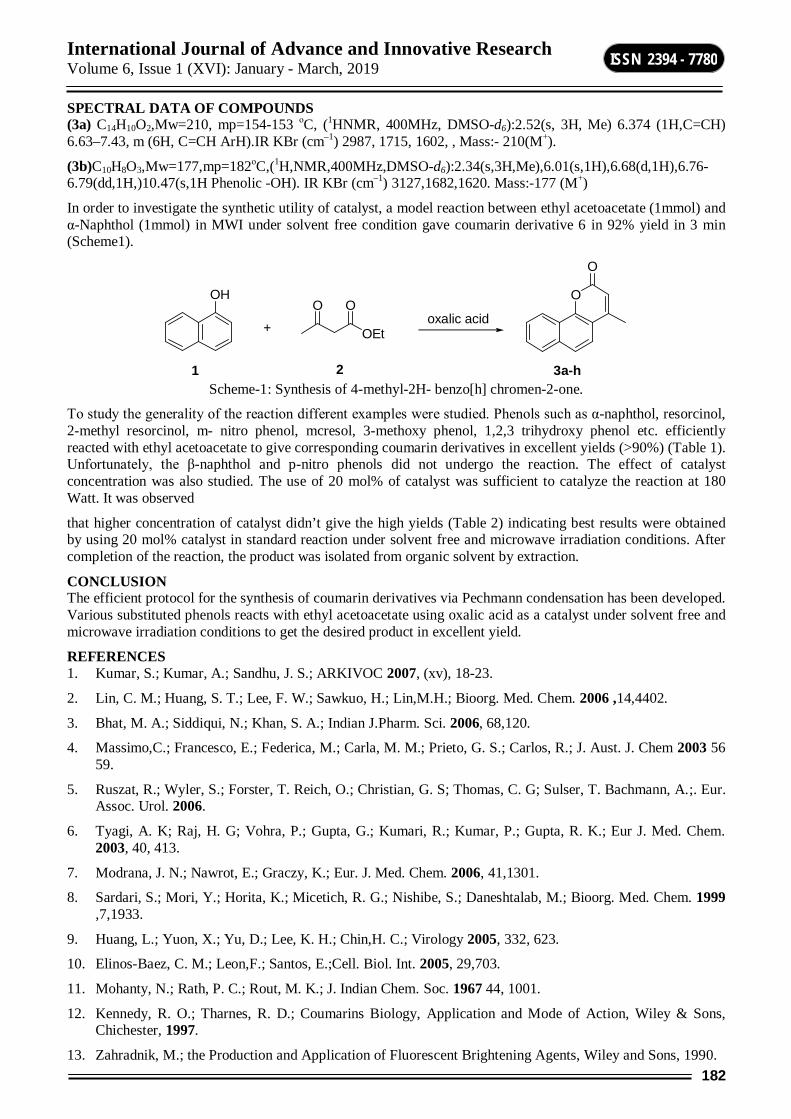
International Journal of Advance and Innovative Research Volume 6, Issue 1 (XVI): January - March, 2019
182
ISSN 2394 - 7780
SPECTRAL DATA OF COMPOUNDS (3a) C14H10O2,Mw=210, mp=154-153 οC, (1HNMR, 400MHz, DMSO-d6):2.52(s, 3H, Me) 6.374 (1H,C=CH) 6.63–7.43, m (6H, C=CH ArH).IR KBr (cm–1) 2987, 1715, 1602, , Mass:- 210(M+).
(3b)C10H8O3,Mw=177,mp=182οC,(1H,NMR,400MHz,DMSO-d6):2.34(s,3H,Me),6.01(s,1H),6.68(d,1H),6.76-6.79(dd,1H,)10.47(s,1H Phenolic -OH). IR KBr (cm–1) 3127,1682,1620. Mass:-177 (M+)
In order to investigate the synthetic utility of catalyst, a model reaction between ethyl acetoacetate (1mmol) and α-Naphthol (1mmol) in MWI under solvent free condition gave coumarin derivative 6 in 92% yield in 3 min (Scheme1).
OH
+ OEt
O OO
O
oxalic acid
1 2 3a-h Scheme-1: Synthesis of 4-methyl-2H- benzo[h] chromen-2-one.
To study the generality of the reaction different examples were studied. Phenols such as α-naphthol, resorcinol, 2-methyl resorcinol, m- nitro phenol, mcresol, 3-methoxy phenol, 1,2,3 trihydroxy phenol etc. efficiently reacted with ethyl acetoacetate to give corresponding coumarin derivatives in excellent yields (>90%) (Table 1). Unfortunately, the β-naphthol and p-nitro phenols did not undergo the reaction. The effect of catalyst concentration was also studied. The use of 20 mol% of catalyst was sufficient to catalyze the reaction at 180 Watt. It was observed
that higher concentration of catalyst didn’t give the high yields (Table 2) indicating best results were obtained by using 20 mol% catalyst in standard reaction under solvent free and microwave irradiation conditions. After completion of the reaction, the product was isolated from organic solvent by extraction.
CONCLUSION The efficient protocol for the synthesis of coumarin derivatives via Pechmann condensation has been developed. Various substituted phenols reacts with ethyl acetoacetate using oxalic acid as a catalyst under solvent free and microwave irradiation conditions to get the desired product in excellent yield.
REFERENCES 1. Kumar, S.; Kumar, A.; Sandhu, J. S.; ARKIVOC 2007, (xv), 18-23.
2. Lin, C. M.; Huang, S. T.; Lee, F. W.; Sawkuo, H.; Lin,M.H.; Bioorg. Med. Chem. 2006 ,14,4402.
3. Bhat, M. A.; Siddiqui, N.; Khan, S. A.; Indian J.Pharm. Sci. 2006, 68,120.
4. Massimo,C.; Francesco, E.; Federica, M.; Carla, M. M.; Prieto, G. S.; Carlos, R.; J. Aust. J. Chem 2003 56 59.
5. Ruszat, R.; Wyler, S.; Forster, T. Reich, O.; Christian, G. S; Thomas, C. G; Sulser, T. Bachmann, A.;. Eur. Assoc. Urol. 2006.
6. Tyagi, A. K; Raj, H. G; Vohra, P.; Gupta, G.; Kumari, R.; Kumar, P.; Gupta, R. K.; Eur J. Med. Chem. 2003, 40, 413.
7. Modrana, J. N.; Nawrot, E.; Graczy, K.; Eur. J. Med. Chem. 2006, 41,1301.
8. Sardari, S.; Mori, Y.; Horita, K.; Micetich, R. G.; Nishibe, S.; Daneshtalab, M.; Bioorg. Med. Chem. 1999 ,7,1933.
9. Huang, L.; Yuon, X.; Yu, D.; Lee, K. H.; Chin,H. C.; Virology 2005, 332, 623.
10. Elinos-Baez, C. M.; Leon,F.; Santos, E.;Cell. Biol. Int. 2005, 29,703.
11. Mohanty, N.; Rath, P. C.; Rout, M. K.; J. Indian Chem. Soc. 1967 44, 1001.
12. Kennedy, R. O.; Tharnes, R. D.; Coumarins Biology, Application and Mode of Action, Wiley & Sons, Chichester, 1997.
13. Zahradnik, M.; the Production and Application of Fluorescent Brightening Agents, Wiley and Sons, 1990.
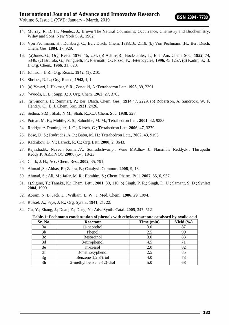
International Journal of Advance and Innovative Research Volume 6, Issue 1 (XVI): January - March, 2019
183
ISSN 2394 - 7780
14. Murray, R. D. H.; Mendez, J.; Brown The Natural Coumarins: Occurrence, Chemistry and Biochemistry, Wiley and Sons, New York S. A. 1982.
15. Von Pechmann, H.; Duisberg, C.; Ber. Dtsch. Chem. 1883,16, 2119. (b) Von Pechmann ,H.; Ber. Dtsch. Chem. Ges. 1884, 17, 929.
16. (a)Jones, G.; Org. React. 1976, 15, 204. (b) Adams,R.; Bockstahler, T.; E. J. Am. Chem. Soc., 1952, 74, 5346. (c) Brufola, G.; Fringuelli, F.; Piermatti, O.; Pizzo, F.; Heterocycles, 1996, 43 1257. (d) Kadin, S.; B. J. Org. Chem., 1966, 31, 620.
17. Johnson, J. R.; Org. React., 1942, (1): 210.
18. Shriner, R. L.; Org. React., 1942, 1, 1.
19. (a) Yavari, I. Hekmat, S.R.; Zonouki, A.;Tetrahedron Lett. 1998, 39, 2391.
20. [Woods, L. L.; Sapp, J.; J. Org. Chem. 1962, 27, 3703.
21. (a)Simonis, H; Remmert, P.; Ber. Dtsch. Chem. Ges., 1914,47, 2229. (b) Robertson, A. Sandrock, W. F. Hendry, C.; B. J. Chem. Soc. 1931, 2426.
22. Sethna, S.M.; Shah, N.M.; Shah, R.;.C.J. Chem. Soc. 1938, 228.
23. Potdar, M. K.; Mohile, S. S.; Salunkhe, M. M.; Tetrahedron Lett. 2001, 42, 9285.
24. Rodríguez-Domínguez, J. C.; Kirsch, G.; Tetrahedron Lett. 2006, 47, 3279.
25. Bose, D. S.; Rudradas ,A. P.; Babu, M. H.; Tetrahedron Lett., 2002, 43, 9195.
26. Kadnikov, D. V.; Larock, R. C.; Org. Lett. 2000, 2, 3643.
27. Rajintha,B.; Naveen Kumar,V.; Someshshwar,p.; Venu MAdhav J.: Narsimha Reddy,P.; Thirupathi Reddy,P; ARKIVOC 2007, (xv), 18-23.
28. Clark, J. H.; Acc. Chem. Res., 2002, 35, 791.
29. Ahmad ,S.; Abbas, R.; Zahra, B.; Catalysis Commun. 2008, 9, 13.
30. Ahmad, S.; Ali, M.; Jafar, M. R.; Ebrahim, S.; Chem. Pharm. Bull. 2007, 55, 6, 957.
31. a).Sigino, T.; Tanaka, K.; Chem. Lett., 2001, 30, 110. b) Singh, P. R.; Singh, D. U.; Samant, S. D.; Synlett 2004, 1909.
32. Abram, N. B; Jack, D.; William, L. W.; J. Med. Chem., 1986, 29, 1094.
33. Russel, A.; Frye, J. R.; Org. Synth., 1941, 21, 22.
34. Gu, Y.; Zhang, J.; Duan, Z.; Deng, Y.; Adv. Synth. Catal. 2005, 347, 512
Table-1: Pechmann condensation of phenols with ethylacetoacetate catalysed by oxalic acid Sr. No. Reactant Time (min) Yield (%)
3a -naphthol 3.0 87 3b Phenol 2.5 90 3c Resorcinol 3.0 83 3d 3-nirophenol 4.5 71 3e m-cresol 2.0 82 3f 3-methoxyphenol 2.5 85 3g Benzene-1,2,3-triol 4.0 73 3h 2-methyl benzene-1,3-diol 5.0 68
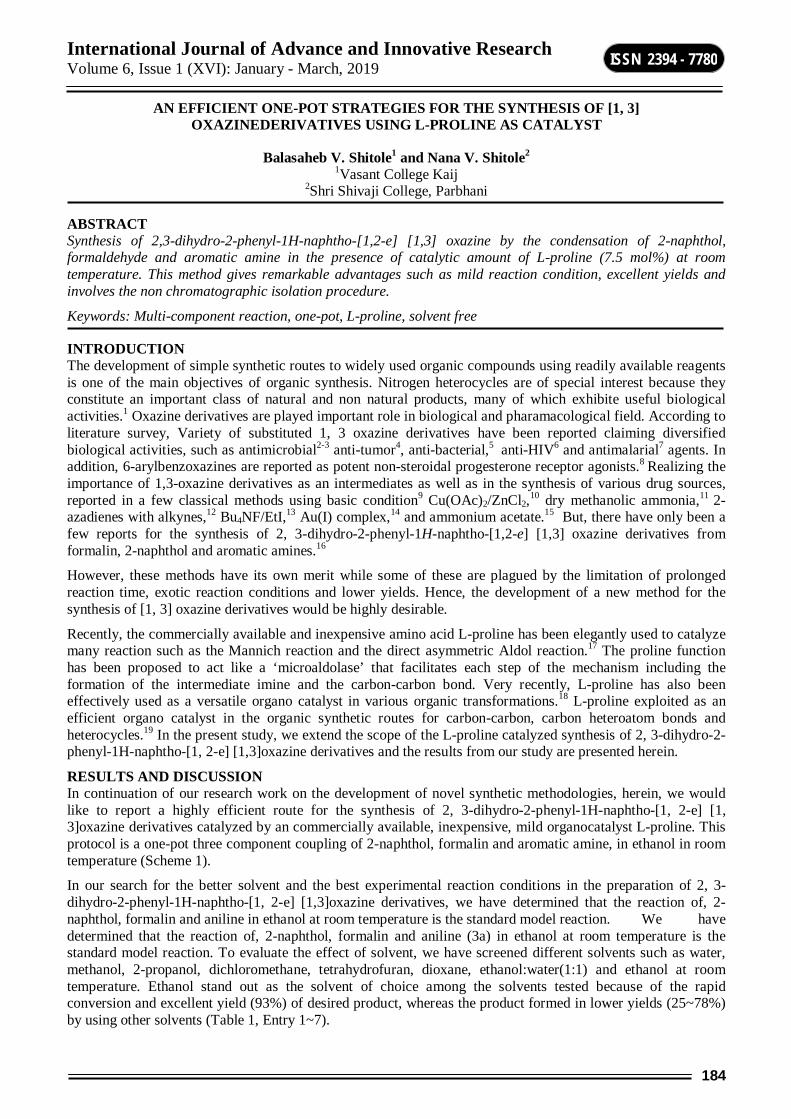
International Journal of Advance and Innovative Research Volume 6, Issue 1 (XVI): January - March, 2019
184
ISSN 2394 - 7780
AN EFFICIENT ONE-POT STRATEGIES FOR THE SYNTHESIS OF [1, 3] OXAZINEDERIVATIVES USING L-PROLINE AS CATALYST
Balasaheb V. Shitole1 and Nana V. Shitole2 1Vasant College Kaij
2Shri Shivaji College, Parbhani
ABSTRACT Synthesis of 2,3-dihydro-2-phenyl-1H-naphtho-[1,2-e] [1,3] oxazine by the condensation of 2-naphthol, formaldehyde and aromatic amine in the presence of catalytic amount of L-proline (7.5 mol%) at room temperature. This method gives remarkable advantages such as mild reaction condition, excellent yields and involves the non chromatographic isolation procedure.
Keywords: Multi-component reaction, one-pot, L-proline, solvent free
INTRODUCTION The development of simple synthetic routes to widely used organic compounds using readily available reagents is one of the main objectives of organic synthesis. Nitrogen heterocycles are of special interest because they constitute an important class of natural and non natural products, many of which exhibite useful biological activities.1 Oxazine derivatives are played important role in biological and pharamacological field. According to literature survey, Variety of substituted 1, 3 oxazine derivatives have been reported claiming diversified biological activities, such as antimicrobial2-3 anti-tumor4, anti-bacterial,5 anti-HIV6 and antimalarial7 agents. In addition, 6-arylbenzoxazines are reported as potent non-steroidal progesterone receptor agonists.8 Realizing the importance of 1,3-oxazine derivatives as an intermediates as well as in the synthesis of various drug sources, reported in a few classical methods using basic condition9 Cu(OAc)2/ZnCl2,10 dry methanolic ammonia,11 2-azadienes with alkynes,12 Bu4NF/EtI,13 Au(I) complex,14 and ammonium acetate.15 But, there have only been a few reports for the synthesis of 2, 3-dihydro-2-phenyl-1H-naphtho-[1,2-e] [1,3] oxazine derivatives from formalin, 2-naphthol and aromatic amines.16
However, these methods have its own merit while some of these are plagued by the limitation of prolonged reaction time, exotic reaction conditions and lower yields. Hence, the development of a new method for the synthesis of [1, 3] oxazine derivatives would be highly desirable.
Recently, the commercially available and inexpensive amino acid L-proline has been elegantly used to catalyze many reaction such as the Mannich reaction and the direct asymmetric Aldol reaction.17 The proline function has been proposed to act like a ‘microaldolase’ that facilitates each step of the mechanism including the formation of the intermediate imine and the carbon-carbon bond. Very recently, L-proline has also been effectively used as a versatile organo catalyst in various organic transformations.18 L-proline exploited as an efficient organo catalyst in the organic synthetic routes for carbon-carbon, carbon heteroatom bonds and heterocycles.19 In the present study, we extend the scope of the L-proline catalyzed synthesis of 2, 3-dihydro-2-phenyl-1H-naphtho-[1, 2-e] [1,3]oxazine derivatives and the results from our study are presented herein.
RESULTS AND DISCUSSION In continuation of our research work on the development of novel synthetic methodologies, herein, we would like to report a highly efficient route for the synthesis of 2, 3-dihydro-2-phenyl-1H-naphtho-[1, 2-e] [1, 3]oxazine derivatives catalyzed by an commercially available, inexpensive, mild organocatalyst L-proline. This protocol is a one-pot three component coupling of 2-naphthol, formalin and aromatic amine, in ethanol in room temperature (Scheme 1).
In our search for the better solvent and the best experimental reaction conditions in the preparation of 2, 3-dihydro-2-phenyl-1H-naphtho-[1, 2-e] [1,3]oxazine derivatives, we have determined that the reaction of, 2-naphthol, formalin and aniline in ethanol at room temperature is the standard model reaction. We have determined that the reaction of, 2-naphthol, formalin and aniline (3a) in ethanol at room temperature is the standard model reaction. To evaluate the effect of solvent, we have screened different solvents such as water, methanol, 2-propanol, dichloromethane, tetrahydrofuran, dioxane, ethanol:water(1:1) and ethanol at room temperature. Ethanol stand out as the solvent of choice among the solvents tested because of the rapid conversion and excellent yield (93%) of desired product, whereas the product formed in lower yields (25~78%) by using other solvents (Table 1, Entry 1~7).
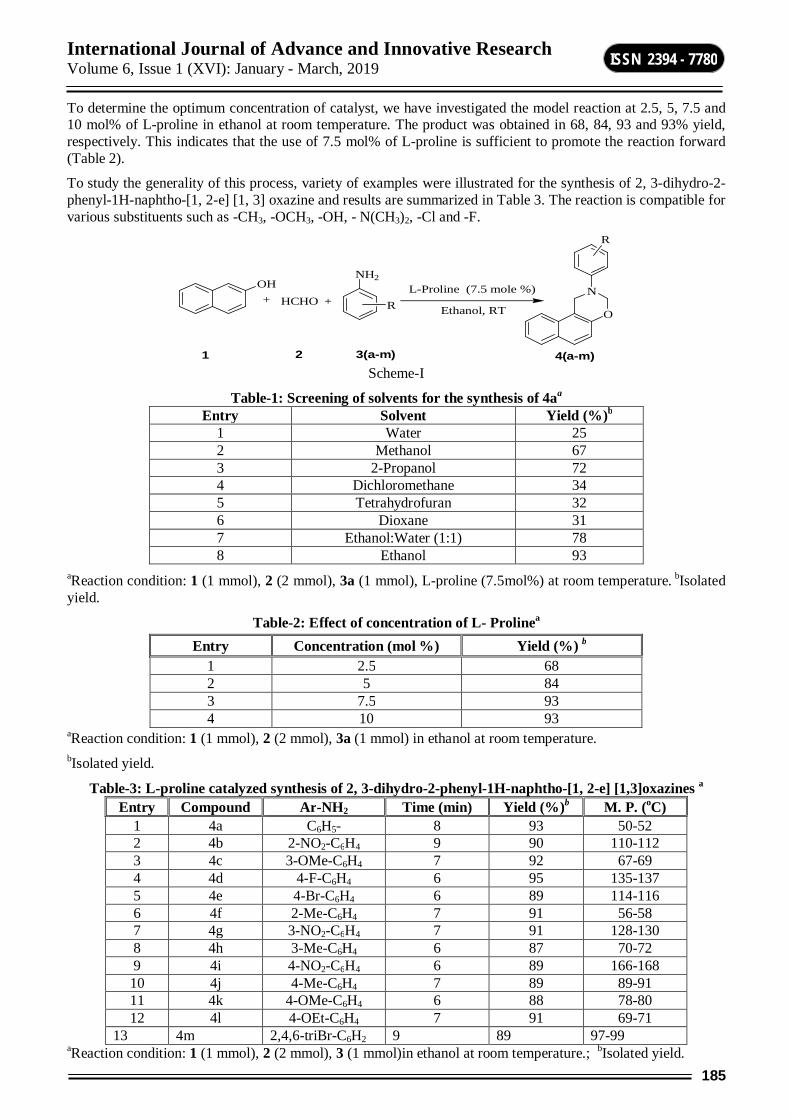
International Journal of Advance and Innovative Research Volume 6, Issue 1 (XVI): January - March, 2019
185
ISSN 2394 - 7780
To determine the optimum concentration of catalyst, we have investigated the model reaction at 2.5, 5, 7.5 and 10 mol% of L-proline in ethanol at room temperature. The product was obtained in 68, 84, 93 and 93% yield, respectively. This indicates that the use of 7.5 mol% of L-proline is sufficient to promote the reaction forward (Table 2).
To study the generality of this process, variety of examples were illustrated for the synthesis of 2, 3-dihydro-2-phenyl-1H-naphtho-[1, 2-e] [1, 3] oxazine and results are summarized in Table 3. The reaction is compatible for various substituents such as -CH3, -OCH3, -OH, - N(CH3)2, -Cl and -F.
+ +OH
HCHO
NH2
RO
N
R
Ethanol, RT
L-Proline (7.5 mole %)
1 2 3(a-m) 4(a-m) Scheme-I
Table-1: Screening of solvents for the synthesis of 4aa
Entry Solvent Yield (%)b 1 Water 25 2 Methanol 67 3 2-Propanol 72 4 Dichloromethane 34 5 Tetrahydrofuran 32 6 Dioxane 31 7 Ethanol:Water (1:1) 78 8 Ethanol 93
aReaction condition: 1 (1 mmol), 2 (2 mmol), 3a (1 mmol), L-proline (7.5mol%) at room temperature. bIsolated yield.
Table-2: Effect of concentration of L- Prolinea
aReaction condition: 1 (1 mmol), 2 (2 mmol), 3a (1 mmol) in ethanol at room temperature. bIsolated yield.
Table-3: L-proline catalyzed synthesis of 2, 3-dihydro-2-phenyl-1H-naphtho-[1, 2-e] [1,3]oxazines a Entry Compound Ar-NH2 Time (min) Yield (%)b M. P. (oC)
1 4a C6H5- 8 93 50-52 2 4b 2-NO2-C6H4 9 90 110-112 3 4c 3-OMe-C6H4 7 92 67-69 4 4d 4-F-C6H4 6 95 135-137 5 4e 4-Br-C6H4 6 89 114-116 6 4f 2-Me-C6H4 7 91 56-58 7 4g 3-NO2-C6H4 7 91 128-130 8 4h 3-Me-C6H4 6 87 70-72 9 4i 4-NO2-C6H4 6 89 166-168 10 4j 4-Me-C6H4 7 89 89-91 11 4k 4-OMe-C6H4 6 88 78-80 12 4l 4-OEt-C6H4 7 91 69-71
13 4m 2,4,6-triBr-C6H2 9 89 97-99 aReaction condition: 1 (1 mmol), 2 (2 mmol), 3 (1 mmol)in ethanol at room temperature.; bIsolated yield.
Entry Concentration (mol %) Yield (%) b 1 2.5 68 2 5 84 3 7.5 93 4 10 93

International Journal of Advance and Innovative Research Volume 6, Issue 1 (XVI): January - March, 2019
186
ISSN 2394 - 7780
General procedure for the synthesis 2, 3-dihydro-2-phenyl-1H-naphtho-[1, 2-e] [1, 3]oxazines derivatives A mixture of 2-naphthol (1 mmol) formalin (2 mmol), aromatic amine (1 mmol),) and L-proline (7.5mol%) as catalyst in ethanol at room temperature was stirred for specified time period as shown in Table 3. The progress of reaction was monitored on TLC. After completion of the reaction, mixture was cooled to room temperature and poured on crushed ice. Thus, solid obtained was filtered, dried and Purified by crystallization in ethanol.
CONCLUSION In conclusion, we have developed efficient and environmentally benign methodology for the synthesis of 2, 3-dihydro-2-phenyl-1H-naphtho-[1, 2-e] [1,3]oxazines derivatives by a one-pot, Multicomponent reaction from cyclocondensation of 2-naphthol, formalin and aromatic amines and under stirring in ethanol at room temperature in the presence of L-proline. The advantages of this method over other existing methods are reduced reaction times, higher yields, mild reaction condition, easy purification and economic viability of the catalyst. We feel that this economically viable procedure will find practical utility for the one pot synthesis of novel [1,3]oxazines derivatives.
ACKNOWLEDGMENTS We are grateful to the Head Department of Chemistry and Principal Shri Shivaji College Parbhani-431401 (MS), India for providing the laboratory facility.
REFERENCES 1 Zhichao, J.; Feng Y.; Xiao, W.; Huicai, H.; Xiaoyan, L.; Xinmiao, L.; Jinxing, Y. Org. Biomol. Chem.
2011, 9, 1809. 2 Mathew, B. P.; Kumar, A.; Sharma, S.; Shukla, P. K.; Nath, M. Eur. J. Med Chem., 2010, 45, 1502. 3 Fringuelli, R.; Pietrella, D.; Schiaffella, F.; Guarraci, A.; Perito, S.; Bistoni, F.; Vecchiarilli, A. Bioorg.
Med. Chem. 2002, 10, 1681. 4 (a) Kuehne, M. E.; Konopke, E. A. J. Med. Chem. 1962, 5 , 257; (b) Chylinska, J. B. ; Urbanski, T. J. Med.
Chem. 1963, 6 , 484; (c). Hsu, L.Y.; Lin, C. H. Heterocycles, 1996, 43, 2687. 5 (a) Chylinska, J. B.; Janowiec, M.; Urbanski, T. B. J. Pharmacol. 1971, 43, 649; (b) Latif, N.; Mishriky,
N.; Massad, F. Aust. J. Chem. 1982, 35, 1037. 6 (a) Pedersen, O. S.; Pedersen, E. B. Synthesis, 2000, 479; (b) Cocuzza, A. J.; Chidester, D. R.; Cordova, B.
C.; Jeffrey, S.; Parsons, R. L.; Bacheler, L. T.; Erickson,V. S.; Trainor, G. L.; Ko, S. S. Bioorg. Med. Chem. Lett. 2001, 11, 1177.
7 Duffin, W. M.; Rollo, I. M. J. Pharmacol. 1957, 12, 171. 8 Zhang, P.; Terefenko, E. A.; Fensome, A.; Zhang, Z. Y. ; Zhu, J.; Cohen, R.; Winneker,J.; Wrobel, J.;
Yardley, Bioorg. Med. Chem. Lett. 2002, 12, 787. 9 Tovar, F.; Ochoa, G. Org. Lett. 2000, 2, 965. 10 Lee, J.; Lee, K.; Kim, H. Bull. Korean Chem. Soc. 1996, 17, 115. 11 Szatmari, I.; Martinek, T. A.; Lazar, L.; Fulop, F. Eur. J.Org. Chem. 2004, 2231. 12 Palacios, F.; Herran, E.; Rubiales, G.; Ezpeleta, M. J. J. Org. Chem. 2002, 67, 2131. 13 Murai, T.; Sano, H.; Kawai, H.; Aso, H.; Shibahara, F. J. Org. Chem. 2005, 70, 8148. 14 Kang, J. E.; Kim, H. B.; Lee, J. W.; Shin, S. Org. Lett. 2006, 8, 3537. 15 Sapkal, S. B.; Shelke, K. F.; Kategaonkar, A. H.; Shingate, B. B.; Shingare, M. S. Green Chem. Lett. Rev.
2009, 2, 57. 16 (a) Burke, W. J.; Murdoc, K. C.; Ec, G. J. Am. Chem. Soc. 1954,76, 1677. 17 (a) Notz, W.; Tanaka, F.; Barbas, C. F., III. Acc. Chem. Res. 2004, 37, 580; (b) Dalko, P. I.; Moisan, L.
Angew. Chem, Int. Ed. 2004, 43, 5138; (c) Lacoste, E. Synlett. 2006, 12, 1973. 18 (a)Wang, Y.; Shang, Z. C.; Wu, T. X.; Fan, J. C.; Chen, X. J. Mol. Catal. A: Chem. 2006, 253, 212; (b)
Srinivasan, M.; Perumal, S.; Selvaraj, S. Arkivoc 2005, xi, 201; (c) Sabitha, G.; Fatima, N.; Reddy, E. V.; Yadav, J. S. Adv. Synth. Catal. 2005, 347, 1353; (d) Dodda, R.; Zhao, C. G. Synthesis, 2006, 19, 3238.
19 (a) Varala, R.; Ramu, E.; Sreelatha, N.; Adapa, S. R. Tetrahedron Lett. 2006, 476, 877; (b) Varala, R.; Adapa, S. R. Org. Proc. Res. Dev. 2005, 9, 853; (b) An, Z.; Zhang, W.: Shi, H.; He, J. Journal of Catalysis 2006, 241, 319-327; (c) Karade, N. N.; Budhewar, V. H.; Shinde, S. V.; Jadhav, W. N. Lett. in Org. Chem. 2007, 4.
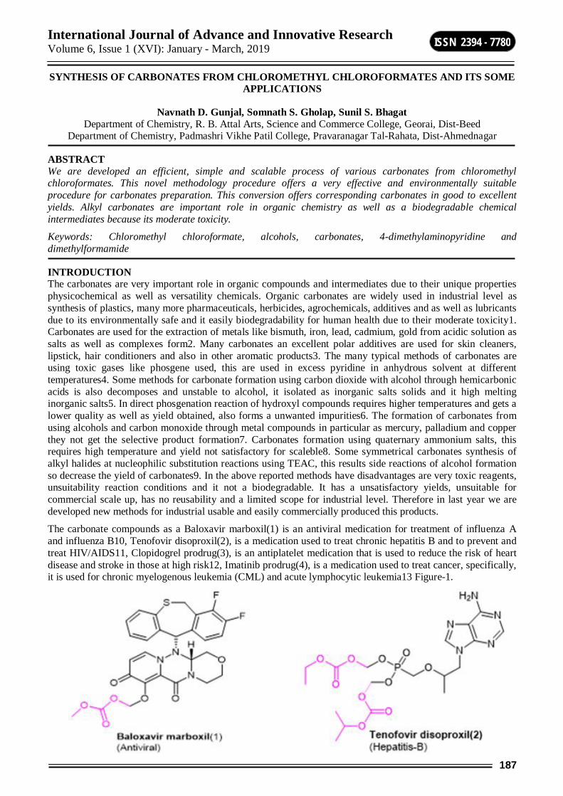
International Journal of Advance and Innovative Research Volume 6, Issue 1 (XVI): January - March, 2019
187
ISSN 2394 - 7780
SYNTHESIS OF CARBONATES FROM CHLOROMETHYL CHLOROFORMATES AND ITS SOME APPLICATIONS
Navnath D. Gunjal, Somnath S. Gholap, Sunil S. Bhagat Department of Chemistry, R. B. Attal Arts, Science and Commerce College, Georai, Dist-Beed
Department of Chemistry, Padmashri Vikhe Patil College, Pravaranagar Tal-Rahata, Dist-Ahmednagar
ABSTRACT We are developed an efficient, simple and scalable process of various carbonates from chloromethyl chloroformates. This novel methodology procedure offers a very effective and environmentally suitable procedure for carbonates preparation. This conversion offers corresponding carbonates in good to excellent yields. Alkyl carbonates are important role in organic chemistry as well as a biodegradable chemical intermediates because its moderate toxicity.
Keywords: Chloromethyl chloroformate, alcohols, carbonates, 4-dimethylaminopyridine and dimethylformamide
INTRODUCTION The carbonates are very important role in organic compounds and intermediates due to their unique properties physicochemical as well as versatility chemicals. Organic carbonates are widely used in industrial level as synthesis of plastics, many more pharmaceuticals, herbicides, agrochemicals, additives and as well as lubricants due to its environmentally safe and it easily biodegradability for human health due to their moderate toxicity1. Carbonates are used for the extraction of metals like bismuth, iron, lead, cadmium, gold from acidic solution as salts as well as complexes form2. Many carbonates an excellent polar additives are used for skin cleaners, lipstick, hair conditioners and also in other aromatic products3. The many typical methods of carbonates are using toxic gases like phosgene used, this are used in excess pyridine in anhydrous solvent at different temperatures4. Some methods for carbonate formation using carbon dioxide with alcohol through hemicarbonic acids is also decomposes and unstable to alcohol, it isolated as inorganic salts solids and it high melting inorganic salts5. In direct phosgenation reaction of hydroxyl compounds requires higher temperatures and gets a lower quality as well as yield obtained, also forms a unwanted impurities6. The formation of carbonates from using alcohols and carbon monoxide through metal compounds in particular as mercury, palladium and copper they not get the selective product formation7. Carbonates formation using quaternary ammonium salts, this requires high temperature and yield not satisfactory for scaleble8. Some symmetrical carbonates synthesis of alkyl halides at nucleophilic substitution reactions using TEAC, this results side reactions of alcohol formation so decrease the yield of carbonates9. In the above reported methods have disadvantages are very toxic reagents, unsuitability reaction conditions and it not a biodegradable. It has a unsatisfactory yields, unsuitable for commercial scale up, has no reusability and a limited scope for industrial level. Therefore in last year we are developed new methods for industrial usable and easily commercially produced this products.
The carbonate compounds as a Baloxavir marboxil(1) is an antiviral medication for treatment of influenza A and influenza B10, Tenofovir disoproxil(2), is a medication used to treat chronic hepatitis B and to prevent and treat HIV/AIDS11, Clopidogrel prodrug(3), is an antiplatelet medication that is used to reduce the risk of heart disease and stroke in those at high risk12, Imatinib prodrug(4), is a medication used to treat cancer, specifically, it is used for chronic myelogenous leukemia (CML) and acute lymphocytic leukemia13 Figure-1.
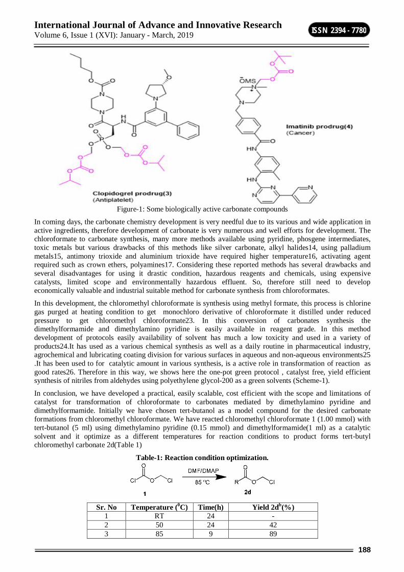
International Journal of Advance and Innovative Research Volume 6, Issue 1 (XVI): January - March, 2019
188
ISSN 2394 - 7780
Figure-1: Some biologically active carbonate compounds
In coming days, the carbonate chemistry development is very needful due to its various and wide application in active ingredients, therefore development of carbonate is very numerous and well efforts for development. The chloroformate to carbonate synthesis, many more methods available using pyridine, phosgene intermediates, toxic metals but various drawbacks of this methods like silver carbonate, alkyl halides14, using palladium metals15, antimony trioxide and aluminium trioxide have required higher temperature16, activating agent required such as crown ethers, polyamines17. Considering these reported methods has several drawbacks and several disadvantages for using it drastic condition, hazardous reagents and chemicals, using expensive catalysts, limited scope and environmentally hazardous effluent. So, therefore still need to develop economically valuable and industrial suitable method for carbonate synthesis from chloroformates.
In this development, the chloromethyl chloroformate is synthesis using methyl formate, this process is chlorine gas purged at heating condition to get monochloro derivative of chloroformate it distilled under reduced pressure to get chloromethyl chloroformate23. In this conversion of carbonates synthesis the dimethylformamide and dimethylamino pyridine is easily available in reagent grade. In this method development of protocols easily availability of solvent has much a low toxicity and used in a variety of products24.It has used as a various chemical synthesis as well as a daily routine in pharmaceutical industry, agrochemical and lubricating coating division for various surfaces in aqueous and non-aqueous environments25 .It has been used to for catalytic amount in various synthesis, is a active role in transformation of reaction as good rates26. Therefore in this way, we shows here the one-pot green protocol , catalyst free, yield efficient synthesis of nitriles from aldehydes using polyethylene glycol-200 as a green solvents (Scheme-1).
In conclusion, we have developed a practical, easily scalable, cost efficient with the scope and limitations of catalyst for transformation of chloroformate to carbonates mediated by dimethylamino pyridine and dimethylformamide. Initially we have chosen tert-butanol as a model compound for the desired carbonate formations from chloromethyl chloroformate. We have reacted chloromethyl chloroformate 1 (1.00 mmol) with tert-butanol (5 ml) using dimethylamino pyridine (0.15 mmol) and dimethylformamide(1 ml) as a catalytic solvent and it optimize as a different temperatures for reaction conditions to product forms tert-butyl chloromethyl carbonate 2d(Table 1)
Table-1: Reaction condition optimization.
Sr. No Temperature (0C) Time(h) Yield 2db(%)
1 RT 24 - 2 50 24 42 3 85 9 89
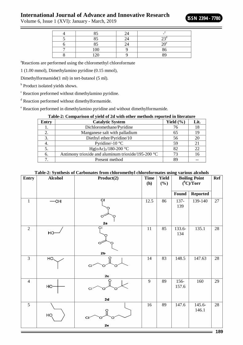
International Journal of Advance and Innovative Research Volume 6, Issue 1 (XVI): January - March, 2019
189
ISSN 2394 - 7780
4 85 24 -c
5 85 24 23d
6 85 24 20e 7 100 9 86 8 120 9 89
aReactions are performed using the chloromethyl chloroformate
1 (1.00 mmol), Dimethylamino pyridine (0.15 mmol),
Dimethylformamide(1 ml) in tert-butanol (5 ml). b Product isolated yields shows. c Reaction preformed without dimethylamino pyridine. d Reaction performed without dimethylformamide. e Reaction performed in dimethylamino pyridine and without dimethylformamide.
Table-2: Comparison of yield of 2d with other methods reported in literature Entry Catalytic System Yield (%) Lit.
1. Dichloromethane/Pyridine 76 18 2. Manganese salt with palladium 65 19 3. Diethyl ether/Pyridine/10 56 20 4. Pyridine/-10 °C 59 21 5. Hg(oAc)2/180-200 °C 82 22 6. Antimony trioxide and aluminum trioxide/195-200 °C 73 16 7. Present method 89 --
Table-2: Synthesis of Carbonates from chloromethyl chloroformates using various alcohols Entry Alcohol Product(2) Time
(h) Yield (%)
Boiling Point (0C)/Torr
Ref
Found Reported 1
12.5 86 137-139
139-140 27
2
11 85 133.6-134
135.1 28
3
14 83 148.5 147.63 28
4
9 89 156-157.6
160 29
5
HO
16 89 147.6 145.6-146.1
28
International Journal of Advance and Innovative Research Volume 6, Issue 1 (XVI): January - March, 2019
190
ISSN 2394 - 7780
6
16 85 163.1-164.8
165 29
7
17 82 165.3 160.5 29
8
14 65 123.1 128.2-128.7
29
9
20 70 166 - 29
10
HO
Br
19 67 136.6 - 30
11
15 88 173-174.6
174.3-175.1
30
12
15 86 186.5 - 30
aReactions are performed using the chloromethyl chloroformate 1 (1.00 mmol), Dimethylamino pyridine (0.15 mmol), dimethylformamide(1ml) in alcohols (5 ml). bProduct isolated yields shows.
At room temperature product 2d was not formed, chloromethyl chloroformate unreacted as such as shown on Table 1.The reaction was further probed by increasing the temperature at 85°C, the reaction was proceeds smoothly and tert-butyl chloromethyl carbonate 2d was obtained 89 % yield (Table 1,Sr.No.3). Therefore it is observed that the good yield of carbonate was obtained in presence of dimethylamino pyridine and dimethylformamide at 850C (Table 1,Sr.No.3). There is more improvement on reaction time and reaction temperature, but product yield not significant improve as per our expectations (Table 1, Sr.No.7 and Sr.No.8), so 850C was chosen the reaction temperature. When we performed reaction without dimethylamino pyridine no product was formed (Table 1,Sr.No.4). We performed reaction without dimethylformamide, the very less product was formed (Table 1,Sr. No.5). It is also important to mention that when the reaction was performed using dimethylamino pyridine and out dimethylformamide, the 2d product was formed very less (Table 1,Sr.No.6). In an developed reaction condition, chloromethyl chloroformate 1(1.00 mmol), dimethylamino pyridine (0.15 mmol) and dimethylformamide (1 ml) was added in tert-butanol (4 ml) and stir at 850C for 9 h to obtained tert-butyl chloromethyl carbonate 2d in 89 % yield (Table 1,Sr.No.3).
We have optimized that the one-pot transformation of chloromethyl chloroformate to corresponding carbonate using alcohol was maintained to the same extent with structurally diverse alcohols (Table 2). The carbonate formation reaction was almost complete in less than nine hours for all substrates tested by TLC, boiling point were recorded using open capillary and is uncorrected. Carbonates were isolated in good to excellent yields as described in Table 2. The interaction of chloromethyl chloroformate in to dimethylamino pyridine and
International Journal of Advance and Innovative Research Volume 6, Issue 1 (XVI): January - March, 2019
191
ISSN 2394 - 7780
dimethylformamide to forms C-N- insitu weak bond. Then the C-N- bond easily cleavage through alcohol nucleophilic substitution addition to forms our desired carbonate. Then subsequent expulsion of carbonate to regenerates dimethylamino pyridine and is acting as a catalyst. These reaction process is mechanistically related to oxidation of chloroformate to carbonates. After the reaction condition optimization, a study regarding the reuse of dimethylamino pyridine was not performed due to after reaction completion the reaction mass quenched in water and extracted to dichloromethane. The dimethylamino pyridine was soluble in water and its isolation is shows31 from water.
In day to day life environment pollution is a serious concern to reduced effluent to reduce the pollution, has been an increasing interests to the design of degradable catalyst reactions, a absence of hazardous solvents, low cost, recyclable and environmentally friendly solvents due to reduced effluent to reduce pollution. In mostly all the chemical and pharmaceutical industries observed that the catalysts and hazardous solvents are not always eco-friendly or biodegradable. In this way our development method is superior and promoting environment friendly method due to our method effluent is biodegradable and it minimizes COD and BOD. The dimethylamino pyridine and dimethylformamide has a different combination of chemical and physical properties such as a polarity, no flammability, high boiling point, low toxicity and it easily availability so it promoted to use as good solvent in organic solvents. Therefore we developed here the efficient, one-pot synthesis of carbonates from chloromethyl chloroformate using alcohol as a solvent. The main aspect of dimethylamino pyridine and dimethylformamide in this reaction was established by the fact that in the absence of dimethylamino pyridine and dimethylformamide the formation of carbonates from chloroformates does not takes place. Therefore it conclude that the dimethylamino pyridine and dimethylformamide is an essential component for reaction. In addition optimization, we have performed reaction using tertiary nitrogen base like triethyl amine to gives the less conversion obtained, therefore the role of dimethylamino pyridine and dimethylformamide in this transformation is required.
In conclusion, we have developed here a practical and cost efficient one-pot protocol for the transformation of chloromethyl chloroformate to corresponding carbonates by using corresponding alcohols in presence of dimethylamino pyridine and dimethylformamide as catalyst. These method advantages are wide scope of transformations, it can readily applied to big scale processes at plant level with excellent yield, cost efficient, selectivity, biodegradable effluent for environment secure, and convenient process for preparation of desired product.
ACKNOWLEDGMENTS The authors are thankful to the DST, New Delhi for financial support and we thanks to Atul Ltd for providing chemicals.
REFERENCES 1. Frevel L. K.; Gilpin J. A, Mich M.; (The Dow Chemical Co.) U.S. Pat. 3,642,868, 1972; Chem. Abstr.
1972, 77, 62818. 2. Masakatsu N.; Yoshuki T.; (Mitsui Toatsu Chemicals) Jap. Pat. 05,77,553, 1993; Chem. Abstr. 1993, 119,
59853. 3. Houben Weyl.; Methoden der Organischen Chemie ; Georg Thieme Verlag: Stuttgart, 1983; Vol. E4, p 64. 4. Chemische Fabrik von Heyden.; Ger. Pat. 109,933, 1900 Friedl, 1901, 5. 5. Houben Weyl.; Methoden der Organischen Chemie; Georg Thieme Verlag: Stuttgart, 1983; Vol. E4, p 64. 6. Nguyen, M. T.; Ha, T. K. J.; Am. Chem. Soc. 1984, 106, 599. 7. Romano U.; Tesei, R.; Massi, M. M.; Rebora, P.; Ind. Eng. Chem. Prod. Res. Dev. 1980, 19, 396; Romano,
U. S. Chim.Ind. 1993, 75. 8. (a)Rokicki, G.; Kuran, W. Monatsh. Chem soc.. 1984, 115, 205. (b) Rokicki, G.; Kuran, W. Bull. Chem.
Soc. Jpn. 1984, 57, 1662. 9. Mucciante, V.; Rossi, L.; Feroci, M.; Sotgiu, G. Synth. Commun. 2002, 32, 1205. 10. Hayden F.G, et al.; Engl J Med 2018; 379:913–23. 11. Wei, Kaiju et al.; Faming Zhuanli Shenqing, 102399149, 04 Apr 2012. 12. Caroff, Eva et al.; Journal of Medicinal Chemistry, 58(23), 9133-9153; 2015.
13. Yang FC, Ingram DA, Chen S, Zhu Y, Yuan J, Li X, Yang X, Knowles S; Cell. 135 (3): 437–48.
International Journal of Advance and Innovative Research Volume 6, Issue 1 (XVI): January - March, 2019
192
ISSN 2394 - 7780
14. Beilstein Handbuch der Organischen Chemie; Springer-Verlag:Berlin, 1921; Vol. III, pp 3, 5.
15. Hallgren, J. E.; Lucas, G. M.; Matthews, R. O. J. Organomet. Chem. 1981, 204, 135.
16. Ball, P.; Fullmann, H.; Heitz, W. Angew. Chem., Int. Ed. Engl.1980, 19 (9), 718.
17. Rokicki, G.; Pawlicki, J.; Kuran, W. Polymer J. 1982, 14 (11), 839.
18. Chemische Fabrik von Heyden .;Ger. Pat. 116,386 1900 Friedl, 1904, 6, 1160.
19. Romano, U.; Tesei, R.; Massi, M. M.; Rebora, P. Ind. Eng.Chem. Prod. Res. Dev. 1980, 19, 396
20. Arimilli, Murty N. et al.; PCT Int. Appl., 9804569, 05 Feb 1998.
21. Thomas, Joshua D. and Sloan, Kenneth B.;Tetrahedron Letters, 48(1), 109-112; 2007.
22. Pews, R. G. J. Chem. Soc., Chem. Commun. 1974, 4, 119.
23. J. prakt. Chem., 1887, 36, 213-305; Bull. soc. chim., 1920, 27, 97; Rev. prod, chim., 1922, 25, 685.
24. Sheftel, Victor O. (2000), CRC. pp. 1114–1116.
25. Taft, R. W.; Abraham, M. H.; Doherty, R. M.; Kamlet, M. J. (1985). Nature. 313 (6001): 384–386.
26. Hofle, G.; Steglich, W.; Vorbruggen, H. (1978). "4-Dialkylaminopyridines as Highly Active Acylation Catalysts". Angew. Chem. Int. Ed. Engl. 17(8): 569–583
27. H. Buysch, N. Schon, and G. Jeromin .;U.S. Pat. 6,175,017.; Jan. 16, 2001.
28. Naesens L1, Bischofberger N, Augustijns P, Annaert P, Van den Mooter G, Arimilli MN, Kim CU, De Clercq E.Antimicrob Agents Chemother. 1998 Jul; 42(7): 1568–1573
29. R. Grigg and V. Savic, Chem. Comm. 2381 (2000).
30. Avid A. Evans, Felix Urpi, Todd C. Somers, J. Stephen Clark, and Mark T. Bilodeau.; J. Am. Chem. Soc., 1990, 112 (22), pp 8215–8216.
31. Zhihui Liu, Qiaoqiao Ma, Yuxiu Liu, and Qingmin Wang.; Org. Lett., 2014, 16 (1), pp 236–239.
32. General procedure for synthesis of carbonates from chloromethyl chloroformate: To a single neck round bottom flask chloromethyl chloroformate (1.00 mmol), dimethylamino pyridine (0.15 mmol) and dimethylformamide (1 ml) in respective alcohols (5 ml). The reaction mixture was heated at respective alcohol boiling point to stir at for the time indicated in Table-2. After the completion of reaction was confirmed by TLC (10 % ethyl acetate in hexane) at TLC indicator, then reaction mixture was cooled to room temperature and diluted with water (5 ml). Product (2a to 2l) was extracted in dichloromethane (3 × 3 ml), solvent dried with magnesium sulphate and solvent evaporated under reduced pressure to give the crude residue. The crude residue material was distilled under column packing with respective carbonate boiling point under vacuum. Compounds were characterized and comparison with melting point, 1H NMR, 13C NMR, mass spectra with literatures.
International Journal of Advance and Innovative Research Volume 6, Issue 1 (XVI): January - March, 2019
193
ISSN 2394 - 7780
GREEN AND EFFICIENT SYNTHESIS OF Α-AMINOPHOSPHONATE
Vijay P. Pagore1, Priti N. Bajad2, Balaji D. Rupnar3, Rajendra P. Pawar4 1,2Department of Chemistry, R. B. Attal College Georai, Dist- Beed
3Department of Chemistry, Shri Muktanand College Gangapur 4Department of Chemistry, Deogiri College Aurangabad
ABSTRACT The present work describes ionic liquid Bmim octyl sulphate catalyzed protocol for the synthesis of α-aminophosphonate by a multi-component reaction between aldehyde, amine and triethyl phosphite at room temperature. This protocol is characterized by mild reaction conditions and high yield.
CHO
R
NH2
R1
P(OEt)3NH
P
R1
R
OEtO OEt
Bmim Octyl Sulphate
Keywords: bmim octyl sulphate, α-aminophosphonate, triethyl phosphite, aldehyde, amine, room temperature.
1. INTRODUCTION α-Aminophosphonate are the biologically important class of compounds in the field of pharmaceuticals due to a wide range of biological activities. α-Aminophosphonates contain a P–C bond and an amino group analogues to that of natural a-amino acids. As aminophosphonic acids were discovered in living systems, intensive research has been done for synthesis of α-aminophosphonic acid. α-Amino phosphonates display diverse and useful biological properties such as anticancer[1], antifungal [2] antibacterial [3], syntheses inhibitors[4], blood pressure regulators [5], enzyme inhibitors [6] and serve as surrogates of α-amino acids [7]. Owing to such impressive array of applications, considerable effort towards the synthesis of aminophosphonic acids was reported. One-pot three component condensation of aldehydes, amines and diethylphosphite or triethylphosphite has been reported in the presence of a variety of catalysts such as BiCl3 [8], BismuthNitrate [9], Sulphonium chloride [10], sulphamic acid [11], amberlyst-15 [12], FeCl3[13].
In recent years, ionic liquids (ILs) are rapidly expanding topic of research on account of their unique properties that include a negligible vapor pressure, nonflammability, excellent thermal stability, reusability and ability to dissolve organic and inorganic compounds and even polymeric materials [14]. ILs are reported as solvent, co-solvent and/or catalyst. In the present work; we report a simple, mild and efficient method for the preparation of the α-aminophosphonate using acidic ionic liquid bmim octyl sulphate as catalyst at room temperature.
2. EXPERIMENTAL Chemicals were purchased from SD or Spectrochem fine chemicals and were used as such without further purification. Reaction progress was monitored by Thin Layer Chromatography (TLC). Melting points were measured in capillaries open at one end and were uncorrected. NMR were recorded on Bruker NMR spectrometer in DMSO-d6 as a solvent and chemical shift values are recorded in units δ (ppm) relative to tetramethylsilane (Me4Si) as an internal standard and IR on Perkin Elmer FT-IR spectrometer using KBr.
3. RESULTS AND DISCUSSION Aniline, benzaldehyde and triethylphosphite were chosen as a starting material for model reaction. From the literature survey Aniline, benzaldehyde and triethylphosphite was taken in the exact ratio 1: 1: 1. Initially, the model reaction was carried out at room temperature in absence of catalyst and solvent; no desired product was obtained (Table 1, entry 1). When the reaction mixture was subjected to microwave heating, the desired product was formed in 16 % yield (Table 1, entry 2). To increase the efficiency of reaction, the same reaction was examined using 5 mol% ionic liquid and microwave irradiation as energy source and the desired product was found to be obtained with the yield of 60% yield in 2 min. Same reaction was examined using 5 mol% ionic liquid at room temperature, the desired product was found to be obtained with the yield of 70% yield in 20 min. Inspired by results we decided to carry out the same reaction at 10 mol % of catalyst at room temperature stirring. The yield of product was increased to 90% . Further increase in catalyst does not increase the yield and product formed was sticky. With these optimized conditions protocol were examined by employing various aromatic aldehydes and aniline. The products are obtained in excellent yield in short time (Table 1).
International Journal of Advance and Innovative Research Volume 6, Issue 1 (XVI): January - March, 2019
194
ISSN 2394 - 7780
Table-1: Optimization of reaction conditions Entry Condition Time Yield %
1 Solvent free, cat. Free, RT 4h ------ 2 Solvent free, cat. Free, MW 1h 16 3 Solvent free, Cat. (5 mol %), RT 20 min 70 4 Solvent free, Cat.(5 mol %), mw 2 min 60 5 Solvent free, Cat.(10 mol %), RT 20 min 90 6 Solvent free, Cat.(10 mol %), mw 2 min 80
Table-2: Synthesis of α-aminophosphonates in presence Bmim octyl sulphate Entry
R
R1
Product Time (min)
Yield %
M.P. Found
M.P. reported
1
Cl
CH3
20
92
91-92
90 [15]
2
H
4-Cl
20
90
116-118
119[15]
3
H
4-F
35
93
112-113
111-112
[15]
4
H
4-Br
30
94
122-123
122[15]
5
3-NO2
4-Me
22
94
120-122
118-120
[15]
6
H
4-NO2
37
90
142-143
145-147
[15]
7
4-Me
H
28
88
69-70
66-68 [15]
8
4-OMe
H
30
85
98-99
99-100
[15]
9
4-OH
H
40
89
86-88
88-89 [15]
10
4-Cl
H
30
92
65-67
68-69 [16]

International Journal of Advance and Innovative Research Volume 6, Issue 1 (XVI): January - March, 2019
195
ISSN 2394 - 7780
CHO
R
NH2
R1
P(OEt)3
Bmim octyl Sulphate
RT, 20-40 min.
NH
P
R1
R
OEtO OEt
Scheme 1 GENERAL PROCEDURE FOR THE SYNTHESIS OF Α-AMINOPHOSPHONATE In a 10 ml round-bottom flask mixture of aldehyde (1 mmol), aniline (1 mmol), triethylphosphite (1.0 mmol) and bmim octyl sulphate (20 mol%) was mixed properly with the help of glass rod and Stirred at room temperature. The progress of reaction was monitored by TLC (ethyl acetate: hexane 4:1) at intervals of 5 min. After completion of reaction, the reaction mixture was cooled to room temperature and poured on 10 ml ice water. The separated solid was filtered and washed with ice cold water. The residue was dried, and recrystallized from ethanol to get the corresponding α-aminophosphonate (3a-l). Ionic liquid was recycled from the filtrate. The products (3a-l) were confirmed by melting points, IR, 1H NMR, mass spectra.
SPECTRAL DATA OF SOME REPRESENTATIVE COMPOUNDS 1. Diethyl (p-tolylamino)(4-chlorophenyl)methylphosphonate. Melting point: 91-92oC. IR (KBr) cm-1: 3359 (NH), 2926 (C-H), 1650, 1595(C=C), 1526, 1489, 1454, 1337, 1283, 1231. 1H NMR (DMSO-d6, 400 MHz, δ ppm): 7.40 (d, 2H, Ar-H),7.30 (d, 2H, Ar-H), 6.91 (d, 2H, Ar-H), 6.47 (d, 2H, Ar-H),4.72 (s, 1H, C-H),4.66 (s, 1H, N-H), 4.12 (m, 2H, CH2), 3.98 (m, 1H, C-H), 3.79 (m, 1H, C-H), 2.18 (s, 3H, CH3), 1.28 (t, 3H, CH3), 1.15 (t, 3H, CH3).
2. Diethyl (p-tolylamino)(3-nitrophenyl)methylphosphonate. Melting point: 118-120oC. IR (KBr) cm-1: 3359 (NH), 2926 (C-H), 1648, 1595 (C=C), 1527, 1455, 1411, 1338, 1284, 1230, 1176. 1H NMR (DMSO-d6, 400 MHz, δ ppm): 8.26 (s, 1H, Ar-H), 8.0 (d, 1H, Ar-H), 7.76 (d, 1H, Ar-H),7.43 (t, 1H, Ar-H), 6.85 (d, 2H, Ar-H), 6.40 (d, 2H, Ar-H),4.80 (d, 1H, N-H),4.73 (d, 1H,C-H), 4.08 (m, 2H, CH2), 3.98 (m, 1H, C-H), 3.82 (m, 1H, C-H), 2.11 (s, 3H, CH3), 1.22 (t, 3H, CH3), 1.10 (t, 3H, CH3).
4. CONCLUSIONS In conclusion, we have developed an efficient, ecofriendly and green method for the synthesis ofα-aminophosphonate via a three-component cascade reaction from aldehyde, amine and triethyl phosphite. This synthetic protocol is advantageous because of merits such as simplicity in operation, cost efficiency and excellent yields.
ACKNOWLEDGEMENTS We gratefully acknowledge Principal Dr. M. N. Sarnaik, Principal Shri Muktanand college, Gangapur for providing Laboratory facility.
REFERENCES 1. Agawane S. M.; Nagarkar J. M.; (2011) Nano ceria catalyzed synthesis of α-aminophosphonates under
ultrasonication. Tetrahedron Letters., 52: (27), 3499-504. 2. Bazgir A.; Hosseini G.; Ghahremanzadeh R.; (2013) Acs Comb. Sci., 15, 530-534. 3. Li H. Y.; Chen R. Y.; Ren K. T.; Phosphorus Sulfur Silicon, (1996), 119, 279. 4. Stowasser B.; Budt K.H.;, Jian-Qi L.;, (1992) Tetrahedron Letters, 33: (44), 6625-6628. 5. Li H. Y.; Chen R. Y.; Ren K. T.; (1996) Phosphorus Sulfur Silicon, (119) 279. 6. Jin L.; Song B.; Zhang G.; (2006) Bioorganic and Med Chem letters, 16: (6), 1537-1543. 7. Kafarski P.; Lejczak B.; (2001) Curr Med Chem. 1: (3), 301-302. 8. Zhan Z. P.; Li J. P.; (2005) Synthetic Communications 35: (19), 2501-2508. 9. Bhattacharya A. K.; Kaur T.; (2007) Synlett, 745-748. 10. Kudrimoti S.; Bommena V.R.; (2005) Tetrahedron Letters, 46: (7), 1209-1210. 11. Mitragotri S. D,; Pore D. M.;, Desai U. V.; (2008) Catalysis Communications, 9: (9), 1822-1826. 12. Tajbakhsh M.; Heydari A.; Alinezhad H.; (2008), 352-354. 13. Rezaei Z.; Firouzabadi H.; Iranpoor N.; (2009), Eur J of Med Chem., 44: (11), 4266-4275. 14. Sarda S.
R.;, Jadhav W. N.;, Soni M. G.; Wasmatkar S. K.;, Dake S. A.;, Ingole P. G.;, Pawar R. P.; (2013), Chemistry & Biology Interface, 3(1): 18-25.
International Journal of Advance and Innovative Research Volume 6, Issue 1 (XVI): January - March, 2019
196
ISSN 2394 - 7780
EFFECT OF PLANT EXTRACTS ON GERMINATION OF OIL SEEDS
Prashant P. Pangrikar and Sudhir N. Solanke Department of Botany, R. B. Attal College Georai, Dist. Beed
INTRODUCTION Different parts of plants and their extracts have been used for various purposes since long time ago due to their chemical properties, availability, and simple use without side effects. Certain plant extracts found to have cytotoxic effects [1], some showed antioxidant properties [2,3] while a group of plant species effectively showed antimicrobial activities [4-6]. Besides protecting plants from different pest and diseases, several investigators reported the effect of plant extracts on germination and growth of different crops. The inhibitory effects, there are a lot of reports indicating positive effects of plant extracts on germination and growth.
Present study was undertaken to observe the effects of aqueous extracts of some herbal plants viz. Leonotis nepetifolia, Mentha spicata, Tinopora quardifolia, Vitex negundo, Achyranthus aspera, Tephrosia purpurea and Pongamia pinnata on the germination of Cathranthus tinctorius, Helianthus annuus, Glycin max, Ricinus communis and Arachis hypogeal.
MATERIAL MENTHOD During the present studies seven plants such as Leonotis nepetifolia, Mentha spicata, Tinopora quardifolia, Vitex negundo, Achyranthus aspera, Tephrosia purpurea and Pongamia pinnata were selected and identified by using Flora of Marathwada (V. N. Naik 1998)[7]. The flowering twigs of the plants were collected, washed and oven dried. The dried twigs with leaves and flowers were powder of each with the help of blender. 10 gm powder of each test plant was dissolved in 100 ml sterile water in borosil galss conical flasks separately. The flasks were kept for 24 hours at room temperature. The contents were filtered through Whatman No. 1 filter paper. The filtrates were used as 10% Whole Plant Extracts (WPE). One hundred seeds of Cathranthus tinctorius, Helianthus annuus, Glycin max, Ricinus communis and Arachis hypogea were soaked in the WPE of the test plants for 24 hrs. the soaked seeds were dried and stored for ninety days in sealed plastic jars with a hole for aeration at room temperature. Similarly the seeds soaked in sterile water were also stored for the same period for comparative studies. The untreated and unstored fresh seeds of the test crops were served as control. After the storage period of ninety days, germination of seeds was tested by seed germination method. For this the stored seeds were plated on moist blotters. The blotter plates were incubated for ten days at room temperature. After incubation, percentage of seed germination of all the test crops was observed and results were recorded. The seeds with more percentage of germination were considered as most viable.
Sr. No.
Plant Extract % of Seed Germination
Cathranthus tinctorius
Helianthus annuus
Glycin max Ricinus communis
Arachis hypogea
1. Leonotis nepetifolia 60 62 60 70 80
2. Mentha spicata 50 52 60 65 75
3. Tinopora quardifolia 50 53 50 70 72
4. Vitex negundo 50 52 54 58 60
5. Achyranthus aspera 55 56 55 60 66
6. Tephrosia purpurea 60 63 60 65 67
7. Pongamia pinnata 40 44 40 45 50
8. Water 40 42 42 40 46
RESULT AND DISCUSSION Form the table it is clear that percent seed germination is highest in Arachis hypogea with WPE of all selected plant extract. In case of Cathranthus tinctorius it is found to be less in percent of germination among all oil seeds used in the experiments. Tephrosia purpurea and Leonotis nepetifolia were found to be more effective for the maintenance of germination of the oil seeds during storage as compare to the other test plants. The seeds treated with water were found to be less viable.
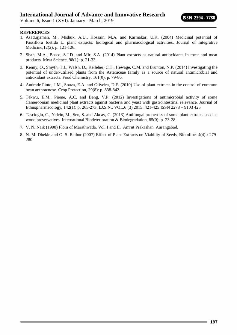
International Journal of Advance and Innovative Research Volume 6, Issue 1 (XVI): January - March, 2019
197
ISSN 2394 - 7780
REFERENCES 1. Asadujjaman, M., Mishuk, A.U., Hossain, M.A. and Karmakar, U.K. (2004) Medicinal potential of
Passiflora foetida L. plant extracts: biological and pharmacological activities. Journal of Integrative Medicine,12(2): p. 121-126.
2. Shah, M.A., Bosco, S.J.D. and Mir, S.A. (2014) Plant extracts as natural antioxidants in meat and meat products. Meat Science, 98(1): p. 21-33.
3. Kenny, O., Smyth, T.J., Walsh, D., Kelleher, C.T., Hewage, C.M. and Brunton, N.P. (2014) Investigating the potential of under-utilised plants from the Asteraceae family as a source of natural antimicrobial and antioxidant extracts. Food Chemistry, 161(0): p. 79-86.
4. Andrade Pinto, J.M., Souza, E.A. and Oliveira, D.F. (2010) Use of plant extracts in the control of common bean anthracnose. Crop Protection, 29(8): p. 838-842.
5. Tekwu, E.M., Pieme, A.C. and Beng, V.P. (2012) Investigations of antimicrobial activity of some Cameroonian medicinal plant extracts against bacteria and yeast with gastrointestinal relevance. Journal of Ethnopharmacology, 142(1): p. 265-273. I.J.S.N., VOL.6 (3) 2015: 421-425 ISSN 2278 – 9103 425
6. Tascioglu, C., Yalcin, M., Sen, S. and Akcay, C. (2013) Antifungal properties of some plant extracts used as wood preservatives. International Biodeterioration & Biodegradation, 85(0): p. 23-28.
7. V. N. Naik (1998) Flora of Marathwada. Vol. I and II, Amrut Prakashan, Aurangabad.
8. N. M. Dhekle and O. S. Rathor (2007) Effect of Plant Extracts on Viability of Seeds, Bioinfloet 4(4) : 279-280.
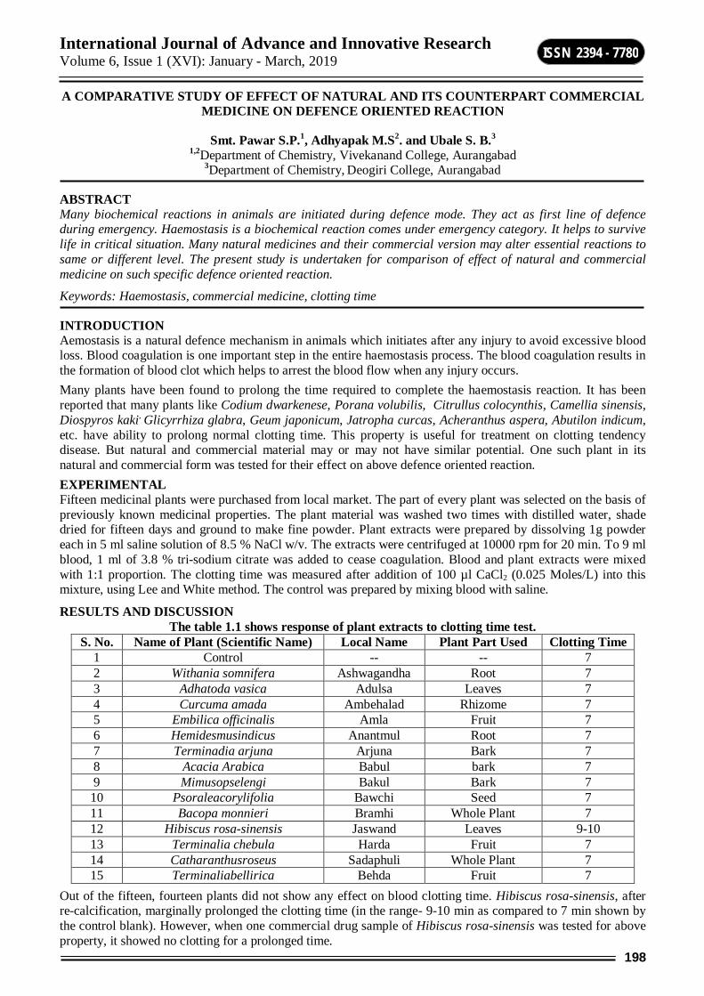
International Journal of Advance and Innovative Research Volume 6, Issue 1 (XVI): January - March, 2019
198
ISSN 2394 - 7780
A COMPARATIVE STUDY OF EFFECT OF NATURAL AND ITS COUNTERPART COMMERCIAL MEDICINE ON DEFENCE ORIENTED REACTION
Smt. Pawar S.P.1, Adhyapak M.S2. and Ubale S. B.3 1,2Department of Chemistry, Vivekanand College, Aurangabad
3Department of Chemistry, Deogiri College, Aurangabad
ABSTRACT Many biochemical reactions in animals are initiated during defence mode. They act as first line of defence during emergency. Haemostasis is a biochemical reaction comes under emergency category. It helps to survive life in critical situation. Many natural medicines and their commercial version may alter essential reactions to same or different level. The present study is undertaken for comparison of effect of natural and commercial medicine on such specific defence oriented reaction.
Keywords: Haemostasis, commercial medicine, clotting time
INTRODUCTION Aemostasis is a natural defence mechanism in animals which initiates after any injury to avoid excessive blood loss. Blood coagulation is one important step in the entire haemostasis process. The blood coagulation results in the formation of blood clot which helps to arrest the blood flow when any injury occurs. Many plants have been found to prolong the time required to complete the haemostasis reaction. It has been reported that many plants like Codium dwarkenese, Porana volubilis, Citrullus colocynthis, Camellia sinensis, Diospyros kaki, Glicyrrhiza glabra, Geum japonicum, Jatropha curcas, Acheranthus aspera, Abutilon indicum, etc. have ability to prolong normal clotting time. This property is useful for treatment on clotting tendency disease. But natural and commercial material may or may not have similar potential. One such plant in its natural and commercial form was tested for their effect on above defence oriented reaction. EXPERIMENTAL Fifteen medicinal plants were purchased from local market. The part of every plant was selected on the basis of previously known medicinal properties. The plant material was washed two times with distilled water, shade dried for fifteen days and ground to make fine powder. Plant extracts were prepared by dissolving 1g powder each in 5 ml saline solution of 8.5 % NaCl w/v. The extracts were centrifuged at 10000 rpm for 20 min. To 9 ml blood, 1 ml of 3.8 % tri-sodium citrate was added to cease coagulation. Blood and plant extracts were mixed with 1:1 proportion. The clotting time was measured after addition of 100 µl CaCl2 (0.025 Moles/L) into this mixture, using Lee and White method. The control was prepared by mixing blood with saline.
RESULTS AND DISCUSSION The table 1.1 shows response of plant extracts to clotting time test.
S. No. Name of Plant (Scientific Name) Local Name Plant Part Used Clotting Time 1 Control -- -- 7 2 Withania somnifera Ashwagandha Root 7 3 Adhatoda vasica Adulsa Leaves 7 4 Curcuma amada Ambehalad Rhizome 7 5 Embilica officinalis Amla Fruit 7 6 Hemidesmusindicus Anantmul Root 7 7 Terminadia arjuna Arjuna Bark 7 8 Acacia Arabica Babul bark 7 9 Mimusopselengi Bakul Bark 7 10 Psoraleacorylifolia Bawchi Seed 7 11 Bacopa monnieri Bramhi Whole Plant 7 12 Hibiscus rosa-sinensis Jaswand Leaves 9-10 13 Terminalia chebula Harda Fruit 7 14 Catharanthusroseus Sadaphuli Whole Plant 7 15 Terminaliabellirica Behda Fruit 7
Out of the fifteen, fourteen plants did not show any effect on blood clotting time. Hibiscus rosa-sinensis, after re-calcification, marginally prolonged the clotting time (in the range- 9-10 min as compared to 7 min shown by the control blank). However, when one commercial drug sample of Hibiscus rosa-sinensis was tested for above property, it showed no clotting for a prolonged time.
International Journal of Advance and Innovative Research Volume 6, Issue 1 (XVI): January - March, 2019
199
ISSN 2394 - 7780
It is clear from above results that plant material can act on body’s essential reactions. Also, processed commercial drug may show less or more efficacy as compared with the original unprocessed herbal medicine.
REFERENCES 1. G. Daniel Boon; An overview of hemostasis; Toxicologic Pathology, 1993, V. 21, No. 2, 170-179.
2. Chris P. M. REUTELING SPERGER, Gerard H ORNSTRA and H. Coenraad HEMKER; Isolation & partial purification of a novel anticoagulant from arteries of human umbilical cord; European J Biochem, 1985,151, 625-629.
3. Earl W. Davie and Oscar D. Ranoff; Waterfall sequence for intrinsic blood clotting; Science, 1964, V. 145, No. 3638, P. 1310-1312.
4. S. M. Stoukova and V. K. Thachuk; Proteinases of the blood coagulation system and fibrinolysis as cell regulators; Biochemistry (Moscow), 2002, Vol. 67, No.1.
5. Kenneth G. Mann, Saulius Butenas, Kathleen Brummel; The dynamics of thrombin formation; Arteriosclerosis, Thrombosis and Vascular Biology, 2003, 23, 17.
6. Narayanan S.; Multifunctional roles of thrombin; Annals of Clinical and Laboratory Science, 1999, 29(4), 275-80.
7. K Kathiresan, Vinoth S. Ravindran and A Muruganantham; Mangrove extracts prevent the blood coagulate; Ind. J. Biotech, 2006, Apr, Vol. 5, P. 252-254.
8. P. M. Ekanayake; Isolation and purification of an anticoagulant from Schizymenia dubyi by fermentation; Food Science and Tech. International, 2007, Oct, 13(5), 355-359.
9. M. Shanmugam, K. H. Mody, R. M. Oza and B. K. Ramavat; Blood anticoagulant activity of a green marine alga Codium dwarkense (Codiaceae, Chlorophyta) in relation to its growth stages; Indian J. of Marine Sciences, 2001, Mar, V. 30, P. 49-52.
10. Yoon SJ, Pereira MS, Pavao MS, Hwang JK, Pyun YR and Mourao PA; The medicinal plant Porana volubilis contains polysaccharides with anticoagulant activity mediated by heparin co-factor II; Thromb. Res., 2002, Apr 1, 106(1), 51-8.
11. S. P. Myers; Interactions between complementary medicines and warfarin; Aust. Presc., 2002, 25, 54-6.
12. Noah Samuels; Herbal remedies and anticoagulant therapy; Throm. Haemost., 2005, 93, 3-7.
13. Hu Z, Yang X, Ho PC, Chan SY, Heng PW, Chan E, Duan W, Koh HL and Zhou S; Herb-drug interactions: A literature Review; Drugs,2005, 65(9), 1239-82.
14. Ara Tachijan, Viqar Muria and Arshad Jahangir; Use of herbal products and potential interactions in patients with cardiovascular diseases; J. of American College of Cardiology, 2010, Feb 9, Vol. 55(6), P.515-525.
15. Amy M. Heck, Beth A. Dewitt and L. Lukes; Potential interactions between alternative therapies and warfarin; American J. of Health-System Pharmacy, 2000, 57(13).
16. Ochei J.and Kolhatkar A; Medical Laboratory Science: Theory and Practice, 2nd Edition, Mcgraw-Hill, 2000.
17. Barbara Ann M. Messina; Herbal supplements: Facts and myths-talking to your patients about herbal supplements; J. of Perianesthesia Nursing, 2006, Aug, 21, 4, 268-278.

International Journal of Advance and Innovative Research Volume 6, Issue 1 (XVI): January - March, 2019
200
ISSN 2394 - 7780
ASSESSMENT OF WATER QUALITY AT THE POLLUTED AREA OF TERNA RIVER IN OSMANABAD DISTRICT MAHARASHTRA STATE INIDA
Shoeb Peerzade1, S. V. Thakur2, R. L. Ware3 and Sayed Abed4
1, 2, 3Department of Chemistry, Milliya Arts Science & Management Science College Beed, Maharashtra 4Department Of Chemistry, Government College Of Arts & Science Aurangabad Maharashtra
ABSTRACT Water pollution is a large set of adverse effects upon water bodies such as lakes, rivers, oceans, and groundwater caused by human activities. Although natural phenomena such as volcanoes, storms, earthquakes, etc. also cause major changes in water quality and the ecological status of water, these are not deemed to be pollution. The present study deal with the Assessment of Water Quality at the Polluted Area of Terna River in Osmanabad District Maharashtra State India.Terna river water samples were collected from five different spots in five different months in Osmanabad district (M. S.) After collection of samples six different parameters like Physical appearance, Temperature, PH, Turbidity, Total Hardness & Total solid were studied. Results showed that seasonal variation in water parameters at different areas. The chemical analysis of water was carried out in the light of APHA (1985) & compared with the WHO, ICMR & ISI standard.
Keywords: Physicochemical character, seasonal variation, water quality, Terna River Five spots.
INTRODUCTION Pollution can be defined as an undesirable change in the physical, chemical, or biological characteristics of the air, water or land that can harmfully affect health, survival, or activities of human or other living organisms. Pollution may interfere with all forms of living systems. Pollutants are substance, especially wastes that have deleterious effects on living organisms. The indiscriminate disposal of water after use in the form of waste water causes water pollution. Most of the rivers in India are polluted due to industrial activity Thus in Bombay; Ulhās river is polluted due to disposal of effluents from rayon and dyestuff industries. Water pollution is a large set of adverse effects upon water bodies such as lakes, rivers, oceans, and groundwater caused by human activities. Although natural phenomena such as volcanoes, storms, earthquakes, etc. also cause major changes in water quality and the ecological status of water, these are not deemed to be pollution. Water pollution has many causes and characteristics. Increases in nutrient loading may lead to eutrophication. Organic wastes such as sewage impose high oxygen demands on the receiving water leading to oxygen depletion with potentially severe impacts on the whole eco-system. Industries discharge a variety of pollutants in their wastewater including heavy metals, organic toxins, oils, nutrients, and solids. Discharges can also have thermal effects, especially those from power stations, and these too reduce the available oxygen. Silt-bearing runoff from many activities including construction sites, deforestation and agriculture can inhibit the penetration of sunlight through the water column, restricting photosynthesis and causing blanketing of the lake or river bed, in turn damaging ecological systems. Pollutants in water include a wide spectrum of chemicals, pathogens, and physical chemistry or sensory changes. Many of the chemical substances are toxic. Pathogens can obviously produce waterborne diseases in either human or animal hosts. Alteration of water’s physical chemistry includes acidity, conductivity, temperature, and eutrophication. Eutrophication is the fertilization of surface water by nutrients that were previously scarce. Even many of the municipal water supplies in developed countries can present health risks.
Water pollution is a major problem in the global context. It has been suggested that it is the leading worldwide cause of deaths and diseases Sinkule et al; (1995), and that it accounts for the deaths of more than 14,000 people daily
MATERIALS & METHODS The water samples of Terna River were collected from five different spots i.e. from Wanewadi, Ternanagar, Takdi, Boregaon&Bebli of Osmanabad district in polythene bottle of capacity 1 to 2 liter in the month of December 2009 to April 2010. The water samples were analyzed to access the physico chemical parameters. The standard procedure was adopted for the determination of physico- chemical parameter given by APHA (1989) & Trivedy&Goel (1986). Each sample was analyzed for important physico- Chemical parameter such as Physical appearance, Temperature, PH, Turbidity, Total Solid, Total Hardness etc.
RESULT AND DISCUSSION Physicochemical analysis of river water from different five spots of Ternariver from month December 2009 to April 2010 is shown in table no. 1 to 5 which shows that

International Journal of Advance and Innovative Research Volume 6, Issue 1 (XVI): January - March, 2019
201
ISSN 2394 - 7780
1). Physical Appearance: - Water sample from all the spots in every month is turbid due to entering of domestic industrial and agriculture waste.
2) Temperature: - Temperature of water depends on the season and on the temperature of the ground with which it is in contact R.K. Trivedy et al; (1984). Temperature important for its on the chemical and biochemical reactions in the organism. R.N. Trivedy et al; A rise in temperature of the water leads to the speed up of the chemical reactions in water, reduces the solubility of gases, the tastes and odour. D.Kelin et al; (1959) and U.N. Mahida et al; (1981). The maximum temperature was recorded at spot no. 5 and minimum temperature was recoded at spot No. 1 in every month.
3) PH:-PH serves as an index to denote the extent of pollution in case of pollution by acidic and alkaline wastes. P.K. saxena et al; (1988). All Chemical and biological reactions are directly dependent upon the PH of water system. A Sreenivason et al; (1967). In present investigation PH indicated almost neutral range (6.8-7.21). Maximum PH value of 7.2 was recorded at spot No. 5 December 2009.Maximum PH value of 7.3 was recorded at spot no. 5 in Jan 2010.Maximum PH value of 7.2 was recorded at spot no. 5 in Feb 2010.
Maximum PH value of 7.3 was recorded at spot No. 5 in March 2010 While. Maximum PH value of 7.5 was recorded at spot No. 5 in April 2010.
The PH limit fixed by Indian standards (ISI) for bathing and drinking in between (6.5-8.5) L.S. Elango et al; (1992)
Hence PH of these water samples is within the standards
4) Turbidity: - Turbidity is an indicator of impurities dissolved solid in the water body. In the present investigation samples from all spots in every month are exceeding the desirable limits that are the sewage domestic water and individual water include increase dissolved solid investigation the turbidity of the solution.
5) Totalsolid: - The total solid content is also a indicator of pollution status sewage, domestic and industrial waste water and dumping of animal and human excreta in water body always increases the total solid. This may be reason for increased total solid at all spots.
In the present investigation in Dec. 2009 the spot No. 5 is showing the above desirable limit while spot No. 1,2,3 and 4 are within the limit In Jan., Feb., March 2010 the spot no.4 and 5 is showing the above desirable limit while spot No. 1,2 and 3 are within the limit while.
In April 2010 the spot No. 3,4 and 5 is showing the above desirable limit while spot No. 1 and 2 are within the limit.
Thus the high quantity of total solids may be attributed to domestic and industrial waste water suspensions entering the river.
6) TotalHardness: - Hardness of water is due to carbonate bicarbonates sulphate calcium silicates magnesium etc. V.S. Lomte et al; (1998). Hardness of samples at spot no. 4 and 5 is showing the above desirable limit while spot No. 1, 2 and 3 are within the limit.
Table-1: Physico- Chemical Analysis OfTernaRiver (December 2009) Parameter Wanewadi Ternanagar Takdi Boregaon Bembli
Physical Appearance Turbid Turbid Turbid Turbid Turbid Temperature C0 21 22 23 23 24
PH 6.8 7.00 7.00 7.1 7.2 Turbidity (NTU) 12 13 13 14 15
Total Solids (mg/lit.) 310.0 330.0 370.0 430.0 505.0 Total Hardness (mg/lit.) 75.0 88.0 96.0 204.0 230.0
Table-2: Physico- Chemical Analysis OfTerna River (January 2010) Parameter Wanewadi Ternanagar Takdi Boregaon Bembli
Physical Appearance Turbid Turbid Turbid Turbid Turbid Temperature C0 22 22 23 24 25
PH 7.00 7.1 7.1 7.2 7.3 Turbidity (NTU) 10.0 11.0 11.0 12.0 13.0
Total Solids (mg/lit.) 280.0 370.0 420.0 500.0 580.0 Total Hardness (mg/lit.) 76.0 90.0 97.0 230.0 250.0

International Journal of Advance and Innovative Research Volume 6, Issue 1 (XVI): January - March, 2019
202
ISSN 2394 - 7780
Table-3: Physico- Chemical Analysis OfTerna River (feberuary2010) Parameter Wanewadi Ternanagar Takdi Boregaon Bembli
Physical Appearance Turbid Turbid Turbid Turbid Turbid Temperature C0 23 24 24 25 26
PH 7.00 7.00 7.00 7.1 7.2 Turbidity (NTU) 13.0 13.0 14.0 14.0 16.0
Total Solids (mg/lit.) 330.0 370.0 450.0 550.0 670.0 Total Hardness (mg/lit.) 75.0 87.0 98.0 220.0 300.0
Table-4: Physico- Chemical Analysis OfTerna River (March 2010) Parameter Wanewadi Ternanagar Takdi Boregaon Bembli
Physical Appearance Turbid Turbid Turbid Turbid Turbid Temperature C0 24 25 26 26 27
PH 6.9 7.1 7.1 7.2 7.3 Turbidity (NTU) 14.0 15.0 15.0 16.0 17.0
Total Solids (mg/lit.) 340.0 380.0 450.0 600.0 700.0 Total Hardness (mg/lit.) 69.0 81.0 96.0 260.0 310.0
Table-5: Physico- Chemical Analysis OfTerna River (April 2010) Parameter Wanewadi Ternanagar Takdi Boregaon Bembli
Physical Appearance Turbid Turbid Turbid Turbid Turbid Temperature C0 26 27 28 30 31
PH 6.9 7.0 7.2 7.3 7.5 Turbidity (NTU) 14.0 15.0 15.0 17.0 18.0
Total Solids (mg/lit.) 350.0 400.0 500.0 690.0 750.0 Total Hardness (mg/lit.) 70.0 78.0 89.0 215.0 350.0
CONCLUSION Analysis of five different samples from five different spot for five consecutive months indicates that degree of pollution increases as the river reaches heart of the village. Similarly the rise in degree of pollution is also observed as we go from autumn to summer. Samples in the autumn season are showing lower degree of pollution while these in summer are showing higher degree of pollution
REFERENCES 1) R.K. Trivedy and P.K. Goel, “Chemical and biological Methods for Water Pollition” Enviro.Pub.,Karad,
India (1984).
2) R.N. Travedi, And Shantranjay Kumar Singh.“Water Resources and Quality Management”, Commonwealth Pub., Delhi (India) pp 75-76.
3) 3)D. Kelin, “River Pollution. Chemical Analysis”, Butterworth Sci. Pub. London, U.K. (1959)
4) P.K. Saxena, S. Jobben and R.Sahai, Geobios. 15 107(1988).
5) ASreenivason, F.A.O. Fish Rep., 44(3), 101(1967)
6) ISI, “ Tolerance Limit for Inland Surface Water Subject to Pollution”. IS 2296, New Delhi (1974)
7) 7) L.S. Elango, Ramchandran and Y.S.R. Chowdary, Ground Water Quality in Coast al
8) Regions of south Madras. Indian J. Environ. Hlth., 34, 318 (1992)
9) M. Varghese, A. Chauhan and L.P.Naik Poll. Res., 11(2), 95 (1992).
10) P.C. MishraandM.C.Desh, Poll. Res., 8(3) (1989).
11) President Bush Discusses Global Climate Change (Transcription of speech) (2001-06-11). Retrieved on 2006-04-09.
12) International Standard ISO 31-8: Quantities and units – Part 8: Physical chemistry and molecular physics, Annex C (normative): pH.International Organization for Standardization, 1992
13) Abdul Raheem & Syed Hussain; Oriental Journal Of Chemistry; 27(3,) (2011) 1273-1275)

International Journal of Advance and Innovative Research Volume 6, Issue 1 (XVI): January - March, 2019
203
ISSN 2394 - 7780
POTENTIOMETRIC STUDY OF COMPLEXATION OF LABETALOL WITH TRANSITION METAL IONS IN ETHANOL-WATER MEDIA
Ramesh Ware1, Shoeb Peerzade2, Kishor Koinkar3, AJ Khan4, Shailendrasingh Thakur5 1,2,4,5Department of Chemistry, Milliya College, Beed
3Department of Chemistry, Gandhi College, Kada
ABSTRACT In the present work we investigate the stability constant of Labetalol hydrochloride drug with transition metal ions Co, Ni, Cu, Zn, and Cd using potentiometric titration technique in 20%(v/v) ethanol-water mixture at 27ºC temperature and at an ionic strength of 0.1M NaClO4.Metal to ligand ratio=1:5 & 1:1The method of Calvin and Bjerrum as adopted by Irving and Rossotti has been employed to determine proton ligand (pKa) and metal-ligand stability constant (logK) values. It is observed that a transition metal ion forms 1:1 and 1:2 complexes.
Keywords: Stability Constant, transition metal ions, Labetalol drug, Potentiometry.
INTRODUCTION Metal complexes are widely used in various fields, such as biological processes pharmaceuticals, separation techniques, analytical processes etc. To understand the complex formation ability of the ligands and the activity of complexes, it is essential to have the knowledge about solution equilibria involved in the reactions. The extent to which the ligand binds to metal ions is normally expressed in terms of stability. Potentiometric titration is accepted as a powerfuland simple electro analytical technique for determination of stability constants. Most of the d-block elements form complexes. There are different kinds of ligand used for complexation. For the present investigation, we selected Labetalol hydrochloride (LBT). It is chemically described as 5-[1-Hydroxy-2-(1-methyl-3-henyl propyl amino) ethyl] salicylamide hydrochloride. LBT is considered as one of the major therapeutic drugs for the treatment of hypertension. LBT is also used to induce hypotension during surgery as it reduces blood pressure more rapidly than other beta blockers. The drug is quite sensitive, even a small dose of the drug gives sufficient blockage, thus indicating that the drug is very much confined to the cardio protective effects. It is used in the treatment of patients with angina pectoris with and without co-existing hypertension. LBT is rapidly absorbed following an oral dose but undergoes extensive first pass metabolism, resulting in only 25% oral bioavailability. Besides these important pharmacological activities, LBT therapy exhibits hepatotoxicity and renal failure due to overdose. LBT is also one of the well known doping agents in sports and hence, it has been banned for Olympic players by International Olympic Committee.
NH
OH
OH
NH2O
.HCl
Figure-1: Labetalol hydrochloride (molecular formula C19H25N2O3Cl )
After a review of literature survey and in continuation of our earlier work with complexation of medicinal drugs1-24, we have carried out a solution study on the complexation of LBT drug with transition metal ions Co2+, Ni2+, Cu2+, Zn2+ and Cd2+ using pH metrically in ethanol-water mixture at constant ionic strength of 0.1M NaClO4.
Experimental Section: I. Materials and Solution. The ligand LBT is soluble in 20% (v/v) ethanol-water mixture. NaOH, NaClO4, HClO4 and metal salts were of AR grade.The solutions used in the pH metric titration were prepared in double distilled water. The NaOH solution was standardized against oxalic acid solution (0.1M) and standard alkali solution was again used for standardization of HClO4. The metal salt solutions were also standardized using EDTA titration. All the measurements were made at 27 °C in 20%(V/V) ethanol-water mixture at constant ionic strength of 0.1M NaClO4.The thermostat model SL-131 was used to maintain the temperatureconstant. The pH measurement were made using a digital pH meter model Elico L1-120 in conjunction with a glass and reference calomel electrode (reading accuracy ±0.01 pH units) the instrument was calibrated at pH 4.00 ,7.00 and 9.18 using the standard buffer solutions .
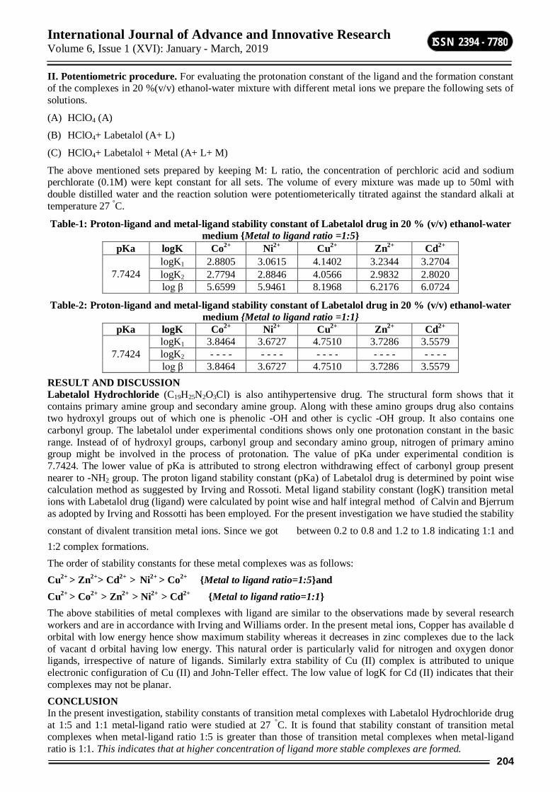
International Journal of Advance and Innovative Research Volume 6, Issue 1 (XVI): January - March, 2019
204
ISSN 2394 - 7780
II. Potentiometric procedure. For evaluating the protonation constant of the ligand and the formation constant of the complexes in 20 %(v/v) ethanol-water mixture with different metal ions we prepare the following sets of solutions.
(A) HClO4 (A)
(B) HClO4+ Labetalol (A+ L)
(C) HClO4+ Labetalol + Metal (A+ L+ M)
The above mentioned sets prepared by keeping M: L ratio, the concentration of perchloric acid and sodium perchlorate (0.1M) were kept constant for all sets. The volume of every mixture was made up to 50ml with double distilled water and the reaction solution were potentiometerically titrated against the standard alkali at temperature 27 °C.
Table-1: Proton-ligand and metal-ligand stability constant of Labetalol drug in 20 % (v/v) ethanol-water medium Metal to ligand ratio =1:5
pKa logK Co2+ Ni2+ Cu2+ Zn2+ Cd2+
7.7424 logK1 2.8805 3.0615 4.1402 3.2344 3.2704 logK2 2.7794 2.8846 4.0566 2.9832 2.8020 log β 5.6599 5.9461 8.1968 6.2176 6.0724
Table-2: Proton-ligand and metal-ligand stability constant of Labetalol drug in 20 % (v/v) ethanol-water medium Metal to ligand ratio =1:1
pKa logK Co2+ Ni2+ Cu2+ Zn2+ Cd2+
7.7424 logK1 3.8464 3.6727 4.7510 3.7286 3.5579 logK2 - - - - - - - - - - - - - - - - - - - - log β 3.8464 3.6727 4.7510 3.7286 3.5579
RESULT AND DISCUSSION Labetalol Hydrochloride (C19H25N2O3Cl) is also antihypertensive drug. The structural form shows that it contains primary amine group and secondary amine group. Along with these amino groups drug also contains two hydroxyl groups out of which one is phenolic -OH and other is cyclic -OH group. It also contains one carbonyl group. The labetalol under experimental conditions shows only one protonation constant in the basic range. Instead of of hydroxyl groups, carbonyl group and secondary amino group, nitrogen of primary amino group might be involved in the process of protonation. The value of pKa under experimental condition is 7.7424. The lower value of pKa is attributed to strong electron withdrawing effect of carbonyl group present nearer to -NH2 group. The proton ligand stability constant (pKa) of Labetalol drug is determined by point wise calculation method as suggested by Irving and Rossoti. Metal ligand stability constant (logK) transition metal ions with Labetalol drug (ligand) were calculated by point wise and half integral method of Calvin and Bjerrum as adopted by Irving and Rossotti has been employed. For the present investigation we have studied the stability
constant of divalent transition metal ions. Since we got between 0.2 to 0.8 and 1.2 to 1.8 indicating 1:1 and 1:2 complex formations. The order of stability constants for these metal complexes was as follows: Cu2+ > Zn2+> Cd2+ > Ni2+ > Co2+ Metal to ligand ratio=1:5and Cu2+ > Co2+ > Zn2+ > Ni2+ > Cd2+ Metal to ligand ratio=1:1 The above stabilities of metal complexes with ligand are similar to the observations made by several research workers and are in accordance with Irving and Williams order. In the present metal ions, Copper has available d orbital with low energy hence show maximum stability whereas it decreases in zinc complexes due to the lack of vacant d orbital having low energy. This natural order is particularly valid for nitrogen and oxygen donor ligands, irrespective of nature of ligands. Similarly extra stability of Cu (II) complex is attributed to unique electronic configuration of Cu (II) and John-Teller effect. The low value of logK for Cd (II) indicates that their complexes may not be planar. CONCLUSION In the present investigation, stability constants of transition metal complexes with Labetalol Hydrochloride drug at 1:5 and 1:1 metal-ligand ratio were studied at 27 °C. It is found that stability constant of transition metal complexes when metal-ligand ratio 1:5 is greater than those of transition metal complexes when metal-ligand ratio is 1:1. This indicates that at higher concentration of ligand more stable complexes are formed.

International Journal of Advance and Innovative Research Volume 6, Issue 1 (XVI): January - March, 2019
205
ISSN 2394 - 7780
Figure-2: The pH metric titration curve for Cu (II)- Labetalol
REFERENCES 1. SV Thakur, Mazahar Farooqui, SD Naikwade, Journal of Chemical and Pharmaceutical Research, 2012,
4(9), 4412-4416.
2. SV Thakur, Mazahar Farooqui, SD Naikwade, Pelagia Research Library, Der Chemica Sinica, 2012, 3(6),1406-1409.
3. SV Thakur, Mazahar Farooqui, SD Naikwade, International Journal of Research in Inorganic Chemistry, 2012, 1(4), 5-7.
4. SV Thakur, RL Ware, Mazahar Farooqui, SD Naikwade, Asian Journal of Research in Chemistry, 2012, 5(12), 1464-1465.
5. SV Thakur, Mazahar Farooqui, SD Naikwade, Journal of Advanced Scientific Research, 2013, 4(1),31-33.
6. SV Thakur, Mazahar Farooqui, SG Shankarwar, SD Naikwade, International Journal of Chemical Sciences, 2013, 11(1), 464 - 468.
7. SV Thakur, Mazahar Farooqui, SD Naikwade, Acta Chimica & Pharmaceutica Indica, 2013, 3(1):35 - 39.
8. SV Thakur, MazaharFarooqui, SD Naikwade, International Journal of Emerging Technologies in Computational and Applied Sciences. 2013,4(4), 342-346.
9. SV Thakur, Mazahar Farooqui, SD Naikwade, International Journal of Emerging Technologies in Computational & Applied Sciences. 2013, 4(4), 389-393.
10. SV Thakur, Mazahar Farooqui, MA Sakhare, SD Naikwade, American Int. J. Research in Formal, Applied & Natural Sciences. 2013, 3(1), 123- 127.
11. SV Thakur, SD Naikwade, Mazahar Farooqui, International Journal of Chemical Studies. 2013, 1(3), 88-92.
12. SV Thakur, Mazahar Farooqui, SD Naikwade, Int. J Recent Trends in Science & Technology, Special Issue,ACTRA-INDIA, Sept.2013, 29-31.
13. SV Thakur, MazaharFarooqui, SD Naikwade, Journal of Chemical Biological & Physical sciences. 2014, 4(1), 1-7.
14. SV Thakur, SD Naikwade,Mazahar Farooqui, Journal of Medicinal Chemistry Drug discovery,(special issue),2015, 107-118.
International Journal of Advance and Innovative Research Volume 6, Issue 1 (XVI): January - March, 2019
206
ISSN 2394 - 7780
15. RL Ware, Mazahar Farooqui, SD Naikwade, Int J Emerging Tech in Computational & Applied Sci, 2013, 5(2),123-128.
16. RL Ware, MazaharFarooqui, SD Naikwade, Int J Emerging Tech in Computational & Applied Sci.2013, 5(4), 398-401.
17. RL Ware, Shoeb Peerzade, SD Naikwade, Mazahar Farooqui, J Chemical & Pharma Res.2013,5(8), 59-63.
18. SV Thakur, Jameel Pathan, Farooque Bashir Ansari, DD Kayande, Journal of Chemical & Pharmacetical Research. 2016, 8(5), 291-294.
19. SV Thakur, MA Sakhare, SN Sampal, HU Joshi, International Multilingual Research Journal Printing Area (Special Issue), Dec.2017, 169-173.
20. Shailendrasingh Thakur, SA Peerzade, AJ Khan, RL Ware, International Multilingual Research Journal Printing Area (Special Issue), Dec.2017,47-51
21. Ramesh Ware, Kishor Koinkar, Shailendrasingh Thakur, International Journal of Universal Science and Technology, 3(1) Jan. 2018, 284-288.
22. Ramesh Ware, Shoeb Peerzade and Shailendrasingh Thakur, International Journal of Universal Science and Technology, 3(1) Jan. 2018, 238-241.
23. Ramesh Ware and Shailendrasingh Thakur, International Journal of Universal print, 4(4) March 2018, 254-260.
24. Ramesh Ware, Shoeb Peerzade and Shailendrasingh Thakur, International Journal of Universal print, 4(5) March 2018, 274-278.
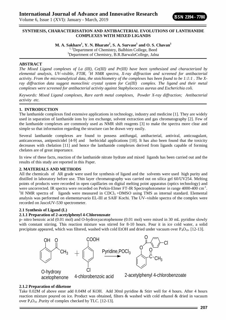
International Journal of Advance and Innovative Research Volume 6, Issue 1 (XVI): January - March, 2019
207
ISSN 2394 - 7780
SYNTHESIS, CHARACTERISATION AND ANTIBACTERIAL EVOLUTIONS OF LANTHANIDE COMPLEXES WITH MIXED LIGANDS
M. A. Sakhare1, Y. N. Bharate2, S. A. Survase1 and O. S. Chavan3
1, 2Department of Chemistry, Balbhim College, Beed 3Department of Chemistry, B.R.BarwaleCollege, Jalna
ABSTRACT The Mixed Ligand complexes of La (III), Ce(III) and Pr(III) have been synthesized and characterized by elemental analysis, UV–visible, FTIR, 1H NMR spectra, X-ray diffraction and screened for antibacterial activity. From the microanalytical data, the stoichiometry of the complexes has been found to be 1:1:1 . The X-ray diffraction data suggest monoclinic crystal system for Ce(III) complex. The ligand and their metal complexes were screened for antibacterial activity against Staphylococcus aureus and Escherichia coli.
Keywords: Mixed Ligand complexes, Rare earth metal complexes, Powder X-ray diffraction; Antibacterial activity etc.
1. INTRODUCTION The lanthanide complexes find extensive applications in technology, industry and medicine [1]. They are widely used in separation of lanthanide ions by ion exchange, solvent extraction and gas chromatography [2]. Few of the lanthanide complexes are commonly used as NMR shift reagents [3] to make the spectra more clear and simple so that information regarding the structure can be drawn very easily.
Several lanthanide complexes are found to possess antifungal, antibacterial, antiviral, anticoagulant, anticancerous, antipesticidel [4-9] and herbicidal applications [10]. It has also been found that the toxicity decreases with chelation [11] and hence the lanthanide complexes derived from ligands capable of forming chelates are of great importance.
In view of these facts, reaction of the lanthanide nitrate hydrate and mixed ligands has been carried out and the results of this study are reported in this Paper.
2. MATERIALS AND METHODS All the chemicals of AR grade were used for synthesis of ligand and the solvents were used high purity and distilled in laboratory before use. Thin layer chromatography was carried out on silica gel 60/UV254. Melting points of products were recorded in open capillaries on digital melting point apparatus (optics technology) and were uncorrected. IR spectra were recorded on Perkin-Elmer FT-IR Spectrophotometer in range 4000-400 cm-1. 1H NMR spectra of ligands were measured in CDCl3 +DMSO using TMS as internal standard. Elemental analysis was performed on elementarvario EL-III at SAIF Kochi. The UV–visible spectra of the complex were recorded on JascoUV-530 spectrometer.
2.1 Synthesis of Ligand (L) 2.1.1 Preparation of 2-acetylphenyl 4-Chloronzoate p- nitro benzoic acid (0.01 mol) and O-hydroxyacetophenone (0.01 mol) were mixed in 30 mL pyridine slowly with constant stirring. This reaction mixture was stirred for 8-10 hours. Pour it in ice cold water, a solid precipitate appeared, which was filtered, washed with cold EtOH and dried under vacuum over P4O10. [12-13].
OH
CH3
O
Pyridine,POCl3
COOH
Cl
O
OCl
CO
CH3
O-hydroxy acetophenone 4-chlorobenzoic acid 2-acetylphenyl 4-chlorobenzoate
2.1.2 Preparation of diketone Take 0.02M of above ester add 0.04M of KOH. Add 30ml pyridine & Stirr well for 4 hours. After 4 hours reaction mixture poured on ice. Product was obtained, filters & washed with cold ethanol & dried in vacuum over P4O10 .Purity of complex checked by TLC. [12-13].
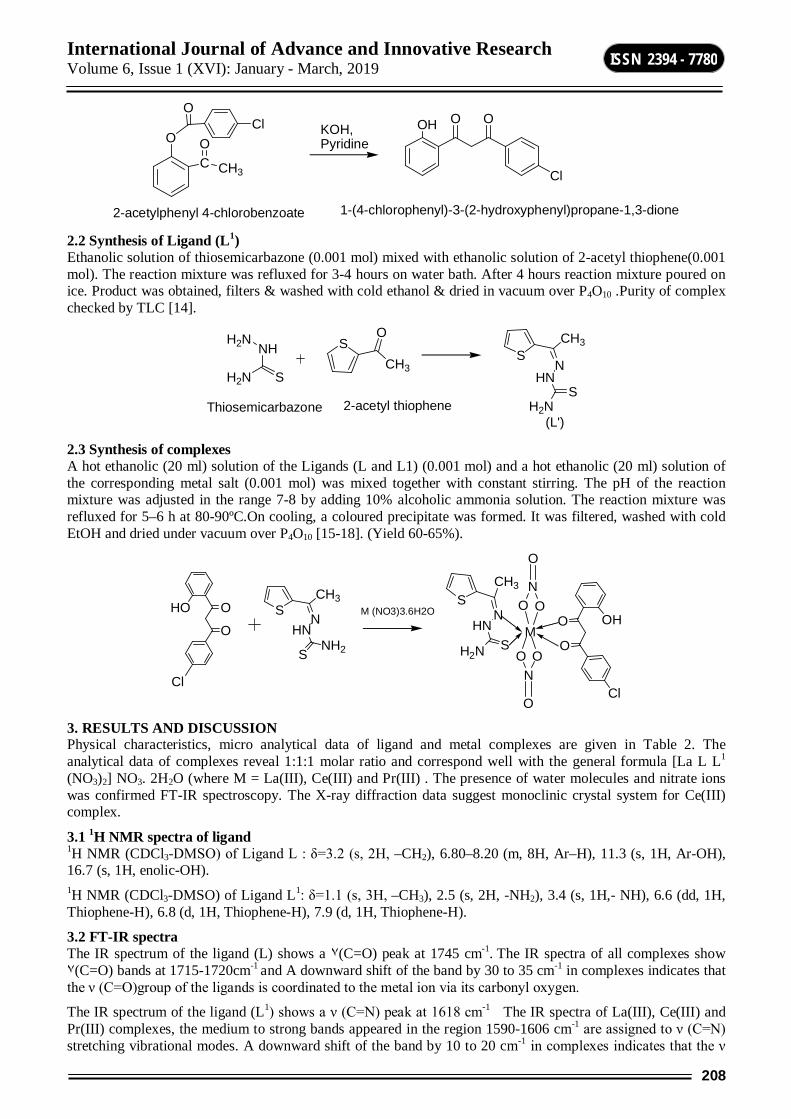
International Journal of Advance and Innovative Research Volume 6, Issue 1 (XVI): January - March, 2019
208
ISSN 2394 - 7780
KOH,Pyridine
OH O O
Cl
O
OCl
CO
CH3
2-acetylphenyl 4-chlorobenzoate 1-(4-chlorophenyl)-3-(2-hydroxyphenyl)propane-1,3-dione 2.2 Synthesis of Ligand (L1) Ethanolic solution of thiosemicarbazone (0.001 mol) mixed with ethanolic solution of 2-acetyl thiophene(0.001 mol). The reaction mixture was refluxed for 3-4 hours on water bath. After 4 hours reaction mixture poured on ice. Product was obtained, filters & washed with cold ethanol & dried in vacuum over P4O10 .Purity of complex checked by TLC [14].
NHH2N
SH2N
SO
CH3
Thiosemicarbazone 2-acetyl thiophene
SN
CH3
HNS
H2N(L')
2.3 Synthesis of complexes A hot ethanolic (20 ml) solution of the Ligands (L and L1) (0.001 mol) and a hot ethanolic (20 ml) solution of the corresponding metal salt (0.001 mol) was mixed together with constant stirring. The pH of the reaction mixture was adjusted in the range 7-8 by adding 10% alcoholic ammonia solution. The reaction mixture was refluxed for 5–6 h at 80-90ºC.On cooling, a coloured precipitate was formed. It was filtered, washed with cold EtOH and dried under vacuum over P4O10 [15-18]. (Yield 60-65%).
SN
CH3
HN
SNH2
HO O
O
Cl
SN
CH3
HNSH2N
OHO
O
Cl
MM (NO3)3.6H2O
NO O
O
NOO
O
3. RESULTS AND DISCUSSION Physical characteristics, micro analytical data of ligand and metal complexes are given in Table 2. The analytical data of complexes reveal 1:1:1 molar ratio and correspond well with the general formula [La L L1
(NO3)2] NO3. 2H2O (where M = La(III), Ce(III) and Pr(III) . The presence of water molecules and nitrate ions was confirmed FT-IR spectroscopy. The X-ray diffraction data suggest monoclinic crystal system for Ce(III) complex.
3.1 1H NMR spectra of ligand 1H NMR (CDCl3-DMSO) of Ligand L : δ=3.2 (s, 2H, –CH2), 6.80–8.20 (m, 8H, Ar–H), 11.3 (s, 1H, Ar-OH), 16.7 (s, 1H, enolic-OH). 1H NMR (CDCl3-DMSO) of Ligand L1: δ=1.1 (s, 3H, –CH3), 2.5 (s, 2H, -NH2), 3.4 (s, 1H,- NH), 6.6 (dd, 1H, Thiophene-H), 6.8 (d, 1H, Thiophene-H), 7.9 (d, 1H, Thiophene-H).
3.2 FT-IR spectra The IR spectrum of the ligand (L) shows a ٧(C=O) peak at 1745 cm-1. The IR spectra of all complexes show ٧(C=O) bands at 1715-1720cm-1 and A downward shift of the band by 30 to 35 cm-1 in complexes indicates that the ν (C=O)group of the ligands is coordinated to the metal ion via its carbonyl oxygen.
The IR spectrum of the ligand (L1) shows a ν (C=N) peak at 1618 cm-1 The IR spectra of La(III), Ce(III) and Pr(III) complexes, the medium to strong bands appeared in the region 1590-1606 cm-1 are assigned to ν (C=N) stretching vibrational modes. A downward shift of the band by 10 to 20 cm-1 in complexes indicates that the ν
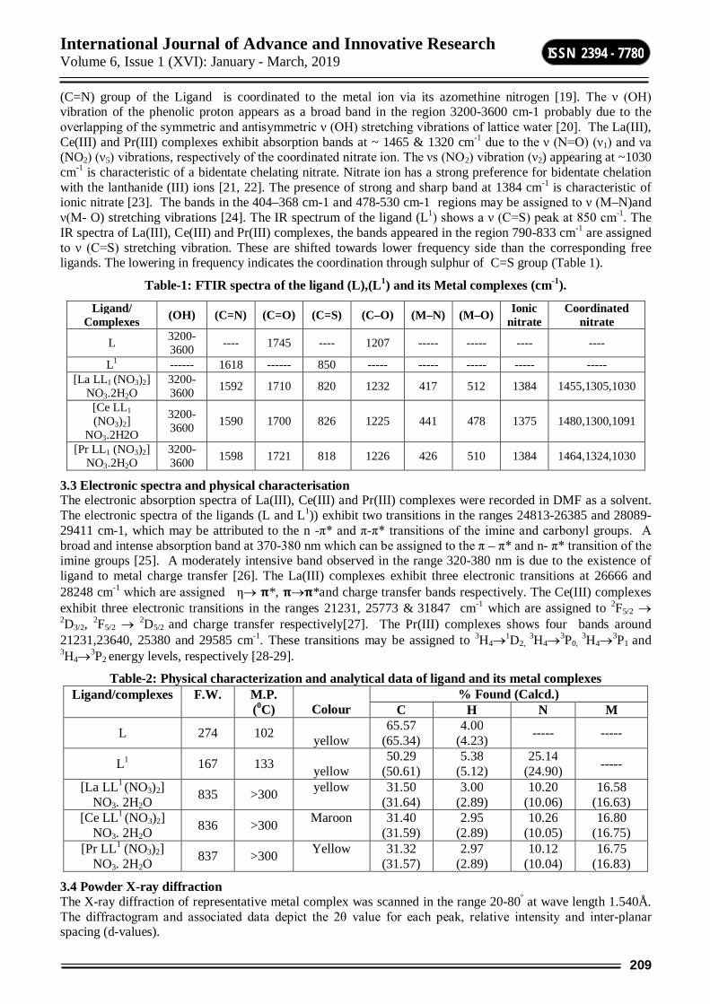
International Journal of Advance and Innovative Research Volume 6, Issue 1 (XVI): January - March, 2019
209
ISSN 2394 - 7780
(C=N) group of the Ligand is coordinated to the metal ion via its azomethine nitrogen [19]. The ν (OH) vibration of the phenolic proton appears as a broad band in the region 3200-3600 cm-1 probably due to the overlapping of the symmetric and antisymmetric ν (OH) stretching vibrations of lattice water [20]. The La(III), Ce(III) and Pr(III) complexes exhibit absorption bands at ~ 1465 & 1320 cm-1 due to the ν (N=O) (ν1) and νa (NO2) (ν5) vibrations, respectively of the coordinated nitrate ion. The νs (NO2) vibration (ν2) appearing at ~1030 cm-1 is characteristic of a bidentate chelating nitrate. Nitrate ion has a strong preference for bidentate chelation with the lanthanide (III) ions [21, 22]. The presence of strong and sharp band at 1384 cm-1 is characteristic of ionic nitrate [23]. The bands in the 404–368 cm-1 and 478-530 cm-1 regions may be assigned to ν (M–N)and ν(M- O) stretching vibrations [24]. The IR spectrum of the ligand (L1) shows a ν (C=S) peak at 850 cm-1. The IR spectra of La(III), Ce(III) and Pr(III) complexes, the bands appeared in the region 790-833 cm-1 are assigned to ν (C=S) stretching vibration. These are shifted towards lower frequency side than the corresponding free ligands. The lowering in frequency indicates the coordination through sulphur of C=S group (Table 1).
Table-1: FTIR spectra of the ligand (L),(L1) and its Metal complexes (cm-1).
3.3 Electronic spectra and physical characterisation The electronic absorption spectra of La(III), Ce(III) and Pr(III) complexes were recorded in DMF as a solvent. The electronic spectra of the ligands (L and L1)) exhibit two transitions in the ranges 24813-26385 and 28089-29411 cm-1, which may be attributed to the n -π* and π-π* transitions of the imine and carbonyl groups. A broad and intense absorption band at 370-380 nm which can be assigned to the π – π* and n- π* transition of the imine groups [25]. A moderately intensive band observed in the range 320-380 nm is due to the existence of ligand to metal charge transfer [26]. The La(III) complexes exhibit three electronic transitions at 26666 and 28248 cm-1 which are assigned η 훑*, 훑훑*and charge transfer bands respectively. The Ce(III) complexes exhibit three electronic transitions in the ranges 21231, 25773 & 31847 cm-1 which are assigned to 2F5/2 2D3/2, 2F5/2 2D5/2 and charge transfer respectively[27]. The Pr(III) complexes shows four bands around 21231,23640, 25380 and 29585 cm-1. These transitions may be assigned to 3H41D2,
3H43P0, 3H43P1 and
3H43P2 energy levels, respectively [28-29].
Table-2: Physical characterization and analytical data of ligand and its metal complexes Ligand/complexes
F.W.
M.P. (0C)
Colour
% Found (Calcd.) C H N M
L 274 102 yellow
65.57 (65.34)
4.00 (4.23) ----- -----
L1 167 133 yellow
50.29 (50.61)
5.38 (5.12)
25.14 (24.90) -----
[La LL1 (NO3)2] NO3. 2H2O 835 >300 yellow 31.50
(31.64) 3.00
(2.89) 10.20
(10.06) 16.58
(16.63) [Ce LL1
(NO3)2] NO3. 2H2O 836 >300 Maroon 31.40
(31.59) 2.95
(2.89) 10.26
(10.05) 16.80
(16.75) [Pr LL1 (NO3)2]
NO3. 2H2O 837 >300 Yellow 31.32 (31.57)
2.97 (2.89)
10.12 (10.04)
16.75 (16.83)
3.4 Powder X-ray diffraction The X-ray diffraction of representative metal complex was scanned in the range 20-80º at wave length 1.540Å. The diffractogram and associated data depict the 2θ value for each peak, relative intensity and inter-planar spacing (d-values).
Ligand/ Complexes (OH) (C=N) (C=O) (C=S) (C–O) (M–N) (M–O) Ionic
nitrate Coordinated
nitrate
L 3200-3600 ---- 1745 ---- 1207 ----- ----- ---- ----
L1 ------ 1618 ------ 850 ----- ----- ----- ----- ----- [La LL1 (NO3)2]
NO3.2H2O 3200-3600 1592 1710 820 1232 417 512 1384 1455,1305,1030
[Ce LL1 (NO3)2]
NO3.2H2O
3200-3600 1590 1700 826 1225 441 478 1375 1480,1300,1091
[Pr LL1 (NO3)2] NO3.2H2O
3200-3600 1598 1721 818 1226 426 510 1384 1464,1324,1030
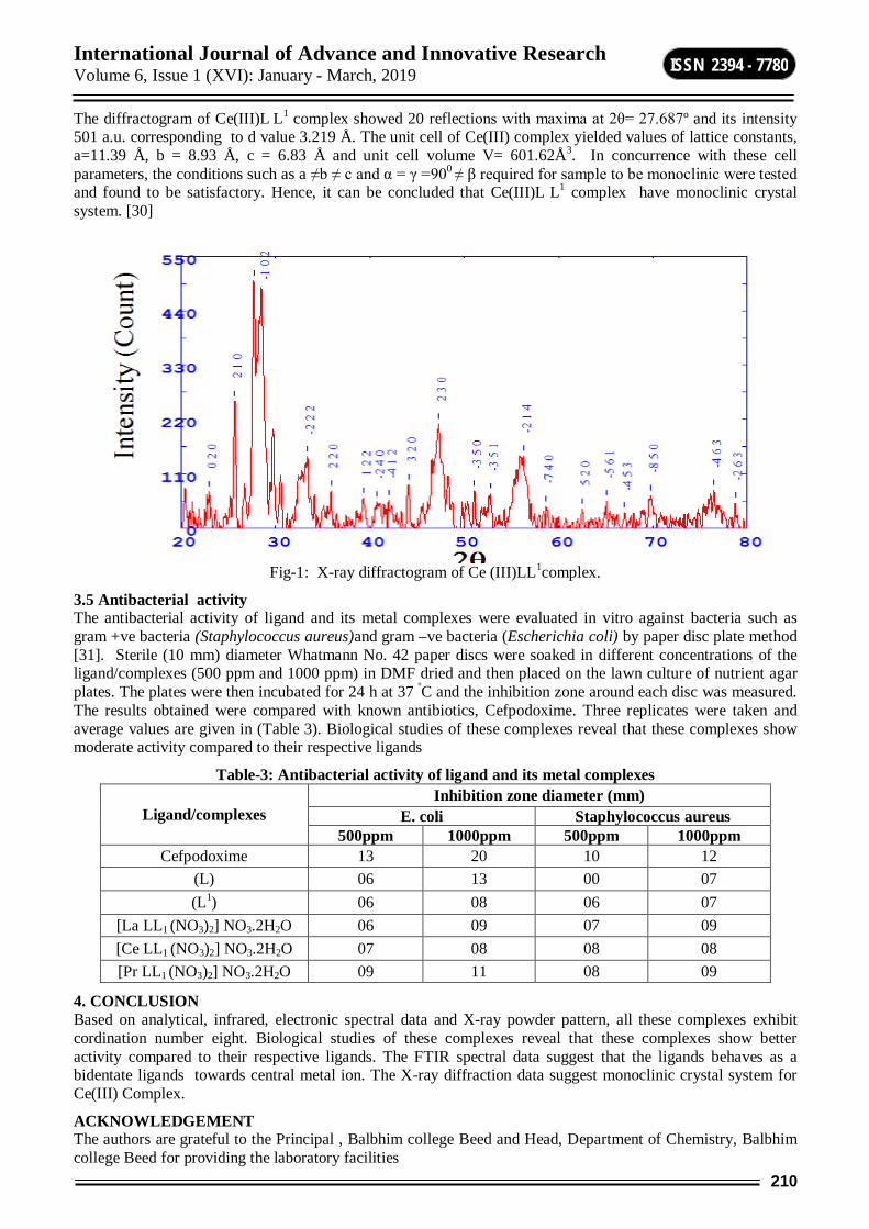
International Journal of Advance and Innovative Research Volume 6, Issue 1 (XVI): January - March, 2019
210
ISSN 2394 - 7780
The diffractogram of Ce(III)L L1 complex showed 20 reflections with maxima at 2θ= 27.687º and its intensity 501 a.u. corresponding to d value 3.219 Å. The unit cell of Ce(III) complex yielded values of lattice constants, a=11.39 Å, b = 8.93 Å, c = 6.83 Å and unit cell volume V= 601.62Å3. In concurrence with these cell parameters, the conditions such as a ≠b ≠ c and α = γ =900 ≠ β required for sample to be monoclinic were tested and found to be satisfactory. Hence, it can be concluded that Ce(III)L L1 complex have monoclinic crystal system. [30]
Fig-1: X-ray diffractogram of Ce (III)LL1complex.
3.5 Antibacterial activity The antibacterial activity of ligand and its metal complexes were evaluated in vitro against bacteria such as gram +ve bacteria (Staphylococcus aureus)and gram –ve bacteria (Escherichia coli) by paper disc plate method [31]. Sterile (10 mm) diameter Whatmann No. 42 paper discs were soaked in different concentrations of the ligand/complexes (500 ppm and 1000 ppm) in DMF dried and then placed on the lawn culture of nutrient agar plates. The plates were then incubated for 24 h at 37 ºC and the inhibition zone around each disc was measured. The results obtained were compared with known antibiotics, Cefpodoxime. Three replicates were taken and average values are given in (Table 3). Biological studies of these complexes reveal that these complexes show moderate activity compared to their respective ligands
Table-3: Antibacterial activity of ligand and its metal complexes
Ligand/complexes Inhibition zone diameter (mm)
E. coli Staphylococcus aureus 500ppm 1000ppm 500ppm 1000ppm
Cefpodoxime 13 20 10 12 (L) 06 13 00 07 (L1) 06 08 06 07
[La LL1 (NO3)2] NO3.2H2O 06 09 07 09 [Ce LL1 (NO3)2] NO3.2H2O 07 08 08 08 [Pr LL1 (NO3)2] NO3.2H2O 09 11 08 09
4. CONCLUSION Based on analytical, infrared, electronic spectral data and X-ray powder pattern, all these complexes exhibit cordination number eight. Biological studies of these complexes reveal that these complexes show better activity compared to their respective ligands. The FTIR spectral data suggest that the ligands behaves as a bidentate ligands towards central metal ion. The X-ray diffraction data suggest monoclinic crystal system for Ce(III) Complex.
ACKNOWLEDGEMENT The authors are grateful to the Principal , Balbhim college Beed and Head, Department of Chemistry, Balbhim college Beed for providing the laboratory facilities
International Journal of Advance and Innovative Research Volume 6, Issue 1 (XVI): January - March, 2019
211
ISSN 2394 - 7780
5. REFERENCES 1. K. J. Eisentrant and K. E. Sievers, J. Am. Chem. Soc., 87 (1965), 5254.
2. M. Liu, W. B. Yuan, Q. Zhang, L. Yan and R. Yang, Spectrochim Acta A., 70, (2008), 1114.
3. Reedijk, J. Curr. Opin. Chem. Biol. 1999, 3 (2), 236.
4. P. G. Avaji, C. H. V. Kumar, S. A. Patil, K. N. Shivananda and C. Nagaraju, Eur. J. Med. Chem., 44 (2009), 3552.
5. A. D. Azaz, S. Celen, H. Namli, O. Turhan, R. Kurtaran, C. Kazak and N. B. Arslan, Trans. Met. Chem., 32 (2007), 884.
6. E. W. Ainscough, A. M. Brodie, A. J. Dobbs, J. D. Ranford, J. M. Waters, Inorg. Chim. Acta., 267 (1998), 27.
7. B. Wang, Z. Y. Yang, P. Crewdson, D. Wang, J. Inorg. Biochem., 101 (2007), 1492.
8. V. Mahalingam, N. Chitrapriya, F. R. Fronczek and K. Natarajan, Polyhedron., 27 (2008), 1917.
9. M. A. Sakhare, S. L.Khillare, M. K. Lande and B. R. Arbad, Advances in Applied Science Research, 2013, 4(1):94-100
10. H.Sahebalzamania, S. Ghammamya, K. Mehrania, F. Salimib, Der Chemica Sinica., 2010, 1 (1), 67-72
11. C. P. Gupta, V. Suri, R. P. Mathaur and R. K. Mehta, Proc. Indian Nat. Sci. Acad., 48A (1982), 232.
12. N.S.korde, S.T.Gaikwad, S.S.Korde and A.S.Rajbhoj , International Journal of Recent technology and engineering vol. 2(4),sept.2013
13. N.S.korde, S.T.Gaikwad, B.C.Khade and A.S.Rajbhoj,Chem.SciTrans, 2013, 2(2), 407-412.
14. S.Chandra and A.kumar, J. Ind. Chem. Soc., 84 (2007), 325-328.
15. S. Chandra and A. Gautam, Spectrochemica Acta Part A., 70 (2008), 1001-1002.
16. S. Chandra and L.K. Gupta, Spectrochemica Acta Part A., 61(2005), 1181-1182.
17. S.Chandra and L.K.Gupta, Spectrochemica Acta Part A., 60(2004), 3079-3080.
18. S. Chandra, M. Tyagi, S. Rani and S. Kumar, Spectrochimica Acta Part A, 75 (2010), 835–840.
19. A. Kulkarni, S. A. Patil and P. S. Badami, Eur. J. Med. Chem., 44 (2009), 2904.
20. W. Radecka-Paryzek Inorg. Chim. Acta., 45 (1980), L447.
21. K.Nakamoto, Infrared and Raman Spectra of Inorganic coordination Compounds, 3rd edn.(John Wiley, New York),1978.
22. J.A. V. Aruna and V.Alexander, Inorganica Chemica Acta A., 249(1996),96.
23. G. Das, R. Shukula, S. Mandal, R. Singh, P. K. Bharadwaj, J.V. Singh and K.H. Whitmire, Inorg. Chem., 36 (1997), 323.
24. S.K. Sengupta, O.P. Pandey, A. Rai and A. Sinha, Spectrochimica Acta Part A., 65 (2006), 139–142.
25. C. Lodeiro, R. Bastida, Inorganica Chemica Acta, 267 (1998), 59
26. G. Das, R. Shukula, S. Mandal, R. Singh, P.K. Bharadwaj, J.V. Singh, K.H. Whitmire, Inorg. Chem. 36 (1997), 323
27. R. C. Chikate, H. A. Bajaj, A. S. Kumbhar, V. C. Kolhe, S. B. Padhye, Themochemica Acta., 249 (1995), 239.
28. S. K. Sengupta, O. P. Pandy, A. Rai, A. Sinha, Spectrochem Acta A., 65 (2006), 139.
29. B. Keshavan, P.G. Chandrashekara, N.M. Made Gowda, J. Mole. Struc. 553 (2000), 193–197
30. V.A. Shelke, S.M. Jadhav, V.R. Patharkar, S.G. Shankarwar, A.S. Munde and T.K.Chondhekar, (2010), Arabian J. Chem.doi:10.1016.
31. A.S. Munde, V. A. Shelke, S.M. Jadhav, A.S. Kirdant, S.R.Vaidya, S.G. Shankarwar, T.K. Chondhekar, Advances in Applied Science Research., 2012, 3 (1), 175-182.
International Journal of Advance and Innovative Research Volume 6, Issue 1 (XVI): January - March, 2019
212
ISSN 2394 - 7780
ANTI FUNGAL POTENTIALITY OF SOME SELECTED MEDICINAL PLANTS AGAINST FRUIT ROT PATHOGENS
Sawant S. G. Assistant Professor, Department of Botany, K. K. M. College, Manwat, Dist. Parbhani
ABSTRACT Fungi have been identified as a major cause of biodeterioration of all kinds of eatables throughout the world leading to both qualitative and quantitative losses. Many fungal pathogens acquired resistance to synthetic fungicides and become a matter of concern in recent years. Many strategies were employed to control the fungal decay. One of them is the use of synthetic fungicides. But it is noticed that the frequent use of fungicides causes health problems to human beings. The fungicidal residues becomes carcinogenic and also creates environmental hazards. So,there is a need to find safer alternatives to replace the fungicidal use.
Therefore, there is urgent need to develop new and effective means for controlling postharvest diseases of fruits or fruit rots, suitable to humanhealth and environment. Hence this research work is carried out to study the potentiality of some selected medicinal plants against fruit rot pathogens.
Keywords: Fungal pathogens, Medicinal plants, Fruit rots
INTRODUCTION Fungi rank second only to insects as a cause of plant diseases, which result in heavy loss of plant products. Pathogenic fungi alone cause 20% reduction in the yield of major food and cash crops(Agrios,2000). Postharvest diseases are posing a major problem to the agriculture industry, where they account to about 50% losses in fruits stored in poor storage conditions(Agrios,2005). Among the most important fungi causing postharvest diseases of plants are Aspergillus species, Penicillium species and Rhizopus species. The most important method of protecting the plants against the fungal attack is the use of synthetic fungicides. But their excessive use complemented with high costs, residues in plants, and development of resistance has left a negative effect on human health and the environment(Bull et.al.,1997; Paster and Bullerman, 1988). Environmentally friendly plant extracts agents have shown to be great potential as an alternative to synthetic fungicides(Zhang and Zhang, 2005). That plant extracts are cheap, locally available, non-toxic and easily biodegradable. Medicinal plant materials have been successfully used for the treatment of fungal and bacterial infections in humans (Akinyosoye and Oldunmoye 2000).
Suggesting that some plant materials may also possess antifungal and antibacterial constituents that are useful in controlling plant diseases(Amadioha,1998). Akinyosoye and Oldunmoye(2000) have reported the antifungal efficacy of stem and leaf extracts of Mirabilis jalapa L. in reducing mycelia growth of four different strains of fungi. The legendary medicinal qualities of Neem tree have been known for a long time and their aqueous leaf extracts have systemic action (Egunjob and Onoyemi,1981;Sownumi and Akinusi,1983). Therefore, the presence of fungitoxic activities by plant extracts is an indication that such plants could be used as fungicides by peasant farmers who cannot afford the costly synthetic agrochemicals to control fungal postharvest diseases.
Therefore, this work is undertaken to study the effect of various concentrations of aqueous leaf extracts of A.indica and A.squamosa against the common fruit rot pathogens as A.niger, A.flavus, P. expansum, and R.stolonifer which are isolated from papaya fruit.
MATERIAL AND METHOD Preparation of plant extracts The powdered leaves extracted with sterile distilled water at room temperature at different level of concentration 10 gm in 100ml, 20gm in 100ml, 30gm in 100ml then filtered by double layered musclin cloth and then with whatmann filter paper no. 1.That extract was stored at 4 in presterilized conical flask.
Antifungal activity of Neem and Annona extracts was studied by poisoned food technique(Nene and Thapliyal,1993). PDA sterilized at 15lb for 20min in autoclave was added to requisite quantiy of 10% ,20% and 30% aqueous extracts of Neem leaves to get 1:1(PDA: plant extract) final concentration. The plant extracts were thoroughly mixed by stirring with PDA. 20ml of this medium was poured in prestrerilized petriplates. After solidification, small disc(0.7mm) of fungus culture grown on PDA for 7 days was cut with sterile cork borer and transferred aseptically in the centre of petridish containing 1:1 medium. Suitable checks were kept where the fungal culture disc were grown under the same conditions on PDA without plant extracts. The fungal colony diameter as compared with checks was taken as a measure of fungitoxicity.
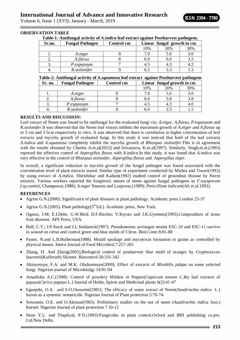
International Journal of Advance and Innovative Research Volume 6, Issue 1 (XVI): January - March, 2019
213
ISSN 2394 - 7780
OBSERVATION TABLE Table-1: Antifungal activity of A.indica leaf extract against Postharvest pathogens. Sr.no. Fungal Pathogen Control cm Linear fungal growth in cm
10% 20% 30% 1. A.niger 8 7.0 5.0 3.0 2. A.flavus 8 6.0 6.0 3.5 3. P.expansum 7 4.5 4.3 4.2 4. R.stolonifer 9 6.5 3.3 1.3
Table-2: Antifungal activity of A.squamosa leaf extract against Postharvest pathogens Sr. no. Fungal Pathogen Control cm Linear fungal growth in cm
10% 20% 30% 1. A.niger 8 7.0 5.0 3.0 2. A.flavus 8 6.0 5.8 3.8 3. P.expansum 7 4.5 4.3 4.0 4. R.stolonifer 9 6.0 3.5 1.5
RESULTS AND DISCUSSION: Leaf extract of Neem was found to be antifungal for the evaluated fungi viz; A.niger, A.flavus, P.expansum and R.stolonifer.It was observed that the Neem leaf extract inhibits the maximum growth of A.niger and A.flavus up to 3 cm and 3.5cm respectively in vitro. It was observed that there is correlation in higher concentration of leaf extracts and mycelia growth of evaluated fungi. In this study it was noticed that both of the leaf extracts A.indica and A.squamosa completely inhibit the mycelia growth of Rhizopus stolonifer.This is in agreement with the results obtained by Charles A.et.al(2012) and Srivastava, K.et.al(1997). Similarly, Singh,et.al.(1993) reported the effective control of Aspergillus flavus with A.indica.In this study, it was found that A.indica was very effective in the control of Rhizopus stolonifer, Aspergillus flavus and Aspergillus niger.
In overall, a significant reduction in mycelia growth of the fungal pathogen was found associated with the concentration level of plant extracts tested. Similar type of experiment conducted by Mishra and Tiwari(1992) by using extract of A.indica. Shirishikar and Kadam(1992) studied control of groundnut disease by Neem extracts. Various workers reported the fungitoxic nature of neem against fungal pathogens as F.oxysporum f.sp.cumini, Champawat,1988); A.niger Naseem and Lanjewar,(1989); Penicillium italicum(Ali et.al.1992).
REFERENCES Agrios G.N.(2000). Significance of plant diseases in plant pathology. Academic press London 25-37
Agrios G.N.(2005). Plant pathology(5th Ed.). Academic press, New York.
Ogawa, J.M; E.I.Dehr; G.W.Bird; D.F.Ritchie; V.Kiyoto and J.K.Uyemoto(1995).Compendium of stone fruit diseases. APS Press, USA.
Bull, C.T.; J.P.Stack and J.L.Smilanick(1997). Pseudomonas syrinagae strains ESC-10 and ESC-11 survive in wound on citrus and control green and blue molds of Citrus. Biol.Contr.8:81-88
Paster, N.and L.B.Bullerman(1988). Mould spoilage and mycotoxin formation in grains as controlled by physical means. Intern Journal of Food Microbiol,7:257-265
Zhang, H. And Zhang(2005),Biological control of postharvest blue mold of oranges by Cryptococcus laurentii(Kufferath) Skinner. Biocontrol 50:331-342
Akinyosoye, F.A. and M.K. Oladunmoye(2000). Effect of extracts of Mirabilis jalapa on some selected fungi. Nigerian journal of Microbiolgy 14:91-94
Amadioha A.C.(1998). Control of powdery Mildew in Pepper(Capsicum annum L.)by leaf extracts of papaya(Carica papaya L.) Journal of Herbs, Spices and Medicinal plants 6(2):41-47
Egunjobi, O.A. and S.O.Onoyemi(1981): The efficacy of water extract of Neem(Azadirachta indica L.) leaves as a systemic nematicide. Nigerian Journal of Plant protection 5:70-74.
Sowunmi, O.E. and O.Akinusi(1983). Preliminary studies on the use of neem (Azadirachta indica Juss.) Kernel. Nigerian Journal of plant protection 7.10-12
Nene Y.L. and Thapliyal, P.N.(1993):Fungicides in plant control.Oxford and IBH publishing co.pvt. Ltd.New Delhi.

International Journal of Advance and Innovative Research Volume 6, Issue 1 (XVI): January - March, 2019
214
ISSN 2394 - 7780
Mishra Mansi and S.N. Tiwari(1992): Toxicity of Polyalthia longifolia against fungal pathogen of rice. Indian phytopathol.45(1):54-61
Shiriskumar,S.P. and D.N.Kadam(1992): Effect of Neem leaf extract against foliar diseases of groundnut. Indian phytopathology.43 and 44(supl.):CXCII
Ali T.E.S., Nasir,M.A. and Shakir, A.S.(1992): in vitro evaluation of certain neem products as mould inhibitors against postharvest fruit rotting fungi of tomato. Pakistan J.phytopathology., 4.58.61
Champwat, R.S.(1988). In vitro toxicity of some fermented oil cakes and organic manure extracts to Fusarium oxysporum f.sp.cumini. Indian Bot. Reptr., 7,98-99
Naseem,M. And Lanjewar R.D.(1989). Studies on the influence of Neem oil on Aspergillus niger. The storage fungus associated with two rice cultivars. Indian Phytopathology, 42,288-289.
Singh, H.N.P.,M.M. Prasad and K.K.Sinha,(1993). Efficacy of leaf extracts of some medicinal plants against disease development in banana Appl. Microbio.17:6,269-271.
Srivastava, K., P.K.Gupta, Y.C. Tripathi and R. Sarvate,1997. Antifungal activity of plant products on spermoplane fungi of Azadirachta indica(neem) seeds.Indian Forester.123:2,157-161.
Charles A., Oneyani, S.O. Osunlaja, O.O.oworu, A.O.Joda(2012). Evaluation of aqueous plant extract in the control of storage fungi. International Journal of Scientific and Technology research volume1,issue 6.
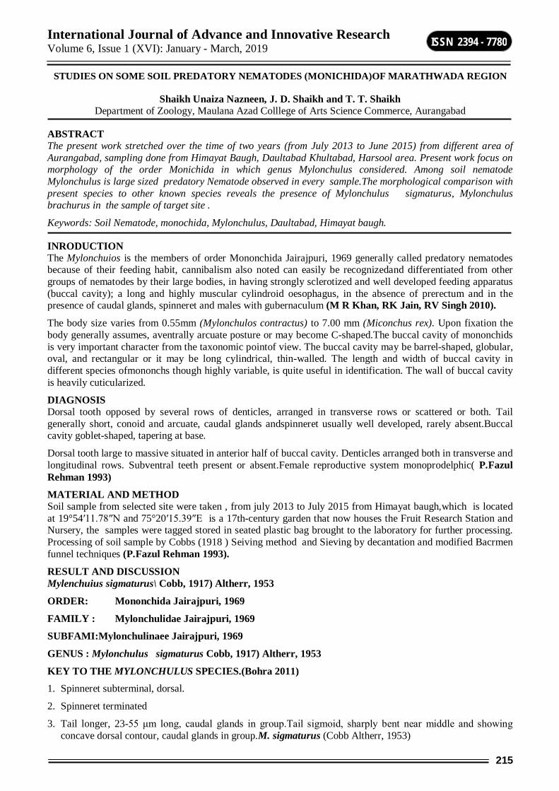
International Journal of Advance and Innovative Research Volume 6, Issue 1 (XVI): January - March, 2019
215
ISSN 2394 - 7780
STUDIES ON SOME SOIL PREDATORY NEMATODES (MONICHIDA)OF MARATHWADA REGION
Shaikh Unaiza Nazneen, J. D. Shaikh and T. T. Shaikh Department of Zoology, Maulana Azad Colllege of Arts Science Commerce, Aurangabad
ABSTRACT The present work stretched over the time of two years (from July 2013 to June 2015) from different area of Aurangabad, sampling done from Himayat Baugh, Daultabad Khultabad, Harsool area. Present work focus on morphology of the order Monichida in which genus Mylonchulus considered. Among soil nematode Mylonchulus is large sized predatory Nematode observed in every sample.The morphological comparison with present species to other known species reveals the presence of Mylonchulus sigmaturus, Mylonchulus brachurus in the sample of target site .
Keywords: Soil Nematode, monochida, Mylonchulus, Daultabad, Himayat baugh.
INRODUCTION The Mylonchuios is the members of order Mononchida Jairajpuri, 1969 generally called predatory nematodes because of their feeding habit, cannibalism also noted can easily be recognizedand differentiated from other groups of nematodes by their large bodies, in having strongly sclerotized and well developed feeding apparatus (buccal cavity); a long and highly muscular cylindroid oesophagus, in the absence of prerectum and in the presence of caudal glands, spinneret and males with gubernaculum (M R Khan, RK Jain, RV Singh 2010).
The body size varies from 0.55mm (Mylonchulos contractus) to 7.00 mm (Miconchus rex). Upon fixation the body generally assumes, aventrally arcuate posture or may become C-shaped.The buccal cavity of mononchids is very important character from the taxonomic pointof view. The buccal cavity may be barrel-shaped, globular, oval, and rectangular or it may be long cylindrical, thin-walled. The length and width of buccal cavity in different species ofmononchs though highly variable, is quite useful in identification. The wall of buccal cavity is heavily cuticularized.
DIAGNOSIS Dorsal tooth opposed by several rows of denticles, arranged in transverse rows or scattered or both. Tail generally short, conoid and arcuate, caudal glands andspinneret usually well developed, rarely absent.Buccal cavity goblet-shaped, tapering at base.
Dorsal tooth large to massive situated in anterior half of buccal cavity. Denticles arranged both in transverse and longitudinal rows. Subventral teeth present or absent.Female reproductive system monoprodelphic( P.Fazul Rehman 1993)
MATERIAL AND METHOD Soil sample from selected site were taken , from july 2013 to July 2015 from Himayat baugh,which is located at 19°54′11.78″N and 75°20′15.39″E is a 17th-century garden that now houses the Fruit Research Station and Nursery, the samples were tagged stored in seated plastic bag brought to the laboratory for further processing. Processing of soil sample by Cobbs (1918 ) Seiving method and Sieving by decantation and modified Bacrmen funnel techniques (P.Fazul Rehman 1993).
RESULT AND DISCUSSION Mylenchuius sigmaturus\ Cobb, 1917) Altherr, 1953
ORDER: Mononchida Jairajpuri, 1969
FAMILY : Mylonchulidae Jairajpuri, 1969
SUBFAMI:Mylonchulinaee Jairajpuri, 1969
GENUS : Mylonchulus sigmaturus Cobb, 1917) Altherr, 1953
KEY TO THE MYLONCHULUS SPECIES.(Bohra 2011) 1. Spinneret subterminal, dorsal.
2. Spinneret terminated
3. Tail longer, 23-55 μm long, caudal glands in group.Tail sigmoid, sharply bent near middle and showing concave dorsal contour, caudal glands in group.M. sigmaturus (Cobb Altherr, 1953)
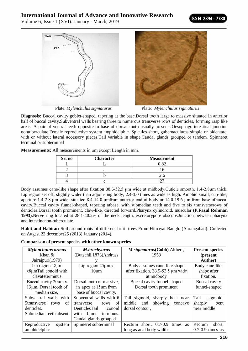
International Journal of Advance and Innovative Research Volume 6, Issue 1 (XVI): January - March, 2019
216
ISSN 2394 - 7780
Plate: Mylenchulus sigmaturus Plate: Mylenchulus sigmaturus
Diagnosis: Buccal cavity goblet-shaped, tapering at the base.Dorsal tooth large to massive situated in anterior half of buccal cavity.Subventral walls bearing three to numerous transverse rows of denticles, forming rasp like areas. A pair of ventral teeth opposite to base of dorsal tooth usually presents.Oesophago-intesitnal junction nontuberculate.Female reproductive system amphidelphic. Spicules short, gubernaculums simple or bidentate, with or without lateral accessory pieces.Tail variable in shape.Caudal glands grouped or tandem. Spinneret terminal or subterminal
Measurements: All measurements in μm except Length in mm.
Sr. no Character Measurment 1 L 0.82 2 a 16 3 b 2.6 4 c 27
Body assumes cane-like shape after fixation 38.5-52.5 μm wide at midbody.Cuticle smooth, 1.4-2.8μm thick. Lip region set off, slightly wider than adjoin- ing body, 2.4-3.0 times as wide as high. Amphid small, cup-like, aperture 1.4-2.8 μm wide, situated 8.4-14.0 μmfrom anterior end of body or 14.0-19.6 μm from base ofbuccal cavity.Buccal cavity funnel-shaped, tapering atbase, with submedian teeth and five to six transverserows of denticles.Dorsal tooth prominent, claw-like, directed forward.Pharynx cylindroid, muscular (P.Fazul Rehman 1993).Nerve ring located at 28.1-40.2% of the neck length, excretorypore obscure.Junction between pharynx and intestinenon-tuberculate.
Habit and Habitat: Soil around roots of different fruit trees From Himayat Baugh. (Aurangabad). Collected on Augest 22 december25 (2013) January (2014).
Comparison of present species with other known species
Mylonchulus armus Khan &
Jairajpuri(1979)
M.brachyurus (Butschli,1873)Andrass
y
M.sigmaturus(Cobb) Altherr, 1953
Present species (present Auther)
Lip region 18μm x8μmTail conoid with
clavateterminus
Lip region 25μm x 10μm
Body assumes cane-like shape after fixation, 38.5-52.5 μm wide
at midbody
Body cane-like shape after
fixation. Buccal cavity 20μm x 13μm. Dorsal tooth of
median size,
Dorsal tooth of massive, its apex at 15μm from base of buccal cavity.
Buccal cavity funnel-shaped Dorsal tooth prominent
Buccal cavity funnel-shaped
Subventral walls with 5transverse rows of denticles. Submedian teeth absent
Subventral walls with 6 transverse rows of DenticlesTail conoid with blunt terminus. Caudal glands grouped.
Tail sigmoid, sharply bent near middle and showing concave dorsal contour,
Tail sigmoid, sharply bent near middle
Reproductive system amphidelphic
Spinneret subterminal Rectum short, 0.7-0.9 times as long as anal body width.
Rectum short, 0.7-0.9 times as

International Journal of Advance and Innovative Research Volume 6, Issue 1 (XVI): January - March, 2019
217
ISSN 2394 - 7780
long as anal body width
Measurement L= 81-85 mm a= 29-30 µm b= 3.1-3.6µm
Measurement L= 1.0 -1.2 mm a= 22-25 µm b= 3.0-3.5 µ
Measurement L= 0.80.86 mm a= 16-24 µm b= 2.4 µm
Measurement L= 0.82 mm a= 16- µm b= 2.6
2. Mylonchulus brachurus (Bütschli, 1873) Altherr, 1953 ACooman (1995) rediscribe this species and according to himMylonchulus brachyuris is close to i) M. brevicaudatus(Cobb, 1917) Altherr, 1954, but can be distinguishedfrom it by its smaller buccal cavity (vs 30-36×17-20 μm); ii) M. subsimilis (Cobb, 1917) Meyl,1957, but differs from it in having a spinneret at tail tip(vs absence); iii) M. parabrachyurus (Thorne, 1924) Schneider, 1939, but differing in its shorter tail and subterminal spinneret .The measurements of present specimens fit wellwith the specimen described by Andrassy (1988) and Reddy Parwatha(1983).
ORDER : Mononchida Jairajpuri, 1969
FAMILY : Mylonchulidae Jairajpuri, 1969
SUBFAMILY : Mylenchulinae Jairajpuri, 1969
GENUS : Mylonchulus brachurus (Bütschli,1873) Altherr,1953
Plate: Mylenchulus brachurus
Plate: Mylonchulus brachurus (head) Plate: Mylonchulus brachurus
Plate: M.brachurus (MaleTail) Plate: Male antire body
International Journal of Advance and Innovative Research Volume 6, Issue 1 (XVI): January - March, 2019
218
ISSN 2394 - 7780
Plate: M.(mouth) Plate: M. ( tail) Plate: Mylonchulus Brachurus antirebody
Measurements: All measurements in μm except Length in mm
Sr. no Character Measurment 1 L 1.0 2 a 22 3 b 3.0 4 c 2.2
Diagnosis Body ventrally arcuate upon fixation, 42-52.5μm wide at midbody.Cuticle smooth, 1.4-2.8 μm thickLip region prominent, well set off from adjoining body, 2.3-3.2 times as wide as high.Amphid small,cup-like, aperture 2.8-4.2μmwide situated 4.2-11.2 μm from anteriorend of body or 18.2-22.4 μm from base of buccalcavity.Buccal cavity goblet-shaped, thick-walled, uniformlytapering to its base, with six rows of rasp-likedenticles and submedian teeth.Dorsal tooth large, directedforward.Pharynx cylindroid, muscular.Nervering located at 30.4-35.4% of the neck length; excretorypore small, situated behind the nerve ring, at 36.8-39.6% of the neck length.
Comparison of present species with other known species
Mylonchulus armus Khan& Jairajpuri, 1979
Mylonchulus armus Khan & Jairajpuri,
1979
M.brachyurus(Butschli, 1873) Andrassy 1958
Present species
Lip region 18μm x 8μm Tail conoid with
clavate terminus.
Spinneret subtermina Lip region 25μm x 10μm
Lip region 25μm x 10μm
Buccal cavity 20μm x 13μm. Dorsal tooth of median size, its apex at 15μm
Lip region 25μm x 10μm
Dorsal tooth of massive, its apex at 15μm from base of
buccal cavity.
Dorsal tooth of massive, its apex at 15μm from base of
buccal cavity.
Subventral walls with 5transverse
rows of denticles.
Submedian teeth absent
Dorsal tooth of massive, its apex
at15μm from base of buccal cavity.
Subventral walls with 6 transverse rows of
denticles.Tail conoid with blunt nterminus.
Caudal glands grouped.
Subventral walls with 6 transverse
rows of denticles.Tail
conoid with blunt terminus. Caudal glands grouped.
Reproductive system
amphidelphic
Subventral walls with 6 transverse rows of
denticles. Tail conoid with blunt terminus.
Caudal glands grouped.
Spinneret subterminal
Spinneret subterminal
Measurement L= 81-85 mm A= 29-30 µm b= 3.1-3.6 µm
Measuremen L= 1.0 -1.2 mm a= 22-25 µm b= 3.0-3.5 µm
Measurement L= 1.0 -1.2 mm
a= 22-25 µm b= 3.0-3.5
Measurement L=1.0 - mm a= 22 µm b= 3.0 µm
International Journal of Advance and Innovative Research Volume 6, Issue 1 (XVI): January - March, 2019
219
ISSN 2394 - 7780
DISCUSSION Discription of some known species of Monochids Bastian, 1865 and Mylenchullus (COBB, 1 916) are given by varios workers from different parts of India in which Padma Bohra(2011) describe soil nematodes from Rjisthan Dasgupa (1985) from WestBngal Fom Marathwada Thombre B.P (1982) did the work on soil nematode .According to him Six known species of mononchids belonging to the genera Mononchus Bastian, 1865 and Mylonchulus (Cobb, 1916) Altherr, 1953 were redescribed from Iran, viz. Mononchus aquaticus Coetzee, 1968; M. pulcher Andrássy, 1993; M. truncatus Bastian, 1865; Mylonchulus brachyuris (Bütschli, 1873) Altherr, 1953; M. paitensis Yeates, 1992 and M. sigmaturus (Cobb, 1917).Present author redescribed the species and it is first time after Thombre B.P (1982) discuss in Marathwada region .
ACKNOWLEDGEMENT Authors are thankful to principal Dr.Razaullah Khan for providing laboratory facility.
REFRENCES A. Cooman etal (1995) “On some predatory nematodes from the okango delta
Botswana”.Hydrobiologica. Vol 2 (5) pp234-250.
AbebeMekete, T. & Thomas, W.K. (2011). A critique of current methods in Nematode taxonomy.African Journal of Biotechnology 10: 312–32
Dasgupta M.K., Sen K., Mukherjee B., Rama K. (1985). “Plant parasitic nematodes associated with horticultural crops in West Bengal. p. 83–93. In: “Horticulture in West Bengal” (D. Mukherjee,ed.) Government of West Bengal, Calcutta, West Bengal, India, 105 pp.
J.Van Bezooijen (2006) “Method and Technique for Nematology.” revised (1984) edition. Department of Nematology, Agricultural University, pp11-25
Koohkan Mina and Ebrahim shookohi (2014) “study of some monochus (monochida) from iran”.Tropical zoology 27(3)pp1-40.
Padma Bohra(2011). “Pictorial handbook on plant and soil nematodesof Rajasthan.”(Published by the Director, Zool.Suru India, Kolkata) ISBN 978-81-8171-286-8.
Reddy Parvatha (1983) “Plant Nematology.” Agricultural publishing Academy Pratibha Printing Acadamy,New delhi Indi.pp87-98
M R Khan, RK Jain, RV Singh (2010) “Economically Important Plant Parasitic Nematodes Distribution ,ATLAS.”Directorate of Information and Publications of Agriculture,Indian council of agriculture Krishi Anusandhan Bhavan 1, Pusa 105p New Delhi 110 012 www.icar.org.in.
Mwangi, J. M. (2014) “Occurance and abundance of plant parasitic nematode in cabbage –ased cropping system in keniya.” VOL. 9, NO. 10, OCTOBER 2014 ISSN 1990-6145
P.Fazul Rehman (1993), “Some studies on plant parasiticnematode of India.”Ph.D Thesis Aligarh Muslim university p12-107.
Seinhorst J.W. 1959. A rapid method for the transfer of nematodes from fixative to anhydrous glycerine. Nematologica 4 (1): 67–69.
Seinhorst, J. W. (1962). On the killing, fixation and transferring to glycerine of the nematodes. Nematologica 8:29-32.
Thombre B.P (1982) “Studies on plant parasitic and soil nematode.”.Ph.D Thesis, Dr BabaSaheb Marathwada University.Maharashtra, India.

International Journal of Advance and Innovative Research Volume 6, Issue 1 (XVI): January - March, 2019
220
ISSN 2394 - 7780
SYNTHESIS AND BIOLOGICAL SCREENING OF NOVEL PYRAZOLE AND ISOOXAZOLE DERIVATIVES
D. W. Shinde1, S. S.Bhagat2, D. R. Nagargoje3 and C. H. Gill4 1,4Department of Chemistry, Dr. Babasaheb Ambedkar Marathwada University, Aurangabad
2 Department of Chemistry, R. B. Attal College, Georai, Dist. Beed. 3 Department of Chemistry, Arts, Science and Commerce College, Mokhada, Dist. Palghar
ABSTRACT A new series of 5-(4-(4-fluorophenylthio) phenyl)-4, 5-dihydro-3-phenyl-1H-pyrazoles and 5-(4-(4-fluorophenylthio) phenyl)-4,5-dihydro-3-phenyl isoxazoles have been prepared efficiently. In view of the vital role played by sulfur and fluorine in numerous pharmacological activities, we thought worthwhile to synthesize important pyrazole and isoxazole derivatives and investigate the biological activities. All the newly synthesized compounds have been screened for their in vitro antimicrobial activity.
Keywords: Pyrazole, Isoxazole and Antimicrobial.
INTRODUCTION Nitrogen containing heterocyclic compounds constitutes largest and most varied family of organic compounds with wide range of biological activities. Five member heterocycles having nitrogen occupy a significant place in synthetic organic chemistry [1]. In particular, pyrazolines are very important five member heterocycles containing two nitrogen atoms at 1-2 position. Pyrazolines and its derivatives reported to possess anti-inflammatory [2], antimicrobial [3], antibacterial [4], and anticancer activities [5]. Differently substituted pyrazolines are reported to show analgesic [6] immunosuppressive [7], antidepressant [8], antitubercular [9], antiviral [10], cardiovascular [11] and antidiabetic activities [12].
Elucidation of physicochemical properties of pyrazolines enables new data which shows considerable importance in pyrazolines aromatic system. Pyrazolines are well known for their antipyretic [13], agrochemicals [14], herbicide [15] and insecticide properties [16].Pyrazolines also work as non-steroidal drugs [17]. Initially, Antipyrine the first pyrazolines used in the management of inflammation and pain [18]. Many pharmacological aspects of pyrazolines have been explored; sulfur containing pyrazolines reported antinociceptive activity [19].Some thiocarbamoyl pyrazolines has shows greater antiamoebic activity compared to commonly used drug metronidazole [20].Certain sulfur containing pyrazolines derivatives reported CNS activity [21]. Sulfur based pyrazolines derivatives e.g. 5-(4-chlorophenyl)-4,5-dihydro-1-(4-methoxyphenyl)-3-(thiophen-2-yl)-1H-pyrazole and 5-(4-chlorophenyl)-4,5-dihydro-3-(thiophen-2-yl)pyrazole-1-carbothioamide as monoamine oxidase (MAO) plays major role in body degradation. The mentioned pyrazolines derivatives showed good MAO inhibition activity through interaction with monoamine oxidase [22] Pyrazolines with fluorine and β-amino acyl group showed blood glucose lowering property and found to be inhibitors of DPP-IV at submicromolar concentration [23].
Isoxazolines, an important class of azoles received much attention in the field of medicinal chemistry as potential anticancer agents [24]. Functionalized isoxazoline exhibited promising antineoplastic properties [25]. Isoxazoline are active pharmacophore in several important molecules which are used as intermediates for the synthesis of a wide variety of bioactive natural products [26, 27]. Isoxazolines reported antimicrobial [28] analgesic [29], antidiabetic [30], antimalarial [31], diuretic [32], hypolipidemic [33], and antihelmintic activities [34].
EXPERIMENTAL Material and Methods Open capillaries in a paraffin bath have been used to determine the melting points of newly synthesized compounds. The progress of the reaction was monitored by using precoated plates of silica gel G254 supplied by Merck. Infrared (IR) spectra (KBr disc) were recorded on a FTIR-4100 spectrometer and the absorption bands are expressed in cm-1. The 1H NMR spectra were recorded on a Bruker Advance II 400 MHz spectrometer using TMS as a reference standard. Macro mass spectrometer (Waters) by electro-spray method (ES) was used to record the mass spectra.
General procedure for the synthesis of substituted 5-(4-(4-fluorophenylthio)phenyl)-4,5-dihydro-3-phenyl-1H-pyrazole In an Round Bottom Flask compound 1a-h (0.001mol) was dissolved in 5ml DMSO. To this reaction, mixture (0.002 mol, 0.20ml) of hydrazine hydrate was added and the mixture was refluxed. The progress of the reaction was monitored with the help of TLC. After the completion of reaction, the reaction
International Journal of Advance and Innovative Research Volume 6, Issue 1 (XVI): January - March, 2019
221
ISSN 2394 - 7780
mixture was slowly added to the flask and the separated product was filtered and washed with cold water. The final compound was purified by recrystallization from ethanol gives 2(a-h).
General procedure for the synthesis of substituted 5-(4-(4-fluorophenylthio)phenyl)-4,5-dihydro-3-phenylisoxazoles Chalcones 1(a-h) (1 mmol), hydroxylamine hydrochloride (3 mmol) and potassium hydroxide (3 mmol) were dissolved in absolute ethanol (10 mL). The mixture was refluxed for 4-5 hr. The progress of reaction was monitored by TLC (Ethyl acetate: Pet ether). After completion of the reaction, the mixture was poured on crushed ice and neutralized with 2N HCl. The precipitate was separated by filtration, washed with cold water, and crystallized from ethanol. The percentage yield and physical constants were recorded in Table 1.
2-(5-(4-((4-fluorophenyl)thio)phenyl)-4,5-dihydro-1H-pyrazol-3-yl)phenol (2b) 1H NMR (400 MHz, DMSO-d6, δppm): 2.32(s,3H,CH3), 3.2 (dd, 1H, CH2 ), 3.65 (dd, 1H, CH2 ), 3.7(s,1H,CH), 7.0-7.32 (m, 12H, Ar-H and pyrazole-H),δ 11.50 (s, 1H, -OH);IR (KBr, cm-1): 3340 (N-H), 3156 (OH), 1480 and 1580 (Aromatic C=C); ES-MS: m/z: 378
2-(5-(4-((4-fluorophenyl)thio)phenyl)-4,5-dihydro-1H-pyrazol-3-yl)phenol (2c) 1H NMR (400 MHz, DMSO-d6, δppm):, 3.25 (dd, 1H, CH2 ), 3.45 (dd, 1H, CH2 ), 3.8(s,1H,CH), 6.9-7.5 (m, 13H, Ar-H and pyrazole-H),δ 11.45 (s, 1H, -OH)
IR (KBr, cm-1): 3332 (N-H), 3154 (OH), 1482 and 1588 (Aromatic C=C); ES-MS: m/z: 364
2, 4-dichloro-6-(5-(4-((4-fluorophenyl)thio)phenyl)-4,5-dihydro-1H-pyrazol-3-yl)phenol (2g) 1H NMR (400 MHz, DMSO-d6, δppm):, 3.1 (dd, 1H, CH2 ), 3.55 (dd, 1H, CH2 ), 7.01-7.42 (m, 11H, Ar-H and pyrazole-H),δ 11.63 (s, 1H, -OH);IR (KBr, cm-1): 3338 (N-H), 3153 (OH), 1487 and 1587 (Aromatic C=C); ES-MS: m/z: 432
2-(5-(4-((4-fluorophenyl) thio) phenyl)-4, 5-dihydroisoxazol-3-yl)phenol (3c) 1H NMR (400 MHz, DMSO-d6, δppm):, 3.64 (dd, 1H, isoxazoline C4-H), 3.90 (dd, 1H, isoxazoline C4-H), 5.4 (t, 1H, isoxazoline CH), 6.84 to 7.74 (m,12H,Ar-H),δ 13.25 (s, 1H, -OH);IR (KBr, cm-1): 3460 (OH), 3060 (C-H), 1689(C=C), and 1489 (Aromatic C=N);
ES-MS: m/z: 367.2
4-chloro-2-(5-(4-((4-fluorophenyl) thio) phenyl)-4, 5-dihydroisoxazol-3-yl) phenol (3f) 1H NMR (400 MHz, DMSO-d6, δppm):, 3.52 (dd, 1H, isoxazoline C4-H), 3.80 (dd, 1H, isoxazoline C4-H), 5.2 (t, 1H, isoxazoline CH), 6.90 to 7.72 (m,11H,Ar-H),δ 12.25 (s, 1H, -OH);IR (KBr, cm-1): 3465 (OH), 3065 (C-H), 1680(C=C), and 1480 (Aromatic C=N);
ES-MS: m/z: 399.1
2,4-dichloro-6-(5-(4-((4-fluorophenyl)thio)phenyl)-4,5-dihydroisoxazol-3-yl)phenol (3g) 1H NMR (400 MHz, DMSO-d6, δppm):, 3.42 (dd, 1H, isoxazoline C4-H), 3.70 (dd, 1H, isoxazoline C4-H), 5.2 (t, 1H, isoxazoline CH), 6.85 to 7.50 (m,10H,Ar-H),δ 12.25 (s, 1H, -OH);IR (KBr, cm-1): 3445 (OH), 3052 (C-H), 1675(C=C), and 1489 (Aromatic C=N);
ES-MS: m/z: 433.2
RESULTS AND DISCUSSION Chemistry The starting precursor’s (E)-1-(2-hydroxyphenyl)-3-(4-(phenylthio)phenyl)prop-2en-1-ones 1a-h was prepared using o-hydroxyacetophenone and 4-F(phenylthio)benzaldehyde ethanol. The mixture was irradiated under ultrasonication for 4-5 hr. The reaction of (E)-1-(2-hydroxyphenyl)-3-(4-(phenylthio) phenyl) prop-2en-1-ones 1a-h with hydrazine hydrate in ethanol gave the target products 2a-h (Scheme 1). Compound (E)-1-(2-hydroxyphenyl)-3-(4-(phenylthio) phenyl) prop-2en-1-ones 1a-h, with hydroxylamine hydrochloride at reflux condition gave the target products 3a-h (Scheme 2). Structural assignments to the newly synthesized compounds were based on their IR, 1H-NMR, Mass spectral data.
1 a-h 2a-h
Scheme-1: Synthesis of pyrazole 2a-h
International Journal of Advance and Innovative Research Volume 6, Issue 1 (XVI): January - March, 2019
222
ISSN 2394 - 7780
O
S
F
NH2OH.HCl
EtOH/Reflux
N O
S
F
OH OHR1
R2
R3
R1
R2
R3
1a-h 3a-h
Scheme-2: Synthesis of isoxazole 3a-h
Table-1: Physical data of compounds 2a-h and 3a-h Comp. R1 R2 R3 Yield (%) M. P. (°C)
2a H CH3 Cl 77 55-57 2b H H CH3 65 99-101 2c H H H 72 120-122 2d CH3 H CH3 65 52-54 2e H H F 64 56-58 2f H H Cl 68 90-92 2g Cl H Cl 74 148-150 2h H H Br 72 140-142 3a H CH3 Cl 72 154-156 3b H H CH3 70 145-147 3c H H H 72 152-154 3d CH3 H CH3 69 142-144 3e H H F 65 148-150 3f H H Cl 67 98-100 3g Cl H Cl 64 204-206 3h H H Br 68 166-168
ANTIMICROBIAL ACTIVITY Antimicrobial activity of all the synthesized compounds was determined by the well-diffusion method Two Gram-positive bacterial strains E. coli and A. flavus strains used to study and investigate the antimicrobial activities. The bacterial liquid cultures were prepared in fusion broth. All the newly synthesized compounds were dissolved in DMSO at concentration of 1 mg/mL. The antibacterial activity of DMSO was checked against the test organisms and was found to be nil. In Petri dishes, the molten nutrient agar was poured and allowed to solidify. The holes of 10 mm diameter were punched using a cork borer and completely filled with the test solutions. The culture plates were incubated for 24 hr at 36 °C. The inhibition zone around the holes in each plate was measured after 24 hr. The diameter of inhibition zone and minimal inhibitory concentrations (MICs) showed the antibacterial activity Table 2.
Table-2: In vitro antibacterial screening of compounds (2a-h) and (3a-h)
Compound Inhibition zone (mm) (Escherichia coli)
Inhibition zone (mm) (Aspergillus flavus)
2a Nil Nil 2b Nil Nil 2c Nil Nil 2d Nil Nil 2e 02 Nil 2f Nil Nil 2g Nil Nil 2h Nil Nil 3a Nil Nil 3b Nil Nil 3c Nil Nil 3d Nil Nil 3e Nil Nil 3f 01 Nil 3h Nil Nil
Control (Solvent) Nil Nil
International Journal of Advance and Innovative Research Volume 6, Issue 1 (XVI): January - March, 2019
223
ISSN 2394 - 7780
The newly synthesized compounds 2a-h and 3a-h were evaluated for in-vitro antimicrobial activities. The preliminary screening data showed that among active compounds and exhibited moderate activity.
CONCLUSIONS In conclusion, synthesis, and antibacterial activities of a novel series of 5-(4-(4-fluorophenylthio) phenyl)-4, 5-dihydro-3-phenyl-1H-pyrazole 2a-h, and 6a-h and 5-(4-(4-fluorophenylthio) phenyl)-4, 5-dihydro-3-phenylisoxazoles 3a-h, have been presented for the first time via synthetic procedure in good yield. In addition, compounds showed moderate activity, indicating the future scope for optimization.
ACKNOWLEDGEMENT D.W.S. is thankful to the Head, Department of Chemistry, Dr. Babasaheb Ambedkar Marathwada University, Aurangabad and Principal, Shivaji Arts, Commerce and Science College, Kannad for providing laboratory facilities.
REFERENCES 1. Yusuf, M.; Jain, P. Arab. J. Chem. 2014, 7, 553.
2. Viveka, S.; Sharma, D. P.; Nagaraja, G. K.; Ballav, S.; Kerkar, S. Eur. J. Med. Chem. 2015, 101, 442.
3. Bano, S.; Alam, M. S.; Javed, K.; Dudeja, M.; Das, A. K.; Dhulap, A. Eur. J. Med. Chem. 2015, 95, 96.
4. Karad, S. C.; Purohita, V. B.; Thakor, P.; Thakkar, V. R.; Raval, D. K. Eur. J. Med. Chem. 2016, 112, 270.
5. Qin, H. L.; Shang, Z. P.; Jantan, I.; Tan, O. U.; Hussain, M. A.; Sher, M.; Bukhari, S. N. A. RSC Adv. 2015, 5, 46330.
6. El-Sehemi, A.; Bondock, S. Med. Chem. Res. 2014, 23, 827.
7. Yusuf, M.; Kaur, A.; Abid, M. J. Hetro. Chem. 2017, 54, 2536.
8. Mathew, B.; Suresh, J.; Anbazhagan, S. Biomed. Agi. Path. 2014, 4, 327.
9. Ahmad, A.; Husain, A.; Khan, A.; Mujeeb, M.; Bhandari, A. J. Sau. Chem. Soc. 2016, 20, 577.
10. Jadav, S.; Kaptein, S.; Timiri, A.; Burghgraeve, T.; Badavath, V. N.; Ganesan, R.; Sinha, B. N.; Neyts, J.; Leyssen, P.; Jayaprakash, V. Bioorg. Med. Chem. Lett. 2015, 25, 1747.
11. Malhotra, V.; Pathak, S.; Nath, R.; Mukerjee, D.; Shanker, K. Ind. J. Chem. 2002, 41B, 1310.
12. Kharbanda, M.; Alam, S.; Hamid, H.; Javed, K.; Shafi, S.; Ali, Y.; Alam, P.; Pasha, M. A. Q.; Dhulap, A.; Bano, S.; Nazreen, S.; Haider, S. Bioorg. Med. Chem. Lett. 2014, 24, 5298.
13. Souza, F. R.; Souza, V. T.; Ratzlaff, V.; Boges, L. P.; Oliveira, M. R.; Bonacorso, H. G.; Zanatta, N.; Martins, M. A. P.; Mello, C. F. Eur. J. Pharm. 2002, 451, 141.
14. Yang, C.; Liu, W.; He, Z.; He, Z. Org. Lett. 2016, 18, 4936.
15. Xie, M. S.; Guo, Z.; Qu, G. R.; Guo, H. M. Org. Lett. 2018.
16. Lu, L.; Cassayre, J. Y.; Berthon, G.; Qacemi, M.E.; Wu, Y. US Patent 2017, US9776994b2.
17. Khaled, R. A. A.; Eman, K. A. A.; Wael, A. A. F.; Gehan, M. K. Med. Chem. Res. 2015, 24, 2632.
18. El Sayed, M. T.; El Sharief, M. A. M.; Zarie, E. S.; Morsy, N. M.; Elsheakh, A. R.; Voronkov, A.; Berishvili, V.; Hassan, G. S. Bioorg. Med. Chem. Lett. 2018, 28, 952.
19. Kaplancikli, Z. A.; Zitouni, G. T.; Ozdemir, A.; Can, O. D.; Chevallet, P. Eur. J. Med. Chem. 2009, 44, 2606.
20. Afrose, E. Dess. Dept. of pharm. 2017, 19.
21. Havrylyuk, D.; Roman, O.; Lesyk, R. Eur. J. Med. Chem. 2016, 113, 145.
22. Das, N.; Dash, B.; Dhanawat, M.; Shrivastava, S. K. Chem. Paper 2012, 66, 67.
23. Marella, A.; Ali, M. R.; Alam. M. T.; Saha, R.; Tanwar, O.; Akhter, M.; Shaquiquzzaman, M.; Alam, M. M. Min. Rev. Med. Chem. 2013, 13, 921. Kamal, A.; Reddy, S.; Ramaiah, M. J.; Dastagiri, D.; Bharathi, E. V.; Azhar, M. A.; Sultana, F.; Pushpavalli, S. N. C. V. L.; Bhadra, M. P.; Juvekar, A.; Sen, S.; Zingde, S. Eur. J. Med. Chem. 2010, 45, 3924.
24. Kaur, K.; Kumar, V.; Sharma, K. Gupta, G. K. Eur. J. Med. Chem. 2014, 77. 121.

International Journal of Advance and Innovative Research Volume 6, Issue 1 (XVI): January - March, 2019
224
ISSN 2394 - 7780
25. Zhu, L.; Yu, H.; Xu, Z.; Jiang, X.; Lin, L.; Wang, R. Org. Lett. 2014, 16, 1562.
26. Schmidt, E. Y.; Tatarinova, I. V.; Ivanova, E. V.; Zorina, N. V.; Ushkov, I. A.; Trofimov, B. A. Org. Lett. 2013, 15, 104.
27. Ismail, T.; Shafi, S.; Singh, S.; Sidiq, T.; Khajuria, A.; Rouf, A.; Yadav, M.; Saikam, V.; Singh, P.; Alam, M. S.; Islam, N.; Sharma, K.; Samapath Kumar, H. M. Eur. J. Med. Chem. 2016, 123, 90.
28. Bano, S.; Alam, M. S.; Javed, K.; Dudeja, M.; Das, A. K.; Dhulap, A. Eur. J. Med. Chem. 2015, 95, 96.
29. Pember, S. O.; Mejia, G. L.; Price, T. J.; Pasteris, R. J. Bioorg. Med. Chem. Lett. 2016, 26, 2965.
30. Gomha, S. M.; Riyadh, S. M.; Abdallam, M. M. Curr. Org. Syn. 2015, 12, 220.
31. Kumar, K. S. V.; Lingaraju, G. S.; Bommegowda, Y. K.; Vinayaka, A. C.; Bhat, P.; Kumar, C. S. P.; Rangappa, K. S.; Channe Gowda, D.; Sadashiva, M. P. RSC Adv. 2015, 5, 90408.
32. Valizadeh, H.; Vesally, E.; Dinparast, L. J. Het. Chem. 2012, 49, 106.
33. Kale, M.; Patwardhan, K. Der Pharma Chemica 2013, 5, 213.
34. Uma Maheswari, S.; Perumal, S. Tet. Lett. 2012, 53, 2012.

International Journal of Advance and Innovative Research Volume 6, Issue 1 (XVI): January - March, 2019
225
ISSN 2394 - 7780
SYNTHESIS, CHARACTERIZATION AND ANTIMICROBIAL ANALYSIS OF VARIOUS SUBSTITUTED 2-(5-(3-(5-BROMOTHIOPHEN-2-YL)-1-PHENYL-1H-PYRAZOL-4-YL)-1H-
PYRAZOL-3-YL) PHENOLS
Sunil S. Bhagat1, Deepak R. Nagargoje2, Dadasaheb W. Shinde3, Dattatraya S. Ghotekar4 1Department of Chemistry, R. B. Attal Arts, Science and Commerce College, Georai, Beed
2Department of Chemistry, Arts, Science and Commerce College, Mokhada, Dist. Palghar 3Department of Chemistry, Shivaji Arts, Science and Commerce College, Aurangabad
4Department of Chemistry, NVP Mandal’s Arts, Commerce and Science College, Nashik
ABSTRACT The title compounds various substituted2-(5-(3-(5-bromothiophen-2-yl)-1-phenyl-1H-pyrazol-4-yl)-1H-pyrazol-3-yl) phenols2(a-h)have been synthesized from chromones 1(a-h) by refluxing with potassium hydroxide. The structures of all newly synthesized compounds have been confirmed by IR, 1H NMR and Mass spectral data. The synthesized compounds have been screened for their antimicrobial activity. Some of the compounds show moderate antimicrobial activity as compared to the reference drugs Ciprofloxacin and Fluconazole.
Keywords: Chromones, Antimicrobial activity
INTRODUCTION Pyrazole is characterized by a 5-membered heterocyclic ring structuremade up of three carbon atoms and two nitrogenatoms in adjacent positions.1-pyrazolyl-alanine,the first natural pyrazole, was obtained from watermelonseedsin 1959.It has pharmacological effects on humans, they are rare innature.They have application inpharmaceuticalindustry and agrochemicals as active pharmaceuticals and herbicides.The currentachievement ofpyrazole COX-2 inhibitor has moreemphasized theprominence of these heterocyclic rings in medicinalchemistry. A logicalexamination of this class ofheterocyclic lead has shown that pyrazole containingpharmacoactive agents play vital role inmedicinal chemistry. The occurrence of pyrazolenuclei in naturally active molecules has encouragedthe need forwell-designed and effectivemethods to make theseheterocyclic leadi.
Nitrogen-linked heterocyclic compounds gained substantialconsiderationin modern times because of their pesticidal andmedicinalsignificanceii-iv. Pyrazole derivatives are important in pesticide industry and extensively used because of their antiviralv, antitumorvi,anti-inflammatoryvii, antibacterialviii, herbicidalix, insecticidalx, fungicidal activitiesxi, Angiotensin-I-converting enzymes inhibitoryxii, molluscidalxiii, and ulcerogenic activityxiv.
Scheme-1
EXPERIMENTAL SECTION General Procedure for the synthesis of2-(5-(3-(5-bromothiophen-2-yl)-1-phenyl-1H-pyrazol-4-yl)-1H-pyrazol-3-yl)-4-methylphenol(2b):Compound 1b (0.003 mol) was taken in 100 ml RBF with 15 ml ethanol. To this reaction mixture 1 ml hydrazine hydrate and 0.5 gm KOH were added and the contents were heated under reflux for five hour. After completion of reaction (monitored by TLC), the contents were cooled to room temperature and poured over crushed ice and acidified with HCl. The solid thus obtained was separated by filtration and crystallized from ethanol. The compounds 2 (a-h) were prepared by following the above procedure. The physical data of the compounds 2 (a-h)were recorded in Table 1. Their structures have been confirmed by 1HNMR, Mass and IR spectra.
International Journal of Advance and Innovative Research Volume 6, Issue 1 (XVI): January - March, 2019
226
ISSN 2394 - 7780
Table-1: Physical data of compounds 2 (a-h) Comp. R1 R2 R3 M.P. (oC) Yield (%)
2a H H H 168-170 78 2b H H CH3 184-186 65 2c H H Cl 136-138 78 2d Cl H Cl 248-250 71 20 H H F 168-170 68 20 H CH3 Cl 238-240 72 20 H H Br 230-232 66 20 CH3 H CH3 208-210 70
SELECTED SPECTRAL DATA OF SOME REPRESENTATIVE COMPOUNDS 2-(5-(3-(5-bromothiophen-2-yl)-1-phenyl-1H-pyrazol-4-yl)-4,5-dihydro-1H-pyrazol-3-yl)-4-chloro-5-methylphenol(2b):IR (cm-1):1054(Ar-Br), 1264(C-O),1534(C=N),1650(Ar C=C), 2835(Ar-CH3),3263(N-H),3438(O-H).1H NMR(DMSO)δ ppm: 2.300 (s, 3H, CH3), 6.8251(s,1H,Ar-H)H), 6.8510 (s, 1H, Ar-H), 6.9621-6.9819(m, 1H, Ar-H), 7.0534-7.1103 (m, 1H, Ar-H), 7.3255-7.4335(m, 4H, Ar-H),7.4952-7.5143(d, 1H, Ar-H,J=7.64 Hz) ,7.5143-7.5333(d, 1H, Ar-H, J=7.6 Hz),7.8733-7.8442(dd, 1H, Ar-H, J= 4.48 & 4.12 Hz), 7.9745(s,1H, Ar-H), 8.6148(s, 1H,Pyrazole-H),10.7410 (s, 1H,N-H),13.3261 (s, 1H, Ar-OH).ES-MS (m/z): 475(M-1), 477(M+2).
RESULTS AND DISCUSSION The pyrazole derivatives were synthesized successfully in moderate to good yields. The newlysynthesized compounds were identified on the basis of melting point range, IR, 1H NMR, Mass spectral analysis. All the newly synthesized derivatives were screened for antimicrobial activity using disc diffusion method.
Antimicrobial activity: Compounds 2(a-h) were screened for their in vitro antimicrobial activity against Escherichia coli (ATCC 25922), Pseudomonas aeruginosa (ATCC 27853), Staphylococcus aureus (ATCC 25923), Staphylococcus albus, Klebsiella pnuemoniae using Ciprofloxacin as a reference standard drug by paper disc diffusion method. Antifungal activity was evaluated against Candida sp. using Fluconazole as standard drug. All the tests were evaluated at 100 μg/ml concentration. The culture media was Muller Hinton agar. The zone of inhibition was measured in mm after 24 hr of incubation at 37οC. DMSO is used as control.
Microbial data for corresponding compounds is summarized in Table 2.
Table-2: In-vitro antimicrobial activity of various substituted 2-(5-(3-(5-bromothiophen-2-yl)-1-phenyl-1H-pyrazol-4-yl)-1H-pyrazol-3-yl) phenols 2(a-h).
Sr. No.
Compound No.
Inhibition Zone Diameter (mm)
Candida sp. S. aureus
S.albus Klebsiella pnuemoniae
E. coli Pseudo monas sp.
1. 20a 3.9 3.9 - - - 5 2. 20b 4 8 6 - 8 3 3. 20c 7 9 12 10.8 9 - 4. 20d 6 4 7 10.2 5 4 5. 20e 9 - 10 1.9 13 12 6. 20f 7 - 7 2 11 10 7. 20g 8 - 9 5 6 8 8. 20h 4 - 6 5 5 7 9. Control 8 3 3 4 6 10
10. Ciprofloxacin --- 20 22 22 21 23 11. Fluconazole 23 --- --- --- --- ---
ACKNOWLEDGEMENTS The authors are thankful to The Head, Department of Chemistry, Dr. Babasaheb Ambedkar Marathwada University, Aurangabad for providing laboratory facilities and Uday Khedkar, Director, BAC-TEST Laboratory, Nashik for antimicrobial analysis. One of the authors (SSB) is thankful to Principal Dr. R. V. Shikhare for constant encouragement.

International Journal of Advance and Innovative Research Volume 6, Issue 1 (XVI): January - March, 2019
227
ISSN 2394 - 7780
REFERENCES 1) Eicher, T.; Hauptmann, S.The Chemistry of Heterocycles: Structure, Reactions, Syntheses, and
Applications, Edition IInd, 2003, Wiley-VCH, ISBN 3527307206.
2) Liu, X.H.; Chen, P.Q.; Wang, B.L.; Li, Y.H.; Wang, S.H.; Li, Z.M. Bioorg. Med. Chem. Lett.(2007), 17, 3784.
3) Liu, X.H.; Zhang, C.Y.; Guo, W.C.; Li, Y.H.; Chen, P.Q.; Wang, T; Dong, W. L.; Sun, H.W.; Li, Z.M. J.Enzym.Inhib.Med. Chem. (2009), 73, 320.
4) Liu, X.H.; Shi, Y.X.; Ma, Y.; Zhang, C.Y.; Dong, W.L.; Li, P.; Wang, B.L.; Li, B.J.; Li, Z.M. Eur. J. Med. Chem. (2009), 44, 2782.
5) (a) Sayed, H.H.; Ali, M.A. Phosphorus Sulfur Silicon Relat Elem. (2008), 183, 156;(b) Rashad, A. E.; Hegab, M. I.; Abdel-Megeid, R. E.; Micky, J. A.; Abdel- Megeid, F. M. E. Bioorg. Med.Chem. (2008), 16, 7102;(c) Park, H.J.; Lee, K.; Park, S.J.;Ahn, B.; Lee, J.C.; Cho, H.; Lee, K.I. Bioorg Med Chem Lett.(2005), 15, 3307;(d).Peng-Cheng, L.; Zhu, H.; Li, H.; Sun, J.; Zhou, Y.Bioorg.Med. Chem.(2010), 18, 4606;(e)Barnes, B. J.; Izydore, R. A.; Hall, I. H. Anticancer Res., (2001), 21(4A), 2313; (f) Baraldi, P. G.; Balboni, G.; Pavani, M. G.; Spalluto, G.; Tabrizi, M. A.; Clercq, E. D. J. Med. Chem.( 2001), 44(16), 2536.
6) Baraldi, P. G.; Pavani, M. G.; Nunez, M. C.; Brigidi, P.; Vitali, B.; Gambari, R., Bioorg. Med. Chem., (2002), 10(2), 449.
7) Burguete, A.; Pontiki, E.; Hadjipavlou-Litina, D.; Villar ,R.; Vicente, E.; Solano, B.;Ancizu, S.; Perez Silanes, S.;Aldana, I.; Monge, A. Bioorg Med Chem Lett. (2007), 17, 6439.
8) (a)Farghaly, A.A.; Vanelle, P.; El-Kashef, H.S. Heterocycl Comm. (2005), 11, 255;(b) Sridhar, R.; Perumal ,P. J.; Etti, S.; Shanmugam, G.; Ponnuswamy, M. N.; Prabavathy, V. R.; Mathivanan, N.Bioorg. Med. Chem. Lett.(2004), 14, 6035.
9) He, F.Q.; Liu, X.H.; Wang, B.L.; Li, Z.M. Heteroatom Chem. (2008), 19, 21.
10) Jiang, L.; Wang, L.Z.; Wang, L.Y.; Xie, X.Y. HechengHuaxue.( 2007), 15, 576.
11) Yang, X.D.; Yu, Y.Y. Struct Chem(.2008), 19, 693.
12) Bonesi, M.; Loizzo, M. R.; Statti, G. A.; Michel, S.; Tillequin, F.; Menichini. Bioorg. Med. Chem. Lett.(2010), 20, 1990.
13) Nawwar, G. A.; Swellem, R. H.; Ibrahim, A. M. Arch. Pharm., (1994), 17(2), 66.
14) Ochi, T.; Sugiyama, A. Y.; Ohkubo, Y.; Sakane, K.; Tanaka, H. J. Pharmacol.(2001), 85(2), 175.

International Journal of Advance and Innovative Research Volume 6, Issue 1 (XVI): January - March, 2019
228
ISSN 2394 - 7780
MIXED LIGAND COMPLEXES OF ZINC METAL ION WITH DRUG CEFOTAXIME DRUG AND AMINO ACIDS IN AQUEOUS MEDIA
Shailendrasingh Thakur1, Mazahar Farooqui2, Ramesh Ware3 1,3Milliya Art’s Science & Management Science College, Beed
2Dr. Rafiq Zakaria College for Women, Aurangabad
ABSTRACT In the present study the stability constant of the mixed ligand complexes of zinc (II) ion with antibacterial drug Cefotaxime sodium as primary ligand and the amino acids viz. glycine, DL-alanine, L-glutamic acid, DL-isoleucine, DL-methionine, DL-β-phenyl alanine, DL-serine and DL-valine as secondary ligands were determined pH metrically in 20%(v/v) ethanol-water medium at 25 °C and at an ionic strength of 0.1 M NaClO4. The formation of complex species has been evaluated by SCOGS computer program and discussed in terms of various relative stability parameters.
Keywords: stability constant, cefotaxime drug, amino acids, mixed ligand complexes.
INTRODUCTION Stability of metal complexes with medicinal drugs plays a major role in the biological and chemical activity. Metal Complexes are widely used in various fields, such as biological processes, pharmaceuticals, separation techniques, analytical processes etc. Amino acids are the structural unit of proteins. These are essential constituents of all living cells and contain one or more amino and carboxylic groups and have good coordination sites for the metal complexation. In continuation of earlier work with complexation of medicinal drug1-22, we study ternary complexes of zinc metal ion with antibacterial drug Cefotaxime sodium (CFO) as primary ligand and a series of eight aminoacids as secondary ligands in ethanol-water medium at 25°C and at 0.1 M NaClO4 ionic strength.
Figure-1: Cefotaxime sodium (molecular formula C16H16N5O7NaS2)
Experimental: I. Materials and Solution: The ligand CFO is soluble in double distilled water. NaOH, NaClO4, HClO4 & metal salts were of AR grade. The solutions used in the potentiometric titration were prepared in double distilled water. The NaOH solution was standardized against oxalic acid solution and standard alkali solution was again used for standardization of HClO4.The metal salt solutions were also standardized using EDTA titration. All the measurements were made at 25˚C in 20% ethanol-water mixture at 0.1M NaClO4 strength. The thermostat model SL-131 was used to maintain the temperature constant.The pH measurement were made using a digital pH meter model Elico L1-120 in conjunction with a glass and reference calomel electrode (reading accuracy ±0.01) The pH-meter was adjusted with buffer of pH 4.00,7.00 and 9.18.
Potentiometric procedure: For evaluating the protonation constant of the ligand & the formation constant of the complexes in 20% ethanol-water mixture with different metal ions we prepared the following six sets of solutions.
(i) HClO4 (A)
(ii) HClO4+Drug (A+ L)
(iii) HClO4+Drug+ Metal (A+ L+ M)
(iv) HClO4+Amino acid (A+ R)
(v) HClO4+Amino acid + Metal (A+ R+ M)
(vi) HClO4+Drug +Amino acid + Metal (A+L+R+ M)
The above mentioned sets prepared by keeping M:L:R ratio, the concentration of perchloric acid & sodium perchlorate were kept constant for all sets. The volume of every mixture was made upto 50 ml with double

International Journal of Advance and Innovative Research Volume 6, Issue 1 (XVI): January - March, 2019
229
ISSN 2394 - 7780
distilled water. The test solutions were magnetically stirred, NaOH was added stepwise and pH reading was recorded until stable values, within ± 0.002 pH units were obtained.Graphs were obtained by plotting pH vs volume of NaOH added.These data were used to determine the pKa of ligands and logK values of metal complexes of primary and secondary ligands. The equilibrium constants of ternary complexes were calculated by using SCOGS program. The total concentrations of metal ions, free metals, free ligands and various possible species that are formed during complexation were obtained as computer output of program.
Table-1: Proton-ligand stability constant and metal-ligand stability constant of Cefotaxime drug and amino acids with zinc (II) at 0.1M ionic strength in 20% (v/v)ethanol-water medium
Ligands Proton-ligand stability constant Metal-ligand stability constant pK1 pK2 logK1 logK2 logβ
Cefotaxime sodium 3.156 10.764 6.328 5.651 11.979 Glycine 2.472 9.582 5.525 4.239 9.764
DL -Alanine 2.364 9.658 4.492 3.561 8.053 Glutamic acid 2.501 4.416 2.921 2.744 5.665 DL -Isoleucine 2.654 9.624 6.960 5.071 12.031 DL -Methionine 2.303 9.079 4.883 3.655 8.538
DL-β-Phenyl alanine 2.255 9.174 4.863 4.056 8.919 DL -Serine 2.344 8.983 4.644 3.476 8.120 DL -Valine 2.488 9.501 5.149 4.175 9.324
Table-2: Parameters based on some relationship between formations of mixed ligand complexes of Zn (II) with CFO drug and amino acids.
Amino Acids β111 β 20 β02 KL KR Kr ∆logk Glycine 10.8524 11.9789 9.7634 4.5245 5.3277 0.9983 -1.0002
DL -Alanine 10.8198 11.9789 8.0529 4.4919 6.3278 1.0803 -0.0004 Glutamic acid 8.7479 11.9789 5.6647 2.420 5.8269 0.9916 -0.5010 DL -Isoleucine 13.2874 11.9789 12.029 6.9595 6.3277 1.1069 -0.0002 DL -Methionine 10.7107 11.9789 8.538 4.3828 5.8276 1.0441 -0.5003
DL-β-Phenyl alanine 10.1897 11.9789 8.918 3.8618 5.3272 0.9752 -1.0007 DL -Serine 10.972 11.9789 8.1205 4.6441 6.3277 1.0918 -0.0002 DL -Valine 11.4762 11.9789 9.3243 5.1483 6.3268 1.0774 -0.0011
RESULT AND DISCUSSION Binary complex: The proton ligand stability constants (pKa) of drugs and amino acids were calculated by point wise and half integral method.The metal ligand stability constant (logK) of Zn(II) transition metal complexes with antibacterial drugs were calculated by using Calvin Bjerrum titration techniques as adopted by Irving and Rossotti. Titration curves were obtained for different sets. During titration no precipate was formed indicating that there is no tendency to form hydroxo complexes. The stability constants of the formed complexes were investigated in the pH range of 4-6.The mean value the average number of protons associated with the
ligand , at different pH values were calculated. The pKa values were determined from . Similarly i.e metal ligand formation numbers, which can be defined as average number of ligand molecules co-ordinate to
the metal ions, were also obtained using Irving & Rossotti method. The values obtained between 0.2 to 0.8
indicates 1:1 complexation and when lies in between 1.2 to 1.8 indicate 1:2 complexation. The values of proton ligand stability constants(pKa) and metal ligand stability constant (logK) are represented in Table 1. Since we got between 0.2 to 0.8 and 1.2 to 1.8 indicating 1:1 and 1:2 complex formation. The order of logK1
> logK2 is commonly observed. The reason is statistical effect, statistically coordination of a second molecule is difficult when compare to the first due to availability of less number of coordinating sites on the metal ion for the second ligand.
Mixed ligand complexes: The formation of 1:1:1 mixed ligand complex were identified by the pH of precipitation of ML, MR and MLR titration curves.These curves indicate the higher value of pH of precipitation of ternary system than corresponding binary systems. The relative stabilities of mixed ligand complexes were quantitatively expressed in terms of ∆logK, Kr, KL and KR values which are defined by equations:

International Journal of Advance and Innovative Research Volume 6, Issue 1 (XVI): January - March, 2019
230
ISSN 2394 - 7780
: equilibrium constant of ternary system.
: overall stability constant of primary complexs.
: overall stability constant of secondary complexs.
The equilibrium constants of ternary systems of Zn(II) transition metal ion and relative stability parameters are shows in Table 2.The ternary complexes of zinc metal ions with DL-isoleucine shows higher values of stability whereas glutamic acid ternary complexesshows low values of stability. This may be attributed to the aliphatic nature of secondary ligand, steric effect and chelation formation.
The order of stability of equilibrium constants of ternary systems of Zn(II) transition metal ion with respect of secondary ligand is
CFO: isoleucine > valine> serine> glycine> alanine > methionine > β-phenyl ala. >glut. acid.
The comparison of β111with β20 and β02 of these systems reveals the preferential formation of ternary complexes over binary complexes. The low positive values of KL and KR indicates less stability of ternary complexes with respect to binary complexes of primary as well as secondary ligands. The Kr values is positive but less, which indicates lower stability of ternary complexes. This may be attributed to the interactions outside the coordinated sphere such as formation of hydrogen bonding between coordinated ligands, charge neutralization, chelate effect and electrostatic interactions between noncoordinated charge group of ligands.The negative values of ΔlogK have been found in all systems, which show the formation of ternary complex but less stable and destabilized nature of complexes which has been reported in N and O coordination of amino acids. The higher negative values than statistical values (-0.4) found in some system indicates relatively less stable complexes with square planar geometry of ternary complexes.The negative value of ΔlogK does not mean that the complex is not formed.The negative value may be due to the higher stability of its binary complexes, reduced number of coordination sites, steric hindrance, electronic consideration, difference in bond type, geometrical structure etc.
Sigel concluded that in the case of bidentate ligand and amino acid, there are twelve edges of a regular octahedron available to the first entering ligand. The experimentally determined value ΔlogK< -0.6 indicate less stabilitization in ternary complexes. The ΔlogK value of some system is higher than the statistically expected value, showing the stabilized nature of the ternary complex. Thompson and Lorass pointed out that more negative ΔlogK value of ternary complexes is due to the electrostatic repulsion between the negative charge on the ligand and amino acids. Steric hindrance consideration is the most important factor because in the present studies of ternary complex, primary ligand coordinates with the metal ion in the lower pH range and form 1:1 and 1:2 complex. Ternary complex forms as the titration curve run below the Zn(II)-drug titration curve. It is evident that the entry of the secondary ligand aminoacids faces steric hindrance due to bigger size of the Zn(II)-drug complex as compared to aquo ion, which tries to restrict the entry of the secondary ligand in the coordination sphere of the Zn(II) metal ion and thus reduces the stability of ternary complexes.
Species distribution curves The result obtained from SCOGS computer programmes, the concentration of different species distributed are as follows:
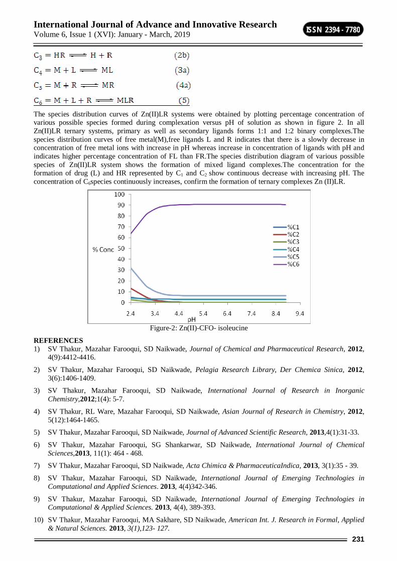
International Journal of Advance and Innovative Research Volume 6, Issue 1 (XVI): January - March, 2019
231
ISSN 2394 - 7780
The species distribution curves of Zn(II)LR systems were obtained by plotting percentage concentration of various possible species formed during complexation versus pH of solution as shown in figure 2. In all Zn(II)LR ternary systems, primary as well as secondary ligands forms 1:1 and 1:2 binary complexes.The species distribution curves of free metal(M),free ligands L and R indicates that there is a slowly decrease in concentration of free metal ions with increase in pH whereas increase in concentration of ligands with pH and indicates higher percentage concentration of FL than FR.The species distribution diagram of various possible species of Zn(II)LR system shows the formation of mixed ligand complexes.The concentration for the formation of drug (L) and HR represented by C1 and C2 show continuous decrease with increasing pH. The concentration of C6species continuously increases, confirm the formation of ternary complexes Zn (II)LR.
Figure-2: Zn(II)-CFO- isoleucine
REFERENCES 1) SV Thakur, Mazahar Farooqui, SD Naikwade, Journal of Chemical and Pharmaceutical Research, 2012,
4(9):4412-4416.
2) SV Thakur, Mazahar Farooqui, SD Naikwade, Pelagia Research Library, Der Chemica Sinica, 2012, 3(6):1406-1409.
3) SV Thakur, Mazahar Farooqui, SD Naikwade, International Journal of Research in Inorganic Chemistry,2012;1(4): 5-7.
4) SV Thakur, RL Ware, Mazahar Farooqui, SD Naikwade, Asian Journal of Research in Chemistry, 2012, 5(12):1464-1465.
5) SV Thakur, Mazahar Farooqui, SD Naikwade, Journal of Advanced Scientific Research, 2013,4(1):31-33.
6) SV Thakur, Mazahar Farooqui, SG Shankarwar, SD Naikwade, International Journal of Chemical Sciences,2013, 11(1): 464 - 468.
7) SV Thakur, Mazahar Farooqui, SD Naikwade, Acta Chimica & PharmaceuticaIndica, 2013, 3(1):35 - 39.
8) SV Thakur, Mazahar Farooqui, SD Naikwade, International Journal of Emerging Technologies in Computational and Applied Sciences. 2013, 4(4)342-346.
9) SV Thakur, Mazahar Farooqui, SD Naikwade, International Journal of Emerging Technologies in Computational & Applied Sciences. 2013, 4(4), 389-393.
10) SV Thakur, Mazahar Farooqui, MA Sakhare, SD Naikwade, American Int. J. Research in Formal, Applied & Natural Sciences. 2013, 3(1),123- 127.
International Journal of Advance and Innovative Research Volume 6, Issue 1 (XVI): January - March, 2019
232
ISSN 2394 - 7780
11) SV Thakur, SD Naikwade, Mazahar Farooqui, International Journal of Chemical Studies. 2013, 1(3), 88-92.
12) SV Thakur, Mazahar Farooqui, SD Naikwade, Int. J Recent Trends in Science & Technology, Special Issue,ACTRA-INDIA, Sept.2013, 29-31.
13) SV Thakur, Mazahar Farooqui, SD Naikwade, Journal of Chemical Biological & Physical sciences. 2014, 4(1),1-7.
14) SV Thakur, SD Naikwade,Mazahar Farooqui, Journal of Medicinal Chemistry Drug discovery,(special issue),2015, 107-118.
15) R.L. Ware, Mazahar Farooqui, S.D.Naikwade, Int J Emerging Tech in Computational & Applied Sci, 2013, 5(2):123-128.
16) R.L. Ware, Mazahar Farooqui, S.D.Naikwade, Int J Emerging Tech in Computational & Applied Sci.2013,5(4):398-401.
17) R.L.Ware, Shoeb Peerzade, S.D. Naikwade, Mazahar Farooqui, J Chemical & Pharma Res.2013,5(8): 59-63.
18) SV Thakur, Jameel Pathan, Farooque Bashir Ansari,D.D. Kayande, Journal of Chemical & Pharmacetical Research. 2016, 8(5), 291-294.
19) S.V. Thakur, M.A.Sakhare, S.N.Sampal, H.U.Joshi, International Multilingual Research Journal Printing Area (Special Issue), Dec.2017, 169-173.
20) Shailendrasingh Thakur,S.A. Peerzade,A.J. Khan, R.L.Ware, International Multilingual Research Journal Printing Area (Special Issue), Dec.2017,47-51
21) Ramesh L. Ware, Kishore N. Koinkar, Shailendrasingh Thakur, International Journal of Universal Science and Technology,3(1) Jan-2018,284-288.
22) Ramesh Ware, Shoeb Peerzade and Shailendrasingh Thakur, International Journal of Universal Science and Technology, 3(1) Jan-2018,238-241
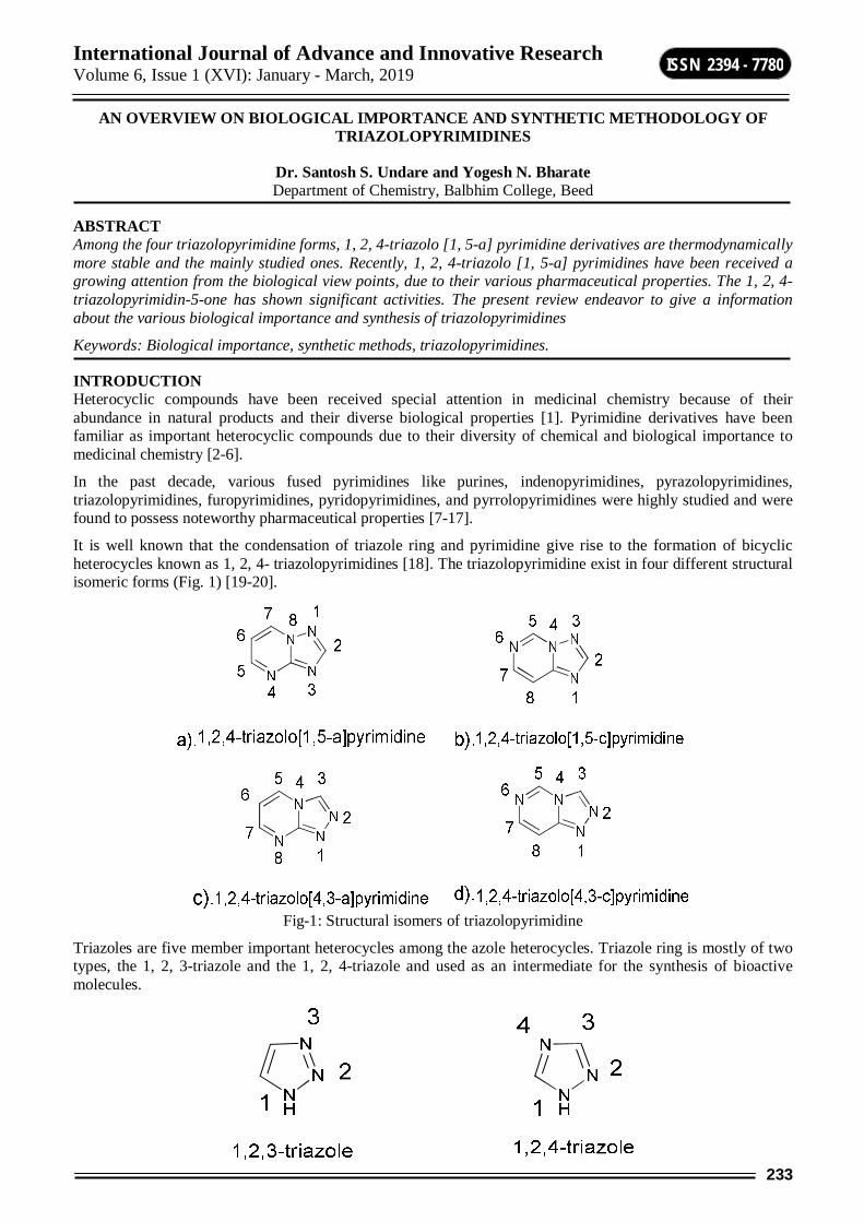
International Journal of Advance and Innovative Research Volume 6, Issue 1 (XVI): January - March, 2019
233
ISSN 2394 - 7780
AN OVERVIEW ON BIOLOGICAL IMPORTANCE AND SYNTHETIC METHODOLOGY OF TRIAZOLOPYRIMIDINES
Dr. Santosh S. Undare and Yogesh N. Bharate Department of Chemistry, Balbhim College, Beed
ABSTRACT Among the four triazolopyrimidine forms, 1, 2, 4-triazolo [1, 5-a] pyrimidine derivatives are thermodynamically more stable and the mainly studied ones. Recently, 1, 2, 4-triazolo [1, 5-a] pyrimidines have been received a growing attention from the biological view points, due to their various pharmaceutical properties. The 1, 2, 4-triazolopyrimidin-5-one has shown significant activities. The present review endeavor to give a information about the various biological importance and synthesis of triazolopyrimidines
Keywords: Biological importance, synthetic methods, triazolopyrimidines.
INTRODUCTION Heterocyclic compounds have been received special attention in medicinal chemistry because of their abundance in natural products and their diverse biological properties [1]. Pyrimidine derivatives have been familiar as important heterocyclic compounds due to their diversity of chemical and biological importance to medicinal chemistry [2-6].
In the past decade, various fused pyrimidines like purines, indenopyrimidines, pyrazolopyrimidines, triazolopyrimidines, furopyrimidines, pyridopyrimidines, and pyrrolopyrimidines were highly studied and were found to possess noteworthy pharmaceutical properties [7-17].
It is well known that the condensation of triazole ring and pyrimidine give rise to the formation of bicyclic heterocycles known as 1, 2, 4- triazolopyrimidines [18]. The triazolopyrimidine exist in four different structural isomeric forms (Fig. 1) [19-20].
Fig-1: Structural isomers of triazolopyrimidine
Triazoles are five member important heterocycles among the azole heterocycles. Triazole ring is mostly of two types, the 1, 2, 3-triazole and the 1, 2, 4-triazole and used as an intermediate for the synthesis of bioactive molecules.

International Journal of Advance and Innovative Research Volume 6, Issue 1 (XVI): January - March, 2019
234
ISSN 2394 - 7780
Biological Importance Before few decades, the chemistry of 1, 2, 4-triazoles and their fused heterocyclic compounds have received significant attention due to their synthetic and useful biological importance [21]. The incorporation of two bioactive motifs like triazole and pyrimidine into a single carbon skeleton leads to formation of wide variety of pharmaceutical interesting fused molecules [22-27]. (Fig-2)
Fig-2: Examples of bioactive triazolopyrimidines
METHODS OF SYNTHESIS Literature survey revealed that various bioactive triazolopyrimidines are synthesized using amino heterocycles
M. Singh et al. reported boric acid catalyzed simple, green, and efficient protocol for the synthesis of aryl-7,8-dihydro[1,2,4]triazolo[ 4,3-a] pyrimidine-6-carbonitriles derivatives through one-pot multi-component reaction using substituted aromatic aldehydes, malononitrile, and 3-amino[1,2,4]triazole [28].(Scheme-.1)
Scheme-1
K. Ablajan and his coworkers synthesized triazolo pyrimidine derivatives by a one-pot reaction of 3-amino-1, 2, 4-triazole, malononitrile and aromatic aldehydes in ethanol under heating or ultrasonic irradiation in the presence of NaOH [29]. (Scheme-2)
Scheme-2
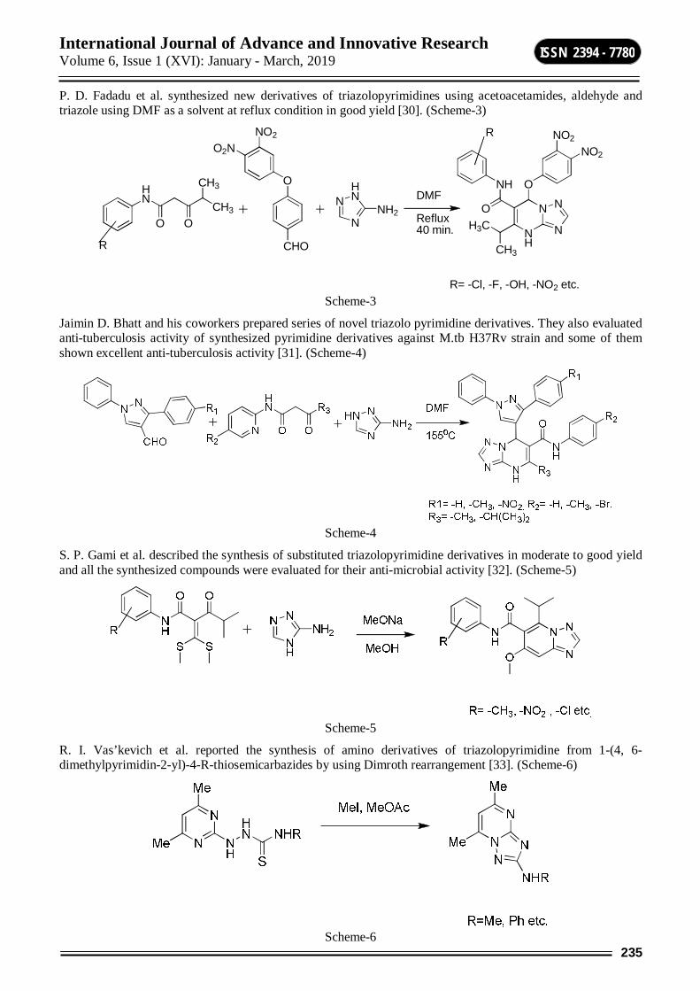
International Journal of Advance and Innovative Research Volume 6, Issue 1 (XVI): January - March, 2019
235
ISSN 2394 - 7780
P. D. Fadadu et al. synthesized new derivatives of triazolopyrimidines using acetoacetamides, aldehyde and triazole using DMF as a solvent at reflux condition in good yield [30]. (Scheme-3)
HN
CH3
O O
CH3
R
N
N
HN
NH2
O
NO2
O2N
CHONH
N
N
N
CH3
H3C
NH
O
O
NO2
NO2
DMF
Reflux40 min.
R
R= -Cl, -F, -OH, -NO2 etc. Scheme-3
Jaimin D. Bhatt and his coworkers prepared series of novel triazolo pyrimidine derivatives. They also evaluated anti-tuberculosis activity of synthesized pyrimidine derivatives against M.tb H37Rv strain and some of them shown excellent anti-tuberculosis activity [31]. (Scheme-4)
Scheme-4
S. P. Gami et al. described the synthesis of substituted triazolopyrimidine derivatives in moderate to good yield and all the synthesized compounds were evaluated for their anti-microbial activity [32]. (Scheme-5)
Scheme-5
R. I. Vas’kevich et al. reported the synthesis of amino derivatives of triazolopyrimidine from 1-(4, 6-dimethylpyrimidin-2-yl)-4-R-thiosemicarbazides by using Dimroth rearrangement [33]. (Scheme-6)
Scheme-6
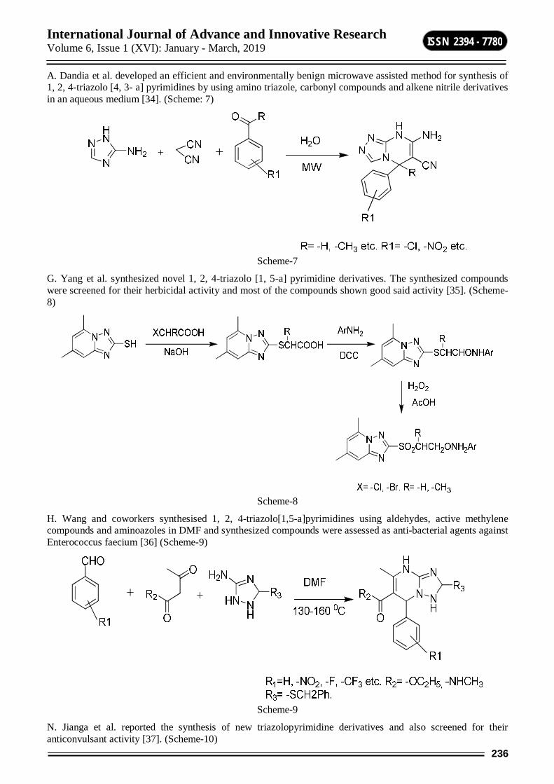
International Journal of Advance and Innovative Research Volume 6, Issue 1 (XVI): January - March, 2019
236
ISSN 2394 - 7780
A. Dandia et al. developed an efficient and environmentally benign microwave assisted method for synthesis of 1, 2, 4-triazolo [4, 3- a] pyrimidines by using amino triazole, carbonyl compounds and alkene nitrile derivatives in an aqueous medium [34]. (Scheme: 7)
Scheme-7
G. Yang et al. synthesized novel 1, 2, 4-triazolo [1, 5-a] pyrimidine derivatives. The synthesized compounds were screened for their herbicidal activity and most of the compounds shown good said activity [35]. (Scheme-8)
Scheme-8
H. Wang and coworkers synthesised 1, 2, 4-triazolo[1,5-a]pyrimidines using aldehydes, active methylene compounds and aminoazoles in DMF and synthesized compounds were assessed as anti-bacterial agents against Enterococcus faecium [36] (Scheme-9)
Scheme-9
N. Jianga et al. reported the synthesis of new triazolopyrimidine derivatives and also screened for their anticonvulsant activity [37]. (Scheme-10)
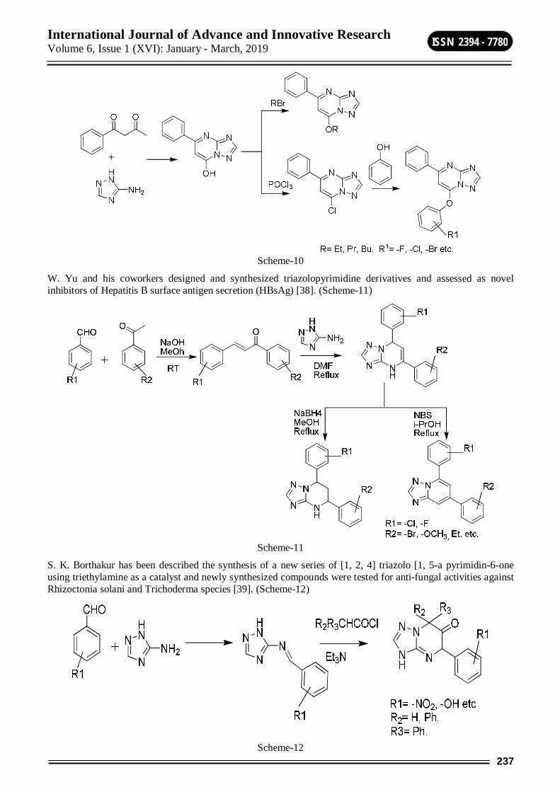
International Journal of Advance and Innovative Research Volume 6, Issue 1 (XVI): January - March, 2019
237
ISSN 2394 - 7780
Scheme-10
W. Yu and his coworkers designed and synthesized triazolopyrimidine derivatives and assessed as novel inhibitors of Hepatitis B surface antigen secretion (HBsAg) [38]. (Scheme-11)
Scheme-11
S. K. Borthakur has been described the synthesis of a new series of [1, 2, 4] triazolo [1, 5-a pyrimidin-6-one using triethylamine as a catalyst and newly synthesized compounds were tested for anti-fungal activities against Rhizoctonia solani and Trichoderma species [39]. (Scheme-12)
Scheme-12

International Journal of Advance and Innovative Research Volume 6, Issue 1 (XVI): January - March, 2019
238
ISSN 2394 - 7780
CONCLUSION Among all heterocyclic compounds, pyrimidines are one of the most important heterocycles shows remarkable biological and pharmaceutical activities because it is an essential constituent of all cells. In medicinal chemistry pyrimidine derivatives have been very well known for their therapeutic applications. The presence of a pyrimidine unit in nucleic acids, DNA and RNA is one possible reason for their biological activities. The versatile synthetic applicability and biological importance of the triazolopyrimidine derivatives will help the chemists to develop new approaches towards finding of new drugs.
REFERENCES 1. M. Jha, S. Guy, Y. C. Ting. Tetrahedron Lett. 52, 4337 (2011)
2. 2. A. H .Abdel-Rahman, E.M. Keshk, M. A. Hanna, S. M. El-Bady, Bioorg. Med. Chem. 12, 2483 (2004)
3. U. S Rai, A. M Isloor, P. Shetty, A. M Vijesh, N. Prabhu, S. Isloor, M. Thiageeswaran, F. Hoong-Kun. Eur. J. Med. Chem. 45, 2695 (2010)
4. M. S. El-Gaby, S. G. Abdel-Hamid, M. M. Ghorab, S. M. El-Sayed. Acta Pharm, 49, 149 (1999)
5. P. G. Baraldi, M. G. Pavani, M. Nunez, P. Brigidi, B. Vitali, R. Gambari, R. Romagnoli. Bioorg Med Chem.10, 44 (2002)
6. S. M. Sondhi, M. Johar, S. Rajvanshi, S. G. Dastidar, R. Shukla, R. Raghubir, J. W. Lown. Aust J Chem.54, 69 (2001)
7. B. O. Buckman, R. Mohan, S. Koovakkat, A. Liang, L. Trinh, M. M. Morrissey. Bioorg. & Med. Chem. Lett. 8, 2235 (1998)
8. K. V. Diveshkumar, S. Sakrikar, S. Harikrishna, V. Dhamodharan, P. I. Pradeepkumar. Chem Med Chem.9, 2754 (2014)
9. I. D. Brahmbhatt, V. Chirag Patel, V. G. Bhila N. H. Patel, A. A. Patel, Med Chem Res. 24, 1596 (2015)
10. K. Vijayakumar, A. Jafar Ahamed. Der Pharma Chem. 2(5), 453 (2010)
11. A. Rajendran, D. Raghupathy, M. Priyadarshini. Int. J. Chem Tech Res. 3, 293 (2011)
12. A. A. Aly, I. A. Gad El-Karim. Journal of the Korean Chemical Society. 55, 781. (2011).
13. X. L. Zhao, Y. F. Zhao, S.C. Guo, H. S. Song, D. W. Ping. Molecules. 12, 1136 (2007)
14. M. I. Hossain, M. M. H. Bhuiyan. J. Sci. Res. 1 (2), 317 (2009)
15. T. Saurat, F. Buron, N. Rodrigues, M. Ludivine de Tauzia, L. Colliandre, S. Bourg, P. Bonnet, G. Guillaumet, M. Akssira, A. Corlu, C. Guillouzo, P. Berthier, P. Rio, M. Lise. J. Med. Chem. 57(3), 613 (2014)
16. M. H. Khalid, H. H. Dalia Soliman, B. A. Esmat, S. Shahin Hala Eur. J. Med. Chem.78, 419 (2014) 17. V. Pittal, A Maria, S.Maria, N. Modic, L. Salerno, A. Pedretti, G. Vistoli, A. Cagnotto, T. Mennini, G.
Romeo. Bioor.& Med. Chem.19, 5260 (2011). 18. S. Salameh, M. Abul-Haj, M. Quirós, J. M. Salas. Inorg. Chim. Acta. 358, 824 (2005). 19. M. Shaban, A. Morgan. Adv. Heterocycl. Chem.73, 131 (2000). 20. M. Shaban, A. Morgan. Adv. Heterocycl. Chem. 77, 345 (2000). 21. I. A. Al-Masoudi, Y. A. Al-Soud, N. J. Al-Salihi, N. A. Al-Masoudi. Chem. Heterocycl. Compd. 42, 1377
(2006) 22. M. A. Phillips, J. Lotharius et. al., Sci Transl Med. 7, 296, (2015) 23. Bioactive heterocyclic compounds . C. Lamberth, J. Dinges. Wiley-VCH, ISBN 978-3-527-32993-9 24. C. F. Beyer, N. Zhang, R. Hernandez, D. Vitale, J. Lucas, T. Nguyen, C. Discafani, S. Ayral-Kaloustian,
J. Gibbons, J. Cancer Research. 68, 2292 (2008) 25. H. Li, J. Tatlock, A. Linton, J. Gonzalez, T. Jewell, L. Patel, S. Ludlum, M. Drowns, S. V. Rahavendran,
H. Skor, R. Hunter, S. T. Shi, K. J. Herlihy, H. Parge, Y, M. Hicke, X. Yu, F. Chau, J. Nonomiya, C. Lewis. J. Med. Chem. 52, 1255 (2009)
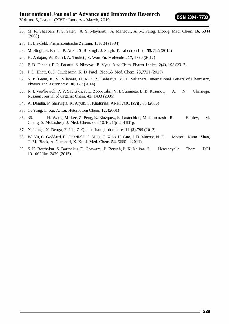
International Journal of Advance and Innovative Research Volume 6, Issue 1 (XVI): January - March, 2019
239
ISSN 2394 - 7780
26. M. R. Shaaban, T. S. Saleh, A. S. Mayhoub, A. Mansour, A. M. Farag. Bioorg. Med. Chem. 16, 6344 (2008)
27. H. Liekfeld. Pharmazeutische Zeitung. 139, 34 (1994)
28. M. Singh, S. Fatma, P. Ankit, S. B. Singh, J. Singh. Tetrahedron Lett. 55, 525 (2014)
29. K. Ablajan, W. Kamil, A. Tuoheti, S. Wan-Fu. Molecules. 17, 1860 (2012)
30. P. D. Fadadu, P. P. Fadadu, S. Nimavat, B. Vyas. Acta Chim. Pharm. Indica. 2(4), 198 (2012)
31. J. D. Bhatt, C. J. Chudasama, K. D. Patel. Bioor.& Med. Chem. 23,7711 (2015)
32. S. P. Gami, K. V. Vilapara, H. R. K. S. Babariya, Y. T. Naliapara. International Letters of Chemistry, Physics and Astronomy. 30, 127 (2014)
33. R. I. Vas’kevich, P. V. Savitskii,Y. L. Zborovskii, V. I. Staninets, E. B. Rusanov, A. N. Chernega. Russian Journal of Organic Chem. 42, 1403 (2006)
34. A. Dandia, P. Sarawgia, K. Aryab, S. Khaturiaa. ARKIVOC (xvi) , 83 (2006)
35. G. Yang, L. Xu, A. Lu. Heteroatom Chem. 12, (2001)
36. 36. H. Wang, M. Lee, Z. Peng, B. Blazquez, E. Lastochkin, M. Kumarasiri, R. Bouley, M. Chang, S. Mobashery. J. Med. Chem. doi: 10.1021/jm501831g.
37. N. Jianga, X. Denga, F. Lib, Z. Quana. Iran. j. pharm. res.11 (3),799 (2012)
38. W. Yu, C. Goddard, E. Clearfield, C. Mills, T. Xiao, H. Guo, J. D. Morrey, N. E. Motter, Kang Zhao, T. M. Block, A. Cuconati, X. Xu. J. Med. Chem. 54, 5660 (2011).
39. S. K. Borthakur, S. Borthakur, D. Goswami, P. Boruah, P. K. Kalitaa. J. Heterocyclic Chem. DOI 10.1002/jhet.2479 (2015).
International Journal of Advance and Innovative Research Volume 6, Issue 1 (XVI): January - March, 2019
240
ISSN 2394 - 7780
GENETIC STUDIES ON COTTON DERIVED THROUGH INTROGRESSION
Ashish Gulhane and M. S. Wadikar Department of Botany, Arts, Commerce and Science College, Dist.Beed
ABSTRACT This investigation were carried out with 38 cotton genotypes including both the species G. arboreum and G. hirsutum in randomized complete block design with three replications. The observations were reported form three randomly selected plant from each treatment and replication on 17 characters viz., Plant height (cm), number of nodes per plant, length of internodes (cm), number of monopodia per plant, number of sympodia per plant, number of bolls per plant. boll weight (g), seed cotton yield per plant (g), ginning out turn (%), lint index (g), seed index (g), fibre quality traits including 2.5% span length, fineness (MIC), bundle strength. fibre elongation, uniformity ratio (UR) and short fibre index (SFI). The maximum genetic variability was observed in plant height, seed cotton, yield per plant, number of nodes per plant number of bolls per plant number of sympodia per plant, ginning out turn, lint index and short fibre index. However, fibre elongation, boll weight, seed index, bundle strength, micronaire value, number of monopodia per plant, 2.5% span length and uniformity ratio exhibited limited genetic variation as indicated by their low estimates of GCV. High heritability estimates coupled with high genetic advance as percentage of mean were noted for lint index, short fibre index, boll weight, plant height and number of nodes per plant and high heritability estimates with low genetic advance as percentage of mean were observed for ginning out turn and uniformity ratio. The results of correlation coefficients revealed that characters boll weight, number of bolls per plant and uniformity ratio had significant and positive correlation with seed cotton yield per plant. Lint index had significant and positive association with 2.5% span length, seed index and ginning out turn while with short fibre index. Micronaire value and fibre elongation it was negative.
Keywords: Introgression, cotton, G.hirsutum and G.arboretum
INTRODUCTION Cotton the “Queen of the fiber plants” is cultivated worldwide in the tropical and subtropical regions and contributes significantly to the economy of many countries as a cash corp. This crop is also known as “White gold” due to its high commercial value. Cotton is one of the most important source of natural fibre which it is unmatched and universally preferred for clothing. Cotton forms the backbone of the textile industry, cellulose from its lint and oil, meal and hull from its seed. In fact cotton is the second best source of proteins. It is the country’s largest private sector providing employment directly or indirectly to around 35 million people. It contributes 14 per cent industrial production, 4 per cent of GDP and accounts for one third of the total export earnings (Jhunjhunwala, 2004).
The cultivated and wild, all the cotton genotypes of the world belongs to the genus Gossypium which a member of the tribe Hibisceae Family Malvaceae, natural order Malvales of the dicotyledons group of plants. It is predominantly less often cross pollinated crop. The genus comprises of 41 species (Fryxel, 1984) including 35 diploid species (2n=26) and six allotetraploids (2n=52). Four species of Gossypium have been commercially exploited. All the four spp. viz., G.arboreum L., G.herbaceum L. (both diploids and equipped with natural resistance to insect pest and diseases) G.hirsutum L. and G.barbadense L. are cultivated in India. The former two are diploids and are commonly referred to as Asiatic cottons owing to its origin while the latter two are allotetraploids and commonly known as New world American cotton. The ploidy level of cotton has become the platform and it has wide genetic diversity within itself.
In India its production is concentrated mainly in nine states namely Gujarat, Andhra Pradesh, Madhya Pradesh, Maharashtra, Haryana, Punjab, Rajasthan, Karnataka and Tamil Nadu which together account for about 95 per cent of total production of cotton (Anonymous, 2004). The major disadvantages, apart from low yields is the poor to medium quality fibre of Indian cotton particularly with respect to 2.5 per cent span length, fibre fineness and bundle strength.
The introduction of intra-hirsutum hybrids since mid-seventies has slightly improved the position because of high yield potential, good fibre properties, and wider adaptability with high degree of resistance to biotic and abiotic stress. Moreover, hybrid is highly uniform and more attractive than the land races. With the exploitation of Bt genes, the position is expected to further improve also design of ideal plant through genetic architecture of yield and its components in desi cotton (G.arboreum L.) for rainfed situation studies by Lenka, R.K. (2004). The role of wild species as a source of new genes for useful characters has been surveyed by Santhanam (1958). The
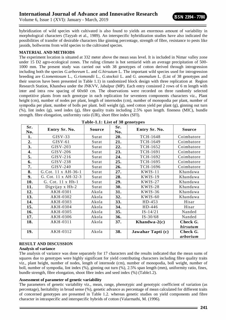
International Journal of Advance and Innovative Research Volume 6, Issue 1 (XVI): January - March, 2019
241
ISSN 2394 - 7780
hybridization of wild species with cultivated is also found to yields an enormous amount of variability in morphological characters (Tayyab et al., 1989). An interspecific hybridization studies have also indicated the possibilities of transfer of desirable characters like ginning percentage, strength of fibre, resistance to pests like jassids, bollworms from wild species to the cultivated species.
MATERIAL AND METHODS The experiment location is situated at 332 meter above the mean seas level. It is included in Nimar valley zone under 15 D2 agro-ecological zones. The ruling climate is hot semiarid with an average precipitation of 500-1000 mm. The present study was carried out with 38 genotypes of cotton derived through introgression including both the species G.arboreum L. and G.hirsutum L. The important wild species used for introgression breeding are G.tomentosum L., G.riomondii L., G.stocksii L. and G. anomalum L. (List of 38 genotypes and their sources have been presented in Table 1.1) in randomized block design with three replication at Region Research Station, Khandwa under the JNKVV, Jabalpur (MP). Each entry consisted 2 rows of 6 m length with inter and intra row spacing of 60x60 cm. The observations were recorded on three randomly selected competitive plants from each genotype in each replication for seventeen components characters viz., Plant height (cm), number of nodes per plant, length of internodes (cm), number of monopodia per plant, number of sympodia per plant, number of bolls per plant. boll weight (g), seed cotton yield per plant (g), ginning out turn (%), lint index (g), seed index (g), fibre quality traits including 2.5% span length. fineness (MIC), bundle strength. fibre elongation, uniformity ratio (UR), short fibre index (SFI).
Table-1.1: List of 38 genotypes Sr. No. Entry Sr. No. Source Sr.
No. Entry Sr. No. Source
1. GISV-33 Surat 20. TCH-1648 Coimbatore 2. GISV-61 Surat 21. TCH-1649 Coimbatore 3. GISV-203 Surat 22. TCH-1652 Coimbatore 4. GISV-206 Surat 23. TCH-1691 Coimbatore 5. GISV-216 Surat 24. TCH-1692 Coimbatore 6. GISV-238 Surat 25. TCH-1695 Coimbatore 7. GISV-240 Surat 26. TCH-1696 Coimbatore 8. G.Cot. 11 x AH-36-1 Surat 27. KWIS-11 Khandawa 9. G. Cot. 11 x AH-32-3 Surat 28. KWIS-19 Khandawa
10. G. Cot. 11 x Hh-1 Surat 29. KWIS-27 Khandawa 11. Digvijay x Hh-2 Surat 30. KWIS-28 Khandawa 12. AKH-0301 Akola 31. KWIS-36 Khandawa 13. AKH-0302 Akola 32. KWIS-60 Khandawa 14. AKH-0303 Akola 33. HD-453 Hisar 15. AKH-0304 Akola 34. HD-446 Hisar 16. AKH-0305 Akola 35. IS-14/21 Nanded 17. AKH-0306 Akola 36. IS-30/68 Nanded 18. AKH-0311 Akola 37. Khandwa-2(c) Check G.
hirsutum 19. AKH-0312 Akola 38. Jawahar Tapti (c) Check G.
arborium
RESULT AND DISCUSSION Analysis of variance The analysis of variance was done separately for 17 characters and the results indicated that the mean sums of squares due to genotypes were highly significant for yield contributing characters including fibre quality traits viz., plant height, number of nodes, length of internode (cm), number of monopodia, boll weight, number of boll, number of sympodia, lint index (%), ginning out turn (%), 2.5% span length (mm), uniformity ratio, fines, bundle strength, fibre elongation, shoot fibre index and seed index (%) (Table1.2).
Assessment of parameter of genetic variability The parameters of genetic variability viz., mean, range, phenotypic and genotypic coefficient of variation (as percentage), heritability in broad sense (%), genetic advance as percentage of mean calculated for different traits of concerned genotypes are presented in Table 1.2. whereas genetic studies on yield components and fibre character in intraspecific and interspecific hybrids of cotton (Valarmathi, M. (1996).
International Journal of Advance and Innovative Research Volume 6, Issue 1 (XVI): January - March, 2019
242
ISSN 2394 - 7780
Mean performance for different characters The expressed variation of different morpho-physiological traits under study focused a lot on the free variability present in the population of different genotypes, which in turn would reflect the unforeseen impact of potential variability on yield. Out of the fore-stated morpho-physiological traits, plant height ranged from 56.55 to 142.66 cm with a mean of 89.01 cm. Number of nodes ranged from 20.30 to 36.41 with an average of 25.68. Length of internode varied from 2.97 cm to 5.93 cm with a mean of 4.17 cm. Number of monopodia ranged from 0.00 to 3.55 with a mean of 0.94. Boll weight ranged from 1.87 g to 4.10 g with an average of 2.95 g. Number of boll varied 13.44 to 26.58 with an average of 20.58. Number of sympodia ranged from 13.00 to 23.89 with a mean of 17.01. Lint index ranged from 3.16 to 5.46 with a mean of 4.22. Ginning out turn varied from 32.24 to 38.19 with an average of 34.55%. Seed index ranged from 6.22 to 9.64 with a mean of 8.04. Seed cotton yield per plant varied from 41.66 to 95.73g with an average 60.42 g. Apart from this similar trend of variation amongst genotypes also observed in the fibre quality parameters 2.5% span length ranged from 22.30 to 29.80 with a mean of 26.30. Uniformity ratio varied from 43.0 to 53.00 with an average of 48.07. Micronaire value ranged from 3.50 to 5.30 with a mean of 4.36. Bundle strength varied from 18.4 to 24.70 with a mean of 21.13. Fibre elongation and short fibre index varied from 4.50 to 6.30 and 8.10 to 16.20 with an average 5.41 and 12.27
Table-1.2: Analysis of variance for yield and its compounds in cotton genotypes
Source
Mean sum of squares
DF
PH /cm
NON/P
INL/ CM
NOM/P
SBW (G)
NOS/P
NOS/P LI GO
T%
2.5% SL
UR
FINE (MIC)
BS
FE
SFI
SI SCY/P
Replications
2 22.49 1.54 0.36 0.31 0.22 8.02 16.87
0.0 0.09 2.19
7.25
0.05 1.43
0.09
0.46
0.03
40.19
Genotypes
37 1321.22**
58.13**
1.00 2.19 0.89 27.82**
23.01**
10.86**
11.84**
10.22**
15.30**
0.76 6.49
0.75
14.28**
2.52
208.08**
Error 74 12.21 5.59 0.09 0.25 0.04 5.82 4.01 0.01 0.52 0.11
0.39
0.003
0.07
0.005
0.02
0.02
58.63
SEd¼ 2.85 1.93 0.24 0.40 0.16 1.96 1.63 0.08 0.58 0.28
0.51
0.04 0.22
0.05
0.13
0.12
6.25
*significance at 5% level **significance at 1% level CORRELATION COEFFICIENTS ANALYSIS Correlation between seed cotton yield per plant and yield component traits including fibre quality seed cotton yield per plant. The trait showed significant positive correlation with boll weight (0.69), number of bolls per plant (0.54) and uniformity ratio (0.32).
REFERENCES Anonymous (2004). All India Coordinated Cotton Improvement Project, pp. 1-3. Fryxel, P.A. (1984). Taxonomy and germplasm resources. American society of Agronomy. Wisconsin. p.
655. Jhunjhunwala, K.F. (2004). Steps for Cotton Production and Improvement Souvenir, AICCIP Annual group
meeting, pp. 25. Lenka, R.K. (2004). Design of ideal plant through genetic architecture of yield and its’ components in desi
cotton (G. arboreum L.) for rainfed situation. M.Sc. (Agri.) Thesis, JNKVV, Jabalpur. Santhanam, V. (1958). Cotton improvement through interspecific hybridization and role of wild germplasm.
8th Conf. Cott. Gr. Profl. India I.C.C.C., Bombay, pp. 667-674. Tayyab, M.A. (1989). Introgression of wild species gene for improvement of cotton genotypes in central
India. A souvenir, ICAR publication, pp. 63-66. Valarmathi, M. (1996). Genetic studies on yield components and fibre character in intraspecific and
interspecific hybrids of cotton. M.Sc.(Agri.) Thesis, TNAU, Coimbatore.
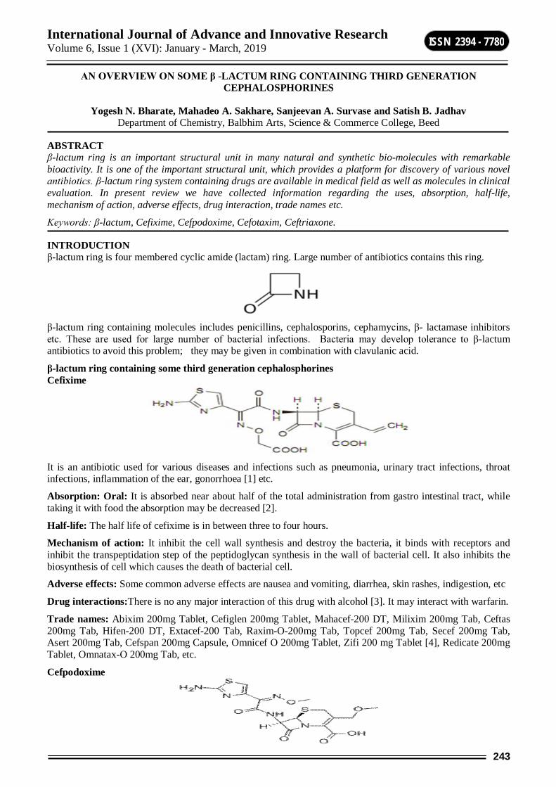
International Journal of Advance and Innovative Research Volume 6, Issue 1 (XVI): January - March, 2019
243
ISSN 2394 - 7780
AN OVERVIEW ON SOME β -LACTUM RING CONTAINING THIRD GENERATION CEPHALOSPHORINES
Yogesh N. Bharate, Mahadeo A. Sakhare, Sanjeevan A. Survase and Satish B. Jadhav Department of Chemistry, Balbhim Arts, Science & Commerce College, Beed
ABSTRACT β-lactum ring is an important structural unit in many natural and synthetic bio-molecules with remarkable bioactivity. It is one of the important structural unit, which provides a platform for discovery of various novel antibiotics. β-lactum ring system containing drugs are available in medical field as well as molecules in clinical evaluation. In present review we have collected information regarding the uses, absorption, half-life, mechanism of action, adverse effects, drug interaction, trade names etc.
Keywords: β-lactum, Cefixime, Cefpodoxime, Cefotaxim, Ceftriaxone.
INTRODUCTION β-lactum ring is four membered cyclic amide (lactam) ring. Large number of antibiotics contains this ring.
β-lactum ring containing molecules includes penicillins, cephalosporins, cephamycins, β- lactamase inhibitors etc. These are used for large number of bacterial infections. Bacteria may develop tolerance to β-lactum antibiotics to avoid this problem; they may be given in combination with clavulanic acid.
β-lactum ring containing some third generation cephalosphorines Cefixime
It is an antibiotic used for various diseases and infections such as pneumonia, urinary tract infections, throat infections, inflammation of the ear, gonorrhoea [1] etc.
Absorption: Oral: It is absorbed near about half of the total administration from gastro intestinal tract, while taking it with food the absorption may be decreased [2].
Half-life: The half life of cefixime is in between three to four hours.
Mechanism of action: It inhibit the cell wall synthesis and destroy the bacteria, it binds with receptors and inhibit the transpeptidation step of the peptidoglycan synthesis in the wall of bacterial cell. It also inhibits the biosynthesis of cell which causes the death of bacterial cell.
Adverse effects: Some common adverse effects are nausea and vomiting, diarrhea, skin rashes, indigestion, etc
Drug interactions:There is no any major interaction of this drug with alcohol [3]. It may interact with warfarin.
Trade names: Abixim 200mg Tablet, Cefiglen 200mg Tablet, Mahacef-200 DT, Milixim 200mg Tab, Ceftas 200mg Tab, Hifen-200 DT, Extacef-200 Tab, Raxim-O-200mg Tab, Topcef 200mg Tab, Secef 200mg Tab, Asert 200mg Tab, Cefspan 200mg Capsule, Omnicef O 200mg Tablet, Zifi 200 mg Tablet [4], Redicate 200mg Tablet, Omnatax-O 200mg Tab, etc.
Cefpodoxime
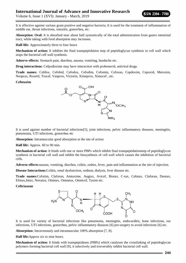
International Journal of Advance and Innovative Research Volume 6, Issue 1 (XVI): January - March, 2019
244
ISSN 2394 - 7780
It is effective against various gram positive and negative bacteria, It is used for the treatment of inflammation of middle ear, throat infections, sinusitis, gonorrhea, etc.
Absorption: Oral: It is absorbed near about half systemically of the total administration from gastro intestinal tract, while taking with food absorption may increases.
Half-life: Approximately three to four hours
Mechanism of action: It inhibits the final transpeptidation step of peptidoglycan synthesis in cell wall which stops the bacterial cell wall synthesis.
Adserve effects: Stomach pain, diarrhea, nausea, vomiting, headache etc.
Drug interactions: Cefpodoxime may have interaction with probenecid, antiviral drugs.
Trade names: Cefdox, Cefobid, Cefodox, Cefodim, Cefomin, Ceforan, Cepdoxim, Cepoxid, Metoxim, Neoprax, Roxetil, Trucef, Vanprox, Victorin, Ximeprox, Ximocef , etc.
Cefotaxim
It is used against number of bacterial infections[5], joint infections, pelvic inflammatory diseases, meningitis, pneumonia, UTI infections, gonorrhea etc
Absorption: Intramuscular good absorption at the site of action
Half life: Approx. 60 to 90 min.
Mechanism of action: It binds with one or more PBPs which inhibit final transpeptidationstep of peptidoglycan synthesis in bacterial cell wall and inhibit the biosynthesis of cell wall which causes the inhibition of bacterial cells.
Adverse effects:nausea, vomiting, diarrhea, colitis, rashes, fever, pain and inflammation at the site of injection.
Disease Interactions:Colitis, renal dysfunction, sodium, dialysis, liver disease etc.
Trade names:Cefotim, Claforan, Amtaxime, Augtax, Avicef, Biotax, C-tax, Cefatax, Claforan, Duotax, Efotax,Intax, Novatax, Ominax, Omnatax, Omnicef, Taxim etc.
Ceftriaxone
It is used for variety of bacterial infections like pneumonia, meningitis, endocarditis, bone infections, ear infections, UTI infections, gonorrhea, pelvic inflammatory diseases [6] pre-surgery to avoid infections [6] etc.
Absorption: Intravenously and intramuscular 100% absorption [7, 8].
Half life:Approx six to nine hours.
Mechanism of action: It binds with transpeptidases (PBPs) which catalyzes the crosslinking of peptidoglycan polymers forming bacterial cell wall [9], it selectively and irreversibly inhibit bacterial cell wall.
International Journal of Advance and Innovative Research Volume 6, Issue 1 (XVI): January - March, 2019
245
ISSN 2394 - 7780
Adverse effects: Changes in WBCs, rashes, diarrhea, fever, shortness in breath, etc.
Disease Interactions: Colitis, gall bladder disease, pancreatitis, renal disease, liver disease etc.
Trade names: Ceftizone, Ceftrix, Enocef, Eracef, Topcef, etc.
CONCLUSION β-lactum ring containing third generation cephalosphorines are world class of antibiotics, which are widely used for bacterial infections and diseases like pneumonia, urinary tract infections, throat infections, inflammation of the ear, gonorrhoea, inflammation of middle ear, throat infections, sinusitis, joint infections, pelvic inflammatory diseases, meningitis, endocarditis, bone infections, ear infections, pelvic inflammatory diseases, etc.
Large number of R&D units are focusing on β-lactum ring containing antibiotics. In future β-lactum ring containing antibiotics may play a pivotal role in treatment of different diseases.
REFERENCES 1. "Cefixime". The American Society of Health System Pharmacists.Archived from the original on 27 Nov.
2016.Retrieved 8 Dec. 2016.
2. Lupin Pharmaceuticals, Inc. Mar. 2013. Archived from the original on 23 Feb. 2015.Retrieved 7 Apr. 2016.
3. Choices, N. H. S. "Medicines information links". Archivedfrom the original on 11 Jul. 2015. Retrieved 22 Aug. 2016.
4. "FDC - Products - Formulations". Retrieved 02 May 2018.
5. "Cefotaxime Sodium". The American Society of Health-System Pharmacists. Archived from the original on 20 Dec. 2016.Retrieved 8 Dec. 2016.
6. "Ceftriaxone Sodium Monograph for Professionals". Archived from the original on 31 May 2016.Retrieved 27 Aug. 2016.
7. "DailyMed – CEFTRIAXONE – ceftriaxone sodium injection, powder, for solution". Archived from the original on 17 Nov. 2015.Retrieved 04 Apr. 2015.
8. "Pharmacokinetic profile of ceftriaxone in man". The American Journal of Medicine. 77 (4C): 17–25. ISSN 0002-9343.PMID 6093513.
9. Lemke, Thomas L.; Williams, David A., eds. (2013). Foye's Principles of Medicinal Chemistry (Seventh ed.). Philadelphia, PA: Lippincott Williams & Wilkins. pp. 1093–1094, 1099–1100. ISBN 9781609133450.

International Journal of Advance and Innovative Research Volume 6, Issue 1 (XVI): January - March, 2019
246
ISSN 2394 - 7780
POPULATION DYNAMICS OF CESTODE PARSITES IN COLUMBA LIVIA FROM BEED DISTRICT, INDIA
A. M. Budrukkar and R. K. Nimbalkar Department of Zoology, R. B. Attal College, Georai
ABSTRACT The present communication deals with the population dynamics of cestode parasite in Beed. Population of the cestode parasites are divided into three genera i.e. Cotugnia, Raillitina and Valipora. After dissecting intestine of Columba livia cestode parasite found are Cotugnia meggitti (Yamaguti) 1935, Cotugnia columbae (Shinde) 1969, Rallitina tetrgona (Mohan) 1958, Rallitina corvine (Shinde) 1977, Valipora mutabilis (Linton).The present study includes application of statistical method to understand and distribution of cone cestode parasites both minimum and maximum population level for three seasons i. e. summer, rainy and winter during 2017- 2018.
Columba livia are collected from different sites of Beed district. The Parasites were collected from the intestine of the host. Cestode parasites were preserved 4% formalin and stained with Harri`s haematoxyline. Then parasites are washed with distilled water and dehydrated with ascending grades of alcohol and mount in D.P.X.
This study of population dynamics show the five cestode species of three genera which are Cotugnia, Raillitina and Valipora.
Formulae Incidence of Infection = B x 100
------------
A
Intensity of Infection = C
-----
B
Density = C
-----
A
The A stands for the number of host examined, B for number of host infected and C for number of parasite collected.
Table I and II show the distribution of infection, density and index of infection.
Table showing values of Cotugnia species Month No. of host
Examined (A)
No. of host
infected (B)
No. of Parasites
Found (C)
Incidence of infection
(%)
Intensity of
infection (%)
Density Index of Infection
(%)
Region
March 2017 15 05 02 33.33 0.4 0.1 0.04 Beed April 2017 15 04 04 26.66 1 0.2 0.07 Georai May 2017 15 02 02 13.33 1 0.1 0.01 Parali June 2017 15 10 06 66.66 0.6 0.4 0.26 Gadhi July 2017 15 10 06 66.66 0.6 0.4 0.26 Ghatnandur
August 2017 15 12 08 80.00 0.6 0.5 0.42 Kej Sept. 2017 15 11 05 73.33 0.4 0.3 0.24 Kalam
October 2017 15 08 05 53.33 0.6 0.3 0.17 Majalgaon November
2017 15 05 03 33.33 0.6 0.2 0.06 Dharur
December 2017 15 06 02 40.00 0.3 0.1 0.05 Massajog January 2017 15 05 02 33.33 0.4 0.1 0.04 Ambajogai
February 22017 15 04 03 26.66 0.7 0.2 0.05 Ashti

International Journal of Advance and Innovative Research Volume 6, Issue 1 (XVI): January - March, 2019
247
ISSN 2394 - 7780
Table showing values of Raillitina species Month No. of
host Examined (A)
No. of host
infected (B)
No. of Parasites
Found (C)
Incidence of
infection (%)
Intensity of
infection (%)
Density Index of Infection
(%)
Region
March 2017 15 05 02 33.33 0.4 0.001 0.04 Beed April 2017 15 04 01 26.66 0.25 0.06 0.01 Georai May 2017 15 02 03 13.33 1.5 0.2 0.02 Parali June 2017 15 10 04 66.66 0.4 0.26 0.17 Gadhi July 2017 15 10 03 66.66 0.3 0.2 0.13 Ghatnandur
August 2017
15 12 04 80.00 0.33 0.26 0.21 Kej
Sept. 2017 15 11 04 73.33 0.36 0.26 0.19 Kalam October
2017 15 08 03 53.33 0.37 0.2 0.10 Majalgaon
November 2017
15 05 03 33.33 0.6 0.2 0.06 Dharur
December 2017
15 06 02 40.00 0.33 0.001 0.05 Massajog
January 2017
15 05 01 33.33 0.2 0.06 0.02 Ambajogai
February 22017
15 04 01 26.66 0.25 0.06 0.01 Ashti
Table showing values of Vailipora species Month No. of
host Examined (A)
No. of host
infected ( B )
No. of Parasites
Found ( C )
Incidence of
infection (%)
Intensity of
infection (%)
Density Index of Infection
(%)
Region
March 2017 15 05 00 33.33 00 00 00 Beed April 2017 15 04 00 26.66 00 00 00 Georai May 2017 15 02 00 13.33 00 00 00 Parali June 2017 15 10 03 66.66 0.3 0.2 0.13 Gadhi July 2017 15 10 03 66.66 0.3 0.2 0.13 Ghatnandur
August 2017
15 12 04 80.00 0.33 0.26 0.21 Kej
Sept. 2017 15 11 02 73.33 0.18 0.13 0.09 Kalam October
2017 15 08 01 13.33 0.12 0.06 0.03 Majalgaon
November 2017
15 05 01 33.33 0.2 0.06 0.02 Dharur
December 2017
15 06 01 40.00 0.1 0.06 0.02 Massajog
January 2017
15 05 02 33.33 0.4 0.13 0.04 Ambajogai
February 22017
15 04 01 26.66 0.25 0.06 0.01 Ashti
RESULTS AND DISCUSSION Genera Cotugnia: In case of Cotugnia the values of incidence, density and Index of infection were maximum in rainy season and low in winter season.
Genra Rallitina: In case of Rallitina values of incidence, density and Index of infection were maximum in rainy season and restrained in winter season.
Genera Valipora: In case of Valipora values of incidence, density and Index of infection were maximum in summer season, restrained in rainy season and low in winter season.

International Journal of Advance and Innovative Research Volume 6, Issue 1 (XVI): January - March, 2019
248
ISSN 2394 - 7780
Above data reveal that Cestode belonging from the genus Cotugnia are dominant. It is due to the host specificity and many other phonological factors.
REFERENCES Aruna Kumari, V 1985: Ecological studies on Helminth of Common Birds, Ph.D. thesis. Osmania
University, Hyderabad. India.
Anderson, R. M. 1975: The regulation of host population growth by parasite species. Parasitology76: 119-157.
B.V. Jadhav, R. M. Khadap, A. M. Budrukkar & L.B. Pawar: Population dyanamics of some cestode parasites in Columba livia from Aurangabad, India, Uttar Pradesh J. Zool. 22 (2) 203-205, 2002.
Yamaguti, S. 1989: The Cestode vertebrate in Systema Helminthum Vol II. Interscience Publication. New York.
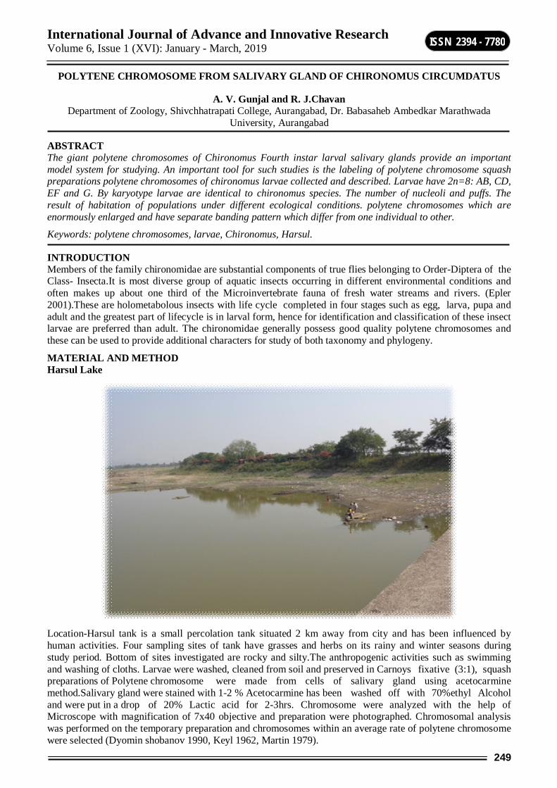
International Journal of Advance and Innovative Research Volume 6, Issue 1 (XVI): January - March, 2019
249
ISSN 2394 - 7780
POLYTENE CHROMOSOME FROM SALIVARY GLAND OF CHIRONOMUS CIRCUMDATUS
A. V. Gunjal and R. J.Chavan Department of Zoology, Shivchhatrapati College, Aurangabad, Dr. Babasaheb Ambedkar Marathwada
University, Aurangabad
ABSTRACT The giant polytene chromosomes of Chironomus Fourth instar larval salivary glands provide an important model system for studying. An important tool for such studies is the labeling of polytene chromosome squash preparations polytene chromosomes of chironomus larvae collected and described. Larvae have 2n=8: AB, CD, EF and G. By karyotype larvae are identical to chironomus species. The number of nucleoli and puffs. The result of habitation of populations under different ecological conditions. polytene chromosomes which are enormously enlarged and have separate banding pattern which differ from one individual to other.
Keywords: polytene chromosomes, larvae, Chironomus, Harsul.
INTRODUCTION Members of the family chironomidae are substantial components of true flies belonging to Order-Diptera of the Class- Insecta.It is most diverse group of aquatic insects occurring in different environmental conditions and often makes up about one third of the Microinvertebrate fauna of fresh water streams and rivers. (Epler 2001).These are holometabolous insects with life cycle completed in four stages such as egg, larva, pupa and adult and the greatest part of lifecycle is in larval form, hence for identification and classification of these insect larvae are preferred than adult. The chironomidae generally possess good quality polytene chromosomes and these can be used to provide additional characters for study of both taxonomy and phylogeny.
MATERIAL AND METHOD Harsul Lake
Location-Harsul tank is a small percolation tank situated 2 km away from city and has been influenced by human activities. Four sampling sites of tank have grasses and herbs on its rainy and winter seasons during study period. Bottom of sites investigated are rocky and silty.The anthropogenic activities such as swimming and washing of cloths. Larvae were washed, cleaned from soil and preserved in Carnoys fixative (3:1), squash preparations of Polytene chromosome were made from cells of salivary gland using acetocarmine method.Salivary gland were stained with 1-2 % Acetocarmine has been washed off with 70%ethyl Alcohol and were put in a drop of 20% Lactic acid for 2-3hrs. Chromosome were analyzed with the help of Microscope with magnification of 7x40 objective and preparation were photographed. Chromosomal analysis was performed on the temporary preparation and chromosomes within an average rate of polytene chromosome were selected (Dyomin shobanov 1990, Keyl 1962, Martin 1979).
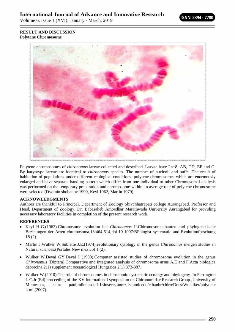
International Journal of Advance and Innovative Research Volume 6, Issue 1 (XVI): January - March, 2019
250
ISSN 2394 - 7780
RESULT AND DISCUSSION Polytene Chromosome
Polytene chromosomes of chironomus larvae collected and described. Larvae have 2n=8: AB, CD, EF and G. By karyotype larvae are identical to chironomus species. The number of nucleoli and puffs. The result of habitation of populations under different ecological conditions. polytene chromosomes which are enormously enlarged and have separate banding pattern which differ from one individual to other Chromosomal analysis was performed on the temporary preparation and chromosome within an average rate of polytene chromosome were selected (Dyomin shobanov 1990, Keyl 1962, Martin 1979).
ACKNOWLEDGMENTS Authors are thankful to Principal, Department of Zoology Shivchhatrapati college Aurangabad. Professor and Head, Department of Zoology, Dr. Babasaheb Ambedkar Marathwada University Aurangabad for providing necessary laboratory facilities in completion of the present research work.
REFERENCES Keyl H-G.(1962).Chromosome evolution bei Chironomus II.Chiromosomenbauten and phylogenetische
Bezihungen der Arten chromosoma.13:464-514,doi-10-1007/BFologist systematic and Evolutionforschung 18 (2).
Martin J.Wulker W,Sublette J.E.(1974).evolutionary cytology in the genus Chironomus meigen studies in Natural sciences.(Portales New mexico) 1 (2).
Wulker W.Devai GY.Devai I (1989).Computer assisted studies of chromosome evolution in the genus Chironomus (Diptera).Comparative and integrated analysis of chromosome arms A,E and F.Acta biologica debrecina 2(1) supplement oceanological Hungarica 2(1),373-387.
Wulker W.(2010).The role of chromosomes in chironomid systematic ecology and phylogeny. In Ferrington L.C.Jr.(Ed) proceeding of the XV International symposium on Chironomidae Research Group ,University of Minnesota, saint paul,miinnesotaI-13insects,unmz,Isaumicedu/ethanbr/chiro/Docs/Wuellker/polytene html.(2007).

International Journal of Advance and Innovative Research Volume 6, Issue 1 (XVI): January - March, 2019
251
ISSN 2394 - 7780
ANALYSIS OF REFLECTION COEFFICIENT OF FOOD PRESERVATIVE UREA AND POTASSIUM META-BISULPHITE (KMS) USING IMPEDANCE SPECTROSCOPY
Badhe S. G., Prajapati T. A. and Helambe S. N. R. B. Attal College, Georai, District Beed
P. G. and Research Center, Department of Electronics, Deogiri College, Aurangabad
ABSTRACT Preservatives play a vital role in food industry. Preservatives stop the microbial action in the food stuffs. Besides this it also changes in molecular properties of the food which affects the quality of the food. These molecular changes can be observed through the Impedance Spectroscopy technique. Reflection coefficient (ρ) of food Preservative Urea and Potassium Meta – Bisulphite (KMS) is studied in the present work. A low frequency Time Domain Reflectometry (TDR) technique is developed and used to study this parameter. Various molar concentration (0.005 – 0.1) with freshly collected distilled water are prepared and studied at four different temperature (25oC, 35oC, 45oC and 55oC). It was observed that Reflection coefficient changes as the concentration and temperature changes. Urea does not show any remarkable changes in Reflection coefficient. In case of Reflection coefficient of KMS it decreases as the concentration as well as temperature increases.
Keywords: Impedance, Reflection coefficient, Urea, Potassium Meta-Bisulphite, TDR Technique
INTRODUCTION Food preservatives play an important role in our fast and busy life. Various types of preservatives chemical as well as natural preservatives are used. Urea and Potassium Meta- Bisulphite are the chemical preservatives which are used to extend the life of food stuff. The Potassium Meta-Bisulphite preservative is an additive that is commonly used in households for wine-making; the winemaking industry uses Potassium Meta-Bisulphite as an additive during the bottling process [1]. It also used for preserving all kinds of vegetables and fruit. Dry urea salt is used to denature or retard the activity of enzymes of the meat in order to extend the shelf life of meat by preventing spoilage [2]. The TDR technique is widely used technique in impedance spectroscopy. This technique is used for study of electrical impedance, conductivity, permittivity of medium etc. The impedance spectroscopy technique is used for variety of systems i.e. conducting, non- conducting, liquids, solids, powder and in various biological systems, food industry, agricultural products and soils for measurement of various electrical properties. Several researchers have used this to detect various pathogens and bacteria in every kind of substance especially in food [3-6]. Many research projects concentrate on the development of so-called ‘electronic noses’. Volatile compounds, produced by microorganisms during their growth, can rapidly be detected using these devices [7]. A sensory evaluation technique such as descriptive analysis is used to detect the quality of milk [8, 9]. TDR technique is useful in medical field. Researcher used this for the detection of blood sugar [10]. TDR technique is superior because it uses a small signal and applied stimulation does not alter the equilibrium conditions of the system. The signal applied to the samples makes it possible to link the properties of the liquid or solid being studied with the variations or changes obtained in its characteristic impedance. This is due to the physical structure of the material, the chemical processes occurring in it, or a combination of both. Consequently, electrochemical impedance spectroscopy is a non-destructive technique providing robust measurements [11]. A low frequency TDR (Time Domain Reflectometry) unit is developed in the laboratory [12, 13] and used for the Impedance analysis. In this technique, a step up pulse is used as an incident pulse which propagated down through the transmission line towards the sample under investigation and reflected back. It reflects the part of the input signal and some part of the input signal is absorbed in it. These signals are monitored by oscilloscope at particular point on line. The resultant signal is analyzed for determination of material properties. In the TDR measurement transmission line, sample cell or probe length plays important role. The calibration of sample cell or probe is important part of TDR measurement. EXPERIMENTAL A low frequency TDR of the Bandwidth 25MHz to 200MHz and 5ns rise time was developed in the laboratory. The experimental setup consists of sampling oscilloscope DS1000, TDR module, a transmission line, and sample cell. The co-axial transmission line with characteristic impedance of 50 ohm was used for study.
Ten solutions of various molar concentrations (0.005 – 0.1) were prepared with freshly collected distilled water. These solutions were kept at four different temperatures (25oC, 35oC, 45oC and 55oC). A water bath was
International Journal of Advance and Innovative Research Volume 6, Issue 1 (XVI): January - March, 2019
252
ISSN 2394 - 7780
developed to maintain the temperature. The water bath was controlled by computer. Different types of probes (sample cell) were designed and tested for the accurate measurement. The TDR unit is used for measurement after warming up for at least 30 minutes. A fast rising voltage pulse was propagated through a coaxial line. The strip line probe connected at the end of a coaxial transmission line was immersed in the sample placed in the glass test tube. The reflected waveform was observed carefully. The probe was cleaned thoroughly every time with acetone and dried with drier.
The computed values of real and imaginary part of complex reflection coefficient for aqueous solutions of preservatives Urea and Potassium Meta – Bisulphite (KMS) are plotted in the figures. The real part is denoted as ρ' and imaginary part as ρ’’. Separate graphs of ρ' and ρ'' are plotted to identify the variation in complex impedance of aqueous solutions of preservatives at different concentrations and at four different temperatures.
RESULTS AND DISCUSSION From the graphs, it can be concluded that at higher frequencies the real part of impedance of aqueous solution of Potassium Meta – Bisulphite decreases. The variation of ρ' with frequency is different in aqueous solutions of different frequencies. The frequency response of Potassium Meta – Bisulphite solutions is observed at lower frequencies. The imaginary part of complex impedance gives the losses or absorption of energy in the medium. The response of aqueous solutions of preservatives is in the lower frequency range of 150 MHz. The aqueous solution of urea doesn’t show any desirable changes in real and imaginary part of reflection coefficient.
Fig 1: Real part of ρ* for Potassium Meta-Bisulphite Solution
Fig.2: Imaginary part of ρ* for Potassium Meta-Bisulphite Solution.
Fig.3: Real part of ρ* for Urea Solution.
Fig.4: Imaginary part of ρ* for Urea
CONCLUSION
The designed low frequency TDR unit successfully detected the variations in the Reflection coefficient of Potassium Meta-Bisulphite. The variations in the value of Reflection coefficient were observed according to the change in concentration as well as temperature. This strategy of change in Reflection Coeficcient affects the food quality. TDR unit failed to detect the changes in Urea. Further research is required.
ACKNOWLEDGEMENT Authors are thankful to Dr. P. B. Patil, former Prof. and Head, Department of Physics, Dr. Babasaheb Ambedkar Marathwada University, Aurangabad (M.S.) India, also thankful to the Principal, Deogiri College, Aurangabad (M.S.) India for providing necessary laboratory and library facilities.
REFERENCES Winemakers Academy, 2013, Feb 27.
Narasimha Rao D, Sreenivasamurthy V., A preliminary study of the effect of urea in the preservation of meat. Meat Sci. 17(4): 1986, 251-65.
Fehlhaber, Kruger, Journal of applied microbiology, volume 84, issue 6, 1998, pp 945- 949.
Miguel A. Pérez, Rocío Muñiz, Cristina de la Torre, Beatriz García, Carlos E. Carleos, Raúl Crespo, Luis M. Cárcel, Fundamental and applied microbiology, Sept 6-11, 2009, Lisbon, Portugal.
Nardo Ramírez, Angel Regueiro, Olimpia Arias, Rolando Contreras, Biotecnología Aplicada 2008; Vol.26, No.1, pp 72-78.
J. Amamcharla, L., E. Metzger, O. Grace, C. Jones, 1Midwest Dairy Foods Research.
Gram, L., Ravn, L., Rasch, M., Bruhn, J.B., Christensen, A.B., Givskov, M., Food spoilageinteractions between food spoilage bacteria. International Journal of Food Microbiology, 2002, 78:79–97.
Barry Byrne, Edwina Gilmartin, Richard O’Kennedy, Sensors, 2009, 9, 4407-4445.
Ellis, D.I. and Goodacre, R., Institute of Biological Sciences, Cledwyn Building, The University of Wales Aberystwyth, Ceredigion SY23 3DD.
Jinan Fadhil Mahdi, S. N. Helambe, and Nazneen Akhter Detection of Human Blood Sugar using Time Domain Reflectometry (TDR) Technique. J. Chem. Bio. Phy. Sci. Sec. B, (2012), Vol.2, No. 3, 1431-1437.

International Journal of Advance and Innovative Research Volume 6, Issue 1 (XVI): January - March, 2019
253
ISSN 2394 - 7780
Miguel A Perez, Racio Muniz, Christina de la Torre et.al. “Impedance Spectrometry for monitoring Alcoholic fermentation kinetics unser wine making industrial conditions”, XIX IMEKO world congress, fundamental and applied metrology, (2009) Portugal.
User Manual for DS 1000 Series Digital Oscilloscope, Copyright RIGOL Technologies, INC. (2007).
S. M. Puranik, A.C.Kumbharkane and S. C. Mehrotra, J. Microwave Power and Energy, 26, (1991), 196-201.
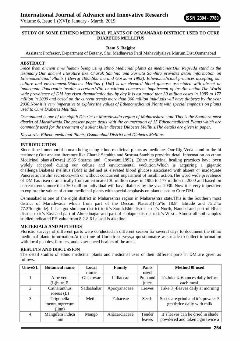
International Journal of Advance and Innovative Research Volume 6, Issue 1 (XVI): January - March, 2019
254
ISSN 2394 - 7780
STUDY OF SOME ETHENO MEDICINAL PLANTS OF OSMANABAD DISTRICT USED TO CURE DIABETES MELLITUS
Ram S .Bajgire Àssistant Professor, Department of Botany, Shri Madhavrao Patil Mahavidyalaya Murum.Dist.Osmanabad
ABSTRACT Since from ancient time human being using ethno Medicinal plants as medicines.Our Regveda stand to the testimony.Our ancient literature like Charak Samhita and Susruta Samhita provides detail information on Ethenomedicinal Plants ( Devraj 1985,Sharma and Goswami 1992). Ethenomedicinal practices accepting our culture and environment.Diabetes Mellitus ( DM) is an elevated blood glucose associated with absent or inadequate Pancreatic insulin secretion.With or without concurrent impairment of insulin action.The World wide prevalence of DM has risen dramatically day by day.It is estimated that 30 million cases in 1985 to 177 million in 2000 and based on the current trends more than 360 million indiduals will have diabetes by the year 2030.Now it is very imperative to explore the values of Ethenomedicinal Plants with special emphasis on plants used to Cure Diabetes Mellitus.
Osmanabad is one of the eighth District in Marathwada region of Maharashtra state.This is the Southern most district of Marathwada.The present paper deals with the enumeration of 15 Ethenomedicinal Plants which are commonly used for the treatment of a silent killer disease Diabetes Mellitus.The details are given in paper.
Keywords: Etheno medicinal Plants, Osmanabad District and Diabetes Mellitus.
INTRODUCTION Since time immemorial human being using ethno medicinal plants as medicines.Our Rig Veda stand to the hi testimony.Our ancient literature like Charak Samhita and Susruta Samhita provides detail information on ethno Medicinal plants(Devraj 1985 Sharma and Goswami,1992). Ethno medicinal healing practices have been widely accepted during our culture and environmental evolution.Which is acquiring a gigantic challenge.Diabetes mellitus (DM) is defined as elevated blood glucose associated with absent or inadequate Pancreatic insulin secretion,with or without concurrent impairment of insulin action.The word wide prevalence of DM has risen dramatically from an estimated 30 million cases in 1985 to 177 million in 2000 and based on current trends more than 360 million individual will have diabetes by the year 2030. Now it is very imperative to explore the values of ethno medicinal plants with special emphasis on plants used to Cure DM.
Osmanabad is one of the eight district in Maharashtra region in Maharashtra state.This is the Southern most district of Marathwada which from part of the Deccan Plateau(17.5°to 18.8° latitude and 75.2°to 77.3°longitude). It has got sholapur district to it’s South,Bhir district to it’s North, Nanded and part of Bhair district to it’s East and part of Ahmednagar and part of sholapur district to it’s West . Almost all soil samples studied indicated PH value from 8.2-8.6 i.e. soil is alkaline.
METERIALS AND METHODS Floristic surveys of different parts were conducted in different season for several days to document the ethno medicinal plants information.At the time of floristic surveys,a questionnaire was made to collect information with local peoples, farmers, and experienced healers of the areas.
RESULTS AND DISCUSSION The detail studies of ethno medicinal plants and medicinal uses of their different parts in DM are given as follows:
UniveSL Botanical name Local name
Family Parts used
Method 0f used
1 Aloe vera (L)burn.F.
Ghekuwar Lilliaceae Pulp and juice
It’sJuice 4-6ounces daily before each meal.
2 Catharanthus roseus (L)
Sadaabahar Apocyanaceae
Leaves Take 3_4leaves daily at morning
3 Trigonella foennumgrecum
(linn)
Methi Fabaceae Seeds Seeds are grind and it’s powder 5 gm thrice daily with milk
4 Mangifera indica linn
Mango Anacardiaceae Tender leaves
It’s leaves can be dried in shade powdered and taken 5gm twice a
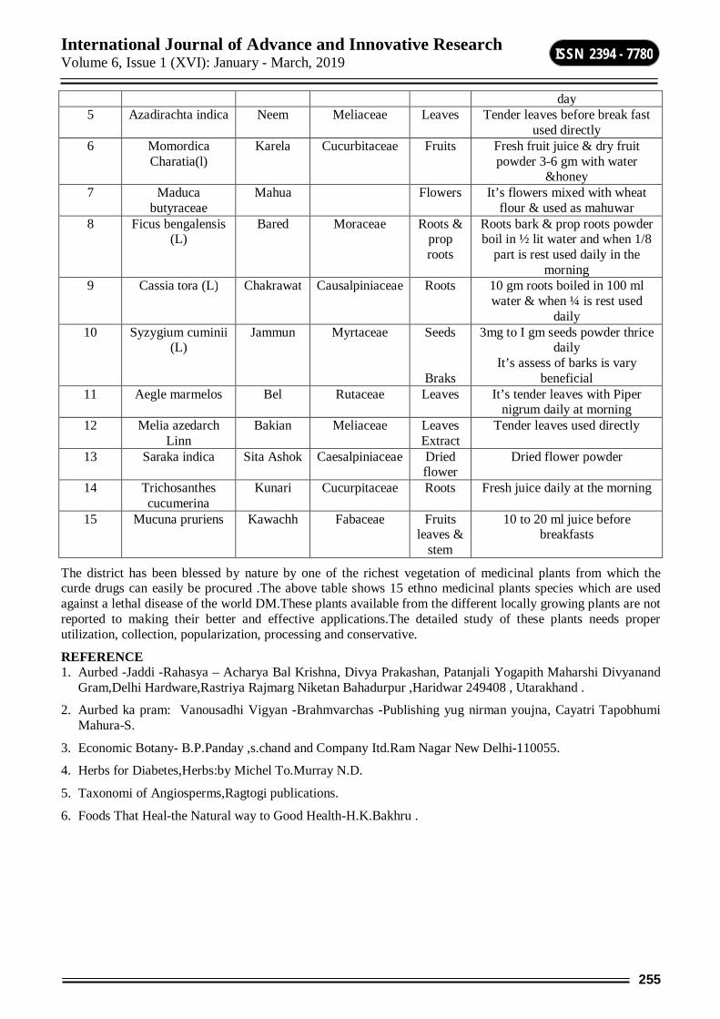
International Journal of Advance and Innovative Research Volume 6, Issue 1 (XVI): January - March, 2019
255
ISSN 2394 - 7780
day 5 Azadirachta indica Neem Meliaceae Leaves Tender leaves before break fast
used directly 6 Momordica
Charatia(l) Karela Cucurbitaceae Fruits Fresh fruit juice & dry fruit
powder 3-6 gm with water &honey
7 Maduca butyraceae
Mahua Flowers It’s flowers mixed with wheat flour & used as mahuwar
8 Ficus bengalensis (L)
Bared Moraceae Roots & prop roots
Roots bark & prop roots powder boil in ½ lit water and when 1/8
part is rest used daily in the morning
9 Cassia tora (L) Chakrawat Causalpiniaceae Roots 10 gm roots boiled in 100 ml water & when ¼ is rest used
daily 10 Syzygium cuminii
(L) Jammun Myrtaceae Seeds
Braks
3mg to I gm seeds powder thrice daily
It’s assess of barks is vary beneficial
11 Aegle marmelos Bel Rutaceae Leaves It’s tender leaves with Piper nigrum daily at morning
12 Melia azedarch Linn
Bakian Meliaceae Leaves Extract
Tender leaves used directly
13 Saraka indica Sita Ashok Caesalpiniaceae Dried flower
Dried flower powder
14 Trichosanthes cucumerina
Kunari Cucurpitaceae Roots Fresh juice daily at the morning
15 Mucuna pruriens Kawachh Fabaceae Fruits leaves &
stem
10 to 20 ml juice before breakfasts
The district has been blessed by nature by one of the richest vegetation of medicinal plants from which the curde drugs can easily be procured .The above table shows 15 ethno medicinal plants species which are used against a lethal disease of the world DM.These plants available from the different locally growing plants are not reported to making their better and effective applications.The detailed study of these plants needs proper utilization, collection, popularization, processing and conservative.
REFERENCE 1. Aurbed -Jaddi -Rahasya – Acharya Bal Krishna, Divya Prakashan, Patanjali Yogapith Maharshi Divyanand
Gram,Delhi Hardware,Rastriya Rajmarg Niketan Bahadurpur ,Haridwar 249408 , Utarakhand .
2. Aurbed ka pram: Vanousadhi Vigyan -Brahmvarchas -Publishing yug nirman youjna, Cayatri Tapobhumi Mahura-S.
3. Economic Botany- B.P.Panday ,s.chand and Company Itd.Ram Nagar New Delhi-110055.
4. Herbs for Diabetes,Herbs:by Michel To.Murray N.D.
5. Taxonomi of Angiosperms,Ragtogi publications.
6. Foods That Heal-the Natural way to Good Health-H.K.Bakhru .
International Journal of Advance and Innovative Research Volume 6, Issue 1 (XVI): January - March, 2019
256
ISSN 2394 - 7780
ETIDRONIC ACID AN EFFICIENT CATALYST FOR THE SYNTHESIS OF Β-ENAMINONES IN AQUEOUS MEDIA
Balaji Madje and Jagdish Bharad Department of Chemistry, Vasantrao Naik Mahavidyalaya, Aurangabad
ABSTRACT An efficient synthesis of β-enaminone derivatives from dimedone and amines at room temperature in the presence of etidronic acid (EDA) as a catalyst in aqueous media. EDA found to be efficient catalyst, experimental procedure is simple, shorter reaction time and excellent yield of the products.
Keywords: β-enaminone, amines, dimedone, etidronic acid.
1. INTRODUCTION Organic heterocyclic compounds containing nitrogen are of special interest because they constitute an important class of natural and non-natural products, and exhibit useful biological activities. Among those compounds, β-enaminones have been employed as synthetic building block of a wide variety of heterocycles [1] key intermediate in synthesis of different heterocycles and naturally occurring alkaloids [2] and in pharmaceuticles [3]. In spit of the importance of β-enaminones as valuable biologically active compounds, their synthesis has received great attention of chemists and hence several routes have been recently reported in literature using Erabium(III) triflate [4], Ytterbium triflate [5], P2O5/SiO2[6], Cu-nanoparticles [7], heteropoly acid [8], CeCl3 [9], AcOH [10], HClO4-SiO2 [11], montmorillonite K10 [12], silica gel [13], ceric ammonium nitrate [14], CoCl2.6H2O [15], NaAuCl4 [16] ZrCl4 [17] and I2 [18]. Recently, β-enaminones synthesized by using Nano-Sio2 [19], NbOPO4 [20] and industrial quality graphene oxide [21]. However, therefore in search of new simple, clean and one-pot method for the synthesis of desired organic compounds is of our prime interest. In search of better alternatives, we have paid attention to find convenient and efficient method based on green approach for the synthesis of β-enaminones.
Since the pioneering studies by Breslow [22] on Diels–Alder reactions, there have been profound research activities in the development of organic reactions in aqueous media offering key advantages such as rate enhancement and insolubility of the final products, which facilitates their isolation by simple filtration. Also, in the context of green chemistry, aqueous media is acting as a stepping stone in the greener synthesis of bioactive heterocyclic compounds. In this respect, the development of water-tolerant catalysts has rapidly become an area of intense research.
Cheap and commercially available etidronic acid (EDA) is a phosphonic acid with mild acidity, is non-volatile, non-corrosive, and is soluble in common organic solvents. It is a white crystalline solid with outstanding physical and chemical properties. Etidronic acid as a catalyst in organic syntheses were found to be effective in the synthesis of 5-nitro-dihydropyrimidine [23], 5-carboxanilide- dihydropyrimidinones [24] thiiranes [25] and
1,4-dihydropyridines [26]. However, there are no examples of the use of etidronic acid as a catalyst for the synthesis of β-enaminones derivatives.
2. RESULT AND DISCUSSION In continuation of our work on the applications of cheap and easily available catalyst for the development of new synthetic methodologies [27], herein we report a simple and efficient EDA catalyzed synthesis of β-enaminone derivatives has been reported. The transformation involves the condensation reaction of dimedone and substituted amines in aqueous media at room temperature (Scheme 1).
In the initial reaction condition, the reaction of dimedone 1 and aniline 2a were selected as standard model reaction to optimize the reaction conditions. The reaction were first carried out in water in the absence of EDA, no product could be detected at room temperature and reflux temperature even after 2 hours (Table 2 compound 3a). To determine the optimal catalyst loading required, the reaction were repeated with varying amounts of EDA (Table 1). A maximum yield of 94% was obtained with 10 mol% of EDA at room temperature in water within 40 minute. Further increase in catalyst loading 15% or 20% did not have any significant effect on the yield of product. Whereas decreasing the amount of catalyst decreased yields (Table 1, Entry 1, 2 and 3).
Scheme-1: Synthesis of β-enaminone derivatives catalyzed by EDA/H2O
International Journal of Advance and Innovative Research Volume 6, Issue 1 (XVI): January - March, 2019
257
ISSN 2394 - 7780
With this exciting result, the method was extended to a number of aromatic amines to investigate its scope and generality. Aromatic amines, including those bearing electron-withdrawing groups (such as Cl, F and Br) or electron donating groups (such as CH3, OCH3) were treated with dimedone catalyzed by EDA at room temperature in water. The corresponding β-enaminone derivatives (3a-j) were formed within short reaction times in excellent yields and confirmed by IR, 1H NMR, Mass spectroscopic analysis. The results are summarized in Table 2.
A plausible mechanism for the formation of β-enaminone has been envisaged [18]. The condensation of dimedone with amine is expected to proceed through an addition–elimination mechanism. It is assumed that dimedone first gets activated in the presence of EDA; subsequently, the lone pair of electrons on nitrogen of the amine adds onto the carbonyl moiety to furnish an intermediate, which may lose a molecule of water to give an imine. This imine may get tautomerized to give β-enaminone as shown in Scheme 2.
OO
NH2R
EDA
O OH
NR
H
O N
H
RO
HN
R
EDA
Scheme-2: Plausible mechanism for the formation of β-enaminone
Table-1: Optimization of molar ratio of catalyst for the model reaction Entry Mole % Yield %a
01 00 -- 02 02 55 03 05 70 04 10 94 05 15 94 06 20 93
aIsolated yield of the product
Table-2: Synthesis of β-enaminone derivatives catalyzed by EDA/H2O at room temperature Entrya R Time (Min) Yield (%) M. P. (°C)
3a H 2 h at r.t. - - 3a H 2 h at reflux - - 3a H 40 94 182-184 3b 4-Cl 40 92 150-152 3c 4-OMe 30 93 105-107 3d 2,3-Cl 40 87 199-201 3e 2-Me 35 89 134-136 3f 3-Me 35 90 140-142 3g 4-Br 40 92 164-166 3h 4-Me 30 93 143-145 3i 3-OMe,4-OH 25 94 112-114 3j 4-F 30 94 190-192
aIsolated yield of the product
3. CONCLUSIONS In conclusion, we have described simple, efficient practical procedure for the synthesis of some β-enaminone derivatives at room temperature in the presence of EDA as a catalyst. The remarkable advantages offered by the methods presented are use of safer and ecofriendly catalysts, mild reaction conditions, simplicity of the reaction procedure and high yielding strategy.
4. EXPERIMENTAL 4.1 General procedure: Synthesis of β-enaminones. A mixture of dimedone 1 (1 mmol), amines 2a-j (1 mmol) and EDA (10 mol%) were taken in a round bottom flask in 5 ml of water. The reaction mixture was stirred at room temperature for a stipulated time (Table 2). The progress of reaction was monitored on TLC. After completion of the reaction, the mixture were diluted with cold water (10 mL), stirred for 5 min., and the resulting solid product was filtered, dried and purified by crystallization in aqueous ethanol.
International Journal of Advance and Innovative Research Volume 6, Issue 1 (XVI): January - March, 2019
258
ISSN 2394 - 7780
4.2 Spectral analysis of representative compound: (Table 2, entry 3a): 1H NMR (CDCl3) δ ppm, 1.2 (S, 6H, 2 x CH3), 2.2 (s, 2H, C-CH2), 2.4 (s, 2H, COCH2), 5.6 (s, 1H, CH), 7.1-7.4.0 (m, 5H, Ar-H), 7.8 (s, 1H, NH); m/z 216 (M+), 214 (M-).
REFERENCES 1. Alan C, Spivey A C, Sirikaran R, Diaper C M, David J, Turner D, Org. Biomol. Chem. (2003) 1638.
2. (a) White J D, Lhle D C, Org. Lett. 8 (2006) 1081. (b) Li G, Watson K., Buckheit R W, Zhang Y, Org. Lett. 9 (2007) 2043.
3. (a) Edafiogho I O, Ananthalakshmi K. V, Kombian S B, Bioorg. Med. Chem. 14 (2006) 5266. (b) Edafiogho I O, Phillips O A, Udo E E, Samuel S, Rethish B, Eur. J. Med. Chem. 44 (2009) 967.
4. Dalpozzo R R, Nino A D, Nardi M, Procopio B A Synthesis 7 (2006) 1127.
5. Epifano F, Genovese S, Curini M, Tetrahedron Lett. 48 (2007) 2717.
6. Mohammadizadeh M R, Hasaninejad A, Bahramzadeh M, Khanjarlou Z S, Synth. Commun. 39 (2009) 1152.
7. Kidwai M, Bhardwaj S, Mishra N, Bansal V, Kumar A, Mozumdar S, Catal. Commun. 10 (2009) 1514.
8. Rafiee E, Joshaghani M, Eavani S, Roshidzadeh S, Green Chem. 10 (2008) 982
9. Khodaei M M, Khosropour A R, Kookhazadeh M, Synlett (2004) 1980.
10. Brandt C A, Da Silva A C M P, Pancote C G, Brito C L, Da Silveria M A B, Synthesis (2004) 1557.
11. Das B, Venkateswarlu K, Majhi A, Reddy M R, Reddy K N, Rao Y K, Ravikumar K, Sridhar B, J. Mol. Catal. A: Chem.246 (2006) 276.
12. Braibante H T S, Braibante M E F, Rosso G B, Oriques D A, J. Braz. Chem. Soc. 14 (2003) 994.
13. Gao Y, Zhang Q, Xu J, Synth. Commun. 34 (2004) 909.
14. Paira M, Misra M, Roy S C, Indian J. Chem.47B (2008) 966.
15. Zhang Z H, Hu J Y, J. Braz. Chem. Soc.17 (2006) 1447.
16. Arcadi A, Bianchi G, Di Giuseppe S, Marinelli F, Green Chem. 5 (2003) 64.
17. Lin J, Zhang L F, Monatsh. Chem. 138 (2007) 77.
18. Bandita D, Madhusudana Reddy M B, Pasha M A, Synth. Commun. 41 (2011) 2331.
19. Alinezhad H, Tajbakhsh M, Sarkati M N et al. Monatsh Chem 147 (2016) 1591.
20. Felipe W, Fernando R X, Samuel R M, Alfredo R M, de Oliveira Rogério A G, Tetrahedron Letter, 58 (2017) 231.
21. Dian D, Lang X, III-min C, Ick S K, Mayakrishnan G, ACS Sustainable Chem & Engineering 5 (2017)1253.
22. (a) Breslow R, Acc. Chem. Res. 24 (1991) 159. (b) Breslow R, Acc. Chem. Res. 37 (2004) 471.
23. Savant M M, Pansuriya A M, Bhuva C V, Kapuriya N P, Naliapara Y T, Catal Lett 132 (2009) 281.
24. Pansuriya A M, Savant M M, Bhuva C V, Singh J, Naliapara Y T, Arkivoc, vii (2009) 79.
25. Liqiang W, Yongxue W, Fulin Y, Chunguang Y, Bull. Korean Chem. Soc. 31 (2010) 1419.
26. Reddy B, Rajesh K, Vijayaparthasarathi V, Indian J. heterocyclic chem.20 (2011) 281.
27. (a) Madje B R, Ubale M B, Bharad J V, Shingare M S, South Afr. J. Chem. 63 (2010) 36.
(b) Madje B R, Ubale M B, Bharad J V, Shingare M S, South. Afr. J. Chem. 63 (2010) 158.
(c) Madje B R, Shelke K F, Sapkal S B Kakde G K, Shingare M S, Green Chem. Lett. Rev. 3 (2010) 269.
(e) Madje B, Chavan R, Bharad J, Ubale M, Chemistry & Biology Interface, 4 (2014) 246.
(f) Gadekar S S, Sapkal S B, Madje B R, Int. J. Green Chem. 2 (2016) 1.
International Journal of Advance and Innovative Research Volume 6, Issue 1 (XVI): January - March, 2019
259
ISSN 2394 - 7780
FIRST PRINCIPLES STUDY OF LYSINE AMINO ACID-SINGLE WALLED CARBON NANOTUBES INTERACTIONS
Ravindra Karde, Avinash Ingale, Prashant Pardeshi, Baliram Lone Nanomaterials Research Laboratory, Department of Physics, Vinayakrao Patil, Mahavidyalaya, Vaijapur,
Aurangabad, Maharashtra
ABSTRACT The adsorption of lysine (Lys) molecule on the side walls of both the intrinsic and Boron-doped single walled (9,0) carbon nanotubes (SWCNTs) were investigated by using density functional theory. A lysine molecule tends to physically adsorb on intrinsic SWCNTs. In the present study lysine (Lys) can form quite stable complexes with intrinsic (9, 0) SWNTs. We investigated parameters like HOMO, LUMO, i.e. highest occupied and lowest occupied molecular orbitals respectively. In this interaction process, band gap and energy formation of compounds reported extensively. The binding mechanism of lysine (Lys) amino acid on SWNT (9,0) have evaluated. The binding of amino acid Lys-SWNT thermodynamically favorable. It depends on the orientation of amino acid lysine. However, it is very weak. To insight chemical bonding through electronic structure, we further studied density of states (DOS) of SWNTs and Lys-SWNT complexes.
Our study will be useful to understand the role of Amino acid (Lys.)-SWNT interaction which has vast applications in various fields like Biophysics, Biochemistry and Bio-Nanotechnology as carriers in targeted drug delivery, biosensors etc.
Keywords: Density Functional Theory,Lysine, Density of States (DOS), HOMO, SWNT
1. INTRODUCTION The interactions of biomolecules with carbon-based substrates such as single-walled carbon nanotubes (SWNT), graphenehold the attention of research community for their potential uses in various fields such asenergy generation, electronics, chemical sensors,DNA sequencing and medicine delivery systems.[1-6]
The common methods to model moleculesurface interactions frequently uses the computational methods such as Hartree−Fock (HF), density functional theory (DFT), density functional tight binding (DFTB), and molecular mechanics (MM)/molecular dynamics (MD). [7-10]
The complex of the studied systems limited the previous studies mainly to the molecules, and hindered the fundamental understanding of the interaction mechanism of biological molecules with graphene and single walled carbon nanotubes. A systematic understanding of the adsorbed structures and the functional control of the complexes still needs further research. These biomolecules are built with amino acids, the interactions between the amino acids and graphene will be the key issue for understanding the complex systems.[11-14]
Lysine is an amino acid (building block of protein). It is used for preventing and treating cold sores (caused by the virus called herpes simplex labialis).The lysine is used to reduce anxiety by blocking stress response receptors, to improve calcium absorption , retention and to promote wound healing by helping create collagen.[15-16]
In present study the adsorption of lysine (Lys) molecule on the side walls of both the intrinsic and Boron-doped single walled (9,0) carbon nanotubes (SWCNTs) were investigated by using DFT method. A lysine molecule tends to physically adsorb on intrinsic SWCNTs. We reported that both the N-centered and C-centered lysine (Lys) can form quite stable complexes with intrinsic and B-doped (9, 0) SWNTs. We investigated parameters like HOMO, LUMO. In this interaction process, band gap and energy formation of compounds reported extensively. The binding mechanism of lysine (Lys) amino acid on SWNT (9,0) have reported. The binding of amino acid Lys-SWNT thermodynamically favorable. It depends on the orientation of amino acid Lysineand is very weak. To understand the chemical bonding through electronic structure, we have analyzed the density of states (DOS) Lys-SWNT complexes.
The insights obtained from our study will be useful to understand the role of SWNT as carriers in targeted drug delivery and other related fields.In addition, the data of SWNT-Lys complex system can be used in health related issues as applications.
EXPERIMENTAL/COMPUTATIONAL DETAILS A DFT method is used to studythe lysine-SWNT interaction. Considering accuracy of normal mode calculations performed using B3LYP sufficiently high. Moreover, optimal cost to benefit ratio, with 6-31G** basis set
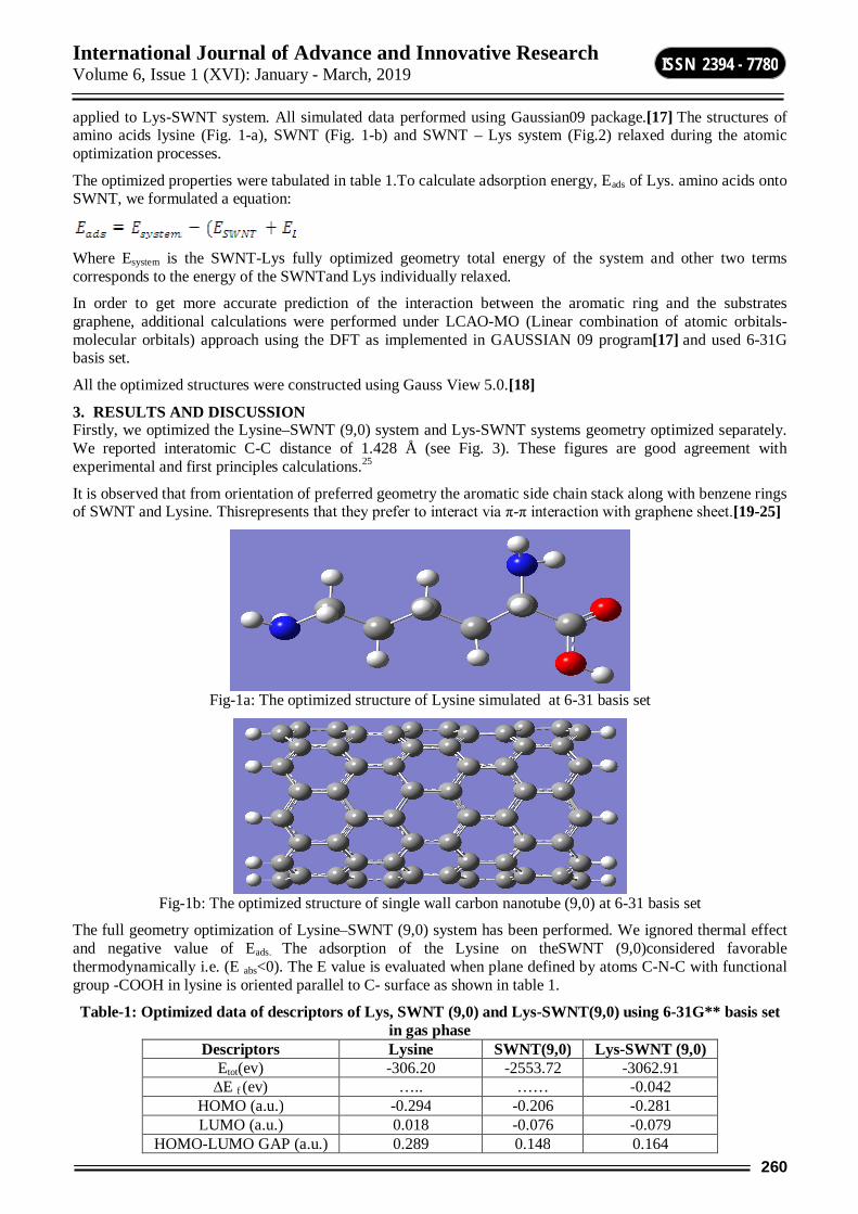
International Journal of Advance and Innovative Research Volume 6, Issue 1 (XVI): January - March, 2019
260
ISSN 2394 - 7780
applied to Lys-SWNT system. All simulated data performed using Gaussian09 package.[17] The structures of amino acids lysine (Fig. 1-a), SWNT (Fig. 1-b) and SWNT – Lys system (Fig.2) relaxed during the atomic optimization processes.
The optimized properties were tabulated in table 1.To calculate adsorption energy, Eads of Lys. amino acids onto SWNT, we formulated a equation:
Where Esystem is the SWNT-Lys fully optimized geometry total energy of the system and other two terms corresponds to the energy of the SWNTand Lys individually relaxed.
In order to get more accurate prediction of the interaction between the aromatic ring and the substrates graphene, additional calculations were performed under LCAO-MO (Linear combination of atomic orbitals-molecular orbitals) approach using the DFT as implemented in GAUSSIAN 09 program[17] and used 6-31G basis set.
All the optimized structures were constructed using Gauss View 5.0.[18]
3. RESULTS AND DISCUSSION Firstly, we optimized the Lysine–SWNT (9,0) system and Lys-SWNT systems geometry optimized separately. We reported interatomic C-C distance of 1.428 Å (see Fig. 3). These figures are good agreement with experimental and first principles calculations.25
It is observed that from orientation of preferred geometry the aromatic side chain stack along with benzene rings of SWNT and Lysine. Thisrepresents that they prefer to interact via π-π interaction with graphene sheet.[19-25]
Fig-1a: The optimized structure of Lysine simulated at 6-31 basis set
Fig-1b: The optimized structure of single wall carbon nanotube (9,0) at 6-31 basis set
The full geometry optimization of Lysine–SWNT (9,0) system has been performed. We ignored thermal effect and negative value of Eads. The adsorption of the Lysine on theSWNT (9,0)considered favorable thermodynamically i.e. (E abs<0). The E value is evaluated when plane defined by atoms C-N-C with functional group -COOH in lysine is oriented parallel to C- surface as shown in table 1.
Table-1: Optimized data of descriptors of Lys, SWNT (9,0) and Lys-SWNT(9,0) using 6-31G** basis set in gas phase
Descriptors Lysine SWNT(9,0) Lys-SWNT (9,0) Etot(ev) -306.20 -2553.72 -3062.91
∆E f (ev) ….. …… -0.042 HOMO (a.u.) -0.294 -0.206 -0.281 LUMO (a.u.) 0.018 -0.076 -0.079
HOMO-LUMO GAP (a.u.) 0.289 0.148 0.164
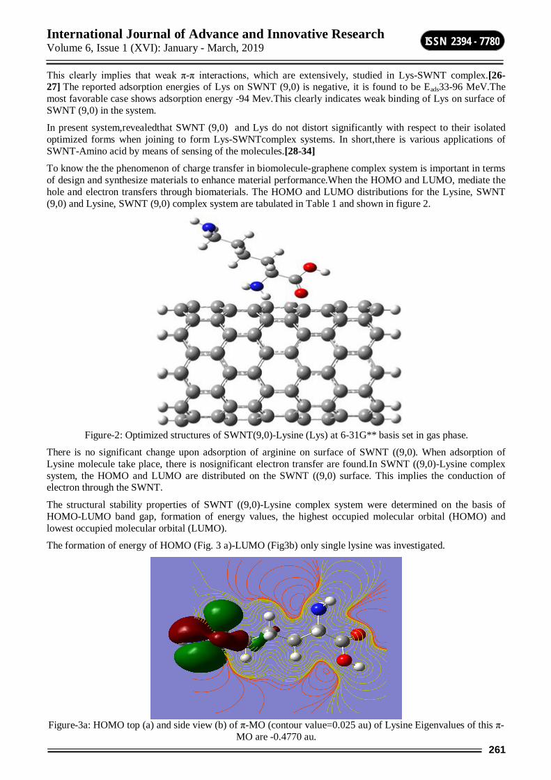
International Journal of Advance and Innovative Research Volume 6, Issue 1 (XVI): January - March, 2019
261
ISSN 2394 - 7780
This clearly implies that weak π-π interactions, which are extensively, studied in Lys-SWNT complex.[26-27] The reported adsorption energies of Lys on SWNT (9,0) is negative, it is found to be Eads33-96 MeV.The most favorable case shows adsorption energy -94 Mev.This clearly indicates weak binding of Lys on surface of SWNT (9,0) in the system.
In present system,revealedthat SWNT (9,0) and Lys do not distort significantly with respect to their isolated optimized forms when joining to form Lys-SWNTcomplex systems. In short,there is various applications of SWNT-Amino acid by means of sensing of the molecules.[28-34]
To know the the phenomenon of charge transfer in biomolecule-graphene complex system is important in terms of design and synthesize materials to enhance material performance.When the HOMO and LUMO, mediate the hole and electron transfers through biomaterials. The HOMO and LUMO distributions for the Lysine, SWNT (9,0) and Lysine, SWNT (9,0) complex system are tabulated in Table 1 and shown in figure 2.
Figure-2: Optimized structures of SWNT(9,0)-Lysine (Lys) at 6-31G** basis set in gas phase.
There is no significant change upon adsorption of arginine on surface of SWNT ((9,0). When adsorption of Lysine molecule take place, there is nosignificant electron transfer are found.In SWNT ((9,0)-Lysine complex system, the HOMO and LUMO are distributed on the SWNT ((9,0) surface. This implies the conduction of electron through the SWNT.
The structural stability properties of SWNT ((9,0)-Lysine complex system were determined on the basis of HOMO-LUMO band gap, formation of energy values, the highest occupied molecular orbital (HOMO) and lowest occupied molecular orbital (LUMO).
The formation of energy of HOMO (Fig. 3 a)-LUMO (Fig3b) only single lysine was investigated.
Figure-3a: HOMO top (a) and side view (b) of π-MO (contour value=0.025 au) of Lysine Eigenvalues of this π-
MO are -0.4770 au.

International Journal of Advance and Innovative Research Volume 6, Issue 1 (XVI): January - March, 2019
262
ISSN 2394 - 7780
Figure-3b: LUMO top view of π-MO (contour value=0.025 au) of Lysine Eigenvalues of this π-MO are -0.4772
au.
To understand the chemical bonding through electronic structure, we have analyzed the density of statesDOS plot of these complexes. Figure 4 indicates the DOS plotof the free SWNT and aromatic ring-Lys complexes obtained by using Gaussian broadening 0.1 eV of the one-electron Kohn-Sham electronic energies. A comparison of the electronic energy spectrum suggests that the energy levels of the SWNT undergo an overall red shift when the amino acids ys bound it. A maximum shift of 0.38eV, was found in the energy spectrum whenLys was bound to the SWNT.
Figure-4: Total and partial DOS for the adsorption system of Lys-SWNT system.
Vibrational spectrum
Frequency, cm**-13,5003,0002,5002,0001,5001,0005000
IR in
tens
ity
40038036034032030028026024022020018016014012010080604020
0
60.474
211.846
386.765
612.142
723.148
871.157
1,103.261
1,352.184
1,439.644
1,560.741
1,739.024
2,919.728
3,599.221
Fig-5: The theoretical evaluated FTIR spectra of Lysine molecule with –COOH group.
International Journal of Advance and Innovative Research Volume 6, Issue 1 (XVI): January - March, 2019
263
ISSN 2394 - 7780
The degree of shift of EFof SWNT-Lys complex varies as function of polarizability of Lys molecules. We conclude thatthe amino acids are the smallest building block for large protein molecules. Moreover,for interaction of proteins that composed of number of amino acid chainslead to changes at small extent will take place in the chemical potential of these complexes. We expect a desirable property for its electrical detection of biomolecule in biophysics.
4. CONCLUSION The adsorption of lysine amino acid on surface of SWNT investigated using DFT calculations. The present study reveals the mechanism process of the complex system Lys-SWNT (9,0) is thermodynamically little favorable.The Lys-SWNT (9, 0) complex more stable than single lysine and SWNT respectively. The comparison of the electronic energy spectrum indicates that the energy levels of the SWNT undergo an overall red shift when the amino acids Lysinebound to it. A maximum shift of 0.38 eV, was found in the energy spectrum whenLys was bound to the SWNT.The degree of shift of EFof Lys-SWNT (9, 0) complex varies as function of polarizability of Lys molecules.
The DOS of the adsorption complex is almost the superposition of the density of states of the individual components. Moreover, the interaction (adsorption) can be used to modify the band properties of SWNT.The insights obtained from our study will be useful to understand the role of SWNT as carriers in targeted drug delivery and other related fields.In addition, the data of SWNT-Lys complex system can be used in health related issues as applications.
ACKNOWLEDGMENTS The authors would like to thanks Department of Science and Technology, New Delhi, India(under FAST TRACK SCHEME for YOUNG SCIENTIST,GRANT No.SR/FT/LS-020/2009) and University Grants Commission, New Delhi, India,(Grant No.33-16/2007-SR) for providing financial support.
The simulation work was conducted in High Performance Computing of Nanomaterials Research laboratoryat department of Physics, Vinayakrao Patil Mahavidyalaya, Vaijapur, Maharashtra, India.
REFERENCES 1. K. Novoselov, A. Geim, S. Morozov, D. Jiang, Y. Zhang,S.Dubonos , I. Grigorieva and A. Firsov,
Science306,666(2004).
2. V. Allain, C. Bourgaux, and P. Couvreur, Nucleic Acids Res. 40, 1891(2012).
3. M.Tavafoghi, N.Brodusch,R.Gauvin and M.Cerruti, J. R. Soc. Interface 13, 20150986 (2016).
4. B.Lone, Adv. Sci. Eng. Med..11, 1-5(2019).
5. T. Wirth, Topics in Current Chemistry; Springer-Verlag,Berlin-Heidelberg (2000).
6. J. Glasel and M. Deutscher, Introduction to Biophysical Methods for Protein and Nucleic Acid Research. Academic Press. p. 456. ISBN 9780080534985(1995).
7. Marchesan, S.; Prato, M.,Chem. Commun.51, 4347, (2015).
8. Y. Guo, X. Lu, J .Weng and Y. Leng, J. Phys. Chem. C, 117,5708(2013).
9. B.Lone, J. Comput. & Theo. Nanoscience, 6, 2146(2009).
10. Z. Sun, S. Kohama, Z. Zhang, J. Lomeda, and J. Tour, Nano Res. 3, 117(2010).
11. F. Naderi, A. Karami, and B. Naderi,Org. Chem. J. 1, 44 (2010).
12. N. Dragneva, W.Floriano, D.Stauffer, R. Mawhinney, G.Fanchini, and O. Rubel, J. Chem. Phys. 139,174711-1(2013).
13. P. Singla ,M.Riyaz, S.Singhal, and N. Goel,Phys. Chem. Chem. Phys.18,5597(2016).
14. B. Lone,S.Scheinerand T. Kar, Carbon 80:405(2014).
15. G. Korshunova, N. Sumbatyan, A. Topin and M.Mtchedlidze,Mol. Biol. 34,823(2000).
16. C. Reilly, Selenium in Food and Health; Springer Science: New York (2006).
17. M. J. Frisch, G. W. Trucks, H. B. Schlegel, G. E. Scuseria, M. A. Robb, J. R. Cheeseman, G. Scalmani, V. Barone, B. Mennucci, G. A. Petersson, H. Nakatsuji, M. Caricato, X. Li, H. P. Hratchian, A. F. Izmaylov, J. Bloino, G. Zheng, J. L. Sonnenberg, M. Hada, M. Ehara, K. Toyota, R. Fukuda, J. Hasegawa, M. Ishida,
International Journal of Advance and Innovative Research Volume 6, Issue 1 (XVI): January - March, 2019
264
ISSN 2394 - 7780
T. Nakajima, Y. Honda, O. Kitao, H. Nakai, T. Vreven, J. A. Montgomery, Jr., J. E. Peralta, F. Ogliaro, M. Bearpark, J. J. Heyd, E. Brothers, K. N. Kudin, V. N. Staroverov, R. Kobayashi, J. Normand, K. Raghavachari, A. Rendell, J. C. Burant, S. S. Iyengar, J. Tomasi, M. Cossi, N. Rega, J. M. Millam, M. Klene, J. E. Knox, J. B. Cross, V. Bakken, C. Adamo, J. Jaramillo, R. Gomperts, R. E. Stratmann, O. Yazyev, A. J. Austin, R. Cammi, C. Pomelli, J. W. Ochterski, R. L. Martin, K. Morokuma, V. G. Zakrzewski, G. A. Voth, P. Salvador, J. J. Dannenberg, S. Dapprich, A. D. Daniels, Ö. Farkas, J. B. Foresman, J. V. Ortiz, J. Cioslowski, and D. J. Fox, Gaussian 09, Revision D.01, Gaussian, Inc., Wallingford CT, (2009).
18. F. Aeleen, H. Hratchian, D. Roy, Dennington II, A. Todd and J. Keith,GaussView 5.0,ISBN 978-1-935522-00-3., Gaussian, Inc: Wallingford, CT.,U.S.A.(2009).
19. T. Kuila, S. Bose, P. Khanra, A.Mishra, N. Kim and J. Lee, Biosens.Bioelectron 26, 46(2011).
20. H. Vovusha, S. Sanyal and B. Sanyal,J. Phys.Chem. Lett. 4,3710(2013).
21. K.Waters, R.Pandey andS.Karna,ACS Omega 2, 76 (2017).
22. C. Rajesh, C. Majumder, H. Mizuseki and Y. Kawazoe, J. Chem. Phys. 130,124911(2009).
23. R. Chen, S. Bangsaruntip, K. Drouvalakis, N. Kam, M. Shim, Y. Li, W. Kim, P. Utz and H. Dai, Proc. Natl. Acad. Sci. U.S.A. 100, 4984 (2003).
24. Singla, P.; Riyaz, M.; Singhal, S.; Goel, N., Phys.Chem .Chem.Phys.18, 5597(2016).
25. C. Tang, R. Ulijn and A. Saiani, Langmuir 27, 14438(2011).
26. F.Ma,Z. Zhang,H. Jia,X. Liu,Y. Hao and B. Xu, J.Molecular Str.: THEOCHEM,955,134(2010).
27. W.Qin, X.Li,Wen-Wen Bian,Xiu-Juan Fan and Jing-Yao Qi, Biomaterials 31, 1007(2010).
28. B. Lone, J.Nanomed Nanotechnol 7, 1(2016).
29. H. S. Nalwa (Ed.), Encyclopedia of Nanoscience and Nanotechnology, Volume 1-10, American Scientific Publishers, Los Angeles (2004).
30. S.-H. Kang, G. Kim and Y.-K. Kwon, Phys. Chem.Chem. Phys. 17, 5072 (2015).
31. L. Kleinman and D. M. Bylander, Phys. Rev. Lett. 48,1425 (1982).
32. P. Hohenberg and W. Kohn, Phys. Rev. 136, B864(1964).
33. B. Y. Lee, M. G. Sung, J. Lee, K. Y. Baik, Y.-K. Kwon,M.-S. Lee and S. Hong, ACS Nano 5, 4373 (2011).
34. Strano, M. S.; Dyke, C. A.; Usrey, M. L.; Barone, P. W.; Allen, M. J.; Shan, H. W.; Kittrell, C.; Hauge, R. H.; Tour, J. M.; Smalley, R. E.,Science,301,1519 (2003)
International Journal of Advance and Innovative Research Volume 6, Issue 1 (XVI): January - March, 2019
265
ISSN 2394 - 7780
REVIEW ON NUTRITIONAL VALUES OF ASIAN CATFISH, CLARIAS BATRACHUS: IDEAL SPECIES FOR AQUACULTURE IN RURAL AREA
Jagtap H. S. Associate Professor & Head, P. G. Department of Zoology & Research Center, Shri Shivaji College, Parbhani
ABSTRACT Magur, Clarias batrachus, the walking catfish, has been introduced throughout the world for the purposes of aquaculture due to their adaptation, high tolerance level in the changing environment, high consumer demand, simple culture characteristic with efficient food conversion. Nutritional values, easily digestible protein, mineral & adequate good cholesterol (HDL concentration >150 mg/dl, HDL > 60% of Total Cholesterol) content of the fish species. Important for pregnant & lactating mothers, the elderly & children. Consumption of Magur is prescribed prophylactically to the anemic & malnourished individuals as well as for the convalescent of the patients due to the nutritional superiority. Intensive Clarias batrachus culture in several Indian states as in rural Bengal & Tripura have much potential towards livelihood development, employment generation & ensuring nutritional enrichment in the regular diet among of the people. The species is well adapted in virtually all Indian aquatic ecosystems, though production remains low. This paper reviews the nutritional values and aquaculture significance with respect to Clarias batrachus.
Keywords: Clarias batrachus, protein, HDL, medicinal values.
INTRODUCTION The walking catfish (Clarias batrachus) is a species of freshwater air breathing catfish native to Southeast Asia. It is named for its ability to "walk" and wiggle across dry land, to find food or suitable environments. This fish normally lives in slow-moving and often stagnant waters in ponds, swamps, streams and rivers, flooded rice paddies or temporary pools which may dry up. When this happens, its "walking" skill allows the fish to move to other sources of water.
Traditional wisdom a propose the nutritional benefits of Indian catfish Clarias batrachus is the domino effect into its high consumer demand (global market value ≈800000 USD). Analytical studies also indicated towards easily digestible protein, mineral & adequate good cholesterol (HDL concentration >150 mg/dl, HDL > 60% of Total Cholesterol) content of the fish species. The species is well adapted in virtually all Indian aquatic ecosystems, though production remains low. This paper reviews recent developments in catfish physiology with respect to Clarias batrachus and its aquaculture significance. Candidates of the genus Clarias has been traveled to many continents, adapting itself successfully & found throughout Asia & Africa. Clarias batrachus in some parts of India, particularly in West Bengal & Tripura is considered as a medicinal fish & traditionally remained a strike among the pregnant & lactating mothers, the elderly & children. Many a times consumption of “Magur” (Local name of Clarias batrachus) is prescribed prophylactically to the anemic & malnourished individuals as well as for the convalescent of the patients due to the nutritional superiority. Intensive Clarias batrachus culture in several Indian states as in rural Bengal & Tripura have much potential towards livelihood development, employment generation & ensuring nutritional enrichment in the regular diet among of the people (Debnath, 2011).
Air breathing fishes suitably for culture due to following reasons Air breathing fishes are very hardy in nature and tolerate wide range of environmental conditions. They are highly adaptable and grow well in controlled pond conditions. They readily accept supplementary feed. They readily breed in captivity. They can withstand low level of dissolved oxygen. Since they have accessory air breathing organs, they can be cultured in derelict and sewage water also. They grow to 0.75 to 1 kg in six months. They can be transported in live condition to markets due to their air breathing nature over longer distances in fresh condition. Consumer preference for air breathing fishes is more than that of carps. It is considered as a delicacy and it has less of bones. Farmers get higher profit since the price/kg is more than that of carps (Rs. 200/kg). These fishes can withstand crowded condition. They adopt easily to intensive aquaculture and can be stocked at the rate of 25,000-50,000 nos/ha. They are well known for highly nutritive recuperative and medicinal qualities are recommended as a diet during convalescence. Air breathing fishes grow faster than the herbivorous fishes. They have also higher protein and iron content and low fat as compared to the carps. Easy digestible protein (comparison to carp or animal protein). Soft flesh with excellent flavor (Felix & Rachael, 1998; Deoraj, A. et al.,1993). These are ideal species for sewage fed fish culture (proved by CIFA).
International Journal of Advance and Innovative Research Volume 6, Issue 1 (XVI): January - March, 2019
266
ISSN 2394 - 7780
Due to above reasons these fishes always more demand than production. There is no well organized cultivation of these species. The natural population of air breathing fishes is dwindling due to over exploitation in wild waters for consumption and also due to habitat stress.
Successful aquaculture of this species may bring about socioeconomic sustainability of the rural people. Intensive Clarias batrachus culture will gain popularity mainly because the species require no special treatment with respect to the conditioning and the growth factors unlike many other aquaculture species. A comparatively simple culture characteristic with efficient food conversion (Ali and Jauncey, 2005) & excellent nutritional profile (Rui et. al., 2007) makes Clarias batrachus very suitable for commercial intensive culture. A common perception of easily digestible high grade protein, high concentration of iron & beneficial lipid content may be instrumental towards its high acceptance as medicinal fish. A yearlong study on the blood plasma lipid of Clarias batrachus in a population (logW= - 0.8628+2.097 logL, Debnath, 2008) revealed that the HDL content ranges from 150 mg/dl – 180 mg/dl which is more than 60% of total cholesterol.
According to the data released by the Fisheries & Aquaculture department, Food & Agriculture Organization of the United Nations, C. batrachus has been propagated throughout the Asia from Thailand & Indonesia (Java). The species has been introduced to as far as Europe (United Kingdom), USA & Australia (Papua New Guinea) from various pockets of South Asia & South East Asia. Clarias batrachus has exceptionally well tolerance level in varied environment that suggests an advantageous evolutionary trait. According to FAO estimates the demand for catfishes throughout the world is increasing & Clarias batrachus with its several beneficial aspects remain as a hit among the Asians in particular.
CONCLUSIONS Clarias batrachus is one of the important air breathing fish globally known due to their nutritional values, high consumer demands, well adaptability towards varied environmental conditions, less technology is required for culture practices, so that this species is also ideal for culture in rural area.
ACKNOWLEDGEMENT The authors are thankful to Principal, Shri Shivaji College, Parbhani (M.S.) India for providing facilities.
REFERENCES Ali, M. Z. and Jauncey K., (2005). Approaches to optimizing dietary protein to energy ratio for African
catfish C. gariepinus (Burchell, 1822). Aquaculture Nutrition, 11, 95–101.
Debnath, S. (2008). Growth Economics & Biometric Comparison of Length Weight Relationship in Two Intra Generic Varieties of C. batrachus Isolated from Dumbur Lake, in a Rural District of Tripura, India. XIth Science Congress, Manipur University, 2008.
Debnath S. (2011). Clarias batrachus, the medicinal fish: An excellent c & idate for aquaculture & employment generation. 2011 International Conference on Asia Agriculture and Animal
IPCBEE vol.13 (2011) IACSIT Press, Singapoore.
Deoraj, A., T. Krishnan, G. P. Talwar and Garg, L. C. (1993). Induced spawning of catfish, Heteropneustes fossilis using D-Lys6 salmon gonadotropin releasing hormone analogue. Aquaculture, 115:159-167.
Felix, S. and Rachael, I. (1998). Prospects of intensive cat fish culture in Tamilnadu. Fishing Chimes, 18(9):7-10.
Rosa Rui, Narcisa Bandarra & Maria Leonor Nunes (2007). Nutritional quality of African catfish C. gariepinus (Burchell 1822): a positive criterion for the future development of the European production of Siluroidei. Int. J. of Food Sci. &Tech., 42.3, Pages 342 – 351.
International Journal of Advance and Innovative Research Volume 6, Issue 1 (XVI): January - March, 2019
267
ISSN 2394 - 7780
APPLICATION OF NATURAL DYE EXTRACTED FROM BEET AND SPINACH AGAINST SILK FIBRE OF BOMBYX MORI
Nagawanshi M. N. and Kirdat P. V. P. G Department of Zoology, Deogiri College, Aurangabad
ABSTRACT Silk is the natural protein fibre commercially used in the textile industry. Silk is dyed in different colours, shades using natural and artificial or synthetic dyes. In the present study dye obtain from available natural resources are from beet root and spinach. The treatment of on dyed silk fibre with mordant and detergent found suitable and fasten the colour.
Keywords: Silk, Dye, Plant Extract, Detergent and Mordant.
INTRODUCTION The silkworm Bombyx moriis a beneficial, eco-friendly insect reared commercially for the production of cocoons and in turn silk. They feed on mulberry leaves.Natural dyes have better biodegradability and generally have higher compatibility with the environment. Natural dyes are non-toxic, non-allergic to skin, eco-friendly and do not create environmental problems due to their biodegradability nature. Demand of natural dyes is increasing continuously. Natural dyes are generally applied along with mordants. The present work, an attempt has been made to dye silk thread with natural dye obtained from a few freely available natural and domestic waste products of plants such as beet and spinach.
MATERIALS AND METHODS Silkworm Bombyx mori was reared in the farm from Belkhandi, Patoda. Dist. Beed (M.S).They were feed with mulberry leaves twice a day at a fix time. If the leaves were too wet then calcium powder was spray.Raw silk was dyed with combinations of already selected three mordents. The mordents are Alum, Ferrous sulphate and Alum + Ferrous sulphate. Dyes derived from various parts of the plant such as root, stem/bark, leaf, flower, fruit and seed. The shades produced by natural dye / colorants are usually soft, lustrousand soothing to the human eye. After mordanting, the metal saltsanchoring to the fibres, attracts the dye/organic pigment molecules to be anchored to the fibres and finally creates the bridging link between the dye molecules and the fibre by forming co-ordinating complexes.
I. MORDANTING AND DYEING METHOD Spinach Mordanting and dyeing method By using alum mordant: 20 % alum powder was prepared by boiling it 15min at 100ºC.1gm of silk thread kept in mordant solution for 15 min at 90ºC containing 50ml fresh spinach and 50ml water were added and boiled. Slowly the temperatureincreased at 90ºC for 30 min.Then the silk threads was squeezed and washed with cold water.
By using ferrous sulphate mordant:1gm of silk thread boiled in 20 % ferrous sulphate mordantfor 15 min at 90ºC. Then 50ml spinach juice and 50ml water were added and boiled. Byslowly increasing the temperature at 90º for 30 min.Then silk thread were squeezed and wash with cold water.
By using alum and ferrous sulphate mordant :10gm of alum powder and 10 gm of ferrous sulphate powder added in boiling water for mordanting and boiled for 15min at 100ºC. 1gm of silk thread added in mordant solution for 15 min at 90ºC.50ml spinach juice and 50ml water were added and boiled. Slowly increase the temperature at 90ºC for 30 min. Then silk threads were squeezed and wash with cold water.
Beet mordanting and dyeing method By using alum mordant: 20 % alum powder was prepared by boiling it 15min at 100ºC.1gm of silk thread kept in mordant solution for 15 min at 90ºC containing 50ml fresh beet and 50ml water wereand boiled. Slowly the temperature increased at 90ºC for 30 min.Then the silk threads were squeezed and washed with cold water. By using ferrous sulphate mordant: 1gm of silk thread boiled in 20 % ferrous sulphate mordantfor 15 min at 90ºC. Then 50ml beet juice and 50ml water were added and boiled by slowly increasing the temperature at 90ºC for 30 min.Then the silk thread were squeezed and wash with cold water. By using alum and ferrous sulphate mordant : 10 gm of alum powder and 10 gm of ferrous sulphate powder added in boiling water for mordanting and boiled for 15min at 100ºC. 1gm of silk thread added in mordant solution for 15 min at 90ºC.50ml beet juice and 50ml water were and boiled. Slowly increase the temperature at 90ºC for 30 min. Then silk threads were squeezed and wash with cold water.
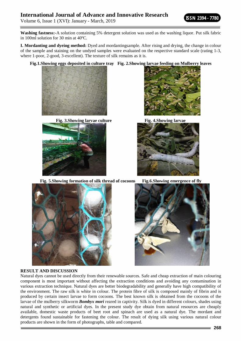
International Journal of Advance and Innovative Research Volume 6, Issue 1 (XVI): January - March, 2019
268
ISSN 2394 - 7780
Washing fastness:-A solution containing 5% detergent solution was used as the washing liquor. Put silk fabric in 100ml solution for 30 min at 40ºC.
I. Mordanting and dyeing method: Dyed and mordantingsample. After rising and drying, the change in colour of the sample and staining on the undyed samples were evaluated on the respective standard scale (rating 1-3, where 1-poor, 2-good, 3-excellent). The texture of silk remains as it is.
Fig.1.Showing eggs deposited in culture tray Fig. 2.Showing larvae feeding on Mulberry leaves
Fig. 3.Showing larvae culture Fig. 4.Showing larvae
Fig. 5.Showing formation of silk thread of cocoons Fig.6.Showing emergence of fly
RESULT AND DISCUSSION Natural dyes cannot be used directly from their renewable sources. Safe and cheap extraction of main colouring component is most important without affecting the extraction conditions and avoiding any contamination in various extraction technique. Natural dyes are better biodegradability and generally have high compatibility of the environment. The raw silk is white in colour. The protein fibre of silk is composed mainly of fibrin and is produced by certain insect larvae to form cocoons. The best known silk is obtained from the cocoons of the larvae of the mulberry silkworm Bombyx mori reared in captivity. Silk is dyed in different colours, shades using natural and synthetic or artificial dyes. In the present study dye obtain from natural resources are cheaply available, domestic waste products of beet root and spinach are used as a natural dye. The mordant and detergents found sustainable for fastening the colour. The result of dying silk using various natural colour products are shown in the form of photographs, table and compared.
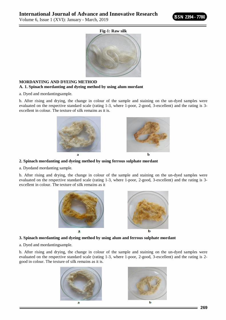
International Journal of Advance and Innovative Research Volume 6, Issue 1 (XVI): January - March, 2019
269
ISSN 2394 - 7780
Fig-1: Raw silk
MORDANTING AND DYEING METHOD A. 1. Spinach mordanting and dyeing method by using alum mordant
a. Dyed and mordantingsample.
b. After rising and drying, the change in colour of the sample and staining on the un-dyed samples were evaluated on the respective standard scale (rating 1-3, where 1-poor, 2-good, 3-excellent) and the rating is 3-excellent in colour. The texture of silk remains as it is.
2. Spinach mordanting and dyeing method by using ferrous sulphate mordant a. Dyedand mordanting sample.
b. After rising and drying, the change in colour of the sample and staining on the un-dyed samples were evaluated on the respective standard scale (rating 1-3, where 1-poor, 2-good, 3-excellent) and the rating is 3-excellent in colour. The texture of silk remains as it
3. Spinach mordanting and dyeing method by using alum and ferrous sulphate mordant
a. Dyed and mordantingsample.
b. After rising and drying, the change in colour of the sample and staining on the un-dyed samples were evaluated on the respective standard scale (rating 1-3, where 1-poor, 2-good, 3-excellent) and the rating is 2-good in colour. The texture of silk remains as it is.

International Journal of Advance and Innovative Research Volume 6, Issue 1 (XVI): January - March, 2019
270
ISSN 2394 - 7780
B. 1. Beet mordanting and dyeing method by using alum mordant a. Dyed and mordantingsample.
b. After rising and drying, the change in colour of the sample and staining on the un-dyed samples were evaluated on the respective standard scale (rating 1-3, where 1-poor, 2-good, 3-excellent) and the rating is 2-good in colour. The texture of silk remains as it is.
2. Beet mordanting and dyeing method by using ferrous sulphate mordant a. Dyed and mordantingsample.
b. After rising and drying, the change in colour of the sample and staining on the un-dyed samples were evaluated on the respective standard scale (rating 1-3, where 1-poor, 2-good, 3-excellent) and the rating is 2-good in colour. The texture of silk remains as it is.
3. Beet mordanting and dyeing method by using alum and ferrous sulphate mordant a. Dyedand mordanting sample.
b. After rising and drying, the change in colour of the sample and staining on the un-dyed samples were evaluated on the respective standard scale (rating 1-3, where 1-poor, 2-good, 3-excellent) and the rating is 2-good in colour. The texture of silk remains as it is.
Table showing
Sr. No. Dyeing method Used Rating After dyeing After washing
I. Mordanting and dyeing method A. 1. Spinach mordanting and dyeing method by using alum
mordant × × × ×
2. Spinach mordanting and dyeing method by using ferrous sulphate mordant
× × × ×
International Journal of Advance and Innovative Research Volume 6, Issue 1 (XVI): January - March, 2019
271
ISSN 2394 - 7780
3. Spinach mordanting and dyeing method by using alum and ferrous sulphate mordant
× × × ×
B. 1. Beet mordanting and dyeing method by using alum mordant
× × × ×
2. Beet mordanting and dyeing method by using ferrous sulphate mordant
× × × ×
3. Beet mordanting and dyeing method by using alum and ferrous sulphate mordant
× × × ×
Regarding eco-friendliness, the colouring matters used here are domestic commonly used materials which as harmless and biodegradable. In usual method of dyeing with natural colourants, various metal salts are used are toxic.But in the present study, we used alum, ferrous sulphate and alum + ferrous sulphate. Hence the proposed process is complete.
Advantages and disadvantages of natural dyes/colorants:-In some aspects natural dyes/colourants are advantageous against synthetic dyes. There are some advantages and disadvantages are given below.
Advantages of natural dyes/colourants:-The shades produced by natural dyes are usually soft, lustrous.Natural dyes produces different shades by using different mordants and it is produces rare colours. Natural dyes are usually renewable and easily available in the market. Natural products do not create a environmental problems because of there biodegradability nature. Therefore, no disposal problem of this natural waste.This is a labour intensive industry, thereby providing job opportunities for engaged in cultivation, extraction and application of these dyes on textile/food/leather etc.Application of natural dyes has potential to earn carbon credit by reducing consumption of fossil fuel (petroleum) based synthetic dyes. Natural dyes are non-toxic and non-allergic to the skin. Some of the natural dyes are enhanced with age, while synthetic dyes fade with time.
Limitation/disadvantages of natural dye/colourants:-Natural dyes are difficult to reproduce shades, as these agro-product vary from one crop season.It is difficult to standardize a recipe for the use of natural dyes, As the natural dyeing process and its colour development depends not only on colour component but also on materials. Itrequires skilled workmanship and is therefore expensive. Scientific backup of a large part of the science involved in natural dyeing is still need to be explored. Lack of availability and technical knowledge of extraction techniques.The dyed textile may change colour when expose to the sun, sweat and air.Most of the natural dyes require the use of mordants to fix them on to the textile substrate. While dyeing, a substantial portion of the mordant remains un-exhausted in the residual dye bath and may pose serious effluent disposal problem.
CONCLUSION The present study concludes that a range of colours were successfully obtained for dyeing of silk fabric using different mordants. New shades were obtained using dyes and mordants combinations. The dyed silk fabrics using all the combinations of dye mordants showed excellent colour values and satisfactory levels of fastness properties.The silk fabrics which are widely used for high value textiles can be successfully dyed using new shades explored in the current work. After detergent test the colour was not fade.The colour strength was improved in most of the cases and tonal variations were obtained depending on the individual cases of dye-mordant combinations.
REFERENCE 1. Kulkarni, S.S., Gokhale, A.V. Bodake, U.M. and Pathade, G.R. (2011). Cotton dyeing with natural dye
extracted from pomegranate (punicagranatum) peel. Universal of Environment Research and Technology, 1:135-139
2. M. Srivastav, D. Mogra and P. Gupta, 2015. Dye extraction from Rheum emodi for colouring silk using natural mordants. (Journal of Applied and Natural Science 7 (1) : 182 – 186
3. Samanata. A.K. and Agrawal, P. (2009). Application of natural dyes on textiles. Indian Journal of Fibre & Textile Research, 34 384-399

International Journal of Advance and Innovative Research Volume 6, Issue 1 (XVI): January - March, 2019
272
ISSN 2394 - 7780
ABO AND RH BLOOD GROUP FREQUENCY DISTRIBUTION AMONG SHINDE TAKLI POPULATION OF PARBHANI DISTRICT, MAHARASHTRA
Nimbalkar R. K. 1, Pawar2 D. A. and S. M. Vadgule3 1,3 Department of Zoology, R. B. Attal College, Georai, Dist. Beed
2Deogiri College, Aurangabad
ABSTRACT ABO and Rh blood groups distribution has been studied randomly from 280 peoples of the population in Shinde Takli of Parbhani District (Maharashtra). The ABO blood group frequency distribution in percentages for A type 24.28%, B type 30.35%, O type 41.06% and type AB 4.27%, while Rh blood group phenotypic frequencies for Rh positive and negative were 96.05% and 3.91% has been recorded. The commonest blood group in studied population was group O and ABO phenotype occurrence order is O > B > A > AB. The allelic frequencies of A, B and O alleles were 0.1675, 0.2043 and 0.6408 were observed from the data. Also, The Rh (D) and Rh (d) allele frequencies of Rh blood groups were found to be 0.8018 and 0.1982 respectively.
Keywords: ABO, Rh, Blood Groups, Allele Frequency, Gene Frequency.
INTRODUCTION Karl Landsteiner in 1901 determined first three A, B and O blood groups by blood transfusion experiments, type of blood is determined on the basis of presence or absence of type of antigen on surface of erythrocytes and antibodies circulating in the plasma [1] later in 1930 Karl Landsteiner received the Nobel Prize in medicine or physiology in corporation with Alexander S Wiener [3]. Blood compatibility is most important, in order for blood transfusions to be securely done. It is necessary for blood to type before blood transfusion to prevent agglutination. In blood transfusions only, certain types of transfusions of blood are safe because the plasma membrane of erythrocytes carry glycoprotein’s that can be antigenic to others. Antigens present on erythrocytes are type A and type B antigens, and their presence or absence is depending on the characteristic inheritance of the individual [4]. An individual with A type of antigen on surface of red blood cell has type A blood and anti- B antibodies in the plasma, while B type of antigen is present in type B blood and anti-A antibodies are circulating in plasma; and a person with O type blood has both A and B antigens are absent on surface of red blood cell but both anti-A and anti-B antibodies are present in plasma, while type AB person has A and B antigens on its red blood cell surface but lacking the antibodies against type A and type B antigens in its plasma [1,4].Hence type AB blood group individuals are universal recipients and they can receive blood from type A, B, O and AB individual. Universal donors are peoples having type O blood group and they will donate blood to persons having blood group A, B, O and AB because type O peoples do not have A and B antigens [1, 4]. The ABO blood group system is under the control of single gene on 9q34 region of chromosome at the ABO locus. Glycosyltransferase enzyme is encoded by this gene, its function is to add sugar residue to H- antigen which is present in red blood cells membrane. The alleles A and B codes for an enzyme that adds an N- acetyl galactosamine and D- galactose respectively over H- antigen in membrane of RBCs. The A and B alleles are different due to few single-base substitutions, changing four amino acid residues that may cause differences in A and B transferase specificity of enzymes encoded by A and B alleles [5, 6]. Rh factor was first discovered in rhesus monkey [1] Rh blood group antigens are non-glycosylated hydrophobic transmembrane proteins with molecular weight 30-32 kDa and genes encoding the Rh proteins are located on chromosome 1p34-p36 [2].ABO blood group incompatibility can cause hemolysis [7] and incompatibility in Rh blood group may produce haemolytic disease of new-born [8]. In haemolytic disease of new-born (Erythroblastosis fetalis), antibodies to the D antigen may be produced by mother, if the mother is Rh negative and the infant is Rh positive in subsequent pregnancies. If anti-D antibodies are produced in large amounts, these antibodies cross the placenta, recognise the Rh-positive cells of infant and cause their destruction [9]. ABO frequencies are varying in different geographical areas, in Western Europeans group O was 46%, group A (42%), group B (9%) and 3% group AB, while frequencies in Americans of blood groups A, B, O and AB are 41%, 10%, 45% and 4% respectively [10]. Also, Rh blood group frequencies differs from population to population. Rh negative phenotypic frequencies are documented as in Andhra Pradesh 1% [14], in Kashmir 4.09% [15], in Punjab 2.7% [22] and in Lucknow 4.55% [23]. This study in Shinde Takli was conducted to determine ABO and Rh blood group frequencies in studied population and also to compare with another similar studies from different geographical areas.
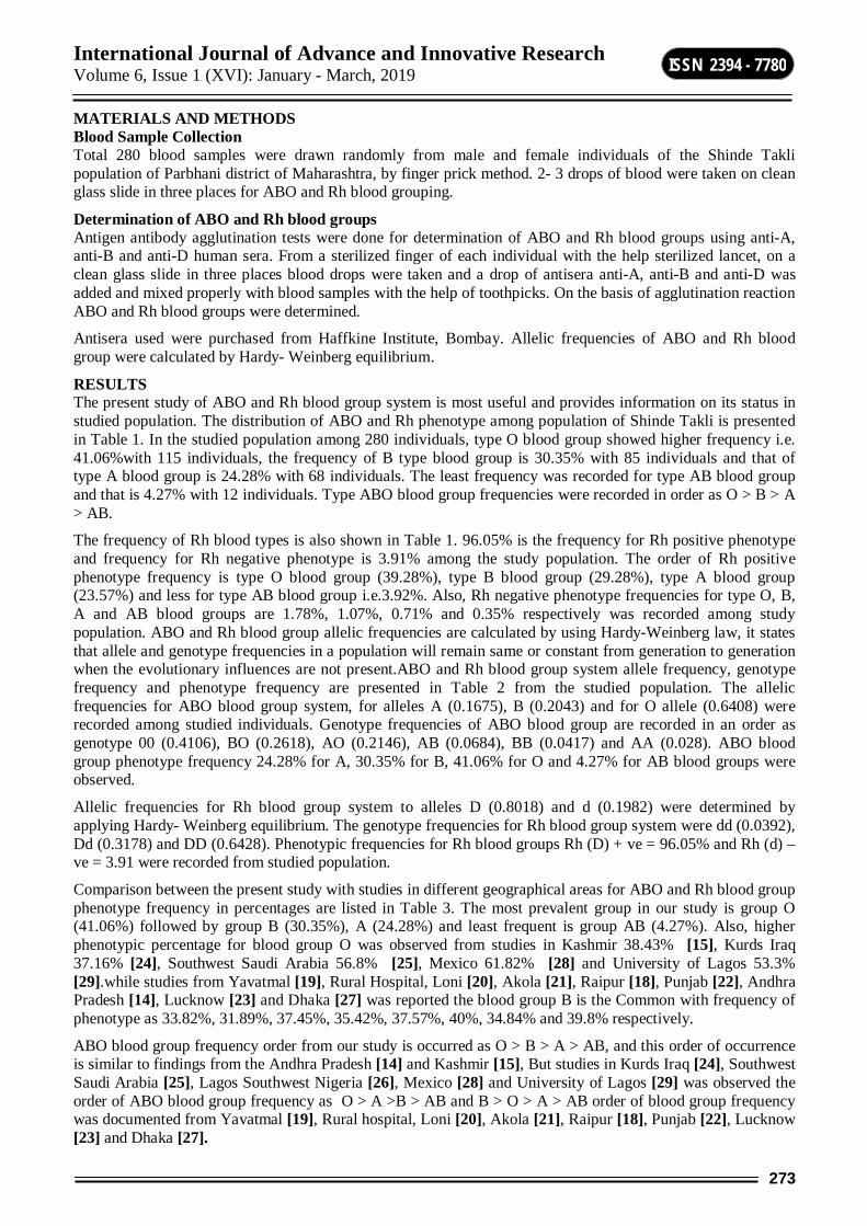
International Journal of Advance and Innovative Research Volume 6, Issue 1 (XVI): January - March, 2019
273
ISSN 2394 - 7780
MATERIALS AND METHODS Blood Sample Collection Total 280 blood samples were drawn randomly from male and female individuals of the Shinde Takli population of Parbhani district of Maharashtra, by finger prick method. 2- 3 drops of blood were taken on clean glass slide in three places for ABO and Rh blood grouping.
Determination of ABO and Rh blood groups Antigen antibody agglutination tests were done for determination of ABO and Rh blood groups using anti-A, anti-B and anti-D human sera. From a sterilized finger of each individual with the help sterilized lancet, on a clean glass slide in three places blood drops were taken and a drop of antisera anti-A, anti-B and anti-D was added and mixed properly with blood samples with the help of toothpicks. On the basis of agglutination reaction ABO and Rh blood groups were determined.
Antisera used were purchased from Haffkine Institute, Bombay. Allelic frequencies of ABO and Rh blood group were calculated by Hardy- Weinberg equilibrium.
RESULTS The present study of ABO and Rh blood group system is most useful and provides information on its status in studied population. The distribution of ABO and Rh phenotype among population of Shinde Takli is presented in Table 1. In the studied population among 280 individuals, type O blood group showed higher frequency i.e. 41.06%with 115 individuals, the frequency of B type blood group is 30.35% with 85 individuals and that of type A blood group is 24.28% with 68 individuals. The least frequency was recorded for type AB blood group and that is 4.27% with 12 individuals. Type ABO blood group frequencies were recorded in order as O > B > A > AB.
The frequency of Rh blood types is also shown in Table 1. 96.05% is the frequency for Rh positive phenotype and frequency for Rh negative phenotype is 3.91% among the study population. The order of Rh positive phenotype frequency is type O blood group (39.28%), type B blood group (29.28%), type A blood group (23.57%) and less for type AB blood group i.e.3.92%. Also, Rh negative phenotype frequencies for type O, B, A and AB blood groups are 1.78%, 1.07%, 0.71% and 0.35% respectively was recorded among study population. ABO and Rh blood group allelic frequencies are calculated by using Hardy-Weinberg law, it states that allele and genotype frequencies in a population will remain same or constant from generation to generation when the evolutionary influences are not present.ABO and Rh blood group system allele frequency, genotype frequency and phenotype frequency are presented in Table 2 from the studied population. The allelic frequencies for ABO blood group system, for alleles A (0.1675), B (0.2043) and for O allele (0.6408) were recorded among studied individuals. Genotype frequencies of ABO blood group are recorded in an order as genotype 00 (0.4106), BO (0.2618), AO (0.2146), AB (0.0684), BB (0.0417) and AA (0.028). ABO blood group phenotype frequency 24.28% for A, 30.35% for B, 41.06% for O and 4.27% for AB blood groups were observed.
Allelic frequencies for Rh blood group system to alleles D (0.8018) and d (0.1982) were determined by applying Hardy- Weinberg equilibrium. The genotype frequencies for Rh blood group system were dd (0.0392), Dd (0.3178) and DD (0.6428). Phenotypic frequencies for Rh blood groups Rh (D) + ve = 96.05% and Rh (d) – ve = 3.91 were recorded from studied population.
Comparison between the present study with studies in different geographical areas for ABO and Rh blood group phenotype frequency in percentages are listed in Table 3. The most prevalent group in our study is group O (41.06%) followed by group B (30.35%), A (24.28%) and least frequent is group AB (4.27%). Also, higher phenotypic percentage for blood group O was observed from studies in Kashmir 38.43% [15], Kurds Iraq 37.16% [24], Southwest Saudi Arabia 56.8% [25], Mexico 61.82% [28] and University of Lagos 53.3% [29].while studies from Yavatmal [19], Rural Hospital, Loni [20], Akola [21], Raipur [18], Punjab [22], Andhra Pradesh [14], Lucknow [23] and Dhaka [27] was reported the blood group B is the Common with frequency of phenotype as 33.82%, 31.89%, 37.45%, 35.42%, 37.57%, 40%, 34.84% and 39.8% respectively.
ABO blood group frequency order from our study is occurred as O > B > A > AB, and this order of occurrence is similar to findings from the Andhra Pradesh [14] and Kashmir [15], But studies in Kurds Iraq [24], Southwest Saudi Arabia [25], Lagos Southwest Nigeria [26], Mexico [28] and University of Lagos [29] was observed the order of ABO blood group frequency as O > A >B > AB and B > O > A > AB order of blood group frequency was documented from Yavatmal [19], Rural hospital, Loni [20], Akola [21], Raipur [18], Punjab [22], Lucknow [23] and Dhaka [27].
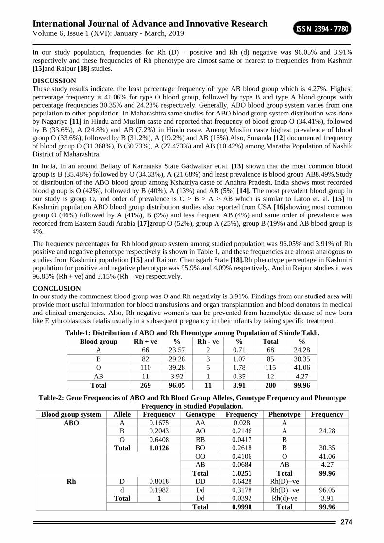
International Journal of Advance and Innovative Research Volume 6, Issue 1 (XVI): January - March, 2019
274
ISSN 2394 - 7780
In our study population, frequencies for Rh (D) + positive and Rh (d) negative was 96.05% and 3.91% respectively and these frequencies of Rh phenotype are almost same or nearest to frequencies from Kashmir [15]and Raipur [18] studies.
DISCUSSION These study results indicate, the least percentage frequency of type AB blood group which is 4.27%. Highest percentage frequency is 41.06% for type O blood group, followed by type B and type A blood groups with percentage frequencies 30.35% and 24.28% respectively. Generally, ABO blood group system varies from one population to other population. In Maharashtra same studies for ABO blood group system distribution was done by Nagariya [11] in Hindu and Muslim caste and reported that frequency of blood group O (34.41%), followed by B (33.6%), A (24.8%) and AB (7.2%) in Hindu caste. Among Muslim caste highest prevalence of blood group O (33.6%), followed by B (31.2%), A (19.2%) and AB (16%).Also, Sunanda [12] documented frequency of blood group O (31.368%), B (30.73%), A (27.473%) and AB (10.42%) among Maratha Population of Nashik District of Maharashtra.
In India, in an around Bellary of Karnataka State Gadwalkar et.al. [13] shown that the most common blood group is B (35.48%) followed by O (34.33%), A (21.68%) and least prevalence is blood group AB8.49%.Study of distribution of the ABO blood group among Kshatriya caste of Andhra Pradesh, India shows most recorded blood group is O (42%), followed by B (40%), A (13%) and AB (5%) [14]. The most prevalent blood group in our study is group O, and order of prevalence is O > B > A > AB which is similar to Latoo et. al. [15] in Kashmiri population.ABO blood group distribution studies also reported from USA [16]showing most common group O (46%) followed by A (41%), B (9%) and less frequent AB (4%) and same order of prevalence was recorded from Eastern Saudi Arabia [17]group O (52%), group A (25%), group B (19%) and AB blood group is 4%.
The frequency percentages for Rh blood group system among studied population was 96.05% and 3.91% of Rh positive and negative phenotype respectively is shown in Table 1, and these frequencies are almost analogous to studies from Kashmiri population [15] and Raipur, Chattisgarh State [18].Rh phenotype percentage in Kashmiri population for positive and negative phenotype was 95.9% and 4.09% respectively. And in Raipur studies it was 96.85% (Rh + ve) and 3.15% (Rh – ve) respectively.
CONCLUSION In our study the commonest blood group was O and Rh negativity is 3.91%. Findings from our studied area will provide most useful information for blood transfusions and organ transplantation and blood donators in medical and clinical emergencies. Also, Rh negative women’s can be prevented from haemolytic disease of new born like Erythroblastosis fetalis usually in a subsequent pregnancy in their infants by taking specific treatment.
Table-1: Distribution of ABO and Rh Phenotype among Population of Shinde Takli. Blood group Rh + ve % Rh - ve % Total %
A 66 23.57 2 0.71 68 24.28 B 82 29.28 3 1.07 85 30.35 O 110 39.28 5 1.78 115 41.06
AB 11 3.92 1 0.35 12 4.27 Total 269 96.05 11 3.91 280 99.96
Table-2: Gene Frequencies of ABO and Rh Blood Group Alleles, Genotype Frequency and Phenotype Frequency in Studied Population.
Blood group system Allele Frequency Genotype Frequency Phenotype Frequency ABO A 0.1675 AA 0.028 A
B 0.2043 AO 0.2146 A 24.28 O 0.6408 BB 0.0417 B
Total 1.0126 BO 0.2618 B 30.35 OO 0.4106 O 41.06
AB 0.0684 AB 4.27 Total 1.0251 Total 99.96
Rh D 0.8018 DD 0.6428 Rh(D)+ve d 0.1982 Dd 0.3178 Rh(D)+ve 96.05
Total 1 Dd 0.0392 Rh(d)-ve 3.91 Total 0.9998 Total 99.96
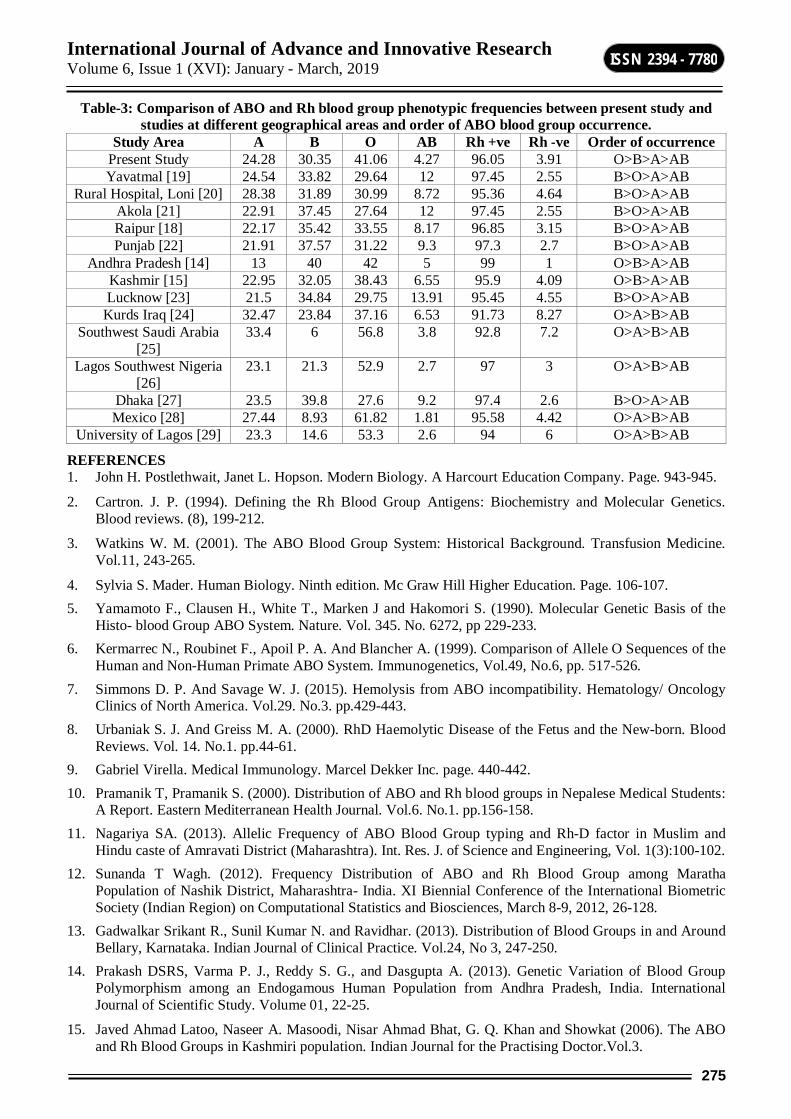
International Journal of Advance and Innovative Research Volume 6, Issue 1 (XVI): January - March, 2019
275
ISSN 2394 - 7780
Table-3: Comparison of ABO and Rh blood group phenotypic frequencies between present study and studies at different geographical areas and order of ABO blood group occurrence.
Study Area A B O AB Rh +ve Rh -ve Order of occurrence Present Study 24.28 30.35 41.06 4.27 96.05 3.91 O>B>A>AB Yavatmal [19] 24.54 33.82 29.64 12 97.45 2.55 B>O>A>AB
Rural Hospital, Loni [20] 28.38 31.89 30.99 8.72 95.36 4.64 B>O>A>AB Akola [21] 22.91 37.45 27.64 12 97.45 2.55 B>O>A>AB Raipur [18] 22.17 35.42 33.55 8.17 96.85 3.15 B>O>A>AB Punjab [22] 21.91 37.57 31.22 9.3 97.3 2.7 B>O>A>AB
Andhra Pradesh [14] 13 40 42 5 99 1 O>B>A>AB Kashmir [15] 22.95 32.05 38.43 6.55 95.9 4.09 O>B>A>AB Lucknow [23] 21.5 34.84 29.75 13.91 95.45 4.55 B>O>A>AB
Kurds Iraq [24] 32.47 23.84 37.16 6.53 91.73 8.27 O>A>B>AB Southwest Saudi Arabia
[25] 33.4 6 56.8 3.8 92.8 7.2 O>A>B>AB
Lagos Southwest Nigeria [26]
23.1 21.3 52.9 2.7 97 3 O>A>B>AB
Dhaka [27] 23.5 39.8 27.6 9.2 97.4 2.6 B>O>A>AB Mexico [28] 27.44 8.93 61.82 1.81 95.58 4.42 O>A>B>AB
University of Lagos [29] 23.3 14.6 53.3 2.6 94 6 O>A>B>AB
REFERENCES 1. John H. Postlethwait, Janet L. Hopson. Modern Biology. A Harcourt Education Company. Page. 943-945.
2. Cartron. J. P. (1994). Defining the Rh Blood Group Antigens: Biochemistry and Molecular Genetics. Blood reviews. (8), 199-212.
3. Watkins W. M. (2001). The ABO Blood Group System: Historical Background. Transfusion Medicine. Vol.11, 243-265.
4. Sylvia S. Mader. Human Biology. Ninth edition. Mc Graw Hill Higher Education. Page. 106-107. 5. Yamamoto F., Clausen H., White T., Marken J and Hakomori S. (1990). Molecular Genetic Basis of the
Histo- blood Group ABO System. Nature. Vol. 345. No. 6272, pp 229-233. 6. Kermarrec N., Roubinet F., Apoil P. A. And Blancher A. (1999). Comparison of Allele O Sequences of the
Human and Non-Human Primate ABO System. Immunogenetics, Vol.49, No.6, pp. 517-526. 7. Simmons D. P. And Savage W. J. (2015). Hemolysis from ABO incompatibility. Hematology/ Oncology
Clinics of North America. Vol.29. No.3. pp.429-443. 8. Urbaniak S. J. And Greiss M. A. (2000). RhD Haemolytic Disease of the Fetus and the New-born. Blood
Reviews. Vol. 14. No.1. pp.44-61. 9. Gabriel Virella. Medical Immunology. Marcel Dekker Inc. page. 440-442. 10. Pramanik T, Pramanik S. (2000). Distribution of ABO and Rh blood groups in Nepalese Medical Students:
A Report. Eastern Mediterranean Health Journal. Vol.6. No.1. pp.156-158. 11. Nagariya SA. (2013). Allelic Frequency of ABO Blood Group typing and Rh-D factor in Muslim and
Hindu caste of Amravati District (Maharashtra). Int. Res. J. of Science and Engineering, Vol. 1(3):100-102. 12. Sunanda T Wagh. (2012). Frequency Distribution of ABO and Rh Blood Group among Maratha
Population of Nashik District, Maharashtra- India. XI Biennial Conference of the International Biometric Society (Indian Region) on Computational Statistics and Biosciences, March 8-9, 2012, 26-128.
13. Gadwalkar Srikant R., Sunil Kumar N. and Ravidhar. (2013). Distribution of Blood Groups in and Around Bellary, Karnataka. Indian Journal of Clinical Practice. Vol.24, No 3, 247-250.
14. Prakash DSRS, Varma P. J., Reddy S. G., and Dasgupta A. (2013). Genetic Variation of Blood Group Polymorphism among an Endogamous Human Population from Andhra Pradesh, India. International Journal of Scientific Study. Volume 01, 22-25.
15. Javed Ahmad Latoo, Naseer A. Masoodi, Nisar Ahmad Bhat, G. Q. Khan and Showkat (2006). The ABO and Rh Blood Groups in Kashmiri population. Indian Journal for the Practising Doctor.Vol.3.
International Journal of Advance and Innovative Research Volume 6, Issue 1 (XVI): January - March, 2019
276
ISSN 2394 - 7780
16. Frances TF. (2002). Blood Groups (ABO Groups). In Common Laboratory and Diagnostic Tests. (3rdedn), Philadelphia, Lippincott.19-25
17. Bashwari LA, Al-Mulhim AA, Ahmad MS, Ahmed MA (2001). Frequency of ABO Blood Groups in the Eastern Region of Saudi Arabia. Saudi Med J.22, 1008-1012.
18. Shruti Shrivastava, Renuka Gahine and Vijay Kapse. (2015). ABO, Rhesus Blood Group and Allele Frequency in and around Raipur (Chattisgarh State), India. Int. J. Cur. Res. Rev. Vol.7, 52-58.
19. Arvind Chavhan, Santosh Pawar, Baig M. M. (2010). Allelic Frequency of ABO and Rh D Blood Group Among the Banjara Backward Caste of Yavatmal District, Maharashtra, India. Nature Precedings.
20. Purushottam A. Giri, Sankalp Yadav, Gaurav Singh Parhar, Deepak B. Phalke. (2011). Frequency of ABO and Rhesus Blood Groups: A Study from a Rural Tertiary Care Teaching Hospital in India. International Journal of Biological and Medical Research. 2 (4), 988-990.
21. Arvind Chavhan, Rajusing Jadhao, Santosh Pawar, (2012). The Study of Allelic Frequency of ABO and Rh D Blood Group among the Banjara Population of Akola District, Maharashtra, India. Physical Anthropology. 327-329.
22. Sharda Sidhu. (2003) Distribution of the ABO Blood Groups and Rh (D) Factor among the Scheduled Caste Population of Punjab. The Anthropologist. 5; 203-204.
23. Tulika Chandra, Ashish Gupta. (2012). Prevalence of ABO and Rhesus Blood Groups in Northern India. Journal of Blood Disorders and Transfusion. 3: 132.doi.10.4172/2155-9864.1000132.
24. Mohamad S Jaff. (2010). ABO and Rhesus Blood Group Distribution in Kurds. Journal of Blood Medicine.143-146.
25. Mohammed A. Sarhan, Kamel A. Saleh and Saad M. Bin- Dajem. (2009). Distribution of ABO Blood Groups and Rhesus Factor in Southwest Saudi Arabia. Saudi Med J. Vol.30 (1), 116-119.
26. Iyiola O. A., Igunnugbemi O. O., Bello O. G. (2012). Gene Frequencies of ABO and Rh (D) Blood Group Alleles in Lagos, South- West Nigeria. The Egyptian Journal of Medical Human Genetics. 13; 147-153.
27. Rayhana Sultana, Zaida Rahman, Asadul Mazid Helali, Rabeya Yousuf, Shyamoli Mustafa, Abdus Salam, Mainul Haque. (2013). Study of ABO and Rh D Blood Groups among the Common People of Capital City of Bangladesh. International Journal of Pharmacy and Pharmaceutical Sciences. Vol.5, 814- 816.
28. Adrian Canizalez, Abraham Campos, Jose. A. et.al. (2018). Blood Groups Distribution and Gene Diversity of the ABO and Rh (D) Loci in the Mexican Population. BioMed Research International.Vol.2018, 1-11.
29. Adeyemo, Oyenike A. and Soboyejo, Omolade B. (2006). Frequency Distribution of ABO, Rh Blood Groups and blood genotypes among the Cell Biology and Genetics Students of University of Lagos, Nigeria. African Journal of Biotechnology. Vol.5 (22). pp, 2062-2065.
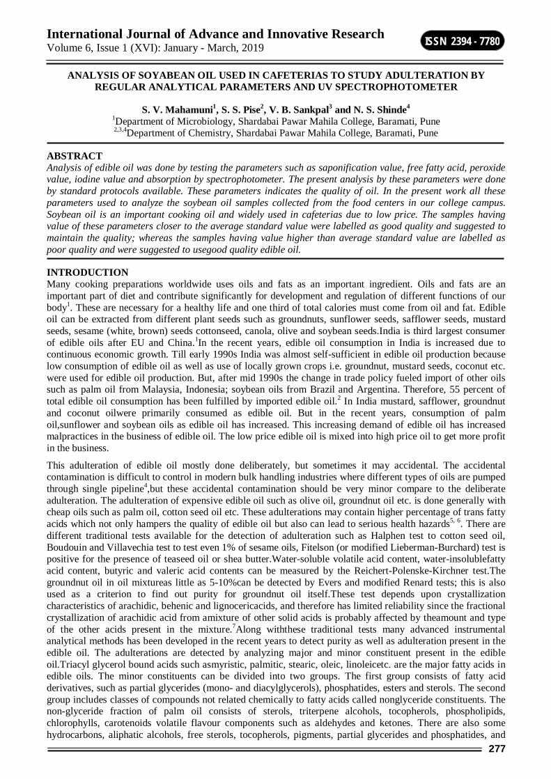
International Journal of Advance and Innovative Research Volume 6, Issue 1 (XVI): January - March, 2019
277
ISSN 2394 - 7780
ANALYSIS OF SOYABEAN OIL USED IN CAFETERIAS TO STUDY ADULTERATION BY REGULAR ANALYTICAL PARAMETERS AND UV SPECTROPHOTOMETER
S. V. Mahamuni1, S. S. Pise2, V. B. Sankpal3 and N. S. Shinde4 1Department of Microbiology, Shardabai Pawar Mahila College, Baramati, Pune 2,3,4Department of Chemistry, Shardabai Pawar Mahila College, Baramati, Pune
ABSTRACT Analysis of edible oil was done by testing the parameters such as saponification value, free fatty acid, peroxide value, iodine value and absorption by spectrophotometer. The present analysis by these parameters were done by standard protocols available. These parameters indicates the quality of oil. In the present work all these parameters used to analyze the soybean oil samples collected from the food centers in our college campus. Soybean oil is an important cooking oil and widely used in cafeterias due to low price. The samples having value of these parameters closer to the average standard value were labelled as good quality and suggested to maintain the quality; whereas the samples having value higher than average standard value are labelled as poor quality and were suggested to usegood quality edible oil.
INTRODUCTION Many cooking preparations worldwide uses oils and fats as an important ingredient. Oils and fats are an important part of diet and contribute significantly for development and regulation of different functions of our body1. These are necessary for a healthy life and one third of total calories must come from oil and fat. Edible oil can be extracted from different plant seeds such as groundnuts, sunflower seeds, safflower seeds, mustard seeds, sesame (white, brown) seeds cottonseed, canola, olive and soybean seeds.India is third largest consumer of edible oils after EU and China.1In the recent years, edible oil consumption in India is increased due to continuous economic growth. Till early 1990s India was almost self-sufficient in edible oil production because low consumption of edible oil as well as use of locally grown crops i.e. groundnut, mustard seeds, coconut etc. were used for edible oil production. But, after mid 1990s the change in trade policy fueled import of other oils such as palm oil from Malaysia, Indonesia; soybean oils from Brazil and Argentina. Therefore, 55 percent of total edible oil consumption has been fulfilled by imported edible oil.2 In India mustard, safflower, groundnut and coconut oilwere primarily consumed as edible oil. But in the recent years, consumption of palm oil,sunflower and soybean oils as edible oil has increased. This increasing demand of edible oil has increased malpractices in the business of edible oil. The low price edible oil is mixed into high price oil to get more profit in the business.
This adulteration of edible oil mostly done deliberately, but sometimes it may accidental. The accidental contamination is difficult to control in modern bulk handling industries where different types of oils are pumped through single pipeline4,but these accidental contamination should be very minor compare to the deliberate adulteration. The adulteration of expensive edible oil such as olive oil, groundnut oil etc. is done generally with cheap oils such as palm oil, cotton seed oil etc. These adulterations may contain higher percentage of trans fatty acids which not only hampers the quality of edible oil but also can lead to serious health hazards5, 6. There are different traditional tests available for the detection of adulteration such as Halphen test to cotton seed oil, Boudouin and Villavechia test to test even 1% of sesame oils, Fitelson (or modified Lieberman-Burchard) test is positive for the presence of teaseed oil or shea butter.Water-soluble volatile acid content, water-insolublefatty acid content, butyric and valeric acid contents can be measured by the Reichert-Polenske-Kirchner test.The groundnut oil in oil mixtureas little as 5-10%can be detected by Evers and modified Renard tests; this is also used as a criterion to find out purity for groundnut oil itself.These test depends upon crystallization characteristics of arachidic, behenic and lignocericacids, and therefore has limited reliability since the fractional crystallization of arachidic acid from amixture of other solid acids is probably affected by theamount and type of the other acids present in the mixture.7Along withthese traditional tests many advanced instrumental analytical methods has been developed in the recent years to detect purity as well as adulteration present in the edible oil. The adulterations are detected by analyzing major and minor constituent present in the edible oil.Triacyl glycerol bound acids such asmyristic, palmitic, stearic, oleic, linoleicetc. are the major fatty acids in edible oils. The minor constituents can be divided into two groups. The first group consists of fatty acid derivatives, such as partial glycerides (mono- and diacylglycerols), phosphatides, esters and sterols. The second group includes classes of compounds not related chemically to fatty acids called nonglyceride constituents. The non-glyceride fraction of palm oil consists of sterols, triterpene alcohols, tocopherols, phospholipids, chlorophylls, carotenoids volatile flavour components such as aldehydes and ketones. There are also some hydrocarbons, aliphatic alcohols, free sterols, tocopherols, pigments, partial glycerides and phosphatides, and
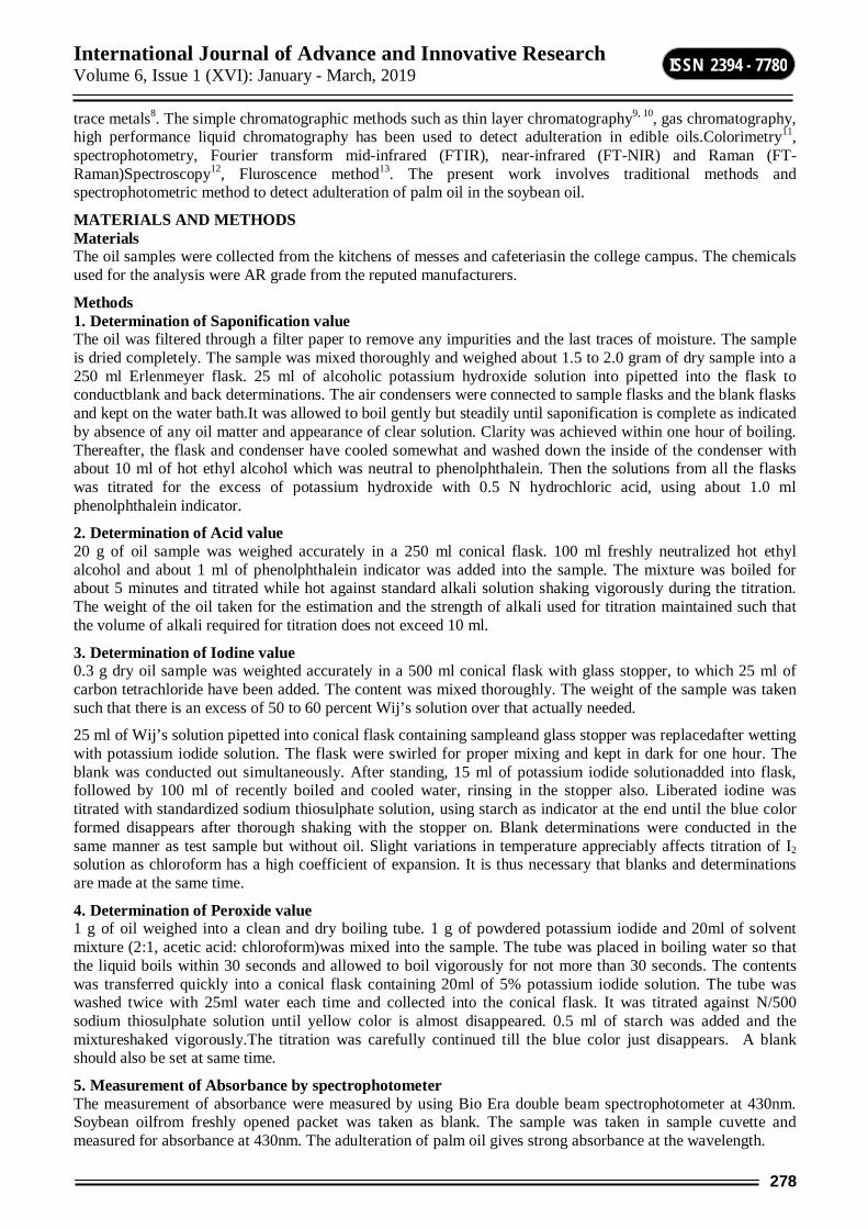
International Journal of Advance and Innovative Research Volume 6, Issue 1 (XVI): January - March, 2019
278
ISSN 2394 - 7780
trace metals8. The simple chromatographic methods such as thin layer chromatography9, 10, gas chromatography, high performance liquid chromatography has been used to detect adulteration in edible oils.Colorimetry11, spectrophotometry, Fourier transform mid-infrared (FTIR), near-infrared (FT-NIR) and Raman (FT-Raman)Spectroscopy12, Fluroscence method13. The present work involves traditional methods and spectrophotometric method to detect adulteration of palm oil in the soybean oil.
MATERIALS AND METHODS Materials The oil samples were collected from the kitchens of messes and cafeteriasin the college campus. The chemicals used for the analysis were AR grade from the reputed manufacturers.
Methods 1. Determination of Saponification value The oil was filtered through a filter paper to remove any impurities and the last traces of moisture. The sample is dried completely. The sample was mixed thoroughly and weighed about 1.5 to 2.0 gram of dry sample into a 250 ml Erlenmeyer flask. 25 ml of alcoholic potassium hydroxide solution into pipetted into the flask to conductblank and back determinations. The air condensers were connected to sample flasks and the blank flasks and kept on the water bath.It was allowed to boil gently but steadily until saponification is complete as indicated by absence of any oil matter and appearance of clear solution. Clarity was achieved within one hour of boiling. Thereafter, the flask and condenser have cooled somewhat and washed down the inside of the condenser with about 10 ml of hot ethyl alcohol which was neutral to phenolphthalein. Then the solutions from all the flasks was titrated for the excess of potassium hydroxide with 0.5 N hydrochloric acid, using about 1.0 ml phenolphthalein indicator.
2. Determination of Acid value 20 g of oil sample was weighed accurately in a 250 ml conical flask. 100 ml freshly neutralized hot ethyl alcohol and about 1 ml of phenolphthalein indicator was added into the sample. The mixture was boiled for about 5 minutes and titrated while hot against standard alkali solution shaking vigorously during the titration. The weight of the oil taken for the estimation and the strength of alkali used for titration maintained such that the volume of alkali required for titration does not exceed 10 ml.
3. Determination of Iodine value 0.3 g dry oil sample was weighted accurately in a 500 ml conical flask with glass stopper, to which 25 ml of carbon tetrachloride have been added. The content was mixed thoroughly. The weight of the sample was taken such that there is an excess of 50 to 60 percent Wij’s solution over that actually needed.
25 ml of Wij’s solution pipetted into conical flask containing sampleand glass stopper was replacedafter wetting with potassium iodide solution. The flask were swirled for proper mixing and kept in dark for one hour. The blank was conducted out simultaneously. After standing, 15 ml of potassium iodide solutionadded into flask, followed by 100 ml of recently boiled and cooled water, rinsing in the stopper also. Liberated iodine was titrated with standardized sodium thiosulphate solution, using starch as indicator at the end until the blue color formed disappears after thorough shaking with the stopper on. Blank determinations were conducted in the same manner as test sample but without oil. Slight variations in temperature appreciably affects titration of I2 solution as chloroform has a high coefficient of expansion. It is thus necessary that blanks and determinations are made at the same time.
4. Determination of Peroxide value 1 g of oil weighed into a clean and dry boiling tube. 1 g of powdered potassium iodide and 20ml of solvent mixture (2:1, acetic acid: chloroform)was mixed into the sample. The tube was placed in boiling water so that the liquid boils within 30 seconds and allowed to boil vigorously for not more than 30 seconds. The contents was transferred quickly into a conical flask containing 20ml of 5% potassium iodide solution. The tube was washed twice with 25ml water each time and collected into the conical flask. It was titrated against N/500 sodium thiosulphate solution until yellow color is almost disappeared. 0.5 ml of starch was added and the mixtureshaked vigorously.The titration was carefully continued till the blue color just disappears. A blank should also be set at same time.
5. Measurement of Absorbance by spectrophotometer The measurement of absorbance were measured by using Bio Era double beam spectrophotometer at 430nm. Soybean oilfrom freshly opened packet was taken as blank. The sample was taken in sample cuvette and measured for absorbance at 430nm. The adulteration of palm oil gives strong absorbance at the wavelength.
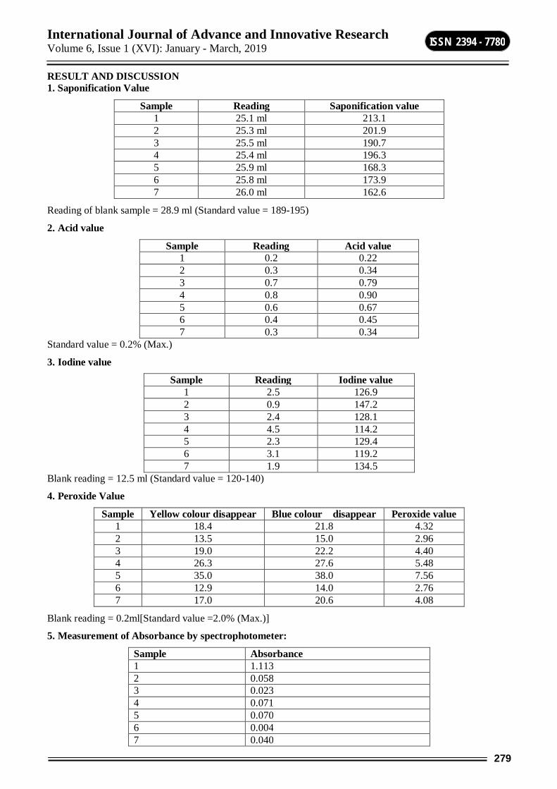
International Journal of Advance and Innovative Research Volume 6, Issue 1 (XVI): January - March, 2019
279
ISSN 2394 - 7780
RESULT AND DISCUSSION 1. Saponification Value
Reading of blank sample = 28.9 ml (Standard value = 189-195)
2. Acid value
Sample Reading Acid value 1 0.2 0.22 2 0.3 0.34 3 0.7 0.79 4 0.8 0.90 5 0.6 0.67 6 0.4 0.45 7 0.3 0.34
Standard value = 0.2% (Max.)
3. Iodine value
Sample Reading Iodine value 1 2.5 126.9 2 0.9 147.2 3 2.4 128.1 4 4.5 114.2 5 2.3 129.4 6 3.1 119.2 7 1.9 134.5
Blank reading = 12.5 ml (Standard value = 120-140)
4. Peroxide Value
Sample Yellow colour disappear Blue colour disappear Peroxide value 1 18.4 21.8 4.32 2 13.5 15.0 2.96 3 19.0 22.2 4.40 4 26.3 27.6 5.48 5 35.0 38.0 7.56 6 12.9 14.0 2.76 7 17.0 20.6 4.08
Blank reading = 0.2ml[Standard value =2.0% (Max.)]
5. Measurement of Absorbance by spectrophotometer:
Sample Absorbance 1 1.113 2 0.058 3 0.023 4 0.071 5 0.070 6 0.004 7 0.040
Sample Reading Saponification value 1 25.1 ml 213.1 2 25.3 ml 201.9 3 25.5 ml 190.7 4 25.4 ml 196.3 5 25.9 ml 168.3 6 25.8 ml 173.9 7 26.0 ml 162.6
International Journal of Advance and Innovative Research Volume 6, Issue 1 (XVI): January - March, 2019
280
ISSN 2394 - 7780
Graphical Representation
Fig-1: Analysis of Soybean oil with respect to Saponification and Iodine Value
Fig-1: Analysis of Soybean oil with respect to Acid and Peroxide Value
DISCUSSION The lowest saponification value is 162.6 (sample 7) and highest value is 213.1 (sample 1).Saponification value 190.7 of sample 3 matches with the standard value. The lowest Acid value is 0.22 (sample 1) and highest value is 0.90 (sample 4). Acid value 0.22 of sample 1 closer to the standard value.The lowest Iodine value is 114.2 (sample 4) and highest value is 147.2 (sample 2). Iodine value 126.9, 128.1, 129.4, 134.5 of samples 1,3,5,7 respectively closely equalizes with the standard value.The lowest Peroxide value is 2.76 (sample 6) and highest value is 7.56 (sample 5). Peroxide value 2.76 (sample 6) this value is match to the standard value.
The lowest absorbance is shown by sample 6 and highest absorbance is shown by sample 1.
CONCLUSION The analysis of oil samples gives variations in the measurement of parameters assigned to detect adulterations. Among these samples sample 3 and sample 6 found to be having values closer to standard values and therefore can be considered as pure samples. These samples are comparatively healthier to be used for food preparations.
REFERENCE 1. S. Johnson, N. Saikia, H. B. Mathur, H. C. Agarwal, Fatty acids profile of oils and fats in India, Center for
Science and Environment, New Delhi, 2009, 9.
2. D. Erik, S. Persaud& R. Landes, “India’s Edible Oil Sector: Imports Fill Rising Demand”, USDA Working Paper, 2003, 2.
3. P. V. Srinivasan,“Impact of Trade Liberalization on India’s Oilseed & Edible oils sector”, Report prepared for IGIDR-ERS/USDA Project, 2005
4. A. K. Shukla, A. K. Dixit, R. P. Singh, Journal of Oleo Science, 54(6), 2005, 317-324.
5. G.Danaei, E.L.Ding, D.Mozaffarian, B.Taylor, J.Rehm, et al. PLoS Med 6(4), 2009,1000058, 1-23.
6. T.E.Clemente, E.B. Cahoon, Plant Physiology, 151, 2009, 1030-1040.

International Journal of Advance and Innovative Research Volume 6, Issue 1 (XVI): January - March, 2019
281
ISSN 2394 - 7780
7. J.B. Rossell, B. King, M.J. Downes, JAOCS, 60(2), 1983, 333-339.
8. R. Sambanthamurthi, K. Sundram, Y. A. Tan, Progress in lipid research,2000, 39, 507-558.
9. P. Sengupta, S. Sil, J.N. Chattarjea,JAOCS, 1981, 701-702.
10. M. M. Chakraborty, A. K. Gayen, J. Oil Techno. Assoc. India, 6, 1974, 69-79
11. A. Salhin, M. Ali , A. M. Abdurrhman, Chemical and Materials Engineering 1(3), 2013, 96-103.
12. H. Yang, J.Irudayaraj, M. M. Paradkar, Food Chemistry, 93,(2005), 25–32.
13. K. Nikolova, T. Eftimov, M. Perifanova, D. Brabant, Journal of Food Science and Engineering 2, 2012, 674-684.
14. Iodine absorption number of oils and fats, in AOAC official methods of analysis, Washington, DC.,14, 1984, 405

International Journal of Advance and Innovative Research Volume 6, Issue 1 (XVI): January - March, 2019
282
ISSN 2394 - 7780
IN VITRO STUDIES IN COCHLOSPERMUM RELIGIOSUM (LINN.)
Jyoti Ghodke, Swati Shilwant, Narayan Pandhure and Sudhir Solanke Tissue Culture Laboratory, Department of Botany, Dr. Babasaheb Ambedkar Marathwada University,
Aurangabad
ABSTRACT Effect of different plant growth regulator viz. BAP, KIN, IBA, IAA, NAA, and 2, 4-D on in vitro multiplication and callus induction was studied in important medicinal plant Cochlospermum religiosum. Apical shoots and nodal explants have shown maximum multiple shoot formation using different combination of cytokinins and Auxins. 1.4 mg/l BAP along with 0.2 mg/l IBA given maximum 10 shoots and 1.0 mg/l KIN and 1.0 mg BAP and 1.2 mg/L2, 4-D was responsible for Callus induction.Cochlospermum contains important medicinal properties, secondary metabolites and gum katira which is important in human welfare. Therefore the present investigation was carried out to conserve the valuable deciduous tree Cochlospermum religiosum.
Keywords: In vitro: Cochlospermum religiosum, callus, multiplication.
INTRODUCTION One of the rare flowering plant Cochlospermum religiosum is belongs to family Bixaceae from the tropical region of Southeast Asia and the Indian Subcontinent. In India it is commonly found in Andhra Pradesh, Maharashtra, Madhya Pradesh, Uttar Pradesh and Bihar (RCDC). It is a small tree growing to a height of 7.5 m usually found in dry deciduous forests. The name religiosum derived from the fact that the flowers are used as temple offerings. It is also known as Silk-Cotton Tree because the capsules containing the seeds have a fluffy cotton-like substance similar to kapok. It is very common and conspicuous tree, characteristic of the hottest, driest and stoniest slopes. It frequently founds in Telangana forest.
Plant can be identified by deeply furrowed bark, palmately 5-lobed leaves and bright golden yellow bisexual flowers. Cochlospermum is genus of trees and shrubs scattered throughout the tropics mostly xerophytes, deciduous in drier season. Capsules containing numerous seeds some species are medicinal, Ornamental, yielding floss, fibre, gum and timber. Cochlospermum quick growing tree yield a gum known as gum katira from a juice orange in colour exudes from the bark. This gum has been exported from India in increasing quantities in recent years and the fact that the price has been low and stable, as compared with tragacanth, has led to new uses and applications for it in industry and pharmacy. Katira is primarily used as a substitute of Tragacanth in most American countries. There are lot other uses of Katira like in calico-printing, polishing paper and leather dressing. It is also used for polishing tusser silk. It is exported to many countries of Latin America to use in cigar and Ice cream industries. As a laxative it is considered to be superior to other gums. As an emulsifying agent it is a good substitute for tragacanth.
This gum is used in medicinefor the treatment of cough, diarrhea and dysentery. The dried leaf and flowers are used as stimulants, antipyretic, laxative and sedative (Kirtikar and Basu, 1975). Root powder mixed with water applied to face reduce wrinkle (RLS sikarwar, et al., 2007). The oral gum powder about 20g mixed with ghee works as an aphrodisiac (SavithrammaN. et al., 2011). Katira used in cosmetics and for bookbinding the floss is used for stuffing pillows, mattresses, cushions, life jackets. Seed cakes are used as manure and cattle feed.Bark Powder of tree is used with water during jaundice (Dinesh K. and Aruna J., 2010).Cochlospermum is propagated by seed broadcast, seeds are broadcasted in primary beds in June and seedlings are pricked out to polythene bags when six months old. Therefore the aim of present work is to conserve important medicinal plant Cochlospermum religiosum through in vitro tissue culture techniques.
MATERIAL AND METHOD Preparation of Explants Seeds of Cochlospermum religiosum were collected from Buldhana district and grown in the green house, Botanical garden, Department of Botany Dr. Babasaheb Ambedkar Marathwada University, Aurangabad. Apical shoot, Axillary leaves and nodal explants of Cochlospermum were collected from 30 days old plants grown in the greenhouse of Departmental Botanical garden. All these explants were used from donor plants during present study. The explants were washed carefully in running tap water for 5 minute and followed by distilled water for 5 minutes. For surface sterilization, chemical such as 70% ethanol, Hgcl2 (0.3 %) were used. Explants were surface sterilized for 5 minute by 0.3% mercuric chloride followed by three subsequent rinses with sterilized double distilled water in a laminar air flow. All these explants were dissected into small pieces and inoculated on culture vessel and test tube containing 25 ml MS medium.
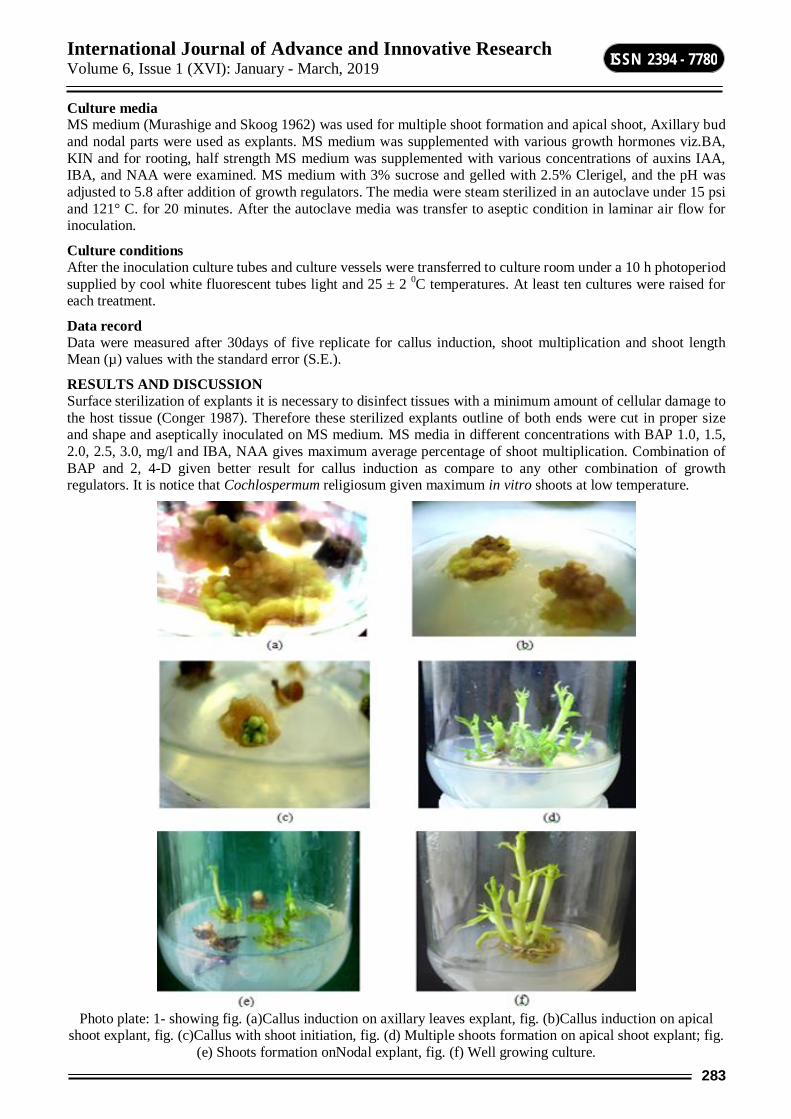
International Journal of Advance and Innovative Research Volume 6, Issue 1 (XVI): January - March, 2019
283
ISSN 2394 - 7780
Culture media MS medium (Murashige and Skoog 1962) was used for multiple shoot formation and apical shoot, Axillary bud and nodal parts were used as explants. MS medium was supplemented with various growth hormones viz.BA, KIN and for rooting, half strength MS medium was supplemented with various concentrations of auxins IAA, IBA, and NAA were examined. MS medium with 3% sucrose and gelled with 2.5% Clerigel, and the pH was adjusted to 5.8 after addition of growth regulators. The media were steam sterilized in an autoclave under 15 psi and 121° C. for 20 minutes. After the autoclave media was transfer to aseptic condition in laminar air flow for inoculation.
Culture conditions After the inoculation culture tubes and culture vessels were transferred to culture room under a 10 h photoperiod supplied by cool white fluorescent tubes light and 25 ± 2 0C temperatures. At least ten cultures were raised for each treatment.
Data record Data were measured after 30days of five replicate for callus induction, shoot multiplication and shoot length Mean (µ) values with the standard error (S.E.).
RESULTS AND DISCUSSION Surface sterilization of explants it is necessary to disinfect tissues with a minimum amount of cellular damage to the host tissue (Conger 1987). Therefore these sterilized explants outline of both ends were cut in proper size and shape and aseptically inoculated on MS medium. MS media in different concentrations with BAP 1.0, 1.5, 2.0, 2.5, 3.0, mg/l and IBA, NAA gives maximum average percentage of shoot multiplication. Combination of BAP and 2, 4-D given better result for callus induction as compare to any other combination of growth regulators. It is notice that Cochlospermum religiosum given maximum in vitro shoots at low temperature.
Photo plate: 1- showing fig. (a)Callus induction on axillary leaves explant, fig. (b)Callus induction on apical
shoot explant, fig. (c)Callus with shoot initiation, fig. (d) Multiple shoots formation on apical shoot explant; fig. (e) Shoots formation onNodal explant, fig. (f) Well growing culture.
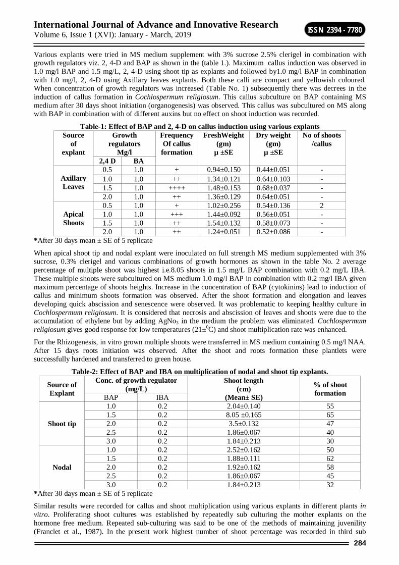
International Journal of Advance and Innovative Research Volume 6, Issue 1 (XVI): January - March, 2019
284
ISSN 2394 - 7780
Various explants were tried in MS medium supplement with 3% sucrose 2.5% clerigel in combination with growth regulators viz. 2, 4-D and BAP as shown in the (table 1.). Maximum callus induction was observed in 1.0 mg/l BAP and 1.5 mg/L, 2, 4-D using shoot tip as explants and followed by1.0 mg/l BAP in combination with 1.0 mg/l, 2, 4-D using Axillary leaves explants. Both these calli are compact and yellowish coloured. When concentration of growth regulators was increased (Table No. 1) subsequently there was decrees in the induction of callus formation in Cochlospermum religiosum. This callus subculture on BAP containing MS medium after 30 days shoot initiation (organogenesis) was observed. This callus was subcultured on MS along with BAP in combination with of different auxins but no effect on shoot induction was recorded.
Table-1: Effect of BAP and 2, 4-D on callus induction using various explants Source
of explant
Growth regulators
Mg/l
Frequency Of callus formation
FreshWeight (gm)
µ ±SE
Dry weight (gm)
µ ±SE
No of shoots /callus
2,4 D BA
Axillary Leaves
0.5 1.0 + 0.94±0.150 0.44±0.051 - 1.0 1.0 ++ 1.34±0.121 0.64±0.103 - 1.5 1.0 ++++ 1.48±0.153 0.68±0.037 - 2.0 1.0 ++ 1.36±0.129 0.64±0.051 -
Apical Shoots
0.5 1.0 + 1.02±0.256 0.54±0.136 2 1.0 1.0 +++ 1.44±0.092 0.56±0.051 - 1.5 1.0 ++ 1.54±0.132 0.58±0.073 - 2.0 1.0 ++ 1.24±0.051 0.52±0.086 -
*After 30 days mean ± SE of 5 replicate
When apical shoot tip and nodal explant were inoculated on full strength MS medium supplemented with 3% sucrose, 0.3% clerigel and various combinations of growth hormones as shown in the table No. 2 average percentage of multiple shoot was highest i.e.8.05 shoots in 1.5 mg/L BAP combination with 0.2 mg/L IBA. These multiple shoots were subcultured on MS medium 1.0 mg/l BAP in combination with 0.2 mg/l IBA given maximum percentage of shoots heights. Increase in the concentration of BAP (cytokinins) lead to induction of callus and minimum shoots formation was observed. After the shoot formation and elongation and leaves developing quick abscission and senescence were observed. It was problematic to keeping healthy culture in Cochlospermum religiosum. It is considered that necrosis and abscission of leaves and shoots were due to the accumulation of ethylene but by adding AgNo3 in the medium the problem was eliminated. Cochlospermum religiosum gives good response for low temperatures (21±0C) and shoot multiplication rate was enhanced.
For the Rhizogenesis, in vitro grown multiple shoots were transferred in MS medium containing 0.5 mg/l NAA. After 15 days roots initiation was observed. After the shoot and roots formation these plantlets were successfully hardened and transferred to green house.
Table-2: Effect of BAP and IBA on multiplication of nodal and shoot tip explants.
Source of Explant
Conc. of growth regulator (mg/L)
Shoot length (cm)
(Mean± SE)
% of shoot formation BAP IBA
Shoot tip
1.0 0.2 2.04±0.140 55 1.5 0.2 8.05 ±0.165 65 2.0 0.2 3.5±0.132 47 2.5 0.2 1.86±0.067 40 3.0 0.2 1.84±0.213 30
Nodal
1.0 0.2 2.52±0.162 50 1.5 0.2 1.88±0.111 62 2.0 0.2 1.92±0.162 58 2.5 0.2 1.86±0.067 45 3.0 0.2 1.84±0.213 32
*After 30 days mean ± SE of 5 replicate
Similar results were recorded for callus and shoot multiplication using various explants in different plants in vitro. Proliferating shoot cultures was established by repeatedly sub culturing the mother explants on the hormone free medium. Repeated sub-culturing was said to be one of the methods of maintaining juvenility (Franclet et al., 1987). In the present work highest number of shoot percentage was recorded in third sub
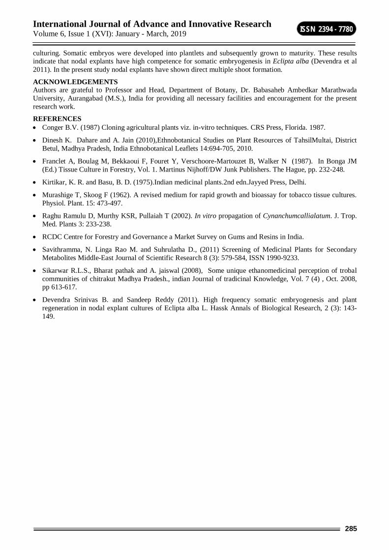
International Journal of Advance and Innovative Research Volume 6, Issue 1 (XVI): January - March, 2019
285
ISSN 2394 - 7780
culturing. Somatic embryos were developed into plantlets and subsequently grown to maturity. These results indicate that nodal explants have high competence for somatic embryogenesis in Eclipta alba (Devendra et al 2011). In the present study nodal explants have shown direct multiple shoot formation.
ACKNOWLEDGEMENTS Authors are grateful to Professor and Head, Department of Botany, Dr. Babasaheb Ambedkar Marathwada University, Aurangabad (M.S.), India for providing all necessary facilities and encouragement for the present research work.
REFERENCES Conger B.V. (1987) Cloning agricultural plants viz. in-vitro techniques. CRS Press, Florida. 1987.
Dinesh K. Dahare and A. Jain (2010),Ethnobotanical Studies on Plant Resources of TahsilMultai, District Betul, Madhya Pradesh, India Ethnobotanical Leaflets 14:694-705, 2010.
Franclet A, Boulag M, Bekkaoui F, Fouret Y, Verschoore-Martouzet B, Walker N (1987). In Bonga JM (Ed.) Tissue Culture in Forestry, Vol. 1. Martinus Nijhoff/DW Junk Publishers. The Hague, pp. 232-248.
Kirtikar, K. R. and Basu, B. D. (1975).Indian medicinal plants.2nd edn.Jayyed Press, Delhi.
Murashige T, Skoog F (1962). A revised medium for rapid growth and bioassay for tobacco tissue cultures. Physiol. Plant. 15: 473-497.
Raghu Ramulu D, Murthy KSR, Pullaiah T (2002). In vitro propagation of Cynanchumcallialatum. J. Trop. Med. Plants 3: 233-238.
RCDC Centre for Forestry and Governance a Market Survey on Gums and Resins in India.
Savithramma, N. Linga Rao M. and Suhrulatha D., (2011) Screening of Medicinal Plants for Secondary Metabolites Middle-East Journal of Scientific Research 8 (3): 579-584, ISSN 1990-9233.
Sikarwar R.L.S., Bharat pathak and A. jaiswal (2008), Some unique ethanomedicinal perception of trobal communities of chitrakut Madhya Pradesh., indian Journal of tradicinal Knowledge, Vol. 7 (4) , Oct. 2008, pp 613-617.
Devendra Srinivas B. and Sandeep Reddy (2011). High frequency somatic embryogenesis and plant regeneration in nodal explant cultures of Eclipta alba L. Hassk Annals of Biological Research, 2 (3): 143-149.
International Journal of Advance and Innovative Research Volume 6, Issue 1 (XVI): January - March, 2019
286
ISSN 2394 - 7780
AGRONOMY OF MEDICAGO SATIVA TO DIFFERENT METHODS OF CULTIVATION
Narayan Pandhure, Laxman Shimple and Prashant P. Pangrikar Department of Botany, Dr. Babasaheb Ambedkar Marathwada University, Aurangabad
R. B. Attal College, Georai, Dist. Beed.
ABSTRACT Lucerne (Medicago sativa) is belongs to family Fabaceae which is one of the important forage crop in Marathwada. Effect of different methods of cultivation on growth performance of forage crop Lucerne was studied during present piece of work. Four cultivation methods viz. mulching, raised seed beds, shed nets and ridges and furrows along with control were carried out in three replicates along with control. Growth pattern of the crop along with chlorophyll content was studied. During the investigations it was observed that, cultivation method mulching produced the tallest plants, along with highest chlorophyll content. These plants were emerged out early with 100% seed germination. Raised bed showed poor performance compare to mulching. Thus it could be concluded that, mulching method is best for the production of forage crop Lucerne in rain feed area like Marathwada.
Keywords: Lucerne, Mulching, Shed net, Raised bed, Furrow and Ridge.
INTRODUCTION Lucerne (Medicago sativa) is considered to be originated in Northern Africa or at the Egyptian-Sudanese border 5,000-8,000 years ago. Sorghum belongs to the tribe Andropogonae of the grass family Poaceae (FAO,1991). Indian sub-continent is its secondary centre of origin, where it is cultivated since 4,500 years. Grain sorghum is the fifth world leading cereal after Sorghum, wheat, rice and barley. The crop can yield reasonably well under adverse conditions of low soil water and high temperature. Sorghum may well offer the best opportunity to satisfy the doubling demand in the developing world by 2020, as a food for the poor and an alternative feed and food to Sorghum (Hunt, E. R. and B. N. Rock. 1989).
Sorghum ranks third in the major food grain crops in India, whereas it is the fourth food grains of the world. Millions of people in Africa and Asia depend on sorghum as the staple food. In addition, the fodder and Stover is fed to millions of animals providing milk and meat for man. It has potential to compete effectively with crops like Sorghum under good environmental and management conditions. Sorghum grain contains about 10-12% proteins, 3% fat and 70% carbohydrates (Hiscox, J. D., and G. F. Isrealstam. 1979). Therefore, it can satisfactorily replace other grains in feeding programme for dairy cattle, poultry and swine. Over 55% of grain produced globally is used for human consumption in the form of flat breads and porridges (thick or thin) and about 33% of grain used in feeding livestock, especially in the Americas.
CLIMATIC REQUIREMENTS Sorghum requires warm climate but it could be grown under a wide range of climatic conditions. The plant can tolerate high temperatures throughout their life-cycle better than any other cereal crop. The minimum temperature for germination of sorghum seed is 7-10oC. It needs 26-30oC temperature for its optimum growth. Though it can withstand temperatures up to 45oC, but the lower temperatures (<8oC) limit its cultivation owing to impaired flowering and pollination [Vanderlip (1993) has described grain sorghum growth and development and has assigned numbers from zero to nine similar to the numbering system used in corn. Sorghum is moderately tolerant to short periods of water logging and salinity (Carter et al., 1989; Maas et al., 1986). In Marathwada, Sorghum is produced for consumption both for human and livestock. The green leaves and stalks are used to feed domestic animals.
One of the problems experienced by the farmers is lodging. In dense population most plants remain barren; ear and ear size remains smaller, crop become susceptible to lodging, disease and pest, while plant population at sub-optimum level resulted lower yield per unit area (ICAR, 2006). High plant population leads to lodging of Sorghum plants (Aikinss, H. M; & Joseph, 2006). Present study was under taken to determine the effect of cultivation practices on growth performance of Sorghum.
Sorghum is more tolerant to high temperature (> 38 oC) and drought than most major agronomic crops. Grain sorghum requires less water than corn, under low to modest yield conditions and is an alternative to corn in production environments with frequent severe water deficits (Bennett et al., 1990; Maman et al., 2004; Carter et al. 1989; AFRIS-FAO, 2006; Wikipidia, 2006). Aurangabad features a semiarid climate (Alessi, J. and J.F.Power, 1771). Annual mean temperatures in Aurangabad range from 17 to 33 °C, with the most comfortable time to visit in the winter – October to February Most of the rainfall occurs in the monsoon season from June to
International Journal of Advance and Innovative Research Volume 6, Issue 1 (XVI): January - March, 2019
287
ISSN 2394 - 7780
September. Average annual rainfall is 710 mm. The city is often cloudy during the monsoon season and the cloud cover may remain together for days. Hence conditions are suitable for the growth of sorghum.
The present experiments were conducted to investigate effect of different planting methods on growth of forage crop Sorghum. Keeping in a view above facts a study has been designed with following objectives:
1. To determine percentage of germination and time taken for germinate by Sorghum seeds during different cultivation practices.
2. To investigate impact of different cultivation method on performance of yield.
3. To determine chlorophyll content of the crop.
2. MATERIAL AND METHODS It comprises of following there parts.
2.1 Experimental site The experiments were conducted in Botanical garden in the Botany Department of Marathwada University Aurangabad and at the village Revgaon of District Jalna.
2.2. Experimental Design and treatment The experiment was carried out in a Randomized complete block design (RCBD) with three replicates. The size of each individual plot was 1.5x2.1 M2=3.15M2. The distance between plots, plant, rows, and blocks would be 70cm, 30cm, 70cm, and 1m respectively. Treatments were assigned randomly to each plot and each treatment appeared only once in each block. The experiments were conducted using different cultivation methods viz. Mulching, Shed net, Raised bed, Furrow and Ridge along with triplicates.
2.3 Data collection Data were collected after 30 days on the following parameters:
Height of plant
Number of leaves of plants
Fresh weight of plant
Dry weight of plant
5) Chlorophyll content of plant.
3) RESULT AND DISCUSSION Plant growth refers to irreversible increase in organ or whole plant size (length, area, volume and weight), while plant development refers to processes related to cell differentiation, organ initiation, member appearance, and extends to plant senescence (Streck et al., 2003). During the present investigation various cultivation methods were employed for growth of Sorghum viz. Mulching, Raised bed, Shed nets, Ridges and furrows along with control.
Number of leaves: Number of leaves recorded was highest in mulching (5.3) and lowest in Raised bed (4.3) as compared to control. The number of leaves recorded in control was (4.0). The size of the leaves recorded was large in mulching compare to control.
Height of the plant: It was recorded that, using mulching method heights of the plants recorded were 35.8cm. Lowest height recorded in case of control (20.6cm) and in raised bed it was (21.3cm).
Chlorophyll content: Chlorophyll content of the leaves of Jowar was estimated which were growing at various methods of cultivation. In case of plants chlorophyll content recorded was (2.58) using mulching method, reading recorded was (1.29) with raised seed beds and (2.10) with shed net and 1.71 with ridge and furrow. The value of control recorded was 1.28.
Fresh and dry weight : In case of mulching method of cultivation, fresh weight of three plants recorded was 19.1 gms whereas dry weight it was 6.7gms while in case of raised bed fresh weight recorded was 10.3gms and dry weight recorded was 2.3gms (Table.1)

International Journal of Advance and Innovative Research Volume 6, Issue 1 (XVI): January - March, 2019
288
ISSN 2394 - 7780
Table-1: Effect of different cultivation methods on growth performance of Sorghum. Days after
sowing Cultivation methods No. of leaves Height in cm FW(g) DW(g) Chl. cont.
30 Days
CN 4.0 20.6 11.3 2.56 1.28 ML 5.3 35.8 19.1 6.7 2.58 RB 4.3 21.3 10.3 2.3 1.29 RF 5.1 27.2 13.9 4.43 1.71 SN 5.0 30.7 15.7 4.3. 2.10
Graph-1: Effect of growth performance on fodder crop sorghum
In nutshell, it could be stated that, mulching is best method for the growth of crop sorghum compare to other methods like raised seed beds, shed nets and ridge and furrow. Due to application of this method, moisture could be held up in the soil which has been covered with plastic sheet. Secondly temperature is the governing factor which is sufficient for germination. This favours the growth of roots. Roots are the main organs for the absorption of the nutrients from the soil. Hence healthy growth of seedlings and crop takes place. Sorghum seeding development is primarily favored by ambient temperature (Cutforth and Shaykewich, 1990). Muchow (1990) showed that seed growth may be directly influenced by air temperature. Different sowing dates might cause favorable environmental conditions from emergence to seed filling. Fischer (1985) recorded that the thermal time requirement needed by a specific growth stage is more or less constant. Marathwada is rain feed area where only kharif crops could be grown. Hence there is severe deficit of forage crops. Unless and until you don’t have green forage for cattle’s there maintenance is obscure. One could have to adopt such novel techniques in scarcity of water.
4. CONCLUSIONS The growth performance of Sorghum is greatly affected by the different cultivation practices. From the recorded result it could be concluded that, mulching method for plantation of forage crop is better than other methods. This method is viable as requirement of water is less and productivity is more. Thus, for similar agro ecologies of Marathwada, this mulching method recommended for higher yield of forage crop.
ACKNOWLEDGEMENT Authors are thankful to Dr. Babasaheb Ambedkar Marathwada University, Aurangabad, for financial assistance.
REFERENCES Ajayi, A. E. and A. A. Olufayo. 2004. Evaluation of two temperature stress indices to estimate grain
sorghum yield and evapotranspiration. Agron. J. 96:1282–1287 Blum, A., and M. Naveh. 1976. Improved water-use efficiency in dryland grain sorghum by promoted
plant competition. Agron. J. 68: 111-116. Bock, B.R. 1984. Efficient use of nitrogen in cropping systems. In Hauck, R.D. (ed.) Nitrogen in Crop
Production. ASA, CSSA, and SSSA: Madison, Wisconsin, 273–294. Aikinss.H.M; Afuakwa, J. J. and Baidoo, D (2006) . Effect of planting depth on sorghum stand
establishment. journal of the chana institution of Engineers,4(2):20-25 Alessi;J. and J.F.Power , 1971. Corn emergence in relation to soil temperature and seedling depth. Agron.J,
63:717-719. Hunt, E. R. and B. N. Rock. 1989. Detection of changes in leaf water content using Near- and Middle-
Infrared reflectance. Remote Sene. Environ. 30:43-54. Hiscox, J. D., and G. F. Isrealstam. 1979. A method for extraction of chlorophyll from leaf tissue without
maceration. Can. J. Bot. 57:1332 – 1334.. Molatud, R.L. and L. K. Marige (2009). The effect of Sorghum seed size and depth of planting on seedling
emergence and seed vigor. School of Agricultural environment Nasir, M. 2000. The effects of different plant population on yield and yield components of different
Sorghum varieties. M. Sc. (Hons) Thesis, Dept. of Agron. KPK Agric. Univ, Peshawar, Pakistan. Trenton, F., S. Stanger and G.L. Joseph. 2006. Optimum plant population of Bacillus thuringiensis and non
Bacillus thuringiensis corn in Wisconsin. Agron. J., 98: 914-921. Trenton, F.S. and G.L. Joseph. 2007. Corn stalk response to plant population and the Bt–European corn
borer trait. Agron. J., 99: 657-664.
International Journal of Advance and Innovative Research Volume 6, Issue 1 (XVI): January - March, 2019
289
ISSN 2394 - 7780
PRODUCTION, EXTRACTION, PURIFICATION AND APPLICATION OF BACTERIAL EXOPOLYSACCHARIDE
Samruddhi P. Joshi and Prashant P. Dixit Departement of Microbiology, Dr. Babasaheb Ambedkar Marathwada University, Sub Campus, Osmanabad
ABSTRACT Exopolysaccharides are high molecular weight polymers that are composed of sugar residues and are secreted by microorganisms into the surrounding environment. It dissolves in water to give thickening or gelling properties in food product formulation. Exopolysaccharide is produced by bacteria during their life cycle. There are wide applications of the exopolysaccharides such as antioxidants, antiulcers, coagulants, flocculants and many more. The present study was undertaken for isolation of exopolysaccharide producing bacteria by using submerged fermentation. The soil samples were collected from the different locations of Dr. B. A. M. U. Subcampus Osmanabad. Serially diluted samples were used for the isolation of bacteria on sterile nutrient agar plate by spread plate technique. The primary screening was done by visual inspection. 13 isolate (EP-1to EP-13) showed positive results. The most potent bacterial colonies were streaked on sugar rich media. Out of thirteen bacterial colonies only one isolate (EP2) showed maximum exopolysaccharide production. The exopolsaccharide was produced by using sugar rich media. The produced exopolysaccharide was confirmed, extracted and was partially purified by ammonium sulfate precipitation. Effect of temperature, pH and incubation time on exopolysaccharide production was determined. The maximum exopolysaccharide was produced at temperature 37˚C, pH 6. The isolated bacterial colony was identified as Bacillus subtilis on the basis of morphological, cultural and biochemical characters. The exopolysaccharide produced by the bacteria was used for waste water treatment.
Keywords: Exopolysaccharide, Bacillus subtilis.
INTRODUCTION Exopolysaccharide Polysaccharides are biopolymer widely distributed in nature. Exopolysaccharide are high molecular weight compounds composed of sugar residues and secreted by microorganisms in their surrounding medium (Ducklow and Mitchell, 1979). These are located in the extracellular medium and not bounded to bacterial membrane covalently. Exopolysacchride protect the cells against dessications, phagocytosis, antibiotics, or toxins.
Composition of exopolysaccharide Exopolysaccharide occurs in bacteria(Pinar Sanlibaba et al; 2016) and micro algae in large amount but less in fungi and yeast. Microorganisms produce different forms of exopolysaccharide to perform diverse functions. There are two types of exopolysaccharide that is homopolysaccharide and heteropolysaccharide. In homopolysaccharide, single type of monosaccharide are included and heteropolysaccharides composed of repeating units of different monosaccharides. Naturally, exopolysaccharide is heteropolymeric. (Ruas – Madiedo et al; 2002). On the basis of their location, microbial exopolysaccharide are of two types that is capsular exopolysaccharide and released exopolysacchride.Capsular exopolysaccharides are cell bound exopolysaccharide which closely adhere to the bacterial surface and released exopolysaccharide are release into the surrounding medium (Beverigde and Graham 1991, Costerton et al ;1992, Herndl, 1993, Hoagland et al;1993). Examples of industrially important microbial exopolysaccharides are dextran, xanthan, pullulan, gellan, yeast glucan and bacterial alginate (Stewart – Tull, D. E. S., 1980). Different factors affect on production of exopolysaccharide such as pH, temperature, carbon source, nitrogen source,etc. Mainly carbon source play important role in the exopolysaccharide production. Production of exopolysaccharide require higher carbon content in the medium and decreased nitrogen content. METHODS AND MATERIALS Collection of samples Soil samples were collected from different locations of Osmanabad. These samples were stored at 40C for further use. Isolation of bacteria Collected soil samples were serially diluted as 10-1, 10-2, 10-3, 10-4, 10-5, up to 10-9. 0.1ml sample from last three dilutions were spread on sterile nutrient agar plates. Plates were incubated at 370C for 24 hours.
International Journal of Advance and Innovative Research Volume 6, Issue 1 (XVI): January - March, 2019
290
ISSN 2394 - 7780
SCREENING OF ISOLATED BACTERIA Primary screening Exopolysaccharide producing colonies were identified by visual inspection and by touching with sterile wireloop. These mucoid colonies were chosen for further screening procedure (M. Vescovo et al; 1989).
Secondary screening Mucoid colonies were streaked on sterile sugar rich media (nutrient agar containing 4% glucose). These plates were incubated at 370C for 24 hours. After incubation the plate was flooded with congo red stain (T. R. Neu, 2000).
Production of exopolysaccharide by submerged fermentation Exopolysaccharide production was carried out by submerged fermentation. Seven isolates were inoculated in 250ml Erlymeyer flasks containing 50ml sterile inoculum media (nutrient broth) and incubated at room temperature on rotary shaker for 24-48 hours. 10% of inoculum medium was transferred into 250 ml Erlymeyer flasks containing 100ml sterile production medium. Production medium contains glucose (40g/L), yeast extract (1g/L), peptone (0.5g/L), KH2PO4 (0.5g/L), MgSO4.7H2O (0.5g/L). Production medium was incubated at room temperature on rotary shaker for 96 hours.
Extraction of exopolysaccharide Fermented broths were centrifuged at 10,000 rpm at 40C for 10min. Supernatant was collected in beaker and pellet was discarded. Three fold chilled iso-propanol was added to the supernatant and stored overnight at 40C to precipitate exopolysaccharide. The crude precipitated exopolysaccharide was separated by centrifugation at 5000 rpm for 10 minutes at room temperature ( Patrick M. Bales et al; 2013). This obtained crude exopolysaccharide was subjected to further purification.
Total carbohydrate content was determined by Anthrone method. The Anthrone method is an example of a colorimetric method of determining the concentration of the total carbohydrate in a sample.
Purification of exopolysaccharide Crude exopolysaccharide contains different proteins in it. It is very necessary to remove those proteins in order to get pure exopolysaccharide. Crude exopolysaccharide was purified with 100% ammonium sulphate saturation at 40C. Protein content was determined by using Lowry’s method. The precipitated protein was removed and exopolysaccharide was obtained in dried form.
Quantification of exopolysaccharide Quantification of exopolysaccharide was carried out by using Ostwald capillary viscometer. The molecular weight of extracted exopolysaccharide was calculated.
Time course for exopolysaccharide producion The flasks containing fermentation media was incubated for 1, 2, 3, 4, 5, 6 and 7 days at 370C in incubator. For maximum production of crude exopolysaccharides produced by bacteria, effects of temperature and pH were studied (Nehad et. al; 2002).
Temperature The inoculated fermentation media was incubated at different temperature such as 250C, 300C, 350C, 400C, 450C and 500C.
pH The pH of fermentation media was adjusted to 4.0, 5.0, 6.0, 7.0, 8.0 and 9.0 at 370C.
Identification and Characterization The exopolysaccharide producer showing maximum exopolysaccharide production in secondary screening was selected for further studies. The isolate number EP – 2 was identified on the basis of cultural, morphological and biochemical characters according to Bergey’s manual of systematic bacteriology.
RESULTS Isolation Total 30 strains were isolated from different soil samples. They were labeled as EP-1 to EP-30
Screening Primary Screening Isolates were screened for exopolysaccharide production. Out of total 30 isolates 13 isolates were showing ability to produce exopolysaccharide. These 13 isolates were chosen for the secondary screening.
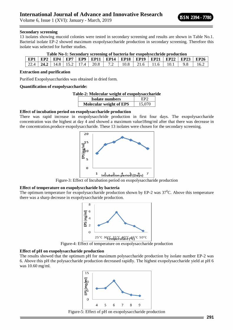
International Journal of Advance and Innovative Research Volume 6, Issue 1 (XVI): January - March, 2019
291
ISSN 2394 - 7780
Secondary screening 13 isolates showing mucoid colonies were tested in secondary screening and results are shown in Table No.1. Bacterial isolate EP-2 showed maximum exopolysaccharide production in secondary screening. Therefore this isolate was selected for further studies.
Table No-1: Secondary screening of bacteria for exopolyscchride production EP1 EP2 EP4 EP7 EP9 EP11 EP14 EP18 EP19 EP21 EP22 EP23 EP26 22.4 24.2 14.8 15.2 17.4 20.8 7.2 10.8 21.6 11.6 10.1 9.8 16.2
Extraction and purification Purified Exopolysaccharides was obtained in dried form.
Quantification of exopolysaccharide:
Table-2: Molecular weight of exopolysaccharide Isolate numbers EP2
Molecular weight of EPS 15,070
Effect of incubation period on exopolysaccharide production There was rapid increase in exopolysacchride production in first four days. The exopolysaccharide concentration was the highest at day 4 and showed a maximum value18mg/ml after that there was decrease in the concentration.produce exopolysaccharide. These 13 isolates were chosen for the secondary screening.
Figure-3: Effect of Incubation period on exopolysaccharide production
Effect of temperature on exopolysccharide by bacteria The optimum temperature for exopolysaccharide production shown by EP-2 was 370C. Above this temperature there was a sharp decrease in exopolysaccharide production.
Figure-4: Effect of temperature on exopolysaccharide production
Effect of pH on exopolysaccharide production The results showed that the optimum pH for maximum polysaccharide production by isolate number EP-2 was 6. Above this pH the polysaccharide production decreased rapidly. The highest exopolysaccharide yield at pH 6 was 10.60 mg/ml.
Figure-5: Effect of pH on exopolysaccharide production
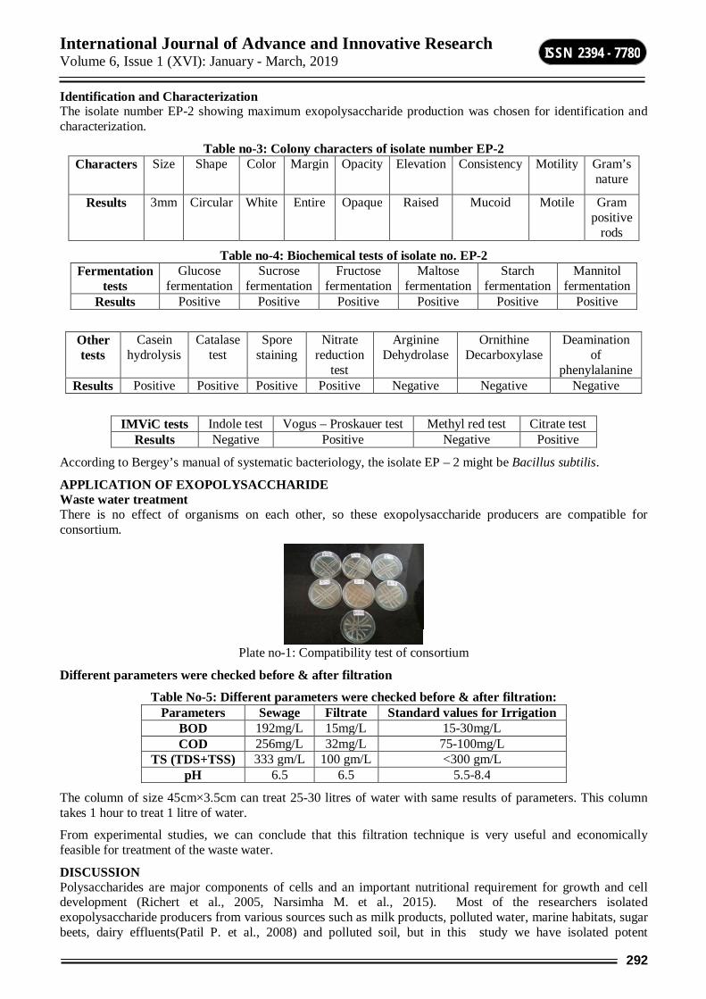
International Journal of Advance and Innovative Research Volume 6, Issue 1 (XVI): January - March, 2019
292
ISSN 2394 - 7780
Identification and Characterization The isolate number EP-2 showing maximum exopolysaccharide production was chosen for identification and characterization.
Table no-3: Colony characters of isolate number EP-2 Characters Size Shape Color Margin Opacity Elevation Consistency Motility Gram’s
nature
Results 3mm Circular White Entire Opaque Raised Mucoid Motile Gram positive
rods
Table no-4: Biochemical tests of isolate no. EP-2 Fermentation
tests Glucose
fermentation Sucrose
fermentation Fructose
fermentation Maltose
fermentation Starch
fermentation Mannitol
fermentation Results Positive Positive Positive Positive Positive Positive
Other tests
Casein hydrolysis
Catalase test
Spore staining
Nitrate reduction
test
Arginine Dehydrolase
Ornithine Decarboxylase
Deamination of
phenylalanine Results Positive Positive Positive Positive Negative Negative Negative
IMViC tests Indole test Vogus – Proskauer test Methyl red test Citrate test Results Negative Positive Negative Positive
According to Bergey’s manual of systematic bacteriology, the isolate EP – 2 might be Bacillus subtilis.
APPLICATION OF EXOPOLYSACCHARIDE Waste water treatment There is no effect of organisms on each other, so these exopolysaccharide producers are compatible for consortium.
Plate no-1: Compatibility test of consortium
Different parameters were checked before & after filtration
Table No-5: Different parameters were checked before & after filtration: Parameters Sewage Filtrate Standard values for Irrigation
BOD 192mg/L 15mg/L 15-30mg/L COD 256mg/L 32mg/L 75-100mg/L
TS (TDS+TSS) 333 gm/L 100 gm/L <300 gm/L pH 6.5 6.5 5.5-8.4
The column of size 45cm×3.5cm can treat 25-30 litres of water with same results of parameters. This column takes 1 hour to treat 1 litre of water.
From experimental studies, we can conclude that this filtration technique is very useful and economically feasible for treatment of the waste water.
DISCUSSION Polysaccharides are major components of cells and an important nutritional requirement for growth and cell development (Richert et al., 2005, Narsimha M. et al., 2015). Most of the researchers isolated exopolysaccharide producers from various sources such as milk products, polluted water, marine habitats, sugar beets, dairy effluents(Patil P. et al., 2008) and polluted soil, but in this study we have isolated potent
International Journal of Advance and Innovative Research Volume 6, Issue 1 (XVI): January - March, 2019
293
ISSN 2394 - 7780
exopolysaccharide producers from soil. We have isolated 13 isolates which were screened for exopolysaccharide production. Out of 13 isolates, isolate EP – 2 produce maximum exopolysaccharide as compared to other isolates. Therefore, isolate EP – 2 was used for optimization and identification.
The higher production of exopolysaccharides at 350C, the reasons for before and after this temperature decrease exopolysaccharides production may be that is more suitable for the exopolysaccharides production at 350C. Similarly pH of culture medium were studied, at pH 6 showed highest exopolysaccharides production. Then we can conclude that the best condition for isolate EP – 2 to produce exopolysaccharide was by incubation at 350C, pH 6 for 96 hrs.
For extracting exopolysaccharides three volumes of isopropanol was used ( Abdellah et. al., 2014). To remove protein contamination in the sample most of the authors reported use of 10% Trichloroacetic acid (TCA) but the acid may react with the exopolysaccharide and the structure will be disturbed. Therefore, we have used ammonium sulphate precipitation method to remove all the protein contamination present in the crude exopolysaccharide.
In this study, we have used Anthrone method (Morris D. L., 1948) for estimation of carbohydrate (exopolysaccharide) and Lowry’s method for protein estimation. Viral Shukla et al., in 2015 checked viscosity of exopolysaccharides by using viscometer but he had used broths containing cells to calculate viscosity. Since broths do not give clear idea about the viscosity of exopolysaccharide, we have used pure exopolysaccharide solutions. By using pure exopolysaccharide solution we have calculated molecular weight of exopolysaccharide. Read S. Al. Wasify et al., used purified exopolysaccharides as coagulants in water purification but no one used directly exopolysaccharide producers in waste water treatment. Therefore to reduce the cost we have used exopolysaccharide producers in consortium for waste water treatment and these bacteria were found to be effective in waste water treatment.
CONCLUSION Ammonium sulphate precipitation method was also effective to remove protein content in the exopolysaccharides. By using pure exopolysaccharides, molecular weight can be calculated with viscometer. Exopolysaccharide producers are very useful in waste water treatment. A filrtation column containing soil,sand,gravel and exopolysaccharide producers showed higher capacity to purify water as compared to available traditional methods. This filtration technique is very useful and economically feasible for treatment of the waste water. The present study clearly indicates that the bacteria Bacillus subtilis can give higher exopolysaccharide production at 350C, pH 6 within 96 hours.
REFERENCES 1. Abdellal M., Ahcene H., Benalia Y., Saad B. and Abdelmalek B., (2014). Screening for exopolysacchaaride
– producing strains of thermophilic lactic acid bacteria isolated from Algerian raw camel milk. African journal of Microbiology research. Vol. 8(22) : 2208-2214.
2. Dong X, Cai M., (2001). Manual of Bacteria identify. Science press.Beijing.
3. Ducklow H. W. Mitchell R., (1979). Bacterial populations and adaptations in the mucous layers on living corals. Limnol oceanogr. Vol. 24 :715 – 725.
4. Sutherland I. W., (2001). Biofilm exopolysaccharides : a strong and sticky framework. Microbiology. Vol. 147 (3-9).
5. Stewart – Tull, D. E. S (1980). The immunological activities of bacterial peptidoglycans. Annu. Rev. Microbiol. Vol. 34 : 311 – 340.
6. Vijayalakshmi, Nemichandrappa M., Sreenivas K. R. and Ayyanagowdar M. S., (2012). Effect of polymers on moisture retention and soil water holding capacity. Karnatka journal of agricultural sciences. Vol. 25 (4) : 469 – 471.
7. Lowry B. O. H., Nira J. R., Farr A. L. and Rose J. R. (1951). Protein measurement with the folin-phenol reagent. Journal of biology and Chemistry. Vol. 193 : 265-275.
8. Nehad, E. A. and El – Shamy A. L., (2010). Physiological studies on production of the exopolysaccharide by fungi. Agriculture and biology Journal of North America. Vol. 1 (6) : 1303 – 1308.

International Journal of Advance and Innovative Research Volume 6, Issue 1 (XVI): January - March, 2019
294
ISSN 2394 - 7780
ZOOPLANKTONIC ANALYSIS AND AQUATIC POLLUTION LOAD OF VANJARWADI RESERVOIR DIST BEED 431122 (MS)
Prashant V. Patil1 and Anirudha M. Budrukkar2 1Department of Zoology Mrs. K. S. K.alias Kaku College, Beed Dist. Beed
2Department of Zoology R.B. Attal College, Gevrai, Beed Dist. Beed
INTRODUCTION There are large number of aquatic animals, which are economically important for human as well as nature as a food. These are large and economical important aquatic crustacean and play an important role in human being while a large number of molluscans and fishes which economically important to mankind.
The zooplanktons are the primary food for the fish.The water productivity is the important bottom fauna as a link in the enegy flow from primary production to fish food has stressed by many workers including Krishnamoorthy (1966) Gupta (1976) Vashishta and Bhandal (1979) Bose and Lakra (1994) Anitha (2004) Chandrashekhar and Kodarkar (1994) worked on the macro-zoo benthos in India.
During the study the rotifer was 145 in no. / L at spot A and 156 in no. / L at spot B , the copepods was 95 in no. / L at spot A and 89 in no. / L at spot B , the ostracoderm in no. / L was 96 in no. / L at spot A and 97 in no. / L at spot B, the cladecera was in no. / L 106 at spot A and 98 a in no. / L t spot B.
MATERIAL AND METHODS The study was carried out for the year June 2016- May 2017.
Zooplanktons were collected from two spots namely spot A in the morning hours i.e. 6.00 am to 7.00 am.
The samples were collected by using plankton net of mesh size 30 mm and transferred to 100 ml bottle and preserved using 4 % formalin solution.
The zooplanktons were identified according to the guidelines given by Ward and Whipple (1958) and Battish (1958).
RESULTS AND DISCUSSION As the reservoir is an minor irrigation reservoir constructed in the year 1965 for irrigation and fish cultural aspects. The reservoir is having total catchment area 26.37 km2 . The 750 farmers taking the use of this reservoir for various activities like agriculture ,drinking ,fish culture.
The present analysis and results shows that the rotifer dominancy at spot. During the study the rotifer was 148 in no. / L at spot A , the copepods was 97 in no. / L at spot A , the ostracoderm in no. / L was 98 in no. / L at spot A ,the cladecera was in no. / L 108 at spot A and .The results are shown in the table no. 1.1
DISCUSSION During study zooplankton community shows that the rotifers are dominant in all season this shows that the water temperature increases in summer while optimum in winter and mansoon the photosynthetic activity is clear in summer the reservoir water is useful for fish cultural activity .
Table no-1.1: Shows the zooplankton study at spot A Spot A
Zooplankton Jun Jul Aug Sept Oct Nov Dec Jan Feb Mar Apr May Total Rotifer 05 07 10 12 13 11 12 13 16 17 18 14 148
Copepod 08 10 15 14 10 08 09 08 04 03 03 04 97 Ostracoderm 05 05 06 08 11 12 09 08 09 10 09 06 98
Cladocera 06 09 08 14 11 13 09 09 08 07 08 06 108
REFERENCES Anitha G. Kodarkar (2004)- Studies on Macrobenthos Mir Ali lake Hyderabad A.P. J. Aqua. Biol.19 (1) 68-
69
Chandrasekhar S.V.A. and Kodarkar.M.S. (1994)- Biodiversity of zooplankton in Saroomsagar lake, Hyderabad J. Aqua. Biol.9 (1-2) 68-69
Wekh P.G. (1996)-Limnology MC Graw Hill bulk G. Dnc Newyark Edmonson W. T. (1996)- Fresh water biology John Willey and Sons Inc. New York.

International Journal of Advance and Innovative Research Volume 6, Issue 1 (XVI): January - March, 2019
295
ISSN 2394 - 7780
Tonapi.G.T. (1980)-Freshwater animals of India (An ecological approach) Oxford and IBH Pub. Co. New Delhi 341
Peijler.B (1957)- Taxonomical and Ecological studies on planktonic Rotifers from central Sweden. K. Snonkavatensk. Akad. Handl. 6 (7) : 52
Baker R. L. (1979)- Species status of Keratella Cochlearis (Goose) and K. Erlinare Alhastorm (Rotifer, Branchionoidae) Morphological and Ecological consideration can. J. Zool. 57 (9) 1719-1722
Battish . S. K. (1992)-Freshwater zooplankton of India Oxford and IBH Pub. Co. PP 233 Ward H. B. and Whipple G. C. (1958)-Freshwater iology Mc. Graw Hill and Co. New York.
Qadri M Y and Yousuf. R. (1982)-Influence of some physicochemical factors on the seasonality of cladocera in lake Manabal. K.. Giobios 7: 273- 276
Shadeeek K.(1983)- Rotifers as indicators of water quality Hydrobiologia 100
International Journal of Advance and Innovative Research Volume 6, Issue 1 (XVI): January - March, 2019
296
ISSN 2394 - 7780
DISTRIBUTION OF SEDGES NEAR JAIKWADI DAM IN AURANGABAD DISTRICT OF MAHARASHTRA
Sudhir Solanke1, Raffique Shaikh2 and Ravi Patil3 1Department of Botany, R. B. Attal College, Georai, Dist. Beed
2Department of Botany, Milliya College, Beed 3Department of Botany, Deogiri College, Aurangabad
ABSTRACT The results of field studies provide a general account of the distribution of cyperaceous plants in the Flora of Marathawada, Maximum cyperaceous plants were collected from wetland and river. Frequent visits were made during September to January near of Jaikwadi dam and specimens were collected and processed. The specimens were identified with the help of floras and available literature. Sedge vegetation can be categorized in different ecological groups which are discussed in the present paper.
Keywords: Distribution, Cyperaceae, Near the Jaikwadi dam.
INTRODUCTION Cyperaceae (Commonly Called sedge family) is the second largest family among monocotyledons and stands next to grasses. Cyperaceae is one of the most intricate families represented by 70-80 genera and 4000 species distributed throughout the world with about 28 genera and 500 species in India. Due to minute to smaller size of flower, least economic importance and intricacy due to narrow generic and specific delimitation, extreme variation in vegetative and floral parts (Khan, 1983; 1198; 1999 and 2000) the sedges have been neglected by most taxonomists. This is probably the reason, why studies could not progress as compared to other families such as grasses. Thus taxonomically Cyperaceae are neglected group of flowering plants in India.Therefore it needs to have been given specialized treatment from different angles and different aspects for the preparations of accounts. Jaikwadi dam is constructed on river Godavari, about 50 kms away from Aurangabad, at Paithan. The water body of the dam is named as “Nathsagar”. The water body of dam is so huge that it covers 339.80 square kms. The dam shows aquatic and semi aquatic and terrestrial diversity of Angiosperms(Anilkumar M. Deshmukh 2015). In present investigation collected specimens fromthe near Jaikwadi dam is identify by various literature
MATERIALS AND METHODS During the present work an attempt is being made to document the occurrence of sedges ofthe near Jaikwadi dam frequent visits were made during September to January to study region and 70 species belonging to 8 genera were collected and processed as per the method described by (Prain, 1996; Rao and Verma, 1990 and Prasad and Singh, 2002). The specimens were identified with the help of floras by (Clarke, 1893; 1902 and 1909; Fischer, 1928; Cooke, 1908; Hooper, 1976 and Sedgwick, 1918;Wadoodkhan M. A. 2015) and available literature (Karthikeyan, 1989; Kern, 1974; Sharma et al., 1996. and Koyama, 1985) and identification is confirmed in herbaria of R. B. Attal College Georai Dist. Beed.
DISSCUSIONS Inland sedge vegetation can be categorised in different ecological groups as under
Oligotropic Habitat The common swamp and marshy sedges with excess of moisture. The characteristic members are Cyperus aloppecuroides, C. digitatus, C. exaltatus, C. pilosus, C. procerus, C. scariosus, Fimbristylis miliacea, F. tertragona, Fuirena sp.Kyllinga brevifolia, Pycreus sanguinolentus, P. diaphanus, P. flavidis, P. polystachyos, P. stramineous and Scripus praelongatus.
Eutrophic Habitat Cyperus bifax, C. alulatus, C. esculentus, C. difformis, C. squarrosus, C. corymbosus, C. distans var. pseudonutans, Kyllinga melanosperma, Fmbristylis alboviridis, F. microcarya, F. complanata, F. bisumbellata, F. tomentosa, F. adenolepis several forms of F. dichotoma, Scirpus laterflorus,
Mixed Dry Land Sedges Many types of sedge are found even in semidry soil or localities providing comparatively less moisture. The common ones are Cyperu scompressus, subsp. compressus, C. distans ssp. pseudonutans, C. iria, C. pygmaeus, C. squarrosus, C. teneriffae, C. rotundus, C. stoloniferous, C. compactus, Fimbristylis tenera, F. ovata, F. polytrichoides, Bulbostylis barbata, Kyllinga bulbosa, Scirpus kyllingioides, Scleria lithosperma.
International Journal of Advance and Innovative Research Volume 6, Issue 1 (XVI): January - March, 2019
297
ISSN 2394 - 7780
Marginals Characteristically a few sedges are found along the margins of rivers, lakes, streams and similar other water bodies the notable ones Cyperus corymbosus, C. pangorei, C. bifax, C. articulates, C. nutans subsp. eleusinoides, C. alopecuroides, C. exaltatus, C. difformis, C. teneriffae, C. articulatus, Pycreus flavidus, Fimbristylis dichotoma, F. miliacea, Scirpus littoralis and S. corymbosus.
Grasss Land Sedges Characteristically a few sedges are found in wet open grass-lands or marshy grass-lands. They are Fimbristylis dichotoma, F. merrillii, F. quinquangularis, Fuirena ciliaris, F. umbellate, Cyperus iria, C. compactus, C. pseudokyllingioides, Scirpus juncoides,
Among the small grass such Pycreus pumilus, Cyperus cuspidatus.
ACKNOWLEDGEMENTS The authers wish to acknowledge their grateful thanks to Dr. M. A. Wadood Khan for his valuable guidance, Principal, Dr. R. K. Nimbalkar for library and laboratory facilities and Vice Principal Dr. R. Shikhare Madam for support.
REFERENCES Clarke CB (1893). Cyperaceae. In J. D. Hooker, The flora of British India. L. Reeve & Co., London 6 453-
663.
Clarke CB (1902). Cyperaceae. In T. Dyer Flora of Tropical Africa. Reeve Kent.
Clarke CB (1909). Illustrations of Cyperaceae. tt. 1-114 London.
Cooke T (1908). The flora of the Presidency of Bombay. Taylor and Francis Publers, London 2 890-896.
P. Chavan, S. A. Survaseand S. N. Solanke (2013). Distribution of Sedges in Vidarbha Region of Maharashtra, Indian Journal of Plant Sciences Vol. 2 (1) January-March, pp.131-133.
Fischer CEC (1928). The flora of the Presidency of Madras. In: J. S. Gamble, Adlard& Sons, London3 1662-1667.
Hooper SS (1976). Cyperaceae. In :Saldanha& Nicolson. Flora of Hassan district Amerind Publisher, New Delhi 655-701.
Karthikeyan SK (1989). Cyperaceae in Florae IndicaeEnumeratioMonocotyledonae. Botanical Survey of India, Kolkatta 32-73.
Kern J (1974). Cyperaceae. In: Van Steenis, Flora Malesiana ser. Noordhoff International Publishing, Leiden 1.7(3) 494-516.
Khan Wadood MA (1983). Cyperaceae of Marathwada. Marathwada University journal of science15 1-4.
Khan Wadood MA (1998). Cyperaceae. In: V. N. Naik Flora of Marathwada. Amruth Publication, Aurangabad 2 965-992.
Khan Wadood MA (1999). The genus Eleocharis R. Br. In Maharashtra in :Sivadasan& Mathew, Biodiversity, Taxonomy and Conservation of Flowering plants 303-313. Mentor Books Calicut. Kerala Cyperaceae
Khan Wadood MA (2000). The genus Scirpus in Marathwada. Rheedea10(1) 19-32.
Khan Wadood M. A.(2015). Book of Cyperaceae of Western Ghat, West Coast and Maharashtra, dattsons, J. Nehru Marg, Nagpur -01.
Koyama T (1985). Cyperaeae In: M. D. Dassanayake& F. R. Fosberg (Edts). Revised Hand Book to the Flora of Ceylon. Oxford & IBH New Delhi 5 126-127, 153-166, et 253-254.
Prain David (1996). Repr. Ed. Cyperaceae in Bengal Plants. BishensinghMahendrapalsingh, Dehradun 2 1127-2261.
Prasad VP and Singh NP (2002). Sedges of Karnataka (Indica) (Family cyperaceae). Scientific Publishers, Jodhpur (India).

International Journal of Advance and Innovative Research Volume 6, Issue 1 (XVI): January - March, 2019
298
ISSN 2394 - 7780
Rao RS and Verma DM (1982). Cyperaceae of North East India. Botanical survery of India, Kolkata 41-47. Sedgwick LJ (1918). The Cyperaceae of Bombay Presidency. Journal of the Bombay Natural History Society 26 200-202.
Sharma BD, Karthikryan S and Singh NP (1996). The Flora of Maharashtra (Monocotyledons). Botanical Survey of India, Kolkata 356-369.
International Journal of Advance and Innovative Research Volume 6, Issue 1 (XVI): January - March, 2019
299
ISSN 2394 - 7780
LITERATURE REVIEW ON FRUIT QUALITY IDENTIFICATION SYSTEM
Rahul J. Mhaske1 and Prapti Deshmukh2 1Siddharth Arts, Commerce and Science College, Jafrabad
2Dr. G.Y. Pathrikar College of CS & IT, Aurangabad
ABSTRACT In day to day life we are less focusing on our health. Therefore, it increases the various health problems. To maintain the good health we have to eat good quality foods. Fruits gives various components like vitamins, minerals, etc. which are directly effect on our body. Fruit plays a vital role to maintain the good health. Hence the quality of fruit should be identified before purchase it from the market it is immense important. This paper is purely focus on the literature review of fruit quality.
Keywords: fruit quality, size detecting, fruit grading, image processing
INTRODUCTION India is an agricultural nation. The production from the farm is at second position in the world. It is observed that near about 60% Indians are directly or indirectly depends on the business which are on agricultural products. It plays very important role in Indian economy. Farmers are mostly depended on the yield of agricultural products, therefore, there is need to focus on agriculture products which can be in good quality. To scale the economic situation of current poor or low land holder farmers, there is a need to export their products in demanding market in a good quality [1]. One of the major farm product is fruit. Fruits are very useful to our health. Physicians are always suggest the good quality fruit to the patients to recover from the diseases. Fruits contains different types of vitamins, proteins, etc. which can be helpful to our body. Hence there is a need to provide good quality fruit in the market.
NEED OF QUALITY FRUIT Health and pleasure are two main reasons for eating fruits. However, the overall quality of fruit is often criticized because of the fact that the organoleptic quality has been given little or no priority by the agronomic research in favor of yield increase, diseases and storage resistance and transportation tolerance [6] [8][9].
It is estimated that a total of 20-40% of all crops in developing countries is lost to postharvest losses. Losses are due to decay, mechanical damage, physiological disorders, and even to the action of several fruit flies, during harvest, storage and transport.
LITERATURE REVIEW 1998: Judith A. Abbot (30 June 1998) in “Quality measurement of fruit and vegetable” in this research used for the technique of X-ray & MRI for the evaluating Quality food [4].
2002: Paolo Gay, Remigio Berruto& Pietro Piccarolo(31, july 2002) in “Fruit color Assessment Quality Grading Purposes”, use in Agronomic techniques for the search the food Quality as per the color Assessment [5].
2003: J. Blasco, N. Aleixos2, E. Molt, (4, August 2003) used the Common Techniques for Automatic Quality Fruit testing & searching for quality attributes: size, colour, stem location and detection of external blemishes. The segmentation procedure used, based on a Bayesian discriminant analysis[10].
2004: Tadhg Brosnan, Da-Wen Sun,(1, January 2004) in “Improving quality inspection of food products by computer vision” they have review , a Image Processing Technique are used respectively for the searching food product by used computer vision system[11].
Cheng-Jin Du and Da-Wen Sun,( 5, May 2004) in ‘Recent developments in the applications of image processing techniques for food quality evaluation’, they reviews recent advances in image processing techniques for food quality evaluation, which include charge coupled device camera, ultrasound, magnetic resonance imaging, computed tomography, and electrical tomography for image acquisition; pixel and local pre-processing approaches for image pre-processing, thresholding-based, gradient-based, region-based, and classification-based methods for image segmentation; size, shape, colour, and texture features for object measurement; and statistical, fuzzy logic, and neural network methods for classification [12].
2005: Adolfo Martínez-Usó, Filiberto Pla, and Pedro García-Sevilla,( Press,2005), multispectral segmentation algorithm based on quadtree(QT) representation is proposed [13].

International Journal of Advance and Innovative Research Volume 6, Issue 1 (XVI): January - March, 2019
300
ISSN 2394 - 7780
Adolfo Mart´ınez-Us´o, Filiberto Pla, and Pedro Garc´ıa-Sevilla,(2005) they used a Pattern Recognition and Image Analysis techniques are Describe and also used Multispectral fruit inspection application & in remote sensing tasks [14].
O. Kleynen, V. Leemans, M.-F. Destain,( Issue 1, July 2005) in perform research on the Multi-spectral image system technique are used for the searching defected apple [15].
2006: Cheng-Jin Du, Da-Wen Sun, (1, January 2006) reviewed use of Learning Techniques for the evaluation of food Quality[16].
Chaoxin Zheng, Da-Wen Sun, Liyun Zheng,(12 December 2006) in this review an image features for food quality evaluation and inspection by using color image[17].
2007: Zhaoshen Qing , Baoping Ji , Manuela Zude, ,( 1, September 2007 ) provide a test for soluble solids content test(SSC) are determines taste of apples, firmness is an important parameter for determining fruit maturity, quality grade, and harvest time[18].
J. Blasco , N. Aleixos , E. Molto,( 3, August 2007) in Computer vision detection of peel defects in citrus by means of a region oriented segmentation algorithm use for the detection defect of fruit using RGB images of the date fruits test result show that the system can sort 80% dates accurately [19].
2009: J. Blasco, N. Aleixos, J. Go´mez-Sanchı´s, E. Molto,( 2, June 2009) explain the contribution of NRI and UVFL played an important role in the detection of serious diseases such as anthracnose and decay[20].
2010: Yosuke Yoshioka and Nobuko Fukino(Number 3,2010) use the colour signature for evaluation fruit color[21].
2011: Abraham Gastélum-Barrios, Rafael A. Bórquez-López, et.al (18 July, 2011) review the tomato quality evaluation by using different types of size, shape, contents, etc.[22].
2009-2014: Haisheng Gao, Fengmei Zhu, Jinxing Cai has review the various approaches for “Non-destructive Detection for Fruit Qualit” like detection of fruit quality using optical properties, sonic vibration, machine vision techniques, nuclear magnetic resonance (NMR), electrical properties, computed tomography, electronic noses, etc. [23].
CONCLUSION This paper highlights the importance of fruits in our life. It is also indicates the need of quality fruits in market as well as individual level. All the important aspects of fruit quality measurement through different techniques reported by the earlier researchers are discussed year wise. At the last fruit quality evaluation plays a vital role in automatic fruit quality evaluation.
REFERENCES [1] S. M. Wasule, “Quality Determination and Grading of Tomatoes using Raspberry Pi,” Int. J. Recent
Innov. Trends Comput. Commun., vol. 6, no. 7, pp. 86–89, 2018.
[2] D. M. Barrett, J. C. Beaulieu, and R. Shewfelt, “Color, flavor, texture, and nutritional quality of fresh-cut fruits and vegetables: Desirable levels, instrumental and sensory measurement, and the effects of processing,” Crit. Rev. Food Sci. Nutr., vol. 50, no. 5, pp. 369–389, 2010.
[3] K. G. Grunert, “Food quality and safety: Consumer perception and demand,” Eur. Rev. Agric. Econ., vol. 32, no. 3, pp. 369–391, 2005.
[4] A. Judith A, “Quality measurement of fruits and vegetables,” Postharvest Biol. Technol., vol. 15, no. 3, pp. 207–225, 1999.
[5] P. Gay, R. Berruto, and P. Piccarolo, “Fruit Color Assessment for Quality Grading Purposes,” 2002 ASAE Annu. Int. Meet. CIGR XVth World Congr., vol. 0300, no. 02, pp. 1–9, 2002.
[6] F. Vesali, M. Gharibkhani, and M. H. Komarizadeh, “An approach to estimate moisture content of apple with image processing method,” Aust. J. Crop Sci., vol. 5, no. 2, pp. 111–115, 2011.
[7] Y. Al Ohali, “Computer vision based date fruit grading system: Design and implementation,” J. King Saud Univ. - Comput. Inf. Sci., vol. 23, no. 1, pp. 29–36, 2011.
[8] S. Cubero, N. Aleixos, E. Moltó, J. Gómez-Sanchis, and J. Blasco, “Advances in Machine Vision Applications for Automatic Inspection and Quality Evaluation of Fruits and Vegetables,” Food
International Journal of Advance and Innovative Research Volume 6, Issue 1 (XVI): January - March, 2019
301
ISSN 2394 - 7780
Bioprocess Technol., vol. 4, no. 4, pp. 487–504, 2011.
[9] J. N. Quinton, Encyclopedia of Agrophysics, vol. 32, no. 3. Springer, 2011.
[10] J. Blasco, N. Aleixos, and E. Moltó, “Machine vision system for automatic quality grading of fruit,” Biosyst. Eng., vol. 85, no. 4, pp. 415–423, 2003.
[11] T. Brosnan and D. W. Sun, “Improving quality inspection of food products by computer vision - A review,” J. Food Eng., vol. 61, no. 1 SPEC., pp. 3–16, 2004.
[12] C. J. Du and D. W. Sun, “Recent developments in the applications of image processing techniques for food quality evaluation,” Trends Food Sci. Technol., vol. 15, no. 5, pp. 230–249, 2004.
[13] A. Martinez-Uso, F. Pla, and P. Garcia-Sevilla, “Multispectral image segmentation for fruit quality estimation,” Lect. Notes Comput. Sci., vol. 3523, no. II, pp. 689–6696, 2005.
[14] A. Martinez-Uso, F. Pla, and P. Garcia-Sevilla, “Multispectral image segmentation by energy minimization for fruit quality estimation,” Lect. Notes Comput. Sci., vol. 3523, no. II, pp. 689–6696, 2005.
[15] O. Kleynen, V. Leemans, and M. F. Destain, “Development of a multi-spectral vision system for the detection of defects on apples,” J. Food Eng., vol. 69, no. 1, pp. 41–49, 2005.
[16] C. J. Du and D. W. Sun, “Learning techniques used in computer vision for food quality evaluation: A review,” J. Food Eng., vol. 72, no. 1, pp. 39–55, 2006.
[17] C. Zheng, D. W. Sun, and L. Zheng, “Recent developments and applications of image features for food quality evaluation and inspection - a review,” Trends Food Sci. Technol., vol. 17, no. 12, pp. 642–655, 2006.
[18] Z. Qing, B. Ji, and M. Zude, “Predicting soluble solid content and firmness in apple fruit by means of laser light backscattering image analysis,” J. Food Eng., vol. 82, no. 1, pp. 58–67, 2007.
[19] J. Blasco, N. Aleixos, and E. Moltó, “Computer vision detection of peel defects in citrus by means of a region oriented segmentation algorithm,” J. Food Eng., vol. 81, no. 3, pp. 535–543, 2007.
[20] J. Blasco, N. Aleixos, J. Gómez-Sanchís, and E. Moltó, “Recognition and classification of external skin damage in citrus fruits using multispectral data and morphological features,” Biosyst. Eng., vol. 103, no. 2, pp. 137–145, 2009.
[21] Y. Yoshioka, N. Fukino, and Received:, “Image-based phenotyping: use of colour signature in evaluation of melon fruit colour,” Euphytica, Springer, vol. 171, no. 3, pp. 409–416, 2010.
[22] A. Gastélum-Barrios, R. A. Bórquez-López, E. Rico-García, M. Toledano-Ayala, and G. M. Soto-Zarazúa, “Tomato quality evaluation with image processing: A review,” African J. Agric. Res., vol. 6, no. 14, pp. 3333–3339, 2011.
[23] H. Gao et al., “A Review of Non-destructive Detection for Fruit Quality To cite this version : HAL Id : hal-01061726,” pp. 0–9, 2014.
[24] H. N. Patel, “Fruit Detection using Improved Multiple Features based Algorithm,” Int. J. Comput. Appl., vol. 13, no. 2, pp. 1–5, 2011.
[25] W. Lang, R. Jedermann, D. Mrugala, A. Jabbari, B. Krieg-Brückner, and K. Schill, “The ‘intelligent container’ - A cognitive sensor network for transport management,” IEEE Sens. J., vol. 11, no. 3, pp. 688–698, 2011.
[26] D. J. Lee, J. K. Archibald, and G. Xiong, “Rapid color grading for fruit quality evaluation using direct color mapping,” IEEE Trans. Autom. Sci. Eng., vol. 8, no. 2, pp. 292–302, 2011.
[27] D. Lorente, N. Aleixos, J. Gómez-Sanchis, S. Cubero, O. L. García-Navarrete, and J. Blasco, “Recent Advances and Applications of Hyperspectral Imaging for Fruit and Vegetable Quality Assessment,” Food Bioprocess Technol., vol. 5, no. 4, pp. 1121–1142, 2012.
[28] K. K. Patel, A. Kar, S. N. Jha, and M. A. Khan, “Machine vision system: A tool for quality inspection of food and agricultural products,” J. Food Sci. Technol., vol. 49, no. 2, pp. 123–141, 2012.
[29] D. M. Madrid, “How India can benefit from exports to Europe – Europe ’ s expectation on quality in

International Journal of Advance and Innovative Research Volume 6, Issue 1 (XVI): January - March, 2019
302
ISSN 2394 - 7780
fresh fruits and vegetables,” Agrochain India trade show, no. 1, pp. 1–5, 2012.
[30] H. K. PURWADARIA and Department, “Issues and Solutions of Fresh Fruit Export in India,” pp. 17–23, 2005.
[31] R. M. Haralick and K. Shanmugam, “Textural Features for Image Classification,” IEEE Trans. Syst. Man. Cybern., vol. 3, pp. 610–621, 1973.
[32] R. M. Haralick and L. G. Shapiro, Computer and Robot Vision, 1st ed. Boston, MA, USA: Addison-Wesley Longman Publishing, 1992.
[33] S. Cubero, M. P. Diago, J. Blasco, J. Tardáguila, B. Millán, and N. Aleixos, “A new method for pedicel/peduncle detection and size assessment of grapevine berries and other fruits by image analysis,” Biosyst. Eng., vol. 117, no. C, pp. 62–72, 2014.

International Journal of Advance and Innovative Research Volume 6, Issue 1 (XVI): January - March, 2019
303
ISSN 2394 - 7780
ACETONIDE PROTECTION OF DIOLS USING IODINE AND DIMETHOXYPROPANE
Ravibhushan S. Kulkarni1, Prashant P. Dixit2, Kishan P. Haval3
1Department of Chemistry, Dr. Babasaheb Ambedkar Marathwada University, Osmanabad 2,3Department of Microbiology, Dr. Babasaheb Ambedkar Marathwada University, Osmanabad
ABSTRACT The protection and cleavage of isopropylidene acetals are important aspect in the chemistry of carbohydrates, nucleosides and alkaloids. As a result, numerous methods using acid catalysts have been developed. Herewith, we have reported novel method for protection of diol as an acetonide using iodine and dimethoxypropane. The ease of handling, cost and activity of the catalyst, good to excellent yields and neutral reaction conditions are some of the highlights of the reported method.
Keywords: Acetonide, Diol protection, Iodine, Dimethoxypropane (DMP), Green chemistry.
1. INTRODUCTION A protecting group (PG) is introduced onto a particular functional group (FG) in a poly-functional molecule to block its reactivity under reaction conditions needed to make alterations elsewhere in the molecule. A decent protecting group should be readily, but selectively introduced to the desired functional group in a poly-functional molecule. It should be stable / resistant to the reagents used in consequent reaction steps in which the group being masked (protected) is desired to remain deactivated (protected) and it should be capable of being selectively removed under mild conditions.The commonly tackled functional groups in organic synthesis that are reactive to nucleophilic or electrophilic reagents whose selective transformation may present challenges do regularly require deactivation by masking with a protecting group. The most important method for the protection 1, 2 diols or 1, 3 diols is to convert them into a cyclic acetals or ketals.1The several methods are reported for the acetonide protection of diols by using acetone in presence of anhy. FeCl3,2iodine,3 CuSO4,4 and cation exchange resin.5Few methods are also described in the literature by suing 2, 2-dimethoxypropane in presence of ZrCl4,6 and p-TsOH.7Although a large number of methods using different catalysts are available for this purpose, the known methods are generally time consuming and under acidic conditions.Herewith, we are reporting a mild, efficient and eco-friendly method for protection of 1,2 or 1,3 diols as their acetonides by usingdimethoxypropane and iodine.
2. RESULTS AND DISCUSSIONS In continuation with our efforts to develop the new methods for the synthesis of bioactive organic compounds,8 we have demonstrated the new and efficient method for acetonide protection of diols. Accordance to our aim, we performed the reaction of 1, 2-diol (20 mmol) (1a) in presence of iodine (20 mol%) in dimethoxypropane (DMP)at room temperature to give the corresponding acetonide protected compound (2a) with 75% yield.To set the generality of reaction, we performed the reaction different diols in presence of catalytic amount of iodine in dimethoxypropane (DMP). All the reactions furnished the corresponding protected diols with 60-80% yields (Scheme 1). The structures of products obtained were confirmed by comparing melting points with reported in literature (Table 1).
Scheme-1
Table-1: Preparation of acetonide derivatives of 1,2 / 1,3-diols 1,2 or 1,3- Diols Products Time Yield (%)
D-Glucose 2a 3 75 D-Mannitol 2b 4 60 D-Xylose 2c 5 65
D-Mannose 2d 4 65 Resorcinol 2e 4 80 Glycerol 2f 5 75
Propane 1,3-diol 2g 3 77 Catechol 2h 3 73
Cyclohexane 1,2-diol 2i 4 70 Cholestane 2,3-diol 2j 4 65

International Journal of Advance and Innovative Research Volume 6, Issue 1 (XVI): January - March, 2019
304
ISSN 2394 - 7780
3. EXPERIMENTAL General procedure for the synthesis acetonides: In a solution of diol (20 mmol) in dimethoxypropane (DMP), iodine (20 mol%) was added. The reaction was stirred at room temperature. The progress of reaction was monitored by TLC. After completion of reaction, the product was extracted with ethyl acetate and purified by column chromatography to furnish the corresponding acetonides (2a-j) with 60-80% yields.
4. CONCLUSION We have demonstrated a simple, efficient and green method for protection of diols. The neutral condition, good for acid sensitive starting materials and high yields are the key advantages of our protocol.
ACKNOWLEDGEMENTS Authors acknowledge the financial support by Dr. Babasaheb Ambedkar Marathwada University, Aurangabad.
5. REFERENCES 1. Green, T. W.; Wuts, P. G. M. Protective Groups in Organic Synthesis,
Wiley-Interscience, New York, 1999, 207-215, 716-719. (b) Kocienski, P. J. Protecting Groups, Georg ThiemeVerlag, 1994. (c) Otto Th. Schmidt, Methods in Carbohyd. Chem., 2, 318 (1966). (d) Whistler, R. L.;Wolfrom, M. L.; TipsonR. S.Methods in Carbohydrate ChemistryII; Academic Press; New York; 1962; 246.
2. Singh, P. P.;Gharia, Y.M.;Dasgupta,F.;Srivastava,H.C. Tetrahedron Lett.1977, 5, 439.
3. (a) Kartha, K. P. R.; Aloui; M.; Field R. A.Tetrahedron Lett.1996, 37, 5175. (b) Dutta, S.; Sarkar, S.; Gupta, S.; Sen A. K.Tetrahedron Lett.2013, 54, 865. (c) Jereb, M.; Vražič, D.; Zupan, M.Tetrahedron2011, 67, 1355. (d) Kartha, K. P. R. Tetrahedron Lett.1986, 27, 3415.
4. (a) Copper, B. E. ChemInd1978, 794. (b) House, H. O.; Czuba, L. J. M.; Olmstead Gall, Olmstead H. D. J. Org. Chem. 1969, 34, 23.
5. Bahule, B. B.; Nandurkar Y. M. IOSR J. App. Chem. 2012, 03, 28.
6. Singh, S.; Duffy, C. D.; Shah, S. T. A.; Guiry, P. J. J. Org. Chem., 2008, 73, 6429.
7. Kumar, P.; Dinesh, C. U.; Reddy, R. S.; Pande, B. Synthesis1993, 1069.
8. (a) Haval, K. P.; Argade N. P. J. Org. Chem. 2008, 73, 6936. (b) Haval, K. P.; Argade N. P. Synthesis2007,2198. (c) Haval, K. P.; Mhaske, S. B.;Argade N. P. Tetrahedron 2006, 62, 937. (d) Haval, K. P.; Argade N. P. Tetrahedron2006, 62, 3557. (e) Tigote, R. M.; Haval, K. P.; Kazi, S. K. J. Med. Chem. & Drug Discovery2017, 2, 654. (f) Shinde, N. V.; Dhake, A. S.; Haval K. P.Oriental Journal of Chemistry, 2016, 32, 515. (g) Shinde, N. V.; Dhake, A. S.; Haval K. P.Der PharmaChemica, 2015, 7, 251.
International Journal of Advance and Innovative Research Volume 6, Issue 1 (XVI): January - March, 2019
305
ISSN 2394 - 7780
BUTTERFLY DIVERSITY OF PARBHANI, MAHARASHTRA STATE, INDIA
S. M. Yeole M. S. P. Mamdal’s, Shri Shivaji College, Parbhani
ABSTRACT Butterflies are part of the class of Insecta in the order Lepidoptera, along with the moths. Adult butterflies have large, often brightly coloured wings, and conspicuous, fluttering flight. Culturally, butterflies are a popular motif in the visual and literary arts. Parbhani is the 4th largest city of Marathwada region, which is located at 19.27°N & 76.78°E and about 347 metres above MSL. Shri Shivaji College, Parbhani is located near about in the heart of the city. College campus is rich in floral diversity and hence, survey was done to observe the butterfly diversity during the period of one year i.e. from October 2017 to September 2018 by regular keen observation. During the present investigation total 15 species of butterflies were recorded which are belonging to various families. Butterfly diversity survey of the college campus has not made yet, hence an attempt has been made to record the same which may become fruitful for the upcoming researchers of this field.
Keywords: Shri Shivaji College, Parbhani, Butterfly diversity.
INTRODUCTION The vast majority of butterflies have a four-stage life cycle; egg, larva (caterpillar), pupa (chrysalis) and imago (adult). In the genera Colias, Erebia, Euchloe andParnassius, a small number of species are known that reproduce semi-parthenogenetically; when the female dies, a partially developed larva emerges from her abdomen (Capinera, J. L., 2008). Some butterflies, especially in the tropics, have several generations in a year, while others have a single generation, and a few in cold locations may take several years to pass through their whole life cycle. Nearly all butterflies are diurnal, have relatively bright colours, and hold their wings vertically above their bodies when at rest, unlike the majority of moths which fly by night, are often cryptically coloured (well camouflaged), and either hold their wings flat (touching the surface on which the moth is standing) or fold them closely over their bodies. Some day-flying moths, such as the hummingbird hawk-moth are exceptions to these rules (Herrera, C. M. 1992). Many butterflies are sexually dimorphic. Most butterflies have the ZW sex-determination system where females are the heterogametic sex (ZW) and males homogametic (ZZ).
Parbhani is located at 19.27°N & 76.78°E and about 347 metres above MSL in Marathwada region of Maharashtra. Shri Shivaji College, Parbhani is located near about in the heart of the city and contains rich floral diversity and hence, survey was done to observe the butterfly diversity as the butterfly diversity survey of the college campus has not made yet. This data may become fruitful for the upcoming researchers of this field.
MATERIALS AND METHODS Parbhani is the 4th largest city in Marathwada region of the Maharashtra after Aurangabad, Nanded and Latur. The city is located at 19.27°N & 76.78°E and about 347 metres above MSL. With the establishment in the year 1961, Shri Shivaji College, is one of the oldest college of swami Ramanand Teerth Marathwada University, Nanded. College is located near about in the heart of the city. Butterfly diversity survey of Shri Shivaji College, Parbhani was done for the period of one year i.e. from October 2017 to September 2018 by regular keen observation with the help of field binocular 8 x 40 and identified by using keys and monographs given by Mani, M. S. (1995) and Kehimkar, I (2014).
RESULTS AND DISCUSSION Butterfly biodiversity survey of Shri Shivaji College Campus, Parbhani was carried out during the study period i.e., from October 2017 to September 2018 and the results are depicted in Table 1. Butterflies are distributed worldwide except Antarctica, totalling some 18,500 species. Of these, 775 are Nearctic; 7,700 Neotropical; 1,575 Palearctic; 3,650 Afrotropical and 4,800 are distributed across the combined Oriental and Australian regions (Williams, et. al., 2015). Butterflies in their adult stage can live from a week to nearly a year depending on the species. Many species have long larval life stages while others can remain dormant in their pupal or egg stages and thereby survive winters (Powell, J. A., 1987). The number of generations per year varies from temperate to tropical regions with tropical regions showing a trend towards multivoltinism (Timothy D. S., 2011).
Butterflies feed primarily on nectar from flowers. Some also derive nourishment from pollen, tree sap, rotting fruit, dung, decaying flesh, and dissolved minerals in wet sand or dirt (Gilbert, L. E., 1972). Butterflies are important as pollinators for some species of plants. In general, they do not carry as much pollen load as bees,
International Journal of Advance and Innovative Research Volume 6, Issue 1 (XVI): January - March, 2019
306
ISSN 2394 - 7780
but they are capable of moving pollen over greater distances (Herrera, C. M., 1987). Flower constancy has been observed for at least one species of butterfly (Goulson, D. et. al., 1997).
Adult butterflies consume only liquids, ingested through the proboscis. They sip water from damp patches for hydration and feed on nectar from flowers, from which they obtain sugars for energy, and sodium and other minerals vital for reproduction. Several species of butterflies need more sodium than that provided by nectar and are attracted by sodium in salt; they sometimes land on people, attracted by the salt in human sweat. Some butterflies also visit dung, rotting fruit or carcasses to obtain minerals and nutrients. In many species, this mud-puddling behaviour is restricted to the males, and studies have suggested that the nutrients collected may be provided as a nuptial gift, along with the spermatophore, during mating (Molleman, F. et. al., 2005).
During the period of investigation total 15 species of butterflies were recorded which are belonging to four different families viz., Hasperiidae, Lycaenidae, Nymphalidae, Papilionidae and Pieridae. Out of which the family Nymphalidae was proved itself a most dominant family with five species namely Danus chrysippu, Danus genutia, Euploea core, Melanitis leda and Tirumala limniace. Second position was taken up by Lycaenidae, Papilionidae and Pieridae each is represented by three species. Family Lycaenidae is represented by Neopithecops zalmora, Zizula hylax and Zizina otis; family Papilionidae is represented by Chilasa clytia, Graphium macareus and Papilio polytes whereas family Pieridae is represented by other three species namely Catopsilia pomona, Eurema hecabe and Gandaca harina. Family Hasperiidae was recorded as least dominant and represented by a single species only i.e., Caprona agama (Table 1). Such satisfied butterfly diversity may be due to rich flora of Shri Shivaji College Campus, Parbhani.
REFERENCES Capinera, John L. (2008): Encyclopedia of Entomology. Springer Science & Business Media.
Gilbert, L. E. (1972): "Pollen Feeding and Reproductive Biology of HeliconiusButterflies". Proceedings of the National Academy of Sciences 69 (6): 1402 – 1407.
Goulson, D.; Ollerton, J.; Sluman, C. (1997): "Foraging strategies in the small skipper butterfly, Thymelicus flavus: when to switch?". Animal Behavior 53 (5): 1009 –1016.
Herrera, C. M. (1987): "Components of Pollinator 'Quality': Comparative Analysis of a Diverse Insect Assemblage" (PDF). Oikos (Oikos) 50 (1): 79 – 90.
Herrera, Carlos M. (1992): "Activity Pattern and Thermal Biology of a Day-Flying Hawkmoth (Macroglossum stellatarum) under Mediterranean summer conditions".Ecological Entomology 17: 52 – 56.
Kehimkar, I. (2014): The Book of Indian Butterflies. BNHS, Oxford University Press, Mumbai. pp 497.
Mani, Ms. S. (1995): Insects. National Book Trust, India. pp 162
Molleman, Freerk; Grunsven, Roy H. A.; Liefting, Maartje; Zwaan, Bas J.; Brakefield, Paul M. (2005): "Is Male Puddling Behaviour of Tropical Butterflies Targeted at Sodium for Nuptial Gifts or Activity?". Biological Journal of the Linnean Society 86 (3): 345 – 361.
Powell, J. A. (1987): "Records of Prolonged Diapause in Lepidoptera". J. Res. Lepid. 25: 83–109.
Timothy Duane Schowalter (2011): Insect Ecology: An Ecosystem Approach. Academic Press. p. 159.
Williams, Ernest; Adams, James; Snyder, John. (2015): "Frequently Asked Questions". TheLepidopterists' Society. Retrieved 9 September 2015.

International Journal of Advance and Innovative Research Volume 6, Issue 1 (XVI): January - March, 2019
307
ISSN 2394 - 7780
Table-1: Butterfly Diversity of Shri Shivaji College Campus, Parbhani During October 2017 – September 2018
Sr. No. Family Scientific Name Common Name
1. Hasperiidae Caprona agama Spotted Angle
2. Lycaenidae Neopithecops zalmora Quaker
3. Lycaenidae Zizula hylax Tiny Grass Blue
4. Lycaenidae Zizina otis Lesser Grass Blue
5. Nymphalidae Danus chrysippus Plain Tiger
6. Nymphalidae Danus genutia Stripped Tiger
7. Nymphalidae Euploea core Common Crow
8. Nymphalidae Melanitis leda Common Evening Brown
9. Nymphalidae Tirumala limniace Blue Tiger
10. Papilionidae Chilasa clytia Common Mime
11. Papilionidae Graphium macareus Lesser Zebra
12. Papilionidae Papilio polytes Common Mormon
13. Pieridae Catopsilia pomona Common Emigrant
14. Pieridae Eurema hecabe Common Grass Yellow
15. Pieridae Gandaca harina Tree Yellow

International Journal of Advance and Innovative Research Volume 6, Issue 1 (XVI): January - March, 2019
308
ISSN 2394 - 7780
E – WASTE MANAGEMENT
S. B. Birajdar and P. V. Kulkarni Department of Mathematics, S.M.P. College, Murum, Tq. Omerga Dist. Osmanaabd
ABSTRACT In order to bridge the digital, it is quite necessary to get an affordable, equitable and quality access to ICT. It is quite clear that two third of world’s population is still offline therefore, there is a need to provide affordable access to internet for all literate persons.
Now a days, there is a tremendous growth in use of ICT devices and services. The faster change in technology and frequent innovations spreads over all the world. This causes a threat of deterioration is environmental conditions and human health. The waste of electronic and electrical equipment which contain hazardous components. These components are still handled in an environmentally unfriendly manner in developing nations. It is huge challenge to face the nations for handling e-waste to protect the environment.
In this paper, an approach is made towards assessing the preoent situation of e – waste management globally as well as in India. We have to consider the present regulations and guidelines. There is a large amount of recycling of e – waste done who have no knowledge about the consequences of exposure to hazardous substances. The reuse of EEE has greater environmental and social benifits than recycling as it in creates the useful life time of the ICT equipment and enables resources efficiency and energy efficiency.
In addition to the technical, social aspects of the EEE – waste management system, it is also crucial to consider the economic aspects if the system has to be made financially viable and sustainable along with being socially acceptable
1. INTRODUCTION It is a hand fact that there is tremendous increase in use of ICT devices to bridge the digital divide. E – waste is defines as “waste electrical and electronic equipment, whole or in pant or rejects from their manufacturing and repair process, which are intended to be discarded” Electrical and electronic equipment is defined as equipment which is dependent on electrical currents or electro-,magnetic fields to be fully utilized.
There is a need for e – waste management e – waste components. There are several health risks and environmental damage due to unused electronic components. When crude, unscientific methods are applied for recovery of useful components while recycling all useful and valuable material, we have to take cave of the natural resources.
The rapid growth of ICT , frequent innovation an technological changes cause in shorter life span of ICT equipment. Moreover, developing country like India, the imported disused Electrical and Electronic equipment is uncontrolled. So the volume of e – waste has also increased drastically. For dving the efficient us of natural resources, e – waste should be minimised by recycling the e – waste substances. Due to disposal of e – waste materials, it may cause serious damage to the environment and public health while doing so, the presence of heavy metals like Arsenic, Cadmium, Barium, Lead, Lithium etc. And other toxic substances like PCB polychlorinated biphenyls are released in the environment which are very harmful human beings.
ITU has accepted the fact that in many developing countries, there is no knowledge about techniques, precautions.. so it causes more damage to their health and environment. By the definition of environmental sustainability, it is the ability to maintain the qualities that are valued in the physical environment by the use of non – renewable resources and environmentally sound recycleing as much as possible. It is very important that policy of e – waste disposal and regulatory aspects should be straingent and rational.
2. AN ESTIMATION OF E –WASTE The exponential growth of internet users from 501 million in 2006 to over 1.3 billion in 2011 at in developing countries, this exponential growth clearly indicates that the sale computer and other terminals has grown at a lightning pace. In 2006, 44% of internet users were in developing countries whereas in 2011. 62%, were prevent in developing countries. Personal computer sales has significantly increased about 170 million units from 2000 to 2010. It is protected that sales in 2014 will reach an estimated 470 million units which is more than double in the last 10 years. ITU data release of June 2012 indicates that total number o mobile cellular subscriptions reached almost 6 billion by the end of 2011 and in the developing countries, about 80 % of the 660 million new mobile cellular subscriptions added in 2011 were generated the report “recycling from E – waste to Resources”
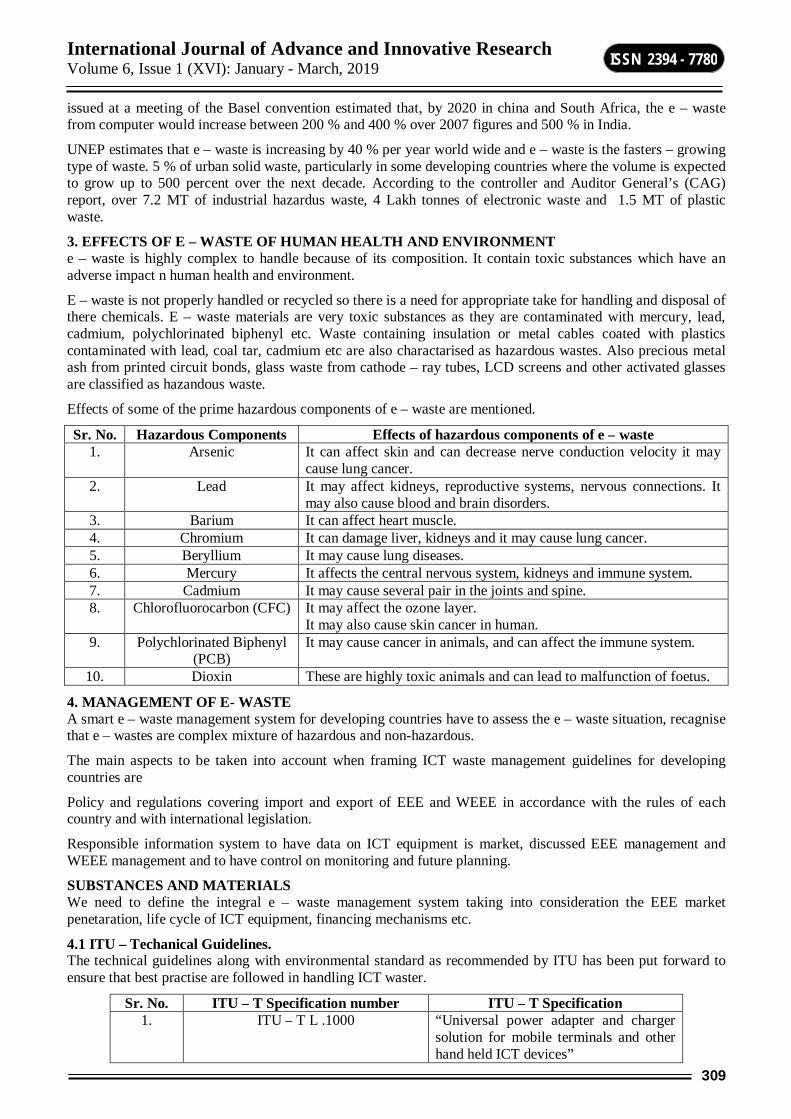
International Journal of Advance and Innovative Research Volume 6, Issue 1 (XVI): January - March, 2019
309
ISSN 2394 - 7780
issued at a meeting of the Basel convention estimated that, by 2020 in china and South Africa, the e – waste from computer would increase between 200 % and 400 % over 2007 figures and 500 % in India.
UNEP estimates that e – waste is increasing by 40 % per year world wide and e – waste is the fasters – growing type of waste. 5 % of urban solid waste, particularly in some developing countries where the volume is expected to grow up to 500 percent over the next decade. According to the controller and Auditor General’s (CAG) report, over 7.2 MT of industrial hazardus waste, 4 Lakh tonnes of electronic waste and 1.5 MT of plastic waste.
3. EFFECTS OF E – WASTE OF HUMAN HEALTH AND ENVIRONMENT e – waste is highly complex to handle because of its composition. It contain toxic substances which have an adverse impact n human health and environment.
E – waste is not properly handled or recycled so there is a need for appropriate take for handling and disposal of there chemicals. E – waste materials are very toxic substances as they are contaminated with mercury, lead, cadmium, polychlorinated biphenyl etc. Waste containing insulation or metal cables coated with plastics contaminated with lead, coal tar, cadmium etc are also charactarised as hazardous wastes. Also precious metal ash from printed circuit bonds, glass waste from cathode – ray tubes, LCD screens and other activated glasses are classified as hazandous waste.
Effects of some of the prime hazardous components of e – waste are mentioned.
Sr. No. Hazardous Components Effects of hazardous components of e – waste 1. Arsenic It can affect skin and can decrease nerve conduction velocity it may
cause lung cancer. 2. Lead It may affect kidneys, reproductive systems, nervous connections. It
may also cause blood and brain disorders. 3. Barium It can affect heart muscle. 4. Chromium It can damage liver, kidneys and it may cause lung cancer. 5. Beryllium It may cause lung diseases. 6. Mercury It affects the central nervous system, kidneys and immune system. 7. Cadmium It may cause several pair in the joints and spine. 8. Chlorofluorocarbon (CFC) It may affect the ozone layer.
It may also cause skin cancer in human. 9. Polychlorinated Biphenyl
(PCB) It may cause cancer in animals, and can affect the immune system.
10. Dioxin These are highly toxic animals and can lead to malfunction of foetus.
4. MANAGEMENT OF E- WASTE A smart e – waste management system for developing countries have to assess the e – waste situation, recagnise that e – wastes are complex mixture of hazardous and non-hazardous.
The main aspects to be taken into account when framing ICT waste management guidelines for developing countries are
Policy and regulations covering import and export of EEE and WEEE in accordance with the rules of each country and with international legislation.
Responsible information system to have data on ICT equipment is market, discussed EEE management and WEEE management and to have control on monitoring and future planning.
SUBSTANCES AND MATERIALS We need to define the integral e – waste management system taking into consideration the EEE market penetaration, life cycle of ICT equipment, financing mechanisms etc.
4.1 ITU – Techanical Guidelines. The technical guidelines along with environmental standard as recommended by ITU has been put forward to ensure that best practise are followed in handling ICT waster.
Sr. No. ITU – T Specification number ITU – T Specification 1. ITU – T L .1000 “Universal power adapter and charger
solution for mobile terminals and other hand held ICT devices”
International Journal of Advance and Innovative Research Volume 6, Issue 1 (XVI): January - March, 2019
310
ISSN 2394 - 7780
2. ITU – T L.1410 Deals with the assessment of the environmental impact of ICT goods, networks and services
3. ITU – T L.1420 Provides specific guidance on energy and greenhouse gas (GHG) impacts.
5. INDIAN SCENARIO FOR E-WASTE MANAGEMENT Last few years India has emerged as one major IT hub and the consumer electronic market has grown in an exponential rate. According to Manufacturers Association of Information Technology (MAIT) the Indian PC industry is growing by 25% compound annual growth rate. Study reports that in 2007, 2.2 million computers were made obsolete and 14 million mobile handsets replaced.
The e-waste generated was estimated to be 3,32,979 tons out of which 144,000 tons was recyclable and actually e-waste recycled was 19,000 tons.
The e- waste processed contained 12000 tons of computers and 7000 tons of TV. It was also estimated that around 50,000 tons of e-waste was generated through import besides 3,32,000 tons generated domestically.
6. TRAI GUIDELINES TRAI has endorsed the key provisions of the regulations issued by MoEF in their recommendations on Approach towards Green Telecom in 2012, where it is specifically clarified that the producer will e responsible for –
Ensuring that, new electrical and electronic equipment does not contain Lead, Mercury, Cadmium, Hexavalent Chromium, ploybrominated biophenyls (PBB) or polybrominateddiphenyl ethers (PBDE).
Collection of e-waste generated during the manufacture of EEE and channelizing the same for recycling or disposal
Collection of e-waste generated from the ‘end of life’ for their products in line with the principle of ‘Extended Producer Rseponsibility’ (EPR) and to ensure that such e-wastes are channelized to registered refurbisher or dismantler or recycler.
Setting up collection centers or take back system either individually or collectively for all EEE at the end of their life.
Creating awareness with regard to information on hazardous components in e-waste electrical and electronic equipment
Maintaining records of the e-waste and filing annual returns to the concerned State Pollution Control Board or Pollution Control Commitee.
7. CONCLUSION ITU has agreed to the fact that there is no unique or ideal model for e-waste management in developing countries, each of which is characterized by its own specific environmental, social, technological, economic and cultural conditions.
With a view to bridge the digital divide, there is exponential growth in the use of Electrical and electronic equipment (EEE) and so there is alarming effect on environment and human health when the ICT wastes are not disposed scientifically. There is an emergent need to implement the existing policies and guidelines in line with the international standards and practices for and healthy e-waste management system.
Government policies should encourage the reuse of EEE aiming to minimize and recycle Waste Electrical and Electronic Equipment (WEEE). The Extended Producer Responsibility (EPR) do need to have clear regulations to mandate the ‘take back’ activity of companied strictly.
Coordination with State Pollution Control Boards.
Preparation of Guidelines for Environmentally Sound Management of e-waste.
Conduct assessment of e-waste generation and processing
Recommend standards and specifications for processing and recycling e – waste
Documentation & compliation of data on e-waste
International Journal of Advance and Innovative Research Volume 6, Issue 1 (XVI): January - March, 2019
311
ISSN 2394 - 7780
Conducting training & awareness program
Enforcement of reduction in use of hazardous substances (ROHS)
Incentives and certification for green design/products.
REFERENCES 1. Central Pollution Board – Imlementation of e waste rules 201.
2. Effective electronic waste management and recycling process involving formal and non-formal sectors by S. Chatterjee and Krishna Kumar, Department of IT, CGO Complex, New Delhi
3. E-waste assessment in Kolkata metropolitan Area – Airpot by IMRB International April 2010 to West Bengal Pollution Control Board, GTZ and ICC Kolkatta.
4. E-waste : A hidden threat to Global environment and health, Deepti Mittal.
5. International Telecommunication Union.
6. Managing e-waste in India A review, Gulsan Sirkek, Gaurv Gupta.
7. Waste Electrical and Electronic Equipment, The EU and India : sharing best practices, Toxics Link.
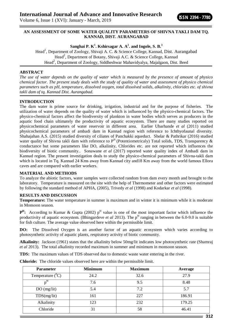
International Journal of Advance and Innovative Research Volume 6, Issue 1 (XVI): January - March, 2019
312
ISSN 2394 - 7780
AN ASSESSMENT OF SOME WATER QUALITY PARAMETERS OF SHIVNA TAKLI DAM TQ. KANNAD, DIST. AURANGABAD
Sanghai P. K1. Kshirsagar A. A2. and Ingole, S. B.3 Head1, Department of Zoology, Shivaji A. C. & Science College, Kannad, Dist. Aurangabad
Head2, Department of Botany, Shivaji A.C. & Science College, Kannad Head2, Department of Zoology, Siddheshwar Mahavidyalya, Majalgaon, Dist. Beed
ABSTRACT The use of water depends on the quality of water which is measured by the presence of amount of physico chemical factor. The present study deals with the study of quality of water and assessment of physico chemical parameters such as pH, temperature, dissolved oxygen, total dissolved solids, alkalinity, chlorides etc. of shivna takli dam of tq. Kannad Dist. Aurangabad.
INTRODUCTION The dam water is prime source for drinking, irrigation, industrial and for the purpose of fisheries. The utilization of water depends on the quality of water which is influenced by the physico-chemical factors. The physico-chemical factors affect the biodiversity of plankton in water bodies which serves as producers in the aquatic food chain ultimately the productivity of aquatic ecosystem. There are many studies reported on physicochemical parameters of water reservoir in different area. Earlier Ubarhande et al (2011) studied physicochemical parameters of ambadi dam in Kannad region with reference to Ichthyofaunal diversity. Shahajahan A.S. (2015) studied diversity of ciliates of Panchakki aqueduct. Shelar & Pathrikar (2016) studied water quality of Shivna takli dam with reference to PH (Potentiometricaly) Total solids, TDS, Transparency & conductance but some parameters like DO, alkalinity, Chlorides etc. are not reported which influences the biodiversity of biotic community.. Sonawane et al (2017) reported water quality index of Ambadi dam in Kannad region. The present investigation deals to study the physico-chemical parameters of Shivna-takli dam which is located in Tq. Kannad 24 Kms away from Kannad city and18 Km away from the world famous Ellora caves and are compared with earlier workers.
MATERIAL AND METHODS To analyze the abiotic factors, water samples were collected random from dam every month and brought to the laboratory. Temperature is measured on the site with the help of Thermometer and other factors were estimated by following the standard method of APHA, (2005), Trivedy et al (1998) and Kodarkar et al (1998).
RESULTS AND DISCUSSION Temperature: The water temperature in summer is maximum and in winter it is minimum while it is moderate in Monsoon season.
PH: According to Kumar & Gupta (2002) pH value is one of the most important factor which influence the productivity of aquatic ecosystem. (Bhingardeve et al 2013). The pH ranging in between the 6.0-9.0 is suitable for fish culture. The average value observed here within the permissible limit.
DO: The Dissolved Oxygen is an another factor of an aquatic ecosystem which varies according to photosynthetic activity of aquatic plants, respiratory activity of biotic community.
Alkalinity: Jackson (1961) states that the alkalinity below 50mg/lit indicates low photosynthetic rate (Shamraj et al 2013). The total alkalinity recorded maximum in summer and minimum in monsoon season.
TDS: The maximum values of TDS observed due to domestic waste water entering in the river.
Chloride: The chloride values observed here are within the permissible limit.
Parameter Minimum Maximum Average Temperature (0C) 24.2 32.6 27.9
pH 7.6 9.5 8.48 DO (mg/lit) 5.4 7.2 5.7 TDS(mg/lit) 161 227 186.91 Alkalinity 123 232 179.25 Chloride 31 58 46.41
International Journal of Advance and Innovative Research Volume 6, Issue 1 (XVI): January - March, 2019
313
ISSN 2394 - 7780
REFERENCES APHA (2005) Standard methods for the examination of water and waste water (21st edition) American
Public Health Association Washington DC.
Bannerjee, G. & Narsimha, R.K. (2013) Physico-chemical factors influenced plankton biodiversity and fish abundance - A case study of Nagaram tank of Warangal, Andhra Pradesh. International journal of Life Sciences Biotechnology and Pharma research 2(2):248-260.
Bhingardeve L.S., J.N. Shamraj, S.S. Survase, & D.A.Kulkarni (2013) Study of physicochemical parameters of chaphal reservoir Tq. Patan Dist. Satara approach to conserve it Proceedings of National conference NCABCMSD at Siddheshwar Mahavidyalya Majalgaon. 161-164
Kodarkar M.S., Diwan A.D., Muruga N. Kulkarni K.M. Anuradha R. (1998) Methodology for water analysis. Indian Association of aquatic Biologist IAAB Pub. No. 2.
Naiknaware V.V. and S.S. Lomte (2009) Physico-chemical studies on Bindusara river water of Beed. (M.S.) Bioinfolet 6 (4): 336-337.
Smita Sonawane, Suniti Barve, & Sunil Anand (2012) Determination of water quality index of Ambadi dam Dist. Aurangabad (M.S.) Bio Nano frontier Ecorevolution 2012 Colombo Srilanaka 104-108.
Shahjahan AS (2018) Impact of climatic changes on the diversity of ciliates: A study of Panchakki aqueduct. International Journal of zoology studies 3(2):65-67.
Shelar M.D. & Pathrikar R.D. (2016) Analysis of physical and chemical parameters of Shivna Lawhali-Takli (medium project) dam water Tq. Kannad District aurangabad state Maharashtra India Der Pharma chemica 8(1): 32-38
Shamraj J.N. ,Bhingardeve L.S. , S.S. Survase, & D.A.Kulkarni (2013) Study of physicochemical study of ‘Terna project’ Ter, Tq. & Dist.Osmanabad Proceedings of National conference NCABCMSD at Siddheshwar Mahavidyalaya, Majalgaon. 165-170
Trivedy R.K., P.K.Goyal and C.L. Trishal (1998) ‘Practical methods in ecology and environmental science Enviro media Publications, Karad (India)
Ubarhande S.J., Jagtap J.T. & Sonawane S.R.(2011) Ichthyofaunal diversity from Ambadi Dam Tq. Kannad Dist. Aurangabad (M.S.) Recent Research in Science and Technology 3(6): 34-37.
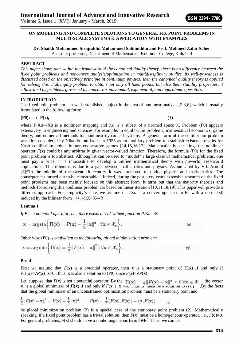
International Journal of Advance and Innovative Research Volume 6, Issue 1 (XVI): January - March, 2019
314
ISSN 2394 - 7780
ON MODELING AND COMPLETE SOLUTIONS TO GENERAL FIX POINT PROBLEMS IN MULTI-SCALE SYSTEMS & APPLICATION WITH EXAMPLES
Dr. Shaikh Mohammed Sirajuddin Mohammed Salimuddin and Prof. Mohmed Zafar Saber Assistant professor, Department of Mathematics, Kohinoor College, Kultabad
ABSTRACT This paper shows that within the framework of the canonical duality theory, there is no difference between the fixed point problems and nonconvex analysis/optimization in multidisciplinary studies. its well-posedness is discussed based on the objectivity principle in continuum physics; then the canonical duality theory is applied for solving this challenging problem to obtain not only all fixed points, but also their stability properties, it sillustrated by problems governed by nonconvex polynomial, exponential, and logarithmic operators.
INTRODUCTION The fixed point problem is a well-established subject in the area of nonlinear analysis [2,3,6], which is usually formulated in the following form:
(P0): x=F(x), (1)
where F:Xa→Xa is a nonlinear mapping and Xa is a subset of a normed space X. Problem (P0) appears extensively in engineering and sciences, for example, in equilibrium problems, mathematical economics, game theory, and numerical methods for nonlinear dynamical systems. A general form of the equilibrium problem was first considered by Nikaido and Isoda in 1955 as an auxiliary problem to establish existence results for Nash equilibrium points in non-cooperative games [14,15,16,17]. Mathematically speaking, the nonlinear operator F(x) could be any arbitrarily given vector-valued function. Therefore, the formula (P0) for the fixed point problem is too abstract. Although it can be used to “model” a large class of mathematical problems, one must pay a price: it is impossible to develop a unified mathematical theory with powerful real-world applications. This dilemma is due to a gap between mathematics and physics. As indicated by V.I. Arnold [1]:“In the middle of the twentieth century it was attempted to divide physics and mathematics. The consequences turned out to be catastrophic.” Indeed, during the past sixty years extensive research on the fixed point problems has been mainly focused on this abstract form. It turns out that the majority theories and methods for solving this nonlinear problem are based on linear iteration [10,11,18,19]. This paper will provide a different approach. For simplicity’s sake, we assume that Xa is a convex open set in Rn with a norm ∥x∥ induced by the bilinear form 〈∗, ∗⟩:X×X→R
Lemma 1 If F is a potential operator, i.e., there exists a real-valued function P:Xa→R
Other wise (P0) is equivalent to the following global minimization problem:
Proof First we assume that F(x) is a potential operator, then x is a stationary point of Π(x) if and only if ∇Π(x)=∇P(x)−x=0 , thus, x is also a solution to (P0) since F(x)=∇P(x)
Let suppose that F(x) is not a potential operator. By the fact that. the vector x is a global minimizer of Π(x) if and only if F(x¯)−x¯=0. Thus, x¯must be a solution to (P0) .By the facts that the global minimizer of an unconstrained optimization problem must be a stationary point and
he global minimization problem (3) is a special case of the stationary point problem (2). Mathematically speaking, if a fixed point problem has a trivial solution, then F(x) must be a homogeneous operator, i.e., F(0)=0. For general problems, F(x) should have a nonhomogeneous term f ∈Rn. Thus, we can let
International Journal of Advance and Innovative Research Volume 6, Issue 1 (XVI): January - March, 2019
315
ISSN 2394 - 7780
where D:X→W⊂Rm is a linear operator, W:W→R is a so-called objective function. Objectivity is a basic concept in continuum physics [6,13] and mathematical modeling [8,9]. Its mathematical definition is given in Goo’s book (Definition 6.1.2 [7]).
Definition 1
Let R be a proper orthogonal group, i.e., R∈R if and only if RT=R−1, det R= 1. A set Wa is said to be objective if
A real-valued function W:Wa→R is said to be objective if
We talk about an objective function does not depend on rigid rotation of the system considered, but only on certain measure of its variable. In the Euclidean space W⊂Rm, the simplest objective function is the ℓ2-norm ∥w∥ in Rm as we have ∥Rw∥2= wTRTRw= ∥w∥2 ∀ R∈R. For general F(x), we can see from (4) that
are objective functions. By the fact that x=F(x), we know that 〈x, F(x)⟩ is also an objective function. Therefore, for a given fixed point problem, the corresponding Π(x) is naturally an objective function.
Let Xa= x∈ X ∣Dx ∈Wa. The fixed point problem (P0) can be reformulated into the following stationary point problem:
Application
2. Exponential and polynomial functions
As our first application, the objective function is assumed to be
where D1∈Rm×n and D2∈Rp×n are two given matrices, α, β, λ are real numbers. Clearly, for a given λ>0, W(Dx) is nonconvex and
is a non-monotone operator. In this case, the fixed point problem (P0) can be equivalently written as
Clearly, traditional methods for solving this nonlinear fixed point problem in Rn are difficult. However, by the canonical duality theory, this problem can be solved easily in R2.
The canonical measure in this problem can be given as
Correspondingly, the canonical function is
and the canonical dual variable is
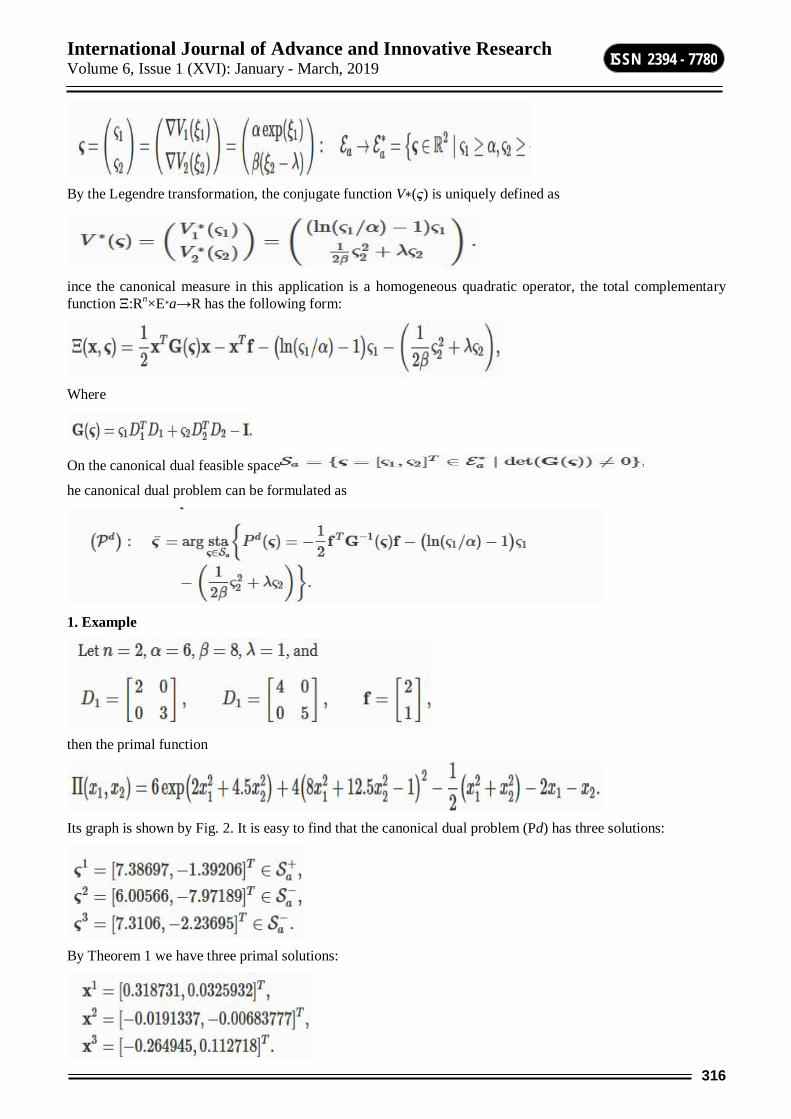
International Journal of Advance and Innovative Research Volume 6, Issue 1 (XVI): January - March, 2019
316
ISSN 2394 - 7780
By the Legendre transformation, the conjugate function V∗(ς) is uniquely defined as
ince the canonical measure in this application is a homogeneous quadratic operator, the total complementary function Ξ:Rn×E∗a→R has the following form:
Where
On the canonical dual feasible space
he canonical dual problem can be formulated as
1. Example
then the primal function
Its graph is shown by Fig. 2. It is easy to find that the canonical dual problem (Pd) has three solutions:
By Theorem 1 we have three primal solutions:

International Journal of Advance and Innovative Research Volume 6, Issue 1 (XVI): January - March, 2019
317
ISSN 2394 - 7780
It is easy to check that
By Theorem 2 we know that x1 is a global minimizer of Π(x), x2 is a local maximizer of Π(x), and x3=[−0.264945,0.112718]T is a local minimizer of Π(x) (see Fig. 1). By the fact that
hold for all i=1,2,3, we know that xi (i=1,2,3) are all fixed points.
CONCLUSION Applications are illustrated by problems governed by nonconvex polynomial, exponential, and polynomial functions. Our examples show that both globally stable and locally stable/unstable fixed point problems in Rn can be obtained easily by solving the associated canonical dual problems in Rm with m<n. generalized to problems with nonsmooth potential functions.
International Journal of Advance and Innovative Research Volume 6, Issue 1 (XVI): January - March, 2019
318
ISSN 2394 - 7780
REFERENCE 1. Anorld, V.I.: On teaching mathematics. Russ. Math. Surv. 53(1), 229–236 (1998)
2. Bierlaire, M., Crittin, F.: Solving noisy, large-scale fixed-point problems and systems of nonlinear equations. Transp. Sci. 40, 44–63 (2006)
3. Border, K.C.: Fixed Point Theorems with Applications to Economics and Game Theory. Cambridge University Press, New York (1985)
4. Chen, Y., Gao, D.Y.: Global solutions to nonconvex optimization of 4th-order polynomial and log-sum-exp functions. J. Glob. Optim. 64(3), 417–431 (2016)
5. Ciarlet, P.G.: Linear and Nonlinear Functional Analysis with Applications. SIAM, Philadelphia (2013)
6. Eaves, B.C.: Homotopies for computation of fixed points. Math. Program. 3, 1–12 (1972)
7. Gao, D.Y.: Duality Principles in Nonconvex Systems: Theory, Methods and Applications. Springer, New York (2000).
8. Gao, D.Y.: On unified modeling, theory, and method for solving multi-scale global optimization problems. In: Sergeyev, Y.D., Kvasov, D.E., Mukhametzhanov, M.S. (eds.) Proceedings of the 2nd International Conference Numerical Computations: Theory and Algorithms. AIP Conference Proceedings, vol. 1776, 020005 (2016).
9. Gao, D.Y.: On unified modeling, canonical duality-triality theory, challenges and breakthrough in
10. Hirsch, M.D., Papadimitriou, C., Vavasis, S.: Exponential lower bounds for finding Brouwer fixed points. J. Complex. 5, 379–416 (1989)
11. Huang, Z., Khachiyan, L., Sikorski, K.: Approximating fixed points of weakly contracting mappings. J. Complex. 15, 200–213 (1999)
12. Jin, Z., Gao, D.Y.: On modeling and global solutions for d.c. optimization problems by canonical duality theory. Appl. Math. Comput. 296, 168–181 (2017).
13. Machalova, J., Netuka, H.: Control variational method approach to bending and contact problems for Gao beam. Appl. Math. 62(6), 1–17 (2017)
14. Scarf, H.: The approximation of fixed point of a continuous mapping. SIAM J. Appl. Math. 35, 1328–1343 (1967)
15. Scarf, H.E., Hansen, T.: Computation of Economic Equilibria. Yale University Press, New Haven (1973)
16. Shellman, S., Sikorski, K.: A two-dimensional bisection envelope algorithm for fixed points. J. Complex.
17. Shellman, S., Sikorski, K.: A recursive algorithm for the infinity-norm fixed point problem. J Complex19
18. Smart, D.R.: Fixed Point Theorems. Cambridge University Press, Cambridge (1980)
19. Yang, Z.: Computing Equilibria and Fixed Points: The Solution of Nonlinear Inequalities. Kluwer Academic Publishers, Dordrecht (1999),

International Journal of Advance and Innovative Research Volume 6, Issue 1 (XVI): January - March, 2019
319
ISSN 2394 - 7780
FUNGAL INCIDENCE ON GROUNDNUT SEED FROM DIFFERENT LOCALITY OF MARATHAWADA
Shrikant B. Mane, Prashant P. Pangrikar and Ashok M. Chavan Microbial Culture laboratory, Department of Botany, Dr. Babasaheb Ambedkar Marathwada University, Aurangabad
ABSTRACT The seed mycoflora of groundnut seed from different localities were screened by standard blotter paper and agar plate method as recommended by ISTA. The seed mycoflora of five localities of Marathwada region viz. Aurangabad, Jalna, Parbhani, Osmanabad and Beed of groundnut seed samples collected and examined. The 08 genera and 14 species of fungi isolated viz. Alternaria alternata, Aspergillus flavus, Aspergillus fumigatus, Aspergillus niger, Aspergillus ustus, Fusarium oxysporum, Curvularia lunata , Macrophomina phaseolina, Penicillium notatum, Rhizoctonia solani, Rhizopus nigricans. Out of these fungi Aspergillus flavus, Aspergillus niger, Fusarium oxysporum and Rhizopus nigricans were found predominant fungi and shows higher percent of seed mycoflora. Higher number of fungi was isolated by method as agar plate compared to blotter paper method.
Keywords: Seed mycoflora, localities, fungi.
INTRODUCTION Groundnut (Arachis hypogaea L.), also known as peanut, is a legume that ranks 6th among the oilseed crops and 13th among the food crops of the world. India is one of the largest producers of oilseeds in the world and occupies an important position in the Indian agricultural economy. Groundnut (Arachis hypogea L.) a valuable legume crop is over in India with production of about 37.19 million tons in 2013. India is second largest producer of groundnut after China. It is estimated that nine oilseeds namely groundnut, rapeseed-mustard, soybean, sunflower, safflower, sesame, castor and linseed, accounted for an area of 23.44 million hectares with the production of 25.14 million tones. Groundnut is called as the ‘King’ of oilseeds. It is one of the most important food and cash crops of our country.
Groundnuts is a high in calories value food stuff and added to diets to improve dietary proteins and supply vitamins of the 13-complex. Groundnuts foods are simple to prepare and there are a diversity of forms in which they can be prepared. Groundnuts produce vegetable oils, fats. Dry legume seeds are frequently the most practical source of storable and transportable proteins in regions lacking refrigeration facilities. Grain legume proteins are the least expensive protein source for both rural and urban populations of India. While being a valuable source of nutrients and low-priced commodity Groundnut is called as wonder nut and poor men’s cashew nut. Groundnut is one of the most important cash crops of our country.
Groundnut is as good source of nutrient therefore storage pathogens were affecting to seed causing loss of seed health. These infected seed were not safe for human health. Storage environment viz., temperature and relative humidity might not be conducive for the survival of field fungi and harvesting and handling practices as well as locality influence seed mycoflora. Ground nut seed is attacked by a number of pathogenic fungi of economic importance. Sullivan (1984), reported that groundnut seeds are highly susceptible to diseases, as they serve as a source of stored nutrients for fungi such as Rhizopus spp., Penicillium spp., Aspergillus niger, and A. flavus. Rasheed et al. (2004) found Macrophomina phaseolina, Rhizoctonia solani, Fusarium solani, F. oxysporum, Aspergillus flavus, A. niger were predominant in groundnut and seed coat was greatly infected by fungi followed by cotyledon and axis. Krishnappa et al. (2003) reported that groundnut pods stored in gunny bag had recorded maximum infection ranged between 16 and 18% of Aspergillus flavus, Aspergillus niger, Fusarium spp. and Penicillium spp. and caused reduction in germination and vigour index. Patra et al. (2000) reported that increase in storage period of groundnut seeds upto nine months, the viability decreased, while pathogen activity, moisture and sugar content in seeds increased gradually. The storage fungi like Aspergillus flavus was responsible for maximum depletion of fat content and reducing sugar in safflower, soybean, sesamum and groundnut due to Fusarium equiseti and Rhizopus stolonifer and a decrease in crude fat content by Curvularia lunata, F. equiseti and penecillium digitatum Kakde and Chavan (2011).
MATERIAL AND METHODS Collection of seed samples The method described by Neergard (1979) has been adopted for the collection of seed samples Of seeds wear collected from different five localities of Marathwada region viz. Aurangabd, Jalna, Parbhani, Osmanabad and Beed of groundnut seed samples fields, store houses, and market places.
International Journal of Advance and Innovative Research Volume 6, Issue 1 (XVI): January - March, 2019
320
ISSN 2394 - 7780
Blotter Paper Method The seed mycoflora was isolated by using standard moist blotter method and agar plate methods as recommended by International seed testing association (ISTA 1975), The method adopted for isolation of seed mycoflora was standard moist blotter technique as recommended by International Seed Testing Association (ISTA, 1975). Petriplates lined with three circular moist blotting papers were sterilized by autoclaving. One plate was treated as one replication and three replications were kept for each treatment 10 seeds were placed aseptically with uniform spacing. The Petriplates were then incubated at 25±10C. Observations and identification of seed mycoflora upto species level were made after seven days of incubation. Pure culture isolations were made on Potato Dextrose Agar medium and fungal growth was observed them under microscope.
The fungal population was expressed in terms of per cent occurrence for each fungal species with the following formula:
Percent occurrence = No. of seeds on which growth of the particular fungal species were detected X 100 Total no. of seeds examined Agar plate Method In this method pre-sterilized petriplates of 10 cm diameter were poured with 20 ml of autoclaved potato dextrose agar (PDA) medium. On cooling the medium, 10 seeds per petriplates were equal space aseptically. After that incubation conditions and other details were same as described for the blotter plate methods. Seeds without any such pre treatment were placed on agar plates for the study of the total seed mycoflora. These petriplates incubated at 25±2˚C and other details of the study were same as described for blotter test method.
Table No-1: Incidence of groundnut seed mycoflora on Blotter plate method from different Marathwada localities
Name of fungi Fungi Percent incidence Aurangabad Jalna Parbhani Osmanabad Beed
Alternaria alternata 3.33 20.00 6.66 - 16.66 Alternaria tenius 6. 66 - 6.66 3.33 - Aspergillus flavus 23.33 13.33 46.66 20.00 36.66 Aspergillus niger 40.00 30.00 40.00 26.66 50.00 Aspergillus fumigates 16.00 3.33 - 36.66 13.33 Aspergillus ustus 3.33 - - 6.66 10.00 Curvularia lunata 10.00 6.66 3.33 - - Fusarium moniliformae 16.66 3.33 6.66 16.66 3.33 Fusarium oxysporum 6.66 10.00 3.33 30.00 13.33 Macrophomina phaseolina 13.33 3.33 - 13.33 6.66 Penicillum notatum 33.33 10.00 26.66 33.33 10.00 Penicillium chrysogenum - 3.33 3.33 6.66 23.33 Rhizopus nigricans 6.66 13.33 36.66 26.66 16.66 Rhizoctonia solani 13.33 - 6.66 - -
Graph-1: Incidence of groundnut seed mycoflora on Blotter plate method from different Marathwada localities
International Journal of Advance and Innovative Research Volume 6, Issue 1 (XVI): January - March, 2019
321
ISSN 2394 - 7780
Table No-2: Incidence of groundnut seed mycoflora on Agar plate method from different Marathwada localities
Name of fungi Fungi Percent incidence Aurangabad Jalna Parbhani Osmanabad Beed
Alternaria alternata 3.33 33.33 - 10.00 - Alternaria tenius - - 6.66 33.33 6.66 Aspergillus flavus 10.00 26.66 13.33 20.00 43.33 Aspergillus niger 43.33 23.33 26.66 46.66 30.00 Aspergillus fumigates 10.00 13.33 3.33 - 23.33 Aspergillus ustus - 13.33 - 6.66 10.00 Curvularia lunata 10.00 - 3.33 3.33 23.33 Fusarium moniliformae 10.00 3.33 16.66 10.00 33.33 Fusarium oxysporum - 23.33 6.66 30.00 36.33 Macrophomina phaseolina 33.33 - 3.33 - 13.33 Penicillum notatum 26.66 3.33 30.00 16.66 - Penicillium chrysogenum 3.33 40.00 53.33 36.66 23.33 Rhizopus nigricans 30.00 33.33 3.33 13.33 6.66 Rhizoctonia solani - 10.00 3.33 36.66 3.33
Graph-2: Incidence of groundnut seed mycoflora on Agar plate method from different Marathwada localities
RESULT AND DISCUSSION The mycological analysis of groundnut seed mycoflora of different localities like Aurangabad, Jalna, Parbhani, Osmanabad and Beed eight dominant genra and 14 different fungal species showed in groundnut seed mycoflora. Six fungal taxa identified in this study included Aspergillus, Rhizophus, Mucor, Curvularia, Fusarium and Penicillium all of which except Curvularia were implicated by Garren (1966) as responsible for rotting of about one-half of rotted groundnut.
In case of standard blotter paper method it was clear from table no. 1 and graph no. 1 the percent incidence of Aspergillus niger (50 %) was highest at Beed locality followed by Aspergillus flavus (46 %) at Parbhani. Aspergillus flavus, Aspergillus niger, Fusarium moniliforme, F. oxysporum, Penicillium notatum and Rhizopus nigricans shows 100 % occurrence at 5 localities. Percent incidence of Alternaria tenius only 3 localities with
International Journal of Advance and Innovative Research Volume 6, Issue 1 (XVI): January - March, 2019
322
ISSN 2394 - 7780
lower (3.33%, 6.66%) were found to be lest. Jovicevic (1980) also repoted that the filter paper method was most practical method for routine analysis of seed health. In agar plate method paper from table no. 1 and graph no. 1 fungal incidence was seen to be more as compare to Blotter paper method the highest fungal percent incidence were seen of Penicillium chrysogenum (53.33 %) at Parbhani locality next to that Aspergillus niger (46.66 %) at Osmanabad followed by Aspergillus flavus (43.33 %) at Beed locality. Mukherjee et al. (1992) also found Aspergillus flavus and Aspergillus niger were predominant storage fungi of groundnut. Surface sterilization of seed reduces the incidence of mycoflora. Therefore need for reducing the fungal growth in groundnut seeds by improving the storage condition.
ACKNOWLEDGEMENT Authors are thankful to the Head, Department of Botany, Dr. Babasaheb Ambedkar Marathwada University Aurangabad for providing the necessary facilities for the present work.
REFERENCE Garren, A.C. 1966. Peanuts, Groundnuts microflora and pathogenesis of peanut pod. Root Phytopathology,
55(4):359-367.
ISTA. 1975. International Rules of Seed Testing. Proc. Int. Seed Test. Assoc. 32: Pp 565-589
Jovicevic B. 1980. Contribution to the knowledge of harmful mycoflora on seed and seedling of wheat, maize and sunflower Zastita Bilija. 31: Pp 101-119.
Kakde, R.B. and A.M. Chavan, 2011. Extracellular lipase enzyme production by seed-borne fungi under the influenc of physical factors. Int. J. Biol., 3: 94-100.
Krishnappa, N., S. Narayanaswamy, P. Balakrishna and K. Lokesh, 2003. Influence of storage mycoflora on seed quality of groundnut (Arachis hypogaea L.) varieties stored in different packing materials. Proceedings of the National Workshop on Groundnut Seed Technology, February 6-7, 2003, UAS, Dharwad, Raichur, pp: 6-19.
Mukherjee, P.S., Nandi, S.K and Nandi, B. 1992. Deteriorative changes in groundnut seeds in storage. Journal of Mycopathological Research. 30(2): 113-119.
Neergaard, P., 1979. Seed Pathology. Vol. 1-2, The MacMillan Press Ltd., London, pp: 1191.
Patra A. K. S. K. Tripathy and R. C. Samui 2000. Effect of drying and storage methods on seed quality of summer ground nut (Arachis hypogaea L.). Seed Res., 28: 32-55
Rasheed, S., S. Dawar and A. Ghaffar, 2004. Location of fungi in groundnut seed. Pak. J. Bot., 36: 663-668.
Sullivan, G. A. 1984. Seed and Seedling Diseases In: Compendium of Peanut Diseases edited by D. M. Porter, D. H. Smith, & R. Rodriguez- Kabana, St. Paul, Minnesota, USA, American Phytopathological Society: 37-38.
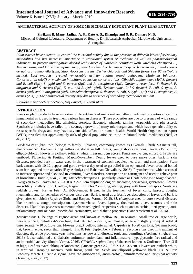
International Journal of Advance and Innovative Research Volume 6, Issue 1 (XVI): January - March, 2019
323
ISSN 2394 - 7780
ANTIBACTERIAL ACTIVITY OF SOME MEDICINALLY IMPORTANT PLANT LEAF EXTRACT
Shrikant B. Mane, Jadhao A. S., Kate A. S., Dhandge and S. R., Damare N. D. Microbial Cultural Laboratory, Department of Botany, Dr. Babasaheb Ambedkar Marathwada University,
Aurangabad
ABSTRACT Plant extract have potential to control the microbial activity due to the presence of different kinds of secondary metabolites and has immense importance in traditional system of medicine as well as pharmacological industries. In present investigation alcohol leaf extract of Gardenia resinifera Roth. Michelia champaca L., Tecoma stans, and Gliricidia sepium Jacq., tested against five human pathogenic bacteria viz., Pseudomonas aeruginosa, Salmonella typhi, Staphylococcus aureus, Escherichia coli and Shigella flexneri by 96 well-plate method. Leaf extracts revealed remarkable activity against tested pathogens. Minimum Inhibitory Concentration (MIC) or maximum inhibitions at various concentrations, Gliricidia sepium have MIC S. flexneri and E. coli (6µl), S. typhi and S. aureus (2µl) and P. aeruginosa (4µl). Gardenia rasenifera: S. flexneri, P. aurginosa and S. Areues (2µl), E. coli and S. typhi (4µl). Tecoma stans: 2µl S. flexneri, E. coli, S. typhi, S. areues (4µl) and P. aurginosa (4µl). Michelia champaca: S. flexneri, E. coli, S. typhi (4µl) and P. aurginosa, S. areues (2, 4µl). The antibacterial activity may due to presence of secondary metabolites in plant.
Keywords: Antibacterial activity, leaf extract, 96 - well plate
INTRODUCTION Plants or plant products have important different kinds of medicinal and ethno medicinal properties since time immemorial as it used in treatment various human diseases. These properties are due to presence of wide range of secondary metabolites like alkaloids, steroids, flavonoid, phenols, tannins, terpenoids and phytosterol. Synthetic antibiotics have failed to discourage growth of many microorganisms which have genetic ability to resist specific drugs and may have saviour side effects on human health. World Health Organization report (WHO) revealed that approximately 80% of global population relies on traditional herbal medicines (Neel, et al., 2017).
Gardenia resinifera Roth. belongs to family Rubiaceae, commonly known as Dikemali. Shrub 2-3 meter tall, much-branched, Frequent along gullies on slopes in hill forests, young shoots resinous, leaves6-15 3-5 cm, elliptic-oblong,. Flower is axillary, solitary, white, fragrant, 3cm across. Fruits are 3-2 cm, ellipsoid. Fruits are unribbed. Flowering & Fruiting: March-November. Young leaves used to cure snake bites, bark in skin diseases, pounded bark in water used in the treatment of stomach troubles, heartburn and constipation. Stem bark extract with 10-12 pepper and garlic twice a day used to get relief from ephemeral fever; crushed boiled stem bark applied to treat caries (Ray Anindya and Rahaman Chowdhury, 2018). Phytochemical which are used to increase appetite and also used in vomiting, liver disorders, constipation as astringent and used to relieve pain of bronchitis (Hindole, et al., 2018). Michelia champaca L. popularly known as Chafa belongs to Magnoliaceae. Evergreen trees, Leaves are 6.5-20.0 X 3.2-7.0 cm elliptic-oblong or lanceolate, coriaceous, glabresent. Flowers are solitary, axillary, bright yellow, fragrant, follicles 2 cm long, oblong, grey with brownish spots. Seeds are reddish brown. Fls. & Frts.: April-September. It used in the treatment of fever, colic, leprosy, coughs, rheumatism and for remedies of various disorders. Bark is used as a febrifuge, decoction of leaves and bark is given after childbirth (Rajshree Sinha and Ranjana Varma, 2016). M. champaca used to cure several diseases like bronchitis, cough, constipation, dysmenorrhoea, fever, leprosy, rheumatism, ulcer, wounds and skin diseases. Plant also possesses numerous pharmacological properties such as anti-microbial, anti-pyretic, anti-inflammatory, anti-oxidant, insecticidal, carminative, anti-diabetic properties (Panneerselvam et al., 2016).
Tecoma stans L. belongs to Bignoniaceae and known as Yellow Bell in Marathi. Small tree or large shrub, Leaves pinnate; petioles 4-5 cm long; leaf lets 3-7, opposite, acuminate, acute and slightly unequal sided at base, serrate, 7-10 X 1.5-2 cm. Flowers bright yellow, interminal. Capsules is 10-20 cm long, 5-6 mm broad, flat, brown, acute, seeds thin, winged. Fls. & Frts: September - February. Tecoma stans used in treatment of diabetes, digestive problems, yeast infections, as powerful diuretic, tonic and vermifuge (Archana Singh, et al., 2013). It also exhibited anti-diabetic, antitumor, antioxidant, anti-inflammatory, hypoglycemic, free radical and antimicrobial activity (Sunita Verma, 2016). Gliricidia sepium Jacq. (Fabaceae) known as Undirmari, Trees 3-5 m high, Leaflets ovate-oblong or lanceolate, glaucous green 2.2 - 8.6 X 1.3 - 3.5 cm. Flowers are pinkish-white, in terminal. Drooping racemes, pods linear, pendulous. Seeds are ellipsoid yellowish black. Fls. & Frts: February-March. Gliricidia sepium have the antibacterial, antimicrobial, antiinflametor and larvisidal activity (Jasmine, et al., 2017).

International Journal of Advance and Innovative Research Volume 6, Issue 1 (XVI): January - March, 2019
324
ISSN 2394 - 7780
Leaves decoction used in utricaria, burns and erysepalas. Bark decoction used against bacterial and protozoal infections. Branches used to reduce fever in children and adults (Beena Jose and Joji Reddy, 2010).
MATERIAL AND METHODS Plant material of Gliricidia sepium, Gardenia rasenifera, Tecoma stans and Michelia champaca were collected from Dr. Babasaheb Ambedkar Marathwada University campus Aurangabad, Maharashtra and identified by using floristic literature (Singh, et al., 2000; Naik, 1996; Naik, 1998).
Preparation of extracts: Leaves sample were shade dried and crush with the help of mortar and pestle to make fine powder and filter through muscline cloth. 5gm fine powder were extracted in a Soxhlet apparatus, at 650C for 18-24 hrs alcohol used as solvent and crude extract were stored in amber coloured bottle to study antibacterial activity.
Test microorganisms: Authentic human pathogenic bacteria culture of Salmonella typhi, Pseudomonas aeruginosa, Shigella flexneri, E. coli and Staphylococcus aureus were collected from department of Botany, Vivekanand Arts, Sardar Dalipsingh Commerce & Science College, Samarth Nagar, Aurangabad (M.S).
Antibacterial Assay: 100μl sterile Mueller-Hinton broths was added in each well of 96-well plates, followed by 2μl serial diluted human pathogenic bacteria suspension, various concentrations of alcohol crude extract of selected plant sample such as 2, 4, 6, 8 and 10μl was loaded in each well, control was prepared by nutrient broth and bacterial suspension without adding plant extract. Prepared 96-well plate was sealed with parafilm and incubated at 37°C for 24 hours in incubator. Optical density were measured on spectrophotometer at 540 nm (Jadhao and Bhuktar, 2017; Kendrekar, et al., 2018).
RESULTS AND DISCUSSION Alcoholic leaf extract of Gliricidia sepium in various concentrations were tested against five different human pathogenic bacteria and results revealed that maximum inhibition and minimum inhibitory concentration (MIC) of S. flexneri and E. coli (6μl), whereas S. typhi and S. arueus (2μl) and (4 μl) for P. aeruginosa (table 1, graph 1). Alcoholic leaf extract of Gardenia rasenifera in various concentrations were tested against five different human pathogenic bacteria and results revealed that maximum inhibition and minimum inhibitory concentration (MIC) of S. flexneri, P. aeruginosa and S. arueus (2μl), while for E. coli and S. typhi (4μl) (table 2, graph 2).
Alcoholic leaf extract of Tecoma stans in various concentrations were tested against five different human pathogenic bacteria and results revealed that maximum inhibition and minimum inhibitory concentration (MIC) for S. flexneri, S. arueus E. coli an S. typhi (2μl) while in case of P. aeruginosa it was recorded as (4μl) (table 3, graph 3). Alcoholic leaf extract of Michelia champaca in various concentrations were tested against five different human pathogenic bacteria and results revealed that maximum inhibition and minimum inhibitory concentration (MIC) for S. flexneri, E. coli an S. typhi (4μl) while for S. arueus and P. aeruginosa it was recorded as (2, 4μl) (table 4, graph 4).
Table no-1: Antibacterial activity of Gliricidia sepium leaf extract. Sr. No Name of Bacteria 2 µl 4 µl 6 µl 8 µl 10 µl MIC
1 S. flexneri 0.15 0.19 0.09 0.20 0.29 6 µl 2 E. coli 0.23 0.21 0.19 0.27 0.27 6 µl 3 S. typhi 0.20 0.24 0.35 0.30 0.35 2 µl 4 P. aurginosa 0.27 0.20 0.33 0.28 0.46 4 µl 5 S. areues 0.23 0.24 0.41 0.32 0.33 2 µl
Graph-1: Antibacterial activity of Gliricidia sepium leaf extract
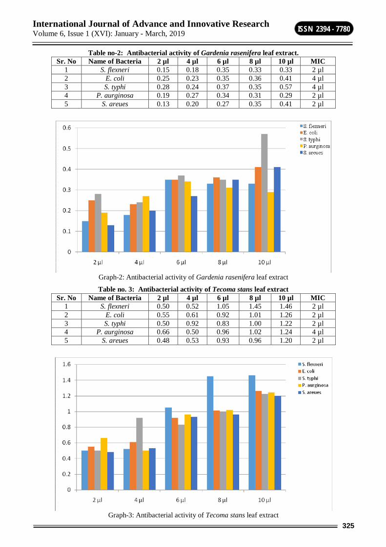
International Journal of Advance and Innovative Research Volume 6, Issue 1 (XVI): January - March, 2019
325
ISSN 2394 - 7780
Table no-2: Antibacterial activity of Gardenia rasenifera leaf extract. Sr. No Name of Bacteria 2 µl 4 µl 6 µl 8 µl 10 µl MIC
1 S. flexneri 0.15 0.18 0.35 0.33 0.33 2 µl 2 E. coli 0.25 0.23 0.35 0.36 0.41 4 µl 3 S. typhi 0.28 0.24 0.37 0.35 0.57 4 µl 4 P. aurginosa 0.19 0.27 0.34 0.31 0.29 2 µl 5 S. areues 0.13 0.20 0.27 0.35 0.41 2 µl
Graph-2: Antibacterial activity of Gardenia rasenifera leaf extract
Table no. 3: Antibacterial activity of Tecoma stans leaf extract Sr. No Name of Bacteria 2 µl 4 µl 6 µl 8 µl 10 µl MIC
1 S. flexneri 0.50 0.52 1.05 1.45 1.46 2 µl 2 E. coli 0.55 0.61 0.92 1.01 1.26 2 µl 3 S. typhi 0.50 0.92 0.83 1.00 1.22 2 µl 4 P. aurginosa 0.66 0.50 0.96 1.02 1.24 4 µl 5 S. areues 0.48 0.53 0.93 0.96 1.20 2 µl
Graph-3: Antibacterial activity of Tecoma stans leaf extract
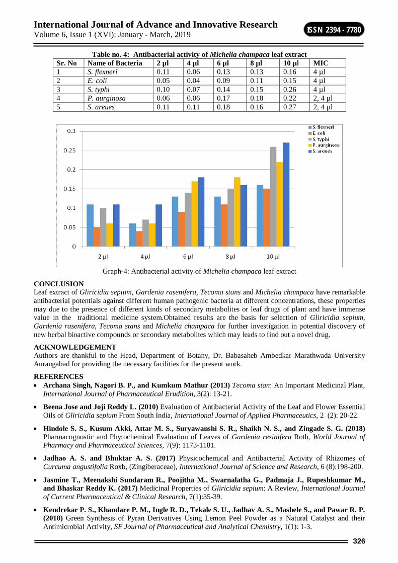
International Journal of Advance and Innovative Research Volume 6, Issue 1 (XVI): January - March, 2019
326
ISSN 2394 - 7780
Table no. 4: Antibacterial activity of Michelia champaca leaf extract Sr. No Name of Bacteria 2 µl 4 µl 6 µl 8 µl 10 µl MIC 1 S. flexneri 0.11 0.06 0.13 0.13 0.16 4 µl 2 E. coli 0.05 0.04 0.09 0.11 0.15 4 µl 3 S. typhi 0.10 0.07 0.14 0.15 0.26 4 µl 4 P. aurginosa 0.06 0.06 0.17 0.18 0.22 2, 4 µl 5 S. areues 0.11 0.11 0.18 0.16 0.27 2, 4 µl
Graph-4: Antibacterial activity of Michelia champaca leaf extract
CONCLUSION Leaf extract of Gliricidia sepium, Gardenia rasenifera, Tecoma stans and Michelia champaca have remarkable antibacterial potentials against different human pathogenic bacteria at different concentrations, these properties may due to the presence of different kinds of secondary metabolites or leaf drugs of plant and have immense value in the traditional medicine system.Obtained results are the basis for selection of Gliricidia sepium, Gardenia rasenifera, Tecoma stans and Michelia champaca for further investigation in potential discovery of new herbal bioactive compounds or secondary metabolites which may leads to find out a novel drug.
ACKNOWLEDGEMENT Authors are thankful to the Head, Department of Botany, Dr. Babasaheb Ambedkar Marathwada University Aurangabad for providing the necessary facilities for the present work.
REFERENCES Archana Singh, Nagori B. P., and Kumkum Mathur (2013) Tecoma stan: An Important Medicinal Plant,
International Journal of Pharmaceutical Erudition, 3(2): 13-21.
Beena Jose and Joji Reddy L. (2010) Evaluation of Antibacterial Activity of the Leaf and Flower Essential Oils of Gliricidia sepium From South India, International Journal of Applied Pharmaceutics, 2 (2): 20-22.
Hindole S. S., Kusum Akki, Attar M. S., Suryawanshi S. R., Shaikh N. S., and Zingade S. G. (2018) Pharmacognostic and Phytochemical Evaluation of Leaves of Gardenia resinifera Roth, World Journal of Pharmacy and Pharmaceutical Sciences, 7(9): 1173-1181.
Jadhao A. S. and Bhuktar A. S. (2017) Physicochemical and Antibacterial Activity of Rhizomes of Curcuma angustifolia Roxb, (Zingiberaceae), International Journal of Science and Research, 6 (8):198-200.
Jasmine T., Meenakshi Sundaram R., Poojitha M., Swarnalatha G., Padmaja J., Rupeshkumar M., and Bhaskar Reddy K. (2017) Medicinal Properties of Gliricidia sepium: A Review, International Journal of Current Pharmaceutical & Clinical Research, 7(1):35-39.
Kendrekar P. S., Khandare P. M., Ingle R. D., Tekale S. U., Jadhav A. S., Mashele S., and Pawar R. P. (2018) Green Synthesis of Pyran Derivatives Using Lemon Peel Powder as a Natural Catalyst and their Antimicrobial Activity, SF Journal of Pharmaceutical and Analytical Chemistry, 1(1): 1-3.

International Journal of Advance and Innovative Research Volume 6, Issue 1 (XVI): January - March, 2019
327
ISSN 2394 - 7780
Naik, V. N. (1996) Flora of Osmanabad district,
Naik, V. N. (1998) Flora of Marathwada, Vol. II, Amrut Prakashan,
Panneerselvam Pradeepa, Vedha Hari, Narayanan B. and Ramya Devi Durai (2016), Pharmacological and Medicinal Potential from Flowers of Perfume Tree M. champaca – A Review, International Journal of Pharmacognosy and Phytochemical Research, 8(11); 1896-1900
Rajshree Sinha and Ranjana Varma, (2016) Michelia champaca L. (Swarna Champa): A Review, International Journal of Enhanced Research in Science, Technology & Engineering, 5 (8): 78-82.
Ray Anindya Sundar and Rahaman Chowdhury Habibur (2018) Pharmacognostic, Phytochemical and Antioxidant Studies of Gardenia latifolia Aiton: An Ethnomedicinal Tree Plant, International Journal of Pharmacognosy and Phytochemical Research, 10(5); 216-228
Singh N. P., Karthikeyan S. and Lakshminarsimhan P. (2000) Flora of Maharashtra state Vol. – I, BSI, Culcutta.
Sunita Verma (2016) Phytochemical and pharmacological review study on Tecoma Stans Linn., Journal of Medicinal Plants Studies, 4 (5): 162-164.

International Journal of Advance and Innovative Research Volume 6, Issue 1 (XVI): January - March, 2019
328
ISSN 2394 - 7780
AN EFFICIENT SYNTHESIS OF TRIAZOLOQUINAZOLINONES AND BENZIMIDAZOLOQUINAZLINONES PROMOTED BY MAGNESIUM SULPHATE AS A GREEN ALTERNATIVE
Somnath S. Gholap, Vinod R. Kadu and Nilesh D. Gaikawad Post Graduate, Department and Research Centre, Padmashri Vikhe Patil College, Pravaranagar, Ahmednagar
ABSTRACT An environmentally benign approach for the synthesis of structurally diverse triazoloquinazolinones and benzimidazoloquinazolinones derivatives has been presented via one pot multi-component reaction (MCR). The reaction of dimedone, 3-amino-1,2,4-triazole or 2-amino-benzimidazole in presence of anhydrous magnesium sulphate in ethanol under reflux conditions has been reported. The corresponding triazoloquinazolinone and benzimidazoloquinazolinone derivatives are obtained in good to excellent yields under optimized reaction conditions. An economically viable catalyst, environment friendliness and ease of purification are the main features of the present method.
Keywords: Dimedone; aldehyde; 2-aminobenzimidazole; 5-amino-1(H)-1,2,4-triazole; magnesium sulphate; triazoloquinazolinones; benzimidazoloquinazolinones.
GRAPHICAL ABSTRACT
INTRODUCTION Quinazoline and its derivatives are well known for their promising biological potential such as antimalarial, anti-tumor, analgesic, anti-inflammatory, anti-pyretic, anti-microbial, anti-convulsant, fungicidal and anti-depressant activities [1-9]. The quinazolinone alkaloids (I) and as several other members of this alkaloid family such as luotonin A (II) [10, 11] and fumiquinazoline A (III) [12] has cytotoxic properties, pyrimidinone (IV) has HIV integrase inhibitor [13] activity, compound (V) has HIF prolyl hydroxylase inhibiting properties [14, 15], benzimidazoquinazolinone (VI) is known for immune-suppressive potential [16] and quinazolinone (VII) is a potential antiproliferative agent [17] (Figure 1).
Owing the biological importance of quinazolinone derivatives, a great deal of efforts has been made since 19th century. Kappe et al has been reported the microwave assisted synthesis of triazole fused pyrimidine-6-carboxamides, pyrimidobenzimidazoles and benzimidazo-quinazoline via multi-component reaction (MCR) of 3-aminotriazole or benzimidazole, aldehyde and 1,3-dicarbonyl compounds [18]. Furthermore, Mourad and Shao independently reported the microwave assisted synthesis of theses class of compounds as modified protocol [19, 20]. Other modified protocols comprising heteropoly acid H6P2W18O62 [21], NH2SO3H [22], nanocatalysts [23-26], ionic liquid [27,28] and ultrasound assisted synthesis [29] are reported so far. Although numerous methods are reported in literature, every method has its own inadequacies viz. prolonged reaction time, expensive catalysts and lack of catalyst biodegradability. In the view of current scenario of development of Green Chemistry, the application of environmentally safe and economically feasible catalysts for multi-component reaction has great demand.
Magnesium sulphate is cheap, easily available, non-toxic and biodegradable commonly used as dehydrating agent. The role of magnesium sulphate as catalyst for organic transformation is limited or very few reports are documented in literature as synthesis of bis-(indolyl)methanes [30], phenazine and quinoxaline derivatives [31] and intermolecular Wittig reaction of dialkyl-2-(1-acetyl-2-oxopropyl)-3-(triphenylphosphoranylidene) succinates with ninhydrin [32]. Herein, we have first time reported the synthesis of structurally diverse triazoloquinazolinones and benzimidazoloquinazolinones derivatives via one pot multi-component reaction of dimedone, 3-amino-1,2,4-triazole or 2-amino-benzimidazole in the presence of anhydrous magnesium sulphate in ethanol under optimized reaction conditions.
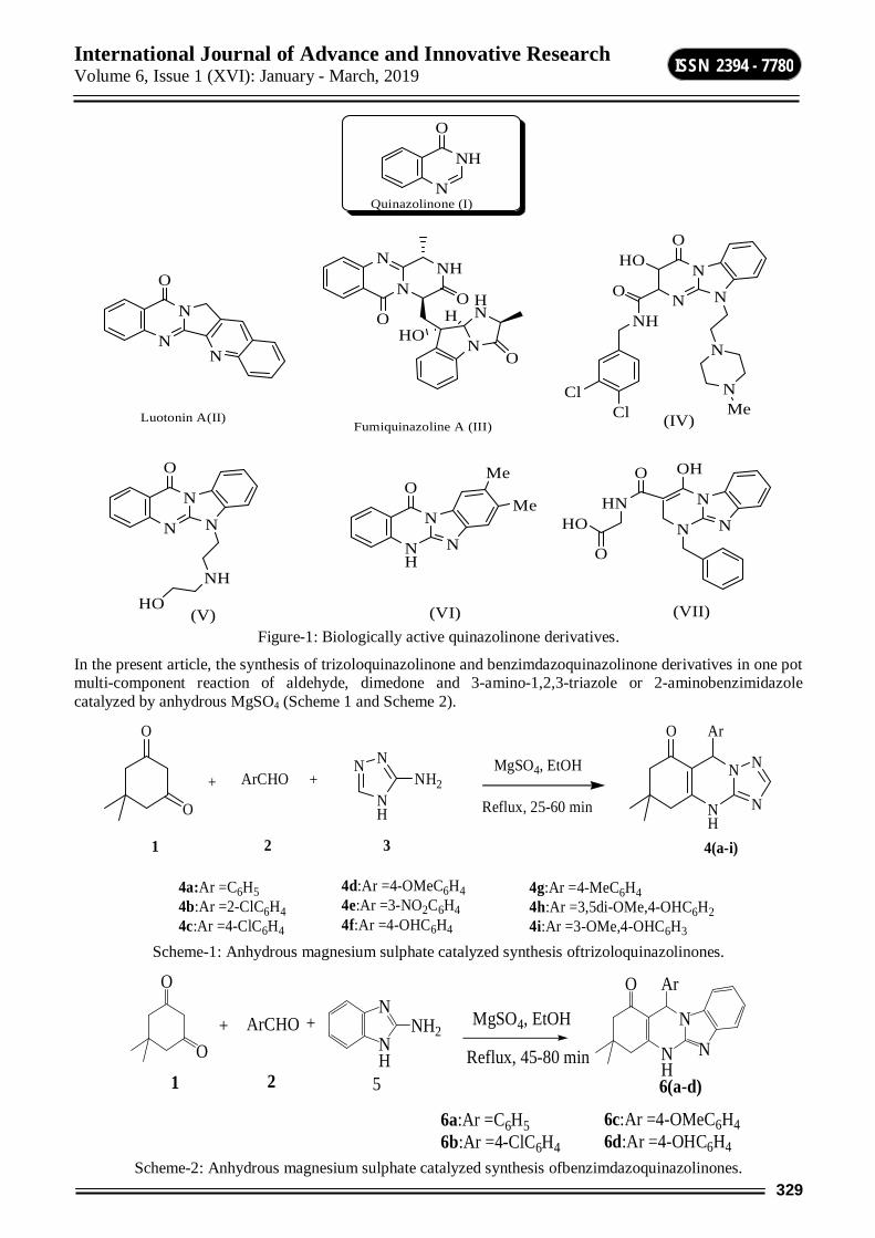
International Journal of Advance and Innovative Research Volume 6, Issue 1 (XVI): January - March, 2019
329
ISSN 2394 - 7780
N
N
O
N
N
N NH
O
N
HN
O
OHO
H
Luotonin A(II)Fumiquinazoline A (III)
NH
N
N
OMe
MeN
N
N
O
NHHO (VII)
N
N
N
OHO
O
NH
ClCl
N
NMe
N
N
N
OH
HN
O
HO
O
(IV)
(V)
N
NH
O
Quinazolinone (I)
(VI) Figure-1: Biologically active quinazolinone derivatives.
In the present article, the synthesis of trizoloquinazolinone and benzimdazoquinazolinone derivatives in one pot multi-component reaction of aldehyde, dimedone and 3-amino-1,2,3-triazole or 2-aminobenzimidazole catalyzed by anhydrous MgSO4 (Scheme 1 and Scheme 2).
O
O
+ ArCHO +N
NH
NNH2
O
NH
N
N
N
Ar
1 2 3 4(a-i)
4g:Ar =4-MeC6H44h:Ar =3,5di-OMe,4-OHC6H24i:Ar =3-OMe,4-OHC6H3
MgSO4, EtOH
Reflux, 25-60 min
4a:Ar =C6H54b:Ar =2-ClC6H44c:Ar =4-ClC6H4
4d:Ar =4-OMeC6H44e:Ar =3-NO2C6H44f:Ar =4-OHC6H4
Scheme-1: Anhydrous magnesium sulphate catalyzed synthesis oftrizoloquinazolinones.
6c:Ar =4-OMeC6H46d:Ar =4-OHC6H4
O
O+ ArCHO +
O
1 2 6(a-d)
NH
NNH2
NH
N
Ar
N
5
MgSO4, EtOH
Reflux, 45-80 min
6a:Ar =C6H56b:Ar =4-ClC6H4
Scheme-2: Anhydrous magnesium sulphate catalyzed synthesis ofbenzimdazoquinazolinones.
International Journal of Advance and Innovative Research Volume 6, Issue 1 (XVI): January - March, 2019
330
ISSN 2394 - 7780
RESULT AND DISCUSSION In continuation to our ongoing research for the development of new methods for the synthesis of biologically active compounds[33-43], herein the synthesis of trizoloquinazolinone and benzimdazoquinazolinone derivatives was conducted via three-component reaction of aromatic aldehydes, dimedone and 3-amino-1,2,3-triazole or 2-aminobenzimidazole in the presence of anhydrous MgSO4 as a green alternative (Scheme 1 and Scheme 2). Hitherto, the application of magnesium sulphate as catalyst for the present multi-component reaction was not reported. In order to study the catalytic efficiency of MgSO4, the reaction of benzaldehyde, dimedone and 3-amino-1,2,3-triazole was conducted in the presence of varied amount of catalyst as 5, 10, 15 and 20 mol%. It has been found that 20 mol% amount of catalyst was sufficient to get satisfactory yields of the desired products ‘4a’and ‘6a’ (Table 1, Entry 1, 10). To study the generality and scope of the present method, reaction was carried out with structurally diverse aldehydes (Table 1). The aldehydes possessing electron withdrawing groups accelerated the reaction and the products were obtained in excellent yields (Table 1, Entry 2, 4, 5 and 11). Whereas, aldehydes with electron donating substituents undergo slow reactions and products obtained slightly less yields (Table 1, Entry 6, 7, 11 and 13). The results obtained were compared with reported methods and represented in Table 2. Further, representative compounds were characterized by IR, 1H-NMR and MASS spectral data and physical constants were compared with literature values.
EXPERIMENTAL Materials and Methods: All reagents were AR grade and used as such. Melting points were recorded in open-capillary in paraffin bath and are uncorrected. The completion of reaction was judged by Thin Layer Chromatography (TLC) technique using petroleum ether-ethyl acetate (4.5:0.5mL) solvent system. IR spectra were recorded using Perkin-Elmer FT spectrophotometer with KBr pallets. 1HNMR were recorded on 300 MHz Varian-NMR-mercury 300 instrument in DMSO-d6 as a solvent.
General procedure for the preparation of triazoloquinazolinones 4(a-i)
To a mixture of dimedone (1 mmol), aldehyde (1 mmol) and 3-amino-1,2,4-triazole (1 mmol) in ethanol (3 mL), anhydrous magnesium sulphate (20 mol%) was added .The reaction mixture was heated under reflux for specified time (Table 1). After completion of reaction (as indicated by TLC), reaction mixture was filtered and cooled in ice bath afforded crystalline products good to excellent yields.
Spectral data for representative compound 4a: IR = 3416, 3225, 3030, 2958, 2837, 1648, 1581, 1548, 1473, 1551, 1414, 1369, 1334, 1245, 703, 697 cm-1; 1HNMR (DMSO-d6, δ ppm) = 0.96 (s, 3H), 1.04 (s, 3H), 2. 04-2.24 (q, J J = 16.14Hz, 2H), 2.50-2.60 (m, 2H), 6.19 (s, 1H), 7.19 (d, J = 8.1Hz, 2H), 7.33 (d, J= 8.1Hz, 2H), 7.70 (s, 1H), 11.19 (s,1H); Mass (C17H17N4OCl) = 329.29 (MW=329.5).
4h: IR: 3550, 3414, 3212, 3120, 3019, 2960, 2837, 1610, 1590, 1550, 1480, 1465, 1350, 1258, 831, 736, 680 cm-1; 1HNMR = 0.91 (s, 3H), 1.01 (s, 3H), 2.05-2.28 (q, J = 16.2 Hz, 2H), 2.48-2.53 (m, 2H),3.64 (s,3H), 3.65(s, 3H), 4.4(s,1H), 6.09 (s,1H), 7.66 (s, 1H), 7.66 (s,1H), 8.16 (s,1H), 8.16 (s,1H), 8.27 (s,1H), 11.04 (s,1H); Mass: (C19H22O4N4) = 369.33 (MW=370).
4i: IR= 3548,3430, 3310, 3252, 3015, 2961, 2840, 1616, 1588, 1549, 1471, 1251, 841, 731, 690 cm-1; 1HNMR = 0.91 (m, 2H), 1.01 (s, 3H), 4.40 (s, 1H), 6.08 (s, 1H), 6.56-6.65 (m, 2H), 7.65 (s,1H), 8.73 (s, 1H),11.02 (s, 1H); Mass (C18H20O3N4) = 341.18 (MW=340.0)
General procedure for the synthesis of benzimidazoquinazolinones 6(a-d) To a mixture of dimedone (1 mmol), aldehyde (1 mmol), 2-amionbenzimidazole (1 mmol) in ethanol (3 mL), anhydrous magnesium sulphate (20 mol%) was added. The reaction mixture was heated under reflux for specified time (Table 1). After completion of reaction (as indicated by TLC), reaction mixture was filtered and cooled in ice bath afforded crystalline benzimidazoquinazolinone in good to excellent yield.
Spectral data for representative compounds, 6a: IR=3405, 3330, 3255, 3107, 3021, 2950, 2817, 1622, 1578, 1535, 1419, 1212, 740, 688cm-1; 1HNMR = 0.86 (s, 3H), 1.01 (s, 3H), 2.09-2.23 (m, 2H), 2.44-2.63 (m, 2H), 6.14 (s, 1H), 6.86-7.02 (m, 5H), 7.10-7.16 (m, 2H), 7.22-7.37 (m, 2H), 11.09 (s, 1H); Mass (C22H21ON3) =344.02(MW=343).
6c: IR = 3417, 3300, 3235, 3010, 2966, 2845, 1620, 1590, 1542, 1468, 1266, 807, 736, 686cm-1; 1HNMR = 0.88 (s, 3H), 1.01 (s, 3H), 2.14-2.26 (m, 2H), 2.42-2.66 (m, 2H), 6.31(s,1H), 6.89-7.04 (m, 2H),11.09 (s, 1H); Mass(C12H20ON32Cl)=376.81(MW=377.5).
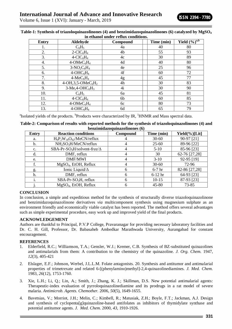
International Journal of Advance and Innovative Research Volume 6, Issue 1 (XVI): January - March, 2019
331
ISSN 2394 - 7780
Table-1: Synthesis of triazoloquinazolinones (4) and benzimidazoquinazolinones (6) catalyzed by MgSO4 in ethanol under reflux conditions.
Entry Aldehyde Compound Time (min) Yield (%)a,b 1. C6H5 4a 40 80 2. 2-ClC6H4 4b 55 93 3. 4-ClC6H4 4c 30 89 4. 4-OMeC6H4 4d 40 80 5. 3-NO2C6H4 4e 25 96 6. 4-OHC6H4 4f 60 72 7. 4-MeC6H4 4g 45 77 8. 4-OH,3,5-OMeC6H2 4h 30 83 9. 3-Me,4-OHC6H3 4i 30 90 10. C6H5 6a 45 81 11. 4-ClC6H4 6b 60 85 12. 4-OMeC6H4 6c 80 73 13. 4-OHC6H4 6d 65 79
aIsolated yields of the products. bProducts were characterized by IR, 1HNMR and Mass spectral data.
Table-2: Comparison of results with reported methods for the synthesis of triazoloquinazolinones (4) and benzimidazoquinazolinones (6)
Entry Reaction conditions Compound Time (min) Yield(%)[Lit] a. H6P2W18O62/MeCN/reflux 4 30-60 90-97 [21] b. NH2SO3H/MeCN/reflux 4 25-60 89-96 [22] c. SBA-Pr-SO3H/solvent-free/Δ 4 5-10 85-96 [23] d. DMF, reflux 4 30 62-76 [27,28] e. DMF/MWI 4 3-10 92-95 [19] f. MgSO4, EtOH, Reflux 4 30-60 72-96 g. Ionic Liquid/Δ 6 6-7 hr 82-86 [27,28] h. DMF, reflux 6 6-12 hr 64-93 [23] i. SBA-Pr-SO3H, reflux 6 10-15 87-93 [23] j. MgSO4, EtOH, Reflux 6 45-80 73-85
CONCLUSION In conclusion, a simple and expeditious method for the synthesis of structurally diverse triazoloquinazolinone and benzimidazoquinazolinone derivatives via multicomponent synthesis using magnesium sulphate as an environment friendly and economically viable catalyst has been reported. The method offers several advantages such as simple experimental procedure, easy work up and improved yield of the final products.
ACKNOWLEDGEMENT Authors are thankful to Principal, P.V.P College, Pravaranagar for providing necessary laboratory facilities and Dr. C. H. Gill, Professor, Dr. Babasaheb Ambedkar Marathwada University, Aurangabad for constant encouragement.
REFERENCES 1. Elderfield, R.C.; Williamson, T.A.; Gensler, W.J.; Kremer, C.B. Synthesis of BZ-substituted quinazolines
and antimalarials from them: A contribution to the chemistry of the quinazoline. J. Org. Chem. 1947, 12(3), 405-421
2. Elslager, E.F.; Johnson, Werbel, J.L.L.M. Folate antagonists. 20. Synthesis and antitumor and antimalarial properties of trimetrexate and related 6-[(phenylamino)methyl]-2,4-quinazolinediamines. J. Med. Chem. 1983, 26(12), 1753-1760.
3. Xie, L.H.; Li, Q.; Lin, A.; Smith, J.; Zhang, K. J.; Skillman, D.S. New potential antimalarial agents: Therapeutic-index evaluation of pyrroloquinazolinediamine and its prodrugs in a rat model of severe malaria. Antimicrob. Agents. Chemother. 2006, 50(5), 1649-1655.
4. Bavetsias, V.; Morriot, J.H.; Melin, C.; Kimbell, R.; Matusiak, Z.H.; Boyle, F.T.; Jackman, A.J. Design and synthesis of cyclopenta[g]quinazoline-based antifolates as inhibitors of thymidylate synthase and potential antitumor agents. J. Med. Chem. 2000, 43, 1910-1926.
International Journal of Advance and Innovative Research Volume 6, Issue 1 (XVI): January - March, 2019
332
ISSN 2394 - 7780
5. Khalil, A.A.; Adbel-Hamide, S.G.; Al-Obiad, A.M.; EI-Subbagh, H.I. Substituted quinazolines, Part 2. Synthesis and in-vitro anticancer evaluation of new 2-substituted mercapto-3H-quinazoline analogs. Arch. Der Pharm. 2003, 2, 95-103.
6. Al-Omary, F.A.M.; Hassan, G.S.; El-Messery, S.M.; El-Subbagh, H.I. Substituted thiazoles V. Synthesis and antitumor activity of novel thiazolo[2,3-b]quinazoline and pyrido[4,3-d]thiazolo[3,2-a]pyrimidine analogues. Eur. J. Med. Chem. 2012, 47, 65-72.
7. Bekhit, A.A.; Khalil, M.A. Non-steroidal anti-inflammatory agents: synthesis of novel benzopyrazolyl, benzoxazolyl and quinazolinyl derivatives of 4(3H)-quinazolinones. Pharmazie, 1998, 53, 539-543.
8. Daidone G.; Maggio, B.; Raffa, D.; Plescia, C.; Bajardi, M.L.; Caruso, A.; Cutuli, V.M.; Amico-Roxas, C.M. Synthesis and pharmacological study of ethyl 1-methyl-5-[2-substituted-4-oxo-3(4H)-quinazolinyl]-1H-pyrazole-4-acetatesEur. J. Med. Chem. 1994, 29, 707-711.
9. Fetter, J.; Czuppon, T.; Hornyk, G. Feller, A. The synthesis of some 3-amino-2-halomethyl-, 2-halomethyl-3-(subst.amino)- and 2-halomethyl-3-hetarylquinazolin-4(3H)-ones as potential plant protecting agents. Tetrahedron 1991, 47, 9393-9410.
10. Ma, Z. Z.; Hano, Y.; Nomura,T.; Chen, Y. J. Novel quinazoline-quinoline alkaloids with cytotoxic and DNA topoisomerase II inhibitory activities. Bioorg. Med. Chem. Lett. 2004, 14, 1193-1196.
11. (a) Ma, Z.; Hano, Y.; Nomura, T.; Chen, Y. Two New Pyrroloquinazolinoquinoline Alkaloids from Peganum nigellastrum Heterocycles 1997, 46, 541-546. (b) Cagir, A.; Jones, S. H.; Gao, R.;Eisenhauer, B. M.; Hecht, S. M. Luotonin A. A Naturally Occurring Human DNA Topoisomerase I Poison. J. Am. Chem. Soc. 2003, 125, 13628-13629. (c) Cagir, A.; Jones, S. H.; Eisenhauer, B. M.;Gao, R.; Hecht, S. M. Synthesis and biochemical properties of E-ring modified luotonin A derivatives. Bioorg. Med. Chem. Lett. 2004, 14, 2051-2054.
12. (a) Numata, A.; Takahashi, C.; Matsushita, T.; Miyamoto, T.; Kawai, K.; Usami, Y.; Matsumura, E.; Inoue, M.; Ohishi, H.; Shingu, T. Fumiquinazolines, novel metabolites of a fungus isolated from a saltfish. Tetrahedron Lett. 1992, 33, 1621-1624. (b) Takahashi, C.; Matsushita, T.; Doi, M.; Minoura, K.; Shingu, T.; Kumeda, Y.; Numata, A. Fumiquinazolines A–G, novel metabolites of a fungus separated from a Pseudolabrus marine fish. J. Chem. Soc., Perkin Trans. 11995, 2345-2353.
13. Dalla Via, L.; Gia, O.; Marciani Magno, S.; Da Settimo, A.; Marini, A. M.; Primofiore, G.; Da Settimo, F.; Salerno, S. Synthesis, in vitro antiproliferative activity and DNA-interaction of benzimidazoquinazoline derivatives as potential anti-tumor agents. Farmaco 2001, 56, 159-167.
14. Jones, E. D.; Coates, J. A. V.; Rhodes, D. I.; Deadman, J. J.; Vandegraff, N. A.; Winfield, L. J.; Thienthong, N.; Issa, W.; Choi, N.; MacFarlane, K. Preparation of bicyclic pyrimidinones and analogues for the treatment of viral infections, particularly HIV infections. WO 2008077188, 2008.
15. Chai, D.; Fitch, D. M. Prolyl hydroxylase inhibitors. WO 2009039322, 2009.
16. Neochoritis, C. G.; Zarganes-Tzitzikas, T.; Tsoleridis, C. A.; Stephanidou-Stephanatou, J.; Kontogiorgis, C. A.; Hadjipavlou-Litina, D. J.; Choli Papadopoulou, T. One- pot microwave assisted synthesis under green chemistry conditions, antioxidant screening, and cytotoxicity assessments of benzimidazole Schiff bases and pyrimido[1,2-a] benzimidazol-3(4H)-ones. Eur. J. Med. Chem. 2011, 46, 297-306.
17. Hubschwerlen, C.; Pflieger, P.; Specklin, J. L.; Gubernator, K.; Gmunder, H.; Angehrn, P.; Kompis, I. Pyrimido[1, 6- a] benzimidazoles: a new class of DNA gyrase inhibitors. J. Med. Chem. 1992, 35, 1385-1392.
18. Chebanov, V. A.; Muravyova, E. A.; Desenko, S. M.; Musatov, V. I.; Knyazeva, I. V.; Shishkina, S. V.; Shishkin, O. V.; Kappe, C. O. Microwave-assisted three- component synthesis of 7-aryl-2-alkylthio-4,7-dihydro-1,2,4-triazolo[1,5-a]- pyrimidine- 6-carboxamides and their selective reduction. J. Comb. Chem. 2006, 8, 427-434.
19. Maurad, E.A.F.; Aly, A.A.; Farag, H.H.; Beshr, A.E. Microwave assisted synthesis of triazoloquinazolinones. Beilstein J. Org. Chem. 2007, 3, 1-5.
20. Shao, Q.; Tu, S.; Li, C.; Zhou, D.; Wang, Q.; Hao, W. Green and high efficient synthesis of triaza-benzo[b]fluoren-6-one derivatives in water under microwave irradiation. J. Heterocycl. Chem. 2008, 45, 411-416.
International Journal of Advance and Innovative Research Volume 6, Issue 1 (XVI): January - March, 2019
333
ISSN 2394 - 7780
21. Heravi, M. M., Oskooie, H. A.; Ranjbar, L.; Derikvand, F.; Alimadadi, B.; Bamoharran, F. A three component one-pot procedure for the synthesis of [1,2,4]triazolo/benzimidazolo-quinazolinone derivatives in the presence of H6P2W18O62·18H2O as a green and reusable catalyst. Mol. Diversity 2008, 1, 181-185.
22. Heravi, M. M.; Derikvand, F.; Ranjbar, I. Sulfamic acid catalyzed, three-component, one-pot synthesis of [1,2,4]triazolo/benzimidazolo quinazolinone derivatives. Synth. Commun. 2010, 40, 677-685.
23. Mohammadi, Z. G.; Badiei, A. R.; Haddadpour, M. Application of sulfonic acid functionalized nanoporous silica (SBA-Pr-SO3H) for one-pot synthesis of quinoxaline derivatives. Int. J. Chem. 2011, 3, 87.
24. Mohammadi, Z.G.; Badiei, A.R.; Khaniana, Y.; Haddadpour, M. One pot synthesis of polyhydroquinolines catalyzed by sulfonic acid functionalized SBA-15 as a new nanoporous acid catalyst under solvent free conditions. Iran. J. Chem. Eng. 2010, 29, 1-10.
25. Kureshy, R. I.; Ahmad, I.; Pathak, K.; Khan, N. H., Abdi, S. H. R.; Jasra, R. A. Sulfonic acid functionalized mesoporous SBA-15 as an efficient and recyclable catalyst for the synthesis of chromenes from chromanols. Catal. Commun. 2009, 10, 572-575.
26. Ziarani, G. M.; Badiei. A.; Aslani, Z.; Lashgari, N.; Application of sulfonic acid functionalized nanoporous silica (SBA-Pr-SO3H) in the green one-pot synthesis of triazoloquinazolinones and benzimidazoquinazolinones. Arabian J. Chem. 2015, 8, 54-61.
27. Lipson, V.V.; Desenko, S.M.; Shirobokova, M.S.; Boradina, V.V. Synthesis of 9-Aryl-6,6-dimethyl-5,6,7,9-tetrahydro-1,2,4-triazolo[5,1-b]quinazolin-8(4H)ones. Chem. Heterocycl. Compound. 2003, 39, 1213-1217.
28. Lipson, V.V.; Desenko, S.M.; Shirobokova, M.S.; Shishkin, O.V.; Orlov, V.D. Cyclocondensation of 2-aminobenzimidazole with dimedone and its arylidene derivatives. Chem. Heterocycl. Compd. 2003, 39, 1041-1047.
29. Li-Hsun Chen,Tsai-Wen Chung,Bharat D. Narhe,Chung-Ming Sun, a novel mechanistic study on ultrasound-assisted, one-pot synthesis of functionalized benzimidazo[2,1-b]quinazolin-1(1h)-ones.ACS Comb. Sci. 2016, 18, 162-169.
30. Hasaninejad A.; Parhami Zare A.; Khalafi-Nezhad A.A.; Nasrolahi Shirazi A.; Moosavi Zare A.R. magnesium sulfate as an efficient and very cheap reagent for the preparation of bis(indolyl)methanes. Pol. J. Chem. 2008, 82, 565.
31. Karami, B.; Khodabahshi, S. A facile synthesis of phenazine and quinoxaline (new 1,4-benzo diazine) derivatives using magnesium sulfate heptahydrate as a catalyst. J. Serb. Chem. Soc. 2011, 76(9), 1191-1198.
32. Ramazani A.; Noshiranzadeh, N. Magnesium sulfate catalyzed intermolecular Wittig reaction of dialkyl 2-(1-acetyl-2-oxopropyl)-3-(triphenylphosphoranylidene) succinates with ninhydrin in solvent-free conditions. N., Phosphorus, Sulfur, and Silicon and the Related Elements, 2003, 178(6), 1321-1324.
33. Gholap, S.S.; Dhakane, V.D.; Gholap, Sandip S. solid-supported dichloro[1,3,5]-triazine: a versatile synthetic auxiliary for direct synthesis of N-sulfonylamines from sulphonic acid and amine,Jordan J. Chem., 2012, 7(3), 279-285.
34. Gholap, S.S. Pyrrole: An emerging scaffold for construction of valuable therapeutic agents. Eur. J. Med. Chem. 2016, 110, 13-31.
35. Gholap, S.S.; Deshmukh, U.P. Synthesis and in-vitro antimicrobial screening of 3-cinnamoyl coumarin and 3-[3-(1H-indol-2-yl)-3-aryl-propanoyl]-2H-chromen-2-ones. Iranian J. Catal. 2013, 3, 171-176.
36. Gholap, S.S.; Gunjal,N. 2,4,6-Trichloro-1,3,5-triazine (TCT) mediated one pot direct synthesis of N-benzoylthioureas from carboxylic acids. Arabian J. Chem. 2013, http://dx.doi.org/10.1016/j.arabjc. 2013.10.021
37. Gholap, S.S.; Bandgar, B.P.; Chavan, H.V.; Dhakane, V.D.; Deshmukh, U.P. An efficient and green method for the synthesis of [1,3]oxazine derivatives catalyzed by thiamine hydrochloride (VB1) in water. C. R. Chim. 2014, 17, 431-436.
38. Gholap S.S.; Gunjal N. Thiamine hydrochloride (Vit-B1): An optimized green alternative for the synthesis of polyhydroquinoline derivatives. Iranian J. Catal. 2016, 8, 6(2), 147-152
International Journal of Advance and Innovative Research Volume 6, Issue 1 (XVI): January - March, 2019
334
ISSN 2394 - 7780
39. Shelke, S.N.; Pawar, Y.J.; Pawar, S.B., Gholap, S.S., Gill, C.H. Ultrasonicated Synthesis of 1-(2-Hydroxyaryl)-3-(pyrrolidin-1-yl)propenones and Their Antimicrobial Screening. J. Indian Chem Soc., 2012, 43, 3.
40. Gholap S, Tambe M, Nawale L,;Sarkar D, Sangshetti J, Damale M. Design, synthesis, and pharmacological evaluation of fluorinated azoles as anti-tubercular agents. Arch Pharm Chem Life Sci.2017,1-14.
41. Gholap S. S. An Efficient Gram Scale Synthesis of Aryl Iodides from Aryl Diazofluoroborates in Water under Mild Conditions. Lett. Org. Chem.2018, 15, 594.
42. Gholap S. S., Ugale S. R. A Total Synthesis of the Cyclic Depsipeptide Chaiyaphumine A.ChemSelect. 2017, 2, 24, 7445.
43. Gholap S. S.; Ugale, S. R. An efficient synthesis of structurally diverse 2-methyl-N-[(3-phenylamino)oxetan-3-yl]-2-propanesulfinamide derivatives under catalyst free conditions.Chem. Pap. 2017, 71(12), 3435-3443.
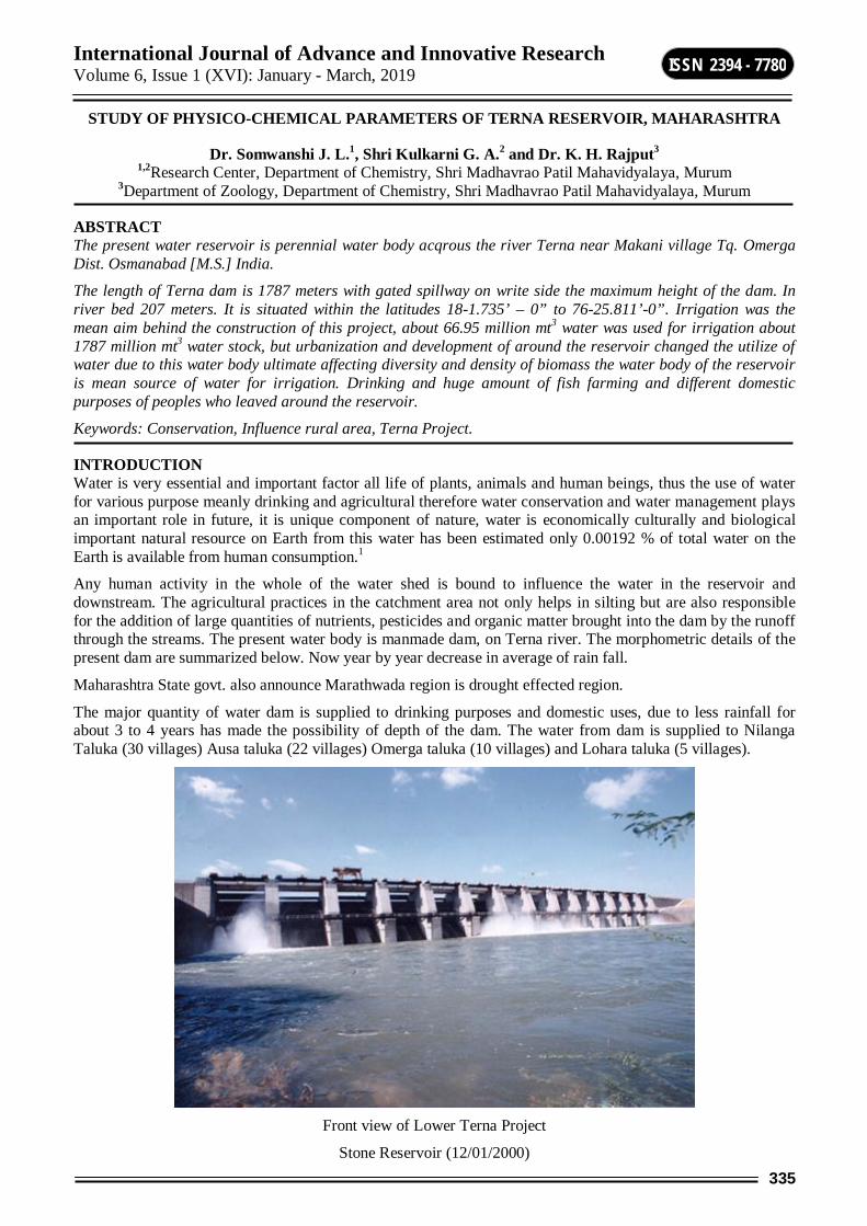
International Journal of Advance and Innovative Research Volume 6, Issue 1 (XVI): January - March, 2019
335
ISSN 2394 - 7780
STUDY OF PHYSICO-CHEMICAL PARAMETERS OF TERNA RESERVOIR, MAHARASHTRA
Dr. Somwanshi J. L.1, Shri Kulkarni G. A.2 and Dr. K. H. Rajput3 1,2Research Center, Department of Chemistry, Shri Madhavrao Patil Mahavidyalaya, Murum
3Department of Zoology, Department of Chemistry, Shri Madhavrao Patil Mahavidyalaya, Murum
ABSTRACT The present water reservoir is perennial water body acqrous the river Terna near Makani village Tq. Omerga Dist. Osmanabad [M.S.] India.
The length of Terna dam is 1787 meters with gated spillway on write side the maximum height of the dam. In river bed 207 meters. It is situated within the latitudes 18-1.735’ – 0” to 76-25.811’-0”. Irrigation was the mean aim behind the construction of this project, about 66.95 million mt3 water was used for irrigation about 1787 million mt3 water stock, but urbanization and development of around the reservoir changed the utilize of water due to this water body ultimate affecting diversity and density of biomass the water body of the reservoir is mean source of water for irrigation. Drinking and huge amount of fish farming and different domestic purposes of peoples who leaved around the reservoir.
Keywords: Conservation, Influence rural area, Terna Project.
INTRODUCTION Water is very essential and important factor all life of plants, animals and human beings, thus the use of water for various purpose meanly drinking and agricultural therefore water conservation and water management plays an important role in future, it is unique component of nature, water is economically culturally and biological important natural resource on Earth from this water has been estimated only 0.00192 % of total water on the Earth is available from human consumption.1
Any human activity in the whole of the water shed is bound to influence the water in the reservoir and downstream. The agricultural practices in the catchment area not only helps in silting but are also responsible for the addition of large quantities of nutrients, pesticides and organic matter brought into the dam by the runoff through the streams. The present water body is manmade dam, on Terna river. The morphometric details of the present dam are summarized below. Now year by year decrease in average of rain fall.
Maharashtra State govt. also announce Marathwada region is drought effected region.
The major quantity of water dam is supplied to drinking purposes and domestic uses, due to less rainfall for about 3 to 4 years has made the possibility of depth of the dam. The water from dam is supplied to Nilanga Taluka (30 villages) Ausa taluka (22 villages) Omerga taluka (10 villages) and Lohara taluka (5 villages).
Front view of Lower Terna Project
Stone Reservoir (12/01/2000)
International Journal of Advance and Innovative Research Volume 6, Issue 1 (XVI): January - March, 2019
336
ISSN 2394 - 7780
MATERIALS AND METHODS Water samples were collected in the morning the month June 2013 to March 2014. The physic-chemical parameters like water temperature. pH, turbidity, electric conductivity at the sampling sites.
According to APHA (1980), Kodarkar (1992) and Trivedi 1986, Goel P. K., Trisal C. L. (1998) method, T.D.S., chlorides, Alkalinity hardness, sulphates, dissolved oxygen, Nitrates, phosphates, carbonates were analyzed in the laboratory.
RESULT AND DISCUSSION The Terna reservoir was under the investigations for the water analysis for about one year June 2013 to March 2014. In the investigation temperature is differs by 2 to 60C. throughout the study period.
The pH of the reservoir is alkaline ranging between 8.0 to 8.6. the volume of the water affected by rain fall and use for the irrigation, drinking for the population of about different villages in addition to natural processes like percolation evaporation.
The parameters like turbidity, T.D.S. were co-related to each other. Turbidity ranged between 21.1 to 143 cm. The T.D.S. value ranged between 100 to 285 mg/litre and the conductivity during the study is in limit. The parameter like dissolved oxygen is the indication of respiration to photosynthesis ratio in the water body. Dissolved oxygen value ranged between 6 to 9 mg/litre.
The total alkalinity of the water from the present water body ranged between 130 to 250 mg/litre. The total hardness was higher in winter and lower in summer. It was 20.1 to 110 mg/litre. Due to Ca++ and Mg++ ions. The chlorides of the water is ranged between 17 to 35 mg/litre during summer it is high and low during winter. The overall result is in limits this indicates water body is unpolluted. Phosphates, sulphates, nitrates are present in very less amount that is within the permissible limits.
Studies on the quality criteria for different uses indicates that the present water body is healthy with different parameters within the permissible limits, but scarce rainfall and drought for long duration, exploitation of water from the water body for different purposes is creating strain on the amount of water volume in the water body.
SILENT FEATURE OF TERNA PROJECT
1 Place of reservoir Maharashtra State, Dist. Osmanbaad Tq. Lohara Village – Makani Latitude – 18-1.735’-0” Longitude – 76-25.811’-0”
2 Water shed area 1787 Sq. Kilometer
3 Type of Dam
Main dam Soil dam Cannal Construction of cement
4 Dead stock 29.967 cmc Live storage 91.221 cmc
Projected storage 160.460cmc 5 Length of canal 207 meter 6 Area under irrigation 3188 7 Crop water requirement 66.95 cmc
8 Overall efficiency at canal release/ H. R. of eh project 14513/66.95=216 cmc
REFERENCES 1) APHA (1995) standard methods for examination of water sewage and industrial waste. 10th edition American
water works Association. New York.
2) Indian standard (IS:2490) (1981) domestric water specification, BIS, New Delhi.
3) Trivedy R. K., Goel P. K. Trisal, C. L. (1998).
4) Kodarkar M. S. (1995), conservation of Lakes IAAB Publication no. 3, Hyderabad.
5) Kodarkar M. S. (1998), Methodology for water analysis IAAB Publication, Hyderabad.
International Journal of Advance and Innovative Research Volume 6, Issue 1 (XVI): January - March, 2019
337
ISSN 2394 - 7780
MEASURING ACCURACY AND PERFORMANCE OF TEXT-TO-SPEECH BY SUBJECTIVE AND OBJECTIVE METHODS
Suhas Mache1 and Sunil Nimbhore2 1Department of Computer Science, R. B. Attal Arts, Science & Commerce College, Georai, Dist. Beed
2Department of Computer Science & IT, Dr. Babasaheb Ambedkar Marathwada University, Aurangabad
ABSTRACT The aim of testing and evaluation of the TTS system is to determine the system performance and accuracy. Text-To-Speech (TTS) framework is to change over a subjective given content into a comparing talked waveform or fake creation of human discourse. This paper presents a TTS system for Pali language that uses concatenation using unit selection method. According to the standard measurement of speech and voice, our result analysis follows the standard measurement of speech and voice. The evaluation methods were designed to check the system accuracy and speech quality i.e. measuring the intelligibility of synthesized speech by Mean Opinion Score (MOS) test; Speaking Rate Test. The test result shows the overall accuracy of the TTS system is excellent and capable of generating natural sounding synthesized speech.
Keywords: Text-To-Speech, unit selection, speech quality, intelligibility, MOS
INTRODUCTION Text to speech system (TTS) converts text into voice using a speech synthesizer it is the artificial creation of human dialogue [1]. In recent years a lot of research is going on speech synthesis. Speech plays important role in day to day life communication. Speech synthesis i.e. Text-To-Speech is the method of converting the written content into machine-generated artificial speech [2]. We have selected concatenative unit selection method to develop Text-To-Speech (TTS) synthesis for Pali language. Concatenative speech synthesis systems read a text and render into speech by joining pre-recorded speech units to each other [3]. The Unit selection based corpus method is bulky corpus methods use to select the speech units and concatenate.
We have designed and developed an intelligible and natural sounding corpus-based concatenative speech synthesis system for Pali language. The implemented system is divided into two sections the front-end deals with text processing [1] and back end speech generation. The inputted text is first analyzed, normalized and transcribed into a phonetic representation [12]. The unit selection algorithm is based on the best path in the network of the units [4]. The second section back-end of the system is responsible for speech waveform generation. In this work, the different unit sizes such as vowels, consonants, syllables, digits, and words have experimented. In unit selection based concatenative speech synthesis, joint cost also known as concatenative cost, which measures how well two units can be joined together [5][6].
METHODOLOGY The aim of testing and evaluation of the TTS system is to determine the system performance and accuracy. It also used to judge the speech quality in terms of its similarity to the human voice and by its ability to be understood.
Test Data Test speech data plays a vital role in testing process and it effect on overall test outcomes. Test data is satisfactory sufficient to cover all the functionalities of the system under test. Different functionalities of the TTS system can be evaluated by investigative in general output speech. It should be designed in such a way that it covers all possible variations including numerals, vowels, consonants, words and connected words.
The Text-to-Speech system is evaluated by three different methods i.e. Objective Test, Subjective Test and Acoustic Measurements of speech. The objective test contains Accuracy Test and subjective test intelligibility test by mean opinion score [7].
Accuracy Test To conduct accuracy tests proper selection of test data is important. All such data whose predictable output is well defined can be measured for accuracy test [8]. For Accuracy measure, it just checks the pronunciation of total correct data such as numerals (digits), vowels, consonant and words with a total number of the text of the above input. The formula is
100
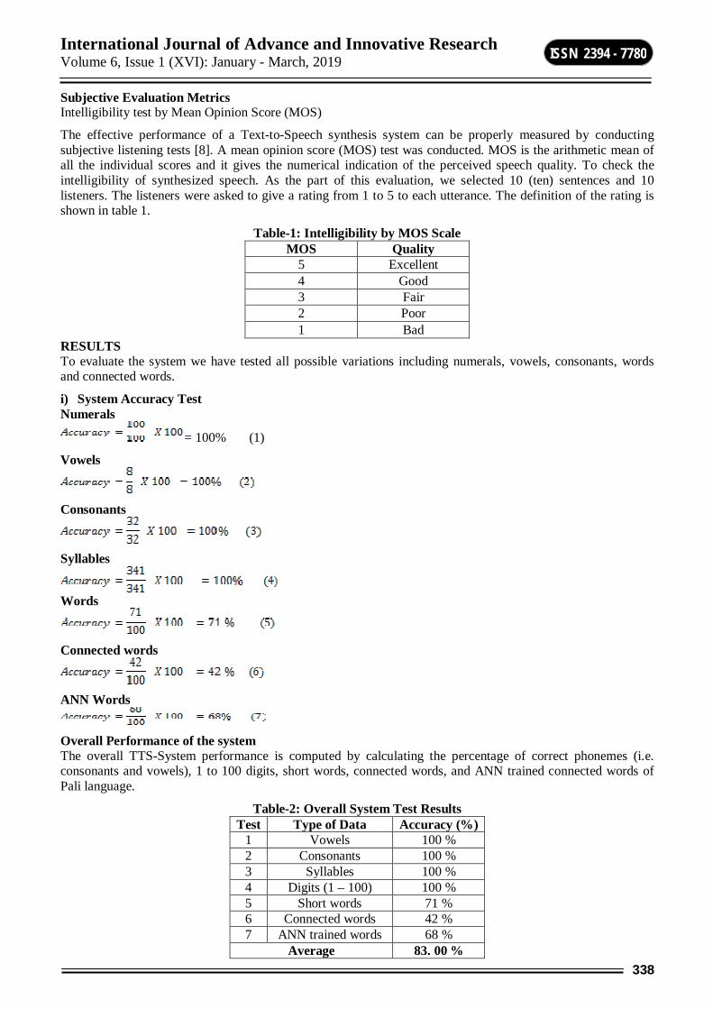
International Journal of Advance and Innovative Research Volume 6, Issue 1 (XVI): January - March, 2019
338
ISSN 2394 - 7780
Subjective Evaluation Metrics Intelligibility test by Mean Opinion Score (MOS)
The effective performance of a Text-to-Speech synthesis system can be properly measured by conducting subjective listening tests [8]. A mean opinion score (MOS) test was conducted. MOS is the arithmetic mean of all the individual scores and it gives the numerical indication of the perceived speech quality. To check the intelligibility of synthesized speech. As the part of this evaluation, we selected 10 (ten) sentences and 10 listeners. The listeners were asked to give a rating from 1 to 5 to each utterance. The definition of the rating is shown in table 1.
Table-1: Intelligibility by MOS Scale MOS Quality
5 Excellent 4 Good 3 Fair 2 Poor 1 Bad
RESULTS To evaluate the system we have tested all possible variations including numerals, vowels, consonants, words and connected words.
i) System Accuracy Test Numerals
= 100% (1)
Vowels
Consonants
Syllables
Words
Connected words
ANN Words
Overall Performance of the system The overall TTS-System performance is computed by calculating the percentage of correct phonemes (i.e. consonants and vowels), 1 to 100 digits, short words, connected words, and ANN trained connected words of Pali language.
Table-2: Overall System Test Results Test Type of Data Accuracy (%)
1 Vowels 100 % 2 Consonants 100 % 3 Syllables 100 % 4 Digits (1 – 100) 100 % 5 Short words 71 % 6 Connected words 42 % 7 ANN trained words 68 %
Average 83. 00 %

International Journal of Advance and Innovative Research Volume 6, Issue 1 (XVI): January - March, 2019
339
ISSN 2394 - 7780
All these tests show that the accuracy of the developed TTS system is 83.00 %.
Subjective evaluation (Listing Tests MOS) The effective performance of a Text-to-Speech synthesis system in terms of similarity with human voice can be properly measured by conducting subjective listening tests [9]. i.e. Mean Opinion Score (MOS) test was conducted. While objective measures are useful in comparing detailed system characteristics, the effective performance of a Text-to-Speech synthesis system can be properly measured by conducting subjective listening tests [9]. This test finding the relationships between intelligibility and comprehensibility in speech synthesizers and tries to design an appropriate comprehension task for evaluating the speech synthesizers' comprehensibility [10,11]. A mean opinion score (MOS) test was conducted. MOS is the arithmetic mean of all the individual scores and it gives the numerical indication of the perceived speech quality. To check the intangibility of synthesized speech. A part of this evaluation, we selected 10 (ten) sentences and 10 listeners to check the quality of speech and give rating from 1 to 5 to each utterance. The definition of rating was 1-bad, 2-poor, 3-fair, 4-good, and 5-excellent. Table 3 shows the Scale of Mean Opinion Score and Table 3 shows Mean Opinion Score (MOS) Test result.
Table-3: Mean Opinion Score Test (MOS) Sentence L1 L2 L3 L4 L5 L6 L7 L8 L9 L 10 MOS
S_1 4 3 4 4 5 4 5 5 4 4 4.2 S_2 3 4 4 4 3 4 4 4 4 4 3.8 S_3 4 4 4 3 4 5 4 4 3 5 4 S_4 4 4 4 4 4 4 3 4 4 5 4 S_5 4 3 3 5 4 4 5 3 4 5 4 S_6 5 4 5 4 5 5 4 5 5 5 4.7 S_7 4 3 4 4 4 4 3 3 4 4 3.7 S_8 3 4 3 4 3 4 3 4 3 4 3.5 S_9 4 3 4 3 4 4 4 3 4 4 3.7 S_10 4 4 3 4 3 3 3 4 4 4 3.6
Average 3.92
In the above table, L1 to L10 are ten different listeners and S_1 to S_10 are ten different sentences. The above MOS test shows average result 3.92, here we conclude that synthesized speech near to good intangibility.
c. Speaking Rate Test The rate of speaking can be defined as the number of syllables or words speak by system per second. The average duration of a syllable is around 250 – 300 ms i.e. 2 – 3 syllables per second. If system’s speaking rate is higher than this range it becomes sloppy speech and slower than normal range leads to elongation of duration of syllables. The table 4 shows speaking rate of system.
Table-4: Speaking Rate Test Type of data Speaking Rate
Syllable 2 -3 / second Word (short) 1 word / 0.7 second Word (long) 1 word / second
CONCLUSION The evaluation methods were designed to check the system accuracy and speech quality. The evaluation has been done at several levels, such as digits, vowels, consonants and words level. The overall test result shows the accuracy of the developed Text-To-Speech system is 83%. Here we have concluded that the Text to Speech conversion provides very good accuracy.
A subjective listing test for measuring the intelligibility of synthesized speech by Mean Opinion Score (MOS) test was also conducted. The MOS test gives 3.92 scores; this numerical indication shows the perceived speech quality is in a good range. The Speaking Rate Test also has been conducted. The test shows the rate of speaking in the number of syllables or words produced (spoken) by the system per second.
REFERENCES [1] Mache, Suhas R., Manasi R. Baheti, and C. Namrata Mahender. "Review on text-to-speech
synthesizer." International Journal of Advanced Research in Computer and Communication Engineering 4.8 (2015): 540.
International Journal of Advance and Innovative Research Volume 6, Issue 1 (XVI): January - March, 2019
340
ISSN 2394 - 7780
[2] Dutoit, Thierry. An introduction to text-to-speech synthesis. Vol. 3. Springer Science & Business Media, 1997.
[3] Tatham, Mark, and Katherine Morton. Developments in speech synthesis. John Wiley & Sons, 2005.
[4] Black, Alan W., and Nick Campbell. "Optimising selection of units from speech databases for concatenative synthesis." (1995).
[5] Vepa, Jithendra, and Simon King. "Subjective evaluation of join cost and smoothing methods for unit selection speech synthesis." IEEE Transactions on audio, speech, and language processing 14.5 (2006): 1763-1771.
[6] Vepa, Jithendra, and Simon King. "Subjective evaluation of join cost and smoothing methods." (2004).
[7] Klatt, Dennis H. "Review of text to speech conversion for English." The Journal of the Acoustical Society of America82.3 (1987): 737-793.
[8] TDIL, Text to Speech Testing Strategy Version 2.1 (2014) pp. 1-46
[9] Rosenberg, Andrew, and Bhuvana Ramabhadran. "Bias and statistical significance in evaluating speech synthesis with mean opinion scores." In Proceedings of the 18th Annual Conference of the International Speech Communication Association (Interspeech 2017), Stockholm, Sweden, pp. 20-24. 2017.
[10] Chang, Yu-Yun. "Evaluation of TTS systems in intelligibility and comprehension tasks." proceedings of the 23rd Conference on Computational Linguistics and Speech Processing. Association for Computational Linguistics, 2011.
[11] Sunil S. Nimbhore, Rakesh J. Ramteke “Implementation of English-Text to Marathi-Speech (ETMS) Synthesizer”, at International Organization of Scientific, Journal of Computer Engineering (IOSR-JCE) e-ISSN: 2278-0661, p-ISSN: 2278-8727, Volume 17, Issue 1, Ver. VI (Jan – Feb. 2015), PP 34-43
[12] Mache, Suhas and C. Mahender, Namrata “Development of Text-to-Speech Synthesizer for Pali Language”, in IOSR Journal of Computer Engineering (IOSR-JCE) e-ISSN: 2278-0661, p-ISSN: 2278-8727, Volume 18, Issue 3, Ver. I (May-Jun. 2016), PP 35-42.
International Journal of Advance and Innovative Research Volume 6, Issue 1 (XVI): January - March, 2019
341
ISSN 2394 - 7780
ISOLATION, EXTRACTION OF COMMERCIALLY VALUABLE PROTEASE ENZYME BY USING AGRO-WASTE SUBSTRATE BY SOLID STATE FERMENTATION (SSF)
Syeda Tasleem S. Gani and Aithal S. C Research Center, Department of Microbiology, Dnyanopasak College, Parbhani, Maharashtra
ABSTRACT Solid state fermentation (SSF) is a process in which growth of microorganisms is carried out on solid substrates, in almost absence of free water. It is employed to produce a wide variety of enzymes and works out best with fungi as sources. Many advantages of SSF over conventional submerged culture have been reported. A protease (also termed peptidases/proteinase) is defined as enzyme that accomplishes proteolysis, which is protein catabolism by hydrolysis of the peptide bonds, linking amino acids, in the polypeptide chain, of any protein. Protease from fungal origin has dominated applications in industrial sectors. Fungal proteases can be produced in large quantities in short time by SSF cost effectively, and have advantage of faster production. The results in present study employing SSF and two fungi, exhibited maximum enzyme production, on groundnut oil cake waste material. Production was enhanced with increasing in pH towards neutrality; maximum protease activity for Aspergillus niger was observed at 0.108 mol/ml at pH 7 and with Penicillium it was 0.195 mol/ml being pH-6. The Protease activity was assayed at different temperatures and a maximum production at 40°C for Aspergillus niger and 30°C for Penicillium was recorded. Fungal protease activity with respect to time showed maximum enzyme activity of A. niger at 5 minutes incubation period while Penicillium showed maximum enzyme activity at 25 minutes incubation time.
Keywords: Solid state fermentation (SSF); Enzyme protease; groundnut oil cake waste
INTRODUCTION Enzyme production is demand of present time, annual world scale figures are close to a billion dollars with increasing number of patents and research articles related to this field. Production of commercially valuable enzyme by SSF is increasing day by day mainly by using fungal strains (Sharma, et al, 2015). SSF employing fungi has greater advantages over other techniques. However, unlike other microorganisms, these typically grow in solid substrates. Advantages of SSF over traditionally submerged cultures for work involving fungi include: (a) minimalism of equipment, (b) low moisture content, prevents bacterial contamination (c) superior volumetric output, (d) use of economical substrates, (d) simpler downstream processing, (e) lower energy constraints and flow wastewater output (Battaglino, et al., 1991). Solid-substrate fermentation (SSF) has the potential for the higher protease yield (Pandey, et al., 1999). Economic point of view this type of fermentation possesses offers many advantages, including greater volumetric productivity, use of simpler equipment, and use of an economical substrate, simpler downstream processing, lower energy requirements and low waste water output (Malathi, et al., 1991).
MATERIALS AND METHODOLOGY Production of Protease Enzyme Proteases are protein-digesting enzymes that are chiefly classified based on their optimum pH as acidic, neutral, and alkaline proteases. Proteases account for approximately 60% of all worldwide sales (Rao, etal.,1998) because they have applications in a wide variety of industries, such as in detergents, pharmaceuticals, leather and food industries, cheese-making industry, and the wastewater processing industry. Proteases are the derivative enzymes, which catalyze the total hydrolysis of proteins. The total molecular weight of proteases ranges from 18-90 kDA (Ikram-ul-haq, et al., 1996). Proteases are the most important industrial enzymes accounting for about 60% of the total enzyme market (Charles, et al., 2008). According to Enzyme Commission (EC) classification, proteases belong to group 3 (hydrolases), and subgroup 4 (which hydrolyze peptide bond) (Alagarsamy, et al., 2006). Proteases are extracellular enzymes that can be produced by both submerged fermentation and solid-state fermentation (Chutmanop, et al., (2008). Present study was aimed to produce Protease enzyme from 1) black sorghum bran, 2) groundnut oil cake 3) wheat bran using A.niger and Penicillium under SSF. Further effect of different parameters such as pH, temperature, and incubation time was checked on its activity.
Isolation and maintenance of fungal cultures Fungal strains isolated from garden soil of DSM College Parbhani were subjected to serial dilutions and those obtained from air by settle plate method, were screened for enzyme protease production. Fungi were cultivated on potato dextrose agar medium) at pH-5.0 on petri plates (90mm) containing potato dextrose agar (PDA). PDA was prepared with 400g boiled and mashed potatoes, dextrose (20gm) and agar (15gm), with a final adjusted pH

International Journal of Advance and Innovative Research Volume 6, Issue 1 (XVI): January - March, 2019
342
ISSN 2394 - 7780
of 5.0, all the ingredients were prepared in deionized water, and sterilized at temperature 15 lbs. pressure for 20 minutes in HEXATECH vertical automatic autoclave. The plates were incubation at room temperature for 48-72 hours. After incubation, black sporulated growth of fungi Aspergillus niger and green fungal growth of Penicillium were identified by using Cotton blue staining technique before using for enzyme production.
Inoculum development The spore suspension was prepared in sterile saline solution (0.85g of Nacl in 100ml of distilled water) and used to inoculate into inoculum media before employing it into SSF production media. A loopful was also streaked on PDA slants to maintain pure cultures. The inoculum media before inoculating it on to SSF consisted of Czapek Dox Broth containing Sucrose 30g/L., NaNO3 3 g/L., dipotassium phosphate 1 g/L., magnesium sulphate 0.50 g/L., potassium chloride 0.50 g/L., ferrous sulphate 0.01g/L. pH at 25°C: 7.3 ± 0.2). In this medium sucrose serves as the sole source of carbon, while sodium nitrate serves as source of nitrogen, dipotassium phosphate buffers the medium, magnesium sulphate, potassium chloride, ferrous sulphate serves as sources of essential ions.
Solid State Fermentation (SSF) Process Solid State Fermentation process, for extraction of crude enzyme, was carried out using three different types of locally available and economically feasible substrates. Wheat bran, black sorghum bran and groundnut oil cake waste, were collected from various regions in Parabhani Dist. Each substrate was taken in six separated Borosil make petri plates, and sterilized in oven at 110°C for 3 hours. An inoculum quantity of 10 ml was spread on each well-labeled, sterilized, substrate containing; Petri plates by using glass pipettes and then incubated at room temperature for 48-72 hours. After completion of incubation, and observing full-fledged growth of fungi in pure cu Culture of A.niger and Penicillium on Sorghum bran lture, were used it for extraction of crude enzymes.
Culture of A.niger and Penicilium on Wheat bran
Culture of A.niger and Penicilium on Peanut press cake waste
Culture of A.niger and Penicillium on Sorghum bran
International Journal of Advance and Innovative Research Volume 6, Issue 1 (XVI): January - March, 2019
343
ISSN 2394 - 7780
Extraction and Essay of Protease Enzyme Extraction of crude enzyme from each SSF plate was done in 1gm quantities by taking fungal colonies along with substrate aseptically with help of spatula and transferred to centrifugation tubes containing 1ml of phosphate buffer of pH-7 and subjecting to centrifugation in (REMI-R-20 research centrifuged machine) at 1000 rpm for 10 minutes. The supernatant were collected as crude enzyme and then purified by solvent extraction process with cold acetone. Its activity was compared with standard protein solution 10µg/ml of egg albumin. Preparative centrifuge was done in 50-100 batches for larger enzyme extractions.
Estimation by Folin Lowry’s method Enzyme activity was defined in terms of unit moles of product released by unit moles of purified enzyme, within unit time at unit concentration, with unit pH and unit temperature. The activity was done using Folin Lowry’s method. Folin Lowry Reagent was prepared by taking Alkaline Na COз as (Reagent A): Sodium carbonate 2g in 0.1N NaoH made up to 100ml volume with distilled water and CuSO4 reagent as (Reagent B) CuSO4.5H2O (0.5%) prepared in sodium-potassium tartrate solution (1%) in 10 ml distilled water. The working solution comprised of adding 1mL of Reagent-B to 50 ml of Reagent-A and estimating the enzyme substrate with Phenol red (Folin reagent) prepared by dissolving 10ml of Follin reagent into 30ml of distilled water.
For enzyme assay 200µl of enzyme extracted by SSF and 500µl of casein/egg albumin (1%Stock) and 300µl of 0.2mol/liter phosphate buffer (pH-7) was added. The reaction mixture was incubated at 60°c for 10 minutes and reaction was arrested by the addition of 1 ml of 10% trichloro acetic acid. The reaction mixture was centrifuged at 8000 rpm at 4°C for 15minutes and to the supernatant, 5 ml of 0.5mol/L Na COз were added, and 1ml of 3-fold diluted Folin and Ciocalteau’s phenol reagent was added. The solution was incubated at room temperature for 30min. and the absorbance of the blue color developed was read at 660nm. The concentration was determined using tyrosine standard curve. One unit of enzyme activity was defined as the amount of enzyme that liberated one microgram of tyrosine from substrate (casein) per minute under assay condition.
Standard stock solution of (Egg albumin) ranging from 10-100µg/ml were employed to prepare a standard graph. The absorbances were recorded at 660nm with spectrophotometer, and standard values of optical density v/s concentration of protein were studied.
Characterization of crude Enzyme Various parameters like pH, temperature and incubation time were studies and optimized. Temperature optimum of the enzyme protease was determined by incubating the mixture at different temperatures (10, 20, 30, 40, 50°C). The pH optimum of the protease enzyme was determined by using different buffer solution of different pH using the following buffer at 0.5M concentration: acetate (pH 4 and 6), phosphate (pH 5, 7, 8). Enzyme activity of protease was determined by varying incubation time as follows; (5, 10, 15, 20, 25, 30minutes).
RESULT AND DISCUSSION The selection of an agro industrial waste for the enzyme production in solid-state fermentation process depends upon several factors, mainly related with cost and availability of the substrate. The results in present study indicated that protease production varied with the types of agro wastes. This could be attributed to solid materials dual role supply of nutrients to the microbial cultures and anchorage for the growing cells. Maximum enzyme production was observed with groundnut oil cake waste material.
Protease production by microbial strains depends on the extracellular pH because culture pH strongly effluences many enzymatic processes and transport of various components across the cell membranes, which in turn support cell growth and product production. It is observed that particularly high or low pH values usually result in complete loss of activity of enzymes. A pH range of 4-10 was varied to estimate protease activity. The synthesis was increased with increase of pH of buffer towards neutrality, maximum (0.108 U /mol) for Aspergillus niger being at pH-7 and (0.195 U/mol) for Penicillium being at pH-6 and further increased resulted in decrease of enzyme activity (fig-4.1).The decrease may be due to the enzyme instability at pH other than its optimum; viz-7.0.
International Journal of Advance and Innovative Research Volume 6, Issue 1 (XVI): January - March, 2019
344
ISSN 2394 - 7780
Fig-4.3: Effect of pH on Enzyme Activity
The protease activity was assayed at different temperature ranging from 10°C to 50°C. Enzyme activity was increased up to 50°C for Aspergillus niger and 30°C for Penicillum and further increase in temperature decrease enzyme activity (fig-4.2) Hence optimum temperature was found to be 50°C for A. niger and 30°C for Penicillium. The reduction in denaturation by losing its catalytic property at high temperature due to stretching, breaking of weak hydrogen bonds within the enzyme structure.
Fig-4.3: Effect of temperature on Enzyme Activity
The protease enzyme activity obtained from all sources, carried out at times ranging from 5 minutes to 30 minutes respectively, showed a maximum activity at 25 minutes.
Fig-4.4: Effect of incubation time on
International Journal of Advance and Innovative Research Volume 6, Issue 1 (XVI): January - March, 2019
345
ISSN 2394 - 7780
CONCLUSION From present study, Penicillium showed remarkable potential to utilize groundnut oil cake waste a byproduct of oil industry as cost effective solid substrate for production of enzyme protease. In conclusion, the purified protease has an optimum activity (0.108 U /mol) for Aspergillus niger being at pH-7 and (0.195 U/ mol) for Penicillium being at pH-6, Optimum temperature was found to be 50°C for A. niger show optimum activity (0.390) and 30°C for Penicillium (0.168). Optimum incubation time for A.niger is 5 minute and 25 minutes for Penicillium. It is concluded that production of proteases via SSF processes using agro-industrial wastes/by-products and can be commercially used in various industry as it adds great value to agro-industrial wastes.
REFERENCES 1. Aidoo KE, Hendry R, Wood JB (1982) Solid substrate fermentations. Adv Appl Microbiol 28:201–237 2. Alagarsamy Sumantha, Paul Deepa, Chandra Sandhya, George Szakacs, CarlosRicardo (2006) Rice Bran
as a Substrate for Proteolytic Enzyme Production. Brazilian Archives of Biology and Technology; 49: 843-851
3. Aunstrup K (1980) Proteinases. In: Rose AH (ed) Economic microbiology, vol 5. Academic Press, London, pp 49–113
4. Battaglino, R.A., Huergo, M., Pilosof, A.M.R. et al. (1991) Culture requirements for the production of protease by Aspergillus oryzae in solid-state fermentation. Appl. Microbiol. Biotechnol. (1991) 35: 292.
5. Charles P, Devanathan V, Anbu P, Ponnuswamy MN, Kalaichelvan PT, Hur B. Purification, characterization and crystallization of an extracellular alkaline protease from Aspergillus nidulans HA-10. J Basic Microbiol. 2008;48(5):347–352
6. Choudhary V, Jain PC (2012). Screening of alkaline protease production by fungal isolates from different habitats of Sagar and Jabalpur district (M.P). J. Acad. Indus. Res., 1(4): 215-220
7. Chutmanop, J., S. Chuichulcherm, Y. Chisti and P. Srinophakun. 2008. Protease production by Aspergillus oryzae in solid-state fermentation using agroindustrial substrates. J. Chem Technol. Biotechnol. 83: 1012–1018.
8. Fukushima Y, Itoh H, Fukase T, Motai H (1989) Continuous protease production in a carbon-limited chemostat culture by salt tolerant Aspergillus oryzae. Appl Microbiol Biotechnol 30:604–608
9. Hesseltine CW (1987) Solid-state fermentation - an overview. Int Biodeterior 23:79–89 10. Ikram Haq and Mukhtar, Hamid (2008). Production of alkaline protease by Bacillus subtilis and its
application as a depilating agent in leather processing. Pak. Journ. Bot. 40(4) 11. Klapper BF, Jameson DM, Mayer RM (1973b) Factors affecting the synthesis and release of the
extracellular protease of Aspergillus oryzae NRRL 2160. Biochim Biophys Acta 304:513–519 12. Malathi, S and Chakraborty, R (1991) Production of Alkaline Protease by a New Aspergillus flavus Isolate
under Solid-Substrate Fermentation Conditions for Use as a Depilation Agent. Appl. Environ. Microbiol. 57(3):712-6.
13. Sailas Benjamin and Ashok Pandey (2001) Isolation and Characterization of Three Distinct Forms of Lipases from Candida rugosa Produced in Solid State. Fermentation. Braz. Arch. of Biol. Tech. Vol. 44, N. 2 : pp. 213 – 221.
14. Sailas Benjamin and Ashok Pandey (2001) Isolation and Characterization of Three Distinct Forms of Lipases from Candida rugosa Produced in Solid State Fermentation. Vol. 44, N. 2 : pp. 213 – 221
15. Rao MB, AM Tanksale, MS Ghatge, Deshpande VV (1998) Molecular and biotechnological aspects of microbial proteases. Microbiol. Mol. Biol. Rev. 62:597-635.
16. Sethi S, Gupta S (2015). Optimization of Protease Production from Fungi Isolated from soil. Int. J. Appl. Biol. Pharm. Technol., 6(3): 149-154.
17. Sharma AK, Sharma V, Saxena J, Yadav B, Alam A, Prakash A (2015). Isolation and Screening of Extracellular Protease Enzyme from Bacterial and Fungal Isolates of Soil. Int. J. Sci. Res. Environ. Sci., 3(9): 0334-0340.
18. Sinha P, Singh RK, Srivastva R, Sharma R, Tiwari SP (2013). Characterization and Optimization of Alkaline Protease Enzyme Produced by Soil Borne Bacteria. Trends Life Sci., 2(2): 38-46.
19. Ueno S, Miyama M, Ohashi Y, Izumiya M, Kusaka I (1987) Secretory enzyme production and conidiation of Aspergillus oryzae in submerged liquid culture. i 26:273–276
International Journal of Advance and Innovative Research Volume 6, Issue 1 (XVI): January - March, 2019
346
ISSN 2394 - 7780
ASSESSMENT OF ENDOPHYTIC DIVERSITY FROM DALBERGIA SISSOO FROM DIFFERENT REGIONS OF AURANGABAD (MAHARASHTRA)
Taware A. S. and Rajurkar S. K. Department of Botany, Deogiri College, Aurangabad
ABSTRACT Dalbergia sissoo collected from various regions of Aurangabad were collected and analysed for presence of endophytic fungi. Different localities like Beed bypass, Shendra, Osmanpura, Bidkin and Khultabad. Endophytic fungi from leaf, leaf with midrib, stem and petiole were isolated, identified and evaluated for their existence. A total 77 isolates belonging to 12 fungal taxa were obtained from 120 segments observed. Phoma putaminum was observed as the dominant endophyte fungi in total screened samples. Simpson's diversity index is low in Osmanpura region indicating higher endophytic diversity.
Keywords: Dalbergia sissoo, Phoma putaminum, Simpson's diversity
INTRODUCTION Dalbergia sissoo is economical important plant as well as has various medicinal uses. Various parts such as roots, bark, wood, leaves and seeds are used as for treatment of diseases including skin diseases, blood diseases, stomach problems, dysentery, nausea, eye and nose disorders, expectorant. The wood and bark for anal disorders, blood diseases, burning sensations, dysentery, dyspepsia, leucoderma, and skin ailments.(Shah et.al.2010)
Endophytic fungi reside within the living tissues of higher plants without producing any disease symptoms (Bills et.al.1992). A wide range of plants have now been examined for endophytes, and endophytes have been found in nearly all of them. An enormous number of different fungi can be isolated from plants growing in their native habitat. A few fungi are widely distributed with the host, suggesting a long standing, close and mutually beneficial interaction. Some fungi are found in many different terrestrial hosts,especially endophytes of crop plants. Although they are present in every plant, the extent of their contribution to fungal biodiversity remains unclear.
Since the discovery of the penicillin, soil microorganisms have been extensively prospected in the search of bioactive small molecules. However, endophytes, marine microorganisms, microorganisms from extreme environments and 53 microorganisms found in association with other microorganisms or insects have been less studied, and should be considered as under-explored sources of new useful natural products. Endophytes might be defined as microorganisms that can be detected at a given moment within the tissues of an apparently healthy plant host (Schulz and Boyle, 2005), and they have been found to produce a significant number of interesting natural products (Tan and Zou, 2001; Zhang et al.,2006).
As Dalbergia has many medicinal uses it is important to get knowledge about endophytic fungi associated with it. Hence the following work is done.Endophytic fungi from leaf, leaf with midrib, stem and petiole were isolated, identified and evaluated for their existence. Plants were collected from five different localities. A total 77 isolates belonging to 12 fungal taxa were obtained from 120 segments observed.
MATERIALS AND METHODS Collection of Samples Samples were collected from five different locations and were denoted as location 1, Zalta corner (Locl); location 2, the Shendra MIDC (Loc2); location 3, Osmanpura (Loc3), location 4 , Bidkin area (Loc 4) and location 5, Khultabad (Loc 5). Samples were labelled and collected, and each was assigned a code .All samples were immediately brought to the laboratory in sterile bags, and the tissues were screened for endophytic fungi.
Screening, Identification of Endophytes Plant parts were washed properly in running tap water for half an hour before processing. The samples were cut into small pieces. Leaves with midrib, leaves without midribs, petiole and stem samples were cut into 1.0 x 1.0 cm pieces. To eliminate epiphytic microorganisms, all the samples were initially surface treated (Schulz and Boyle, 2005).The samples were immersed in 0.1 % mercuric chloride for two minutes followed by 70% ethanol for 1-3 min and then sterilized with distilled water for 3-5 min. Each sample was then dried under aseptic conditions. Segments of each sample were placed on potato dextrose agar (PDA) . The parafilm -sealed petri dishes were then incubated for 72 hrs. The endophytic fungi were identified according to their macroscopic and microscopic characteristics such as the morphology of fruiting structures and spore morphology. Standard

International Journal of Advance and Innovative Research Volume 6, Issue 1 (XVI): January - March, 2019
347
ISSN 2394 - 7780
taxonomic manuals were used to identify the fungal genera (Ainsworth et.al. 1973,Barnett and Hunter 1998). All isolated and identified endophytic fungi were assigned specific code and subcultured and cultures were kept in deep freeze.
Analysis of Data The relative frequency (percent CF) of colonization of endophytic species wascalculated as the number of segments colonized by a single endophyte divided by the total number of segments observed x 100 (Hata and Futai 1995).This is expressed as %CF = (NRcolR/ NRtR) x 100; where NRcol Rthe number of segments colonized by each fungus, and NRtR = the total number of segments. The dominant endophytes were calculated as the percentage colony frequency of a given endophyte divided by the sum of the percentage of colony frequencies of all endophytes x 100 ( Kumaresan and Suryanarayanan 2002)..Utilizing the data of percentage colony frequency in leaves with midrib, leaves without midribs, petiole and stem of different locations, Simpson's Diversity indices and Shannon-Wiener indices were calculated (http://wwwU3T.countrysideinfo. co.uk /simpsons.htm.).
RESULTS AND DISCUSSION Endophytic fungi from leaf, leaf with midrib, stem and petiole were isolated, identified and evaluated for theirexistence. Plants were collected from five different localities. A total 77 isolates belonging to 12 fungal taxa were obtained from 120 segments observed.Maximum endophytes isolated from Loc 2 followed by Loc1 and minimum endophytes were recovered from Loc 3 .Among 77 isolates, 18 isolates (5 from leaf with midrib, 2 from leaf, 7 from stem and 4 from petiole ) were separated from Loc 1, 22 isolates(4 from leaf with midrib, 8 from leaf, 9 from stem and 1 from petiole ) recovered from Loc 2, While Loc 3 shows 10 isolates (3 from leaf with midrib, 3 from leaf and 4 from petiole ),Loc 4 exhibited 11 isolates (4 from leaf with midrib, 4 from leaf, 1 from stem and 2 from petiole) and Loc 5 recovered 16 isolates (4 from leaf with midrib, 4 from leaf, 5 from stem and 3 from petiole) (Table I.1).(Fig 1.A-C)
According to study done by Varma et. al. (2007), according to their research leaf samples harboring higher colonizing percentage, the present study also supporting the above said statement. Phoma putaminum and Fusarium were observed as the dominant endophytes fungi in total screened samples. Phoma putaminum was isolatedt from three different locations. (Table No. I.2.) One of ascomycetes isolate and Helminthosporium sp. was isolate only from loc 5(Khultabad region). According to studies done by Khan et. al. 2007, most of the endophytic isolates were belonging to duteromycetes and prominent isolate was Phoma sp. followed by Aspergillus and Penicillum. In the present study Phoma was dominant but not able to isolate Aspergillus and Penicillum as endophytes. Results from present study are not in accordance with Aharwal et al. (2014) where they also recorded Aspergillus as a dominant fungal sp.along with Alternaria and Curvularia. Only from two locations Alternaria was isolated. Nigrospora is also one of the endophytes isolated from various regions Beed bypass, Shendra region and Osmanpura. In other regions Nigrospora was not present. Previously Nigrospora as an endophyte was isolated from Tinospora cordifolia.(Thakur et.al.2012). Aspergillus sp. was recorded as an endophyte from Dalbergia latifolia (Prathyusha et.al.2015)but Nigrospora was not yet isolated from Dalbergia sissoo.
Very few studies are done on endophytic isolation from Dalbergia sissoo. Hence this study will provide knowledge about endophytic diversity and potential isolates for production secondary metabolites. Diversity of endophytes vary according to host species and geographical areas. Sampling were done from different five locations around Aurangabad and it was observed that some of the fungal strains were present only in the particular region. May be the distribution of endophytes is region wise. Simpson's diversity index is low in Osmanpura region indicating higher endophytic diversity (Table No. I.3)
Isolation and identification of the endophytic fungi from Dalbergia was done in the present study. Their distribution with the help of indices was studied. This work focuses on occurrence of endophytic fungi is somewhat related with the environmental condition. Isolated strains can be further used for screening presence of various metabolites. May be these fungal strains can be potential source of different bioactive compounds.
ACKNOWLEDGEMENT The authors are very much thankful to the Marathwada Shikshan Prasark Mandal for providing the all necessary facilities. The authors also extend their thanks to University Grant Commission, New Delhi for providing financial assistance as Major Research Project.

International Journal of Advance and Innovative Research Volume 6, Issue 1 (XVI): January - March, 2019
348
ISSN 2394 - 7780
Table No-I.1: Occurrence and identification of endophytic fungi from leaf with midrib and leaf without midrib, stem and petiole samples of Dalbergia sissoo growing at five different location
Beed bypass Shendra Osmanpura Bidkin Khultabad Dalbergia sissoo LW L S P LW L S P LW L S P LW L S P LW L S P
Phoma putaminum 3 2 4 2 1 1
Cladosporium 2 2 2 1 2 1 Nigrospora 2 1 6 1
Bispora 2 Trichoderma 1
Alternaria 3 1 Periconia 2 2 3
Sterile hyphae 1 3 1 Fusarium 2 3 4 1 2
Colletotrichum 3 2 3 Ascomycetes 2 Corynespora
cassicola 3
Table No-I.2: Colonizing frequency and dominance of fungi isolated from Dalbergia sissoo Dalbergia sissoo Total isolates CF Dominance of fungi
Phoma putaminum 13 10.83 16.88 Cladosporium 10 8.33 12.99
Nigrospora 10 8.33 12.99 Bispora 2 1.67 2.60
Trichoderma 1 0.83 1.30 Alternaria 4 3.33 5.19 Periconia 7 5.83 9.09
Sterile hyphae 5 4.17 6.49 Fusarium 12 10.00 15.58
Colletotrichum 8 6.67 10.39 Ascomycetes 2 1.67 2.60
Helminthosporium 3 2.50 3.90
Table No-I.3: Different diversity indices for each location Dalbergia sissoo
Loc1 Loc2 Loc3 Loc4 Loc5 Simpson's diversity index 0.18 0.19 0.15 0.81 0.2
Shannon wiener index 0.68 0.65 0.65 0.13 0.64 Eveness 0.88 0.94 0.93 0.43 0.91
Fig-1.A: Endophytic fungi isolation from Dalbergia sissoo
International Journal of Advance and Innovative Research Volume 6, Issue 1 (XVI): January - March, 2019
349
ISSN 2394 - 7780
Fig-1.B: Phoma putaminum Fig-1.C: Nigrospora
REFERENCES Aharwal R. P., Kumar S. and Sandhu S. S.(2014) Isolation and antibacterial property of endophytic Fungi
isolated from indian medicinal plant calotropis Procera (linn.) R.br.World Journal of Pharmacy and Pharmaceutical Sciences 3(5): 678-691.
Ainsworth G.C, Sparrow, FK, Sussman, AS (1973) The Fungi? advanced Treaties,Taxonomic Review with Keys (Vol. IV A). Academic Press, New York, USA
Barnett, H.L., Hunter B.B. (1998) Illustrated Genera of Imperfect Fungi. (4Pth ed.) MacMillan Publ. Co. ISBN: 0-89054-192-2, New York 4
Bills, G.F., Giacobbe, R.A., Lee, S.H., Pelaez, F, Tracz, J.S. (1992) Tremorgenic mycotoxins Paspalitrems A and C from tropical Phomopsis. Mycol Res 96: 977-983.
Hata, K, Futai, K (1995) Endophytic fungi associated healthy Pine needles and needles infested by Pine needle gall midge Thecodi plosis japonensis. Can J Bot 73: 384-390
Khan R., Shahzad S., Choudhary M I., Khan S. A. and Ahmad A. (2007). Biodiversity of the endophytic fungi isolated From calotropis procera (ait.) R. Br. Pak. J. Bot., 39(6): 2233-2239.
Kumaresan, V, Suryanarayanan, T.S. (2002) Endophytic assemblage in young, mature and senescent roots of Rhizophora apiculata, evidence for the role of endophytes in mangrove litter degradation. Fungal Divers 9: 81-91.
Prathyusha P., Rajitha Sri A. B. and Satya Prasad K. 2015, Diversity and enzymatic activity of foliar endophytic fungi isolated from medicinal plants of Indian dry deciduous forest .
Scholars Research Library Der Pharmacia Lettre, 7 (8):244-251.
Schutz B, Boyle C, Draeger S, Rommert A.K., Krohn K. (2002). Endophytic fungi; a source of novel biologically active secondary metabolites. Mycol. Res., 106: 996-1004.
Shah M. H., Mukhtar I. and Khan S. N. (2010) Medicinal importance and association of pathological constraints with Dalbergia sissoo. Pak. J. Phytopathol, 22 (2):135-138.
Tan R.X., Zou W.X. (2001). Endophytes: a rich source of functional metabolites. Nat. Prod. Rep., 18: 448-459.
Thakur A., Kaur S., Kaur A. and Singh V. (2012) Detrimental effects of endophytic fungus Nigrospora sp. on survival and development of Spodoptera litura Biocontrol Science and Technology \ 22 (2): 151-161 DOI:10.1080/09583157.2011.646952
Verma V.C., Gond S.K, Kumar A., Kharwar R.N. and Strobel G. (2007) The Endophytic Mycoflora of Bark, Leaf, and Stem Tissues of Azadirachta indica A. Juss (Neem) from Varanasi (India) Microbial Ecology, Vol. 54(1), pp. 119-12
Zhang, H. W., Song, Y. C. and Tan, R. X. (2006). Biology and chemistry of endophytes. Nat. Prod. Rep. 23: 753-771. http://wwwU3T.countrysideinfo. co.uk /simpsons.htm.
International Journal of Advance and Innovative Research Volume 6, Issue 1 (XVI): January - March, 2019
350
ISSN 2394 - 7780
MICROMORPHOLOGICAL STUDIES ON SOME TEPHROSIA SPECIES
T. A. Gitte1 and A. S. Dhabe2 1Department of Botany, Vaidyanath College, Parli-Vaijnath, Dist. Beed
2Department of Botany, Dr. Babasaheb Ambedkar Marathwada University, Aurangabad
ABSTRACT Genus Tephrosia Pers. with c. 400 species is the largest genus in the tribe Millettieae (Fabaceae, Papilionoideae) (Mabberley, 2008) and one of the largest legume genera (Geesink 1984; Schrire 2005). It has a pantropical distribution, occurring mainly in seasonally dry tropical forests, savannas and campos rupestres (open rocky field) vegetation (Schrire, 2005). The species of Tephrosia are widely distributed in tropical, subtropical and arid regions of the world (Willis, 1973; Al-Zahrani, 2007). In this paper micromorphology of two species of Tephrosia viz. T. spinosa and T. strigosa is studied. The details of trichomes and stomata are studied by using Labomed Lx-400 microscope. Micromorphological variations exhibited are important for the delimitation of the species.
Keywords: Micromorphology, Tephrosia, trichome, stomata.
INTRODUCTION Genus Tephrosia Pers. with c. 400 species is the largest genus in the tribe Millettieae (Fabaceae, Papilionoideae) (Mabberley, 2008) and one of the largest legume genera (Geesink 1984; Schrire 2005). It has a pantropical distribution, occurring mainly in seasonally dry tropical forests, savannas and campos rupestres (open rocky field) vegetation (Schrire, 2005). The species of Tephrosia are widely distributed in tropical, subtropical and arid regions of the world (Willis, 1973; Al-Zahrani, 2007). The genus Tephrosia was first described by Persoon (1807), after that the most significant contributions to the taxonomy of Tephrosia have been made by De Candolle (1825), Bentham (1862), Baker (1876), Hassler (1919), Wood (1949) and Brummitt (1980).
Baker (1876) has divided genus into three subgenera as Macronyx characterized by annuals and simple leaves, second subgenus Brissonia characterized by shrubs with leaves imparipinnate, calyx teeth short, deltoid and third subgenus Reineria characterized by perennial herbs or shrubs, leaves simple or imparipinnate, calyx teeth narrow, cuspidate as long as tube. The most recent infrageneric classification was proposed by Brummitt (1980), which based on the work of Wood (1949), Forbes (1948), Cronquist (1954) and Gillet (1958) divided the genus into two subgenera, Tephrosia subg. Tephrosia Pers. and Tephrosia subg. Barbistyla Brummitt. The diagnostic characters that supported this subgeneric classification were the presence or absence of indumentum along the style, and the presence or absence of trichomes at the base of the stigma.
Morphologically Tephrosia plants are prostrate or erect herbs or in the form of soft or woody shrubs (Hacker, 1990). Stipules caducous. Leaves imparipinnate; stipels absent; leaflet blades opposite, abaxially often sericeous, secondary veins to c. 30 on each side of midvein and closely parallel, margin entire. Pseudoracemes terminal or axillary, sometimes opposite a leaf; bracts usually caducous. Bracteoles absent. Corolla white, cream-colored, or mauve, occasionally orange or red; standard reflexed, suborbicular, outside villous or sericeous. Stamens monadelphous; vexillary stamen somewhat distinct from other 9. Ovary sessile, with trichomes, with numerous ovules. Legume flat, occasionally inflated, dehiscent, apex often beaked; valves twisted. Seeds 5-16 per legume, oblong, ellipsoid, or occasionally globose; radicle folded (Bosman & Haas, 1983).
This paper deals with micro-morphological investigation of two species of genus Tephrosia Pers. viz. Tephrosia spinosa (L.f.) Pers. and Tephrosia strigosa (Dalzell) Santapau & Maheshw. Details of epidermal structure like trichomes and stomata are studied. In addition maceration of the stems of the two species is studied.
MATERIALS AND METHODS Based on Literature and consulted herbarium information, critical field observations on each taxon collected have been made in the field itself. The species collected have been followed by standard procedures for herbarium and deposited in the Herbarium of the Botany Department, Vaidyanath College, Parli-Vaijnath. The plants have been photographed in their natural habitats with Sony Cybershot and Nikon cameras. The plant material viz. stem, leaves flowers and fruits have been preserved in the laboratory in 70 % alcohol or FAA for further detailed laboratory studies. The materials of the stem were also studied by maceration techniques. The pieces of stem were boiled in Jeffery’s fluid (Chromic acid 10% and Nitric acid 10% in 1:1 proportion) the macerated cells were studied in detail (Johanson, 1940; Choudhary et al. 1992 and Khandelwal, 2006). The
International Journal of Advance and Innovative Research Volume 6, Issue 1 (XVI): January - March, 2019
351
ISSN 2394 - 7780
dimensions of the cells were measured with help of Pixel-pro software connected to Labomed Lx-400 Microscope.
MICROMORPHOLOGY OF LEAVES i) Trichomes Trichomes are outgrowths of epidermal cells (Roy, 2006). To study the trichomes following procedure have been followed:
1. The trichomes were scraped from leaf surfaces with the help of blade.
2. Trichomes were stained in safranin and mounted in glycerin on a slide.
3. The slides were observed under microscope and noted down the type of trichome.
4. The dimensions were taken with the help pixel- pro software connected to Labomed Lx-400 microscope
5. The photographs were taken with the help of digital camera connected to Labomed Lx-400 microscope.
6) The results were recorded for ten fields and the average and range of dimensions of trichomes were recorded.
ii) Stomata Stomata are microscopic pores on the epidermal surface of aerial parts of higher plants formed by a pairs of specialized epidermal cell termed guard cells, which control opening and closing of the pore by changing their turgidity and thus regulates the gaseous exchange between plants and environment (Roy, 2006). To study the stomata following procedure was followed: 1. The epidermis was peeled out by means of forceps, kept on slide and mounted in glycerin water. 2. Adjusted the digital camera attached to Labomed Lx-400 microscope and the photographs were taken. 3. Slide with epidermal peel observed under the microscope. 4. The dimensions of the stomata were taken with the help of pixel pro software connected to Labomed Lx-400 microscope. 5. The results were recorded for ten fields and calculated the average and range of dimensions of stomata.
Table-I: GPS Locations of Plants Name of Species Latitude Longitude Altitude Field
Numbers Locations
Tephrosia spinosa (L.f.) Pers.
N 17O98’06” E 079O53’29” 0990 ft 1069 Warangal Outskirts, Warangal (AP).
Tephrosia strigosa (Dalzell) Santapau
& Maheshw.
N 21O62’68”
E 073O58’59”
0790 ft
1045 Dediyapada outskirts (Gujarat).
Maceration of Stem 1. Tephrosia spinosa (L.f.) Pers. Parenchyma are of three types Parenchyma with few pits: Cells rectangular or squarish, cells thick walled, pits few circular or oval,
distributed overall the cell, their size range is 15.00 – 25.00 × 6.00 – 12.00 µm. Parenchyma with Circular crystals: cells rectangular, squarish or rhomboid, pits simple, cell wall interrupted,
circular or spherical crystals seen, range is 20.00 – 35.00 × 10.00 – 13.00 µm. Parenchyma with Bordered pits: cells rhomboid or squarish, pits alternate, with borders to apparently simple,
pits circular, distributed throughout, cell wall interrupted, with or without deposition of starch grains, 20.00 – 29.00 × 9.00 – 14.00 µm.
Fibres are of two types Simple fibres, short, slender, thick walled, pointed sharp and tapering at both ends, outline entire, measured
range 210.0 – 250.0 × 6.00 – 10.00 µm. Simple fibres longer, broader lumen, thick walled, tapering and sharply pointed at both the ends, outline
entire, measured range 320.0 – 370.0 × 6.00 – 12.00 µm. Tracheids shorter than fibres, slender, blunt at both the ends, with few simple pits 230.0 – 380.0 × 10.00 –
23.00 µm.
International Journal of Advance and Innovative Research Volume 6, Issue 1 (XVI): January - March, 2019
352
ISSN 2394 - 7780
Vessel elements are of two types Vessel elements longer and broader, end walls oblique with simple perforations, lateral walls with bordered
pits, pits circular or oval, beak short or long, present on both the ends, 235.0 – 290.0 × 20.00 – 35.00 µm.
Vessel elements short, broad, beak on one end, end walls horizontal, perforation simple, lateral wall with bordered pits, uniformly distributed, circular or oval, 90.00 – 110.0 × 19.00 – 32.00 µm (Fig. 1).
2. Tephrosia strigosa (Dalz.) Sant & Mahesw Parenchyma are of four types Parenchyma without pits: Cells rectangular or squarish, cells thin walled, pits not seen, their size range is
32.00 – 40.00 × 9.00 – 14.00 µm.
Parenchyma with many pits: cells larger, rectangular, squarish or rhomboid, occurred singly, thin walled, pits simple, distributed throughout, 40.00 – 65.00 × 9.00 – 16.00 µm.
Parenchyma with many pits: cells oblong, rectangular or squarish, pits simple, distributed throughout, cell wall interrupted, with or without deposition of starch grains, 23.00 – 45.00 × 12.00 – 16.00 µm.
Parenchyma with thin walled, hexagonal to polygonal without pits, 16.00 – 32.00 × 10.00 – 17.00 µm.
Fibres are of two types Simple fibres, short, slender, thick walled, pointed sharp and tapering at both ends, outline entire, measured
range 140.0 – 210.0 × 6.00 – 9.00 µm.
Simple fibres longer, broader lumen, thick walled, tapering and sharply pointed at one end and blunt at the other, outline entire, measured range is 220.0 – 310.0 × 9.00 – 12.00 µm.
Tracheids simple, slender, blunt at both the ends, pits not seen 340.0 – 420.0 × 9.00 – 17.00 µm.
Vessel elements broader, thicker, end walls horizontal with simple perforation, alternate pits, arranged in rows, distribution uniform, beak long, present on one end, range is 360.0 – 470.0 × 15.00 – 35.00 µm (Fig. 2).
Table-II: Dimensions of Macerated cells. Type of Macerated
Cells Dimensions observed (µm)
T. spinosa T. strigosa Parenchyma – Type I 15.00 – 25.00 × 6.00 – 12.00 32.00 – 40.00 × 9.00 – 14.00 Parenchyma – Type II 20.00 – 35.00 × 10.00 – 13.00 40.00 – 65.00 × 9.00 – 16.00 Parenchyma – Type III 20.00 – 29.00 × 9.00 – 14.00 23.00 – 45.00 × 12.00 – 16.00 Parenchyma – Type IV NA 16.00 – 32.00 × 10.00 – 17.00 Tracheids 230.0 – 380.0 × 10.00 – 23.00 340.0 – 420.0 × 9.00 – 17.00 Fibres – Type I 210.0 – 250.0 × 6.00 – 10.00 140.0 – 210.0 × 6.00 – 9.00 Fibres – Type II 320.0 – 370.0 × 6.00 – 12.00 220.0 – 310.0 × 9.00 – 12.00 Vessels – Type I 235.0 – 290.0 × 20.00 – 35.00 360.0 – 470.0 × 15.00 – 35.00 Vessels – Type I 90.00 – 110.0 × 19.00 – 32.00 NA
MICROMORPHOLOGY OF LEAVES Taxonomic significance of micromorphic characters in vascular plants is now widely recognized (De Bary, 1884; Solereder, 1908 & Metcalfe & Chalk, 1950). Micromorphology of leaves include study of epidermal tissue system.
1. Tephrosia spinosa (L.f.) Pers Leaf showed presence two types of trichomes viz. simple, unicellular, long trichomes with bulbous base and pointed end, their average length is 1519.60 µm and range 1060 – 1881 µm and simple, unicellular, short trichomes average length 327.70 µm and range 133 – 551 µm, present on both the surfaces, but however, they are more common on lower surface.
Stomata anomocytic (Ranunculaceous), amphistomatic, 17.66 × 8.53 µm in average and range 12.70 – 22.70 × 7.60 – 8.30 µm.
Upper epidermal cells much larger (average 22.143 × 15.812 µm and range 17.03 – 26.95 × 9.60 – 17.78 µm.) than lower epidermal cells (the average cell size 12.381 × 9.379 µm and range 10.00 – 16.58 × 4.40 – 14.88 µm) (Plate – 1).
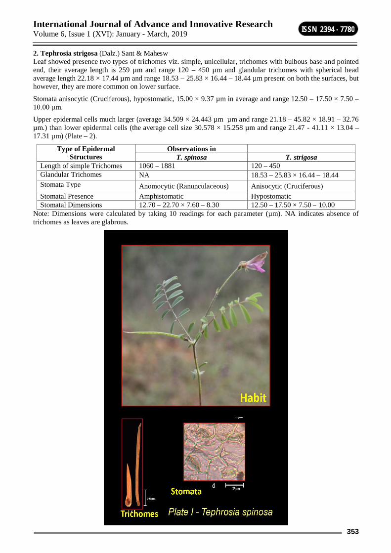
International Journal of Advance and Innovative Research Volume 6, Issue 1 (XVI): January - March, 2019
353
ISSN 2394 - 7780
2. Tephrosia strigosa (Dalz.) Sant & Mahesw Leaf showed presence two types of trichomes viz. simple, unicellular, trichomes with bulbous base and pointed end, their average length is 259 µm and range 120 – 450 µm and glandular trichomes with spherical head average length 22.18 × 17.44 µm and range 18.53 – 25.83 × 16.44 – 18.44 µm present on both the surfaces, but however, they are more common on lower surface.
Stomata anisocytic (Cruciferous), hypostomatic, 15.00 × 9.37 µm in average and range 12.50 – 17.50 × 7.50 – 10.00 µm.
Upper epidermal cells much larger (average 34.509 × 24.443 µm µm and range 21.18 – 45.82 × 18.91 – 32.76 µm.) than lower epidermal cells (the average cell size 30.578 × 15.258 µm and range 21.47 - 41.11 × 13.04 – 17.31 µm) (Plate – 2).
Type of Epidermal Structures
Observations in T. spinosa T. strigosa
Length of simple Trichomes 1060 – 1881 120 – 450 Glandular Trichomes NA 18.53 – 25.83 × 16.44 – 18.44 Stomata Type Anomocytic (Ranunculaceous) Anisocytic (Cruciferous) Stomatal Presence Amphistomatic Hypostomatic Stomatal Dimensions 12.70 – 22.70 × 7.60 – 8.30 12.50 – 17.50 × 7.50 – 10.00
Note: Dimensions were calculated by taking 10 readings for each parameter (µm). NA indicates absence of trichomes as leaves are glabrous.

International Journal of Advance and Innovative Research Volume 6, Issue 1 (XVI): January - March, 2019
354
ISSN 2394 - 7780

International Journal of Advance and Innovative Research Volume 6, Issue 1 (XVI): January - March, 2019
355
ISSN 2394 - 7780
DISCUSSION 1. Maceration: Three types of parenchyma is seen in T. spinosa viz. parenchyma with few pits, parenchyma with circular crystals and parenchyma with bordered pits whereas in T. strigosa, four types of parenchyma is seen. Fibres are of two types in T. spinosa viz. short, slender (ranged 210.0 – 250.0 × 6.00 – 10.00 µm.) and longer with wider lumen (ranged 320.0 – 370.0 × 6.00 – 12.00 µm.); whilst T. strigosa showed simple fibres, short, slender, thick walled (ranged 140.0 – 210.0 × 6.00 – 9.00 µm.) and simple fibres with longer, broader lumen (220.0 – 310.0 × 9.00 – 12.00 µm). In T. spinosa, tracheids shorter than fibres, slender, blunt at both the ends, with few simple pits 230.0 – 380.0 × 10.00 – 23.00 µm. While in T. strigosa, tracheids simple, slender, blunt at both the ends, pits not seen 340.0 – 420.0 × 9.00 – 17.00 µm. In T. spinosa vessel elements short, broad beak and ranged 90.00 – 110.0 × 19.00 – 32.00 µm (Fig. 1). Whereas in T. strigosa, vessels were broader, thicker with a range 360.0 – 470.0 × 15.00 – 35.00 µm (Fig. 2).
International Journal of Advance and Innovative Research Volume 6, Issue 1 (XVI): January - March, 2019
356
ISSN 2394 - 7780
2. Micromorphology of Leaf: Only simple type of trichomes were seen in T. spinosa with a length range of 1060 – 1881 µm. T. strigosa showed two types of trichomes viz. glandular (18.53 – 25.83 × 16.44 – 18.44 µm) and simple trichomes (120 -450 µm). Simple trichomes in T. strigosa were much shorter than those in T. spinosa. Anomocytic (Ranunculaceous) stomata were seen in T. spinosa whereas anisocytic (Cruciferous) stomata revealed by T. strigosa. In T. spinosa leaves were amphistomatic (ranged 12.70 – 22.70 × 7.60 – 8.30 µm) whilst that in T. strigosa were hypostomatic (ranged 12.50 -17.50 × 7.50 -10.00 µm).
CONCLUSION The qualitative and quantitative characters studied in this investigation are diagnostic to each species. These observation may find helpful for the taxonomic identification and delimitation of these taxa.
ACKNOWLEDGEMENT Authors are thankful to Dr. R. K. Ippar, Principal, Vaidyanath College, Parli-Vaijnath, Professor and Head, Department of Botany, Dr. B. A. M. University, Aurangabad for providing necessary facilities during this study. Thanks are due to Dr. Ram Gore for providing illustrations.
REFERENCES 1. Mabberley, D.J. 2008. Mabberley’s Plant-Book: A portable dictionary of plants, their classification and
uses. Third edition. Cambridge University Press, Cambridge.
2. Geesink, R. 1984. Scala Millettiearum. A survey of the genera of the tribe Millettieae (Leguminosae. Papilionoideae). EJ Brill/Leiden University Press, Leiden (Leiden Botanical Series v8), XVI: 131.
3. Schrire, L.G., B., MacKinder, B. & M. Lock 2005. Legumes of the world. Royal Botanical Gardens, Kew, UK.
4. Willis, J.C. 1973. The Dictionary of Flowering Plants and Ferns. 8th ed. Cambridge University Press, Cambridge, UK. Pp. 1135.
5. Al-Zahrani, R.M. 2007. Systematic Study of the Genus Tephrosia Pers. (Fabaceae) in Saudi Arabia. Post graduate thesis.
6. De Candolle. 1825. Prodromus systematis naturalis regni vegetabilis, sive, Enumeratio contracta ordinum generum specierumque plantarum hue usque cognitarium, juxta methodi naturalis. Prodromus Systematis Universalis Regni Vegetabilis.
7. Bentham, G. 1862. Leguminosae L. Galegeae. In: Martius, C.F.P. (Ed.), Flora Brusiliensis, Lipsiae, Munich. pp. 41-51.
8. Baker, J.G. 1876. Leguminosae In: Flora of British India Hooker, J.D. (Ed.) Reeve and co. Ltd., England pp.209-214.
9. Hassler, E. 1919. Ex herbario Hassleriano: Novitates paraguariensis XXIII. Feddes Repert 161-166. doi:10.1002/fedr 19190161314
10. Wood, C.E. 1949. The American barbistyled species of Tephrosia (Leguminosae). Rhodora 51: 193-231.
11. Brummitt, R.K. 1980. Reconsideration of the genera Ptycholobium, Caulocarpus, Lupinophyllum and Requenia in relation to Tephrosia. Kew Bull. 35: 459-473.
12. Cronquist, A. 1954. Papilionaceac Flore du Congo Belge et du Ruanda Burunsdi Spermatophytes. INEAC, Bruxelles.
13. Gillett, J.B. 1958. Notes on Tephrosia in tropical Africa. Kew Bull. 13: 111-132.
14. Hacker, J.B. 1990. A guide to herbaceous and shrub legumes of Queensland. University of Queensland Press. Pp. 275.
15. Bosman, M.T.M. & A.J.P. Haas 1983. A revision of the genus Tephrosia. Blumea 28(2): 421-487.

International Journal of Advance and Innovative Research Volume 6, Issue 1 (XVI): January - March, 2019
357
ISSN 2394 - 7780
ATTRACTVITY RESULTS FOR QUADRATIC FUNCTIONAL DIFFERENTIAL EQUATION
V. T. Ghuge1 and A. B. Mundhe2 1Department of Mathematics, R. M. I. G College, Jalna
2Department of Mathematics, A. S. C. College, Badnapur
ABSTRACT In this paper, we discuss the quadraic fnctional differential equation on unbounded intervals for uniformly global attractivity of the solution.Weapplyclassical hybrid fixed point theory to investigate the results.
Keywords: Functional differential equation; Fixed point theorem, uniformly global attractivity, Quadratic functional differential equation.
i) INTRODUCTION Let R be the real line and let R be set of nonnegative real numbers. Let 0 [ ,0]I be a closed and bounded interval in R for some real number 0 and let 0J I R .Let C denote the Banach space of continuous real-
valuied functions on 0I with the supremum norm defined by
0
sup | ( ) |C
t It
Forfixed t R , let tx denote the element of C defined by
( ) ( ( ), ) [ ,0].tx x t
The space C is called the history space of the past interval 0I for the functional differential equations to describing the past history.
Let ( )CRB R denote the class of functions : 0a R R satisfying the following properties:
(i).a is continuous,
(ii). lim ( ) ,t
a t
and
(iii). a(0) = 1.
The class of continuous and strictly monotone functions : 0 (0) 1a R R with a satisfy the above
criteria.If ( ),a CRB R then the reciprocal function ( ),a CRB R defined by1( )( )
a ta t
is continuous and
lim ( ) 0.t
a t
Given a function .
Consider the following quadratic functional differential equation on unbounded intervals,,
0
( ) ( ) ( , ( ), ) ( , ( ), ) . .( , ( )) t t
d a t x t g t x t x h t x t x a e t Rdt f t x t
x
(1)
Where ( ), : \0a CRB R f R R R , : : .g R R C R and h R R C R
The quadratic functional differential equation (1) is new to the theory of nonlinear differential equations and some special cases of these quadratic functional differential equation with have been studied in the literature on closed and bounded intervals for various aspects of the solutionsHale [13] ,Ntouyas [16] Dhage [11].The QFDE (1)is not discussed on closed but unbounded intervals of real line. In this paper,we discuss the quadratic perturbations of the first order ordinary differential equation for existence as well as for different characterizations of the solutions such as attractivity, asymptotic attractivity.
International Journal of Advance and Innovative Research Volume 6, Issue 1 (XVI): January - March, 2019
358
ISSN 2394 - 7780
Theorem 2.1 (Granas and Dugundji) [12]. Let S be a non-empty, closed, convex and bounded subset of the Banach space X and let be a continuous and compact operator. Then the operator equation Q x x has a solution in S.
Thefollowing fixed point theorem of Burton [3] which is a special case of a hybrid fixed point theorem [11] of Banach spaces.
Theorem2.2 (Dhage[7]). Let S be a closed, convex and bounded subset of the Banach space X and let be two operator such that
(i) A is nonlinear D-contraction,
(ii) B is completely continuous,
(iii) .x Ax By x S for all y S
Then the operator equation Ax Bx x has a solution in S.
Theorem2.3 (Dhage[10]). Let S be a non-empty, closed convex and bounded subset of the Banach algebra X and Let : :A X X and B S X be two operators such that
A is D-Lipschitz with D-function ,
B is completely continuous,
,x Ax By x S for all y S and ( ) , ( ) sup : M t r where M B S Bx x S
Then the operator equation Ax Bx x has a solution in S.
ii) CHARACTERIZATIONS OF SOLUTIONS We find the solutions of the FDE (1)in the space 0( , )BC I R R of continuous and bounded real-valued functions defined on 0I R . Define a standard supremum norm || .|| and a multiplication “ . ” in BC 0( , )I R R by
0
sup | ( ) | ( ) ( ) ( ) ( ),t I R
x x t and xy t x t y t t R
Clearly, BC 0( , )I R R becomes a Banach algebra with respect to the above norm and the multiplication in it.
By 1( , )L R R we denote the space oflebesgueintegrable functions on R and the norm 11( , )
Lin L R R is
defined by
10
| ( ) | .L
x x t ds
We assumethat and let Ω be a non-empty subset of X. LetQ : be a operator and consider the following operator equation in E, ( ) ( )Qx t x t for all 0 .t I R
Wegive different characterizations of the solutions for the operator equation ( ) ( )Qx t x t in the space 0( , )BC I R R .
Definition 3.1. We say that solutions of the operator equation ( ) ( )Qx t x t are locally attractive if there exists a closed ball 0( )rB x in the space 0( , )BC I R R for some 0 0( , )x BC I R R such that for arbitrary solutions
( ) ( )x x t and y y t of equation ( ) ( )Qx t x t belonging to 0( )rB x we have that
lim( ( ) ( )) 0t
x t y t
In the case when the limit ,isuniform with respect to the set 0( )rB x , i.e., when for each there exists T >0 such that

International Journal of Advance and Innovative Research Volume 6, Issue 1 (XVI): January - March, 2019
359
ISSN 2394 - 7780
| ( ) ( ))x t y t
for all 0( )rB x being solutions of ( ) ( )Qx t x t and for we will say that solutions of equation ( ) ( )Qx t x t are uniformly locally attractive on 0 .I R Definition3.2. A solution of equation ( ) ( )Qx t x t is said to be globally attractive if lim( ( ) ( )) 0t
x t y t
holds for each solution of ( ) ( )Qx t x t in 0( , )BC I R R .In other words, we
may say that solutions of the equation ( ) ( )Qx t x t are globally attractive if for arbitrary solutions x(t) and y(t) of ( ) ( )Qx t x t in 0( , )BC I R R The condition lim( ( ) ( )) 0
tx t y t
is satisfied. In the case when the condition
lim( ( ) ( )) 0t
x t y t
is satisfied uniformly with respect to the space 0( , )BC I R R i.e., if for every >0 there
exists T>0 such that the inequality lim( ( ) ( )) 0t
x t y t
is satisfied for alx,y, being the
solutions of ( ) ( )Qx t x t and for we will say that solutions of the equation ( ) ( )Qx t x t are uniformly globally attractive on 0 .I R
We introduce the new concept of local and global ultimate positivity of the solutions for the operator equation ( ) ( )Qx t x t in the space 0( , )BC I R R .
Definition3.3. A solution x of the equation ( ) ( )Qx t x t is called locally ultimately positive if there exists a closed ball 0( )rB x in the space 0( , )BC I R R for some 0( , )BC I R R such that 0( )rB x and
lim | ( ) | ( ) 0.t
x t x t
In the case when this limit, is uniform with respect to the solution set of the operator equation ( ) ( )Qx t x t in
0( , )BC I R R i.e., when for each there exists T > 0 such that
( ) ( ) ( )x t x t x t
For all x being solutions of ( ) ( )Qx t x t in 0( , )BC I R R and for we will say that solutions of equation ( ) ( )Qx t x t are uniformly locally ultimately positive on R .
Definition 3.4.A solution 0( , )BC I R R of the equation ( ) ( )Qx t x t is called globally ultimately
positive if lim | ( ) | ( ) 0.t
x t x t
is satisfied. In the case when the limit ( ) ( ) ( )x t x t x t is uniform with
respect to the solution set of the operator equation ( ) ( )Qx t x t in 0( , )BC I R R i.e., when for each there exists T > 0 such that limit, is satisfied for all x being solutions of ( ) ( )Qx t x t in 0( , )BC I R R .
and for , we will say that solutions of equation ( ) ( )Qx t x t are uniformly globally ultimately positive on
0I R .
Main Result We prove the global attractivity and positivity results for the functional differential equation(1) on 0I R under some suitable conditions.Let I be a closed interval in R and let ( , )AC I R be the space of functions which are defined and absolutely continuous on I.
First, we prove the global attractivity and ultimate positivity results for the functional differential equation (1) on 0I R .
Definition3.5. By a solution for the functional differential equation (1) we mean a function 0( , ) ( , )x BC I R R AC R R
International Journal of Advance and Innovative Research Volume 6, Issue 1 (XVI): January - March, 2019
360
ISSN 2394 - 7780
the function ( ) ( )( , ( ))
a t x ttf t x t
a is absolutely continuous on R and
x satisfies the equations in (1) on 0I R .
where ( , )AC R R is the space of absolutely continuous real-valued functions on right half real axis R .
Consider the following set of hypotheses There exists a continuous function h : R R such that | ( , , ) | ( ) . .g t x y h t a e t R
for all x R and y C . Moreover, we assume that 0
lim| ( ) | ( ) 0t
ta t h s ds
2( ) (0) 0A .
The function ( ,0,0)t f t is bounded on R with 0 sup| ( ,0,0) | : .F f t t R
) .The function :f R R R is continuous and there exists a function ( , )BC R Rl and a real number K > 0 such that
| || ( , ) ( , ) | ( )| |
x yf t x f t y tK x y
l
for all ,t R and x y R Moreover, we assume 0sup ( ) .t t L l
lim | ( , ) | ( , ) 0 .t
f t x f t x for all x R
6( ) (0, (0)) 0.A f
( ). (0, (0)) 1f
( ). The function (0, )xx
f xa .
Theorem3.1.Assume that the hypotheses ( ), ( ), ( ), ( )and( )) hold. Further, assume that
max|| ||, | (0) | || || .L a W K (3)
Then the functional differential equation (1) has a solution and solutions are uniformly globally attractive on 0I R .
Proof. Now, using hypotheses ( )and ) it can be shown that the FDE (1) is equivalent to the functional integral equation
0
0
( , ( )) (0) ( ) ( ) ( , ( ), ) ,( )
( ),
t
sf t x t a t a t g s x s x ds if t Rx t
t if t I
(4)
Set 0( , )X BC I R R and define a closed ball (0)rB in X centered at origin of radius r given by
0max 1, max || ||, | (0) | || ||r L F a W
Define the operators A on X and B on (0)rB by
0
( , ( )),( )
1,f t x t if t R
Ax tif t I
(5)
International Journal of Advance and Innovative Research Volume 6, Issue 1 (XVI): January - March, 2019
361
ISSN 2394 - 7780
And 0
0
(0) ( ) ( ) ( , ( ), ) ,( )
( ), .
t
sa t a t g s x s x ds if t RBx t
t if t I
(6)
Then the FIE (4) is transformed into the operator equation as
0( ) ( ) ( ), .Ax t Bx t x t t I R (7)
We Show that A and B satisfy all the conditions of Theorem2.3 on 0( , )BC I R R First we show that the
operators A and B define the mappings : : (0) .rA X X and B B X be arbitrary. Obviously, Ax is a continuous function on 0I R We show that Ax is bounded on 0I R . Thus, if t R , then we obtain:
0 0
| ( ) | | ( , ( )) | | ( , ( )) ( , 0) | | ( , 0) || ( ) |( )
| ( ) |
Ax t f t x t f t x t f t f tx tt F L F
K x t
l
Similarly, 0| ( ) | 1 .Ax t for all t I Therefore, taking the supremum over t,
0|| || max 1,Ax L F N
Thus Ax is continuous and bounded on 0I R . As a result It can be shown that and in
particular, :A X X and : (0) .rB B X We show that A is a Lipschitz on X. Let be arbitrary. Then, by hypothesis
0
0
|| || sup | ( ) ( ) |
max sup | ( ) ( ) |, sup | ( ) ( ) |
| ( ) ( ) |max 0, sup ( )| ( ) ( ) |
|| |||| ||
t I
t I t
t
Ax Ay Ax t Ay t
Ax t Ay t Ax t Ay t
x t y ttK x t y t
L x yK x y
¡
¡
¡l
for all This shows that A is a D-Lipschitz on X with D-function 휓 ( ) LrrK r
next, it can be shown
that B is a compact and continuous operator on X and in particular on (0)rB Next , we estimate the value of the
constant M. By efinition of M, one has
0
0
(0 )
0
|| ( (0)) || sup || || : (0)
sup sup | ( ) | : (0)
sup max sup | ( ) |, sup | ( ) | : (0)
sup max || ||, | (0) || ( ) |
sup | ( ) | | ( , ( ), ) |
max || ||, |
r
r r
rt I
rt I t
x B
t
st
B B Bx x B
Bx t x B
Bx t Bx t x B
a t
a t g s x s x ds
¡
¡
¡
(0)) || ||,
|| || max || ||, | (0) | || ||
a WThus
Bx a W M
for all . Next ,let be arbitrary. Then,

International Journal of Advance and Innovative Research Volume 6, Issue 1 (XVI): January - March, 2019
362
ISSN 2394 - 7780
0
0
| ( ) | | ( ) | | ( ) ||| || || |||| ( ) || || ( (0)) ||max 1,
max 1, max || ||, | (0) | || ||
r
x t Ax t By tAx ByA X B B
L MFL a WF
r
For all 0I R
0|| || max 1, max || || ,| (0) | || ||x L F a W r
This shows that (0)rB and hypothesis (c) of Theorem 2.3 is satisfied. Again,
max || || | (0) | || ||( )
L a W rM r r
K r
For r>0, because
max || || | (0) | || || .L a W K
Therefore, hypothesis (d) of Theorem 2.3 is satisfied. Now we apply Theorem 2.3 to the operator equation to yield that the FDE (1) has a solution on 0I R Moreover, the solutions of the FDE (1) are
in (0)rB Hence, solutions are global in nature.
Finally, let (0)rB be any two solutions of the FDE (1) on 0I R . Then
0
0
0
0
| ( ) ( ) | | ( , ( )) (0) ( ) ( ) ( , ( ), )
( , ( )) (0) ( ) ( ) ( , ( ), )
| ( , ( )) ( , ( )) | (0) ( ) ( ) ( , ( ), )
| ( , ( )) ( ) ( , ( ), ) ( , ( ), )
t
s
t
s
t
s
t
s s
x t y t f t x t a t a t g s x s x ds
f t y t a t a t g s y s y ds
f t x t f t y t a t a t g s x s x ds
f t y t a t g s x s x ds g s y s y ds
0
0
| ( , ( )) ( , ( )) | | (0) || ( ) | | ( ) | ( )
2 | ( , ( )) ( , 0) | | ( , 0) | ( )| ( ) ( ) |( ) (| (0) | || || )
| ( ) ( ) |
( ) | ( ) |2 ( )| ( ) |
(| (0) | || || ) | ( ) ( ) || ( ) ( )
tf t x t f t y t a t a t h s ds
f t x t f t f t w tx t y tt a W
K x t y t
t y t F w tK y t
L a W x t y tK x t y t
l
l
02( ) ( ) (8)|
L F w t
Taking the limit superior as in the above inequality yields,
lim | ( ) ( )| 0t
x t y t
Therefore, there is a real number T > 0 such that Consequently, the solutions of FDE (1) are uniformly globally attractive on 0I R .This completes the proof.
Theorem 3.2. Assume that the hypotheses ( ) – hold. Then the functional differential equation (1) has a solution and solutions are uniformly globally attractive and ultimately positive defined on 0I R .

International Journal of Advance and Innovative Research Volume 6, Issue 1 (XVI): January - March, 2019
363
ISSN 2394 - 7780
Proof. By Theorem 4.1 the FDE (1) has a global solution in the closed ball (0)rB .where the radius r is given as
in the proof of Theorem 4.1, and the solutions are uniformly globally attractive on 0I R We know that for any one has the inequality,
| | | | | | ,x y xy xy
and therefore,
|| | ( ) | | | | | | | || | | | |xy xy x y y x x y (9)
forall . Now for any solution (0)rB on has
0
0
|| ( )| ( ) | | [ ( , ( ))] (0) ( ) ( ) ( , ( ), ) |
([ ( , ( ))] (0) ( ) ( ) ( , ( ), )
t
s
t
s
x t x t f t x t a t a t g s x s x ds
f t x t a t a t g s x s x ds
Taking the limit superior as in the above inequality , we obtain lim || ( ) | ( )| 0t
x t x t
Therefore, there
is a real number T > 0 such that Hence, Solutions of the FDE (1) are uniformly globally attractive as well as ultimately positive defined on 0I R . This completed the proof.
4. Example
Let 0 / 2,0I be a closed and bounded interval in R and define a function
0: ( ) cosI R by t t
Consider the quadratic functional differential equation,
1
0
( ) . .( ) | ( ) | || ||( 4)1 tan (| ( ) |)
2( 6)( 1).
t tt
t
x t xe a e t Rd e x t x t x ctdt x t
tx
Here, ( ) ta t e for t R As , ( ) || || 1.a CRB R and a Again, we have:
1( 4)( , ) 1 tan (| |) ( , , )2( 6)( 1) | | || ||
tt x yf t x x and g t x y et x y c
For all ,t R x R first, we show that the function f satisfies hypothesis . ( , ), ( , )R R Let t x t y R R be arbitrary. Then,
1 1( 4) ( 4)| ( , ) ( , ) | tan (| |) tan (| |)2( 6)( 1) 2( 6)( 1)
( 4) || | | | |.2( 6)( 1) 1 | | | | ||
( 4) | |.2( 6)( 1) 1 | |
t tf t x f t y x yt tt x y
t x yt x y
t x y
Therefore, here ( 4)( )
2( 6)( 1)tt
t
l for all t R so that 12
L Furthermore, he function g is Caratheodory
and | ( , , ) | ( , , ) .tg t x y e for t x y R R C Clearly,
0lim 0.
tt s
te e ds
Finally,

International Journal of Advance and Innovative Research Volume 6, Issue 1 (XVI): January - March, 2019
364
ISSN 2394 - 7780
max || ||, | (0) | || || 1 .L a W K
Now, we apply Theorem 4.1 to the FDE (8) and conclude that it has a solution on 0I R Moreover, the solutions are uniformly globally attractive on 0I R .
And,
1( 4)| ( , ) | 1 tan (| |) ( , )2( 6)( 1)
tf t x x f t xt
For all ,t R x R and hence solutions of the quadratic FDE (8) are also uniformly globally ultimately positive on 0I R .
REFERENCES 1. J.Banas, B. Rzepka, An application of a measure of non-compactness inthe study of asymptotic stability,
Appl. Math. Letter 16(2003),1-6.
2. J.Banas, B.C. Dhage, Global asymptotic stability of solutions of a functional integral equeations,Nonlinear Analysis 69 (2008), 1945-1952.
3. T.A. Burton, A fixed point theorem of Krasnoselskii, Appl.Math. Lett. 11(1998),85-88.
4. T.A. Burton, B.Zhagng, Fixed points and stability of an integral equations: nonuniqueness,Appl. Math. Letters 17(2004), 839-846.
5. T.A. Burton and T. Furumochi, A note on stability by Schauder’s theorem, FunkcialajiEkvacioj 445(2001), 73-82.
6. K. Deimling, Nonlinear Functional Analysis, Springer Verlag, Berlin, 1985.
7. B.C. Dhage, A nonlinear alternative with applications to nonlinear perturbed differential equiations,Nonlinear Studies,13(4) (2006), 343-354.
8. B.C. Dhage, Local asymptotic attractivity for nonlinear quadratic functional integral equation,Nonlinear Analysis 70 (5) (2009), 1912-1922.
9. B.C. Dhage, Global attractivity result for nonlinear functional integral equations via a Krasnoselskii type fixed point theorem, Nonlinear Analysis 70 (2009), 2485-2493
10. B.C. Dhage, A fixed point theorem in Banach algebras withapplications to functional integral equations, Kyungpook Math. J. 44(2004),145-155.
11. B.C. Dhage, S.N. Salunkhe, R.P. Agrawal and W. Zhang, A functional differential equations in Banach algebras, Math.Ineq. Appl.8 (1) (2005),89-99.
12. A.Granas and J. Dugundji, Fixed Point Theory, Springer Verlag,New York, 2003.
13. H.K. Hale, Theory of Functional Differential Equations, Springer Verlag, New York,
International Journal of Advance and Innovative Research Volume 6, Issue 1 (XVI): January - March, 2019
365
ISSN 2394 - 7780
SYNTHESIS OF AZODYES BASED ON 8-HYDROXY QUINOLINE WITH ITS CHARACTERIZATIONS, ANTIFUNGAL AND ANTIMICROBIAL ACTIVITY
Prashant Vibhute1 and Sachin A. Khiste2
1Department of Chemistry, Balbhim Arts, Science and Commerce College, Beed 2Department of Chemistry, Vaishanvi Mahavidyalay, Wadwani
ABSTRACT This present work deals with the antibacterial and antifungal applications of the Azodyes which synthesized from 8-hydroxyquinoline. Quinoline and its derivatives are receiving increasing importance due to their wide range of applications in biological and pharmacological area. Substituted quinolines are prominent building blocks in both organic and inorganic molecular chemistry with their p-stacking ability and coordination properties.
In this research scheme, we synthesized azodyes base on 8-hydroxy quinoline by diazotization method. It was observed that 5 & 6 gives better yield. Structure of the compounds were identified by elemental analysis, 1H-NMR and UV-VIS Spectroscopic methods.
Keywords: Azodyes, 8-hydroxyquinoline, antibacterial, antifungal activities.
INTRODUCTION Quinolines were first discovered from coal tar by Friedlieb Ferdinand Runge in 18341.After half century Skraup-Doebner–Miller successfully synthesized the quinolines in laboratory 2-5. Quinoline derivatives are used as drugs for different diseases such as tuberculosis, schizophrenia and malaria 6–12.
The quinoline and its derivatives has paying attention due to its therapeutic properties. Quinoline sulphonamides have been used in the treatment of cancer, tuberculosis, diabetes, malaria, and convulsion. Apart from the magnetic and physical properties, the biological activities such as antimicrobial, antitumor, antivirus, anticoagulant action. Apart from its medicinal value quinoline derivatives are also studied as a supramolecular host 13–18.
In the current years, quinoline nucleus has gathered an immense attention not only by the chemists but also biologists because it is one of the key building block for many naturally occurring compounds 19. 8-hydroxyquinolines have been given new options to available drug in many instances. Metal complexes of 8-hydroxyquinoline as ligand can exhibit different biological activities because most of the transition metal complexes and in particular those with N-and O– donor atoms have been known to have antimicrobial properties.20 It can exert their physiological properties through bidentate chelation of metal ions. It is evident that formation of chelates metal ions increases the lipophilicity of the bioactive compounds through diverse array of biological oxidation–reduction mechanism for the effective permeability of the compounds into the site of action21.
Various strategies can be synthesize or modify metal complex structures by exploiting the reactivity of this heterocycle with their derivatives by various researchers22-25. 8-hydroxyquinoline and its derivatives were also reported to have promising bioactivities, including anticancer 26-27, antibacterial 28-29, antidyslipidemic and antioxidative properties 30, vasorelaxing properties 31, antivirus and antiplatelet activities32-33.
EXPERIMENTAL All the reagents were of analytical grade and were used without further purification. The solvents used were of high purity and distilled in laboratory before use. Melting points were recorded on digital melting point apparatus (optics technology) and were uncorrected. The reactions were monitored with TLC and also the purity was ascertained with TLC. Thin layer chromatography was carried out on silica gel 60/UV254. Infrared spectra analysis was obtained on SHIMADZU spectrophotometer using KBr discs.
GENERAL PROCEDURE FOR SYNTHESIS OF AZO DYES:- 8-Hydroxyquinoline (1.85gm, 12 mmol) was dissolved in 10 ml Methanol. The solution was cooled to 0-5°C in an ice bath. Meanwhile in another beaker a solution of NaNO2 (0.82 gm, 12 mmol) in 10 ml ice cold water was prepared and allow to cool the solution in ice bath. Take 10 ml Conc.HCl in 250 ml beaker, 5ml ice cold water and aniline (10 mmol) to this solution add NaNO2 slowly with constant stirring in ice cold water. Allow this reaction mixture to 30 min. at 0-5 °C in ice bath.

International Journal of Advance and Innovative Research Volume 6, Issue 1 (XVI): January - March, 2019
366
ISSN 2394 - 7780
Then 8-Hydroyquinoline in 10 ml methanol added dropwise over a period of 2 hr with constant stirring, then reaction mixture allow to keep stirred at R.T. for 6 hr. reaction was monitored with help of TLC. After completion of reaction, orange coloured crude product was obtained. Crude product was filtered and wash with water. The compound was dried and recrystallized with Methanol.
5-((2-chlorophenyl)diazenyl)quinolin-8-ol (1): yield 63.41% solid. 1H-NMR (200 MHz, CDCl3, ) 5.33 (s, 1H),7.23(d, 1H),7.80(d, 1H),7.52- 7.69 (m, 4H)7.90(d, 1H), 8.87(d, 1H),8.30(d, 1H). UV: 411nm, m/z: 283.051 (100.0%), 285.048 (32.0%), 284.055 (16.3%), 286.052 (5.2%), 285.058 (1.2%), 284.048 (1.1%)
5-((3-chlorophenyl)diazenyl)quinolin-8-ol (2): yield 69.33% solid. 1H-NMR (200 MHz, CDCl3, ) 5.31(s,1H),7.24(d,1H),7.62-7.88(m,5H),8.01(s,1H),8.37(d, 1H),8.84(d,1H). UV: 403nm.
5-((4-chlorophenyl)diazenyl)quinolin-8-ol (3): yield 61.30% solid. 1H-NMR (200 MHz, CDCl3,
)5.31(s,1H),7.22(d,1H),7.63(dd,1H),7.71(d,2H),7.83(d,1H),7.96(d,2H), 8.39(d,1H),8.87(d,1H). UV: 417nm
RESULT AND DISCUSSION In this research scheme, we synthesized azodyes base on 8-hydroxy quinoline by diazotization method (Table-1).It was observed that, (5 & 6) it gives better yield. The progress of reaction was monitored into thin-layer chromatography (TLC) using eluent methanol and carbon tetrachloride (3:7). After completion of the reaction, the crude products were recrystallized from methanol to gets pure products (1-7).
The present study tested antimicrobial activity of Azo compounds (1-7) (Table-2) against Escherichia coli and antifungal activity against Aspergillus niger using cup plate diffusion method. The diameter of well was 4mm for antibacterial activity nutrient agar and for antifungal activity potato dextrose agar (PDA) were used.
In antimicrobial activity the compound 7 was highly active, 2 & 6 show moderately active while 1,3,4,5 less active but the compound 3 was more active, 4 & 7 ordinary active and 1,2,5,6 was less active against antifungal activity(Table-2).
Table-1 Sr. No. Molecular formula Melting Point % yield 1
200°C 63.41%
2
160°C 69.33%
3
242°C 61.30%

International Journal of Advance and Innovative Research Volume 6, Issue 1 (XVI): January - March, 2019
367
ISSN 2394 - 7780
4
220°C 41.34%
5
160°C 70.25%
6
250°C 87.68%
7
139°C 11.57%
Table-2: Biological Activity of the compounds Sample Diameter of zone inhibition of growth (In mm) at 1ppm
Bacterial Culture (E.coli) Fungal Culture (A.niger) 1 6 5 2 8 7 3 6 9 4 6 8 5 5 7 6 9 6 7 12 8
ACKNOWLEDGEMENTS: Acknowledge to the Dr. V.G Sanap, principal Balbhim College, Beed who is instrumental in fostering research culture in the college and also providing infrastructure and instrumental facility.
REFERENCES 1. F. Runge, Pogg. Ann. (Annalen der Phys. Chem.) 31 (1834) 65–80.
2. Z.H. Skraup, Monatsh Chem. 1 (1880) 316–318.
3. Z.H. Skraup, Monatsh Chem. 2 (1881) 139–170.
4. Z.H. Skraup, Monatsh Chem. 2 (1881) 587–609.
5. Z.H. Skraup, Ber Chem. 15 (1882) 897.
6. J. Mao, H. Yuan, Y. Wang, B. Wan, M. Pieroni, Q. Huang, R.B. van Breemen, A.P. Kozikowski, S.G. Franzblau, J. Med. Chem. 52 (2009) 6966.
7. P.R. Verhoest, D.S. Chapin, M. Corman, K. Fonseca, J.F. Harms, X. Hou, E.S. Marr, F.S. Menniti, F. Nelson, R. O’Connor, J. Pandit, C.P. LaFrance, A.W. Schmidt, C.J. Schmidt, J.A. Suiciak, S. Liras, J. Med. Chem. 52 (2009) 5188.
International Journal of Advance and Innovative Research Volume 6, Issue 1 (XVI): January - March, 2019
368
ISSN 2394 - 7780
8. G.X. Li, Z.Q. Liu, X.Y. Luo, Eur. J. Med. Chem. 45 (2010) 1821.
9. H. Zeng, R. Cao, H. Zhang, Chem. Biol. Drug Des. 74 (2009) 596.
10. E. Milner, W. McCalmont, J. Bhonsle, D. Caridha, D. Carroll, S. Gardner, L. Gerena, M. Gettayacamin, C. Lanteri, T. Luong, V. Melendez, J. Moon, N. Roncal, J. Sousa, A. Tungtaeng, P. Wipf, G. Dow, Bioorg. Med. Chem. Lett. 20 (2010)1347.
11. I. Deb, P. Paira, A. Hazra, S. Banerjee, P.K. Dutta, N.B. Mondal, S. Das, Bioorg. Med. Chem. 17 (2009) 5782.
12. M. Albrecht, Triyanti, S. Schiffers, O. Osetska, G. Raabe, T. Wieland, L. Russo, Kari Rissanen, Eur. J. Org. Chem. (2007) 2850.
13. S.F. Alshahateet, R. Bishop, D.C. Craig, F. Kooli, M.L. Scudder, CrystEngComm 10 (2008) 297.
14. R.F. Semeniuc, T.J. Reamer, M.D. Smith, New J. Chem. 34 (2010) 439.
15. A. Karmakar, R.J. Sarma, J.B. Baruah, CrystEngComm 9 (2007) 379.
16. Y. Liu, K. Chen, D.S. Guo, Q. Li, H.B. Song, Cryst. Growth Des. 7 (2007) 2601.
17. C.R. Bondy, P.A. Gale, S.J. Loeb, J. Am. Chem. Soc. 126 (2004) 5030.
18. M.H. Filby, J.W. Steed, Coord. Chem. Rev. 250 (2006) 3200.
19. SumeshEswaran, AirodyVasudevaAdhikari, N. SuchethaShetty.Synthesis and antimicrobial activities of novel quinoline derivatives carrying 1,2,4-triazole moiety. European Journal of Medicinal Chemistry. 44, (2009), 4637–4647.
20. Prafulla, M.S., Jahanvi, P., Yogini, P., Metal complexes: current trends and future potential. IJPCBS 2 (3), (2012), 251–265.
21. Catherine Caris, Paul Baret,Jean-Louis Pierre,GuySerratrice.Synthesis and NMR study of two lipophilic iron(III) sequestering agents based on 8-hydroxyquinoline; H-bonding and conformational changes.Tetrahedron.Vol 52(13),25 March,4659-4672, (1996).
22. Zborowski K.K., Solá M., Poater J., Proniewicz L.M., Cent. Eur. J. Chem., (2013), 11(5), 655.
23. Maayan G., Dayagi Y., Arad-Yellin R., Shimon L.J.W., Shanzer A., Polyhedron (2013), http://dx.doi.org/10.1016/j.poly.2013.06.038.
24. Patel Y. S., Res ChemIntermed., (2014) DOI 10.1007/s11164-014-1764-9 (ii) Patel P.N., Patel Y. S., Res ChemIntermed.,(2015)DOI 10.1007/s11164-014-1903-3.
25. Chen B. Xu, L., Liu X., Zhou H., Xu H., Fang X., Wang Y., Applied Physics Letters. (2008), 92, 103305.
26. Shen A.Y., Wu S.N., Chiu C.T., J. Pharm. Pharmacol.(1999), 51, 543.
27. Ding W.Q., Liu B., Vaught J.L., Yamauchi H., Lind S.E., Cancer Res.,(2005), 65, 3389.
28. Darby C.M., Nathan C.F., J Antimicrob. Chemother.(2010), 65, 1424.
29. Ibrahim S.A., Makhlouf M.T., Abdel-Hafez A.A., Moharram A.M., J. Inorg. Biochem. (1986), 28, 57.
30. Kay K., J. Periodontol. (1978), 49, 47.
31. Sashidhara K.V., Kumar A., Bhatia G., Khan M.M., Khanna A.K., Saxena J.K., Eur. J. Med. Chem. (2009), 44,1813.
32. Bertini S., Calderone V., Carboni I., Maffei R., Martelli A., Martinelli F., MinutoloRajabi M., Testai L., Tuccinardi T., Ghidoni R., Macchia M., Bioorg. Med. Chem. (2010), 18, 6715
33. Liu Y.J., Zhao Y., Zhai X., Feng X., Wang J., Gong P., Bioorg. Med. Chem. (2008), 16, 6522

International Journal of Advance and Innovative Research Volume 6, Issue 1 (XVI): January - March, 2019
369
ISSN 2394 - 7780
QUANTITATIVE ESTIMATION OF BIOACTIVE PHYTOCONSTITUENTS PRESENT IN DALBERGIA LANCEOLARIA SUBSP. PANICULATA (ROXB.) THOTH. METHANOLIC LEAF AND BARK EXTRACT
Wankhade, M. S1., Solanke S. N.2 and Pangikar P. P.3
1Department of Botany, Sunderrao Solanke Mahavidyalaya, Majalgaon, Dist. Beed 2,3Department of Botany, R. B. Attal Arts, Science and Commerce College, Georai, Dist. Beed
ABSTRACTS Dalbergia lanceolaria subsp. paniculata (Roxb.) Thoth. is a very important medicinal plant in the deciduous forest. It is large tree belongs to the family Fabaceae. Whole parts of the plant are rich in secondary metabolite, which impart miraculous medicinal uses to the plants. A decoction of bark used in dyspepsia. Oil applied to rheumatic affections, and cutaneous diseases. Leaves are used in leprosy and allied obstinate skin diseases. Leaf paste with Castor oil is applied on filarial swellings. Leaves and flowers possess properties to treat arthritic affections and inflammations. Aqueous extract of leaves exhibited antiarthritic activity in rats.
Present investigation was designed for quantitative estimation of bioactive constituents present in Dalbergia lanceolaria subsp. paniculata (Roxb.) Thoth. methanolic leaf and bark extract. The methanolic extracts of the plants leaves and bark were screened for the presence of various phytoconstituents such as steroids, alkaloids, terpenoids, glycosides, flavonoids and carbohydrates. Quantitative estimation results shows leaves extract has (1.82 mg/g) alkaloid, (0.407 mg/g) carbohydrate, (0.22 µg/ml) protein, (0.59 mg/g) phenols, (0.28 mg/g) flavonoids, (0.99 mg/g) saponins, and (0.09 mg/g) tannins. While bark extract showed (2.07 mg/g) alkaloid, (0.179 mg/g) carbohydrate, (0.08 µg/ml) proteins,(0.88 mg/g) phenols, (0.44 mg/g) flavonoids, (1.28 mg/g) saponins and (0.095 mg/g) tannins.
Keywords: Dalbergia lanceolaria subsp. paniculata (Roxb.) Thoth., Quantitative estimation, Phytoconstituents.
INTRODUCTION India has the rich biodiversity in which 2 out of 25 biodiversity hot spot of the world are present. Biodiversity of India is important for its religious, spiritual and other traditional uses (Ganesan, et al., 2009). Many medicinal plants are used in Indian traditional health care system, and proposed for their interesting multilevel activities. Modern medicine is evolved from folk medicine and traditional system thorough chemical and pharmaceutical screening. Till this date plants remain a major source of medicinal compounds. Traditional medicines are used because it is cheaper, with minimal side effect and safe (Umadevi, et al., 2013). To ensure the safety of its products and practices standardization is very much importance. The knowledge of medicinal plants percolated from our ancient literature such as Vedas. More over in the Indian system of medicine, most herbal practitioners formulate and dispense their own medicinal therapy. All these formulation requires proper documentation and research (Tambekar , et al., 2010).
Dalbergia lanceolaria subsp. paniculata (Roxb.) Thoth. was known to use for timber yielding tree belonging to family leguminosae. It was reported that it has potent antioxidant activity, ant-inflammatory activities, antimicrobial activity, oestrogenic and larvicidal properties (kumar, et.al., 2015). It was evaluated that stem bark used for baldness and dysmennorhea (Krishna, et.al., 2011, Murthy, 2012). It was reported that leaves were used as antifilariasis (Kumar and Suryanarayana, 2013). Number of compounds were isolated from the plant (Saha, et.al., 2013). Four isoflavonoids were isolated from ethanolic extracts of stem bark and leaves of plant (Amin, et.al., 2012).
MATERIALS AND METHOD Collection of Plant Materials Leaves and bark of Dalbergia lanceolaria subsp. paniculata (Roxb.) Thoth. were collected from Mahur forest (N 19049.513’ E 77055’.442’) in Nanded district of Maharashtra. Specimen were identified and authenticated by Harbarium, Department of Botany, Dr. Babasaheb Ambedkar Marathwada University, Aurangabad (Accession No.- 17395). Freshly collected leaves and stem bark of the plants were dried in shade and pulverized to coarse powder. The powder was stored in an airtight container and kept in a cool, dark, and dry place (Hassan, et.al., 2014; Das, et.al., 2014).

International Journal of Advance and Innovative Research Volume 6, Issue 1 (XVI): January - March, 2019
370
ISSN 2394 - 7780
Fig-1: Showing D. lanceolaria subsp. Fig-2: Showing D. lanceolaria subsp.
paniculata (Roxb.) Thoth. Bark paniculata (Roxb.) Thoth. Leaf METHOD OF PREPARATION OF METHANOL EXTRACT The extraction was done by hot continuous method using Soxhlet apparatus. The 25 gm powder of leaves and bark were extracted using 250 ml methanol for 72 hours. The methanolic extract of bark and leaves of the plants were used for the further study (Vijayalakshmi, et.al., 2012).
Quantitative estimation
Quantitative estimation of Alkoloids, Carbohydrates, Phenols, Flavonoids, Proteins, Tannins Saponins was carried out by the following methods.
Alkaloids determination - (Harborne, 1973).
Determination of Total Carbohydrates by Anthrone Method (Hedge and Hofreiter, 1962; Sadasivam, and Manickam, 2008).
Determination of total flavonoids content (Zhishen et.al., 1999).
Estimation of proteins by Lowry’s method (Lowry, et.al.,1951; Sadasivam, and Manickam, 2008).
ESTIMATION OF TANNINS ( Schanderl, 1970; Sadasivam and Manikam, 2008)
Phenols (Mallick and Singh, 1980; Sadasivam and manickam,1980)
Saponins determination (Igwenyi and Elekwa, 2014)
RESULTS AND DISCUSSION The medicinal value of plant depends upon the bioactive phytoconstituents of the plant and which shows various physiological effects on human body (Sheikh, et.al., 2013). So the knowledge of phytoconstituents present in the plant can be important to detect with the help of phytochemical screening (Kumar and Hemalatha, 2013).
Quantitative analysis of the bark extract showed the presence of 1.82 mg/g and 2.07 mg/g alkaloids in the leaves and bark extract of the plant. This study also revealed that presence of 0.407 mg/g carbohydrates, 0.22 µg/ ml proteins, 0.59 mg/g phenols, 0.28 mg/g flavonoids, 0.99 mg/g saponins and 0.09 mg/g tannins in the leaves extract of the plant. This study also evaluated the 0.179 mg/g carbohydrates, 0.8 mg/g proteins, 0.88 mg/g phenols, 0.44 mg/g flavonoids, 1.28 mg/g saponins and 0.095 mg/g tannin in the bark extract of the plant.
Quantitative estimation was carried out to correlate relationship of the secondary metabolites present in the leaves and bark extract of plant and possible biological activities to evaluate as a potential source of natural bioactive chemicals (Patel, et.al., 2013). Total phenolic and flavonoid contain was found 210±1.56 and 46±3.61 respectively in the Dalbergia latifolia bark extracts (Khalid, et.al., 2015). Dalbergia sisso ethyl acetate and ethanol extract study evaluated that the presence of 0.22 mg/g and 0.18 mg/g phenols while 0.17 mg/g and 0.16 flavonoid respectively (Muthu, et.al., 2014). Artemisia persica methanolic extract reveled that it contain 407 mg/g total phenol and 308 mg/g flavonoids (Rashid, et.al., 2010). Significant amount of total phenolic, total flavonoid content was found in Pandanus conoideus Lam. (Rohman, et.al., 2010). Tetracarpidium conophorum root extract showed the presence of Tannin,0.545mg/g Saponins,10.705mg/g, Alkaloids, 0.41mg/g, Oxalate,0.895mg/g and Phenols, 0.215mg/g (Ayoola, et.al., 2011).
International Journal of Advance and Innovative Research Volume 6, Issue 1 (XVI): January - March, 2019
371
ISSN 2394 - 7780
Fig.- Graphical representation of different bioactive constituents present in leaves and bark extracts of
Dalbergia lanceolaria subsp. paniculata (Roxb.) Thoth.
CONCLUSION Quantitative analysis showed that plants are rich in the phytoconstituents. Quantitative estimation was carried out to correlate relationship of the secondary metabolites present in the leaves and bark extract of plant and possible biological activities to evaluate as a potential source of natural bioactive chemicals. Present investigation is useful in differentiating the species from the adulterant and act as a biochemical marker for this medicinally important plant in the pharmaceutical industry and plant systematic studies.
BIBLIOGRAPHY Ganesan, S., Ponnuchamy, M., Kesavan, L., Selvaraj, A. (2009). Floristic composition and practices on the
selected sacred groves of Pallapatty village (Reserved Forest) Tamil Nadu. Indian Journal of Traditional Knowledge. Vol. 8(2), 154- 162.
International Journal of Advance and Innovative Research Volume 6, Issue 1 (XVI): January - March, 2019
372
ISSN 2394 - 7780
Umadevi, M., Kumar, K.P., Bhowmik,D.,Duraivel, S.( 2013).Traditionally Used Anticancer Herbs In India. Journal of Medicinal Plants Studies.Vol. 1, (3). 56-74.
Tambekar, D. H., Khante, B. S., Khante,B. S. (2010). Evaluation of Antibacterial Properties of Ethnomedicinal Herbs used by Korkus in Melghat of India against Enteric pathogens. International Journal of Pharma and Bio Sciences. V1 (1).
Krishna, M. B., Mythili, S., Kumar, K. S., Ravinder, B., Murali, T., Mahender, T. (2011). Ethno Botanical Survey of Medicinal Plants in Khammam District, Andhra Pradesh, India. International Journal of Applied Biology and Pharmaceutical Technology. Vol. 2(4). 366-370.
Murthy, E. N. (2012). Ethno medicinal plants used by gonds of Adilabad district, Andhra Pradesh, India. Int. J. of Pharm. & Life Sci. Vol. 3(10), 2034-2043.
Kumar, R. B., Suryanarayana, B. (2013). Ethnomedicinal recipes for Skin and dermatitis & allied diseases from Tribals of Sriharikota Island, Andhra Pradesh. Journal of Pharmacognosy and Phytochemistry. Vol. 2 (2). 234- 249.
Saha, S., Shilpi, J. A., Mondal, H., Hossain, F., Anisuzzman, M., Hasan, M. M., Cordell, G. A., (2013). Ethnomedicinal, phytochemical, and pharmacological profile of the Genus Dalbergia L. (Fabaceae). Phytopharmacology. 4(2), 291-346.
Amin, E., Abouzid, S., Seida, A., (2012). Phytochemical and Biological Studies on Isoflavonoids from Dalbergia paniculata Cultivated in Egypt. Pharmacologia. Vol. 3(3). 84- 90.
Hassan, L.E., Ahamed, M.K., Majid, A. A., Baharetha, H. M., Muslim, N.S., Nassar, Z.D. and Majid, A. A. (2014). Correlation of antiangiogenic, antioxidant and cytotoxic activities of some Sudanese medicinal plants with phenolic and flavonoid contents. BMC Complementary and Alternative Medicine. Vol. 14, 406.
Das, S., Vasudeva, N., Sharma, S. (2014). Chemical composition of ethanol extract of Macrotyloma uniflorum (Lam.) Verdc. using GC-MS Spectroscopy. Organic and Medicinal Chemistry Letters. Vol. 4(13), 1- 4.
Vijayalakshmi, A., Ravichandiran, V., Malarkodi V., Nirmala, S. and Jayakumari, S. (2012). Screening of flavonoid “quercetin” from the rhizome of Smilax china Linn.for anti-psoriatic activity. Asian Pacific Journal of Tropical Biomedicine. 269-275.
Harborne, J.B., (1973). Phytochemical Methods. Chapman and Hall, Ltd., London, 49-188.
Hedge, J.E., Hofreiter, B.T. (1962). In: Carbohydrate Chemistry, 17 (Eds. Whistler R.L. and Be Miller, J.N.). Academic Press, New York.
Sadasivam, S., Manickam, A. (2008). Biochemical methods. New Age International Pvt. Ltd, New Delhi.
Zhishen, J.,Mengcheng, T., Jianming, W., (1999). The determination of flavonoid contents in mulberry and their scavenging effect on superoxide radicals. Food Chemistry. Vol. 64, 555-559.
Schanderl, S. H. (1970). In: Method in Food Analysis. Academic Press New York. 709.
Mallick, C.P., Singh, M. B. (1980). Plant enzymology and Histoenzymology. Kalyani publishers, New Delhi, 286.
Patel, A., Rathod, D., Dave, M., Patil, I. (2013). Study on Qualitative and Quantitative Estimation of Phytochemicals of Triphala Plants. Indian journal of applied research. Vol. 3(9). 542-544.
Khalid, M., Akhtar, J., Badruddeen, M., Singh, A. K. (2015). Pharmacognostical investigation and total phenolic content of Dalbergia latifolia (Roxb) bark. International journal of Pharmacognosy.Vol. 2(5), 248-253.
Muthu L. T., Radha, R., Jayshree, N. (2014). Invitro antioxidant activity, total phenolic and total flavonoid content in extracts from the bark of Dalbergia sissoo Roxb. International Journal of Pharma Sciences and Research. Vol.5 (5).226-231.
Rashid, A. C., Qureshi, M. Z., Raza, S. A., William, J., Arshad, M. (2010). Quantitative determination of antioxidant potential of Artemisia persica. Analele University Bucuresti- Chimie. Vol. 19( 1), 23 – 30.

International Journal of Advance and Innovative Research Volume 6, Issue 1 (XVI): January - March, 2019
373
ISSN 2394 - 7780
Rohman, A., Riyanto, S., Yuniarti, N., Saputra, W. R., Utami, R., Mulatsih, W. (2010). Antioxidant activity, total phenolic, and total flvaonoid of extracts and fractions of red fruit (Pandanus conoideus Lam). International Food Research Journal. Vol. 17, 97-106.
Ayoola, P. B., Adeyeye, A., Onawumi, O. O., Faboya, O. O. P., 2011. Phytochemical and nutrient evaluation of Tetracarpidium conophorum (Nigerian walnut) root. International Journal of Research and Reviews in Applied Sciences. Vol.7(2).197- 202.
MANUSCRIPT SUBMISSION
GUIDELINES FOR CONTRIBUTORS 1. Manuscripts should be submitted preferably through email and the research article / paper
should preferably not exceed 8 – 10 pages in all.
2. Book review must contain the name of the author and the book reviewed, the place of publication and publisher, date of publication, number of pages and price.
3. Manuscripts should be typed in 12 font-size, Times New Roman, single spaced with 1” margin on a standard A4 size paper. Manuscripts should be organized in the following order: title, name(s) of author(s) and his/her (their) complete affiliation(s) including zip code(s), Abstract (not exceeding 350 words), Introduction, Main body of paper, Conclusion and References.
4. The title of the paper should be in capital letters, bold, size 16” and centered at the top of the first page. The author(s) and affiliations(s) should be centered, bold, size 14” and single-spaced, beginning from the second line below the title.
First Author Name1, Second Author Name2, Third Author Name3 1Author Designation, Department, Organization, City, email id 2Author Designation, Department, Organization, City, email id 3Author Designation, Department, Organization, City, email id
5. The abstract should summarize the context, content and conclusions of the paper in less than 350 words in 12 points italic Times New Roman. The abstract should have about five key words in alphabetical order separated by comma of 12 points italic Times New Roman.
6. Figures and tables should be centered, separately numbered, self explained. Please note that table titles must be above the table and sources of data should be mentioned below the table. The authors should ensure that tables and figures are referred to from the main text.
EXAMPLES OF REFERENCES All references must be arranged first alphabetically and then it may be further sorted chronologically also.
Single author journal article: Fox, S. (1984). Empowerment as a catalyst for change: an example for the food industry. Supply Chain Management, 2(3), 29–33.
Bateson, C. D.,(2006), ‘Doing Business after the Fall: The Virtue of Moral Hypocrisy’, Journal of Business Ethics, 66: 321 – 335
Multiple author journal article: Khan, M. R., Islam, A. F. M. M., & Das, D. (1886). A Factor Analytic Study on the Validity of a Union Commitment Scale. Journal of Applied Psychology, 12(1), 129-136.
Liu, W.B, Wongcha A, & Peng, K.C. (2012), “Adopting Super-Efficiency And Tobit Model On Analyzing the Efficiency of Teacher’s Colleges In Thailand”, International Journal on New Trends In Education and Their Implications, Vol.3.3, 108 – 114.
Text Book: Simchi-Levi, D., Kaminsky, P., & Simchi-Levi, E. (2007). Designing and Managing the Supply Chain: Concepts, Strategies and Case Studies (3rd ed.). New York: McGraw-Hill.
S. Neelamegham," Marketing in India, Cases and Reading, Vikas Publishing House Pvt. Ltd, III Edition, 2000.
Edited book having one editor: Raine, A. (Ed.). (2006). Crime and schizophrenia: Causes and cures. New York: Nova Science.
Edited book having more than one editor: Greenspan, E. L., & Rosenberg, M. (Eds.). (2009). Martin’s annual criminal code:Student edition 2010. Aurora, ON: Canada Law Book.
Chapter in edited book having one editor: Bessley, M., & Wilson, P. (1984). Public policy and small firms in Britain. In Levicki, C. (Ed.), Small Business Theory and Policy (pp. 111–126). London: Croom Helm.
Chapter in edited book having more than one editor: Young, M. E., & Wasserman, E. A. (2005). Theories of learning. In K. Lamberts, & R. L. Goldstone (Eds.), Handbook of cognition (pp. 161-182). Thousand Oaks, CA: Sage.
Electronic sources should include the URL of the website at which they may be found, as shown:
Sillick, T. J., & Schutte, N. S. (2006). Emotional intelligence and self-esteem mediate between perceived early parental love and adult happiness. E-Journal of Applied Psychology, 2(2), 38-48. Retrieved from http://ojs.lib.swin.edu.au/index.php/ejap
Unpublished dissertation/ paper: Uddin, K. (2000). A Study of Corporate Governance in a Developing Country: A Case of Bangladesh (Unpublished Dissertation). Lingnan University, Hong Kong.
Article in newspaper: Yunus, M. (2005, March 23). Micro Credit and Poverty Alleviation in Bangladesh. The Bangladesh Observer, p. 9.
Article in magazine: Holloway, M. (2005, August 6). When extinct isn't. Scientific American, 293, 22-23.
Website of any institution: Central Bank of India (2005). Income Recognition Norms Definition of NPA. Retrieved August 10, 2005, from http://www.centralbankofindia.co.in/ home/index1.htm, viewed on 7. The submission implies that the work has not been published earlier elsewhere and is not
under consideration to be published anywhere else if selected for publication in the journal of Indian Academicians and Researchers Association.
8. Decision of the Editorial Board regarding selection/rejection of the articles will be final.