Identification of novel DNA methylation inhibitors via a two-component reporter gene system
8
RESEARCH Open Access Identification of novel DNA methylation inhibitors via a two-component reporter gene system Yi-Shiuan Lin 1† , Arthur Y Shaw 2† , Shi-Gang Wang 1 , Chia-Chen Hsu 1 , I-Wen Teng 1 , Min-Jen Tseng 1 , Tim HM Huang 3 , Ching-Shih Chen 4 , Yu-Wei Leu 1* , Shu-Huei Hsiao 1* Abstract Background: Targeting abnormal DNA methylation represents a therapeutically relevant strategy for cancer treatment as demonstrated by the US Food and Drug Administration approval of the DNA methyltransferase inhibitors azacytidine and 5-aza-2’-deoxycytidine for the treatment of myelodysplastic syndromes. But their use is associated with increased incidences of bone marrow suppression. Alternatively, procainamide has emerged as a potential DNA demethylating agent for clinical translation. While procainamide is much safer than 5-aza-2’- deoxycytidine, it requires high concentrations to be effective in DNA demethylation in suppressing cancer cell growth. Thus, our laboratories have embarked on the pharmacological exploitation of procainamide to develop potent DNA methylation inhibitors through lead optimization. Methods: We report the use of a DNA methylation two-component enhanced green fluorescent protein reporter system as a screening platform to identify novel DNA methylation inhibitors from a compound library containing procainamide derivatives. Results: A lead agent IM25, which exhibits substantially higher potency in GSTp1 DNA demethylation with lower cytotoxicity in MCF7 cells relative to procainamide and 5-aza-2’-deoxycytidine, was identified by the screening platform. Conclusions: Our data provide a proof-of-concept that procainamide could be pharmacologically exploited to develop novel DNA methylation inhibitors, of which the translational potential in cancer therapy/prevention is currently under investigation. Background As DNA methylation-mediated silencing of genes has been implicated in the pathogenesis of many diseases including cancer [1-7], targeting aberrant DNA methyla- tion is considered as a therapeutically relevant strategy for cancer treatment. Among many agents with DNA methy- lation-modifying capability, 5-aza-2’-deoxycytidine (decita- bine; 5-Aza) is the best-known DNA demethylation agent. 5-Aza exerts its effect by inhibiting DNA methyltrans- ferases (DNMTs), the key enzymes responsible for initiat- ing or maintaining the DNA methylation status, thereby facilitating the re-expression of tumor suppressor genes through DNA hypomethylation. Its therapeutic efficacy is manifest by the Food and Drug Administration approval for the treatment of myelodysplastic syndromes. While 5-Aza is a potent DNA demethylation agent [8,9], its use is associated with increased incidences of bone marrow suppression, including neutropenia and thrombocytopenia, due to the disruption of DNA synthesis. In addition, shorter half-life hinders the effective delivery of 5-Aza to the tumor site [10]. Recently, procainamide has emerged as a potential DNA demethylating agent for clinical translation. Evi- dence indicates that procainamide inhibits DNMT1 by reducing the affinity with its two substrates: hemimethy- lated DNA and S-adenosylmethionine [11-13]. Through DNA demethylation, procainamide causes growth arrest [9] and reactivation of tumor suppressor genes in cancer cells [14]. Moreover, as an anti-arrhythmic drug, procai- namide has a well-characterized safety profile without * Correspondence: [email protected]; [email protected] † Contributed equally 1 Human Epigenomics Center, Department of Life Science, Institute of Molecular Biology and Institute of Biomedical Science, National Chung Cheng University, Chia-Yi, 621, Taiwan Full list of author information is available at the end of the article Lin et al. Journal of Biomedical Science 2011, 18:3 http://www.jbiomedsci.com/content/18/1/3 © 2011 Lin et al; licensee BioMed Central Ltd. This is an Open Access article distributed under the terms of the Creative Commons Attribution License (http://creativecommons.org/licenses/by/2.0), which permits unrestricted use, distribution, and reproduction in any medium, provided the original work is properly cited.
-
Upload
independent -
Category
Documents
-
view
3 -
download
0
Transcript of Identification of novel DNA methylation inhibitors via a two-component reporter gene system
RESEARCH Open Access
Identification of novel DNA methylation inhibitorsvia a two-component reporter gene systemYi-Shiuan Lin1†, Arthur Y Shaw2†, Shi-Gang Wang1, Chia-Chen Hsu1, I-Wen Teng1, Min-Jen Tseng1, Tim HM Huang3,
Ching-Shih Chen4, Yu-Wei Leu1*, Shu-Huei Hsiao1*
Abstract
Background: Targeting abnormal DNA methylation represents a therapeutically relevant strategy for cancer
treatment as demonstrated by the US Food and Drug Administration approval of the DNA methyltransferase
inhibitors azacytidine and 5-aza-2’-deoxycytidine for the treatment of myelodysplastic syndromes. But their use is
associated with increased incidences of bone marrow suppression. Alternatively, procainamide has emerged as a
potential DNA demethylating agent for clinical translation. While procainamide is much safer than 5-aza-2’-
deoxycytidine, it requires high concentrations to be effective in DNA demethylation in suppressing cancer cell
growth. Thus, our laboratories have embarked on the pharmacological exploitation of procainamide to develop
potent DNA methylation inhibitors through lead optimization.
Methods: We report the use of a DNA methylation two-component enhanced green fluorescent protein reporter
system as a screening platform to identify novel DNA methylation inhibitors from a compound library containing
procainamide derivatives.
Results: A lead agent IM25, which exhibits substantially higher potency in GSTp1 DNA demethylation with lower
cytotoxicity in MCF7 cells relative to procainamide and 5-aza-2’-deoxycytidine, was identified by the screening
platform.
Conclusions: Our data provide a proof-of-concept that procainamide could be pharmacologically exploited to
develop novel DNA methylation inhibitors, of which the translational potential in cancer therapy/prevention is
currently under investigation.
Background
As DNA methylation-mediated silencing of genes has
been implicated in the pathogenesis of many diseases
including cancer [1-7], targeting aberrant DNA methyla-
tion is considered as a therapeutically relevant strategy for
cancer treatment. Among many agents with DNA methy-
lation-modifying capability, 5-aza-2’-deoxycytidine (decita-
bine; 5-Aza) is the best-known DNA demethylation agent.
5-Aza exerts its effect by inhibiting DNA methyltrans-
ferases (DNMTs), the key enzymes responsible for initiat-
ing or maintaining the DNA methylation status, thereby
facilitating the re-expression of tumor suppressor genes
through DNA hypomethylation. Its therapeutic efficacy is
manifest by the Food and Drug Administration approval
for the treatment of myelodysplastic syndromes. While
5-Aza is a potent DNA demethylation agent [8,9], its use
is associated with increased incidences of bone marrow
suppression, including neutropenia and thrombocytopenia,
due to the disruption of DNA synthesis. In addition,
shorter half-life hinders the effective delivery of 5-Aza to
the tumor site [10].
Recently, procainamide has emerged as a potential
DNA demethylating agent for clinical translation. Evi-
dence indicates that procainamide inhibits DNMT1 by
reducing the affinity with its two substrates: hemimethy-
lated DNA and S-adenosylmethionine [11-13]. Through
DNA demethylation, procainamide causes growth arrest
[9] and reactivation of tumor suppressor genes in cancer
cells [14]. Moreover, as an anti-arrhythmic drug, procai-
namide has a well-characterized safety profile without
* Correspondence: [email protected]; [email protected]
† Contributed equally1Human Epigenomics Center, Department of Life Science, Institute of
Molecular Biology and Institute of Biomedical Science, National Chung
Cheng University, Chia-Yi, 621, Taiwan
Full list of author information is available at the end of the article
Lin et al. Journal of Biomedical Science 2011, 18:3
http://www.jbiomedsci.com/content/18/1/3
© 2011 Lin et al; licensee BioMed Central Ltd. This is an Open Access article distributed under the terms of the Creative CommonsAttribution License (http://creativecommons.org/licenses/by/2.0), which permits unrestricted use, distribution, and reproduction inany medium, provided the original work is properly cited.
side effects commonly associated with nucleoside analo-
gues [15,16]. However, in contrast to 5-Aza, procaina-
mide requires high concentrations (≥ 50 μM) to be
effective in DNA demethylation in suppressing cancer
cell growth [9,11]. Thus, our laboratories have embarked
on the pharmacological exploitation of procainamide to
develop potent DNA methylation inhibitors through
lead optimization.
Previously, we reported a two-component enhanced
green fluorescent protein (EGFP) reporter gene system
for the visualization and quantization of dynamic
changes in targeted DNA methylation in bone marrow-
derived mesenchymal stem cells or cancer cell lines
[17,18]. This system gives a direct and concomitant
measurement and evaluation of DNA demethylation and
cytotoxicity in living cells, thus providing an expedient
screening platform for identifying demethylating agents.
As the exact mode of action of procainamide in decreas-
ing the binding DNMT1 with its substrate remains
undefined, we used procainamide as a scaffold to
develop a focused compound library, which in combina-
tion with other in-house compound libraries, was used
for screening via this two-component system.
Methods
Cell culture and drug treatment
MCF7 breast cancer cells, obtained from American Type
Culture Collection, were grown in Minimal Essential Med-
ium (MEM; Invitrogen), supplemented with 10% FBS,
2 mM L-glutamine, and 100 μg/ml penicillin/streptomy-
cin. Cells were cultured at 37°C in a humidified incubator
containing 5% CO2. Medium changes were performed
twice weekly and cell passages were performed at 90%
confluence. To maintain the two-component constructs in
MCF7 cells, 200 μg/mL of hygromycin B (Invitrogen) and
500 μg/mL of Geneticin (G418, Calbiochem) were
included in culture medium. 5-Aza and procainamide
were purchased from Sigma-Aldrich. Synthesis of procai-
namide derivatives and other tested agents (structures,
Additional file 1: Figure S1) will be described elsewhere.
Tested agents were dissolved in DMSO as stock solutions,
and added to culture medium with final DMSO concen-
trations of 0.3% and 1.2% (v/v) for 7.5 μM and 30 μM of
testing drugs, respectively. Control cells received DMSO
vehicle. During the 5-day treatment period, medium was
changed on the third day of treatment along with the addi-
tion of 17b-estrodial (E2, 10 ng/ml).
In vitro DNA methylation
PCR-amplified and purified Trip10 promoter (4 μg) was
incubated with 20 U of CpG methyltransferase (SssI,
New England BioLabs) at 37°C for 4 h in the presence
of 160 μM S-adenosylmethionine to induce methylation
at the Trip10 promoter DNA. Complete conversion was
indicated by the resistance of methylated Trip10 DNA
to methylation-sensitive restriction enzymes (HpaII,
New England BioLabs).
Transfection
In vitro methylated Trip10 promoter DNAs (0.4 μg) were
denatured and used to transfect 1 × 10⁵ cells/well in 6-well
plate at day 1, 3, and 5 using DMRIE-C (Invitrogen)
according to the manufacturer’s instruction. Unmethylated
PCR products were transfected as mock controls. Tracking
of the transfected DNAs was performed by using the Labe-
lIT Tracker Cy5 Intracellular Nucleic Acid Localization Kit
(Mirus) by following the manufacture’s instruction. Cells
were monitored by fluorescence microscopy (Axiovert
200M, ZEISS) and analyzed with the MetaMorph software.
Bisulfite conversion
Genomic DNA (0.5 μg) was bisulfite-converted by the
EZ DNA Methylation Kit (Zymo) by following the man-
ufacturer’s instruction.
Qmsp
Bisulfite converted DNAs were PCR-amplified by using
PCR primers listed in Additional file 1: Table S1. Uni-
versal methylated DNAs (Millipore) were used as posi-
tive control. Col2A1 was used as a loading control and
to amplify the serial diluted (1/10, 1/100 and 1/1000)
bisulfite-converted universal methylated DNAs to gener-
ate the standard curve for quantization in real time PCR
machine (Bio-Rad, iQ5). The methylation percentage
was calculated as [(Intensity of Amplifications by Trip10
MSP primer set) × 100]/(Intensity of Amplifications by
Col2A1 MSP primer set) (%). The qMSP was performed
in a 25 μL of reaction mixture containing 4 μL of tem-
plate (bisulfite treated DNA), 2 μL of primer pair (2.5
μM), 12.5 μL of 2X reaction buffer (SYBR Green real
time PCR Master Mix, Toyobo), and 6.5 μL of H2O.
Analysis of EGFP expression was performed by
fluorescence microscopy (Axiovert 200M, ZEISS) with
the MetaMorph software. Images and intensities of
EGFP of more than 600 cells were analyzed by the
image analysis program NIH Image (Image J).
Western blotting
Equal amounts of protein in cell lysates were separated
in 10% SDS polyacrylamide gels and then transblotted
to nitrocellulose membranes. After blocking with non-
fat milk, the membrane was washed and incubated with
antibodies against GFP (Abcam) or b-actin (Sigma-
Aldrich). The membrane was washed, and then incu-
bated with anti-rabbit immunoglobulin G-horseradish
peroxidase. After final wash, the proteins were visualized
by enhanced chemiluminescence. Immunoblotting data
were analyzed by NIH Image.
Lin et al. Journal of Biomedical Science 2011, 18:3
http://www.jbiomedsci.com/content/18/1/3
Page 2 of 8
Cell viability assay
Cell viability was assessed using MTT [3-(4,
5-dimethylthiazol- 2-yl)-2,5-diphenyl-2H-tetrazolium
bromide, Sigma-Aldrich) in six replicates. MCF7 cells
were seeded at 4,000 cells per well in 96-well plates, and
treated with test agents at different concentrations. After
5-day treatment, cells were incubated in medium con-
taining 0.5 mg/ml MTT at 37°C for 4 h. Reduced MTT
was solubilized in 200 μl/well of DMSO for determina-
tion of absorbance of 595 nm using a microplate reader.
ELISA
Enzyme-linked immunosorbent assay (ELISA) was per-
formed using the GFP ELISA kit (Cell Biolabs, San
Diego, CA) according to the manufacture’s instruction.
Differential Methylation Hybridization (DMH) Array
DMH was performed according to a reported procedure
[19]. Amplicons prepared from control (vehicle-treated)
MCF7 cells were labeled with Cy5 and the amplicons
prepared from drug-treated cells were labeled with Cy3.
Labeled DNAs were co-hybridized onto 244K Agilent
promoter array. Array hybridization results were nor-
malized with LOWESS and a cutoff of 4 folds was used.
Statistics
ELISA and MTT results were analyzed by F-test. Signifi-
cant demethylated loci were confirmed by ANOVA
from DMH results. Linear regression was used to
deduce the global methylation states before and after
the demethylation agent treatment.
Results
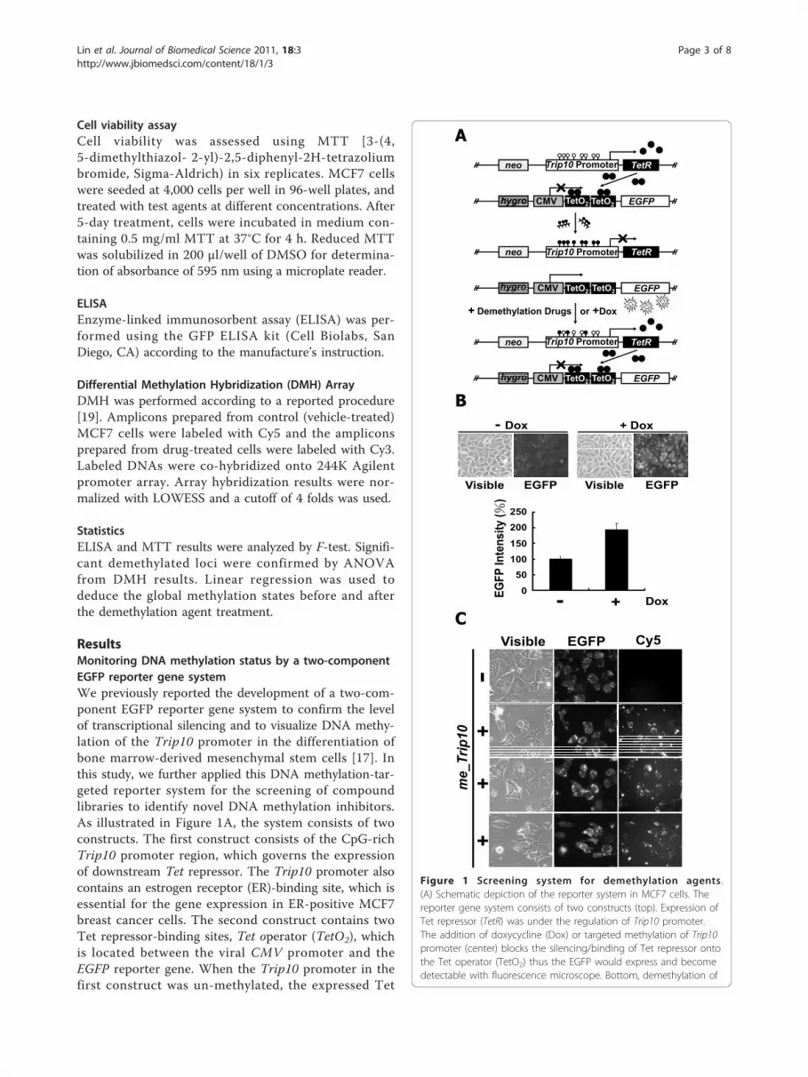
Monitoring DNA methylation status by a two-component
EGFP reporter gene system
We previously reported the development of a two-com-
ponent EGFP reporter gene system to confirm the level
of transcriptional silencing and to visualize DNA methy-
lation of the Trip10 promoter in the differentiation of
bone marrow-derived mesenchymal stem cells [17]. In
this study, we further applied this DNA methylation-tar-
geted reporter system for the screening of compound
libraries to identify novel DNA methylation inhibitors.
As illustrated in Figure 1A, the system consists of two
constructs. The first construct consists of the CpG-rich
Trip10 promoter region, which governs the expression
of downstream Tet repressor. The Trip10 promoter also
contains an estrogen receptor (ER)-binding site, which is
essential for the gene expression in ER-positive MCF7
breast cancer cells. The second construct contains two
Tet repressor-binding sites, Tet operator (TetO2), which
is located between the viral CMV promoter and the
EGFP reporter gene. When the Trip10 promoter in the
first construct was un-methylated, the expressed Tet
CMV
TetRTrip10 Promoter
TetO2 EGFP
neo
hygro TetO2
CMV
TetRTrip10 Promoter
TetO2 EGFP
neo
hygro TetO2
CMV
TetRTrip10 Promoter
TetO2 EGFP
neo
hygro TetO2
+ Demethylation Drugs or +Dox
Visible EGFP Cy5
me_Trip10
-+
++
- Dox + Dox
Visible EGFP Visible EGFP
- + Dox0
50
100
150
200
250
EG
FP
In
ten
sit
y (
)
A
B
C
Figure 1 Screening system for demethylation agents .
(A) Schematic depiction of the reporter system in MCF7 cells. The
reporter gene system consists of two constructs (top). Expression of
Tet repressor (TetR) was under the regulation of Trip10 promoter.
The addition of doxycycline (Dox) or targeted methylation of Trip10
promoter (center) blocks the silencing/binding of Tet repressor onto
the Tet operator (TetO2) thus the EGFP would express and become
detectable with fluorescence microscope. Bottom, demethylation of
Lin et al. Journal of Biomedical Science 2011, 18:3
http://www.jbiomedsci.com/content/18/1/3
Page 3 of 8

repressor would block the EGFP expression by binding
to the TetO2. Conversely, methylation of Trip10 promo-
ter would suppress the expression of Tet repressor and
thus de-repress the EGFP expression. These two con-
structs were co-transfected into MCF7 cells, and trans-
fectants containing both constructs were selected by
hygromycin and G418 and confirmed by PCR. To vali-
date the co-transfection and the two-component regula-
tion, doxycycline (Dox) was added to interfere with the
expressed Tet repressor and de-repress the EGFP
expression (Figure 1B). To examine the transfection effi-
ciency, the in vitro methylated Trip10 (me_Trip10)
DNAs were coupled with Cy5 and then transfected into
the MCF7 cells. Our data indicate a strong correlation
between Cy5-expression and EGFP expression in these
cells (Figure 1C), suggesting high transfection efficiency.
We further validated this two-component EGFP
reporter gene system by exposing the transfected
MCF7 cells to different concentrations of 5-Aza and
procainamide. After 5-day treatment, both agents
showed dose-dependent, repressive effects on EGFP
expression levels relative to DMSO control, as visua-
lized by fluorescence microscopy, with relative potency
of 5-Aza greater than procainamide (Additional file 1:
Figure S2 and Figure 2B). Equally important, as these
two drugs did not cause morphological changes [light
microscopy (Vis), Additional file 1: Figure S2] or
suppression of cell viability (Figure 3A, Statistics in
Additional file 1: Table S2), these findings indicate that
this reduction in EGFP expression was not caused by
drug-induced cell death, suggesting a direct conse-
quence of DNA demethylation.
Screening of a compound library using the two-
component reporter system
After validating this reporter system, we set to assess the
abilities of individual compounds in our compound
library, each at 7.5 μM, to mediate DNA demethylation.
This library consisted of 169 compounds, the majority
of which were procainamide derivatives (Additional file
1: Figure S1). Based on this screening platform, 46 deri-
vatives significantly inhibited EGFP expression, of which
36 did not cause apparent cell death (Additional file 1:
Figure S3). Interestingly, these 36 compounds belonged
to three structural series, i.e., IM, CI, and 4x, of which
IM25, CI-4-1, and 4e (structures, Figure 2A) represented
optimal agents. The dose-responses of these agents,
along with three structurally related analogues IM9, CI-
5-1 and 4o, in suppressing EGFP expression are
depicted in Figure 2B.
These agents vis-à-vis 5-Aza and procainamide were
further subjected to ELISA (statistics in Additional file
1: Table S3) and Western blotting to assess their abil-
ities to suppress EGFP expression as well as MTT assays
(statistics in Additional file 1: Table S2) for their
the Trip10 promoter restores the expression of Tet repressor and
thus silences the EGFP. Open circles: unmethylated CpG
dinucleotides; filled circles: methylated CpG dinucleotides.
(B) Validation of the reporter gene system. As shown, presence of
doxycycline did not affect the cell density, but increased the
intensity of EGFP substantially (top). Histograms show the quantified
EGFP intensity in the presence or absence of doxycycline (bottom).
The EGFP intensity was analyzed and quantified by Image
J. Columns, mean (n = 3); error bars, S.D. (C) Tracking of the
transfected me_Trip10 DNAs. In vitro methylated Trip10 promoter
DNAs were labelled with or without (control) Cy5 and then used to
transfect the MCF7 cells. There is a high degree of overlap between
the EGFP-expressing cells and Cy5-positive cells, suggesting the high
transfection efficiency in this system.
A
B
IM25
IM9
CI-4-1
Vis
FL
0 1.87 3.75 7.5 15 30
Vis
FL
Vis
FL
Vis
FL
Vis
FL
FL
Vis
CI-5-1
4e
4o
Conc.( M)
0
40
80
120
0
40
80
120
0
40
80
120
0
40
80
120
0
40
80
120
0
40
80
120
EG
FP
In
ten
sit
y (
%)
Proc.
Vis
FL
0
40
80
120
0
1.8
7
3.7
5 30
15
7.5 ( M)
Figure 2 Identification of optimal DNA demethylating agents.
(A) Chemical structures of procainamide, the three selected agents
IM25, CI-4-1, and 4e and their structurally related analogues IM9, CI-
5-1, and 4o. (B) Potencies of procainamide, IM25, IM9, C-4-1, CI-5-1,
4e, and 4o in attenuating EGFP expression in the two-component
reporter system. The EGFP intensities in individual treatments are
illustrated on the right. Columns, mean (n = 3); error bars, S.D.
Lin et al. Journal of Biomedical Science 2011, 18:3
http://www.jbiomedsci.com/content/18/1/3
Page 4 of 8

respective cytotoxicity. As revealed in Figure 3A and,
only 5-Aza, 4e, and 4o at high concentrations (30 μM)
substantially suppressed MCF7 cell viability, while other
compounds examined had no apparent effect (statistic
in Additional file 1: Table S2). Data obtained from
ELISA assay support the visualization results in Figure 2B,
in which IM25 exhibits greater demethylation potency
than 5-Aza in terms of inhibiting the EGFP expression.
In another set of experiment, cells were treated with
7.5 μM of candidate drugs for 5 days, then the cell
lysates were harvested and subjected to Western blot.
As shown in Figure 3C, the protein level of EGFP was
substantially suppressed by IM25 as well. However, we
did notice that the protein level appears to be
significantly inhibited by IM9 as well. The inconsistence
between the Western blot results versus the ELISA/
images is not immediately clear. Results obtained from
ELISA and Western blot confirmed that IM25 is an
effective demethylation agent in inhibiting EGFP
expression.
Targeted and global loci demethylation
Both 5-Aza and procainamide have been shown to cause
demethylation of GSTp1 (GSTπ1) in colon, prostate, and
breast cancer cells [11-14]. Thus, we conducted MSP to
determine the DNA methylation levels of Trip10 and
GSTp1 in MCF7 cells treated with IM25, CI-4-1, and 4e
vis-à-vis 5-Aza and procainamide. Among these testing
A
0
40
80
120
0
40
80
120
Via
bili
ty (%
)
Conc. ( M)
0
1.8
7
3.7
5
30
15
7.5 0
1.8
7
3.7
5
30
15
7.5 0
1.8
7
3.7
5
30
15
7.5 0
1.8
7
3.7
5
30
15
7.5
DMSO4e
DMSO4o
DMSOCI-4-1
DMSOCI-5-1
DMSOIM25
DMSOIM9
DMSO5-Aza
DMSOProc.
B
0
40
80
120
0
40
80
120
EG
FP
Inte
nsity (%
)
Conc. ( M)
0
1.8
7
3.7
5
30
15
7.5 0
1.8
7
3.7
5
30
15
7.5 0
1.8
7
3.7
5
30
15
7.5 0
1.8
7
3.7
5
30
15
7.5
DMSO4e
DMSO4o
DMSOCI-4-1
DMSOCI-5-1
DMSOIM25
DMSOIM9
DMSO5-Aza
DMSOProc.
C
EGFP
-Actin
kDa
27
42
Ctr
l
DM
SO
Pro
c.
5-A
za
IM9
IM2
5
4e
CI-
4-1
Figure 3 Potency and safety of DNA demethylating agents. (A) Comparison of the dose-dependent suppressive effects of 5-Aza,
procainamide, IM25, and IM9 on the viability of MCF7 cells by MTT assays. Columns, mean (n = 3); error bars, S.D. (B) Parallel ELISA analysis of
the suppressive effects on EGFP expression in the two-component reporter system. Columns, mean (n = 3); error bars, S.D. (C) Western blot
analysis of the effects of procainamide, 5-Aza, IM9, and IM25, each at 7.5 μM, on EGFP expression.
Lin et al. Journal of Biomedical Science 2011, 18:3
http://www.jbiomedsci.com/content/18/1/3
Page 5 of 8
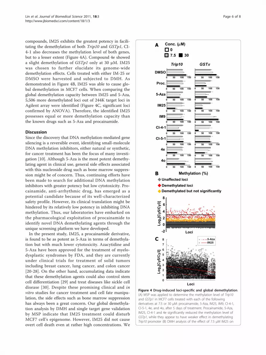
compounds, IM25 exhibits the greatest potency in facili-
tating the demethylation of both Trip10 and GSTp1, CI-
4-1 also decreases the methylation level of both genes,
but to a lesser extent (Figure 4A). Compound 4e showed
a slight demethylation of GSTp1 only at 30 μM. IM25
was chosen to further elucidate its genome-wide
demethylation effects. Cells treated with either IM-25 or
DMSO were harvested and subjected to DMH. As
demonstrated in Figure 4B, IM25 was able to cause glo-
bal demethylation in MCF7 cells. When comparing the
global demethylation capacity between IM25 and 5-Aza,
5,506 more demethylated loci out of 244K target loci in
Agilent array were identified (Figure 4C, significant loci
confirmed by ANOVA). Therefore, the identified IM25
possesses equal or more demethylation capacity than
the known drugs such as 5-Aza and procainamide.
Discussion
Since the discovery that DNA methylation-mediated gene
silencing is a reversible event, identifying small-molecule
DNA methylation inhibitors, either natural or synthetic,
for cancer treatment has been the focus of many investi-
gation [10]. Although 5-Aza is the most potent demethy-
lating agent in clinical use, general side effects associated
with this nucleoside drug such as bone marrow suppres-
sion might be of concern. Thus, continuing efforts have
been made to search for additional DNA methylation
inhibitors with greater potency but low cytotoxicity. Pro-
cainamide, anti-arrhythmic drug, has emerged as a
potential candidate because of its well-characterized
safety profile. However, its clinical translation might be
hindered by its relatively low potency in inhibiting DNA
methylation. Thus, our laboratories have embarked on
the pharmacological exploitation of procainamide to
identify novel DNA demethylating agents through the
unique screening platform we have developed.
In the present study, IM25, a procainamide derivative,
is found to be as potent as 5-Aza in terms of demethyla-
tion but with much lower cytotoxicity. Azacytidine and
5-Aza have been approved for the treatment of myelo-
dysplastic syndromes by FDA, and they are currently
under clinical trials for treatment of solid tumors
including breast cancer, lung cancer, and colon cancer
[20-28]. On the other hand, accumulating data indicate
that these demethylation agents could also control stem
cell differentiation [29] and treat diseases like sickle cell
disease [30]. Despite these promising clinical and in
vitro studies for cancer treatment and cell fate manipu-
lation, the side effects such as bone marrow suppression
has always been a great concern. Our global demethyla-
tion analysis by DMH and single target gene validation
by MSP indicate that IM25 treatment could disturb
MCF7 cell’s epigenome. However, IM25 did not cause
overt cell death even at rather high concentrations. We
A
BUnaffected loci
Demethylated loci
Demethylated but not significantly
-2
0
2
4
6
8
Loci
Lo
g2[C
y5
/Cy3
]
Methylation (%)
Trip10 GST
DMSO
Proc.
5-Aza
IM25
IM9
CI-4-1
CI-5-1
4e
4o
0 50 100 150 0 50 100 150
0 50 100 150
0 50 100 150
0 50 100 150
0 50 100 150
0 50 100 150
0 50 100 150
0 50 100 150
0 50 100 150
0 50 100 150
0 50 100 150
0 50 100 150
0 50 100 150
0 50 100 150
0 50 100 150
0 50 100 150
0 50 100 150
0
7.5 30
Conc. ( M)
C
Figure 4 Drug-induced loci-specific and global demethylation.
(A) MSP was applied to determine the methylation level of Trip10
and GSTp1 in MCF7 cells treated with each of the following
derivatives at 7.5 or 30 μM: procainamide, 5-Aza, IM25, IM9, CI-4-1,
CI-5-1, 4e, and 4o, after 5 days of treatment. Procainamide, 5-Aza,
IM25, CI-4-1 and 4e significantly reduced the methylation level of
GSTp1, while they appear to have weaker effect in demethylating
Trip10 promoter (B) DMH analysis of the effect of 7.5 μM IM25 on
Lin et al. Journal of Biomedical Science 2011, 18:3
http://www.jbiomedsci.com/content/18/1/3
Page 6 of 8

reason that IM25 treatment may act via altering the
gene expression profile and subsequently cause changes
in cell physiology/behaviour without affecting cell viabi-
lity. It will be crucial to determine if IM25 has the same
demethylation potency and safety in other cancer cell
types or stem cells. In addition, to determine the IM25’
effect in an animal model system will provide in-depth
information regarding its efficacy and safety upon sys-
temic administration, which can serve as justification for
its future clinical application as a direct antitumor drug,
an adjuvant, or other therapeutic uses.
On the other hand, present study also demonstrates
the feasibility of the established two-component reporter
system as an efficient drug screening platform in live
cells. While the efficacy of 5-Aza in reducing GSTp1
methylation is lower in the two-component system than
in the MSP assay, which possibly is attributed to the
engineered two-component stable clones, we reason that
the direct, non-invasive measurement and comparisons
of testing drugs in the two-component platform can
serve as a powerful while economic tool for rapid drug
screening at preclinical stage.
Conclusions
In summary, the impetus of this study is twofold. First,
our DNA methylation-targeted two-component reporter
system represents an expedient screening platform to
identify agents with DNA methylation-altering capabil-
ity. Second, we have identified the procainamide deriva-
tive IM25 which exhibits high global demethylation
potency, of which the translational value in cancer treat-
ment represents the focus of this investigation.
Additional material
Additional file 1: Supplementary materials. Additional file contains the
supplementary materials which include: Supplementary Figures S1 to S3
and Supplementary Tables S1 to S3
Acknowledgements
This work was supported, in part, by National Science Council in Taiwan
(NSC-96-2320-B-194-004 and NSC-95-2320-B-194-003 to SHH, NSC-98-3112-B-
194-001, NSC- 97-2320-B-194-003-MY3, and NSC-97-2627-B-006-003 to YWL),
and the National Institutes of Health (CA112250 to CSC).
Author details1Human Epigenomics Center, Department of Life Science, Institute of
Molecular Biology and Institute of Biomedical Science, National Chung
Cheng University, Chia-Yi, 621, Taiwan. 2Department of Pharmacology and
Toxicology, College of Pharmacy, Southwest Comprehensive Center for Drug
Discovery and Development, University of Arizona, Tucson, AZ 85721, USA.3Human Cancer Genetics Program, Department of Molecular Virology,
Immunology, and Medical Genetics, and the Comprehensive Cancer Center,
The Ohio State University, Columbus, OH 43210, USA. 4Division of Medicinal
Chemistry, College of Pharmacy, The Ohio State University, Columbus, OH
43210, USA.
Authors’ contributions
YSL, SGW, CCH, I.-W. Teng and MJT performed the experiments; AYS and
CSC designed and synthesized the compound library; THMH, CSC, SHH and
YWL supervised the research and wrote the manuscript. All authors read and
approved the final manuscript.
Competing interests
The authors declare that they have no competing interests.
Received: 21 September 2010 Accepted: 10 January 2011
Published: 10 January 2011
References
1. Balch C, Nephew KP, Huang TH, Bapat SA: Epigenetic “bivalently marked”
process of cancer stem cell-driven tumorigenesis. Bioessays 2007,
29(9):842-5.
2. Feinberg AP, Ohlsson R, Henikoff S: The epigenetic progenitor origin of
human cancer. Nat Rev Genet 2006, 7(1):21-33.
3. Hewagama A, Richardson B: The genetics and epigenetics of
autoimmune diseases. J Autoimmun 2009, 33(1):3-11.
4. Hsiao SH, Huang TH, Leu YW: Excavating relics of DNA methylation
changes during the development of neoplasia. Semin Cancer Biol 2009,
19(3):198-208.
5. Liu L, van Groen T, Kadish I, Tollefsbol TO: DNA methylation impacts on
learning and memory in aging. Neurobiol Aging 2009, 30(4):549-60.
6. Ohm JE, McGarvey KM, Yu X, Cheng L, Schuebel KE, Cope L,
Mohammad HP, Chen W, Daniel VC, Yu W, Berman DM, Jenuwein T,
Pruitt K, Sharkis SJ, Watkins DN, Herman JG, Baylin SB: A stem cell-like
chromatin pattern may predispose tumor suppressor genes to DNA
hypermethylation and heritable silencing. Nat Genet 2007, 39(2):237-42.
7. Richardson B: Primer: epigenetics of autoimmunity. Nat Clin Pract
Rheumatol 2007, 3(9):521-7.
8. Chuang JC, Yoo CB, Kwan JM, Li TW, Liang G, Yang AS, Jones PA: Comparison
of biological effects of non-nucleoside DNA methylation inhibitors versus
5-aza-2’-deoxycytidine. Mol Cancer Ther 2005, 4(10):1515-20.
9. Villar-Garea A, Fraga MF, Espada J, Esteller M: Procaine is a DNA-
demethylating agent with growth-inhibitory effects in human cancer
cells. Cancer Res 2003, 63(16):4984-9.
10. Yoo PA, Jones PA: Epigenetic therapy of cancer: past, present and future.
Nat Rev Drug Discov 2006, 5(1):37-50.
11. Lee BH, Yegnasubramanian S, Lin X, Nelson WG: Procainamide is a specific
inhibitor of DNA methyltransferase 1. J Biol Chem 2005, 280(49):40749-56.
12. Lin X, Asgari K, Putzi MJ, Gage WR, Yu X, Cornblatt BS, Kumar A,
Piantadosi S, DeWeese TL, De Marzo AM, Nelson WG: Reversal of GSTP1
CpG island hypermethylation and reactivation of pi-class glutathione S-
transferase (GSTP1) expression in human prostate cancer cells by
treatment with procainamide. Cancer Res 2001, 61(24):8611-6.
13. Lu Q, Wu A, Richardson BC: Demethylation of the same promoter
sequence increases CD70 expression in lupus T cells and T cells treated
with lupus-inducing drugs. J Immunol 2005, 174(10):6212-9.
14. Segura-Pacheco B, Trejo-Becerril C, Perez-Cardenas E, Taja-Chayeb L,
Mariscal I, Chavez A, Acuña C, Salazar AM, Lizano M, Dueñas-Gonzalez A:
Reactivation of tumor suppressor genes by the cardiovascular drugs
hydralazine and procainamide and their potential use in cancer therapy.
Clin Cancer Res 2003, 9(5):1596-603.
15. Martelli A, Campart GB, Canonero R, Mattioli F, Brambilla G: Testing of
metoclopramide and procainamide for their ability to induce genotoxic
effects in cultured mammalian cells. Toxicol Appl Pharmacol 1995,
131(2):185-91.
genome-wide methylation. The higher Log2[Cy5/Cy3] values indicate
stronger demethylation effects. (C) Genome-wide comparison of the
demetylation after IM25 and 5-Aza treatment. DMH microarrays
identified 5,506 more loci that are demethylated after IM25
treatment (open circles) than the result obtained after 5-Aza
treatment (close gray circles). The regressed lines depicted the
distribution of demethylation (dashed line for 5-Aza; solid line for
IM25) and all the listed 5,506 loci were confirmed significant by
ANOVA.
Lin et al. Journal of Biomedical Science 2011, 18:3
http://www.jbiomedsci.com/content/18/1/3
Page 7 of 8

16. Mereto E, Robbiano L, Ghia M, Allavena A, Martelli A, Brambilla G:
Evaluation of DNA-damaging, clastogenic, and promoting activities of
metoclopramide and procainamide in rats. Toxicol Appl Pharmacol 1995,
131(2):192-7.
17. Hsiao SH, Lee KD, Hsu CC, Tseng MJ, Jin VX, Sun WS, Hung YC, Yeh KT,
Yan PS, Lai YY, Sun HS, Lu YJ, Chang YS, Tsai SJ, Huang TH, Leu YW: DNA
methylation of the Trip10 promoter accelerates mesenchymal stem cell
lineage determination. Biochem Biophys Res Commun 2010, 400(3):305-12.
18. Hsu CC, Li HP, Hung YH, Leu YW, Wu WH, Wang FS, Lee KD, Chang PJ,
Wu CS, Lu YJ, Huang TH, Chang YS, Hsiao SH: Targeted methylation of
CMV and E1A viral promoters. Biochem Biophys Res Commun 2010,
402:228-34.
19. Yan PS, Chen CM, Shi H, Rahmatpanah F, Wei SH, Huang TH: Applications
of CpG island microarrays for high-throughput analysis of DNA
methylation. J Nutr 2002, 132(8 Suppl):2430S-2434S.
20. Bender CM, Pao MM, Jones PA: Inhibition of DNA methylation by 5-aza-
2’-deoxycytidine suppresses the growth of human tumor cell lines.
Cancer Res 1998, 58(1):95-101.
21. Chen J, JonesJ PA: Potentiation of MyoD1 activity by 5-aza-2’-
deoxycytidine. Cell Growth Differ 1990, 1(8):383-92.
22. Michalowsky LA, Jones PA: Gene structure and transcription in mouse
cells with extensively demethylated DNA. Mol Cell Biol 1989, 9(3):885-92.
23. Michalowsky LA, Jones PA: DNA methylation and drug resistance in
variants of C3H10T1/2 C1 8 cells. Prog Clin Biol Res 1986, 226:391-9.
24. Jones PA: Altering gene expression with 5-azacytidine. Cell 1985,
40(3):485-6.
25. Jones PA: Effects of 5-azacytidine and its 2’-deoxyderivative on cell
differentiation and DNA methylation. Pharmacol Ther 1985, 28(1):17-27.
26. Jones PA, Taylor SM, Mohandas T, Shapiro LJ: Cell cycle-specific
reactivation of an inactive X-chromosome locus by 5-azadeoxycytidine.
Proc Natl Acad Sci USA 1982, 79(4):1215-9.
27. Jones PA, Taylor SM: Cellular differentiation, cytidine analogs and DNA
methylation. Cell 1980, 20(1):85-93.
28. Constantinides PG, Jones PA, Gevers W: Functional striated muscle cells
from non-myoblast precursors following 5-azacytidine treatment. Nature
1977, 267(5609):364-6.
29. Konieczny SF, Emerson Cp Jr: 5-Azacytidine induction of stable
mesodermal stem cell lineages from 10T1/2 cells: evidence for
regulatory genes controlling determination. Cell 1984, 38(3):791-800.
30. Saunthararajah Y, Hillery CA, Lavelle D, Molokie R, Dorn L, Bressler L,
Gavazova S, Chen YH, Hoffman R, DeSimone J: Effects of 5-aza-2’-
deoxycytidine on fetal hemoglobin levels, red cell adhesion, and
hematopoietic differentiation in patients with sickle cell disease. Blood
2003, 102(12):3865-70.
doi:10.1186/1423-0127-18-3Cite this article as: Lin et al.: Identification of novel DNA methylationinhibitors via a two-component reporter gene system. Journal ofBiomedical Science 2011 18:3.
Submit your next manuscript to BioMed Centraland take full advantage of:
• Convenient online submission
• Thorough peer review
• No space constraints or color figure charges
• Immediate publication on acceptance
• Inclusion in PubMed, CAS, Scopus and Google Scholar
• Research which is freely available for redistribution
Submit your manuscript at www.biomedcentral.com/submit
Lin et al. Journal of Biomedical Science 2011, 18:3
http://www.jbiomedsci.com/content/18/1/3
Page 8 of 8