How to cite Complete issue More information about ... - Redalyc
10
How to cite Complete issue More information about this article Journal's webpage in redalyc.org Scientific Information System Redalyc Network of Scientific Journals from Latin America and the Caribbean, Spain and Portugal Project academic non-profit, developed under the open access initiative Revista Família, Ciclos de Vida e Saúde no Contexto Social ISSN: 2318-8413 [email protected] Universidade Federal do Triângulo Mineiro Brasil França Abrahão, Cesar Augusto; Massa, Virgínia; Tavares de Resende e Silva Abate, Débora; Reis Machado, Juliana; Maia Queiroz, Natália; Antônia dos Reis, Marlene Morphological kidney and heart analysis of autopsied hypertensive patients Revista Família, Ciclos de Vida e Saúde no Contexto Social, vol. 5, no. 3, 2017, September-December, pp. 372-380 Universidade Federal do Triângulo Mineiro Brasil Available in: https://www.redalyc.org/articulo.oa?id=497954858003
-
Upload
khangminh22 -
Category
Documents
-
view
0 -
download
0
Transcript of How to cite Complete issue More information about ... - Redalyc
How to cite
Complete issue
More information about this article
Journal's webpage in redalyc.org
Scientific Information System Redalyc
Network of Scientific Journals from Latin America and the Caribbean, Spain andPortugal
Project academic non-profit, developed under the open access initiative
Revista Família, Ciclos de Vida e Saúde no ContextoSocialISSN: [email protected] Federal do Triângulo MineiroBrasil
França Abrahão, Cesar Augusto; Massa, Virgínia; Tavares de Resende e Silva Abate,Débora; Reis Machado, Juliana; Maia Queiroz, Natália; Antônia dos Reis, Marlene
Morphological kidney and heart analysis of autopsied hypertensive patientsRevista Família, Ciclos de Vida e Saúde no Contexto Social,
vol. 5, no. 3, 2017, September-December, pp. 372-380Universidade Federal do Triângulo Mineiro
Brasil
Available in: https://www.redalyc.org/articulo.oa?id=497954858003
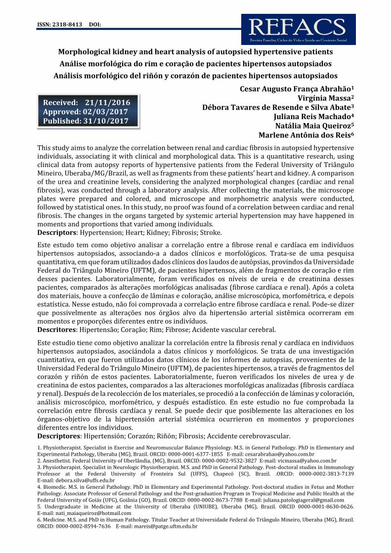
ISSN: 2318-8413 DOI:
1. Physiotherapist. Specialist in Exercise and Neuromuscular Balance Physiology. M.S. in General Pathology. PhD in Elementary and Experimental Pathology, Uberaba (MG), Brazil. ORCID: 0000-0001-6377-1855 E-mail: [email protected] 2. Anesthetist. Federal University of Uberlândia, (MG), Brazil. ORCID: 0000-0002-9532-3827 E-mail: [email protected] 3. Physiotherapist. Specialist in Neurologic Physiotherapist. M.S. and PhD in General Pathology. Post-doctoral studies in Immunology Professor at the Federal University of Fronteira Sul (UFFS), Chapecó (SC), Brazil. .ORCID: 0000-0002-3813-7139 E-mail: [email protected] 4. Biomedic. M.S. in General Pathology. PhD in Elementary and Experimental Pathology. Post-doctoral studies in Fetus and Mother Pathology. Associate Professor of General Pathology and the Post-graduation Program in Tropical Medicine and Public Health at the Federal University of Goiás (UFG), Goiânia (GO), Brazil. ORCID: 0000-0002-8673-7788 E-mail: [email protected] 5. Undergraduate in Medicine at the University of Uberaba (UNIUBE), Uberaba (MG), Brazil. ORCID 0000-0001-8630-0626. E-mail: [email protected] 6. Medicine. M.S. and PhD in Human Pathology. Titular Teacher at Universidade Federal do Triângulo Mineiro, Uberaba (MG), Brazil. ORCID: 0000-0002-8594-7636 E-mail: [email protected]
Morphological kidney and heart analysis of autopsied hypertensive patients
Análise morfológica do rim e coração de pacientes hipertensos autopsiados Análisis morfológico del riñón y corazón de pacientes hipertensos autopsiados
Cesar Augusto França Abrahão1 Virgínia Massa2
Débora Tavares de Resende e Silva Abate3 Juliana Reis Machado4 Natália Maia Queiroz5
Marlene Antônia dos Reis6
This study aims to analyze the correlation between renal and cardiac fibrosis in autopsied hypertensive individuals, associating it with clinical and morphological data. This is a quantitative research, using clinical data from autopsy reports of hypertensive patients from the Federal University of Triângulo Mineiro, Uberaba/MG/Brazil, as well as fragments from these patients’ heart and kidney. A comparison of the urea and creatinine levels, considering the analyzed morphological changes (cardiac and renal fibrosis), was conducted through a laboratory analysis. After collecting the materials, the microscope plates were prepared and colored, and microscope and morphometric analysis were conducted, followed by statistical ones. In this study, no proof was found of a correlation between cardiac and renal fibrosis. The changes in the organs targeted by systemic arterial hypertension may have happened in moments and proportions that varied among individuals. Descriptors: Hypertension; Heart; Kidney; Fibrosis; Stroke.
Este estudo tem como objetivo analisar a correlação entre a fibrose renal e cardíaca em indivíduos hipertensos autopsiados, associando-a a dados clínicos e morfológicos. Trata-se de uma pesquisa quantitativa, em que foram utilizados dados clínicos dos laudos de autópsias, provindos da Universidade Federal do Triângulo Mineiro (UFTM), de pacientes hipertensos, além de fragmentos de coração e rim desses pacientes. Laboratorialmente, foram verificados os níveis de ureia e de creatinina desses pacientes, comparados às alterações morfológicas analisadas (fibrose cardíaca e renal). Após a coleta dos materiais, houve a confecção de lâminas e coloração, análise microscópica, morfométrica, e depois estatística. Nesse estudo, não foi comprovada a correlação entre fibrose cardíaca e renal. Pode-se dizer que possivelmente as alterações nos órgãos alvo da hipertensão arterial sistêmica ocorreram em momentos e proporções diferentes entre os indivíduos. Descritores: Hipertensão; Coração; Rim; Fibrose; Acidente vascular cerebral.
Este estudio tiene como objetivo analizar la correlación entre la fibrosis renal y cardíaca en individuos hipertensos autopsiados, asociándola a datos clínicos y morfológicos. Se trata de una investigación cuantitativa, en que fueron utilizados datos clínicos de los informes de autopsias, provenientes de la Universidad Federal do Triângulo Mineiro (UFTM), de pacientes hipertensos, a través de fragmentos del corazón y riñón de estos pacientes. Laboratorialmente, fueron verificados los niveles de urea y de creatinina de estos pacientes, comparados a las alteraciones morfológicas analizadas (fibrosis cardíaca y renal). Después de la recolección de los materiales, se procedió a la confección de láminas y coloración, análisis microscópico, morfométrico, y después estadístico. En este estudio no fue comprobada la correlación entre fibrosis cardíaca y renal. Se puede decir que posiblemente las alteraciones en los órganos-objetivo de la hipertensión arterial sistémica ocurrieron en momentos y proporciones diferentes entre los individuos. Descriptores: Hipertensión; Corazón; Riñón; Fibrosis; Accidente cerebrovascular.
Received: 21/11/2016 Approved: 02/03/2017 Published: 31/10/2017

Abrahão CAF, Massa V, Abate DTRS, Machado JR, Queiroz NM, Reis MA Non-transmissible Disease and Grievances
372 ISSN 2318-8413 seer.uftm.edu.br/revistaeletronica/index.php/refacs REFACS (online) 2017; 5(3-Special Edition):372-380
INTRODUCTION ccording to the VII Brazilian Directive for Arterial Hypertension from the Brazilian Society of Cardiology,
Systemic Arterial Hypertension (SAH) happens when the levels of arterial pressure (AP) are above the reference levels for the general population. It can be classified as pre-hypertension (121x81mmHg a 139x89 mmHg) or actual hypertension, which has the stages: 1 (light - 140x90mmHg a 159x99 mmHg), 2 (moderate - 160x100mmHg a 179x109mmHg) and 3 (severe - equal or above 180x110mmHg)1.
The systemic arterial hypertension exposes the patient to the development of a series of functional and structural organic changes, and the hypertensive crisis is a clinical urgency that can damage many organs, leading to cerebral vascular accident, pulmonary edema, congestive cardiac insufficiency, aortic dissection, acute myocardial infarction, unstable angina, acute kidney failure and hypertensive encephalopathy2.
The SAH affects 25% of the adult world population, and the cases of the disease are predicted to grow 60% in 2025. Cerebrovascular and coronariopathy diseases have increasingly been associated to arterial pressure disturbances, which shows their high socioeconomic impact and their influence in the morbimortality of the population3.
In Brazil, the prevalence of SAH varies from 24.8 to 44.4%, making it the most prevalent of all cardiovascular diseases and affecting more than 36 million Brazilian adults. It is the greatest risk factor for cardiac and cerebrovascular lesions and the third greatest cause of disabilities4,5.
The SAH leads to a number of changes in the composition of the cardiac tissue, leading to a structural remodeling of the myocardium. Therefore, hypertensive patients are exposed to the development of the hypertensive hearth disease syndrome, which is the hypertrophy of the left ventricle in the absence of aortic stenosis and hypertrophic myocardiopathy. In hypertensive cardiopathy, the hypertrophy
and the apoptosis of cardiomyocytes, interstitial fibrosis and the hypertrophy of the cardiac microvasculature, through the exacerbated retention of collagen fibers types I and III in the interstice and around the intra-myocardiac arteries and arterioles, cosntitute the structural elements that define myocardial remodeling. The functional consequences of such remodelling are diverse, and the most representative is the development of congestive cardiac insufficiency6,7.
The essential SAH has devastating effects on the brain, associated to high rights of morbimortality. It is the main risk factor for cerebral vascular accidents and the main cause for cognitive deficits and dementia8. Hypertensive encephalopathy is a neurological syndrome brought forth by a sudden and sustained increase in systemic blood arterial pressure. It can be asymptomatic or lead to silent cerebral infarction and symptoms such as headaches, mental confusion, vision alterations, severe hypertension, vomit, sensory alterations and convultions9.
Chronic hypertension leads to a structural adaptation of cerebral vascular resistance, and consequently, to an elevation of the cerebral self-regulation threshold. Thus, hypertensive encephalopathy is believed to result from a sustained increase in blood pressure that exceeds the upper limit of self-regulation, dilating the cerebral arterioles and dissolving the blood-brain barrier, generating vasogenic edemas2.
Although reversible if readily recognized and treated, hypertensive encephalopathy can evolve into coma and even death, if treatment is late. Even if it occurs in people with primary hypertension, it is more common in the mutual presence of hypertension and kidney disease, and it may be a complication of kindey transplants10.
The mechanism through which SAH could cause kidney lesions can be divided into three categories: glomerular ischemia secondary to vasoconstriction, glomerulosclerosis due to intracapillary hypertension, and interstitial fibrosis. Goldblatt et al., in the decade of 1930,
A

Abrahão CAF, Massa V, Abate DTRS, Machado JR, Queiroz NM, Reis MA Non-transmissible Disease and Grievances
373 ISSN 2318-8413 seer.uftm.edu.br/revistaeletronica/index.php/refacs REFACS (online) 2017; 5(3-Special Edition):372-380
demonstrated that the reduction of kidney perfusion can generate a sustained elevation of blood pressure, subsequently associated to the renin-angiotensin-aldosterone system11,12. Aldosterone plays an important role in the pathogenesis of hypertension, vascular remodeling, left ventricular hypertrophy and kidney diseases, especially those which cause with proteinuria and glomerulosclerosis in hypertensive individuals13.
In the advanced phase of kidney failure, aldosterone values increase significantly with the decrease of glomerular filtration due to the activation of the renin-angiotensin-aldosterone system, secondary to the alteration of glomerular hemodynamics. This contributes to the lesion of target organs, affecting primarily kidneys, brain and heart13.
Kidney interstitial fibrosis is the final pathway for almost all forms of renal disease. The study of fibrosis is of great value for several kidney diseases that become terminal. During SAH, such a study can be worthwhile, since interstitial fibrosis, in addition to being a bad prognosis, is an efficient marker to evaluate all pathways that injure the kidney in the SAH (ischemia, glomerular hypertension and inflammatory state) 14.
Chronic kidney disease has demonstrated to be an independent risk factor for cardiovascular disease, which in turn is the main cause of morbidity and mortality in chronic kidney disease patients. Similar to this, numerous studies indicate that chronic kidney diseases are associated with the high prevalence of cerebral vascular accidents15.
Hypertension is a systemic disease with high morbidity and mortality, and thus, it becomes important to understand the relationship between the organs affected by the SAH, and compare the intensity of lesions such as cardiac fibrosis, kidney fibrosis and the presence of CVAs in autopsied hypertensive patients. The present study aims to analyze kidney and heart fibrosis in autopsied subjects, associating them to clinical and morphological data. METHOD
Clinical data of the autopsy reports of twenty-three hypertensive patients were used, all from the General Hospital of the Federal University of the Triângulo Mineiro (HC-UFTM), Uberaba, Brazil, and who had been studied in General Pathology classes in the period from 1986 to 2007. Heart and kidney fragments from these patients were also used for morphometric analyses, laboratory data (urea and creatinine levels compared to morphological changes) and morphological aspects of autopsied patients (cardiac and kidney fibrosis). This study included patients who presented hypertensive heart disease and/or kidney sclerosis (benign or malignant) as changes provoked by the SAH. Patients with primary kidney diseases (glomerulopathy, interstitial nephritis such as pyelonephritis, vasculopathy) or carriers of systemic diseases damaging to the kidney (diabetes, lupus erythematosus, hepatitis or acute or chronic inflammatory diseases) were excluded from the study. To analyze the kidney material, fragments of the middle pole of the right kidney were used, and to analyze the cardiac material, fragments of the middle third of the left ventricle were removed from the autopsied patients. Após o processamento, o fragmento parafinizado foi submetido a cortes seriados de espessura adequada para confecção das lâminas. Then, the colorations of picro-sirius (PS) and hematoxylin and eosin (HE) were carried out. The microscope plates that had been colored with picro-sirius were used to analyze cardiac fibrosis and quantify the collagen in the mesangial matrix. The morphometry was performed through the capturing of images subsequently sent and recorded for analysis in the ImageJ software(an Image ProPlus software). The morphometric analysis of the fibrous tissue was performed in the kidney and heart samples using digital morphometry. In the polarized image, the fibrous connective tissue showed itself to be birefringent and was marked by the observer, and through it, the percentage of fibrosis per area of the analyzed field was obtained, as shown in Image 1.

Abrahão CAF, Massa V, Abate DTRS, Machado JR, Queiroz NM, Reis MA Non-transmissible Disease and Grievances
374 ISSN 2318-8413 seer.uftm.edu.br/revistaeletronica/index.php/refacs REFACS (online) 2017; 5(3-Special Edition):372-380
For the statistical analysis, a spreadsheet was created in Microsoft Excel. The clinical, laboratory and morphological variables were tested to verify that they presented normal distribution, through the test of Kolmogorov-Smirnov. The following tests were used: student's t"tests (to compare two groups with parametric variables) Mann-Whitney's test (to compare two groups with
non-parametric variables), Pearson's (to correlate groups that presented normal distribution) and Spearman's correlation (to correlate non-parametric samples between groups). The differences were considered statistically significant when p was less than 5% (P < 0.05).
Image 1. Computer system for the quantification of fibrosis. Autopsied patients from 1986 to 2007, HC-UFTM. Uberaba, MG, 2016.
Automatic microscopic computed morphometry. In (A) the light microscope can be seen, connected to a camera which in turn is connected to the computer. In (B) the steps followed by the KS 300 Carl Zeiss software can be seen; in (C), the capture of the image to be quantified with polarized light; and in (D), the result of the quantification of the captured image, in percentages.
RESULTS The age average of the 23 autopsied hypertensive patients was 59 years of age (SD=16.14), 54.5% of which were elders (>65 years old) (Image 2A).16 patients (72.6%) were male (Image2B). The most common cause of death was cardiovascular (45.5%), followed by infection (27.3%), and by
digestive (22.7%) and neoplastic (4.5%) causes (Image 2C). The average Body Mass Index (BMI) was 22.27 (SD=4.96) (Image 2D). The average cardiac fibrosis, on the other hand, was 2.78% (SD=1.03) while renal fibrosis had an average of 12.91% (SD =2.62) (Image 3).
Image 2. Clinical and epidemiological data. Autopsied patients from 1986 to 2007, HC-UFTM. Uberaba, MG, 2016.

Abrahão CAF, Massa V, Abate DTRS, Machado JR, Queiroz NM, Reis MA Non-transmissible Disease and Grievances
375 ISSN 2318-8413 seer.uftm.edu.br/revistaeletronica/index.php/refacs REFACS (online) 2017; 5(3-Special Edition):372-380
Image 3. Percentage of cardiac fibrosis and kidney fibrosis. Autopsied patients from 1986 to 2007, HC-UFTM. Uberaba, MG, 2016.
Percentage of cardiac fibrosis and kidney fibrosis. Horizontal lines represent the averages and vertical lines represent the standard deviation from the average. Statistical analysis calculated through
Student's T-Test. A positive but not significant correlation was observed between the percentage of cardiac and kidney fibrosis (p = 0.15 and r = 0.31) (Image 4).
Image 4. Correlation between cardiac fibrosis and kidney fibrosis. Autopsied patients from 1986 to 2007, HC-UFTM. Uberaba, MG, 2016.
Note: correlation of the percentage of cardiac fibrosis (Y-axis) relative to the percentage of kidney fibrosis (X-axis). Statistical analysis conducted through Spearman's correlation test.

Abrahão CAF, Massa V, Abate DTRS, Machado JR, Queiroz NM, Reis MA Non-transmissible Disease and Grievances
376 ISSN 2318-8413 seer.uftm.edu.br/revistaeletronica/index.php/refacs REFACS (online) 2017; 5(3-Special Edition):372-380
Among the hypertensive patients evaluated in this study, 16 (72.7%) had had previous episodes of cerebral vascular accidents. From these, 80% had hemorrhagic CVAs and 20%, ischemic CVAs. When comparing the percentage of cardiac fibrosis to that of kidney fibrosis
among the patients who had had CVAs and those who had not, a statistically significant difference was not observed for any of the two measures (P=0.52 and P=0.53, respectively) (Images 5A and B).
Image 5. Percentage of cardiac and kidney fibrosis according to CVA. Autopsied patients from 1986 to 2007. Uberaba, MG, 2016.
A. Percentage of cardiac fibrosis in individuals with and without previous CVA instances. Horizontal lines represent the medians, the bars represent the percentages 25-75% and the vertical lines represent the percentages 10-90%. Statistical analysis through the Mann-Whitney test. B. Percentage of kidney fibrosis in individuals with and without previous CVA instances. Horizontal lines represent the averages and vertical lines represent the standard deviation from the average. Statistical analysis calculated through Student's T-Test.
When comparing the percentage of cardiac fibrosis to that of kidney fibrosis among the hyperuricemic patients and those who had normal urea levels, a statistically significant difference was not observed for any of the two measures (P=0.59 and P=0.76, respectively). Similarly, there were no statistically significant differences in the comparison between cardiac fibrosis and kidney fibrosis, when comparing normal serum creatinine levels to those who had alterations in these levels. DISCUSSION Systemic arterial hypertension (SAH) is responsible for approximately 46% of all deaths in Brazil. This disease, frequent and disturbing, affects several organs, such as heart, kidney and brain, which are therefore said to be the target organs of SAH. The mechanisms through which SAH damages target organs and leads to vascular events are still not well-known16. The SAH is closely linked to the kidney, because lesions in that organ can be both the
cause and the consequence of high blood pressure. The brain is another target organ of the SAH, since it leads to the deterioration of the walls of cerebral arteries, causing cerebral vascular accidents (CVAs), as the final results of predisposing conditions17. Some studies suggest an intimate relationship between blood pressure and CVA mortality, being that among patients treated with antihypertensive medication, an increase of one mmHg in the systolic arterial BP increases mortality by CVA in 2% 18. In this work, most individuals were male, corroborating previous studies 19-22. Cardiovascular complications were the main causes of death in this study. The BMI of the patients was within a normal range (< 25) suggesting that the SAH can occur in non-obese patients, although literature findings show that increased body fat is associated with an increase in the risk of SAH. Within the morphological alterations of hypertensive heart disease, fibrosis is one of the most common changes among hypertensive patients. It is characterized by

Abrahão CAF, Massa V, Abate DTRS, Machado JR, Queiroz NM, Reis MA Non-transmissible Disease and Grievances
377 ISSN 2318-8413 seer.uftm.edu.br/revistaeletronica/index.php/refacs REFACS (online) 2017; 5(3-Special Edition):372-380
excessive collagen fibers or their diffuse accumulation, increasing myocardial rigidity, which can lead to a dysfunction of the left ventricle, finally culminating in cardiac insufficiency7. In this study, not all patients presented fibrosis of the heart tissue in large quantities. These results may be due to the intensity of the SAH and to the age group of the individuals analyzed in the study, since some individuals were non-elderly adults, with an average age of 59 years, perhaps not yet presenting an increase in the accumulation of collagen fibers fibers, corroborating works that show that the elderly are more susceptible to fibrosis, since in this age group there is an increase in the synthesis and a decrease in collagen degradation. One of the main morphological findings in the kidney of hypertensive patients is that glomerulosclerosis may occur through 3 main pathways: glomerular ischemia due to vascular lesion; glomerular hypertension, through the loss of self-regulation; and through the activation of the renina-angiotensin system (RAS). All these pathways can compromise not only kidney vessels (arterial hyalinosis, fibrinoid necrosis) or the glomerulus (glomerulosclerosis), but also the kidney interstitium, leading to fibrosis14. In this present study, the percentage of fibrosis in the medium portion of the right kidney was evaluated, showing areas with the presence of collagen in all evaluated individuals, corroborating a work in which the increase in the production and an accumulation of collagen fibers type I and III was proportionate to the intensity of the lesions caused by the SAH23. In this study, more than 70% of individuals presented episodes of CVA, and 80% of these were hemorrhagic. This can be justified by the fact that hemorrhagic CVAs seem to be directly related to the elevation of blood pressure, while the ischemic CVA is explained by atherosclerotic lesions, although these are also related to the SAH24. In this study no correlation was found between cardiac and kidney fibrosis; what explains this result is the fact that some patients presented cardiac fibrosis in small
quantities, even presenting high kidney fibrosis. Literature, however, reports that the increase of arterial BP is a determining factor that acts on the structure of the heart25.
Similarly, there was no relationship was observed between the intensity of cardiac and kidney fibrosis and CVA episodes. No previous reports were found in the literature highlighting this relationship. Considering this, it is suggested that the involvement of the target organs can occur at different times and that they are very changeable, from one individual to another.. However, studies show that there is a higher prevalence of CVA in hypertensive patients with glomerulossclerosis than in those without it26,27. The laboratory analysis of the patients who presented episodes of CVA and those who did not found a similarity between their levels of urea and creatinine. However, concerning creatinine concentration, studies show that high concentration levels are significantly related to CVA cases in patients with or without hypertension 17,28. In another study, however, urea and creatinine levels showed no significant difference between patients with CVA and patients from a control group29. CONCLUSION The most common causes of death had cardiovascular origins, and all patients presented cardiac and kidneyfibrosis. Although the correlation test between cardiac and kidney fibrosis was positive and regular, it was also not significant. 72% of the surveyed patients had had previous instances of cerebral vascular accident, 80% of the hemorrhagic type. Considering all patients, the percentages of cardiac and kidney fibrosis were not significant. The percentage of cardiac and kidney fibrosis did not vary between patients who presented or not changes in urea (p=0.59 and p=0.76, respectively) and creatinine levels (p=0.74 and p=0.67, respectively). It can be concluded that, in the group of patients surveyed in the period considered, among the individuals with systemic arterial
Abrahão CAF, Massa V, Abate DTRS, Machado JR, Queiroz NM, Reis MA Non-transmissible Disease and Grievances
378 ISSN 2318-8413 seer.uftm.edu.br/revistaeletronica/index.php/refacs REFACS (online) 2017; 5(3-Special Edition):372-380
hypertension, there is kidney fibrosis and cardiac fibrosis, as well as other changes in the target organs of systemic arterial hypertension, which probably took place at different times and in varying proportions from one individual to the other. REFERENCES 1. Sociedade Brasileira de Cardiologia. VII Diretrizes Brasileiras de Hipertensão Arterial. Arq Bras Cardiol. 2016; 107(Supl. 3):10-3. 2. Lagi A, Cencetti S. Hypertensive emergencies: a new clinical approach. Clin Hypertens. 2015; 21(20):1-7; doi:10.1186/s40885-015-0027-4 3. Balu S, Thomas J. Incremental expenditure of treating hypertension in the United States. Am J Hypertens. 2006; 19(8):810-6. 4. Cipullo JP, Martin JFV, Ciorlia LAS, Godoy MRP, Cação JC, Loureiro AAC, et al. Prevalência e fatores de risco para hipertensão em uma população urbana brasileira. Arq Bras Cardiol. 2010; 94(4): 519-26. doi: 10.1590/S0066-782X2010005000014 5. Picon RV, Fuchs FD, Moreira LB, Riegel G, Fuchs SC. Trends in prevalence of hypertension in Brazil: a systematic review with metaanalysis. PLos One. 2012; 7(10):e48255. 6. Anguita M, Crespo M, Galván E, Jiménez M, Alonso L, Muniz J. Prevalence of heart failure in the spanish general population aged over 45 years. The PRICE Study. Rev Esp Cardiol. 2008; 61:1041-9. 7. Winterberg, PD, Jiang R, Maxwell JT, Wang B, Wagner MB. Myocardial dysfunction occurs prior to changes in ventricular geometry in mice with chronic kidney disease (CKD). Physiol Rep. [Internet]. 2016 [cited in 25 mar 2016]; 4(5): e12732. Available in: https://www.ncbi.nlm.nih.gov/pmc/articles/PMC4823595/pdf/PHY2-4-e12732.pdf. doi: 10.14814/phy2.12732,PMCID:PMC4823595 8. Dahlof B. Prevention of stroke in patients with hypertension. Am J Cardiol. 2007; 100 (3 Suppl):S17-S24. 9. Kobayashi S, Hoshi A, Tanaka K., Ugawa Y. Bikateral insular lesions related to malignant hypertension. Intern Med. 2012;
51(13):1805-6, doi: 10.2169/internalmedicine.51.6692 10. Thambisetty M, Biousse V, Newman NJ. Hypertensive brainstem encephalopathy: clinical and radiographic features. J Neurol Sci. 2003; 208(1-2):93-9. 11. Goldblatt H, Lynch J, Hanzal RF, Summerville WW. Studies on experimental hypertension. J Exp Med. 1934; 59(3):347-79. 12. Yoshimoto T, Hirata Y. Aldosterone as a Cardiovascular Risk Hormone. Endocr J. 2007; 54(3):359-70. 13. Sánchez-Lozada LG, Soto V, Tapia E, Avila-Casado C, Sautin YY, Nakagawa T, et al. Role of oxidative stress in the renal abnormalities induced by experimental hyperuricemia. Am J Physiol, Ren Physiol. 2008; 295(4): 1134-41. 14. Zucchelli P, Zuccala A. The kidney as a victim of essential hypertension. J Nephrol. 1997; 10(4):203-6. 15. Subbiah AK, Chhabra YK., Mahajan S. Cardiovascular disease in patients with chronic kidney disease: a neglected subgroup. Heart Asia. 2016; 8(2):56-61. doi:10.1136/heartasia-2016-010809 16. Rothwell PM, Howard SC, Dolan E, Dobson JE, Dahlof B, Sever PS, et al. Prognostic significance of visit-to-visit variability, maximum systolic blood pressure and episodic hypertension. Lancet. 2010; 375(9718):895-905. 17. Lekawanvijit S, Krum H. Cardiorenal syndrome: acute kidney injury secondary to cardiovascular disease and role of protein-bound uraemic toxins. J Physiol. 2014; 592(Pt 18):3969-83. doi:10.1113/jphysiol.2014.273078 18. Palmer AJ, Bulpitt, CJ, Fletcher AE, Beevers DG, Goles EC, Ledingham JG, et al. Relation between blood pressure and stroke mortality. Hypertension. 1992; 20(5): 601-5. 19. Petrea RE, Beiser AS, Seshadri S, Kelly-Hayes M, Kase CS, Wolf PA. Gender differences in stroke incidence and poststroke disability in the framingham heart study. Stroke. 2009; 40(4):1032-7. 20. Sarafidis PA, Li S, Chen CS, Collins AJ, Brow WW, Kagl MJ, et al. Hypertension awareness, treatment, and control in chronic kidney disease. Am Med. 2008; 121(4): 332-40. doi:10.1016/j.amjmed.2007.11.025

Abrahão CAF, Massa V, Abate DTRS, Machado JR, Queiroz NM, Reis MA Non-transmissible Disease and Grievances
379 ISSN 2318-8413 seer.uftm.edu.br/revistaeletronica/index.php/refacs REFACS (online) 2017; 5(3-Special Edition):372-380
21. Pereira M, Lunet N, Azevedo A, Barros H. Differences in prevalence, awareness, treatment and control of hypertension between developing and developed countries. J Hypertens. 2009; 27(5):963-75. 22. Chor D, Ribeiro AL, Carvalho MS, Duncan BB, Lotufo PA, Nobre AA, et al. Prevalence, awareness, treatment and influence of socioeconomic variables on control of high blood pressure: results of the ELSA-Brasil Study. PLos One. 2015; 10(6):e0127382. 23. Kiuchi MG, Mion D. Chronic kidney disease and risk factors responsible for sudden cardiac death: a whiff of hope? Kidney Res Clin Pract.. 2016; 35(1):3-9. doi:10.1016/j.krcp.2015.11.003 24. Kaplan NM. Hypertension trials: 1900-2000. Curr Opin Nephrol Hypertens. 2001; 10(4):501-5. 25. Deo R, Lin F, Vittinghoff E, Tseng ZH, Hulley SB, Shlipak MG. Kidney disfunction and sudden cardiac death among women with coronary heart disease. Hypertension. 2008; 51(6):1578-82. doi: 10.1161/hypertensionaha.107.103804
26. Thrift AG, Dewey HM, Macdonell RA, Mcneill JJ, Donnan GA. Incidence of the major stroke subtypes: initial findings from the North East: Melbourne stroke incidence study. Stroke. 2001; 32(8):1732-8. 27. Gorostidi M, Marin R. Nefropatia isquêmica e aterosclerótica. Nefrologia (Madrid). 2004; 24:73-83. 28. Wannamethee SG, Shaper AG, Perry Ij. Serum creatinine concentration and risk of cardiovascular disease: a possible marker for increased risk of stroke. Stroke. 1997; 28(3):557-63. 29. Urbanska EM, Luchowski P, Luchowska E, Pniewski J, Woźniak R, Chodakowska-Zebrowska M, et al. Serum kynurenic acid positively correlates with cardiovascular disease risk fator, homocysteine a study instroke patients. Pharmacol Rep. 2006; 58:507-11.
CONTRIBUTIONS All authors contributed equally in the various stages of the research and in the writing of the article.
How to cite this article (Vancouver) Abrahão CAF, Massa V, Abate DTRS, Machado JR, Queiroz NM, Reis MA. Morphological kidney and heart analysis of autopsied hypertensive patients. REFACS [Internet]. 2017 [Cited in insert day, month and year of access];5(3):372-380. Available from: insert access link. DOI: Insert the DOI link.
How to cite this article (ABNT) ABRAHÃO, C. A. F. et al. Morphological kidney and heart analysis of autopsied hypertensive patients. REFACS, Uberaba, MG, v. 5, n. 3, p. 372-380, 2017. Available from: < insert access link >. Access in: insert day, month and year of access. DOI: Insert the DOI link.
How to cite this article (APA) Abrahão, C. A. F., Massa, V., Abate, D. T. R. S., Machado, J. R., Queiroz, N. M. & Reis, M. A. (2017). Morphological kidney and heart analysis of autopsied hypertensive patients. REFACS, 5(3), 372-380. Recovered in insert day, month and year of access from... Insert access link. DOI: Insert the DOI link.