Performance modeling and automatic ghost zone optimization for iterative stencil loops on GPUs
Upload
independentCategory
view
1download
0
High-Resolution, High-Throughput, Positive-TonePatterning of Poly(ethylene glycol) by Helium BeamExposure through Stencil MasksEliedonna E. Cacao1, Azeem Nasrullah2, Tim Sherlock2, Steven Kemper1, Katerina Kourentzi1,
Paul Ruchhoeft2,3*, Gila E. Stein1, Richard C. Willson1,4,5*
1 Department of Chemical and Biomolecular Engineering, University of Houston, Houston, Texas, United States of America, 2 Department of Electrical and Computer
Engineering, University of Houston, Houston, Texas, United States of America, 3 Department of Biomedical Engineering, University of Houston, Houston, Texas, United
States of America, 4 Department of Biology and Biochemistry, University of Houston, Houston, Texas, United States of America, 5 The Methodist Hospital Research
Institute, Houston, Texas, United States of America
Abstract
In this work, a collimated helium beam was used to activate a thiol-poly(ethylene glycol) (SH-PEG) monolayer on gold toselectively capture proteins in the exposed regions. Protein patterns were formed at high throughput by exposing a stencilmask placed in proximity to the PEG-coated surface to a broad beam of helium particles, followed by incubation in a proteinsolution. Attenuated Total Reflectance–Fourier Transform Infrared Spectroscopy (ATR–FTIR) spectra showed that SH-PEGmolecules remain attached to gold after exposure to beam doses of 1.5–60 mC/cm2 and incubation in PBS buffer for onehour, as evidenced by the presence of characteristic ether and methoxy peaks at 1120 cm21 and 2870 cm21, respectively.X-ray Photoelectron Spectroscopy (XPS) spectra showed that increasing beam doses destroy ether (C–O) bonds in PEGmolecules as evidenced by the decrease in carbon C1s peak at 286.6 eV and increased alkyl (C–C) signal at 284.6 eV. XPSspectra also demonstrated protein capture on beam-exposed PEG regions through the appearance of a nitrogen N1s peakat 400 eV and carbon C1s peak at 288 eV binding energies, while the unexposed PEG areas remained protein-free. Thecharacteristic activities of avidin and horseradish peroxidase were preserved after attachment on beam-exposed regions.Protein patterns created using a 35 mm mesh mask were visualized by localized formation of insoluble diformazanprecipitates by alkaline phosphatase conversion of its substrate bromochloroindoyl phosphate-nitroblue tetrazolium (BCIP-NBT) and by avidin binding of biotinylated antibodies conjugated on 100 nm gold nanoparticles (AuNP). Patterns createdusing a mask with smaller 300 nm openings were detected by specific binding of 40 nm AuNP probes and by localized HRP-mediated deposition of silver nanoparticles. Corresponding BSA-passivated negative controls showed very few bound AuNPprobes and little to no enzymatic formation of diformazan precipitates or silver nanoparticles.
Citation: Cacao EE, Nasrullah A, Sherlock T, Kemper S, Kourentzi K, et al. (2013) High-Resolution, High-Throughput, Positive-Tone Patterning of Poly(ethyleneglycol) by Helium Beam Exposure through Stencil Masks. PLoS ONE 8(5): e56835. doi:10.1371/journal.pone.0056835
Editor: Wei-Chun Chin, University of California, Merced, United States of America
Received April 3, 2012; Accepted January 16, 2013; Published May 24, 2013
This is an open-access article, free of all copyright, and may be freely reproduced, distributed, transmitted, modified, built upon, or otherwise used by anyone forany lawful purpose. The work is made available under the Creative Commons CC0 public domain dedication.
Funding: This work was supported by Grant No. U54 AI057156 from the National Institute of Allergy and Infectious Diseases/National Institutes of Health. Itscontents are solely the responsibility of the authors and do not necessarily represent the official views of the RCE Programs Office, NIAID, or NIH. Support was alsoprovided by the National Science Foundation, Grant No. CMMI-0900743, CBET-1133965 and CBET-0932971, the Alliance for Nanohealth Competitive ResearchProgram, Grant No. W81XWH-09-2-0139, and the Welch Foundation, Grant No. E-1264. The funders had no role in study design, data collection and analysis,decision to publish, or preparation of the manuscript.
Competing Interests: Co-author R. Willson is a PLOS ONE Editorial Board member. This does not alter the authors’ adherence to all the PLOS ONE policies onsharing data and materials.
* E-mail: [email protected] (PR); [email protected] (RCW)
Introduction
Surface patterning of biomolecules is important in the study of
cell adhesion, in tissue engineering, and in the development of
diagnostics and biomedical assays such as protein nanoarrays
[1,2,3,4]. Controlled attachment of biomolecules can be achieved
by approaches generally categorized as ‘‘top-down’’ (e.g., photo-
lithography, focused ion beam lithography, electron beam
lithography, nanografting), or ‘‘bottom-up’’ (e.g., self-assembly of
monolayers, dip-pen nanolithography, micro/nano contact print-
ing or stamping), or combinations of these techniques. Top-down
techniques manipulate an instrument to modify the bulk material
to create patterns. Photolithography is a high-throughput litho-
graphic process but its resolution is diffraction-limited below the
micron-scale, and it is expensive to use for low-volume manufac-
turing. Electron beam and focused ion beam lithography have
very high resolution but very low throughput. Bottom-up
techniques take advantage of the spontaneous organization of
molecules to produce a more complex and functional patterned
material. These methods can allow patterning below the current
resolution of lithographic techniques, but often involve high costs
(for large areas) and imperfect patterning. Current approaches to
biomolecule patterning, therefore, often employ combinations of
top-down and bottom-up techniques [4,5,6].
Several approaches have been taken to biomolecule, specifically
protein, patterning on surfaces. Utilizing self-assembly and
electron-beam lithography, Geyer et al. [7] and Golzhauser et
al. [8] have demonstrated the use of nitro-terminated aromatic
thiols assembled on gold surfaces to create surface patterns of
PLOS ONE | www.plosone.org 1 May 2013 | Volume 8 | Issue 5 | e56835

chemical functionality. Upon exposure to the electron beam, the
nitro groups are converted to amines while the underlying
aromatic groups are dehydrogenated and cross-linked. The amine
groups were then used to attach molecules (carboxylic acid
anhydrides and rhodamine dyes) to the exposed regions of the
surface. Perhaps the most popular approach, however, involves the
use of poly(ethylene glycol) (PEG) [9,10,11], which is well-known
for its resistance to non-specific adsorption of proteins through a
combination of entropic (steric stabilization) and enthalpic
(hydrogen bonding) mechanisms [12,13]. Entropic passivation is
favored by longer chains which can assume a greater variety of
configurations [13,14,15], but even smaller ethylene oxide chains
of 3–6 monomer units can resist protein adsorption as long as the
molecular conformation is helical or amorphous, favoring a more
stable interfacial layer of tightly-bound water [12,13,15,16,17,18].
Electron beam lithography is commonly used to create protein
patterns on PEG surfaces. Hong et al. [19] observed that proteins
selectively attach to electron beam-exposed regions of amine-
terminated PEG spin-coated on silicon. Patterns consisting of
multiple proteins also can be formed by introducing PEG bearing
different functionalities (e.g., biotin, maleimide, aminooxy or metal
chelate) that are patterned over several exposures [20]. Harnett et
al. [21] used low-energy electron beam exposure to destroy the
amine functionality in selected regions of an amine-functionalized
silane-PEG on silicon surfaces, allowing proteins to attach only on
the unexposed regions (negative-tone patterning). Inert silane-PEG
on silicon and thiol-PEG on gold have also been used to create a
protein-resistant surface, with electron beam exposure used to
remove the SAM on exposed regions and protein-reactive PEGs
used to backfill the exposed regions (positive-tone patterning)
[22,23].
In this study, we explored the use of helium beam proximity
lithography to create micron and nanoscale patterns of proteins on
grafted PEG in a high-throughput manner. Unlike focused ion
beam lithography techniques, proximity printing forms its patterns
by exposing a thin membrane with etched openings corresponding
to the desired pattern (a ‘‘stencil mask’’) to a broad beam of helium
particles, which are either stopped in the opaque regions of the
mask or pass through the openings. In this way, the pattern can be
formed very quickly and without the need for expensive ion optics.
After exposure to the beam, protein patterns were detected and
visualized using three different methods chosen to be representa-
tive of common approaches: (1) formation of localized diformazan
precipitates by patterned alkaline phosphatase (AP) from its
substrate BCIP-NBT [24,25,26], (2) binding of gold nanoparticle
probes (40 nm and 100 nm gold particles conjugated with
antibodies) [27,28,29], by patterned antibodies and avidin, and
(3) localized redox formation of silver deposits mediated by
patterned horseradish peroxidase (HRP) [30,31,32,33].
Materials and Methods
MaterialsAlkaline phosphatase from bovine intestinal mucosa (Product
number P6772), goat polyclonal anti-rabbit antibodies (R2004),
goat polyclonal anti-mouse antibodies (M8645), hen egg white
lysozyme (L6876) and BCIP-NBT phosphatase substrate (72091)
were purchased from Sigma-Aldrich (St. Louis, MO), while avidin
(21121) was from Pierce Protein Research Products, Thermo
Scientific (Rockford, IL). Rabbit polyclonal anti-Rickettsia antibod-
ies were generously supplied by Dr. Juan Olano (University of
Texas Medical Branch, Galveston, TX) and mouse anti-lysozyme
monoclonal D1.3 antibody [34] was produced for us in hollow-
fiber hybridoma culture by Biovest (Minneapolis, MN). Strepta-
vidin-horseradish peroxidase (strep-polyHRP80, 65R-S105PHRP)
conjugate from Fitzgerald Industries International (Action, MA) is
a polymer consisting of 80 streptavidin molecules per conjugate
and 5 HRP monomer molecules per streptavidin giving an
Figure 1. A schematic of helium beam-induced proteinpatterning on PEG. Panels A and B show the electron images of35 mm mesh and 300 nm masks, respectively. Top-down helium beamexposure through a mask (C) of grafted PEG on surface allowed proteinspreferentially to attach on irradiated regions (D) to form patterns.doi:10.1371/journal.pone.0056835.g001
Figure 2. ATR-FTIR spectra of beam protected (unexposed) andirradiated (exposed) PEG at different helium beam doses.Spectra of grafted PEG subjected to different beam doses andincubated in PBS for 1 hour show the characteristic ether and methoxypeaks at 1120 cm21 and 2870 cm21, respectively, which also arepresent in unexposed PEG.doi:10.1371/journal.pone.0056835.g002
Patterning of Poly(ethylene glycol) by Helium Beam
PLOS ONE | www.plosone.org 2 May 2013 | Volume 8 | Issue 5 | e56835
estimated total of 400 HRP molecules per conjugate. Biotinylated
antibodies and AP were prepared using the EZ-Link Sulfo-NHS-
LC-Biotinylation Kit (21435, Pierce Protein Research Products,
Thermo Scientific, Rockford, IL). Gold nanoparticle probes were
prepared via EDC-NHS chemistry (based on the manufacturer’s
protocol) on carboxylated 100 nm gold nanoparticles (20-PC-100,
Nanopartz Inc., Loveland, CO). Thiol-polyethylene glycol (MW
5000, ,112 PEG monomers, PEG3-0021) was from Nanocs
(Boston, MA). HRP redox silver staining solution (6010,
EnzMetTM staining kit) was purchased from Nanoprobes, Inc.
(Yaphank, NY).
Preparation of assembled thiol-polyethylene glycol ongold
Gold surfaces were prepared by thermally evaporating 100 nm
gold (with a 20 nm nickel-chromium adhesion layer) on 4-inch
silicon wafers. Before any further treatment, these gold surfaces
were cleaved (ca. 4 cm2) and cleaned by dipping into anhydrous
ethanol for at least 2 minutes, then thoroughly rinsed with
deionized water (18 MV), and finally dried with a stream of
compressed nitrogen. The clean surfaces were then immersed in a
1 mM solution of SH-PEG in 90% ethanol and allowed to
incubate overnight (at least 18 hours) at room temperature.
Afterwards, the surfaces were washed thrice with DI water and
dried with compressed nitrogen.
Helium beam exposure of PEG and surfacecharacterization
The helium beam is generated in a saddle-field ion source that is
based on the designs of Hogg [35] and Franks [36]. In this source,
a plasma is ignited at low pressure when electrons are trapped
along long oscillatory paths through a saddle-point in the electric
potential distribution, and ions escape through a small aperture
machined into one end of the source. As the ions escape, a fraction
is neutralized through charge-exchange collisions with the neutral
helium gas ambient and a beam of mixed atoms and ions drifts
along the length of a vacuum beam line to an exposure chamber,
located about 1.5 meters from the source [37]. To form patterns, a
stencil mask is held in proximity to the wafer by a fixture and the
wafer can be moved below the mask using an in-vacuum x-y stage
to allow for step-and-repeat patterning of large surfaces. The
exposure time is controlled through a computer that actuates a
mechanical shutter.
Patterns of beam-modified PEG (and, subsequently, proteins) on
the gold surfaces were formed by casting shadows using a stencil
mask in proximity to the substrate (see Figure 1). The mixed ion
and atom flux was equivalent to a helium ion beam current density
of about 70 nA/cm2 with a beam energy of about 6.5 keV (for a
source power supply setting of 10 kV at 1 mA), and three doses
were tested: 30, 150 and 600 seconds ( approximately 2, 10 and 45
mC/cm2, respectively). After exposure, samples were stored dry at
4uC until use. Surfaces were analyzed by Attenuated Total
Reflectance-Fourier Transform Infrared Spectroscopy, ATR-
FTIR (Nicolet 4700 FT-IR, Thermo Scientific) before and after
beam exposure, and after PBS buffer incubation. In addition,
surfaces before and after incubation with protein (15 mg/mL
avidin) were analyzed by X-ray Photoelectron Spectroscopy
(Physical Electronics model 5700 XPS instrument) using a
monochromatic Al-ka X-ray source (1486.6 eV) operated at 350
W. The analyzed area, collection solid cone and take-off angle
were set at 0.8 mm diameter, 5u and 45u, respectively. Applying a
low pass energy filter of 11.75 eV resulted in an energy resolution
of better than 0.51 eV. All spectra were acquired at room
temperature under a vacuum of 561029 torr or better. A survey
scan was first performed to determine the major elements present,
followed by element-specific scans of at least 15 minutes per scan.
Data processing was carried out using the MultipakTM software
package (Physical Electronics, Inc.). A Shirley background [38]
subtraction routine was applied.
Protein patterning on PEGAfter helium beam exposure, surfaces were further cleaved into
smaller pieces (ca. 25 mm2) to fit into micro-centrifuge tubes.
Samples used for patterning were incubated with 15 mg/mL of
protein solution (either avidin, streptavidin-polyHRP, goat anti-
mouse antibodies or goat anti-rabbit antibodies) in PBS buffer for
1 hour at room temperature, while negative control surfaces were
incubated with protein-free PBS buffer. Before pattern detection,
all surfaces were immersed in 4% BSA in PBS for 1 hour to
further passivate the back and edges of the gold-coated silicon
chips, as well as the walls of the micro-centrifuge tube. All surfaces
were washed at least three times with PBS (except where specified)
between reagents.
For pattern detection by formation of alkaline phosphatase
diformazan precipitate, surfaces were incubated on an orbital
shaker in a solution of 2 mg/mL of biotinylated enzyme (with ca. 3
biotin molecules per enzyme molecule, as assessed by HABA assay
[39]) in 100 mM diethanolamine, 100 mM NaCl, 5 mM MgCl2,
pH 9.2 (DEA buffer), for 1 hour at room temperature, then
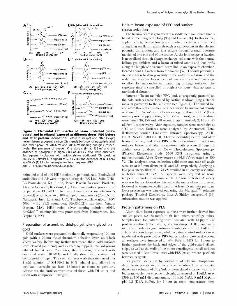
Figure 3. Elemental XPS spectra of beam protected (unex-posed) and irradiated (exposed at different doses) PEG beforeand after protein incubation. Before (‘‘unexp’’) and after (‘‘exp’’)helium beam exposure, carbon C1s signals (A) show characteristic alkyland ether peaks at 284.6 eV and 286.6 eV binding energies, respec-tively. The presence of oxygen O1s signals (B) at 532 eV and theabsence of nitrogen N1s signals (C) at 400 eV also were observed.Subsequent incubation with avidin shows additional C1s peak at288 eV (D), similar O1s signals at 532 eV (E) and existence of N1s peakat 400 eV (F) binding energies for beam exposed PEG.doi:10.1371/journal.pone.0056835.g003
Patterning of Poly(ethylene glycol) by Helium Beam
PLOS ONE | www.plosone.org 3 May 2013 | Volume 8 | Issue 5 | e56835

washed with DEA buffer. BCIP-NBT (0.15 mg/mL BCIP and
0.30 mg/mL NBT) substrate was then added and allowed to react
for 10 minutes. The surfaces were then washed with water, dried
with compressed nitrogen, and imaged using an optical micro-
scope. For pattern testing by gold nanoparticle probes, 300 mL of a
suspension of ca. 109 gold nanoparticles/mL conjugated with
antibodies (100 nm particles with biotinylated goat anti-rabbit
polyclonal antibodies, or 40 nm particles with goat anti-mouse
antibodies) in PBS buffer was added to the surface and allowed to
incubate for at least 12 hours at room temperature, with mixing
on an orbital shaker. For HRP-mediated silver staining, surfaces
were incubated with streptavidin-polyHRP conjugates (20 mL
spot, 10 mg/mL) for at least one hour at room temperature, and
then washed thrice with PBS and twice with DI water. Surfaces
were then silver-stained using the EnzMetTM staining solution,
with 2 minutes incubation time each for the manufacturer’s
reagents Detect A, B and C (20 mL each). Finally, surfaces were
washed thrice with water, dried with compressed nitrogen and
imaged by scanning electron microscopy (Zeiss/LEO 1525 Field
Emission SEM).
Figure 4. Formation of diformazan precipitates on protein-PEG patterns (35 mm mesh). Top row images show a schematic diagram (not toscale) of helium beam- patterned PEG incubated with avidin (A) or BSA (D) followed by addition of biotinylated alkaline phosphatase (AP) andsubstrate BCIP-NBT. Optical microscopic images below the diagram show the protected (light) and irradiated (dark) regions which display the specificpatterned capture of biotinylated AP enzymes for avidin-incubated surface (B) and no pattern for BSA-incubated surface (E). Panels C and F showhigher magnifications of images in B and E, respectively.doi:10.1371/journal.pone.0056835.g004
Patterning of Poly(ethylene glycol) by Helium Beam
PLOS ONE | www.plosone.org 4 May 2013 | Volume 8 | Issue 5 | e56835
Patterning of Poly(ethylene glycol) by Helium Beam
PLOS ONE | www.plosone.org 5 May 2013 | Volume 8 | Issue 5 | e56835

Results and Discussion
As shown in Figure 2, ATR spectra confirmed the presence of
SH-PEG assembled on gold surfaces before helium beam
exposure, after beam exposure and after post-exposure incubation
in PBS buffer for one hour. Peaks near 2870 cm21 and
1120 cm21 were observed, corresponding to the C–H stretch of
methoxy and C–O stretch of ether groups, respectively [40]. We
found that our MW 5000 SH-PEG SAM monolayers at least
partially survived 6.5 kV helium beam exposures with doses of
1.5–60 mC/cm2. It is notable that previous researchers [22,23]
found that monolayers of smaller PEG molecules (MW: 290 and
750) were damaged by doses of 5–80 mC/cm2 and completely
removed by doses over 160 mC/cm2 when using 1 kV electrons.
The radiation chemistry of electron and ion/atom beams on
PEG and alkanethiols would be expected to be very similar,
mainly involving hydrogen abstraction of the PEG hydrocarbons
[41] and dissociation of C–H, C–C, C–S and substrate–SH bonds
[23,34]. Hydrogen abstraction and bond dissociation form
radicals, which eventually cause cross-linking within the polymer
[42,43], affect hydrogen bonding and disrupt the highly organized
interface between water and PEG. Our results are consistent with
a similar effect of helium beam exposure. Cross-linking of PEG
was observed on helium beam exposed surfaces as evidenced by
the water insolubility of exposed spin-coated thiol-PEG on silicon
(unexposed thiol-PEG film on silicon is soluble). Furthermore,
disruption of PEG chains was confirmed by XPS, as shown in
Figure 3. As presented in Figure 3A, prominent carbon (C1s) peaks
were observed at 284.6 eV and 286.6 eV binding energies, which
correspond to alkyl and ether C1s bonding states, respectively
[44,45]. As the helium beam dose was increased, the C1s ether
peak at 286.6 eV decreased while the alkyl peak at 284.6 eV
increased, indicating the destruction of ether bonds and formation
of more alkyl bonds in the PEG molecules. A decrease in the
oxygen (O1s) signal (532 eV binding energy) also was observed as
the beam dose was increased. Helium beam exposure renders the
grafted PEG on the surface less hydrophilic by a decrease in C–O
ether and an increase in C–C alkyl functionalities, and thus
potentially more amenable to protein adsorption. There also is
literature evidence that electron [9] or ion (argon) beam [10,11]
exposure can create carbonyl functionalities (which could be
charged carboxylate or protein amine-aldehyde) in PEG samples.
A relatively small number of these functional groups could alter
the local protein-capture properties of the PRG surface, while
being difficult to detect against the background of much-more-
numerous ether and alkyl functionalities.
Elemental XPS spectra of exposed and unexposed samples
incubated with avidin are shown in Figures 3D, 3E and 3F for
carbon (C1s), oxygen (O1s) and nitrogen (N1s) signals, respective-
ly. In Figure 3D, aside from the observed decrease in the C1s peak
at 286.6 eV and increase in the C1s peak at 284.6 eV discussed
previously, the C1s peak at 288 eV, a characteristic binding
energy of amide C1s [46], appeared as the helium beam dose was
increased, confirming that avidin attaches to the beam-exposed
surfaces. This is further supported by the increase in the nitrogen
N1s signal (400 eV) with increasing beam dose, as shown in
Figure 3F. Unlike in Figure 3B, the differences in oxygen signal
intensities in Figure 3E were not distinct, particularly in the
unexposed and exposed (30 sec and 150 sec) samples. This might
be due to the added attenuation length for photoelectrons in XPS
provided by the avidin layer (there is little nonspecific adsorption
of avidin on unexposed PEG as shown by the nitrogen signal in
Figure 3F). The oxygen signal intensities for unexposed and
exposed (30 sec and 150 sec) PEG samples are similar because the
avidin layer somewhat overshadows the small oxygen signal
differences observed in Figure 3B. However, since more avidin
molecules were found to attach on the exposed (600 sec) sample,
its oxygen signal is distinctly different from the latter samples.
Among the helium beam doses tested, we chose to use the
150 second exposure time for further work as a balance between
protein attachment effectiveness and processing throughput.
Patterning of proteins using a 35 mm mesh is presented in
Figures 4 and 5, wherein attachment of proteins on beam-exposed
regions is indicated by enzymatic formation of localized diforma-
zan precipitates and also by binding of gold nanoparticle probes.
As shown in Figure 4, the helium beam was able to pattern avidin
over a large surface area (25 mm2, with 2.3 mm2 shown in
Figure 4). The darkened regions indicate the formation of
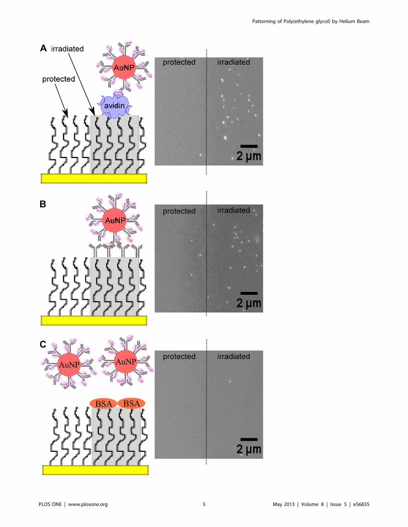
Figure 5. Gold nanoparticle probes on protein-PEG patterns (35 mm mesh). Images on the left column show the schematic diagram (not toscale) of helium beam-patterned PEG incubated with avidin (A), polyclonal anti-rabbit antibodies (B), or BSA (C), followed by the addition of 100 nmgold nanoparticles conjugated with biotinylated rabbit antibodies. Electron microscope images on the right column show the protected andirradiated PEG regions after incubation with gold nanoparticle probes.doi:10.1371/journal.pone.0056835.g005
Figure 6. Gold nanoparticle probes and silver nanoparticledeposition on protein-PEG patterns (300 nm mask). Electronmicroscope images on the left column (with zoom-in pictures as insets)correspond to helium beam-patterned PEG incubated with polyclonalanti-mouse antibodies (A), streptavidin-polyHRP conjugates (B), orbiotinylated lysozyme (C), while images on the right display beam-patterned PEG surfaces incubated with PBS buffer (no proteins); allsamples were then passivated with BSA. Patterns were visualized bybinding of 40 nm gold nanoparticles conjugated with D1.3 mouseantibodies (A), or silver staining through HRP conjugates (B). Capturedbiotinylated lysozyme (C) was detected by addition of streptavidin-polyHRP conjugates and silver staining.doi:10.1371/journal.pone.0056835.g006
Patterning of Poly(ethylene glycol) by Helium Beam
PLOS ONE | www.plosone.org 6 May 2013 | Volume 8 | Issue 5 | e56835
diformazan precipitates by captured biotinylated alkaline phos-
phatase via dephosphorylation and reduction of the substrate
BCIP-NBT. These regions signify the successful attachment of
avidin in active form on beam-exposed PEG. Similar results were
obtained using 100 nm gold nanoparticles, as shown in Figure 5A.
In Figure 5B, where the surface was incubated with polyclonal
anti-rabbit antibodies after beam exposure, gold nanoparticle
probes conjugated with biotinylated rabbit antibodies were shown
to bind to the irradiated PEG regions. BSA control surfaces, by
contrast, showed little to no formation of diformazan precipitate
(Figure 4) and bound very few gold nanoparticles (Figure 5C).
To demonstrate that the helium beam could create high-
resolution nanoscale protein patterns on PEG surfaces, a smaller
mask with 300 nm openings at 1 mm spacing was used. As shown
in Figure 6A, gold nanoparticles conjugated with monoclonal
mouse antibodies were observed to bind to the 300 nm irradiated
PEG regions incubated with polyclonal anti-mouse antibodies,
while very few bound nanoparticles were seen on a BSA control
surface. Similar results were obtained by silver staining, as shown
in Figures 6B and 6C. Silver particles of approximately 300 nm
diameter were formed on the beam-exposed areas, both on
surfaces incubated with streptavidin-HRP, then silver stained with
EnzMetTM solution (Figure 6B) and on surfaces incubated with
biotinylated lysozyme, followed by streptavidin-HRP conjugate
and silver staining (Figure 6C). These results showed that helium
beam exposure through stencil masks can be used to activate PEG
and create protein patterns on the order of hundreds of
nanometers. Corresponding negative controls showed very few
bound gold nanoparticles and little to no formation of silver
nanoparticles, as BSA was able effectively to passivate the beam-
exposed regions.
Summary
We have demonstrated that helium beam exposure through a
stencil mask in proximity to the substrate can be used for the
massively-parallel creation of micro- and nano-scale protein
patterns on PEG-grafted surfaces. Proteins captured on irradiated
PEG regions were shown to retain their functionality, as the
patterned avidin could bind biotinylated molecules and patterned
HRP conjugates were able to produce silver nanoparticles. Protein
attachment on helium beam-exposed PEG may be due to
hydrophobic interactions, electrostatic interactions, formation of
reactive oxidized products, or a combination of these mechanisms.
Further studies will be needed to establish the governing
mechanism(s) and also to maximize processing throughput.
Author Contributions
Conceived and designed the experiments: EEC PR RCW. Performed the
experiments: EEC AN TS. Analyzed the data: EEC SK KK PR GES
RCW. Contributed reagents/materials/analysis tools: EEC SK KK GES
PR RCW. Wrote the paper: EEC PR RCW.
References
1. Yun Y-H, Eteshola E, Bhattacharya A, Dong Z, Shim J-S, et al. (2009) Tiny
medicine: nanomaterial-based biosensors. Sensors 9: 9275–9299.
2. Huebsch N, Mooney DJ (2009) Inspiration and application in the evolution of
biomaterials. Nature 462: 426–432.
3. Mrksich M, Whitesides GM (1996) Using self-assembled monolayers to
understand the interactions of man-made surfaces with proteins and cells.
Annual Review of Biophysics and Biomolecular Structure 25: 55–78.
4. Christman KL, Enriquez-Rios VD, Maynard HD (2006) Nanopatterning
proteins and peptides. Soft Matter 2: 928–939.
5. Smith KH, Tejeda-Montes E, Poch M, Mata A (2011) Integrating top-down and
self-assembly in the fabrication of peptide and protein-based biomedical
materials. Chemical Society Reviews 40: 4563–4577.
6. Bretagnol F, Kylian O, Hasiwa M, Ceriotti L, Rauscher H, et al. (2007) Micro-
patterned surfaces based on plasma modification of PEO-like coating for
biological applications. Sensors and Actuators B: Chemical 123: 283–292.
7. Geyer W, Stadler V, Eck W, Golzhauser A, Grunze M, et al. (2001) Electron
induced chemical nanolithography with self-assembled monolayers. Journal of
Vacuum Science & Technology B: Microelectronics and Nanometer Structures
19: 2732.
8. Golzhauser A, Eck W, Geyer W, Stadler V, Weimann T, et al. (2001) Chemical
nanolithography with electron beams. Advanced Materials 13: 806–809.
9. Krsko P, Saaem I, Clancy R, Geller H, Soteropoulos P, et al. (2005) E-beam-
patterned hydrogels to control nanoscale surface bioactivity. Nanofabrication:
Technologies, Devices, and Applications II SPIE: 600201.
10. Manso-Silvan M, Valsesia A, Gilliland D, Ceccone G, Rossi F (2004) Ion-beam
treatment of PEO; towards a physically stabilized anti-fouling film. Surface and
interface analysis 36: 733–736.
11. Manso Silvan M, Valsesia A, Hasiwa M, Gilliland D, Ceccone G, et al. (2007)
Surface Characterization of Biopolymer Micropatterns Processed by Ion-Beam
Modification and PECVD. Chemical Vapor Deposition 13: 211–218.
12. Morra M (2000) On the molecular basis of fouling resistance. Journal of
Biomaterials Science, Polymer Edition 11: 547–569.
13. Wang RLC, Kreuzer HJ, Grunze M (1997) Molecular conformation and
solvation of oligo(ethylene glycol)-terminated self-assembled monolayers and
their resistance to protein adsorption. The Journal of Physical Chemistry B 101:
9767–9773.
14. Szleifer I (1997) Protein adsorption on surfaces with grafted polymers: a
theoretical approach. Biophysical Journal 72: 595–612.
15. Harder P, Grunze M, Dahint R, Whitesides GM, Laibinis PE (1998) Molecular
conformation in oligo(ethylene glycol)-terminated self-assembled monolayers on
gold and silver surfaces determines their ability to resist protein adsorption. The
Journal of Physical Chemistry B 102: 426–436.
16. Heuberger M, Drobek T, Voros J (2004) About the role of water in surface-
grafted poly (ethylene glycol) layers. Langmuir 20: 9445–9448.
17. Besseling NAM (1997) Theory of hydration forces between surfaces. Langmuir
13: 2113–2122.
18. Israelachvili J, Wennerstrom H (1996) Role of hydration and water structure in
biological and colloidal interactions. Nature 379: 219–225.
19. Hong Y, Krsko P, Libera M (2004) Protein surface patterning using nanoscale
PEG hydrogels. Langmuir 20: 11123–11126.
20. Christman KL, Schopf E, Broyer RM, Li RC, Chen Y, et al. (2008) Positioning
multiple proteins at the nanoscale with electron beam cross-linked functional
polymers. Journal of the American Chemical Society 131: 521–527.
21. Harnett C, Satyalakshmi K, Craighead H (2000) Low-energy electron-beam
patterning of amine-functionalized self-assembled monolayers. Applied Physics
Letters 76: 2466.
22. Harnett C, Satyalakshmi K, Craighead H (2001) Bioactive templates fabricated
by low-energy electron beam lithography of self-assembled monolayers.
Langmuir 17: 178–182.
23. Rundqvist J, Hoh JH, Haviland DB (2006) Directed immobilization of protein-
coated nanospheres to nanometer-scale patterns fabricated by electron beam
lithography of poly (ethylene glycol) self-assembled monolayers. Langmuir 22:
5100–5107.
24. Lee SW, Ahn J, Kim MG, Shin YB, Lee JJ, et al. (2010) Enhanced biomolecular
detection based on localized surface plasmon resonance (LSPR) using enzyme-
precipitation reaction. Journal of Nanoscience and Nanotechnology 10: 3246–
3249.
25. Riemenschneider L, Blank S, Radmacher M (2005) Enzyme-assisted nanolitho-
graphy. Nano Letters 5: 1643–1646.
26. Leary JJ, Brigati DJ, Ward DC (1983) Rapid and sensitive colorimetric method
for visualizing biotin-labeled DNA probes hybridized to DNA or RNA
immobilized on nitrocellulose: Bio-blots. Proceedings of the National Academy
of Sciences 80: 4045.
27. Lyon LA, Musick MD, Natan MJ (1998) Colloidal Au-enhanced surface
plasmon resonance immunosensing. Analytical Chemistry 70: 5177–5183.
28. Nam JM, Jang KJ, Groves JT (2007) Detection of proteins using a colorimetric
bio-barcode assay. Nature Protocols 2: 1438–1444.
29. Reynolds III RA, Mirkin CA, Letsinger RL (2000) Homogeneous, nanoparticle-
based quantitative colorimetric detection of oligonucleotides. Journal of the
American Chemical Society 122: 3795–3796.
30. Hainfeld JF, Liu W (2010) Method for detecting a target molecule by metal
deposition. Nanoprobes Inc.
31. Moller R, Powell RD, Hainfeld JF, Fritzsche W (2005) Enzymatic control of
metal deposition as key step for a low-background electrical detection for DNA
chips. Nano Letters 5: 1475–1482.
32. Hering KK, Moller R, Fritzsche W, Popp J (2008) Microarray-based detection
of dye-labeled DNA by SERRS using particles formed by enzymatic silver
deposition. ChemPhysChem 9: 867–872.
33. Francis GD, Jones MA, Beadle GF, Stein SR (2009) Bright-field in situ
hybridization for HER2 gene amplification in breast cancer using tissue
microarrays: correlation between chromogenic (CISH) and automated silver-
Patterning of Poly(ethylene glycol) by Helium Beam
PLOS ONE | www.plosone.org 7 May 2013 | Volume 8 | Issue 5 | e56835

enhanced (SISH) methods with patient outcome. Diagnostic molecular
pathology : the American journal of surgical pathology, part B 18: 88–95.34. Braden BC, Goldman ER, Mariuzza RA, Poljak RJ (1998) Anatomy of an
antibody molecule: structure, kinetics, thermodynamics and mutational studies
of the antilysozyme antibody D1. 3. Immunological Reviews 163: 45–57.35. Hogg A (1983) Conversion of mass spectrometers for fast-atom bombardment
using easily constructed components. International Journal of Mass Spectrom-etry and Ion Physics 49: 25–34.
36. Franks J, Ghander A (1974) A saddle field ion source of spherical configuration
for etching and thinning applications. Vacuum 24: 489–491.37. Nasrullah A, Smith D, Sherlock T, Ruchhoeft P, Litvinov D (2009) Near
neighbor averaging: A technique for improving image uniformity in aperturearray lithography. Journal of Vacuum Science Technology B: Microelectronics
and Nanometer Structures 27: 2674.38. Castle J, Salvi A (2001) Interpretation of the Shirley background in x-ray
photoelectron spectroscopy analysis. Journal of Vacuum Science & Technology
A: Vacuum, Surfaces, and Films 19: 1170.39. Green N (1965) A spectrophotometric assay for avidin and biotin based on
binding of dyes by avidin. The Biochemical Journal 94: 23C.40. Coates J (2000) Interpretation of infrared spectra, a practical approach.
Encyclopedia of Analytical Chemistry: John Wiley and Sons, Ltd.
41. Sofia SJ, Merrill EW (1998) Grafting of PEO to polymer surfaces using electron
beam irradiation. Journal of Biomedical Materials Research 40: 153–163.
42. Zharnikov M, Grunze M (2002) Modification of thiol-derived self-assembling
monolayers by electron and x-ray irradiation: scientific and lithographic aspects.
Journal of Vacuum Science & Technology B: Microelectronics and Nanometer
Structures 20: 1793–1807.
43. Feulner P, Niedermayer T, Eberle K, Schneider R, Menzel D, et al. (2004)
Strong temperature dependence of irradiation effects in organic layers. Physical
Review Letters 93: 178302.
44. Yoshimoto K, Nozawa M, Matsumoto S, Echigo T, Nemoto S, et al. (2009)
Studies on the adsorption property and structure of polyamine-ended
poly(ethylene glycol) derivatives on a gold surface by surface plasmon resonance
and angle-resolved x-ray photoelectron spectroscopy. Langmuir 25: 12243–
12249.
45. Lu HB, Campbell CT, Castner DG (2000) Attachment of functionalized poly
(ethylene glycol) films to gold surfaces. Langmuir 16: 1711–1718.
46. Denis FA, Hanarp P, Sutherland DS, Gold J, Mustin C, et al. (2002) Protein
adsorption on model surfaces with controlled nanotopography and chemistry.
Langmuir 18: 819–828.
Patterning of Poly(ethylene glycol) by Helium Beam
PLOS ONE | www.plosone.org 8 May 2013 | Volume 8 | Issue 5 | e56835
Copyright © 2022 FDOKUMEN