Hassles with Taking Out the Garbage: Aggravating Aggresomes
9
Copyright C Blackwell Munksgaard 2002 Traffic 2002; 3: 388–396 Blackwell Munksgaard ISSN 1398-9219 Review Hassles with Taking Out the Garbage: Aggravating Aggresomes Rafael Garcia-Mata, Ya-Sheng Gao and Elizabeth Sztul* University of Alabama Medical Science, Department of Cell Biology, 1918 University Blvd, MCLM 668, Box 88, Birmingham, AL 35294–0005, USA * Corresponding author: Elizabeth Sztul, [email protected] Diverse human diseases ranging from amyloidosis to neurodegenerative diseases are now recognized as ‘conformational diseases’ caused by protein misfolding and protein aggregation. Misfolded and aggregated pro- teins are usually handled in the cell through chaperone- mediated refolding, or when that is impossible, de- stroyed by proteasomal degradation. Recent evidence suggests that cells might have evolved a third pathway that involves the sequestration of aggregated proteins into specialized ‘holding stations’ called aggresomes. The aggresomal pathway provides a mechanism by which aggregated proteins form particulate (, 200 nm) mini-aggregates that are transported on microtubules (MTs) towards the MT organizing center (MTOC) by a process mediated by the minus-end motor protein dyn- ein. Once at the MTOC, the individual particles pack into a single, usually spherical aggresome (1–3 mm) that sur- rounds the MTOC. Aggresomes are dynamic: they re- cruit various chaperones and proteasomes, presumably to aid in the disposal of the aggregated proteins. In ad- dition, the formation of an aggresome is likely to acti- vate the autophagic clearance mechanism that termin- ates in lysosomal degradation. Hence, the aggresome pathway may provide a novel system to deliver aggre- gated proteins from the cytoplasm to lysosomes for degradation. Although it is clear that many pathological states correlate with the formation of aggresomes, their causal relationships remain hotly debated. Here, we de- scribe the current state of our knowledge of the aggre- some pathway and outline the open questions that pro- vide the focus of current research. Key words: aggregation, aggresome, autophagy, chaperones, degradation, intermediate filament, mis- folding, neurodegeneration, proteasome Received 18 March 2002, revised and accepted for publication 20 March 2002 Protein (Mis)folding Newly synthesized proteins must overcome several obstacles on their way to becoming functional molecules. Small mono- 388 meric proteins fold through a succession of folding intermedi- ates (1). During folding, partially folded proteins expose hydrophobic domains, which sometimes lead to nonproduc- tive associations and protein aggregation (2). Aggregation is especially problematic in vivo, where the high cytosolic con- centration of macromolecules [protein concentration inside a cell can be as high as 300 mg/ml (3)] causes a ‘crowding effect’ in the cytosol that can result in a significant increase in the association constants of unfolded polypeptides over those in dilute solution (4,5). The aggregation problem is amplified by the fact that stable folding of a folding domain cannot occur until the entire domain is synthesized. This is especially important during synthesis of identical nascent polypeptides on polysomes, where numerous polypeptides expose analogous aggregation-prone domains leading to in- creased chances of aggregation (4). The ultimate outcome in the life of a protein is either correct folding or aggregation. Whether a polypeptide chain suc- ceeds in folding correctly or whether it aggregates depends on the kinetic competition between folding and aggregation. In cells, misfolding can occur due to particular mutations, RNA modification, and amino acid misincorporation during translation, or unequal synthesis of individual subunits of multisubunit proteins (2,6,7). Misfolding can also be pro- moted by other factors such as pH, temperature, ionic strength, and redox environment. Since a certain level of pro- tein misfolding seems inescapable, cells have evolved several different ‘quality control’ mechanisms that act in concert to minimize misfolding and to dispose of misfolded proteins prior to aggregation (8). First, various molecular chaperones have evolved to assist folding of newly synthesized proteins and refolding of pro- teins damaged by stress and cellular injuries. Chaperones bind to and stabilize exposed hydrophobic residues, and through an ATP-dependent cycle of controlled binding and release (9), allow the protein to achieve proper folding. Thus, chaperones do not catalyze folding per se; instead, they pre- vent incorrect intra– and intermolecular interactions between partially folded or unfolded polypeptides (10). Chaperones oc- cur ubiquitously in all cellular compartments that sustain pro- tein synthesis and folding reactions(10). It appears that many proteins require chaperones to fold correctly. In bacteria, the folding of approximately 30% of total proteins is affected in cells containing a mutant chaperone, GroEL (11). Similarly, in eukaryotes, the chaperone Hsp70 co-immunoprecipitates with 15–20% of newly made proteins (12). While small
-
Upload
independent -
Category
Documents
-
view
0 -
download
0
Transcript of Hassles with Taking Out the Garbage: Aggravating Aggresomes

Copyright C Blackwell Munksgaard 2002Traffic 2002; 3: 388–396
Blackwell Munksgaard ISSN 1398-9219
Review
Hassles with Taking Out the Garbage: AggravatingAggresomes
Rafael Garcia-Mata, Ya-Sheng Gao andElizabeth Sztul*
University of Alabama Medical Science, Department of Cell
Biology, 1918 University Blvd, MCLM 668, Box 88,
Birmingham, AL 35294–0005, USA
* Corresponding author: Elizabeth Sztul,
Diverse human diseases ranging from amyloidosis toneurodegenerative diseases are now recognized as‘conformational diseases’ caused by protein misfoldingand protein aggregation. Misfolded and aggregated pro-teins are usually handled in the cell through chaperone-mediated refolding, or when that is impossible, de-stroyed by proteasomal degradation. Recent evidencesuggests that cells might have evolved a third pathwaythat involves the sequestration of aggregated proteinsinto specialized ‘holding stations’ called aggresomes.The aggresomal pathway provides a mechanism bywhich aggregated proteins form particulate (,200nm)mini-aggregates that are transported on microtubules(MTs) towards the MT organizing center (MTOC) by aprocess mediated by the minus-end motor protein dyn-ein. Once at the MTOC, the individual particles pack intoa single, usually spherical aggresome (1–3mm) that sur-rounds the MTOC. Aggresomes are dynamic: they re-cruit various chaperones and proteasomes, presumablyto aid in the disposal of the aggregated proteins. In ad-dition, the formation of an aggresome is likely to acti-vate the autophagic clearance mechanism that termin-ates in lysosomal degradation. Hence, the aggresomepathway may provide a novel system to deliver aggre-gated proteins from the cytoplasm to lysosomes fordegradation. Although it is clear that many pathologicalstates correlate with the formation of aggresomes, theircausal relationships remain hotly debated. Here, we de-scribe the current state of our knowledge of the aggre-some pathway and outline the open questions that pro-vide the focus of current research.
Key words: aggregation, aggresome, autophagy,chaperones, degradation, intermediate filament, mis-folding, neurodegeneration, proteasome
Received 18 March 2002, revised and accepted forpublication 20 March 2002
Protein (Mis)folding
Newly synthesized proteins must overcome several obstacleson their way to becoming functional molecules. Small mono-
388
meric proteins fold through a succession of folding intermedi-ates (1). During folding, partially folded proteins exposehydrophobic domains, which sometimes lead to nonproduc-tive associations and protein aggregation (2). Aggregation isespecially problematic in vivo, where the high cytosolic con-centration of macromolecules [protein concentration inside acell can be as high as 300mg/ml (3)] causes a ‘crowdingeffect’ in the cytosol that can result in a significant increasein the association constants of unfolded polypeptides overthose in dilute solution (4,5). The aggregation problem isamplified by the fact that stable folding of a folding domaincannot occur until the entire domain is synthesized. This isespecially important during synthesis of identical nascentpolypeptides on polysomes, where numerous polypeptidesexpose analogous aggregation-prone domains leading to in-creased chances of aggregation (4).
The ultimate outcome in the life of a protein is either correctfolding or aggregation. Whether a polypeptide chain suc-ceeds in folding correctly or whether it aggregates dependson the kinetic competition between folding and aggregation.In cells, misfolding can occur due to particular mutations,RNA modification, and amino acid misincorporation duringtranslation, or unequal synthesis of individual subunits ofmultisubunit proteins (2,6,7). Misfolding can also be pro-moted by other factors such as pH, temperature, ionicstrength, and redox environment. Since a certain level of pro-tein misfolding seems inescapable, cells have evolved severaldifferent ‘quality control’ mechanisms that act in concert tominimize misfolding and to dispose of misfolded proteinsprior to aggregation (8).
First, various molecular chaperones have evolved to assistfolding of newly synthesized proteins and refolding of pro-teins damaged by stress and cellular injuries. Chaperonesbind to and stabilize exposed hydrophobic residues, andthrough an ATP-dependent cycle of controlled binding andrelease (9), allow the protein to achieve proper folding. Thus,chaperones do not catalyze folding per se; instead, they pre-vent incorrect intra– and intermolecular interactions betweenpartially folded or unfolded polypeptides (10). Chaperones oc-cur ubiquitously in all cellular compartments that sustain pro-tein synthesis and folding reactions (10). It appears that manyproteins require chaperones to fold correctly. In bacteria, thefolding of approximately 30% of total proteins is affected incells containing a mutant chaperone, GroEL (11). Similarly, ineukaryotes, the chaperone Hsp70 co-immunoprecipitateswith 15–20% of newly made proteins (12). While small

Aggresomal Pathway for Protein Degradation
monomeric proteins are able to fold in the absence ofchaperones in vivo, chaperones are critically important for thefolding of medium- to large-sized multidomain proteins.
Second, cellular proteins that are unable to fold properly aretargeted for degradation by the ubiquitin–proteasome system.The proteasome is a multisubunit complex found in the cytosoland nucleus that mediates degradation of cytosolic, nuclear(13), secretory and transmembrane proteins (14). Misfoldedsecretory and transmembrane proteins undergo a circuitousroute to the proteasome: they are retained in the lumen or themembrane of the ER by the ER ‘quality control’ mechanism(15), then are retrotranslocated back to the cytosol and deliver-ed to the proteasome (16). When correct folding is difficult orimpossible and degradation is not initiated rapidly, proteins in-teract with other unfolded or partially folded proteins, leadingto the formation of aggregates (8). How cells deal with suchaggregates is the subject of this review.
How Aggresomes Form
The initial aggregation process is likely to occur cotransla-tionally, while nascent chains are coming off the polysome
Figure1: Model of aggresome formation. Unfolded or misfolded protein can originate from translating polysomes, from proteins retro-translocated from the ER to the cytosol, or from proteins damaged by stress. If these unfolded/misfolded proteins fail to fold correctly andare not degraded by the proteasome, they can form aggregates throughout the cells. Such aggregates are transported in a microtubule-dependent manner to the MTOC in a process that requires the function of the dynein/dynactin motor complex. Aggresome formation isusually accompanied by a dramatic reorganization of the intermediate filaments cytoskeleton which collapse to form a ‘cage’ surroundingthe aggresome. The Golgi in aggresome-containing cells is usually displaced and localizes to the periphery of the aggresome.
389Traffic 2002; 3: 388–396
(Figure 1). An mRNA coding for a protein of ,60kDa wouldcontain ,20 ribosomes at any time (,20 ribosomes every80 nucleotides on a 1600-nucleotide mRNA) and could eas-ily continue translation for multiple rounds (polypeptide trans-lation takes approximately 5amino acids/second, so between20 and 200s for a 10–200kDa protein), resulting in hun-dreds of nascent peptides being produced in minutes at asingle cellular site. If these nascent peptides cannot fold cor-rectly, they will co-aggregate to form a single aggresomalparticle. In cells, such particles are uniform in size (17,18), sup-porting the notion that a fixed number of proteins (output ofa single polysome?) aggregate to form a single particle. Par-ticles are produced throughout the cytoplasm, presumablyreflecting a diffuse distribution of the cognate mRNA (17).Very quickly after their formation, aggresomal particles aretransported towards the microtubule (MT) organizing center(MTOC), where they are sequestered into a single large ‘cel-lular landfill’ structure called the aggresome.
Particle movement is an active process and requires intactMTs and the minus-end-directed motor dynein (17,19). In cellstreated with the MT depolymerizing drug nocodazole, oroverexpressing the p50/dynamitin component of the dynein/dynactin complex that inhibits dynein-mediated transport,

Garcia-Mata et al.
aggresomal particles remain distributed throughout the cyto-plasm (17,19). In agreement with the involvement of the dyn-ein motor, transport of aggresomal particles on individual MTsoccurs with kinetics (velocities of aggregated particles oscil-late between 0 and 0.25mm/s) analogous to those reportedfor dynein-mediated movement of membrane-bound organ-elles (17,20). Motor linkage to specific membranes is me-diated by various adaptor proteins (21), but whether thesame, similar or completely distinct groups of linkers partici-pate in microtubule-based motility of aggresomal particles isunknown. It is possible that the linkage of aggresomal par-ticles is mediated by non-conventional adapter proteins. Thisis suggested by studies showing that the huntingtin-associ-ated protein HAP1 binds the huntingtin protein and the dyn-actin component of the dynein motor, and thus could ‘load’a huntingtin aggregate onto a microtubule (22). Whethersimilar cargo-dependent adapters exist for moving other ag-gregates remains to be determined.
The ‘individualistic’ formation of aggresomal particles andtheir subsequent transport on individual MTs implies that anaggresome is an aggregate of aggregates. Indeed, EM ex-amination of aggresomes has confirmed that within eachstructure, particles are loosely associated with each other anddo not coalesce into a single large aggregate (17,19). Thisimplies that each particle is effectively screened from expos-ing hydrophobic domains, but the molecular mechanismsthat ‘coat’ the particle surface to protect it from further aggre-gation remain to be characterized.
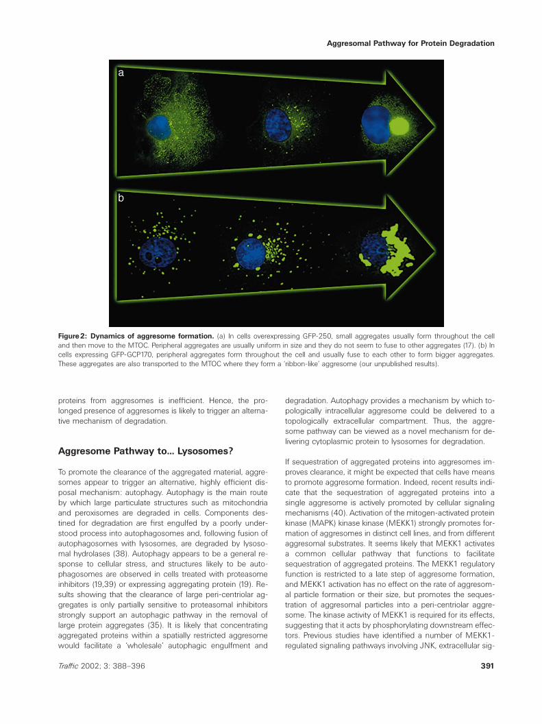
Despite the common pathway of aggresome formation, theiroverall structure varies depending both on the aggregatingsubstrate and the ‘host’ cell. Most aggresomes appear eitheras a single sphere with a diameter of 1–3mm (Figure 2a),or as an extended ribbon (Figure 2b). The spherical type ofaggresome can be formed by different types of substrates,such as multispanning transmembrane proteins (e.g. CFTR(19)), cytosolic proteins (e.g. mutant forms of Huntingtin(23,24)), or secretory proteins [e.g. mutant form of light chain(LC)] IgG (25–27). The ribbon type of aggresomes also canbe formed by distinct types of substrates, such as a multis-panning transmembrane protein (e.g. presenilin) (28), orcytosolic proteins [ex: glial fibrillary acidic protein (GFAP)](28). The molecular mechanisms that define the final shapeof an aggresome arising from a particular substrate are un-known, but the cellular milieu does influence the final struc-ture: a mutant version of the ATP7B protein responsible forWilson disease forms spherical aggresomes in HEK293 cells,but forms ribbon aggresomes in Huh7 cells (29).
Recruitment of Degradative Machinery toAggresomes
Aggresomes are not just static garbage depositories; theyrecruit a number of cytosolic components such as chaper-ones, ubiquitination enzymes and proteasome components,presumably to aid in the clearance of the aggregated pro-
390 Traffic 2002; 3: 388–396
teins. Chaperones are ubiquitously associated with aggre-somes, and all the major classes are represented. Membersof the HSC70 family (Hsc70), the HSP40 family (Hdj1 andHdj2), the HSP70 family (Hsp70), and the small HSP familycharacterized by the presence of a signature ‘a-crystallin do-main’ (HSP27 and ab-crystallin), as well as the chaperoninTriC/TCP are recruited to aggresomes (17,30,31). In addition,two proteins that may function as chaperones, a-synucleinand 14-3-3 (32) have been shown to associate with aggre-somes (33). Despite the recruitment of cellular chaperonesto the aggresome, the clearance of aggregated proteins isstill limited by the overall chaperone activity. Overexpressionof Hsp70 prevents aggresome formation (27,34), suggestingthat the aggresomal ‘bottle-neck’ is caused by lack of suf-ficient chaperones to unfold substrates into a state in whichthey can be presented to the proteasome for degradation,rather than inactivation of proteasomes. Surprisingly, the ERlumenal chaperone BiP/GRP78 has been shown to colocalizewith aggresomes (33), suggesting that GRP78 can undergoretrograde transport from the ER. GRP78 is associated withthe core of the aggresome at a time when the cytoplasmicchaperones Hsp70 and Hsp40 are barely detectable, indi-cating that recruitment of the cytosolic chaperones occurslate during aggresome formation, and is subsequent to as-sociation of the misfolded substrates with ER chaperones.However, the participation of the cytosolic chaperones seemsmore important in reducing aggregation since overexpressionof Hsp70, but not overexpression of BiP can cause a dramaticdecrease in protein aggregation (27). The function of thissequential chaperone exchange in unfolding of aggregatedsubstrates remains to be investigated.
Like chaperones, proteasomes have been shown to be in-variably associated with aggresomes. Proteasomes are re-cruited relatively late, after small aggresomal particles are de-livered to the MTOC region (35), in agreement with the ideathat proteasomes and proteasomal degradation preferentiallyoccurs at the peri-centriolar degrasome region (discussedbelow). This fits well with the finding that proteasome recruit-ment appears to be MT-independent, suggesting that thepresence of substrate might be sufficient to directly attractthe proteasome from a cytoplasmic pool (36). The pathwaysinvolved in signaling the presence of aggregated protein tocellular proteasomes, and the mechanisms mediating pro-teasomal relocation to the aggresome remain to be defined.
Proteasomal substrates are often ubiquitinated, but ubiquitin-ation is not directly linked to aggresome formation since ag-gresomes containing CFTR or influenza virus nucleoproteinare recognized by anti-ubiquitin antibodies and those sub-strates have been shown to be ubiquitinated (19,37), whileaggregated GFP-250 (17), mutant SOD (18) and ATP7B (29)do not appear ubiquitinated. It is more likely that the levelof ubiquitination and recruitment of ubiquitinating enzymesreflects the cellular response to distinct proteins and is not aconsequence of aggresome formation. Despite the recruit-ment of the normal refolding/degradative machinery, and theon-going proteasomal degradation, disposal of aggregated
Aggresomal Pathway for Protein Degradation
Figure2: Dynamics of aggresome formation. (a) In cells overexpressing GFP-250, small aggregates usually form throughout the celland then move to the MTOC. Peripheral aggregates are usually uniform in size and they do not seem to fuse to other aggregates (17). (b) Incells expressing GFP-GCP170, peripheral aggregates form throughout the cell and usually fuse to each other to form bigger aggregates.These aggregates are also transported to the MTOC where they form a ‘ribbon-like’ aggresome (our unpublished results).
proteins from aggresomes is inefficient. Hence, the pro-longed presence of aggresomes is likely to trigger an alterna-tive mechanism of degradation.
Aggresome Pathway to... Lysosomes?
To promote the clearance of the aggregated material, aggre-somes appear to trigger an alternative, highly efficient dis-posal mechanism: autophagy. Autophagy is the main routeby which large particulate structures such as mitochondriaand peroxisomes are degraded in cells. Components des-tined for degradation are first engulfed by a poorly under-stood process into autophagosomes and, following fusion ofautophagosomes with lysosomes, are degraded by lysoso-mal hydrolases (38). Autophagy appears to be a general re-sponse to cellular stress, and structures likely to be auto-phagosomes are observed in cells treated with proteasomeinhibitors (19,39) or expressing aggregating protein (19). Re-sults showing that the clearance of large peri-centriolar ag-gregates is only partially sensitive to proteasomal inhibitorsstrongly support an autophagic pathway in the removal oflarge protein aggregates (35). It is likely that concentratingaggregated proteins within a spatially restricted aggresomewould facilitate a ‘wholesale’ autophagic engulfment and
391Traffic 2002; 3: 388–396
degradation. Autophagy provides a mechanism by which to-pologically intracellular aggresome could be delivered to atopologically extracellular compartment. Thus, the aggre-some pathway can be viewed as a novel mechanism for de-livering cytoplasmic protein to lysosomes for degradation.
If sequestration of aggregated proteins into aggresomes im-proves clearance, it might be expected that cells have meansto promote aggresome formation. Indeed, recent results indi-cate that the sequestration of aggregated proteins into asingle aggresome is actively promoted by cellular signalingmechanisms (40). Activation of the mitogen-activated proteinkinase (MAPK) kinase kinase (MEKK1) strongly promotes for-mation of aggresomes in distinct cell lines, and from differentaggresomal substrates. It seems likely that MEKK1 activatesa common cellular pathway that functions to facilitatesequestration of aggregated proteins. The MEKK1 regulatoryfunction is restricted to a late step of aggresome formation,and MEKK1 activation has no effect on the rate of aggresom-al particle formation or their size, but promotes the seques-tration of aggresomal particles into a peri-centriolar aggre-some. The kinase activity of MEKK1 is required for its effects,suggesting that it acts by phosphorylating downstream effec-tors. Previous studies have identified a number of MEKK1-regulated signaling pathways involving JNK, extracellular sig-

Garcia-Mata et al.
nal-regulated kinase (ERK) and p38 (41). Surprisingly, the ef-fect of MEKK1 on aggresome formation does not seem toinvolve any of these well-characterized components. Identi-fication of the signaling pathway that promotes aggresomeformation is likely to define novel cellular signaling circuits,perhaps overlapping with signaling pathways activated dur-ing other stress situations.
Are Aggresomes Formed at Pre-Existing‘Waste Disposal’ Stations?
Why are aggresomes formed at the MTOC? Most likely it isbecause the peri-centriolar region represents a cellular sitespecialized for protein degradation. Studies by Wojcik et al.were the first to suggest that cells contain pre-existing ‘pro-teolysis centers’ in which they degrade proteins via the pro-teasomal pathway (39). Subsequent studies confirmed thatthe peri-centriolar region represents a pre-existing structure(termed ‘degrasome’), containing proteasomes, proteasomalactivators, ubiquitin, and the Hsp70 and Hsp90 chaperones(30). It is likely therefore that the peri-centriolar region is aspecialized degradative site to which the aggregated proteinsare targeted for proteasomal elimination. Only a small pro-portion (,1%) of proteasomes are associated with the de-grasome under basal conditions, but proteasome compo-nents are recruited to aggresomes on a direct ‘per need’basis (36). It is likely that a pre-existing degrasome enlargesduring deposition of aggregated proteins and recruitment ofchaperones and proteasomes, ultimately morphing into anaggresome. In this view, aggresome formation is the resultof the exaggeration of a normal cellular process by whichaggregated proteins are cleared by the degrasome, and ag-gresomes represent obese versions of degrasomes.
Aggresome-Induced Cytoskeletal andOrganellar Rearrangements
Aggresomes are often associated with disease states (dis-cussed below), suggesting that they trigger deregulation ofnormal cellular homeostasis. How do cells respond to aggre-some formation? The most common and dramatic conse-quence of aggresome formation is the rearrangement of theintermediate filament (IF) cytoskeleton. Vimentin is the mostcommon IF component in cultured cells and most of the cellmodels of aggresome formation have vimentin as their soleor major IF constituent. In normal interphase cells, the IFcytoskeleton forms a delicate array of peripheral fibers com-posed of ,10nm vimentin filaments (Figure 3a). In cells withaggresomes, a drastic collapse of vimentin IFs leads to theformation of a cage around the aggresome (Figure 3b) (19).The collapse does not correlate with the type of the aggreso-mal substrate since both transmembrane proteins (such asCFTR) (19) and cytosolic proteins [such as GFP-250 (17) orHDQ83 (33)] can induce vimentin rearrangements. As listedin Table1, some aggresomes do not trigger vimentin re-arrangement [e.g. a-synuclein (35)], indicating that cage for-
392 Traffic 2002; 3: 388–396
mation is not essential for aggresome deposition or stability.It is unlikely that IF collapse is a direct consequence of thephysical aspect of aggresome deposition, since the collapseof IFs into peri-centriolar cages can be triggered in cellstreated with proteasome inhibitors before any significant ag-gresomal deposition at the MTOC (19), and in cells where allaggregates are in the cell periphery (42). Most neurode-generative diseases characterized by accumulation of cyto-plasmic inclusions also show dramatic changes in IF architec-ture. GFAP, a vimentin ‘equivalent’ in glia, redistributes into‘Rosenthal fibers’ that surround aggresomes (43). Similarly,neurofilaments that represent the predominant IF type inneurons frequently reorganize to surround aggregated pro-teins (44). The molecular events that lead to IF rearrange-ments are not understood.
Although aggresome formation has profound effects on IFs,this does not appear to be the case for MTs and the actincytoskeletons. In some cells, MTs are slightly displacedaround the aggresome, but in the vast majority of cells, theMT cytoskeleton appears normal (17). Similarly, the MTOCsare normal, although aggresome formation can cause expan-sion of the peri-centriolar region and expanded localization ofcentriolar matrix proteins g-tubulin and pericentrin (19,30).The actin cytoskeleton appears unperturbed by aggresomaldeposition or proteasomal inhibition (19,34).
Similarly, the effects of aggresome formation on organellarstructure are limited. The localization and structure of mostcellular organelles such as the ER, endosomes, and lyso-somes do not appear significantly perturbed [(19), our un-published results]. Only the Golgi which in interphase mam-malian cells is positioned around the MTOC is sometimesdisplaced by the aggresome and can be fragmented (17).However, Golgi function does not seem to be compromisedand cargo moves from the ER through the Golgi to the PMwith apparently normal kinetics. Mitochondria are alsoaffected and are often observed in close association with thesurface of the aggresomes, although their overall structureand function appear normal. It seems surprising that cells donot seem significantly perturbed in their gross organizationby the presence of a foreign and presumably unhealthy ag-gresome that is approximately 1/3 the size of a nucleus. How-ever, the functional consequences of aggresomal depositionmay provide clues as to whether formation of aggresomes iscausally linked to cytotoxicity.
Pathogenic Consequences of AggresomeFormation
The commonality of aggresome formation in numerous dis-ease states outlined in Table1 suggests that aggresomesmay be causally linked to pathogenesis. For example, expres-sion of mutant forms of aggregation-prone proteins such assuperoxide dismutase (45,46), a-synuclein (47), or huntingtin(48,49) in transgenic mice-models of human disease resultsin the formation of large aggregates in selected neurons and

Aggresomal Pathway for Protein Degradation
Figure3: Aggresomal architecture and reorganization of the intermediate filament cytoskeleton. (a) Overexpression of a GFP-tagged Golgi protein (GFP-GCP170) induces the formation of an aggresome with a ‘ribbon-like’ shape. Interestingly, this aggresome doesnot perturb the vimentin cytoskeleton, suggesting that aggresome formation is independent of the formation of a vimentin ‘cage’. (b) Incontrast, overexpression of GFP-250 promotes the formation of a ‘rounded’ aggresome, and induces the formation of a vimentin ‘cage’surrounding the aggregate (17).
neurodegeneration of the same neurons that mimic the path-ology of FALS, Parkinson’s disease or Huntington’s disease,respectively. Furthermore, time-course studies strongly corre-late the formation of aggresomes with the onset of behavioralabnormalities (48), and cellular degeneration (50,51) in Hunt-ington’s disease. A recent analysis of SOD aggregation in atransgenic model of Parkinson’s disease also suggests thataggresome formation rather than the presence of the aggre-gated protein is cytotoxic. It appears that the initial aggre-gation of SOD into mini-particles precedes the manifestationof FALS pathology, and occurs months before the visible ac-cumulation of aggresomes that coincide with motor neurondysfunction and death (18).
Various mechanisms have been proposed to mediate cyto-toxicity in response to aggresomes. First, cell death could bedue to co-aggregation of essential cellular components withthe aggregation-prone protein. The finding that aggresomes
393Traffic 2002; 3: 388–396
can contain normally non-aggregating endogenous proteins(such as presenilin 1 and b-amyloid) (33), led to the hypoth-esis that an aggregation-prone protein may ‘force’ interac-tions with cellular components required for normal cellularfunction and cause them to co-aggregate. However, recentfluorescence resonance energy transfer (FRET) experimentsshow that co-aggregation of distinct proteins does not occurin vivo: expression of two aggregation-prone proteins in asingle cell does not lead to co-aggregation of both proteinsinto a single particle, but leads to the formation of indepen-dent aggresomal particles containing only one or the otherprotein (52). So, it is just as unlikely that cytosolic proteinsare forced to co-aggregate with aggregation-prone proteins.The eventual appearance of normally non-aggregating cellu-lar proteins such as presenilin 1 and b-amyloid into aggre-somes (33) is probably caused by stress-induced damage tothe proteins and the inability of cells to refold them (see be-low). Although co-aggregation does not seem to happen in
Garcia-Mata et al.
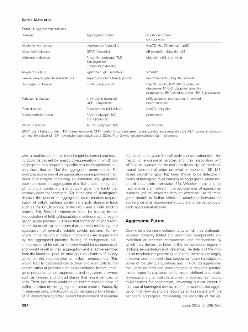
Table1: Aggresomal diseases
Disease Aggregated protein Additional proteincomponents
Alcoholic liver disease cytokeratins (cytosolic) Hsp70, Hsp25, ubiquitin, p62
Alexander’s disease GFAP (cytosolic) aB-crystallin, ubiquitin, p62
Alzheimer’s disease Presenilin (polytopic TM) ubiquitin, p62, b-amyloidTau (cytosolic),b-amyloid (cytosolic)
Amyloidosis (LC) light chain IgG (secretory) vimentin
Familial amyotrophic lateral sclerosis superoxide dismutase (cytosolic) neurofilaments, ubiquitin, vimentin
Huntington’s disease huntingtin (cytosolic) Hsp70, Hsp40, BiP/GRP78, potentialchaperone 14-3-3, ubiquitin, vimentin,proteasome, RNA-binding protein TIA-1, a-synuclein
Parkinson’s disease a-synuclein (cytosolic) p62, ubiquitin, proteasome, b-amyloidUCH-LI (cytosolic) neurofilaments
Prion diseases Prion protein (GPI-linked) Hsc70, ubiquitin
Spinocerebellar ataxia SCAs (polytopic TM) proteasomeaxins (cytosolic)
Wilson’s disease ATP7B (polytopic TM) cytokeratins
GFAP: glial fibrillary protein; TM: transmembrane; CFTR: cystic fibrosis transmembrane conductance regulator; UCH-L1: ubiquitin carboxy-terminal hydrolase L1; GPI: glycosylphosphatidylinositol; SCAs: P or Q-type voltage-sensitive Caππ channels.
vivo, a modification of this model might be correct and toxic-ity could be caused by ‘analog co-aggregation’ in which co-aggregation may sequester specific cellular components, butonly those that are ‘like’ the aggregation-prone protein. Forexample, expression of an aggregation prone protein (a frag-ment of huntingtin containing an extended poly glutaminetrack) promotes the aggregation of a ‘like’ protein (a fragmentof huntingtin containing a short poly glutamine track) thatnormally does not aggregate (52). In the case of Huntington’sdisease, this type of co-aggregation could mediate seques-tration of cellular proteins containing a poly glutamine tracksuch as the CREB-binding protein (53) and a TATA bindingprotein (54). Second, cytotoxicity could be caused by thesequestration of folding/degradative machinery by the aggre-gation-prone proteins. It is likely that formation of aggresom-es results in cellular conditions that promote misfolding andaggregation of normally soluble cellular proteins. For ex-ample, if the majority of cellular chaperones are sequesteredby the aggregated proteins, folding of endogenous sub-strates essential for cellular function would be compromised,and would result in their aggregation and effective removalfrom the functional pool. An analogous mechanism of toxicitycould be the sequestration of cellular proteasomes. Thiswould lead to decreased degradation and eventually to theaccumulation of proteins such as transcription factors, onco-gene products, tumor suppressors and regulatory enzymessuch as kinases and phosphatases that might be toxic tocells. Third, cell death could be an indirect consequence oftraffic inhibition by the aggregation-prone proteins. Especiallyin neuronal cells, cytotoxicity could be caused by inhibitionof MT-based transport that is used for movement of essential
394 Traffic 2002; 3: 388–396
components between the cell body and cell extremities. For-mation of aggresomal particles and their association withMTs could override the neuron’s ability for dynein-mediatedaxonal transport of other essential components (18). MT-based axonal transport has been shown to be defective inaxons of transgenic mice carrying an aggregation-prone mu-tant of superoxide dismutase (55). Whether these or othermechanisms are involved in the pathogenesis of aggresomaldiseases will be answered through extensive use of trans-genic models to further refine the correlation between theappearance of an aggresomal structure and the pathology ofeach aggresomal disease.
Aggresome Future
Clearly, cells contain mechanisms by which they distinguishbetween correctly folded and assembled components andmisfolded or defective components, and mechanisms bywhich they deliver the latter to the peri-centriolar region tofacilitate sequestration and clearance. The details of the mol-ecular mechanisms governing each of these steps are largelyunknown and represent clear targets for future investigation.Some of the obvious questions are: (i) How do aggresomalmini-particles form and what therapeutic reagents (confor-mation specific peptides, conformation-defined chemicals,biological and chemical chaperones), or approaches (routingto lysosomes for degradation, preventing nuclear import inthe case of huntingtin) can be used to prevent or alter aggre-gation? (ii) How do motors recognize and associate with theperipheral aggregates, considering the variability of the ag-

Aggresomal Pathway for Protein Degradation
gregated substrates? (iii) What are the signaling pathwaysresponsible for chaperone and proteasome recruitment to theaggresome, and can they be amplified to facilitate degrada-tion or initiated early to facilitate degradation of aggresomalparticles before they have a chance to form aggresomes? (iv)What are the signaling pathways involved in IF rearrange-ments, and are changes in IF architecture relevant for patho-genesis or a benign consequence of aggresome formation?(v) What are the signaling pathways regulating autophagy,and can the process be selectively amplified (by biological orchemical means) for disposal of aggresomal deposits? (vi)What are the global transcriptional and translational changesthat accompany aggresome formation, how do they affectcell function, and can they be regulated to negate the pro-duction of harmful components? Answers to these questionsare likely to significantly enhance our ability to treat confor-mational diseases and provide hope for those at risk andthose affected with such diseases.
Acknowledgments
We apologize to those whose work could not be cited due to space limi-tations. We are grateful to Dr Jim Collawn, Dr Gerry Fuller, Dr StephenFuller for critically reading the manuscript.
References
1. Jaenicke R. Protein folding. local structures, domains, subunits, andassemblies. Biochemistry 1991;30:3147–3161.
2. Wetzel R. Mutations and off-pathway aggregation of proteins. TrendsBiotechnol 1994;12:193–198.
3. Zimmerman SB, Trach SO. Estimation of macromolecule concen-trations and excluded volume effects for the cytoplasm of Escherichia
coli. J Mol Biol 1991;222:599–620.4. Ellis RJ, Hartl FU. Principles of protein folding in the cellular environ-
ment. Curr Opin Struct Biol 1999;9:102–110.5. Minton AP. Implications of macromolecular crowding for protein as-
sembly. Curr Opin Struct Biol 2000;10:34–39.6. Bonifacino JS, Suzuki CK, Lippincott-Schwartz J, Weissman AM,
Klausner RD. Pre-Golgi degradation of newly synthesized T-cell anti-gen receptor chains: intrinsic sensitivity and the role of subunit as-sembly. J Cell Biol 1989;109:73–83.
7. Hurle MR, Helms LR, Li L, Chan W, Wetzel R. A role for destabilizingamino acid replacements in light-chain amyloidosis. Proc Natl AcadSci USA 1994;91:5446–5450.
8. Wickner S, Maurizi MR, Gottesman S. Posttranslational quality control.folding, refolding, and degrading proteins. Science 1999;286:1888–1893.
9. Netzer WJ, Hartl FU. Protein folding in the cytosol: chaperonin-de-pendent and -independent mechanisms. Trends Biochem Sci1998;23:68–73.
10. Hartl FU. Molecular chaperones in cellular protein folding. Nature1996;381:571–579.
11. Weissman JS, Rye HS, Fenton WA, Beechem JM, Horwich AL. Char-acterization of the active intermediate of a GroEL-GroES-mediatedprotein folding reaction. Cell 1996;84:481–490.
12. Thulasiraman V, Yang CF, Frydman J. In vivo newly translated polypep-tides are sequestered in a protected folding environment. EMBO J1999;18:85–95.
395Traffic 2002; 3: 388–396
13. Hershko A, Ciechanover A. The ubiquitin system. Annu Rev Biochem1998;67:425–479.
14. Hirsch C, Ploegh HL. Intracellular targeting of the proteasome. TrendsCell Biol 2000;10:268–272.
15. Ellgaard L, Helenius A. ER quality control: towards an understandingat the molecular level. Curr Opin Cell Biol 2001;13:431–437.
16. Bonifacino JS, Weissman AM. Ubiquitin and the control of proteinfate in the secretory and endocytic pathways. Annu Rev Cell Dev Biol1998;14:19–57.
17. Garcia-Mata R, Bebok Z, Sorscher EJ, Sztul ES. Characterization anddynamics of aggresome formation by a cytosolic GFP-chimera. J CellBiol 1999;146:1239–1254.
18. Johnston JA, Dalton MJ, Gurney ME, Kopito RR. Formation of highmolecular weight complexes of mutant Cu, Zn-superoxide dismutasein a mouse model for familial amyotrophic lateral sclerosis. Proc NatlAcad Sci USA 2000;97:12571–12576.
19. Johnston JA, Ward CL, Kopito RR. Aggresomes: A cellular responseto misfolded proteins. J Cell Biol 1998;143:1883–1898.
20. Presley JF, Cole NB, Schroer TA, Hirschberg K, Zaal KJ, Lippincott-Schwartz J. ER-to-Golgi transport visualized in living cells. Nature1997;389:81–85.
21. Goodson HV, Valetti C, Kreis TE. Motors and membrane traffic. CurrOpin Cell Biol 1997;9:18–28.
22. Li SH, Gutekunst CA, Hersch SM, Li XJ. Interaction of huntingtin-as-sociated protein with dynactin P150Glued. J Neurosci 1998;18:1261–1269.
23. Kuemmerle S, Gutekunst CA, Klein AM, Li XJ, Li SH, Beal MF, HerschSM, Ferrante RJ. Huntington aggregates may not predict neuronaldeath in Huntington’s disease. Ann Neurol 1999;46:842–849.
24. Gutekunst CA, Li SH, Yi H, Mulroy JS, Kuemmerle S, Jones R, Rye D,Ferrante RJ, Hersch SM, Li XJ. Nuclear and neuropil aggregates inHuntington’s disease: relationship to neuropathology. J Neurosci1999;19:2522–2534.
25. Cogne M, Silvain C, Khamlichi AA, Preud’homme JL. Structurally ab-normal immunoglobulins in human immunoproliferative disorders.Blood 1992;79:2181–2195.
26. Buxbaum J. Mechanisms of disease: Monoclonal immunoglobulin de-position. Amyloidosis, light chain deposition disease, and light andheavy chain deposition disease. Hematol Oncol Clin North Am1992;6:323–346.
27. Dul JL, Davis DP, Williamson EK, Stevens FJ, Argon Y. Hsp70 andantifibrillogenic peptides promote degradation and inhibit intracellularaggregation of amyloidogenic light chains. J Cell Biol 2001;152:705–716.
28. Zatloukal K, Stumptner C, Lehner M, Denk H, Baribault H, Eshkind LG,Franke WW. Cytokeratin 8 protects from hepatotoxicity, and its ratioto cytokeratin 18 determines the ability of hepatocytes to form Mallorybodies. Am J Pathol 2000;156:1263–1274.
29. Harada M, Sakisaka S, Terada K, Kimura R, Kawaguchi T, Koga H, KimM, Taniguchi E, Hanada S, Suganuma T, Furuta K, Sugiyama T, SataM. A mutation of the Wilson disease protein, ATP7B, is degraded inthe proteasomes and forms protein aggregates. Gastroenterology2001;120:967–974.
30. Wigley WC, Fabunmi RP, Lee MG, Marino CR, Muallem S, DeMartinoGN, Thomas PJ. Dynamic association of proteasomal machinery withthe centrosome. J Cell Biol 1999;145:481–490.
31. Ito H, Kamei K, Iwamoto I, Inaguma Y, Nohara D, Kato K. Phosphoryla-tion-induced change of the oligomerization state of alpha B-crystallin.J Biol Chem 2001;276:5346–5352.
32. Souza JM, Giasson BI, Lee VM, Ischiropoulos H. Chaperone-like activ-ity of synucleins. FEBS Lett 2000;474:116–119.
33. Waelter S, Boeddrich A, Lurz R, Scherzinger E, Lueder G, Lehrach H,Wanker EE. Accumulation of mutant huntingtin fragments in aggre-some-like inclusion bodies as a result of insufficient protein degrada-tion. Mol Biol Cell 2001;12:1393–1407.
Garcia-Mata et al.
34. Muchowski PJ, Schaffar G, Sittler A, Wanker EE, Hayer-Hartl MK, HartlFU. Hsp70 and hsp40 chaperones can inhibit self-assembly of polyg-lutamine proteins into amyloid-like fibrils. Proc Natl Acad Sci USA2000;97:7841–7846.
35. Lee HJ, Shin SY, Choi C, Lee YH, Lee SJ. Formation and removal ofalpha-synuclein aggregates in cells exposed to mitochondrial inhibi-tors. J Biol Chem 2002;277:5411–5417.
36. Fabunmi RP, Wigley WC, Thomas PJ, DeMartino GN. Activity andregulation of the centrosome-associated proteasome. J Biol Chem2000;275:409–413.
37. Ward CL, Omura S, Kopito RR. Degradation of CFTR by the ubiquitin-proteasome pathway. Cell 1995;83:121–127.
38. Mortimore GE, Miotto G, Venerando R, Kadowaki M, Autophagy. Sub-cell Biochem 1996;27:93–135.
39. Wojcik C, Schroeter D, Wilk S, Lamprecht J, Paweletz N. Ubiquitin-mediated proteolysis centers in HeLa cells: Indication from studies ofan inhibitor of the chymotrypsin-like activity of the proteasome. Eur JCell Biol 1996;71:311–318.
40. Meriin AB, Gabai VL, Yaglom J, Shifrin VI, Sherman MY. Proteasomeinhibitors activate stress kinases and induce Hsp72. Diverse effectson apoptosis. J Biol Chem 1998;273:6373–6379.
41. Kyriakis JM, Avruch J. Protein kinase cascades activated by stress andinflammatory cytokines. Bioessays 1996;18:567–577.
42. Manganas LN, Akhtar S, Antonucci DE, Campomanes CR, Dolly JO,Trimmer JS. Episodic ataxia type-1 mutations in the Kv1.1 potassiumchannel display distinct folding and intracellular trafficking properties.J Biol Chem 2001;276:49427–49434.
43. Mayer RJ, Lowe J, Lennox G, Landon M, MacLennan K, Doherty FJ.Intermediate filament-ubiquitin diseases: Implications for cell sanitiz-ation. Biochem Soc Symp 1989;55:193–201.
44. Lowe J, Mayer J, Landon M, Layfield R. Ubiquitin and the molecularpathology of neurodegenerative diseases. Adv Exp Med Biol2001;487:169–186.
45. Durham HD, Roy J, Dong L, Figlewicz DA. Aggregation of mutantCu/Zn superoxide dismutase proteins in a culture model of ALS. JNeuropathol Exp Neurol 1997;56:523–530.
396 Traffic 2002; 3: 388–396
46. Bruijn LI, Houseweart MK, Kato S, Anderson KL, Anderson SD, OhamaE, Reaume AG, Scott RW, Cleveland DW. Aggregation and motor neu-ron toxicity of an ALS-linked SOD1 mutant independent from wild-type SOD1. Science 1998;281:1851–1854.
47. Masliah E, Rockenstein E, Veinbergs I, Mallory M, Hashimoto M, Take-da A, Sagara Y, Sisk A, Mucke L. Dopaminergic loss and inclusionbody formation in alpha-synuclein mice: Implications for neurode-generative disorders. Science 2000;287:1265–1269.
48. Lin CH, Tallaksen-Greene S, Chien WM, Cearley JA, Jackson WS,Crouse AB, Ren S, Li XJ, Albin RL, Detloff PJ. Neurological abnormali-ties in a knock-in mouse model of Huntington’s disease. Hum MolGenet 2001;10:137–144.
49. Davies SW, Turmaine M, Cozens BA, DiFiglia M, Sharp AH, Ross CA,Scherzinger E, Wanker EE, Mangiarini L, Bates GP. Formation of neur-onal intranuclear inclusions underlies the neurological dysfunction inmice transgenic for the HD mutation. Cell 1997;90:537–548.
50. Hackam AS, Wellington CL, Hayden MR. The fatal attraction of polyg-lutamine-containing proteins. Clin Genet 1998;53:233–242.
51. Zoghbi HY, Orr HT. Glutamine repeats and neurodegeneration. AnnuRev Neurosci 2000;23:217–247.
52. Rajan RS, Illing ME, Bence NF, Kopito RR. Specificity in intracellularprotein aggregation and inclusion body formation. Proc Natl Acad SciUSA 2001;98:13060–13065.
53. Nucifora FC Jr, Sasaki M, Peters MF, Huang H, Cooper JK, YamadaM, Takahashi H, Tsuji S, Troncoso J, Dawson VL, Dawson TM, RossCA. Interference by huntingtin and atrophin-1 with cbp-mediated tran-scription leading to cellular toxicity. Science 2001;291:2423–2428.
54. Huang CC, Faber PW, Persichetti F, Mittal V, Vonsattel JP, MacDonaldME, Gusella JF. Amyloid formation by mutant huntingtin: Threshold,progressivity and recruitment of normal polyglutamine proteins. Som-at Cell Mol Genet 1998;24:217–233.
55. Zhang B, Tu P, Abtahian F, Trojanowski JQ, Lee VM. Neurofilamentsand orthograde transport are reduced in ventral root axons of trans-genic mice that express human SOD1 with a G93A mutation. J CellBiol 1997;139:1307–1315.